EP1751181B1 - BINDING MOIETIES BASED ON SHARK IgNAR DOMAINS - Google Patents
BINDING MOIETIES BASED ON SHARK IgNAR DOMAINS Download PDFInfo
- Publication number
- EP1751181B1 EP1751181B1 EP05744734A EP05744734A EP1751181B1 EP 1751181 B1 EP1751181 B1 EP 1751181B1 EP 05744734 A EP05744734 A EP 05744734A EP 05744734 A EP05744734 A EP 05744734A EP 1751181 B1 EP1751181 B1 EP 1751181B1
- Authority
- EP
- European Patent Office
- Prior art keywords
- loop
- misc
- feature
- residues
- domain
- Prior art date
- Legal status (The legal status is an assumption and is not a legal conclusion. Google has not performed a legal analysis and makes no representation as to the accuracy of the status listed.)
- Expired - Lifetime
Links
Images


Classifications
-
- C—CHEMISTRY; METALLURGY
- C07—ORGANIC CHEMISTRY
- C07K—PEPTIDES
- C07K14/00—Peptides having more than 20 amino acids; Gastrins; Somatostatins; Melanotropins; Derivatives thereof
- C07K14/435—Peptides having more than 20 amino acids; Gastrins; Somatostatins; Melanotropins; Derivatives thereof from animals; from humans
- C07K14/46—Peptides having more than 20 amino acids; Gastrins; Somatostatins; Melanotropins; Derivatives thereof from animals; from humans from vertebrates
- C07K14/461—Peptides having more than 20 amino acids; Gastrins; Somatostatins; Melanotropins; Derivatives thereof from animals; from humans from vertebrates from fish
-
- C—CHEMISTRY; METALLURGY
- C07—ORGANIC CHEMISTRY
- C07K—PEPTIDES
- C07K16/00—Immunoglobulins [IG], e.g. monoclonal or polyclonal antibodies
- C07K16/46—Hybrid immunoglobulins
- C07K16/461—Igs containing Ig-regions, -domains or -residues form different species
- C07K16/464—Igs containing CDR-residues from one specie grafted between FR-residues from another
-
- A—HUMAN NECESSITIES
- A61—MEDICAL OR VETERINARY SCIENCE; HYGIENE
- A61P—SPECIFIC THERAPEUTIC ACTIVITY OF CHEMICAL COMPOUNDS OR MEDICINAL PREPARATIONS
- A61P43/00—Drugs for specific purposes, not provided for in groups A61P1/00-A61P41/00
-
- C—CHEMISTRY; METALLURGY
- C07—ORGANIC CHEMISTRY
- C07K—PEPTIDES
- C07K16/00—Immunoglobulins [IG], e.g. monoclonal or polyclonal antibodies
-
- C—CHEMISTRY; METALLURGY
- C07—ORGANIC CHEMISTRY
- C07K—PEPTIDES
- C07K2299/00—Coordinates from 3D structures of peptides, e.g. proteins or enzymes
-
- C—CHEMISTRY; METALLURGY
- C07—ORGANIC CHEMISTRY
- C07K—PEPTIDES
- C07K2317/00—Immunoglobulins specific features
- C07K2317/20—Immunoglobulins specific features characterized by taxonomic origin
-
- C—CHEMISTRY; METALLURGY
- C07—ORGANIC CHEMISTRY
- C07K—PEPTIDES
- C07K2317/00—Immunoglobulins specific features
- C07K2317/20—Immunoglobulins specific features characterized by taxonomic origin
- C07K2317/24—Immunoglobulins specific features characterized by taxonomic origin containing regions, domains or residues from different species, e.g. chimeric, humanized or veneered
-
- C—CHEMISTRY; METALLURGY
- C07—ORGANIC CHEMISTRY
- C07K—PEPTIDES
- C07K2317/00—Immunoglobulins specific features
- C07K2317/50—Immunoglobulins specific features characterized by immunoglobulin fragments
- C07K2317/56—Immunoglobulins specific features characterized by immunoglobulin fragments variable (Fv) region, i.e. VH and/or VL
-
- C—CHEMISTRY; METALLURGY
- C07—ORGANIC CHEMISTRY
- C07K—PEPTIDES
- C07K2317/00—Immunoglobulins specific features
- C07K2317/50—Immunoglobulins specific features characterized by immunoglobulin fragments
- C07K2317/56—Immunoglobulins specific features characterized by immunoglobulin fragments variable (Fv) region, i.e. VH and/or VL
- C07K2317/565—Complementarity determining region [CDR]
Definitions
- the present invention relates to immunoglobulin new antigen receptors (IgNARs) from fish and uses thereof.
- IgNARs immunoglobulin new antigen receptors
- the present invention relates to modified IgNAR variable domains and to domains from members of the immunoglobulin superfamily that have been modified to include structural features derived from IgNAR variable domains.
- immunoglobulin superfamily includes immunoglobulins and numerous other cell surface and soluble molecules that mediate recognition, adhesion or binding functions in the immune system. They share partial amino acid sequence homology and tertiary structural features that were originally identified in immunoglobulin (Ig) heavy and light chains.
- Molecules of the IgSF are identified by a characteristic IgSF fold structure, a ⁇ -sandwich structure formed by two ⁇ -sheets, packed face-to-face and linked by a disulphide bond between the B and F strands (Bork 1994; Chothia 1998).
- IgSF frameworks are further classified into 3-4 major categories, the Variable (V)-, Constant (C)-, I- and 12-sets, based on ⁇ -strand number, configuration and hydrogen bond patterns (Bork 1994; Cassasnovas 1998).
- T-cell receptors are heterodimers having ⁇ and ⁇ chains of equal size, each chain consisting of an N-terminal variable domain (V ⁇ or V ⁇ ) and a constant domain.
- variable domains on different polypeptide chains interact across hydrophobic interfaces to form binding sites designed to receive a particular target molecule.
- each pair of VH/VL domains form an antigen binding site, the CH and CL domains not being directly involved in binding the antibody to the antigen.
- the V ⁇ and V ⁇ domains form the binding site for target molecules, namely peptides presented by a histocompatibility antigen.
- variable domains vary particularly from one molecule to another. This variation in sequence enables the molecules to recognise an extremely wide variety of target molecules.
- Variable domains are often viewed as comprising four framework regions, whose sequences are relatively conserved, connected by three hypervariable or complementarity determining loop regions (CDRs) (Kabat 1983 & 1987; Bork 1994). The CDRs are held in close proximity by the framework regions and, with the CDRs from the other variable domain, contribute to the formation of the binding site.
- CDRs hypervariable or complementarity determining loop regions
- MAbs monoclonal antibodies
- chimeric antibodies have been constructed in which an animal antigen-binding variable domain is coupled to a human constant domain.
- the isotype of the human constant domain may be selected to tailor the chimeric antibody for participation in antibody-dependent cellular cytotoxicity (ADCC) and complement dependent cytotoxicity.
- ADCC antibody-dependent cellular cytotoxicity
- chimeric antibodies typically may contain about one third rodent (or other non-human species) sequence and consequently are often still capable of eliciting a significant antigenic response in humans.
- the TCR has two variable domains that combine into a structure similar to the Fv module of an antibody that results from combination of the VH and VL domains.
- Novotny (1991) described how the V ⁇ and V ⁇ domains of the TCR can be fused and expressed as a single chain polypeptide and, further, how to alter surface residues to reduce the hydrophobicity directly analogous to an antibody scFv.
- Other publications describe the expression characteristics of single-chain TCRs comprising two V ⁇ and V ⁇ domains (Wulfing 1994; Ward 1991).
- the three-dimensional crystal structures have been published for intact immunoglobulins, a variety of immunoglobulin fragments, antibody-antigen complexes and for other IgSF molecules such as the TCR. It is known that the function of IgSF molecules is dependent on their three dimensional structure, and that amino acid substitutions can change the three-dimensional structure of, for example, an antibody (Snow and Amzel 1986). Based upon molecular modelling, it has been shown that the antigen binding affinity of a humanized antibody can be increased by mutagenesis (Riechmann 1988; Queen 1989).
- IgNARs Immunoglobulin New Antigen Receptors
- C NAR s constant domains
- V NAR variable domain
- IgNARs have been identified in all shark species studied to date. In particular, IgNARs have been identified in the serum of nurse sharks Ginglymostoma cirratum (Greenberg 1995) and wobbegong sharks Orectolobus maculatus (Nuttall 2001). The cell-surface expression of IgNARs has also been reported (Rumfelt 2002). Research has implicated IgNARs as true molecules of the immune armoury, and as the most probable agents of the shark antigen-driven affinity-maturation antibody response (Diaz 1999; Nuttall 2002; Dooley 2003).
- Type 1 V NAR topology is characterised by an extra framework disulphide linkage and, usually, cysteines in the extended loop region analogous to a conventional CDR3 loop, which it has been suggested may form intra-loop disulphide bonds.
- Type 2 V NAR topology is characterised by cysteines in the loop regions analogous to conventional CDR1 and CDR3 loops in approximately two thirds of cases, which it has been postulated may form inter-loop disulphide bonds.
- Type 3 V NAR topology is characterised by a relatively constant sized loop region analogous to a conventional CDR3 loop of limited diversity and a characteristic conserved tryptophan residue within the loop region analogous to a CDR1 loop.
- IgNARs identified to date are reported as having minimally variable loop regions analogous to conventional CDR1 and CDR2 loops, with diversity being concentrated in an elongated loop region analogous to a conventional CDR3 loop (Greenberg 1995; Nuttall 2001; Diaz 2002).
- the elongated loop region can reportedly vary in length from 5 to 23 residues in length, though the modal classes are more in the order of 15 to 17 residues (Nuttall 2003). This is significantly larger than for conventional murine and human antibodies, but approximate to the extended CDR3 loops found in camelid single VH antibodies (Wu 1993; Muyldermans 1994).
- the present invention provides a method of altering a property of an IgNAR variable domain comprising: (a) providing an IgNAR variable domain comprising eight ⁇ -strand regions, designated A, A', B, C, D, E, F and G according to Figure 2 , and nine loop regions, designated 1 to 9 according to Figure 2 ; (b) modifying loop region 8, which is a CDR3 analogous region, by addition of up to 3 amino acids or the deletion of up to 3 amino acid residues; and (c) identifying modifications that result in one or more of the following properties being altered: (i) the binding affinity of the IgNAR variable domain is increased by at least 5% compared to the unmodified domain; (ii) the solubility of the IgNAR variable domain being improved by at least 5% compared to the unmodified domain; and (iii) the stability of the IgNAR variable domain being improved by at least 5% compared to the unmodified domain.
- hydrophobic residues or “nonpolar residues” as used herein is meant valine, leucine, isoleucine, methionine, phenylalanine, tyrosine, and tryptophan.
- polar residues herein is meant serine, threonine, histidine, aspartic acid, asparagine, glutamic acid, glutamine, arginine, and lysine.
- V NAR s IgNAR variable domains targeting the apical membrane antigen-1 (AMAl) of Plasmodium falciparum malarial parasites.
- AMAl apical membrane antigen-1
- 12Y-1 SEQ ID NOs: 1 & 2
- 12Y-2 SEQ ID NOs: 3 & 4
- V NAR 12A-9 (SEQ ID NOs: 9 & 10) was isolated from a library containing a broad mixture of naturally occurring Type 2 V NAR framework scaffolds derived from the wobbegong shark by biopanning against the Gingipain K protease from Porphyromonas gingivalis (Nuttall et al. 2002).
- This IgNAR is completely natural, including the CDR3 analogous loop region (loop region 8).
- a disulphide bridge links and stabilises the CDR1 and CDR3 analogous loop regions, in this case connecting residues Cys29 and Cys89.
- V NAR 1A-7 (SEQ ID NOs: 5 & 6) is specific for the anti-AMA-1 mouse IgG 5G8 and was isolated using the same procedure as described for the isolation of 12Y-1 and 12Y-2, i.e. biopanning of the phage displayed IgNAR library. Binding is through an "SYP" motif found in the 1A-7 "synthetic" CDR3 analogous loop region (i.e. loop region 8).
- 12A-9 is a naturally occurring shark IgNAR and has a loop region of length 13 residues analogous to a conventional CDR3 loop.
- Figure 1 presents the sequence alignments of 12Y-1, 12Y-2, 12A-9, 1A-7 and twelve naturally occurring Type 2 V NAR s: 7E-22 (SEQ ID NO: 17), 7E-23 (SEQ ID NO: 18), 7E-51 (SEQ ID NO: 19), 7E-54 (SEQ ID NO: 20), 7E-56 (SEQ ID NO: 21), 7E-58 (SEQ ID NO: 22), 7E-68 (SEQ ID NO: 23), 7E-77 (SEQ ID NO: 24), 7E-80 (SEQ ID NO: 25), 7E-87 (SEQ ID NO: 26), 7E-91 (SEQ ID NO: 27), 7E-93 (SEQ ID NO: 28).
- 7E-22 SEQ ID NO: 17
- 7E-23 SEQ ID NO: 18
- 7E-51 SEQ ID NO: 19
- 7E-54 SEQ ID NO: 20
- 7E-56 SEQ ID NO: 21
- 7E-58 SEQ ID NO: 22
- Table 1 shows the amino acid variation across the sixteen Type 2 V NAR s sequences in Figure 1 and the Type 2 V NAR s sequences reported by Nuttall (2002 & 2003). It is evident from Figure 1 and Table 1 that there is a large degree of conservation of sequence outside of the loop regions analogous to conventional CDR1 and CDR3 loops.
- 12Y-1 and 12Y-2 V NAR s were expressed in Escherichia coli and placed into crystallization trials in the presence or absence of AMA1 antigen. No crystal leads were observed in the presence of antigen, possibly due to the intrinsically flexible domain structure of the AMA1 protein (Hodder 1996). In contrast, good quality crystals were obtained for both 12Y-1 (space group I4 1 22) and 12Y-2 (space group I2 1 2 1 2 1 ) in the absence of antigen.
- 12A-9 and 1A-7 V NAR s were expressed in Escherichia coli and placed into crystallization trials. Successful conditions were scaled up and diffraction quality crystals obtained for both 12A-9 (space group P2 1 2 1 2) and 1A-7 (space group I2 1 2 1 2 1 ).
- the 12Y-1 asymmetric unit contains one molecule
- the 12Y-2 crystal asymmetric units contain two molecules (Chains A; B); the relative disposition of these two 12Y-2 monomers requires rotation by 176.2° and screw translation by -1.1 ⁇ to overlay the C ⁇ atoms.
- the final 12Y-2 structure was refined to 2.18 ⁇ resolution, with 93.4% of residues in the most favoured regions of the Ramachandran plot with no residues in the generously allowed or disallowed regions. Details of the diffraction data and refinement statistics are presented in Table 2.
- the coordinates for 12Y-1 and 12Y-2 are attached as Appendix I(a) and (b) respectively.
- the structures of 12A-9 and 1A-7 were determined by molecular replacement.
- the search model for 12A-9 was the 12Y-1 structure (above) without the CDR3 loop.
- the search model for 1A-7 was the 12Y-1 two-fold dimer structure without the CDR3 loops.
- 88.4% of the residues are in the most favoured regions of the Ramachandran plot, with one residue in the generously allowed or disallowed regions.
- 90.9% of the residues are in the most favoured regions of the Ramachandran plot, with two residues for chain C in the generously allowed or disallowed regions. Details of the diffraction data and refinement statistics are presented in Table 2.
- the coordinates for 12A-9 and 1A-7 are attached as Appendix I(c) and (d), respectively.
- the IgNAR domain structures set out in Appendices I(a), (b), (c) and (d) are monomer structures. This is the first time that a monomer has been observed crystallographically for an IgNAR variable domain.
- the third column denotes the atom type, the fourth column the residue type, the fifth column the chain identification, the sixth column the residue number (the atom numbering as described in Hong (2000)), the seventh, eighth and ninth columns the X, Y, Z coordinates, respectively, of the atom in question, the tenth column the occupancy of the atom, the eleventh column the temperature factor of the atom, and the last the atom type.
- Each of the Appendices is presented in an internally consistent format.
- the coordinates of the atoms of each amino acid residue are listed such that the backbone nitrogen atom is first, followed by the C- ⁇ backbone carbon atom, designated CA, followed by the carbon and oxygen of the protein backbone and finally side chain residues (designated according to one standard convention).
- Alternative file formats e.g. such as a format consistent with that of the EBI Macromolecular Structure Database (Hinxton, UK) which may include a different ordering of these atoms, or a different designation of the side-chain residues, may be used or preferred by others of skill in the art.
- IgNAR variable domains that are important for antigen binding or solubility/stability of these domains. These features can be introduced into domains of other members of the IgSF (for example, I-set or V-set domains) in order to alter binding properties or to improve solubility and/or stability.
- the information presented in Appendix I can be used, for example, to compare structures of IgSF domains that have been modified so as to more closely resemble the structure of IgNAR variable domains.
- Root mean square deviation measures the difference in positioning in space between two sets of atoms.
- root mean square deviation we mean the square root of the arithmetic mean of the squares of the deviations from the mean.
- the r.m.s.d. measures distance between equivalent atoms after their optimal superposition.
- the r.m.s.d. can be calculated over all atoms, over residue backbone atoms (i.e. the nitrogen-carbon-carbon backbone atoms of the protein amino acid residues), main chain atoms only (i.e.
- the nitrogen-carbon-oxygen-carbon backbone atoms of the protein amino acid residues can be calculated over any of these, using any of the methods outlined below.
- the user can define the residues in the two proteins that are to be paired for the purpose of the calculation.
- the pairing of residues can be determined by generating a sequence alignment of the two proteins.
- the atomic coordinates can then be superimposed according to this alignment and an r.m.s.d.. value calculated.
- the program Sequoia (Bruns 1999) performs the alignment of homologous protein sequences, and the superposition of homologous protein atomic coordinates. Once aligned, the r.m.s.d.. can be calculated using programs detailed above. For sequence identical, or highly identical, the structural alignment of proteins can be done manually or automatically as outlined above. Another approach would be to generate a superposition of protein atomic coordinates without considering the sequence.
- Varying the atomic positions of the atoms of the structure by up to about 0.5 ⁇ in a concerted way, preferably up to about 0.3 ⁇ in any direction will result in a structure which is substantially the same as the structure of Appendix I(a) or (b) in terms of both its structural characteristics and utility e.g. for molecular structure-based analysis.
- the structures comprise eight ⁇ -strand regions, designated A, A', B, C, D, E, F and G according to Figure 2 , and nine loop regions, designated 1 to 9 according to Figure 2 .
- a "loop region” is a portion of peptide sequence that extends either from or between a ⁇ -strand conformation or ⁇ -strand conformations.
- a " ⁇ -strand region” contains an extended ⁇ -strand conformation, i.e. ⁇ -strands comprising at least 4, preferably at least 5 amino acids. Loop regions are typically free of extended ⁇ -strand conformations but may include shortened ⁇ -strand conformations, i.e. ⁇ -strands comprising less than 4 amino acids. Apart from N- and C-terminal loop regions, the loop regions connect ⁇ -strands running in opposite directions.
- loop region 5 contains shortened ⁇ -strand conformations.
- loop region 8 may contain ⁇ -strand conformations, which may be either shortened or extended, and these are discussed in more detail below.
- no other loop regions contain ⁇ -strand conformations.
- the structures comprise 8 ⁇ -strand regions, designated A, A', B, C, D, E, F and G according to Figure 2 , and nine loop regions, designated 1 to 9 according to Figure 2 , in which loop region 5 comprises 2 shortened ⁇ -strand regions, designated D' and C', and 3 loop regions, designated 5a, 5b and 5c, according to Figure 3 .
- Loop region 4 is analogous to a conventional CDR1 loop.
- Loop region 8 is analogous to a conventional CDR3 loop.
- loop region 8 can contain a variable number of amino acids ranging from about 5 to 30, a default value of 18 is used on which to base the residue numbering for subsequent regions. Consequently, the residue numbering does not necessarily correlate with the total number of amino acid residues present in the sequence. It is based on loop region 8 having a default value of 18 amino acid residues.
- Loop region 5 comprises 2 shortened ⁇ -strand regions, designated D' and C', and 3 loop regions, designated 5a, 5b and 5c.
- V-set and I-set proteins have a typical kink in the first strand (A'), which allows the first part of the strand (A) to hydrogen bond to one part of the ⁇ -sandwich sheet and the second part (A') to the extended G strand of the other ⁇ -sheet.
- This first-strand kink is found in the V NAR proteins as depicted in Figure 4 . It starts with the highly conserved cis-proline (Pro7), the most typical residue in first-strand kinks of variable domains (Spada 1998).
- 12Y-1, 12Y-2, 12A-9 and 1A-7 also have bulges in the C terminal G strand (conserved Gly-Ala-Gly motif) and in the C' strand.
- 12Y-1, 12Y-2, 12A-9 and 1A-7 resemble I-set proteins in having a short C' strand (three H-bonds) and a very short C" strand (labelled as D' in Figure 3 ) which atypically switches from one ⁇ -sheet to the other, such that a single hydrogen bond links it to the D strand rather than the C' strand as in V-set domains.
- the 12Y-2 chain A and B (r.m.s.d. of 0.53 ⁇ for C ⁇ of 113 residues), and the 12Y-1 framework (r.m.s.d. of 0.72 ⁇ for C ⁇ of 100 residues) are closely related (see Figure 5 ).
- NCAMs N eural C ell A dhesion M olecules
- V H H domains found in the Camelidae may be expected to be similar to the V NAR structure, however, these single domain antibodies clearly arose from a mammalian IgG-like progenitor, successively acquiring solubilizing, stabilizing, and extensive CDR3 mutations (Nguyen 2002).
- the low structural homology between V H H and V NAR s is consistent with their very low sequence homology, and reflects convergent evolutionary solutions to the problem of achieving solvent solubility and binding affinity.
- FIG 8 the relative positions of the three classically defined antibody CDR or hypervariable loop regions are shown. Sequence alignments show IgNAR antibody variability confined to the loop regions 4 and 8 corresponding to conventional CDR1 and CDR-3 regions, and this is confirmed by our structural analysis where a loop region analogous to a typical "CDR2 loop" is missing and its bottom turn appears to be well separated from the antigen-binding face (or paratope). Sequence analysis also suggests that loop region 4 is the minor loop component, invariant in length and limited in diversity.
- the 12Y-2 CDR3 loop is present in two crystal forms, corresponding to chains A and B, and extends from residues Phe86 to Glu103.
- the chain A loop region 8 adopts a clear ⁇ -hairpin configuration with ⁇ -strands from Phe86-Leu89, and Leu98-Glu103, separated by a flexible loop (Pro90-Ser97).
- the ⁇ -hairpin extends even further into loop region 8 with residues Phe86-Asp93 and Tyr96-Glu103 involved in ⁇ -strand formation (see Figure 11 ).
- the ⁇ -hairpins are formed by the main chain hydrogen bonds ( ⁇ 3 ⁇ ): Tyr87 (O) - Phe100 (N); Tyr87 (N) - Phe100 (O); Leu89 (N) - Leu98 (O); Leu89 (O) - Leu98 (N), and, Asp93(O) - Tyr96(O) ( Figure 11 ).
- Additional H-bonds for loop region 8 of chain B are: Asp93 (N) - Tyr96 (O); Asp93 (O) - Tyr96 (N); and, Leu91 (O) - Tyr96 (O).
- the 12Y-2 loop region 8 extends outward and upward from the immunoglobulin framework, at the furthest point extending ⁇ 20 ⁇ above the conserved ⁇ -sheet framework, and tipped by the bulky side-chains of tyrosine residues at positions 94 and 96 (see Figures 8 and 11 ).
- extended antigen binding paratopes have been observed in but a limited number of antibodies, for example the camel anti-lysozyme V H H cAb-Lys3 (Desmyter 1996), and the H3 loop of human antibody b12, which penetrates deeply into the HIV gp120 binding cleft (Sapphire 2001).
- binding of 12Y-2 to the AMA1 target is probably mediated by the rigid ⁇ -hairpin, with increased access to the antigen mediated by flexibility at the bottom of the loop structure.
- the 12Y-1 loop region 8 is unresolved in the crystal structure and is probably highly flexible in solution around similar hinge-like residues.
- the aromatic loop region 8 residues of 12Y-2 are replaced with Arg87 and Pro98 in 12Y-1, reducing the stability of the loop region, i.e. the Phe29 of loop region 4 is now hydrogen bonded to Glu101 (Glu103 in 12Y-2) (Tyr29(OH) - Glu101(O ⁇ )) leaving the hinges of the loop region 8 unsupported.
- a comparison of the 12Y-2 and 12Y-1 dimeric forms shows that the interaction surface between the 2-fold monomers is not continuous and can be subdivide into three areas: (i) the main-chain ⁇ -sheet interactions between D strands; (ii) the interaction between loop regions 4; and (iii) the interactions between loop regions 8 (see Figure 13 and Table 4). While the contact between loop regions 8 in 12Y-2 is extensive, the dimeric arrangement is preserved in 12Y-1 crystals notwithstanding the more flexible and significantly distorted loop region 8, indicating that the conformation of loop region 8 is not absolutely required. Thus, the most significant dimer contacts are probably mediated by the loop regions 4 and especially by the D strands, where the main-chain interactions are independent of side-chain variation.
- heterodimeric immune receptors such as V H /V L antibodies and V ⁇ /V ⁇ TCRs
- the paired domains interact across a broad hydrophobic interface. This non-solvent exposed region is formed by a conserved patch of residues on the AGFCC' ⁇ -strand, with additional CDR3 interactions.
- IgSF-based cell surface receptors are single domains in solution and this face of the ⁇ -sandwich takes on a more charged/polar character.
- the hydrophobic region of inter-domain contact is immediately apparent for the TCR and antibody domains, centred around aromatic residues at the centre of the interface.
- the surface character is altered for V H H domains, for example by mutations Leu45Arg and Gly44Glu, to give a more charged character.
- the relatively short evolutionary time since the development of these single domain antibodies in the Camelidae mean that other solutions have also been adopted, for example the illustrated antibody where part of the loop region 8 (analogous to a conventional CDR3 loop) descends to partly cover the former V L interface, for example residues Asp121 and Tyr120.
- the central Tyr37 is well-conserved as an aromatic species across the immunoglobulin superfamily, and it and residues Gln84 and Arg101 also participate in forming a framework-CDR3 hydrogen bond network (Arg101(NH2) - Gln84(N ⁇ 2); Tyr87(OH) - Arg101(NH 2 ).
- the combined effect of these residues is to form a conserved charged pocket, which displays a high degree of solvent solubility such that it is ringed by water molecules in the crystal forms.
- the conservation of the Glu46, Lys82, and Lys104 trio suggests a stable and well-established face. A similar situation is observed in NCAM, where this face is dominated by the charged residues Lys76 and Glu88, and for Telokin, where a charged and polar interface is maintained by a combination of hydrogen bonds.
- V NAR proteins as, for example, therapeutic, diagnostic and bioarray reagents.
- V NAR proteins have potential to act as cleft-binding antibodies in which the ⁇ -hairpin structures are extended to form paratopes capable of penetrating otherwise cryptic antigenic sites.
- these proteins have a high degree of stability which offers significant advantages in terms of their manipulation and practical application.
- the present invention provides a method of altering a property of an IgNAR variable domain as defined in the appended claims .
- the IgNAR variable domain is modified such that a property of the IgNAR variable domains is altered.
- a property of an IgNAR variable domain, I-set domain or V-set domain is altered if any characteristic or attribute of the domain differs from the corresponding property of the unmodified domain.
- These properties include, but are not limited to, substrate specificity, substrate affinity, binding affinity, binding selectivity, catalytic activity, thermal stability, alkaline stability, pH activity profile, resistance to proteolytic degradation, kinetic association, kinetic dissociation, immunogenicity, ability to be secreted, ability to activate receptors, ability to treat disease, solubility, cytotoxic activity and oxidative stability.
- a property of an IgNAR variable domain, I-set domain or V-set domain is considered to be altered when the property exhibits at least a 5%, preferably at least 10%, more preferably at least a 20%, yet more preferably at least a 50%, and most preferably at least a 2-fold increase or decrease relative to the corresponding property in the unmodified domain.
- the solubility of the modified IgNAR variable domain, and concomitantly the binding moiety is altered, preferably improved, relative to the corresponding unmodified IgNAR variable domain.
- the stability of the IgNAR variable domain, and concomitantly the binding moiety is altered, preferably improved, relative to the corresponding unmodified IgNAR variable domain.
- altering the stability include changing one of the following properties:- thermal stability, alkaline stability, pH activity profile and resistance to proteolytic degradation.
- the binding characteristics of the IgNAR variable domain are altered relative to the corresponding unmodified IgNAR variable domain.
- altering the binding characteristics include changing one of the following properties: substrate specificity, substrate affinity, catalytic activity, kinetic association, kinetic dissociation, binding affinity and binding selectivity.
- Modifications can be prepared by introducing appropriate nucleotide changes into a nucleic acid of the present invention, or by in vitro synthesis of the desired polypeptide. Such mutants include deletions or insertions of residues within the amino acid sequence.
- the location of the mutation site and the nature of the mutation will depend on characteristic(s) to be modified.
- the sites for mutation can be modified individually or in series, for example by , (1) deleting the target residue, or (2) inserting other residues adjacent to the located site.
- unnatural amino acids or chemical amino acid analogues can be introduced into the polypeptides .
- Such amino acids include, but are not limited to, the D-isomers of the common amino acids, 2,4-diaminobutyric acid, ⁇ -amino isobutyric acid, 4-aminobutyric acid, 2-aminobutyric acid, 6-amino hexanoic acid, 2-amino isobutyric acid, 3-amino propionic acid, ornithine, norleucine, norvaline, hydroxyproline, sarcosine, citrulline, homocitrulline, cysteic acid, t-butylglycine, t-butylalanine, phenylglycine, cyclohexylalanine, ⁇ -alanine, fluoro-amino acids, designer amino acids such as ⁇ -methyl amino acids, C ⁇ -methyl amino acids, N ⁇ -methyl amino acids, and amino acid analogue
- chemically modified derivates of IgNAR variable domains which may provide advantages such as increasing stability and circulating time of the polypeptide, or decreasing immunogenicity (see U.S. Patent No. 4,179,337 ).
- the chemical moieties for derivatization may be selected from water-soluble polymers such as polyethylene glycol, ethylene glycol/propylene glycol copolymers, carboxymethylcellulose, dextran, polyvinyl alcohol and the like.
- variable domains of the present invention that are differentially modified during or after synthesis, for example, by biotinylation, benzylation, glycosylation, acetylation, phosphorylation, amidation, derivatization by known protecting/blocking groups, proteolytic cleavage, linkage to an antibody molecule or other cellular ligand, etc.
- the IgNAR variable domain may be modified at random positions within the molecule or at predetermined positions within the molecule and may include one, two, three or more attached chemical moieties. These modifications may, for example, serve to increase the stability and/or bioactivity of the modified domains of the invention.
- the IgNAR variable domains may also be modified by having C- or N-terminal truncations. However, the scope for such modifications is limited and it is preferred that no more than 8, preferably no more than 6 and more preferably no more than 4 residues be removed. Preferably there is no truncation at the N-terminal and more preferably there is no truncation at either the N- or C-terminals.
- Modified domains of the present invention can be produced in a variety of ways, including production and recovery of natural proteins, production and recovery of recombinant proteins, and chemical synthesis of the proteins.
- an isolated polypeptide of the present invention is produced by culturing a cell capable of expressing the polypeptide under conditions effective to produce the polypeptide, and recovering the polypeptide.
- the modification comprises insertion of 1 or 2 amino acids.
- the modification comprises deletion of 1 or 2 amino acids.
- the modification is also made to one or more amino acid residues within the patch defined by residues 33, 37, 46, 48, 50, 51, 59, 61, 86, 94, 95, 96, 98, 99 and 101 as shown in Table 1.
- loop region 8 of the IgNAR is modified, at least one of the ⁇ -strand regions or loop regions 1-3, 5-7 or 9 is also modified.
- At least one of ⁇ -strand regions C, D, E or F or loop regions 5, 6 or 7 has been modified. In a further preferred embodiment, at least one of ⁇ -strand regions C or D or loop region 5 has been modified. In a further preferred embodiment, loop region 5 has been modified.
- loop region 8 when the amino acid residues at the N-terminal and C-terminal ends of loop region 8 are each capable of adopting a ⁇ -strand configuration, loop region 8 is modified by deletion or addition, preferably by addition, of up to 3 amino acid within that part of the loop not capable of adopting the ⁇ -strand configuration.
- amino acid residues at the N-terminal and C-terminal ends of loop region 8 are capable of adopting ⁇ -strand configurations.
- loop region 8 is modified by deletion or addition, preferably by addition of up to 3 amino acid residues at the C- and/or N-terminal ends of the loop region to facilitate the adoption of ⁇ -strand configurations at the C- and/or N-terminal ends.
- loop region 8 is modified so as to facilitate the adoption of ⁇ -strand configurations at the C- and/or N-terminal ends of 2 to 10, preferably from 3 to 8, amino acid residues in length.
- the modification increases or decreases the binding characteristics, e.g. the affinity, of the modified IgNAR variable domain for a predetermined target molecule compared to the unmodified IgNAR variable domain.
- That part of the IgNAR variable domain which normally contacts a ligand (e.g. an antigen) or which appear, from the studies we have undertaken, to be available for interacting with a ligand (e.g. a receptor, enzyme etc.) are typically the solvent exposed regions of the IgNAR variable domain. In particular, they are generally made up of the surface exposed loops, and in particular loop regions 8 and 4 of the IgNAR variable domain.
- the unmodified IgNAR variable domain has had one or more loop regions modified.
- this can be achieved by replacing one or more solvent exposed loops of the IgNAR variable domain with one or more loops from the variable domains of other members of the IgSF.
- loop regions 4 and/or 8, or part thereof is modified, preferably replaced, by a corresponding loop structure (e.g. a CDR1 or CDR3 loop structure, respectively) from another molecule.
- Modifications can also be made to regions of the IgNAR variable domain that are not solvent exposed and/or which do not form part of a binding loop, e.g. the ⁇ strand regions.
- the modification increases or decreases the propensity for the IgNAR variable domain to form homodimers compared to the unmodified IgNAR variable domains.
- the modification increases the solubility of the IgNAR variable domain compared to the unmodified IgNAR variable domain.
- one or more solvent exposed loops is/are modified to improve solubility.
- Solubility may be improved by, for example, either removing disulphide bond-forming cysteines and/or replacing disulphide bond-forming cysteines from within the solvent exposed loops with amino acids such as alanine or serine.
- Modifications to improve solubility may be desirable where the IgNAR variable domains are being designed to function in an intracellular context and/or their method of production favours expression in a soluble form. It will also be evident to the skilled person that it may be necessary to modify the solubility characteristics of the IgNAR variable domains at the same time or even prior to making other modifications, such as, changing the binding characteristics.
- the physicochemical properties, such as stability and solubility, of the IgNAR variable domains may be qualitatively and/or quantitatively determined using a wide range of methods known in the art.
- Methods which may find use in the present invention for characterizing the biophysical/physicochemical properties of the binding moieties include gel electrophoresis, chromatography such as size exclusion chromatography, reversed-phase high performance liquid chromatography, mass spectrometry, ultraviolet absorbance spectroscopy, fluorescence spectroscopy, circular dichroism spectroscopy, isothermal titration calorimetry, differential scanning calorimetry, analytical ultra-centrifugation, dynamic light scattering, proteolysis, crosslinking, turbidity measurement, filter retardation assays, immunological assays, fluorescent dye binding assays, protein-staining assays, microscopy, and detection of aggregates via ELISA or other binding assay.
- Protein stability (e.g. structural integrity) may, for example, be determined by measuring the thermodynamic equilibrium between folded and unfolded states.
- stability and/or solubility may be measured by determining the amount of soluble protein after some defined period of time.
- the protein may or may not be exposed to some extreme condition, for example elevated temperature, low pH, or the presence of denaturant. Because unfolded and aggregated protein is not expected to maintain its function, e.g. be capable of binding to a predetermined target molecule, the amount of activity remaining provides a measure of the binding moieties stability and solubility.
- one method of assessing solubility and/or stability is to assay a solution comprising a binding moiety for its ability to bind a target molecule, then expose the solution to elevated temperature for one or more defined periods of time, then assay for antigen binding again.
- the modified IgNAR binding domains could be expressed in prokaryotic expression systems and the protein isolated from the cell lysate by a series of biochemical purification steps including differential centrifugation, affinity isolation chromatography using attached tags such as poly histidine, ion-exchange chromatography and gel filtration chromatography.
- a measure of the improvement in the solubility of the modified polypeptide can be obtained by making a comparison of the amount of soluble protein obtained at the end of the purification procedure to that obtained using the unmodified polypeptide, when starting with a similar amount of expressed unfractionated product.
- Levels of expression of product in culture can be normalised by a comparison of product band densities after polyacrylamide gel electrophoresis of equivalent aliquots of SDS detergent-solubilised cell lysate.
- IgNAR variable domains can be unfolded using chemical denaturant, heat, or pH, and this transition be monitored using methods including, but not limited to, circular dichroism spectroscopy, fluorescence spectroscopy, absorbance spectroscopy, NMR spectroscopy, calorimetry, and proteolysis.
- circular dichroism spectroscopy fluorescence spectroscopy
- absorbance spectroscopy absorbance spectroscopy
- NMR spectroscopy nuclear magnetic resonance
- proteolysis proteolysis
- the solubility of the IgNAR variable domains of the present invention preferably correlates with the production of correctly folded, monomeric polypeptide.
- the solubility of the modified IgNAR variable domains may therefore also be assessed by HPLC or FPLC. For example, soluble (non-aggregated) domains will give rise to a single peak on a HPLC or FPLC chromatograph, whereas insoluble (aggregated) domains will give rise to a plurality of peaks.
- the ability to be able to correctly fold and form ordered crystal leads and structures is also often indicative of good solubility.
- aliquots of the IgNAR variable domain can be stored at different temperatures, such as -20°C, 4°C, 20°C and 37°C and an activity of the IgNAR variable domain assayed at different time intervals. For example, successful maintenance of activity during storage at 37°C for 12 weeks is roughly equivalent to storage stability for 12 months at 4°C.
- the trial can also be conducted to compare the effect of different protecting additives in the storage buffer on the stability of the protein.
- Such additives can include compounds such as glycerol, sorbitol, non-specific protein such as bovine serum albumin, or other protectants that might be used to increase the shelf life of the protein.
- Recombinant proteins 12Y-1 (SEQ ID NOs: 1 & 2) and 12Y-2 (SEQ ID NOs: 3 & 4) were expressed into the E . coli periplasm in frame with 21 residue C-terminal dual octapeptide FLAG epitopes and linker regions ( N- AAADYKDDDDKAADYKDDDDK -c ) as described (Nuttall 2004). Briefly, E .
- Periplasmic fractions were isolated by the method of Minsky (Minsky 1994) and recombinant protein purified by affinity chromatography through an anti-FLAG antibody-Sepharose column (10 x 1cm).
- the affinity column was equilibrated in TBS, pH 7.4 and bound protein eluted with ImmunoPure TM gentle elution buffer (Pierce).
- Eluted proteins were dialysed against two changes of 0.02M Tris pH7.5, concentrated by ultrafiltration over a 3000 Da cutoff membrane (YM3, Diaflo), and analysed for purity and activity by size exclusion chromatography, SDS-polyacrylamide gel electrophoresis, and biosensor.
- Recombinant proteins 1A-7 (SEQ ID NOs: 5 & 6) and 12A-9 (SEQ ID NOs: 9 & 10) were expressed into the E . coli periplasm, purified and analysed exactly as described in Example 1 above.
- Recombinant protein 12Y-2 (14mg/ml) was set up in 2 ⁇ l hanging drops using the Hampton Research sparse matrix crystallization screening kit. Plates were incubated at 25°C. Final crystallization conditions were 0.1M Sodium citrate pH4.6/20% v/v iso-Propanol/20% PEG4000. Diffraction quality crystals were obtained after 48h.
- Recombinant protein 12Y-1 (6mg/ml) was set up as 0.2 ⁇ l sitting drops using a Cartesian Honey Bee robot. Plates were incubated at 25°C. Successful conditions were scaled up to 2 ⁇ l hanging drops, using 12Y-1 protein at 13mg/ml. Final crystallization conditions were 0.1M bis-tris Propane pH6.5/45% PPG P400. Diffraction quality crystals were obtained after 7 days.
- Recombinant protein 12A-9 (7mg/ml) was set up as 0.2 ⁇ l sitting drops using a Cartesian Honey Bee robot. Plates were incubated at 25°C. Successful conditions were scaled up to 2 ⁇ l hanging drops. Final crystallization conditions were 0.1M CHES pH 9.5/50% PEG200. Diffraction quality crystals (space group P2 1 2 1 2) were obtained after 40 days.
- Recombinant protein 1A-7 (6mg/ml) was set up as 0.2 ⁇ l sitting drops using a Cartesian Honey Bee robot. Plates were incubated at 25°C. Successful conditions were scaled up to 2 ⁇ l hanging drops. Final crystallization conditions were 0.1M acetate pH 4.6/20% PEP (17/8 PO/OH). Diffraction quality crystals (space group I2 1 2 1 2 1 ) were obtained after 10 days.
- X-ray diffraction data collections from all crystals were conducted in-house using Rigaku RAXIS IV (Rigaku-MSC) and Mar 180 (MarResearch) image plate detectors mounted on a Rigaku HR3 HB X-ray generator equipped with monocapillary focusing optics (AXCO). Data were collected at -160°C; the crystals required no added cryoprotectant. All data processing was carried out using the DENZO/SCALEPACK suite (Otwinoski 1997). Diffraction data statistics are summarized in Table 2.
- the model was manually built using XtalView (McRee 1999) into the electron-density map (centroid map) produced by SHARP.
- the model was then refined against the native 12Y-1 data using CNS (Brunger 1998) and CCP4 ( CCP ) packages. Difference electron density maps 2m
- 5% of the data was flagged for cross-validation to monitor the progress of refinement.
- the final R and R free values were 0.166 and 0.254, respectively for a 6-2.82 ⁇ range of refined data.
- the final 12Y-1 model contains 100 amino acids (residues 1-87 and 99-111) and 97 water molecules. Of the residues in the 12Y-1 model, 84.5% fall in the most favourable regions of a Ramachandran plot generated by CCP4 PROCHECK (Laskowski 1993) with no residues in the generously allowed or disallowed regions. Further details are given in Table 2.
- the structure of 12Y-2 was determined by molecular replacement using CCP4 MOLREP.
- the search model was the 12Y-1 structure (above) without the CDR3 analogous loop.
- Two 12Y-2 monomers (A and B) were identified in the asymmetric unit of the I2 1 2 1 2 1 space group.
- Iterative model building using XtalView and refinement using REFMAC5 allowed a complete trace of A and B monomers including extended CDR3 analogous loops.
- the electron density was well defined in the CDR3 analogous loop region.
- Progress of the refinement was monitored using the R free statistic based on a set encompassing 5% of the observed diffraction amplitudes. Water molecules were added automatically with the program ARP as described for 12Y-1.
- the final refinement included the TLS parameters for each molecule individually as a TLS group in the asymmetric unit and converged to R and R free values of 0.176 and 0.247 , respectively, for the 18.12-2.18 ⁇ range of experimental data.
- 12Y-1 only the libration tensor was significant, though less anisotropic.
- the final model comprises residues 1 to 113 of the 12Y-2 A and B chains, and 358 water molecules. In total, 93.4% of residues are in the most favoured regions of the Ramachandran plot, with no residues in the generously allowed or disallowed regions. This indicates that the 12Y-2 model is consistent with a highly refined protein structure. Further details are in Table 2.
- EXAMPLE 3a Data collection and structure determination for 12A-9 and 1A-7
- X-ray diffraction data collection for 1A-7 crystal was conducted in-house using Mar 180 (MarResearch) image plate detectors mounted on a Rigaku HR3 HB X-ray generator equipped with monocapillary focusing optics (AXCO).
- X-ray diffraction data for 12A-9 crystal was collected at the Photon Factory synchrotron BL5 beamline in Japan. Data for both crystals were collected at -160°C; the crystals required no added cryoprotectant. All data processing was carried out using the DENZO/SCALEPACK suite ( Otwinoski 1997 ). Diffraction data statistics are summarized in Table 2.
- the structures of 1A-7 and 12A-9 were determined by molecular replacement using CCP4 MOLREP.
- the search model for 1A-7 was the 12Y-1 two-fold dimer without the CDR3 analogous loops.
- 1A-7 monomers A, B, C and D
- Iterative model building using XtalView and refinement using REFMAC5 allowed a complete trace of A and C monomers including CDR3 analogous loops.
- the electron density was not well defined in the CDR3 analogous loop region (89-98) for monomers B and D.
- the A & B and C & D chains form two approximately 2-fold dimers (see Figure 15 ) similar to those observed in 12Y-1 and 12Y-2 structures.
- the final refinement included the TLS parameters for each molecule individually as a TLS group in the asymmetric unit and converged to R and R free values of 0.176 and 0.265, respectively, for the full 21.6-2.7 ⁇ range of experimental data.
- the final model comprises residues 1 to 111 of the 1A-7 A and C chains, and residues 1-88 and 99-111 for B and D chains, and 489 water molecules. In total, 90.9% of residues are in the most favoured regions of the Ramachandran plot, with 2 residues for chain C in the generously allowed or disallowed regions.
- Overlay of 1A-7 full chains A and C, and 12Y-2 chain A is shown in Figure 5a . Further details are given in Table 2.
- the search model for 12A-9 was the 12Y-1 structure (above) without the CDR3 analogous loop.
- One molecule of 12A-9 was identified in the asymmetric unit of the P2 1 2 1 2 space group.
- the electron density was traceable in the CDR3 analogous loop, however with somewhat diffuse 92-95 region.
- the final refinement included the TLS parameters for whole molecule and converged to R and R free values of 0.217 and 0.280, respectively, for the full 39.5-2.1 ⁇ range of experimental data.
- the final model comprises residues 1 to 108 of the 12A-9 (see Figure 14 ) and 140 water molecules. In total, 88.4% of residues are in the most favoured regions of the Ramachandran plot, with 1 residue in the generously allowed regions. Further details are in Table 2.
- the inter-dimer relative disposition of monomers can be described as rotation by 6.9° and screw translation by -0.43 ⁇ . This was calculated as follows.
- the 12Y-1 dimer was overlaid onto that of the 12Y-2 dimer using a least-squares superposition of corresponding C ⁇ atoms selected from a single monomer only, then the magnitude of the rotation (about the centre of mass) and translation then required to superimpose the remaining monomer from the first crystal form onto that from the second crystal form was calculated.
- EXAMPLE 5a Coordinates for 12A-9 and 1A-7
- Loop region 8 of 12Y-2 adopts a ⁇ -hairpin configuration with ⁇ -strands extending for a significant portion of its length, stabilized by main-chain hydrogen bonds.
- This ⁇ -hairpin configuration is conserved by main-chain hydrogen bonds, for example, between: Tyr87 (O) - Phe100 (N); Leu89 (N) - Leu98 (O); Leu89 (O) - Leu98 (N).
- the elongated loop extends outward and upward from the immunoglobulin framework and creates a structure ideal for penetrating buried clefts and cavities in, for example, enzyme active sites, parasite coat proteins, or viral canyons.
- the following table is a comparison of the length of loop region 8 of 12Y-2 with long CDR3 loops from cleft binding antibodies such as b12 Ig (targeting HIV gp120; Saphire 2001); camelid VHH 1MEL (targeting lysozyme, Desmyter 1994); and T cell receptor 1 QRN. Domain Framework Loop Tip Distance b/w residues Comments 12Y-2 V NAR Phe86 Gly93 20 ⁇ ⁇ -hairpin b12 VH 1HZH Va199 Trp 104 15 ⁇ anti-HIV gp120 VHH 1MEL Asp99 Ala104 14 ⁇ Anti-lysozyme TCR V ⁇ 1QRN Thr93 Trp101 09A Comparison
- the sequence RVGPYSWDDSPQDNYYM may be grafted onto the 12Y-2 scaffold in the vicinity of loop region 8 to form an extended loop corresponding to the anti-HIV antibody b12 (1HZH) and thereby provide novel binding moiety with an IgNAR scaffold capable of binding HIV gp120.
- the grafting may involve, for example, replacement of amino acid residues 86 to 103 of 12Y-2 (or a portion of these residues) with RVGPYSWDDSPQDNYYM.
- sequence CSKPSDSNC representing a protruding loop of the major surface antigen (HBsAg) from hepatitis B virus (HBV) may be grafted onto the V NAR scaffold 24G-3 (SEQ ID NO: 101), in place of the CDR3 loop.
- the resulting IgNAR could then be used to assess the interaction of the HBsAg loop with other HBV proteins.
- sequence GYRFSNFVI of the anti-HIV antibody b12 (1HZH) may be grafted onto the 12Y-2 scaffold in the vicinity of loop region 4 and, when combined with the CDR3 loop graft of the anti-HIV antibody b12 described in (1) above, will enhance the binding affinity to gp120.
- the grafting may involve, for example, replacement of amino acid residues 28 to 33 of 12Y-2 (or a portion of these residues) with the sequence GYRFSNFVI.
- mutating residues Pro90 and/or Phe100 may enhance the flexibility of loop region 8 thereby resulting in improved antigen binding.
- V NAR library was expanded by designing new degenerate oligonucleotide primers for CDR3 (loop region 8) with loop lengths of either 12 or 13 residues and framework residue combination of either: Gly84 + Glu103, or Gln84 + Gly102. These and other CDR3 combinations were used to construct a further V NAR library of > 2x 10 8 members.
- the following new primer oligonucleotides were used: A0295 (SEQ ID NO: 64), A0296 (SEQ ID NO: 62), A0297 (SEQ ID NO: 63) and A0298 (SEQ ID NO: 61).
- the IgNAR domain was completed using combinations of the primers disclosed in SEQ ID Nos: 47-60.
- Randomisation of the residues at the tip of loop region 8 for example, from residues Pro90-Ser97, or other such variations, and expansion or contraction of this loop by incorporation or removal of residues, and differing number and strategy of randomised residues.
- loop region 8 residues Leu98, Leu99 may be made in a similar manner. For example:
- a library based on 12Y-2 of size ⁇ 1x10 8 independent clones was constructed using equal representations of these 6 oligonucleotide primers.
- the library was screened against different strains of Plasmodium falciparum AMA1, i.e. W2MEF and HB3 (not recognised by the parent 12Y-2), and original antigen AMA-1 3D7 (positive control).
- Figure 18 shows the KH (12Y-2 loop) library panned against different malarial strains.
- results presented herein show that the V NAR "CDR2" loop is non-existent, replaced by a short ⁇ -turn at the bottom of the molecule. This is graphically illustrated in Figure 12 , where the V NAR "CDR2" is aligned with that of a typical human antibody. The "bottom” position of this loop, combined with the low sequence variability, strongly suggests that this region has little impact on the interaction with antigen. However, the loss of the conventional C" and D strands suggests a possible alternative model for antigen binding, where the extended 12Y-2 loop region 8 combines with the large concave pocket opened in the absence of the conventional CDR2. This concave pocket is a potential antigen binding face. The pocket comprises residues loop region 8, loop region 5 and C & D ⁇ -strands.
- residues include: Asp33, Tyr37, Glu46, Ser48, Ser50, Ile51, Val59, Lys61, Phe86, Tyr94, Asn95, Tyr96, Leu98, Leu99 & Arg101.
- This antigen binding surface is unlike any antibody paratope (the antigen-binding surface of an antibody), since the loop regions are distant from each other (are not in contact) and the antigen contact residues may include framework residues between the loop regions.
- Suitable target residues are, singly and in combination: Residue 12Y-2 Residue 12Y-1 Designation Position Tyr37 Tyr37 Bottom jaw C strand Glu46 Glu46 Bottom jaw C' strand Ser48 Thr48 Bottom jaw C' strand (Loop region 5) Ser50 Ser50 Bottom jaw Loop region 5 Ile51 Ile51 Bottom jaw Loop region 5 Asp33 Ser33 Top jaw Loop region 4 Val59 Val59 Top jaw D strand Lys61 Lys61 Top jaw Loop region 6 Phe86 Phe86 Top jaw Loop region 8 Arg101 N/A Top jaw Loop region 8 Tyr94 N/A Overbite Loop region 8 Asn95 N/A Overbite Loop region 8 Tyr96 N/A Overbite Loop region 8 Leu98 N/A Overbite Loop region
- EXAMPLE 8 Generation of therapeutics or diagnostics by loop grafting from shark to human domains
- IgNARs and human Immunoglobulin superfamily I-set domains are homologous enough to allow prediction of framework/loop region junctions, take-off angles of strands, and loop orientations.
- Binding loops discovered for V NAR s can be grafted to human I-set frameworks such as but not limited to NCAM, ICAM, and Telokin. This will form a human binding domain Ig-like reagent with only the variable loop regions derived from non-human sources. These may be particularly useful as cleft-binding "human-Ig-like" reagents, since they possess antigen-binding surfaces different from any known naturally occurring antibody.
- I-set framework molecules such as NCAM, ICAM or Telokin:
- NCAM The Neural Cell Adhesion Molecule 1
- CD56 The Neural Cell Adhesion Molecule 1
- the extracellular domain consists of 5 Ig superfamily domains followed by 2 fibronectin Type 3 domains (see Figure 23(a) ).
- Domain 1 of NCAM is classified within the I-set of the Ig superfamily and is unmodified by glycosylation or other post-translational modification.
- Coding sequences for the wild-type human NCAM domain 1 (SEQ ID NO: 37) and wild-type human NCAM domains 1+2 (SEQ ID NO: 39) were amplified from a human cDNA library and cloned in-frame into the E . coli cloning/expression vector pGC.
- A0657 (SEQ ID No: 77) was used as the forward 5' primer for domain 1.
- A0658 (SEQ ID No: 78) was used as the reverse 3' primer for domain 1.
- A0659 (SEQ ID NO: 79) was used as the reverse primer for domain 2.
- A0979 (SEQ ID NO: 80) was used as the NCAM secondary extension primer.
- Myosin Light Chain Kinase consists of 3 N-terminal Ig-like domains, a calmodulin-binding catalytic domain, and one C-terminal Ig-like domain (see Figure 24(a) ).
- Activation of MLCK following binding by calcium-calmodulin results in phosphorylation of a specific serine in the N-terminus of a myosin light chain, leading to the formation of calmodulin/MLCK signal transduction complexes which allow selective transduction of calcium signals, ultimately causing muscle contraction.
- the Ig-like domains flanking the catalytic domain enable binding of MLCK to myosin.
- Telokin Independent transcription of the C-terminal Ig-like domain of MLCK from an internal promoter gives rise to the production of the mature protein, Telokin.
- Telokin is an I-set Ig domain and has phosphorylation sites at Ser-12 and Ser-18. It appears to modulate MLCK activity by binding to unphosphorylated myosin thus preventing phosphorylation by MLCK.
- the coding sequence for the human Telokin domain 1 was amplified from a human cDNA library and cloned in-frame into the E . coli cloning/expression vector pGC.
- A0678 (SEQ ID No: 88) was used as the forward 5' primer.
- A0677 (SEQ ID NO: 89) was used as the reverse 3'primer.
- A0999 (SEQ ID NO: 96) was used as the Telokin N-terminus secondary extension primer.
- Clone 21J-4 was correct as verified by DNA sequencing (SEQ ID NO: 42).
- Telokin domain was successfully expressed as soluble protein (SEQ ID NO: 41) into the E . coli periplasmic space as measured by FPLC (see Figure 24(c) ) and SDS-PAGE.
- V NAR 1A-7 In order to produce human binding domain Ig-like reagent with only the variable loop regions derived from non-human sources, i.e. shark IgNAR antibodies, the CDR3 loop from V NAR 1A-7 was modeled for loop-grafting to NCAM domain 1 and Telokin I-set domains.
- Shark V NAR s, NCAM domain 1 and Telokin were least squares aligned to determine where framework structural homology was greatest to assess grafting points for variable "V NAR CDR3" loops. These loops varied in both length and sequence and were modelled as chimeras onto NCAM domain 1 and Telokin frameworks to produce human binding domain Ig-like reagents.
- a variety of chimeras were then designed and constructed using Modeller6v2 with variation of theV NAR CDR3 (i.e. loop region 8) length and sequence according to Table 5. Each model was then assessed for deviation from the human framework and V NAR CDR3 structures by visual inspection and for potential stability (energy) according to the modeller objective scores.
- the best scoring models were for NCAM model 5, and for Telokin model 3 and model 5.
- NCAM - 1A-7 loop grafts NCAM - 1A-7 loop grafts
- the best NCAM/shark 1A-7 CDR3 loop graft (model 5) was constructed by overlap PCR using oligonucleotide primer A0989 (SEQ ID NO: 81). The resulting clone designated 23B-2 was verified by DNA sequencing (SEQ ID NO: 40).
- Recombinant protein 23B-2 specifically interacted with the target antigen (monoclonal antibody 5G-8) but not to a negative control antigen (lysozyme) in an ELISA (see Figure 25(d) ). Specific binding of the 23B-2 but not wild type NCAM domain 1 was confirmed by Biosensor (see Figure 25(e) ).
- the best Telokin/shark 1A-7 CDR3 loop grafts (models 3 & 5) were constructed by overlap PCR using oligonucleotide primers A1022 (primary extension primer) (SEQ ID NO: 90) and A1023 (secondary extension primer) (SEQ ID NO: 91) (Model 3), and primers A1024 (primary extension primer) (SEQ ID NO: 92) and A1025 (secondary extension primer) (SEQ ID NO: 93) (Model 5).
- the resulting clones were designated 24F-4 (SEQ ID NOs: 43 & 44) (model 3) and 23C-7 (SEQ ID NOs: 45 & 46) (model 5), and were verified by DNA sequencing.
- EXAMPLE 9 Generation of therapeutics or diagnostics by loop grafting from human domains to shark V NAR s.
- IgNARs human Immunoglobulin superfamily I-set domains are homologous enough to allow prediction of framework/loop region junctions, take-off angles of strands, and loop orientations.
- I-set domains such as ICAM-1 have been implicated as receptors for viruses such a rhinovirus. Binding loops on ICAM-1 specific for rhinovirus binding can be grafted to V NAR frameworks, giving rise to novel binding moieties. This can be further expanded to other viral diagnostics based on I-set domains.
- shark NARs For example, the following modifications may be made to shark NARs:
- VLRs are the variable loop regions of V-set and I-set domains, these being the loop regions which typically extend between ⁇ -strand conformations and which demonstrate natural amino acid variation without compromising the framework structure of the domain.
- VLRs include those regions typically referred to as CDRs.
- CDR2 loops generated by the C' and C" strands of the V-set immunoglobulin superfamily proteins are important in antigen binding and maintenance of the solvent solubility of the immunoglobulin.
- shark domain CDR2 loop equivalent in “bottom” position there is now a solvent-exposed patch of residues at the C-terminus of loop region 4 and in the C and D ⁇ -strands, which in other immunoglobulin domains is shielded by the CDR2 loop.
- This solvent-exposed face consists mainly of the 12Y-2 residues Lys32, Asp33, Thr34, Gly35, Tyr55, Glu57, Thr58.
- the charged and polar patch formed by residues Asp33-Thr34-Glu57 appears to be particularly significant.
- these residues may be "transferred" to I-set variable domains, for example neural cell adhesion molecules (NCAMs), to render these proteins more soluble when expressed in a single domain format.
- NCAMs neural cell adhesion molecules
- these residues may be "transferred" to V-set domains such as TCRs and antibodies, where the CDR2 loop has been removed to avoid superantigen stimulation.
- Residue 12Y-2 Residue 12Y-1 Conservation TCR ⁇ TCR ⁇ conservation Lys32 Glu32 minimal Gly30 minimal Asp33 Ser33 Charged/polar Ser3.1 S,F,P,T Thr34 Thr34 100%* Phe32 F,L,I Gly35 Gly35 G, Y, D, S ⁇ Phe33 F,L,H,Q Tyr55 Tyr55 100%* Phe62 F,M,L Glu57 Glu57 100%* Thr63/A1 a64 varies Thr58 Thr58 100%* Gln65 Q,F,E,L ⁇ For TCR ⁇ of 1ao7. ⁇ In Nurse shark Type1, this position occupied by half-cysteine. *Across Wobbegong, Nurse, and bamboo sharks, Types 1, 2, & 3 I
- Table 6 presents a comparison of TCR interfaces with the 12Y-2 and camelid variable domains. This analysis suggests that the following modifications may be made to enhance solubility of TCR domains:
- the 12Y-1 & 12Y-2 dimer forms are a continuous 8-stranded ⁇ -sheet underneath the loop regions (buried surface area at interface ⁇ 1760 A2).
- the interactions between the 2-fold monomers involve main-chain ⁇ sheet interactions between D strands and between loop region 8 as well as side-chain interactions and water mediated contacts. This suggests a significant propensity for dimer interactions with non-standard involvement of loop regions in complex formation.
- the dimer form may act as a single binding entity with the loop region 8 residues and framework residues of the dimer available for mutation and library selection. This suggests that the following modifications may be made to generate binding moieties with potential diagnostic or therapeutic applications:
- the crystallographic dimer is a natural form, and the continuous 8-stranded ⁇ -sheet is formed under physiological conditions, then various residues are paired across the dimer interface.
- the side chains of Leu99 residue are adjacent, in the correct orientation, and of appropriate distance ( ⁇ 3 ⁇ ) to from a disulphide bridge.
- the purified disulphide-linked dimer did not bind the target antigen, demonstrating both the importance of the CDR3 and that the novel dimer form appears to present a different interface to antigen than the monomeric protein.
- the dimer species could be crystallised, is stable, and may form the basis of a binding interface different from that of monomeric IgNAR forms.
- EXAMPLE 14 Design, Construction, and Screening of Human I-set domains based on shark IgNAR principles.
- oligonucleotide primers were designed to build human-scaffold-based libraries, based on both NCAM domain 1 and Telokin domain.
- NCAM library oligonucleotide primers A0980 NCAM CDR1 randomisation 6-loop (SEQ ID NO: 82); A0981 NCAM CDR1 randomisation 7-loop (SEQ ID NO: 83); A0982 NCAM CDR3 randomisation: rev compl; 8 loop (SEQ ID NO: 84); A0987 NCAM CDR3 randomisation: rev compl; 11 loop (SEQ ID NO: 85); A1018 NCAM CDR3 randomisation: rev compl; 14 loop (SEQ ID NO: 86); A0988 NCAM CDR3 randomisation: rev compl; 8 loop based on Model 5 (SEQ ID NO: 87).
- Telokin library oligonucleotide primers A1001 Telokin CDR1 randomisation; 7 loop (SEQ ID NO: 94); A1002 Telokin CDR1 randomisation; 9 loop (SEQ ID NO: 95); A1000 Telokin CDR3 randomisation: rev compl; 6 loop (SEQ ID NO: 97); A1003 Telokin CDR3 randomisation: rev compl; 9 loop (SEQ ID NO: 98); A1004 Telokin CDR3 randomisation: rev compl; 12 loop (SEQ ID NO: 99); A1017 Telokin CDR3 randomisation: rev compl; 9 loop based on Model 5 (SEQ ID NO: 100).
- Figure 30 shows the results of the panning against amyloid a ⁇ (1-42) peptide (panels and A and B) and CEA (panels and C and D). In particular, binding to antigens (panels A & C) and comparative expression levels of individual clones (panels B & D) are shown.
- Figure 31 shows the titres of eluted phage from the NCAM library panned against monoclonal antibody 5G8 (- ⁇ -), AMA1 (- ⁇ -), Hepatitis B virus E antigen (- ⁇ -), ab 1-42 peptide (-x-), Carcino Embryonic Antigen (*); and, the Telokin library panned against monoclonal antibody 5G8 ( ⁇ ).
- EXAMPLE 15 12Y-2 IgNAR protein stability.
- IgNAR domains may be hypothesised to have evolved to be unusually resistant to treatment with such harsh chemical agents.
- Recombinant IgNAR 14M-15 (12Y-2 Pro90Leu variant) (SEQ ID NO: 11) was tested for its ability to refold after denaturation in 8M urea. Regeneration was measured by intrinsic fluorescent intensity (see Figure 32 ). The IgNAR domain re-folded to its native conformation following removal of the urea.
- EXAMPLE 16 Full IgNAR Coding Sequence and the production of variable and constant domain reagents for biosensor analysis.
- the full wobbegong shark ( Orectolobus maculatus ) IgNAR coding sequence was cloned from shark cDNA (clone designated 18H-2 (SEQ ID NOs: 31 & 32)).
- the full DNA and amino acid sequences are given in Figure 33 .
- the DNA sequence encodes a single polypeptide chain encompassing one IgNAR I-set domain and 5 C-domains (see Figure 34(a) ).
- these chains form a dimer mediated by half-cystine residues at positions Cys430 and Cys660.
- the resulting two disulphide bridges are located (1) C-terminal to constant domain 3 and N-terminal to constant domain 4 and (2) C-terminal to constant domain 5.
- the numbering adopted is for protein 18H-2; residues numbers will be different in each IgNAR due to the size differences in the variable domains.
- Each constant domain is ⁇ 12 kDa in molecular weight.
- Sequential addition of constant domains 1, 2, and 3 to an IgNAR variable domain produce a set of single chain monovalent proteins with identical affinity for antigen, but varying in their molecular weight (see table below).
- addition of the 12Y-2 variable domain to varying numbers of constant domains produces a set of molecules with identical affinities for the target antigen AMA1, but with varying molecular weights.
- biosensors many rely on mass differences. These reagents provide an ideal test system of measuring the effect of mass differences for a single affinity.
- Type 2 IgNAR variable domains solved structures is 12A-9.
- This structure has a disulphide linkage between loops regions 4 and 8 (i.e. between the CDR1 - CDR3 analogous regions).
- This particular IgNAR is similar in CDR3 length and disulphide-bond position to Type 3 IgNARs, which are found in embryonic sharks. See Genbank AAM77190 (SEQ ID NO: 29) and AAM77191 (SEQ ID NO: 30) (Nurse shark Type 3 IgNARs).
- Type 3 IgNAR variable domains are characterized by constant length loop regions analogous to CDR3s, disulphide bonds connecting the CDR1 and CDR3 analogous loops (which happens to be in the same position as in 12A-9), and a conserved tryptophan residue at position 31.
- Figure 35 shows the modeling of Type 3 V NAR AAM77191 based on the 12A-9 crystal structure. The results of four modelling runs are shown compared with the template 12A-9.
- Figure 36 shows a model of a V NAR Type 3 CDR1 and CDR3 analogous regions based on the 12A-9 structure.
- the isotype has limited diversity with the hypervariable residues (by sequence alignment) depicted in dark grey.
- the modeling suggests that the conserved Phe96 can adopt a number of structural conformations, dramatically enhancing the antigen-binding range of this antibody, despite the low sequence variability
Landscapes
- Chemical & Material Sciences (AREA)
- Health & Medical Sciences (AREA)
- Organic Chemistry (AREA)
- Life Sciences & Earth Sciences (AREA)
- Medicinal Chemistry (AREA)
- General Health & Medical Sciences (AREA)
- Proteomics, Peptides & Aminoacids (AREA)
- Molecular Biology (AREA)
- Genetics & Genomics (AREA)
- Biophysics (AREA)
- Biochemistry (AREA)
- Immunology (AREA)
- Zoology (AREA)
- Toxicology (AREA)
- Gastroenterology & Hepatology (AREA)
- Nuclear Medicine, Radiotherapy & Molecular Imaging (AREA)
- Veterinary Medicine (AREA)
- Public Health (AREA)
- Animal Behavior & Ethology (AREA)
- Pharmacology & Pharmacy (AREA)
- Engineering & Computer Science (AREA)
- General Chemical & Material Sciences (AREA)
- Chemical Kinetics & Catalysis (AREA)
- Bioinformatics & Cheminformatics (AREA)
- Peptides Or Proteins (AREA)
- Medicines Containing Antibodies Or Antigens For Use As Internal Diagnostic Agents (AREA)
- Micro-Organisms Or Cultivation Processes Thereof (AREA)
- Preparation Of Compounds By Using Micro-Organisms (AREA)
- Measuring Or Testing Involving Enzymes Or Micro-Organisms (AREA)
- Investigating Or Analysing Biological Materials (AREA)
Abstract
Description
- The present invention relates to immunoglobulin new antigen receptors (IgNARs) from fish and uses thereof. In particular, the present invention relates to modified IgNAR variable domains and to domains from members of the immunoglobulin superfamily that have been modified to include structural features derived from IgNAR variable domains.
- The immunoglobulin superfamily (IgSF) includes immunoglobulins and numerous other cell surface and soluble molecules that mediate recognition, adhesion or binding functions in the immune system. They share partial amino acid sequence homology and tertiary structural features that were originally identified in immunoglobulin (Ig) heavy and light chains.
- Molecules of the IgSF are identified by a characteristic IgSF fold structure, a β-sandwich structure formed by two β-sheets, packed face-to-face and linked by a disulphide bond between the B and F strands (Bork 1994; Chothia 1998). IgSF frameworks are further classified into 3-4 major categories, the Variable (V)-, Constant (C)-, I- and 12-sets, based on β-strand number, configuration and hydrogen bond patterns (Bork 1994; Cassasnovas 1998).
- Conventional immunoglobulins have two heavy polypeptide chains linked by disulphide bonds at a hinge portion, and two light polypeptide chains, each of which is linked to a respective heavy chain by disulphide bonding. Each heavy chain comprises a variable (VH) domain at the N-terminal end and a number of constant (CH) domains. Each light chain has a variable (VL) domain at the N-terminal end and a constant (CL) domain at the C-terminal end, the VL and CL domains aligning with the VH domain and the first CH domain, respectively. Unlike immunoglobulins, T-cell receptors (TCRs) are heterodimers having α and β chains of equal size, each chain consisting of an N-terminal variable domain (Vα or Vβ) and a constant domain.
- Typically, the variable domains on different polypeptide chains interact across hydrophobic interfaces to form binding sites designed to receive a particular target molecule. In the case of immunoglobulins, each pair of VH/VL domains form an antigen binding site, the CH and CL domains not being directly involved in binding the antibody to the antigen. Similarly, in the case of TCRs, the Vα and Vβ domains form the binding site for target molecules, namely peptides presented by a histocompatibility antigen.
- The amino acid sequences of variable domains vary particularly from one molecule to another. This variation in sequence enables the molecules to recognise an extremely wide variety of target molecules. Variable domains are often viewed as comprising four framework regions, whose sequences are relatively conserved, connected by three hypervariable or complementarity determining loop regions (CDRs) (Kabat 1983 & 1987; Bork 1994). The CDRs are held in close proximity by the framework regions and, with the CDRs from the other variable domain, contribute to the formation of the binding site.
- With the development of new molecular biology and recombinant DNA techniques, research interest in the IgSF field has increased. Among the main reasons for this increased interest is the desire to develop novel therapeutics and diagnostics based on immunoglobulins or other IgSF molecules.
- Using the hybridoma technique developed by Kohler and Milstein, the production of monoclonal antibodies (MAbs) of almost any specificity is now well known. However, the production of human antibodies remains difficult, with the vast majority of MAbs produced being of rodent, in particular mouse, origin. Such antibodies are often antigenic in humans.
- Researchers have therefore investigated producing modified immunoglobulins which are as "human" as possible, but which still retain the appropriate specificity. For example, "chimeric" antibodies have been constructed in which an animal antigen-binding variable domain is coupled to a human constant domain. The isotype of the human constant domain may be selected to tailor the chimeric antibody for participation in antibody-dependent cellular cytotoxicity (ADCC) and complement dependent cytotoxicity. However, chimeric antibodies typically may contain about one third rodent (or other non-human species) sequence and consequently are often still capable of eliciting a significant antigenic response in humans.
- In a further effort to resolve the antigen binding functions of antibodies and to minimize the use of heterologous sequences in human antibodies, others have modified the specific domains by, for example, substituting rodent CDRs for CDR sequences from the corresponding segments of a human antibody. In some cases, substituting CDRs from rodent antibodies for the human CDRs in human frameworks is sufficient to transfer high antigen binding affinity as described in
EP 239400 - An alternative approach has been to use fragments of immunoglobulins or other molecules of the IgSF. For example, specific binding reagents can be formed by association of only the VH and VL domains into a Fv module: Bacterial expression is then enhanced by joining the variable domains with a linker polypeptide into a single-chain scFv molecule.
- Methods to improve the expression and folding characteristics of single-chain Fv molecules have been described by Nieba (1997). The properties of single V-domains, derived from natural mammalian antibodies, have been described in
WO 90/05144EP 368684 WO 91/08482WO/96/34103 WO/94/25591 - Several attempts to engineer high-affinity single domain binding reagents using either the VH or VL domains alone, have been unsuccessful, due to lack of binding specificity and the inherent insolubility of single domains exposing unpaired hydrophobic VH/VL binding faces (Kortt 1995).
- The TCR has two variable domains that combine into a structure similar to the Fv module of an antibody that results from combination of the VH and VL domains. Novotny (1991) described how the Vα and Vβ domains of the TCR can be fused and expressed as a single chain polypeptide and, further, how to alter surface residues to reduce the hydrophobicity directly analogous to an antibody scFv. Other publications describe the expression characteristics of single-chain TCRs comprising two Vα and Vβ domains (Wulfing 1994; Ward 1991).
- The three-dimensional crystal structures have been published for intact immunoglobulins, a variety of immunoglobulin fragments, antibody-antigen complexes and for other IgSF molecules such as the TCR. It is known that the function of IgSF molecules is dependent on their three dimensional structure, and that amino acid substitutions can change the three-dimensional structure of, for example, an antibody (Snow and Amzel 1986). Based upon molecular modelling, it has been shown that the antigen binding affinity of a humanized antibody can be increased by mutagenesis (Riechmann 1988; Queen 1989).
- The Immunoglobulin New Antigen Receptors (IgNARs) are an unconventional subset of antibodies recently identified in fish. In domain structure, IgNAR proteins are reportedly similar to other immune effector molecules, being disulphide-bonded homodimers of two polypeptide chains having five constant domains (CNARs) and one variable domain (VNAR) (Greenberg 1995). However, unlike conventional antibodies, there are no associated light chains and the individual variable domains are independent in solution and do not appear to associate across a hydrophobic interface (as seen for conventional VH/VL type antibodies) (Roux 1998).
- IgNARs have been identified in all shark species studied to date. In particular, IgNARs have been identified in the serum of nurse sharks Ginglymostoma cirratum (Greenberg 1995) and wobbegong sharks Orectolobus maculatus (Nuttall 2001). The cell-surface expression of IgNARs has also been reported (Rumfelt 2002). Research has implicated IgNARs as true molecules of the immune armoury, and as the most probable agents of the shark antigen-driven affinity-maturation antibody response (Diaz 1999; Nuttall 2002; Dooley 2003).
- IgNARs identified to date have been placed into three categories based on their time of appearance during the shark development and on their postulated disulphide bonding pattern within the variable domains (Diaz 2002; Nuttall 2003). Type 1 VNAR topology is characterised by an extra framework disulphide linkage and, usually, cysteines in the extended loop region analogous to a conventional CDR3 loop, which it has been suggested may form intra-loop disulphide bonds. Type 2 VNAR topology is characterised by cysteines in the loop regions analogous to conventional CDR1 and CDR3 loops in approximately two thirds of cases, which it has been postulated may form inter-loop disulphide bonds. Type 3 VNAR topology is characterised by a relatively constant sized loop region analogous to a conventional CDR3 loop of limited diversity and a characteristic conserved tryptophan residue within the loop region analogous to a CDR1 loop.
- Regardless of type, all IgNARs identified to date are reported as having minimally variable loop regions analogous to conventional CDR1 and CDR2 loops, with diversity being concentrated in an elongated loop region analogous to a conventional CDR3 loop (Greenberg 1995; Nuttall 2001; Diaz 2002). The elongated loop region can reportedly vary in length from 5 to 23 residues in length, though the modal classes are more in the order of 15 to 17 residues (Nuttall 2003). This is significantly larger than for conventional murine and human antibodies, but approximate to the extended CDR3 loops found in camelid single VH antibodies (Wu 1993; Muyldermans 1994).
- Large bacteriophage libraries have been generated based upon the Type 2 VNAR repertoire from wobbegong sharks and used to isolate a number of Type 2 VNARS proteins encapsulating significant variability within the framework and the loop region analogous to a conventional CDR1 loop. However, the most significant diversity was within the extended loop region analogous to a conventional CDR3 loop, the extended loop region varying in both length and amino acid composition (Nuttall 2001; Nuttall 2003).
- Various computer-modelled structures for Type 2 VNARs have been reported in the literature (Roux 1998; Nuttall 2001; Diaz 2002; Nuttall 2004). Although such computer modelling can offer key insights into structure, the definitive structure remains to be determined from crystallographic analysis. In the case of VNARs, the elucidation of the crystal structure is particularly important.
- In a first aspect, the present invention provides a method of altering a property of an IgNAR variable domain comprising: (a) providing an IgNAR variable domain comprising eight β-strand regions, designated A, A', B, C, D, E, F and G according to
Figure 2 , and nine loop regions, designated 1 to 9 according toFigure 2 ; (b) modifyingloop region 8, which is a CDR3 analogous region, by addition of up to 3 amino acids or the deletion of up to 3 amino acid residues; and (c) identifying modifications that result in one or more of the following properties being altered: (i) the binding affinity of the IgNAR variable domain is increased by at least 5% compared to the unmodified domain; (ii) the solubility of the IgNAR variable domain being improved by at least 5% compared to the unmodified domain; and (iii) the stability of the IgNAR variable domain being improved by at least 5% compared to the unmodified domain. -
-
Figure 1 . Amino acid sequence alignment of VNARs 12Y-1, 12Y-2, 12A-9 and 1A-7 with 12 naturally occurringType 2 IgNAR variable domain sequences. -
Figure 2 . 2D topology diagram of the 12Y-1, 12Y-2, 12A-9 and 1A-7 folds showing the loop regions and β-strand regions. The labelled 8 strands form a sandwich of 2 sheets: front and back sheets are shown with thick and thin arrows, respectively. The switch of N-terminal strand A to A', adjacent to the bulge in the C-terminal strand G, is shown as a kink between the A and A' strands. -
Figure 3 . Detailed 2D topology diagram of the 12Y-1, 12Y-2, 12A-9 and 1A-7 folds showing the loop regions and β-strand regions. The labelled 10 strands form a sandwich of 2 sheets: front and back sheets are shown with thick and thin arrows, respectively. The disulfide bond is indicated as a horizontal line connecting the B and F strands. The switch of N-terminal strand A to A', adjacent to the bulge in the C-terminal strand G, is shown as a kink between the A and A' strands. -
Figure 4 . H-bond pattern of the 12Y-2 β-sheets. β-sheets are presented as filled circles, and broken lines represent H-bonds between main-chain atoms. -
Figure 5 . Stereo images of superimposed IgSF domains in ribbon representation of the 12Y-2 A chain, the 12Y-2 B chain and the 12Y-1 chain. Figures were produced using VMD. The CDR1 analogous region (loop region 4) and CDR3 analogous region (loop region 8) are labelled. -
Figure 5a . Stereo images of superimposed 1A-7 A chain, 1A-7 C chain and 12Y-2 A chain. Figures were produced using VMD. The CDR1 analogous region (loop region 4) and CDR3 analogous region (loop region 8) are labeled. -
Figure 6 . Stereo images of superimposed IgSF domains in ribbon representation of the 12Y-2 A chain, Telokin(1FHG) and the NCAM domain 1 (1QZ1). Figures were produced using VMD. -
Figure 7 . Stereo images of superimposed IgSF domains in ribbon representation of the 12Y-2 A chain, human TCR Vα (1A07), human VH and VL (1IGM), and camel VHH (1MEL). Figures were produced using VMD. -
Figure 8 . CDR analogous regions inV NAR 12Y-2. The 12Y-2 chain A structure in ribbon representation is overlayed with transparent molecular surface. The positions of the CDR1, "CDR2" and CDR3 analogous regions are indicated. -
Figure 9 . CDR analogous regions inV NAR 12Y-2. Overlay of the CDR1 analogous region (loop region 4) of the 12Y-2 chain A and the CDR1 of human VL (1HZH) (r.m.s.d. 1.22Å2) in liquorice representation. -
Figure 10 . CDR analogous regions inV NAR 12Y-2. Positioning of the CDR1 analogous region (loop region 4) and the CDR3 analogous region (loop region 8) in the 12Y-2 chain A in ribbon representation. Residues Phe29 and Lys32 (possible half-cystine positions) are oriented toward the CDR3 analogous region (loop region 8), ideally placed to make inter-loop contacts. -
Figure 11 . CDR analogous regions inV NAR 12Y-2. Backbone of CDR3 analogous region (loop region 8) in the 12Y-2 chain A (86-103). β-hairpin main-chain hydrogen bonds are shown by dashed lines. Residues at the mutation positions (Pro90Leu and Phe100Leu) and residues at the tip of the loop (Tyr94 and Tyr96) are shown with side-chains. -
Figure 12 . CDR analogous regions inV NAR 12Y-2. Overlay of the backbone "CDR2" region (loop region 5) of the 12Y-2 chain A in liquorice representation and CDR2 of human VH (1hzh) in ribbon semitransparent representation. This figure was produced using VMD. -
Figure 13 . Structures of theV NAR 12Y-2 2-fold symmetry dimer. (a) View from the top of the CDR3 analogous region (loop region 8) and (b) side view. Each chain is shown in ribbon representation. The N and C termini, and the CDR1 analogous region (loop region 4) and CDR3 analogous region (loop region 8) of each chain are labelled. -
Figure 14 . MOLSCRIPT/RASTERED3D diagram of the 12A-9 chain. The chain is shown in ribbon representation. Cysteine residues are shown as ball-and-sticks. -
Figure 15 . MOLSCRIPT/RASTERED3D diagrams of (a) 1A-7 chain A, (b) 1A-7 chain A and B dimer, and (c) 1A-7 chains A, B, C and D in the asymmetric unit of crystal structure. Each chain is shown as a ribbon representation. The N- and C-termini and the CDR analogous regions are labeled. -
Figure 16 . ELISA analysis of 12 14M-15 variants (12Y-2 with Pro90Leu) for binding to AMA1 and a control negative antigen. Comparative expression levels are also shown. The CDR1 analogous region (loop region 4) has been shuffled in the variants. -
Figure 17 . Amino acid sequence alignment of 14M-15 variant IgNAR clones in which the CDR1 analogous region (loop region 4) has been shuffled. Five clones with affinity for the AMA-1 antigen (24A-82, 24A-72, 24A-58, 24A-75, 24A-46) and five with no affinity (24A-24, 24A-28, 24A-33, 24A-10, 24A-19) are shown. Sequence differences map predominantly to the CDR1 analogous region (loop region 4), with some contribution from framework residues. -
Figure 18 . Titres of total eluted phage from 12Y-2 loop library panned against different malarial strains: W2MEF (-◆-), W2MEF (-■-), W2/HB/W2/3D7 (-▲-), HB3 (-x-), HB3 (-*-), HB/W2/HB/3D7 (-●-). The initial titre of phage added for reading 1 pan is taken as the zero reading. -
Figure 19 . Schematic diagram of loop region 8 (analogous to CDR3) variability for IgNAR libraries.Loop region 8 varies in length and in coding sequence and randomization strategy.Generations 1 to 3 are based on the existing shark libraries.Generation 4 libraries were designed with reference to the structures of 12Y-1 and 12Y-2. Oligonucleotides A0298, A0296, A0297, A0295, 8477, 8476, 7210, 7211, 6980 and 6981 correspond to SEQ ID Nos: 61-70, respectively. -
Figure 20 . Schematic diagram of loop region 8 (analogous to CDR3) variability for IgNAR libraries based on the 12Y-2 structure. The tip of the12Y02 loop region 8 is modified by 6 different strategies, varying in amino acid randomisation, loop length and amino acid variation pattern. Oligonucleotides KH0001RC, KH0002RC, KH0003RC, KH0004RC, KH0005RC and KH0006RC correspond to SEQ ID Nos: 71-76, respectively. -
Figure 21 . (a) Modelled liquorice representation of CDR3 analogous loop (loop region 8) of the 12Y-2 structure showing the position of the Leu89 and Ser97 residues. (b) Portion of the 12Y-2 nucleotide/amino acid sequences showing the CDR3 analogous region (light shade). (c) Schematic diagram of loop region 8 (analogous to CDR3) libraries based on the 12Y-2 structure, where the tip of the 12Y-2loop region 8 has been modified by 6 different strategies, varying in amino acid randomisation, loop length, and amino acid variation pattern. -
Figure 22 : (a) ELISA analysis of 40 12Y-2 variants binding to AMA-1 and a control negative antigen. (b) Alignment of 12Y-2variants 22A-2 (Thr39Ser; Pro90Leu) and 14M-15 (Pro90Leu). (c) Three-dimensional structure of 12Y-2 illustrating affinity-enhancing CDR1 and CDR3 mutation and the framework affinity-enhancing mutation Thr39Ser. (d) FPLC traces of 12Y-2variants 14M-15 and 22A-2. Both show identical expression and folding characteristics. (e) Biosensor traces of purified 12Y-2variant proteins 14M-15 and 22A-2. 22A-2 shows 2-fold enhanced affinity over 14M-15 (20-fold better affinity than 12Y-2). -
Figure 23 : (a) Schematic figure of NCAM (CD56) ectodomain showing Ig superfamily and fibronectin domains. (b) Amino acid sequences of NCAM domain 1 (21H-5) anddomain 1+2 (21G-1) recombinant proteins. Dual C-terminal FLAG affinity tags are not shown. (c) FPLC traces of NCAM domain 1 (21H-5) anddomain 1+2 (21G-1) recombinant proteins. -
Figure 24 : (a) Schematic figure of Myosin Light Chain Kinase showing Ig, catalytic, and Telokin domains. (b) Protein sequences of human wild type Telokin (21J-4). Dual C-terminal FLAG affinity tags are not shown. (c) FPLC trace of Telokin (21J-4) recombinant protein. -
Figure 25 : (a) Alignment of NCAM domain 1 (21H-5) and loop-graft variant 23B-2. Dual C-terminal FLAG affinity tags are not shown. (b) FPLC traces of NCAM domain 1 (21H-5) and loop-graft variant 23B-2 recombinant proteins.Protein 23B-2 shows a "cleaner" profile. (c) SDS-PAGE profiles of NCAM domain 1 (21H-5) anddomain 1 loop-graft variant 23B-2.NCAM domain 1+2 (21G-1) is shown for comparison. (d) ELISA analysis showing binding ofNCAM domain 1 loop-graft variant 23B-2 to the target antigen (monoclonal antibody 5G-8) but not to negative control antigens. The parental anti-5G-8 IgNAR (1A-7) similarly binds, but not the wildtype NCAM domain 1 ordomain 1+2. (e) Biosensor traces of NCAM domain 1 (21H-5) and loop-graft variant 23B-2 recombinant proteins. The loop graft protein binds. The wild type does not. -
Figure 26 : (a) Alignment of Telokin (21J-4) and loop-graft variants 23C-7 and 23F-4. Dual C-terminal FLAG affinity tags are not shown. (b) FPLC traces ofrecombinant proteins 21J-4, 23C-7, and 23F-4. (c) SDS-PAGE profiles ofrecombinant proteins 21J-4, 23C-7, and 23F-4. (d) ELISA analysis showing binding of Telokin loop-graft variants 23C-7 and 23F-4 to the target antigen (monoclonal antibody 5G-8) but not to negative control antigens. The parental anti-5G-8 IgNAR (1A-7) similarly binds, but not the wild type Telokin (21J-4). (e) Biosensor traces of Telokin (21J-4) and loop-graft variant 23F-4 recombinant proteins. The loop graft protein binds. The wild type does not. -
Figure 27 : (a) Alignment of 12Y-2variants 14M-15 and 21B-5. (b) Affinity purifiedproteins 21B-5 and 14M-15 binding to the target antigen AMA1 and a negative control antigen. (c) FPLC traces of 12Y-2variants 14M-15 and 21B-5, the disulphide-bonded dimer runs as a dimer. (d) Biosensor traces of purified 21B-5 monomeric and dimeric forms. The disulphide-bonded dimer does not bind the target antigen, demonstrating both the importance of the CDR3 and that the novel dimer form presents a different interface to antigen than the monomeric protein. (e) SDS-PAGE andwestern blot analysis 12Y-2variants 14M-15 and 21B-5. The disulphide-bonded dimer runs as a dimeric form in the absence of reducing agent (β-mercaptoethanol). Incorporation of Leu99Cys drives the dominant protein form to dimer. -
Figure 28 : Schematic diagram of CDR1 and CDR3 variability forNCAM domain 1 libraries. CDR1 and CDR3 both vary in length and in coding sequence and randomization strategy. The NCAM wild type sequence is given for comparison. Framework residues N- and C-terminal to the CDR loop regions are also shown. -
Figure 29 : Schematic diagram of CDR1 and CDR3 variability for Telokin libraries. CDR1 and CDR3 both vary in length and in coding sequence and randomization strategy. The Telokin wild type sequence is given for comparison. Framework residues N- and C-terminal to the CDR loop regions are also shown. -
Figure 30 . Immunopanning ofNCAM domain 1 library by bacteriophage display against amyloid aβ (1-42) peptide (panels A & B) and the Carcino Embryonic Antigen (CEA) (panels C & D). Binding to antigens (panels A & C) and comparative expression levels of individual clones (panels B & D) are shown. -
Figure 31. Figure 30a : Titres of eluted phage/ml from NCAM library panned against monoclonal antibody 5G8 (-◆-), AMA1 (-■-), Hepatitis B virus E antigen (-▲-), ab 1-42 peptide (-x-), Carcino Embryonic Antigen (*); and, the Telokin library panned against monoclonal antibody 5G8 (●). -
Figure 32 : Fluorescent intensity graph following regeneration ofV NAR 14M-15 (12Y-2 Pro90Leu variant) after denaturation in 8M urea. -
Figure 33 : The full wobbegong shark (Orectolobus maculatus) IgNAR coding sequence. -
Figure 34 : (a) Cartoon of 17T-6 protein, with 12Y-2 variable and constant domain.1. (b) Comparative FPLC traces ofIgNARs 12Y-2 and 17T-6. (c) SDS PAGE of IgNARs 12Y-2 and 17T-6. (d) Biosensor traces of equal masses ofIgNARs 12Y-2 and 17T-6 binding to immobilized AMA1. -
Figure 35 . Modelling ofType 3 IgNAR AAM77191 based on the 12A-9 crystal structure. The results of four modelling runs are shown compared with thetemplate 12A-9.Run 1= Loopref; run 2 = Loopref_cis; run 3 = Nolloopref; run 4 = Noloopref_cis. Two solutions were selected from the top ten of each with buriedTrp 31 residues and illustrating the variability of thePhe 96 in the modelling solutions. -
Figure 36 . Model ofIgNAR Type 3 CDR1 and CDR3 analogous regions (and some framework residues) based on the 12A-9 structure. This isotype has limited diversity. Hypervariable residues (by sequence alignment) are shown in dark grey. - Any discussion of documents, acts, materials, devices, articles or the like which has been included in the present specification is solely for the purpose of providing a context for the present invention. It is not to be taken as an admission that any or all of these matters form part of the prior art base or were common general knowledge in the field relevant to the present invention as it existed before the priority date of each claim of this application.
- Unless defined otherwise, all technical and scientific terms used herein have the same meaning as commonly understood by one of ordinary skill in the art (e.g., in molecular biology and biochemistry). Standard techniques are used for molecular and biochemical methods (see generally, Sambrook et al., Molecular Cloning: A Laboratory Manual, 3rd ed. (2001) Cold Spring Harbor Laboratory Press, Cold Spring Harbor, N.Y. and Ausubel et al., Short Protocols in Molecular Biology (1999) 4th Ed, John Wiley & Sons, Inc. - and the full version entitled Current Protocols in Molecular Biology, which are incorporated herein by reference) and chemical methods.
- Throughout the specification the word "comprise", or variations such as "comprises" or "comprising", will be understood to imply the inclusion of a stated element, integer or step, or group of elements, integers or steps, but not the exclusion of any other element, integer or step, or group of elements, integers or steps.
- By "hydrophobic residues" or "nonpolar residues" as used herein is meant valine, leucine, isoleucine, methionine, phenylalanine, tyrosine, and tryptophan.
- By "polar residues" herein is meant serine, threonine, histidine, aspartic acid, asparagine, glutamic acid, glutamine, arginine, and lysine.
- We have previously identified two closely related IgNAR variable domains (VNARs) targeting the apical membrane antigen-1 (AMAl) of Plasmodium falciparum malarial parasites (Nuttall 2004). These proteins, designated 12Y-1 (SEQ ID NOs: 1 & 2) and 12Y-2 (SEQ ID NOs: 3 & 4), were isolated from a library containing a broad mixture of Type 2 VNAR framework scaffolds derived from the native wobbegong shark repertoire, combined with both naturally occurring and synthetic loop regions analogous to CDR3 sequences (Nuttall 2003).
- We have now isolated two further IgNAR variable domains designated 12A-9 and 1A-7.
-
V NAR 12A-9 (SEQ ID NOs: 9 & 10) was isolated from a library containing a broad mixture of naturally occurring Type 2 VNAR framework scaffolds derived from the wobbegong shark by biopanning against the Gingipain K protease from Porphyromonas gingivalis (Nuttall et al. 2002). This IgNAR is completely natural, including the CDR3 analogous loop region (loop region 8). In common with many IgNAR variable domains, a disulphide bridge links and stabilises the CDR1 and CDR3 analogous loop regions, in this case connecting residues Cys29 and Cys89. -
V NAR 1A-7 (SEQ ID NOs: 5 & 6) is specific for the anti-AMA-1 mouse IgG 5G8 and was isolated using the same procedure as described for the isolation of 12Y-1 and 12Y-2, i.e. biopanning of the phage displayed IgNAR library. Binding is through an "SYP" motif found in the 1A-7 "synthetic" CDR3 analogous loop region (i.e. loop region 8). - While the 12Y-1, 12Y-2 and 1A-7 loop regions analogous to conventional CDR3 loops fit into the synthetic category, their lengths (16, 18 and 16 residues, respectively) and amino acid composition are typical of naturally occurring IgNAR antibodies. 12A-9 is a naturally occurring shark IgNAR and has a loop region of
length 13 residues analogous to a conventional CDR3 loop. -
Figure 1 presents the sequence alignments of 12Y-1, 12Y-2, 12A-9, 1A-7 and twelve naturally occurring Type 2 VNARs: 7E-22 (SEQ ID NO: 17), 7E-23 (SEQ ID NO: 18), 7E-51 (SEQ ID NO: 19), 7E-54 (SEQ ID NO: 20), 7E-56 (SEQ ID NO: 21), 7E-58 (SEQ ID NO: 22), 7E-68 (SEQ ID NO: 23), 7E-77 (SEQ ID NO: 24), 7E-80 (SEQ ID NO: 25), 7E-87 (SEQ ID NO: 26), 7E-91 (SEQ ID NO: 27), 7E-93 (SEQ ID NO: 28). Half cysteine residues occur in ∼2/3rds of cases inloop regions 4 and 8 (as defined herein), forming a disulphide bridge between the loops analogous to conventional CDR1 and CDR3 loops. 12A-9 has cysteine residues is bothloop region 4 andloop region 8. 12Y-1, 12Y2 and 1A-7 contain no cysteine residues inloop regions - Table 1 shows the amino acid variation across the sixteen Type 2 VNARs sequences in
Figure 1 and the Type 2 VNARs sequences reported by Nuttall (2002 & 2003). It is evident fromFigure 1 and Table 1 that there is a large degree of conservation of sequence outside of the loop regions analogous to conventional CDR1 and CDR3 loops. - We have now successfully generated crystals of 12Y-1, 12Y-2, 12A-9 and 1A-7 and have determined the structures of these proteins. Furthermore, we have compared these structures with a range of known immune molecules, i.e. members of the immunoglobulin superfamily.
- 12Y-1 and 12Y-2 VNARs were expressed in Escherichia coli and placed into crystallization trials in the presence or absence of AMA1 antigen. No crystal leads were observed in the presence of antigen, possibly due to the intrinsically flexible domain structure of the AMA1 protein (Hodder 1996). In contrast, good quality crystals were obtained for both 12Y-1 (space group I4122) and 12Y-2 (
space group I2 12121) in the absence of antigen. - 12A-9 and 1A-7 VNARs were expressed in Escherichia coli and placed into crystallization trials. Successful conditions were scaled up and diffraction quality crystals obtained for both 12A-9 (
space group P2 1212) and 1A-7 (space group I2 12121). - Data sets generated for 12Y-1 and 12Y-2 crystal forms resisted solution by standard molecular replacement techniques, using a broad range of immunoglobulin superfamily proteins as template. We believed this was indicative of the unique nature of these proteins, thus the 12Y-1 structure was solved ab initio by phasing with two isomorphous heavy atom derivatives (Lutetium (III) Acetate Hydrate: LAH, and Potassium Hexachloro Rhenium: PHR). However, this structure was incomplete, lacking residues Phe88 to Pro98, most likely due to inherent flexibility of the loop region analogous to a conventional CDR3 loop within this crystal form. The complete 12Y-2 structure was then solved by molecular replacement using the 2.8 Å 12Y-1 structure as a model.
- Whereas the 12Y-1 asymmetric unit contains one molecule, the 12Y-2 crystal asymmetric units contain two molecules (Chains A; B); the relative disposition of these two 12Y-2 monomers requires rotation by 176.2° and screw translation by -1.1Å to overlay the Cα atoms. The final 12Y-2 structure was refined to 2.18Å resolution, with 93.4% of residues in the most favoured regions of the Ramachandran plot with no residues in the generously allowed or disallowed regions. Details of the diffraction data and refinement statistics are presented in Table 2. The coordinates for 12Y-1 and 12Y-2 are attached as Appendix I(a) and (b) respectively.
- The structures of 12A-9 and 1A-7 were determined by molecular replacement. The search model for 12A-9 was the 12Y-1 structure (above) without the CDR3 loop. The search model for 1A-7 was the 12Y-1 two-fold dimer structure without the CDR3 loops. In the final 12A-9 structure, 88.4% of the residues are in the most favoured regions of the Ramachandran plot, with one residue in the generously allowed or disallowed regions. In the final 1A-7 structure, 90.9% of the residues are in the most favoured regions of the Ramachandran plot, with two residues for chain C in the generously allowed or disallowed regions. Details of the diffraction data and refinement statistics are presented in Table 2. The coordinates for 12A-9 and 1A-7 are attached as Appendix I(c) and (d), respectively.
- The IgNAR domain structures set out in Appendices I(a), (b), (c) and (d) are monomer structures. This is the first time that a monomer has been observed crystallographically for an IgNAR variable domain.
- In Appendices I(a), (b), (c) and (d), the third column denotes the atom type, the fourth column the residue type, the fifth column the chain identification, the sixth column the residue number (the atom numbering as described in Hong (2000)), the seventh, eighth and ninth columns the X, Y, Z coordinates, respectively, of the atom in question, the tenth column the occupancy of the atom, the eleventh column the temperature factor of the atom, and the last the atom type.
- Each of the Appendices is presented in an internally consistent format. For example, the coordinates of the atoms of each amino acid residue are listed such that the backbone nitrogen atom is first, followed by the C-α backbone carbon atom, designated CA, followed by the carbon and oxygen of the protein backbone and finally side chain residues (designated according to one standard convention). Alternative file formats (e.g. such as a format consistent with that of the EBI Macromolecular Structure Database (Hinxton, UK)) which may include a different ordering of these atoms, or a different designation of the side-chain residues, may be used or preferred by others of skill in the art.
- As discussed herein, we have identified structural features in 12Y-1, 12Y-2, 12A-9 and 1A-7 IgNAR variable domains that are important for antigen binding or solubility/stability of these domains. These features can be introduced into domains of other members of the IgSF (for example, I-set or V-set domains) in order to alter binding properties or to improve solubility and/or stability. The information presented in Appendix I can be used, for example, to compare structures of IgSF domains that have been modified so as to more closely resemble the structure of IgNAR variable domains.
- Protein structure similarity is routinely expressed and measured by the root mean square deviation (r.m.s.d.), which measures the difference in positioning in space between two sets of atoms. By "root mean square deviation" we mean the square root of the arithmetic mean of the squares of the deviations from the mean. The r.m.s.d. measures distance between equivalent atoms after their optimal superposition. The r.m.s.d. can be calculated over all atoms, over residue backbone atoms (i.e. the nitrogen-carbon-carbon backbone atoms of the protein amino acid residues), main chain atoms only (i.e. the nitrogen-carbon-oxygen-carbon backbone atoms of the protein amino acid residues), side chain atoms only or more usually over C-α atoms only. For the purposes of this invention, the r.m.s.d. can be calculated over any of these, using any of the methods outlined below.
- Methods of comparing protein structures are discussed in Methods of Enzymology, vol 115, pg 397-420. The necessary least-squares algebra to calculate r.m.s.d.. has been given by Rossman (1975) although faster methods have been described by Kabsch (1976 & 1978), Hendrickson (1979) and McLachan (1979). Some algorithms use an iterative procedure in which the one molecule is moved relative to the other, such as that described by Ferro (1977). Other methods e.g. Kabsch's algorithm locate the best fit directly.
- It is usual to consider C-α atoms and the r.m.s.d. can then be calculated using programs such as LSQKAB (
Collaborative Computational Project 4. (CCP4 1994)), MNYFIT (part of a collection of programs called COMPOSER) (Sutcliffe (1987)), MAPS (Lu 1998), QUANTA (Jones 1991 and commercially available from Accelerys, San Diego, Calif.), Insight (commercially available from Accelerys, San Diego, Calif.), Sybyl.RTM. (commercially available from Tripos, Inc., St Louis), O (Jones 1991) and other coordinate fitting programs. - In, for example, the programs LSQKAB and O, the user can define the residues in the two proteins that are to be paired for the purpose of the calculation. Alternatively, the pairing of residues can be determined by generating a sequence alignment of the two proteins. The atomic coordinates can then be superimposed according to this alignment and an r.m.s.d.. value calculated. The program Sequoia (Bruns 1999) performs the alignment of homologous protein sequences, and the superposition of homologous protein atomic coordinates. Once aligned, the r.m.s.d.. can be calculated using programs detailed above. For sequence identical, or highly identical, the structural alignment of proteins can be done manually or automatically as outlined above. Another approach would be to generate a superposition of protein atomic coordinates without considering the sequence.
- It is more normal when comparing significantly different sets of coordinates to calculate the r.m.s.d. value over C-α atoms only. It is particularly useful when analysing side chain movement to calculate the r.m.s.d. over all atoms and this can be done using LSQKAB and other programs.
- Varying the atomic positions of the atoms of the structure by up to about 0.5 Å in a concerted way, preferably up to about 0.3 Å in any direction will result in a structure which is substantially the same as the structure of Appendix I(a) or (b) in terms of both its structural characteristics and utility e.g. for molecular structure-based analysis.
- The structures comprise eight β-strand regions, designated A, A', B, C, D, E, F and G according to
Figure 2 , and nine loop regions, designated 1 to 9 according toFigure 2 . As used herein, a "loop region" is a portion of peptide sequence that extends either from or between a β-strand conformation or β-strand conformations. As used herein, a "β-strand region" contains an extended β-strand conformation, i.e. β-strands comprising at least 4, preferably at least 5 amino acids. Loop regions are typically free of extended β-strand conformations but may include shortened β-strand conformations, i.e. β-strands comprising less than 4 amino acids. Apart from N- and C-terminal loop regions, the loop regions connect β-strands running in opposite directions. - Preferably,
loop region 5 contains shortened β-strand conformations. We have also found thatloop region 8 may contain β-strand conformations, which may be either shortened or extended, and these are discussed in more detail below. Preferably, no other loop regions contain β-strand conformations. - Detailed analysis of the 12Y-1, 12Y-2, 12A-9 and 1A-7 frameworks indicates a novel folding topology which resembles the intermediate (I-set) fold in a number of important characteristics, but also with distinct structural features found in variable (V-set) domains. More particularly, the structures comprise 8 β-strand regions, designated A, A', B, C, D, E, F and G according to
Figure 2 , and nine loop regions, designated 1 to 9 according toFigure 2 , in whichloop region 5 comprises 2 shortened β-strand regions, designated D' and C', and 3 loop regions, designated 5a, 5b and 5c, according toFigure 3 . - From the data assimilated, Table 3 presents a breakdown of the number of amino acid residues present in the various loop and β-strand regions of Type 2 VNARs.
Loop region 4 is analogous to a conventional CDR1 loop.Loop region 8 is analogous to a conventional CDR3 loop. Asloop region 8 can contain a variable number of amino acids ranging from about 5 to 30, a default value of 18 is used on which to base the residue numbering for subsequent regions. Consequently, the residue numbering does not necessarily correlate with the total number of amino acid residues present in the sequence. It is based onloop region 8 having a default value of 18 amino acid residues. - Table 3A presents a breakdown of the number of amino acid residues present in the
loop region 5 of Type 2 VNARs.Loop region 5 comprises 2 shortened β-strand regions, designated D' and C', and 3 loop regions, designated 5a, 5b and 5c. - Both V-set and I-set proteins have a typical kink in the first strand (A'), which allows the first part of the strand (A) to hydrogen bond to one part of the β-sandwich sheet and the second part (A') to the extended G strand of the other β-sheet. This first-strand kink is found in the VNAR proteins as depicted in
Figure 4 . It starts with the highly conserved cis-proline (Pro7), the most typical residue in first-strand kinks of variable domains (Spada 1998). Like V-set proteins, 12Y-1, 12Y-2, 12A-9 and 1A-7 also have bulges in the C terminal G strand (conserved Gly-Ala-Gly motif) and in the C' strand. Most significantly, 12Y-1, 12Y-2, 12A-9 and 1A-7 resemble I-set proteins in having a short C' strand (three H-bonds) and a very short C" strand (labelled as D' inFigure 3 ) which atypically switches from one β-sheet to the other, such that a single hydrogen bond links it to the D strand rather than the C' strand as in V-set domains. - The 12Y-2 chain A and B (r.m.s.d. of 0.53 Å for Cα of 113 residues), and the 12Y-1 framework (r.m.s.d. of 0.72 Å for Cα of 100 residues) are closely related (see
Figure 5 ). Further structural comparison (seeFigures 6 and7 ) of the 12Y-2 chain A to diverse variable and intermediate set proteins for which structural information is available, shows VNARs to be most closely related to I-set molecules such as the Neural Cell Adhesion Molecules (NCAMs) (Harpaz 1994; Chothia 1997; Soroka 2003,) and Telokin (Chothia 1997; Holden 1992). This similarity is heavily biased by the absence of extended C' and C" strands, connected by a CDR2 loop, that in conventional antibodies extends up to the top of the molecule and participates in immune recognition. In contrast, comparison of the 12Y-2 chain A to conventional T cell receptor (TCR) Vα, and VH, VL, and single-domain VHH antibodies, shows little consistent structural identity beyond a core of residues based around the major strands, minus the C', C" and CDR regions. The single VHH domains found in the Camelidae may be expected to be similar to the VNAR structure, however, these single domain antibodies clearly arose from a mammalian IgG-like progenitor, successively acquiring solubilizing, stabilizing, and extensive CDR3 mutations (Nguyen 2002). The low structural homology between VHH and VNARs is consistent with their very low sequence homology, and reflects convergent evolutionary solutions to the problem of achieving solvent solubility and binding affinity. - In
Figure 8 , the relative positions of the three classically defined antibody CDR or hypervariable loop regions are shown. Sequence alignments show IgNAR antibody variability confined to theloop regions loop region 4 is the minor loop component, invariant in length and limited in diversity. This region is confined to residues 28-33 (12Y-1: NSYGLESC; 12Y-2: NSFELKDC), with a topology close to that ofcanonical structure 2 observed in antibody light chain variable domains (seeFigure 9 ; Cα r.m.s.d. of 1.22Å with 1hzh VL) (Chothia 1989). Where a half cysteine is present in the VNAR loop region 4, it is exclusively found atpositions extended loop region 8, and are ideally positioned to make contact with the concomitant half cysteine in this region (seeFigure 10 ). Given the enormous topological latitude inherent in the highlydiverse loop regions 8, a wide variety of conformations can clearly be adopted byloop region 8, despite the restraints of these stabilizing disulphide linkages. Such extreme diversity has also been observed for the CDR3 loop of VHH single domain antibodies, with a single antigen eliciting a highly varied immune response with significantly different loop topologies (Desmyter 2002). - The 12Y-2 CDR3 loop is present in two crystal forms, corresponding to chains A and B, and extends from residues Phe86 to Glu103. Unusually, the chain
A loop region 8 adopts a clear β-hairpin configuration with β-strands from Phe86-Leu89, and Leu98-Glu103, separated by a flexible loop (Pro90-Ser97). For chain B, the β-hairpin extends even further intoloop region 8 with residues Phe86-Asp93 and Tyr96-Glu103 involved in β-strand formation (seeFigure 11 ). Structurally, the β-hairpins are formed by the main chain hydrogen bonds (< 3Å): Tyr87 (O) - Phe100 (N); Tyr87 (N) - Phe100 (O); Leu89 (N) - Leu98 (O); Leu89 (O) - Leu98 (N), and, Asp93(O) - Tyr96(O) (Figure 11 ). Additional H-bonds forloop region 8 of chain B are: Asp93 (N) - Tyr96 (O); Asp93 (O) - Tyr96 (N); and, Leu91 (O) - Tyr96 (O). Thus, the 12Y-2loop region 8 extends outward and upward from the immunoglobulin framework, at the furthest point extending ∼20Å above the conserved β-sheet framework, and tipped by the bulky side-chains of tyrosine residues at positions 94 and 96 (seeFigures 8 and11 ). Such extended antigen binding paratopes have been observed in but a limited number of antibodies, for example the camel anti-lysozyme VHH cAb-Lys3 (Desmyter 1996), and the H3 loop of human antibody b12, which penetrates deeply into the HIV gp120 binding cleft (Sapphire 2001). A comparison of the extended loop lengths of the 12Y-2region 8 with these antibodies reveals that the 12Y-2loop region 8 is of greater length and may bind antigen in a similar manner. Thus, it is apparent that structures based on the 12Y-2loop region 8 with its extended β-hairpin structure may prove ideal for penetrating buried clefts and cavities in for example enzyme active sites, parasite coat proteins, or viral canyons. - We previously identified two mutations in the 12Y-2
loop region 8 which independently enhanced AMA1 antigen binding affinity ∼10-fold (Nuttall 2004). Without being limited by theory, it is believed that these mutations (Pro90Leu and Phe100Leu) probably act to increase the flexibility of the β-hairpin around hinge regions relative to the rest of the framework (seeFigure 11 ). For example, the three aromatic residues: Phe29 of CDR1, and Tyr87 and Phe100 ofloop region 8 have side chains involved in stabilizing C-H··π-interactions (dc-x <4.8Å, where X is the centre-of-mass of the π-system (Brandl 2001)). Additional stability comes from hydrogen bond Tyr87 (OH) - Glu103(Oε). Thus, binding of 12Y-2 to the AMA1 target is probably mediated by the rigid β-hairpin, with increased access to the antigen mediated by flexibility at the bottom of the loop structure. In contrast, the 12Y-1loop region 8 is unresolved in the crystal structure and is probably highly flexible in solution around similar hinge-like residues. Thearomatic loop region 8 residues of 12Y-2 are replaced with Arg87 and Pro98 in 12Y-1, reducing the stability of the loop region, i.e. the Phe29 ofloop region 4 is now hydrogen bonded to Glu101 (Glu103 in 12Y-2) (Tyr29(OH) - Glu101(Oε)) leaving the hinges of theloop region 8 unsupported. - We turn now to the impact of the unusual VNAR C'C"D strand topology on antigen recognition. The VNAR "CDR2" loop is non-existent, replaced by a short β-turn at the bottom of the molecule. This is graphically illustrated in
Figure 12 , where the VNAR "CDR2" is aligned with that of a typical human antibody. The "bottom" position of this loop, combined with the low sequence variability, strongly suggests that this region has little impact on the interaction with antigen. However, the loss of the conventional C" and D strands suggests a possible alternative model for antigen binding, where the extended 12Y-2loop region 8 combines with the large concave pocket opened in the absence of the conventional CDR2 (Figure 8 ). Additional structural variability is also observed in the 12Y-1 and 12Y-2 structures for the C strand loop ranging from residues Lys40 to Glu46, just prior to the "CDR2" (Figure 12 ). Comparison of VNARs from different shark species shows significant sequence heterogeneity in this region, which most likely reflects an area under less intensive selection pressure than the rest of the molecule, and susceptible to some degree of structural plasticity. - Lack of an extended CDR2 loop also has a significant impact on the interaction between isolated VNARs. Both 12Y-1 and 12Y-2 form crystallographic 2-fold symmetry dimers, which form a continuous 8-stranded β-sheet underneath the
loop regions Figure 13 ). Contact areas are highly conserved between the 12Y-1 and 12Y-2 proteins, despite different crystal forms (12Y-1 tetragonal; 12Y-2 orthorhombic). A comparison of the 12Y-2 and 12Y-1 dimeric forms shows that the interaction surface between the 2-fold monomers is not continuous and can be subdivide into three areas: (i) the main-chain β-sheet interactions between D strands; (ii) the interaction betweenloop regions 4; and (iii) the interactions between loop regions 8 (seeFigure 13 and Table 4). While the contact betweenloop regions 8 in 12Y-2 is extensive, the dimeric arrangement is preserved in 12Y-1 crystals notwithstanding the more flexible and significantly distortedloop region 8, indicating that the conformation ofloop region 8 is not absolutely required. Thus, the most significant dimer contacts are probably mediated by theloop regions 4 and especially by the D strands, where the main-chain interactions are independent of side-chain variation. With a buried surface area of ∼1760 Å2, the 12Y-2 dimer appears to be a true protein-protein interaction site, as the statistical probability of finding a non-specific interface of such dimensions in a crystal is <1% (Lo Conte 1999). We suggest that this configuration is a general phenomenon for independent IgNAR variable domains (i.e. not tethered to constant domains), as we have also observed such dimeric species in other recombinant VNARs (Nuttall 2002). - In heterodimeric immune receptors such as VH/VL antibodies and Vα/Vβ TCRs, the paired domains interact across a broad hydrophobic interface. This non-solvent exposed region is formed by a conserved patch of residues on the AGFCC' β-strand, with additional CDR3 interactions. In contrast, many IgSF-based cell surface receptors are single domains in solution and this face of the β-sandwich takes on a more charged/polar character. We have compared this region on the 12Y-2 VNAR, a camel VHH, a TCR Vα, an antibody VH, and NCAM and Telokin domains. The hydrophobic region of inter-domain contact is immediately apparent for the TCR and antibody domains, centred around aromatic residues at the centre of the interface. The surface character is altered for VHH domains, for example by mutations Leu45Arg and Gly44Glu, to give a more charged character. However, the relatively short evolutionary time since the development of these single domain antibodies in the Camelidae mean that other solutions have also been adopted, for example the illustrated antibody where part of the loop region 8 (analogous to a conventional CDR3 loop) descends to partly cover the former VL interface, for example residues Asp121 and Tyr120. Although these camelid adaptations can be directly transplanted to murine and human antibody variable domains, the resulting proteins often achieve increased solubility by non-predictable conformational changes (Riechmann 1996). Isolation of soluble human single variable domains can also depend on conformational perturbations, for example the side chain of conserved residue Trp47 flipping into a cavity on the VL interface (Jespers 2004).
- In contrast, for the VNARs this face is dominated by the charged and polar residues Tyr37, Glu46, Lys82, Gln84, Arg101, and Lys104. Residues Glu46, Lys82, and Lys104 especially are well conserved across type 2 VNARs, and in this instance form a charged pocket with a pattern of hydrogen bonds between side-chains (i.e. Glu46Oε1-Lys104 Nξ) and to adjacent water molecules (i.e. Glu46Oε2 - H20 - Lys82 Nξ). The central Tyr37 is well-conserved as an aromatic species across the immunoglobulin superfamily, and it and residues Gln84 and Arg101 also participate in forming a framework-CDR3 hydrogen bond network (Arg101(NH2) - Gln84(Nξ 2); Tyr87(OH) - Arg101(NH2). The combined effect of these residues is to form a conserved charged pocket, which displays a high degree of solvent solubility such that it is ringed by water molecules in the crystal forms. The conservation of the Glu46, Lys82, and Lys104 trio suggests a stable and well-established face. A similar situation is observed in NCAM, where this face is dominated by the charged residues Lys76 and Glu88, and for Telokin, where a charged and polar interface is maintained by a combination of hydrogen bonds.
- Analysis of the crystal structures has revealed the potential of VNAR proteins as, for example, therapeutic, diagnostic and bioarray reagents. For example, VNAR proteins have potential to act as cleft-binding antibodies in which the β-hairpin structures are extended to form paratopes capable of penetrating otherwise cryptic antigenic sites. Furthermore, these proteins have a high degree of stability which offers significant advantages in terms of their manipulation and practical application.
- Thus, the present invention provides a method of altering a property of an IgNAR variable domain as defined in the appended claims .
- The IgNAR variable domain is modified such that a property of the IgNAR variable domains is altered. A property of an IgNAR variable domain, I-set domain or V-set domain is altered if any characteristic or attribute of the domain differs from the corresponding property of the unmodified domain. These properties include, but are not limited to, substrate specificity, substrate affinity, binding affinity, binding selectivity, catalytic activity, thermal stability, alkaline stability, pH activity profile, resistance to proteolytic degradation, kinetic association, kinetic dissociation, immunogenicity, ability to be secreted, ability to activate receptors, ability to treat disease, solubility, cytotoxic activity and oxidative stability.
- Unless otherwise specified, a property of an IgNAR variable domain, I-set domain or V-set domain is considered to be altered when the property exhibits at least a 5%, preferably at least 10%, more preferably at least a 20%, yet more preferably at least a 50%, and most preferably at least a 2-fold increase or decrease relative to the corresponding property in the unmodified domain.
- In a preferred embodiment, the solubility of the modified IgNAR variable domain, and concomitantly the binding moiety, is altered, preferably improved, relative to the corresponding unmodified IgNAR variable domain.
- In another preferred embodiment, the stability of the IgNAR variable domain, and concomitantly the binding moiety, is altered, preferably improved, relative to the corresponding unmodified IgNAR variable domain. Examples of altering the stability include changing one of the following properties:- thermal stability, alkaline stability, pH activity profile and resistance to proteolytic degradation.
- In a particularly preferred embodiment, the binding characteristics of the IgNAR variable domain are altered relative to the corresponding unmodified IgNAR variable domain. Examples of altering the binding characteristics include changing one of the following properties: substrate specificity, substrate affinity, catalytic activity, kinetic association, kinetic dissociation, binding affinity and binding selectivity.
- Modifications can be prepared by introducing appropriate nucleotide changes into a nucleic acid of the present invention, or by in vitro synthesis of the desired polypeptide. Such mutants include deletions or insertions of residues within the amino acid sequence.
- In designing amino acid sequence mutants, the location of the mutation site and the nature of the mutation will depend on characteristic(s) to be modified. The sites for mutation can be modified individually or in series, for example by , (1) deleting the target residue, or (2) inserting other residues adjacent to the located site.
- Furthermore, if desired, unnatural amino acids or chemical amino acid analogues can be introduced into the polypeptides . Such amino acids include, but are not limited to, the D-isomers of the common amino acids, 2,4-diaminobutyric acid, α-amino isobutyric acid, 4-aminobutyric acid, 2-aminobutyric acid, 6-amino hexanoic acid, 2-amino isobutyric acid, 3-amino propionic acid, ornithine, norleucine, norvaline, hydroxyproline, sarcosine, citrulline, homocitrulline, cysteic acid, t-butylglycine, t-butylalanine, phenylglycine, cyclohexylalanine, β-alanine, fluoro-amino acids, designer amino acids such as β-methyl amino acids, Cα-methyl amino acids, Nα-methyl amino acids, and amino acid analogues in general.
- Also included within the scope of the invention are chemically modified derivates of IgNAR variable domains which may provide advantages such as increasing stability and circulating time of the polypeptide, or decreasing immunogenicity (see
U.S. Patent No. 4,179,337 ). The chemical moieties for derivatization may be selected from water-soluble polymers such as polyethylene glycol, ethylene glycol/propylene glycol copolymers, carboxymethylcellulose, dextran, polyvinyl alcohol and the like. - Also included within the scope of the invention are variable domains of the present invention that are differentially modified during or after synthesis, for example, by biotinylation, benzylation, glycosylation, acetylation, phosphorylation, amidation, derivatization by known protecting/blocking groups, proteolytic cleavage, linkage to an antibody molecule or other cellular ligand, etc. The IgNAR variable domain may be modified at random positions within the molecule or at predetermined positions within the molecule and may include one, two, three or more attached chemical moieties. These modifications may, for example, serve to increase the stability and/or bioactivity of the modified domains of the invention.
- The IgNAR variable domains may also be modified by having C- or N-terminal truncations. However, the scope for such modifications is limited and it is preferred that no more than 8, preferably no more than 6 and more preferably no more than 4 residues be removed. Preferably there is no truncation at the N-terminal and more preferably there is no truncation at either the N- or C-terminals.
- Modified domains of the present invention can be produced in a variety of ways, including production and recovery of natural proteins, production and recovery of recombinant proteins, and chemical synthesis of the proteins. In one embodiment, an isolated polypeptide of the present invention is produced by culturing a cell capable of expressing the polypeptide under conditions effective to produce the polypeptide, and recovering the polypeptide.
- In a preferred embodiment the modification comprises insertion of 1 or 2 amino acids.
- In another preferred embodiment the modification comprises deletion of 1 or 2 amino acids.
- In another preferred embodiment, the modification is also made to one or more amino acid residues within the patch defined by
residues - In a preferred embodiment,
loop region 8 of the IgNAR is modified, at least one of the β-strand regions or loop regions 1-3, 5-7 or 9 is also modified. - In a further preferred embodiment, at least one of β-strand regions C, D, E or F or
loop regions loop region 5 has been modified. In a further preferred embodiment,loop region 5 has been modified. - In a further preferred embodiment, when the amino acid residues at the N-terminal and C-terminal ends of
loop region 8 are each capable of adopting a β-strand configuration,loop region 8 is modified by deletion or addition, preferably by addition, of up to 3 amino acid within that part of the loop not capable of adopting the β-strand configuration. - In a further preferred embodiment from 2 to 10, preferably from the 3 to 8, amino acid residues at the N-terminal and C-terminal ends of
loop region 8 are capable of adopting β-strand configurations. - In a further preferred embodiment,
loop region 8 is modified by deletion or addition, preferably by addition of up to 3 amino acid residues at the C- and/or N-terminal ends of the loop region to facilitate the adoption of β-strand configurations at the C- and/or N-terminal ends. - In a further preferred embodiment,
loop region 8 is modified so as to facilitate the adoption of β-strand configurations at the C- and/or N-terminal ends of 2 to 10, preferably from 3 to 8, amino acid residues in length. - It will be appreciated by those skilled in the art that it is possible to predict whether or not any given sequence is capable of forming a β-strand configuration by in silico modelling. Many computer programs are available and are know to the skilled person (see, for example, Wolfson et al. 2005 and Xu et al. 2000). Examples of suitable programs can also be found on the secondary structure prediction server at http://www.predictprotein.org.
- In one embodiment, the modification increases or decreases the binding characteristics, e.g. the affinity, of the modified IgNAR variable domain for a predetermined target molecule compared to the unmodified IgNAR variable domain.
- That part of the IgNAR variable domain which normally contacts a ligand (e.g. an antigen) or which appear, from the studies we have undertaken, to be available for interacting with a ligand (e.g. a receptor, enzyme etc.) are typically the solvent exposed regions of the IgNAR variable domain. In particular, they are generally made up of the surface exposed loops, and in
particular loop regions - Preferably, the unmodified IgNAR variable domain has had one or more loop regions modified. In particular, this can be achieved by replacing one or more solvent exposed loops of the IgNAR variable domain with one or more loops from the variable domains of other members of the IgSF. Preferably,
loop regions 4 and/or 8, or part thereof, is modified, preferably replaced, by a corresponding loop structure (e.g. a CDR1 or CDR3 loop structure, respectively) from another molecule. - Modifications can also be made to regions of the IgNAR variable domain that are not solvent exposed and/or which do not form part of a binding loop, e.g. the β strand regions.
- In another preferred embodiment, the modification increases or decreases the propensity for the IgNAR variable domain to form homodimers compared to the unmodified IgNAR variable domains.
- In another preferred embodiment, the modification increases the solubility of the IgNAR variable domain compared to the unmodified IgNAR variable domain.
- In a preferred embodiment, one or more solvent exposed loops is/are modified to improve solubility. Solubility may be improved by, for example, either removing disulphide bond-forming cysteines and/or replacing disulphide bond-forming cysteines from within the solvent exposed loops with amino acids such as alanine or serine.
- Modifications to improve solubility may be desirable where the IgNAR variable domains are being designed to function in an intracellular context and/or their method of production favours expression in a soluble form. It will also be evident to the skilled person that it may be necessary to modify the solubility characteristics of the IgNAR variable domains at the same time or even prior to making other modifications, such as, changing the binding characteristics.
- The physicochemical properties, such as stability and solubility, of the IgNAR variable domains may be qualitatively and/or quantitatively determined using a wide range of methods known in the art. Methods which may find use in the present invention for characterizing the biophysical/physicochemical properties of the binding moieties include gel electrophoresis, chromatography such as size exclusion chromatography, reversed-phase high performance liquid chromatography, mass spectrometry, ultraviolet absorbance spectroscopy, fluorescence spectroscopy, circular dichroism spectroscopy, isothermal titration calorimetry, differential scanning calorimetry, analytical ultra-centrifugation, dynamic light scattering, proteolysis, crosslinking, turbidity measurement, filter retardation assays, immunological assays, fluorescent dye binding assays, protein-staining assays, microscopy, and detection of aggregates via ELISA or other binding assay. Structural analysis employing X-ray crystallographic techniques and NMR spectroscopy may also find use.
- Protein stability (e.g. structural integrity) may, for example, be determined by measuring the thermodynamic equilibrium between folded and unfolded states.
- In one embodiment, stability and/or solubility may be measured by determining the amount of soluble protein after some defined period of time. In such an assay, the protein may or may not be exposed to some extreme condition, for example elevated temperature, low pH, or the presence of denaturant. Because unfolded and aggregated protein is not expected to maintain its function, e.g. be capable of binding to a predetermined target molecule, the amount of activity remaining provides a measure of the binding moieties stability and solubility. Thus, one method of assessing solubility and/or stability is to assay a solution comprising a binding moiety for its ability to bind a target molecule, then expose the solution to elevated temperature for one or more defined periods of time, then assay for antigen binding again.
- Alternatively, the modified IgNAR binding domains could be expressed in prokaryotic expression systems and the protein isolated from the cell lysate by a series of biochemical purification steps including differential centrifugation, affinity isolation chromatography using attached tags such as poly histidine, ion-exchange chromatography and gel filtration chromatography. A measure of the improvement in the solubility of the modified polypeptide can be obtained by making a comparison of the amount of soluble protein obtained at the end of the purification procedure to that obtained using the unmodified polypeptide, when starting with a similar amount of expressed unfractionated product. Levels of expression of product in culture can be normalised by a comparison of product band densities after polyacrylamide gel electrophoresis of equivalent aliquots of SDS detergent-solubilised cell lysate.
- In addition, IgNAR variable domains can be unfolded using chemical denaturant, heat, or pH, and this transition be monitored using methods including, but not limited to, circular dichroism spectroscopy, fluorescence spectroscopy, absorbance spectroscopy, NMR spectroscopy, calorimetry, and proteolysis. As will be appreciated by those skilled in the art, the kinetic parameters of the folding and unfolding transitions may also be monitored using these and other techniques.
- The solubility of the IgNAR variable domains of the present invention preferably correlates with the production of correctly folded, monomeric polypeptide. The solubility of the modified IgNAR variable domains may therefore also be assessed by HPLC or FPLC. For example, soluble (non-aggregated) domains will give rise to a single peak on a HPLC or FPLC chromatograph, whereas insoluble (aggregated) domains will give rise to a plurality of peaks. Furthermore, the ability to be able to correctly fold and form ordered crystal leads and structures is also often indicative of good solubility.
- As an example of an accelerated stability trial, aliquots of the IgNAR variable domain can be stored at different temperatures, such as -20°C, 4°C, 20°C and 37°C and an activity of the IgNAR variable domain assayed at different time intervals. For example, successful maintenance of activity during storage at 37°C for 12 weeks is roughly equivalent to storage stability for 12 months at 4°C. The trial can also be conducted to compare the effect of different protecting additives in the storage buffer on the stability of the protein. Such additives can include compounds such as glycerol, sorbitol, non-specific protein such as bovine serum albumin, or other protectants that might be used to increase the shelf life of the protein.
-
Recombinant proteins 12Y-1 (SEQ ID NOs: 1 & 2) and 12Y-2 (SEQ ID NOs: 3 & 4) were expressed into the E. coli periplasm in frame with 21 residue C-terminal dual octapeptide FLAG epitopes and linker regions (N-AAADYKDDDDKAADYKDDDDK-c) as described (Nuttall 2004). Briefly, E. coli TG1 starter cultures were grown overnight in 2YT medium/ ampicillin (100 µg/mL)/ glucose (2.0% w/v.), diluted 1/100 into fresh 2YT/100 µg/mL ampicillin/ glucose (0.1% w/v) and then grown at 37°C/200 rpm until OD550nm= 0.2-0.4. Cultures were then induced with IPTG (1mM final), grown for a further 16 hours at 28°C and harvested by centrifugation (Beckman JA-14/6K/10min/4°C). Periplasmic fractions were isolated by the method of Minsky (Minsky 1994) and recombinant protein purified by affinity chromatography through an anti-FLAG antibody-Sepharose column (10 x 1cm). The affinity column was equilibrated in TBS, pH 7.4 and bound protein eluted with ImmunoPure™ gentle elution buffer (Pierce). Eluted proteins were dialysed against two changes of 0.02M Tris pH7.5, concentrated by ultrafiltration over a 3000 Da cutoff membrane (YM3, Diaflo), and analysed for purity and activity by size exclusion chromatography, SDS-polyacrylamide gel electrophoresis, and biosensor. -
Recombinant proteins 1A-7 (SEQ ID NOs: 5 & 6) and 12A-9 (SEQ ID NOs: 9 & 10) were expressed into the E. coli periplasm, purified and analysed exactly as described in Example 1 above. -
Recombinant protein 12Y-2 (14mg/ml) was set up in 2µl hanging drops using the Hampton Research sparse matrix crystallization screening kit. Plates were incubated at 25°C. Final crystallization conditions were 0.1M Sodium citrate pH4.6/20% v/v iso-Propanol/20% PEG4000. Diffraction quality crystals were obtained after 48h. -
Recombinant protein 12Y-1 (6mg/ml) was set up as 0.2µl sitting drops using a Cartesian Honey Bee robot. Plates were incubated at 25°C. Successful conditions were scaled up to 2µl hanging drops, using 12Y-1 protein at 13mg/ml. Final crystallization conditions were 0.1M bis-tris Propane pH6.5/45% PPG P400. Diffraction quality crystals were obtained after 7 days. -
Recombinant protein 12A-9 (7mg/ml) was set up as 0.2 µl sitting drops using a Cartesian Honey Bee robot. Plates were incubated at 25°C. Successful conditions were scaled up to 2 µl hanging drops. Final crystallization conditions were 0.1M CHES pH 9.5/50% PEG200. Diffraction quality crystals (space group P2 1212) were obtained after 40 days. -
Recombinant protein 1A-7 (6mg/ml) was set up as 0.2µl sitting drops using a Cartesian Honey Bee robot. Plates were incubated at 25°C. Successful conditions were scaled up to 2µl hanging drops. Final crystallization conditions were 0.1M acetate pH 4.6/20% PEP (17/8 PO/OH). Diffraction quality crystals (space group I2 12121) were obtained after 10 days. - X-ray diffraction data collections from all crystals were conducted in-house using Rigaku RAXIS IV (Rigaku-MSC) and Mar 180 (MarResearch) image plate detectors mounted on a Rigaku HR3 HB X-ray generator equipped with monocapillary focusing optics (AXCO). Data were collected at -160°C; the crystals required no added cryoprotectant. All data processing was carried out using the DENZO/SCALEPACK suite (Otwinoski 1997). Diffraction data statistics are summarized in Table 2.
- Initial heavy atom screening for 12Y-1 protein was performed by native polyacrylamide gel electrophoresis using the Heavy Atom Screen M2 kit (Hampton Research. Band shifts were observed for Lutetium (III) Acetate Hydrate (LAH; Lu(O2C2H3)2) and Potassium Hexachloro Rhenium (PHR; K2ReCl6). Isomorphous heavy atom derivatives were obtained by soaking 12Y-1 crystals for ∼30 min in 0.8 1 of 50mM of LAH or PHR. Heavy atom sites were identified and refined with the statistical phasing program SHARP (La fortell 1997), and solvent-fattening procedures DM and SOLOMON used to resolve the phase ambiguity. The residual and anomalous difference Fourier maps produced by SHARP were examined in order to locate further heavy atom peaks, which were included in subsequent cycles of phase refinement and calculation using SHARP. Several iterations of this cycle located additional positions and improved phases to 2.82A. This result was achieved using the phasing power of both Lu and Re.
- The model was manually built using XtalView (McRee 1999) into the electron-density map (centroid map) produced by SHARP. The model was then refined against the native 12Y-1 data using CNS (Brunger 1998) and CCP4 (CCP) packages. Difference electron density maps 2m|F o|-D|F c| and m|F o|-|F c| were used to improve the model in the XtalView program. During the model building and refinement, 5% of the data was flagged for cross-validation to monitor the progress of refinement.
- The electron density map allowed unambiguous tracing of all residues except the CDR3 analogous loop residues (88-98), which disordered. Water molecules were located automatically with the program ARP (Lamzin 1997) for >2σ peaks in the m|F o|-|F c| map and retained if they satisfied H-bond criteria and returned 2m|F o|-D|F c| density after refinement. Following the convergence in standard refinement, a further improvement of more than 2% in R factors was achieved by refining all protein atoms as one anisotropic domain with the TLS procedure in CCP4 REFMAC5 (Wins 2001). The libration tensor showed significant anisotropy. The final R and R free values were 0.166 and 0.254, respectively for a 6-2.82Å range of refined data. The final 12Y-1 model contains 100 amino acids (residues 1-87 and 99-111) and 97 water molecules. Of the residues in the 12Y-1 model, 84.5% fall in the most favourable regions of a Ramachandran plot generated by CCP4 PROCHECK (Laskowski 1993) with no residues in the generously allowed or disallowed regions. Further details are given in Table 2.
- The structure of 12Y-2 was determined by molecular replacement using CCP4 MOLREP. The search model was the 12Y-1 structure (above) without the CDR3 analogous loop. Two 12Y-2 monomers (A and B) were identified in the asymmetric unit of the
I2 12121 space group. Iterative model building using XtalView and refinement using REFMAC5 allowed a complete trace of A and B monomers including extended CDR3 analogous loops. The electron density was well defined in the CDR3 analogous loop region. Progress of the refinement was monitored using the R free statistic based on a set encompassing 5% of the observed diffraction amplitudes. Water molecules were added automatically with the program ARP as described for 12Y-1. The final refinement included the TLS parameters for each molecule individually as a TLS group in the asymmetric unit and converged to R and R free values of 0.176 and 0.247 , respectively, for the 18.12-2.18Å range of experimental data. As for 12Y-1, only the libration tensor was significant, though less anisotropic. The final model comprisesresidues 1 to 113 of the 12Y-2 A and B chains, and 358 water molecules. In total, 93.4% of residues are in the most favoured regions of the Ramachandran plot, with no residues in the generously allowed or disallowed regions. This indicates that the 12Y-2 model is consistent with a highly refined protein structure. Further details are in Table 2. - X-ray diffraction data collection for 1A-7 crystal was conducted in-house using Mar 180 (MarResearch) image plate detectors mounted on a Rigaku HR3 HB X-ray generator equipped with monocapillary focusing optics (AXCO). X-ray diffraction data for 12A-9 crystal was collected at the Photon Factory synchrotron BL5 beamline in Japan. Data for both crystals were collected at -160°C; the crystals required no added cryoprotectant. All data processing was carried out using the DENZO/SCALEPACK suite (Otwinoski 1997). Diffraction data statistics are summarized in Table 2.
- The structures of 1A-7 and 12A-9 were determined by molecular replacement using CCP4 MOLREP. The search model for 1A-7 was the 12Y-1 two-fold dimer without the CDR3 analogous loops. Four 1A-7 monomers (A, B, C and D) were identified in the asymmetric unit of the
I2 12121 space group. Iterative model building using XtalView and refinement using REFMAC5 allowed a complete trace of A and C monomers including CDR3 analogous loops. The electron density was not well defined in the CDR3 analogous loop region (89-98) for monomers B and D. The A & B and C & D chains form two approximately 2-fold dimers (seeFigure 15 ) similar to those observed in 12Y-1 and 12Y-2 structures. Water molecules were added and progress of the refinement was monitored as described for 12Y-1 and 12Y-2. The final refinement included the TLS parameters for each molecule individually as a TLS group in the asymmetric unit and converged to R and R free values of 0.176 and 0.265, respectively, for the full 21.6-2.7Å range of experimental data. The final model comprisesresidues 1 to 111 of the 1A-7 A and C chains, and residues 1-88 and 99-111 for B and D chains, and 489 water molecules. In total, 90.9% of residues are in the most favoured regions of the Ramachandran plot, with 2 residues for chain C in the generously allowed or disallowed regions. Overlay of 1A-7 full chains A and C, and 12Y-2 chain A is shown inFigure 5a . Further details are given in Table 2. - The search model for 12A-9 was the 12Y-1 structure (above) without the CDR3 analogous loop. One molecule of 12A-9 was identified in the asymmetric unit of the
P2 1212 space group. The electron density was traceable in the CDR3 analogous loop, however with somewhat diffuse 92-95 region. The final refinement included the TLS parameters for whole molecule and converged to R and R free values of 0.217 and 0.280, respectively, for the full 39.5-2.1Å range of experimental data. The final model comprisesresidues 1 to 108 of the 12A-9 (seeFigure 14 ) and 140 water molecules. In total, 88.4% of residues are in the most favoured regions of the Ramachandran plot, with 1 residue in the generously allowed regions. Further details are in Table 2. - The inter-dimer relative disposition of monomers can be described as rotation by 6.9° and screw translation by -0.43Å. This was calculated as follows. The 12Y-1 dimer was overlaid onto that of the 12Y-2 dimer using a least-squares superposition of corresponding Cα atoms selected from a single monomer only, then the magnitude of the rotation (about the centre of mass) and translation then required to superimpose the remaining monomer from the first crystal form onto that from the second crystal form was calculated.
- The coordinates for 12Y-1 and 12Y-2 are attached as Appendices I(a) and I(b) respectively.
- The coordinates for 12A-9 and 1A-7 are attached as Appendices I(c) and I(d) respectively.
-
Loop region 8 of 12Y-2 adopts a β-hairpin configuration with β-strands extending for a significant portion of its length, stabilized by main-chain hydrogen bonds. This β-hairpin configuration is conserved by main-chain hydrogen bonds, for example, between: Tyr87 (O) - Phe100 (N); Leu89 (N) - Leu98 (O); Leu89 (O) - Leu98 (N). The elongated loop extends outward and upward from the immunoglobulin framework and creates a structure ideal for penetrating buried clefts and cavities in, for example, enzyme active sites, parasite coat proteins, or viral canyons. The following table is a comparison of the length ofloop region 8 of 12Y-2 with long CDR3 loops from cleft binding antibodies such as b12 Ig (targeting HIV gp120; Saphire 2001); camelid VHH 1MEL (targeting lysozyme, Desmyter 1994); andT cell receptor 1 QRN.Domain Framework Loop Tip Distance b/ w residues Comments 12Y-2 VNAR Phe86 Gly93 20Å β-hairpin b12 VH 1HZH Va199 Trp 104 15Å anti-HIV gp120 VHH 1MEL Asp99 Ala104 14Å Anti-lysozyme TCR Vα 1QRN Thr93 Trp101 09A Comparison - These figures show that the 12Y-2
loop region 8 is relatively long, suggesting that the VNAR scaffold is ideal for displaying such long CDR3-like loops. This analysis indicates that modifications toloop region 8 may lead to the generation of novel diagnostic or therapeutic binding moieties. Additionally, modifications to other regions of the VNAR scaffold, and in particular the 12Y-2 scaffold, may also lead to the generation of novel diagnostic or therapeutic binding moieties. Examples of modifications include: - For example, the sequence RVGPYSWDDSPQDNYYM may be grafted onto the 12Y-2 scaffold in the vicinity of
loop region 8 to form an extended loop corresponding to the anti-HIV antibody b12 (1HZH) and thereby provide novel binding moiety with an IgNAR scaffold capable of binding HIV gp120. The grafting may involve, for example, replacement ofamino acid residues 86 to 103 of 12Y-2 (or a portion of these residues) with RVGPYSWDDSPQDNYYM. - In another example, the sequence CSKPSDSNC, representing a protruding loop of the major surface antigen (HBsAg) from hepatitis B virus (HBV) may be grafted onto the VNAR scaffold 24G-3 (SEQ ID NO: 101), in place of the CDR3 loop. The resulting IgNAR could then be used to assess the interaction of the HBsAg loop with other HBV proteins.
- For example, the sequence GYRFSNFVI of the anti-HIV antibody b12 (1HZH) may be grafted onto the 12Y-2 scaffold in the vicinity of
loop region 4 and, when combined with the CDR3 loop graft of the anti-HIV antibody b12 described in (1) above, will enhance the binding affinity to gp120. The grafting may involve, for example, replacement ofamino acid residues 28 to 33 of 12Y-2 (or a portion of these residues) with the sequence GYRFSNFVI. - To determine if CDR1 loop grafting would change the affinity and expression qualities of the 12Y-2 VNAR, the N-terminal half of the 14M-15 clone (SEQ ID NOs: 11 & 12) of 12Y-2 was replaced with a library of sequences. This effectively produced a library of CDR1/N-terminus framework variants combined with a fixed CDR3. 88 CDR1 shuffled VNAR clones were analysed by ELISA for binding to AMA1 antigen. Nineteen clones had affinity for AM-1, whilst the remaining 69 showed no binding. No clones had detectable affinity for a negative control antigen (bovine serum albumin). This indicates that the CDR1 sequence and conformation is vital to IgNAR binding.
Figure 16 presents the results of ELISA analysis of five clones with high affinity and 5 clones with no detectable affinity. Comparative expression levels are also given.Figure 17 presents the amino acid sequence alignment of the clones fromFigure 16 . - Of the clones with highest affinity, three had similar CDR1 sequences (24A-58, 24A-75, 24A-46) but the remaining two were significantly different (24A-82 and 24A-72). The similar clones shared the common CDR1 sequence "RDTSCAFSSTG" and had 1-3 residues differing in the framework region near the N-terminus. The sequences for 5 clones with no detectable antigen affinity also had significant variability. Additionally, the amount of IgNAR protein present varied among the clones (only 4 of 88 produced no detectable protein). This indicates that the CDR1 sequence and conformation are also vital to IgNAR expression levels and protein production. Sequence differences map predominantly to the CDR1 loop region, with some contribution from framework residues, and have a marked affect on both affinity and protein expression levels.
- For example, mutating residues Pro90 and/or Phe100 may enhance the flexibility of
loop region 8 thereby resulting in improved antigen binding. - A randomly generated library of 12Y-2 variant containing on average one amino acid change per 100 residues was screened against AMA1 antigen. Two high affinity clones were isolated each separately showing 10-fold enhanced affinity over the
wild type 12Y-2. These were designated 14M-15 (Pro90Leu) (SEQ ID NOs: 11 & 12) and 14M-8 (Phe100Leu) (Nuttall et al. 2004). - This is in effect the creation of new shark libraries which are described in various Examples below.
- The VNAR library was expanded by designing new degenerate oligonucleotide primers for CDR3 (loop region 8) with loop lengths of either 12 or 13 residues and framework residue combination of either: Gly84 + Glu103, or Gln84 + Gly102. These and other CDR3 combinations were used to construct a further VNAR library of >
2x 10⁸ members. - The following new primer oligonucleotides were used: A0295 (SEQ ID NO: 64), A0296 (SEQ ID NO: 62), A0297 (SEQ ID NO: 63) and A0298 (SEQ ID NO: 61). The IgNAR domain was completed using combinations of the primers disclosed in SEQ ID Nos: 47-60.
- Modification of
loop region 8 residues Leu98, Leu99 may be made in a similar manner. For example: - Set 1 (
Short loop 8 residues)
Leu89 to Ser97 replaced by 7 randomised residues; Leu98 constrained modification according to the nucleic acid encoding formula: (NNK)7 + (SNK)1. - Set 2 (
Randomised loop 10 residues)
Leu89 to Ser97 replaced by 9 randomised residues; Leu98 constrained modification according to the nucleic acid encoding formula: (NNK)9 + (SNK)1. - Set 3 (Long Loop Leu98-99)
Leu89 to Ser97 replaced by 8 randomised residues with a tyrosine/ring amino acid bias and Leu99 constrained modifications according to the nucleic acid encoding formula: (NNK)1 (YMC)1 (NNK)1 (YMC)1 (NNK)2 (YMC)1 (NNK)1 (SNK)1 (SNK)1. - Set 4 (
Long Loop 11 residues)
Leu89 to Ser97 replaced by 10 randomised residues with a central tyrosine/ring amino acid bias and Leu98 constrained modification according to the nucleic acid encoding formula: (NNK)3 (YMC)4 (NNK)3 (SNK)1. - Set 5 (
Long Loop 12 residues)
Leu89 to Ser97 replaced by 12 randomised residues with an aromatic bias and no Leu98 constraints according to the nucleic acid encoding formula: (NNK)3 (WDB)1 (NNB)2 (WDB)1 (NNK)2 (WDS)1(NNB)2 - Set 6 (
Long Loop 12 residues)
Leu89 to Ser97 replaced by 12 randomised residues with an aromatic bias and no Leu98 constraints according to the nucleic acid encoding formula: (WDB)1 (NNK)2 (WDS)1 (NNB)2 (WDB)1 (NNK)2 (WDB)1 (NNB)2 - A library based on 12Y-2 of size ∼1x10⁸ independent clones was constructed using equal representations of these 6 oligonucleotide primers. The library was screened against different strains of Plasmodium falciparum AMA1, i.e. W2MEF and HB3 (not recognised by the
parent 12Y-2), and original antigen AMA-1 3D7 (positive control). -
Figure 18 shows the KH (12Y-2 loop) library panned against different malarial strains. - Results presented herein show that the VNAR "CDR2" loop is non-existent, replaced by a short β-turn at the bottom of the molecule. This is graphically illustrated in
Figure 12 , where the VNAR "CDR2" is aligned with that of a typical human antibody. The "bottom" position of this loop, combined with the low sequence variability, strongly suggests that this region has little impact on the interaction with antigen. However, the loss of the conventional C" and D strands suggests a possible alternative model for antigen binding, where the extended 12Y-2loop region 8 combines with the large concave pocket opened in the absence of the conventional CDR2. This concave pocket is a potential antigen binding face. The pocket comprisesresidues loop region 8,loop region 5 and C & D β-strands. These residues (for 12Y-2) include: Asp33, Tyr37, Glu46, Ser48, Ser50, Ile51, Val59, Lys61, Phe86, Tyr94, Asn95, Tyr96, Leu98, Leu99 & Arg101. This antigen binding surface is unlike any antibody paratope (the antigen-binding surface of an antibody), since the loop regions are distant from each other (are not in contact) and the antigen contact residues may include framework residues between the loop regions. - This analysis suggests that the randomisation of selected residues within the C, C', D strands, and the
loop regions Residue 12Y-2Residue 12Y-1Designation Position Tyr37 Tyr37 Bottom jaw C strand Glu46 Glu46 Bottom jaw C' strand Ser48 Thr48 Bottom jaw C' strand (Loop region 5) Ser50 Ser50 Bottom jaw Loop region 5 Ile51 Ile51 Bottom jaw Loop region 5 Asp33 Ser33 Top jaw Loop region 4 Val59 Val59 Top jaw D strand Lys61 Lys61 Top jaw Loop region 6 Phe86 Phe86 Top jaw Loop region 8 Arg101 N/A Top jaw Loop region 8 Tyr94 N/A Overbite Loop region 8 Asn95 N/A Overbite Loop region 8 Tyr96 N/A Overbite Loop region 8 Leu98 N/A Overbite Loop region 8 Leu99 N/A Overbite Loop region 8 - A randomly generated library of 12Y-2 variants containing on average one amino acid change per 100 residues was screened against AMA1 antigen. A high affinity clone was isolated incorporating the mutations Pro90Leu (identical to 14M-15) and the additional mutation Thr39Ser. This protein (22A-2: SEQ ID NO: 15) had two-fold better affinity than 14M-15, by ELISA (see
Figure 22(a) ), biosensor (seeFigure 22(e) ), and protein bioarray (KD 2.6nM vs KD 6.5nM). Protein expression characteristics were unchanged (seeFigure 22(d) ). This mutation is atposition 39 in the framework just N-terminal to β-strand region C (seeFigures 22(b) and (c) ). This indicates that framework region mutations can have a significant effect upon affinity, especially in the vicinity of "CDR2". Without being limited by theory, it is believed that this may affect the torsions of the strands which alter the CDR conformations sufficiently to enhance binding. - The structures of IgNARs and human Immunoglobulin superfamily I-set domains are homologous enough to allow prediction of framework/loop region junctions, take-off angles of strands, and loop orientations. Binding loops discovered for VNARs can be grafted to human I-set frameworks such as but not limited to NCAM, ICAM, and Telokin. This will form a human binding domain Ig-like reagent with only the variable loop regions derived from non-human sources. These may be particularly useful as cleft-binding "human-Ig-like" reagents, since they possess antigen-binding surfaces different from any known naturally occurring antibody.
- The following modifications may, for example, be made to I-set framework molecules such as NCAM, ICAM or Telokin:
- 1. Grafting of
anti-AMA1 loop region 8 from 12Y-2 onto I-set framework. - 2. Grafting of
anti-AMA1 loop region - 3. Grafting of
anti-AMA1 loop region 8 from 12Y-1 onto I-set framework. - 4. Grafting of
anti-AMA1 loop region - 5. Grafting other VNAR loop region 8 onto I-set framework.
- 6. Grafting other VNAR loop region 4 onto I-set framework.
- 7. Grafting of
loop regions - 8. Incorporation of loop region 4-8 disulphide bridges into loops grafted onto I-set framework.
- 9. Generation of a library of I-set frameworks by randomising equivalent loops the
loop regions - 10. Generation of a library of I-set frameworks by randomising equivalent loops to
loop regions - The Neural Cell Adhesion Molecule 1 (NCAM; CD56) is a mammalian cell-surface glycoprotein which mediates neuronal cell adhesion. The extracellular domain consists of 5 Ig superfamily domains followed by 2
fibronectin Type 3 domains (seeFigure 23(a) ).Domain 1 of NCAM is classified within the I-set of the Ig superfamily and is unmodified by glycosylation or other post-translational modification. - Coding sequences for the wild-type human NCAM domain 1 (SEQ ID NO: 37) and wild-type
human NCAM domains 1+2 (SEQ ID NO: 39) were amplified from a human cDNA library and cloned in-frame into the E. coli cloning/expression vector pGC. A0657 (SEQ ID No: 77) was used as the forward 5' primer fordomain 1. A0658 (SEQ ID No: 78) was used as the reverse 3' primer fordomain 1. A0659 (SEQ ID NO: 79) was used as the reverse primer fordomain 2. A0979 (SEQ ID NO: 80) was used as the NCAM secondary extension primer. - Clones were verified by DNA sequencing. Both the wild-type domain 1 (clone 21H-5: SEQ ID NO: 36) and wild-
type domains 1+2 (clone 21G-1: SEQ ID NO: 38) were successfully expressed as soluble protein into the E. coli periplasmic space as measured by Fast Protein Liquid Chromatography (FPLC) (seeFigure 23(c) ) and SDS-PAGE. Crystal leads were obtained for providing evidence for folding into an ordered conformation. - Myosin Light Chain Kinase (MLCK) consists of 3 N-terminal Ig-like domains, a calmodulin-binding catalytic domain, and one C-terminal Ig-like domain (see
Figure 24(a) ). Activation of MLCK following binding by calcium-calmodulin results in phosphorylation of a specific serine in the N-terminus of a myosin light chain, leading to the formation of calmodulin/MLCK signal transduction complexes which allow selective transduction of calcium signals, ultimately causing muscle contraction. The Ig-like domains flanking the catalytic domain enable binding of MLCK to myosin. Independent transcription of the C-terminal Ig-like domain of MLCK from an internal promoter gives rise to the production of the mature protein, Telokin. Telokin is an I-set Ig domain and has phosphorylation sites at Ser-12 and Ser-18. It appears to modulate MLCK activity by binding to unphosphorylated myosin thus preventing phosphorylation by MLCK. - The coding sequence for the
human Telokin domain 1 was amplified from a human cDNA library and cloned in-frame into the E. coli cloning/expression vector pGC. A0678 (SEQ ID No: 88) was used as the forward 5' primer. A0677 (SEQ ID NO: 89) was used as the reverse 3'primer. A0999 (SEQ ID NO: 96) was used as the Telokin N-terminus secondary extension primer.Clone 21J-4 was correct as verified by DNA sequencing (SEQ ID NO: 42). Telokin domain was successfully expressed as soluble protein (SEQ ID NO: 41) into the E. coli periplasmic space as measured by FPLC (seeFigure 24(c) ) and SDS-PAGE. - In order to produce human binding domain Ig-like reagent with only the variable loop regions derived from non-human sources, i.e. shark IgNAR antibodies, the CDR3 loop from
V NAR 1A-7 was modeled for loop-grafting toNCAM domain 1 and Telokin I-set domains. Shark VNARs,NCAM domain 1 and Telokin were least squares aligned to determine where framework structural homology was greatest to assess grafting points for variable "VNAR CDR3" loops. These loops varied in both length and sequence and were modelled as chimeras ontoNCAM domain 1 and Telokin frameworks to produce human binding domain Ig-like reagents. - A variety of chimeras were then designed and constructed using Modeller6v2 with variation of theVNAR CDR3 (i.e. loop region 8) length and sequence according to Table 5. Each model was then assessed for deviation from the human framework and VNAR CDR3 structures by visual inspection and for potential stability (energy) according to the modeller objective scores.
- The best scoring models were for
NCAM model 5, and forTelokin model 3 andmodel 5. - The best NCAM/
shark 1A-7 CDR3 loop graft (model 5) was constructed by overlap PCR using oligonucleotide primer A0989 (SEQ ID NO: 81). The resulting clone designated 23B-2 was verified by DNA sequencing (SEQ ID NO: 40). - Protein expression and purification showed that the resulting recombinant protein appeared more stable than the wild type, for example there appeared little degradation by SDS-PAGE (see
Figure 25(c) ), and a single peak by FPLC (seeFigure 25(b) ). This protein when placed in crystallisation trials gave several strong crystal leads, indicating that it was folded into a stable and ordered structure.Recombinant protein 23B-2 specifically interacted with the target antigen (monoclonal antibody 5G-8) but not to a negative control antigen (lysozyme) in an ELISA (seeFigure 25(d) ). Specific binding of the 23B-2 but not wildtype NCAM domain 1 was confirmed by Biosensor (seeFigure 25(e) ). - The best Telokin/
shark 1A-7 CDR3 loop grafts (models 3 & 5) were constructed by overlap PCR using oligonucleotide primers A1022 (primary extension primer) (SEQ ID NO: 90) and A1023 (secondary extension primer) (SEQ ID NO: 91) (Model 3), and primers A1024 (primary extension primer) (SEQ ID NO: 92) and A1025 (secondary extension primer) (SEQ ID NO: 93) (Model 5). The resulting clones were designated 24F-4 (SEQ ID NOs: 43 & 44) (model 3) and 23C-7 (SEQ ID NOs: 45 & 46) (model 5), and were verified by DNA sequencing. - Protein expression and purification showed that the resulting recombinant proteins were expressed into the E. coli periplasmic space. FPLC traces and SDS-PAGE profiles of the recombinant proteins are shown in
Figure 26(b) & (c) . Both loop graft model recombinant protein (23C-7, 24F-4) specifically interacted with the target antigen (monoclonal antibody 5G-8) but not a negative control antigen (lysozyme) (seeFigure 26(d) ). Specific binding of the 24F-4, but not wild type Telokin, was confirmed by Biosensor (seeFigure 26(e) ). - The structures of IgNARs human Immunoglobulin superfamily I-set domains are homologous enough to allow prediction of framework/loop region junctions, take-off angles of strands, and loop orientations. I-set domains such as ICAM-1 have been implicated as receptors for viruses such a rhinovirus. Binding loops on ICAM-1 specific for rhinovirus binding can be grafted to VNAR frameworks, giving rise to novel binding moieties. This can be further expanded to other viral diagnostics based on I-set domains.
- For example, the following modifications may be made to shark NARs:
- 1. Grafting of rhinovirus-binding ICAM-1 VLR loops onto the 12Y-2 framework.
- 2. Grafting of rhinovirus-binding ICAM-1 VLR loops onto the 12Y-1 framework.
- 3. Grafting of rhinovirus-binding ICAM-1 VLR loops onto a VNAR framework.
- 4. Grafting of rhinovirus-binding ICAM-1 VLR loops onto a dimeric or multimeric VNAR framework.
- 5. Grafting of rabies virus-binding NCAM VLR loops onto the 12Y-2 framework.
- 6. Grafting of rabies virus-binding NCAM VLR loops onto the 12Y-1 framework.
- 7. Grafting of rabies virus-binding NCAM VLR loops onto a VNAR framework.
- 8. Grafting of rabies virus-binding NCAM VLR loops onto a dimeric or multimeric VNAR framework.
- 9. Grafting of virus-binding I-set VLR loops onto a VNAR framework.
- 10. Grafting of virus-binding I-set VLR loops onto a dimeric or multimeric VNAR framework.
- VLRs are the variable loop regions of V-set and I-set domains, these being the loop regions which typically extend between β-strand conformations and which demonstrate natural amino acid variation without compromising the framework structure of the domain. VLRs include those regions typically referred to as CDRs.
- CDR2 loops generated by the C' and C" strands of the V-set immunoglobulin superfamily proteins are important in antigen binding and maintenance of the solvent solubility of the immunoglobulin. With the shark domain CDR2 loop equivalent in "bottom" position, there is now a solvent-exposed patch of residues at the C-terminus of
loop region 4 and in the C and D β-strands, which in other immunoglobulin domains is shielded by the CDR2 loop. This solvent-exposed face consists mainly of the 12Y-2 residues Lys32, Asp33, Thr34, Gly35, Tyr55, Glu57, Thr58. The charged and polar patch formed by residues Asp33-Thr34-Glu57 appears to be particularly significant. - In one example, these residues may be "transferred" to I-set variable domains, for example neural cell adhesion molecules (NCAMs), to render these proteins more soluble when expressed in a single domain format.
- In another example, these residues may be "transferred" to V-set domains such as TCRs and antibodies, where the CDR2 loop has been removed to avoid superantigen stimulation. See, for example, the following table:
Residue 12Y-2Residue 12Y-1Conservation TCR α§ TCR α conservation Lys32 Glu32 minimal Gly30 minimal Asp33 Ser33 Charged/polar Ser3.1 S,F,P, T Thr34 Thr34 100%* Phe32 F,L,I Gly35 Gly35 G, Y, D, S ¶ Phe33 F,L,H, Q Tyr55 Tyr55 100%* Phe62 F,M, L Glu57 Glu57 100%* Thr63/A1 a64 varies Thr58 Thr58 100%* Gln65 Q,F,E,L § For TCR α of 1ao7.
¶ In Nurse shark Type1, this position occupied by half-cysteine.
*Across Wobbegong, Nurse, and Bamboo sharks, Types 1, 2, & 3 IgNARs. - Attempts in the past to produce single human TCR domains have been problematic at best, due to low solubility and difficulty in expression. Comparison of the 12Y-1 and 12Y-2 VNARs to the TCR α (and β) domains show a number of aspects where the human TCR could be modified by reference to the shark structure. "Sharkisation" gives us the opportunity to separately produce the TCR Vα or Vβ domain and enhance its solubility by directed mutation.
- Table 6 presents a comparison of TCR interfaces with the 12Y-2 and camelid variable domains. This analysis suggests that the following modifications may be made to enhance solubility of TCR domains:
- 1. Residue Ser31 in the TCR interface is almost always a serine or tyrosine: thus a polar residue. In VNARs it can be a charged (Asp) residue, or a serine in ∼50% of cases. Mutation of
residue 31 of a TCR domain to Asp may therefore enhance solubility. - 2. Significant residues in the TCR domain interface are Pro43/Leu89/Phe106 (and equivalents). This is an extensive hydrophobic patch in TCR Vα and Vβ domains. Mutation of these residues to form a charged pocket, similar to that formed by residues Glu46, Lys82, Lys104 of 12Y-2 may therefore enhance solubility.
- Attempts in the past to produce single antibody variable domains have encountered solubility and expression problems. Comparison of the 12Y-1 and 12Y-2 VNARs to antibody VH and VL domains shows a number of aspects where these individual domains could be modified by reference to the shark structure, to improve solubility and expression levels. For example:
- 1. Modification of the VH or VL interface to enhance solubility of the isolated single domain, using residues described in Example 11 above. Mutation of these residues to form a charged pocket, similar to that formed by residues Glu46, Lys82, Lys104 of 12 Y-2 may therefore enhance solubility.
- The 12Y-1 & 12Y-2 dimer forms are a continuous 8-stranded β-sheet underneath the loop regions (buried surface area at interface ∼1760 A2). The interactions between the 2-fold monomers involve main-chain β sheet interactions between D strands and between
loop region 8 as well as side-chain interactions and water mediated contacts. This suggests a significant propensity for dimer interactions with non-standard involvement of loop regions in complex formation. - The dimer form may act as a single binding entity with the
loop region 8 residues and framework residues of the dimer available for mutation and library selection. This suggests that the following modifications may be made to generate binding moieties with potential diagnostic or therapeutic applications: - 1. Stabilisation of a recombinant dimer by introduction of cysteine residues at positions Lys61 and Glu57 in the D strand of a VNAR. Alternatively, stabilisation may be achieved by the introduction of cysteine residues at positions Ile51 and either Lys61 or Gly62 in the D strand.
- 2. Stabilisation of the recombinant dimer by introduction of cysteine residues at positions Lys32 and Asp33 in
loop region 4 of the VNAR. - 3. Stabilisation of the recombinant dimer by introduction of cysteine residues at
position Val 59. - 4. Stabilisation of a recombinant dimer by introduction of cysteine residues at positions Leu98 and/or Leu99 in
loop region 8 of the VNAR region. - 5. Randomisation of
loop region 8 of 12Y-2 and β sheet residues to form a binding surface. For eg 12Y-2 residues Asp26 - Glu30 inclusive (27 should be hydrophobic); Tyr87- Glu103, though buried residues should be hydrophobic. In this case residues Asp93-Tyr96 are from the symmetry related molecules. - 6. Randomisation of the
loop regions - 7. Multimerisation by using the dimer to present two copies of the same binding specificity.
- 8. Multimerisation by using the dimer to present two different binding specificities.
- If the crystallographic dimer is a natural form, and the continuous 8-stranded β-sheet is formed under physiological conditions, then various residues are paired across the dimer interface. Thus, in the crystallographic structure the side chains of Leu99 residue are adjacent, in the correct orientation, and of appropriate distance (∼3Å) to from a disulphide bridge.
- To test this hypothesis, the double 12Y-2 mutant Pro90Leu and Leu99Cys was created. These mutations enhance the natural affinity for the target antigen (Pro90Leu), making binding easier to detect, and are in the correct position to moderate dimerisation (Leu99Cys).
- Overlap PCR using oligonucleotide primers was used to construct
DNA clone 21B-5 (SEQ ID NO: 13 & 14). This was confirmed as correct by DNA sequencing. Theresultant clone 2 1 B-5 was clearly visible as a dimer form by FPLC and by SDS-PAGE in the absence of reducing agent (seeFigure 27(c) & (e) ), compared to the wild type (14M-15, Pro90Leu only). Purified monomer bound immobilised AMA1 by ELISA and by biosensor (SeeFigure 27(b) & (d) ). The purified disulphide-linked dimer did not bind the target antigen, demonstrating both the importance of the CDR3 and that the novel dimer form appears to present a different interface to antigen than the monomeric protein. The dimer species could be crystallised, is stable, and may form the basis of a binding interface different from that of monomeric IgNAR forms. - Shark IgNAR antibodies are structurally close to I-set domain immunoglobulins such as Telokin and
domain 1 of NCAM. Specifically, what would otherwise be a CDR2 loop is at the "bottom" of the molecule. The foregoing structural, protein engineering, and library selection experiments suggest that the principles learnt from shark IgNAR antibody structures can be successfully applied to the generation of binding repertoires of human I-set immunoglobulins. Such libraries are anticipated to primarily contain variability in the CDR1 and CDR3 analogous regions. The foregoing experiments also indicated that downstream affinity maturation strategies targeting framework regions (as well as the loop regions) of the NCAM and Telokin domains may result in altered and enhanced binding affinities and specificities. - Shark IgNAR antibodies and
NCAM domain 1 and Telokin were modelled to determine the best CDR-framework junction residues for mutation and incorporation of library randomisation. This allowed incorporation of randomised "CDR" loops which varied in both length and sequence. Additionally, in at least one variant for each of NCAM and Telokin, the human CDR3 framework was extended by analogy with the successful human/shark model 5 loop grafts, to provide a CDR3 loop extending several residues above the NCAM or Telokin scaffold (NCAM/A0988 library; Telokin/A1017 library). - Following identification of framework junctions, oligonucleotide primers were designed to build human-scaffold-based libraries, based on both
NCAM domain 1 and Telokin domain. - NCAM library oligonucleotide primers: A0980 NCAM CDR1 randomisation 6-loop (SEQ ID NO: 82); A0981 NCAM CDR1 randomisation 7-loop (SEQ ID NO: 83); A0982 NCAM CDR3 randomisation: rev compl; 8 loop (SEQ ID NO: 84); A0987 NCAM CDR3 randomisation: rev compl; 11 loop (SEQ ID NO: 85); A1018 NCAM CDR3 randomisation: rev compl; 14 loop (SEQ ID NO: 86); A0988 NCAM CDR3 randomisation: rev compl; 8 loop based on Model 5 (SEQ ID NO: 87).
- Telokin library oligonucleotide primers: A1001 Telokin CDR1 randomisation; 7 loop (SEQ ID NO: 94); A1002 Telokin CDR1 randomisation; 9 loop (SEQ ID NO: 95); A1000 Telokin CDR3 randomisation: rev compl; 6 loop (SEQ ID NO: 97); A1003 Telokin CDR3 randomisation: rev compl; 9 loop (SEQ ID NO: 98); A1004 Telokin CDR3 randomisation: rev compl; 12 loop (SEQ ID NO: 99); A1017 Telokin CDR3 randomisation: rev compl; 9 loop based on Model 5 (SEQ ID NO: 100).
- Three molecular libraries were constructed as follows:
No Library CDR coverage Final Size 1 NCAM 1+3CDR1, CDR3 6 x 10⁷ 2 NCAM 3CDR3 7 x 10⁴ 3 Telokin 1CDR1 5. x 106 -
Library numbers Figure 30 shows the results of the panning against amyloid aβ (1-42) peptide (panels and A and B) and CEA (panels and C and D). In particular, binding to antigens (panels A & C) and comparative expression levels of individual clones (panels B & D) are shown.Figure 31 shows the titres of eluted phage from the NCAM library panned against monoclonal antibody 5G8 (-◆-), AMA1 (-■-), Hepatitis B virus E antigen (-▲-), ab 1-42 peptide (-x-), Carcino Embryonic Antigen (*); and, the Telokin library panned against monoclonal antibody 5G8 (●). - Shark blood is rich in urea. Thus IgNAR domains may be hypothesised to have evolved to be unusually resistant to treatment with such harsh chemical agents.
Recombinant IgNAR 14M-15 (12Y-2 Pro90Leu variant) (SEQ ID NO: 11) was tested for its ability to refold after denaturation in 8M urea. Regeneration was measured by intrinsic fluorescent intensity (seeFigure 32 ). The IgNAR domain re-folded to its native conformation following removal of the urea. - The full wobbegong shark (Orectolobus maculatus) IgNAR coding sequence was cloned from shark cDNA (clone designated 18H-2 (SEQ ID NOs: 31 & 32)). The full DNA and amino acid sequences are given in
Figure 33 . The DNA sequence encodes a single polypeptide chain encompassing one IgNAR I-set domain and 5 C-domains (seeFigure 34(a) ). In the mature IgNAR antibody, these chains form a dimer mediated by half-cystine residues at positions Cys430 and Cys660. The resulting two disulphide bridges are located (1) C-terminal toconstant domain 3 and N-terminal toconstant domain 4 and (2) C-terminal toconstant domain 5. The numbering adopted is for protein 18H-2; residues numbers will be different in each IgNAR due to the size differences in the variable domains. - Each constant domain is ∼12 kDa in molecular weight. Sequential addition of
constant domains Clone Designation SEQ ID NOs Format Product MW No. of Residues 12Y-2 3&4 V 12,469 113 17T-6 33 V + C1 24,260 219 18C-4 34 V + C1 + C2 35,092 319 18J-1 35 V + C1 + C2 + C3 46,821 425 Figure 34(b)-(d) ). Results indicate that 17T-6 (i.e. 12Y-2 + constant domain 1) is produced as a soluble monomeric protein of the correct molecular weight. It has identical binding affinity for the target AMA1 as does theparent 12Y-2, when adjusted for mass. - One of the
Type 2 IgNAR variable domains solved structures is 12A-9. This structure has a disulphide linkage betweenloops regions 4 and 8 (i.e. between the CDR1 - CDR3 analogous regions). This particular IgNAR is similar in CDR3 length and disulphide-bond position toType 3 IgNARs, which are found in embryonic sharks. See Genbank AAM77190 (SEQ ID NO: 29) and AAM77191 (SEQ ID NO: 30) (Nurse shark Type 3 IgNARs). - As a class, the
Type 3 IgNAR variable domains are characterized by constant length loop regions analogous to CDR3s, disulphide bonds connecting the CDR1 and CDR3 analogous loops (which happens to be in the same position as in 12A-9), and a conserved tryptophan residue atposition 31. -

-
Figure 35 shows the modeling of Type 3 VNAR AAM77191 based on the 12A-9 crystal structure. The results of four modelling runs are shown compared with thetemplate 12A-9. -
Figure 36 shows a model of a VNAR Type 3 CDR1 and CDR3 analogous regions based on the 12A-9 structure. The isotype has limited diversity with the hypervariable residues (by sequence alignment) depicted in dark grey. The modeling suggests that the conserved Phe96 can adopt a number of structural conformations, dramatically enhancing the antigen-binding range of this antibody, despite the low sequence variability -
- Agrawal, A., Eastman, Q.M. & Schatz, D.G. Transposition mediated by RAG1 and RAG2 and its implications for the evolution of the immune system. Nature 394, 744-751 (1998).
- Blundell et al., in Protein Crystallography, Academic Press, New York, London and San Francisco, (1976).
- Blundell et al., Eur. J. Biochem, 172, 513 (1988).
- Bork, P., Holm, L. & Sander, C. The immunoglobulin fold: structural classification, sequence patterns and common core. J. Mol. Biol. 242, 309-320 (1994).
- Brandl, M., Weiss, M.S., Jabs A., Sühnel, J. & Hilgenfeld, R. C-H. P-I-Interactions in Proteins. J. Mol. Biol. 307, 357-377 (2001).
- Brunger, A.T. et al. Crystallography & NMR system: a new software suite for macromolecular structure determination. Acta Crystallog. 54, 905-921 (1998).
- Brunger A. T., Adams P. D. & Rice L. M., Current Opinion in Structural Biology, 8(5), 606-611 (1998a).
- Bruns, C.M., Hubatsch, I., Ridderstrom, M., Mannervik, B. & Tainer, J.A. Human Glutathione Transferase A4-4 Crystal Structures and Mutagenesis Reveal the Basis of High Catalytic Efficiency with Toxic Lipid Peroxidation Products, J. Mol. Biol., 288(3): 427-439 (1999).
- Casasnovas, J.M., Stehle, T., Liu, J-H., Wang, J-H. & Springer, T.A. A dimeric crystal structure for the N-terminal two domains of ICAM-1. Proc. Natl. Acad. Sci. USA; 95, 4134-4139 (1998).
- Chothia, C., et al. Conformations of immunoglobulin hypervariable regions. Nature, 342, 877-883 (1989).
- Chothia, C. & Jones, E.Y. The molecular Structure of Cell Adhesion Molecules. Annu. Rev. Biochem. 66, 823-862 (1997).
- Chothia, C., Gelfand, I. & Kister, A. Structural Determinants in the Sequences of Immunoglobulin Variable Domain. J. Mol. Biol. 278,457-479 (1998).
- Collaborative ).
- Davies, J. & Riechmann, L. Camelising human antibody fragments: NMR studies on VH domains. FEBS Lett. 339(3):285-90 (1994).
- Davies, J. & Riechmann, L. Single antibody domains as small recognition units: design and in vitro antigen selection of camelized, human VH domains with improved protein stability. Protein Eng 9(6):531-7 (1996).
- Desmyter, A., Transue, T.R., Ghahroudi, M.A., Dao Thi, M.-H., Poortmans, F., Hamers, R., Muyldermans, S. & Wyns, L. Crystal structure of a camel single-domain VH antibody fragment in complex with lysozyme. Nat. Struct. Biol. 3, 803-811 (1996).
- Desmyter, A., Crystal structure of a camel single-domain VH antibody fragment in complex with lysozyme. Nat. Struct. Biol. 3, 803-811 (1996).
- Desmyter, A. et al. Three camelid VHH domains in complex with porcine pancreatic alpha-amylase. Inhibition and versatility of binding topology. J. Biol. Chem. 277, 23645-23650 (2002).
- Diaz, M., Velez, J., Singh, M., Cerny, J. & Flajnik, M.F. Mutational pattern of the nurse shark antigen receptor gene (NAR) is similar to that of mammalian Ig genes and to spontaneous mutations in evolution: the translesion synthesis model of somatic hypermutation. Int. Immunol. 11, 825-833 (1999).
- Diaz, M., Stanfield, R.L., Greenberg, A.S. & Flajnik, M.F. Structural analysis, selection, and ontogeny of the shark new antigen receptor (IgNAR): identification of a new locus preferentially expressed in early development. ).
- Dooley, H., Flajnik, M.F. & Porter, A.J. Selection and characterization of naturally occurring single-domain (IgNAR) antibody fragments from immunized sharks by phage display. Mol. Immunol. 40, 25-33 (2003).
- Dunbrack et al., Folding and Design, 2, 27-42 (1997).
- Ferro & Hermans. Acta Cryst., A33, 345-347 (1977).
- Greenberg, A.S. et al. A new antigen receptor gene family that undergoes rearrangement and extensive somatic diversification in sharks. Nature 374, 168-173 (1995).
- Greer. Science, 228, 1055 (1985),
- Harpaz, Y. & Chothia, C. Many of the immunoglobulin superfamily domains in cell adhesion molecules and surface receptors belong to a new structural set which is close to that containing variable domains. J. Mol. Biol. 238, 528-539 (1994).
- Hendrickson. Acta Cryst., Section A, A35, 158 (1979)
- Hodder, A.N. et al. The disulfide bond structure of Plasmodium apical membrane antigen-1. J. Biol. Chem. 271, 29446-29452 (1996).
- Holden, H.M., Ito, M., Hartshorne, D.J. & Rayment, I. X-Ray Structure Determination of Telokin, the C-Terminal Domain of Myosin Light Chain Kinase, at 2.8 Angstroms Resolution. J. Mol. Biol. 227, 840-851 (1992).
- Hong, L., Koelsch, G., Lin, X., Wu, S., Terzyan, S., Ghosh, A.K., Zhang, X.C. and Tang, J. Science, 290, 150-153.
- Jespers, L., Schon, O., James, L.C., Veprintsev, D. & Winter, G. Crystal Structure of HEL4, a Soluble, Refoldable Human VH Single Domain with a Germ-line Scaffold. J. Mol. Biol., 337, 893-903 (2004).
- Jones et al., Acta Cryst., A47, 110-119 (1991)
- Kabat, E. A., Wu, T. T., Bilofsky, H., Reid-Miller, M. & Perry, H., in "Sequences of Proteins of Immunological Interest", U.S. Dept. Health and Human Services, 1983 and 1987.
- Kabsch. Acta Cryst., Section A, A92, 922 (1976).
- Kabsch. Acta Cryst., A34, 827-828 (1978).
- Kortt, A.A., Guthrie, R.E., Hinds, M.G., Power, B.E., Ivancic, C., Caldwell, J.B., Gruen, L.C., Norton, R.S. and Hudson, P.J. Solution properties of E. coli epressed VH domain of anti-neuraminidase antibody NC41. J. Protein Chem. 14, 167-178 (1995).
- La Fortelle, E. de, & Bricogne, G. "SHARP: A Maximum-Likelihood Heavy-Atom Parameter Refinement Program for the MIR and MAD Methods". In Carter, C. W., and Sweet, R. M., eds, Meth. Enzym. 276, 472-494. Academic Press, Orlando, FL, 1997.
- Lamzin, V.S. & Wilson, K.S. Automated refinement for protein crystallography. Methods Enzymol. 277, 269-305 (1997).
- Laskowski, R.A., MacArthur, M.W., Moss, D.S. & Thornton, J.M. PROCHECK: a program to check the stereochemical quality of protein structures. J. Appl. Crystallog. 26, 283-291 (1993).
- Lo Conte, L., Chothia, C. & Janin, J. The atomic structure of protein-protein recognition sites. J. Mol. Biol. 285, 2177-2198 (1999).
- Lu, G. An Approach for Multiple Alignment of Protein Structures, in manuscript (1998).
- McLachan. J. Mol. Biol., 128, 49 (1979).
- McRee, D.E. XtalView/Xfit - a versatile program for manipulating atomic coordinates and electron density. J. Struct. Biol. 125, 156-165 (1999).
- Minsky, A., Summers, R.G. & Knowles, J.R. Secretion of beta-lactamase into the periplasm of Escherichia coli: evidence for a distinct release step associated with a conformational change. Proc. Natl. Acad. Sci. USA. 83,4180-4184 (1986).
- Muyldermans, S., Atarhouch, T., Saldanha, J., Barbosa, J.A. & Hamers, R. Sequence and structure of VH domain from naturally occurring camel heavy chain immunoglobulins lacking light chains. Protein Eng. 7, 1129-1135 (1994).
- Navaza, J. AMoRe: an automated package for molecular replacement. Acta Cryst., A50, 157-163 (1994).
- Nieba, L., Honegger, A., Krebber, C. & Pluckthun, A. Disrupting the hydrophobic patches at the antibody variable/constant domain interface: improved in vivo folding and physical characterization of an engineered scFv fragment. Protein Eng. 4, 435-444 (1997).
- Nguyen V.K., Su, C., Muyldermans, S. & van der Loo, W. Heavy-chain antibodies in Camelidae; a case of evolutionary innovation. ).
- Novotny, J., Ganju, R.K., Smiley, S.T., Hussey, R.E., Luther, M.A., Recny, M.A., Siliciano, R.F., Reinherz, E.L. A soluble, single-chain T-cell receptor fragment endowed with antigen-combining properties. PNAS USA. 88(19), 8646-8650 (1991).
- Nuttall, S.D. et al. Isolation of the New Antigen Receptor from Wobbegong Sharks, and Use as a Scaffold for the Display of Protein Loop Libraries. Mol. Immunol. 38, 313-326 (2001).
- Nuttall, S.D. et al. A naturally occurring NAR variable domain against the Gingipain K protease from Porphyromonas gingivalis. FEBS Lett. 516, 80-86 (2002).
- Nuttall, S.D. et al. Isolation and characterisation of an IgNAR variable domain specific for the human mitochondrial translocase receptor Tom70. Eur. J. Biochem. 270, 3543-3554 (2003).
- Nuttall, S.D., et al. S election and affinity maturation of IgNAR variable domains targeting Plasmodium falciparum AMA1. Proteins 55, 187-197 (2004).
- Otwinowski, Z. & Minor, W. Processing of X-ray diffraction data collected in oscillation mode. Methods Enzymol. 276, 307-326 (1997).
- Queen, M. et al. A humanized antibody that binds to the ).
- Riechmann, L. et al. Reshaping human antibodies for therapy. Nature 332:323-327 (1988).
- Riechmann, L. Rearrangement of the former VL interface in the solution structure of a camelised, single antibody VH domain. J. Mol. Biol. 259, 957-69 (1996).
- Rossman & Argos. J. Biol. Chem., 250, 7525 (1975).
- Roux, K.H. et al. Structural analysis of the nurse shark (new) antigen receptor (NAR): molecular convergence of NAR and unusual mammalian immunoglobulins. Proc. Natl. Acad. Sci. USA 95, 11804-11809 (1998).
- Rumfelt et al. The development of primary and secondary lymphoid tissues in the nurse shark Ginglymostoma cirratum: B-cell zones precede dendritic cell immigration and T-cell zone formation during ontogeny of the spleen. Scand. J. Immunol. 56:130-148 (2002).
- Saphire, E.O. et al. Structure of an intact human IgG with potent and broad activity against primary HIV-1 isolates: A template for HIV vaccine design. Science 293, 1155-1159 (2001).
- Sambrook et al. Molecular Cloning: A Laboratory Manual, Second Edition, Cold Spring Harbor, N.Y., (1989).
- Sayle et al. TIBS, 20, 374 (1995).
- Snow & Amzel. Calculating three-dimensional changes in protein structure due to amino acid substitutions: the variable region of immunoglobulins" Protein: Structure, Function and Genetics, Alan R. Liss, Inc 1:267-279 (1986).
- Soroka V. et al. Structure and Interactions of NCAM Ig1-2-3 Suggest a Novel Zipper Mechanism for Homophilic Adhesion. ).
- Spada, S., Honegger, A. & Plückthun, A. Reproducing the natural evolution of protein structural features with the selectively infective phage (SIP) technology. The kink in the first strand of antibody kappa domains. J. Mol.Biol. 283, 395-407 (1998).
- Sutcliffe, M. J., Haneef, I., Carney, D. & Blundell, T. L. Protein Engineering, 1, 377-384 (1987).
- Ward, E.S. Expression and secretion of T-cell receptor V alpha and V beta domains using Escherichia coli as a host. Scand. J. Immunol. 34(2), 215-220 (1991).
- Winn, M.D., Isupov, M.N. & Murshudov, G.N. Use of TLS parameters to model anisotropic displacements in macromolecular refinement. Acta. Crystallog. 57, 122-133 (2001).
- Wolfson, H.J., Shatsky, M., Schneidman-Duhovny, D., Dror, O., Shulman-Peleg, A., Ma, B. & Nussinov, R. From structure to function: methods and applications. Curr. Protein Pept. Sci., 6(2):171-83 (2005).
- Wu, T.T., Johnson, G. & Kabat, E.A. Length distribution of CDRH3 in antibodies. ).
- Wulfmg, C. & Pluckthun, A. Correctly folded T-cell receptor fragments in the periplasm of Escherichia coli - Influence of folding catalysts. J. Mol. Biol. 245(5), 655-669 (1994).
- The CCP4 Suite: Programs for Protein Crystallography, Acta Cryst., D50, 760-763 (1994).
- Xu, D., Xu, Y. & Uberbacher, E.C. Computational tools for protein modeling, Curr. Protein Pept. Sci.,1(1), 1-21 (2000).
-


-


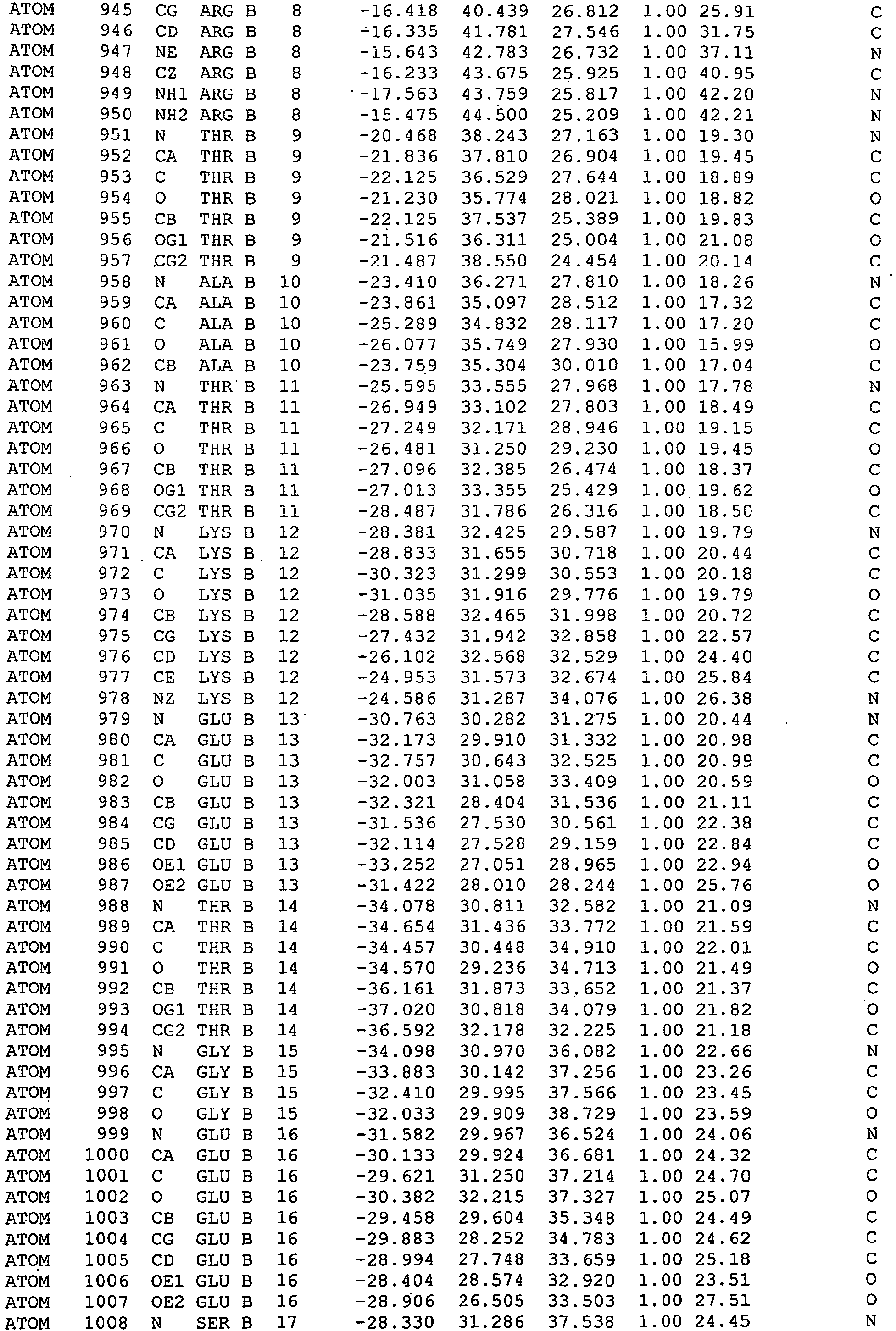


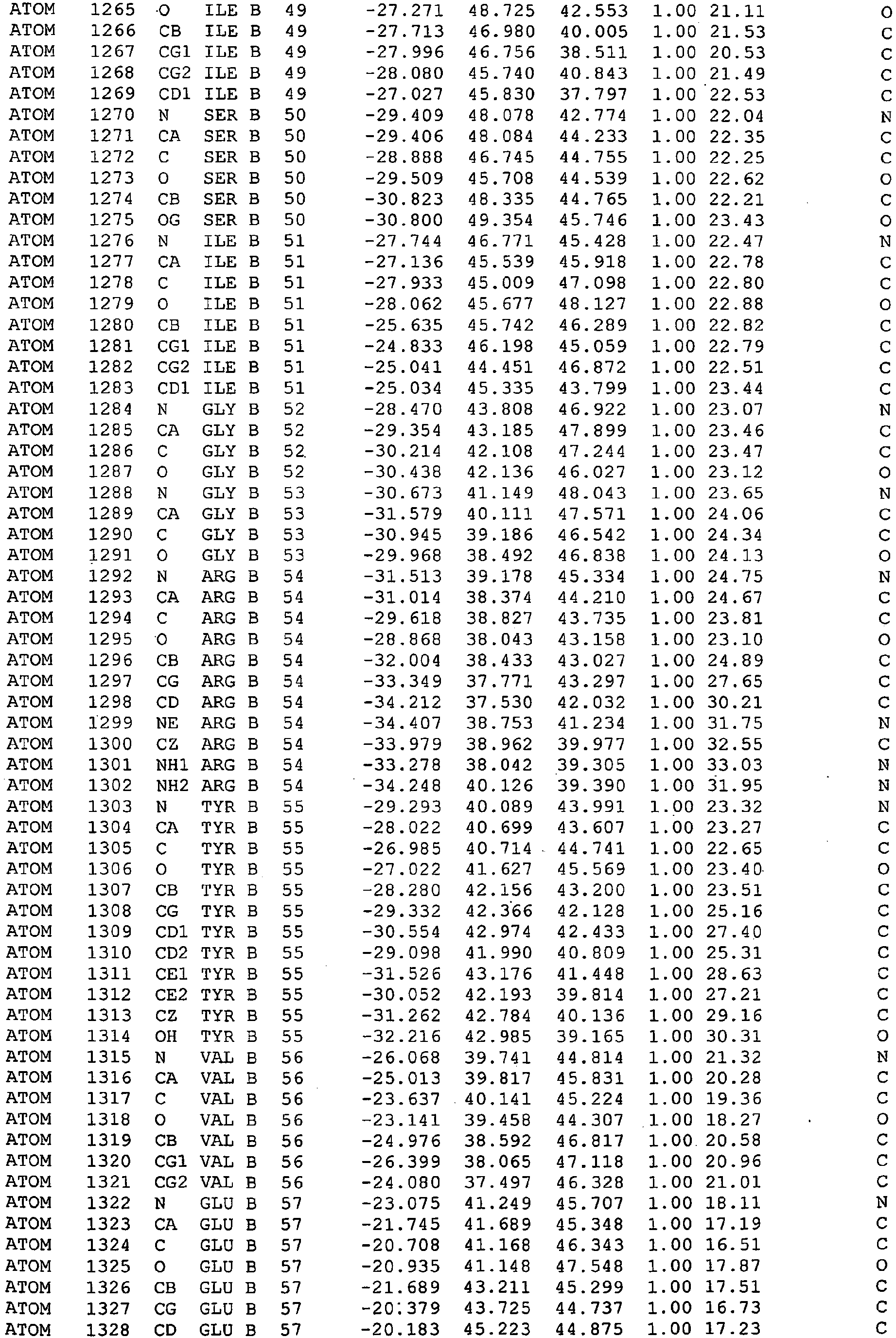


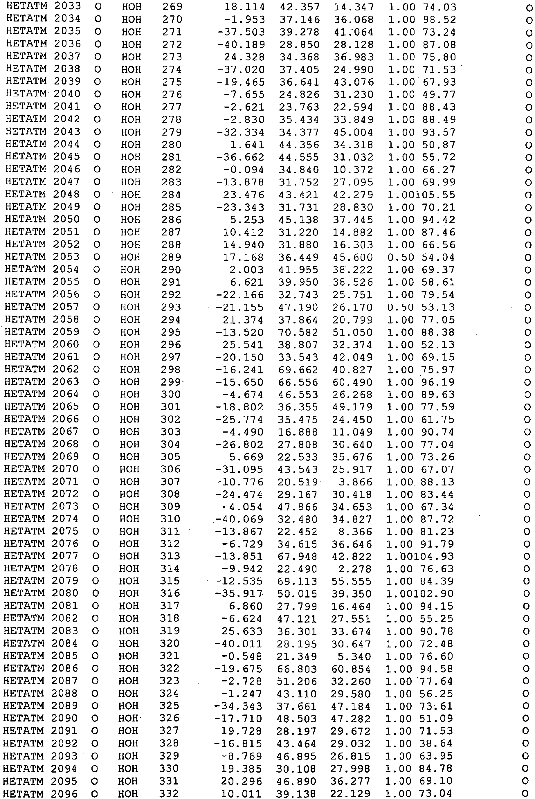

-


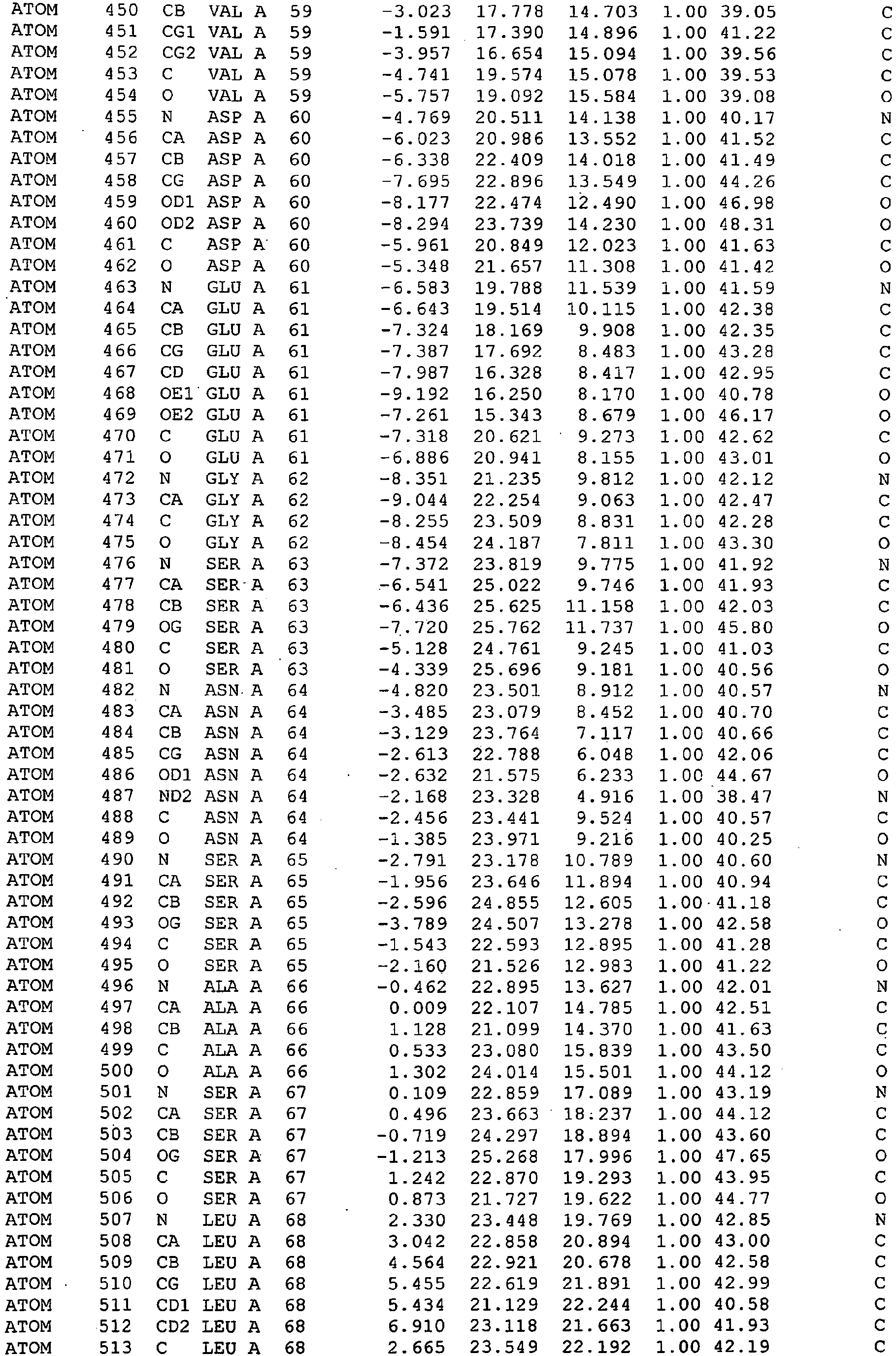

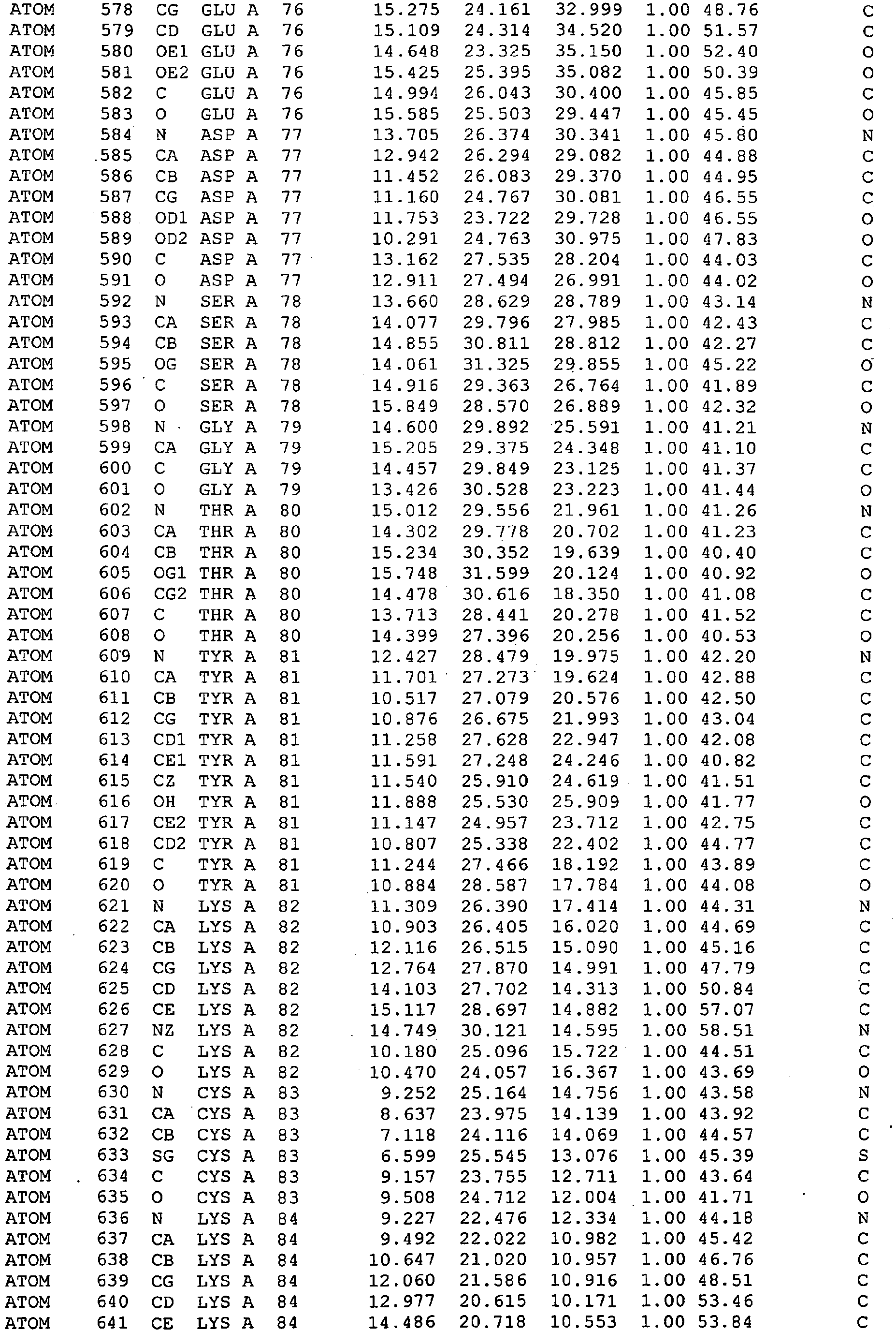


-



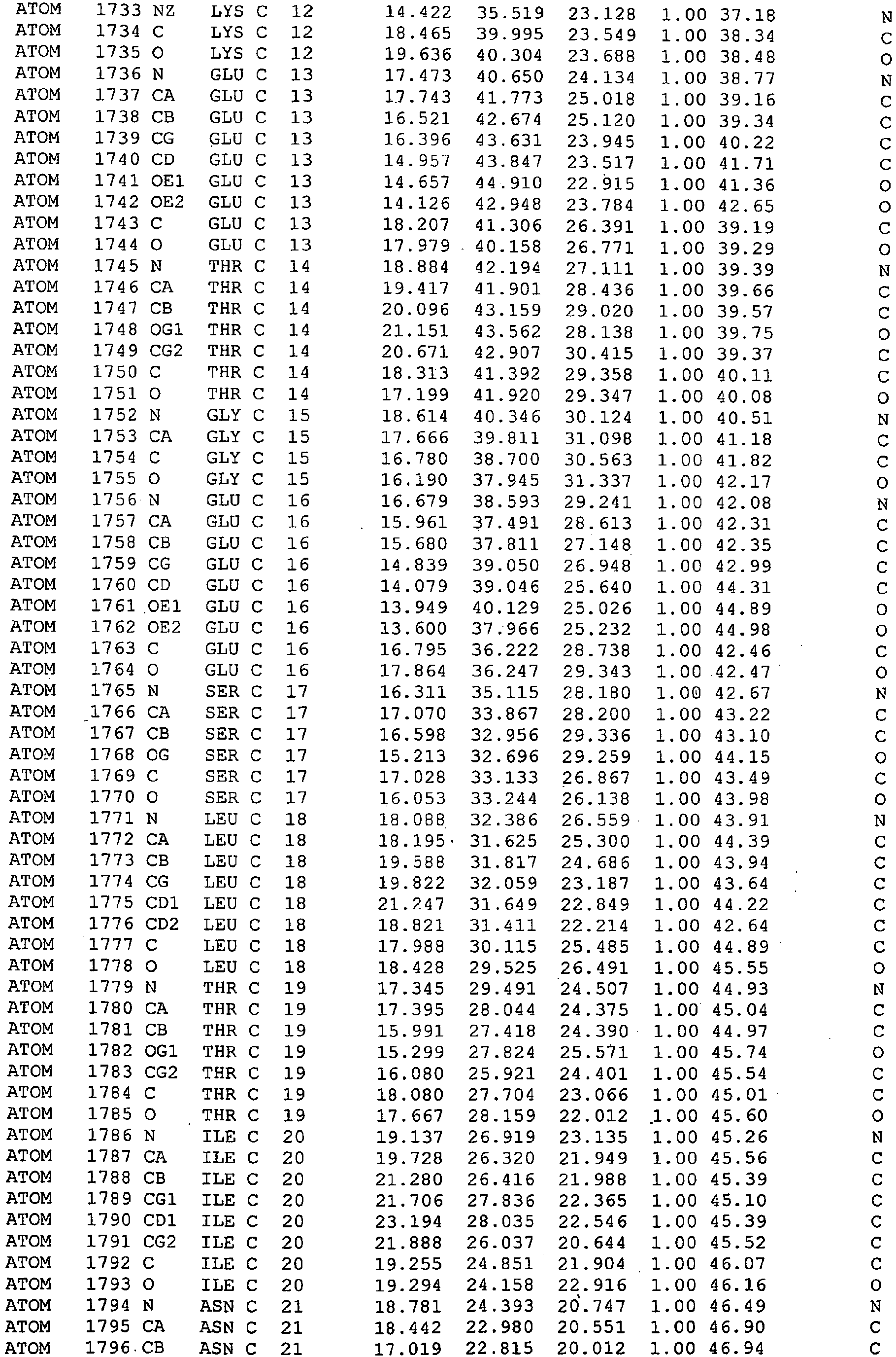


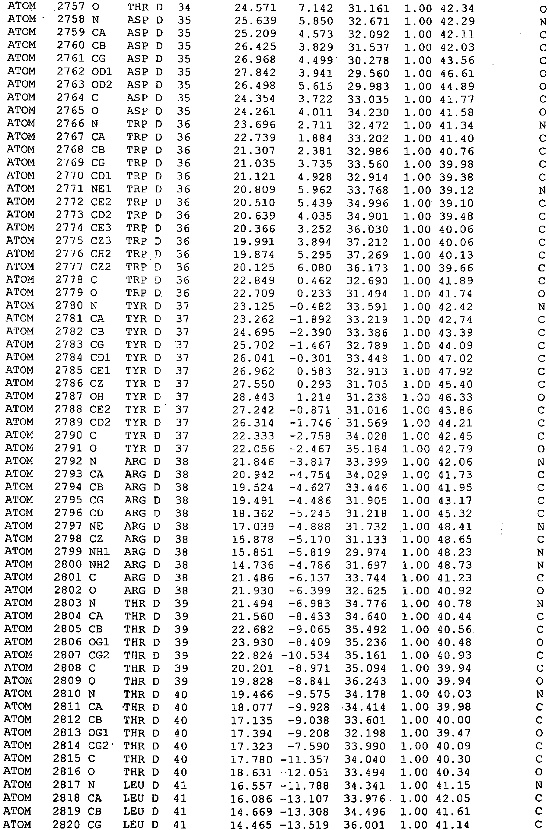


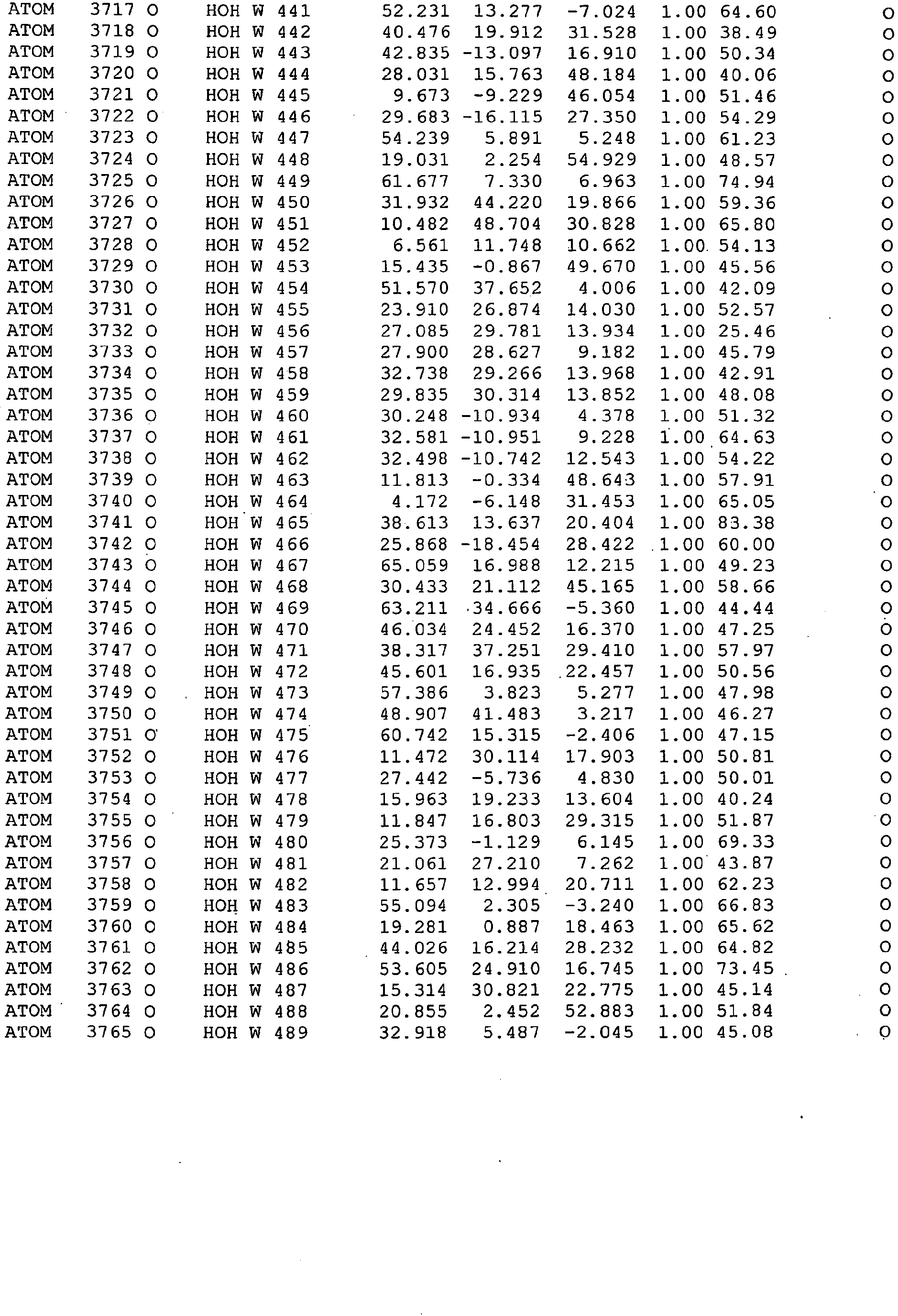
-
- <110> Diatech Pty Ltd
- <120> Binding moieties based on shark IgNar domains
- <130> 503393
- <160> 111
- <170> PatentIn version 3.3
- <210> 1
<211> 111
<212> PRT
<213> Artificial - <220>
<223> Variable domain from aType 2 immunoglobulin new antigen receptor from Orectolobus maculatus - <400> 1

- <210> 2
<211> 333
<212> DNA
<213> Artificial - <220>
<223> Variable domain from aType 2 immunoglobulin new antigen receptor from Orectolobus maculatus - <400> 2

- <210> 3
<211> 113
<212> PRT
<213> Artificial - <220>
<223> Variable domain from aType 2 immunoglobulin new antigen receptor from Orectolobus maculatus - <400> 3

- <210> 4
<211> 339
<212> DNA
<213> Artificial - <220>
<223> Variable domain from aType 2 immunoglobulin new antigen receptor from Orectolobus maculatus. - <400> 4

- <210> 5
<211> 111
<212> PRT
<213> Artificial - <220>
<223> Variable domain from aType 2 immunoglobulin new antigen receptor from Orectolobus maculatus. - <400> 5

- <210> 6
<211> 333
<212> DNA
<213> Artificial - <220>
<223> Variable domain from aType 2 immunoglobulin new antigen receptor from Orectolobus maculatus. - <400> 6

- <210> 7
<211> 111
<212> PRT
<213> Artificial - <220>
<223> Modified variable domain from aType 2 immunoglobulin new antigen receptor from Orectolobus maculatus. - <400> 7


- <210> 8
<211> 333
<212> DNA
<213> Artificial - <220>
<223> Modified variable domain from aType 2 immunoglobulin new antigen receptor from Orectolobus maculatus. - <400> 8

- <210> 9
<211> 108
<212> PRT
<213> Artificial - <220>
<223> Variable domain from aType 2 immunoglobulin new antigen receptor from Orectolobus maculatus. - <400> 9

- <210> 10
<211> 324
<212> DNA
<213> Artificial - <220>
<223> Variable domain from aType 2 immunoglobulin new antigen receptor from Orectolobus maculatus. - <400> 10

- <210> 11
<211> 113
<212> PRT
<213> Artificial - <220>
<223> Modified variable domain from aType 2 immunoglobulin new antigen receptor from Orectolobus maculatus. - <400> 11

- <210> 12
<211> 339
<212> DNA
<213> Artificial - <220>
<223> Modified variable domain from aType 2 immunoglobulin new antigen receptor from Orectolobus maculatus. - <400> 12

- <210> 13
<211> 113
<212> PRT
<213> Artificial - <220>
<223> Modified variable domain from aType 2 immunoglobulin new antigen receptor from Orectolobus maculatus. - <400> 13

- <210> 14
<211> 339
<212> DNA
<213> Artificial - <220>
<223> Modified variable domain from aType 2 immunoglobulin new antigen receptor from Orectolobus maculatus. - <400> 14

- <210> 15
<211> 113
<212> PRT
<213> Artificial - <220>
<223> Modified variable domain from aType 2 immunoglobulin new antigen receptor from Orectolobus maculatus. - <400> 15

- <210> 16
<211> 339
<212> DNA
<213> Artificial - <220>
<223> Modified variable domain from aType 2 immunoglobulin new antigen receptor from Orectolobus maculatus. - <400> 16

- <210> 17
<211> 113
<212> PRT
<213> Artificial - <220>
<223> Modified variable domain from aType 2 immunoglobulin new antigen receptor from Orectolobus maculatus. - <400> 17

- <210> 18
<211> 112
<212> PRT
<213> Artificial - <220>
<223> Modified variable domain from aType 2 immunoglobulin new antigen receptor from Orectolobus maculatus. - <400> 18

- <210> 19
<211> 110
<212> PRT
<213> Artificial - <220>
<223> Modified variable domain from aType 2 immunoglobulin new antigen receptor from Orectolobus maculatus. - <400> 19

- <210> 20
<211> 113
<212> PRT
<213> Artificial - <220>
<223> Modified variable domain from aType 2 immunoglobulin new antigen receptor from Orectolobus maculatus. - <400> 20


- <210> 21
<211> 109
<212> PRT
<213> Artificial - <220>
<223> Modified variable domain from aType 2 immunoglobulin new antigen receptor from Orectolobus maculatus. - <400> 21

- <210> 22
<211> 109
<212> PRT
<213> Artificial - <220>
<223> Modified variable domain from aType 2 immunoglobulin new antigen receptor from Orectolobus maculatus. - <400> 22


- <210> 23
<211> 110
<212> PRT
<213> Artificial - <220>
<223> Modified variable domain from aType 2 immunoglobulin new antigen receptor from Orectolobus maculatus. - <400> 23

- <210> 24
<211> 111
<212> PRT
<213> Artificial - <220>
<223> Modified variable domain from aType 2 immunoglobulin new antigen receptor from Orectolobus maculatus. - <400> 24


- <210> 25
<211> 113
<212> PRT
<213> Artificial - <220>
<223> Modified variable domain from aType 2 immunoglobulin new antigen receptor from Orectolobus maculatus. - <400> 25

- <210> 26
<211> 112
<212> PRT
<213> Artificial - <220>
<223> Modified variable domain from aType 2 immunoglobulin new antigen receptor from Orectolobus maculatus. - <400> 26


- <210> 27
<211> 108
<212> PRT
<213> Artificial - <220>
<223> Modified variable domain from aType 2 immunoglobulin new antigen receptor from Orectolobus maculatus. - <400> 27

- <210> 28
<211> 111
<212> PRT
<213> Artificial - <220>
<223> Modified variable domain from aType 2 immunoglobulin new antigen receptor from Orectolobus maculatus. - <400> 28

- <210> 29
<211> 108
<212> PRT
<213> Artificial - <220>
<223> Variable domain from aType 3 immunoglobulin new antigen receptor from Ginglymostoma cirratum - <400> 29
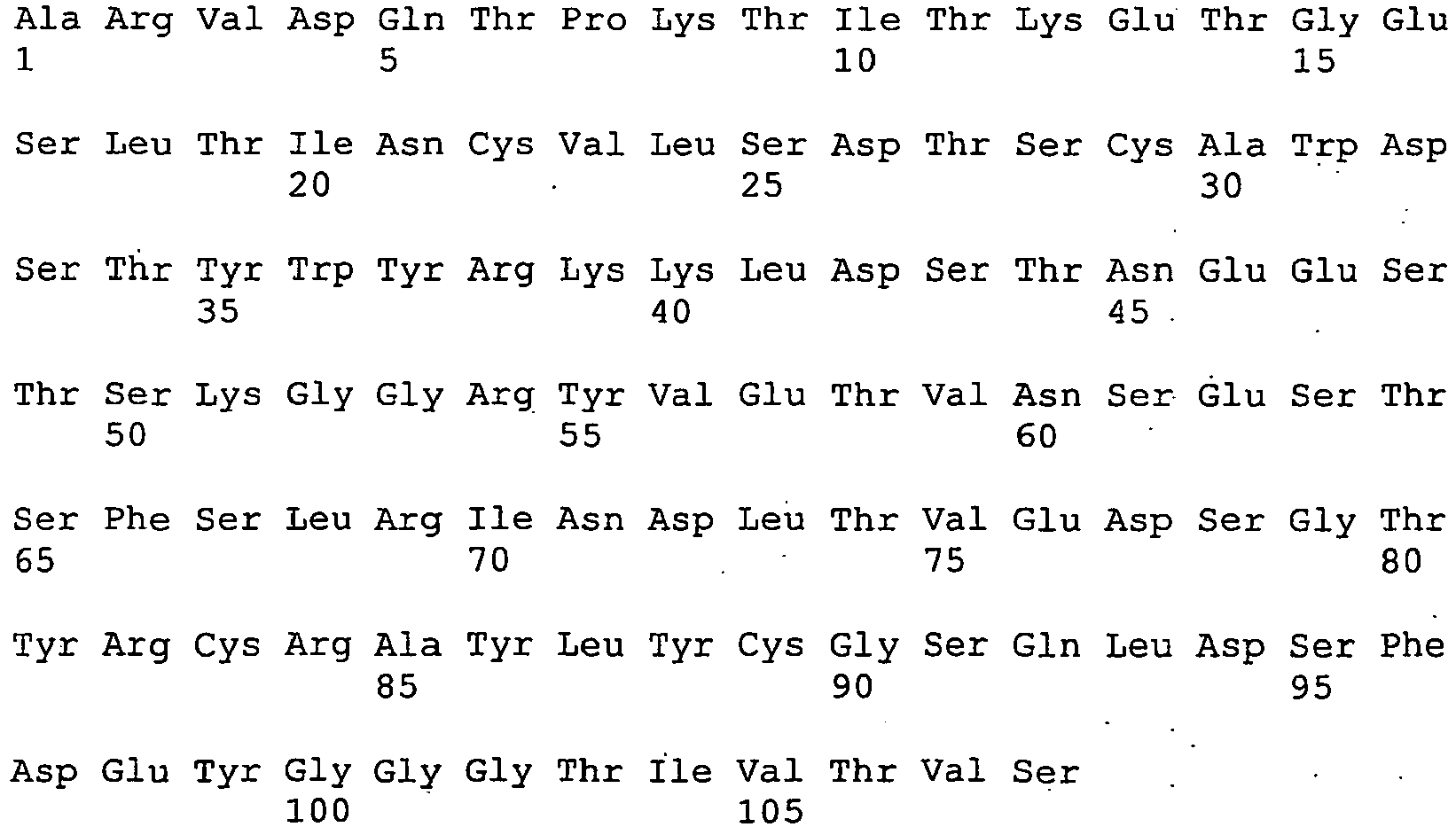
- <210> 30
<211> 108
<212> PRT
<213> Artificial - <220>
<223> Variable domain from aType 3 immunoglobulin new antigen receptor from Ginglymostoma cirratum - <400> 30

- <210> 31
<211> 661
<212> PRT
<213> Orectolobus maculatus - <400> 31


- <210> 32
<211> 2328
<212> DNA
<213> Orectolobus maculatus - <400> 32


- <210> 33
<211> 219
<212> PRT
<213> Artificial - <220>
<223> Variable domain and constant domain C1 fromType 2 immunoglobulin new antigen receptor from Orectolobus maculatus - <400> 33


- <210> 34
<211> 319
<212> PRT
<213> Artificial - <220>
<223> Variable domain and constant domain C1 and C2 fromType 2 immunoglobulin new antigen receptor from Orectolobus maculatus - <400> 34
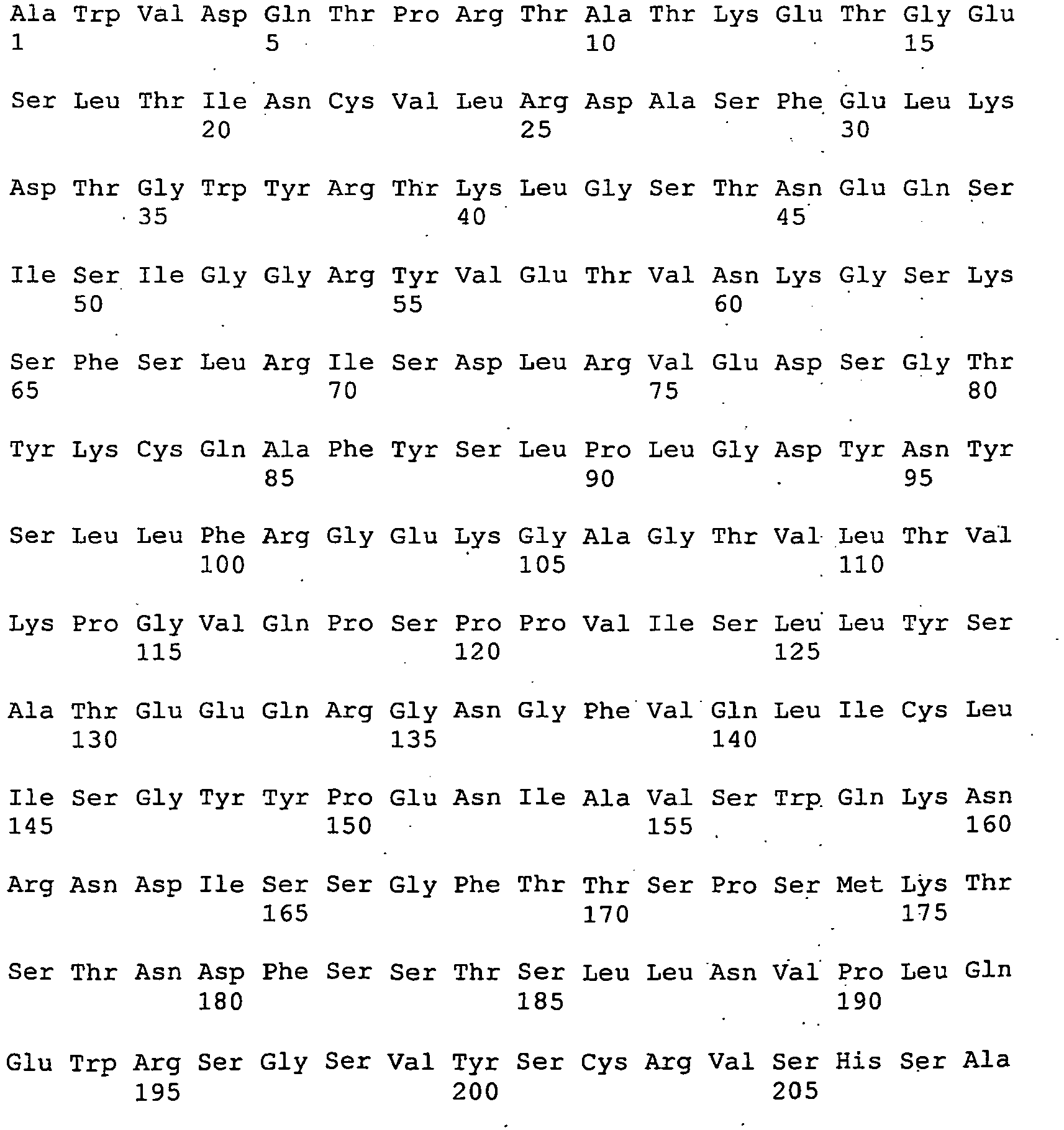

- <210> 35
<211> 425
<212> PRT
<213> Artificial - <220>
<223> Variable domain and constant domains C1 and C2 and C3 fromType 2 immunoglobulin new antigen receptor from Orectolobus maculatus - <400> 35


- <210> 36
<211> 97
<212> PRT
<213> Artificial - <220>
<223>Variable domain 1 from human neural cell adhesion molecule (NCAM) - <400> 36

- <210> 37
<211> 291
<212> DNA
<213> Artificial - <220>
<223>Variable domain 1 from human neural cell adhesion molecule (NCAM) - <400> 37

- <210> 38
<211> 190
<212> PRT
<213> Artificial - <220>
<223>Variable domain 1 anddomain 2 from human neural cell adhesion molecule (NCAM) - <400> 38

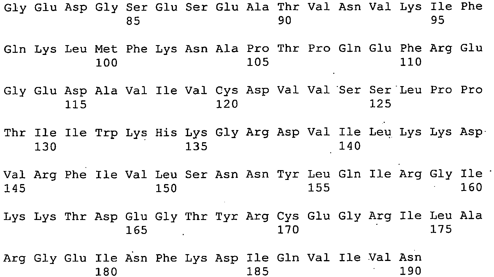
- <210> 39
<211> 570
<212> DNA
<213> Artificial - <220>
<223>Variable domain 1 anddomain 2 from human neural cell adhesion molecule (NCAM) - <400> 39

- <210> 40
<211> 105
<212> PRT
<213> Artificial - <220>
<223> Modifiedvariable domain 1 from human neural cell adhesion molecule (NCAM) - <400> 40


- <210> 41
<211> 92
<212> PRT
<213> Artificial - <220>
<223> Human telokin domain - <400> 41

- <210> 42
<211> 276
<212> DNA
<213> Artificial - <220>
<223> Human telokin domain - <400> 42

- <210> 43
<211> 100
<212> PRT
<213> Artificial - <220>
<223> Modified human telokin domain - <400> 43
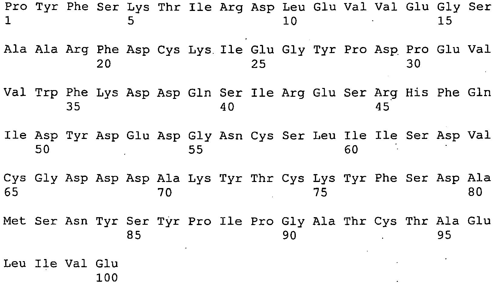
- <210> 44
<211> 300
<212> DNA
<213> Artificial - <220>
<223> Modified human telokin domain - <400> 44

- <210> 45
<211> 101
<212> PRT
<213> Artificial - <220>
<223> Modified human telokin domain - <400> 45


- <210> 46
<211> 303
<212> DNA
<213> Artificial - <220>
<223> Modified human telokin domain - <400> 46

- <210> 47
<211> 43
<212> DNA
<213> Artificial - <220>
<223> Oligonucleotide - <400> 47
tcgcggccca gccggccatg gccacaaggg tagaccaaacacc 43 - <210> 48
<211> 46
<212> DNA
<213> Artificial - <220>
<223> Oligonucleotide - <400> 48
gtctcgcggc ccagccggcc atggccacaa gggtagacca aacacc 46 - <210> 49
<211> 43
<212> DNA
<213> Artificial - <220>
<223> Oligonucleotide - <400> 49
tcgcggccca gccggccatg gccgcaaggg tggaccaaacacc 43 - <210> 50
<211> 46
<212> DNA
<213> Artificial - <220>
<223> Oligonulceotide - <400> 50
gtctcgcggc ccagccggcc atggccgcaa gggtggacca aacacc 46 - <210> 51
<211> 43
<212> DNA
<213> Artificial - <220>
<223> Oligonucleotide - <400> 51
tcgcggccca gccggccatg gccgcatggg tagaccaaacacc 43 - <210> 52
<211> 46
<212> DNA
<213> Artificial - <220>
<223> Oligonucleotide - <400> 52
gtctcgcggc ccagccggcc atggccgcat gggtagacca aacacc 46 - <210> 53
<211> 42
<212> DNA
<213> Artificial - <220>
<223> Oligonucleotide - <400> 53
cgcggcccag ccggccatgg ccgcaagcct ggaccaaacacc 42 - <210> 54
<211> 42
<212> DNA
<213> Artificial - <220>
<223> Oligonucleotide - <400> 54
cgcggcccag ccggccatgg ccgcattgac ggaccaaacacc 42 - <210> 55
<211> 33
<212> DNA
<213> Artificial - <220>
<223> Oligonucleotide - <400> 55
atctgcggcc gctttcacgg ttaatgcggtgcc 33 - <210> 56
<211> 39
<212> DNA
<213> Artificial - <220>
<223> Oligonucleotide - <400> 56
cacgttatct gcggccgctt tcacggttaa tgcggtgcc 39 - <210> 57
<211> 39
<212> DNA
<213> Artificial - <220>
<223> Oligonucleotide - <400> 57
atctgcggcc gctttcacgg ttaatacggt gccagctcc 39 - <210> 58
<211> 45
<212> DNA
<213> Artificial - <220>
<223> Oligonucleotide - <400> 58
cacgttatct gcggccgctt tcacggttaa tacggtgcca gctcc 45 - <210> 59
<211> 33
<212> DNA
<213> Artificial - <220>
<223> Oligonucleotide - <400> 59
atctgcggcc gctttcacgg tcagtagggtgcc 33 - <210> 60
<211> 33
<212> DNA
<213> Artificial - <220>
<223> Oligonucleotide - <400> 60
atctgcggcc gctctcacgg tcagtacggtgcc 33 - <210> 61
<211> 87
<212> DNA
<213> Artificial - <220>
<223> Oligonucleotide - <220>
<221> misc_feature
<222> (31)..(31)
<223> n is a, c, g, or t - <220>
<221> misc_feature
<222> (35)..(36)
<223> n is a, c, g, or t - <220>
<221> misc_feature
<222> (38).. (39)
<223> n is a, c, g, or t - <220>
<221> misc_feature
<222> (41)..(42)
<223> n is a, c, g, or t - <220>
<221> misc_feature
<222> (44).. (45)
<223> n is a, c, g, or t - <220>
<221> misc_feature
<222> (56)... (57)
<223> n is a, c, g, or t - <220>
<221> misc_feature
<222> (59).. (60)
<223> n is a, c, g, or t - <220>
<221> misc_feature
<222> (62)..(63)
<223> n is a, c, g, or t - <220>
<221> misc_feature
<222> (65)..(65)
<223> n is a, c, g, or t - <400> 61

- <210> 62
<211>. 87
<212> DNA
<213> Artificial - <220>
<223> Oligonucleotide - <220>
<221> misc_feature
<222> (32).. (33)
<223> n is a, c, g, or t - <220>
<221> misc_feature
<222> (35)..(36)
<223> n is a, c, g, or t - <220>
<221> misc_feature
<222> (38)..(39)
<223> n is a, c, g, or t - <220>
<221> misc_feature
<222> (41)..(42)
<223> n is a, c, g, or t - <220>
<221> misc_feature
<222> (53)..(54)
<223> n is a, c, g, or t - <220>
<221> misc_feature
<222> (56)..(57)
<223> n is a, c, g, or t - <220>
<221> misc_feature
<222> (59)..(60)
<223> n is a, c, g, or t - <220>
<221> misc_feature
<222> (62)..(63)
<223> n is a, c, g, or t - <220>
<221> misc_feature
<222> (65)..(65)
<223> n is a, c, g, or t - <400> 62

- <210> 63
<211> 84
<212> DNA
<213> Artificial - <220>
<223> Oligonucleotide - <220>
<221> misc_feature
<222> (31)..(31)
<223> n is a, c, g, or t - <220>
<221> misc_feature
<222> (35)..(36)
<223> n is a, c, g, or t - <220>
<221> misc_feature
<222> (38).. (39)
<223> n is a, c, g, or t - <220>
<221> misc_feature
<222> (41)..(42)
<223> n is a, c, g, or t - <220>
<221> misc_feature
<222> (53).. (54)
<223> n is a, c, g, or t - <220>
<221> misc_feature
<222> (56)..(57)
<223> n is a, c, g, or t - <220>
<221> misc_feature
<222> (59)..(60)
<223> n is a, c, g, or t - <220>
<221> misc_feature
<222> (62)..(62)
<223> n is a, c, g, or t - <400> 63

- <210> 64
<211> 84
<212> DNA
<213> Artificial - <220>
<223> Oligonucleotide - <220>
<221> misc_feature
<222> (32)..(33)
<223> n is a, c, g, or t - <220>
<221> misc_feature
<222> (35)..(36)
<223> n is a, c, g, or t - <220>
<221> misc_feature
<222> (38)..(39)
<223> n is a, c, g, or t - <220>
<221> misc_feature
<222> (50)..(51)
<223> n is a, c, g, or t - <220>
<221> misc_feature
<222> (53)..(54)
<223> n is a, c, g, or t - <220>
<221> misc_feature
<222> (56)..(57)
<223> n is a, c, g, or t - <220>
<221> misc_feature
<222> (59)..(60)
<223> n is a, c, g, or t - <220>
<221> misc_feature
<222> (62)..(62)
<223> n is a, c, g, or t - <400> 64

- <210> 65
<211> 84
<212> DNA
<213> Artificial - <220>
<223> Oligonucleotide - <220>
<221> misc_feature
<222> (32)..(33)
<223> n is a, c, g, or t - <220>
<221> misc_feature
<222> (35)..(36)
<223> n is a, c, g, or t - <220>
<221> misc_feature
<222> (38)..(39)
<223> n is a, c, g, or t - <220>
<221> misc_feature
<222> (50)..(51)
<223> n is a, c, g, or t - <220>
<221> misc_feature
<222> (53)..(54)
<223> n is a, c, g, or t - <220>
<221> misc_feature
<222> (56)..(57)
<223> n is a, c, g, or t - <220>
<221> misc_feature
<222> (59)..(60)
<223> n is a, c, g, or t - <220>
<221> misc_feature
<222> (62)..(62)
<223> n is a, c, g, or t - <400> 65

- <210> 66
<211> 102
<212> DNA
<213> Artificial - <220>
<223> Oligonucleotide - <220>
<221> misc_feature
<222> (35)..(36)
<223> n is a, c, g, or t - <220>
<221> misc_feature
<222> (38)..(39)
<223> n is a, c, g, or t - <220>
<221> misc_feature
<222> (41)..(42)
<223> n is a, c, g, or t - <220>
<221> misc_feature
<222> (44)..(45)
<223> n is a, c, g, or t - <220>
<221> misc_feature
<222> (47)..(48)
<223> n is a, c, g, or t - <220>
<221> misc_feature
<222> (65)..(66).
<223> n is a, c, g, or t - <220>
<221> misc_feature
<222> (68)..(69)
<223> n is a, c, g, or t - <220>
<221> misc_feature
<222> (71)..(72)
<223> n is a, c, g, or t - <220>
<221> misc_feature
<222> (74)..(75)
<223> n is a, c, g, or t - <220>
<221> misc_feature
<222> (77)..(78)
<223> n is a, c, g, or t - <220>
<221> misc_feature
<222> (80)..(80)
<223> n is a, c, g, or t - <400> 66

- <210> 67
<211> 105
<212> DNA
<213> Artificial - <220>
<223> Oligonucleotide - <220>
<221> misc_feature
<222> (35)..(36)
<223> n is a, c, g, or t - <220>
<221> misc_feature
<222> (38)..(39)
<223> n is a, c, g, or t - <220>
<221> misc_feature
<222> (41)..(42)
<223> n is a, c, g, or t - <220>
<221> misc_feature
<222> (44)..(45)
<223> n is a, c, g, or t - <220>
<221> misc_feature
<222> (47)..(48)
<223> n is a, c, g, or t - <220>
<221> misc_feature
<222> (50)..(51)
<223> n is a, c, g, or t - <220>
<221> misc_feature
<222> (68)..(69)
<223> n is a, c, g, or t - <220>
<221> misc_feature
<222> (71)..(72)
<223> n is a, c, g, or t - <220>
<221> misc_feature
<222> (74)..(75)
<223> n is a, c, g, or t - <220>
<221> misc_feature
<222> (77)..(78)
<223> n is a, c, g, or t - <220>
<221> misc_feature
<222> (80)..(81)
<223> n is a, c, g, or t - <220>
<221> misc_feature
<222> (83)..(83)
<223> n is a, c, g, or t - <400> 67

- <210> 68
<211> 99
<212> DNA
<213> Artificial - <220>
<223> Oligonucleotide - <220>
<221> misc_feature
<222> (35)..(36)
<223> n is a, c, g, or t - <220>
<221> misc_feature
<222> (38)..(39)
<223> n is a, c, g, or t - <220>
<221> misc_feature
<222> (41)..(42)
<223> n is a, c, g, or t - <220>
<221> misc_feature
<222> (44)..(45)
<223> n is a, c, g, or t - <220>
<221> misc_feature
<222> (62)..(63)
<223> n is a, c, g, or t - <220>
<221> misc_feature
<222> (65)..(66)
<223> n is a, c, g, or t - <220>
<221> misc_feature
<222> (68) .. (69)
<223> n is a, c, g, or t - <220>
<221> misc_feature
<222> (71)..(72)
<223> n is a, c, g, or t - <220>
<221> misc_feature
<222> (74)..(75)
<223> n is a, c, g, or t - <220>
<221> misc_feature
<222> (77)..(77)
<223> n is a, c, g, or t - <400> 68

- <210> 69
<211> 100
<212> DNA
<213> Artificial - <220>
<223> Oligonucleotide - <220>
<221> misc_feature
<222> (26)..(27)
<223> n is a, c, g, or t - <220>
<221> misc_feature
<222> (29)..(30)
<223> n is a, c, g, or t - <220>
<221> misc_feature
<222> (32)..(33)
<223> n is a, c, g, or t - <220>
<221> misc_feature
<222> (35)..(36)
<223> n is a, c, g, or t - <220>
<221> misc_feature
<222> (38)..(39)
<223> n is a, c, g, or t - <220>
<221> misc_feature
<222> (41)..(42)
<223> n is a, c, g, or t - <220>
<221> misc_feature
<222> (44)..(45)
<223> n is a, c, g, or t - <220>
<221> misc_feature
<222> (59) .. (60)
<223> n is a, c, g, or t - <220>
<221> misc_feature
<222> (62)..(63)
<223> n is a, c, g, or t - <220>
<221> misc_feature
<222> (65)..(66)
<223> n is a, c, g, or t - <220>
<221> misc_feature
<222> (68)..(69)
<223> n is a, c, g, or t - <220>
<221> misc_feature
<222> (71)..(72)
<223> n is a, c, g, or t - <220>
<221> misc_feature
<222> (74)..(75)
<223> n is a, c, g, or t - <400> 69

- <210> 70
<211> 94
<212> DNA
<213> Artificial - <220>
<223> Oligonucleotide - <220>
<221> misc_feature
<222> (26).. (27)
<223> n is a, c, g, or t - <220>
<221> misc_feature
<222> (29)..(30)
<223> n is a, c, g, or t - <220>
<221> misc_feature
<222> (32)..(33)
<223> n is a, c, g, or t - <220>
<221> misc_feature
<222> (35)..(36)
<223> n is a, c, g, or t - <220>
<221> misc_feature
<222> (38)..(39)
<223> n is a, c, g, or t - <220>
<221> misc_feature
<222> (53)..(54)
<223> n is a, c, g, or t - <220>
<221> misc_feature
<222> (56)..(57)
<223> n is a, c, g, or t - <220>
<221> misc_feature
<222> (59)..(60)
<223> n is a, c, g, or t - <220>
<221> misc_feature
<222> (62)..(63)
<223> n is a, c, g, or t - <220>
<221> misc_feature
<222> (65)..(66)
<223> n is a, c, g, or t - <220>
<221> misc_feature
<222> (68)..(69)
<223> n is a, c, g, or t - <400> 70

- <210> 71
<211> 95
<212> DNA
<213> Artificial - <220>
<223> Oligonucleotide - <220>
<221> misc_feature
<222> (47)..(47)
<223> n is a, c, g, or t - <220>
<221> misc_feature
<222> (50)..(51)
<223> n is a, c, g, or t - <220>
<221> misc_feature
<222> (53)..(54)
<223> n is a, c, g, or t - <220>
<221> misc_feature
<222> (56)..(57)
<223> n is a, c, g, or t - <220>
<221> misc_feature
<222> (59)..(60)
<223> n is a, c, g, or t - <220>
<221> misc_feature
<222> (62)..(63)
<223> n is a, c, g, or t - <220>
<221> misc_feature
<222> (65)..(66)
<223> n is a, c, g, or t - <220>
<221> misc_feature
<222> (68)..(69)
<223> n is a, c, g, or t - <400> 71

- <210> 72
<211> 101
<212> DNA
<213> Artificial - <220>
<223> Oligonucleotide - <220>
<221> misc_feature
<222> (47) ..(47)
<223> n is a, c, g, or t - <220>
<221> misc_feature
<222> (50)..(51)
<223> n is a, c, g, or t - <220>
<221> misc_feature
<222> (53)..(54)
<223> n is a, c, g, or t - <220>
<221> misc_feature
<222> (56)..(57)
<223> n is a, c, g, or t - <220>
<221> misc_feature
<222> (59)..(60)
<223> n is a, c, g, or t - <220>
<221> misc_feature
<222> (62)..(63)
<223> n is a, c, g, or t - <220>
<221> misc_feature
<222> (65)..(66)
<223> n is a, c, g, or t - <220>
<221> misc_feature
<222> (68)..(69)
<223> n is a, c, g, or t - <220>
<221> misc_feature
<222> (71)..(72)
<223> n is a, c, g, or t - <220>
<221> misc_feature
<222> (74)..(75)
<223> n is a, c, g, or t - <400> 72

- <210> 73
<211> 98
<212> DNA
<213> Artificial - <220>
<223> Oligonucleotide - <220>
<221> misc_feature
<222> (44)..(44)
<223> n is a, c, g, or t - <220>
<221> misc_feature
<222> (47)..(47)
<223> n is a, c, g, or t - <220>
<221> misc_feature
<222> (50)..(51)
<223> n is a, c, g, or t - <220>
<221> misc_feature
<222> (56)..(57)
<223> n is a, c, g, or t - <220>
<221> misc_feature
<222> (59)..(60)
<223> n is a, c, g, or t - <220>
<221> misc_feature
<222> (65)..(66)
<223> n is a, c, g, or t - <220>
<221> misc_feature
<222> (71)..(72)
<223> n is a, c, g, or t - <400> 73

- <210> 74
<211> 104
<212> DNA
<213> Artificial - <220>
<223> Oligonucleotide - <220>
<221> misc_feature
<222> (47)..(47)
<223> n is a, c, g, or t - <220>
<221> misc_feature
<222> (50)..(51)
<223> n is a, c, g, or t - <220>
<221> misc_feature
<222> (53)..(54)
<223> n is a, c, g, or t - <220>
<221> misc_feature
<222> (56)..(57)
<223> n is a, c, g, or t - <220>
<221> misc_feature
<222> (71)..(72)
<223> n is a, c, g, or t - <220>
<221> misc_feature
<222> (74)..(75)
<223> n is a, c, g, or t - <220>
<221> misc_feature
<222> (77)..(78)
<223> n is a, c, g, or t - <400> 74

- <210> 75
<211> 107
<212> DNA
<213> Artificial - <220>
<223> Oligonucleotide - <220>
<221> misc_feature
<222> (47).. (48)
<223> n is a, c, g, or t - <220>
<221> misc_feature
<222> (50)..(51)
<223> n is a, c, g, or t - <220>
<221> misc_feature
<222> (56)..(57)
<223> n is a, c, g, or t - <220>
<221> misc_feature
<222> (59).. (60)
<223> n is a, c, g, or t - <220>
<221> misc_feature
<222> (65)..(66)
<223> n is a, c, g, or t - <220>
<221> misc_feature
<222> (68)..(69)
<223> n is a, c, g, or t - <220>
<221> misc_feature
<222> (74)..(75)
<223> n is a, c, g, or t - <220>
<221> misc_feature
<222> (77)..(78)
<223> n is a, c, g, or t - <220>
<221> misc_feature
<222> (80)..(81)
<223> n is a, c, g, or t - <400> 75

- <210> 76
<211> 107
<212> DNA
<213> Artificial - <220>
<223> Oligonucleotide - <220>
<221> misc_feature
<222> (47)..(48)
<223> n is a, c, g, or t - <220>
<221> misc_feature
<222> (50)..(51)
<223> n is a, c, g, or t - <220>
<221> misc_feature
<222> (56)..(57)
<223> n is a, c, g, or t - <220>
<221> misc_feature
<222> (59)..(60)
<223> n is a, c, g, or t - <220>
<221> misc_feature
<222> (65)..(66)
<223> n is a, c, g, or t - <220>
<221> misc_feature
<222> (68) ..(69)
<223> n is a, c, g, or t - <220>
<221> misc_feature
<222> (74)..(75)
<223> n is a, c, g, or t - <220>
<221> misc_feature
<222> (77)..(78)
<223> n is a, c, g, or t - <400> 76

- <210> 77
<211> 53
<212> DNA
<213> Artificial - <220>
<223> Oligonucleotide - <400> 77
gtctcgcggc ccagccggcc atggccctgc aggtggatat tgttcccagc cag 53 - <210> 78
<211> 42
<212> DNA
<213> Artificial - <220>
<223> Oligonucleotide - <400> 78
gctggttgcg gccgcctgaa agatcttcac gttgacggtg gc 42 - <210> 79
<211> 43
<212> DNA
<213> Artificial - <220>
<223> Oligonucleotide - <400> 79
gctggttgcg gccgcattca caatgacctg aatgtccttg aag 43 - <210> 80
<211> 74
<212> DNA
<213> Artificial - <220>
<223> Oligonucleotide - <400> 80

- <210> 81
<211> 93
<212> DNA
<213> Artificial - <220>
<223> Oligonucleotide - <400> 81

- <210> 82
<211> 69
<212> DNA
<213> Artificial - <220>
<223> Oligonucleotide - <220>
<221> misc_feature
<222> (29)..(30)
<223> n is a, c, g, or t - <220>
<221> misc_feature
<222> (32)..(33)
<223> n is a, c, g, or t - <220>
<221> misc_feature
<222> (35)..(36)
<223> n is a, c, g, or t - <220>
<221> misc_feature
<222> (38)..(39)
<223> n is a, c, g, or t - <220>
<221> misc_feature
<222> (41)..(42)
<223> n is a, c, g, or t - <220>
<221> misc_feature
<222> (44).. (45)
<223> n is a, c, g, or t - <400> 82

- <210> 83
<211> 72
<212> DNA
<213> Artificial - <220>
<223> Oligonucleotide - <220>
<221> misc_feature
<222> (29)..(30)
<223> n is a, c, g, or t - <220>
<221> misc_feature
<222> (32).. (33)
<223> n is a, c, g, or t - <220>
<221> misc_feature
<222> (35)..(36)
<223> n is a, c, g, or t - <220>
<221> misc_feature
<222> (38)..(39)
<223> n is a, c, g, or t - <220>
<221> misc_feature
<222> (41)..(42)
<223> n is a, c, g, or t - <220>
<221> misc_feature
<222> (44)..(45)
<223> n is a, c, g, or t - <220>
<221> misc_feature
<222> (47)..(48)
<223> n is a, c, g, or t - <400> 83

- <210> 84
<211> 69
<212> DNA
<213> Artificial - <220>
<223> Oligonucleotide - <220>
<221> misc_feature
<222> (26).. (27)
<223> n is a, c, g, or t - <220>
<221> misc_feature
<222> (29)..(30)
<223> n is a, c, g, or t - <220>
<221> misc_feature
<222> (32)..(33)
<223> n is a, c, g, or t - <220>
<221> misc_feature
<222> (35).. (36)
<223> n is a, c, g, or t - <220>
<221> misc_feature
<222> (38)..(39)
<223> n is a, c, g, or t - <220>
<221> misc_feature
<222> (41)..(42)
<223> n is a, c, g, or t - <220>
<221> misc_feature
<222> (44)..(45)
<223> n is a, c, g, or t - <220>
<221> misc_feature
<222> (47)..(48)
<223> n is a, c, g, or t - <400> 84

- <210> 85
<211> 78
<212> DNA
<213> Artificial - <220>
<223> Oligonucleotide - <220>
<221> misc_feature
<222> (26).. (27)
<223> n is a, c, g, or t - <220>
<221> misc_feature
<222> (29)..(30)
<223> n is a, c, g, or t - <220>
<221> misc_feature
<222> (32)..(33)
<223> n is a, c, g, or t - <220>
<221> misc_feature
<222> (35)..(36)
<223> n is a, c, g, or t - <220>
<221> misc_feature
<222> (38)..(39)
<223> n is a, c, g, or t - <220>
<221> misc_feature
<222> (41).. (42)
<223> n is a, c, g, or t - <220>
<221> misc_feature
<222> (44).. (45)
<223> n is a, c, g, or t - <220>
<221> misc_feature
<222> (47)..(48)
<223> n is a, c, g, or t - <220>
<221> misc_feature
<222> (50)..(51)
<223> n is a, c, g, or t - <220>
<221> misc_feature
<222> (53)..(54)
<223> n is a, c, g, or t - <220>
<221> misc_feature
<222> (56)..(57)
<223> n is a, c, g, or t - <400> 85

- <210> 86
<211> 87
<212> DNA
<213> Artificial - <220>
<223> Oligonucleotide - <220>
<221> misc_feature
<222> (26).. (27)
<223> n is a, c, g, or t - <220>
<221> misc_feature
<222> (29).. (30)
<223> n is a, c, g, or t - <220>
<221> misc_feature
<222> (32)..(33)
<223> n is a, c, g, or t - <220>
<221> misc_feature
<222> (35)..(36)
<223> n is a, c, g, or t - <220>
<221> misc_feature
<222> (38)..(39)
<223> n is a, c, g, or t - <220>
<221> misc_feature
<222> (41)..(42)
<223> n is a, c, g, or t - <220>
<221> misc_feature
<222> (44)...(45)
<223> n is a, c, g, or t - <220>
<221> misc_feature
<222> (47)..(48)
<223> n is a, c, g, or t - <220>
<221> misc_feature
<222> (50)..(51)
<223> n is a, c, g, or t - <220>
<221> misc_feature
<222> (53)..(54)
<223> n is a, c, g, or t - <220>
<221> misc_feature
<222> (56)..(57)
<223> n is a, c, g, or t - <220>
<221> misc_feature
<222> (59)..(60)
<223> n is a, c, g, or t - <220>
<221> misc_feature
<222> (62)..(63)
<223> n is a, c, g, or t - <220>
<221> misc_feature
<222> (65)..(66)
<223> n is a, c, g, or t - <400> 86

- <210> 87
<211> 92
<212> DNA
<213> Artificial - <220>
<223> Oligonucleotide - <220>
<221> misc_feature
<222> (35)..(36)
<223> n is a, c, g, or t - <220>
<221> misc_feature
<222> (38)..(39)
<223> n is a, c, g, or t - <220>
<221> misc_feature
<222> (41)..(42)
<223> n is a, c, g, or t - <220>
<221> misc_feature
<222> (44)..(45)
<223> n is a, c, g, or t - <220>
<221> misc_feature
<222> (47)..(48)
<223> n is a, c, g, or t - <220>
<221> misc_feature
<222> (50)..(51)
<223> n is a, c, g, or t - <220>
<221> misc_feature
<222> (53)..(54)
<223> n is a, c, g, or t - <220>
<221> misc_feature
<222> (56)..(57)
<223> n is a, c, g, or t - <220>
<221> misc_feature
<222> (59)..(60)
<223> n is a, c, g, or t - <220>
<221> misc_feature
<222> (62)..(63)
<223> n is a, c, g, or t - <220>
<221> misc_feature
<222> (65)..(66)
<223> n is a, c, g, or t - <400> 87

- <210> 88
<211> 53
<212> DNA
<213> Artificial - <220>
<223> Oligonucleotide - <400> 88
gtctcgcggc ccagccggcc atggccccct atttctctaa gaccattcgc gat 53 - <210> 89
<211> 42
<212> DNA
<213> Artificial - <220>
<223> Oligonucleotide - <400> 89
acctgcacag cagagctcat tgtggaagcg gccgcaacca gc 42 - <210> 90
<211> 54
<212> DNA
<213> Artificial - <220>
<223> Oligonucleotide - <400> 90
atagctatag ttgctcatgg catcgctaaa atacttgcag gtgtacttgg catc 54 - <210> 91
<211> 57
<212> DNA
<213> Artificial - <220>
<223> Oligonucleotide - <400> 91
aatgagctct gctgtgcagg tggcgcccgg aatcggatag ctatagttgc tcatggc 57 - <210> 92
<211> 50
<212> DNA
<213> Artificial - <220>
<223> Oligonucleotide - <400> 92
atcggatagc tatagttgct catggcatca ctgttgacag ccttgcaggt 50 - <210> 93
<211> 52
<212> DNA
<213> Artificial - <220>
<223> Oligonucleotide - <400> 93
aatgagctct gctgtgcagg tggcttctcc aatcggatag ctatagttgc tc 52 - <210> 94
<211> 67
<212> DNA
<213> Artificial - <220>
<223> Oligonucleotide - <220>
<221> misc_feature
<222> (25)..(26)
<223> n is a, c, g, or t - <220>
<221> misc_feature
<222> (28)..(29)
<223> n is a, c, g, or t - <220>
<221> misc_feature
<222> (31)..(32)
<223> n is a, c, g, or t - <220>
<221> misc_feature
<222> (34)..(35)
<223> n is a, c, g, or t - <220>
<221> misc_feature
<222> (37)..(38)
<223> n is a, c, g, or t - <220>
<221> misc_feature
<222> (40)..(41)
<223> n is a, c, g, or t - <220>
<221> misc_feature
<222> (43)..(44)
<223> n is a, c, g, or t - <400> 94

- <210> 95
<211> 73
<212> DNA
<213> Artificial - <220>.
<223> Oligonucleotide - <220>
<221> misc_feature
<222> (25).. (26)
<223> n is a, c, g, or t - <220>
<221> misc_feature
<222> (28).. (29)
<223> n is a, c, g, or t - <220>
<221> misc_feature
<222> (31)..(32)
<223> n is a, c, g, or t - <220>
<221> misc_feature
<222> (34)..(35)
<223> n is a, c, g, or t - <220>
<221> misc_feature
<222> (37)..(38)
<223> n is a, c, g, or t - <220>
<221> misc_feature
<222> (40)..(41)
<223> n is a, c, g, or t - <220>
<221> misc_feature
<222> (43)..(44)
<223> n is a, c, g, or t - <220>
<221> misc_feature
<222> (46)..(47)
<223> n is a, c, g, or t - <220>
<221> misc_feature
<222> (49)..(50)
<223> n is a, c, g, or t - <400> 95

- <210> 96
<211> 51
<212> DNA
<213> Artificial - <220>
<223> Oligonucleotide - <400> 96
aagaccattc gcgatttaga agttgtggag ggaagtgctg ctagatttgac 51 - <210> 97
<211> 60
<212> DNA
<213> Artificial - <220>
<223> Oligonucleotide - <220>
<221> misc_feature
<222> (23).. (24)
<223> n is a, c, g, or t - <220>
<221> misc_feature
<222> (26)..(27)
<223> n is a, c, g, or t - <220>
<221> misc_feature
<222> (29).. (30)
<223> n is a, c, g, or t - <220>
<221> misc_feature
<222> (32)..(33)
<223> n is a, c, g, or t - <220>
<221> misc_feature
<222> (35)..(36)
<223> n is a, c, g, or t - <220>
<221> misc_feature
<222> (38)..(39)
<223> n is a, c, g, or t - <400> 97
gagctctgct gtgcaggtgg cmnnmnnmnn mnnmnnmnna gccttgcagg tgtacttggc 60 - <210> 98
<211> 69
<212> DNA
<213> Artificial - <220>
<223> Oligonucleotide - <220>
<221> misc_feature
<222> (23)..(24)
<223> n is a, c, g, or t - <220>
<221> misc_feature
<222> (26)..(27)
<223> n is a, c, g, or t - <220>
<221> misc_feature
<222> (29)..(30)
<223> n is a, c, g, or t - <220>
<221> misc_feature
<222> (32)..(33)
<223> n is a, c, g, or t - <220>
<221> misc_feature
<222> (35)..(36)
<223> n is a, c, g, or t - <220>
<221> misc_feature
<222> (38)..(39)
<223> n is a, c, g, or t - <220>
<221> misc_feature
<222> (41)..(42)
<223> n is a, c; g, or t - <220>
<221> misc_feature
<222> (44)..(45)
<223> n is a, c, g, or t - <220>
<221> misc_feature
<222> (47)..(48)
<223> n is a, c, g, or t - <400> 98

- <210> 99
<211> 78
<212> DNA
<213> Artificial - <220>
<223> Oligonucleotide - <220>
<221> misc_feature
<222> (23)..(24)
<223> n is a, c, g, or t - <220>
<221> misc_feature
<222> (26)..(27)
<223> n is a, c, g, or t - <220>
<221> misc_feature
<222> (29)..(30)
<223> n is a, c, g, or t - <220>
<221> misc_feature
<222> (32)..(33)
<223> n is a, c, g, or t - <220>
<221> misc_feature
<222> (35)..(36)
<223> n is a, c, g, or t - <220>
<221> misc_feature
<222> (38)..(39)
<223> n is a, c, g, or t - <220>
<221> misc_feature
<222> (41)..(42)
<223> n is a, c, g, or t - <220>
<221> misc_feature
<222> (44)..(45)
<223> n is a, c, g, or t - <220>
<221> misc_feature
<222> (47)..(48)
<223> n is a, c, g, or t - <220>
<221> misc_feature
<222> (50)..(51)
<223> n is a, c, g, or t - <220>
<221> misc_feature
<222> (53)..(54)
<223> n is a, c, g, or t - <220>
<221> misc_feature
<222> (56)..(57)
<223> n is a, c, g, or t - <400> 99

- <210> 100
<211> 69
<212> DNA
<213> Artificial - <220>
<223> Oligonucleotide - <220>
<221> misc_feature
<222> (23).. (24)
<223> n is a, c, g, or t - <220>
<221> misc_feature
<222> (26)..(27)
<223> n is a, c, g, or t - <220>
<221> misc_feature
<222> (29).. (30)
<223> n is a, c, g, or t - <220>
<221> misc_feature
<222> (32)..(33)
<223> n is a, c, g, or t - <220> .
<221> misc_feature
<222> (35)..(36)
<223> n is a, c, g, or t - <220>
<221> misc_feature
<222> (38)..(39)
<223> n is a, c, g, or t - <220>
<221> misc_feature
<222> (41)..(42)
<223> n is a, c, g, or t - <220>
<221> misc_feature
<222> (44)..(45)
<223> n is a, c, g, or t - <220>
<221> misc_feature
<222> (47)..(48)
<223> n is a, c, g, or t - <400> 100

- <210> 101
<211> 112
<212> PRT
<213> Artificial - <220>
<223> Modified variable domain fromType 2 immunoglobulin new antigen receptor from Orectolobus maculatus - <400> 101

- <210> 102
<211> 60
<212> PRT
<213> Artificial - <220>
<223> Variable domain from aType 2 immunoglobulin new antigen receptor from Orectolobus maculatus. - <400> 102

- <210> 103
<211> 60
<212> PRT
<213> Artificial - <220>
<223> Variable domain from aType 2 immunoglobulin new antigen receptor from Orectolobus maculatus. - <400> 103

- <210> 104
<211> 60
<212> PRT
<213> Artificial - <220>
<223> Variable domain from aType 2 immunoglobulin new antigen receptor from Orectolobus maculatus. - <400> 104


- <210> 105
<211> 60
<212> PRT
<213> Artificial - <220>
<223> Variable domain from aType 2 immunoglobulin new antigen receptor from Orectolobus maculatus. - <400> 105

- <210> 106
<211> 60
<212> PRT
<213> Artificial - <220>
<223> Variable domain from aType 2 immunoglobulin new antigen receptor from Orectolobus maculatus. - <400> 106

- <210> 107
<211> 60
<212> PRT
<213> Artificial - <220>
<223> Variable domain from aType 2 immunoglobulin new antigen receptor from Orectolobus maculatus. - <400> 107
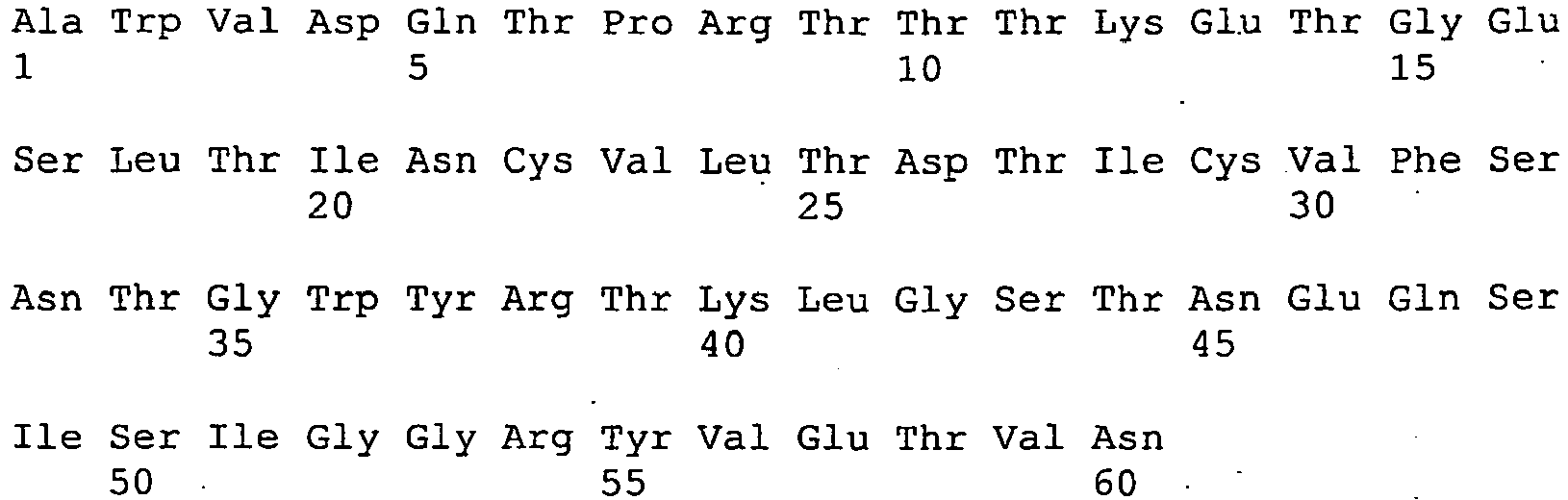
- <210> 108
<211> 60
<212> PRT
<213> Artificial - <220>
<223> Variable domain from aType 2 immunoglobulin new antigen receptor from Orectolobus maculatus. - <400> 108

- <210> 109
<211> 60
<212> PRT
<213> Artificial - <220>
<223> Variable domain from aType 2 immunoglobulin new antigen receptor from Orectolobus maculatus. - <400> 109


- <210> 110
<211> 60
<212> PRT
<213> Artificial - <220>
<223> Variable domain from aType 2 immunoglobulin new antigen receptor from Orectolobus maculatus. - <400> 110

- <210> 111
<211> 60
<212> PRT
<213> Artificial - <220>
<223> Variable domain from aType 2 immunoglobulin new antigen receptor from Orectolobus maculatus. - <400> 111

| No | 12Y-1 | Var | Con | No | 12Y-1 | Var | Con | No | 12Y-1 | Var | Con |
| 12Y-2 | 12Y-2 | 12Y-2 | |||||||||
| 12A-9 | 12A-9 | 12A-9 | |||||||||
| 1A-7 | 1A-7 | 1A-7 | |||||||||
| 1 | A | VQ GT | AVOGT | 37 | Y | Y | 68 | L | - | L | |
| 2 | WR | L | WRL | 38 | R | - | R | 69 | RT | S | RTS |
| 3 | V | AT | VAT | 39 | T | absent | Tor absent | 70 | I | - | I |
| 4 | D | E | DE | 40 | KT | R | KTR | 71 | RS | - | RS |
| 5 | Q | P | QP | 41 | L | F | LF | 72 | D | E | DE |
| 6 | T | KP | TKP | 42 | G | DS | GDS | 73 | L | - | L |
| 7 | P | - P | 43 | S | - S | 74 | R | S | RS | ||
| 8 | R | KTH | RKTH | 44 | T | - | T | 75 | V | - | V |
| 9 | TIS | P | TISP | 45 | NK | - | NK | 76 | E | - | E |
| 10 | AV | E | AVE | 46 | E | L | EL | 77 | D | - | D |
| 11 | T | - | T | 47 | Q | - | Q | 78 | S | - | S |
| 12 | K | - | K | 48 | TSK | FAH | TSKFA H | 79 | G | SA | GSA |
| 13 | E | - | E | 49 | I | M | 1M | 80 | T | - | T |
| 14 | T | R | TR | 50 | S | TP | STP | 81 | Y | F | YF |
| 15 | G | - | G | 51 | I | L | IL | 82 | K | QR | KQR |
| 16 | E | G | EG | 52 | G | - | G | 83 | C | - | C |
| 17 | S | - | S | 53 | G | - | G | 84 | GQK | SH | GQKSH |
| 18 | L | - | L | 54 | R | - | R | 85 | A | GNV | AGNV |
| 19 | T | - | T | 55 | Y | H | YH | Variable region | |||
| 20 | I | - | I | 56 | VS | L | VSL | 104 | KE | PW | KEPW |
| 21 | N | SYH | NSYH | 57 | E | - | E | 105 | G | - | G |
| 22 | C | - | C | 58 | T | - | T | 106 | A | GFV | AGFV |
| 23 | VA | - | VA | 59 | V | E | VE | 107 | G | DL | GDL |
| 24 | L | VI | LVI | 60 | ND | S | NDS | 108 | T | - | T |
| 25 | RK | - | RK | 61 | KE | RL | RL | 109 | VA | DLI | VADLI |
| 26 | DN | ES | DNES | 62 | G | TER | GTER | 110 | L | - | L |
| 27 | AT | S | ATS | 63 | S | - | S | 111 | T | - | T |
| Variable region | 64 | KN | E | KNE | 112 | V | - | V | |||
| 34 | T | - | T | 65 | S | - | S | 113 | K | RT | KRT |
| 35 | GND | RY W | GNDRY W | 66 | FA | - | FA | ||||
| 36 | W | W | 67 | S | - | S | |||||
| Var : Variant amino acids : These columns show the variation in amino acid residues found in the corresponding positions in the twelve other Type 2 VNARs sequences in Con: Consensus sequence |
| Native 12Y-1 | LAH 12Y-1 | PHR 12Y-1 | Native 12Y-2 | Native 12A-9 | Native 1A-7 | ||
| Diffraction data | |||||||
| Space Group | I4122 | I4122 | I4122 | 1212121 | P21212 | 1212121 | |
| Unit cell (Å) | (a) | 97.26 | 97.97 | 97.60 | 65.28 | 38.27 | 80.50 |
| (b) | 97.26 | 97.97 | 97.60 | 92.05 | 68.32 | 88.66 | |
| (c) | 65.23 | 65.61 | 65.27 | 98.22 | 39.51 | 101.75 | |
| Resolution (Å) | 69-2.8 | 20-3.0 | 20-2.5 | 67.4-2.18 | 39.5-2.1 | 21.6-2.7 | |
| - (outer shell) | (2.9-2.8) | (3.11-3.0) | (2.6-2.5) | (2.24-2.18) | (2.15-2.1) | (2.78-2.7) | |
| Measured reflections | 60022 | 42615 | 48364 | 99606 | 43653 | 52866 | |
| Unique reflections | 3975 | 3304 | 5472 | 15764 | 6048 | 18372 | |
| Multiplicity | 15.1 (4.3) | 12.9 (8.3) | 8.8 (5.0) | 6.6 (6.3) | 7.2 (6.3) | 2.9 (2.2) | |
| Completeness (%) (outer shell) | 99 (94.3) | 97.2 (82.5) | 96 (77) | 100 (99.4) | 98.6 (100) | 95.1 (87.9) | |
| I/σ(I) (outer shell) | 18.1 (1.8) | 18.4 (1.8) | 8.6 (1.5) | 32.5 (4.0) | 15.2 (2.0) | 7.6 (1.2) | |
| χ2 (outer shell) | 1.21 (1.10) | 1.74 (1.60) | 1.09 (1.40) | 1.19 (0.95) | 1.03 (0.97) | 1.27 (1.03) | |
| Rmerge (%) | 4.5 | 10.2 | 10.0 | 5.4 | 12.6 | 14.8 | |
| Refinement | |||||||
| Resolution range (Å) | 6.0 - 2.82 | 18.12-2.18 | 34.2-2.10 | 21.6-2.7 | |||
| R % (outer shell) | 16.6 (33.9) | 17.6 (22.5) | 21.7 (29.5) | 17.6 (32.7) | |||
| Rfree % (outer shell) | 25.4 (54.3) | 24.7 (31.1) | 28:0 (39.9) | 26.5 (32.8) | |||
| RMS deviations: | |||||||
| Bond length (Å) | 0.0012 | 0.012 | 0.012 | 0.012 | |||
| Bond angles (deg.) | 1.552 | 1.484 | 1.494 | 1.453 | |||
| Average B values (Å2) | 40.2 | 21.3 | 49.9 | 43.3 | |||
| Region | Number of residues | Residue Numbering |
| Loop region 1 | 1-2 | 1 to 2 |
| β-strand region A | 5-6 | 2 to 6-7 |
| Loop region 2 | 2-3 | 6-7 to 8-9 |
| β-strand region A | 5-6 | 8-9 to 13-14 |
| Loop region 3 | 4-5 | 13-14 to 17-18 |
| β-strand region B | 8-10 | 17-18 to 26-28 |
| Loop region 4 | 5-9 | 26-28 to 32-34 |
| β-strand region C | 7-9 | 32-34 to 40-41 |
| Loop region 5 | 14-17 | 40-41 to 54-55 |
| β-strand region D | 6-7 | 54-55 to 60-61 |
| Loop region 6 | 4-5 | 60-61 to 64-65 |
| β-strand region E | 6-7 | 64-65 to 70-71 |
| Loop region 7 | 8-9 | 70-71 to 78-79 |
| β-strand region F | 6-8 | 78-79 to 85-86 |
| Loop region 8 | 5-25 | 85-86 to # |
| β-strand region G | 10-12 | 97-98 to 105-120* |
| Region | Number of residues | Residue Numbering |
| Loop region 5 | 14-17 | 40-41 to 54-55 |
| β-strand region C | ...1 | ...40 |
| Loop region 5a | 6 | 41-46 |
| β-strand region C' | 2 | 47,48 |
| Loop region 5b | 3 | 49-51 |
| β-strand region D' | 1 | 52 |
| Loop region 5c | 2 | 53,54 |
| Hydrogen bond distance (Å) <4Å | ||||
| Interface* | Hydrogen bonds | 12Y-2 A | 12Y-2 B | 12Y-1 |
| D-Ds strands | Main-chain H-bonds: | |||
| Val59(N) - Val59(O)s | 2.88 | 2.93 | 3.15 | |
| Val59(O) - Val59(N)s | 2.88 | 2.93 | 3.15 | |
| Side-chain H-bonds: | ||||
| Lys61(NZ) - Glu57(OE1)s | 2.65 | 3.16 | ||
| Water mediated H-bonds: | ||||
| W(20)/W(37) - Lys61(N) | 3.37 | 3.12 | ||
| W(20)/W(37) - Gly62(N) | 3.09 | 2.64 | ||
| W(20)/W(37) - Glu57(O) s | 2.73 | 2.88 | ||
| W(20)/W(37) - Glu57(N)s | 2.75 | 2.92 | ||
| W(1)/W(11) - Glu57(OE1)s | 2.96 | 2.66 | ||
| W(1)/W(11) - Lys61(N) | 2.81 | 3.00 | ||
| W(1)/W(11) - Val59(O) | 2.81 | 3.13 | ||
| W(81) - Thr58(OG1) | 3.01 | |||
| W(81)-Asn60(OD1))s | 3.13 | |||
| CDR3-CDR3s | Main-chain H-bonds: | |||
| Asn95(N)-Arg101(O)s | 3.25 | 3.05 | ||
| Arg101(N) - Asn95(O)s | 3.25 | 3.05 | ||
| Ser97(N) - Leu99(O) s | 2.9 | 3.11 | ||
| Ser97(O) - Leu99(N)s | 3.09 | 2.74 | ||
| Side-chain H-bonds: | ||||
| Asn95(OD1) - Arg101(NE)s | 2.72 | |||
| Tyr94(O) - Arg101(NE)s | 2.69 | |||
| Tyr94(OH) - Arg101(NH1)s | 3.36 | |||
| CDR1-CDR1s | Side-chain H-bonds: | |||
| Lys 32(O) -Asp33(OD1)s | 2.42 | 3.25 | ||
| Lys 32(O) -Lys61 (NZ) s | 2.84 | |||
| *Subscript "s" is for a symmetry (2-fold) related molecule. |

| TCR Vα 1AO7 | TCR Vα 1J8H | TCR Vα 1KGC | | 12Y-2 | Camelid | Comments | |
| Ser31 | Tyr31 | Tyr31 | Tyr31 | Asp33 | |||
| Phe33 | Phe33 | His33 | Pro33 | Gly35 | |||
| Tyr35 | Tyr35 | Tyr35 | Tyr35 | Tyr37 | | ||
| Gln3 | |||||||
| 7 | Gln37 | Gln37 | Gln37 | Thr39 | |||
| Ser42 | Gly42 | Gly42 | Gly42 | Asn45 | G44E£ | Deviation¥ | |
| Pro43 | Leu43 | Pro43 | Pro43 | Glu46 | L45R£ | ||
| Leu45 | Leu45 | Tyr45 | Leu45 | Ser48 | W47G/S£ | ||
| Ser48 | Lys48 | His48 | Ser48 | - * | CDR2 | ||
| Tyr50 | Thr50 | Leu50 | Arg50 | - * | CDR2 | ||
| Ser51 | Ser51 | Thr51 | Ser51 | - * | CDR2 | ||
| Asn52 | Ala52 | Ser52 | Val52 | - * | CDR2 | ||
| Leu89 | Phe89 | Tyr89 | Phe89 | Lys82 | |||
| Thr93 | Ser93 | Pro93 | Arg93 | Phe86 | Deviation¥ | ||
| Leu104 | Leu104 | Leu104 | Leu104 | Gly102 | Deviation¥ | ||
| Phe106 | Phe106 | | Phe106 | Lys | 104 |
| * Residues is TCR CDR2 loop, no equivalent position in IgNAR variable domains. ¥ Deviation between Cα traces of shark and TCR structures. £ Camelisation of human domains, difficult to correlate. |
Claims (16)
- A method of altering a property of an IgNAR variable domain comprising:(a) providing an IgNAR variable domain comprising eight β-strand regions, designated A, A', B, C, D, E, F and G according to Figure 2, and nine loop regions, designated 1 to 9 according to Figure 2;(b) modifying loop region 8, which is a CDR3 analogous region, by addition of up to 3 amino acids or the deletion of up to 3 amino acid residues; and(c) identifying modifications that result in one or more of the following properties being altered:(i) the binding affinity of the IgNAR variable domain is increased by at least 5% compared to the unmodified domain;(ii) the solubility of the IgNAR variable domain being improved by at least 5% compared to the unmodified domain; and(iii) the stability of the IgNAR variable domain being improved by at least 5% compared to the unmodified domain.
- The method according to claim 1, in which the amino acid sequence of the unmodified β-strand regions A, A', B, C, D, E, F and G and loop regions 1, 2, 3, 6, 7 and 9 comprises an amino acid sequence according to Figure 1 and/or Table 1.
- The method according to any preceding claim, in which the unmodified β-strand regions and loop regions have the amino acid residue numbering according to Table 3.
- The method according to any preceding claim, in which the unmodified loop region 5 comprises two β-strand regions designated C' and D' according to Figure 3 and three loop regions designated 5a, 5b and 5c according to Figure 3.
- The method according to claim 4, in which the unmodified β-strand regions C, C' and D' have the amino acid residue numbering according to Table 3A.
- The method according to claim 4 or claim 5, in which the Cα trace of loop region 5b in the unmodified IgNAR variable domain is no more than 5Å above the plane formed by the Cα trace of residues 22, 83 and 36 as defined in Table 1.
- The method according to any preceding claim, in which the unmodified IgNAR is derived from a shark.
- The method according to any preceding claim, in which one or more additional loop regions of the IgNAR are modified.
- The method according to any preceding claim, in which the IgNAR variable domain is also modified by grafting a CDR loop or portion thereof from a V-set or an I-set domain into loop region 1, 2, 3, 4, 5, 6, 7 or 9 of the IgNAR variable domain.
- The method according to claim 9, in which the CDR loop or portion thereof is grafted into loop region 4 of the IgNAR variable domain.
- The method according to claim 9, in which amino acids from loop region 4 are replaced by grafting a CDR1 loop or portion thereof into loop region 4 of the IgNAR variable domain.
- The method according to claim 9, in which amino acids 28 to 33 as defined in Table 1 or a portion thereof from loop region 4 are replaced by grafting a CDR1 loop or portion thereof into loop region 4 of the IgNAR variable domain.
- The method of claim 1, in which one or more amino acid residues within the patch defined by residues 33, 37, 46, 48, 50, 51, 59, 61, 86, 94, 95, 96, 98, 99 and 101 as shown in Table 1 are also modified.
- The method according to any preceding claim, in which at least one of the β-strand regions or loop regions 1-3, 5-7 or 9 is also modified.
- The method according to claim 14, in which at least one of β-strand regions C, D, E or F or loop regions 5, 6 or 7 is modified.
- The method according to claim 15, in which at least one of β-strand regions C or D or loop region 5 is modified.
Priority Applications (3)
| Application Number | Priority Date | Filing Date | Title |
|---|---|---|---|
| DK10179638.1T DK2330121T3 (en) | 2004-06-02 | 2005-06-02 | Binding moieties based on shark IgNar domains |
| EP10179611A EP2330120A3 (en) | 2004-06-02 | 2005-06-02 | Binding moieties based on Shark IgNAR domains |
| EP10179638.1A EP2330121B1 (en) | 2004-06-02 | 2005-06-02 | Binding moieties based on Shark IgNAR domains |
Applications Claiming Priority (2)
| Application Number | Priority Date | Filing Date | Title |
|---|---|---|---|
| US57584504P | 2004-06-02 | 2004-06-02 | |
| PCT/AU2005/000789 WO2005118629A1 (en) | 2004-06-02 | 2005-06-02 | BINDING MOIETIES BASED ON SHARK IgNAR DOMAINS |
Related Child Applications (3)
| Application Number | Title | Priority Date | Filing Date |
|---|---|---|---|
| EP10179638.1A Division EP2330121B1 (en) | 2004-06-02 | 2005-06-02 | Binding moieties based on Shark IgNAR domains |
| EP10179638.1A Division-Into EP2330121B1 (en) | 2004-06-02 | 2005-06-02 | Binding moieties based on Shark IgNAR domains |
| EP10179611A Division-Into EP2330120A3 (en) | 2004-06-02 | 2005-06-02 | Binding moieties based on Shark IgNAR domains |
Publications (3)
| Publication Number | Publication Date |
|---|---|
| EP1751181A1 EP1751181A1 (en) | 2007-02-14 |
| EP1751181A4 EP1751181A4 (en) | 2008-03-12 |
| EP1751181B1 true EP1751181B1 (en) | 2012-08-15 |
Family
ID=35462883
Family Applications (3)
| Application Number | Title | Priority Date | Filing Date |
|---|---|---|---|
| EP05744734A Expired - Lifetime EP1751181B1 (en) | 2004-06-02 | 2005-06-02 | BINDING MOIETIES BASED ON SHARK IgNAR DOMAINS |
| EP10179611A Withdrawn EP2330120A3 (en) | 2004-06-02 | 2005-06-02 | Binding moieties based on Shark IgNAR domains |
| EP10179638.1A Expired - Lifetime EP2330121B1 (en) | 2004-06-02 | 2005-06-02 | Binding moieties based on Shark IgNAR domains |
Family Applications After (2)
| Application Number | Title | Priority Date | Filing Date |
|---|---|---|---|
| EP10179611A Withdrawn EP2330120A3 (en) | 2004-06-02 | 2005-06-02 | Binding moieties based on Shark IgNAR domains |
| EP10179638.1A Expired - Lifetime EP2330121B1 (en) | 2004-06-02 | 2005-06-02 | Binding moieties based on Shark IgNAR domains |
Country Status (7)
| Country | Link |
|---|---|
| US (3) | US7977071B2 (en) |
| EP (3) | EP1751181B1 (en) |
| JP (2) | JP2008511286A (en) |
| AU (3) | AU2005250055B2 (en) |
| CA (1) | CA2567655C (en) |
| DK (2) | DK2330121T3 (en) |
| WO (1) | WO2005118629A1 (en) |
Families Citing this family (83)
| Publication number | Priority date | Publication date | Assignee | Title |
|---|---|---|---|---|
| US9453251B2 (en) | 2002-10-08 | 2016-09-27 | Pfenex Inc. | Expression of mammalian proteins in Pseudomonas fluorescens |
| EP1774017B1 (en) | 2004-07-26 | 2013-05-15 | Pfenex Inc. | Process for improved protein expression by strain engineering |
| US9394571B2 (en) | 2007-04-27 | 2016-07-19 | Pfenex Inc. | Method for rapidly screening microbial hosts to identify certain strains with improved yield and/or quality in the expression of heterologous proteins |
| US9580719B2 (en) | 2007-04-27 | 2017-02-28 | Pfenex, Inc. | Method for rapidly screening microbial hosts to identify certain strains with improved yield and/or quality in the expression of heterologous proteins |
| AU2008270274B2 (en) | 2007-07-03 | 2012-06-28 | Ablynx N.V. | Providing improved immunoglobulin sequences by mutating CDR and/or FR positions |
| WO2009026638A1 (en) * | 2007-08-31 | 2009-03-05 | Melbourne Health | Marine-animal derived therapeutic and diagnostic agents for hepatitis b |
| EP2310850B1 (en) | 2008-07-25 | 2018-10-17 | Cellestis Limited | A diagnostic method |
| US20100136584A1 (en) * | 2008-09-22 | 2010-06-03 | Icb International, Inc. | Methods for using antibodies and analogs thereof |
| US20100092470A1 (en) * | 2008-09-22 | 2010-04-15 | Icb International, Inc. | Antibodies, analogs and uses thereof |
| US8524863B2 (en) | 2008-10-06 | 2013-09-03 | Commonwealth Scientific And Industrial Research Organisation | Amyloid-beta peptide crystal structure |
| EP2427486B1 (en) | 2009-05-07 | 2015-02-25 | Novozymes Biopharma DK A/S | Method for purifying albumin |
| EP2448966B1 (en) | 2009-07-03 | 2018-11-14 | Avipep Pty Ltd | Immuno-conjugates and methods for producing them |
| US8496933B2 (en) | 2009-11-04 | 2013-07-30 | Laboratorios Silanes, S.A. De C.V. | VHNAR anti-cytokine domains |
| WO2011075786A1 (en) | 2009-12-23 | 2011-06-30 | Avipep Pty Ltd | Immuno-conjugates and methods for producing them 2 |
| US9459253B2 (en) | 2009-12-23 | 2016-10-04 | Cellestis Limited | Assay for measuring cell-mediated immunoresponsiveness |
| US9624294B2 (en) | 2011-03-14 | 2017-04-18 | Cellmid Limited | Antibody recognizing N-domain of midkine |
| CN103620051A (en) | 2011-04-29 | 2014-03-05 | 塞尔雷斯蒂斯有限公司 | Cell Mediated Immune Response Assay |
| CA2838246C (en) | 2011-06-13 | 2018-07-10 | Csl Limited | Antibodies against g-csfr and uses thereof |
| LT2726883T (en) | 2011-06-29 | 2018-06-11 | Cellestis Limited | A cell mediated immune response assay with enhanced sensitivity |
| AU2012327878A1 (en) | 2011-10-28 | 2014-05-29 | Patrys Limited | PAT-LM1 epitopes and methods for using same |
| US10112988B2 (en) * | 2012-01-09 | 2018-10-30 | Icb International, Inc. | Methods of assessing amyloid-beta peptides in the central nervous system by blood-brain barrier permeable peptide compositions comprising a vab domain of a camelid single domain heavy chain antibody against an anti-amyloid-beta peptide |
| US10774132B2 (en) | 2012-01-09 | 2020-09-15 | The Scripps Research Instittue | Ultralong complementarity determining regions and uses thereof |
| US10112987B2 (en) * | 2012-01-09 | 2018-10-30 | Icb International, Inc. | Blood-brain barrier permeable peptide compositions comprising a vab domain of a camelid single domain heavy chain antibody against an amyloid-beta peptide |
| MX347154B (en) * | 2012-02-24 | 2017-02-21 | Centro De Investigación Científica Y De Educación Superior De Ensenada Baja California (Cicese) | Diagnostic test for infectious diseases in cattle. |
| WO2013167883A1 (en) | 2012-05-07 | 2013-11-14 | The University Court Of The University Of Aberdeen | Single domain binding molecule |
| CN110118871A (en) | 2012-12-28 | 2019-08-13 | 塞尔雷斯蒂斯有限公司 | Cell-mediated immune response test |
| CN105073781A (en) | 2013-01-11 | 2015-11-18 | 加州生物医学研究所 | Bovine fusion antibodies |
| EP2762496A1 (en) | 2013-02-05 | 2014-08-06 | EngMab AG | Method for the selection of antibodies against BCMA |
| US9963513B2 (en) | 2013-02-05 | 2018-05-08 | Engmab Sàrl | Method for the selection of antibodies against BCMA |
| KR102489953B1 (en) | 2013-02-07 | 2023-01-20 | 씨에스엘 리미티드 | Il-11r binding proteins and uses thereof |
| EP2789630A1 (en) | 2013-04-09 | 2014-10-15 | EngMab AG | Bispecific antibodies against CD3e and ROR1 |
| EP3683312B1 (en) * | 2013-04-23 | 2022-09-07 | Elasmogen Limited | Synthetic library of specific binding molecules |
| RU2718054C2 (en) | 2013-11-28 | 2020-03-30 | СиЭсЭл ЛИМИТЕД | Method of treating nephropathy |
| CN105979965B (en) | 2013-12-18 | 2021-07-09 | 杰特有限公司 | Method of treating wounds |
| WO2015100246A1 (en) | 2013-12-24 | 2015-07-02 | Ossianix, Inc. | Baff selective binding compounds and related methods |
| CN106459193A (en) * | 2014-04-17 | 2017-02-22 | 特瑞克隆艾迪福公司 | vNAR recombinant monoclonal antibodies that neutralize vascular endophelial growth factor VEGF |
| JP6454981B2 (en) * | 2014-04-24 | 2019-01-23 | 住友電気工業株式会社 | Semiconductor laminate and light receiving element |
| JP6355020B2 (en) * | 2014-05-14 | 2018-07-11 | パナソニックIpマネジメント株式会社 | Eye beauty equipment |
| EP3160995A2 (en) | 2014-06-26 | 2017-05-03 | Ossianix Inc. | Semi-synthetic nurse shark vnar libraries for making and using selective binding compounds |
| CA2963692A1 (en) | 2014-10-09 | 2016-04-14 | Engmab Ag | Bispecific antibodies against cd3epsilon and ror1 |
| WO2016061632A1 (en) | 2014-10-23 | 2016-04-28 | La Trobe University | Fn14-binding proteins and uses thereof |
| KR20240045370A (en) | 2014-10-23 | 2024-04-05 | 퀴아젠 사이언시스, 엘엘씨 | Peptide composition and uses thereof |
| WO2016077840A2 (en) | 2014-11-14 | 2016-05-19 | Ossianix, Inc. | TfR SELECTIVE BINDING COMPOUNDS AND RELATED METHODS |
| EP3233120A4 (en) | 2014-12-19 | 2018-05-30 | Monash University | Il-21 antibodies |
| EP3242685B1 (en) * | 2015-01-09 | 2022-05-04 | AdAlta Limited | Cxcr4 binding molecules |
| US20180267024A1 (en) | 2015-06-08 | 2018-09-20 | Lophius Biosciences Gmbh | Composition for determination of cell-mediated immune responsiveness |
| AU2016302881B2 (en) | 2015-08-03 | 2022-09-15 | Bristol-Myers Squibb Company | Monoclonal antibodies against BCMA |
| AU2016318230B2 (en) | 2015-09-11 | 2020-05-21 | Biosceptre (Aust) Pty Ltd | Chimeric antigen receptors and uses thereof |
| US10273196B2 (en) | 2015-09-25 | 2019-04-30 | Exxonmobil Chemical Patents Inc. | Hydrocarbon dehydrocyclization |
| CN108368167B (en) | 2015-11-27 | 2023-09-05 | 杰特有限公司 | CD131 binding proteins and uses thereof |
| CA3022697A1 (en) | 2016-05-02 | 2017-11-09 | Ablynx Nv | Treatment of rsv infection |
| AU2017331739A1 (en) | 2016-09-23 | 2019-03-07 | Csl Limited | Coagulation factor binding proteins and uses thereof |
| EP3535295A1 (en) | 2016-11-02 | 2019-09-11 | EngMab Sàrl | Bispecific antibody against bcma and cd3 and an immunological drug for combined use in treating multiple myeloma |
| JP7258762B2 (en) | 2016-11-07 | 2023-04-17 | デーゲンエルイックス べスローテン フェンノートシャップ | A novel amyloid-beta oligomer-specific binding molecule |
| MX2019012566A (en) | 2017-04-21 | 2019-12-02 | Centro De Investig Cientifica Y De Educacion Superior De Ensenada Baja California | Vnar having anti-angiogenic activity in solid tumours of pet animals. |
| EP3652209A2 (en) | 2017-07-11 | 2020-05-20 | Compass Therapeutics LLC | Agonist antibodies that bind human cd137 and uses thereof |
| JP7733443B2 (en) | 2017-09-11 | 2025-09-03 | モナシュ ユニバーシティー | Binding protein for human thrombin receptor PAR4 |
| CA3075367A1 (en) | 2017-09-27 | 2019-04-04 | Elasmogen Ltd | Specific binding molecules |
| WO2019067015A1 (en) | 2017-09-29 | 2019-04-04 | City Of Hope | Chimeric antigen receptors and bispecific antibodies for treatment of mantle cell lymphoma |
| US11718679B2 (en) | 2017-10-31 | 2023-08-08 | Compass Therapeutics Llc | CD137 antibodies and PD-1 antagonists and uses thereof |
| US11180544B2 (en) * | 2017-11-07 | 2021-11-23 | City University Of Hong Kong | Method of producing antibody fragment |
| EP3713961A2 (en) | 2017-11-20 | 2020-09-30 | Compass Therapeutics LLC | Cd137 antibodies and tumor antigen-targeting antibodies and uses thereof |
| JP7465210B2 (en) | 2017-11-29 | 2024-04-10 | シーエスエル リミティド | Method for treating or preventing ischemia-reperfusion injury |
| US12281160B2 (en) | 2018-03-23 | 2025-04-22 | Csl Limited | Method of treating asthma |
| CN112166193A (en) | 2018-05-21 | 2021-01-01 | 生物权威(英国)有限公司 | Chimeric antigen receptors with modified linker domains and uses thereof |
| CN112867503A (en) | 2018-08-24 | 2021-05-28 | 希望之城 | Masked cytokine conjugates |
| US12351642B2 (en) | 2018-09-14 | 2025-07-08 | Ossianix, Inc. | TFR-specific binding moieties and transcytosis method to select VNARs that cross cellular barriers |
| US12016885B2 (en) | 2018-11-06 | 2024-06-25 | Alsatech, Inc. | Method of solubilizing TDP-43 protein aggregates in amyotrophic lateral sclerosis using a microvascular endothelial cell expressing an anti-TDP-43 Fab |
| WO2020102387A1 (en) | 2018-11-13 | 2020-05-22 | Compass Therapeutics Llc | Multispecific binding constructs against checkpoint molecules and uses thereof |
| CA3158174A1 (en) | 2019-10-28 | 2021-05-06 | Monash University | Antibodies for binding plasmin |
| CN115768475A (en) | 2020-06-04 | 2023-03-07 | Csl创新私人有限公司 | Methods of treating or preventing acute respiratory distress syndrome |
| WO2022035998A1 (en) | 2020-08-11 | 2022-02-17 | City Of Hope | Compositions and uses of sars-cov-2 targeted chimeric antigen receptor modified nk cells |
| GB202020152D0 (en) | 2020-12-18 | 2021-02-03 | Elasmogen Ltd | Fast-track humanisation of specific binding molecules |
| US20250297020A1 (en) * | 2021-11-23 | 2025-09-25 | Adalta Limited | Rank-l binding molecules |
| US12098400B2 (en) | 2022-07-08 | 2024-09-24 | Novo Nordisk A/S | Highly potent ISVD compounds capable of substituting for FVIII(A) |
| PE20251082A1 (en) | 2022-07-08 | 2025-04-10 | Novo Nordisk As | Highly potent ISVD compounds capable of replacing FVIII(A) |
| CN116284423B (en) * | 2022-12-29 | 2023-10-03 | 江苏百英生物科技有限公司 | A polyclonal antibody targeting striped bamboo shark IgNAR-CH1, its preparation method and its application |
| WO2024200846A1 (en) | 2023-03-30 | 2024-10-03 | 272BIO Limited | Gnrh-binding polypeptides and uses thereof |
| AU2024290964A1 (en) | 2023-07-05 | 2026-01-08 | CSL Innovation Pty Ltd | Methods of treating or preventing a complication of sickle cell disease |
| CN118702816B (en) * | 2024-06-03 | 2025-03-28 | 内蒙古正德缘生物科技有限责任公司 | A kind of NK cell and its application in anti-tumor treatment |
| WO2026093923A1 (en) | 2024-10-29 | 2026-05-07 | Csl Behring Ag | Method for detection of a plasma protein |
| CN119529102B (en) * | 2025-01-22 | 2025-06-17 | 青岛科技大学 | Preparation method and application of shark nanoantibody targeting pancreatic lipase |
| CN120173114B (en) * | 2025-03-24 | 2025-09-26 | 广州泽朗生物医药科技有限公司 | CART (cell-associated antigen receptor) cells and application thereof in preparation of anti-cancer drugs |
Family Cites Families (15)
| Publication number | Priority date | Publication date | Assignee | Title |
|---|---|---|---|---|
| US4179337A (en) | 1973-07-20 | 1979-12-18 | Davis Frank F | Non-immunogenic polypeptides |
| GB8607679D0 (en) | 1986-03-27 | 1986-04-30 | Winter G P | Recombinant dna product |
| ES2052027T5 (en) | 1988-11-11 | 2005-04-16 | Medical Research Council | IMMUNOGLOBULINE VARIABLE DOMAIN SEQUENCE CLONING. |
| GB8927230D0 (en) | 1989-12-01 | 1990-01-31 | Unilever Plc | Reagents |
| US5955363A (en) | 1990-01-03 | 1999-09-21 | Promega Corporation | Vector for in vitro mutagenesis and use thereof |
| US6838254B1 (en) | 1993-04-29 | 2005-01-04 | Conopco, Inc. | Production of antibodies or (functionalized) fragments thereof derived from heavy chain immunoglobulins of camelidae |
| EP0739981A1 (en) | 1995-04-25 | 1996-10-30 | Vrije Universiteit Brussel | Variable fragments of immunoglobulins - use for therapeutic or veterinary purposes |
| US6153380A (en) | 1996-01-23 | 2000-11-28 | Rigel Pharmaceuticals, Inc. | Methods for screening for transdominant intracellular effector peptides and RNA molecules |
| AU747637B2 (en) * | 1998-03-06 | 2002-05-16 | Arana Therapeutics (Vic) Pty Ltd | V-like domain binding molecules |
| AUPP221098A0 (en) * | 1998-03-06 | 1998-04-02 | Diatech Pty Ltd | V-like domain binding molecules |
| EP2141243A3 (en) * | 2000-10-16 | 2010-01-27 | Brystol-Myers Squibb Company | Protein scaffolds for antibody mimics and other binding proteins |
| CA2457636C (en) * | 2001-08-10 | 2012-01-03 | Aberdeen University | Antigen binding domains |
| MXPA04001974A (en) * | 2001-08-31 | 2004-07-16 | Avidex Ltd | Soluble t cell receptor. |
| US20050214857A1 (en) | 2001-12-11 | 2005-09-29 | Algonomics N.V. | Method for displaying loops from immunoglobulin domains in different contexts |
| US20040266993A1 (en) * | 2003-06-30 | 2004-12-30 | Evans Glen A. | Non-immunoglobulin binding polypeptides |
-
2005
- 2005-06-02 DK DK10179638.1T patent/DK2330121T3/en active
- 2005-06-02 AU AU2005250055A patent/AU2005250055B2/en not_active Expired
- 2005-06-02 CA CA2567655A patent/CA2567655C/en not_active Expired - Lifetime
- 2005-06-02 DK DK05744734.4T patent/DK1751181T3/en active
- 2005-06-02 JP JP2007513617A patent/JP2008511286A/en active Pending
- 2005-06-02 EP EP05744734A patent/EP1751181B1/en not_active Expired - Lifetime
- 2005-06-02 EP EP10179611A patent/EP2330120A3/en not_active Withdrawn
- 2005-06-02 US US11/628,475 patent/US7977071B2/en not_active Expired - Lifetime
- 2005-06-02 EP EP10179638.1A patent/EP2330121B1/en not_active Expired - Lifetime
- 2005-06-02 WO PCT/AU2005/000789 patent/WO2005118629A1/en not_active Ceased
-
2008
- 2008-09-29 AU AU2008229687A patent/AU2008229687B2/en not_active Expired
-
2009
- 2009-04-28 AU AU2009201692A patent/AU2009201692B2/en not_active Expired
-
2011
- 2011-06-09 US US13/157,205 patent/US20120003214A1/en not_active Abandoned
- 2011-06-10 JP JP2011130413A patent/JP5631812B2/en not_active Expired - Fee Related
-
2016
- 2016-02-16 US US15/044,731 patent/US20160237169A1/en not_active Abandoned
Non-Patent Citations (2)
| Title |
|---|
| BOND C J ET AL: "Contributions of CDR3 to VHH Domain Stability and the Design of Monobody Scaffolds for Naive Antibody Libraries", JOURNAL OF MOLECULAR BIOLOGY, LONDON, GB, vol. 332, no. 3, 19 September 2003 (2003-09-19), pages 643 - 655, XP004454278, ISSN: 0022-2836, DOI: 10.1016/S0022-2836(03)00967-7 * |
| DIAZ M ET AL: "Structural analysis, selection, and ontogeny of the shark new antigen receptor (IgNAR): identification of a new locus preferentially expressed in early development", IMMUNOGENETICS, SPRINGER VERLAG, BERLIN, DE, vol. 54, no. 7, 1 October 2002 (2002-10-01), pages 501 - 512, XP003013542, ISSN: 0093-7711, DOI: 10.1007/S00251-002-0479-Z * |
Also Published As
| Publication number | Publication date |
|---|---|
| AU2009201692B2 (en) | 2012-08-30 |
| AU2005250055A1 (en) | 2005-12-15 |
| CA2567655C (en) | 2015-06-30 |
| AU2008229687B2 (en) | 2009-01-29 |
| EP1751181A4 (en) | 2008-03-12 |
| AU2008229687A1 (en) | 2008-10-30 |
| EP1751181A1 (en) | 2007-02-14 |
| JP5631812B2 (en) | 2014-11-26 |
| EP2330121A2 (en) | 2011-06-08 |
| WO2005118629A1 (en) | 2005-12-15 |
| DK2330121T3 (en) | 2014-12-15 |
| US7977071B2 (en) | 2011-07-12 |
| EP2330121A3 (en) | 2011-11-16 |
| JP2011244822A (en) | 2011-12-08 |
| EP2330121B1 (en) | 2014-09-03 |
| US20120003214A1 (en) | 2012-01-05 |
| EP2330120A2 (en) | 2011-06-08 |
| US20090148438A1 (en) | 2009-06-11 |
| DK1751181T3 (en) | 2012-11-26 |
| EP2330120A3 (en) | 2011-11-16 |
| AU2005250055B2 (en) | 2008-09-11 |
| US20160237169A1 (en) | 2016-08-18 |
| AU2009201692A1 (en) | 2009-05-21 |
| JP2008511286A (en) | 2008-04-17 |
| CA2567655A1 (en) | 2005-12-15 |
Similar Documents
| Publication | Publication Date | Title |
|---|---|---|
| US7977071B2 (en) | Binding moieties based on shark ignar domains | |
| Conrath et al. | Antigen binding and solubility effects upon the veneering of a camel VHH in framework-2 to mimic a VH | |
| Streltsov et al. | Structure of a shark IgNAR antibody variable domain and modeling of an early‐developmental isotype | |
| JP7097364B2 (en) | Gremlins-1 crystal structure and inhibitory antibody | |
| DK2114996T3 (en) | A process for the preparation of hypo allergens | |
| Duhoo et al. | Camelid nanobodies used as crystallization chaperones for different constructs of PorM, a component of the type IX secretion system from Porphyromonas gingivalis | |
| Langley et al. | Crystal structures of human neuropeptide Y (NPY) and peptide YY (PYY) | |
| Johnson et al. | Structural and biophysical characterization of an epitope-specific engineered Fab fragment and complexation with membrane proteins: implications for co-crystallization | |
| Riechmann et al. | Early protein evolution: building domains from ligand-binding polypeptide segments | |
| Malia et al. | Structure and specificity of an antibody targeting a proteolytically cleaved IgG hinge | |
| Krauß et al. | The structure of the anti‐c‐myc antibody 9E10 Fab fragment/epitope peptide complex reveals a novel binding mode dominated by the heavy chain hypervariable loops | |
| AU2012258331B2 (en) | Binding moieties based on shark IgNAR domains | |
| Clarke et al. | The impact of exchanging the light and heavy chains on the structures of bovine ultralong antibodies | |
| Shahid et al. | Crystal structure of a bivalent antibody Fab fragment | |
| Maritan et al. | Crystal structures of human Fabs targeting the Bexsero meningococcal vaccine antigen NHBA | |
| Veitl et al. | Engineering and Structural Elucidation of a Sac7d‐Derived IgG Fc‐Specific Affitin and Its Application for the Light‐Controlled Affinity Purification of Antibodies | |
| Niemann et al. | Barnase fusion as a tool to determine the crystal structure of the small disulfide-rich protein McoEeTI | |
| Smaldone et al. | The non-swapped monomeric structure of the arginine-binding protein from Thermotoga maritima | |
| Nagae et al. | Structure of the F-spondin reeler domain reveals a unique β-sandwich fold with a deformable disulfide-bonded loop | |
| Scheerer et al. | Structure of an anti‐cholera toxin antibody Fab in complex with an epitope‐derived D‐peptide: a case of polyspecific recognition | |
| Patterson-Orazem | Development of Conformational Myocilin Antibodies, and Biophysical Characterization of the Mouse Myocilin Olfactomedin Domain | |
| Essen et al. | 1.3 Å X-ray structure of an antibody Fv fragment used for induced membrane-protein crystallization | |
| Legg et al. | Monoclonal antibody 7H2. 2 binds the C-terminus of the cancer-oocyte antigen SAS1B through the hydrophilic face of a conserved amphipathic helix corresponding to one of only two regions predicted to be ordered | |
| Frey et al. | Structure of the recombinant antibody Fab fragment f3p4 | |
| WO2026008041A1 (en) | Antigen binding molecule specifically binding to latent-myostatin and medical use thereof |
Legal Events
| Date | Code | Title | Description |
|---|---|---|---|
| PUAI | Public reference made under article 153(3) epc to a published international application that has entered the european phase |
Free format text: ORIGINAL CODE: 0009012 |
|
| 17P | Request for examination filed |
Effective date: 20061120 |
|
| AK | Designated contracting states |
Kind code of ref document: A1 Designated state(s): AT BE BG CH CY CZ DE DK EE ES FI FR GB GR HU IE IS IT LI LT LU MC NL PL PT RO SE SI SK TR |
|
| RIN1 | Information on inventor provided before grant (corrected) |
Inventor name: HENDERSON, KYLIE, ANNE Inventor name: BARRACLOUGH, MILES, MACKAY Inventor name: VARGHESE, JOSEPH, NOOZHUMUTRY Inventor name: IRVING, ROBERT, ALEXANDER Inventor name: HUDSON, PETER Inventor name: NUTTAL, STEWART Inventor name: SIMMONS, DAVID, PETER Inventor name: CARMICHAEL, JENNIFER, ANN Inventor name: GRIFFITHS, KATHERINE, MERNE Inventor name: STRELTSOV, VICTOR |
|
| DAX | Request for extension of the european patent (deleted) | ||
| A4 | Supplementary search report drawn up and despatched |
Effective date: 20080207 |
|
| 17Q | First examination report despatched |
Effective date: 20080602 |
|
| RAP1 | Party data changed (applicant data changed or rights of an application transferred) |
Owner name: ADALTA PTY LTD |
|
| RIC1 | Information provided on ipc code assigned before grant |
Ipc: C07K 14/46 20060101AFI20120207BHEP Ipc: C07K 16/00 20060101ALI20120207BHEP |
|
| GRAP | Despatch of communication of intention to grant a patent |
Free format text: ORIGINAL CODE: EPIDOSNIGR1 |
|
| GRAS | Grant fee paid |
Free format text: ORIGINAL CODE: EPIDOSNIGR3 |
|
| GRAA | (expected) grant |
Free format text: ORIGINAL CODE: 0009210 |
|
| AK | Designated contracting states |
Kind code of ref document: B1 Designated state(s): AT BE BG CH CY CZ DE DK EE ES FI FR GB GR HU IE IS IT LI LT LU MC NL PL PT RO SE SI SK TR |
|
| REG | Reference to a national code |
Ref country code: GB Ref legal event code: FG4D Ref country code: CH Ref legal event code: EP Ref country code: AT Ref legal event code: REF Ref document number: 570790 Country of ref document: AT Kind code of ref document: T Effective date: 20120815 |
|
| REG | Reference to a national code |
Ref country code: IE Ref legal event code: FG4D |
|
| REG | Reference to a national code |
Ref country code: DE Ref legal event code: R096 Ref document number: 602005035654 Country of ref document: DE Effective date: 20121018 |
|
| REG | Reference to a national code |
Ref country code: CH Ref legal event code: NV Representative=s name: BOVARD AG, CH |
|
| REG | Reference to a national code |
Ref country code: DK Ref legal event code: T3 |
|
| REG | Reference to a national code |
Ref country code: NL Ref legal event code: VDEP Effective date: 20120815 |
|
| REG | Reference to a national code |
Ref country code: AT Ref legal event code: MK05 Ref document number: 570790 Country of ref document: AT Kind code of ref document: T Effective date: 20120815 |
|
| PG25 | Lapsed in a contracting state [announced via postgrant information from national office to epo] |
Ref country code: FI Free format text: LAPSE BECAUSE OF FAILURE TO SUBMIT A TRANSLATION OF THE DESCRIPTION OR TO PAY THE FEE WITHIN THE PRESCRIBED TIME-LIMIT Effective date: 20120815 Ref country code: LT Free format text: LAPSE BECAUSE OF FAILURE TO SUBMIT A TRANSLATION OF THE DESCRIPTION OR TO PAY THE FEE WITHIN THE PRESCRIBED TIME-LIMIT Effective date: 20120815 Ref country code: CY Free format text: LAPSE BECAUSE OF FAILURE TO SUBMIT A TRANSLATION OF THE DESCRIPTION OR TO PAY THE FEE WITHIN THE PRESCRIBED TIME-LIMIT Effective date: 20120815 Ref country code: AT Free format text: LAPSE BECAUSE OF FAILURE TO SUBMIT A TRANSLATION OF THE DESCRIPTION OR TO PAY THE FEE WITHIN THE PRESCRIBED TIME-LIMIT Effective date: 20120815 Ref country code: IS Free format text: LAPSE BECAUSE OF FAILURE TO SUBMIT A TRANSLATION OF THE DESCRIPTION OR TO PAY THE FEE WITHIN THE PRESCRIBED TIME-LIMIT Effective date: 20121215 |
|
| PG25 | Lapsed in a contracting state [announced via postgrant information from national office to epo] |
Ref country code: BE Free format text: LAPSE BECAUSE OF FAILURE TO SUBMIT A TRANSLATION OF THE DESCRIPTION OR TO PAY THE FEE WITHIN THE PRESCRIBED TIME-LIMIT Effective date: 20120815 Ref country code: SE Free format text: LAPSE BECAUSE OF FAILURE TO SUBMIT A TRANSLATION OF THE DESCRIPTION OR TO PAY THE FEE WITHIN THE PRESCRIBED TIME-LIMIT Effective date: 20120815 Ref country code: PT Free format text: LAPSE BECAUSE OF FAILURE TO SUBMIT A TRANSLATION OF THE DESCRIPTION OR TO PAY THE FEE WITHIN THE PRESCRIBED TIME-LIMIT Effective date: 20121217 Ref country code: SI Free format text: LAPSE BECAUSE OF FAILURE TO SUBMIT A TRANSLATION OF THE DESCRIPTION OR TO PAY THE FEE WITHIN THE PRESCRIBED TIME-LIMIT Effective date: 20120815 Ref country code: GR Free format text: LAPSE BECAUSE OF FAILURE TO SUBMIT A TRANSLATION OF THE DESCRIPTION OR TO PAY THE FEE WITHIN THE PRESCRIBED TIME-LIMIT Effective date: 20121116 Ref country code: PL Free format text: LAPSE BECAUSE OF FAILURE TO SUBMIT A TRANSLATION OF THE DESCRIPTION OR TO PAY THE FEE WITHIN THE PRESCRIBED TIME-LIMIT Effective date: 20120815 |
|
| PG25 | Lapsed in a contracting state [announced via postgrant information from national office to epo] |
Ref country code: NL Free format text: LAPSE BECAUSE OF FAILURE TO SUBMIT A TRANSLATION OF THE DESCRIPTION OR TO PAY THE FEE WITHIN THE PRESCRIBED TIME-LIMIT Effective date: 20120815 |
|
| PG25 | Lapsed in a contracting state [announced via postgrant information from national office to epo] |
Ref country code: ES Free format text: LAPSE BECAUSE OF FAILURE TO SUBMIT A TRANSLATION OF THE DESCRIPTION OR TO PAY THE FEE WITHIN THE PRESCRIBED TIME-LIMIT Effective date: 20121126 Ref country code: EE Free format text: LAPSE BECAUSE OF FAILURE TO SUBMIT A TRANSLATION OF THE DESCRIPTION OR TO PAY THE FEE WITHIN THE PRESCRIBED TIME-LIMIT Effective date: 20120815 Ref country code: CZ Free format text: LAPSE BECAUSE OF FAILURE TO SUBMIT A TRANSLATION OF THE DESCRIPTION OR TO PAY THE FEE WITHIN THE PRESCRIBED TIME-LIMIT Effective date: 20120815 Ref country code: RO Free format text: LAPSE BECAUSE OF FAILURE TO SUBMIT A TRANSLATION OF THE DESCRIPTION OR TO PAY THE FEE WITHIN THE PRESCRIBED TIME-LIMIT Effective date: 20120815 |
|
| PG25 | Lapsed in a contracting state [announced via postgrant information from national office to epo] |
Ref country code: SK Free format text: LAPSE BECAUSE OF FAILURE TO SUBMIT A TRANSLATION OF THE DESCRIPTION OR TO PAY THE FEE WITHIN THE PRESCRIBED TIME-LIMIT Effective date: 20120815 |
|
| PLBE | No opposition filed within time limit |
Free format text: ORIGINAL CODE: 0009261 |
|
| STAA | Information on the status of an ep patent application or granted ep patent |
Free format text: STATUS: NO OPPOSITION FILED WITHIN TIME LIMIT |
|
| 26N | No opposition filed |
Effective date: 20130516 |
|
| PG25 | Lapsed in a contracting state [announced via postgrant information from national office to epo] |
Ref country code: BG Free format text: LAPSE BECAUSE OF FAILURE TO SUBMIT A TRANSLATION OF THE DESCRIPTION OR TO PAY THE FEE WITHIN THE PRESCRIBED TIME-LIMIT Effective date: 20121115 |
|
| REG | Reference to a national code |
Ref country code: DE Ref legal event code: R097 Ref document number: 602005035654 Country of ref document: DE Effective date: 20130516 |
|
| PG25 | Lapsed in a contracting state [announced via postgrant information from national office to epo] |
Ref country code: MC Free format text: LAPSE BECAUSE OF FAILURE TO SUBMIT A TRANSLATION OF THE DESCRIPTION OR TO PAY THE FEE WITHIN THE PRESCRIBED TIME-LIMIT Effective date: 20120815 |
|
| PG25 | Lapsed in a contracting state [announced via postgrant information from national office to epo] |
Ref country code: TR Free format text: LAPSE BECAUSE OF FAILURE TO SUBMIT A TRANSLATION OF THE DESCRIPTION OR TO PAY THE FEE WITHIN THE PRESCRIBED TIME-LIMIT Effective date: 20120815 |
|
| PG25 | Lapsed in a contracting state [announced via postgrant information from national office to epo] |
Ref country code: HU Free format text: LAPSE BECAUSE OF FAILURE TO SUBMIT A TRANSLATION OF THE DESCRIPTION OR TO PAY THE FEE WITHIN THE PRESCRIBED TIME-LIMIT; INVALID AB INITIO Effective date: 20050602 Ref country code: LU Free format text: LAPSE BECAUSE OF NON-PAYMENT OF DUE FEES Effective date: 20130602 |
|
| REG | Reference to a national code |
Ref country code: FR Ref legal event code: PLFP Year of fee payment: 12 |
|
| REG | Reference to a national code |
Ref country code: FR Ref legal event code: PLFP Year of fee payment: 13 |
|
| REG | Reference to a national code |
Ref country code: FR Ref legal event code: PLFP Year of fee payment: 14 |
|
| P01 | Opt-out of the competence of the unified patent court (upc) registered |
Effective date: 20230518 |
|
| PGFP | Annual fee paid to national office [announced via postgrant information from national office to epo] |
Ref country code: IE Payment date: 20240501 Year of fee payment: 20 |
|
| PGFP | Annual fee paid to national office [announced via postgrant information from national office to epo] |
Ref country code: GB Payment date: 20240430 Year of fee payment: 20 |
|
| PGFP | Annual fee paid to national office [announced via postgrant information from national office to epo] |
Ref country code: DE Payment date: 20240617 Year of fee payment: 20 |
|
| PGFP | Annual fee paid to national office [announced via postgrant information from national office to epo] |
Ref country code: DK Payment date: 20240620 Year of fee payment: 20 |
|
| PGFP | Annual fee paid to national office [announced via postgrant information from national office to epo] |
Ref country code: FR Payment date: 20240501 Year of fee payment: 20 |
|
| PGFP | Annual fee paid to national office [announced via postgrant information from national office to epo] |
Ref country code: IT Payment date: 20240628 Year of fee payment: 20 |
|
| PGFP | Annual fee paid to national office [announced via postgrant information from national office to epo] |
Ref country code: CH Payment date: 20240701 Year of fee payment: 20 |
|
| REG | Reference to a national code |
Ref country code: DE Ref legal event code: R071 Ref document number: 602005035654 Country of ref document: DE |
|
| REG | Reference to a national code |
Ref country code: DK Ref legal event code: EUP Expiry date: 20250602 |
|
| REG | Reference to a national code |
Ref country code: CH Ref legal event code: PL |
|
| REG | Reference to a national code |
Ref country code: GB Ref legal event code: PE20 Expiry date: 20250601 |
|
| PG25 | Lapsed in a contracting state [announced via postgrant information from national office to epo] |
Ref country code: GB Free format text: LAPSE BECAUSE OF EXPIRATION OF PROTECTION Effective date: 20250601 |
|
| REG | Reference to a national code |
Ref country code: IE Ref legal event code: MK9A |
|
| PG25 | Lapsed in a contracting state [announced via postgrant information from national office to epo] |
Ref country code: IE Free format text: LAPSE BECAUSE OF EXPIRATION OF PROTECTION Effective date: 20250602 |