EP1748785B1 - Peptides for the modulation of cartilage homeostasis - Google Patents
Peptides for the modulation of cartilage homeostasis Download PDFInfo
- Publication number
- EP1748785B1 EP1748785B1 EP05745198A EP05745198A EP1748785B1 EP 1748785 B1 EP1748785 B1 EP 1748785B1 EP 05745198 A EP05745198 A EP 05745198A EP 05745198 A EP05745198 A EP 05745198A EP 1748785 B1 EP1748785 B1 EP 1748785B1
- Authority
- EP
- European Patent Office
- Prior art keywords
- absent
- cells
- seq
- chondroadherin
- cell
- Prior art date
- Legal status (The legal status is an assumption and is not a legal conclusion. Google has not performed a legal analysis and makes no representation as to the accuracy of the status listed.)
- Expired - Lifetime
Links
Images






Classifications
-
- C—CHEMISTRY; METALLURGY
- C07—ORGANIC CHEMISTRY
- C07K—PEPTIDES
- C07K14/00—Peptides having more than 20 amino acids; Gastrins; Somatostatins; Melanotropins; Derivatives thereof
- C07K14/435—Peptides having more than 20 amino acids; Gastrins; Somatostatins; Melanotropins; Derivatives thereof from animals; from humans
- C07K14/78—Connective tissue peptides, e.g. collagen, elastin, laminin, fibronectin, vitronectin or cold insoluble globulin [CIG]
-
- A—HUMAN NECESSITIES
- A61—MEDICAL OR VETERINARY SCIENCE; HYGIENE
- A61P—SPECIFIC THERAPEUTIC ACTIVITY OF CHEMICAL COMPOUNDS OR MEDICINAL PREPARATIONS
- A61P11/00—Drugs for disorders of the respiratory system
-
- A—HUMAN NECESSITIES
- A61—MEDICAL OR VETERINARY SCIENCE; HYGIENE
- A61P—SPECIFIC THERAPEUTIC ACTIVITY OF CHEMICAL COMPOUNDS OR MEDICINAL PREPARATIONS
- A61P11/00—Drugs for disorders of the respiratory system
- A61P11/06—Antiasthmatics
-
- A—HUMAN NECESSITIES
- A61—MEDICAL OR VETERINARY SCIENCE; HYGIENE
- A61P—SPECIFIC THERAPEUTIC ACTIVITY OF CHEMICAL COMPOUNDS OR MEDICINAL PREPARATIONS
- A61P17/00—Drugs for dermatological disorders
- A61P17/02—Drugs for dermatological disorders for treating wounds, ulcers, burns, scars, keloids, or the like
-
- A—HUMAN NECESSITIES
- A61—MEDICAL OR VETERINARY SCIENCE; HYGIENE
- A61P—SPECIFIC THERAPEUTIC ACTIVITY OF CHEMICAL COMPOUNDS OR MEDICINAL PREPARATIONS
- A61P17/00—Drugs for dermatological disorders
- A61P17/06—Antipsoriatics
-
- A—HUMAN NECESSITIES
- A61—MEDICAL OR VETERINARY SCIENCE; HYGIENE
- A61P—SPECIFIC THERAPEUTIC ACTIVITY OF CHEMICAL COMPOUNDS OR MEDICINAL PREPARATIONS
- A61P19/00—Drugs for skeletal disorders
-
- A—HUMAN NECESSITIES
- A61—MEDICAL OR VETERINARY SCIENCE; HYGIENE
- A61P—SPECIFIC THERAPEUTIC ACTIVITY OF CHEMICAL COMPOUNDS OR MEDICINAL PREPARATIONS
- A61P19/00—Drugs for skeletal disorders
- A61P19/02—Drugs for skeletal disorders for joint disorders, e.g. arthritis, arthrosis
-
- A—HUMAN NECESSITIES
- A61—MEDICAL OR VETERINARY SCIENCE; HYGIENE
- A61P—SPECIFIC THERAPEUTIC ACTIVITY OF CHEMICAL COMPOUNDS OR MEDICINAL PREPARATIONS
- A61P19/00—Drugs for skeletal disorders
- A61P19/06—Antigout agents, e.g. antihyperuricemic or uricosuric agents
-
- A—HUMAN NECESSITIES
- A61—MEDICAL OR VETERINARY SCIENCE; HYGIENE
- A61P—SPECIFIC THERAPEUTIC ACTIVITY OF CHEMICAL COMPOUNDS OR MEDICINAL PREPARATIONS
- A61P19/00—Drugs for skeletal disorders
- A61P19/08—Drugs for skeletal disorders for bone diseases, e.g. rachitism, Paget's disease
- A61P19/10—Drugs for skeletal disorders for bone diseases, e.g. rachitism, Paget's disease for osteoporosis
-
- A—HUMAN NECESSITIES
- A61—MEDICAL OR VETERINARY SCIENCE; HYGIENE
- A61P—SPECIFIC THERAPEUTIC ACTIVITY OF CHEMICAL COMPOUNDS OR MEDICINAL PREPARATIONS
- A61P21/00—Drugs for disorders of the muscular or neuromuscular system
-
- A—HUMAN NECESSITIES
- A61—MEDICAL OR VETERINARY SCIENCE; HYGIENE
- A61P—SPECIFIC THERAPEUTIC ACTIVITY OF CHEMICAL COMPOUNDS OR MEDICINAL PREPARATIONS
- A61P25/00—Drugs for disorders of the nervous system
- A61P25/04—Centrally acting analgesics, e.g. opioids
-
- A—HUMAN NECESSITIES
- A61—MEDICAL OR VETERINARY SCIENCE; HYGIENE
- A61P—SPECIFIC THERAPEUTIC ACTIVITY OF CHEMICAL COMPOUNDS OR MEDICINAL PREPARATIONS
- A61P29/00—Non-central analgesic, antipyretic or antiinflammatory agents, e.g. antirheumatic agents; Non-steroidal antiinflammatory drugs [NSAID]
-
- A—HUMAN NECESSITIES
- A61—MEDICAL OR VETERINARY SCIENCE; HYGIENE
- A61P—SPECIFIC THERAPEUTIC ACTIVITY OF CHEMICAL COMPOUNDS OR MEDICINAL PREPARATIONS
- A61P31/00—Antiinfectives, i.e. antibiotics, antiseptics, chemotherapeutics
- A61P31/04—Antibacterial agents
-
- A—HUMAN NECESSITIES
- A61—MEDICAL OR VETERINARY SCIENCE; HYGIENE
- A61P—SPECIFIC THERAPEUTIC ACTIVITY OF CHEMICAL COMPOUNDS OR MEDICINAL PREPARATIONS
- A61P43/00—Drugs for specific purposes, not provided for in groups A61P1/00-A61P41/00
-
- A—HUMAN NECESSITIES
- A61—MEDICAL OR VETERINARY SCIENCE; HYGIENE
- A61P—SPECIFIC THERAPEUTIC ACTIVITY OF CHEMICAL COMPOUNDS OR MEDICINAL PREPARATIONS
- A61P9/00—Drugs for disorders of the cardiovascular system
-
- A—HUMAN NECESSITIES
- A61—MEDICAL OR VETERINARY SCIENCE; HYGIENE
- A61P—SPECIFIC THERAPEUTIC ACTIVITY OF CHEMICAL COMPOUNDS OR MEDICINAL PREPARATIONS
- A61P9/00—Drugs for disorders of the cardiovascular system
- A61P9/10—Drugs for disorders of the cardiovascular system for treating ischaemic or atherosclerotic diseases, e.g. antianginal drugs, coronary vasodilators, drugs for myocardial infarction, retinopathy, cerebrovascula insufficiency, renal arteriosclerosis
-
- A—HUMAN NECESSITIES
- A61—MEDICAL OR VETERINARY SCIENCE; HYGIENE
- A61K—PREPARATIONS FOR MEDICAL, DENTAL OR TOILETRY PURPOSES
- A61K38/00—Medicinal preparations containing peptides
Definitions
- the invention relates to the use of specified peptides in the manufacture of a medicament for treatment and/or prophylaxis of a disease associated with inflammatory mediated cartilage destruction such as, e.g. an inflammatory joint disease such as, e.g., rheumatoid arthritis, psoriatic arthritis, juvenile chronic arthritis, systemic lupus erythematosus (SLE), infectious arthritis and juvenile idiopathic arthritis, other diseases involving joint structures such as, e.g., osteoarthritis, meniscal damage, gout, diseases affecting structural elements of the musculoskeletal system such as, e.g., tendon damage, ligament damage, and bone disease such as, e.g., osteoporosis, diseases affecting major arteries including atherosclerosis.
- a disease associated with inflammatory mediated cartilage destruction such as, e.g. an inflammatory joint disease such as, e.g., rheumatoid arthritis, psoriatic arthritis
- the invention also relates to novel compounds for use in medicine, and compositions, and in a preferred aspect the invention relates to novel peptide compounds with the core peptide sequence WLEAK (SEQ ID No. 1).
- WLEAK SEQ ID No. 1
- the type of tissue changes of main interest is changes involved with inflammatory mediated cartilage destruction.
- Destruction of tissues is a feature of diseases affecting many organ types and represents an increasing problem to the population with particular emphasis on the aging. Many of these diseases have an element of chronic inflammation. Examples of such diseases are rheumatoid arthritis, osteoarthritis, osteoporosis, artheriosclerosis, chronic lung diseases, tendinitis, spine problems with lower back pain and intervertebral disc hernia. These diseases encompass by far the largest expenditure among major disease categories for society and suffering for individuals. Arthritis is a common name for several inflammatory joint diseases. Joint diseases are extremely common in the population. While rheumatoid arthritis sufferers represent some 1%, osteoarthritis is much more common with an incidence of at least some 10%. Major features of the two disease entities are progressive destruction of joint structures as well as inflammation causing pain and disability.
- the symptoms of e.g. rheumatoid arthritis can for some patients be alleviated by medical treatment, e.g. anti-inflammatory agents including corticosteroids, physiotherapy or surgery. More recent use of inhibitors of cytokines, particularly TNF- ⁇ has improved the situation for two third of the patients with severe rheumatoid arthritis. However, many patients do not effectively respond to the above-mentioned treatments or the treatments lead to a number of undesired side-effects (see e.g. Smolen & Steiner, Nature Reviews, Vol. 2, (2003), 473-488 ) Shen et al. (Biochem J.
- chondroadherin conjugated with additional compounds has an effect on tissue changes ( WO0137861 ) and it has also been shown that collagen II in combination with chondroadherin ( WO03049669 ) could be used for treating intervertebral discs.
- the present invention shows to our surprise that chondroadherin and fragments thereof as pure compounds have a desired effect in removing and/or inhibit parameters related to tissue damaging diseases.
- the present invention provides the use of specified peptides of formula 1 in the manufacture of a medicament for the treatment and/or prophylaxis of a disease associated with inflammatory mediated cartilage destruction, such as, e.g. an inflammatory joint disease such as, e.g., rheumatoid arthritics, psoriatic arthritis, juvenile chronic arthritis, systemic lupus erythematosus (SLE), infectious arthritis and juvenile Idiopathic arthritis, other diseases involving joint structures such as, e.g., osteoarthritis, meniscal damage, gout, diseases affecting structural elements of the musculoskeletal system such as, e.g., tendon damage, ligament damage, and bone disease such as, e.g., osteoporosis, diseases affecting major arteries including atherosclerosis.
- a method comprising administering to a subject in need thereof a therapeutic and/or prophylactic amount of a compound capable of at least one of the following
- the general purpose of the method described above is to direct the cells into performing specific actions, all of which have a positive effect on a disease associated with tissue changes.
- tissue changes of main interest is changes involved with inflammatory mediated cartilage destruction.
- the type of cells involved in a disease associated with tissue changes depends on the disease in question and the affected tissues. Examples of cells involved in a disease as described above are chondrocytes, osteoblasts, osteocytes, fibroblasts, tenocytes, smooth muscle cells, macrophages and pro-inflammatory cells like synovial fibroblasts and dendritic cells.
- the cells involved in the diseases are chondrocytes.
- the following description of the above is generally given for all type of cells involved in a disease associated with tissue changes, but with references to specific cells, such as, e.g. chondrocyte and smooth muscle cells and how the behavior of these cells should be regulated in order to treat or prevent a tissue change relating to chondrocytes.
- the method described above comprises administering to a subject in need thereof a therapeutic and/or prophylactic amount of a compound capable of modulating changes of the phenotype of cells involved in a disease associated with tissue changes.
- phenotype in the present context is not simple and a person skilled in the art will understand that the term relates to the character of the cell and can be defined at many levels. The simplest and traditionally used parameter is cell shape. Other ways of identifying phenotypes are by e.g. the cells' production of molecules such as production of distinct patterns of matrix proteins, proteinases and growth factors, identification of stationary, migratory, catabolic and/or anabolic states of the cells, and identification of whether cells are dividing or non-dividing.
- the compound for use according to the invention may solely affect the phenotype of the cells.
- at least one other factor may be influenced by use of a compound according to the invention without - as a main effect - having effect on the phenotype of the cells.
- An example includes e.g. by regulating the production of growth factors.
- tissue destruction one of the processes leading to tissue changes, such as tissue destruction, is failure to repair tissue damage.
- tissue often responds by an increased production of some matrix constituents, it is becoming increasingly dear that there is a lack of a coordinated production of all the components needed for proper assembly of tissue structures. The outcome seems to be an inhibited assembly, which is even worse than an insufficiently active repair process.
- a proper assembly of tissue structures may be obtained.
- Item c of the method described above Another of the processes that leads to tissue changes is an increased breakdown of tissue structural molecules leading to tissue failure and destruction. This may be accomplished e.g. by proteinases produced by the tissue cells themselves. In all tissues with a component of tissue destruction, there is an increased proteinase production. By stimulating the cells to decrease the production of proteinases, the breakdown of tissue structural molecules may be prevented. As will be described in further details below, binding of specific ligands to a receptor such as, e.g. an integrin receptor, on chondrocytes will lead to a decrease in production of proteinases.
- a receptor such as, e.g. an integrin receptor
- Item d of the method described above When cells are proliferating, the production of matrix components by the cells is at a minimum, i.e. the cells are not contributing to the repair of tissues. Accordingly, by preventing cell division, the focus of the cells may be shifted to production of matrix components and tissue repair.
- Cell division is believed to be dependent on the activity of integrin receptors, i.e. by regulating the activity of an integrin receptor by adding ligands that either stimulates or inhibits the receptor, cell division may be regulated.
- Item e A way of treating a disease associated by tissue changes is to inhibit the migration of cells involved in such a disease.
- athreriosclerosis there can be a migration of smooth muscle cells into the arterial intima. Theses cells will accumulate and many of them will divide. At the same time, fat builds up within these cells and around them and they also form connective tissue. Accordingly, the innermost layer of the artery becomes markedly thickened by these accumulating cells and surrounding material. If the wall is thickened sufficiently, the lumen of the artery will be reduced and the blood flow decreased, thus decreasing the oxygen supply.
- Cell migration is believed to be dependent on the activity of integrin receptors, i.e. by regulating the activity of an integrin receptor by adding ligands that interacts with the receptor, cell migration may be inhibited.
- the present inventors have found that inhibition of chondrocyte cell migration in specific situations is advantageous for ameliorating diseases associated with tissue changes.
- growth factors are gene products playing important roles in the regulation of e.g. cell division, tissue proliferation and production of matrix constituents.
- Each growth factor has a specific cell-surface receptor and binding of a growth factor to its receptor may initiate or, in some cases, block cell division. Too high levels of a particular growth factor, tgf-beta, has been shown to induce the changes seen in osteoarthritis, and tgf-beta levels are also high in lung fibrosis leading to excessive collagen production.
- the compounds for use according to the invention mediate their actions on the cells associated with the diseases mentioned above by interacting with structures on the cell surface of the cells.
- structures on the cell surface of the cells are receptor and/or polyanionic structures, particularly heparinoids including heparin and heparan sulphate.
- heparinoids including heparin and heparan sulphate.
- a compound is capable of interacting with both a receptor and with a polyanionic structure, or with two or more receptors and/or two or more polyanionic structures.
- Receptors often need to polymerize for optimal activity, i.e. by providing one compound capable of crosslinking two or more receptors or polyanionic structures, dimerization or polymerization is ensured, leading to optimal activity.
- binding of a compound to a receptor and/or polyanionic structure will be increased, as binding is tighter when two or more binding points are involved. For example, chondroadherin is believed to have two specific binding domains, one for the receptor integrin and the other for heparinoid binding. Accordingly, such substances may have more than one functionality.
- chondrocytes being the only type of cell in cartilage, have a key function in cartilage homeostasis.
- the roles of chondrocytes include normal turnover of matrix molecules and deposition of molecules into functioning matrix.
- chondrocytes express several cell surface structures that are relevant for the regulation of the cells.
- the integrin receptor is a complex of one alpha and one beta chain, which may be chosen from 18 different alpha chains and 8 different beta chains.
- the various integrin receptors may have different ligands and elicit different responses.
- chondrocytes which are relevant for the regulation of the cells, is polyanionic structures such as, e.g. heparinoids like heparin/heparan sulfate, chondroitin/dermatan sulfate chains, or clusters of sulphated gangliosides or phospholipids.
- heparinoids like heparin/heparan sulfate, chondroitin/dermatan sulfate chains, or clusters of sulphated gangliosides or phospholipids.
- the heparinoid heparan sulfate is ubiquitous at the cell surface, where it predominantly may be found on proteoglycans of either the transmembrane syndecan family or the glycosylphosphatidylinositol (GPI)-anchored glypican family.
- proteoglycans of either the transmembrane syndecan family or the glycosylphosphatidylinositol (GPI)-anchored glypican family.
- heparinoid also include heparinoid mimics.
- chondroadherin inhibits the binding of cells to the integrin ⁇ 2 ⁇ 1 receptor on chondrocytes, strongly indicating that chondroadherin binds to chondrocytes via the integrin ⁇ 2 ⁇ 1 receptor.
- Chondroadherin is a leucine-rich, cartilage matrix protein. The binding of chondroadherin induces the cells to maintain a rounded shape and stay in an anabolic mode, appears to stimulate the cells to production of ECM-molecules, and prevents cell spreading and/or migration as well as cell division. Studies have also shown that most other natural integrin ligands, such as, e.g. collagen II have the almost opposite effects on the cells.
- the receptor a) is an integrin receptor and the one or more compounds are peptides which may comprise one or more chondroadherin fragments or analogs thereof.
- the present inventors have identified a specific fragment of chondroadherin (CQLRGLRRWLEAK - SEQ ID NO. 3), which is contemplated upon binding to chondrocytic cells via an integrin receptor, such as, e.g. the integrin ⁇ 2 ⁇ 1 receptor, to have the effects on the chondrocytes as described above, i.e. prevents spreading and migration of the cells and at the same time the cells tend to maintain the rounded phenotype.
- integrin receptor such as, e.g. the integrin ⁇ 2 ⁇ 1 receptor
- the activity for integrin binding is located in the C-terminal residues of the sequence, involving the WLEAK sequence (SEQ ID NO. 1).
- the inventors have found that the integrin binding properties of the sequence are lost if lysine is exchanged with another amino acid with a basic side chain, namely arginine, i.e. the lysine in the WLEAK (SEQ ID No. 1) sequence is necessary for activity.
- the inventors have also found, that the exchange of tryptophan for another amino acid with an aromatic side chain, namely tyrosine and the exchange of glutamic acid for another acidic amino add, namely aspartic acid, do not have any influence on the integrin binding activity of the sequence.
- chondroadherin fragments has at least about 60% such as at least about 70%, at least about 75%, at least about 80%, at least about 85%, at least about 90% or at least about 95% identity to the amino acid sequence of SEQ ID No: 1 (WLEAK).
- SEQ ID No: 1 An example of a sequence having at least 60% identity to SEQ ID No: 1 is the following sequence; Xaa-L-Yaa-A-K. wherein Xaa may be chosen from tryptophan and tyrosin, and Yaa may be chosen from glutamic acid and aspartic acid
- chondroadherin fragments has at least about 75% such as at least about 80%, at least about 85%, at least about 90% or at least about 95% identity to the amino acid sequence of SEQ ID No: 2 (LRRWLEAK).
- chondroadherin fragments has at least about 75% such as at least about 80%, at least about 85%, at least about 90% or at least about 95% identity to the amino acid sequence of SEQ ID No: 3 (QLRGLRRWLEAK).
- the inventors have also found that binding of chondroadherin to syndecan on the cell surfaces prevents cell division, and leads to increased adhesion.
- the syndecans are a family of four transmembrane heparan sulfate proteoglycans, which are recognized as important cell surface adhesion co-receptors that actively participate in adhesion and signaling.
- Chondroadherin binds to syndecan via the heparin/heparan sulfate chains.
- the present inventors have identified a specific fragment of chondroadherin, which interact with heparin/heparan sulfate.
- chondroadherin fragments has at least about 75% such as at least about 80%, at least about 85%, at least about 90% or at least about 95% identity to the amino acid sequence of SEQ ID No: 4 (KRSKK).
- chondroadherin fragments has at least about 75% such as at least about 80%, at least about 85%, at least about 90% or at least about 95% identity to the amino acid sequence of SEQ ID No: 5 (KKAGRH).
- chondroadherin fragments has at least about 75% such as at least about 80%, at least about 85%, at least about 90% or at least about 95% identity to the amino acid sequence of SEQ ID No: 6 (KFPTKRSKKAGRH),
- chondroadherin fragments has at least about 75% such as at least about 80%, at least about 85%, at least about 90% or at least about 95% identity to the amino acid sequence of SEQ ID No: 7 (CKFPTKRSKKAGRH).
- the peptide comprises at least one chondroadherin fragment capable of binding the integrin receptor, and at least one chondroadherin fragment capable of binding to heparin/heparan sulfate in order to modulate signaling, wherein heparin/heparan sulfate may be found on the extracellular domains of syndecans and glypicans.
- the peptide comprises an amino acid sequence having at least about 75% such as at least about 80%, at least about 85%, at least about 90% or at least about 95% identity to
- sequence identity refers to sequences which have the stated value when assessed using e.g. using the SWISS-PROT protein sequence databank using FASTA pep-cmp with a variable pamfactor, and gap creation penalty set at 12.0 and gap extension penalty set at 4.0, and a window of 2 amino acids. Sequence identity at a particular residue is intended to include identical residues, which have simply been derivatized.
- the peptides may be designed to be cyclic, as cyclic peptides are more resistant to exo-peptidases.
- protecting groups such as acetyl, amide or methyl, tools well known in the art, are synthetic modifications, which further help to stabilize peptides against decomposition and metabolic degradation.
- the present invention particularly provides a novel linear or cyclic peptide consisting of the general Formula (I) and their uses: FORMULA I P1-x1-x2-x3-x4-x5-x6-x7-x8-: W-L-E-A-K-y1-y2-y3-y4-y5-y6-y7-y8-P2
- the invention relates to novel peptides as further exemplified in the appended claims whereto reference is made. An analogous discussion as given above is also relevant for these peptides.
- composition comprising a compound capable of
- composition comprising a compound capable of
- the receptor is an integrin receptor.
- compositions according to the invention normally comprise the specific compound together with one or more physiologically acceptable excipients, i.e. a therapeutically inert substance or carrier.
- the carrier may take a wide variety of forms depending on the desired dosage form and administration route.
- the pharmaceutically acceptable excipients may be e.g. fillers, binders, disintegrants, diluents, glidants, solvents, emulsifying agents, suspending agents, stabilizers, enhancers, flavors, colors, pH adjusting agents, retarding agents, wetting agents, surface active agents, preservatives, antioxidants etc. Details can be found in pharmaceutical handbooks such as, e.g., Remington's Pharmaceutical Science or Pharmaceutical Excipient Handbook.
- the pharmaceutical composition may also comprise release controlling agents such as, e.g., material normally used in the formulation of matrix tablets (e.g. cellulose derivatives like hydroxypropyl methylcellulose and the like).
- the composition may be coated with a controlled release coating such as an enteric coating or a film coating.
- a suitable coating may be a substantially water-insoluble but water-permeable coating.
- the compound may be administered by any suitable route, such as, e.g. the oral, buccal, nasal, ocular, pulmonary, topical, transdermal, vaginal, rectal, ocular, parenteral (including inter alia subcutaneous, intramuscular, intraaricular and intravenous), route in a dose that is effective for the individual purposes.
- suitable route such as, e.g. the oral, buccal, nasal, ocular, pulmonary, topical, transdermal, vaginal, rectal, ocular, parenteral (including inter alia subcutaneous, intramuscular, intraaricular and intravenous), route in a dose that is effective for the individual purposes.
- suitable route such as, e.g. the oral, buccal, nasal, ocular, pulmonary, topical, transdermal, vaginal, rectal, ocular, parenteral (including inter alia subcutaneous, intramuscular, intraaricular and intravenous), route in a dose that is effective for the individual purposes.
- the pharmaceutical composition comprising a compounds according to the invention may be in the form of a solid, semi-solid or fluid composition.
- the solid composition may be in the form of tablets such as, e.g. conventional tablets, effervescent tablets, coated tablets, melt tablets or sublingual tablets, pellets, powders, granules, granulates, particulate material, solid dispersion or solid solutions.
- a semi-solid form of the composition may be a paste, cream, ointment, a gel or a hydrogel.
- the fluid form of the composition may be a solution, an emulsion including nano-emulsions, a suspension, a dispersion, a liposomal composition, a spray, a mixture, a syrup or an elixir.
- suitable dosages forms of the pharmaceutical compositions according to the invention may be vagitories, suppositories, plasters, patches, tablets, capsules, cachets, troches, devices etc.
- compositions may be prepared by any of the methods well known to a person skilled in pharmaceutical formulation.
- the receptor is an integrin receptor.
- the interaction between the compound and the integrin receptor may be detected by any suitable method.
- a suitable method is microscopic analysis of the cells in order to determine any of cell shape.
- Another detection method involved detection of alterations in cell behavior.
- Another method for detection of binding between a compound and an integrin receptor includes the addition of the compound in question to a assay comprising cells expressing the integrin receptor and a immobilized natural ligand to the integrin receptor, and evaluation of whether the compound inhibit binding of the cells to the natural ligands
- Binding can also be determined by FACS analyses of tagged peptide, and plasmon resonance (Biacore).
- the interaction between the compounds and the polyanionic structure may be detected by any suitable method.
- a suitable method is microscopic analysis of the cells in order to determine any alterations of cell shape.
- Another detection method involved detection of alterations in cell behavior, such as, e.g. cell migration.
- Another method for detection of binding between a compound and an polyanionic cell surface structure includes the addition of the compound in question to a assay comprising cells expressing polyanionic structure and a immobilized natural ligand to the polyanionic structure, and evaluation of whether the compound inhibit binding of the cells to the natural ligand.
- Binding can also be determined by FACS analyses of tagged peptide, and isothermal titration colometry (ITC).
- chondroadherin-derived peptides modulates changes of the phenotype of cells involved in a disease associated with tissue changes.
- Human chondroadherin cDNA was ligated into the pQE8 vector (QIAGEN Inc. Valencia, CA) and expressed in M15 E-coli. Overnight cultures (8ml) of stationary phase bacteria were used to inoculate 400 ml LB-medium (Luria-Bertani) and the cells were allowed to grow for 2.5 hour at 37°C (OD 600 of 0-7-0.9). Protein expression was induced by the addition of 2mM IPTG (isopropyl- ⁇ -D-thiogalactopyranoside), (Sigma Chemicals CO., St Louis, MO) and the culture was grown for another 4.5 hour.
- IPTG isopropyl- ⁇ -D-thiogalactopyranoside
- lysis buffer (6M GuHCl, 0.1 M NaH 2 PO 4 and 0.5M NaCl, pH 8.0) containing 5mM NEM (Sigma).
- the suspension was passed 10 times through a 21 gauge needle (21 G, 0.8x50) to lyse the cells. Insoluble debris was removed by centrifugation. His-tagged recombinant protein was purified using a Hi-Trap Ni-chelating column (Pharmacia LKB Biotechnology, Uppsala, Sweden) essentially according to instructions from the manufacturer.
- Intact recombinant protein (1 mg, EBNA-cell expressed (8) was proteolytically digested with Lys-C (Roche Molecular Biochemicals) at an enzyme to substrate ratio of 1:50 according to the instructions of the manufacturer. Generated peptides were separated by reversed phase chromatography on a Sephasil C8 column (Pharmacia LKB Biotechnology, Uppsala, Sweden) with a gradient of 0-70% acetonitrile, 0.1 % TFA (0.1 ml/min. over 50 min.) using a SMART-System(Pharmacia LKB Biotechnology). The effluent was monitored for absorbance at 280 nm. Fractions were pooled, lyophilized and resuspended in PBS and then used in cell adhesion experiments to block adhesion to intact chondroadherin.
- Mass spectrometry was performed using a Bruker Scout 384 Reflex III matrix-assisted laser desorption/ionization time-of-flight (MALDI-TOF) mass spectrometer.
- the instrument was used in the positive ion mode with delayed extraction and an acceleration voltage of 26 kV.
- Peptide samples were mainly analyzed using the reflector detector and 50-150 single-shot spectra were accumulated for improved signal to noise ratio.
- the software used to identify the obtained peptide masses was ProFound
- Polypeptides as described herein may be prepared by any conventional modes of synthesis, including chemical synthesis or recombinant DNA technology. Chemical synthesis may be performed by methods well known in the art involving cyclic sets of reactions of selective deprotection of the functional groups of a terminal amino acid and coupling of selectively protected amino acids, followed by complete deprotection of all functional groups. Synthesis may be performed in solution or on a solid support using suitable solid phases known in the art.
- the polypeptides are substantially purified, e.g. pyrogen-free, e.g. more than 70%, especially preferably more than 90% pure (as assessed for example, in the case of peptides, by an appropriate technique such as peptide mapping, sequencing or chromatography). Purification may be performed for example by chromatography (e.g. HPLC, size-exclusion, ion-exchange, affinity, hydrophobic interaction, reverse-phase) or capillary electrophoresis.
- chromatography e.g. HPLC, size-exclusion
- Intact recombinant protein (100 el 10 ⁇ g, EBNA-cell expressed) diluted in PBS was passed over Heparin-Sepharose (Amersham Pharmacia) (1 ml), washed with 20 volumes of PBS and eluted with 2 M NaCl (6x1ml). Eluted material was precipitated with ethanol, separated by SDS-PAGE (10%) and visualized by western blot (CHAD ab ⁇ -80, Larsson et al).
- the peptide CKFPTKRSKKAGRH (SEQ ID No:7) was diluted in PBS and passed over heparin-sepharose (100 ⁇ l), the column was washed with 20 volumes PBS and bound material eluted with 0.5, 1, and 2M NaCl (3 column volumes of each).
- Human cells (K9) originating from a human chondrosarcoma were a kind gift from Drs. Sven Inerot and Anders Lindahl, The Salgrenska University Hospital Gothenburg, Sweden.
- the K9 cells were cultured in Dulbeccos Modified Eagle Medium: Nutrient Mixture F12 (DMEM/F12 1:1) with GLUTAMAX 1, supplemented with 10% fetal calf serum, 25 ⁇ g/ml ascorbic acid, 50 IU of penicillin and 50 ⁇ g/ml streptomycin (Life Technologies, Inc.).
- a different human cells line (K105) originating from a human chondrosarcoma was a kind gift from dr. Sven Inerot, The Salgrenska University Hospital, Gothenburg, Sweden.
- the K105 cells were cultured in a mix of 40% Dulbeccos Modified Eagle Medium, 40% Minimum Essential Media (MEM) alpha Medium, 10% Nutrient Mixture F1 supplemented with 100 nM hydrocortisone, 100 ng/ml insulin, 10% fetal calf serum, 25 ⁇ g/ml ascorbic acid, 50 IU of penicillin and 50 ⁇ g/ml streptomycin (Life Technologies, Inc.).
- MEM Minimum Essential Media
- Detached cells were suspended in PBS containing 1mg/ml trypsin inhibitor (Sigma) and then washed in PBS. To remove membrane-bound collagens the cells were treated with collagenase 100 U/ml (CLSPA; Worthington Biochemical Corp., Lancewood, NJ) in PBS, 30 min. 37°C and then washed in PBS.
- CLSPA collagenase 100 U/ml
- Bovine chondrocytes were isolated by collagenase (CLS1; Worthington Biochemical Corp., Lacewood, NJ) digestion of articular cartilage from 4-6 month old calves as described (10). Briefly, cartilage slices were digested for 16 hours with collagenase in EBSS (Earle's balanced salt solution, GIBCO BRL Gaitersburg, MD). The cells were filtered through a 100 ⁇ m nylon filter, washed three times in PBS containing 0.2 % BSA (SERVA).
- Tissue culture 48-wells dishes (Nunclon, Nunc, Denmark) were coated overnight with 5 ⁇ g/ml chondroadherin in 4M Guanidine-HCl, 50mM sodium acetate, pH 5.8, or for native protein in PBS.
- Alternatively coating was with 5 ⁇ g/ml collagen type II in PBS followed by after-coating with 0.5% BSA in PBS for 3 hours.
- Collagen type II was isolated from bovine nasal cartilage by pepsin digestion (11).
- Bacterially expressed chondroadherin was isolated as described above.
- Recombinant chondroadherin expressed in EBNA cell was cloned and purified as described elsewhere (8).
- the chondrosarcoma cells or bovine chondrocytes were suspended in PBS containing 0.1 % BSA, 25 U/ml collagenase and added to the wells (50,000/well), in the absence or presence of various peptide pools or the synthetic peptides (200, 100, 50, 25, 12.5 ⁇ g/ml).
- Non-adherent cells were removed after 1 hour and adhesion was determined by measuring lysosomal N-acetylglucosaminidase (12). Adhesion and spreading was visualized by light microscopy.
- k105 cells were allowed to adhere for 30 minutes, unbound cells were removed and medium without serum supplemented with or without peptides (250 ⁇ g/ml), ⁇ 1 integrin antibody (clone P4C10, Gibco BRL), EDTA (5mM) was added. Cell morphology was visualized by light microscopy after 1, 3 and 20 hours.
- Recombinant chondroadherin (2.5 mg expressed in EBNA-cells per ml agarose) was coupled to Mini-Leak agarose (Kem En Tec, A/S, Denmark) according to the manufacturers instructions.
- Mini-Leak agarose (Kem En Tec, A/S, Denmark) according to the manufacturers instructions.
- the control agarose was treated in the same manner but with no protein added.
- Human chondrosarcoma cells (k105) suspended in PBS were incubated with EZ-link sulfo NHS-LC biotin (Pierce Chemical Co) at a final concentration of 1 mg/ml for 1 hour at room temperature. The cells were incubated for 10 minutes in 0.1 M glycine in PBS to block free biotin, washed three times in PBS and then resuspended and homogenized in 10 mM KCl, 20 mM Tris/HCl, pH 7.4, 1 mM EDTA (1ml/10 6 cells).
- the chondroadherin- and controlagarose (1ml) were packed in mini columns (Bio-Rad, Hercules. CA) and equilibrated with 20 volumes running buffer (6M UREA, 1% Triton-X-100 in TBS pH 7.4 supplemented with COMPLET ® protease inhibitor (Roche)). Lysate of biotin labeled cells were passed twice over the control-column, incubated overnight with the with continuous end over end mixing at 4° C. The chondroadherin- and controlagarose were washed with 20 volumes of running buffer and eluted with 0.5 M NaCl, 6 M UREA in TBS pH 7.4 supplemented with COMPLET (1 ml fractions).
- Eluted proteins were precipitated with ethanol, resuspended and part of the material was digested with heparitinase and chondroitinase ABC at 37°C overnight Undigested and digested material was separated by SDS-PAGE 4-12%, electro-transferred to PVDF-membrane (pall), and blocked with 2% BSA in 10 mM Tris/HCl, pH 7.4, 0.15 M NaCl and 2% Tween (blocking buffer). Biotin-labeled proteins were visualized after incubation with streptavidin-HRP (1:5000 in blocking buffer) by chemiluminescence detection using the ECL system (Amersham).
- Biotin-labeled cell surface material was affinity purified on immobilized chondroadherin eluted with 0.5 M NaCl and passed over a desalting-column (PD-10. Pharmacia) equilibrated and eluted with 0.1 % triton X-100 in TBS supplemented with COMPLET protease inhibitor. Eluted material was incubated over night with a syndecan-pan-antibody (5 ⁇ g/ml, Rapraeger) and the immune complex was collected using prot-A sepharose. The sepharose was washed 3 times with heparitinase buffer (50 mM Hepes, 50 mM NaOAc.
- heparitinase buffer 50 mM Hepes, 50 mM NaOAc.
- Syndecans expressed by the k105 cells were detected with the syndecan-pan-antibody (1 ⁇ g/ml, Rapraeger), anti rabbit HRP (DAKO) antibody and chemiluminescence detection using the ECL system (Amersham) ECL
- Cells were centrifugated (8 min, rt, 1100 rpm.) and suspended in RPMI 1640 supplemented with 10% FCS (InVitrogen, Sweden), PEST and Fungizone and seeded in a 25 cm2 flask at 37°C, 5% CO 2 . The following day, cells were rinsed once with medium and further incubated. When confluent, cells were trypsinated for 1 min (0.25% Trypsin with EDTA, InVitrogen, Sweden) counted and seeded in 96 well plates, 10000 cell/well/200 ⁇ l. After 24 hours, the medium was changed and the cells were stimulated with human recombinant IL-1a, 50 ng/ml (Roche, Sweden).
- the peptide to be investigated was tested in triples in the concentration interval 50-400 ⁇ M. After 72 hour incubation at 37°C, 5% CO 2 the medium was collected for measurement of NO (Griess reaction) and IL-6 was analyzed by an ELISA, acoording to the manufacturer's instructions (BD Biosciences. USA).
- the following model experiment measure the effect of the investigated compound on knee-joint swelling using antigen induced arthritis.
- mice treated with saline or Methotrexate were used as controls.
- Study design Female Dark Agouti rats, weighing approximately 150g (B&K breeding center. Denmark) were used. The animals were sensitised intradermally at the tail root with 1 mg mBSA (Sigma A-1009) dissolved in 50 mg saline and emulsified in 50 ml Freund's complete adjuvant (Sigma F 5881). Ten days later the rats were shaved (left knee) and challenged with an intraarticular (i.a) injection of 50 mg mBSA dissolved in 50 ml saline. Test compounds were administered intra-articular (left knee) in 50ml saline four hours before challenge and thereafter twice daily. The development of arthritis (knee diameters) was registered with an odimeter/calliper. The procedure were carried out under a brief Enflurane anaesthesia. The animals were sacrificed at day 4 after challenge/first treatment.
- the candidate peptides were synthesized and purified with reversed phase chromatography by Schafer-N, Denmark.
- the most preferred peptide sequences are listed in Table 1. wherein cyclisation is indicated by brackets [and ], H- N-terminal, and -OH indicates C-terminal, i.e . unprotected amino acid terminal.
- Table 1 List of preferred peptide sequences. SEQ ID No: No.
- tissue culture dishes were coated with 5 ⁇ g/ml collagen type II, tissue extracted bovine chondroadherin, human chondroadherin expressed in EBNA cells and human chondroadherin expressed in E-coli and blocked for non-specific binding with BSA.
- the cells were allowed to adhere for 1 hour at 37°C, Nonadherent cells were removed by washing and adhesion was determined by analyzing lysosomal N-acetylglucosaminidase. Adhesion is expressed in number of cells, 50 000 cells/well was seeded.
- the cells adhered equally well to the various substrates.
- the native chondroadherin expressed by the EBNA cells showed binding virtually identical to that of the bacterially expressed most likely unfolded protein since coating was done under denaturing conditions of 4M guanidine-HCl and there are no disulphide bonds as the cysteine residues were blocked by N-ethylmaleimide. Therefore binding is most likely mediated by a linear sequence.
- Recombinant chondroadherin (1mg) expressed in EBNA-cell was digested with endoproteinase Lys-C and fractionated on a Sephasil C8 column using the SMART TM system (Pharmacia). The effluent was monitored at 280 nm and fractions were pooled as indicated in Figure 2A (P1-P11). Peptide pools were lyophilized and dissolved in PBS and used in inhibition experiment 48-wells tissue culture dishes were coated with 5 ⁇ g/ml chondroadherin expressed in E-coli and blocked for non-specific binding with BSA.
- the cells (50 000 cells/well) were allowed to adhere for 1 hour at 37° in the presence of the peptide pools P1-P11 (see Figure 2B ).
- Nonadherent cells were removed by washing, and adhesion was determined by analyzing lysosomal N-acetylglucosaminidase.
- Adhesion to collagen type II was used as a positive control and adhesion to BSA coated wells as a negative control.
- the inhibitory effect of the peptide pools was determined and it was found that P7 was most potent in inhibiting cell adhesion to chondroadherin.
- a 12-mer peptide (1947.01) plus an N-terminal cysteine (CQLRGLRRWLEAK) representing the candidate sequence identified by mass spectrometry were synthesized and its ability to inhibit cell adhesion was determined.
- Tissue culture dishes were coated with 5 ⁇ g/ml chondroadherin expressed in E . coli and blocked for non-specific binding with BSA.
- the cells were suspended in PBS containing 0.1 % BSA, 25 U/ml collagenase and added to the wells (50,000/well) in the presence of synthetic peptide (200, 100, 50, 25, 12.5 ⁇ g/ml) and allowed to adhere for 1 hour at 37°C.
- Nonadherent cells were removed by washing, and adhesion was determined by analyzing lysosomal hexosaminidase.
- Adhesion is expressed as number of cells where the data represent the average of two wells. Independent experiment showed similar results.
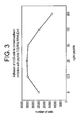
- Figure 3 shows that adhesion of k105 cells to chondroadherin decreased in a dose dependent manner in the presence of increasing amounts of the 12-mer peptide. This peptide therefore contain a cell binding sequence.
- chondroadherin (10 or 100 ⁇ g) expressed in EBNA-cells was passed over Heparin-Sepharose-cotumn (Pharmacia). The column was washed and eluted with 2M NaCI in PBS. Eluted material was separated on SDS-PAGE (10%) and detected by western blot using a chondroadherin antibody (see Figure 4 ). The band at 36 kDa represents chondroadherin and the band at 72 kDa represents a dimer of chondroadherin. It was found that chondroadherin did indeed bind to the heparin-sepharose.
- the candidate peptide CKFPTKRSKKAGRH (SEQ ID No: 7) was synthesized. The ability of the peptide to bind heparin was confirmed on heparin-sepharose, where material eluted with 0.5, 1 and 2M NaCl was identified by MALDI-TOF MS (data not shown).
- Tissue culture dishes were coated with 5 ⁇ g/ml chondroadherin expressed in E coli or collagen type II and blocked for non-specific binding with BSA.
- the cells 50 000/well
- the cells were suspended in PBS containing 0.1% BSA, 25 U/ml collagenase and added to the wells in the presence or absence of synthetic peptides (250 ⁇ g/ml), allowed to adhere for 1 hour at 37°C.
- Nonadherent cells were removed by washing.
- Spreading was visualized by light microscopy, and adhesion was determined by analyzing lysosomal hexosaminidase. Adhesion is expressed as number of cells where the data represent the average of two wells. Independent experiment showed similar results.
- cell (k105) adhesion to chrondroadherin decreased in the presence of 250 ⁇ g/ml of the 12-mer peptide (CQLRGLRRWLEAK) while the adhesion and spreading increased in the presence of 250 ⁇ g/ml of the 13-mer heparin binding peptide (CKFPTKRSKKAGRH, SEQ ID no:7).
- Adhesion to collagen type II was not significantly affected by any of the two peptides.
- Tissue culture dishes were coated with 5 ⁇ g/ml chondroadherin expressed in E-coli or collagen type II and blocked for non-specific binding with BSA
- the bovine chondrocytes (100,000/well) were suspended in PBS containing 0.1% BSA, 25 Ulml collagenase and added to the wells in the presence or absence of synthetic peptide (250 ⁇ g/ml) and allowed to adhere for 1 hour at 37°C. Nonadherent cells were removed by washing.
- Adhesion was determined by analyzing lysosomal hexosaminidase. Adhesion is expressed as number of cells, 100 000 cells/well was seeded.
- Tissue culture dishes were coated with 5 ⁇ g/ml chondroadherin expressed in E . coli and blocked for non-specific binding with BSA.
- the k105 cells were suspended in PBS containing 0.1% BSA, and added to the wells (50,000/well). Nonadherent cells were removed after 30 min by washing, plain medium without serum with or without peptides (250 ⁇ g/ml) was added and bound cells were visualized by light microscopy after 1, 3 and 20 hours.
- Figure 7 shows, that the 12-mer peptide (CQLRGLRRWLEAK) stimulated the cells to form aggregate while spreading and migration increased in the presence of the 13-mer heparin binding peptide (CKFPTKRSKKAGRH, SEQ ID No:7).
- Tissue culture dishes were coated with 33 ⁇ g/ml (20 ⁇ M) peptide solution.
- the cells (50,000/well) were suspended in PBS containing 0.1 % BSA, 25 U/ml collagenase and added to the wells in the presence or absence of synthetic peptide (250 ⁇ g/ml) and allowed to adhere for 1 hour at 37°C. Nonadherent cells were removed by washing. Spreading was visualized by light microscopy.
- Figure 8 shows that cells plated on peptide CQLRGLRRWLEAK adhere and remain round and the adhesion was inhibited in the presence of the same peptide in the solution.
- Tissue culture dishes were coated with 5 ⁇ g/ml chondroadherin expressed in E . coli and blocked for non-specific binding with BSA.
- the cells were suspended in PBS containing 0.1% BSA, and added to the wells (50,000/well). Nonadherent cells were removed after 30 min by washing, plain medium without serum with or without peptides (250 ⁇ g/ml) was added and bound cells were visualized by light microscopy after 1, 3 and 20 hours.
- the peptides QLRGLRRWLEAK SEQ ID 3
- LRRWLEAK LRRWLEAK
- the activity for integrin binding is located in the C-terminal eight residues of the sequence involving the WLEAK (SEQ ID No. 1) sequence.
- Tissue culture dishes were coated with 5 ⁇ g/ml chondroadherin expressed in E . coli and blocked for non-specific binding with BSA.
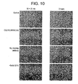
- the k105 cells were suspended in PBS containing 0.1% BSA, and added to the wells (50,000/well). Nonadherent cells were removed after 30 min by washing, peptide, anti ⁇ 1 antibody, 5mM EDTA or plain medium without serum was added and bound cells were visualized by light microscopy after 1 and 3 hours.
- Figure 10 shows, that the peptide CQLRGLRRWLEAK and anti ⁇ 1 antibody prevented spreading and the cells formed aggregate while addition of EDTA lead to cells detaching.
- Syndecans expressed by the K105 cells were detected (after heparitinase) using the syndecan-pan-antibody (see Figure 12B ). Membrane proteins were separated by SDS-PAGE (10%), transferred to a PVDF membrane, incubated with the pan-antibody and detected using an anti rabbit HRP antibody and the ECL system. K105 cells were shown to express Syndecans 1, 2, 3 and 4. The component bound to chondroadherin represents Syndecan 3.
- FIG. 13 Testing of inhibiting cell adhesion, example II.
- the figure shows that the cyclic peptide, H-[CQLRGLRRWLEAKASRPDATC]-OH (SEQ ID No:9), and the corresponding protected peptide Ac-[CQLRGLRRWLEAKASRPDATC]-NH2 (SEQ ID No:12), show inhibitory effect on cell adhesion.
- FIG. 14 Cartilage explants testing. SEQ ID 9 was in concentration 400 uM able to decrease the NO-release from IL-1 stimulated cartilage explants.
- FIG. 15 Synovial fibroblasts and IL-6 release.
- SEQ ID 9 was able to decrease IL-6-release from IL-1 stimulated synoviocytes in a dose-dependent manner.
- FIG 16 In vivo antigen induced arthritis. Locally administered peptide SEQ ID 9 decreased the knee-joint swelling compared with saline treated animals. The disease-ameliorating effect was in the same magnitude as methotrexate treatment.
- CQLRGLRRWLEAK chondroadherin peptide able to inhibit cell adhesion to the intact protein in a dose dependent manner.
- Cell adhesion to chondroadherin is mediated via the integrin ⁇ 2 ⁇ 1, where the adhesion appears to be mediated though its I-domain (9).
- the peptides SSVYGLR and AEIDGIEL previously shown to inhibit binding to osteopontin and tenascin-C (14) on the other hand had no effect on ⁇ 9 ⁇ 1 adhesion to VCAM-1.
- Another peptide RRETAWA compete directly with the ⁇ 5 ⁇ 1 binding of RGD-containing fibronectin fragments (15).
- the CPCFLLGCC-peptide inhibits ⁇ 2 adhesion to ICAM-1 and also to von Willebrand factor (16).
- ⁇ 2 integrin leukocyte adhesion but not ⁇ 1 or ⁇ 3 integrin-mediated cell adhesion.
- the LLG sequence exposed on ICAM-1 and von Willebrand factor at sites of vascular injury has been suggested to play a role in binding leukocytes and could possibly be used in a therapeutic approach in inflammatory reactions.
- the CHAD interaction with cellular ⁇ 2 ⁇ 1 has rather specific effects on the cellular phenotype with modulation of matrix protein synthesis (data not shown) and apparent blocked cell division and migration.
- This phenotype that differs from that induced by collagen II binding should be central in maintaining the chondrocyte role in adult cartilage. This would primarily involve repair and adaptation of the extracellular matrix that is continuously exposed to load that needs to be efficiently dealt with.
- ⁇ 2 ⁇ 1 integrin has a role in preventing cell death, as indicated by studies of T-lymphocytes ( Gendron, S., Couture, J., Aoudjit, F.; Integrin alpha2 beta1 inhibits Fas-mediated apoptosis in T-lymphocytes by protein phosphatase 2A-ependent activation of the MAPK/ERK pathway, J. Biol.Chem 2003, Sept 17 ).
- Integrins are the best characterized adhesion receptor family but the syndecans are now recognized as important cell surface adhesion co-receptors that can actively participate in adhesion and signaling (1, 2).
- the peptide (aa346) CKFPTKRSKKAGRH (aa359) , SEQ ID No:7, has a profound stimulative effects on cell adhesion, spreading and migration in vitro. Similar effects on cell adhesion have been shown after (PMA) treatment (3, 9, 4, 5). PMA is known to activate protein kinase C signaling and thereby stimulate adhesion and spreading in a process involving syndecan 4 and integrins (3).
- Mesenchymal cells use syndecans to adhere to ADAM 12 and ⁇ 1 integrins to induce spreading (6).
- Chondroadherin exist as two forms in the tissue with unknown functional difference where one form lacks the nine last amino acids ( Neame PJ, Sommarin Y, Boynton RE, Heinegard D., J Biol Chem. 1994 Aug 26;269(34):21547-54 .).
- the heparin binding peptide CKFPTKRSKKAGRH, SEQ ID No: 7, that was synthesized corresponds to this part of chondroadherin.
Landscapes
- Health & Medical Sciences (AREA)
- Chemical & Material Sciences (AREA)
- Organic Chemistry (AREA)
- Life Sciences & Earth Sciences (AREA)
- General Health & Medical Sciences (AREA)
- Medicinal Chemistry (AREA)
- General Chemical & Material Sciences (AREA)
- Nuclear Medicine, Radiotherapy & Molecular Imaging (AREA)
- Chemical Kinetics & Catalysis (AREA)
- Pharmacology & Pharmacy (AREA)
- Animal Behavior & Ethology (AREA)
- Public Health (AREA)
- Veterinary Medicine (AREA)
- Bioinformatics & Cheminformatics (AREA)
- Engineering & Computer Science (AREA)
- Physical Education & Sports Medicine (AREA)
- Rheumatology (AREA)
- Orthopedic Medicine & Surgery (AREA)
- Pain & Pain Management (AREA)
- Pulmonology (AREA)
- Neurology (AREA)
- Gastroenterology & Hepatology (AREA)
- Heart & Thoracic Surgery (AREA)
- Proteomics, Peptides & Aminoacids (AREA)
- Cardiology (AREA)
- Molecular Biology (AREA)
- Genetics & Genomics (AREA)
- Biophysics (AREA)
- Biochemistry (AREA)
- Dermatology (AREA)
- Toxicology (AREA)
- Zoology (AREA)
- Urology & Nephrology (AREA)
- Communicable Diseases (AREA)
- Vascular Medicine (AREA)
- Immunology (AREA)
- Neurosurgery (AREA)
- Biomedical Technology (AREA)
- Oncology (AREA)
- Medicines That Contain Protein Lipid Enzymes And Other Medicines (AREA)
Abstract
Description
- The invention relates to the use of specified peptides in the manufacture of a medicament for treatment and/or prophylaxis of a disease associated with inflammatory mediated cartilage destruction such as, e.g. an inflammatory joint disease such as, e.g., rheumatoid arthritis, psoriatic arthritis, juvenile chronic arthritis, systemic lupus erythematosus (SLE), infectious arthritis and juvenile idiopathic arthritis, other diseases involving joint structures such as, e.g., osteoarthritis, meniscal damage, gout, diseases affecting structural elements of the musculoskeletal system such as, e.g., tendon damage, ligament damage, and bone disease such as, e.g., osteoporosis, diseases affecting major arteries including atherosclerosis. The invention also relates to novel compounds for use in medicine, and compositions, and in a preferred aspect the invention relates to novel peptide compounds with the core peptide sequence WLEAK (SEQ ID No. 1). In the current context the type of tissue changes of main interest is changes involved with inflammatory mediated cartilage destruction.
- Destruction of tissues is a feature of diseases affecting many organ types and represents an increasing problem to the population with particular emphasis on the aging. Many of these diseases have an element of chronic inflammation. Examples of such diseases are rheumatoid arthritis, osteoarthritis, osteoporosis, artheriosclerosis, chronic lung diseases, tendinitis, spine problems with lower back pain and intervertebral disc hernia. These diseases encompass by far the largest expenditure among major disease categories for society and suffering for individuals.
Arthritis is a common name for several inflammatory joint diseases. Joint diseases are extremely common in the population. While rheumatoid arthritis sufferers represent some 1%, osteoarthritis is much more common with an incidence of at least some 10%. Major features of the two disease entities are progressive destruction of joint structures as well as inflammation causing pain and disability. - At present, the symptoms of e.g. rheumatoid arthritis can for some patients be alleviated by medical treatment, e.g. anti-inflammatory agents including corticosteroids, physiotherapy or surgery. More recent use of inhibitors of cytokines, particularly TNF-α has improved the situation for two third of the patients with severe rheumatoid arthritis. However, many patients do not effectively respond to the above-mentioned treatments or the treatments lead to a number of undesired side-effects (see e.g. Smolen & Steiner, Nature Reviews, Vol. 2, (2003), 473-488)
Shen et al. (Biochem J. (1998) 300, 549-557) discloses that chondroadherin expression changes in skeletal development. It also discloses that an antiserum recognizing a C-terminal peptide (KRSKKAGRH) of rat/bovine chondroadherin raised in rabbits was used for immunocytochemistry. - It is known in the art that chondroadherin (CHAD) conjugated with additional compounds has an effect on tissue changes (
WO0137861 WO03049669
There is a need for new approaches with respect to treatment of diseases associated with tissue changes, especially with a view to the efficiency of the treatment and to reduction of side-effects. - Accordingly, the present invention provides the use of specified peptides of
formula 1 in the manufacture of a medicament for the treatment and/or prophylaxis of a disease associated with inflammatory mediated cartilage destruction, such as, e.g. an inflammatory joint disease such as, e.g., rheumatoid arthritics, psoriatic arthritis, juvenile chronic arthritis, systemic lupus erythematosus (SLE), infectious arthritis and juvenile Idiopathic arthritis, other diseases involving joint structures such as, e.g., osteoarthritis, meniscal damage, gout, diseases affecting structural elements of the musculoskeletal system such as, e.g., tendon damage, ligament damage, and bone disease such as, e.g., osteoporosis, diseases affecting major arteries including atherosclerosis. Also disclosed is a method comprising administering to a subject in need thereof a therapeutic and/or prophylactic amount of a compound capable of at least one of the following - a) modulating changes of the phenotype of cells involved in a disease associated with tissue changes,
- b) regulating the production of extracellular matrix molecules by cells involved in a disease associated with tissue changes,
- c) inhibiting the normal production and activity of catabolic factors, such as, e.g., NO, cytokines and/or proteinases of cells involved in a disease associated with tissue changes,
- d) regulating division of cells involved in a disease associated with tissue changes,
- e) regulating migration of cells involved in a disease associated with tissue changes,
- f) regulating the production of growth factors and/or
- g) regulating the response of cells involved in a disease associated with tissue changes to external stimuli.
- The general purpose of the method described above is to direct the cells into performing specific actions, all of which have a positive effect on a disease associated with tissue changes.
- In the current context the type of tissue changes of main interest is changes involved with inflammatory mediated cartilage destruction. The type of cells involved in a disease associated with tissue changes depends on the disease in question and the affected tissues. Examples of cells involved in a disease as described above are chondrocytes, osteoblasts, osteocytes, fibroblasts, tenocytes, smooth muscle cells, macrophages and pro-inflammatory cells like synovial fibroblasts and dendritic cells.
- In a specific embodiment, the cells involved in the diseases are chondrocytes. The following description of the above is generally given for all type of cells involved in a disease associated with tissue changes, but with references to specific cells, such as, e.g. chondrocyte and smooth muscle cells and how the behavior of these cells should be regulated in order to treat or prevent a tissue change relating to chondrocytes.
- In a specific aspect, the method described above comprises administering to a subject in need thereof a therapeutic and/or prophylactic amount of a compound capable of modulating changes of the phenotype of cells involved in a disease associated with tissue changes. A specific definition of the term "phenotype" in the present context is not simple and a person skilled in the art will understand that the term relates to the character of the cell and can be defined at many levels. The simplest and traditionally used parameter is cell shape. Other ways of identifying phenotypes are by e.g. the cells' production of molecules such as production of distinct patterns of matrix proteins, proteinases and growth factors, identification of stationary, migratory, catabolic and/or anabolic states of the cells, and identification of whether cells are dividing or non-dividing. Accordingly, the compound for use according to the invention may solely affect the phenotype of the cells. However, in a specific embodiment of the invention at least one other factor may be influenced by use of a compound according to the invention without - as a main effect - having effect on the phenotype of the cells. An example includes e.g. by regulating the production of growth factors.
- Without being bound to any specific theory, the inventors have in a specific experiment found that especially a rounded shape of chondrocytes correlate to the cell behavior.
- With respect to item b in the method described above, one of the processes leading to tissue changes, such as tissue destruction, is failure to repair tissue damage. Although the tissue often responds by an increased production of some matrix constituents, it is becoming increasingly dear that there is a lack of a coordinated production of all the components needed for proper assembly of tissue structures. The outcome seems to be an inhibited assembly, which is even worse than an insufficiently active repair process. Thus, by regulating the production of extracellular matrix components a proper assembly of tissue structures may be obtained.
- Item c of the method described above : Another of the processes that leads to tissue changes is an increased breakdown of tissue structural molecules leading to tissue failure and destruction. This may be accomplished e.g. by proteinases produced by the tissue cells themselves. In all tissues with a component of tissue destruction, there is an increased proteinase production. By stimulating the cells to decrease the production of proteinases, the breakdown of tissue structural molecules may be prevented. As will be described in further details below, binding of specific ligands to a receptor such as, e.g. an integrin receptor, on chondrocytes will lead to a decrease in production of proteinases.
- Item d of the method described above : When cells are proliferating, the production of matrix components by the cells is at a minimum, i.e. the cells are not contributing to the repair of tissues. Accordingly, by preventing cell division, the focus of the cells may be shifted to production of matrix components and tissue repair. Cell division is believed to be dependent on the activity of integrin receptors, i.e. by regulating the activity of an integrin receptor by adding ligands that either stimulates or inhibits the receptor, cell division may be regulated.
- Item e according to the method described above: A way of treating a disease associated by tissue changes is to inhibit the migration of cells involved in such a disease. In e.g. athreriosclerosis there can be a migration of smooth muscle cells into the arterial intima. Theses cells will accumulate and many of them will divide. At the same time, fat builds up within these cells and around them and they also form connective tissue. Accordingly, the innermost layer of the artery becomes markedly thickened by these accumulating cells and surrounding material. If the wall is thickened sufficiently, the lumen of the artery will be reduced and the blood flow decreased, thus decreasing the oxygen supply. Cell migration is believed to be dependent on the activity of integrin receptors, i.e. by regulating the activity of an integrin receptor by adding ligands that interacts with the receptor, cell migration may be inhibited.
- The present inventors have found that inhibition of chondrocyte cell migration in specific situations is advantageous for ameliorating diseases associated with tissue changes.
- Item f of the method described above another entry point for the treatment of a disease associated with tissue changes is the regulation of production of growth factors. Growth factors are gene products playing important roles in the regulation of e.g. cell division, tissue proliferation and production of matrix constituents. Each growth factor has a specific cell-surface receptor and binding of a growth factor to its receptor may initiate or, in some cases, block cell division. Too high levels of a particular growth factor, tgf-beta, has been shown to induce the changes seen in osteoarthritis, and tgf-beta levels are also high in lung fibrosis leading to excessive collagen production.
- More specifically, the compounds for use according to the invention, mediate their actions on the cells associated with the diseases mentioned above by interacting with structures on the cell surface of the cells. Examples of such structures are receptor and/or polyanionic structures, particularly heparinoids including heparin and heparan sulphate. Thus, disclosed herein is a method wherein the one or more compounds are capable of
- a) interacting with a receptor expressed at a surface of a cell involved in a disease associated with tissue changes,
- b) interacting with polyanionic structures, such as, but not limited to heparan sulfate, expressed at or present on a surface of a cell involved in a disease associated with tissue changes, and/or
- c) combined interactions with one or more receptor and/or one or more polyanionic structure.
- In the method a compound is capable of interacting with both a receptor and with a polyanionic structure, or with two or more receptors and/or two or more polyanionic structures. Receptors often need to polymerize for optimal activity, i.e. by providing one compound capable of crosslinking two or more receptors or polyanionic structures, dimerization or polymerization is ensured, leading to optimal activity. Furthermore, binding of a compound to a receptor and/or polyanionic structure will be increased, as binding is tighter when two or more binding points are involved. For example, chondroadherin is believed to have two specific binding domains, one for the receptor integrin and the other for heparinoid binding. Accordingly, such substances may have more than one functionality.
- The chondrocytes, being the only type of cell in cartilage, have a key function in cartilage homeostasis. The roles of chondrocytes include normal turnover of matrix molecules and deposition of molecules into functioning matrix.
- Studies by the present inventors have shown that chondrocytes express several cell surface structures that are relevant for the regulation of the cells.
- Relevant cell surface structures include the integrin receptors. The integrin receptor is a complex of one alpha and one beta chain, which may be chosen from 18 different alpha chains and 8 different beta chains. The various integrin receptors may have different ligands and elicit different responses.
- Another example of cell surface structures on chondrocytes, which are relevant for the regulation of the cells, is polyanionic structures such as, e.g. heparinoids like heparin/heparan sulfate, chondroitin/dermatan sulfate chains, or clusters of sulphated gangliosides or phospholipids.
- The heparinoid heparan sulfate is ubiquitous at the cell surface, where it predominantly may be found on proteoglycans of either the transmembrane syndecan family or the glycosylphosphatidylinositol (GPI)-anchored glypican family.
- During the text, the term heparinoid also include heparinoid mimics.
- The present inventors have found that chondroadherin inhibits the binding of cells to the integrin α 2 β 1 receptor on chondrocytes, strongly indicating that chondroadherin binds to chondrocytes via the integrin α 2 β 1 receptor. Chondroadherin is a leucine-rich, cartilage matrix protein. The binding of chondroadherin induces the cells to maintain a rounded shape and stay in an anabolic mode, appears to stimulate the cells to production of ECM-molecules, and prevents cell spreading and/or migration as well as cell division. Studies have also shown that most other natural integrin ligands, such as, e.g. collagen II have the almost opposite effects on the cells.
- As shown herein human chondrocytic cells adhere equally well to native as to unfolded chondroadherin showing that a linear peptide sequence is mediating the adhesion. Digestion of chondroadherin has also shown that smaller fragments of chondroadherin are capable of mediating the effects mentioned above.
- Accordingly, in one specific embodiment the receptor a) is an integrin receptor and the one or more compounds are peptides which may comprise one or more chondroadherin fragments or analogs thereof.
- The present inventors have identified a specific fragment of chondroadherin (CQLRGLRRWLEAK - SEQ ID NO. 3), which is contemplated upon binding to chondrocytic cells via an integrin receptor, such as, e.g. the integrin α 2 β 1 receptor, to have the effects on the chondrocytes as described above, i.e. prevents spreading and migration of the cells and at the same time the cells tend to maintain the rounded phenotype. Further studies have shown that a sub fragment LRRWLEAK (SEQ ID NO. 2) similarly to CQLRGLRRWLEAK prevents cell spreading and migration and maintain the rounded shape of the cells. The activity for integrin binding is located in the C-terminal residues of the sequence, involving the WLEAK sequence (SEQ ID NO. 1). The inventors have found that the integrin binding properties of the sequence are lost if lysine is exchanged with another amino acid with a basic side chain, namely arginine, i.e. the lysine in the WLEAK (SEQ ID No. 1) sequence is necessary for activity.
- However, it cannot be ruled out, that a sequence wherein lysine is exchanged with another amino acid may maintain its activity.
- The inventors have also found, that the exchange of tryptophan for another amino acid with an aromatic side chain, namely tyrosine and the exchange of glutamic acid for another acidic amino add, namely aspartic acid, do not have any influence on the integrin binding activity of the sequence.
- Accordingly, disclosed herein is a method, wherein at least one of the chondroadherin fragments has at least about 60% such as at least about 70%, at least about 75%, at least about 80%, at least about 85%, at least about 90% or at least about 95% identity to the amino acid sequence of SEQ ID No: 1 (WLEAK).
- An example of a sequence having at least 60% identity to SEQ ID No: 1 is the following sequence;
Xaa-L-Yaa-A-K.
wherein Xaa may be chosen from tryptophan and tyrosin, and Yaa may be chosen from glutamic acid and aspartic acid - Also disclosed is a method, wherein at least one of the chondroadherin fragments has at least about 75% such as at least about 80%, at least about 85%, at least about 90% or at least about 95% identity to the amino acid sequence of SEQ ID No: 2 (LRRWLEAK).
- Furthermore, a method is disclosed wherein at least one of the chondroadherin fragments has at least about 75% such as at least about 80%, at least about 85%, at least about 90% or at least about 95% identity to the amino acid sequence of SEQ ID No: 3 (QLRGLRRWLEAK).
- The inventors have also found that binding of chondroadherin to syndecan on the cell surfaces prevents cell division, and leads to increased adhesion.
- The syndecans are a family of four transmembrane heparan sulfate proteoglycans, which are recognized as important cell surface adhesion co-receptors that actively participate in adhesion and signaling.
- Chondroadherin binds to syndecan via the heparin/heparan sulfate chains. The present inventors have identified a specific fragment of chondroadherin, which interact with heparin/heparan sulfate.
- Accordingly, disclosed herein is a method, wherein at least one of the chondroadherin fragments has at least about 75% such as at least about 80%, at least about 85%, at least about 90% or at least about 95% identity to the amino acid sequence of SEQ ID No: 4 (KRSKK).
- Also disclosed is a method, wherein at least one of the chondroadherin fragments has at least about 75% such as at least about 80%, at least about 85%, at least about 90% or at least about 95% identity to the amino acid sequence of SEQ ID No: 5 (KKAGRH).
- Moreover, a method is disclosed wherein at least one of the chondroadherin fragments has at least about 75% such as at least about 80%, at least about 85%, at least about 90% or at least about 95% identity to the amino acid sequence of SEQ ID No: 6 (KFPTKRSKKAGRH),
- Furthermore, a method is disclosed wherein at least one of the chondroadherin fragments has at least about 75% such as at least about 80%, at least about 85%, at least about 90% or at least about 95% identity to the amino acid sequence of SEQ ID No: 7 (CKFPTKRSKKAGRH).
- In another embodiment, the peptide comprises at least one chondroadherin fragment capable of binding the integrin receptor, and at least one chondroadherin fragment capable of binding to heparin/heparan sulfate in order to modulate signaling, wherein heparin/heparan sulfate may be found on the extracellular domains of syndecans and glypicans.
- In a specific example of the method disclosed herein the peptide comprises an amino acid sequence having at least about 75% such as at least about 80%, at least about 85%, at least about 90% or at least about 95% identity to
- i) one of the amino add sequences of SEQ ID NO. 1, SEQ ID NO. 2 or SEQ ID NO. 3, and
- ii) one of the amino acid sequences of SEQ ID NO. 4, SEQ ID NO.5 or SEQ ID NO. 6.
- In connection with amino acid sequences, "sequence identity" refers to sequences which have the stated value when assessed using e.g. using the SWISS-PROT protein sequence databank using FASTA pep-cmp with a variable pamfactor, and gap creation penalty set at 12.0 and gap extension penalty set at 4.0, and a window of 2 amino acids. Sequence identity at a particular residue is intended to include identical residues, which have simply been derivatized.
- As mentioned above, studies have shown that linear fragments of chondroadherin interact strongly with their cell surface structures. For other peptides and cell surface structures, the interaction may be better if the peptides are non-linear.
- In order to make more stable peptides, the peptides may be designed to be cyclic, as cyclic peptides are more resistant to exo-peptidases. In addition, protecting groups such as acetyl, amide or methyl, tools well known in the art, are synthetic modifications, which further help to stabilize peptides against decomposition and metabolic degradation.
- The present invention particularly provides a novel linear or cyclic peptide consisting of the general Formula (I) and their uses:
FORMULA I P1-x1-x2-x3-x4-x5-x6-x7-x8-: W-L-E-A-K-y1-y2-y3-y4-y5-y6-y7-y8-P2
- Wherein x1 is selected from C or absent
- Wherein x2 is selected from Q or absent
- Wherein x3 is selected from L or absent
- Wherein x4 is selected from R, C or absent
- Wherein x5 is selected from G, K or absent
- Wherein x6 is selected from L. F or absent
- Wherein x7 is selected from R, P or absent
- Wherein x8 is selected from R, C, T or absent
- Wherein y1 is selected from A, C or absent
- Wherein y2 is selected from S, G or absent
- Wherein y3 is selected from R or absent
- Wherein y4 is selected from P, H or absent
- Wherein y5 is selected from D or absent
- Wherein y6 is selected from A or absent
- Wherein y7 is selected from T or absent
- Wherein y8 is selected from C or absent
- Wherein P1 and P2 are absent or a protective group
- In another embodiment, the invention relates to novel peptides as further exemplified in the appended claims whereto reference is made. An analogous discussion as given above is also relevant for these peptides.
- Also disclosed is a pharmaceutical composition comprising a compound capable of
- a) modulating changes of the phenotype of cells involved in a disease associated with tissue changes,
- b) regulating the production of extracellular matrix molecules by cells involved in a disease associated with tissue changes,
- c) inhibiting the normal production and activity of catabolic factors, such as, e.g., NO, cytokines and/or proteinases of cells involved in a disease associated with tissue changes,
- d) regulating division of cells involved in a disease associated with tissue changes,
- e) regulating migration of cells involved in a disease associated with tissue changes,
- f) regulating the production of growth factors, and/or
- g) regulating the response of cells involved in a disease associated with tissue changes to external stimuli
- Furthermore, also disclosed is a pharmaceutical composition comprising a compound capable of
- a) interacting with a receptor expressed at a surface of a cell involved in a disease associated with tissue changes,
- b) interacting with polyanionic structures expressed at or present on a surface of a cell involved in a disease associated with tissue changes, and/or
- c) combined interactions with one or more receptor and/or one or more polyanionic structure.
- In one embodiment the receptor is an integrin receptor.
- The pharmaceutical compositions according to the invention normally comprise the specific compound together with one or more physiologically acceptable excipients, i.e. a therapeutically inert substance or carrier.
- The carrier may take a wide variety of forms depending on the desired dosage form and administration route.
- The pharmaceutically acceptable excipients may be e.g. fillers, binders, disintegrants, diluents, glidants, solvents, emulsifying agents, suspending agents, stabilizers, enhancers, flavors, colors, pH adjusting agents, retarding agents, wetting agents, surface active agents, preservatives, antioxidants etc. Details can be found in pharmaceutical handbooks such as, e.g., Remington's Pharmaceutical Science or Pharmaceutical Excipient Handbook. In those cases, where the pharmaceutical composition is intended for controlled release, it may also comprise release controlling agents such as, e.g., material normally used in the formulation of matrix tablets (e.g. cellulose derivatives like hydroxypropyl methylcellulose and the like). Alternatively, the composition may be coated with a controlled release coating such as an enteric coating or a film coating. A suitable coating may be a substantially water-insoluble but water-permeable coating.
- The compound may be administered by any suitable route, such as, e.g. the oral, buccal, nasal, ocular, pulmonary, topical, transdermal, vaginal, rectal, ocular, parenteral (including inter alia subcutaneous, intramuscular, intraaricular and intravenous), route in a dose that is effective for the individual purposes. A person skilled in the art will know how to chose a suitable administration route.
- The pharmaceutical composition comprising a compounds according to the invention may be in the form of a solid, semi-solid or fluid composition.
- The solid composition may be in the form of tablets such as, e.g. conventional tablets, effervescent tablets, coated tablets, melt tablets or sublingual tablets, pellets, powders, granules, granulates, particulate material, solid dispersion or solid solutions.
- A semi-solid form of the composition may be a paste, cream, ointment, a gel or a hydrogel.
- The fluid form of the composition may be a solution, an emulsion including nano-emulsions, a suspension, a dispersion, a liposomal composition, a spray, a mixture, a syrup or an elixir.
- Other suitable dosages forms of the pharmaceutical compositions according to the invention may be vagitories, suppositories, plasters, patches, tablets, capsules, cachets, troches, devices etc.
- The pharmaceutical compositions may be prepared by any of the methods well known to a person skilled in pharmaceutical formulation.
- Also disclosed is the use of a compound capable of
- a) modulating changes of the phenotype of cells involved in a disease associated with tissue changes.
- b) regulating the production of extracellular matrix molecules by cells involved in a disease associated with tissue changes,
- c) inhibiting the normal production and activity of catabolic factors, such as, e.g., NO, cytokines and/or proteinases of cells involved in a disease associated with tissue changes,
- d) regulating division of cells involved in a disease associated with tissue changes,
- e) regulating migration of cells involved in a disease associated with tissue changes,
- f) regulating the production of growth factors, and/or
- g) regulating the response of cells involved in a disease associated with tissue changes to external stimuli
- Also disclosed is a use of compound capable of
- a) interacting with a receptor expressed at a surface of a cell involved in a disease associated with tissue changes,
- b) interacting with polyanionic structures expressed at or present on a surface of a cell involved in a disease associated with tissue changes, and/or
- c) combined interactions with one or more receptor and/or one or more polyanionic structure.
- The receptor is an integrin receptor.
- The interaction between the compound and the integrin receptor may be detected by any suitable method. One example of a suitable method is microscopic analysis of the cells in order to determine any of cell shape. Another detection method involved detection of alterations in cell behavior.
- Another method for detection of binding between a compound and an integrin receptor includes the addition of the compound in question to a assay comprising cells expressing the integrin receptor and a immobilized natural ligand to the integrin receptor, and evaluation of whether the compound inhibit binding of the cells to the natural ligands
- Binding can also be determined by FACS analyses of tagged peptide, and plasmon resonance (Biacore).
- The interaction between the compounds and the polyanionic structure may be detected by any suitable method. One example of a suitable methods is microscopic analysis of the cells in order to determine any alterations of cell shape. Another detection method involved detection of alterations in cell behavior, such as, e.g. cell migration.
- Another method for detection of binding between a compound and an polyanionic cell surface structure includes the addition of the compound in question to a assay comprising cells expressing polyanionic structure and a immobilized natural ligand to the polyanionic structure, and evaluation of whether the compound inhibit binding of the cells to the natural ligand.
- Binding can also be determined by FACS analyses of tagged peptide, and isothermal titration colometry (ITC).
- Also disclosed is an in vitro method for testing the activity of a compound or a combination of two or more compounds with respect to production of extracellular matrix constituents, the method comprising
- a) culturing cells in the presence of the one or more compounds to be tested,
- b) monitoring release of matrix components from cultured tissue explants,
- c) monitoring modified release of matrix components from cultures of tissue explants stimulated with catabolic agents, such as, e.g. IL-1 and fibronectin fragments,
- d) monitoring modified release of component from tissue explants cultured in the presence of growth factors,
- e) monitoring modulation of progression of model disease in animals, e.g. collagen arthritis, bleomycin lung fibrosis and development of osteoarthritis.
- The following examples are intended to illustrate the invention without limiting it thereto.
- To obtain evidence for that chondroadherin-derived peptides modulates changes of the phenotype of cells involved in a disease associated with tissue changes, we have performed experiments with such cells (chondrocytes, synoviocytes), and in cartilage explants and an arthritis animal model.
- Human chondroadherin cDNA was ligated into the pQE8 vector (QIAGEN Inc. Valencia, CA) and expressed in M15 E-coli. Overnight cultures (8ml) of stationary phase bacteria were used to inoculate 400 ml LB-medium (Luria-Bertani) and the cells were allowed to grow for 2.5 hour at 37°C (OD600 of 0-7-0.9). Protein expression was induced by the addition of 2mM IPTG (isopropyl-β-D-thiogalactopyranoside), (Sigma Chemicals CO., St Louis, MO) and the culture was grown for another 4.5 hour. Bacteria were then collected by centrifugation, frozen and resuspended in lysis buffer (6M GuHCl, 0.1 M NaH2PO4 and 0.5M NaCl, pH 8.0) containing 5mM NEM (Sigma). The suspension was passed 10 times through a 21 gauge needle (21 G, 0.8x50) to lyse the cells. Insoluble debris was removed by centrifugation. His-tagged recombinant protein was purified using a Hi-Trap Ni-chelating column (Pharmacia LKB Biotechnology, Uppsala, Sweden) essentially according to instructions from the manufacturer. The lysate was applied to the Ni2+-charged column, washed with 10 volumes of lysis buffer and bound protein was eluted with 8M Urea, 0.1M NaH2PO4 and 0.5M NaCl pH 6.3. Fractions were immediately neutralized by the addition of 1 M Tris pH 9.0. Fractions containing the desired protein as determined by SDS-PAGE and western blotting (data not shown) were pooled, dialyzed against water and lyophilized. EBNA-cell expressed chondroadherin was produced as described elsewhere (8).
- Intact recombinant protein (1 mg, EBNA-cell expressed (8)) was proteolytically digested with Lys-C (Roche Molecular Biochemicals) at an enzyme to substrate ratio of 1:50 according to the instructions of the manufacturer. Generated peptides were separated by reversed phase chromatography on a Sephasil C8 column (Pharmacia LKB Biotechnology, Uppsala, Sweden) with a gradient of 0-70% acetonitrile, 0.1 % TFA (0.1 ml/min. over 50 min.) using a SMART-System(Pharmacia LKB Biotechnology). The effluent was monitored for absorbance at 280 nm. Fractions were pooled, lyophilized and resuspended in PBS and then used in cell adhesion experiments to block adhesion to intact chondroadherin.
- Mass spectrometry was performed using a Bruker Scout 384 Reflex III matrix-assisted laser desorption/ionization time-of-flight (MALDI-TOF) mass spectrometer. The instrument was used in the positive ion mode with delayed extraction and an acceleration voltage of 26 kV. Peptide samples were mainly analyzed using the reflector detector and 50-150 single-shot spectra were accumulated for improved signal to noise ratio. The software used to identify the obtained peptide masses was ProFound
- Polypeptides as described herein may be prepared by any conventional modes of synthesis, including chemical synthesis or recombinant DNA technology. Chemical synthesis may be performed by methods well known in the art involving cyclic sets of reactions of selective deprotection of the functional groups of a terminal amino acid and coupling of selectively protected amino acids, followed by complete deprotection of all functional groups. Synthesis may be performed in solution or on a solid support using suitable solid phases known in the art. Preferably the polypeptides are substantially purified, e.g. pyrogen-free, e.g. more than 70%, especially preferably more than 90% pure (as assessed for example, in the case of peptides, by an appropriate technique such as peptide mapping, sequencing or chromatography). Purification may be performed for example by chromatography (e.g. HPLC, size-exclusion, ion-exchange, affinity, hydrophobic interaction, reverse-phase) or capillary electrophoresis.
- Intact recombinant protein (100
el 10 µg, EBNA-cell expressed) diluted in PBS was passed over Heparin-Sepharose (Amersham Pharmacia) (1 ml), washed with 20 volumes of PBS and eluted with 2 M NaCl (6x1ml). Eluted material was precipitated with ethanol, separated by SDS-PAGE (10%) and visualized by western blot (CHAD ab α-80, Larsson et al). The peptide CKFPTKRSKKAGRH (SEQ ID No:7) was diluted in PBS and passed over heparin-sepharose (100 µl), the column was washed with 20 volumes PBS and bound material eluted with 0.5, 1, and 2M NaCl (3 column volumes of each). - Human cells (K9) originating from a human chondrosarcoma were a kind gift from Drs. Sven Inerot and Anders Lindahl, The Salgrenska University Hospital Gothenburg, Sweden. The K9 cells were cultured in Dulbeccos Modified Eagle Medium: Nutrient Mixture F12 (DMEM/F12 1:1) with
GLUTAMAX 1, supplemented with 10% fetal calf serum, 25 µg/ml ascorbic acid, 50 IU of penicillin and 50µg/ml streptomycin (Life Technologies, Inc.). A different human cells line (K105) originating from a human chondrosarcoma was a kind gift from dr. Sven Inerot, The Salgrenska University Hospital, Gothenburg, Sweden. The K105 cells were cultured in a mix of 40% Dulbeccos Modified Eagle Medium, 40% Minimum Essential Media (MEM) alpha Medium, 10% Nutrient Mixture F1 supplemented with 100 nM hydrocortisone, 100 ng/ml insulin, 10% fetal calf serum, 25 µg/ml ascorbic acid, 50 IU of penicillin and 50µg/ml streptomycin (Life Technologies, Inc.). To harvest cells, culture dishes were rinsed three times with Ca 2+/Mg 2+- free PBS and the cells were incubated with 0.5% trypsin and 1 mM EDTA for 5 min. Detached cells were suspended in PBS containing 1mg/ml trypsin inhibitor (Sigma) and then washed in PBS. To remove membrane-bound collagens the cells were treated with collagenase 100 U/ml (CLSPA; Worthington Biochemical Corp., Lancewood, NJ) in PBS, 30 min. 37°C and then washed in PBS. - Bovine chondrocytes were isolated by collagenase (CLS1; Worthington Biochemical Corp., Lacewood, NJ) digestion of articular cartilage from 4-6 month old calves as described (10). Briefly, cartilage slices were digested for 16 hours with collagenase in EBSS (Earle's balanced salt solution, GIBCO BRL Gaitersburg, MD). The cells were filtered through a 100 µm nylon filter, washed three times in PBS containing 0.2 % BSA (SERVA).
- Tissue culture 48-wells dishes (Nunclon, Nunc, Denmark) were coated overnight with 5 µg/ml chondroadherin in 4M Guanidine-HCl, 50mM sodium acetate, pH 5.8, or for native protein in PBS. Alternatively coating was with 5µg/ml collagen type II in PBS followed by after-coating with 0.5% BSA in PBS for 3 hours. Collagen type II was isolated from bovine nasal cartilage by pepsin digestion (11). Bacterially expressed chondroadherin was isolated as described above. Recombinant chondroadherin expressed in EBNA cell was cloned and purified as described elsewhere (8).
- To test cell binding, the chondrosarcoma cells or bovine chondrocytes were suspended in PBS containing 0.1 % BSA, 25 U/ml collagenase and added to the wells (50,000/well), in the absence or presence of various peptide pools or the synthetic peptides (200, 100, 50, 25, 12.5 µg/ml). Non-adherent cells were removed after 1 hour and adhesion was determined by measuring lysosomal N-acetylglucosaminidase (12). Adhesion and spreading was visualized by light microscopy. In the time studies, k105 cells were allowed to adhere for 30 minutes, unbound cells were removed and medium without serum supplemented with or without peptides (250 µg/ml), β1 integrin antibody (clone P4C10, Gibco BRL), EDTA (5mM) was added. Cell morphology was visualized by light microscopy after 1, 3 and 20 hours.
- Recombinant chondroadherin (2.5 mg expressed in EBNA-cells per ml agarose) was coupled to Mini-Leak agarose (Kem En Tec, A/S, Denmark) according to the manufacturers instructions. The control agarose was treated in the same manner but with no protein added.
- Human chondrosarcoma cells (k105) suspended in PBS were incubated with EZ-link sulfo NHS-LC biotin (Pierce Chemical Co) at a final concentration of 1 mg/ml for 1 hour at room temperature. The cells were incubated for 10 minutes in 0.1 M glycine in PBS to block free biotin, washed three times in PBS and then resuspended and homogenized in 10 mM KCl, 20 mM Tris/HCl, pH 7.4, 1 mM EDTA (1ml/106 cells). Nuclei were removed by centrifugation at 1,500 g for 5 minutes, 0.15 M NaCl was added to the supernatant and the membrane fraction was collected by centrifugation at 50,000 g for 30 minutes. The pellet was suspended in 2 ml 8 M UREA, 2% Triton-X-1000 in TABS pH 7.4 supplemented with COMPLET® protease inhibitor (Roche). The lysate was cleared by centrifugation at 14,000 g for 20 min.
- The chondroadherin- and controlagarose (1ml) were packed in mini columns (Bio-Rad, Hercules. CA) and equilibrated with 20 volumes running buffer (6M UREA, 1% Triton-X-100 in TBS pH 7.4 supplemented with COMPLET® protease inhibitor (Roche)). Lysate of biotin labeled cells were passed twice over the control-column, incubated overnight with the with continuous end over end mixing at 4° C. The chondroadherin- and controlagarose were washed with 20 volumes of running buffer and eluted with 0.5 M NaCl, 6 M UREA in TBS pH 7.4 supplemented with COMPLET (1 ml fractions). Eluted proteins were precipitated with ethanol, resuspended and part of the material was digested with heparitinase and chondroitinase ABC at 37°C overnight Undigested and digested material was separated by SDS-PAGE 4-12%, electro-transferred to PVDF-membrane (pall), and blocked with 2% BSA in 10 mM Tris/HCl, pH 7.4, 0.15 M NaCl and 2% Tween (blocking buffer). Biotin-labeled proteins were visualized after incubation with streptavidin-HRP (1:5000 in blocking buffer) by chemiluminescence detection using the ECL system (Amersham).
- Biotin-labeled cell surface material was affinity purified on immobilized chondroadherin eluted with 0.5 M NaCl and passed over a desalting-column (PD-10. Pharmacia) equilibrated and eluted with 0.1 % triton X-100 in TBS supplemented with COMPLET protease inhibitor. Eluted material was incubated over night with a syndecan-pan-antibody (5µg/ml, Rapraeger) and the immune complex was collected using prot-A sepharose. The sepharose was washed 3 times with heparitinase buffer (50 mM Hepes, 50 mM NaOAc. 150 mM NaCl, pH 6.5), resuspended in the same buffer and digested with heparitinase at 37°C. Immunoprecipitated proteins and cell lysate were separated by SDS-
PAGE 10% and electro-transferred to PVDF-membrane (pall). The membrane was blocked with 2% BSA in 10 mM Tris/HCl, pH 7.4, 0.15 M NaCl and 2% Tween (blocking buffer). Immunoprecipitated proteins were visualized after incubation with streptavidin-HRP (1:5000 in blocking buffer) by chemiluminescence detection using the ECL system (Amersham). Syndecans expressed by the k105 cells were detected with the syndecan-pan-antibody (1 µg/ml, Rapraeger), anti rabbit HRP (DAKO) antibody and chemiluminescence detection using the ECL system (Amersham) ECL - The following experiment is made in order to determine the ability to inhibit cell adhesion.
Study design: Tissue culture dishes (48-wells) were coated with 5 microg/ml chondroadherin expressed in E. coli and blocked for non-specific binding with BSA. K 105 cells were suspended in PBS containing 0.1% BSA, 25 U/ml collagenase and added to the wells (50,000/well) in the presence of synthetic peptide to be tested and allowed to adhere for 1 hour at 37°C Nonadherent cells were removed by washing, and adhesion was determined by analyzing lysosomal hexosaminidase. - The following experiments measure the effect of a peptide on NO release in IL-1 stimulated cartilage.
Study design: A skinned bovine nose (from cows 18-24 months old) was collected at Horby slaughter house (Team Uggarp Sweden). The septum inside the norse was cut out and the mucosa and the perichondrium was removed before the cartilage was placed in PBS with PEST (100 IU penicillin, 100 µg/ml streptomycin) and 2.5 ug/ml Fungizone (all from Invitrogen, Sweden) for 2 hours at rt. - Two mm pieces were punched out of the cartilage. Each piece was placed in a 24-well cell culture plate (Falcon, Sweden) containing 1 ml cell culture medium, HAMs F12 (invitrogen, Sweden) supplemented with 10 µg/ml BSA, 25 mg/ml ascorbate (both from Sigma, Sweden), PEST (100 IU penicillin, 100 mg/ml streptomycin) and 2.5 ug/ml Fungizone. After 24 hours, the medium was changed and the cartilage pieces were stimulated with human recombinant IL-1a, 10 ng/ml (Roche. Sweden). The peptides where tested in triplet at 20 and 400 µM. The cartilage tissue was incubated for another six days, mediums were exchanged every third day. On each occasion the mediums were collected for measurement of NO (Griess reaction).
- The following experiments the IL-6 release in IL-1 stimulated synoviocytes. Study design: From rats with antigen induced arthritis, the hyperproliferative synovium, pannus, was taken from the inflamed knee day four after disease onset The pannus tissue was cut to small pieces in PBS with PEST (100 IU penicillin, 100 µg/ml streptomycin) and Fungizone (25 µg/ml) (all from InVitrogen, Sweden), before incubation in collagenase (400 U/ml, Worthington, USA) for 3 hours at 37°C, 5% CO2. Cells were centrifugated (8 min, rt, 1100 rpm.) and suspended in RPMI 1640 supplemented with 10% FCS (InVitrogen, Sweden), PEST and Fungizone and seeded in a 25 cm2 flask at 37°C, 5% CO2. The following day, cells were rinsed once with medium and further incubated. When confluent, cells were trypsinated for 1 min (0.25% Trypsin with EDTA, InVitrogen, Sweden) counted and seeded in 96 well plates, 10000 cell/well/200 µl.
After 24 hours, the medium was changed and the cells were stimulated with human recombinant IL-1a, 50 ng/ml (Roche, Sweden). The peptide to be investigated was tested in triples in the concentration interval 50-400 µM. After 72 hour incubation at 37°C, 5% CO2 the medium was collected for measurement of NO (Griess reaction) and IL-6 was analyzed by an ELISA, acoording to the manufacturer's instructions (BD Biosciences. USA). - The following model experiment measure the effect of the investigated compound on knee-joint swelling using antigen induced arthritis.
The effect of locally administered test compound, 400mM given twice daily, on the development of arthritis in m-BSA antigen induced monoarthritis in rat was evaluated. - Animals treated with saline or Methotrexate (in clinically relevant dose) were used as controls.
Study design: Female Dark Agouti rats, weighing approximately 150g (B&K breeding center. Denmark) were used. The animals were sensitised intradermally at the tail root with 1 mg mBSA (Sigma A-1009) dissolved in 50 mg saline and emulsified in 50 ml Freund's complete adjuvant (Sigma F 5881). Ten days later the rats were shaved (left knee) and challenged with an intraarticular (i.a) injection of 50 mg mBSA dissolved in 50 ml saline. Test compounds were administered intra-articular (left knee) in 50ml saline four hours before challenge and thereafter twice daily. The development of arthritis (knee diameters) was registered with an odimeter/calliper. The procedure were carried out under a brief Enflurane anaesthesia. The animals were sacrificed atday 4 after challenge/first treatment. - The candidate peptides were synthesized and purified with reversed phase chromatography by Schafer-N, Denmark. The most preferred peptide sequences are listed in Table 1. wherein cyclisation is indicated by brackets [and ], H- N-terminal, and -OH indicates C-terminal, i.e. unprotected amino acid terminal.
Table 1. List of preferred peptide sequences. SEQ ID No: No. of amino acids Linear (L) Cyclic (C) SEQUENCE 1 5 L H-WLEAK- OH 2 8 L H-LRRWLEAK- OH 3 12 L H-QLRGLRRWLEAK- OH 9 21 C H-[CQLRGLRRWLEAKASRPDATC]-OH 11 14 L H-QLRGLRRWLEAKAS- OH 12 21 C Ac-[CQLRGLRRWLEAKASRPDATC]- NH2 13 7 C H-[CWLEAKC]- OH 14 14 C H-[CQLRGLRRWLEAKC]-OH - 48-wells tissue culture dishes were coated with 5 µg/ml collagen type II, tissue extracted bovine chondroadherin, human chondroadherin expressed in EBNA cells and human chondroadherin expressed in E-coli and blocked for non-specific binding with BSA. The cells were allowed to adhere for 1 hour at 37°C, Nonadherent cells were removed by washing and adhesion was determined by analyzing lysosomal N-acetylglucosaminidase. Adhesion is expressed in number of cells, 50 000 cells/well was seeded.
- As seen in
Figure 1 , the cells adhered equally well to the various substrates. Notably, the native chondroadherin expressed by the EBNA cells showed binding virtually identical to that of the bacterially expressed most likely unfolded protein since coating was done under denaturing conditions of 4M guanidine-HCl and there are no disulphide bonds as the cysteine residues were blocked by N-ethylmaleimide. Therefore binding is most likely mediated by a linear sequence. - Recombinant chondroadherin (1mg) expressed in EBNA-cell was digested with endoproteinase Lys-C and fractionated on a Sephasil C8 column using the SMART™ system (Pharmacia). The effluent was monitored at 280 nm and fractions were pooled as indicated in
Figure 2A (P1-P11). Peptide pools were lyophilized and dissolved in PBS and used in inhibition experiment 48-wells tissue culture dishes were coated with 5 µg/ml chondroadherin expressed in E-coli and blocked for non-specific binding with BSA. The cells (50 000 cells/well) were allowed to adhere for 1 hour at 37° in the presence of the peptide pools P1-P11 (seeFigure 2B ). Nonadherent cells were removed by washing, and adhesion was determined by analyzing lysosomal N-acetylglucosaminidase. Adhesion to collagen type II was used as a positive control and adhesion to BSA coated wells as a negative control. The inhibitory effect of the peptide pools was determined and it was found that P7 was most potent in inhibiting cell adhesion to chondroadherin. - MALDI-TOF MS was used to identify and determine the molecular masses of the chondroadherin fragments in P7. The identified peptides are listed below in Table I.
Table I: Chondroadherin fragments from P7 Mass Amino acid sequence 1029.63 IPKVSEKTK 1355.72 FSDGAFLGVTTLK 1775.05 LRWEELKLSHNPLK 1947.01 CTCQLRGLRRWLEAK - By comparing which peptides were present in fractions of a Glu-C digested recombinant chondroadherin (data not shown), the peptide 1947.01 was selected as the optimal candidate for cell binding.
- A 12-mer peptide (1947.01) plus an N-terminal cysteine (CQLRGLRRWLEAK) representing the candidate sequence identified by mass spectrometry were synthesized and its ability to inhibit cell adhesion was determined. Tissue culture dishes were coated with 5 µg/ml chondroadherin expressed in E. coli and blocked for non-specific binding with BSA. The cells were suspended in PBS containing 0.1 % BSA, 25 U/ml collagenase and added to the wells (50,000/well) in the presence of synthetic peptide (200, 100, 50, 25, 12.5 µg/ml) and allowed to adhere for 1 hour at 37°C. Nonadherent cells were removed by washing, and adhesion was determined by analyzing lysosomal hexosaminidase.
- Adhesion is expressed as number of cells where the data represent the average of two wells. Independent experiment showed similar results.
Figure 3 shows that adhesion of k105 cells to chondroadherin decreased in a dose dependent manner in the presence of increasing amounts of the 12-mer peptide. This peptide therefore contain a cell binding sequence. - Recombinant chondroadherin (10 or 100 µg) expressed in EBNA-cells was passed over Heparin-Sepharose-cotumn (Pharmacia). The column was washed and eluted with 2M NaCI in PBS. Eluted material was separated on SDS-PAGE (10%) and detected by western blot using a chondroadherin antibody (see
Figure 4 ). The band at 36 kDa represents chondroadherin and the band at 72 kDa represents a dimer of chondroadherin. It was found that chondroadherin did indeed bind to the heparin-sepharose. - The candidate peptide CKFPTKRSKKAGRH (SEQ ID No: 7) was synthesized. The ability of the peptide to bind heparin was confirmed on heparin-sepharose, where material eluted with 0.5, 1 and 2M NaCl was identified by MALDI-TOF MS (data not shown).
- Tissue culture dishes were coated with 5 µg/ml chondroadherin expressed in E coli or collagen type II and blocked for non-specific binding with BSA. The cells (50 000/well) were suspended in PBS containing 0.1% BSA, 25 U/ml collagenase and added to the wells in the presence or absence of synthetic peptides (250 µg/ml), allowed to adhere for 1 hour at 37°C. Nonadherent cells were removed by washing. Spreading was visualized by light microscopy, and adhesion was determined by analyzing lysosomal hexosaminidase. Adhesion is expressed as number of cells where the data represent the average of two wells. Independent experiment showed similar results. As shown in
Figure 5 , cell (k105) adhesion to chrondroadherin decreased in the presence of 250 µg/ml of the 12-mer peptide (CQLRGLRRWLEAK) while the adhesion and spreading increased in the presence of 250 µg/ml of the 13-mer heparin binding peptide (CKFPTKRSKKAGRH, SEQ ID no:7). Adhesion to collagen type II was not significantly affected by any of the two peptides. The data clearly demonstrate two specific cell-binding domains in chondroadherin. One is represented by the heparin binding peptide and one by the apparent integrin binding peptide. - Tissue culture dishes were coated with 5 µg/ml chondroadherin expressed in E-coli or collagen type II and blocked for non-specific binding with BSA The bovine chondrocytes (100,000/well) were suspended in PBS containing 0.1% BSA, 25 Ulml collagenase and added to the wells in the presence or absence of synthetic peptide (250 µg/ml) and allowed to adhere for 1 hour at 37°C. Nonadherent cells were removed by washing. Adhesion was determined by analyzing lysosomal hexosaminidase. Adhesion is expressed as number of cells, 100 000 cells/well was seeded. As
Figure 6 shows, bovine chondrocyte adhesion to chondroadherin decreased in the presence of the 12-mer peptide (CQLRGLRRWLEAK) while the adhesion increased in the presence of the 13-mer heparin binding peptide (CKFPTKRSKKAGRH, SEQ ID No:7). The binding of this peptide to its cell surface molecule will induce signals that increase activity of the cell surface receptor. - Tissue culture dishes were coated with 5 µg/ml chondroadherin expressed in E. coli and blocked for non-specific binding with BSA. The k105 cells were suspended in PBS containing 0.1% BSA, and added to the wells (50,000/well). Nonadherent cells were removed after 30 min by washing, plain medium without serum with or without peptides (250 µg/ml) was added and bound cells were visualized by light microscopy after 1, 3 and 20 hours.
Figure 7 shows, that the 12-mer peptide (CQLRGLRRWLEAK) stimulated the cells to form aggregate while spreading and migration increased in the presence of the 13-mer heparin binding peptide (CKFPTKRSKKAGRH, SEQ ID No:7). - Tissue culture dishes were coated with 33 µg/ml (20 µM) peptide solution. The cells (50,000/well) were suspended in PBS containing 0.1 % BSA, 25 U/ml collagenase and added to the wells in the presence or absence of synthetic peptide (250 µg/ml) and allowed to adhere for 1 hour at 37°C. Nonadherent cells were removed by washing. Spreading was visualized by light microscopy.
-
Figure 8 shows that cells plated on peptide CQLRGLRRWLEAK adhere and remain round and the adhesion was inhibited in the presence of the same peptide in the solution. Cells plated on peptide CKFPTKRSKKAGRH, SEQ ID No:7, adhere and spread but this peptide was unable to inhibit adhesion at the concentrations tested. It thus appears that the binding to the heparan sulfate binding peptide is considerably tighter or that the cell behavior changes upon binding to this peptide. - Tissue culture dishes were coated with 5 µg/ml chondroadherin expressed in E. coli and blocked for non-specific binding with BSA. The cells were suspended in PBS containing 0.1% BSA, and added to the wells (50,000/well). Nonadherent cells were removed after 30 min by washing, plain medium without serum with or without peptides (250 µg/ml) was added and bound cells were visualized by light microscopy after 1, 3 and 20 hours. As seen in
Figure 9 , the peptides QLRGLRRWLEAK (SEQ ID 3) and LRRWLEAK (SEQ ID No:2) similarly prevented spreading and induced cells to form aggregates while the peptide QLRGLRRW had no effect on cell spreading and migration. The activity for integrin binding is located in the C-terminal eight residues of the sequence involving the WLEAK (SEQ ID No. 1) sequence. - Tissue culture dishes were coated with 5 µg/ml chondroadherin expressed in E. coli and blocked for non-specific binding with BSA. The k105 cells were suspended in PBS containing 0.1% BSA, and added to the wells (50,000/well). Nonadherent cells were removed after 30 min by washing, peptide, anti β1 antibody, 5mM EDTA or plain medium without serum was added and bound cells were visualized by light microscopy after 1 and 3 hours.
Figure 10 shows, that the peptide CQLRGLRRWLEAK and anti β1 antibody prevented spreading and the cells formed aggregate while addition of EDTA lead to cells detaching. - Cell surface proteins on human chondrosarcoma cells (K105) were biotin labeled and affinity purified on chondroadherin-agarose in urea. Bound material was eluted with 0.5 M NaCl, separated on SDS-PAGE (4-12%) and detected by Western blot before or after heparitinase and chondroitinase ABC treatment. In separate experiment similar results were obtained by digestion with heparitinase only (see
Figure 11 ). The component migrating to a position corresponding to some 50-60 kDa after enzyme digestion corresponds to a heparan sulfate proteoglycan found above the 250 kDa marker without enzyme treatment. - Human chondrosarcoma cells (K105) were surface labelled with biotin and the membranes were extracted with detergent and affinity purified on immobilised chondroadherin. Bound material was eluted with 0.5 M NaCI and immunoprecipitated with a syndecan-pan-antibody, separated by SDS-PAGE (10%) and detected by western blot using streptavidin HRP and the ECL system. As
Figure 12A shows, the antibody immunoprecipitated a protein of about 50 kDa (after heparitinase) from material eluted of the chondroadherin-column whereas no distinct protein band was detected from the control-column (seeFigure 12B ). - Syndecans expressed by the K105 cells were detected (after heparitinase) using the syndecan-pan-antibody (see
Figure 12B ). Membrane proteins were separated by SDS-PAGE (10%), transferred to a PVDF membrane, incubated with the pan-antibody and detected using an anti rabbit HRP antibody and the ECL system. K105 cells were shown to expressSyndecans Syndecan 3. -
Figure 13 . Testing of inhibiting cell adhesion, example II. The figure shows that the cyclic peptide, H-[CQLRGLRRWLEAKASRPDATC]-OH (SEQ ID No:9), and the corresponding protected peptide Ac-[CQLRGLRRWLEAKASRPDATC]-NH2 (SEQ ID No:12), show inhibitory effect on cell adhesion. -
Figure 14 . Cartilage explants testing.SEQ ID 9 was in concentration 400 uM able to decrease the NO-release from IL-1 stimulated cartilage explants. -
Figure 15 . Synovial fibroblasts and IL-6 release.SEQ ID 9 was able to decrease IL-6-release from IL-1 stimulated synoviocytes in a dose-dependent manner. -
Figure 16 . In vivo antigen induced arthritis. Locally administeredpeptide SEQ ID 9 decreased the knee-joint swelling compared with saline treated animals. The disease-ameliorating effect was in the same magnitude as methotrexate treatment. - A chondroadherin peptide (CQLRGLRRWLEAK) able to inhibit cell adhesion to the intact protein in a dose dependent manner is identified. Cell adhesion to chondroadherin is mediated via the integrin α2β1, where the adhesion appears to be mediated though its I-domain (9).
- In pilot experiments aimed at identifying whether binding might be mediated via a linear sequence, it was shown that human chondrosarcoma cells (K9) adhere equally well to native as to unfolded chondroadherin. This provided strong evidence that a linear peptide sequence mediates binding, facilitating the search for the structure. The use of bacterially expressed chondroadherin lacking substituents provided further support for the notion that binding is mediated via a peptide sequence not involving posttranslational modifications. Knowing that a linear peptide sequence is involved in binding a strategy was devised based on proteolytic cleavage of the EBNA cell expressed protein using two different proteinases to at least in one case retain the active structure. The peptides generated were fractionated and used to inhibit binding of cells to intact protein. Active peptides were identified using MALDITOF mass spectrometry.
- Using this protocol, a peptide CQLRGLRRWLEAK that was shown to inhibit cell (k105) binding in a dose dependent manner was identified. Cells adhering to the peptide CQLRGLRRWLEAK remain rounded as is observed upon adherence to the intact protein. This 13 residues long peptide should contain a shorter stretch of amino acids still capable of binding to the receptor. To address this question two overlapping peptides, CQLRGLRR and LRRWLEAK (SEQ ID No:2) were synthesized. The peptides were added to cells plated on immobilized chondroadherin and it was found that LRRWLEAK (SEQ ID No:2) like CQLRGLRRWLEAK prevented spreading and induced cells to form aggregate. These results show that the activity for cell binding to be located in the C-terminal eight residues of the sequence involving the WLEAK (SEQ ID No. 1) sequence.
- Several reports describe the finding of short peptides with specific activity in blocking the adhesion between one matrix protein and a specific integrin. Many of the peptides are found in the loop structure of disintegrins for example MLD (EC3B and EC6A), VGD (EC3A) (13) and RGD (in several disintegrins) purified from the venom of the viper E. carinatus. The α9 and α4 subunits are structurally similar and both recognise VCAM-1 as a ligand. A motif of MLDG found in the disintegrins EC3 and EC6 inhibited α9β1 and α4β1 adhesion to VCAM-1 although concentrations that abolished adhesion of α9 transfected cells to VCAM-1 showed little or no effect on adhesion to the other α9β1 ligands, osteopontin and tenascin-C (13). The peptides SSVYGLR and AEIDGIEL previously shown to inhibit binding to osteopontin and tenascin-C (14) on the other hand had no effect on α9β1 adhesion to VCAM-1. Another peptide RRETAWA compete directly with the α5β1 binding of RGD-containing fibronectin fragments (15). The CPCFLLGCC-peptide inhibits β2 adhesion to ICAM-1 and also to von Willebrand factor (16). When immobilised on plastic support β2 integrin leukocyte adhesion but not β1 or β3 integrin-mediated cell adhesion. The LLG sequence exposed on ICAM-1 and von Willebrand factor at sites of vascular injury has been suggested to play a role in binding leukocytes and could possibly be used in a therapeutic approach in inflammatory reactions.
- The CHAD interaction with cellular α2β1 has rather specific effects on the cellular phenotype with modulation of matrix protein synthesis (data not shown) and apparent blocked cell division and migration. This phenotype that differs from that induced by collagen II binding should be central in maintaining the chondrocyte role in adult cartilage. This would primarily involve repair and adaptation of the extracellular matrix that is continuously exposed to load that needs to be efficiently dealt with. It is furthermore likely that the activation of the α2β1 integrin has a role in preventing cell death, as indicated by studies of T-lymphocytes (Gendron, S., Couture, J., Aoudjit, F.; Integrin alpha2 beta1 inhibits Fas-mediated apoptosis in T-lymphocytes by protein phosphatase 2A-ependent activation of the MAPK/ERK pathway, J. Biol.Chem 2003, Sept 17).
- Sequence analysis identified clusters of basic amino acids in the very C-terminal part of chondroadherin indicating heparin-binding features. This lead to the investigation of whether chondroadherin can bind to heparin. Recombinant chondroadherin was found to bind heparin and the candidate peptide (aa346)CKFPTKRSKKAGRH(aa359), SEQ ID No:7, was synthesized and shown to bind heparin. The receptor responsible for the binding was identified to be syndecan 3.
- Integrins are the best characterized adhesion receptor family but the syndecans are now recognized as important cell surface adhesion co-receptors that can actively participate in adhesion and signaling (1, 2). The peptide (aa346)CKFPTKRSKKAGRH(aa359), SEQ ID No:7, has a profound stimulative effects on cell adhesion, spreading and migration in vitro. Similar effects on cell adhesion have been shown after (PMA) treatment (3, 9, 4, 5). PMA is known to activate protein kinase C signaling and thereby stimulate adhesion and spreading in a
process involving syndecan 4 and integrins (3). - Mesenchymal cells use syndecans to adhere to
ADAM 12 and β1 integrins to induce spreading (6). - Chondroadherin exist as two forms in the tissue with unknown functional difference where one form lacks the nine last amino acids (Neame PJ, Sommarin Y, Boynton RE, Heinegard D., J Biol Chem. 1994 Aug 26;269(34):21547-54.). The heparin binding peptide CKFPTKRSKKAGRH, SEQ ID No: 7, that was synthesized corresponds to this part of chondroadherin.
-
- 1. Couchman, J.R., L.Chen, and A.Woods. 2001. Syndecans and cell adhesion. Int. Rev. Cytol. 207:113-150.
- 2. Rapraeger, A.C. 2000. Syndecan-regulated receptor signaling. J. Cell Biol. 149:995-998.
- 3. Woods, A., R.L.Longley, S.Tumova, and J.R.Couchman. 2000. Syndecan-4 binding to the high affinity heparin-binding domain of fibronectin drives focal adhesion formation in fibroblasts. Arch. Biochem. Biophys. 374:66-72.
- 4. Danilov, Y.N. and R.L.Juliano. 1989. Phorbol ester modulation of integrin-mediated cell adhesion: A post receptor event. J. Cell Biol. 108:1925-1933.
- 5. Wilcox-Adelman, S.A., F.Denhez, and P.F.Goetinck. 2002. Syndecan-4 modulates focal adhesion kinase phosphorylation. J. Biol. Chem. 277:32970-32977.
- 6. Iba, K., R.Albrechtsen, B.Gilpin, C.Frohlich, F.Loechel, A.Zolkiewska, K.Ishiguro, T.Kojima, W.Liu, J.K.Langford, R.D.Sanderson, C.Brakebusch, R.Fassler, and U.M.Wewer. 2000. The cysteine-rich domain of .
- 8. Mansson, B., Wenglen, C., Morgelin, M., Saxne, T., and Heinegard, D. (2001) J. Biol. Chem. 276, 32883-32888
- 9. Camper, L., Heinegård, D., and Lundgren-Åkerlund, E. (1997) J. Cell Biol 138, 1159-1167
- 10. Sommarin, Y. and Heinegard, D. (1983) Biochem. J. 214, 777-784
- 11. Miller, E. J. (1972) Biochemistry 11, 4903-4909
- 12. Landegren, U. (1984) J. Immunol. Methods 67, 379-388
- 13. Sommarin, Y., Larsson, T., and Heinegård, D. (1989) Exp. Cell Res. 184, 181-192
- 14. Barry, S. T., Ludbrook, S. B., Murrison, E., and Horgan, C. M. (2000) Exp. Cell Res. 258, 342-351
- 15. Mould, A. P., Burrows, L., and Humphries, M. J. (1998) J. Biol. Chem. 273, 25664-25672
- 16. Koivunen, E., Ranta, T. M., Annila, A., Taube, S., Uppala, A., Jokinen, M., van Willigen, G., Ihanus, E., and Gahmberg, C. G. (2001) J. Cell Biol. 153, 905-916
-
- <110> AnaMar Medical AB
- <120> Modulation of cartilage homeostasis by active domains of cell binding extracellular matrix molecules
- <130> P10799
- <160> 16
- <170> PatentIn version 3.1
- <210> 1
<211> 5
<212> PRT
<213> Homo sapiens - <400> 1

- <210> 2
<211> 8
<212> PRT
<213> Homo sapiens - <400> 2

- <210> 3
<211> 12
<212> PRT
<213> Homo sapiens - <400> 3

- <210> 4
<211> 5
<212> PRT
<213> Homo sapiens - <400> 4

- <210> 5
<211> 6
<212> PRT
<213> Homo sapiens - <400> 5

- <210> 6
<211> 13
<212> PRT
<213> homo sapiens - <400> 6

- <210> 7
<211> 14
<212> PRT
<213> homo sapiens - <400> 7

- <210> 8
<211> 338
<212> PRT
<213> homo sapiens - <400> 8


- <210> 9
<211> 21
<212> PRT
<213> homo sapiens - <400> 9

- <210> 10
<211> 52
<212> PRT
<213> homo sapiens - <400> 10


- <210> 11
<211> 14
<212> PRT
<213> homo sapiens - <400> 11

- <210> 12
<211> 21
<212> PRT
<213> Homo sapiens - <220>
<221> MOD_RES
<222> (1)..(1)
<223> ACETYLATION - <220>
<221> MOD_RES
<222> (21) .. (21)
<223> AMIDATION - <400> 12

- <210> 13
<211> 7
<212> PRT
<213> Homo sapiens - <400> 13

- <210> 14
<211> 14
<212> PRT
<213> Homo sapiens - <400> 14

- <210> 15
<211> 14
<212> PRT
<213> Homo sapiens - <220>
<221> MOD_RES
<222> (1)..(1)
<223> ACETYLATION - <220>
<221> MOD_RES
<222> (14)..(14)
<223> AMIDATION - <400> 15

- <210> 16
<211> 7
<212> PRT
<213> Homo sapiens - <400> 16

Claims (8)
- A linear or cyclic peptide consisting of the formula 1
P1-x1-x2-x3-x4-x5-x6-x7-x8-W-L-E-A-K-y1-y2-y3-y4-y5-y6-y7-y8-P2
whereinx1 is selected from C or absentx2 is selected from Q or absentx3 is selected from L or absentx4 is selected from R, C or absentx5 is selected from G, K or absentx6 is selected from L, F or absentx7 is selected from R, P or absentx8 is selected from R, C, T or absenty1 is selected from A, C or absenty2 is selected from S, G or absenty3 is selected from R or absenty4 is selected from P, H or absenty5 is selected from D or absenty6 is selected from A or absenty7 is selected from T or absenty8 is selected from C or absentand P1 and P2 are absent or a protective group. - A linear or cyclic peptide according to claim 1, wherein P1 is acetyl or absent and P2 is an amide or absent.
- A linear or cyclic peptide according to any of claims 1 and 2, wherein x2-x3 is selected from Q-L or absent and y5-y6-y7 is D-A-T or absent.
- A linear or cyclic peptide according to any of claims 1 to 3, wherein x4-x5-x6-x7 is selected from R-G-L-R, C-K-F-P or absent and y2-y3-y4 is selected from S-R-P, G-R-H or absent.
- A linear or cyclic peptide according to any one of claims 1 to 4, wherein formula 1 is WLEAK (SEQ. ID No. 1), or LRRWLEAK (SEQ. ID No. 2), QLRGLRRWLEAK (SEQ. ID. No. 3), QLRGLRRWLEAKAS (SEQ. ID. No. 11), CWLEAKC (SEQ. ID. No. 13), CQLRGLRRWLEAKASRPDATC (SEQ ID No. 9) or its protected form (SEQ ID No.12), or CQLRGLRRWLEAKC (SEQ ID No. 14).
- A linear or cyclic peptide as defined in any one of claims 1 to 5 for use in medicine.
- A pharmaceutical composition comprising a linear or cyclic peptide as defined in any of claims 1 to 5 together with one or more adjuvants, carriers or excipients.
- Use of a linear or cyclic peptide as claimed in any one of claims 1 to 7, in the manufacture of a medicament for the treatment and/or prophylaxis of a disease associated with inflammatory mediated cartilage destruction, such as, e.g. an inflammatory joint disease such as, e.g., rheumatoid arthritis, psoriatic arthritis, juvenile chronic arthritis, systemic lupus erythematosus (SLE), infectious arthritis and juvenile idiopathic arthritis, other diseases involving joint structures such as, e.g., osteoarthritis, meniscal damage, gout, diseases affecting structural elements of the musculoskeletal system such as, e.g., tendon damage, ligament damage and bone disease such as, e.g., osteoporosis, diseases affecting major arteries including atherosclerosis.
Applications Claiming Priority (2)
| Application Number | Priority Date | Filing Date | Title |
|---|---|---|---|
| DKPA200400692 | 2004-04-30 | ||
| PCT/EP2005/004728 WO2005105131A1 (en) | 2004-04-30 | 2005-04-29 | Modulation of cartilage homeostasis |
Publications (2)
| Publication Number | Publication Date |
|---|---|
| EP1748785A1 EP1748785A1 (en) | 2007-02-07 |
| EP1748785B1 true EP1748785B1 (en) | 2008-08-06 |
Family
ID=34966657
Family Applications (1)
| Application Number | Title | Priority Date | Filing Date |
|---|---|---|---|
| EP05745198A Expired - Lifetime EP1748785B1 (en) | 2004-04-30 | 2005-04-29 | Peptides for the modulation of cartilage homeostasis |
Country Status (11)
| Country | Link |
|---|---|
| US (2) | US20070225215A1 (en) |
| EP (1) | EP1748785B1 (en) |
| JP (1) | JP2007535517A (en) |
| CN (1) | CN1950102B (en) |
| AT (1) | ATE403434T1 (en) |
| CA (1) | CA2564968A1 (en) |
| DE (1) | DE602005008735D1 (en) |
| DK (1) | DK1748785T3 (en) |
| ES (1) | ES2311989T3 (en) |
| WO (1) | WO2005105131A1 (en) |
| ZA (1) | ZA200609223B (en) |
Families Citing this family (2)
| Publication number | Priority date | Publication date | Assignee | Title |
|---|---|---|---|---|
| EP2948005B1 (en) * | 2013-01-22 | 2020-07-15 | Lycored Ltd. | Compositions comprising heat-treated clear tomato concentrate |
| RU2547787C1 (en) * | 2013-12-16 | 2015-04-10 | Федеральное государственное бюджетное учреждение "Уральский научно-исследовательский институт травматологии и ортопедии имени В.Д. Чаклина" Министерства здравоохранения Российской Федерации | Method for optimising articular cartilage condition accompanying experimental immobilisation osteoporosis |
Family Cites Families (1)
| Publication number | Priority date | Publication date | Assignee | Title |
|---|---|---|---|---|
| SE9904237D0 (en) * | 1999-11-22 | 1999-11-22 | Ana Mar Diagnostics Ab | New compound |
-
2005
- 2005-04-29 ES ES05745198T patent/ES2311989T3/en not_active Expired - Lifetime
- 2005-04-29 WO PCT/EP2005/004728 patent/WO2005105131A1/en not_active Ceased
- 2005-04-29 AT AT05745198T patent/ATE403434T1/en not_active IP Right Cessation
- 2005-04-29 ZA ZA200609223A patent/ZA200609223B/en unknown
- 2005-04-29 EP EP05745198A patent/EP1748785B1/en not_active Expired - Lifetime
- 2005-04-29 JP JP2007509990A patent/JP2007535517A/en active Pending
- 2005-04-29 CN CN2005800139013A patent/CN1950102B/en not_active Expired - Fee Related
- 2005-04-29 DK DK05745198T patent/DK1748785T3/en active
- 2005-04-29 DE DE602005008735T patent/DE602005008735D1/en not_active Expired - Lifetime
- 2005-04-29 US US11/579,052 patent/US20070225215A1/en not_active Abandoned
- 2005-04-29 CA CA002564968A patent/CA2564968A1/en not_active Abandoned
-
2009
- 2009-03-09 US US12/381,211 patent/US8158577B2/en not_active Expired - Fee Related
Also Published As
| Publication number | Publication date |
|---|---|
| DK1748785T3 (en) | 2008-11-24 |
| ES2311989T3 (en) | 2009-02-16 |
| US20090291879A1 (en) | 2009-11-26 |
| ZA200609223B (en) | 2008-06-25 |
| JP2007535517A (en) | 2007-12-06 |
| WO2005105131A1 (en) | 2005-11-10 |
| US8158577B2 (en) | 2012-04-17 |
| CN1950102B (en) | 2012-05-23 |
| CA2564968A1 (en) | 2005-11-10 |
| ATE403434T1 (en) | 2008-08-15 |
| DE602005008735D1 (en) | 2008-09-18 |
| EP1748785A1 (en) | 2007-02-07 |
| CN1950102A (en) | 2007-04-18 |
| US20070225215A1 (en) | 2007-09-27 |
Similar Documents
| Publication | Publication Date | Title |
|---|---|---|
| Faassen et al. | A cell surface chondroitin sulfate proteoglycan, immunologically related to CD44, is involved in type I collagen-mediated melanoma cell motility and invasion. | |
| US5773412A (en) | Use of peptides for altering αV β3 -mediated binding | |
| Fischer et al. | Concerted action of tenascin-C domains in cell adhesion, anti-adhesion and promotion of neurite outgrowth | |
| CA2059124C (en) | Platelet aggregation inhibitors | |
| US5654273A (en) | Synducin mediated modulation of tissue repair | |
| JPH0780780B2 (en) | Inhibition of transforming growth factor-beta to prevent extracellular matrix accumulation | |
| McLane et al. | Disintegrins in health and disease | |
| Nunes et al. | Syndecan‐4 contributes to endothelial tubulogenesis through interactions with two motifs inside the pro‐angiogenic N‐terminal domain of thrombospondin‐1 | |
| Nam et al. | Up‐regulated transforming growth factor β–inducible gene h3 in rheumatoid arthritis mediates adhesion and migration of synoviocytes through αvβ3 integrin: regulation by cytokines | |
| WO1993010808A1 (en) | INHIBITING TRANSFORMING GROWTH FACTOR β TO PREVENT ACCUMULATION OF EXTRACELLULAR MATRIX | |
| Zhao et al. | The role of fibromodulin in inflammatory responses and diseases associated with inflammation | |
| US8017725B2 (en) | Use of heparin-binding domain of fibronectin for diagnosis and treatment of cancer | |
| JPH09505555A (en) | Methods and compositions for stimulating and inhibiting the activity of TGF-β | |
| CA2361612A1 (en) | Peptides modulating activities of heparin, other glycosaminoglycans or proteoglycans | |
| Sarras Jr et al. | Hydra and Niccolo Paganini (1782–1840)—two peas in a pod? The molecular basis of extracellular matrix structure in the invertebrate, Hydra | |
| US8158577B2 (en) | Modulation of cartilage homeostasis by active domains of cell binding extracellular matrix molecules | |
| US20020173465A1 (en) | Compositions and methods for inhibiting cellular proliferation comprising TFPI fragments | |
| KR100718589B1 (en) | Peptides, Fragments and Derivatives thereof Promoting Cell Attachment and Spread | |
| EP0837689A1 (en) | Anti-metastatic and anti-angiogenic pharmaceutical compositions | |
| EP0922710A1 (en) | PEPTIDES PROMOTING THE ACTIVATION OF LATENT TGF-$g(b) AND METHOD FOR SCREENING TGF-$g(b) ACTIVITY REGULATORS | |
| WO2005086578A2 (en) | Anti-inflammatory peptides and methods of use thereof | |
| KR101055319B1 (en) | L1 domain and active peptide of human laminin α2 chain to promote cell adhesion | |
| Hui et al. | Extracellular matrix | |
| Pauli et al. | Tumor invasion and its local regulation | |
| Kleinman | The laminins |
Legal Events
| Date | Code | Title | Description |
|---|---|---|---|
| PUAI | Public reference made under article 153(3) epc to a published international application that has entered the european phase |
Free format text: ORIGINAL CODE: 0009012 |
|
| 17P | Request for examination filed |
Effective date: 20061127 |
|
| AK | Designated contracting states |
Kind code of ref document: A1 Designated state(s): AT BE BG CH CY CZ DE DK EE ES FI FR GB GR HU IE IS IT LI LT LU MC NL PL PT RO SE SI SK TR |
|
| 17Q | First examination report despatched |
Effective date: 20070305 |
|
| DAX | Request for extension of the european patent (deleted) | ||
| GRAP | Despatch of communication of intention to grant a patent |
Free format text: ORIGINAL CODE: EPIDOSNIGR1 |
|
| RTI1 | Title (correction) |
Free format text: PEPTIDES FOR THE MODULATION OF CARTILAGE HOMEOSTASIS |
|
| GRAS | Grant fee paid |
Free format text: ORIGINAL CODE: EPIDOSNIGR3 |
|
| GRAA | (expected) grant |
Free format text: ORIGINAL CODE: 0009210 |
|
| AK | Designated contracting states |
Kind code of ref document: B1 Designated state(s): AT BE BG CH CY CZ DE DK EE ES FI FR GB GR HU IE IS IT LI LT LU MC NL PL PT RO SE SI SK TR |
|
| REG | Reference to a national code |
Ref country code: GB Ref legal event code: FG4D |
|
| REG | Reference to a national code |
Ref country code: CH Ref legal event code: EP |
|
| REG | Reference to a national code |
Ref country code: IE Ref legal event code: FG4D |
|
| REF | Corresponds to: |
Ref document number: 602005008735 Country of ref document: DE Date of ref document: 20080918 Kind code of ref document: P |
|
| REG | Reference to a national code |
Ref country code: CH Ref legal event code: NV Representative=s name: SUSI PRYDE-HAENI BSC |
|
| REG | Reference to a national code |
Ref country code: DK Ref legal event code: T3 |
|
| REG | Reference to a national code |
Ref country code: SE Ref legal event code: TRGR |
|
| PG25 | Lapsed in a contracting state [announced via postgrant information from national office to epo] |
Ref country code: LT Free format text: LAPSE BECAUSE OF FAILURE TO SUBMIT A TRANSLATION OF THE DESCRIPTION OR TO PAY THE FEE WITHIN THE PRESCRIBED TIME-LIMIT Effective date: 20080806 Ref country code: IS Free format text: LAPSE BECAUSE OF FAILURE TO SUBMIT A TRANSLATION OF THE DESCRIPTION OR TO PAY THE FEE WITHIN THE PRESCRIBED TIME-LIMIT Effective date: 20081206 |
|
| REG | Reference to a national code |
Ref country code: ES Ref legal event code: FG2A Ref document number: 2311989 Country of ref document: ES Kind code of ref document: T3 |
|
| PG25 | Lapsed in a contracting state [announced via postgrant information from national office to epo] |
Ref country code: AT Free format text: LAPSE BECAUSE OF FAILURE TO SUBMIT A TRANSLATION OF THE DESCRIPTION OR TO PAY THE FEE WITHIN THE PRESCRIBED TIME-LIMIT Effective date: 20080806 Ref country code: FI Free format text: LAPSE BECAUSE OF FAILURE TO SUBMIT A TRANSLATION OF THE DESCRIPTION OR TO PAY THE FEE WITHIN THE PRESCRIBED TIME-LIMIT Effective date: 20080806 Ref country code: SI Free format text: LAPSE BECAUSE OF FAILURE TO SUBMIT A TRANSLATION OF THE DESCRIPTION OR TO PAY THE FEE WITHIN THE PRESCRIBED TIME-LIMIT Effective date: 20080806 |
|
| PG25 | Lapsed in a contracting state [announced via postgrant information from national office to epo] |
Ref country code: BE Free format text: LAPSE BECAUSE OF FAILURE TO SUBMIT A TRANSLATION OF THE DESCRIPTION OR TO PAY THE FEE WITHIN THE PRESCRIBED TIME-LIMIT Effective date: 20080806 |
|
| PG25 | Lapsed in a contracting state [announced via postgrant information from national office to epo] |
Ref country code: BG Free format text: LAPSE BECAUSE OF FAILURE TO SUBMIT A TRANSLATION OF THE DESCRIPTION OR TO PAY THE FEE WITHIN THE PRESCRIBED TIME-LIMIT Effective date: 20081106 |
|
| PGFP | Annual fee paid to national office [announced via postgrant information from national office to epo] |
Ref country code: DK Payment date: 20090116 Year of fee payment: 5 |
|
| PG25 | Lapsed in a contracting state [announced via postgrant information from national office to epo] |
Ref country code: SK Free format text: LAPSE BECAUSE OF FAILURE TO SUBMIT A TRANSLATION OF THE DESCRIPTION OR TO PAY THE FEE WITHIN THE PRESCRIBED TIME-LIMIT Effective date: 20080806 Ref country code: CZ Free format text: LAPSE BECAUSE OF FAILURE TO SUBMIT A TRANSLATION OF THE DESCRIPTION OR TO PAY THE FEE WITHIN THE PRESCRIBED TIME-LIMIT Effective date: 20080806 Ref country code: RO Free format text: LAPSE BECAUSE OF FAILURE TO SUBMIT A TRANSLATION OF THE DESCRIPTION OR TO PAY THE FEE WITHIN THE PRESCRIBED TIME-LIMIT Effective date: 20080806 Ref country code: PT Free format text: LAPSE BECAUSE OF FAILURE TO SUBMIT A TRANSLATION OF THE DESCRIPTION OR TO PAY THE FEE WITHIN THE PRESCRIBED TIME-LIMIT Effective date: 20090106 |
|
| PLBE | No opposition filed within time limit |
Free format text: ORIGINAL CODE: 0009261 |
|
| STAA | Information on the status of an ep patent application or granted ep patent |
Free format text: STATUS: NO OPPOSITION FILED WITHIN TIME LIMIT |
|
| 26N | No opposition filed |
Effective date: 20090507 |
|
| PG25 | Lapsed in a contracting state [announced via postgrant information from national office to epo] |
Ref country code: EE Free format text: LAPSE BECAUSE OF FAILURE TO SUBMIT A TRANSLATION OF THE DESCRIPTION OR TO PAY THE FEE WITHIN THE PRESCRIBED TIME-LIMIT Effective date: 20080806 |
|
| PGFP | Annual fee paid to national office [announced via postgrant information from national office to epo] |
Ref country code: LU Payment date: 20090504 Year of fee payment: 5 Ref country code: NL Payment date: 20090430 Year of fee payment: 5 |
|
| PG25 | Lapsed in a contracting state [announced via postgrant information from national office to epo] |
Ref country code: MC Free format text: LAPSE BECAUSE OF NON-PAYMENT OF DUE FEES Effective date: 20090430 |
|
| PG25 | Lapsed in a contracting state [announced via postgrant information from national office to epo] |
Ref country code: PL Free format text: LAPSE BECAUSE OF FAILURE TO SUBMIT A TRANSLATION OF THE DESCRIPTION OR TO PAY THE FEE WITHIN THE PRESCRIBED TIME-LIMIT Effective date: 20080806 |
|
| PGFP | Annual fee paid to national office [announced via postgrant information from national office to epo] |
Ref country code: IE Payment date: 20100426 Year of fee payment: 6 |
|
| PG25 | Lapsed in a contracting state [announced via postgrant information from national office to epo] |
Ref country code: GR Free format text: LAPSE BECAUSE OF FAILURE TO SUBMIT A TRANSLATION OF THE DESCRIPTION OR TO PAY THE FEE WITHIN THE PRESCRIBED TIME-LIMIT Effective date: 20081107 |
|
| PGFP | Annual fee paid to national office [announced via postgrant information from national office to epo] |
Ref country code: CH Payment date: 20100414 Year of fee payment: 6 |
|
| REG | Reference to a national code |
Ref country code: NL Ref legal event code: V1 Effective date: 20101101 |
|
| PGFP | Annual fee paid to national office [announced via postgrant information from national office to epo] |
Ref country code: SE Payment date: 20100423 Year of fee payment: 6 |
|
| REG | Reference to a national code |
Ref country code: DK Ref legal event code: EBP |
|
| PG25 | Lapsed in a contracting state [announced via postgrant information from national office to epo] |
Ref country code: NL Free format text: LAPSE BECAUSE OF NON-PAYMENT OF DUE FEES Effective date: 20101101 |
|
| PG25 | Lapsed in a contracting state [announced via postgrant information from national office to epo] |
Ref country code: DK Free format text: LAPSE BECAUSE OF NON-PAYMENT OF DUE FEES Effective date: 20100503 |
|
| PG25 | Lapsed in a contracting state [announced via postgrant information from national office to epo] |
Ref country code: HU Free format text: LAPSE BECAUSE OF FAILURE TO SUBMIT A TRANSLATION OF THE DESCRIPTION OR TO PAY THE FEE WITHIN THE PRESCRIBED TIME-LIMIT Effective date: 20090207 |
|
| PG25 | Lapsed in a contracting state [announced via postgrant information from national office to epo] |
Ref country code: TR Free format text: LAPSE BECAUSE OF FAILURE TO SUBMIT A TRANSLATION OF THE DESCRIPTION OR TO PAY THE FEE WITHIN THE PRESCRIBED TIME-LIMIT Effective date: 20080806 |
|
| PG25 | Lapsed in a contracting state [announced via postgrant information from national office to epo] |
Ref country code: CY Free format text: LAPSE BECAUSE OF FAILURE TO SUBMIT A TRANSLATION OF THE DESCRIPTION OR TO PAY THE FEE WITHIN THE PRESCRIBED TIME-LIMIT Effective date: 20080806 |
|
| REG | Reference to a national code |
Ref country code: SE Ref legal event code: EUG |
|
| REG | Reference to a national code |
Ref country code: CH Ref legal event code: PL |
|
| PG25 | Lapsed in a contracting state [announced via postgrant information from national office to epo] |
Ref country code: LI Free format text: LAPSE BECAUSE OF NON-PAYMENT OF DUE FEES Effective date: 20110430 Ref country code: CH Free format text: LAPSE BECAUSE OF NON-PAYMENT OF DUE FEES Effective date: 20110430 |
|
| REG | Reference to a national code |
Ref country code: IE Ref legal event code: MM4A |
|
| PG25 | Lapsed in a contracting state [announced via postgrant information from national office to epo] |
Ref country code: IE Free format text: LAPSE BECAUSE OF NON-PAYMENT OF DUE FEES Effective date: 20110429 |
|
| REG | Reference to a national code |
Ref country code: DE Ref legal event code: R082 Ref document number: 602005008735 Country of ref document: DE |
|
| REG | Reference to a national code |
Ref country code: GB Ref legal event code: 732E Free format text: REGISTERED BETWEEN 20120726 AND 20120801 |
|
| REG | Reference to a national code |
Ref country code: ES Ref legal event code: PC2A Owner name: LISBET AGNETA CAMBER Effective date: 20120810 Ref country code: DE Ref legal event code: R081 Ref document number: 602005008735 Country of ref document: DE Owner name: CAMPER, LISBET, AGNETA, MONTREAL, CA Free format text: FORMER OWNER: ANAMAR MEDICAL AB, GOETEBORG, SE Effective date: 20120702 Ref country code: DE Ref legal event code: R081 Ref document number: 602005008735 Country of ref document: DE Owner name: HEINEGARD, DICK, SE Free format text: FORMER OWNER: ANAMAR AB, GOETEBORG, SE Effective date: 20120703 Ref country code: DE Ref legal event code: R081 Ref document number: 602005008735 Country of ref document: DE Owner name: HEINEGARD, DICK, SE Free format text: FORMER OWNER: ANAMAR MEDICAL AB, GOETEBORG, SE Effective date: 20120702 Ref country code: DE Ref legal event code: R081 Ref document number: 602005008735 Country of ref document: DE Owner name: CAMPER, LISBET, AGNETA, MONTREAL, CA Free format text: FORMER OWNER: ANAMAR AB, GOETEBORG, SE Effective date: 20120703 |
|
| REG | Reference to a national code |
Ref country code: FR Ref legal event code: CD Owner name: LISBET AGNETA CAMPER, CA Effective date: 20120806 Ref country code: FR Ref legal event code: TQ Owner name: LISBET AGNETA CAMPER, CA Effective date: 20120806 Ref country code: FR Ref legal event code: CD Owner name: DICK HEINEGAERD, SE Effective date: 20120806 Ref country code: FR Ref legal event code: TQ Owner name: DICK HEINEGAERD, SE Effective date: 20120806 Ref country code: FR Ref legal event code: CD Owner name: ANAMAR AB Effective date: 20120806 Ref country code: FR Ref legal event code: TQ Owner name: ANAMAR AB Effective date: 20120806 |
|
| PG25 | Lapsed in a contracting state [announced via postgrant information from national office to epo] |
Ref country code: LU Free format text: LAPSE BECAUSE OF NON-PAYMENT OF DUE FEES Effective date: 20100429 |
|
| PGFP | Annual fee paid to national office [announced via postgrant information from national office to epo] |
Ref country code: ES Payment date: 20120419 Year of fee payment: 8 |
|
| PG25 | Lapsed in a contracting state [announced via postgrant information from national office to epo] |
Ref country code: SE Free format text: LAPSE BECAUSE OF NON-PAYMENT OF DUE FEES Effective date: 20110430 |
|
| PGFP | Annual fee paid to national office [announced via postgrant information from national office to epo] |
Ref country code: DE Payment date: 20130423 Year of fee payment: 9 Ref country code: GB Payment date: 20130425 Year of fee payment: 9 |
|
| PGFP | Annual fee paid to national office [announced via postgrant information from national office to epo] |
Ref country code: FR Payment date: 20130527 Year of fee payment: 9 Ref country code: IT Payment date: 20130423 Year of fee payment: 9 |
|
| REG | Reference to a national code |
Ref country code: DE Ref legal event code: R119 Ref document number: 602005008735 Country of ref document: DE |
|
| GBPC | Gb: european patent ceased through non-payment of renewal fee |
Effective date: 20140429 |
|
| REG | Reference to a national code |
Ref country code: FR Ref legal event code: ST Effective date: 20141231 |
|
| REG | Reference to a national code |
Ref country code: DE Ref legal event code: R119 Ref document number: 602005008735 Country of ref document: DE Effective date: 20141101 |
|
| PG25 | Lapsed in a contracting state [announced via postgrant information from national office to epo] |
Ref country code: DE Free format text: LAPSE BECAUSE OF NON-PAYMENT OF DUE FEES Effective date: 20141101 Ref country code: GB Free format text: LAPSE BECAUSE OF NON-PAYMENT OF DUE FEES Effective date: 20140429 |
|
| PG25 | Lapsed in a contracting state [announced via postgrant information from national office to epo] |
Ref country code: FR Free format text: LAPSE BECAUSE OF NON-PAYMENT OF DUE FEES Effective date: 20140430 |
|
| PG25 | Lapsed in a contracting state [announced via postgrant information from national office to epo] |
Ref country code: IT Free format text: LAPSE BECAUSE OF NON-PAYMENT OF DUE FEES Effective date: 20140429 |
|
| REG | Reference to a national code |
Ref country code: ES Ref legal event code: FD2A Effective date: 20150527 |
|
| PG25 | Lapsed in a contracting state [announced via postgrant information from national office to epo] |
Ref country code: ES Free format text: LAPSE BECAUSE OF NON-PAYMENT OF DUE FEES Effective date: 20140430 |