EP1747287B1 - Genotoxic testing - Google Patents
Genotoxic testing Download PDFInfo
- Publication number
- EP1747287B1 EP1747287B1 EP05744681.7A EP05744681A EP1747287B1 EP 1747287 B1 EP1747287 B1 EP 1747287B1 EP 05744681 A EP05744681 A EP 05744681A EP 1747287 B1 EP1747287 B1 EP 1747287B1
- Authority
- EP
- European Patent Office
- Prior art keywords
- cell
- expression cassette
- cells
- gene
- fluorescence
- Prior art date
- Legal status (The legal status is an assumption and is not a legal conclusion. Google has not performed a legal analysis and makes no representation as to the accuracy of the status listed.)
- Expired - Lifetime
Links
- 230000001738 genotoxic effect Effects 0.000 title description 43
- 238000012360 testing method Methods 0.000 title description 30
- 231100000024 genotoxic Toxicity 0.000 title description 25
- 210000004027 cell Anatomy 0.000 claims description 177
- 108090000623 proteins and genes Proteins 0.000 claims description 96
- 230000014509 gene expression Effects 0.000 claims description 63
- 238000000034 method Methods 0.000 claims description 60
- 239000013598 vector Substances 0.000 claims description 54
- 108010043121 Green Fluorescent Proteins Proteins 0.000 claims description 52
- 102000004144 Green Fluorescent Proteins Human genes 0.000 claims description 52
- 239000005090 green fluorescent protein Substances 0.000 claims description 52
- 230000005778 DNA damage Effects 0.000 claims description 49
- 231100000277 DNA damage Toxicity 0.000 claims description 49
- 239000003795 chemical substances by application Substances 0.000 claims description 45
- 239000013612 plasmid Substances 0.000 claims description 40
- 102000004169 proteins and genes Human genes 0.000 claims description 38
- AUNGANRZJHBGPY-SCRDCRAPSA-N Riboflavin Chemical compound OC[C@@H](O)[C@@H](O)[C@@H](O)CN1C=2C=C(C)C(C)=CC=2N=C2C1=NC(=O)NC2=O AUNGANRZJHBGPY-SCRDCRAPSA-N 0.000 claims description 37
- 230000001105 regulatory effect Effects 0.000 claims description 36
- 102100025064 Cellular tumor antigen p53 Human genes 0.000 claims description 26
- 108020004414 DNA Proteins 0.000 claims description 25
- 241000282414 Homo sapiens Species 0.000 claims description 24
- 108091028043 Nucleic acid sequence Proteins 0.000 claims description 24
- 101001066158 Homo sapiens Growth arrest and DNA damage-inducible protein GADD45 alpha Proteins 0.000 claims description 20
- AUNGANRZJHBGPY-UHFFFAOYSA-N D-Lyxoflavin Natural products OCC(O)C(O)C(O)CN1C=2C=C(C)C(C)=CC=2N=C2C1=NC(=O)NC2=O AUNGANRZJHBGPY-UHFFFAOYSA-N 0.000 claims description 18
- 229960002477 riboflavin Drugs 0.000 claims description 18
- 235000019192 riboflavin Nutrition 0.000 claims description 18
- 239000002151 riboflavin Substances 0.000 claims description 18
- 108010048367 enhanced green fluorescent protein Proteins 0.000 claims description 16
- 210000005260 human cell Anatomy 0.000 claims description 14
- BELBBZDIHDAJOR-UHFFFAOYSA-N Phenolsulfonephthalein Chemical compound C1=CC(O)=CC=C1C1(C=2C=CC(O)=CC=2)C2=CC=CC=C2S(=O)(=O)O1 BELBBZDIHDAJOR-UHFFFAOYSA-N 0.000 claims description 13
- 229960003531 phenolsulfonphthalein Drugs 0.000 claims description 13
- 239000001963 growth medium Substances 0.000 claims description 7
- 239000003814 drug Substances 0.000 claims description 5
- 238000012544 monitoring process Methods 0.000 claims description 5
- 239000002537 cosmetic Substances 0.000 claims description 4
- 235000013373 food additive Nutrition 0.000 claims description 4
- 239000002778 food additive Substances 0.000 claims description 4
- 238000000338 in vitro Methods 0.000 claims description 4
- 239000012979 RPMI medium Substances 0.000 claims description 3
- 238000006467 substitution reaction Methods 0.000 claims description 3
- 125000003275 alpha amino acid group Chemical group 0.000 claims 1
- 108020004707 nucleic acids Proteins 0.000 claims 1
- 102000039446 nucleic acids Human genes 0.000 claims 1
- 150000007523 nucleic acids Chemical class 0.000 claims 1
- 108700026220 vif Genes Proteins 0.000 claims 1
- 150000001875 compounds Chemical class 0.000 description 60
- 230000006698 induction Effects 0.000 description 33
- 235000018102 proteins Nutrition 0.000 description 33
- 238000002835 absorbance Methods 0.000 description 31
- 238000003556 assay Methods 0.000 description 23
- IAZDPXIOMUYVGZ-UHFFFAOYSA-N Dimethylsulphoxide Chemical compound CS(C)=O IAZDPXIOMUYVGZ-UHFFFAOYSA-N 0.000 description 21
- 231100000025 genetic toxicology Toxicity 0.000 description 19
- 238000010790 dilution Methods 0.000 description 18
- 239000012895 dilution Substances 0.000 description 18
- MBABOKRGFJTBAE-UHFFFAOYSA-N methyl methanesulfonate Chemical compound COS(C)(=O)=O MBABOKRGFJTBAE-UHFFFAOYSA-N 0.000 description 17
- 239000000523 sample Substances 0.000 description 15
- 239000012980 RPMI-1640 medium Substances 0.000 description 14
- 239000002585 base Substances 0.000 description 14
- 239000002773 nucleotide Substances 0.000 description 14
- 125000003729 nucleotide group Chemical group 0.000 description 14
- 231100000135 cytotoxicity Toxicity 0.000 description 12
- 230000003013 cytotoxicity Effects 0.000 description 12
- 239000012623 DNA damaging agent Substances 0.000 description 10
- 230000000694 effects Effects 0.000 description 10
- 239000000126 substance Substances 0.000 description 10
- 230000005284 excitation Effects 0.000 description 8
- 230000001988 toxicity Effects 0.000 description 8
- 231100000419 toxicity Toxicity 0.000 description 8
- 238000005259 measurement Methods 0.000 description 7
- 239000002609 medium Substances 0.000 description 7
- 230000004044 response Effects 0.000 description 7
- 108020003589 5' Untranslated Regions Proteins 0.000 description 6
- 238000004113 cell culture Methods 0.000 description 6
- 230000003389 potentiating effect Effects 0.000 description 6
- 108020005345 3' Untranslated Regions Proteins 0.000 description 5
- 102000053602 DNA Human genes 0.000 description 5
- 108010018242 Transcription Factor AP-1 Proteins 0.000 description 5
- 102100023132 Transcription factor Jun Human genes 0.000 description 5
- 239000006143 cell culture medium Substances 0.000 description 5
- 230000001413 cellular effect Effects 0.000 description 5
- 239000012634 fragment Substances 0.000 description 5
- 230000012010 growth Effects 0.000 description 5
- 238000011534 incubation Methods 0.000 description 5
- 239000011159 matrix material Substances 0.000 description 5
- 230000002062 proliferating effect Effects 0.000 description 5
- 108091026890 Coding region Proteins 0.000 description 4
- ROHFNLRQFUQHCH-YFKPBYRVSA-N L-leucine Chemical compound CC(C)C[C@H](N)C(O)=O ROHFNLRQFUQHCH-YFKPBYRVSA-N 0.000 description 4
- AYFVYJQAPQTCCC-GBXIJSLDSA-N L-threonine Chemical compound C[C@@H](O)[C@H](N)C(O)=O AYFVYJQAPQTCCC-GBXIJSLDSA-N 0.000 description 4
- 230000005540 biological transmission Effects 0.000 description 4
- 238000010276 construction Methods 0.000 description 4
- 230000006378 damage Effects 0.000 description 4
- 238000001506 fluorescence spectroscopy Methods 0.000 description 4
- 230000007246 mechanism Effects 0.000 description 4
- 108020004999 messenger RNA Proteins 0.000 description 4
- 231100000219 mutagenic Toxicity 0.000 description 4
- 230000003505 mutagenic effect Effects 0.000 description 4
- 230000035772 mutation Effects 0.000 description 4
- 230000028617 response to DNA damage stimulus Effects 0.000 description 4
- 238000013518 transcription Methods 0.000 description 4
- 230000035897 transcription Effects 0.000 description 4
- XLYOFNOQVPJJNP-UHFFFAOYSA-N water Substances O XLYOFNOQVPJJNP-UHFFFAOYSA-N 0.000 description 4
- 241000242764 Aequorea victoria Species 0.000 description 3
- 241000282465 Canis Species 0.000 description 3
- CMSMOCZEIVJLDB-UHFFFAOYSA-N Cyclophosphamide Chemical compound ClCCN(CCCl)P1(=O)NCCCO1 CMSMOCZEIVJLDB-UHFFFAOYSA-N 0.000 description 3
- 241000701022 Cytomegalovirus Species 0.000 description 3
- 102000054184 GADD45 Human genes 0.000 description 3
- 206010025323 Lymphomas Diseases 0.000 description 3
- 241000288906 Primates Species 0.000 description 3
- 238000011481 absorbance measurement Methods 0.000 description 3
- 230000004913 activation Effects 0.000 description 3
- 239000006285 cell suspension Substances 0.000 description 3
- 238000010367 cloning Methods 0.000 description 3
- 238000011109 contamination Methods 0.000 description 3
- 238000012937 correction Methods 0.000 description 3
- 229960004397 cyclophosphamide Drugs 0.000 description 3
- 238000011161 development Methods 0.000 description 3
- 239000003344 environmental pollutant Substances 0.000 description 3
- 239000007789 gas Substances 0.000 description 3
- 231100000097 genotoxicity assay Toxicity 0.000 description 3
- 238000002746 genotoxicity assay Methods 0.000 description 3
- 230000001976 improved effect Effects 0.000 description 3
- 238000001727 in vivo Methods 0.000 description 3
- 238000003780 insertion Methods 0.000 description 3
- 230000037431 insertion Effects 0.000 description 3
- 210000004185 liver Anatomy 0.000 description 3
- 210000004962 mammalian cell Anatomy 0.000 description 3
- 239000003550 marker Substances 0.000 description 3
- 239000012528 membrane Substances 0.000 description 3
- 230000002503 metabolic effect Effects 0.000 description 3
- 239000000203 mixture Substances 0.000 description 3
- 231100000804 nongenotoxic Toxicity 0.000 description 3
- 239000004033 plastic Substances 0.000 description 3
- 231100000719 pollutant Toxicity 0.000 description 3
- 238000012545 processing Methods 0.000 description 3
- 230000009467 reduction Effects 0.000 description 3
- 230000008439 repair process Effects 0.000 description 3
- 230000001177 retroviral effect Effects 0.000 description 3
- 210000002966 serum Anatomy 0.000 description 3
- 230000035882 stress Effects 0.000 description 3
- 230000000007 visual effect Effects 0.000 description 3
- YBJHBAHKTGYVGT-ZKWXMUAHSA-N (+)-Biotin Chemical compound N1C(=O)N[C@@H]2[C@H](CCCCC(=O)O)SC[C@@H]21 YBJHBAHKTGYVGT-ZKWXMUAHSA-N 0.000 description 2
- MTCFGRXMJLQNBG-REOHCLBHSA-N (2S)-2-Amino-3-hydroxypropansäure Chemical compound OC[C@H](N)C(O)=O MTCFGRXMJLQNBG-REOHCLBHSA-N 0.000 description 2
- ALYNCZNDIQEVRV-UHFFFAOYSA-N 4-aminobenzoic acid Chemical compound NC1=CC=C(C(O)=O)C=C1 ALYNCZNDIQEVRV-UHFFFAOYSA-N 0.000 description 2
- 238000010953 Ames test Methods 0.000 description 2
- 231100000039 Ames test Toxicity 0.000 description 2
- 108700010070 Codon Usage Proteins 0.000 description 2
- 102100029145 DNA damage-inducible transcript 3 protein Human genes 0.000 description 2
- 102000004163 DNA-directed RNA polymerases Human genes 0.000 description 2
- 108090000626 DNA-directed RNA polymerases Proteins 0.000 description 2
- 108090000790 Enzymes Proteins 0.000 description 2
- 102000004190 Enzymes Human genes 0.000 description 2
- 241000134884 Ericales Species 0.000 description 2
- 108700039691 Genetic Promoter Regions Proteins 0.000 description 2
- DHMQDGOQFOQNFH-UHFFFAOYSA-N Glycine Chemical compound NCC(O)=O DHMQDGOQFOQNFH-UHFFFAOYSA-N 0.000 description 2
- GRRNUXAQVGOGFE-UHFFFAOYSA-N Hygromycin-B Natural products OC1C(NC)CC(N)C(O)C1OC1C2OC3(C(C(O)C(O)C(C(N)CO)O3)O)OC2C(O)C(CO)O1 GRRNUXAQVGOGFE-UHFFFAOYSA-N 0.000 description 2
- DCXYFEDJOCDNAF-REOHCLBHSA-N L-asparagine Chemical compound OC(=O)[C@@H](N)CC(N)=O DCXYFEDJOCDNAF-REOHCLBHSA-N 0.000 description 2
- WHUUTDBJXJRKMK-VKHMYHEASA-N L-glutamic acid Chemical compound OC(=O)[C@@H](N)CCC(O)=O WHUUTDBJXJRKMK-VKHMYHEASA-N 0.000 description 2
- HNDVDQJCIGZPNO-YFKPBYRVSA-N L-histidine Chemical compound OC(=O)[C@@H](N)CC1=CN=CN1 HNDVDQJCIGZPNO-YFKPBYRVSA-N 0.000 description 2
- KDXKERNSBIXSRK-YFKPBYRVSA-N L-lysine Chemical compound NCCCC[C@H](N)C(O)=O KDXKERNSBIXSRK-YFKPBYRVSA-N 0.000 description 2
- COLNVLDHVKWLRT-QMMMGPOBSA-N L-phenylalanine Chemical compound OC(=O)[C@@H](N)CC1=CC=CC=C1 COLNVLDHVKWLRT-QMMMGPOBSA-N 0.000 description 2
- QIVBCDIJIAJPQS-VIFPVBQESA-N L-tryptophane Chemical compound C1=CC=C2C(C[C@H](N)C(O)=O)=CNC2=C1 QIVBCDIJIAJPQS-VIFPVBQESA-N 0.000 description 2
- OUYCCCASQSFEME-QMMMGPOBSA-N L-tyrosine Chemical compound OC(=O)[C@@H](N)CC1=CC=C(O)C=C1 OUYCCCASQSFEME-QMMMGPOBSA-N 0.000 description 2
- KZSNJWFQEVHDMF-BYPYZUCNSA-N L-valine Chemical compound CC(C)[C@H](N)C(O)=O KZSNJWFQEVHDMF-BYPYZUCNSA-N 0.000 description 2
- CSNNHWWHGAXBCP-UHFFFAOYSA-L Magnesium sulfate Chemical compound [Mg+2].[O-][S+2]([O-])([O-])[O-] CSNNHWWHGAXBCP-UHFFFAOYSA-L 0.000 description 2
- 241001602730 Monza Species 0.000 description 2
- DFPAKSUCGFBDDF-UHFFFAOYSA-N Nicotinamide Chemical compound NC(=O)C1=CC=CN=C1 DFPAKSUCGFBDDF-UHFFFAOYSA-N 0.000 description 2
- 108020004511 Recombinant DNA Proteins 0.000 description 2
- 108700008625 Reporter Genes Proteins 0.000 description 2
- UIIMBOGNXHQVGW-UHFFFAOYSA-M Sodium bicarbonate Chemical compound [Na+].OC([O-])=O UIIMBOGNXHQVGW-UHFFFAOYSA-M 0.000 description 2
- FAPWRFPIFSIZLT-UHFFFAOYSA-M Sodium chloride Chemical compound [Na+].[Cl-] FAPWRFPIFSIZLT-UHFFFAOYSA-M 0.000 description 2
- 108091036066 Three prime untranslated region Proteins 0.000 description 2
- 108010057666 Transcription Factor CHOP Proteins 0.000 description 2
- 229940024606 amino acid Drugs 0.000 description 2
- 235000001014 amino acid Nutrition 0.000 description 2
- 150000001413 amino acids Chemical group 0.000 description 2
- 238000004458 analytical method Methods 0.000 description 2
- 230000002547 anomalous effect Effects 0.000 description 2
- -1 argon ion Chemical class 0.000 description 2
- 239000012911 assay medium Substances 0.000 description 2
- ZCCIPPOKBCJFDN-UHFFFAOYSA-N calcium nitrate Chemical compound [Ca+2].[O-][N+]([O-])=O.[O-][N+]([O-])=O ZCCIPPOKBCJFDN-UHFFFAOYSA-N 0.000 description 2
- 238000005119 centrifugation Methods 0.000 description 2
- 238000013480 data collection Methods 0.000 description 2
- 239000003085 diluting agent Substances 0.000 description 2
- 238000004520 electroporation Methods 0.000 description 2
- 239000000284 extract Substances 0.000 description 2
- 238000001917 fluorescence detection Methods 0.000 description 2
- OVBPIULPVIDEAO-LBPRGKRZSA-N folic acid Chemical compound C=1N=C2NC(N)=NC(=O)C2=NC=1CNC1=CC=C(C(=O)N[C@@H](CCC(O)=O)C(O)=O)C=C1 OVBPIULPVIDEAO-LBPRGKRZSA-N 0.000 description 2
- 239000012458 free base Substances 0.000 description 2
- 230000004927 fusion Effects 0.000 description 2
- 231100000446 genotoxin Toxicity 0.000 description 2
- RWSXRVCMGQZWBV-WDSKDSINSA-N glutathione Chemical compound OC(=O)[C@@H](N)CCC(=O)N[C@@H](CS)C(=O)NCC(O)=O RWSXRVCMGQZWBV-WDSKDSINSA-N 0.000 description 2
- 230000036541 health Effects 0.000 description 2
- GRRNUXAQVGOGFE-NZSRVPFOSA-N hygromycin B Chemical compound O[C@@H]1[C@@H](NC)C[C@@H](N)[C@H](O)[C@H]1O[C@H]1[C@H]2O[C@@]3([C@@H]([C@@H](O)[C@@H](O)[C@@H](C(N)CO)O3)O)O[C@H]2[C@@H](O)[C@@H](CO)O1 GRRNUXAQVGOGFE-NZSRVPFOSA-N 0.000 description 2
- 229940097277 hygromycin b Drugs 0.000 description 2
- 230000002045 lasting effect Effects 0.000 description 2
- 239000007788 liquid Substances 0.000 description 2
- 229940040511 liver extract Drugs 0.000 description 2
- 230000010534 mechanism of action Effects 0.000 description 2
- 238000004806 packaging method and process Methods 0.000 description 2
- 230000008488 polyadenylation Effects 0.000 description 2
- 229920001184 polypeptide Polymers 0.000 description 2
- 238000002360 preparation method Methods 0.000 description 2
- 230000008569 process Effects 0.000 description 2
- 108090000765 processed proteins & peptides Proteins 0.000 description 2
- 102000004196 processed proteins & peptides Human genes 0.000 description 2
- 239000003891 promutagen Substances 0.000 description 2
- 230000010076 replication Effects 0.000 description 2
- 230000003362 replicative effect Effects 0.000 description 2
- 238000012216 screening Methods 0.000 description 2
- 238000013207 serial dilution Methods 0.000 description 2
- 239000000243 solution Substances 0.000 description 2
- 230000004083 survival effect Effects 0.000 description 2
- 238000010998 test method Methods 0.000 description 2
- 241001430294 unidentified retrovirus Species 0.000 description 2
- 238000011144 upstream manufacturing Methods 0.000 description 2
- 229940088594 vitamin Drugs 0.000 description 2
- 229930003231 vitamin Natural products 0.000 description 2
- 235000013343 vitamin Nutrition 0.000 description 2
- 239000011782 vitamin Substances 0.000 description 2
- 239000001763 2-hydroxyethyl(trimethyl)azanium Substances 0.000 description 1
- 102100039358 3-hydroxyacyl-CoA dehydrogenase type-2 Human genes 0.000 description 1
- KWTQSFXGGICVPE-WCCKRBBISA-N Arginine hydrochloride Chemical compound Cl.OC(=O)[C@@H](N)CCCN=C(N)N KWTQSFXGGICVPE-WCCKRBBISA-N 0.000 description 1
- 208000005623 Carcinogenesis Diseases 0.000 description 1
- 235000019743 Choline chloride Nutrition 0.000 description 1
- 208000032170 Congenital Abnormalities Diseases 0.000 description 1
- 241000699800 Cricetinae Species 0.000 description 1
- CKLJMWTZIZZHCS-UHFFFAOYSA-N D-OH-Asp Natural products OC(=O)C(N)CC(O)=O CKLJMWTZIZZHCS-UHFFFAOYSA-N 0.000 description 1
- 239000011665 D-biotin Substances 0.000 description 1
- 235000000638 D-biotin Nutrition 0.000 description 1
- 108020005124 DNA Adducts Proteins 0.000 description 1
- 102000011724 DNA Repair Enzymes Human genes 0.000 description 1
- 108010076525 DNA Repair Enzymes Proteins 0.000 description 1
- 230000005971 DNA damage repair Effects 0.000 description 1
- 230000033616 DNA repair Effects 0.000 description 1
- 230000004543 DNA replication Effects 0.000 description 1
- 108010031111 EBV-encoded nuclear antigen 1 Proteins 0.000 description 1
- YQYJSBFKSSDGFO-UHFFFAOYSA-N Epihygromycin Natural products OC1C(O)C(C(=O)C)OC1OC(C(=C1)O)=CC=C1C=C(C)C(=O)NC1C(O)C(O)C2OCOC2C1O YQYJSBFKSSDGFO-UHFFFAOYSA-N 0.000 description 1
- 241000588724 Escherichia coli Species 0.000 description 1
- 108700024394 Exon Proteins 0.000 description 1
- 239000007755 F10 Nutrient Mixture Substances 0.000 description 1
- WQZGKKKJIJFFOK-GASJEMHNSA-N Glucose Natural products OC[C@H]1OC(O)[C@H](O)[C@@H](O)[C@@H]1O WQZGKKKJIJFFOK-GASJEMHNSA-N 0.000 description 1
- 239000004471 Glycine Substances 0.000 description 1
- 101001035740 Homo sapiens 3-hydroxyacyl-CoA dehydrogenase type-2 Proteins 0.000 description 1
- 101100282039 Homo sapiens GADD45A gene Proteins 0.000 description 1
- 241000701044 Human gammaherpesvirus 4 Species 0.000 description 1
- PMMYEEVYMWASQN-DMTCNVIQSA-N Hydroxyproline Chemical compound O[C@H]1CN[C@H](C(O)=O)C1 PMMYEEVYMWASQN-DMTCNVIQSA-N 0.000 description 1
- 108091092195 Intron Proteins 0.000 description 1
- CKLJMWTZIZZHCS-UWTATZPHSA-N L-Aspartic acid Natural products OC(=O)[C@H](N)CC(O)=O CKLJMWTZIZZHCS-UWTATZPHSA-N 0.000 description 1
- 235000019766 L-Lysine Nutrition 0.000 description 1
- FFEARJCKVFRZRR-UHFFFAOYSA-N L-Methionine Natural products CSCCC(N)C(O)=O FFEARJCKVFRZRR-UHFFFAOYSA-N 0.000 description 1
- ONIBWKKTOPOVIA-BYPYZUCNSA-N L-Proline Chemical compound OC(=O)[C@@H]1CCCN1 ONIBWKKTOPOVIA-BYPYZUCNSA-N 0.000 description 1
- CKLJMWTZIZZHCS-REOHCLBHSA-N L-aspartic acid Chemical compound OC(=O)[C@@H](N)CC(O)=O CKLJMWTZIZZHCS-REOHCLBHSA-N 0.000 description 1
- LEVWYRKDKASIDU-IMJSIDKUSA-N L-cystine Chemical compound [O-]C(=O)[C@@H]([NH3+])CSSC[C@H]([NH3+])C([O-])=O LEVWYRKDKASIDU-IMJSIDKUSA-N 0.000 description 1
- 239000004158 L-cystine Substances 0.000 description 1
- 235000019393 L-cystine Nutrition 0.000 description 1
- AGPKZVBTJJNPAG-WHFBIAKZSA-N L-isoleucine Chemical compound CC[C@H](C)[C@H](N)C(O)=O AGPKZVBTJJNPAG-WHFBIAKZSA-N 0.000 description 1
- 229930182844 L-isoleucine Natural products 0.000 description 1
- 239000004395 L-leucine Substances 0.000 description 1
- 235000019454 L-leucine Nutrition 0.000 description 1
- FFEARJCKVFRZRR-BYPYZUCNSA-N L-methionine Chemical compound CSCC[C@H](N)C(O)=O FFEARJCKVFRZRR-BYPYZUCNSA-N 0.000 description 1
- 229930195722 L-methionine Natural products 0.000 description 1
- 229930182821 L-proline Natural products 0.000 description 1
- 108091026898 Leader sequence (mRNA) Proteins 0.000 description 1
- 108060001084 Luciferase Proteins 0.000 description 1
- 239000005089 Luciferase Substances 0.000 description 1
- 239000004472 Lysine Substances 0.000 description 1
- 241000124008 Mammalia Species 0.000 description 1
- 241001465754 Metazoa Species 0.000 description 1
- 241001529936 Murinae Species 0.000 description 1
- OVBPIULPVIDEAO-UHFFFAOYSA-N N-Pteroyl-L-glutaminsaeure Natural products C=1N=C2NC(N)=NC(=O)C2=NC=1CNC1=CC=C(C(=O)NC(CCC(O)=O)C(O)=O)C=C1 OVBPIULPVIDEAO-UHFFFAOYSA-N 0.000 description 1
- 229930193140 Neomycin Natural products 0.000 description 1
- 108091092724 Noncoding DNA Proteins 0.000 description 1
- 108020004485 Nonsense Codon Proteins 0.000 description 1
- 108010051791 Nuclear Antigens Proteins 0.000 description 1
- 102000019040 Nuclear Antigens Human genes 0.000 description 1
- 239000004793 Polystyrene Substances 0.000 description 1
- 108020005091 Replication Origin Proteins 0.000 description 1
- 239000006146 Roswell Park Memorial Institute medium Substances 0.000 description 1
- 230000018199 S phase Effects 0.000 description 1
- 230000027151 SOS response Effects 0.000 description 1
- 241000235070 Saccharomyces Species 0.000 description 1
- 241000242583 Scyphozoa Species 0.000 description 1
- 208000000102 Squamous Cell Carcinoma of Head and Neck Diseases 0.000 description 1
- 108091081024 Start codon Proteins 0.000 description 1
- 239000004473 Threonine Substances 0.000 description 1
- 101710183280 Topoisomerase Proteins 0.000 description 1
- 108091023040 Transcription factor Proteins 0.000 description 1
- 102000040945 Transcription factor Human genes 0.000 description 1
- 108091023045 Untranslated Region Proteins 0.000 description 1
- 108091005971 Wild-type GFP Proteins 0.000 description 1
- 238000009825 accumulation Methods 0.000 description 1
- 210000004102 animal cell Anatomy 0.000 description 1
- 239000003242 anti bacterial agent Substances 0.000 description 1
- 238000003782 apoptosis assay Methods 0.000 description 1
- 230000006907 apoptotic process Effects 0.000 description 1
- XKRFYHLGVUSROY-UHFFFAOYSA-N argon Substances [Ar] XKRFYHLGVUSROY-UHFFFAOYSA-N 0.000 description 1
- 229910052786 argon Inorganic materials 0.000 description 1
- 229960001230 asparagine Drugs 0.000 description 1
- 229960005261 aspartic acid Drugs 0.000 description 1
- 230000033590 base-excision repair Effects 0.000 description 1
- 230000003115 biocidal effect Effects 0.000 description 1
- 230000029918 bioluminescence Effects 0.000 description 1
- 238000005415 bioluminescence Methods 0.000 description 1
- 239000000090 biomarker Substances 0.000 description 1
- FAPWYRCQGJNNSJ-UBKPKTQASA-L calcium D-pantothenic acid Chemical compound [Ca+2].OCC(C)(C)[C@@H](O)C(=O)NCCC([O-])=O.OCC(C)(C)[C@@H](O)C(=O)NCCC([O-])=O FAPWYRCQGJNNSJ-UBKPKTQASA-L 0.000 description 1
- 230000036952 cancer formation Effects 0.000 description 1
- 231100000504 carcinogenesis Toxicity 0.000 description 1
- 230000032823 cell division Effects 0.000 description 1
- 230000010261 cell growth Effects 0.000 description 1
- 230000003833 cell viability Effects 0.000 description 1
- 230000019522 cellular metabolic process Effects 0.000 description 1
- 230000036755 cellular response Effects 0.000 description 1
- 230000004637 cellular stress Effects 0.000 description 1
- 239000003153 chemical reaction reagent Substances 0.000 description 1
- SGMZJAMFUVOLNK-UHFFFAOYSA-M choline chloride Chemical compound [Cl-].C[N+](C)(C)CCO SGMZJAMFUVOLNK-UHFFFAOYSA-M 0.000 description 1
- 229960003178 choline chloride Drugs 0.000 description 1
- 230000003541 clastogenic effect Effects 0.000 description 1
- AGVAZMGAQJOSFJ-WZHZPDAFSA-M cobalt(2+);[(2r,3s,4r,5s)-5-(5,6-dimethylbenzimidazol-1-yl)-4-hydroxy-2-(hydroxymethyl)oxolan-3-yl] [(2r)-1-[3-[(1r,2r,3r,4z,7s,9z,12s,13s,14z,17s,18s,19r)-2,13,18-tris(2-amino-2-oxoethyl)-7,12,17-tris(3-amino-3-oxopropyl)-3,5,8,8,13,15,18,19-octamethyl-2 Chemical compound [Co+2].N#[C-].[N-]([C@@H]1[C@H](CC(N)=O)[C@@]2(C)CCC(=O)NC[C@@H](C)OP(O)(=O)O[C@H]3[C@H]([C@H](O[C@@H]3CO)N3C4=CC(C)=C(C)C=C4N=C3)O)\C2=C(C)/C([C@H](C\2(C)C)CCC(N)=O)=N/C/2=C\C([C@H]([C@@]/2(CC(N)=O)C)CCC(N)=O)=N\C\2=C(C)/C2=N[C@]1(C)[C@@](C)(CC(N)=O)[C@@H]2CCC(N)=O AGVAZMGAQJOSFJ-WZHZPDAFSA-M 0.000 description 1
- 238000003271 compound fluorescence assay Methods 0.000 description 1
- 229960003067 cystine Drugs 0.000 description 1
- 210000000172 cytosol Anatomy 0.000 description 1
- 229940127089 cytotoxic agent Drugs 0.000 description 1
- 231100000599 cytotoxic agent Toxicity 0.000 description 1
- 239000002254 cytotoxic agent Substances 0.000 description 1
- 238000002784 cytotoxicity assay Methods 0.000 description 1
- 231100000263 cytotoxicity test Toxicity 0.000 description 1
- 230000003247 decreasing effect Effects 0.000 description 1
- BNIILDVGGAEEIG-UHFFFAOYSA-L disodium hydrogen phosphate Chemical compound [Na+].[Na+].OP([O-])([O-])=O BNIILDVGGAEEIG-UHFFFAOYSA-L 0.000 description 1
- 229910000397 disodium phosphate Inorganic materials 0.000 description 1
- 231100000673 dose–response relationship Toxicity 0.000 description 1
- 238000007877 drug screening Methods 0.000 description 1
- 238000005516 engineering process Methods 0.000 description 1
- 239000003623 enhancer Substances 0.000 description 1
- 230000006846 excision repair Effects 0.000 description 1
- 238000002474 experimental method Methods 0.000 description 1
- 239000013604 expression vector Substances 0.000 description 1
- 238000000684 flow cytometry Methods 0.000 description 1
- 238000002875 fluorescence polarization Methods 0.000 description 1
- 102000034287 fluorescent proteins Human genes 0.000 description 1
- 108091006047 fluorescent proteins Proteins 0.000 description 1
- 229960000304 folic acid Drugs 0.000 description 1
- 235000019152 folic acid Nutrition 0.000 description 1
- 239000011724 folic acid Substances 0.000 description 1
- 230000037433 frameshift Effects 0.000 description 1
- 239000000417 fungicide Substances 0.000 description 1
- 238000001415 gene therapy Methods 0.000 description 1
- 230000002068 genetic effect Effects 0.000 description 1
- 239000011521 glass Substances 0.000 description 1
- 229960002989 glutamic acid Drugs 0.000 description 1
- 201000000459 head and neck squamous cell carcinoma Diseases 0.000 description 1
- 210000003494 hepatocyte Anatomy 0.000 description 1
- 229960002885 histidine Drugs 0.000 description 1
- 230000001939 inductive effect Effects 0.000 description 1
- 239000003295 industrial effluent Substances 0.000 description 1
- 239000003317 industrial substance Substances 0.000 description 1
- 230000036512 infertility Effects 0.000 description 1
- 230000002401 inhibitory effect Effects 0.000 description 1
- CDAISMWEOUEBRE-GPIVLXJGSA-N inositol Chemical compound O[C@H]1[C@H](O)[C@@H](O)[C@H](O)[C@H](O)[C@@H]1O CDAISMWEOUEBRE-GPIVLXJGSA-N 0.000 description 1
- 229960000310 isoleucine Drugs 0.000 description 1
- 229960003136 leucine Drugs 0.000 description 1
- 210000004698 lymphocyte Anatomy 0.000 description 1
- 229910052943 magnesium sulfate Inorganic materials 0.000 description 1
- 238000004519 manufacturing process Methods 0.000 description 1
- 230000004060 metabolic process Effects 0.000 description 1
- 229960004452 methionine Drugs 0.000 description 1
- 230000001035 methylating effect Effects 0.000 description 1
- 239000003068 molecular probe Substances 0.000 description 1
- 231100000350 mutagenesis Toxicity 0.000 description 1
- 238000002703 mutagenesis Methods 0.000 description 1
- 239000003471 mutagenic agent Substances 0.000 description 1
- 231100000707 mutagenic chemical Toxicity 0.000 description 1
- 208000025113 myeloid leukemia Diseases 0.000 description 1
- 229960004927 neomycin Drugs 0.000 description 1
- 229960003966 nicotinamide Drugs 0.000 description 1
- 235000005152 nicotinamide Nutrition 0.000 description 1
- 239000011570 nicotinamide Substances 0.000 description 1
- 230000037434 nonsense mutation Effects 0.000 description 1
- 231100000252 nontoxic Toxicity 0.000 description 1
- 230000003000 nontoxic effect Effects 0.000 description 1
- 238000010606 normalization Methods 0.000 description 1
- 230000020520 nucleotide-excision repair Effects 0.000 description 1
- 235000015097 nutrients Nutrition 0.000 description 1
- 230000002018 overexpression Effects 0.000 description 1
- 239000000575 pesticide Substances 0.000 description 1
- 239000007793 ph indicator Substances 0.000 description 1
- 229960005190 phenylalanine Drugs 0.000 description 1
- 229920002223 polystyrene Polymers 0.000 description 1
- 239000013641 positive control Substances 0.000 description 1
- 229960002429 proline Drugs 0.000 description 1
- FCHXJFJNDJXENQ-UHFFFAOYSA-N pyridoxal hydrochloride Chemical compound Cl.CC1=NC=C(CO)C(C=O)=C1O FCHXJFJNDJXENQ-UHFFFAOYSA-N 0.000 description 1
- 238000006862 quantum yield reaction Methods 0.000 description 1
- 230000005855 radiation Effects 0.000 description 1
- 230000006798 recombination Effects 0.000 description 1
- 238000005215 recombination Methods 0.000 description 1
- 230000008263 repair mechanism Effects 0.000 description 1
- 230000000630 rising effect Effects 0.000 description 1
- 150000003839 salts Chemical class 0.000 description 1
- 229920006395 saturated elastomer Polymers 0.000 description 1
- 238000007423 screening assay Methods 0.000 description 1
- CDAISMWEOUEBRE-UHFFFAOYSA-N scyllo-inosotol Natural products OC1C(O)C(O)C(O)C(O)C1O CDAISMWEOUEBRE-UHFFFAOYSA-N 0.000 description 1
- 238000002805 secondary assay Methods 0.000 description 1
- 230000035945 sensitivity Effects 0.000 description 1
- 229960001153 serine Drugs 0.000 description 1
- 229910000030 sodium bicarbonate Inorganic materials 0.000 description 1
- 239000011780 sodium chloride Substances 0.000 description 1
- 230000006641 stabilisation Effects 0.000 description 1
- 229960003495 thiamine Drugs 0.000 description 1
- DPJRMOMPQZCRJU-UHFFFAOYSA-M thiamine hydrochloride Chemical compound Cl.[Cl-].CC1=C(CCO)SC=[N+]1CC1=CN=C(C)N=C1N DPJRMOMPQZCRJU-UHFFFAOYSA-M 0.000 description 1
- 229960002898 threonine Drugs 0.000 description 1
- FGMPLJWBKKVCDB-UHFFFAOYSA-N trans-L-hydroxy-proline Natural products ON1CCCC1C(O)=O FGMPLJWBKKVCDB-UHFFFAOYSA-N 0.000 description 1
- 238000001890 transfection Methods 0.000 description 1
- 230000001052 transient effect Effects 0.000 description 1
- 230000014621 translational initiation Effects 0.000 description 1
- 229960004799 tryptophan Drugs 0.000 description 1
- 229960004441 tyrosine Drugs 0.000 description 1
- 229960004295 valine Drugs 0.000 description 1
- 230000029812 viral genome replication Effects 0.000 description 1
- 239000011715 vitamin B12 Substances 0.000 description 1
- 150000003722 vitamin derivatives Chemical class 0.000 description 1
- 210000005253 yeast cell Anatomy 0.000 description 1
Images




Classifications
-
- C—CHEMISTRY; METALLURGY
- C07—ORGANIC CHEMISTRY
- C07K—PEPTIDES
- C07K14/00—Peptides having more than 20 amino acids; Gastrins; Somatostatins; Melanotropins; Derivatives thereof
- C07K14/435—Peptides having more than 20 amino acids; Gastrins; Somatostatins; Melanotropins; Derivatives thereof from animals; from humans
- C07K14/46—Peptides having more than 20 amino acids; Gastrins; Somatostatins; Melanotropins; Derivatives thereof from animals; from humans from vertebrates
- C07K14/47—Peptides having more than 20 amino acids; Gastrins; Somatostatins; Melanotropins; Derivatives thereof from animals; from humans from vertebrates from mammals
Definitions
- the present invention relates to methods for detecting agents that cause or potentiate DNA damage, and to molecules and transfected cell lines that may be employed in such methods.
- the invention relates to biosensors for detecting DNA damage in human cell cultures.
- DNA damage is induced by a variety of agents such as ultraviolet light, X rays, free radicals, methylating agents and other mutagenic compounds. DNA damage can also be caused indirectly either by agents that affect enzymes and proteins which interact with DNA (including polymerases and topoisomerases) or by promutagens (agents that can be metabolised to become mutagenic). Any of these agents may cause damage to the DNA that comprises the genetic code of an organism and cause mutations in genes. In animals, such mutations can lead to carcinogenesis or may damage the gametes to give rise to congenital defects in offspring. Such DNA damaging agents can be collectively known as genotoxins.
- DNA damaging agents may chemically modify the nucleotides that comprise DNA and may also break the phosphodiester bonds that link the nucleotides or disrupt association between bases (T-A or C-G).
- T-A or C-G The DNA damaging agents may chemically modify the nucleotides that comprise DNA and may also break the phosphodiester bonds that link the nucleotides or disrupt association between bases (T-A or C-G).
- SOS response in E. coli is a well-characterised cellular response induced by DNA damage in which a series of proteins are expressed, including DNA repair enzymes, which repair the damaged DNA.
- nucleotide excision repair and base excision repair mechanisms play a prominent role in DNA damage repair, and are the primary mechanism for removal of bulky DNA adducts and modified bases.
- a method of detecting these agents may be used as a genotoxicity assay for screening compounds that are candidate medicaments, food additives or cosmetics to assess whether or not the compound of interest induces DNA damage.
- methods of detecting DNA damaging agents may be used to monitor for contamination of water supplies with pollutants that contain mutagenic compounds.
- WO 98/44149 An improved genotoxic test is disclosed in WO 98/44149 , which concerns recombinant DNA molecules comprising a Saccharomyces cerevisiaie regulatory element that activates gene expression in response to DNA damage operatively linked to a DNA sequence that encodes a light emitting reporter protein, such as Green Fluorescent Protein (GFP).
- a light emitting reporter protein such as Green Fluorescent Protein (GFP).
- GFP Green Fluorescent Protein
- Such DNA molecules may be used to transform a yeast cell for use in a genotoxic test for detecting for the presence of an agent that causes or potentiates DNA damage.
- the cells may be subjected to an agent and the expression of the light emitting reporter protein (GFP) from the cell indicates that the agent causes DNA damage.
- the genotoxic tests described in WO 98/44149 detect the induction of repair activity that can prevent an endpoint being reached. The method described in WO 98/44149 may therefore be used to detect for the
- US 6,344,324 discloses a recombinant DNA molecule comprising the regulatory element of the hamster GADD153 upstream promoter region that activates gene expression in response to a wide range of cellular stress conditions, linked to a DNA sequence that encodes GFP.
- This reporter system is carried out in a human head and neck squamous-cell carcinoma cell line.
- problems associated with this reporter system are that it requires at least a four day treatment period at test agent concentrations that result in less than 10% cell survival, followed by analysis of fluorescence by flow cytometry.
- the biological relevance of any gene induction when tested with agents at this level of toxicity is debatable.
- this development does not disclose a means of specifically monitoring for the presence of agents that may cause or potentiate DNA damage, and the mechanism of GADD153 induction remains unclear.
- this system is of very limited use as a human DNA damage biosensor.
- an expression cassette comprising a DNA sequence encoding a reporter protein, which DNA sequence is operatively linked to a human GADD45 ⁇ gene promoter and a human GADD45 ⁇ gene regulatory element arranged to activate expression of the DNA sequence in response to DNA damage.
- regulatory element we mean a DNA sequence that regulates the transcription of a gene with which it is associated, i.e. the DNA sequence encoding the reporter protein.
- operatively linked we mean that the regulatory element is able to induce the expression of the reporter protein.
- a recombinant vector comprising an expression cassette according to the first aspect.
- a cell containing a recombinant vector in accordance with the second aspect of the present invention there is provided a cell containing a recombinant vector in accordance with the second aspect of the present invention.
- a method of detecting for the presence of an agent that causes or potentiates DNA damage comprising subjecting a cell in accordance with the third aspect of the present invention to an agent; and monitoring the expression of the reporter protein from the cell.
- the method of the fourth aspect of the invention represents a novel cost-effective genotoxicity screen that may be used to provide a pre-regulatory screening assay for use by the pharmaceutical industry and in other applications where significant numbers of agents or compounds need to be tested. It provides a higher throughput and a lower compound consumption than existing in vitro and in vivo mammalian genotoxicity assays, and is sensitive to a broad spectrum of mutagens.
- the method of the fourth aspect of the invention is suitable for assessing whether or not an agent may cause DNA damage. It is particularly useful for detecting agents that cause DNA damage when assessing whether it is safe to expose a person to DNA damaging agents.
- the method may be used as a genotoxicity assay for screening whether or not known agents, such as candidate medicaments, pharmaceutical and industrial chemicals, pesticides, fungicides, foodstuffs or cosmetics, induce DNA damage.
- the method of the invention may be used to monitor for contamination of water supplies, leachates and effluents with pollutants containing DNA damaging agents.
- the method of the fourth aspect of the invention may also be used for assessing whether an agent may potentiate DNA damage.
- an agent may potentiate DNA damage.
- certain agents can cause accumulation of DNA damage by inhibiting DNA repair (for instance by preventing expression or function of a repair protein) without directly inflicting DNA damage.
- the use of a human GADD45 ⁇ gene regulatory element in addition to the human GADD45 ⁇ gene promoter in the expression cassette according to the first aspect of the invention radically enhances the response of the cassette to genotoxic stress and, hence, DNA damage in the cell according to the third aspect.
- the cassette can be analysed for expression of the reporter protein within or after only 24 hours simply by assaying for the protein in a test culture.
- the cells may be subjected to the test agent or compound, and expression of the reporter protein in the cell indicates whether the test agent causes DNA damage.
- cassettes may comprise the whole of the GADD45 ⁇ gene (including coding sequences) provided that it is operatively linked to DNA encoding a light emitting reporter.
- cassettes may be made according to the first aspect of the invention comprising the whole of, or substantially all of, the GADD45 ⁇ gene (comprising regulatory elements and promoter) with DNA encoding a reporter inserted 3' of the GADD45 ⁇ promoter (e.g. within the GADD45 ⁇ coding sequence or at the 3' of the coding sequence). end of the arranged to activate expression of the DNA sequence in response to DNA damage.
- the human GADD45 ⁇ gene promoter sequence induces RNA polymerase to bind to the DNA molecule and start transcribing the DNA encoding the reporter protein.
- the promoter sequence comprises the human GADD45 ⁇ gene promoter sequence and the 5' untranslated region.
- the promoter sequence may be obtained from the pHG45-HC plasmid, which is illustrated in Figure 3 .
- the promoter sequence can be seen in each of the expression cassettes in accordance with the invention in Figure 9 , and the nucleotide sequence of the GADD45 ⁇ gene promoter is shown as bases 4-2254 in SEQ ID No.s 1, 2, 3, 4 & 5 in the sequence listing.
- the promoter may comprise each of the bases 4 -2254 or alternatively may be a functional derivative or functional fragment thereof.
- Functional derivatives and functional fragments may be readily identified be assessing whether or not transcriptase will bind to a putative promoter region and will then lead to the transcription of the marker protein.
- functional derivatives and fragments may be examined by conducting mutagenesis on the GADD45 ⁇ promoter, when in natural association with the GADD45 ⁇ gene, and assessing whether or not GADD45 ⁇ expression may occur.
- the regulatory element in the expression cassette according to the invention may comprise sequences downstream of the GADD45 ⁇ gene promoter sequence.
- the regulatory element may comprise functional DNA sequences such as those encoding translation initiation sequences for ribosome binding or DNA sequences that bind transcription factors which promote gene expression following DNA damage.
- the regulatory element in the expression cassette according to the invention may comprise at least one exon of the GADD45 ⁇ gene.
- the regulatory element may comprise Exon 1, Exon 2, Exon 3, and/or Exon 4 of the GADD45 ⁇ gene, or at least a region thereof, or any combination thereof.
- the regulatory element may comprise any combination of the four exons of the GADD45 ⁇ gene, or at least a region thereof.
- the regulatory element comprises at least a region of Exon 1 of the GADD45 ⁇ gene, and preferably at least a region of Exon 3 of the GADD45 ⁇ gene, and more preferably, at least a region of Exon 4 of the GADD45 ⁇ gene. It is especially preferred that the regulatory element comprises all of Exon 1 of the GADD45 ⁇ gene, and preferably at least a region of Exon 3 of the GADD45 ⁇ gene, and more preferably, all of Exon 4 of the GADD45 ⁇ gene.
- Preferred regulatory elements are illustrated in the each of the expression cassettes in accordance with the invention in Figure 9 .
- the nucleotide sequence of Exon 3 of the GADD45 ⁇ gene is shown as bases 3405-3642 in SEQ ID No.s 2, 3 & 4 in the sequence listing.
- the nucleotide sequence of the preferred region of Exon 3 of the GADD45 ⁇ gene is shown as bases 3503-3642 in SEQ ID No.4.
- the nucleotide sequence of Exon 4 of the GADD45 ⁇ gene is shown as bases 4716-5391 in SEQ ID No.s 2, 3 & 4 in the sequence listing.
- the regulatory element may comprise a non-coding DNA sequence, for example, at least one intron of the GADD45 ⁇ gene.
- the regulatory element may comprise Intron 1, Intron 2, and/or Intron 3 of the GADD45 ⁇ gene, or at least a region thereof, or any combination thereof.
- the regulatory element may comprise any combination of the three introns of the GADD45 ⁇ gene, or at least a region thereof.
- the regulatory element in the expression cassette according to the invention comprises at least a region of Intron 3 of the GADD45 ⁇ gene.
- Intron 3 of GADD45 ⁇ is illustrated in Figure 9 , and the nucleotide sequence of Intron 3 of the GADD45 ⁇ gene is shown as bases 3643-4715 in SEQ ID No.s 2, 3 & 4 in the sequence listing.
- the expression cassette in accordance with the invention comprises the promoter sequence of the GADD45 ⁇ gene and also gene regulatory elements found within Intron 3 of the genomic GADD45 ⁇ gene sequence itself. While the inventors do not wish to be bound by any hypothesis, they believe that Intron 3 of the GADD45 ⁇ gene, contains a putative p53 binding motif, and that it is this p53 motif which surprisingly enhances the response of the expression cassette to genotoxic stress.
- the putative p53 binding motif is shown as nucleotide bases 3830-3849 of SEQ ID No.s 2, 3 and 4.
- Intron 3 of the GADD45 ⁇ gene may contain a putative TRE motif, which may encode a AP-1 binding site.
- the putative TRE motif is shown as nucleotide bases 3879-3885 of SEQ ID No.s 2, 3 and 4. Hence, while the inventors do not wish to be bound by any hypothesis, they postulate that this putative AP-1 binding site may also contribute to the improved response to genotoxic agents.
- the expression cassette comprises at least the p53 binding motif and/or the AP-1 binding motif from Intron 3 of the GADD45 ⁇ gene.
- the regulatory element may comprise a 3' untranslated (UTR) region of the GADD45 ⁇ gene, the nucleotide sequence of which is shown as bases 4830-5391 in SEQ ID No.s 2, 3, and 4. While the inventors do not wish to be bound by any hypothesis, they believe that this 3' UTR may be involved with stabilisation of mRNA cassette, and hence, may be surprisingly important when used with the rest of the regulatory element, such as Intron 3.
- UTR 3' untranslated
- reporter protein we mean a protein which when expressed in response to the regulatory element of the DNA molecule of the invention, is detectable by means of a suitable assay procedure.
- the reporter protein comprises a light emitting reporter protein.
- the DNA sequence that encodes a light emitting reporter protein may code for any light emitting protein, for example Luciferase or Green Fluorescent Protein. However, it is preferred that the DNA sequence codes for a protein that is substantially fluorescent.
- GFP Green Fluorescent Protein
- GFP is from the jelly fish Aequorea victoria and is able to absorb blue light and re-emits an easily detectable green light and is thus suitable as a reporter protein.
- GFP may be advantageously used as a reporter protein because its measurement is simple and reagent free and the protein is non-toxic.
- Derivatives of GFP include DNA sequences encoding for polypeptide analogues or polypeptide fragments of GFP, which are able to emit light. Many of these derivatives absorb and re-emit light at wavelengths different to GFP found endogenously in Aequorea victoria .
- preferred expression cassettes according to the first aspect of the invention comprise a DNA sequence that encodes the human enhanced GFP (hEGFP), also known as GFPmut1 variant of GFP.
- hEGFP human enhanced GFP
- GFPmut1 variant of GFP This consists of the GFP wild type containing a double amino acid substitution of Phe-64 to Leu, and Ser-65 to Thr.
- the coding sequence of the hEGFP gene also contains more than 190 silent base changes, which correspond to human codon usage preferences relative to the native Aequorea victoria sequence. This is particularly advantageous for use in a human DNA damage reporter system.
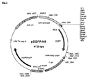
- the hEGFP sequence may be obtained from the pEGFP-N1 plasmid (e.g. obtained from Clontech), which is shown in Figure 1 .
- the hEGFP sequence can be seen in each of the expression cassettes in accordance with the invention in Figure 9 , and the nucleotide sequence of the hEGFP is shown as bases 2550-3278 in SEQ ID No.s 1, 2, 3, 4 & 5 in the sequence listing.
- GFP produces a good quantum yield of fluorescence and matches the output of argon ion lasers used in fluorescence activated cell sorters.
- Cells according to the third aspect of the invention which contain DNA molecules coding hEGFP, may be used according to the method of the fourth aspect of the invention.
- preferred expression cassettes according to the invention comprise a human GADD45 ⁇ gene regulatory element and human GADD45 ⁇ gene promoter operatively linked to a DNA sequence encoding a GFP, or light emitting derivative or analogues thereof (e.g. YFP etc).
- Most preferred expression cassettes comprise a human GADD45 ⁇ gene promoter operatively linked to a DNA sequence encoding a GFP or light emitting derivative thereof, and Intron 3 of the GADD45 ⁇ gene.
- the expression cassette according to the first aspect is preferably 5-31, (the cassette illustrated in Figure 9 as GD531).
- the nucleotide sequence of expression cassette 5-31 is shown in the sequence listing as SEQ ID No.2.
- the expression cassette according to the first aspect is preferably 5-32 (the cassette illustrated in Figure 9 as GD532).
- the nucleotide sequence of expression cassette 5-32 is shown in the sequence listing as SEQ ID No.3.
- the expression cassette according to the first aspect is preferably 5-33 (the cassette illustrated in Figure 9 as GD533)
- the nucleotide sequence of expression cassette 5-33 is shown in the sequence listing as SEQ ID No.4.
- the recombinant vector according to the second aspect of the present invention may for example be a plasmid, cosmid or phage.
- Such recombinant vectors are of great utility when replicating the expression cassette.
- recombinant vectors are highly useful for transfecting cells with the expression cassette, and may also promote expression of the reporter protein.
- Recombinant vectors may be designed such that the vector will autonomously replicate in the cytosol of the cell or can be used to integrate into the genome.
- elements that induce DNA replication may be required in the recombinant vector.
- Suitable elements are well known in the art, and for example, may be derived from pCEP4 (Invitrogen, 3 Fountain Drive, Inchinnan Business Park, Paisley, PA4 9RF, UK) pEGFP-N1 (BD Biosciences Clontech UK, 21 In Between Towns Road, Cowley, Oxford, OX4 LY, United Kingdom) or pCI and pSI (Promega UK ltd, Delta house, chilworth Science Park, Victoria SO16 7NS, UK).
- replicating vectors can give rise to multiple copies of the DNA molecule in a transformant and are therefore useful when over-expression (and thereby increased light emission) of the reporter protein is required.
- the vector is able to replicate in human, primate and/or canine cells.
- the vector comprises an origin of replication, and preferably, at least one selectable marker.
- the selectable marker may confer resistance to an antibiotic, for example, hygromycin or neomycin.
- a suitable element is derived from the pCEP4 plasmid (Invitrogen, 3 Fountain Drive, Inchinnan Business Park, Paisley, PA4 9RF, UK), which is illustrated in Figure 2 .
- the recombinant vector according to the second aspect is preferably pEP-GD531, as illustrated in Figure 5 .
- the recombinant vector according to the second aspect is preferably pEP-GD532, as illustrated in Figure 6 .
- the recombinant vector according to the second aspect is preferably pEP-GD533, as illustrated in Figure 7 .
- the recombinant vector is incorporated within a cell.
- the cell is eukaryotic.
- Such host cells may be mammalian derived cells and cell lines. Preferred mammalian cells include human, primate, murine or canine cells.
- the host cells may be lymphoma cells or cell lines, such as mouse lymphoma cells.
- the host cells may be immortalised, for example, lymphocytes.
- Preferred host cells are human cell lines.
- the host cells are human cell lines having a fully functional p53, for example, ML-1 (a human myeloid leukaemia cell line with wild-type p53), TK6 (a human lymphoblastoid cell line with wild-type p53).
- ML-1 a human myeloid leukaemia cell line with wild-type p53
- TK6 a human lymphoblastoid cell line with wild-type p53
- WI-L2-NS and WTK1 both of which are sister lines of TK6 and have mutant p53 proteins
- TK6 human cells are particularly preferred cell lines for use according to the method of the invention. While the inventors do not wish to be bound by any hypothesis, they believe that TK6 cells are most useful because they have a fully functional p53.
- Host cells used for expression of the protein encoded by the DNA molecule are ideally stably transfected, although the use of unstably transfected (transient) cells is not precluded.
- Transfected cells according to the third aspect of the invention may be formed by following procedures described in the Example.
- the cell is ideally a human cell line, for example TK6.
- Such transfected cells may be used according to the method of the fourth aspect of the invention to assess whether or not agents induce or potentiate DNA damage.
- GFP expression is induced in response to DNA damage and the light emitted by GFP may be easily measured using known appropriate techniques.
- the expression cassette according to the invention may be integrated into the genome of a host cell.
- suitable methods for integrating the cassette into the genome For example, the expression cassette may be harboured on a retroviral vector, which in combination with a packaging cell line may produce helper-free recombinant retrovirus, which may then be introduced into the host cell. The cassette may then integrate itself into the genome.
- suitable helper-free retroviral vector systems include the pBabePuro plasmid with the BING retroviral packaging cell line [ Kinsella and Nolan, 1996. Episomal Vectors Rapidly and Stably Produce High-Titer Recombinant Retroviruses. Human Gene Therapy. 7:1405-1413 .]
- the method of the fourth aspect of the invention is particularly useful for detecting agents that induce DNA damage at low concentrations.
- the methods may be used to screen compounds, such as candidate medicaments, food additives or cosmetics, to assess whether it is safe to expose a living organism, particularly people, to such compounds.
- the method of the fourth aspect of the invention may be employed to detect whether or not water supplies are contaminated by DNA damaging agents or agents that potentiate DNA damage.
- the methods may be used to monitor industrial effluents for the presence of pollutants that may lead to increased DNA damage in people or other organisms exposed to the pollution.
- the method of the invention is preferably performed by growing cells transfected with a recombinant vector according to the second aspect of the invention (such as pEP-GD531, pEP-GD532, or pEP-GD533), incubating the cells with the agent which putatively causes DNA damage for a predetermined time and monitoring the expression of the light emitting reporter protein directly from a sample of the cells.
- a recombinant vector according to the second aspect of the invention such as pEP-GD531, pEP-GD532, or pEP-GD533
- Cells are preferably grown in low fluorescence growth medium. This can obviate the need to wash the cells before measurements are made and therefore reduce the number of steps in the method further.
- preferred human cells according to the third aspect of the invention may be grown in a low fluorescent media.
- Suitable media may be a modified RPMI 1640 media and most preferably contains either low concentrations of, or preferably, no riboflavin or phenol red. The inventors have found that the use of such media is surprisingly useful in assays of fluorescent markers such as GFP. The use of such media reduces the "signal to noise ratio" when measuring fluorescence.
- a media that does not contain, or contains reduced levels (relative to standard cell culture media) of riboflavin or phenol red for use in fluorescent assays.
- the media contains no riboflavin and no phenol red.
- the media is modified such that it does not contain riboflavin.
- the use of such a modified media would be counter-intuitive to a skilled person because it would be understood that riboflavin is usually required in media. This is because most cells require riboflavin to be able to grow and survive.
- the inventors have realised that cells, which have been grown in conventional growth media containing fiboflavin, may be removed from such media; placed in media without riboflavin; and will remain viable for the purposes of conducting a reliable fluorescence assay. Cells left in conventional media (containing riboflavin and/or phenol red) tend to provide less satisfactory fluorescence values when tested according to the method of the fourth aspect of the invention (particularly when the reporter protein is GFP or a derivative thereof).
- media is used according to the fifth aspect of the invention when conducting assays according to the fourth aspect of the invention. It is most preferred such media is used when performing a genotoxic test with GFP as the reporter.
- a preferred media with low autofluorescence is a modified RPMI 1640 media that does not contain riboflavin or Phenol red.
- a most preferred media is disclosed in table 1 of Example 1.
- TK6 cells may be transfected with pEP-GD531, pEP-GD532, or pEP-GD533, and grown in RPMI medium containing no riboflavin.
- a putative DNA damaging agent e.g.
- a food additive or potential medicament or an agent contained within a water sample or effluent sample may then be added to the medium containing the cells.
- the cells are then allowed to grow for a defined period of time after which a sample of the cells is removed and fluorescence measured therefrom.
- Suitable methods of fluorescence detection and quantitation will be known to the skilled technician, and a method is described in the Examples.
- a preferred method of fluorescence detection is described in US 6, 509, 161 . This method is particularly useful when the light emitting report is GFP or a derivative thereof.
- fluorescence and absorbance readings may be recorded from TK6 cells transfected with pEP-GD531, pEP-GD532, or pEP-GD533, for example, from the well of a microplate.
- a suitable microplate is a 96 well, black, clear-bottomed microplates, manufactured by Matrix ScreenMates, Cat. No. 4929, Apogent Discoveries, USA, or Corning (BV, Netherlands: Cat. No. 3651).
- Fluorescence and absorbance measurements may be recorded using a suitable microplate reader, for example, the Tecan Ultra-384 (Tecan UK Ltd.) microplate reader: excitation 485 nm / emission 535 nm with an additional dichroic mirror (reflectance 320 nm - 500 nm, transmission 520 nm - 800 nm). Absorbance may be measured through a suitable filter, for example, a 620 nm filter.
- GFP fluorescence data
- absorbance data cell density
- Biovison Bioluminescence Cytotoxicity Assay Biovison Incoperated, 2455-D Old Middlefield Way, Mountain View, California 94043, USA
- Vybrant® Apoptosis Assay Kit Molecular probes Inc., 29851 Willow Creek Road, Eugene, OR 97402, USA
- control cell line is subtracted from the brightness values of a reporter cell line. This removes the interference from the data. It is therefore preferred that methods according to the fourth aspect of the invention employ cells containing control vectors based upon reporter vectors according to the second aspect of the invention. Such control vectors may correspond to the reporter vector, but with a non-sense mutation introduced into the light emitting reporter gene.
- the recombinant control vector may preferably be the vectors described as pEP-GD500C or pEP-GD532C, as illustrated in Figures 18 and 19 respectively.
- GenTK-T01 a control cell line based upon TK6 cells transformed with the vector pEP-GD532C (referred to herein asGenTK-C01).
- this Example describes the construction of the biosensors (cells according to the third aspect of the invention), and illustrates how they respond to a genotoxic agent, for example, methanesulphonic acid methyl ester (MMS) when utilised in a method according to the fourth aspect of the invention.
- a genotoxic agent for example, methanesulphonic acid methyl ester (MMS)
- the source of the GADD45 ⁇ promotor is the plasmid pHG45-HC from Albert J. Fornace Jr., M.D. (National Cancer Institute, Building: 37, Room: 6144, National Institutes of Health, Bethesda, Maryland 20892, USA), which is illustrated in Figure 3 .
- The- source-of the Green fluorescent protein is the Clontech plasmid pEGFP-N1 (BD Biosciences Clontech UK, 21 In Between Towns Road, Cowley, Oxford, OX4 LY, United Kingdom), which is illustrated in Figure 1 .
- the plasmid pEGFP-N1 encodes the GFPmut1 variant, which contains the double-amino-acid substitution of Phe-64 to Leu and Ser-65 to Thr.
- the coding sequence of the EGFP gene contains more than 190 silent base changes, which correspond to human codon-usage preferences.
- Plasmid pCEP4 is an episomal mammalian expression vector that uses the cytomegalovirus (CMV) immediate early enhancer/promoter for high level transcription of recombinant genes inserted into the multiple cloning site.
- CMV cytomegalovirus
- the Epstein-Barr Virus replication origin (oriP) and nuclear antigen (encoded by the EBNA-1 gene) is carried by this plasmid to permit extrachromosomal replication in human, primate and canine cells.
- pCEP4 also carries the hygromycin B resistance gene for stable selection in transfected cells.
- DNA sequences of each of the expression cassettes in the plasmids pEP-GD500, pEP-GD531, pEP-GD532, pEP-GD533, and pEP-GD534 are shown in the sequence listing as SEQ ID No.1, SEQ ID No.2, SEQ ID No.3, SEQ ID No.4, and SEQ ID No.5, respectively.
- FIG. 9 there are shown the series of reporter cassettes in accordance with the invention each aligned underneath the Human GADD45 ⁇ gene.
- the GADD45 ⁇ EGFP reporter gene fusions of plasmids pEP-GD531, pEP-GD532, and pEP-GD533 were constructed in such a way that the overall length of the GADD45 ⁇ -EGFP fusion is identical to the native GADD45 ⁇ gene, as shown in Figure 9 .
- the GADD45 ⁇ DNA downstream of the hEGFP is transcribed into mRNA as well as the hEGFP itself, and is subject to splicing.
- plasmid pEP-GD533 In plasmid pEP-GD533, only the hEGFP gene is transcribed into mRNA. The transcription is stopped at the SV40 polyadenylation signal before the GADD45 ⁇ gene sequence. These plasmids are in accordance with the first aspect of the invention.
- the pEP-GD534 cassette is much shorter and does not include the p53 motif in Intron 3 of the GADD45 ⁇ gene.
- a dysfunctional p53 protein can lead to increased radioresistance (i.e. increased survival) and an increased sensitivity to mutagenic and clastogenic effects of radiation. While these properties make a cell line suitable for detecting mutation endpoints, the same properties imply that the induction of surrogate biomarkers for DNA damage in a cell line with a mutant p53 status would be at best poor.
- the TK6 cell line was chosen for the biosensor (i.e. a cell according to the third aspect of the invention). It is believed that additional data of scientific interest may be gained from the use of its sister cell lines with the recombinant GADD45 ⁇ -EGFP reporter vector.
- TK6 cells are transfected with the vectors by electroporation using a method adapted from Xia and Liber [ Methods in Molecular biology, Vol.48: Animal Cell Electroporation and Electrofusion Protocols, 1995. Edited by J.A. Nickoloff. Humana Press Inc., Totowa, NJ, USA, Pages 151-160 ], and clones bearing the reporter plasmids are selected over two weeks with Hygromycin B.
- GenTK6 cells transformed with pEP-GAD532 are referred to herin as GenTK-T01.
- GenTK-C01 An equivalent control cell line, termed GenTK-C01, was also produced in which the GFPreporter gene was deliberately placed out of frame. Accordingly GFP could not be expressed from this control cell line.
- a number of such media may be produced and, by way of example, the inventors developed a modified low autofluorescent RPMI 1640 media (see Table 1).
- This media represents a preferred media for use according to the fifth aspect of the invention that does not contain any riboflavin, and may be used with the biosensor, to facilitate direct fluorescence reading of biosensor cultures.
- Table 1 working concentrations of components of a low autofluorescent mammalian cell culture media.
- MMS is known to those skilled in he art as a genotoxic and cytotoxic agent and may be used as a positive control when using the method of the invention.
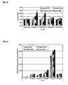
- FIG. 11 there is shown unprocessed brightness values from the five reporter cell lines and the parent untransfected cell line TK6. Brightness values were calculated for each cell line-MMS combination by measuring the total GFP fluorescence and then normalising this with absorbance at 620nm.
- each of the plasmids pEP-GD531, pEP-GD532, and pEP-GD533 show an increase in background fluorescence as compared to the untransfected parent cell line TK6, and these cell lines showed a substantial degree of dose dependent increase in fluorescence in the human cell line TK6 when treated with MMS.
- the present invention uses the regulatory sequences of the human DNA-Damage inducible gene GADD45 ⁇ to control the production of EGFP in the p53 wild-type human lymphoblastiod cell line TK6.
- the human gene GADD45 ⁇ stimulates DNA excision repair in vitro and inhibits entry of cells into S phase.
- the reporters in accordance with the invention not only use the upstream promoter of the GADD45 ⁇ gene, but also incorporate the gene regulatory elements found within the genomic gene sequence itself. This includes a putative p53 binding site in the third Intron of the GADD45 ⁇ gene. Surprisingly, the use of these sequences enhances the response of the biosensors to genotoxic stress.
- This reporter system can be analysed for fluorescence induction after only 24 hours by simply measuring the fluorescence of the test culture.
- the cells may be subjected to an agent, and the expression of the light emitting reporter protein (GFP) from the cell indicates that the agents cause DNA damage. It is believed that this biosensor has applications in short term pre-regulatory drug screening.
- GFP light emitting reporter protein
- Example 1 The constructs, cells and protocols described in Example 1 were used unless indicated to the contrary.
- phenol red a pH indicator dye commonly used in culture media
- media used according to the invention is formulated as a modified low autofluorescence medium whereby phenol red or riboflavin, but preferably both phenol red and riboflavin, is omitted (see table 1).
- the use of conventional RPMI 1640 medium decreased fluorescence signal to noise ratios, and increased variability of fluorescence measurements. Accordingly the use of normal RPMI 1640 media was not optimal for direct fluorescence readings. In contrast, the use of a modified low autofluorescent cell culture media (corresponding to normal media save riboflavain and phenol red was omitted) facilitated the direct measurement of genotoxin induced fluorescence readings.
- media used in the method according to the fourth aspect of the invention may comprise conventional media (e.g. RPMI 1640) but is preferably modified, according to the fifth aspect of the invention, such that it does not contain Phenol Red or riboflavin.
- Example 1 The constructs, cells and protocols described in Example 1 were used unless indicated to the contrary.
- a preferred method of testing for DNA damage comprises the steps of : (1) preparing a microplate for use in an assay; (2) conducting the assay in the microplates; (3) collecting and analysing the data; and (4) making a judgment on DNA damage and the consequences.
- microplates used according to the invention preferably have consistent absorbance and fluorescence between plates and batches thereof. It will be appreciated that a skilled person should give careful consideration when selecting a suitable microplate.
- the assay plates can be filled using a liquid handling robot.
- a liquid handling robot For example the MicroLabS single probe, from Hamilton GB Ltd., Birmingham or a Genesis 8-probe robot (Tecan UK Ltd. Theale. UK).
- Microplates can also be filled rapidly and effectively using a multi-channel pipette.
- test chemical/sample is 1 mM or 500 ⁇ g/ml on the microplate.
- Exponentially growing cultures of cells lines according to the invention were washed in D-PBS and suspended at a density of 2 x 10 6 cells/ml in double strength low autofluorescent assay medium according to the invention.
- 75 microlitres of the cell suspension were added to each well of the diluted chemical: GenTK-C01 to one series and GenTK-T01 to the second, and to appropriate standards and controls.
- the plates were filled, they were sealed using either a gas permeable membrane (for example Breath-easy, Diversified Biotech, USA) or a plastic lid, and then incubated without shaking, for 24 hours at 37°C, 5% CO 2 , 95% humidity.
- a gas permeable membrane for example Breath-easy, Diversified Biotech, USA
- the cytotoxicity threshold is set at 80 % of the cell density reached by the untreated control cells. This is greater than 3 times the standard deviation of the background.
- a positive cytotoxicity result (+) is concluded if 1 or 2 compound dilutions produce a final cell density lower than the 80% threshold.
- a strong positive cytotoxicity result positive (++) is concluded when either (i) three or more compound dilutions produce a final cell density lower than the 80% threshold or (ii) at least one compound dilution produces a final cell density lower than a 60% threshold.
- a negative result (-) is concluded when no compound dilutions produce a final cell density lower than the 80% threshold.
- the lowest effective concentration (LEC) is the lowest test compound concentration that produces a final cell density below the 80% threshold.
- the compound absorbance control allows a warning to be generated if a test compound is significantly absorbing. If the ratio of the absorbance of the compound control well to a well filled with media alone is > 2, there is a risk of interference with interpretation.
- the cytotoxicity controls indicate that the cell lines are behaving normally.
- the 'high' MMS standard should reduce the final cell density to below the 80% threshold, and should be a lower value than the 'low' standard.
- the genotoxic threshold is set at a relative GFP induction of 1.5 (i.e. a 50% increase). This is greater than 3 times the standard deviation of the background.
- a positive genotoxicity result (+) is concluded if 1 or 2 compound dilutions produce a relative GFP induction greater than the 1.5 threshold.
- a strong positive genotoxicity result (++) is concluded if either (i) three or more compound dilutions produce a relative GFP induction greater than the 1.5 threshold or (ii) at least one compound dilution produces a relative GFP induction greater than a 1.8 threshold.
- a negative genotoxicity result (-) is concluded where no compound dilutions produce a relative GFP induction greater than the 1.5 threshold.
- the LEC is the lowest test compound concentration that produces a relative GFP induction greater than the 1.5 threshold.
- the genotoxic controls demonstrate that the cell lines are responding normally to DNA damage.
- the 'high' MMS standard must produce a fluorescence induction > 2, and be a greater value than the 'low' MMS standard.
- Anomalous brightness data is generated when the toxicity leads to a final cell density less than 30% that of the blank. Genotoxicity data is not calculated above this toxicity threshold.
- Compounds that tested negative for genotoxicity were re-tested up to 10mM or 5000 ⁇ g/ml, or to the limit of solubility or cytotoxicity.
- the compound fluorescence control allows a warning to be generated when a compound is highly auto-fluorescent. If the ratio of the fluorescence of the compound control well to a media filled well is >5, there is a risk of interference with interpretation. In these cases, fluorescence polarisation can be used to distinguish GFP from other sources of fluorescence (Knight et al. , 2000, 2002). The Tecan instrument has this facility. Occasionally, compounds though not fluorescent themselves, induce cellular autofluorescence. This is apparent from rising brightness in the control (GenTK-C01) cell line in the absence of fluorescence from the chemical-only control. The routine subtraction of GenTK-C01 from GenTK-T01 data removes this interference from the data.
- S9 is a liver extract (known to the skilled person) that makes it possible to discriminate between genotoxic compounds and some non-genotoxic compounds that may be chemically altered by hepatocyte metabolism to generate a genotoxic compound in vivo .
- Example 1 The constructs, cells and protocols described in Example 1 were used unless indicated to the contrary.
- a preferred method of testing for DNA damage comprises the steps of : (1) preparing a 24 well plate for use in an assay with metabolic activation; (2) conducting the assay in 24 well plates and processing samples in to 96 well plates; (3) collecting and analysing the data; and (4) making a judgment on DNA damage and the consequences.
- microplates used according to the invention preferably have consistent absorbance and fluorescence between plates and batches thereof.
- the assay plates can be filled using a liquid handling robot.
- a liquid handling robot For example the MicroLabS single probe, from Hamilton GB Ltd., Birmingham or a Genesis 8-probe robot (Tecan UK Ltd. Theale. UK).
- Microplates can also be filled rapidly and effectively using a multi-channel pipette.
- the RPMI 1640 media is preferably a modified media as defined by the fifth aspect of the invention (e.g. the media disclosed in Table 1).
- 200 microlitres of the test chemical solution were put into a sterile 7ml glass vial or 1.5 ml microfuge tube.
- Each sample was serially diluted by transferring 100 microlitres into 100 microlitres of 100% DMSO, mixing, and then taking 100 microlitres out and into the next well. This produced 3 serial dilutions of 100 microlitres each.
- the final top concentration of test chemical/sample is 1 mg/ml on the 24 well plate.
- the plates were filled, they were sealed using either a gas permeable membrane (for example Breath-easy, Diversified Biotech, USA) or a plastic lid, and then incubated without shaking, for 24 hours at 37°C, 5% CO 2 , 95% humidity. Following 24 hours incubation cells were transferred to 1.5 ml microfuge tubes and collected by centrifugation at 2500 rcf for 5 minutes. Cells were washed in 500 ⁇ l pre-warmed D-PBS (Sigma-Aldrich, Gillingham, UK) and collected by centrifugation as before.
- a gas permeable membrane for example Breath-easy, Diversified Biotech, USA
- Cells were resuspended in 150 ⁇ l D-PBS, and cell suspensions transferred to a well of an optically clear bottom, black polystyrene 96 well microplate (Matrix Technologies, Wilmslow, UK). As blanks, 150 ⁇ l of D-PBS were added to 2 wells.
- the cytotoxicity threshold is set at 80 % of the cell density reached by the untreated control cells. This is greater than 3 times the standard deviation of the background.
- a positive cytotoxicity result (+) is concluded if 1 or 2 compound dilutions produce a final cell density lower than the 80% threshold.
- a strong positive cytotoxicity result positive (++) is concluded when either (i) three or more compound dilutions produce a final cell density lower than the 80% threshold or (ii) at least one compound dilution produces a final cell density lower than a 60% threshold.
- a negative result (-) is concluded when no compound dilutions produce a final cell density lower than the 80% threshold.
- the lowest effective concentration (LEC) is the lowest test compound concentration that produces a final cell density below the 80% threshold.
- the compound absorbance control allows a warning to be generated if a test compound is significantly absorbing. If the ratio of the absorbance of the compound control well to a well filled with media alone is > 2, there is a risk of interference with interpretation.
- the cytotoxicity controls indicate that the cell lines are behaving normally.
- the genotoxic threshold is set at a relative GFP induction of 1.3 (i.e. a 30% increase). This is greater than 3 times the standard deviation of the background.
- a positive genotoxicity result (+) is concluded if 1 or 2 compound dilutions produce a relative GFP induction greater than the 1.3 threshold.
- a strong positive genotoxicity result (++) is concluded if either (i) three or more compound dilutions produce a relative GFP induction greater than the 1.3 threshold or (ii) at least one compound dilution produces a relative GFP induction greater than a 1.6 threshold.
- a negative genotoxicity result (-) is concluded where no compound dilutions produce a relative GFP induction greater than the 1.3 threshold.
- the LEC is the lowest test compound concentration that produces a relative GFP induction greater than the 1.3 threshold.
- the genotoxic controls demonstrate that the cell lines are responding normally to DNA damage.
- the Cyclophosphomide standard must produce a fluorescence induction > 1.3 with S9, and not produce a fluorescence induction > 1.1 without S9.
- Anomalous brightness data is generated when the toxicity leads to a final cell density less than 30% that of the blank. Genotoxicity data is not calculated above this toxicity threshold.
- Compounds that tested negative for genotoxicity were re-tested up to 10mM or 5000 ⁇ g/ml, or to the limit of solubility or cytotoxicity.
Landscapes
- Chemical & Material Sciences (AREA)
- Health & Medical Sciences (AREA)
- Life Sciences & Earth Sciences (AREA)
- Organic Chemistry (AREA)
- General Health & Medical Sciences (AREA)
- Gastroenterology & Hepatology (AREA)
- Biochemistry (AREA)
- Biophysics (AREA)
- Zoology (AREA)
- Genetics & Genomics (AREA)
- Medicinal Chemistry (AREA)
- Molecular Biology (AREA)
- Proteomics, Peptides & Aminoacids (AREA)
- Toxicology (AREA)
- Measuring Or Testing Involving Enzymes Or Micro-Organisms (AREA)
- Micro-Organisms Or Cultivation Processes Thereof (AREA)
Description
- The present invention relates to methods for detecting agents that cause or potentiate DNA damage, and to molecules and transfected cell lines that may be employed in such methods. In particular, the invention relates to biosensors for detecting DNA damage in human cell cultures.
- DNA damage is induced by a variety of agents such as ultraviolet light, X rays, free radicals, methylating agents and other mutagenic compounds. DNA damage can also be caused indirectly either by agents that affect enzymes and proteins which interact with DNA (including polymerases and topoisomerases) or by promutagens (agents that can be metabolised to become mutagenic). Any of these agents may cause damage to the DNA that comprises the genetic code of an organism and cause mutations in genes. In animals, such mutations can lead to carcinogenesis or may damage the gametes to give rise to congenital defects in offspring. Such DNA damaging agents can be collectively known as genotoxins.
- These DNA damaging agents may chemically modify the nucleotides that comprise DNA and may also break the phosphodiester bonds that link the nucleotides or disrupt association between bases (T-A or C-G). To counter the effect of these DNA damaging agents cells have evolved a number of mechanisms. For example, the SOS response in E. coli is a well-characterised cellular response induced by DNA damage in which a series of proteins are expressed, including DNA repair enzymes, which repair the damaged DNA. In mammalians, nucleotide excision repair and base excision repair mechanisms play a prominent role in DNA damage repair, and are the primary mechanism for removal of bulky DNA adducts and modified bases.
- There are numerous circumstances when it is important to identify what agents may cause or potentiate DNA damage. It is particularly important to detect agents that cause DNA damage when assessing whether it is safe to expose a person to these agents. For instance, a method of detecting these agents may be used as a genotoxicity assay for screening compounds that are candidate medicaments, food additives or cosmetics to assess whether or not the compound of interest induces DNA damage. Alternatively, methods of detecting DNA damaging agents may be used to monitor for contamination of water supplies with pollutants that contain mutagenic compounds.
- Various methods, such as the Ames Test, the in vitro micronucleus test and the mouse lymphoma assay (MLA), for determining the toxicity of an agent are known but are unsatisfactory for a number of reasons. For instance, incubation of samples can take many weeks, when it is often desirable to obtain genotoxic data in a shorter time frame. Furthermore, many known methods of detecting DNA damage (including the Ames Test and related methods) assay lasting DNA damage, as an endpoint, either in the form of mis-repaired DNA (mutations and recombinations) or unrepaired damage in the form of fragmented DNA. However, most DNA damage is repaired before such an endpoint can be measured and lasting DNA damage only occurs if the conditions are so severe that the repair mechanisms have been saturated.
- An improved genotoxic test is disclosed in
WO 98/44149 WO 98/44149 WO 98/44149 -
US 6,344,324 discloses a recombinant DNA molecule comprising the regulatory element of the hamster GADD153 upstream promoter region that activates gene expression in response to a wide range of cellular stress conditions, linked to a DNA sequence that encodes GFP. This reporter system is carried out in a human head and neck squamous-cell carcinoma cell line. However, problems associated with this reporter system are that it requires at least a four day treatment period at test agent concentrations that result in less than 10% cell survival, followed by analysis of fluorescence by flow cytometry. In addition, the biological relevance of any gene induction when tested with agents at this level of toxicity is debatable. Furthermore, this development does not disclose a means of specifically monitoring for the presence of agents that may cause or potentiate DNA damage, and the mechanism of GADD153 induction remains unclear. Hence, this system is of very limited use as a human DNA damage biosensor. - Therefore, it is an aim of embodiments of the present invention to address problems associated with the prior art, and to provide an improved biosensor for detecting DNA damage in human cell cultures.
- According to a first aspect of the present invention, there is provided an expression cassette comprising a DNA sequence encoding a reporter protein, which DNA sequence is operatively linked to a human GADD45α gene promoter and a human GADD45α gene regulatory element arranged to activate expression of the DNA sequence in response to DNA damage.
- By the term "regulatory element", we mean a DNA sequence that regulates the transcription of a gene with which it is associated, i.e. the DNA sequence encoding the reporter protein.
- By the term "operatively linked", we mean that the regulatory element is able to induce the expression of the reporter protein.
- According to a second aspect of the invention, there is provided a recombinant vector comprising an expression cassette according to the first aspect.
- According to a third aspect of the invention, there is provided a cell containing a recombinant vector in accordance with the second aspect of the present invention.
- According to a fourth aspect of the present invention, there is provided a method of detecting for the presence of an agent that causes or potentiates DNA damage comprising subjecting a cell in accordance with the third aspect of the present invention to an agent; and monitoring the expression of the reporter protein from the cell.
- The method of the fourth aspect of the invention represents a novel cost-effective genotoxicity screen that may be used to provide a pre-regulatory screening assay for use by the pharmaceutical industry and in other applications where significant numbers of agents or compounds need to be tested. It provides a higher throughput and a lower compound consumption than existing in vitro and in vivo mammalian genotoxicity assays, and is sensitive to a broad spectrum of mutagens.
- The method of the fourth aspect of the invention is suitable for assessing whether or not an agent may cause DNA damage. It is particularly useful for detecting agents that cause DNA damage when assessing whether it is safe to expose a person to DNA damaging agents. For instance, the method may be used as a genotoxicity assay for screening whether or not known agents, such as candidate medicaments, pharmaceutical and industrial chemicals, pesticides, fungicides, foodstuffs or cosmetics, induce DNA damage. Alternatively, the method of the invention may be used to monitor for contamination of water supplies, leachates and effluents with pollutants containing DNA damaging agents.
- The method of the fourth aspect of the invention may also be used for assessing whether an agent may potentiate DNA damage. For example, certain agents can cause accumulation of DNA damage by inhibiting DNA repair (for instance by preventing expression or function of a repair protein) without directly inflicting DNA damage.
- Surprisingly, the use of a human GADD45α gene regulatory element in addition to the human GADD45α gene promoter in the expression cassette according to the first aspect of the invention radically enhances the response of the cassette to genotoxic stress and, hence, DNA damage in the cell according to the third aspect. Advantageously, the cassette can be analysed for expression of the reporter protein within or after only 24 hours simply by assaying for the protein in a test culture. The cells may be subjected to the test agent or compound, and expression of the reporter protein in the cell indicates whether the test agent causes DNA damage.
- The inventors have found that DNA encoding a human GADD45a gene promoter and a human GADD45α gene regulatory element may be operatively linked a reporter protein to form a cassette according to the first aspect of the invention and then advantageously used in a genotoxic test according to the fourth aspect of the invention. Such cassettes may comprise the whole of the GADD45α gene (including coding sequences) provided that it is operatively linked to DNA encoding a light emitting reporter. For instance cassettes may be made according to the first aspect of the invention comprising the whole of, or substantially all of, the GADD45α gene (comprising regulatory elements and promoter) with DNA encoding a reporter inserted 3' of the GADD45α promoter (e.g. within the GADD45α coding sequence or at the 3' of the coding sequence). end of the arranged to activate expression of the DNA sequence in response to DNA damage.
- Preferably, the human GADD45α gene promoter sequence induces RNA polymerase to bind to the DNA molecule and start transcribing the DNA encoding the reporter protein. It is preferred that the promoter sequence comprises the human GADD45α gene promoter sequence and the 5' untranslated region. The promoter sequence may be obtained from the pHG45-HC plasmid, which is illustrated in
Figure 3 . The promoter sequence can be seen in each of the expression cassettes in accordance with the invention inFigure 9 , and the nucleotide sequence of the GADD45α gene promoter is shown as bases 4-2254 in SEQ ID No.s 1, 2, 3, 4 & 5 in the sequence listing. It will be appreciated that the promoter may comprise each of the bases 4 -2254 or alternatively may be a functional derivative or functional fragment thereof. Functional derivatives and functional fragments may be readily identified be assessing whether or not transcriptase will bind to a putative promoter region and will then lead to the transcription of the marker protein. Alternatively such functional derivatives and fragments may be examined by conducting mutagenesis on the GADD45α promoter, when in natural association with the GADD45α gene, and assessing whether or not GADD45α expression may occur. - The regulatory element in the expression cassette according to the invention may comprise sequences downstream of the GADD45α gene promoter sequence. The regulatory element may comprise functional DNA sequences such as those encoding translation initiation sequences for ribosome binding or DNA sequences that bind transcription factors which promote gene expression following DNA damage. The regulatory element in the expression cassette according to the invention may comprise at least one exon of the GADD45α gene. For example, the regulatory element may comprise Exon 1, Exon 2, Exon 3, and/or Exon 4 of the GADD45α gene, or at least a region thereof, or any combination thereof. Hence, the regulatory element may comprise any combination of the four exons of the GADD45α gene, or at least a region thereof.
- In a preferred embodiment, the regulatory element comprises at least a region of
Exon 1 of the GADD45α gene, and preferably at least a region ofExon 3 of the GADD45α gene, and more preferably, at least a region of Exon 4 of the GADD45α gene. It is especially preferred that the regulatory element comprises all ofExon 1 of the GADD45α gene, and preferably at least a region ofExon 3 of the GADD45α gene, and more preferably, all ofExon 4 of the GADD45α gene. - Preferred regulatory elements are illustrated in the each of the expression cassettes in accordance with the invention in
Figure 9 . The nucleotide sequence ofExon 3 of the GADD45α gene is shown as bases 3405-3642 in SEQID No.s Exon 3 of the GADD45α gene is shown as bases 3503-3642 in SEQ ID No.4. The nucleotide sequence ofExon 4 of the GADD45α gene is shown as bases 4716-5391 in SEQID No.s - Alternatively, or additionally, the regulatory element may comprise a non-coding DNA sequence, for example, at least one intron of the GADD45α gene. For example, the regulatory element may comprise
Intron 1,Intron 2, and/orIntron 3 of the GADD45α gene, or at least a region thereof, or any combination thereof. Hence, the regulatory element may comprise any combination of the three introns of the GADD45α gene, or at least a region thereof. - In a preferred embodiment, the regulatory element in the expression cassette according to the invention comprises at least a region of
Intron 3 of the GADD45α gene.Intron 3 of GADD45α is illustrated inFigure 9 , and the nucleotide sequence ofIntron 3 of the GADD45α gene is shown as bases 3643-4715 in SEQID No.s - In a preferred embodiment, the expression cassette in accordance with the invention comprises the promoter sequence of the GADD45α gene and also gene regulatory elements found within
Intron 3 of the genomic GADD45α gene sequence itself. While the inventors do not wish to be bound by any hypothesis, they believe thatIntron 3 of the GADD45α gene, contains a putative p53 binding motif, and that it is this p53 motif which surprisingly enhances the response of the expression cassette to genotoxic stress. The putative p53 binding motif is shown as nucleotide bases 3830-3849 of SEQID No.s - The inventors also believe that
Intron 3 of the GADD45α gene may contain a putative TRE motif, which may encode a AP-1 binding site. The putative TRE motif is shown as nucleotide bases 3879-3885 of SEQID No.s - It is preferred that the expression cassette comprises at least the p53 binding motif and/or the AP-1 binding motif from
Intron 3 of the GADD45α gene. - The regulatory element may comprise a 3' untranslated (UTR) region of the GADD45α gene, the nucleotide sequence of which is shown as bases 4830-5391 in SEQ
ID No.s Intron 3. - By the term "reporter protein", we mean a protein which when expressed in response to the regulatory element of the DNA molecule of the invention, is detectable by means of a suitable assay procedure. Preferably, the reporter protein comprises a light emitting reporter protein. The DNA sequence that encodes a light emitting reporter protein may code for any light emitting protein, for example Luciferase or Green Fluorescent Protein. However, it is preferred that the DNA sequence codes for a protein that is substantially fluorescent.
- Preferred DNA sequences that encode a light emitting reporter protein code for Green Fluorescent Protein (GFP), and light emitting derivatives thereof. GFP is from the jelly fish Aequorea victoria and is able to absorb blue light and re-emits an easily detectable green light and is thus suitable as a reporter protein. GFP may be advantageously used as a reporter protein because its measurement is simple and reagent free and the protein is non-toxic.
- Derivatives of GFP include DNA sequences encoding for polypeptide analogues or polypeptide fragments of GFP, which are able to emit light. Many of these derivatives absorb and re-emit light at wavelengths different to GFP found endogenously in Aequorea victoria.
- For instance, preferred expression cassettes according to the first aspect of the invention comprise a DNA sequence that encodes the human enhanced GFP (hEGFP), also known as GFPmut1 variant of GFP. This consists of the GFP wild type containing a double amino acid substitution of Phe-64 to Leu, and Ser-65 to Thr. The coding sequence of the hEGFP gene also contains more than 190 silent base changes, which correspond to human codon usage preferences relative to the native Aequorea victoria sequence. This is particularly advantageous for use in a human DNA damage reporter system. The hEGFP sequence may be obtained from the pEGFP-N1 plasmid (e.g. obtained from Clontech), which is shown in
Figure 1 . The hEGFP sequence can be seen in each of the expression cassettes in accordance with the invention inFigure 9 , and the nucleotide sequence of the hEGFP is shown as bases 2550-3278 in SEQID No.s - GFP produces a good quantum yield of fluorescence and matches the output of argon ion lasers used in fluorescence activated cell sorters. Cells according to the third aspect of the invention, which contain DNA molecules coding hEGFP, may be used according to the method of the fourth aspect of the invention.
- Hence, preferred expression cassettes according to the invention comprise a human GADD45α gene regulatory element and human GADD45α gene promoter operatively linked to a DNA sequence encoding a GFP, or light emitting derivative or analogues thereof (e.g. YFP etc). Most preferred expression cassettes comprise a human GADD45α gene promoter operatively linked to a DNA sequence encoding a GFP or light emitting derivative thereof, and
Intron 3 of the GADD45α gene. - In a first embodiment, the expression cassette according to the first aspect is preferably 5-31, (the cassette illustrated in
Figure 9 as GD531). The nucleotide sequence of expression cassette 5-31 is shown in the sequence listing as SEQ ID No.2. - In a second embodiment, the expression cassette according to the first aspect is preferably 5-32 (the cassette illustrated in
Figure 9 as GD532). The nucleotide sequence of expression cassette 5-32 is shown in the sequence listing as SEQ ID No.3. - In a third embodiment, the expression cassette according to the first aspect is preferably 5-33 (the cassette illustrated in
Figure 9 as GD533) The nucleotide sequence of expression cassette 5-33 is shown in the sequence listing as SEQ ID No.4. - The recombinant vector according to the second aspect of the present invention may for example be a plasmid, cosmid or phage. Such recombinant vectors are of great utility when replicating the expression cassette. Furthermore, recombinant vectors are highly useful for transfecting cells with the expression cassette, and may also promote expression of the reporter protein.
- Recombinant vectors may be designed such that the vector will autonomously replicate in the cytosol of the cell or can be used to integrate into the genome. In this case, elements that induce DNA replication may be required in the recombinant vector. Suitable elements are well known in the art, and for example, may be derived from pCEP4 (Invitrogen, 3 Fountain Drive, Inchinnan Business Park, Paisley, PA4 9RF, UK) pEGFP-N1 (BD Biosciences Clontech UK, 21 In Between Towns Road, Cowley, Oxford, OX4 LY, United Kingdom) or pCI and pSI (Promega UK ltd, Delta house, chilworth Science Park, Southampton SO16 7NS, UK).
- Such replicating vectors can give rise to multiple copies of the DNA molecule in a transformant and are therefore useful when over-expression (and thereby increased light emission) of the reporter protein is required. In addition, it is preferable that the vector is able to replicate in human, primate and/or canine cells. It is preferred that the vector comprises an origin of replication, and preferably, at least one selectable marker. The selectable marker may confer resistance to an antibiotic, for example, hygromycin or neomycin. Hence, a suitable element is derived from the pCEP4 plasmid (Invitrogen, 3 Fountain Drive, Inchinnan Business Park, Paisley, PA4 9RF, UK), which is illustrated in
Figure 2 . - In a first embodiment, the recombinant vector according to the second aspect is preferably pEP-GD531, as illustrated in
Figure 5 . - In a second embodiment, the recombinant vector according to the second aspect is preferably pEP-GD532, as illustrated in
Figure 6 . - In a third embodiment, the recombinant vector according to the second aspect is preferably pEP-GD533, as illustrated in
Figure 7 . - According to the second aspect of the invention the recombinant vector is incorporated within a cell. It is preferred that the cell is eukaryotic. Such host cells may be mammalian derived cells and cell lines. Preferred mammalian cells include human, primate, murine or canine cells. The host cells may be lymphoma cells or cell lines, such as mouse lymphoma cells. The host cells may be immortalised, for example, lymphocytes.
- Preferred host cells are human cell lines. Preferably, the host cells are human cell lines having a fully functional p53, for example, ML-1 (a human myeloid leukaemia cell line with wild-type p53), TK6 (a human lymphoblastoid cell line with wild-type p53). However, host cell lines of WI-L2-NS and WTK1 (both of which are sister lines of TK6 and have mutant p53 proteins) are also envisaged. These cell lines may all be found at the European Collection of Cell Cultures (ECACC General Office, CAMR, Porton Down, Salisbury, Wiltshire, SP4 OJG, United Kingdom).
- The inventors have found that TK6 human cells are particularly preferred cell lines for use according to the method of the invention. While the inventors do not wish to be bound by any hypothesis, they believe that TK6 cells are most useful because they have a fully functional p53.
- Host cells used for expression of the protein encoded by the DNA molecule are ideally stably transfected, although the use of unstably transfected (transient) cells is not precluded.
- Transfected cells according to the third aspect of the invention may be formed by following procedures described in the Example. The cell is ideally a human cell line, for example TK6. Such transfected cells may be used according to the method of the fourth aspect of the invention to assess whether or not agents induce or potentiate DNA damage. GFP expression is induced in response to DNA damage and the light emitted by GFP may be easily measured using known appropriate techniques.
- Most preferred cells according to the third aspect of the invention are TK6 cells transformed with the vector pEP-GAD532. These cells are referred to herein as GenTK-T01.
- It is also envisaged that the expression cassette according to the invention may be integrated into the genome of a host cell. The skilled technician will appreciate suitable methods for integrating the cassette into the genome. For example, the expression cassette may be harboured on a retroviral vector, which in combination with a packaging cell line may produce helper-free recombinant retrovirus, which may then be introduced into the host cell. The cassette may then integrate itself into the genome. Examples of suitable helper-free retroviral vector systems include the pBabePuro plasmid with the BING retroviral packaging cell line [Kinsella and Nolan, 1996. Episomal Vectors Rapidly and Stably Produce High-Titer Recombinant Retroviruses. Human Gene Therapy. 7:1405-1413.]
- The method of the fourth aspect of the invention is particularly useful for detecting agents that induce DNA damage at low concentrations. The methods may be used to screen compounds, such as candidate medicaments, food additives or cosmetics, to assess whether it is safe to expose a living organism, particularly people, to such compounds. Alternatively, the method of the fourth aspect of the invention may be employed to detect whether or not water supplies are contaminated by DNA damaging agents or agents that potentiate DNA damage. For instance, the methods may be used to monitor industrial effluents for the presence of pollutants that may lead to increased DNA damage in people or other organisms exposed to the pollution.
- The method of the invention is preferably performed by growing cells transfected with a recombinant vector according to the second aspect of the invention (such as pEP-GD531, pEP-GD532, or pEP-GD533), incubating the cells with the agent which putatively causes DNA damage for a predetermined time and monitoring the expression of the light emitting reporter protein directly from a sample of the cells.
- Cells are preferably grown in low fluorescence growth medium. This can obviate the need to wash the cells before measurements are made and therefore reduce the number of steps in the method further. For instance, preferred human cells according to the third aspect of the invention may be grown in a low fluorescent media. Suitable media may be a modified RPMI 1640 media and most preferably contains either low concentrations of, or preferably, no riboflavin or phenol red. The inventors have found that the use of such media is surprisingly useful in assays of fluorescent markers such as GFP. The use of such media reduces the "signal to noise ratio" when measuring fluorescence. Therefore, according to a fifth aspect of the invention there is provided a media that does not contain, or contains reduced levels (relative to standard cell culture media) of riboflavin or phenol red for use in fluorescent assays. Most preferably the media contains no riboflavin and no phenol red.
- It is preferred that the media is modified such that it does not contain riboflavin. The use of such a modified media would be counter-intuitive to a skilled person because it would be understood that riboflavin is usually required in media. This is because most cells require riboflavin to be able to grow and survive. The inventors have realised that cells, which have been grown in conventional growth media containing fiboflavin, may be removed from such media; placed in media without riboflavin; and will remain viable for the purposes of conducting a reliable fluorescence assay. Cells left in conventional media (containing riboflavin and/or phenol red) tend to provide less satisfactory fluorescence values when tested according to the method of the fourth aspect of the invention (particularly when the reporter protein is GFP or a derivative thereof).
- It is preferred that media is used according to the fifth aspect of the invention when conducting assays according to the fourth aspect of the invention. It is most preferred such media is used when performing a genotoxic test with GFP as the reporter.
- A preferred media with low autofluorescence is a modified RPMI 1640 media that does not contain riboflavin or Phenol red. A most preferred media is disclosed in table 1 of Example 1.
- Other media that could be modified for low autofluorescence include Dulbecco's Modified Eagle media (D-MEM), F-10 Nutrient mixture (Ham), F-12 Nutrient mixture (Ham), and Fischer's medium (GIBCO, Invitrogen, Paisley, UK). The most appropriate medium may be used for different cell types that could be used as a host for recombinant reporter vectors.
According to a preferred embodiment of the method of the invention TK6 cells may be transfected with pEP-GD531, pEP-GD532, or pEP-GD533, and grown in RPMI medium containing no riboflavin. A putative DNA damaging agent (e.g. a food additive or potential medicament or an agent contained within a water sample or effluent sample) may then be added to the medium containing the cells. The cells are then allowed to grow for a defined period of time after which a sample of the cells is removed and fluorescence measured therefrom. - Suitable methods of fluorescence detection and quantitation will be known to the skilled technician, and a method is described in the Examples. A preferred method of fluorescence detection is described in
US 6, 509, 161 . This method is particularly useful when the light emitting report is GFP or a derivative thereof. - According to a preferred embodiment of the method of the invention, fluorescence and absorbance readings may be recorded from TK6 cells transfected with pEP-GD531, pEP-GD532, or pEP-GD533, for example, from the well of a microplate. An example of a suitable microplate is a 96 well, black, clear-bottomed microplates, manufactured by Matrix ScreenMates, Cat. No. 4929, Apogent Discoveries, USA, or Corning (BV, Netherlands: Cat. No. 3651). Fluorescence and absorbance measurements may be recorded using a suitable microplate reader, for example, the Tecan Ultra-384 (Tecan UK Ltd.) microplate reader: excitation 485 nm / emission 535 nm with an additional dichroic mirror (reflectance 320 nm - 500 nm, transmission 520 nm - 800 nm). Absorbance may be measured through a suitable filter, for example, a 620 nm filter.
- Most preferred protocols for conducting the method of the fourth aspect of the invention are described in Examples 1, 3 and 4.
- From the fluorescence and absorbance measurements, "brightness values" and fluorescence induction ratios for cells according to the third aspect of the invention can be made as described in the examples.
- There is background ("constitutive") expression of GFP from the GADD45α-EGFP constructs, thus the higher the cell density, the more fluorescent the culture. In order to correct for fluorescence increase that is consequent on growth, the fluorescence data (GFP) are divided by absorbance data (cell density) to give 'brightness units', i.e. the measure of average fluorescence per cell. This is independent of culture density. Accordingly, measurement of absorbance may be used primarily for normalisation of fluorescence signals rather than a measurement of the toxicity of the DNA damaging agent. Accordingly, it is envisaged that a secondary assay may be used in conjunction with the absorbance measurement in order to determine toxicity via cell viability and apoptosis. For example, using the Biovison Bioluminescence Cytotoxicity Assay (Biovison Incoperated, 2455-D Old Middlefield Way, Mountain View, California 94043, USA), or the Vybrant® Apoptosis Assay Kit (Molecular probes Inc., 29851 Willow Creek Road, Eugene, OR 97402, USA).
- Occasionally, compounds show fluorescence themselves or induce cellular autofluorescence. Accordingly, when evaluating increased GFP expression the brightness values of a control cell line is subtracted from the brightness values of a reporter cell line. This removes the interference from the data. It is therefore preferred that methods according to the fourth aspect of the invention employ cells containing control vectors based upon reporter vectors according to the second aspect of the invention. Such control vectors may correspond to the reporter vector, but with a non-sense mutation introduced into the light emitting reporter gene.
- The recombinant control vector may preferably be the vectors described as pEP-GD500C or pEP-GD532C, as illustrated in
Figures 18 and19 respectively. - Preferred methods according to the fourth aspect of the invention will utilise cells according to the third aspect of the invention (e.g. GenTK-T01) amd a control cell line based upon TK6 cells transformed with the vector pEP-GD532C (referred to herein asGenTK-C01).
- It will be appreciated that some non-genotoxic compounds can be chemically altered by cellular metabolism. In mammals this process is often called metabolic activation (MA). MA can convert certina non-genotoxic compounds (for example promutagens) into genotoxic compounds. Most frequently MA occurs in the liver. For this reason it is often preferred that genotoxicity tests are adapted such that assays of test compound are carried out in the presence and absence of liver extracts that are capable of metabolising a compound is if it were being metabolised in vivo. Example 4 illustrates a preferred method according to the fourth aspect of the invention which utilises a liver extract (known to the skilled person) called S9. Inclusion of such an extract allows assays to detect compounds that only become genotoxic after passage through the liver.
- All of the features described herein (including any accompanying claims, abstract and drawings), and/or all of the steps of any method or process so disclosed, may be combined with any of the above aspects in any combination, except combinations where at least some of such features and/or steps are mutually exclusive.
- Embodiments of the invention will now be further described, by way of example only, with reference to the following Examples and Figures in which:-
-
Figure 1 shows a restriction map of vector pEGFP-N1; -
Figure 2 shows a restriction map of vector pCEP4; -
Figure 3 shows a restriction map of vector pHG45-HC; -
Figure 4 shows a restriction map of vector pEP-GD500; -
Figure 5 shows a restriction map of vector pEP-GD531, a preferred recombinant vector according to the present invention; -
Figure 6 shows a restriction map of vector pEP-GD532, a preferred recombinant vector according to the present invention; -
Figure 7 shows a restriction map of vector pEP-GD533, a preferred recombinant vector according to the present invention; -
Figure 8 shows a restriction map of vector pEP-GD534; -
Figure 9 shows embodiments of reporter expression cassettes according to the invention; -
Figure 10 shows the effect of MMS induction on GADD45α reporter cell lines; -
Figure 11 shows the effect on brightness by MMS induction on GADD45α reporter cell lines; -
Figure 12 shows methyl methanesulphonate induced fluorescence measured directly from a reporter cell line cultured in either RPMI 1640 with serum (A) or modified low autofluorescence medium with serum (B) as discussed in Example 2; -
Figure 13 shows the growth of a human lymphoblastoid cell line in RPMI 1640 or modified low autofluorescence medium in Example 2; -
Figure 14 shows the effect of MMS on relative cell density in a 96 well plate assay after 48 hours incubation in Example 3; -
Figure 15 shows the effect of MMS on florescence induction ratios in a 96 well plate assay in Example 3; -
Figure 16 shows the effect of Cyclophosphamide with and without S9 on the relative cell densitys of GenTK-C01 cells in Example 4; -
Figure 17 shows the effect of Cyclophosphamide with and without S9 on the fluorescence induction in GenTK-C01 cells in Example 4; -
Figure 18 shows a restriction map of a control vector named pEP-GD500C; and -
Figure 19 shows a restriction map of a control vector named pEP-GD532C. - The following Example outlines the components that have been used in the construction of a series of GADD45α-EGFP Human cell culture DNA damage biosensors/reporters according to the first and second aspects of the invention. In addition, this Example describes the construction of the biosensors (cells according to the third aspect of the invention), and illustrates how they respond to a genotoxic agent, for example, methanesulphonic acid methyl ester (MMS) when utilised in a method according to the fourth aspect of the invention.
- The source of the GADD45α promotor is the plasmid pHG45-HC from Albert J. Fornace Jr., M.D. (National Cancer Institute, Building: 37, Room: 6144, National Institutes of Health, Bethesda, Maryland 20892, USA), which is illustrated in
Figure 3 . - The- source-of the Green fluorescent protein is the Clontech plasmid pEGFP-N1 (BD Biosciences Clontech UK, 21 In Between Towns Road, Cowley, Oxford, OX4 LY, United Kingdom), which is illustrated in
Figure 1 . pEGFP-N1 encodes a red-shifted variant of wild-type GFP that has been optimised for brighter fluorescence and higher expression in mammalian cells. (Excitation maximum = 488 nm; emission maximum = 507 nm.) The plasmid pEGFP-N1 encodes the GFPmut1 variant, which contains the double-amino-acid substitution of Phe-64 to Leu and Ser-65 to Thr. The coding sequence of the EGFP gene contains more than 190 silent base changes, which correspond to human codon-usage preferences. - The construction of a set of recombinant vectors each containing a GADD45α-EGFP reporter cassette was carried out in several stages using the Invitrogen pCEP4 plasmid as a backbone, which is illustrated in
Figure 2 . Plasmid pCEP4 is an episomal mammalian expression vector that uses the cytomegalovirus (CMV) immediate early enhancer/promoter for high level transcription of recombinant genes inserted into the multiple cloning site. The Epstein-Barr Virus replication origin (oriP) and nuclear antigen (encoded by the EBNA-1 gene) is carried by this plasmid to permit extrachromosomal replication in human, primate and canine cells. pCEP4 also carries the hygromycin B resistance gene for stable selection in transfected cells. - Step 1: (Insertion of an EGFP module into pCEP4) - An EGFP module (consisting of the Human optimised Green Fluorescent Protein and the SV40 poly adenylation signal) was amplified by PCR from the Clontech plasmid pEGFP-N1 (BD Biosciences Clontech UK, 21 In Between Towns Road, Cowley, Oxford, OX4 LY, United Kingdom), which is shown in
Figure 1 . The EGFP gene is flanked by Bgl II-Asc I (5') and Pac I-XhoI (3') sites. This was inserted into Bgl II- Xho I cut pCEP4 (Invitrogen), which is shown inFigure 2 . This step also resulted in the removal of the CMV promoter from pCEP4. - Step 2: (Insertion of GADD45α promoter) - The GADD45α 5' promoter sequence was PCR amplified (from a plasmid pHG45-HC, kindly donated by Albert J. Fornace Jr., M.D. (National Cancer Institute, Building: 37, Room: 6144, National Institutes of Health, Bethesda, Maryland 20892, USA), illustrated in
Figure 3 , from -2253bp up to and including the 5' UTR (untranslated region) and start codon (+298bp) with flanking Bgl II (5') and AscI I (3') sites. This was inserted into the plasmid resulting fromstep 1. The resultant plasmid was named pEP-GD500, which is shown inFigure 4 . - Step 3: (Insertion of GADD45α gene sequence) - Various GADD45α gene sequences (3' end of gene) were amplified with cloning sites by PCR and cloned into the plasmid resulting from step 2 (pEP-GD500) to give four further reporter plasmids.
-
- (i) pEP-GD531 (3' sequence from +1034 to +3149) - as shown in
Figure 5 . This plasmid includes all ofIntron 3 of the Human GADD45 α gene, including the p53 binding motif; - (ii) pEP-GD532 (3' sequence from +1147 to +3149) - as shown in
Figure 6 . This plasmid includes all ofIntron 3 of the Human GADD45 α gene, including the p53 binding motif; - (iii) pEP-GD533 (3' sequence from +1248 to +3149) as shown in
Figure 7 . This plasmid includes all ofIntron 3 of the Human GADD45α gene, including the p53 binding motif; and - (iv) pEP-GD534 (3' sequence from +3155 to +3149) - as shown in
Figure 8 . This plasmid does not include all ofIntron 3 of the Human GADD45α gene, and does not include the p53 binding motif. This plasmid includes the GADD45α 3' untranslated region. -
- pEP-GD531: PCR product flanked by Not I (5') and Xho I (3'). Cloned into Not I - Xho I cut pEP-GD500.
- pEP-GD532: PCR product flanked by EcoRV (5') and Xho I (3'). Cloned into Hpa I - Xho I cut pEP-GD500.
- pEP-GD533: PCR product flanked by Pac I (5') and Xho I (3'). Cloned into Pac I - Xho I cut pEP-GD500.
- pEP-GD534: PCR product flanked by Not I (5') and Xho I (3'). Cloned into Not I - Xho I cut pEP-GD500.
- Step 4: (creation of control vectors) - Reporter plasmids were digested with Asc I and the 5' overhangs filled in with Klenow enzyme. The modified DNA fragments were then recirculised. This introduced a 4 bp frameshift in the EGFP gene. The resultant protein is non-functional. The resultant control plasmids are suffixed with the letter C and illustrated in
Figures 18 and19 . - The DNA sequences of each of the expression cassettes in the plasmids pEP-GD500, pEP-GD531, pEP-GD532, pEP-GD533, and pEP-GD534 are shown in the sequence listing as SEQ ID No.1, SEQ ID No.2, SEQ ID No.3, SEQ ID No.4, and SEQ ID No.5, respectively.
- Referring to
Figure 9 , there are shown the series of reporter cassettes in accordance with the invention each aligned underneath the Human GADD45α gene. The GADD45α EGFP reporter gene fusions of plasmids pEP-GD531, pEP-GD532, and pEP-GD533 were constructed in such a way that the overall length of the GADD45α -EGFP fusion is identical to the native GADD45α gene, as shown inFigure 9 . In plasmids pEP-GD531 and pEP-GD532, the GADD45α DNA downstream of the hEGFP is transcribed into mRNA as well as the hEGFP itself, and is subject to splicing. In plasmid pEP-GD533, only the hEGFP gene is transcribed into mRNA. The transcription is stopped at the SV40 polyadenylation signal before the GADD45α gene sequence. These plasmids are in accordance with the first aspect of the invention. The pEP-GD534 cassette is much shorter and does not include the p53 motif inIntron 3 of the GADD45α gene. - When considering a cell line as a host for a new mammalian DNA-damage reporter system, it was important to consider the ability of the chosen cell line to respond to genotoxic damage in a way that would allow the required response of the reporter. The p53 status and effects on downstream genes of two cells lines commonly used in molecular biology and/or genetic toxicology were explored and this work is described herein. These cell lines were:- (i) TK6, a human lymphoblastoid cell line (wt p53); and a sister line of TK6, (ii) WI-L2-NS (mutant p53 protein).
- A dysfunctional p53 protein can lead to increased radioresistance (i.e. increased survival) and an increased sensitivity to mutagenic and clastogenic effects of radiation. While these properties make a cell line suitable for detecting mutation endpoints, the same properties imply that the induction of surrogate biomarkers for DNA damage in a cell line with a mutant p53 status would be at best poor. To this end, the TK6 cell line was chosen for the biosensor (i.e. a cell according to the third aspect of the invention). It is believed that additional data of scientific interest may be gained from the use of its sister cell lines with the recombinant GADD45α -EGFP reporter vector.
- TK6 cells are transfected with the vectors by electroporation using a method adapted from Xia and Liber [Methods in Molecular biology, Vol.48: Animal Cell Electroporation and Electrofusion Protocols, 1995. Edited by J.A. Nickoloff. Humana Press Inc., Totowa, NJ, USA, Pages 151-160], and clones bearing the reporter plasmids are selected over two weeks with Hygromycin B.
- TK6 cells transformed with pEP-GAD532 are referred to herin as GenTK-T01. An equivalent control cell line, termed GenTK-C01, was also produced in which the GFPreporter gene was deliberately placed out of frame. Accordingly GFP could not be expressed from this control cell line.
- Conventional media may be used in the methods of the invention. However the inventors have found that many traditional cell culture media are autofluorescent at the wavelengths associated with EGFP excitation (488nm max) and emission (520nm max). In this work, the major contributing factor of this fluorescence was found to be the vitamin riboflavin. It is therefore preferred that a modified low autofluorescent media is used.
- A number of such media may be produced and, by way of example, the inventors developed a modified low autofluorescent RPMI 1640 media (see Table 1). This media represents a preferred media for use according to the fifth aspect of the invention that does not contain any riboflavin, and may be used with the biosensor, to facilitate direct fluorescence reading of biosensor cultures.
Table 1: working concentrations of components of a low autofluorescent mammalian cell culture media. Component mg/L INORGANIC SALTS: Ca(NO3)2 • 4H 20100.00 KCl 400.00 MgSO4 (anhyd.) 48.84 NaCl 6000.00 NaHCO3 2000.00 Na2HPO4 (anhyd.) 800.00 OTHER COMPONENTS: D-Glucose 2000.00 Glutathione (reduced) 1.00 AMINO ACIDS: L-Arginine HCl 241.86 L-Asparagine (free base) 50.00 L-Aspartic Acid 20.00 L-Cystine•2HCl 65.20 L-Glutamic Acid 20.00 Glycine 10.00 L-Histidine (free base) 15.00 L-Hydroxyproline 20.00 L-Isoleucine 50.00 L-Leucine 50.00 L-Lysine • HCl 40.00 L-Methionine 15.00 L-Phenylalanine 15.00 L-Proline 20.00 L-Serine 30.00 L-Threonine 20.00 L-Tryptophan 5.00 L-Tyrosine (disodium salt) 28.83 L-Valine 20.00 VITAMINS: D-Biotin 0.20 D-Ca Pantothenate 0.25 Choline Chloride 3.00 Folic Acid 1.00 i-Inositol 35.00 Nicotinamide 1.00 Para-aminobenzoic Acid 1.00 Pyridoxal HCl 1.00 Thiamine HCl 1.00 Vitamin B12 0.005 - Fluorescence and absorbance data was collected from microplates using the Tecan Ultra-384 (Tecan UK Ltd.) microplate reader: excitation 485 nm / emission 535 nm with an additional dichroic mirror (reflectance 320 nm - 500 nm, transmission 520 nm - 800 nm). Absorbance was measured through a 620 nm filter. The data were transported into a Microsoft Excel spreadsheet, and converted to graphical data. The absorbance data give an indication of reduction in proliferative potential and these data were normalised to the untreated control (=100% growth). Fluorescence data were divided by absorbance data to give 'brightness units', the measure of average GFP induction per cell. These data were normalised to the untreated control (=1). In this way, one can distinguish between a small number of highly fluorescent cells and a large number of weakly fluorescent cells. In order to correct for induced cellular autofluorescence and intrinsic compound fluorescence, the brightness values for the untransfected TK6 cell line were subtracted from those of the five cell lines bearing a reporter plasmid. This makes visual assessment of the data more reliable. All the data were checked with and without this correction. From this "brightness" data the fluorescence induction ratios were calculated.
- As an example of reporter induction, six cell lines (TK6, without a plasmid, and bearing one of the five reporter plasmids) were treated with varying concentrations of MMS. MMS is known to those skilled in he art as a genotoxic and cytotoxic agent and may be used as a positive control when using the method of the invention.
- 1×10⁶ cells in 1 ml of modified RPMI 1640 media (see 1.4 above)+ 10% horse Serum were incubated overnight (37°C, 5% CO2, 100% humidity) with 0µg/ml, 6.25µg/ml, 12.5µg/ml, and 25µg/ml MMS.
- After incubation, the cells were washed in PBS, resuspended in 300µl PBS, and then split into two wells of a 96 well, black, clear-bottomed microplates. For example Matrix ScreenMates, Cat. No. 4929, Apogent Discoveries, USA, or Corning (BV, Netherlands: Cat. No.3651). Fluorescence and absorbance readings were recorded and fluorescence induction ratios were calculated.
- This was repeated three times and the average inductions and standard errors were calculated. The results are illustrated in
Figure 10 . - Referring to
Figure 11 , there is shown unprocessed brightness values from the five reporter cell lines and the parent untransfected cell line TK6. Brightness values were calculated for each cell line-MMS combination by measuring the total GFP fluorescence and then normalising this with absorbance at 620nm. Hence, it will be appreciated that each of the plasmids pEP-GD531, pEP-GD532, and pEP-GD533 show an increase in background fluorescence as compared to the untransfected parent cell line TK6, and these cell lines showed a substantial degree of dose dependent increase in fluorescence in the human cell line TK6 when treated with MMS. In contrast while the cell lines bearing the plasmid pEP-GD500 or pEP-GD534 show an increase in background fluorescence as compared to TK6, this fluorescence is not further induced by treatment with MMS. While the inventors do not wish to be bound by any hypothesis, they believe that the addition of the GADD45α 3' untranslated region in plasmid pEP-GD534 stabilises the resultant mRNA, leading to a higher background level of fluorescence in cell lines bearing this plasmid as compared to cell lines bearing the plasmid pEP-GD500. - In conclusion, the present invention uses the regulatory sequences of the human DNA-Damage inducible gene GADD45α to control the production of EGFP in the p53 wild-type human lymphoblastiod cell line TK6. The human gene GADD45α stimulates DNA excision repair in vitro and inhibits entry of cells into S phase. The reporters in accordance with the invention not only use the upstream promoter of the GADD45α gene, but also incorporate the gene regulatory elements found within the genomic gene sequence itself. This includes a putative p53 binding site in the third Intron of the GADD45α gene. Surprisingly, the use of these sequences enhances the response of the biosensors to genotoxic stress. This reporter system can be analysed for fluorescence induction after only 24 hours by simply measuring the fluorescence of the test culture. The cells may be subjected to an agent, and the expression of the light emitting reporter protein (GFP) from the cell indicates that the agents cause DNA damage. It is believed that this biosensor has applications in short term pre-regulatory drug screening.
- Further experiments were conducted to evaluate the effects of autofluorescence caused by conventional media in order that preferred media may be developed for use in the method according to the fourth aspect of the invention.
- The constructs, cells and protocols described in Example 1 were used unless indicated to the contrary.
- As-well-as riboflavin (see 1.4) the inventors established that a second major contribution factor to cell culture media autofluorescence was phenol red (a pH indicator dye commonly used in culture media). The fluorescence of phenol red also varies with pH. Therefore it is preferred that media used according to the invention is formulated as a modified low autofluorescence medium whereby phenol red or riboflavin, but preferably both phenol red and riboflavin, is omitted (see table 1).
- Referring to
figure 12 , the use of conventional RPMI 1640 medium decreased fluorescence signal to noise ratios, and increased variability of fluorescence measurements. Accordingly the use of normal RPMI 1640 media was not optimal for direct fluorescence readings. In contrast, the use of a modified low autofluorescent cell culture media (corresponding to normal media save riboflavain and phenol red was omitted) facilitated the direct measurement of genotoxin induced fluorescence readings. - Referring to
figure 13 , the omission of both riboflavin and phenol red from media was found to result in a low autofluorescence medium that did not affect cell growth over 96 hours, and four cell divisions. Accordingly such a media is a preferred media for use according to the fifth aspect of the invention. - It will therefore be appreciated that it is preferred that media used in the method according to the fourth aspect of the invention may comprise conventional media (e.g. RPMI 1640) but is preferably modified, according to the fifth aspect of the invention, such that it does not contain Phenol Red or riboflavin.
- Further development work was conducted to develop a most preferred protocol for conducting the method according to the fourth aspect of the invention in 96 well plates.
- The constructs, cells and protocols described in Example 1 were used unless indicated to the contrary.
- A preferred method of testing for DNA damage according to the fourth aspect of the invention comprises the steps of : (1) preparing a microplate for use in an assay; (2) conducting the assay in the microplates; (3) collecting and analysing the data; and (4) making a judgment on DNA damage and the consequences.
- Details of steps (1) - (4) for conducting a most preferred test for DNA damage are given below.
- Assays were carried out in 96 well, black, clear-bottomed microplates. For example Matrix ScreenMates, Cat. No. 4929, Apogent Discoveries, USA, or Corning (BV, Netherlands: Cat. No. 3651). A number of alternative microplates were assessed, though the variable background absorbance and fluorescence both within and between plates from individual manufacturers were found to be variable and in several cases unacceptable. It will therefore be appreciated that microplates used according to the invention preferably have consistent absorbance and fluorescence between plates and batches thereof. It will be appreciated that a skilled person should give careful consideration when selecting a suitable microplate.
- The assay plates can be filled using a liquid handling robot. For example the MicroLabS single probe, from Hamilton GB Ltd., Birmingham or a Genesis 8-probe robot (Tecan UK Ltd. Theale. UK). Microplates can also be filled rapidly and effectively using a multi-channel pipette.
- The following standard protocol may be followed. A 2 mM or 1000µg/ml (whichever is lowest) stock of a test chemical, or sample containing an agent that putatively caused DNA damage, was prepared in 2% v/v aqueous DMSO, and used to make 2 identical dilution series across a 96 well microplate and a 'control' (see below). To achieve this, 150 microlitres of the test chemical solution were put into 2 microplate wells. Each sample was serially diluted by transferring 75 microlitres into 75 microlitres of 2% DMSO, mixing, and then taking 75 microlitres out and into the next well. This produced 9 serial dilutions of 75 microlitres each. The final top concentration of test chemical/sample is 1 mM or 500 µg/ml on the microplate.
- Controls were added as follows:
- a. Test compound/ sample containing agent in assay medium alone, to provide information on compound absorbance/fluorescence
- b. Human cell cultures diluted with 1% DMSO alone, to give a measure of maximum proliferative potential
- c. MMS as a genotoxicity and cytotoxicity control: 'high' = 50 µg/ml, 'low' = 10 µg/ml v/v
- d. 2% DMSO diluent alone, to confirm lack of diluent absorbance/fluorescence
- e. Growth medium alone, to confirm sterility/lack of contamination
- Exponentially growing cultures of cells lines according to the invention (e.g. GenTK-C01 and GenTK-T01) were washed in D-PBS and suspended at a density of 2 x 10⁶ cells/ml in double strength low autofluorescent assay medium according to the invention. 75 microlitres of the cell suspension were added to each well of the diluted chemical: GenTK-C01 to one series and GenTK-T01 to the second, and to appropriate standards and controls. After the plates were filled, they were sealed using either a gas permeable membrane (for example Breath-easy, Diversified Biotech, USA) or a plastic lid, and then incubated without shaking, for 24 hours at 37°C, 5% CO2, 95% humidity.
- Following 24 hour incubation, fluorescence and absorbance data were collected from the microplates. Two different microplate readers which combine fluorescence and absorbance functionality have been used, and comparable data were obtained. These were a Tecan Ultra-384 (Tecan UK Ltd.): excitation 485 nm / emission 535 nm with an additional dichroic mirror (reflectance 320 nm - 500 nm, transmission 520 nm - 800 nm), and a Wallac Victor2 (PerkinElmer Life and Analytical Sciences, Monza, Italy): excitation 485 nm / emission 535 nm. Absorbance was measured through a 620 nm filter in both instruments. Microplates were re-sealed using either a gas permeable membrane (for example Breath-easy, Diversified Biotech, USA) or a plastic lid, and then incubated without shaking, for a further 24 hours at 37°C, 5% CO2, 95% humidity. Further absorbance and fluorescence measurements were then collected. The data were transported into a Microsoft Excel spreadsheet, and converted to graphical data (see typical data in
Figs 14 and 15 for Example 2). Data processing is minimal: absorbance data give an indication of reduction in proliferative potential and these data were normalised to the untreated control (=100% growth). Fluorescence data were divided by absorbance data to give 'brightness units', the measure of average GFP induction per cell. These data were normalised to the untreated control (=1). In this way, one can distinguish between a small number of highly fluorescent cells and a large number of weakly fluorescent cells. In order to correct for induced cellular autofluorescence and intrinsic compound fluorescence, the brightness values for the GenTK-C01 cell line were subtracted from those of GenTK-T01. This makes visual assessment of the data more reliable. All the data were checked with and without this correction, and the decision (see below), on whether or not a compound was classified as being genotoxic, was not affected. - It is useful to have clear definitions of positive and negative results from routine assays and such definitions have been derived, taking into account the maximum noise in the system and data from chemicals where there is a clear consensus on genotoxicity and mechanism of action. Naturally it is also possible for users to inspect the numerical and graphical data and draw their own conclusions. For example an upward trend in genotoxicity data that did not cross the threshold might still distinguish two compounds. The decision thresholds were set as follows:
- The cytotoxicity threshold is set at 80 % of the cell density reached by the untreated control cells. This is greater than 3 times the standard deviation of the background. A positive cytotoxicity result (+) is concluded if 1 or 2 compound dilutions produce a final cell density lower than the 80% threshold. A strong positive cytotoxicity result positive (++) is concluded when either (i) three or more compound dilutions produce a final cell density lower than the 80% threshold or (ii) at least one compound dilution produces a final cell density lower than a 60% threshold. A negative result (-) is concluded when no compound dilutions produce a final cell density lower than the 80% threshold. The lowest effective concentration (LEC) is the lowest test compound concentration that produces a final cell density below the 80% threshold.
- The compound absorbance control allows a warning to be generated if a test compound is significantly absorbing. If the ratio of the absorbance of the compound control well to a well filled with media alone is > 2, there is a risk of interference with interpretation. The cytotoxicity controls indicate that the cell lines are behaving normally. The 'high' MMS standard should reduce the final cell density to below the 80% threshold, and should be a lower value than the 'low' standard.
- The genotoxic threshold is set at a relative GFP induction of 1.5 (i.e. a 50% increase). This is greater than 3 times the standard deviation of the background. A positive genotoxicity result (+) is concluded if 1 or 2 compound dilutions produce a relative GFP induction greater than the 1.5 threshold. A strong positive genotoxicity result (++) is concluded if either (i) three or more compound dilutions produce a relative GFP induction greater than the 1.5 threshold or (ii) at least one compound dilution produces a relative GFP induction greater than a 1.8 threshold. A negative genotoxicity result (-) is concluded where no compound dilutions produce a relative GFP induction greater than the 1.5 threshold. The LEC is the lowest test compound concentration that produces a relative GFP induction greater than the 1.5 threshold. The genotoxic controls demonstrate that the cell lines are responding normally to DNA damage. The 'high' MMS standard must produce a fluorescence induction > 2, and be a greater value than the 'low' MMS standard. Anomalous brightness data is generated when the toxicity leads to a final cell density less than 30% that of the blank. Genotoxicity data is not calculated above this toxicity threshold. Compounds that tested negative for genotoxicity, were re-tested up to 10mM or 5000 µg/ml, or to the limit of solubility or cytotoxicity.
- The compound fluorescence control allows a warning to be generated when a compound is highly auto-fluorescent. If the ratio of the fluorescence of the compound control well to a media filled well is >5, there is a risk of interference with interpretation. In these cases, fluorescence polarisation can be used to distinguish GFP from other sources of fluorescence (Knight et al., 2000, 2002). The Tecan instrument has this facility. Occasionally, compounds though not fluorescent themselves, induce cellular autofluorescence. This is apparent from rising brightness in the control (GenTK-C01) cell line in the absence of fluorescence from the chemical-only control. The routine subtraction of GenTK-C01 from GenTK-T01 data removes this interference from the data.
- Further development work was conducted to develop another most preferred protocol for conducting the method according to the fourth aspect of the invention in 24 well plates with and without S9 metabolic activation.
- S9 is a liver extract (known to the skilled person) that makes it possible to discriminate between genotoxic compounds and some non-genotoxic compounds that may be chemically altered by hepatocyte metabolism to generate a genotoxic compound in vivo.
- The constructs, cells and protocols described in Example 1 were used unless indicated to the contrary.
- A preferred method of testing for DNA damage according to the fourth aspect of the invention comprises the steps of : (1) preparing a 24 well plate for use in an assay with metabolic activation; (2) conducting the assay in 24 well plates and processing samples in to 96 well plates; (3) collecting and analysing the data; and (4) making a judgment on DNA damage and the consequences.
- Details of steps (1) - (4) for conducting a most preferred test for DNA damage are given below.
- Assays were carried out in 24 well plates (Corning BV, Schiphol-Rijk The Netherlands) and 96 well, black, clear-bottomed microplates. For example Matrix ScreenMates, Cat. No. 4929, Apogent Discoveries, USA, or Corning (BV, Netherlands: Cat. No. 3651). A number of alternative microplates were assessed, though the variable background absorbance and fluorescence both within and between plates from individual manufacturers were generally unacceptable, leading to the currently preferred choice. It will therefore be appreciated that microplates used according to the invention preferably have consistent absorbance and fluorescence between plates and batches thereof.
- The assay plates can be filled using a liquid handling robot. For example the MicroLabS single probe, from Hamilton GB Ltd., Birmingham or a Genesis 8-probe robot (Tecan UK Ltd. Theale. UK). Microplates can also be filled rapidly and effectively using a multi-channel pipette.
- The following standard protocol may be followed. To each row of a 24 well plate 1ml of the following Cell line media combinations were added to each well:
- i) 1 ×10⁶ cells/ml GenTK-C01 in RPMI 1640 media
- ii) 1 ×10⁶ cells/ml GenTK-T01 in RPMI 1640 media
- iii) 1 × 10⁶ cells/ml GenTK-C01 in RPMI 1640 media + 10% S9 mix
- iv) 1 × 10⁶ cells/ml GenTK-T01 in RPMI 1640 media +10% S9 mix
- The RPMI 1640 media is preferably a modified media as defined by the fifth aspect of the invention (e.g. the media disclosed in Table 1).
- A 100mg/ml stock of a test chemical, or sample containing an agent that putatively caused DNA damage, was prepared in 100% v/v aqueous DMSO, and used to make 4 identical dilution series across a 24 well microplate and a 'control' (see below). To achieve this, 200 microlitres of the test chemical solution were put into a sterile 7ml glass vial or 1.5 ml microfuge tube. Each sample was serially diluted by transferring 100 microlitres into 100 microlitres of 100% DMSO, mixing, and then taking 100 microlitres out and into the next well. This produced 3 serial dilutions of 100 microlitres each. To each well of a 24 well plate The final top concentration of test chemical/sample is 1 mg/ml on the 24 well plate.
- Controls were added as follows:
- a. Human cell cultures diluted with
10µl 100% DMSO alone, to give a measure of maximum proliferative potential - b. Cyclophosphamide as a positive genotoxicity and cytotoxicity control at 30 µg/ml in the presence of S9.
- After the plates were filled, they were sealed using either a gas permeable membrane (for example Breath-easy, Diversified Biotech, USA) or a plastic lid, and then incubated without shaking, for 24 hours at 37°C, 5% CO2, 95% humidity. Following 24 hours incubation cells were transferred to 1.5 ml microfuge tubes and collected by centrifugation at 2500 rcf for 5 minutes. Cells were washed in 500 µl pre-warmed D-PBS (Sigma-Aldrich, Gillingham, UK) and collected by centrifugation as before. Cells were resuspended in 150 µl D-PBS, and cell suspensions transferred to a well of an optically clear bottom, black polystyrene 96 well microplate (Matrix Technologies, Wilmslow, UK). As blanks, 150 µl of D-PBS were added to 2 wells.
- After transferring cell suspensions to 96 well plates fluorescence and absorbance data were collected. Two different microplate readers which combine fluorescence and absorbance functionality have been used, and comparable data were obtained. These were a Tecan Ultra-384 (Tecan UK Ltd.): excitation 485 nm / emission 535 nm with an additional dichroic mirror (reflectance 320 nm - 500 nm, transmission 520 nm - 800 nm), and a Wallac Victor2 (PerkinElmer Life and Analytical Sciences, Monza, Italy): excitation 485 nm / emission 535 nm. Absorbance was measured through a 620 nm filter in both instruments. The data were transported into a Microsoft Excel spreadsheet, and converted to graphical data (see typical data in
Figs 16 and 17 for Example 3). Data processing is minimal: absorbance data give an indication of reduction in proliferative potential and these data were normalised to the untreated control (=100% growth). Fluorescence data were divided by absorbance data to give 'brightness units', the measure of average GFP induction per cell. These data were normalised to the untreated control (=1). In this way, one can distinguish between a small number of highly fluorescent cells and a large number of weakly fluorescent cells. In order to correct for induced cellular autofluorescence and intrinsic compound fluorescence, the brightness values for the GenTK-C01 cell line were subtracted from those of GenTK-T01. This makes visual assessment of the data more reliable. All the data were checked with and without this correction, and the decision (see below), on whether or not a compound was classified as being genotoxic, was not affected. - It is useful to have clear definitions of positive and negative results from routine assays and such definitions have been derived, taking into account the maximum noise in the system and data from chemicals where there is a clear consensus on genotoxicity and mechanism of action. Naturally it is also possible for users to inspect the numerical and graphical data and draw their own conclusions. For example an upward trend in genotoxicity data that did not cross the threshold might still distinguish two compounds. The decision thresholds were set as follows:
- The cytotoxicity threshold is set at 80 % of the cell density reached by the untreated control cells. This is greater than 3 times the standard deviation of the background. A positive cytotoxicity result (+) is concluded if 1 or 2 compound dilutions produce a final cell density lower than the 80% threshold. A strong positive cytotoxicity result positive (++) is concluded when either (i) three or more compound dilutions produce a final cell density lower than the 80% threshold or (ii) at least one compound dilution produces a final cell density lower than a 60% threshold. A negative result (-) is concluded when no compound dilutions produce a final cell density lower than the 80% threshold. The lowest effective concentration (LEC) is the lowest test compound concentration that produces a final cell density below the 80% threshold.
- The compound absorbance control allows a warning to be generated if a test compound is significantly absorbing. If the ratio of the absorbance of the compound control well to a well filled with media alone is > 2, there is a risk of interference with interpretation. The cytotoxicity controls indicate that the cell lines are behaving normally.
- The genotoxic threshold is set at a relative GFP induction of 1.3 (i.e. a 30% increase). This is greater than 3 times the standard deviation of the background. A positive genotoxicity result (+) is concluded if 1 or 2 compound dilutions produce a relative GFP induction greater than the 1.3 threshold. A strong positive genotoxicity result (++) is concluded if either (i) three or more compound dilutions produce a relative GFP induction greater than the 1.3 threshold or (ii) at least one compound dilution produces a relative GFP induction greater than a 1.6 threshold. A negative genotoxicity result (-) is concluded where no compound dilutions produce a relative GFP induction greater than the 1.3 threshold. The LEC is the lowest test compound concentration that produces a relative GFP induction greater than the 1.3 threshold. The genotoxic controls demonstrate that the cell lines are responding normally to DNA damage. The Cyclophosphomide standard must produce a fluorescence induction > 1.3 with S9, and not produce a fluorescence induction > 1.1 without S9. Anomalous brightness data is generated when the toxicity leads to a final cell density less than 30% that of the blank. Genotoxicity data is not calculated above this toxicity threshold. Compounds that tested negative for genotoxicity, were re-tested up to 10mM or 5000 µg/ml, or to the limit of solubility or cytotoxicity.
-
- <110> Gentronix Gentronix Ltd
- <120> GENOTOXIC TESTING
- <130> PCB/AH/P89625PWO
- <160> 5
- <170> PatentIn version 3.1
- <210> 1
<211> 3516
<212> DNA
<213> Homo sapiens - <220>
<221> GADD45 alpha promoter
<222> (4)..(2254)
<223> - <220>
<221> GADD45 alpha 5' UTR
<222> (2255)..(2549)
<223> - <220>
<221> EGFP gene
<222> (2550)..(3278)
<223> - <220>
<221> SV40 polyA
<222> (3432)..(3482)
<223> - <400> 1


- <210> 2
<211> 5411
<212> DNA
<213> Homo sapiens - <220>
<221> GADD45alpha promoter
<222> (4) .. (2254)
<223> - <220>
<221> GADD45alpha 5' UTR
<222> (2255)..(2549)
<223> - <220>
<221> EGFP gene
<222> (2550)..(3278)
<223> - <220>
<221>GAD45alpha Exon 3
<222> (3405)..(3642)
<223> - <220>
<221>GAD45alpha Intron 3
<222> (3643)..(4715)
<223> - <220>
<221> p53 motif
<222> (3830)..(3849)
<223> - <220>
<221> AP-1 motif
<222> (3879) .. (3885)
<223> - <220>
<221>GADD45alpha Exon 4
<222> (4716)..(5391)
<223> - <220>
<221> GADD45alpha 3' UTR
<222> (4830)..(5391)
<223> - <400> 2


- <210> 3
<211> 5411
<212> DNA
<213> Homo sapiens - <220>
<221> GADD45alpha promoter
<222> (4)..(2254)
<223> - <220>
<221> GADD45alpha 5' UTR
<222> (2255)..(2549)
<223> - <220>
<221> EGFP gene
<222> (2550)..(3278)
<223> - <220>
<221>GADD45alpha Exon 3
<222> (3405)..(3642)
<223> - <220>
<221>GADD45alpha Intron 3
<222> (3643)..(4715)
<223> - <220>
<221> p53 motif
<222> (3830)..(3849)
<223> - <220>
<221>GADD45alpha Exon 4
<222> (4716)..(5391)
<223> - <220>
<221> GADD45alpha 3' UTR
<222> (4830)..(5391)
<223> - <400> 3


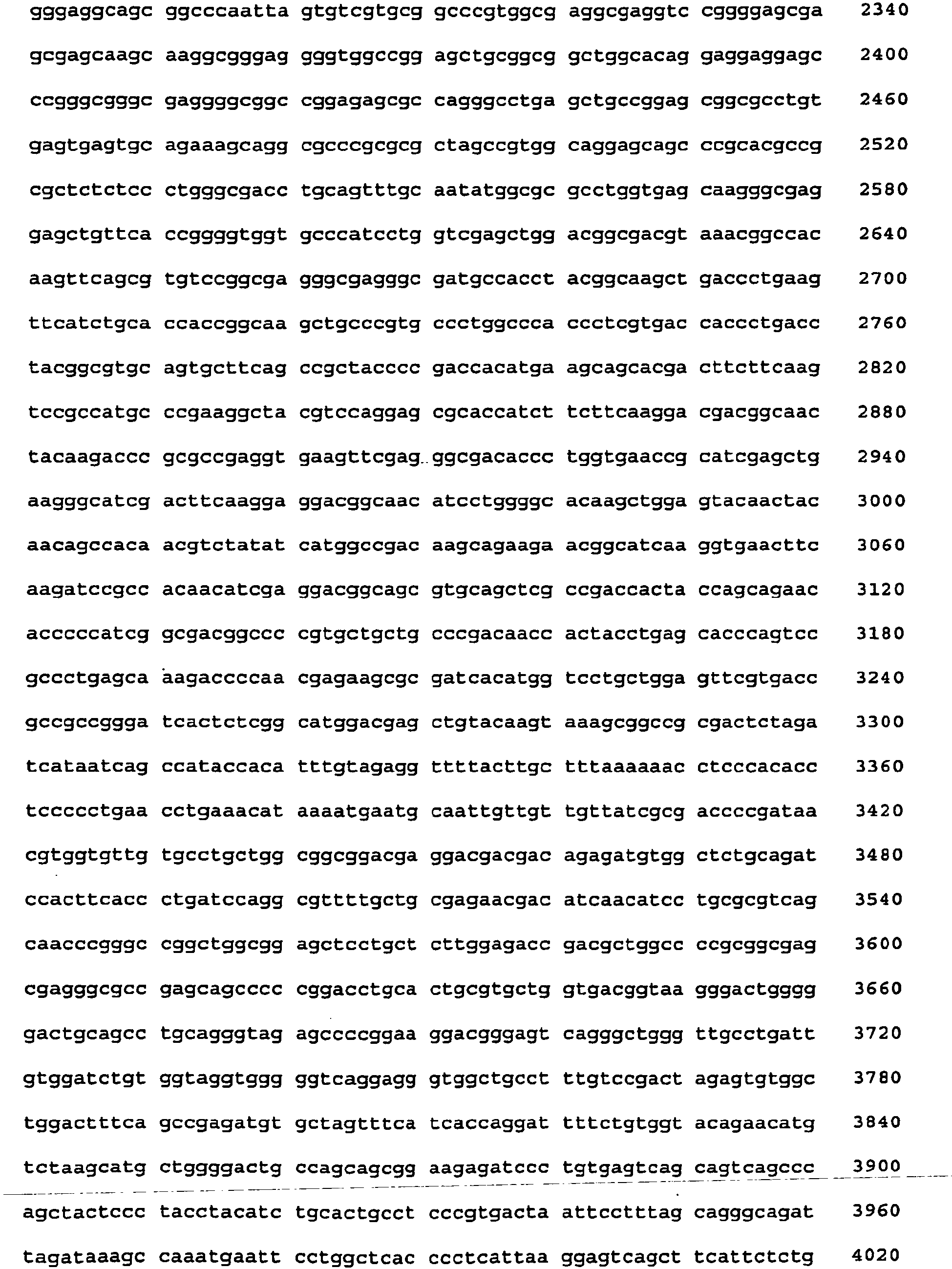

- <210> 4
<211> 5413
<212> DNA
<213> Homo sapiens - <220>
<221> GADD45alpha promoter
<222> (4) .. (2254)
<223> - <220>
<221> GADD45alpha 5' UTR
<222> (2255)..(2549)
<223> - <220>
<221> EGFP gene
<222> (2550)..(3278)
<223> - <220>
<221> SV40 polyA
<222> (3432)..(3482)
<223> - <220>
<221>GADD45alpha Exon 3
<222> (3502)..(3642)
<223> - <220>
<221>GADD45alpha Intron 3
<222> (3643)..(4715)
<223> - <220>
<221> p53 motif
<222> (3830)..(3849)
<223> - <220>
<221> AP-1 motif
<222> (3879) .. (3885)
<223> - <220>
<221>GADD45alpha Exon 4
<222> (4716)..(5391)
<223> - <220>
<221> GADD45alpha 3' UTR
<222> (4830)..(5391)
<223> - <400> 4


- <210> 5
<211> 3871
<212> DNA
<213> Homo sapiens - <220>
<221> GADD45alpha promoter
<222> (4)..(2254)
<223> - <220>
<221> GADD45alpha 5' UTR
<222> (2255) .. (2549)
<223> - <220>
<221> EGFP gene
<222> (2550)..(3278)
<223> - <220>
<221> GADD45alpha 3' UTR
<222> (3288) .. (3849)
<223> - <400> 5


Claims (31)
- An expression cassette comprising a DNA sequence encoding a reporter protein, which DNA sequence is operatively linked to a human GADD45α gene promoter and an additional human GADD45α gene regulatory element, the DNA sequence being place 3' to the GADD45α gene promoter characterised in that the regulatory element comprises a region of Intron 3 of the GADD45α gene including putative p53 binding motif.
- An expression cassette according to claim 1, wherein the regulatory element comprises Exon 1, Exon 2, Exon 3, and/or Exon 4 of the GADD45α gene, or a region thereof, or any combination thereof.
- An expression cassette according to claim 2, wherein the regulatory element comprises a region of Exon 1 of the GADD45α gene, a region of Exon 3 of the GADD45α gene, and a region of Exon 4 of the GADD45α gene.
- An expression cassette according to any preceding claim, wherein the regulatory element additionally comprises Intron and/or Intron 2 of the GADD45α gene, or a region thereof.
- An expression cassette according to any preceding claim, wherein the regulatory element comprises Intron 3 of the GADD45α gene.
- An expression cassette according to claim 5 wherein the regulatory element comprises a putative AP-1 motif.
- An expression cassette according to any preceding claim, wherein the DNA sequence encodes a light emitting reporter protein.
- An expression cassette according to any preceding claim, wherein the DNA sequence encodes Green Fluorescent Protein (GFP), and light emitting derivatives thereof.
- An expression cassette according to any preceding claim, wherein the DNA sequence encodes a human enhanced GFP (hEGFP), which consists of the GFP wild type containing a double amino acid substitution of Phe-64 to Leu, and SER-65 to Thr.
- An expression cassette GD531, as illustrated in Figure 9, and as provided in SEQ ID No.2.
- An expression cassette GD532, as illustrated in Figure 9, and as provided in SEQ ID No.3.
- An expression cassette GD533, as illustrated in Figure 9, and as provided in SEQ ID No.4.
- A recombinant vector comprising an expression cassette according to any one of claims 1-12.
- A recombinant vector according to claim 13, wherein the vector comprises DNA from the pCEP4 plasmid.
- A recombinant vector PEP-GD531, as illustrated in Figure 5.
- A recombinant vector PEP-GD532, as illustrated in Figure 6.
- A recombinant vector PEP-GD422, as illustrated in Figure 7.
- A cell containing a recombinant vector according to any one of claims 14 to 17.
- A cell according to claim 18, wherein the cell is a human cell.
- A cell according to claim 19, wherein the cell is a human cell having a fully functional p53.
- A cell according to claim 20, wherein the cell is a TK6 human cell line.
- An in vitro method of detecting for the presence of an agent that causes or potentiates DNA damage comprising subjecting a cell according to any one of claims 18 to 21 to an agent; and monitoring the expression of the reporter protein from the cell.
- The method according to claim 22, wherein the agent is further screened to assess whether it is safe to expose a living organism to the agent.
- The method according to either claim 22 or claim 23, wherein the agent is a candidate medicament, food additive or cosmetic.
- The method according to any one of claims 22 to 24, wherein the expression of a light emitting reporter protein from the cell is monitored.
- The method according to claim 26, wherein the light emitting reporter protein is Green Fluorescent Protein,
- The method according to claim 26, comprising growing cells transfected with a recombinant vector according to any one of claims 14-18, incubating the cells with the agent for a pre-determined time, and monitoring the expression of the Green Fluorescent Protein directly from a sample of the cells.
- The method according to any one of claims 22 to 27, wherein the cells are grown in a low fluorescence growth medium.
- The method according to claim 28, wherein the low fluorescence growth medium is an RPMI medium containing no riboflavin.
- The method according to claim 28 or 29, wherein the low fluorescence growth medium is an RPMI medium containing no phenol red.
- The method according to any one of claims 25 to 30, wherein fluorescence is determined by comparing fluorescence from a cell according to claims 18 to 21 with fluorescence from a control cell comprising a vector in which the nucleic acid encoding the light emitting reporter is placed out of frame.
Applications Claiming Priority (2)
| Application Number | Priority Date | Filing Date | Title |
|---|---|---|---|
| GBGB0411198.5A GB0411198D0 (en) | 2004-05-20 | 2004-05-20 | Genotoxic testing |
| PCT/GB2005/001913 WO2005113802A2 (en) | 2004-05-20 | 2005-05-18 | Genotoxic testing |
Publications (2)
| Publication Number | Publication Date |
|---|---|
| EP1747287A2 EP1747287A2 (en) | 2007-01-31 |
| EP1747287B1 true EP1747287B1 (en) | 2013-10-23 |
Family
ID=32607606
Family Applications (1)
| Application Number | Title | Priority Date | Filing Date |
|---|---|---|---|
| EP05744681.7A Expired - Lifetime EP1747287B1 (en) | 2004-05-20 | 2005-05-18 | Genotoxic testing |
Country Status (10)
| Country | Link |
|---|---|
| US (1) | US20070224609A1 (en) |
| EP (1) | EP1747287B1 (en) |
| JP (1) | JP5270155B2 (en) |
| CN (1) | CN1961080B (en) |
| AU (1) | AU2005245661B2 (en) |
| CA (1) | CA2566925C (en) |
| DK (1) | DK1747287T3 (en) |
| ES (1) | ES2435401T3 (en) |
| GB (1) | GB0411198D0 (en) |
| WO (1) | WO2005113802A2 (en) |
Families Citing this family (10)
| Publication number | Priority date | Publication date | Assignee | Title |
|---|---|---|---|---|
| US8969008B2 (en) | 2004-05-20 | 2015-03-03 | Gentronix Limited | Genotoxic testing |
| EP2199794A1 (en) | 2008-12-17 | 2010-06-23 | Universiteit Maastricht | Method for the identification of carcinogenic compounds |
| GB0905410D0 (en) * | 2009-03-28 | 2009-05-13 | Gentronix Ltd | Genotoxicity testing |
| US9068972B2 (en) * | 2009-11-02 | 2015-06-30 | The Regents Of The University Of California | GFP mutagenesis amplification: use of a fluorescence-antibiotic resistance fusion dual reporter construct to provide quantitative and highly sensitive detection of mutations |
| GB0921712D0 (en) | 2009-12-11 | 2010-01-27 | Ge Healthcare Uk Ltd | Methods of detecting DNA damage |
| SI23294A (en) | 2010-02-25 | 2011-08-31 | Nacionalni inštitut za biologijo | Cell system for quick sensing and determination of genotoxicity |
| JP5877741B2 (en) * | 2012-03-16 | 2016-03-08 | 株式会社日立製作所 | Cell number monitoring method |
| AU2013276950B2 (en) * | 2012-06-13 | 2018-07-26 | Merck Patent Gmbh | Protein expression analyses for identifying genotoxic compounds |
| CN104894164A (en) * | 2015-05-20 | 2015-09-09 | 山西大学 | Construction and application of GADD45betaRNA interference report vector based on EGFP |
| CN114606269B (en) * | 2022-03-17 | 2024-02-23 | 国家食品安全风险评估中心 | High-throughput detection method for genetic toxicity of food contact material migration object and application |
Citations (1)
| Publication number | Priority date | Publication date | Assignee | Title |
|---|---|---|---|---|
| US20030134288A1 (en) * | 2002-01-16 | 2003-07-17 | Xianqiang Li | Screening methods involving the detection of short-lived proteins |
Family Cites Families (7)
| Publication number | Priority date | Publication date | Assignee | Title |
|---|---|---|---|---|
| JP3662333B2 (en) * | 1995-03-27 | 2005-06-22 | 浜松ホトニクス株式会社 | Fluorescent antiserum and IgG fraction |
| JP3980683B2 (en) * | 1995-08-29 | 2007-09-26 | 仁 岩崎 | Reagent and method for measuring reduced nicotinamide coenzyme |
| GB9706414D0 (en) * | 1997-03-27 | 1997-05-14 | Univ Manchester | Detection of DNA damage |
| US6523041B1 (en) * | 1997-07-29 | 2003-02-18 | Acxiom Corporation | Data linking system and method using tokens |
| EP1120653B1 (en) * | 1999-07-23 | 2005-10-19 | Olympus Corporation | Method for examining substance interacting with hormone receptor |
| US6344324B2 (en) * | 2000-01-07 | 2002-02-05 | Research Development Foundation | Quantification of cellular injury using expression of a fluorescent protein under the control of the GADD153 promoter |
| FR2845097B1 (en) * | 2002-10-01 | 2006-06-16 | Metis Biotechnologies | METHOD FOR DETECTING AND COUNTING MICROORGANISMS IN A SAMPLE |
-
2004
- 2004-05-20 GB GBGB0411198.5A patent/GB0411198D0/en not_active Ceased
-
2005
- 2005-05-18 US US11/596,766 patent/US20070224609A1/en not_active Abandoned
- 2005-05-18 WO PCT/GB2005/001913 patent/WO2005113802A2/en not_active Ceased
- 2005-05-18 AU AU2005245661A patent/AU2005245661B2/en not_active Ceased
- 2005-05-18 CN CN2005800161727A patent/CN1961080B/en not_active Expired - Fee Related
- 2005-05-18 CA CA2566925A patent/CA2566925C/en not_active Expired - Fee Related
- 2005-05-18 EP EP05744681.7A patent/EP1747287B1/en not_active Expired - Lifetime
- 2005-05-18 DK DK05744681.7T patent/DK1747287T3/en active
- 2005-05-18 JP JP2007517410A patent/JP5270155B2/en not_active Expired - Fee Related
- 2005-05-18 ES ES05744681T patent/ES2435401T3/en not_active Expired - Lifetime
Patent Citations (1)
| Publication number | Priority date | Publication date | Assignee | Title |
|---|---|---|---|---|
| US20030134288A1 (en) * | 2002-01-16 | 2003-07-17 | Xianqiang Li | Screening methods involving the detection of short-lived proteins |
Also Published As
| Publication number | Publication date |
|---|---|
| CN1961080B (en) | 2013-07-10 |
| JP2007537745A (en) | 2007-12-27 |
| CA2566925A1 (en) | 2005-12-01 |
| GB0411198D0 (en) | 2004-06-23 |
| DK1747287T3 (en) | 2014-01-20 |
| CA2566925C (en) | 2013-07-09 |
| WO2005113802A3 (en) | 2006-04-20 |
| AU2005245661B2 (en) | 2010-12-09 |
| CN1961080A (en) | 2007-05-09 |
| ES2435401T3 (en) | 2013-12-19 |
| WO2005113802A2 (en) | 2005-12-01 |
| JP5270155B2 (en) | 2013-08-21 |
| EP1747287A2 (en) | 2007-01-31 |
| US20070224609A1 (en) | 2007-09-27 |
| AU2005245661A1 (en) | 2005-12-01 |
Similar Documents
| Publication | Publication Date | Title |
|---|---|---|
| Lavut et al. | Sequestration of highly expressed mRNAs in cytoplasmic granules, P-bodies, and stress granules enhances cell viability | |
| US9285358B2 (en) | Vitro method for high throughput screening of genotoxic agents in eukaryotic cells | |
| US20120035079A1 (en) | Genotoxicity testing | |
| AU2005245661B2 (en) | Genotoxic testing | |
| CA3081687A1 (en) | Dna plasmids for the fast generation of homologous recombination vectors for cell line development | |
| US6344324B2 (en) | Quantification of cellular injury using expression of a fluorescent protein under the control of the GADD153 promoter | |
| US8969008B2 (en) | Genotoxic testing | |
| WO2011105968A2 (en) | A cell based system for rapid detection and quantification of genotoxicity | |
| EP2574925B1 (en) | Method for determining genotoxicity using non-fluorescent proteins | |
| US8658363B2 (en) | Cell, method, and assay kit for measuring level of aryl hydrocarbon receptor transcriptional activation | |
| US6982169B2 (en) | Chemical inhibitors of mismatch repair | |
| CA2533160A1 (en) | Genotoxicity testing | |
| KR101893702B1 (en) | Methods for Analyzing a Genotoxicity Using Non-fluorescent Fluorescence Proteins | |
| CN121574932A (en) | A cell sensor, its preparation method and application | |
| HK1194437A (en) | An in vitro method for high throughput screening of genotoxic agents in eukaryotic cells | |
| JP2004248604A5 (en) |
Legal Events
| Date | Code | Title | Description |
|---|---|---|---|
| PUAI | Public reference made under article 153(3) epc to a published international application that has entered the european phase |
Free format text: ORIGINAL CODE: 0009012 |
|
| 17P | Request for examination filed |
Effective date: 20061113 |
|
| AK | Designated contracting states |
Kind code of ref document: A2 Designated state(s): AT BE BG CH CY CZ DE DK EE ES FI FR GB GR HU IE IS IT LI LT LU MC NL PL PT RO SE SI SK TR |
|
| DAX | Request for extension of the european patent (deleted) | ||
| 17Q | First examination report despatched |
Effective date: 20071008 |
|
| RAP1 | Party data changed (applicant data changed or rights of an application transferred) |
Owner name: GENTRONIX LIMITED |
|
| GRAP | Despatch of communication of intention to grant a patent |
Free format text: ORIGINAL CODE: EPIDOSNIGR1 |
|
| GRAJ | Information related to disapproval of communication of intention to grant by the applicant or resumption of examination proceedings by the epo deleted |
Free format text: ORIGINAL CODE: EPIDOSDIGR1 |
|
| GRAL | Information related to payment of fee for publishing/printing deleted |
Free format text: ORIGINAL CODE: EPIDOSDIGR3 |
|
| GRAS | Grant fee paid |
Free format text: ORIGINAL CODE: EPIDOSNIGR3 |
|
| GRAP | Despatch of communication of intention to grant a patent |
Free format text: ORIGINAL CODE: EPIDOSNIGR1 |
|
| INTG | Intention to grant announced |
Effective date: 20130516 |
|
| GRAA | (expected) grant |
Free format text: ORIGINAL CODE: 0009210 |
|
| AK | Designated contracting states |
Kind code of ref document: B1 Designated state(s): AT BE BG CH CY CZ DE DK EE ES FI FR GB GR HU IE IS IT LI LT LU MC NL PL PT RO SE SI SK TR |
|
| REG | Reference to a national code |
Ref country code: GB Ref legal event code: FG4D |
|
| BECA | Be: change of holder's address |
Owner name: BIOHUB AT ALDERLEY PARK,BLOCK 3 ALDERLEY PARK,CHE Effective date: 20131023 Owner name: GENTRONIX LTD Effective date: 20131023 |
|
| REG | Reference to a national code |
Ref country code: CH Ref legal event code: EP |
|
| REG | Reference to a national code |
Ref country code: AT Ref legal event code: REF Ref document number: 637647 Country of ref document: AT Kind code of ref document: T Effective date: 20131115 |
|
| REG | Reference to a national code |
Ref country code: IE Ref legal event code: FG4D |
|
| REG | Reference to a national code |
Ref country code: ES Ref legal event code: FG2A Ref document number: 2435401 Country of ref document: ES Kind code of ref document: T3 Effective date: 20131219 Ref country code: DE Ref legal event code: R096 Ref document number: 602005041618 Country of ref document: DE Effective date: 20131219 |
|
| REG | Reference to a national code |
Ref country code: NL Ref legal event code: T3 Ref country code: CH Ref legal event code: NV Representative=s name: MEYER AND KOLLEGEN, CH |
|
| REG | Reference to a national code |
Ref country code: DK Ref legal event code: T3 Effective date: 20140117 |
|
| RAP2 | Party data changed (patent owner data changed or rights of a patent transferred) |
Owner name: GENTRONIX LIMITED |
|
| REG | Reference to a national code |
Ref country code: DE Ref legal event code: R082 Ref document number: 602005041618 Country of ref document: DE Representative=s name: PATENTANWALTSKANZLEI MEYER, DE |
|
| REG | Reference to a national code |
Ref country code: CH Ref legal event code: PK Free format text: BERICHTIGUNG INHABER |
|
| REG | Reference to a national code |
Ref country code: SE Ref legal event code: TRGR |
|
| REG | Reference to a national code |
Ref country code: DE Ref legal event code: R081 Ref document number: 602005041618 Country of ref document: DE Owner name: GENTRONIX LTD., GB Free format text: FORMER OWNER: GENTRONIX LTD., MANCHESTER, GB Effective date: 20140122 Ref country code: DE Ref legal event code: R082 Ref document number: 602005041618 Country of ref document: DE Representative=s name: PATENTANWALTSKANZLEI MEYER, DE Effective date: 20140122 |
|
| REG | Reference to a national code |
Ref country code: AT Ref legal event code: MK05 Ref document number: 637647 Country of ref document: AT Kind code of ref document: T Effective date: 20131023 |
|
| REG | Reference to a national code |
Ref country code: LT Ref legal event code: MG4D |
|
| PG25 | Lapsed in a contracting state [announced via postgrant information from national office to epo] |
Ref country code: IS Free format text: LAPSE BECAUSE OF FAILURE TO SUBMIT A TRANSLATION OF THE DESCRIPTION OR TO PAY THE FEE WITHIN THE PRESCRIBED TIME-LIMIT Effective date: 20140223 Ref country code: LT Free format text: LAPSE BECAUSE OF FAILURE TO SUBMIT A TRANSLATION OF THE DESCRIPTION OR TO PAY THE FEE WITHIN THE PRESCRIBED TIME-LIMIT Effective date: 20131023 |
|
| PG25 | Lapsed in a contracting state [announced via postgrant information from national office to epo] |
Ref country code: AT Free format text: LAPSE BECAUSE OF FAILURE TO SUBMIT A TRANSLATION OF THE DESCRIPTION OR TO PAY THE FEE WITHIN THE PRESCRIBED TIME-LIMIT Effective date: 20131023 Ref country code: CY Free format text: LAPSE BECAUSE OF FAILURE TO SUBMIT A TRANSLATION OF THE DESCRIPTION OR TO PAY THE FEE WITHIN THE PRESCRIBED TIME-LIMIT Effective date: 20131023 |
|
| PG25 | Lapsed in a contracting state [announced via postgrant information from national office to epo] |
Ref country code: PT Free format text: LAPSE BECAUSE OF FAILURE TO SUBMIT A TRANSLATION OF THE DESCRIPTION OR TO PAY THE FEE WITHIN THE PRESCRIBED TIME-LIMIT Effective date: 20140224 |
|
| REG | Reference to a national code |
Ref country code: DE Ref legal event code: R097 Ref document number: 602005041618 Country of ref document: DE |
|
| PG25 | Lapsed in a contracting state [announced via postgrant information from national office to epo] |
Ref country code: EE Free format text: LAPSE BECAUSE OF FAILURE TO SUBMIT A TRANSLATION OF THE DESCRIPTION OR TO PAY THE FEE WITHIN THE PRESCRIBED TIME-LIMIT Effective date: 20131023 |
|
| PG25 | Lapsed in a contracting state [announced via postgrant information from national office to epo] |
Ref country code: RO Free format text: LAPSE BECAUSE OF FAILURE TO SUBMIT A TRANSLATION OF THE DESCRIPTION OR TO PAY THE FEE WITHIN THE PRESCRIBED TIME-LIMIT Effective date: 20131023 Ref country code: CZ Free format text: LAPSE BECAUSE OF FAILURE TO SUBMIT A TRANSLATION OF THE DESCRIPTION OR TO PAY THE FEE WITHIN THE PRESCRIBED TIME-LIMIT Effective date: 20131023 Ref country code: PL Free format text: LAPSE BECAUSE OF FAILURE TO SUBMIT A TRANSLATION OF THE DESCRIPTION OR TO PAY THE FEE WITHIN THE PRESCRIBED TIME-LIMIT Effective date: 20131023 Ref country code: SK Free format text: LAPSE BECAUSE OF FAILURE TO SUBMIT A TRANSLATION OF THE DESCRIPTION OR TO PAY THE FEE WITHIN THE PRESCRIBED TIME-LIMIT Effective date: 20131023 |
|
| PLBE | No opposition filed within time limit |
Free format text: ORIGINAL CODE: 0009261 |
|
| STAA | Information on the status of an ep patent application or granted ep patent |
Free format text: STATUS: NO OPPOSITION FILED WITHIN TIME LIMIT |
|
| 26N | No opposition filed |
Effective date: 20140724 |
|
| REG | Reference to a national code |
Ref country code: DE Ref legal event code: R097 Ref document number: 602005041618 Country of ref document: DE Effective date: 20140724 |
|
| PG25 | Lapsed in a contracting state [announced via postgrant information from national office to epo] |
Ref country code: LU Free format text: LAPSE BECAUSE OF FAILURE TO SUBMIT A TRANSLATION OF THE DESCRIPTION OR TO PAY THE FEE WITHIN THE PRESCRIBED TIME-LIMIT Effective date: 20140518 |
|
| PG25 | Lapsed in a contracting state [announced via postgrant information from national office to epo] |
Ref country code: MC Free format text: LAPSE BECAUSE OF FAILURE TO SUBMIT A TRANSLATION OF THE DESCRIPTION OR TO PAY THE FEE WITHIN THE PRESCRIBED TIME-LIMIT Effective date: 20131023 |
|
| PG25 | Lapsed in a contracting state [announced via postgrant information from national office to epo] |
Ref country code: SI Free format text: LAPSE BECAUSE OF FAILURE TO SUBMIT A TRANSLATION OF THE DESCRIPTION OR TO PAY THE FEE WITHIN THE PRESCRIBED TIME-LIMIT Effective date: 20131023 |
|
| REG | Reference to a national code |
Ref country code: FR Ref legal event code: PLFP Year of fee payment: 12 |
|
| PG25 | Lapsed in a contracting state [announced via postgrant information from national office to epo] |
Ref country code: BG Free format text: LAPSE BECAUSE OF FAILURE TO SUBMIT A TRANSLATION OF THE DESCRIPTION OR TO PAY THE FEE WITHIN THE PRESCRIBED TIME-LIMIT Effective date: 20131023 |
|
| PG25 | Lapsed in a contracting state [announced via postgrant information from national office to epo] |
Ref country code: GR Free format text: LAPSE BECAUSE OF FAILURE TO SUBMIT A TRANSLATION OF THE DESCRIPTION OR TO PAY THE FEE WITHIN THE PRESCRIBED TIME-LIMIT Effective date: 20140124 |
|
| PG25 | Lapsed in a contracting state [announced via postgrant information from national office to epo] |
Ref country code: TR Free format text: LAPSE BECAUSE OF FAILURE TO SUBMIT A TRANSLATION OF THE DESCRIPTION OR TO PAY THE FEE WITHIN THE PRESCRIBED TIME-LIMIT Effective date: 20131023 Ref country code: HU Free format text: LAPSE BECAUSE OF FAILURE TO SUBMIT A TRANSLATION OF THE DESCRIPTION OR TO PAY THE FEE WITHIN THE PRESCRIBED TIME-LIMIT; INVALID AB INITIO Effective date: 20050518 |
|
| REG | Reference to a national code |
Ref country code: FR Ref legal event code: PLFP Year of fee payment: 13 |
|
| REG | Reference to a national code |
Ref country code: FR Ref legal event code: PLFP Year of fee payment: 14 |
|
| PGFP | Annual fee paid to national office [announced via postgrant information from national office to epo] |
Ref country code: NL Payment date: 20190517 Year of fee payment: 15 |
|
| PGFP | Annual fee paid to national office [announced via postgrant information from national office to epo] |
Ref country code: DK Payment date: 20190517 Year of fee payment: 15 Ref country code: FI Payment date: 20190512 Year of fee payment: 15 Ref country code: IE Payment date: 20190517 Year of fee payment: 15 Ref country code: DE Payment date: 20190512 Year of fee payment: 15 Ref country code: IT Payment date: 20190518 Year of fee payment: 15 |
|
| PGFP | Annual fee paid to national office [announced via postgrant information from national office to epo] |
Ref country code: SE Payment date: 20190517 Year of fee payment: 15 Ref country code: FR Payment date: 20190516 Year of fee payment: 15 Ref country code: BE Payment date: 20190513 Year of fee payment: 15 |
|
| PGFP | Annual fee paid to national office [announced via postgrant information from national office to epo] |
Ref country code: CH Payment date: 20190516 Year of fee payment: 15 |
|
| PGFP | Annual fee paid to national office [announced via postgrant information from national office to epo] |
Ref country code: ES Payment date: 20190805 Year of fee payment: 15 Ref country code: GB Payment date: 20190513 Year of fee payment: 15 |
|
| REG | Reference to a national code |
Ref country code: DE Ref legal event code: R119 Ref document number: 602005041618 Country of ref document: DE |
|
| REG | Reference to a national code |
Ref country code: FI Ref legal event code: MAE |
|
| REG | Reference to a national code |
Ref country code: DK Ref legal event code: EBP Effective date: 20200531 |
|
| REG | Reference to a national code |
Ref country code: NL Ref legal event code: MM Effective date: 20200601 |
|
| PG25 | Lapsed in a contracting state [announced via postgrant information from national office to epo] |
Ref country code: CH Free format text: LAPSE BECAUSE OF NON-PAYMENT OF DUE FEES Effective date: 20200531 Ref country code: FI Free format text: LAPSE BECAUSE OF NON-PAYMENT OF DUE FEES Effective date: 20200518 Ref country code: LI Free format text: LAPSE BECAUSE OF NON-PAYMENT OF DUE FEES Effective date: 20200531 Ref country code: SE Free format text: LAPSE BECAUSE OF NON-PAYMENT OF DUE FEES Effective date: 20200519 |
|
| PG25 | Lapsed in a contracting state [announced via postgrant information from national office to epo] |
Ref country code: NL Free format text: LAPSE BECAUSE OF NON-PAYMENT OF DUE FEES Effective date: 20200601 |
|
| REG | Reference to a national code |
Ref country code: BE Ref legal event code: MM Effective date: 20200531 |
|
| GBPC | Gb: european patent ceased through non-payment of renewal fee |
Effective date: 20200518 |
|
| PG25 | Lapsed in a contracting state [announced via postgrant information from national office to epo] |
Ref country code: IE Free format text: LAPSE BECAUSE OF NON-PAYMENT OF DUE FEES Effective date: 20200518 Ref country code: DK Free format text: LAPSE BECAUSE OF NON-PAYMENT OF DUE FEES Effective date: 20200531 Ref country code: FR Free format text: LAPSE BECAUSE OF NON-PAYMENT OF DUE FEES Effective date: 20200531 Ref country code: GB Free format text: LAPSE BECAUSE OF NON-PAYMENT OF DUE FEES Effective date: 20200518 |
|
| PG25 | Lapsed in a contracting state [announced via postgrant information from national office to epo] |
Ref country code: BE Free format text: LAPSE BECAUSE OF NON-PAYMENT OF DUE FEES Effective date: 20200531 Ref country code: DE Free format text: LAPSE BECAUSE OF NON-PAYMENT OF DUE FEES Effective date: 20201201 |
|
| REG | Reference to a national code |
Ref country code: ES Ref legal event code: FD2A Effective date: 20211001 |
|
| PG25 | Lapsed in a contracting state [announced via postgrant information from national office to epo] |
Ref country code: IT Free format text: LAPSE BECAUSE OF NON-PAYMENT OF DUE FEES Effective date: 20200518 |
|
| PG25 | Lapsed in a contracting state [announced via postgrant information from national office to epo] |
Ref country code: ES Free format text: LAPSE BECAUSE OF NON-PAYMENT OF DUE FEES Effective date: 20200519 |