EP1747007B1 - Use of chondroitin sulphate e (cs-e ) for the treatment diseases or conditions related to collagen fibril formation. - Google Patents
Use of chondroitin sulphate e (cs-e ) for the treatment diseases or conditions related to collagen fibril formation. Download PDFInfo
- Publication number
- EP1747007B1 EP1747007B1 EP05735125A EP05735125A EP1747007B1 EP 1747007 B1 EP1747007 B1 EP 1747007B1 EP 05735125 A EP05735125 A EP 05735125A EP 05735125 A EP05735125 A EP 05735125A EP 1747007 B1 EP1747007 B1 EP 1747007B1
- Authority
- EP
- European Patent Office
- Prior art keywords
- wound
- healing
- collagen
- chondroitin sulphate
- fibril formation
- Prior art date
- Legal status (The legal status is an assumption and is not a legal conclusion. Google has not performed a legal analysis and makes no representation as to the accuracy of the status listed.)
- Expired - Lifetime
Links
- 102000008186 Collagen Human genes 0.000 title claims description 27
- 108010035532 Collagen Proteins 0.000 title claims description 27
- 229920001436 collagen Polymers 0.000 title claims description 27
- 229920001287 Chondroitin sulfate Polymers 0.000 title claims description 22
- 230000015572 biosynthetic process Effects 0.000 title claims description 22
- 208000037265 diseases, disorders, signs and symptoms Diseases 0.000 title claims description 11
- 201000010099 disease Diseases 0.000 title claims description 8
- 238000011282 treatment Methods 0.000 title claims description 6
- 206010052428 Wound Diseases 0.000 claims description 13
- 208000027418 Wounds and injury Diseases 0.000 claims description 13
- 230000029663 wound healing Effects 0.000 claims description 11
- 150000003839 salts Chemical class 0.000 claims description 9
- 230000035876 healing Effects 0.000 claims description 7
- 206010039073 rheumatoid arthritis Diseases 0.000 claims description 6
- 210000000988 bone and bone Anatomy 0.000 claims description 5
- 238000001356 surgical procedure Methods 0.000 claims description 5
- 230000001154 acute effect Effects 0.000 claims description 4
- 230000001684 chronic effect Effects 0.000 claims description 4
- 238000004519 manufacturing process Methods 0.000 claims description 4
- 210000002435 tendon Anatomy 0.000 claims description 4
- 238000002054 transplantation Methods 0.000 claims description 4
- 208000008469 Peptic Ulcer Diseases 0.000 claims description 3
- 210000003041 ligament Anatomy 0.000 claims description 3
- 230000004936 stimulating effect Effects 0.000 claims description 3
- 208000025865 Ulcer Diseases 0.000 claims 2
- 208000011906 peptic ulcer disease Diseases 0.000 claims 2
- 231100000397 ulcer Toxicity 0.000 claims 2
- 239000003814 drug Substances 0.000 claims 1
- 102100036597 Basement membrane-specific heparan sulfate proteoglycan core protein Human genes 0.000 description 14
- 108010049224 perlecan Proteins 0.000 description 14
- 210000001519 tissue Anatomy 0.000 description 14
- 239000000556 agonist Substances 0.000 description 11
- 210000000845 cartilage Anatomy 0.000 description 10
- 210000002744 extracellular matrix Anatomy 0.000 description 10
- 102000010834 Extracellular Matrix Proteins Human genes 0.000 description 9
- 108010037362 Extracellular Matrix Proteins Proteins 0.000 description 9
- 229920002971 Heparan sulfate Polymers 0.000 description 9
- 210000004027 cell Anatomy 0.000 description 9
- 229920002683 Glycosaminoglycan Polymers 0.000 description 8
- 210000002469 basement membrane Anatomy 0.000 description 8
- 230000035557 fibrillogenesis Effects 0.000 description 8
- 102000016611 Proteoglycans Human genes 0.000 description 7
- 108010067787 Proteoglycans Proteins 0.000 description 7
- 230000000694 effects Effects 0.000 description 7
- 108010067219 Aggrecans Proteins 0.000 description 6
- 102000016284 Aggrecans Human genes 0.000 description 6
- 208000014674 injury Diseases 0.000 description 6
- 206010016654 Fibrosis Diseases 0.000 description 5
- 230000004761 fibrosis Effects 0.000 description 5
- 229920002674 hyaluronan Polymers 0.000 description 5
- 235000018102 proteins Nutrition 0.000 description 5
- 108090000623 proteins and genes Proteins 0.000 description 5
- 102000004169 proteins and genes Human genes 0.000 description 5
- 102000012422 Collagen Type I Human genes 0.000 description 4
- 108010022452 Collagen Type I Proteins 0.000 description 4
- AEMOLEFTQBMNLQ-AQKNRBDQSA-N D-glucopyranuronic acid Chemical compound OC1O[C@H](C(O)=O)[C@@H](O)[C@H](O)[C@H]1O AEMOLEFTQBMNLQ-AQKNRBDQSA-N 0.000 description 4
- 229940096422 collagen type i Drugs 0.000 description 4
- 230000006378 damage Effects 0.000 description 4
- 230000003176 fibrotic effect Effects 0.000 description 4
- KIUKXJAPPMFGSW-MNSSHETKSA-N hyaluronan Chemical compound CC(=O)N[C@H]1[C@H](O)O[C@H](CO)[C@@H](O)C1O[C@H]1[C@H](O)[C@@H](O)[C@H](O[C@H]2[C@@H](C(O[C@H]3[C@@H]([C@@H](O)[C@H](O)[C@H](O3)C(O)=O)O)[C@H](O)[C@@H](CO)O2)NC(C)=O)[C@@H](C(O)=O)O1 KIUKXJAPPMFGSW-MNSSHETKSA-N 0.000 description 4
- 229940099552 hyaluronan Drugs 0.000 description 4
- HQKMJHAJHXVSDF-UHFFFAOYSA-L magnesium stearate Chemical compound [Mg+2].CCCCCCCCCCCCCCCCCC([O-])=O.CCCCCCCCCCCCCCCCCC([O-])=O HQKMJHAJHXVSDF-UHFFFAOYSA-L 0.000 description 4
- 239000000243 solution Substances 0.000 description 4
- 101710132601 Capsid protein Proteins 0.000 description 3
- 102000000503 Collagen Type II Human genes 0.000 description 3
- 108010041390 Collagen Type II Proteins 0.000 description 3
- IAJILQKETJEXLJ-UHFFFAOYSA-N Galacturonsaeure Natural products O=CC(O)C(O)C(O)C(O)C(O)=O IAJILQKETJEXLJ-UHFFFAOYSA-N 0.000 description 3
- 241000699670 Mus sp. Species 0.000 description 3
- HEMHJVSKTPXQMS-UHFFFAOYSA-M Sodium hydroxide Chemical compound [OH-].[Na+] HEMHJVSKTPXQMS-UHFFFAOYSA-M 0.000 description 3
- QAOWNCQODCNURD-UHFFFAOYSA-L Sulfate Chemical compound [O-]S([O-])(=O)=O QAOWNCQODCNURD-UHFFFAOYSA-L 0.000 description 3
- 239000004480 active ingredient Substances 0.000 description 3
- 238000003556 assay Methods 0.000 description 3
- 150000002016 disaccharides Chemical class 0.000 description 3
- 208000035475 disorder Diseases 0.000 description 3
- 239000000835 fiber Substances 0.000 description 3
- 230000003993 interaction Effects 0.000 description 3
- KXCLCNHUUKTANI-RBIYJLQWSA-N keratan Chemical compound CC(=O)N[C@@H]1[C@@H](O)C[C@@H](COS(O)(=O)=O)O[C@H]1O[C@@H]1[C@@H](O)[C@H](O[C@@H]2[C@H](O[C@@H](O[C@H]3[C@H]([C@@H](COS(O)(=O)=O)O[C@@H](O)[C@@H]3O)O)[C@H](NC(C)=O)[C@H]2O)COS(O)(=O)=O)O[C@H](COS(O)(=O)=O)[C@@H]1O KXCLCNHUUKTANI-RBIYJLQWSA-N 0.000 description 3
- 239000000178 monomer Substances 0.000 description 3
- 150000002772 monosaccharides Chemical class 0.000 description 3
- 150000002482 oligosaccharides Polymers 0.000 description 3
- 230000004044 response Effects 0.000 description 3
- 238000012552 review Methods 0.000 description 3
- 210000003491 skin Anatomy 0.000 description 3
- 229910021653 sulphate ion Inorganic materials 0.000 description 3
- 230000008733 trauma Effects 0.000 description 3
- FPJHWYCPAOPVIV-VOZMEZHOSA-N (2R,3S,4R,5R,6R)-6-[(2R,3R,4R,5R,6R)-5-acetamido-2-(hydroxymethyl)-6-methoxy-3-sulfooxyoxan-4-yl]oxy-4,5-dihydroxy-3-methoxyoxane-2-carboxylic acid Chemical compound CO[C@@H]1O[C@H](CO)[C@H](OS(O)(=O)=O)[C@H](O[C@@H]2O[C@H]([C@@H](OC)[C@H](O)[C@H]2O)C(O)=O)[C@H]1NC(C)=O FPJHWYCPAOPVIV-VOZMEZHOSA-N 0.000 description 2
- 241000283690 Bos taurus Species 0.000 description 2
- 102000055007 Cartilage Oligomeric Matrix Human genes 0.000 description 2
- 101710176668 Cartilage oligomeric matrix protein Proteins 0.000 description 2
- SRBFZHDQGSBBOR-IOVATXLUSA-N D-xylopyranose Chemical compound O[C@@H]1COC(O)[C@H](O)[C@H]1O SRBFZHDQGSBBOR-IOVATXLUSA-N 0.000 description 2
- 102000004237 Decorin Human genes 0.000 description 2
- 108090000738 Decorin Proteins 0.000 description 2
- 229920000045 Dermatan sulfate Polymers 0.000 description 2
- DHMQDGOQFOQNFH-UHFFFAOYSA-N Glycine Chemical compound NCC(O)=O DHMQDGOQFOQNFH-UHFFFAOYSA-N 0.000 description 2
- 102000003886 Glycoproteins Human genes 0.000 description 2
- 108090000288 Glycoproteins Proteins 0.000 description 2
- 229920000288 Keratan sulfate Polymers 0.000 description 2
- AEMOLEFTQBMNLQ-HNFCZKTMSA-N L-idopyranuronic acid Chemical compound OC1O[C@@H](C(O)=O)[C@@H](O)[C@H](O)[C@H]1O AEMOLEFTQBMNLQ-HNFCZKTMSA-N 0.000 description 2
- MBLBDJOUHNCFQT-UHFFFAOYSA-N N-acetyl-D-galactosamine Natural products CC(=O)NC(C=O)C(O)C(O)C(O)CO MBLBDJOUHNCFQT-UHFFFAOYSA-N 0.000 description 2
- FAPWRFPIFSIZLT-UHFFFAOYSA-M Sodium chloride Chemical compound [Na+].[Cl-] FAPWRFPIFSIZLT-UHFFFAOYSA-M 0.000 description 2
- 238000002835 absorbance Methods 0.000 description 2
- 239000002253 acid Substances 0.000 description 2
- 239000013543 active substance Substances 0.000 description 2
- 239000007864 aqueous solution Substances 0.000 description 2
- 239000006172 buffering agent Substances 0.000 description 2
- 239000002775 capsule Substances 0.000 description 2
- 239000003795 chemical substances by application Substances 0.000 description 2
- 238000013270 controlled release Methods 0.000 description 2
- 230000008021 deposition Effects 0.000 description 2
- 206010012601 diabetes mellitus Diseases 0.000 description 2
- 230000008030 elimination Effects 0.000 description 2
- 238000003379 elimination reaction Methods 0.000 description 2
- 238000002474 experimental method Methods 0.000 description 2
- 239000012634 fragment Substances 0.000 description 2
- 229940097043 glucuronic acid Drugs 0.000 description 2
- 125000002887 hydroxy group Chemical group [H]O* 0.000 description 2
- 238000000338 in vitro Methods 0.000 description 2
- 238000001727 in vivo Methods 0.000 description 2
- 208000015181 infectious disease Diseases 0.000 description 2
- -1 linker tri-saccharide Chemical class 0.000 description 2
- 235000019359 magnesium stearate Nutrition 0.000 description 2
- 239000011159 matrix material Substances 0.000 description 2
- 208000010125 myocardial infarction Diseases 0.000 description 2
- 229920001223 polyethylene glycol Polymers 0.000 description 2
- 231100000241 scar Toxicity 0.000 description 2
- 239000003381 stabilizer Substances 0.000 description 2
- MTCFGRXMJLQNBG-REOHCLBHSA-N (2S)-2-Amino-3-hydroxypropansäure Chemical compound OC[C@H](N)C(O)=O MTCFGRXMJLQNBG-REOHCLBHSA-N 0.000 description 1
- KIUKXJAPPMFGSW-DNGZLQJQSA-N (2S,3S,4S,5R,6R)-6-[(2S,3R,4R,5S,6R)-3-Acetamido-2-[(2S,3S,4R,5R,6R)-6-[(2R,3R,4R,5S,6R)-3-acetamido-2,5-dihydroxy-6-(hydroxymethyl)oxan-4-yl]oxy-2-carboxy-4,5-dihydroxyoxan-3-yl]oxy-5-hydroxy-6-(hydroxymethyl)oxan-4-yl]oxy-3,4,5-trihydroxyoxane-2-carboxylic acid Chemical compound CC(=O)N[C@H]1[C@H](O)O[C@H](CO)[C@@H](O)[C@@H]1O[C@H]1[C@H](O)[C@@H](O)[C@H](O[C@H]2[C@@H]([C@@H](O[C@H]3[C@@H]([C@@H](O)[C@H](O)[C@H](O3)C(O)=O)O)[C@H](O)[C@@H](CO)O2)NC(C)=O)[C@@H](C(O)=O)O1 KIUKXJAPPMFGSW-DNGZLQJQSA-N 0.000 description 1
- JKMHFZQWWAIEOD-UHFFFAOYSA-N 2-[4-(2-hydroxyethyl)piperazin-1-yl]ethanesulfonic acid Chemical compound OCC[NH+]1CCN(CCS([O-])(=O)=O)CC1 JKMHFZQWWAIEOD-UHFFFAOYSA-N 0.000 description 1
- SQDAZGGFXASXDW-UHFFFAOYSA-N 5-bromo-2-(trifluoromethoxy)pyridine Chemical compound FC(F)(F)OC1=CC=C(Br)C=N1 SQDAZGGFXASXDW-UHFFFAOYSA-N 0.000 description 1
- QGZKDVFQNNGYKY-UHFFFAOYSA-O Ammonium Chemical compound [NH4+] QGZKDVFQNNGYKY-UHFFFAOYSA-O 0.000 description 1
- 206010002091 Anaesthesia Diseases 0.000 description 1
- OYPRJOBELJOOCE-UHFFFAOYSA-N Calcium Chemical compound [Ca] OYPRJOBELJOOCE-UHFFFAOYSA-N 0.000 description 1
- 229920002134 Carboxymethyl cellulose Polymers 0.000 description 1
- 241000238366 Cephalopoda Species 0.000 description 1
- 229920002567 Chondroitin Polymers 0.000 description 1
- 208000017667 Chronic Disease Diseases 0.000 description 1
- 208000007342 Diabetic Nephropathies Diseases 0.000 description 1
- 108090000790 Enzymes Proteins 0.000 description 1
- 102000004190 Enzymes Human genes 0.000 description 1
- LFQSCWFLJHTTHZ-UHFFFAOYSA-N Ethanol Chemical compound CCO LFQSCWFLJHTTHZ-UHFFFAOYSA-N 0.000 description 1
- 102000003974 Fibroblast growth factor 2 Human genes 0.000 description 1
- 108090000379 Fibroblast growth factor 2 Proteins 0.000 description 1
- 102000017177 Fibromodulin Human genes 0.000 description 1
- 108010013996 Fibromodulin Proteins 0.000 description 1
- 108010067306 Fibronectins Proteins 0.000 description 1
- 102000016359 Fibronectins Human genes 0.000 description 1
- 239000001828 Gelatine Substances 0.000 description 1
- 239000004471 Glycine Substances 0.000 description 1
- 239000007995 HEPES buffer Substances 0.000 description 1
- HTTJABKRGRZYRN-UHFFFAOYSA-N Heparin Chemical compound OC1C(NC(=O)C)C(O)OC(COS(O)(=O)=O)C1OC1C(OS(O)(=O)=O)C(O)C(OC2C(C(OS(O)(=O)=O)C(OC3C(C(O)C(O)C(O3)C(O)=O)OS(O)(=O)=O)C(CO)O2)NS(O)(=O)=O)C(C(O)=O)O1 HTTJABKRGRZYRN-UHFFFAOYSA-N 0.000 description 1
- 101000611663 Homo sapiens Prolargin Proteins 0.000 description 1
- 101000666896 Homo sapiens V-type immunoglobulin domain-containing suppressor of T-cell activation Proteins 0.000 description 1
- 102000001842 Hyalectins Human genes 0.000 description 1
- 108010040314 Hyalectins Proteins 0.000 description 1
- 206010020772 Hypertension Diseases 0.000 description 1
- 201000009794 Idiopathic Pulmonary Fibrosis Diseases 0.000 description 1
- DGAQECJNVWCQMB-PUAWFVPOSA-M Ilexoside XXIX Chemical compound C[C@@H]1CC[C@@]2(CC[C@@]3(C(=CC[C@H]4[C@]3(CC[C@@H]5[C@@]4(CC[C@@H](C5(C)C)OS(=O)(=O)[O-])C)C)[C@@H]2[C@]1(C)O)C)C(=O)O[C@H]6[C@@H]([C@H]([C@@H]([C@H](O6)CO)O)O)O.[Na+] DGAQECJNVWCQMB-PUAWFVPOSA-M 0.000 description 1
- 208000026350 Inborn Genetic disease Diseases 0.000 description 1
- 206010023421 Kidney fibrosis Diseases 0.000 description 1
- GUBGYTABKSRVRQ-QKKXKWKRSA-N Lactose Natural products OC[C@H]1O[C@@H](O[C@H]2[C@H](O)[C@@H](O)C(O)O[C@@H]2CO)[C@H](O)[C@@H](O)[C@H]1O GUBGYTABKSRVRQ-QKKXKWKRSA-N 0.000 description 1
- 108010085895 Laminin Proteins 0.000 description 1
- FYYHWMGAXLPEAU-UHFFFAOYSA-N Magnesium Chemical compound [Mg] FYYHWMGAXLPEAU-UHFFFAOYSA-N 0.000 description 1
- 108010072582 Matrilin Proteins Proteins 0.000 description 1
- 102000055008 Matrilin Proteins Human genes 0.000 description 1
- 208000024556 Mendelian disease Diseases 0.000 description 1
- 241001465754 Metazoa Species 0.000 description 1
- 241001529936 Murinae Species 0.000 description 1
- 241000699666 Mus <mouse, genus> Species 0.000 description 1
- OVRNDRQMDRJTHS-CBQIKETKSA-N N-Acetyl-D-Galactosamine Chemical compound CC(=O)N[C@H]1[C@@H](O)O[C@H](CO)[C@H](O)[C@@H]1O OVRNDRQMDRJTHS-CBQIKETKSA-N 0.000 description 1
- OVRNDRQMDRJTHS-KEWYIRBNSA-N N-acetyl-D-galactosamine Chemical compound CC(=O)N[C@H]1C(O)O[C@H](CO)[C@H](O)[C@@H]1O OVRNDRQMDRJTHS-KEWYIRBNSA-N 0.000 description 1
- 206010028980 Neoplasm Diseases 0.000 description 1
- 102000057297 Pepsin A Human genes 0.000 description 1
- 108090000284 Pepsin A Proteins 0.000 description 1
- ZLMJMSJWJFRBEC-UHFFFAOYSA-N Potassium Chemical compound [K] ZLMJMSJWJFRBEC-UHFFFAOYSA-N 0.000 description 1
- 102000019204 Progranulins Human genes 0.000 description 1
- 108010012809 Progranulins Proteins 0.000 description 1
- 102100040659 Prolargin Human genes 0.000 description 1
- 206010039580 Scar Diseases 0.000 description 1
- 206010039710 Scleroderma Diseases 0.000 description 1
- MTCFGRXMJLQNBG-UHFFFAOYSA-N Serine Natural products OCC(N)C(O)=O MTCFGRXMJLQNBG-UHFFFAOYSA-N 0.000 description 1
- VYPSYNLAJGMNEJ-UHFFFAOYSA-N Silicium dioxide Chemical compound O=[Si]=O VYPSYNLAJGMNEJ-UHFFFAOYSA-N 0.000 description 1
- 229920002472 Starch Polymers 0.000 description 1
- 102100038282 V-type immunoglobulin domain-containing suppressor of T-cell activation Human genes 0.000 description 1
- 208000036142 Viral infection Diseases 0.000 description 1
- 108010006886 Vitrogen Proteins 0.000 description 1
- HCHKCACWOHOZIP-UHFFFAOYSA-N Zinc Chemical compound [Zn] HCHKCACWOHOZIP-UHFFFAOYSA-N 0.000 description 1
- 230000001464 adherent effect Effects 0.000 description 1
- 229910052783 alkali metal Inorganic materials 0.000 description 1
- 230000037005 anaesthesia Effects 0.000 description 1
- PYMYPHUHKUWMLA-UHFFFAOYSA-N arabinose Natural products OCC(O)C(O)C(O)C=O PYMYPHUHKUWMLA-UHFFFAOYSA-N 0.000 description 1
- 238000000149 argon plasma sintering Methods 0.000 description 1
- 125000000613 asparagine group Chemical group N[C@@H](CC(N)=O)C(=O)* 0.000 description 1
- SRBFZHDQGSBBOR-UHFFFAOYSA-N beta-D-Pyranose-Lyxose Natural products OC1COC(O)C(O)C1O SRBFZHDQGSBBOR-UHFFFAOYSA-N 0.000 description 1
- AEMOLEFTQBMNLQ-UHFFFAOYSA-N beta-D-galactopyranuronic acid Natural products OC1OC(C(O)=O)C(O)C(O)C1O AEMOLEFTQBMNLQ-UHFFFAOYSA-N 0.000 description 1
- 238000004166 bioassay Methods 0.000 description 1
- 210000004204 blood vessel Anatomy 0.000 description 1
- 229910052792 caesium Inorganic materials 0.000 description 1
- TVFDJXOCXUVLDH-UHFFFAOYSA-N caesium atom Chemical compound [Cs] TVFDJXOCXUVLDH-UHFFFAOYSA-N 0.000 description 1
- 229910052791 calcium Inorganic materials 0.000 description 1
- 239000011575 calcium Substances 0.000 description 1
- 150000001720 carbohydrates Chemical class 0.000 description 1
- 235000010948 carboxy methyl cellulose Nutrition 0.000 description 1
- 239000001768 carboxy methyl cellulose Substances 0.000 description 1
- 239000008112 carboxymethyl-cellulose Substances 0.000 description 1
- 150000001793 charged compounds Chemical class 0.000 description 1
- 230000000973 chemotherapeutic effect Effects 0.000 description 1
- DLGJWSVWTWEWBJ-HGGSSLSASA-N chondroitin Chemical compound CC(O)=N[C@@H]1[C@H](O)O[C@H](CO)[C@H](O)[C@@H]1OC1[C@H](O)[C@H](O)C=C(C(O)=O)O1 DLGJWSVWTWEWBJ-HGGSSLSASA-N 0.000 description 1
- 229940059329 chondroitin sulfate Drugs 0.000 description 1
- 239000008119 colloidal silica Substances 0.000 description 1
- 150000001875 compounds Chemical class 0.000 description 1
- 230000001010 compromised effect Effects 0.000 description 1
- 230000003247 decreasing effect Effects 0.000 description 1
- 230000007812 deficiency Effects 0.000 description 1
- 208000033679 diabetic kidney disease Diseases 0.000 description 1
- 231100000673 dose–response relationship Toxicity 0.000 description 1
- 239000003344 environmental pollutant Substances 0.000 description 1
- 229940088598 enzyme Drugs 0.000 description 1
- 108060002894 fibrillar collagen Proteins 0.000 description 1
- 102000013373 fibrillar collagen Human genes 0.000 description 1
- IJJVMEJXYNJXOJ-UHFFFAOYSA-N fluquinconazole Chemical compound C=1C=C(Cl)C=C(Cl)C=1N1C(=O)C2=CC(F)=CC=C2N=C1N1C=NC=N1 IJJVMEJXYNJXOJ-UHFFFAOYSA-N 0.000 description 1
- 238000009472 formulation Methods 0.000 description 1
- 229930182830 galactose Natural products 0.000 description 1
- 229920000159 gelatin Polymers 0.000 description 1
- 235000019322 gelatine Nutrition 0.000 description 1
- 150000002337 glycosamines Chemical class 0.000 description 1
- 239000008187 granular material Substances 0.000 description 1
- 239000003102 growth factor Substances 0.000 description 1
- 229920000669 heparin Polymers 0.000 description 1
- 229960002897 heparin Drugs 0.000 description 1
- UYXAWHWODHRRMR-UHFFFAOYSA-N hexobarbital Chemical compound O=C1N(C)C(=O)NC(=O)C1(C)C1=CCCCC1 UYXAWHWODHRRMR-UHFFFAOYSA-N 0.000 description 1
- 229960002456 hexobarbital Drugs 0.000 description 1
- 229960003160 hyaluronic acid Drugs 0.000 description 1
- 229920003088 hydroxypropyl methyl cellulose Polymers 0.000 description 1
- 235000010979 hydroxypropyl methyl cellulose Nutrition 0.000 description 1
- 239000007943 implant Substances 0.000 description 1
- 238000002513 implantation Methods 0.000 description 1
- 238000002347 injection Methods 0.000 description 1
- 239000007924 injection Substances 0.000 description 1
- 102000006495 integrins Human genes 0.000 description 1
- 108010044426 integrins Proteins 0.000 description 1
- 208000036971 interstitial lung disease 2 Diseases 0.000 description 1
- 210000003734 kidney Anatomy 0.000 description 1
- 239000008101 lactose Substances 0.000 description 1
- 229940080435 lactose 250 mg Drugs 0.000 description 1
- 108700041430 link Proteins 0.000 description 1
- 210000004185 liver Anatomy 0.000 description 1
- 210000004072 lung Anatomy 0.000 description 1
- 229910052749 magnesium Inorganic materials 0.000 description 1
- 239000011777 magnesium Substances 0.000 description 1
- 238000005259 measurement Methods 0.000 description 1
- 239000000203 mixture Substances 0.000 description 1
- 238000010172 mouse model Methods 0.000 description 1
- 230000007935 neutral effect Effects 0.000 description 1
- 108010008217 nidogen Proteins 0.000 description 1
- 229920001542 oligosaccharide Polymers 0.000 description 1
- 210000004789 organ system Anatomy 0.000 description 1
- 201000008482 osteoarthritis Diseases 0.000 description 1
- 238000007911 parenteral administration Methods 0.000 description 1
- 229940111202 pepsin Drugs 0.000 description 1
- 239000002985 plastic film Substances 0.000 description 1
- 229920000642 polymer Polymers 0.000 description 1
- 229920001282 polysaccharide Polymers 0.000 description 1
- 229910052700 potassium Inorganic materials 0.000 description 1
- 239000011591 potassium Substances 0.000 description 1
- 229920001592 potato starch Polymers 0.000 description 1
- 238000002360 preparation method Methods 0.000 description 1
- 230000000750 progressive effect Effects 0.000 description 1
- 208000005069 pulmonary fibrosis Diseases 0.000 description 1
- 230000005855 radiation Effects 0.000 description 1
- 108020003175 receptors Proteins 0.000 description 1
- 102000005962 receptors Human genes 0.000 description 1
- 238000009256 replacement therapy Methods 0.000 description 1
- 230000037390 scarring Effects 0.000 description 1
- 230000028327 secretion Effects 0.000 description 1
- 125000003607 serino group Chemical group [H]N([H])[C@]([H])(C(=O)[*])C(O[H])([H])[H] 0.000 description 1
- 231100001055 skeletal defect Toxicity 0.000 description 1
- 230000012488 skeletal system development Effects 0.000 description 1
- 229910052708 sodium Inorganic materials 0.000 description 1
- 239000011734 sodium Substances 0.000 description 1
- 239000011780 sodium chloride Substances 0.000 description 1
- 239000002904 solvent Substances 0.000 description 1
- 241000894007 species Species 0.000 description 1
- 235000019698 starch Nutrition 0.000 description 1
- 239000008107 starch Substances 0.000 description 1
- 210000000130 stem cell Anatomy 0.000 description 1
- 238000003860 storage Methods 0.000 description 1
- 229910052712 strontium Inorganic materials 0.000 description 1
- CIOAGBVUUVVLOB-UHFFFAOYSA-N strontium atom Chemical compound [Sr] CIOAGBVUUVVLOB-UHFFFAOYSA-N 0.000 description 1
- 230000019635 sulfation Effects 0.000 description 1
- 238000005670 sulfation reaction Methods 0.000 description 1
- 238000002198 surface plasmon resonance spectroscopy Methods 0.000 description 1
- 230000009885 systemic effect Effects 0.000 description 1
- 239000003826 tablet Substances 0.000 description 1
- 239000007916 tablet composition Substances 0.000 description 1
- 239000000454 talc Substances 0.000 description 1
- 229910052623 talc Inorganic materials 0.000 description 1
- 238000012360 testing method Methods 0.000 description 1
- 125000000341 threoninyl group Chemical group [H]OC([H])(C([H])([H])[H])C([H])(N([H])[H])C(*)=O 0.000 description 1
- 230000009772 tissue formation Effects 0.000 description 1
- 238000011200 topical administration Methods 0.000 description 1
- 239000012049 topical pharmaceutical composition Substances 0.000 description 1
- 239000003053 toxin Substances 0.000 description 1
- 231100000765 toxin Toxicity 0.000 description 1
- 108700012359 toxins Proteins 0.000 description 1
- 238000012762 unpaired Student’s t-test Methods 0.000 description 1
- 230000009385 viral infection Effects 0.000 description 1
- 210000001835 viscera Anatomy 0.000 description 1
- 238000005303 weighing Methods 0.000 description 1
- 229910052725 zinc Inorganic materials 0.000 description 1
- 239000011701 zinc Substances 0.000 description 1
Images
Classifications
-
- A—HUMAN NECESSITIES
- A61—MEDICAL OR VETERINARY SCIENCE; HYGIENE
- A61K—PREPARATIONS FOR MEDICAL, DENTAL OR TOILETRY PURPOSES
- A61K31/00—Medicinal preparations containing organic active ingredients
- A61K31/70—Carbohydrates; Sugars; Derivatives thereof
- A61K31/715—Polysaccharides, i.e. having more than five saccharide radicals attached to each other by glycosidic linkages; Derivatives thereof, e.g. ethers, esters
- A61K31/737—Sulfated polysaccharides, e.g. chondroitin sulfate, dermatan sulfate
-
- A—HUMAN NECESSITIES
- A61—MEDICAL OR VETERINARY SCIENCE; HYGIENE
- A61P—SPECIFIC THERAPEUTIC ACTIVITY OF CHEMICAL COMPOUNDS OR MEDICINAL PREPARATIONS
- A61P17/00—Drugs for dermatological disorders
-
- A—HUMAN NECESSITIES
- A61—MEDICAL OR VETERINARY SCIENCE; HYGIENE
- A61P—SPECIFIC THERAPEUTIC ACTIVITY OF CHEMICAL COMPOUNDS OR MEDICINAL PREPARATIONS
- A61P17/00—Drugs for dermatological disorders
- A61P17/02—Drugs for dermatological disorders for treating wounds, ulcers, burns, scars, keloids, or the like
-
- A—HUMAN NECESSITIES
- A61—MEDICAL OR VETERINARY SCIENCE; HYGIENE
- A61P—SPECIFIC THERAPEUTIC ACTIVITY OF CHEMICAL COMPOUNDS OR MEDICINAL PREPARATIONS
- A61P19/00—Drugs for skeletal disorders
Definitions
- glycoproteins Many proteins of the extracellular matrix (ECM) are modified post-translationally by addition of oligosaccharide chains and are thus known as glycoproteins.
- the oligosaccharides are linked either O-glycosidically to serine or threonine residues, or N-glycosidically to an asparagine residue.
- Proteoglycans are glycoproteins that are substituted with a particular class of carbohydrate polymers, known as the glycosaminoglycans (GAGs). Proteoglycans are found in the ECM, at the cell surface and intracellularly in storage granules.
- glycosaminoglycans (with the exception of hyaluronan) are synthesised on a core protein acceptor, and they are thus an integral component of proteoglycans (Wight et al., 1981; Heineg ⁇ rd and Paulsson, 1984, review).
- Glycosaminoglycans are named to indicate that one of the monosaccharides in the repeating sequence of disaccharides is an amino sugar.
- the other monosaccharide is an uronic acid (glucuronic acid or iduronic acid), with the exception of keratan sulphate where it is a galactose. While other oligosaccharide substituents maybe branched, GAG chains are linear (again, with the exception of kcratan sulphate).
- Proteoglycans may be substituted with one (e.g. decorin) and up to some one hundred (e.g. aggrecan) GAG chains.
- glycosaminoglycans There are 4 types of glycosaminoglycans: hyaluronic acid, chondroitin sulphate/ dermatan sulphate, heparan sulphate/heparin and keratan sulphate.
- the disaccharides in all glycosaminoglycan chains except hyaluronan are sulphated, increasing their negative charge and leading to an extended conformation of the chain.
- the molecule will occupy large solvent domains, observed as a high viscosity of a solution. This property is essential in cartilage and is the basis on which the tissue's resistance lies.
- CS glucuronic acid-N-acetyl-galactosamine
- GlcA-GaINAc glucuronic acid-N-acetyl-galactosamine
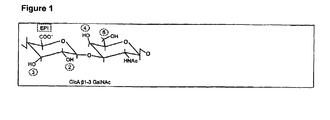
- Figure 1 Chondroitin sulphate is found in several forms, named chondroitin -4 sulphate, -6 sulphate and -D and -E respectively. These forms differ in the sulphation of saccharides.
- CS-E is a highly sulphated species, which is attached to perlecan in the I and V domains.
- Figure 1 Basic structure of Chondroitin sulfate. Repeating dimeric units of GlcA ⁇ 1-3 GalNAc. All hydroxy positions may be sulfated or/ and epimerised.
- Chondroitin sulphate/ dermatan sulphate is found in all extracellular matrices. Cartilage and invertebral disc are the tissues richest in chondroitin sulphate (Wight et al., 1981, review). Chondroitin sulphate is synthesised by specific enzymes located in the Golgi. The polymers are assembled onto a linker tri-saccharide. The hydroxyl group of serine residues followed by a glycine in the protein is substituted with a xylose and two successive galactose residues. Thereafter alternating monosaccharides of glucuronic acid and N-acetylgalactosamine are added successively to form the chain.
- cartilage aggrecan a member of the family hyalectins, is a chondroitin sulphate proteoglycan and is substituted with some one hundred CS chain and some thirty keratan sulphate chains.
- Aggrecan molecules are clustered along HA strands bound via their N-terminal globular domain.
- a protein known as link protein contacts both the HA-binding G1 domain of the aggrecan molecule and HA, and stabilises the complex. In this manner hundreds of aggrecan molecules are joined at one end to the HA.
- cartilage matrix chondroitin sulphate is by far the most abundant GAG.
- Perlecan was first identified as a large heparan sulphate proteoglycan isolated from the Engelbrecht-Holm-Swarm (EHS) murine basement membrane tumour. In basement membranes, it has been shown to bind several different classes of molecules. In each instance the core protein, the heparan sulphate (HS) side chains or both in concert, are involved in mediating the interaction. The proteoglycan binds to extracellular matrix components integral to basement membrane such as collagen IV, nidogen, laminin, and fibronectin ( Timpl, R. and Brown, J. C. (1996) Bioassays 18, 123-132 ). Perlecan has also been shown to bind extracellular matrix components outside the basement membrane, e.g.
- Perlecan supports cell-attachment both by binding and clustering integrins ( Brown, J. C., Sasaki, T., Gohring, W., Yamada, Y., and Timpl, R. (1997) Eur.JBiochem. 250, 39-46 ).
- Binding to growth factors has been shown for both the HS side-chains (FGF-2 ( Aviezer, D., Hecht, D., Safran, M., Eisinger, M., David, G., and Yayon, A. (1994) Cell 79, 1005-1013 )) and the core protein (progranulin, ( Gonzalez, E. M., Mongiat, M., Slater, S. J., Baffa, R., and Iozzo, R V. (2003) J Biol Chem )). Based on its interactions, perlecan is assumed to have a role in basement membrane integrity.
- FGF-2 Aviezer, D., Hecht, D., Safran, M., Eisinger, M., David, G., and Yayon, A. (1994) Cell 79, 1005-1013
- progranulin progranulin, ( Gonzalez, E. M., Mongiat, M., Slater, S. J., Baffa, R., and Iozzo
- Perlecan was originally thought to be substituted with HS exclusively, but later studies revealed that it is also present in a variant partially substituted with chondroitin sulphate (CS) ( Couchman, J. R., Kapoor, R., Sthanam, M., and Wu, R R (1996) J Biol Chem 271, 9595-9602 ). Both the HS- and the HS/CS- substituted variants of perlecan have been found in tissues other than basement membrane, for example cartilage.
- CS chondroitin sulphate
- Mature collagen fibres may contain several different types of bound accessory proteins. They are part in the organisation of these fibres and regulate links to other molecules thereby contributing to the architecture of the fibrillar collagen network.
- modulator molecules which regulate the early steps in the assembly of collagen monomers to fibres.
- Our laboratory has found that cartilage oligomeric matrix protein (COMP) accelerates the formation of fibres from monomers (Mörgelin and Heineg ⁇ rd, manuscript).
- Other molecules have the opposite effect and slow down fibre formation in vitro, e.g. decorin ( Vogel, K. G., Paulsson, M., and Heineg ⁇ rd, D. (1984) Biochem.J. 223, 587-597 ) and fibromodulin ( Hedborn, E.
- Perlecan exists as HS and CS substituted forms and it has been shown that these forms can be used to facilitate collagen fibril formation. To our surprise, the addition of free CS-E was effective in collagen fibril formation, but none of the other CS variants had any significant effect (e.g. CS-D or CS-6).
- CS-E chondroitin sulphate E
- CFF collagen fibril formation
- CS-E is highly sulphated and thus may be a charged compound and the invention also comprises pharmaceutical acceptable salts, such as alkali metal salts (sodium, potassium, cesium) and alkaline earth salts (e.g. magnesium, zinc, calcium, strontium) and ammonium, as well as organic salts.
- pharmaceutical acceptable salts such as alkali metal salts (sodium, potassium, cesium) and alkaline earth salts (e.g. magnesium, zinc, calcium, strontium) and ammonium, as well as organic salts.
- Indications comprised in this application are conditions and diseases related to disorders in collagen fibrillogenesis, including wound healing, in particular chronic wound healing, rheumatoid arthritis (RA), reconstructive skeletal formation and skeletal repair.
- wound healing in particular chronic wound healing, rheumatoid arthritis (RA), reconstructive skeletal formation and skeletal repair.
- RA rheumatoid arthritis
- Chronic wound healing can be stimulated by application of a CFF agonist, alone or together with exogenous collagen. This is also useful for treatment of acute open wounds.
- CFF agonists also facilitates healing of internal wounds including peptic ulcers.
- CFF agonists can be used to facilitate healing after surgical treatment and transplantation.
- CFF agonists can be used to stimulate repair in damaged ligaments and tendons.
- CFF agonist can be used to facilitate bone repair after severe fractures.
- Tissue engineering can be used to repair damaged tissue (e.g. skin in burn wound healing or cartilage in OA) via autologous cell replacement therapy or transplantation.
- CFF agonists can be used to stimulate collagen matrix formation in cell or tissue culture for production of transplantable skin, tendon, cartilage, bone or blood vessels from patient cells or tissue or from various types of stem cells.
- CFF agonists can be used to produce well-formed artificial collagen matrix scaffolds for use in healing of e.g. burn wounds and OA cartilage. After implantation these scaffolds would be populated by cells recruited from the surrounding tissue of the patient.
- CFF agonists are also useful in the production of well-organised collagen fibril matrices for corneal implants.
- Scarring is a natural response of the body to trauma and injury. In fibrotic conditions the normal wound healing response continues out of control, with excessive production and deposition of collagen. This leads to a loss of function when normal tissue is replaced with scar tissue. Fibrosis can effect virtually all organ systems in the body.
- fibrosis e.g. trauma, surgery, infection, environmental pollutants and toxins (including alcohol).
- Some examples of fibrotic conditions are formation of scar tissue following heart attack, kidney fibrosis as a complication of diabetes, lung fibrosis and surgical scar tissue formation between internal organs.
- Acute fibrosis is a response to various forms of trauma, such as injury, infections, surgery, burns, radiation damage and chemotherapeutic treatments. Many chronic conditions, e.g. diabetes, viral infection and hypertension, induce a progressive fibrosis causing continuous loss of tissue function. The liver, kidney and lung are commonly affected. Systemic fibrotic diseases include diabetic nephropathy, scleroderma, idiopathic pulmonary fibrosis and reactive fibrosis following myocardial infarct.
- Bovine pepsin-extracted collagen type I was purchased from Vitrogen. Collagen II was pepsin extracted from bovine tracheal cartilage, as previously described ( Vogel, K. G., Paulsson, M., and Heineg ⁇ rd, D. (1984) Biochem.J. 223, 587-597 ). The fibrillogenesis assay has been previously described ( Hedbom, E. and Heineg ⁇ rd, D. (1989) J.Biol.Chem. 264, 6898-6905 ).
- a solution of collagen monomers (330 nM) was brought to neutral pH by addition of an appropriate volume of 0,012M NaOH, and buffered by 20 mM HEPES, 150 mM NaCl at pH 7,4.
- Perlecan fragment was added at concentrations equimolar to that of collagen or at one tenth the molar concentration of collagen.
- the sample was mixed vigorously and briefly, and transferred to a cuvette.
- the sample was incubated at 37°C (collagen type I) or 35°C (collagen type II) in water-jacketed cuvettes in the spectrophotometer, and the absorbance due to light scattering at 400 nm (collagen type I) or 313 nm (collagen type II) was monitored over a duration of 5 - 18 hrs.
- the increased absorbance/turbidity depends on increasing fibre formation.
- CS-E from squid was purchased from Calbiochem. 0.13 ⁇ g/ml corresponding to the molar concentration of GAG chains in previous experiments using recombinant perlecan domain I variants with HS/CS (PG IB) (33 nM).
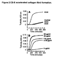
- FIG. 2 CS-E accelerated collagen fibril formation.
- Different types of purified CS-chains were tested for effect in the collagen fibrillogenesis assay (panel A).
- the highly sulphated CS-E had dramatic effects on fibril formation but neither CS-6 nor CS-D had any effect.
- the stimulatory effect of CS-E was dose-dependent, reaching saturation at concentration of 30 ⁇ g/ml (panel B).
- mice Groups of 5 ICR male mice weighing 22 ⁇ 2 gms are used. Under hexobarbital (90 mg/kg, i.p.) anesthesia, the shoulder and back region of each animal is shaved. A sharp punch (ID 12 mm) is used to remove the skin including panniculus carnosus and adherent tissues. The wound area, traced onto clear plastic sheets on days 3, 5, 7, 9 and 11, are quantitated by use of an Image Analyzer (Life Science Resources VISTA, Version 3.0). Test compound and/or vehicle (20 ⁇ l, 0.5% carboxymethylcellulose in PBS pH 7.4) is applied topically immediately following injury and once daily thereafter for a total of 10 consecutive days.
- Test compound and/or vehicle (20 ⁇ l, 0.5% carboxymethylcellulose in PBS pH 7.4
- the wound half closure time (CT 50 ) is determined and unpaired Student's t-test is applied for comparison between treated and vehicle group at each measurement time point. Differences are considered statistical significance at P ⁇ 0.05.
- Example of a preparation comprising a capsule
- the amount of lactose used may be reduced.
- Example of a suitable tablet formulation Example of a suitable tablet formulation.
- a solution for parenteral administration by injection can be prepared in aqueous solution of a water-soluble pharmaceutically acceptable acid addition salt of the active substance preferably in a concentration of 0.1% to about 10% by weight.
- These solutions may also contain stabilising agents, buffering agents and/or gelating agents such as but not limited to hyaluronan, PEG, HPMC, EHEC, to obtain a controlled release and/or elimination.
- stabilising agents such as but not limited to hyaluronan, PEG, HPMC, EHEC, to obtain a controlled release and/or elimination.
- a gel for topical administration can be prepared an active substance in a concentration of 0.1% to 10% by weight, optionally containing stabilising agents, buffering agents and/or additional gelating agents such as but not limited to hyaluronan, PEG, HMPC, EHEC to obtain controlled release and/or elimination
Landscapes
- Health & Medical Sciences (AREA)
- Life Sciences & Earth Sciences (AREA)
- Veterinary Medicine (AREA)
- Pharmacology & Pharmacy (AREA)
- Chemical & Material Sciences (AREA)
- Public Health (AREA)
- General Health & Medical Sciences (AREA)
- Medicinal Chemistry (AREA)
- Animal Behavior & Ethology (AREA)
- Dermatology (AREA)
- Bioinformatics & Cheminformatics (AREA)
- Organic Chemistry (AREA)
- Nuclear Medicine, Radiotherapy & Molecular Imaging (AREA)
- General Chemical & Material Sciences (AREA)
- Chemical Kinetics & Catalysis (AREA)
- Engineering & Computer Science (AREA)
- Molecular Biology (AREA)
- Epidemiology (AREA)
- Physical Education & Sports Medicine (AREA)
- Medicines That Contain Protein Lipid Enzymes And Other Medicines (AREA)
- Pharmaceuticals Containing Other Organic And Inorganic Compounds (AREA)
Description
- Many proteins of the extracellular matrix (ECM) are modified post-translationally by addition of oligosaccharide chains and are thus known as glycoproteins. The oligosaccharides are linked either O-glycosidically to serine or threonine residues, or N-glycosidically to an asparagine residue. Proteoglycans are glycoproteins that are substituted with a particular class of carbohydrate polymers, known as the glycosaminoglycans (GAGs). Proteoglycans are found in the ECM, at the cell surface and intracellularly in storage granules. In the ECM they contribute to the structure and organisation, and at the cell surface often function as receptors and/or co-receptors. All glycosaminoglycans (with the exception of hyaluronan) are synthesised on a core protein acceptor, and they are thus an integral component of proteoglycans (Wight et al., 1981; Heinegård and Paulsson, 1984, review).
- Glycosaminoglycans (GAGs) are named to indicate that one of the monosaccharides in the repeating sequence of disaccharides is an amino sugar. The other monosaccharide is an uronic acid (glucuronic acid or iduronic acid), with the exception of keratan sulphate where it is a galactose. While other oligosaccharide substituents maybe branched, GAG chains are linear (again, with the exception of kcratan sulphate). Proteoglycans may be substituted with one (e.g. decorin) and up to some one hundred (e.g. aggrecan) GAG chains.
- There are 4 types of glycosaminoglycans: hyaluronic acid, chondroitin sulphate/ dermatan sulphate, heparan sulphate/heparin and keratan sulphate. The disaccharides in all glycosaminoglycan chains except hyaluronan are sulphated, increasing their negative charge and leading to an extended conformation of the chain. The molecule will occupy large solvent domains, observed as a high viscosity of a solution. This property is essential in cartilage and is the basis on which the tissue's resistance lies.
- The repeating disaccharide sequence in CS is glucuronic acid-N-acetyl-galactosamine (GlcA-GaINAc), see
Figure 1 . Chondroitin sulphate is found in several forms, named chondroitin -4 sulphate, -6 sulphate and -D and -E respectively. These forms differ in the sulphation of saccharides. CS-E is a highly sulphated species, which is attached to perlecan in the I and V domains. -
Figure 1 . Basic structure of Chondroitin sulfate. Repeating dimeric units of GlcA β1-3 GalNAc. All hydroxy positions may be sulfated or/ and epimerised. - The various positions open for sulfatation are numbered.
- Chondroitin sulphate/ dermatan sulphate is found in all extracellular matrices. Cartilage and invertebral disc are the tissues richest in chondroitin sulphate (Wight et al., 1981, review). Chondroitin sulphate is synthesised by specific enzymes located in the Golgi. The polymers are assembled onto a linker tri-saccharide. The hydroxyl group of serine residues followed by a glycine in the protein is substituted with a xylose and two successive galactose residues. Thereafter alternating monosaccharides of glucuronic acid and N-acetylgalactosamine are added successively to form the chain. Some glucuronate residues are converted to iduronate by an epimerase and sulfation is the last event just prior to secretion (Wight et al., 1981, review). In cartilage aggrecan, a member of the family hyalectins, is a chondroitin sulphate proteoglycan and is substituted with some one hundred CS chain and some thirty keratan sulphate chains. Aggrecan molecules are clustered along HA strands bound via their N-terminal globular domain. A protein known as link protein contacts both the HA-binding G1 domain of the aggrecan molecule and HA, and stabilises the complex. In this manner hundreds of aggrecan molecules are joined at one end to the HA. Thus, in cartilage matrix chondroitin sulphate is by far the most abundant GAG.
- Perlecan was first identified as a large heparan sulphate proteoglycan isolated from the Engelbrecht-Holm-Swarm (EHS) murine basement membrane tumour. In basement membranes, it has been shown to bind several different classes of molecules. In each instance the core protein, the heparan sulphate (HS) side chains or both in concert, are involved in mediating the interaction. The proteoglycan binds to extracellular matrix components integral to basement membrane such as collagen IV, nidogen, laminin, and fibronectin (Timpl, R. and Brown, J. C. (1996) Bioassays 18, 123-132). Perlecan has also been shown to bind extracellular matrix components outside the basement membrane, e.g. PRELP and collagen type I (Bengtsson, E., Mörgelin, M., Sasaki, T., Timpl, R., Heinegård, D., and Aspberg, A. (2002) J. Biol. Chem). Perlecan supports cell-attachment both by binding and clustering integrins (Brown, J. C., Sasaki, T., Gohring, W., Yamada, Y., and Timpl, R. (1997) Eur.JBiochem. 250, 39-46). Binding to growth factors has been shown for both the HS side-chains (FGF-2 (Aviezer, D., Hecht, D., Safran, M., Eisinger, M., David, G., and Yayon, A. (1994) Cell 79, 1005-1013)) and the core protein (progranulin, (Gonzalez, E. M., Mongiat, M., Slater, S. J., Baffa, R., and Iozzo, R V. (2003) J Biol Chem)). Based on its interactions, perlecan is assumed to have a role in basement membrane integrity.
- Perlecan was originally thought to be substituted with HS exclusively, but later studies revealed that it is also present in a variant partially substituted with chondroitin sulphate (CS) (Couchman, J. R., Kapoor, R., Sthanam, M., and Wu, R R (1996) J Biol Chem 271, 9595-9602). Both the HS- and the HS/CS- substituted variants of perlecan have been found in tissues other than basement membrane, for example cartilage.
- The generation of perlecan null mice revealed two particularly intriguing findings (Arikawa-Hirasawa, E., Watanabe, H., Takami, H., Hassell, J. R, and Yamada, Y. (1999) Nat Genet. 23, 354-358; Costell, M., Gustafsson, E., Aszódi, A., Morgelin, M., Bloch, W., Hunziker, E., Addicks, K., Timpl, R., and Fässler, R (1999) J Cell Biol 147, 1109-1122). First, though mice lacking perlecan did develop grave disorders caused by compromised basement membrane strength or integrity (e.g. rupture of pericardial sac), the initial assembly of basement membranes seemed to be without complication. The second striking finding was the severe skeletal defects exhibited, apparently caused by the lack of perlecan in cartilage.
- Following the publication of these results at least two human hereditary diseases with skeletal deficiencies have been ascribed to an underlying scarcity or complete lack of perlecan, underscoring the relevance of this finding in the mouse model (Nicole, S., Davoine, C. S., Topaloglu, H., Cattolico, L., Barral, D., Beighton, P., Hamida, C. B., Hammouda, H., Cruaud, C., White, P. S., Samson, D., Urtizberea, J. A., Lehmann-Horn, F., Weissenbach, J., Hentati, F., and Fontaine, B. (2000) Nat Genet. 26,480-483; Arikawa-Hirasawa, E., Wilcox, W. R., Le, A. H., Silverman, N., Govindraj, P., Hassell, J. R, and Yamada, Y. (2001) Nat Genet. 27, 431-434).
- In skeletal development, the deposition of a cartilaginous template precedes the formation of bones. The integrity of this template is a prerequisite for proper assembly of the skeleton. Perlecan-null mouse cartilage shows fewer and less organised collagen type II fibrils, and decreased levels of aggrecan, indicating a failure to organise the extracellular matrix (Costell, M., Gustafsson, E., Aszódi, A., Morgelin, M., Bloch, W., Hunziker, E., Addicks, K., Timpl, R., and Fässler, R (1999) J Cell Biol 147, 1109-1122).
- Mature collagen fibres may contain several different types of bound accessory proteins. They are part in the organisation of these fibres and regulate links to other molecules thereby contributing to the architecture of the fibrillar collagen network. A recent concept is that of modulator molecules, which regulate the early steps in the assembly of collagen monomers to fibres. Our laboratory has found that cartilage oligomeric matrix protein (COMP) accelerates the formation of fibres from monomers (Mörgelin and Heinegård, manuscript). Other molecules have the opposite effect and slow down fibre formation in vitro, e.g. decorin (Vogel, K. G., Paulsson, M., and Heinegård, D. (1984) Biochem.J. 223, 587-597) and fibromodulin (Hedborn, E. and Heinegård, D. (1989) J.Biol.Chem. 264, 6898-6905). Gene targeting of these molecules lead to abnormal collagen fibrils and disturbed mechanical properties of the tissues (Danielson, K. G., Baribault, H., Holmes, D. F., Graham, H., Kadler, K. E., and Iozzo, R. V. (1997) J Cell Biol 136, 729-743; Svensson, L., Aszódi, A., Reinholt, F. P., Fässler, R., Heinegård, D., and Oldberg, Å. (1999) J.Biol.Chem. 274, 9636-9647). A picture is emerging where proteins in the vicinity of the cell regulate the early stages of collagen fibre formation.
- Perlecan exists as HS and CS substituted forms and it has been shown that these forms can be used to facilitate collagen fibril formation. To our surprise, the addition of free CS-E was effective in collagen fibril formation, but none of the other CS variants had any significant effect (e.g. CS-D or CS-6).
- A number of publications describing the effect of chondroitin sulphate on wound healing and for treating arthrosis exist (
US5929050 ,JP10120577 RU2216332 - Munakata et al. (Glycobiology (1999) 9(10):1023-1027) investigate the interaction between collagens and glycosaminoglycans using a surface plasmon resonance biosensor.
- Comprised in the invention is chondroitin sulphate E (CS-E) for use in the treatment of conditions and diseases related to collagen fibril formation (CFF) as defined in claim 1.
- CS-E is highly sulphated and thus may be a charged compound and the invention also comprises pharmaceutical acceptable salts, such as alkali metal salts (sodium, potassium, cesium) and alkaline earth salts (e.g. magnesium, zinc, calcium, strontium) and ammonium, as well as organic salts.
- Indications comprised in this application are conditions and diseases related to disorders in collagen fibrillogenesis, including wound healing, in particular chronic wound healing, rheumatoid arthritis (RA), reconstructive skeletal formation and skeletal repair.
- Chronic wound healing can be stimulated by application of a CFF agonist, alone or together with exogenous collagen. This is also useful for treatment of acute open wounds.
- CFF agonists also facilitates healing of internal wounds including peptic ulcers.
- CFF agonists can be used to facilitate healing after surgical treatment and transplantation.
- CFF agonists can be used to stimulate repair in damaged ligaments and tendons.
- CFF agonist can be used to facilitate bone repair after severe fractures.
- Tissue engineering can be used to repair damaged tissue (e.g. skin in burn wound healing or cartilage in OA) via autologous cell replacement therapy or transplantation. CFF agonists can be used to stimulate collagen matrix formation in cell or tissue culture for production of transplantable skin, tendon, cartilage, bone or blood vessels from patient cells or tissue or from various types of stem cells.
- CFF agonists can be used to produce well-formed artificial collagen matrix scaffolds for use in healing of e.g. burn wounds and OA cartilage. After implantation these scaffolds would be populated by cells recruited from the surrounding tissue of the patient.
- CFF agonists are also useful in the production of well-organised collagen fibril matrices for corneal implants.
- Scarring is a natural response of the body to trauma and injury. In fibrotic conditions the normal wound healing response continues out of control, with excessive production and deposition of collagen. This leads to a loss of function when normal tissue is replaced with scar tissue. Fibrosis can effect virtually all organ systems in the body.
- There are many different causes of fibrosis, e.g. trauma, surgery, infection, environmental pollutants and toxins (including alcohol). Some examples of fibrotic conditions are formation of scar tissue following heart attack, kidney fibrosis as a complication of diabetes, lung fibrosis and surgical scar tissue formation between internal organs.
- Acute fibrosis is a response to various forms of trauma, such as injury, infections, surgery, burns, radiation damage and chemotherapeutic treatments. Many chronic conditions, e.g. diabetes, viral infection and hypertension, induce a progressive fibrosis causing continuous loss of tissue function. The liver, kidney and lung are commonly affected. Systemic fibrotic diseases include diabetic nephropathy, scleroderma, idiopathic pulmonary fibrosis and reactive fibrosis following myocardial infarct.
- Bovine pepsin-extracted collagen type I was purchased from Vitrogen. Collagen II was pepsin extracted from bovine tracheal cartilage, as previously described (Vogel, K. G., Paulsson, M., and Heinegård, D. (1984) Biochem.J. 223, 587-597). The fibrillogenesis assay has been previously described (Hedbom, E. and Heinegård, D. (1989) J.Biol.Chem. 264, 6898-6905).
- Briefly, a solution of collagen monomers (330 nM) was brought to neutral pH by addition of an appropriate volume of 0,012M NaOH, and buffered by 20 mM HEPES, 150 mM NaCl at
pH 7,4. Perlecan fragment was added at concentrations equimolar to that of collagen or at one tenth the molar concentration of collagen. The sample was mixed vigorously and briefly, and transferred to a cuvette. The sample was incubated at 37°C (collagen type I) or 35°C (collagen type II) in water-jacketed cuvettes in the spectrophotometer, and the absorbance due to light scattering at 400 nm (collagen type I) or 313 nm (collagen type II) was monitored over a duration of 5 - 18 hrs. The increased absorbance/turbidity depends on increasing fibre formation. - Fibrillogenesis was performed as described above. CS-E from squid was purchased from Calbiochem. 0.13 µg/ml corresponding to the molar concentration of GAG chains in previous experiments using recombinant perlecan domain I variants with HS/CS (PG IB) (33 nM).
- The results of the fibrillogenesis experiment is shown in
figure 2 . As can be seen from this figure, CS-E but none of the other tested chondroitin sulphate variants (CS-D and CS-6) gives a positive result on collagen fibril formation (CFF). -
Figure 2 CS-E accelerated collagen fibril formation. Different types of purified CS-chains were tested for effect in the collagen fibrillogenesis assay (panel A). The highly sulphated CS-E had dramatic effects on fibril formation but neither CS-6 nor CS-D had any effect. The stimulatory effect of CS-E was dose-dependent, reaching saturation at concentration of 30 µg/ml (panel B). - Groups of 5 ICR male mice weighing 22±2 gms are used. Under hexobarbital (90 mg/kg, i.p.) anesthesia, the shoulder and back region of each animal is shaved. A sharp punch (ID 12 mm) is used to remove the skin including panniculus carnosus and adherent tissues. The wound area, traced onto clear plastic sheets on
days 3, 5, 7, 9 and 11, are quantitated by use of an Image Analyzer (Life Science Resources VISTA, Version 3.0). Test compound and/or vehicle (20 µl, 0.5% carboxymethylcellulose in PBS pH 7.4) is applied topically immediately following injury and once daily thereafter for a total of 10 consecutive days. The wound half closure time (CT50) is determined and unpaired Student's t-test is applied for comparison between treated and vehicle group at each measurement time point. Differences are considered statistical significance at P < 0.05. (Montesinos, M.C., Gadangi, P., Longaker, M., Sung, J., Levine, J., Nilsen, D., Reibman, J., Li, M., Jiang, C.K., Hirschorn, R., Recht, P.A., Ostad, E., Levin, R.I. and Crostein, B.N. Wound healing is accelerated by agonists of Adenosine A2 (Gαs-linked) receptors. J. Exp. Med. 186: 1615 -1620, 1997.) -
Per capsule Active ingredient, as salt 5 mg Lactose 250 mg Starch 120 mg Magnesium stearate 5 mg Total up to 385 mg - In case higher amounts of active ingredient are required, the amount of lactose used may be reduced.
-
Per tablet Active ingredient, as salt 5 mg Potato starch 90 mg Colloidal silica 10 mg Talc 20 mg Magnesium stearate 2 mg 5% aqueous solution of gelatine 25 mg Total up to 385 mg - A solution for parenteral administration by injection can be prepared in aqueous solution of a water-soluble pharmaceutically acceptable acid addition salt of the active substance preferably in a concentration of 0.1% to about 10% by weight.
- These solutions may also contain stabilising agents, buffering agents and/or gelating agents such as but not limited to hyaluronan, PEG, HPMC, EHEC, to obtain a controlled release and/or elimination.
- A gel for topical administration can be prepared an active substance in a concentration of 0.1% to 10% by weight, optionally containing stabilising agents, buffering agents and/or additional gelating agents such as but not limited to hyaluronan, PEG, HMPC, EHEC to obtain controlled release and/or elimination
Claims (4)
- Chondroitin sulphate E (CS-E) or a pharmaceutically acceptable salt thereof for use in the treatment of diseases or conditions related to collagen fibril formation, wherein the disease or condition related to collagen fibril formation is wound healing, rheumatoid arthritis (RA), reconstructive skeletal formation, skeletal repair, facilitating healing after surgical treatment or transplantation, stimulating repair in damaged ligaments or tendons, facilitating bone repair after severe fractures, or wounds (ulcers).
- Chondroitin sulphate E (CS-E) or a pharmaceutically acceptable salt thereof for use as claimed in claim 1, wherein the wound healing is healing of a chronic wound, an acute open wound or an internal wound such as a peptic ulcer.
- Use of chondroitin sulphate E (CS-E) or a pharmaceutically acceptable salt thereof for the manufacture of a medicament for the treatment of diseases or conditions related to collagen fibril formation, wherein the disease or condition related to collagen fibril formation is wound healing, rheumatoid arthritis (RA), reconstructive skeletal formation, skeletal repair, facilitating healing after surgical treatment or transplantation, stimulating repair in damaged ligaments or tendons, facilitating bone repair after severe fractures, or wounds (ulcers).
- Use as claimed in claim 3, wherein the wound healing is healing of a chronic wound, an acute open wound or an internal wound such as a peptic ulcer.
Applications Claiming Priority (2)
| Application Number | Priority Date | Filing Date | Title |
|---|---|---|---|
| SE0401069A SE0401069D0 (en) | 2004-04-26 | 2004-04-26 | Use of Compounds for the Treatment of Diseases and Conditions |
| PCT/SE2005/000599 WO2005102362A1 (en) | 2004-04-26 | 2005-04-26 | Use of chondroitin sulphate e (cs-e ) for the treatment diseases or conditions related to collagen fibril formation. |
Publications (2)
| Publication Number | Publication Date |
|---|---|
| EP1747007A1 EP1747007A1 (en) | 2007-01-31 |
| EP1747007B1 true EP1747007B1 (en) | 2013-03-20 |
Family
ID=32322669
Family Applications (1)
| Application Number | Title | Priority Date | Filing Date |
|---|---|---|---|
| EP05735125A Expired - Lifetime EP1747007B1 (en) | 2004-04-26 | 2005-04-26 | Use of chondroitin sulphate e (cs-e ) for the treatment diseases or conditions related to collagen fibril formation. |
Country Status (10)
| Country | Link |
|---|---|
| US (1) | US7956047B2 (en) |
| EP (1) | EP1747007B1 (en) |
| JP (1) | JP2007534749A (en) |
| AU (1) | AU2005235171B2 (en) |
| CA (1) | CA2564436C (en) |
| ES (1) | ES2409343T3 (en) |
| NZ (1) | NZ550893A (en) |
| SE (1) | SE0401069D0 (en) |
| WO (1) | WO2005102362A1 (en) |
| ZA (1) | ZA200608926B (en) |
Families Citing this family (6)
| Publication number | Priority date | Publication date | Assignee | Title |
|---|---|---|---|---|
| SE0401069D0 (en) * | 2004-04-26 | 2004-04-26 | Anamar Medical Ab | Use of Compounds for the Treatment of Diseases and Conditions |
| JP2008195705A (en) * | 2007-01-15 | 2008-08-28 | Daiichi Sankyo Healthcare Co Ltd | Loxoprofen-containing oral composition |
| WO2008102568A1 (en) * | 2007-02-22 | 2008-08-28 | Pg Research Co., Ltd. | Bone/cartilage formation-stimulating agent |
| GB201001203D0 (en) | 2010-01-25 | 2010-03-10 | Anamar Medical Ab | Use of pharmaceutically active compounds |
| JP2012031122A (en) * | 2010-08-02 | 2012-02-16 | Kracie Home Products Ltd | Collagen production promoter |
| CN108727492B (en) * | 2017-04-20 | 2021-07-13 | 广州文瑞生物科技有限公司 | Immunotherapy for regression of atherosclerosis with anti-collagen VI fully human antibodies |
Family Cites Families (15)
| Publication number | Priority date | Publication date | Assignee | Title |
|---|---|---|---|---|
| FI912858A0 (en) | 1988-12-20 | 1991-06-13 | Jolla Cancer Res Found | POLYPEPTIDPOLYMERKJJUGAT AKTIVA VID BOTANDE AV SAOR. |
| AU7681091A (en) | 1990-04-05 | 1991-10-30 | John R Hoidal | Method and medicament for prevention or medication of human leucocyte elastase-mediated pulmonary diseases |
| US5605938A (en) * | 1991-05-31 | 1997-02-25 | Gliatech, Inc. | Methods and compositions for inhibition of cell invasion and fibrosis using dextran sulfate |
| JPH10120577A (en) * | 1996-10-16 | 1998-05-12 | Sekisui Chem Co Ltd | Wound healing agent |
| US5929050A (en) * | 1998-02-27 | 1999-07-27 | Petito; George D. | Chondroitin sulfate composition and method for wound treatment |
| FR2791262B1 (en) | 1999-03-22 | 2001-09-28 | Virbac Sa | COMPOSITIONS BASED ON CHONDROITINE AND CHITOSAN FOR THE PROTECTION, TREATMENT OR REPLACEMENT OF CONNECTIVE TISSUES |
| US6162787A (en) * | 1999-04-02 | 2000-12-19 | Immudyne, Inc. | Methods for treating arthritis using collagen type II, glucosamine chondroitin sulfate, and compositions |
| US6372794B1 (en) | 1999-08-26 | 2002-04-16 | Marcel E. Nimni | Method for alleviating arthritis in mammals |
| AU2001264817A1 (en) | 2000-05-23 | 2001-12-17 | Exhale Therapeutics, Inc. | Method for treating respiratory disorders associated with pulmonary elastic fiber injury comprising the use of clycosaminoglycans |
| JP2002080371A (en) * | 2000-06-23 | 2002-03-19 | Seikagaku Kogyo Co Ltd | Plasminogen activator activity promoter |
| CA2425145C (en) | 2000-10-16 | 2011-08-30 | Genentech, Inc. | Methods of treatment using wisp polypeptides |
| RU2216332C1 (en) | 2002-02-18 | 2003-11-20 | Акционерное Курганское общество медицинских препаратов и изделий "Синтез" | Curative preparation for treatment of arthrosis |
| JP2004059506A (en) * | 2002-07-29 | 2004-02-26 | Taisho Pharmaceut Co Ltd | Persulfated oligosaccharides acting on selectins and chemokines |
| JP4567942B2 (en) | 2002-12-27 | 2010-10-27 | 生化学工業株式会社 | Osteoclast formation inhibitor |
| SE0401069D0 (en) * | 2004-04-26 | 2004-04-26 | Anamar Medical Ab | Use of Compounds for the Treatment of Diseases and Conditions |
-
2004
- 2004-04-26 SE SE0401069A patent/SE0401069D0/en unknown
-
2005
- 2005-04-26 AU AU2005235171A patent/AU2005235171B2/en not_active Ceased
- 2005-04-26 US US11/587,675 patent/US7956047B2/en not_active Expired - Fee Related
- 2005-04-26 EP EP05735125A patent/EP1747007B1/en not_active Expired - Lifetime
- 2005-04-26 WO PCT/SE2005/000599 patent/WO2005102362A1/en not_active Ceased
- 2005-04-26 NZ NZ550893A patent/NZ550893A/en not_active IP Right Cessation
- 2005-04-26 ES ES05735125T patent/ES2409343T3/en not_active Expired - Lifetime
- 2005-04-26 JP JP2007510653A patent/JP2007534749A/en active Pending
- 2005-04-26 CA CA2564436A patent/CA2564436C/en not_active Expired - Lifetime
-
2006
- 2006-10-26 ZA ZA2006/08926A patent/ZA200608926B/en unknown
Also Published As
| Publication number | Publication date |
|---|---|
| ZA200608926B (en) | 2008-04-30 |
| WO2005102362A8 (en) | 2006-06-22 |
| CA2564436A1 (en) | 2005-11-03 |
| US20080200426A1 (en) | 2008-08-21 |
| US7956047B2 (en) | 2011-06-07 |
| ES2409343T3 (en) | 2013-06-26 |
| AU2005235171A2 (en) | 2005-11-03 |
| AU2005235171A1 (en) | 2005-11-03 |
| WO2005102362A1 (en) | 2005-11-03 |
| SE0401069D0 (en) | 2004-04-26 |
| JP2007534749A (en) | 2007-11-29 |
| AU2005235171B2 (en) | 2010-11-11 |
| EP1747007A1 (en) | 2007-01-31 |
| CA2564436C (en) | 2014-06-17 |
| NZ550893A (en) | 2010-07-30 |
Similar Documents
| Publication | Publication Date | Title |
|---|---|---|
| Funderburgh et al. | Macrophage receptors for lumican. A corneal keratan sulfate proteoglycan. | |
| US5705178A (en) | Methods and compositions based on inhibition of cell invasion and fibrosis by anionic polymers | |
| Toole | Developmental role of hyaluronate | |
| EP0154447B1 (en) | Compositions and method for improving wound healing | |
| Al-Khateeb et al. | Hyaluronic acid: the reason for its variety of physiological and biochemical functional properties | |
| EP0161887A2 (en) | Crosslinked hyaluronic acid and its use | |
| Ryan et al. | Glycosaminoglycans in tendon physiology, pathophysiology, and therapy | |
| JPH09510493A (en) | Novel heparin-like sulfated polysaccharide | |
| HUT66427A (en) | Compositions containing anionic polymers to inhibit cell invasion and fibrosis | |
| EP0640647A2 (en) | Ionically crosslinked glycosaminoglycan gels for soft tissue augmentation and drug delivery | |
| DE69214355T2 (en) | Agents containing hepatocyte growth factor | |
| Mcdevitt | Cell biology of the eye | |
| DE69217104T2 (en) | Oligosaccharide with an affinity for the fibroblast growth factor and method for its production | |
| US6486140B2 (en) | Agents, and methods employing them, for the prevention or reduction of tissue adhesion at a wound site | |
| US7956047B2 (en) | Use of chondroitin sulphate E (CS-E) for the treatment of diseases or conditions related to collagen fibril formation | |
| Hall | A simple, single‐injection method for inducing long‐term paralysis in embryonic chicks, and preliminary observations on growth of the tibia | |
| JPH10287570A (en) | Curative promoter for keratopathy | |
| West et al. | A specificity for cellular fibronectin in its effect on cultured chondroblasts | |
| EP2155208B1 (en) | Disaccharides for the treatment of tendons and ligaments | |
| EP4737486A1 (en) | Hyaluronic acid derivative, and preparation method therefor and use thereof | |
| Chau et al. | Heparan sulphates upregulate regeneration of transected sciatic nerves of adult guinea‐pigs | |
| US20230119502A1 (en) | Collagen Nucleation Inhibitors | |
| Nakamura et al. | Section Review: Recent developments in the use of hyaluronan in wound healing: Pulmonary-Allergy, Dermatological, Gastrointestinal & Arthritis | |
| WO2011089446A1 (en) | Use of pharmaceutically active compounds | |
| Mathews | The Tissues |
Legal Events
| Date | Code | Title | Description |
|---|---|---|---|
| PUAI | Public reference made under article 153(3) epc to a published international application that has entered the european phase |
Free format text: ORIGINAL CODE: 0009012 |
|
| 17P | Request for examination filed |
Effective date: 20061127 |
|
| AK | Designated contracting states |
Kind code of ref document: A1 Designated state(s): AT BE BG CH CY CZ DE DK EE ES FI FR GB GR HU IE IS IT LI LT LU MC NL PL PT RO SE SI SK TR |
|
| RIN1 | Information on inventor provided before grant (corrected) |
Inventor name: KVIST, ALEXANDER Inventor name: JOHNSON, ANNA Inventor name: ASPBERG, ANDERS Inventor name: HEINEGAERD, DICK |
|
| 17Q | First examination report despatched |
Effective date: 20070622 |
|
| DAX | Request for extension of the european patent (deleted) | ||
| RAP1 | Party data changed (applicant data changed or rights of an application transferred) |
Owner name: ANAMAR AB |
|
| GRAP | Despatch of communication of intention to grant a patent |
Free format text: ORIGINAL CODE: EPIDOSNIGR1 |
|
| GRAP | Despatch of communication of intention to grant a patent |
Free format text: ORIGINAL CODE: EPIDOSNIGR1 |
|
| GRAS | Grant fee paid |
Free format text: ORIGINAL CODE: EPIDOSNIGR3 |
|
| GRAA | (expected) grant |
Free format text: ORIGINAL CODE: 0009210 |
|
| AK | Designated contracting states |
Kind code of ref document: B1 Designated state(s): AT BE BG CH CY CZ DE DK EE ES FI FR GB GR HU IE IS IT LI LT LU MC NL PL PT RO SE SI SK TR |
|
| REG | Reference to a national code |
Ref country code: GB Ref legal event code: FG4D |
|
| REG | Reference to a national code |
Ref country code: CH Ref legal event code: NV Representative=s name: BOHEST AG, CH Ref country code: CH Ref legal event code: EP |
|
| REG | Reference to a national code |
Ref country code: IE Ref legal event code: FG4D |
|
| REG | Reference to a national code |
Ref country code: AT Ref legal event code: REF Ref document number: 601626 Country of ref document: AT Kind code of ref document: T Effective date: 20130415 |
|
| REG | Reference to a national code |
Ref country code: DE Ref legal event code: R096 Ref document number: 602005038655 Country of ref document: DE Effective date: 20130516 |
|
| REG | Reference to a national code |
Ref country code: ES Ref legal event code: FG2A Ref document number: 2409343 Country of ref document: ES Kind code of ref document: T3 Effective date: 20130626 |
|
| PG25 | Lapsed in a contracting state [announced via postgrant information from national office to epo] |
Ref country code: LT Free format text: LAPSE BECAUSE OF FAILURE TO SUBMIT A TRANSLATION OF THE DESCRIPTION OR TO PAY THE FEE WITHIN THE PRESCRIBED TIME-LIMIT Effective date: 20130320 Ref country code: BG Free format text: LAPSE BECAUSE OF FAILURE TO SUBMIT A TRANSLATION OF THE DESCRIPTION OR TO PAY THE FEE WITHIN THE PRESCRIBED TIME-LIMIT Effective date: 20130620 Ref country code: SE Free format text: LAPSE BECAUSE OF FAILURE TO SUBMIT A TRANSLATION OF THE DESCRIPTION OR TO PAY THE FEE WITHIN THE PRESCRIBED TIME-LIMIT Effective date: 20130320 |
|
| REG | Reference to a national code |
Ref country code: AT Ref legal event code: MK05 Ref document number: 601626 Country of ref document: AT Kind code of ref document: T Effective date: 20130320 |
|
| REG | Reference to a national code |
Ref country code: LT Ref legal event code: MG4D |
|
| PG25 | Lapsed in a contracting state [announced via postgrant information from national office to epo] |
Ref country code: FI Free format text: LAPSE BECAUSE OF FAILURE TO SUBMIT A TRANSLATION OF THE DESCRIPTION OR TO PAY THE FEE WITHIN THE PRESCRIBED TIME-LIMIT Effective date: 20130320 Ref country code: SI Free format text: LAPSE BECAUSE OF FAILURE TO SUBMIT A TRANSLATION OF THE DESCRIPTION OR TO PAY THE FEE WITHIN THE PRESCRIBED TIME-LIMIT Effective date: 20130320 Ref country code: GR Free format text: LAPSE BECAUSE OF FAILURE TO SUBMIT A TRANSLATION OF THE DESCRIPTION OR TO PAY THE FEE WITHIN THE PRESCRIBED TIME-LIMIT Effective date: 20130621 |
|
| REG | Reference to a national code |
Ref country code: NL Ref legal event code: VDEP Effective date: 20130320 |
|
| PG25 | Lapsed in a contracting state [announced via postgrant information from national office to epo] |
Ref country code: BE Free format text: LAPSE BECAUSE OF FAILURE TO SUBMIT A TRANSLATION OF THE DESCRIPTION OR TO PAY THE FEE WITHIN THE PRESCRIBED TIME-LIMIT Effective date: 20130320 |
|
| PG25 | Lapsed in a contracting state [announced via postgrant information from national office to epo] |
Ref country code: SK Free format text: LAPSE BECAUSE OF FAILURE TO SUBMIT A TRANSLATION OF THE DESCRIPTION OR TO PAY THE FEE WITHIN THE PRESCRIBED TIME-LIMIT Effective date: 20130320 Ref country code: PT Free format text: LAPSE BECAUSE OF FAILURE TO SUBMIT A TRANSLATION OF THE DESCRIPTION OR TO PAY THE FEE WITHIN THE PRESCRIBED TIME-LIMIT Effective date: 20130722 Ref country code: IS Free format text: LAPSE BECAUSE OF FAILURE TO SUBMIT A TRANSLATION OF THE DESCRIPTION OR TO PAY THE FEE WITHIN THE PRESCRIBED TIME-LIMIT Effective date: 20130720 Ref country code: AT Free format text: LAPSE BECAUSE OF FAILURE TO SUBMIT A TRANSLATION OF THE DESCRIPTION OR TO PAY THE FEE WITHIN THE PRESCRIBED TIME-LIMIT Effective date: 20130320 Ref country code: NL Free format text: LAPSE BECAUSE OF FAILURE TO SUBMIT A TRANSLATION OF THE DESCRIPTION OR TO PAY THE FEE WITHIN THE PRESCRIBED TIME-LIMIT Effective date: 20130320 Ref country code: EE Free format text: LAPSE BECAUSE OF FAILURE TO SUBMIT A TRANSLATION OF THE DESCRIPTION OR TO PAY THE FEE WITHIN THE PRESCRIBED TIME-LIMIT Effective date: 20130320 Ref country code: CZ Free format text: LAPSE BECAUSE OF FAILURE TO SUBMIT A TRANSLATION OF THE DESCRIPTION OR TO PAY THE FEE WITHIN THE PRESCRIBED TIME-LIMIT Effective date: 20130320 Ref country code: RO Free format text: LAPSE BECAUSE OF FAILURE TO SUBMIT A TRANSLATION OF THE DESCRIPTION OR TO PAY THE FEE WITHIN THE PRESCRIBED TIME-LIMIT Effective date: 20130320 |
|
| PG25 | Lapsed in a contracting state [announced via postgrant information from national office to epo] |
Ref country code: CY Free format text: LAPSE BECAUSE OF FAILURE TO SUBMIT A TRANSLATION OF THE DESCRIPTION OR TO PAY THE FEE WITHIN THE PRESCRIBED TIME-LIMIT Effective date: 20130320 |
|
| PG25 | Lapsed in a contracting state [announced via postgrant information from national office to epo] |
Ref country code: MC Free format text: LAPSE BECAUSE OF FAILURE TO SUBMIT A TRANSLATION OF THE DESCRIPTION OR TO PAY THE FEE WITHIN THE PRESCRIBED TIME-LIMIT Effective date: 20130320 |
|
| PLBE | No opposition filed within time limit |
Free format text: ORIGINAL CODE: 0009261 |
|
| STAA | Information on the status of an ep patent application or granted ep patent |
Free format text: STATUS: NO OPPOSITION FILED WITHIN TIME LIMIT |
|
| REG | Reference to a national code |
Ref country code: IE Ref legal event code: MM4A |
|
| PG25 | Lapsed in a contracting state [announced via postgrant information from national office to epo] |
Ref country code: DK Free format text: LAPSE BECAUSE OF FAILURE TO SUBMIT A TRANSLATION OF THE DESCRIPTION OR TO PAY THE FEE WITHIN THE PRESCRIBED TIME-LIMIT Effective date: 20130320 |
|
| 26N | No opposition filed |
Effective date: 20140102 |
|
| REG | Reference to a national code |
Ref country code: DE Ref legal event code: R097 Ref document number: 602005038655 Country of ref document: DE Effective date: 20140102 |
|
| PG25 | Lapsed in a contracting state [announced via postgrant information from national office to epo] |
Ref country code: IE Free format text: LAPSE BECAUSE OF NON-PAYMENT OF DUE FEES Effective date: 20130426 |
|
| REG | Reference to a national code |
Ref country code: CH Ref legal event code: PCAR Free format text: NEW ADDRESS: HOLBEINSTRASSE 36-38, 4051 BASEL (CH) |
|
| REG | Reference to a national code |
Ref country code: FR Ref legal event code: PLFP Year of fee payment: 11 |
|
| PG25 | Lapsed in a contracting state [announced via postgrant information from national office to epo] |
Ref country code: TR Free format text: LAPSE BECAUSE OF FAILURE TO SUBMIT A TRANSLATION OF THE DESCRIPTION OR TO PAY THE FEE WITHIN THE PRESCRIBED TIME-LIMIT Effective date: 20130320 |
|
| PG25 | Lapsed in a contracting state [announced via postgrant information from national office to epo] |
Ref country code: LU Free format text: LAPSE BECAUSE OF NON-PAYMENT OF DUE FEES Effective date: 20130426 Ref country code: HU Free format text: LAPSE BECAUSE OF FAILURE TO SUBMIT A TRANSLATION OF THE DESCRIPTION OR TO PAY THE FEE WITHIN THE PRESCRIBED TIME-LIMIT; INVALID AB INITIO Effective date: 20050426 |
|
| PGFP | Annual fee paid to national office [announced via postgrant information from national office to epo] |
Ref country code: ES Payment date: 20150421 Year of fee payment: 11 Ref country code: DE Payment date: 20150424 Year of fee payment: 11 Ref country code: GB Payment date: 20150417 Year of fee payment: 11 Ref country code: CH Payment date: 20150414 Year of fee payment: 11 |
|
| PG25 | Lapsed in a contracting state [announced via postgrant information from national office to epo] |
Ref country code: PL Free format text: LAPSE BECAUSE OF FAILURE TO SUBMIT A TRANSLATION OF THE DESCRIPTION OR TO PAY THE FEE WITHIN THE PRESCRIBED TIME-LIMIT Effective date: 20130320 |
|
| PGFP | Annual fee paid to national office [announced via postgrant information from national office to epo] |
Ref country code: IT Payment date: 20150427 Year of fee payment: 11 Ref country code: FR Payment date: 20150430 Year of fee payment: 11 |
|
| REG | Reference to a national code |
Ref country code: CH Ref legal event code: PUE Owner name: HPF IP HOLDING S.A., LU Free format text: FORMER OWNER: ANAMAR AB, SE |
|
| REG | Reference to a national code |
Ref country code: ES Ref legal event code: PC2A Owner name: HPF IP HOLDING S.A. Effective date: 20160603 |
|
| REG | Reference to a national code |
Ref country code: DE Ref legal event code: R119 Ref document number: 602005038655 Country of ref document: DE |
|
| REG | Reference to a national code |
Ref country code: CH Ref legal event code: PL |
|
| GBPC | Gb: european patent ceased through non-payment of renewal fee |
Effective date: 20160426 |
|
| REG | Reference to a national code |
Ref country code: FR Ref legal event code: ST Effective date: 20161230 |
|
| PG25 | Lapsed in a contracting state [announced via postgrant information from national office to epo] |
Ref country code: DE Free format text: LAPSE BECAUSE OF NON-PAYMENT OF DUE FEES Effective date: 20161101 Ref country code: LI Free format text: LAPSE BECAUSE OF NON-PAYMENT OF DUE FEES Effective date: 20160430 Ref country code: FR Free format text: LAPSE BECAUSE OF NON-PAYMENT OF DUE FEES Effective date: 20160502 Ref country code: CH Free format text: LAPSE BECAUSE OF NON-PAYMENT OF DUE FEES Effective date: 20160430 Ref country code: GB Free format text: LAPSE BECAUSE OF NON-PAYMENT OF DUE FEES Effective date: 20160426 |
|
| PG25 | Lapsed in a contracting state [announced via postgrant information from national office to epo] |
Ref country code: IT Free format text: LAPSE BECAUSE OF NON-PAYMENT OF DUE FEES Effective date: 20160426 |
|
| PG25 | Lapsed in a contracting state [announced via postgrant information from national office to epo] |
Ref country code: ES Free format text: LAPSE BECAUSE OF NON-PAYMENT OF DUE FEES Effective date: 20160427 |
|
| REG | Reference to a national code |
Ref country code: ES Ref legal event code: FD2A Effective date: 20181205 |