EP1741387A1 - Device for non invasive detection of occurence of sleep apnea or hypopnea - Google Patents
Device for non invasive detection of occurence of sleep apnea or hypopnea Download PDFInfo
- Publication number
- EP1741387A1 EP1741387A1 EP06291002A EP06291002A EP1741387A1 EP 1741387 A1 EP1741387 A1 EP 1741387A1 EP 06291002 A EP06291002 A EP 06291002A EP 06291002 A EP06291002 A EP 06291002A EP 1741387 A1 EP1741387 A1 EP 1741387A1
- Authority
- EP
- European Patent Office
- Prior art keywords
- threshold
- crossing
- parameter
- analysis
- endocardial acceleration
- Prior art date
- Legal status (The legal status is an assumption and is not a legal conclusion. Google has not performed a legal analysis and makes no representation as to the accuracy of the status listed.)
- Granted
Links
Images

Classifications
-
- A—HUMAN NECESSITIES
- A61—MEDICAL OR VETERINARY SCIENCE; HYGIENE
- A61B—DIAGNOSIS; SURGERY; IDENTIFICATION
- A61B5/00—Measuring for diagnostic purposes; Identification of persons
- A61B5/08—Detecting, measuring or recording devices for evaluating the respiratory organs
- A61B5/0816—Measuring devices for examining respiratory frequency
-
- A—HUMAN NECESSITIES
- A61—MEDICAL OR VETERINARY SCIENCE; HYGIENE
- A61B—DIAGNOSIS; SURGERY; IDENTIFICATION
- A61B5/00—Measuring for diagnostic purposes; Identification of persons
- A61B5/103—Detecting, measuring or recording devices for testing the shape, pattern, colour, size or movement of the body or parts thereof, for diagnostic purposes
- A61B5/11—Measuring movement of the entire body or parts thereof, e.g. head or hand tremor, mobility of a limb
- A61B5/1126—Measuring movement of the entire body or parts thereof, e.g. head or hand tremor, mobility of a limb using a particular sensing technique
-
- A—HUMAN NECESSITIES
- A61—MEDICAL OR VETERINARY SCIENCE; HYGIENE
- A61B—DIAGNOSIS; SURGERY; IDENTIFICATION
- A61B5/00—Measuring for diagnostic purposes; Identification of persons
- A61B5/48—Other medical applications
- A61B5/4806—Sleep evaluation
- A61B5/4818—Sleep apnoea
-
- A—HUMAN NECESSITIES
- A61—MEDICAL OR VETERINARY SCIENCE; HYGIENE
- A61N—ELECTROTHERAPY; MAGNETOTHERAPY; RADIATION THERAPY; ULTRASOUND THERAPY
- A61N1/00—Electrotherapy; Circuits therefor
- A61N1/18—Applying electric currents by contact electrodes
- A61N1/32—Applying electric currents by contact electrodes alternating or intermittent currents
- A61N1/36—Applying electric currents by contact electrodes alternating or intermittent currents for stimulation
- A61N1/362—Heart stimulators
- A61N1/37—Monitoring; Protecting
- A61N1/3702—Physiological parameters
-
- A—HUMAN NECESSITIES
- A61—MEDICAL OR VETERINARY SCIENCE; HYGIENE
- A61B—DIAGNOSIS; SURGERY; IDENTIFICATION
- A61B5/00—Measuring for diagnostic purposes; Identification of persons
- A61B5/103—Detecting, measuring or recording devices for testing the shape, pattern, colour, size or movement of the body or parts thereof, for diagnostic purposes
- A61B5/11—Measuring movement of the entire body or parts thereof, e.g. head or hand tremor, mobility of a limb
- A61B5/1107—Measuring contraction of parts of the body, e.g. organ, muscle
-
- A—HUMAN NECESSITIES
- A61—MEDICAL OR VETERINARY SCIENCE; HYGIENE
- A61N—ELECTROTHERAPY; MAGNETOTHERAPY; RADIATION THERAPY; ULTRASOUND THERAPY
- A61N1/00—Electrotherapy; Circuits therefor
- A61N1/18—Applying electric currents by contact electrodes
- A61N1/32—Applying electric currents by contact electrodes alternating or intermittent currents
- A61N1/36—Applying electric currents by contact electrodes alternating or intermittent currents for stimulation
- A61N1/3601—Applying electric currents by contact electrodes alternating or intermittent currents for stimulation of respiratory organs
Definitions
- the invention relates to an apparatus for the noninvasive detection of the occurrence of apneas or hypopneas in a patient.
- SAS respiratory pathology
- apnea syndrome characterized by the frequent occurrence (at least 10 to 20 times per hour) of apnea during a sleep phase of the patient, an "apnea” (or breathing pause) being defined as a temporary stopping of the respiratory function lasting longer than 10 seconds.
- hypopnea or breathing pause
- hypopnea being defined as a significant (but uninterrupted) decrease in respiratory rate, typically a decrease of more than 50% compared to a reference average.
- the autonomic nervous system adapts but with a deleterious effect on sleep, interruption or reduction of respiratory rate leading to a decrease in blood oxygen concentration and unconscious micro-awakenings. It follows in the wake phase of daytime sleepiness with loss of attention and an increased risk of road accident.
- the adaptive physiological and then pathological response of certain organs including the heart and the circulatory system leads to a greater incidence of disorders such as arterial hypertension, ventricular arrhythmias, myocardial infarction and cardiac insufficiency.
- the EP-A-0 970 713 discloses a device diagnosing the occurrence of apnea from the minute ventilation signal (VE signal, also called MV signal), physiological paramount parameter generally obtained by a measurement of transthoracic impedance continuously giving an indication respiratory rhythm of the patient.
- VE signal also called MV signal
- a disadvantage of this technique is that it presupposes that the patient is fitted with an implant, which strictly limits its usefulness and effectiveness.
- the present invention proposes another approach to the diagnosis of pathologies of respiratory activity, by means of an external apparatus, therefore non-invasive, which can be placed easily on the patient, possibly by the patient himself, and which introduces no discomfort comparable to that of some devices using respiratory masks to be worn during sleep.
- One of the aims of the present invention is to provide practitioners with such an apparatus which, on the one hand, is simple to use and non-invasive (and therefore applicable to any patient, whether or not provided with an implant) and which, on the other hand, delivers to the practitioner not only raw data, but also an aid to diagnosis, in particular from the criteria that led to the diagnosis of apnea or hypopnea.
- the invention provides detection of apneas or hypopneas from a measurement of endocardial acceleration, more specifically based on the analysis of endocardial acceleration peaks, which is a non-artifactally reflecting parameter and with a very low response time changes in contractility of the myocardium.
- this endocardial acceleration signal can be cross-analyzed - but in a subsidiary manner - with other physiological signals such as heart rate.
- EP-A-1,413,330 EVA Medical
- a device comprising means for measuring endocardial acceleration, and capable of diagnosing and treating respiratory disorders.
- pacemaker-type implant which can therefore be implemented only in special situations and for a limited patient population.
- the device described by this document analyzes the ventilatory activity and detects the occurrence of apneas or hypopneas by analysis of the signal delivered by a ventilation-minute sensor operating by intrathoracic impedance measurement.
- the endocardial acceleration is not used for the diagnosis of apnea or hypopnea, but only to adapt a therapy (modulation of the stimulation frequency) applied to the patient by the device, after an apnea or hypopnea has been detected.
- the apparatus comprises means for collecting the endocardial acceleration of the patient, comprising an accelerometric sensor.
- external device able to be kept in contact with the patient's chest wall, and analysis means, able to determine at least one parameter that is a function of said collected endocardial acceleration and conditionally deliver an apnea or hypopnea alert signal in function of the value taken by said at least one parameter.
- Said parameter is advantageously a parameter which is a function of one and / or the other of two peaks of the endocardial acceleration during a given cycle, these two peaks comprising a first peak during the isovolumic ventricular contraction phase and a second peak during the isovolumic ventricular relaxation phase.
- the parameter may be, in particular, a function of (i) an average, and / or (ii) the variation, and / or (iii) a difference or ratio between a long-term average and a short-term average.
- endocardial acceleration peak (s) values recorded during a plurality of successive cycles may also be a function of the interval between QRS complex and at least one of the endocardial acceleration peaks, and / or the interval between the first and second endocardial acceleration peak.
- the analysis means may comprise means capable of comparing the parameter (s) with a predetermined threshold and delivering the warning signal at the crossing of this threshold.
- the parameter is a function of a ratio between a long-term average and a short-term average of the endocardial acceleration peak (s) values recorded during a plurality of successive cycles
- the analysis means comprise means for comparing this ratio with a first predetermined threshold and delivering the warning signal at the crossing of this threshold.
- the warning signal can be produced after crossing the first threshold only if this first threshold remains crossed for a predetermined minimum duration, or for a predetermined minimum number of cardiac cycles.
- the analysis means may further comprise means capable, after crossing said first threshold, to detect an inverse crossing of this first threshold followed by the crossing of a second threshold, greater than the first, and to deliver a signal of confirmation of apnea or hypopnea episode when crossing this second threshold.
- This confirmation signal is preferably issued only if the crossing of the second threshold occurs during a predetermined maximum duration, or during a predetermined maximum number of cardiac cycles, after the reverse crossing of the first threshold.
- the analysis means may comprise a state machine or neural network capable of comparing a plurality of parameters to a plurality of predetermined thresholds, to detect the crossing of the different thresholds, to analyze the sequence of these crossings. and delivering said alert signal upon detection of one or more predetermined crossing sequences.
- Means may further be provided for applying autocorrelation processing, morphological analysis, frequency analysis, wavelet analysis and / or principal component analysis to endocardial acceleration.
- the apparatus further comprises means for collecting at least one physiological data item from: the heart rate, the nasal pressure and / or the oxygen saturation of the patient, and the analysis means are able to deliver conditionally the alert signal according to both of said at least one physiological data and the value taken by said at least one parameter.
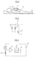
- Figure 1 is a schematic view of the apparatus of the invention connected to an elongate patient.
- FIG. 3 is a diagram of the input and signal conditioning circuits of the signal delivered by the sensor of FIG. 2.
- Figure 4 is a timing chart showing the variations over three successive cardiac cycles of the endocavitary acceleration as well as the corresponding surface electrocardiogram.
- Figure 5 illustrates the changes in the ratio of short-term average to long-term average of endocavitary acceleration during a typical apnea episode.
- reference numeral 10 denotes an elongated patient, on whom it is desired to diagnose sleep disorders.
- the patient is equipped with an external accelerometric sensor 12 placed in the region of the sternum and kept pressed against the chest wall, for example (but in a non-limiting manner) by a "patch" such as that used to hold the ECG electrodes, all possibly covered with an adhesive bandage.
- the implementation of the apparatus of the invention is particularly simple and non-invasive, insofar as it is sufficient to apply the accelerometer sensor on the patient's ribcage. And, for the latter, it does not imply any more embarrassment than wearing a Holter recorder, a technique commonly used for other types of ambulatory diagnostics.
- the sensor 12 is connected to a recording device 14, for example a Holter recorder capable of recording for a long time (at least that of a period of sleep) the signals delivered by the sensor, and to process and convert these to derive the information needed to diagnose apnea or hypopnoea.
- a recording device 14 for example a Holter recorder capable of recording for a long time (at least that of a period of sleep) the signals delivered by the sensor, and to process and convert these to derive the information needed to diagnose apnea or hypopnoea.
- the patient may also be equipped with electrodes 16 for collecting a surface ECG, also connected to the recorder 14, in particular making it possible to obtain a heart rate signal with which the information derived from the delivered signal can be crossed. by the accelerometer sensor 12.
- FIG. 2 shows in greater detail the structure of the accelerometric sensor 12, which comprises a piezoelectric sensitive element 18 biased by a resistor 20 and associated with a pre-amplification MOS transistor 22.
- the sensor 12 is biased by a voltage source 24 in series with a resistor 26.
- the output signal of the sensor is amplified by an amplifier 28 and then shaped by a bandpass filter 30 and output 32 for subsequent digitization and processing.
- FIG. 4 shows the variations of the endocardial acceleration (EA) measured by the sensor 12. illustrated in this figure the drawing of a corresponding surface electrocardiogram (ECG), during three consecutive cardiac cycles.
- EA endocardial acceleration
- ECG surface electrocardiogram
- the endocardial acceleration signal measured during a cardiac cycle forms, among other things, two peaks corresponding to the two major noises that can be recognized in each cycle of a healthy heart.
- the EP-A-0 655 260 (Sorin Biomedica Cardio SpA) describes a way of treating an endocardial acceleration signal (EA) to derive from them, in particular, these two values of endocardial acceleration peak (PEA), useful in particular for the detection of cardiac disorders and the triggering or not of a defibrillation therapy.
- peak is meant the maximum peak-to-peak value of the acceleration signal separating the two extrema, positive and negative, corresponding to the differences PEA I and PEA II indicated on the timing diagram of FIG.
- the first peak of endocardial acceleration corresponds to the closure of the mitral and tricuspid valves, at the beginning of the isovolumic ventricular contraction phase (systole).
- the variations of this first peak are closely related to the variations of the pressure in the ventricle (the amplitude of the PEA I peak being, more precisely, correlated with the positive maximum of the pressure variation dP / dt in the left ventricle) and can therefore constitute a representative parameter of myocardial contractility, itself related to the level of activity of the sympathetic system.
- the second peak of endocardial acceleration corresponds to the closure of the aortic and pulmonary valves, at the time of the isovolumic ventricular relaxation phase.
- This second peak which is produced by the abrupt deceleration of the moving blood mass in the aorta, is a representative parameter of the peripheral blood pressure at the beginning of the diastole. It is also a key parameter in the physiological process leading to the onset of vasovagal syncope.
- PEA I and / or PEA II peaks are recorded during successive cycles, as well as possibly the heart rate.
- a first technique is to determine, cycle to cycle, the absolute values that these parameters take and to set triggering thresholds alarm - or, preferably, determine an averaged value of these parameters over a predetermined number of cycles, to avoid influences of cycle-to-cycle variability (dispersion of measurements) and those of insignificant brief events.
- one or more thresholds are set, and each of the parameters PEA I or PEA II (or a combination of these two parameters) is compared to a predetermined threshold.
- the result of the comparison can be combined in various ways with the result of similar comparisons of other parameters (including heart rate) to produce a two-state output signal, one of the states being associated with a normal situation and the other another state being associated with an apnea or hypopnea alert.
- This alarm can be triggered immediately when crossing the threshold, or conditionally, for example only if the ratio R goes below the threshold for a predetermined minimum duration (for example 5 seconds) or a predetermined number of cycles (for example 5 cycles).
- Activation of the alarm thus indicates an episode of sudden depression of cardiac contractility, which is associated with an episode of apnea or hypopnea.
- This triggers, in response, the entry in a memory of the recorder 14 of diagnostic information such as apnea occurrence marker, timestamp of this apnea, duration of the alarm, etc.
- diagnostic information such as apnea occurrence marker, timestamp of this apnea, duration of the alarm, etc.
- This memorization can possibly be triggered according to certain programmed conditions or on detection of certain states (confirmed sleep) or events (occurrence of an apneic episode).
- the stored signals may later be viewed by a practitioner equipped with a programmer for reading the contents of the memory of the implant.
- It will also be possible at this stage to analyze the PEA signal to to extract some parameters that are not
- the detection of such a typical PEA variation profile can be used to calculate a specific index, making it possible to diagnose in the subject the presence or absence of instability of the sympathico-vagal system.
- PEA II can be used as an indicator of reduced contractility, as it correlates with changes in blood pressure. More specifically, a reduced contractility combined with a slowing of the heart rate causes a decrease in blood pressure, which results in a reduction in the amplitude of PEA II. The analysis of this parameter thus makes it possible to confirm the episodes of apnea.
- analyzes more complex, can also be implemented to further improve the quality of the detection method, for example correlation techniques, signal morphology analysis, frequency analysis, analysis wavelet, principal component analysis, etc.
- the system is self-adaptive, that is to say that it can adapt to variations in the long term, in order to further improve the specificity of the detection system.
- PEA plasma e.g., a nasal pressure signal
- a pressure sensor for example collected by means of a pressure sensor via a nasal cannula, or a signal from a patient. oxygen saturation sensor.
Abstract
Description
L'invention concerne un appareillage pour la détection non invasive de la survenue d'apnées ou d'hypopnées chez un patient.The invention relates to an apparatus for the noninvasive detection of the occurrence of apneas or hypopneas in a patient.
De façon générale, la pathologie respiratoire connue sous le nom de "syndrome d'apnée du sommeil" (SAS) est caractérisée par la survenue fréquente (au moins 10 à 20 fois par heure) d'apnées pendant une phase de sommeil du patient, une "apnée" (ou pause respiratoire) étant définie comme un arrêt temporaire de la fonction respiratoire de durée supérieure à 10 secondes. Elle peut également être caractérisée par la survenue d'hypopnées dans les mêmes conditions, une "hypopnée" étant définie comme une décroissance importante (mais sans interruption) du débit respiratoire, typiquement une décroissance de plus de 50 % par rapport à une moyenne de référence antérieure.In general, the respiratory pathology known as "sleep apnea syndrome" (SAS) is characterized by the frequent occurrence (at least 10 to 20 times per hour) of apnea during a sleep phase of the patient, an "apnea" (or breathing pause) being defined as a temporary stopping of the respiratory function lasting longer than 10 seconds. It can also be characterized by the occurrence of hypopneas under the same conditions, a "hypopnea" being defined as a significant (but uninterrupted) decrease in respiratory rate, typically a decrease of more than 50% compared to a reference average. earlier.
Face à cette pathologie, qui atteint plus de 4% de la population et plus de 50% des patients souffrant d'insuffisance cardiaque, le système nerveux autonome s'adapte mais avec un effet délétère sur le sommeil, l'interruption ou la réduction du débit respiratoire entraînant une diminution de la concentration en oxygène du sang ainsi que des micro-réveils inconscients. Il s'ensuit en phase d'éveil une somnolence diurne avec perte d'attention et un accroissement des risques d'accident de la route. Par ailleurs, la réponse adaptative physiologique, puis pathologique, de certains organes dont le coeur et l'appareil circulatoire entraîne une plus grande incidence de troubles tels qu'hypertension artérielle, arythmies ventriculaires, infarctus myocardique et insuffisance cardiaque.Faced with this pathology, which affects more than 4% of the population and more than 50% of patients suffering from heart failure, the autonomic nervous system adapts but with a deleterious effect on sleep, interruption or reduction of respiratory rate leading to a decrease in blood oxygen concentration and unconscious micro-awakenings. It follows in the wake phase of daytime sleepiness with loss of attention and an increased risk of road accident. Moreover, the adaptive physiological and then pathological response of certain organs including the heart and the circulatory system leads to a greater incidence of disorders such as arterial hypertension, ventricular arrhythmias, myocardial infarction and cardiac insufficiency.
On connaît diverses techniques pour détecter de tels troubles respiratoires du sommeil.Various techniques are known for detecting such respiratory sleep disorders.
Ainsi, le
Un inconvénient de cette technique tient au fait qu'elle présuppose que le patient soit appareillé d'un implant, ce qui en limite très strictement son utilité et son efficacité.A disadvantage of this technique is that it presupposes that the patient is fitted with an implant, which strictly limits its usefulness and effectiveness.
La présente invention propose une autre approche du diagnostic des pathologies de l'activité respiratoire, au moyen d'un appareillage externe, donc non invasif, qui puisse être mis en place aisément sur le patient, éventuellement par le patient lui-même, et qui n'introduise aucune gêne comparable à celle de certains appareils mettant en oeuvre des masques respiratoires devant être portés pendant le sommeil.The present invention proposes another approach to the diagnosis of pathologies of respiratory activity, by means of an external apparatus, therefore non-invasive, which can be placed easily on the patient, possibly by the patient himself, and which introduces no discomfort comparable to that of some devices using respiratory masks to be worn during sleep.
Un des buts de la présente invention est de mettre à la disposition des praticiens un tel appareillage qui, d'une part, soit simple à mettre en oeuvre et non invasif (et donc applicable à tout patient, qu'il soit ou non muni d'un implant) et qui, d'autre part, délivre au praticien non seulement des données brutes, mais également une aide au diagnostic, notamment à partir des critères ayant abouti au diagnostic des apnées ou hypopnées.One of the aims of the present invention is to provide practitioners with such an apparatus which, on the one hand, is simple to use and non-invasive (and therefore applicable to any patient, whether or not provided with an implant) and which, on the other hand, delivers to the practitioner not only raw data, but also an aid to diagnosis, in particular from the criteria that led to the diagnosis of apnea or hypopnea.
Essentiellement, l'invention propose une détection des apnées ou hypopnées à partir d'une mesure de l'accélération endocardiaque, plus précisément basée sur l'analyse des pics d'accélération endocardiaque, qui est un paramètre reflétant de façon non artefactée et avec un très faible temps de réponse les variations de la contractilité du myocarde.Essentially, the invention provides detection of apneas or hypopneas from a measurement of endocardial acceleration, more specifically based on the analysis of endocardial acceleration peaks, which is a non-artifactally reflecting parameter and with a very low response time changes in contractility of the myocardium.
Afin d'obtenir une meilleure détection des évènements respiratoires pathologiques, ce signal d'accélération endocardiaque peut être analysé de manière croisée ― mais de façon subsidiaire ― avec d'autres signaux physiologiques tels que la fréquence cardiaque.In order to obtain better detection of pathological respiratory events, this endocardial acceleration signal can be cross-analyzed - but in a subsidiary manner - with other physiological signals such as heart rate.
On connaît, par exemple d'après le
Mais, en premier lieu, il s'agit d'un implant de type stimulateur cardiaque, qui ne peut donc être mis en oeuvre que dans des situations particulières et pour une population de patients limitée.But, first, it is a pacemaker-type implant, which can therefore be implemented only in special situations and for a limited patient population.
D'autre part, le dispositif décrit par ce document analyse l'activité ventilatoire et détecte la survenue d'apnées ou d'hypopnées par analyse du signal délivré par un capteur de ventilation-minute opérant par mesure d'impédance intrathoracique. L'accélération endocardiaque n'y est pas utilisée pour le diagnostic des apnées ou hypopnées, mais seulement pour adapter une thérapie (modulation de la fréquence de stimulation) appliquée au patient par le dispositif, après qu'une apnée ou hypopnée a été détectée.On the other hand, the device described by this document analyzes the ventilatory activity and detects the occurrence of apneas or hypopneas by analysis of the signal delivered by a ventilation-minute sensor operating by intrathoracic impedance measurement. The endocardial acceleration is not used for the diagnosis of apnea or hypopnea, but only to adapt a therapy (modulation of the stimulation frequency) applied to the patient by the device, after an apnea or hypopnea has been detected.
Pour permettre la détection non invasive de la survenue d'apnées ou d'hypopnées, l'appareil selon l'invention comporte des moyens de recueil de l'accélération endocardiaque du patient, comprenant un capteur accélérométrique externe apte à être maintenu en contact avec la paroi thoracique du patient, et des moyens d'analyse, aptes à déterminer au moins un paramètre fonction de ladite accélération endocardiaque recueillie et délivrer conditionnellement un signal d'alerte d'apnée ou d'hypopnée en fonction de la valeur prise par ledit au moins un paramètre.To allow the non-invasive detection of the occurrence of apneas or hypopneas, the apparatus according to the invention comprises means for collecting the endocardial acceleration of the patient, comprising an accelerometric sensor. external device able to be kept in contact with the patient's chest wall, and analysis means, able to determine at least one parameter that is a function of said collected endocardial acceleration and conditionally deliver an apnea or hypopnea alert signal in function of the value taken by said at least one parameter.
Ledit paramètre est avantageusement un paramètre fonction de l'un et/ou de l'autre de deux pics de l'accélération endocardiaque au cours d'un cycle donné, ces deux pics comprenant un premier pic lors de la phase de contraction ventriculaire isovolumique et un second pic lors de la phase de relaxation ventriculaire isovolumique. Le paramètre peut être notamment fonction (i) d'une moyenne, et/ou (ii) de la variation, et/ou (iii) d'une différence ou d'un ratio entre une moyenne à long terme et une moyenne à court terme, des valeurs de pic(s) d'accélération endocardiaque relevées au cours d'une pluralité de cycles successifs. En variante, il peut aussi être fonction de l'intervalle entre complexe QRS et au moins l'un des pics d'accélération endocardiaque, et/ou de l'intervalle entre le premier et le second pic d'accélération endocardiaque.Said parameter is advantageously a parameter which is a function of one and / or the other of two peaks of the endocardial acceleration during a given cycle, these two peaks comprising a first peak during the isovolumic ventricular contraction phase and a second peak during the isovolumic ventricular relaxation phase. The parameter may be, in particular, a function of (i) an average, and / or (ii) the variation, and / or (iii) a difference or ratio between a long-term average and a short-term average. term, endocardial acceleration peak (s) values recorded during a plurality of successive cycles. Alternatively, it may also be a function of the interval between QRS complex and at least one of the endocardial acceleration peaks, and / or the interval between the first and second endocardial acceleration peak.
Les moyens d'analyse peuvent comprendre des moyens aptes à comparer le(s) paramètre(s) à un seuil prédéterminé et délivrer le signal d'alerte au franchissement de ce seuil.The analysis means may comprise means capable of comparing the parameter (s) with a predetermined threshold and delivering the warning signal at the crossing of this threshold.
Dans un mode de mise en oeuvre particulier de l'invention, le paramètre est fonction d'un ratio entre une moyenne à long terme et une moyenne à court terme des valeurs de pic(s) d'accélération endocardiaque relevées au cours d'une pluralité de cycles successifs, et les moyens d'analyse comprennent des moyens pour comparer ce ratio à un premier seuil prédéterminé et délivrer le signal d'alerte au franchissement de ce seuil.In a particular embodiment of the invention, the parameter is a function of a ratio between a long-term average and a short-term average of the endocardial acceleration peak (s) values recorded during a plurality of successive cycles, and the analysis means comprise means for comparing this ratio with a first predetermined threshold and delivering the warning signal at the crossing of this threshold.
De préférence, le signal d'alerte peut être produit après franchissement du premier seuil seulement si ce premier seuil reste franchi pendant une durée minimale prédéterminée, ou pendant un nombre minimal prédéterminé de cycles cardiaques.Preferably, the warning signal can be produced after crossing the first threshold only if this first threshold remains crossed for a predetermined minimum duration, or for a predetermined minimum number of cardiac cycles.
Par ailleurs, les moyens d'analyse peuvent en outre comprendre des moyens aptes, après franchissement dudit premier seuil, à détecter un franchissement inverse de ce premier seuil suivi du franchissement d'un second seuil, supérieur au premier, et à délivrer un signal de confirmation d'épisode d'apnée ou d'hypopnée au franchissement de ce second seuil.Moreover, the analysis means may further comprise means capable, after crossing said first threshold, to detect an inverse crossing of this first threshold followed by the crossing of a second threshold, greater than the first, and to deliver a signal of confirmation of apnea or hypopnea episode when crossing this second threshold.
Ce signal de confirmation est de préférence délivré seulement si le franchissement du second seuil intervient au cours d'une durée maximale prédéterminée, ou pendant un nombre maximal prédéterminé de cycles cardiaques, après le franchissement inverse du premier seuil.This confirmation signal is preferably issued only if the crossing of the second threshold occurs during a predetermined maximum duration, or during a predetermined maximum number of cardiac cycles, after the reverse crossing of the first threshold.
En variante ou en complément, les moyens d'analyse peuvent comprendre une machine à états ou réseau de neurones apte à comparer une pluralité des paramètres à une pluralité de seuils prédéterminés, à détecter le franchissement des différents seuils, à analyser la séquence de ces franchissements et à délivrer ledit signal d'alerte sur détection d'une ou plusieurs séquence(s) de franchissement prédéterminées.Alternatively or in addition, the analysis means may comprise a state machine or neural network capable of comparing a plurality of parameters to a plurality of predetermined thresholds, to detect the crossing of the different thresholds, to analyze the sequence of these crossings. and delivering said alert signal upon detection of one or more predetermined crossing sequences.
Des moyens peuvent en outre être prévus pour appliquer à l'accélération endocardiaque un traitement d'autocorrélation, d'analyse morphologique, d'analyse fréquentielle, d'analyse par ondelettes et/ou d'analyse en composantes principales.Means may further be provided for applying autocorrelation processing, morphological analysis, frequency analysis, wavelet analysis and / or principal component analysis to endocardial acceleration.
Avantageusement, l'appareillage comprend en outre des moyens de recueil d'au moins une donnée physiologique parmi : la fréquence cardiaque, la pression nasale et/ou la saturation sanguine en oxygène du patient, et les moyens d'analyse sont aptes à délivrer conditionnellement le signal d'alerte en fonction à la fois de ladite au moins une donnée physiologique et de la valeur prise par ledit au moins un paramètre.Advantageously, the apparatus further comprises means for collecting at least one physiological data item from: the heart rate, the nasal pressure and / or the oxygen saturation of the patient, and the analysis means are able to deliver conditionally the alert signal according to both of said at least one physiological data and the value taken by said at least one parameter.
On va maintenant décrire un exemple de réalisation de l'invention, en référence aux dessins annexés.An embodiment of the invention will now be described with reference to the accompanying drawings.
La figure 1 est une vue schématique de l'appareillage de l'invention, relié à un patient allongé.Figure 1 is a schematic view of the apparatus of the invention connected to an elongate patient.
La figure 2 précise la structure interne du capteur accélérométrique utilisé. La figure 3 est un schéma des circuits d'entrée et de mise en forme du signal délivré par le capteur de la figure 2.Figure 2 specifies the internal structure of the accelerometer sensor used. FIG. 3 is a diagram of the input and signal conditioning circuits of the signal delivered by the sensor of FIG. 2.
La figure 4 est un chronogramme montrant les variations au cours de trois cycles cardiaques successifs de l'accélération endocavitaire ainsi que de l'électrocardiogramme de surface correspondant.Figure 4 is a timing chart showing the variations over three successive cardiac cycles of the endocavitary acceleration as well as the corresponding surface electrocardiogram.
La figure 5 illustre les variations du rapport entre moyenne à court terme et moyenne à long terme de l'accélération endocavitaire au cours d'un épisode d'apnée typique.Figure 5 illustrates the changes in the ratio of short-term average to long-term average of endocavitary acceleration during a typical apnea episode.
Sur la figure 1, la référence 10 désigne un patient allongé, sur lequel on souhaite diagnostiquer les troubles du sommeil. De façon caractéristique de l'invention, le patient est équipé d'un capteur accélérométrique externe 12 placé dans la région du sternum et maintenu plaqué contre la paroi thoracique, par exemple (mais de manière non limitative) par un "patch" tel que celui utilisé pour le maintien des électrodes ECG, le tout étant éventuellement recouvert d'un bandage adhésif. Comme on peut le voir, la mise en oeuvre de l'appareillage de l'invention est particulièrement simple et non invasive, dans la mesure où il suffit d'appliquer le capteur accélérométrique sur la cage thoracique du patient. Et, pour ce dernier, elle n'implique pas d'autre gêne que le port d'un enregistreur Holter, technique couramment utilisée pour d'autres types de diagnostics ambulatoire.In Figure 1,
Le capteur 12 est relié à un dispositif enregistreur 14, par exemple un enregistreur Holter capable d'enregistrer sur une longue durée (au moins celle d'une période de sommeil) les signaux délivrés par le capteur, et de traiter et convertir ceux-ci pour en dériver les informations nécessaires au diagnostic des apnées ou hypopnées.The
Le patient peut être également équipé d'électrodes 16 de recueil d'un ECG de surface, également reliées à l'enregistreur 14, permettant en particulier d'obtenir un signal de fréquence cardiaque avec lequel pourra être croisée l'information dérivée du signal délivré par le capteur accélérométrique 12.The patient may also be equipped with
La figure 2 montre plus en détail la structure du capteur accélérométrique 12, qui comporte un élément sensible piézoélectrique 18 polarisé par une résistance 20 et associé à un transistor MOS 22 de préamplification. Comme on peut le voir sur la figure 3, le capteur 12 est polarisé par une source de tension 24 en série avec une résistance 26. Le signal de sortie du capteur est amplifié par un amplificateur 28 puis mis en forme par un filtre passe-bande 30 et délivré à la sortie 32 pour numérisation et traitement ultérieurs.FIG. 2 shows in greater detail the structure of the
Sur la figure 4 on a représenté (courbe du haut), les variations de l'accélération endocardiaque (EA) mesurée par le capteur 12. On a également illustré sur cette figure le tracé d'un électrocardiogramme de surface (ECG) correspondant, au cours de trois cycles cardiaques consécutifs.FIG. 4 (top curve) shows the variations of the endocardial acceleration (EA) measured by the
Le signal d'accélération endocardiaque mesuré au cours d'un cycle cardiaque forme, entre autres, deux pics correspondant aux deux bruits majeurs qu'il est possible de reconnaître dans chaque cycle d'un coeur sain. Le
Par "pic" on entendra la valeur maximale crête-à-crête du signal d'accélération séparant les deux extrema, positif et négatif, correspondant aux écarts PEA I et PEA II indiqués sur le chronogramme de la figure 4.By "peak" is meant the maximum peak-to-peak value of the acceleration signal separating the two extrema, positive and negative, corresponding to the differences PEA I and PEA II indicated on the timing diagram of FIG.
Plus précisément, le premier pic d'accélération endocardiaque ("PEA I") correspond à la fermeture des valvules mitrale et tricuspide, au début de la phase de contraction ventriculaire isovolumique (systole). Les variations de ce premier pic sont étroitement liés aux variations de la pression dans le ventricule (l'amplitude du pic PEA I étant, plus précisément, corrélée au maximum positif de la variation de pression dP/dt dans le ventricule gauche) et peuvent donc constituer un paramètre représentatif de la contractilité du myocarde, elle-même liée au niveau d'activité du système sympathique.More specifically, the first peak of endocardial acceleration ("PEA I") corresponds to the closure of the mitral and tricuspid valves, at the beginning of the isovolumic ventricular contraction phase (systole). The variations of this first peak are closely related to the variations of the pressure in the ventricle (the amplitude of the PEA I peak being, more precisely, correlated with the positive maximum of the pressure variation dP / dt in the left ventricle) and can therefore constitute a representative parameter of myocardial contractility, itself related to the level of activity of the sympathetic system.
Le second pic d'accélération endocardiaque ("PEA II"), quant à lui, correspond à la fermeture des valvules aortique et pulmonaire, au moment de la phase de relaxation ventriculaire isovolumique. Ce second pic, qui est produit par la décélération brusque de la masse sanguine en mouvement dans l'aorte, constitue un paramètre représentatif de la pression sanguine périphérique au début de la diastole. Il constitue également un paramètre-clé du processus physiologique conduisant à la survenue d'une syncope vasovagale.The second peak of endocardial acceleration ("PEA II"), meanwhile, corresponds to the closure of the aortic and pulmonary valves, at the time of the isovolumic ventricular relaxation phase. This second peak, which is produced by the abrupt deceleration of the moving blood mass in the aorta, is a representative parameter of the peripheral blood pressure at the beginning of the diastole. It is also a key parameter in the physiological process leading to the onset of vasovagal syncope.
Les valeurs de pics PEA I et/ou PEA II sont relevées au cours de cycles successifs, ainsi qu'éventuellement la fréquence cardiaque.The values of PEA I and / or PEA II peaks are recorded during successive cycles, as well as possibly the heart rate.
Ces signaux peuvent être traités de diverses manières.These signals can be processed in a variety of ways.
Une première technique consiste à déterminer, cycle à cycle, les valeurs absolues que prennent ces paramètres et à fixer des seuils de déclenchement d'alarme ― ou, de préférence, déterminer une valeur moyennée de ces paramètres sur un nombre prédéterminé de cycles, pour éviter les influences de la variabilité cycle-à-cycle (dispersion des mesures) et celles des évènements brefs non significatifs.A first technique is to determine, cycle to cycle, the absolute values that these parameters take and to set triggering thresholds alarm - or, preferably, determine an averaged value of these parameters over a predetermined number of cycles, to avoid influences of cycle-to-cycle variability (dispersion of measurements) and those of insignificant brief events.
Pour diagnostiquer la présence ou non d'une apnée ou hypopnée, un ou plusieurs seuils sont fixés, et chacun des paramètres PEA I ou PEA II (ou une combinaison de ces deux paramètres) est comparé à un seuil prédéterminé. Le résultat de la comparaison peut être combiné de diverses manières avec le résultat de comparaisons semblables d'autres paramètres (notamment la fréquence cardiaque) pour produire un signal de sortie à deux états, l'un des états étant associé à une situation normale et l'autre état étant associé à une alerte d'apnée ou hypopnée.To diagnose the presence or absence of an apnea or hypopnea, one or more thresholds are set, and each of the parameters PEA I or PEA II (or a combination of these two parameters) is compared to a predetermined threshold. The result of the comparison can be combined in various ways with the result of similar comparisons of other parameters (including heart rate) to produce a two-state output signal, one of the states being associated with a normal situation and the other another state being associated with an apnea or hypopnea alert.
Pour améliorer la spécificité de la détection, et notamment pour tenir compte des différences de valeur de base des paramètres PEA d'un individu à l'autre, il peut être avantageux d'analyser les variations de ces paramètres plutôt que leurs valeurs absolues.To improve the specificity of the detection, and in particular to take into account the differences in basic value of the PEA parameters from one individual to another, it may be advantageous to analyze the variations of these parameters rather than their absolute values.
Une manière de procéder consiste à analyser la différence entre une moyenne à court terme et une moyenne à long terme du même paramètre. Si ce paramètre varie peu, la différence sera faible et les deux valeurs finiront par coïncider. En revanche, dès que le paramètre deviendra instable, la moyenne à court terme va suivre les variations du paramètre plus rapidement que la moyenne à long terme. La différence entre les deux moyennes ne sera plus nulle ou quasi-nulle, mais prendra une valeur positive (en cas d'augmentation du paramètre) ou négative (en cas de diminution), la valeur absolue de cet écart dépendant du paramètre analysé et de la vitesse de variation de celui-ci.One way to do this is to analyze the difference between a short-term average and a long-term average of the same parameter. If this parameter varies little, the difference will be small and the two values will eventually coincide. On the other hand, as soon as the parameter becomes unstable, the short-term average will follow the changes in the parameter faster than the long-term average. The difference between the two means will no longer be zero or almost zero, but will take a positive value (in case of a parameter increase) or a negative value (in case of a decrease), the absolute value of this difference depends on the parameter analyzed and the speed of variation of it.
Il est également possible de suivre non pas la différence, mais le ratio entre moyenne à court terme et moyenne à long terme des paramètres PEA I et/ou PEA II.It is also possible to track not the difference, but the ratio between the short-term average and the long-term average of the PEA I and / or PEA II parameters.
Un exemple d'algorithme de détection consiste à calculer une moyenne mobile à court terme des valeurs cycle à cycle du paramètre PEA I, par exemple sur quatre cycles consécutifs. Simultanément, une moyenne mobile à long terme, par exemple sur 500 cycles, de ce même paramètre est calculée et mise à jour. Les deux moyennes sont alors comparées pour donner un ratio R, où R = moyenne à court terme/moyenne à long terme.An example of a detection algorithm consists in calculating a short-term moving average of the cycle-to-cycle values of parameter PEA I, for example over four consecutive cycles. Simultaneously, a long-term moving average, for example over 500 cycles, of this same parameter is calculated and updated. The two averages are then compared to give a ratio R, where R = average short term / average long term.
Les variations de ce ratio sont illustrées sur la figure 5 : en régime stationnaire, le ratio R présente une valeur à peu près constante et voisine de l'unité ; en revanche, lors d'un épisode d'apnée, la diminution de la saturation en oxygène dans le sang induit une réduction de la contractilité cardiaque qui se traduit par une diminution correspondante de l'amplitude du paramètre PEA I pendant plusieurs battements consécutifs, et donc une diminution corrélative de la valeur de R. Cette situation est illustrée en A sur la figure 5, où est indiqué le début de l'épisode d'apnée, et en B pour le moment où l'accélération endocardiaque chute brutalement suite à la réduction de la contractilité.The variations of this ratio are illustrated in Figure 5: in steady state, the ratio R has a value that is approximately constant and close to unity; on the other hand, during an apnea episode, the decrease in oxygen saturation in the blood induces a reduction in cardiac contractility which results in a corresponding decrease in the amplitude of the PEA I parameter during several consecutive beats, and therefore a correlative decrease in the value of R. This situation is illustrated in A in Figure 5, where is indicated the beginning of the apnea episode, and in B for the moment when the endocardial acceleration drops sharply following the reduction of contractility.
Il est possible de déclencher une alarme en définissant pour le ratio R un seuil approprié S, par exemple S = 0,85 ou S = 0,9, et en comparant la valeur courante du ratio R à ce seuil. Chaque fois que la ratio R tombe en dessous de ce seuil (situation illustrée en C sur la figure 5), une alarme (ALM) est déclenchée.It is possible to trigger an alarm by defining for the ratio R an appropriate threshold S, for example S = 0.85 or S = 0.9, and comparing the current value of the ratio R to this threshold. Whenever the ratio R falls below this threshold (situation illustrated in C in Figure 5), an alarm (ALM) is triggered.
Cette alarme peut être déclenchée immédiatement au moment du franchissement du seuil, ou bien de manière conditionnelle, par exemple seulement si le ratio R descend sous le seuil pendant une durée minimale prédéterminée (par exemple 5 secondes) ou un nombre de cycles prédéterminé (par exemple 5 cycles).This alarm can be triggered immediately when crossing the threshold, or conditionally, for example only if the ratio R goes below the threshold for a predetermined minimum duration (for example 5 seconds) or a predetermined number of cycles (for example 5 cycles).
L'activation de l'alarme indique ainsi un épisode de dépression soudaine de la contractilité cardiaque, qui est associé à un épisode d'apnée ou hypopnée. Ceci déclenche en réponse l'inscription dans une mémoire de l'enregistreur 14d'informations de diagnostic telles que marqueur de survenue d'une apnée, horodatage de cette apnée, durée de l'alarme, ... Il est également possible d'y enregistrer simultanément les signaux d'accélération endocardiaque et/ou les valeurs des différents paramètres PEA. Cette mémorisation peut être éventuellement déclenchée selon certaines conditions programmées ou sur détection de certains états (sommeil confirmé) ou évènements (survenue d'un épisode apnéique). Les signaux mémorisés pourront être ultérieurement visualisés par un praticien équipé d'un programmateur permettant de lire le contenu de la mémoire de l'implant. II sera également possible, à ce stade, d'analyser le signal PEA afin d'en extraire certains paramètres qui ne sont pas déterminables en temps réel, tels que la variabilité sinusale, ou pour simuler l'application d'algorithmes de détection afin de choisir les plus approprié au cas du patient.Activation of the alarm thus indicates an episode of sudden depression of cardiac contractility, which is associated with an episode of apnea or hypopnea. This triggers, in response, the entry in a memory of the
Par ailleurs on observe généralement après la fin de l'épisode d'apnée un phénomène de rebond, correspondant à une augmentation transitoire de la contractilité cardiaque (ce rebond est illustré en E sur la figure 2). Plus précisément, à la fin d'un épisode d'apnée survient souvent un micro-réveil au cours duquel le système sympathique est activé en réaction aux évènements antérieurs ; cette réaction prend fin spontanément après un certain nombre de cycles cardiaques, le ratio R retournant progressivement à sa valeur de base voisine de l'unité.Furthermore, after the end of the apnea episode, a rebound phenomenon is generally observed, corresponding to a transient increase in cardiac contractility (this rebound is illustrated in E in FIG. 2). More specifically, at the end of an episode of apnea often occurs a micro-alarm during which the sympathetic system is activated in response to previous events; this reaction ends spontaneously after a certain number of cardiac cycles, the ratio R gradually returning to its base value close to unity.
Ce phénomène de rebond peut être détecté et utilisé pour confirmer la survenue de l'épisode d'apnée. À cet effet, on définit un second seuil S' de valeur supérieure à l'unité, par exemple S' = 1,1 et on détecte le franchissement de ce second seuil S' après détection d'un épisode d'apnée. On notera que le franchissement du seuil S' révèle seulement une réaction positive du système sympathico-vagal à l'état anormal induit par l'apnée, réaction qui n'est pas en soi pathologique et ne doit pas déclencher d'alarme et ni de thérapie particulière.This rebound phenomenon can be detected and used to confirm the occurrence of the apnea episode. For this purpose, a second threshold S 'of value greater than unity is defined, for example S' = 1.1 and the crossing of this second threshold S 'is detected after detection of an apnea episode. It will be noted that crossing the threshold S 'reveals only a positive reaction of the sympathico-vagal system to the abnormal state induced by apnea, a reaction which is not inherently pathological and must not trigger an alarm and neither special therapy.
En revanche, la détection conjointe (i) d'une diminution du PEA révélant une réduction de la contractilité, détectée par le franchissement du premier seuil S (ratio R < 0,85), suivie (ii) d'une augmentation du PEA, détectée par le franchissement du second seuil (ratio R > 1,1), et ce (iii) dans un intervalle de temps prédéterminé, par exemple dans un laps de temps d'une minute, est un indice fort d'un état non physiologique rencontré typiquement chez des patients souffrant de troubles du sommeil.On the other hand, joint detection of (i) a decrease in PEA revealing a reduction in contractility, detected by crossing the first threshold S (ratio R <0.85), followed (ii) by an increase in PEA, detected by crossing the second threshold (ratio R> 1.1), and this (iii) in a predetermined time interval, for example in a period of time of one minute, is a strong index of a non-physiological state typically encountered in patients with sleep disorders.
La détection d'un tel profil typique de variation du PEA peut être utilisée pour calculer un indice spécifique, permettant de diagnostiquer chez le sujet la présence ou non d'une instabilité du système sympathico-vagal.The detection of such a typical PEA variation profile can be used to calculate a specific index, making it possible to diagnose in the subject the presence or absence of instability of the sympathico-vagal system.
En variante ou en complément du paramètre PEA I, il est possible de prendre en compte le PEA II comme dans l'exemple qui précède. Le PEA II peut être utilisé comme indicateur d'une contractilité réduite, dans la mesure où il est corrélé aux variations de la pression sanguine. Plus précisément, une contractilité réduite combinée à un ralentissement du rythme cardiaque provoque une diminution de la pression sanguine, qui se traduit par une réduction de l'amplitude du PEA II. L'analyse de ce paramètre permet ainsi de confirmer les épisodes d'apnée.Alternatively or in addition to the parameter PEA I, it is possible to take into account the PEA II as in the preceding example. PEA II can be used as an indicator of reduced contractility, as it correlates with changes in blood pressure. More specifically, a reduced contractility combined with a slowing of the heart rate causes a decrease in blood pressure, which results in a reduction in the amplitude of PEA II. The analysis of this parameter thus makes it possible to confirm the episodes of apnea.
D'autres types d'analyses, plus complexes, peuvent également être mises en oeuvre pour améliorer encore la qualité du procédé de détection, par exemple des techniques de corrélation, d'analyse de morphologie du signal, d'analyse fréquentielle, d'analyse par ondelettes, d'analyse en composantes principales, etc.Other types of analyzes, more complex, can also be implemented to further improve the quality of the detection method, for example correlation techniques, signal morphology analysis, frequency analysis, analysis wavelet, principal component analysis, etc.
Il est également possible d'utiliser un processus de type "machine à états", où les résultats des comparaisons aux divers seuils sont appliqués à un système à transition d'états et à mémoire, qui prend la décision de déclencher une alerte d'apnée ou hypopnée en fonction d'un schéma d'évolution plus complexe.It is also possible to use a "state machine" type process, where the results of comparisons at various thresholds are applied to a state transition and memory system, which makes the decision to trigger an apnea alert. or hypopnea according to a more complex evolution pattern.
On peut en outre prévoir que le système soit auto-adaptatif, c'est-à-dire qu'il puisse s'adapter à des variations sur le long terme, afin d'améliorer ultérieurement la spécificité du système de détection.It can further be expected that the system is self-adaptive, that is to say that it can adapt to variations in the long term, in order to further improve the specificity of the detection system.
Enfin, pour améliorer la sensibilité et la spécificité de la détection des épisodes d'apnée ou d'hypopnée, il est possible de combiner l'analyse du PEA à une analyse parallèle de la fréquence cardiaque. En effet, cette dernière décroît au début de l'évènement respiratoire et augmente au moment du micro-réveil qui suit l'évènement, ou bien présente une variabilité caractéristique pendant la survenue de l'évènement.Finally, to improve the sensitivity and specificity of detection of apnea or hypopnea episodes, it is possible to combine the analysis of PEA with a parallel analysis of the heart rate. Indeed, the latter decreases at the beginning of the respiratory event and increases at the time of the micro-wake following the event, or has a characteristic variability during the occurrence of the event.
Pour améliorer le diagnostic, il est également possible de combiner l'analyse du PEA à une analyse d'un signal de pression nasale, par exemple recueilli grâce à un capteur de pression via une canule nasale, ou encore d'un signal d'un capteur de saturation sanguine en oxygène.To improve the diagnosis, it is also possible to combine the analysis of PEA with an analysis of a nasal pressure signal, for example collected by means of a pressure sensor via a nasal cannula, or a signal from a patient. oxygen saturation sensor.
Claims (12)
ou d'hypopnées chez un patient, caractérisé en ce qu'il comporte :
or hypopneas in a patient, characterized in that it comprises:
des valeurs de pic(s) d'accélération endocardiaque relevées au cours d'une pluralité de cycles successifs.
endocardial acceleration peak (s) values recorded during a plurality of successive cycles.
Applications Claiming Priority (2)
| Application Number | Priority Date | Filing Date | Title |
|---|---|---|---|
| FR0507121A FR2888121A1 (en) | 2005-07-05 | 2005-07-05 | Active implantable medical device e.g. pacemaker, for diagnosing e.g. sleep apnea syndrome, has sensor measuring endocardiac acceleration whose parameters are determined and provide value for delivering alert signal of apnea or hypopnoea |
| FR0603484A FR2888123A1 (en) | 2005-07-05 | 2006-04-20 | APPARATUS FOR THE NON-INVASIVE DETECTION OF APPEARANCE OR HYPNOPES IN A PATIENT |
Publications (2)
| Publication Number | Publication Date |
|---|---|
| EP1741387A1 true EP1741387A1 (en) | 2007-01-10 |
| EP1741387B1 EP1741387B1 (en) | 2009-06-03 |
Family
ID=36968505
Family Applications (1)
| Application Number | Title | Priority Date | Filing Date |
|---|---|---|---|
| EP06291002A Active EP1741387B1 (en) | 2005-07-05 | 2006-06-21 | Device for non invasive detection of occurence of sleep apnea or hypopnea |
Country Status (5)
| Country | Link |
|---|---|
| US (1) | US7613507B2 (en) |
| EP (1) | EP1741387B1 (en) |
| AT (1) | ATE432651T1 (en) |
| DE (1) | DE602006007063D1 (en) |
| FR (1) | FR2888123A1 (en) |
Cited By (8)
| Publication number | Priority date | Publication date | Assignee | Title |
|---|---|---|---|---|
| EP2092885A1 (en) | 2008-02-20 | 2009-08-26 | Ela Medical | Device for analysing an endocardiac acceleration signal |
| EP2353641A1 (en) | 2010-02-09 | 2011-08-10 | Sorin CRM SAS | Active implantable medical device for cardiac resynchronisation with automatic, real-time optimisation of the interventricular and atrioventricular delays |
| EP2408353A4 (en) * | 2009-03-17 | 2015-02-25 | Advanced Brain Monitoring Inc | A system for the assessment of sleep quality in adults and children |
| EP2839859A1 (en) | 2013-08-20 | 2015-02-25 | Sorin CRM SAS | Active medical device, in particular a CRT resynchroniser, including predictive warning means for cardiac decompensation in the presence of central sleep apnoea |
| EP2904969A1 (en) * | 2014-02-11 | 2015-08-12 | Sorin CRM SAS | Device for treating the sleep apnea syndrome in a patient by kinaesthetic stimulation |
| EP2904964A1 (en) | 2014-02-11 | 2015-08-12 | Sorin CRM SAS | Assembly including a medical device with kinaesthetic stimulation for non-invasive assessment of the sympathovagal balance of a patient |
| CN107468212A (en) * | 2017-08-15 | 2017-12-15 | 杭州医电园科技有限公司 | Sleep apnea event method of discrimination |
| US10953192B2 (en) | 2017-05-18 | 2021-03-23 | Advanced Brain Monitoring, Inc. | Systems and methods for detecting and managing physiological patterns |
Families Citing this family (56)
| Publication number | Priority date | Publication date | Assignee | Title |
|---|---|---|---|---|
| US9016565B2 (en) * | 2011-07-18 | 2015-04-28 | Dylan T X Zhou | Wearable personal digital device for facilitating mobile device payments and personal use |
| US8255029B2 (en) | 2003-02-27 | 2012-08-28 | Nellcor Puritan Bennett Llc | Method of analyzing and processing signals |
| US7848792B2 (en) * | 2005-07-05 | 2010-12-07 | Ela Medical S.A.S. | Detection of apneae and hypopneae in an active implantable medical device |
| US20090038849A1 (en) | 2007-08-07 | 2009-02-12 | Schlumberger Technology Corporation | Communication Connections for Wired Drill Pipe Joints |
| US20110098583A1 (en) * | 2009-09-15 | 2011-04-28 | Texas Instruments Incorporated | Heart monitors and processes with accelerometer motion artifact cancellation, and other electronic systems |
| US20090324033A1 (en) * | 2008-06-30 | 2009-12-31 | Nellcor Puritan Bennett Ireland | Signal Processing Systems and Methods for Determining Slope Using an Origin Point |
| US7944551B2 (en) * | 2008-06-30 | 2011-05-17 | Nellcor Puritan Bennett Ireland | Systems and methods for a wavelet transform viewer |
| US8295567B2 (en) * | 2008-06-30 | 2012-10-23 | Nellcor Puritan Bennett Ireland | Systems and methods for ridge selection in scalograms of signals |
| US8827917B2 (en) * | 2008-06-30 | 2014-09-09 | Nelleor Puritan Bennett Ireland | Systems and methods for artifact detection in signals |
| US8660799B2 (en) | 2008-06-30 | 2014-02-25 | Nellcor Puritan Bennett Ireland | Processing and detecting baseline changes in signals |
| US8077297B2 (en) | 2008-06-30 | 2011-12-13 | Nellcor Puritan Bennett Ireland | Methods and systems for discriminating bands in scalograms |
| US8082110B2 (en) * | 2008-07-15 | 2011-12-20 | Nellcor Puritan Bennett Ireland | Low perfusion signal processing systems and methods |
| US8385675B2 (en) * | 2008-07-15 | 2013-02-26 | Nellcor Puritan Bennett Ireland | Systems and methods for filtering a signal using a continuous wavelet transform |
| US8285352B2 (en) | 2008-07-15 | 2012-10-09 | Nellcor Puritan Bennett Llc | Systems and methods for identifying pulse rates |
| US8761855B2 (en) | 2008-07-15 | 2014-06-24 | Nellcor Puritan Bennett Ireland | Systems and methods for determining oxygen saturation |
| US8358213B2 (en) | 2008-07-15 | 2013-01-22 | Covidien Lp | Systems and methods for evaluating a physiological condition using a wavelet transform and identifying a band within a generated scalogram |
| US8506498B2 (en) | 2008-07-15 | 2013-08-13 | Nellcor Puritan Bennett Ireland | Systems and methods using induced perturbation to determine physiological parameters |
| US20100016692A1 (en) * | 2008-07-15 | 2010-01-21 | Nellcor Puritan Bennett Ireland | Systems and methods for computing a physiological parameter using continuous wavelet transforms |
| US8370080B2 (en) | 2008-07-15 | 2013-02-05 | Nellcor Puritan Bennett Ireland | Methods and systems for determining whether to trigger an alarm |
| US8679027B2 (en) | 2008-07-15 | 2014-03-25 | Nellcor Puritan Bennett Ireland | Systems and methods for pulse processing |
| US20100016676A1 (en) * | 2008-07-15 | 2010-01-21 | Nellcor Puritan Bennett Ireland | Systems And Methods For Adaptively Filtering Signals |
| US8226568B2 (en) * | 2008-07-15 | 2012-07-24 | Nellcor Puritan Bennett Llc | Signal processing systems and methods using basis functions and wavelet transforms |
| US8660625B2 (en) * | 2008-07-15 | 2014-02-25 | Covidien Lp | Signal processing systems and methods for analyzing multiparameter spaces to determine physiological states |
| US8410951B2 (en) * | 2008-09-30 | 2013-04-02 | Covidien Lp | Detecting a signal quality decrease in a measurement system |
| US8968193B2 (en) | 2008-09-30 | 2015-03-03 | Covidien Lp | System and method for enabling a research mode on physiological monitors |
| US8696585B2 (en) * | 2008-09-30 | 2014-04-15 | Nellcor Puritan Bennett Ireland | Detecting a probe-off event in a measurement system |
| US9155493B2 (en) | 2008-10-03 | 2015-10-13 | Nellcor Puritan Bennett Ireland | Methods and apparatus for calibrating respiratory effort from photoplethysmograph signals |
| US9011347B2 (en) | 2008-10-03 | 2015-04-21 | Nellcor Puritan Bennett Ireland | Methods and apparatus for determining breathing effort characteristics measures |
| US20100087714A1 (en) * | 2008-10-03 | 2010-04-08 | Nellcor Puritan Bennett Ireland | Reducing cross-talk in a measurement system |
| US20100298728A1 (en) * | 2009-05-20 | 2010-11-25 | Nellcor Puritan Bennett Ireland | Signal Processing Techniques For Determining Signal Quality Using A Wavelet Transform Ratio Surface |
| US8364225B2 (en) * | 2009-05-20 | 2013-01-29 | Nellcor Puritan Bennett Ireland | Estimating transform values using signal estimates |
| US8444570B2 (en) * | 2009-06-09 | 2013-05-21 | Nellcor Puritan Bennett Ireland | Signal processing techniques for aiding the interpretation of respiration signals |
| US20100324827A1 (en) * | 2009-06-18 | 2010-12-23 | Nellcor Puritan Bennett Ireland | Fluid Responsiveness Measure |
| US20100331716A1 (en) * | 2009-06-26 | 2010-12-30 | Nellcor Puritan Bennett Ireland | Methods and apparatus for measuring respiratory function using an effort signal |
| US20100331715A1 (en) * | 2009-06-30 | 2010-12-30 | Nellcor Puritan Bennett Ireland | Systems and methods for detecting effort events |
| US8636667B2 (en) | 2009-07-06 | 2014-01-28 | Nellcor Puritan Bennett Ireland | Systems and methods for processing physiological signals in wavelet space |
| US20110021941A1 (en) * | 2009-07-23 | 2011-01-27 | Nellcor Puritan Bennett Ireland | Systems and methods for respiration monitoring |
| US20110021892A1 (en) * | 2009-07-23 | 2011-01-27 | Nellcor Puritan Bennett Ireland | Systems and methods for respiration monitoring |
| US8346333B2 (en) * | 2009-07-30 | 2013-01-01 | Nellcor Puritan Bennett Ireland | Systems and methods for estimating values of a continuous wavelet transform |
| US8594759B2 (en) * | 2009-07-30 | 2013-11-26 | Nellcor Puritan Bennett Ireland | Systems and methods for resolving the continuous wavelet transform of a signal |
| US8478376B2 (en) * | 2009-07-30 | 2013-07-02 | Nellcor Puritan Bennett Ireland | Systems and methods for determining physiological information using selective transform data |
| US8755854B2 (en) | 2009-07-31 | 2014-06-17 | Nellcor Puritan Bennett Ireland | Methods and apparatus for producing and using lightly filtered photoplethysmograph signals |
| US8628477B2 (en) | 2009-07-31 | 2014-01-14 | Nellcor Puritan Bennett Ireland | Systems and methods for non-invasive determination of blood pressure |
| US8400149B2 (en) | 2009-09-25 | 2013-03-19 | Nellcor Puritan Bennett Ireland | Systems and methods for gating an imaging device |
| US20110077484A1 (en) * | 2009-09-30 | 2011-03-31 | Nellcor Puritan Bennett Ireland | Systems And Methods For Identifying Non-Corrupted Signal Segments For Use In Determining Physiological Parameters |
| US20110098933A1 (en) * | 2009-10-26 | 2011-04-28 | Nellcor Puritan Bennett Ireland | Systems And Methods For Processing Oximetry Signals Using Least Median Squares Techniques |
| US9050043B2 (en) | 2010-05-04 | 2015-06-09 | Nellcor Puritan Bennett Ireland | Systems and methods for wavelet transform scale-dependent multiple-archetyping |
| US8834378B2 (en) | 2010-07-30 | 2014-09-16 | Nellcor Puritan Bennett Ireland | Systems and methods for determining respiratory effort |
| US20150038856A1 (en) * | 2011-05-03 | 2015-02-05 | Heart Force Medical Inc | Method and apparatus for estimating myocardial contractility using precordial vibration |
| US9113830B2 (en) | 2011-05-31 | 2015-08-25 | Nellcor Puritan Bennett Ireland | Systems and methods for detecting and monitoring arrhythmias using the PPG |
| US9597022B2 (en) | 2011-09-09 | 2017-03-21 | Nellcor Puritan Bennett Ireland | Venous oxygen saturation systems and methods |
| EP2684515B1 (en) * | 2012-07-13 | 2014-12-17 | Sorin CRM SAS | Active medical device comprising means for monitoring the condition of a patient suffering from a risk of heart failure |
| US10022068B2 (en) | 2013-10-28 | 2018-07-17 | Covidien Lp | Systems and methods for detecting held breath events |
| US9955894B2 (en) | 2014-01-28 | 2018-05-01 | Covidien Lp | Non-stationary feature relationship parameters for awareness monitoring |
| US10028677B2 (en) * | 2015-10-12 | 2018-07-24 | Corsens Medical Ltd. | Quantitatively differentiating cardiac from non-cardiac related chest pain and other cardiac diagnostics |
| US10765359B2 (en) | 2016-12-16 | 2020-09-08 | Medtronic, Inc. | Device-based detection and monitoring of sleep apnea conditions |
Citations (7)
| Publication number | Priority date | Publication date | Assignee | Title |
|---|---|---|---|---|
| EP1336422A1 (en) * | 2002-02-15 | 2003-08-20 | Ela Medical | Active medical device, in particular pacemaker, having improved means for detecting and treating ventilation sleep disorders |
| EP1413330A1 (en) * | 2002-10-25 | 2004-04-28 | Ela Medical | Active implantable medical device, of the type pacemaker, defibrillator, cardioverter, and/or multisite device, with improved control of respiratory pauses or hypopneas |
| US20040111038A1 (en) * | 2002-12-04 | 2004-06-10 | Salla Prathyusha K. | Determination of arbitrary cardiac phases using non-electrical signals |
| EP1433496A1 (en) * | 2002-12-24 | 2004-06-30 | Ela Medical | Active medical device, in particular implantable device such as pacemaker, defibrillator, cardioverter or multisite device, having means for detecting sleep disorders |
| EP1533001A1 (en) * | 2003-11-21 | 2005-05-25 | Ela Medical | Active implantable medical device monitoring the sympatho-vagal activity by endocardial acceleration analysis |
| EP1537894A1 (en) * | 2003-12-03 | 2005-06-08 | Ela Medical | Implantable medical device with Holter's recording functions |
| EP1674035A1 (en) * | 2004-12-21 | 2006-06-28 | Ela Medical | Diagnostic apparatus for non invasive diagnotic of vasovagal syncope states of a patient |
Family Cites Families (5)
| Publication number | Priority date | Publication date | Assignee | Title |
|---|---|---|---|---|
| IT1245814B (en) | 1991-05-21 | 1994-10-18 | Sorin Biomedica Spa | RATE RESPONSIVE CARDIOSTIMULATOR DEVICE |
| IT1260692B (en) | 1993-10-05 | 1996-04-22 | Sorin Biomedica Spa | DEVICE FOR DETERMINING THE MYOCARDIC FUNCTION AND ITS PROCEDURE. |
| FR2780654B1 (en) | 1998-07-06 | 2000-12-01 | Ela Medical Sa | ACTIVE IMPLANTABLE MEDICAL DEVICE FOR ELECTROSTIMULATION TREATMENT OF SLEEP APNEA SYNDROME |
| AU5359901A (en) * | 2000-04-17 | 2001-10-30 | Vivometrics Inc | Systems and methods for ambulatory monitoring of physiological signs |
| US7572225B2 (en) * | 2003-09-18 | 2009-08-11 | Cardiac Pacemakers, Inc. | Sleep logbook |
-
2006
- 2006-04-20 FR FR0603484A patent/FR2888123A1/en active Pending
- 2006-06-21 DE DE602006007063T patent/DE602006007063D1/en active Active
- 2006-06-21 AT AT06291002T patent/ATE432651T1/en not_active IP Right Cessation
- 2006-06-21 EP EP06291002A patent/EP1741387B1/en active Active
- 2006-07-05 US US11/428,798 patent/US7613507B2/en active Active
Patent Citations (7)
| Publication number | Priority date | Publication date | Assignee | Title |
|---|---|---|---|---|
| EP1336422A1 (en) * | 2002-02-15 | 2003-08-20 | Ela Medical | Active medical device, in particular pacemaker, having improved means for detecting and treating ventilation sleep disorders |
| EP1413330A1 (en) * | 2002-10-25 | 2004-04-28 | Ela Medical | Active implantable medical device, of the type pacemaker, defibrillator, cardioverter, and/or multisite device, with improved control of respiratory pauses or hypopneas |
| US20040111038A1 (en) * | 2002-12-04 | 2004-06-10 | Salla Prathyusha K. | Determination of arbitrary cardiac phases using non-electrical signals |
| EP1433496A1 (en) * | 2002-12-24 | 2004-06-30 | Ela Medical | Active medical device, in particular implantable device such as pacemaker, defibrillator, cardioverter or multisite device, having means for detecting sleep disorders |
| EP1533001A1 (en) * | 2003-11-21 | 2005-05-25 | Ela Medical | Active implantable medical device monitoring the sympatho-vagal activity by endocardial acceleration analysis |
| EP1537894A1 (en) * | 2003-12-03 | 2005-06-08 | Ela Medical | Implantable medical device with Holter's recording functions |
| EP1674035A1 (en) * | 2004-12-21 | 2006-06-28 | Ela Medical | Diagnostic apparatus for non invasive diagnotic of vasovagal syncope states of a patient |
Non-Patent Citations (1)
| Title |
|---|
| BOMBARDINI T ET AL: "OPERATOR INDEPENDENT LEFT VENTRICULAR FUNCTION MONITORING DURING PHARMACOLOGICAL STRESS ECHO WITH THE NEW PEAK TRANSCUTANEOUS ACCELERATION SIGNAL", CANADIAN JOURNAL OF INFORMATION SCIENCE - REVUE CANADIENNE DES SCIENCES DE L'INFORMATION, TORONTO, CA, vol. 85, no. 3, March 2001 (2001-03-01), pages 286 - 289, XP008018630, ISSN: 0380-9218 * |
Cited By (14)
| Publication number | Priority date | Publication date | Assignee | Title |
|---|---|---|---|---|
| EP2092885A1 (en) | 2008-02-20 | 2009-08-26 | Ela Medical | Device for analysing an endocardiac acceleration signal |
| US8554313B2 (en) | 2008-02-20 | 2013-10-08 | Sorin Crm S.A.S. | Device for the analysis of an endocardiac signal of acceleration |
| US8805486B2 (en) | 2008-02-20 | 2014-08-12 | Sorin Crm S.A.S. | Device for the analysis of an endocardiac signal of acceleration |
| EP2408353A4 (en) * | 2009-03-17 | 2015-02-25 | Advanced Brain Monitoring Inc | A system for the assessment of sleep quality in adults and children |
| EP2353641A1 (en) | 2010-02-09 | 2011-08-10 | Sorin CRM SAS | Active implantable medical device for cardiac resynchronisation with automatic, real-time optimisation of the interventricular and atrioventricular delays |
| US8359096B2 (en) | 2010-02-09 | 2013-01-22 | Sorin Crm S.A.S. | Apparatus and methods for automatic optimization of interventricular and atrio-ventricular delays in real time for cardiac resynchronization in an active implantable medical device |
| EP2839859A1 (en) | 2013-08-20 | 2015-02-25 | Sorin CRM SAS | Active medical device, in particular a CRT resynchroniser, including predictive warning means for cardiac decompensation in the presence of central sleep apnoea |
| US9392950B2 (en) | 2013-08-20 | 2016-07-19 | Sorin Crm Sas | Systems and methods for diagnosing heart failure based on sleep apneic indicators |
| EP2904969A1 (en) * | 2014-02-11 | 2015-08-12 | Sorin CRM SAS | Device for treating the sleep apnea syndrome in a patient by kinaesthetic stimulation |
| EP2904964A1 (en) | 2014-02-11 | 2015-08-12 | Sorin CRM SAS | Assembly including a medical device with kinaesthetic stimulation for non-invasive assessment of the sympathovagal balance of a patient |
| US9782117B2 (en) | 2014-02-11 | 2017-10-10 | Sorin Crm Sas | System with kinesthetic stimulation medical device for the non-invasive assessment of the sympathovagal balance of a patient |
| US10953192B2 (en) | 2017-05-18 | 2021-03-23 | Advanced Brain Monitoring, Inc. | Systems and methods for detecting and managing physiological patterns |
| US11850060B2 (en) | 2017-05-18 | 2023-12-26 | Advanced Brain Monitoring, Inc. | Systems and methods for detecting and managing physiological patterns |
| CN107468212A (en) * | 2017-08-15 | 2017-12-15 | 杭州医电园科技有限公司 | Sleep apnea event method of discrimination |
Also Published As
| Publication number | Publication date |
|---|---|
| ATE432651T1 (en) | 2009-06-15 |
| US7613507B2 (en) | 2009-11-03 |
| EP1741387B1 (en) | 2009-06-03 |
| DE602006007063D1 (en) | 2009-07-16 |
| FR2888123A1 (en) | 2007-01-12 |
| US20070167851A1 (en) | 2007-07-19 |
Similar Documents
| Publication | Publication Date | Title |
|---|---|---|
| EP1741387B1 (en) | Device for non invasive detection of occurence of sleep apnea or hypopnea | |
| EP1584288B1 (en) | Active implantable medical device comprising means for diagnosis of respiratory conditions, having enhanced detection of respiratory artifacts | |
| EP1433496B1 (en) | Active medical device, in particular implantable device such as pacemaker, defibrillator, cardioverter or multisite device, having means for detecting sleep disorders | |
| US10098561B2 (en) | Method and apparatus for detection of heartbeat characteristics | |
| EP1295623B1 (en) | Active implantable medical device with diagnostic means of the respiratory profil | |
| FR2833177A1 (en) | ACTIVE MEDICAL DEVICE INCLUDING ADVANCED MEANS OF DISCRIMINATION IN THE WAKING AND SLEEPING PHASES | |
| US20090171220A1 (en) | Methods for detecting and monitoring sleep disordered breathing using an implantable medical device | |
| WO2014074723A2 (en) | Methods for detection of respiratory effort and sleep apnea monitoring devices | |
| EP1336422B1 (en) | Active medical device, in particular pacemaker, having improved means for detecting and treating ventilation sleep disorders | |
| FR2862543A1 (en) | ACTIVE IMPLANTABLE MEDICAL DEVICE COMPRISING A FUNCTION FOR MONITORING SYMPATHIC-VAGAL ACTIVITY BY ANALYZING ENDOCARDIAL ACCELERATION | |
| EP2904969A1 (en) | Device for treating the sleep apnea syndrome in a patient by kinaesthetic stimulation | |
| EP1413330B1 (en) | Active implantable medical device, of the type pacemaker, defibrillator, cardioverter, and/or multisite device, with improved control of respiratory pauses or hypopneas | |
| US20100121207A1 (en) | Apparatus and method for detecting an apnea using signals sensed in dependence on the blood pressure | |
| Kalkbrenner et al. | Sleep monitoring using body sounds and motion tracking | |
| EP2839859B1 (en) | Active medical device, in particular a CRT resynchroniser, including predictive warning means for cardiac decompensation in the presence of central sleep apnoea | |
| FR3090387A1 (en) | Device and method for detecting ventricular fibrillation | |
| US20210000385A1 (en) | Systems, methods and apparatuses for monitoring hypoxic events | |
| US7848792B2 (en) | Detection of apneae and hypopneae in an active implantable medical device | |
| EP1424037B1 (en) | Device for non-invasive measurement of arterial pressure, especially for the continuous ambulatory tracking of arterial pressure | |
| EP3854308B1 (en) | System for method for determining a cardiac frequency by predictive filtering and intercorrelation of sounds captured on the trachea of an individual | |
| US20170020446A1 (en) | Systems, methods and apparatuses for monitoring hypoxia events | |
| EP1741386B1 (en) | Implantable medical device with means for detection of sleep apnea or hypopnea | |
| EP1674035B1 (en) | Diagnostic apparatus for non invasive diagnotic of vasovagal syncope states of a patient | |
| EP2184011A1 (en) | Medical device such as an implantable artificial heart, including spirometric means for diagnosing pulmonary pathologies | |
| EP3017752A1 (en) | Active medical device for selective, early treatment of hypopnoea |
Legal Events
| Date | Code | Title | Description |
|---|---|---|---|
| PUAI | Public reference made under article 153(3) epc to a published international application that has entered the european phase |
Free format text: ORIGINAL CODE: 0009012 |
|
| AK | Designated contracting states |
Kind code of ref document: A1 Designated state(s): AT BE BG CH CY CZ DE DK EE ES FI FR GB GR HU IE IS IT LI LT LU LV MC NL PL PT RO SE SI SK TR |
|
| AX | Request for extension of the european patent |
Extension state: AL BA HR MK YU |
|
| 17P | Request for examination filed |
Effective date: 20070219 |
|
| 17Q | First examination report despatched |
Effective date: 20070404 |
|
| AKX | Designation fees paid |
Designated state(s): AT BE BG CH CY CZ DE DK EE ES FI FR GB GR HU IE IS IT LI LT LU LV MC NL PL PT RO SE SI SK TR |
|
| GRAP | Despatch of communication of intention to grant a patent |
Free format text: ORIGINAL CODE: EPIDOSNIGR1 |
|
| GRAS | Grant fee paid |
Free format text: ORIGINAL CODE: EPIDOSNIGR3 |
|
| GRAA | (expected) grant |
Free format text: ORIGINAL CODE: 0009210 |
|
| AK | Designated contracting states |
Kind code of ref document: B1 Designated state(s): AT BE BG CH CY CZ DE DK EE ES FI FR GB GR HU IE IS IT LI LT LU LV MC NL PL PT RO SE SI SK TR |
|
| REG | Reference to a national code |
Ref country code: GB Ref legal event code: FG4D Free format text: NOT ENGLISH |
|
| REG | Reference to a national code |
Ref country code: CH Ref legal event code: EP |
|
| REG | Reference to a national code |
Ref country code: IE Ref legal event code: FG4D Free format text: LANGUAGE OF EP DOCUMENT: FRENCH |
|
| REF | Corresponds to: |
Ref document number: 602006007063 Country of ref document: DE Date of ref document: 20090716 Kind code of ref document: P |
|
| REG | Reference to a national code |
Ref country code: SE Ref legal event code: TRGR |
|
| PG25 | Lapsed in a contracting state [announced via postgrant information from national office to epo] |
Ref country code: AT Free format text: LAPSE BECAUSE OF FAILURE TO SUBMIT A TRANSLATION OF THE DESCRIPTION OR TO PAY THE FEE WITHIN THE PRESCRIBED TIME-LIMIT Effective date: 20090603 Ref country code: LT Free format text: LAPSE BECAUSE OF FAILURE TO SUBMIT A TRANSLATION OF THE DESCRIPTION OR TO PAY THE FEE WITHIN THE PRESCRIBED TIME-LIMIT Effective date: 20090603 Ref country code: FI Free format text: LAPSE BECAUSE OF FAILURE TO SUBMIT A TRANSLATION OF THE DESCRIPTION OR TO PAY THE FEE WITHIN THE PRESCRIBED TIME-LIMIT Effective date: 20090603 |
|
| NLV1 | Nl: lapsed or annulled due to failure to fulfill the requirements of art. 29p and 29m of the patents act | ||
| PG25 | Lapsed in a contracting state [announced via postgrant information from national office to epo] |
Ref country code: PL Free format text: LAPSE BECAUSE OF FAILURE TO SUBMIT A TRANSLATION OF THE DESCRIPTION OR TO PAY THE FEE WITHIN THE PRESCRIBED TIME-LIMIT Effective date: 20090603 Ref country code: LV Free format text: LAPSE BECAUSE OF FAILURE TO SUBMIT A TRANSLATION OF THE DESCRIPTION OR TO PAY THE FEE WITHIN THE PRESCRIBED TIME-LIMIT Effective date: 20090603 Ref country code: SI Free format text: LAPSE BECAUSE OF FAILURE TO SUBMIT A TRANSLATION OF THE DESCRIPTION OR TO PAY THE FEE WITHIN THE PRESCRIBED TIME-LIMIT Effective date: 20090603 Ref country code: NL Free format text: LAPSE BECAUSE OF FAILURE TO SUBMIT A TRANSLATION OF THE DESCRIPTION OR TO PAY THE FEE WITHIN THE PRESCRIBED TIME-LIMIT Effective date: 20090603 |
|
| BERE | Be: lapsed |
Owner name: ELA MEDICAL Effective date: 20090630 |
|
| PG25 | Lapsed in a contracting state [announced via postgrant information from national office to epo] |
Ref country code: RO Free format text: LAPSE BECAUSE OF FAILURE TO SUBMIT A TRANSLATION OF THE DESCRIPTION OR TO PAY THE FEE WITHIN THE PRESCRIBED TIME-LIMIT Effective date: 20090603 Ref country code: EE Free format text: LAPSE BECAUSE OF FAILURE TO SUBMIT A TRANSLATION OF THE DESCRIPTION OR TO PAY THE FEE WITHIN THE PRESCRIBED TIME-LIMIT Effective date: 20090603 Ref country code: IS Free format text: LAPSE BECAUSE OF FAILURE TO SUBMIT A TRANSLATION OF THE DESCRIPTION OR TO PAY THE FEE WITHIN THE PRESCRIBED TIME-LIMIT Effective date: 20091003 Ref country code: MC Free format text: LAPSE BECAUSE OF NON-PAYMENT OF DUE FEES Effective date: 20090630 Ref country code: ES Free format text: LAPSE BECAUSE OF FAILURE TO SUBMIT A TRANSLATION OF THE DESCRIPTION OR TO PAY THE FEE WITHIN THE PRESCRIBED TIME-LIMIT Effective date: 20090914 Ref country code: CZ Free format text: LAPSE BECAUSE OF FAILURE TO SUBMIT A TRANSLATION OF THE DESCRIPTION OR TO PAY THE FEE WITHIN THE PRESCRIBED TIME-LIMIT Effective date: 20090603 |
|
| PG25 | Lapsed in a contracting state [announced via postgrant information from national office to epo] |
Ref country code: SK Free format text: LAPSE BECAUSE OF FAILURE TO SUBMIT A TRANSLATION OF THE DESCRIPTION OR TO PAY THE FEE WITHIN THE PRESCRIBED TIME-LIMIT Effective date: 20090603 |
|
| PG25 | Lapsed in a contracting state [announced via postgrant information from national office to epo] |
Ref country code: PT Free format text: LAPSE BECAUSE OF FAILURE TO SUBMIT A TRANSLATION OF THE DESCRIPTION OR TO PAY THE FEE WITHIN THE PRESCRIBED TIME-LIMIT Effective date: 20091003 Ref country code: BG Free format text: LAPSE BECAUSE OF FAILURE TO SUBMIT A TRANSLATION OF THE DESCRIPTION OR TO PAY THE FEE WITHIN THE PRESCRIBED TIME-LIMIT Effective date: 20090903 |
|
| PLBE | No opposition filed within time limit |
Free format text: ORIGINAL CODE: 0009261 |
|
| STAA | Information on the status of an ep patent application or granted ep patent |
Free format text: STATUS: NO OPPOSITION FILED WITHIN TIME LIMIT |
|
| PG25 | Lapsed in a contracting state [announced via postgrant information from national office to epo] |
Ref country code: DK Free format text: LAPSE BECAUSE OF FAILURE TO SUBMIT A TRANSLATION OF THE DESCRIPTION OR TO PAY THE FEE WITHIN THE PRESCRIBED TIME-LIMIT Effective date: 20090603 |
|
| 26N | No opposition filed |
Effective date: 20100304 |
|
| PG25 | Lapsed in a contracting state [announced via postgrant information from national office to epo] |
Ref country code: BE Free format text: LAPSE BECAUSE OF NON-PAYMENT OF DUE FEES Effective date: 20090630 |
|
| PG25 | Lapsed in a contracting state [announced via postgrant information from national office to epo] |
Ref country code: GR Free format text: LAPSE BECAUSE OF FAILURE TO SUBMIT A TRANSLATION OF THE DESCRIPTION OR TO PAY THE FEE WITHIN THE PRESCRIBED TIME-LIMIT Effective date: 20090904 |
|
| REG | Reference to a national code |
Ref country code: CH Ref legal event code: PL |
|
| PG25 | Lapsed in a contracting state [announced via postgrant information from national office to epo] |
Ref country code: LI Free format text: LAPSE BECAUSE OF NON-PAYMENT OF DUE FEES Effective date: 20090630 Ref country code: CH Free format text: LAPSE BECAUSE OF NON-PAYMENT OF DUE FEES Effective date: 20090630 |
|
| PG25 | Lapsed in a contracting state [announced via postgrant information from national office to epo] |
Ref country code: CH Free format text: LAPSE BECAUSE OF NON-PAYMENT OF DUE FEES Effective date: 20100630 Ref country code: LU Free format text: LAPSE BECAUSE OF NON-PAYMENT OF DUE FEES Effective date: 20090621 Ref country code: LI Free format text: LAPSE BECAUSE OF NON-PAYMENT OF DUE FEES Effective date: 20100630 |
|
| PG25 | Lapsed in a contracting state [announced via postgrant information from national office to epo] |
Ref country code: HU Free format text: LAPSE BECAUSE OF FAILURE TO SUBMIT A TRANSLATION OF THE DESCRIPTION OR TO PAY THE FEE WITHIN THE PRESCRIBED TIME-LIMIT Effective date: 20091204 |
|
| PG25 | Lapsed in a contracting state [announced via postgrant information from national office to epo] |
Ref country code: TR Free format text: LAPSE BECAUSE OF FAILURE TO SUBMIT A TRANSLATION OF THE DESCRIPTION OR TO PAY THE FEE WITHIN THE PRESCRIBED TIME-LIMIT Effective date: 20090603 |
|
| PG25 | Lapsed in a contracting state [announced via postgrant information from national office to epo] |
Ref country code: CY Free format text: LAPSE BECAUSE OF FAILURE TO SUBMIT A TRANSLATION OF THE DESCRIPTION OR TO PAY THE FEE WITHIN THE PRESCRIBED TIME-LIMIT Effective date: 20090603 |
|
| PGFP | Annual fee paid to national office [announced via postgrant information from national office to epo] |
Ref country code: IE Payment date: 20120612 Year of fee payment: 7 |
|
| PGFP | Annual fee paid to national office [announced via postgrant information from national office to epo] |
Ref country code: SE Payment date: 20120612 Year of fee payment: 7 |
|
| PG25 | Lapsed in a contracting state [announced via postgrant information from national office to epo] |
Ref country code: SE Free format text: LAPSE BECAUSE OF NON-PAYMENT OF DUE FEES Effective date: 20130622 |
|
| REG | Reference to a national code |
Ref country code: SE Ref legal event code: EUG |
|
| REG | Reference to a national code |
Ref country code: IE Ref legal event code: MM4A |
|
| PG25 | Lapsed in a contracting state [announced via postgrant information from national office to epo] |
Ref country code: IE Free format text: LAPSE BECAUSE OF NON-PAYMENT OF DUE FEES Effective date: 20130621 |
|
| REG | Reference to a national code |
Ref country code: FR Ref legal event code: PLFP Year of fee payment: 11 |
|
| REG | Reference to a national code |
Ref country code: FR Ref legal event code: PLFP Year of fee payment: 12 |
|
| REG | Reference to a national code |
Ref country code: FR Ref legal event code: PLFP Year of fee payment: 13 |
|
| PGFP | Annual fee paid to national office [announced via postgrant information from national office to epo] |
Ref country code: IT Payment date: 20230608 Year of fee payment: 18 Ref country code: FR Payment date: 20230622 Year of fee payment: 18 Ref country code: DE Payment date: 20230613 Year of fee payment: 18 |
|
| P01 | Opt-out of the competence of the unified patent court (upc) registered |
Effective date: 20230714 |
|
| PGFP | Annual fee paid to national office [announced via postgrant information from national office to epo] |
Ref country code: GB Payment date: 20230620 Year of fee payment: 18 |