EP1740167B1 - Treatment of type 1 diabetes with inhibitors of macrophage migration inhibitory factor - Google Patents
Treatment of type 1 diabetes with inhibitors of macrophage migration inhibitory factor Download PDFInfo
- Publication number
- EP1740167B1 EP1740167B1 EP05746462.0A EP05746462A EP1740167B1 EP 1740167 B1 EP1740167 B1 EP 1740167B1 EP 05746462 A EP05746462 A EP 05746462A EP 1740167 B1 EP1740167 B1 EP 1740167B1
- Authority
- EP
- European Patent Office
- Prior art keywords
- mif
- diabetes
- stz
- mice
- antibody
- Prior art date
- Legal status (The legal status is an assumption and is not a legal conclusion. Google has not performed a legal analysis and makes no representation as to the accuracy of the status listed.)
- Expired - Lifetime
Links
- 0 CC1*[C@@](*)CC1 Chemical compound CC1*[C@@](*)CC1 0.000 description 1
- UORQAXQHLSZGRH-UHFFFAOYSA-N CCC(C)CC1CCCC1 Chemical compound CCC(C)CC1CCCC1 UORQAXQHLSZGRH-UHFFFAOYSA-N 0.000 description 1
Images



Classifications
-
- C—CHEMISTRY; METALLURGY
- C07—ORGANIC CHEMISTRY
- C07K—PEPTIDES
- C07K16/00—Immunoglobulins [IG], e.g. monoclonal or polyclonal antibodies
- C07K16/18—Immunoglobulins [IG], e.g. monoclonal or polyclonal antibodies against material from animals or humans
- C07K16/24—Immunoglobulins [IG], e.g. monoclonal or polyclonal antibodies against material from animals or humans against cytokines, lymphokines or interferons
-
- A—HUMAN NECESSITIES
- A61—MEDICAL OR VETERINARY SCIENCE; HYGIENE
- A61K—PREPARATIONS FOR MEDICAL, DENTAL OR TOILETRY PURPOSES
- A61K31/00—Medicinal preparations containing organic active ingredients
- A61K31/33—Heterocyclic compounds
- A61K31/395—Heterocyclic compounds having nitrogen as a ring hetero atom, e.g. guanethidine or rifamycins
- A61K31/41—Heterocyclic compounds having nitrogen as a ring hetero atom, e.g. guanethidine or rifamycins having five-membered rings with two or more ring hetero atoms, at least one of which being nitrogen, e.g. tetrazole
- A61K31/42—Oxazoles
-
- A—HUMAN NECESSITIES
- A61—MEDICAL OR VETERINARY SCIENCE; HYGIENE
- A61P—SPECIFIC THERAPEUTIC ACTIVITY OF CHEMICAL COMPOUNDS OR MEDICINAL PREPARATIONS
- A61P3/00—Drugs for disorders of the metabolism
- A61P3/08—Drugs for disorders of the metabolism for glucose homeostasis
- A61P3/10—Drugs for disorders of the metabolism for glucose homeostasis for hyperglycaemia, e.g. antidiabetics
-
- A—HUMAN NECESSITIES
- A61—MEDICAL OR VETERINARY SCIENCE; HYGIENE
- A61P—SPECIFIC THERAPEUTIC ACTIVITY OF CHEMICAL COMPOUNDS OR MEDICINAL PREPARATIONS
- A61P43/00—Drugs for specific purposes, not provided for in groups A61P1/00-A61P41/00
-
- A—HUMAN NECESSITIES
- A61—MEDICAL OR VETERINARY SCIENCE; HYGIENE
- A61K—PREPARATIONS FOR MEDICAL, DENTAL OR TOILETRY PURPOSES
- A61K39/00—Medicinal preparations containing antigens or antibodies
- A61K2039/505—Medicinal preparations containing antigens or antibodies comprising antibodies
-
- C—CHEMISTRY; METALLURGY
- C07—ORGANIC CHEMISTRY
- C07K—PEPTIDES
- C07K2317/00—Immunoglobulins specific features
- C07K2317/70—Immunoglobulins specific features characterized by effect upon binding to a cell or to an antigen
- C07K2317/73—Inducing cell death, e.g. apoptosis, necrosis or inhibition of cell proliferation
-
- C—CHEMISTRY; METALLURGY
- C07—ORGANIC CHEMISTRY
- C07K—PEPTIDES
- C07K2317/00—Immunoglobulins specific features
- C07K2317/70—Immunoglobulins specific features characterized by effect upon binding to a cell or to an antigen
- C07K2317/76—Antagonist effect on antigen, e.g. neutralization or inhibition of binding
Definitions
- the present invention generally relates to diabetes treatment. More specifically, the invention is directed to the use of inhibitors of macrophage migration inhibitory factor for treatment or prevention of type 1 diabetes.
- US 6 080 487 describes the development of anti-MIF monoclonal antibodies provides a specific means for disrupting the mechanism by which MIF exerts its biological activity.
- the anti-MIF monoclonal antibodies may be used as a therapeutic for conditions involving MIF-mediated adverse effects.
- Cytokine 21(2003)179-186 asserts that MIF may play a possible role in autoimmune-inflammatory events such as type 1 diabetes. This asserted role was based on preliminary studies in which it was shown that MIF mRNA expression was elevated in the splenic lymphocytes of NOD mice in which diabetes was spontaneously induced. However, the MIF-protein levels in the diabetic animals where less than in normal controls.
- WO 01/64749 A2 describes the production of a variety of MIF antibodies, and speculates that the antibodies can be used to treat inflammatory diseases such as asthma, rheumatoid arthritis or nephritis.
- WO 01/32606 A1 describes the use of small molecule MIF inhibitors for treating a laundry list of diseases such as inflammatory diseases, autoimmune diseases, arthritis, ARDS, EAE, sepsis, septic shock, tumor grown and neovascularization.
- WO 02/100332 A2 describes small molecule MIF inhibitors and specifically isooxazoline-based molecules.
- Type 1 diabetes mellitus is a multifactorial syndrome caused by the lack of endogenous insulin, thought to be due to an immune attack mediated by autoreactive T cells and macrophages against pancreatic ⁇ -cells.
- the disease afflicts approximately 4 million people in North America and epidemiological data concur that the incidence and thus the prevalence of the disease is increasing worldwide (I).
- Extensive research efforts have greatly expanded understanding of disease pathogenesis, and have revealed a critical role for several pro-inflammatory mediators.
- no effective anti-inflammatory therapeutics have been approved for the clinical management of type 1 DM.
- Several animal models of the disease have enhanced understanding of the molecular events that underlie the pathogenesis of diabetes.
- Macrophage migration inhibitory factor is a critical cytokine in local and systemic inflammation, but its role in diabetes has not been explored thoroughly. MIF is a pleiotropic cytokine produced during immune responses by activated T cells, macrophages and a variety of nonimmune cells (13,14). It acts as a critical mediator of host defense, and is being explored as a therapeutic target in septic shock as well as chronic inflammatory and autoimmune diseases (15-17). Elevated MIF gene expression has been detected in spontaneously non-obese diabetic (NOD) mice (18), but its importance in the pathogenesis of type 1 DM is unclear.
- NOD spontaneously non-obese diabetic
- MIF macrophage migration inhibitory factor
- Subject-matter of the invention is an antibody that inhibits macrophage migration inhibitory factor (MIF) for use in treating a mammal having type 1 diabetes or at risk for type 1 diabetes to prevent or attenuate the development of type 1 diabetes.
- MIF macrophage migration inhibitory factor
- FIG. 1 is micrographs and graphs demonstrating elevated MIF protein expression in pancreas and peritoneal cells of diabetic mice.
- Panels A-C are light immunomicrographs of pancreata. MIF is weakly expressed by islet cells of non-diabetic control mice (Panel A); in pancreatic islets of day 10 diabetic mice, there is massive mononuclear cell (MNC) infiltration and MIF expression is markedly up-regulated (Panel B); negative staining in a serial section of diabetic mice islet (Panel C).
- Panel D shows a graph of quantitative image analysis showing different stages of MIF expression by islet cells from diabetic versus non-diabetic control mice.
- Panel E is a graph of quantitative analysis of intracellular expression of MIF protein in peritoneal cells of non-diabetic mice (Control), MLD-STZ diabetic mice (STZ), and MLD-STZ diabetic mice treated with anti-MIF antibody (STZ- ⁇ MIF), measured by cell-based ELISA performed with MIF-specific antibodies, as described in Materials and Methods of the Example. The results are presented as fold increase of control absorbance value (OD 492 nm 0.687 ⁇ 0.013). *p ⁇ 0.05 refers to otherwise untreated MLD-STZ diabetic animals.
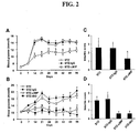
- FIG. 2 is graphs of experimental results demonstrating that MIF blockade with anti-MIF antibody and ISO-1 suppress the development of hyperglycemia and insulitis.
- Blood glucose levels were determined in C57B1/6 mice (Panel A) and CBA/H mice (Panel B) starting at the beginning of the STZ treatment and continued through weekly measurements. Animals received MLD-STZ injections and were treated with either vehicle (STZ), non-immune rabbit IgG (STZ-IgG), anti-MIF IgG (STZ- ⁇ MIF), or ISO-1 (STZ-ISO).
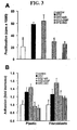
- FIG. 3 is graphs of experimental results demonstrating that neutralization of MIF activity reduces splenocyte proliferation and adherence.
- Panel A shows 3 H-thymidine incorporation, as a measure of cell proliferation, as determined in SMNC isolated from mice untreated with STZ (control), treated with STZ and vehicle (STZ), with STZ and non-immune rabbit IgG (STZ-IgG), STZ and anti-MIF IgG (STZ- ⁇ MIF), or STZ and ISO-1 (STZ-ISO).
- Panel B shows adhesion to plastic surface, or L929 fibroblast, as determined for SMNC isolated from the same groups of mice.
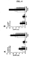
- FIG. 4 is graphs of experimental results demonstrating that neutralization of MIF activity reduces the production of TNF- ⁇ .
- Spleen MNC SMNC
- PC peritoneal cells
- pancreatic islets were isolated from control untreated mice (control), mice treated with STZ and non-immune rabbit IgG (STZ-IgG), STZ and anti-MIF IgG (STZ- ⁇ MIF), STZ and vehicle (STZ), and STZ and ISO-1 (STZ-ISO).
- TNF production was measured in the cell culture supernatants as described in Materials and Methods of the Example. Results are representative of three independent experiments with similar results.
- *p ⁇ 0.05 refers to corresponding STZ-IgG (Panel A) or STZ (Panel B) animals.
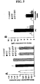
- FIG. 5 is graphs of experimental results demonstrating that neutralization of MIF activity down-regulates the expression of iNOS and NO production.
- Peritoneal cells (PC) and pancreatic islets were isolated from mice treated as described in FIG. 3 .
- iNOS expression was determined by cell-based ELISA, and presented as fold increase compared to control value (O.D. 492 nm 0.445 ⁇ 0.027).
- Panel B after isolation from mice, PC were cultivated in medium for 48 hours, and pancreatic islets in the presence of 250 U/ml IFN- ⁇ + IL-1 ⁇ for 72 hours. Subsequently, nitrite accumulation in cell culture supernatants was determined. Results are representative of three independent experiments with similar results.
- *p ⁇ 0.05 refers to corresponding STZ or STZ-IgG animals.
- FIG. 6 is graphs of experimental results demonstrating that neutralization of MIF activity does not affect the expression of IFN- ⁇ and MHC class II.
- Spleen MNC SMNC
- PC peritoneal cells
- Panel A IFN- ⁇ expression in SMNC was determined by cell-based ELISA, and presented as fold increase compared to control value (O.D. 492 nm 0.070 ⁇ 0.014).
- Panel B MHC II molecules expression in PC and SMNC was determined by cell-based ELISA, and presented as fold increase compared to control value (O.D. 492 nm 1.678 ⁇ 0.151 and 1.204 ⁇ 0.124, respectively). Results are representative of three independent experiments with similar results.
- *p ⁇ 0.05 refers to corresponding STZ or STZ-IgG animals.
- FIG. 7 is a graph of experimental results showing inhibition of diabetes in streptozotocin-treated mice that were treated with an MIF inhibitor.
- FIG. 8 is a graph of experimental results showing the effect of timing of MIF inhibitor ISO-1 and anti-MIF antibody on control of diabetes in streptozotocin-treated mice.
- FIG. 9 is a graph of experimental results showing that MIF-null mice do not acquire diabetes after treatment with streptozotocin.
- MIF macrophage migration inhibitory factor
- ISO-1 ( S , R )-3-(4-hydroxyphenyl)-4,5-dihydro-5-isoxazole acetic acid methyl ester
- iNOS inducible nitric oxide synthase
- STZ streptozotocin
- MLD-STZ multiple low doses of streptozotocin
- SMNC spleen mononuclear cells
- PC peritoneal cells
- IFN- ⁇ ⁇ -interferon
- TNF- ⁇ tumor necrosis factor- ⁇
- IL-1 ⁇ interleukin-1 ⁇ .
- the present invention is based on the discovery that MIF is a critical factor in the pathogenesis of type 1 diabetes. Therefore, inhibition of MIF prevents or attenuates the development of type 1 diabetes. See Examples.
- the invention is directed to an antibody that inhibits macrophage migration inhibitory factor (MIF) for use in treating a mammal having type 1 diabetes or at risk for type 1 diabetes.
- MIF macrophage migration inhibitory factor
- These uses comprise administering to the mammal a pharmaceutical composition comprising an agent that inhibits a macrophage migration inhibitory factor (MIF) in the mammal.
- the antibody that binds specifically to the MIF can be an Fab fragment or an F(ab)2 fragment of an antibody.
- Such agents can be produced by well known methods. Non-limiting methods include: immunization of animals with MIF, followed by isolation of anti-MIF antibodies from serum or production of anti-MIF monoclonal antibodies from hybridomas made by fusion of spenocytes with myeloma cells; or phage display or other recombinant methods.
- the monoclonal antibody is chosen or adapted to match the species to be treated.
- the anti-MIF antibody or antigen-binding fragment thereof
- Such antigen-specific human or humanized monoclonal antibodies may be developed by a variety of methods well known in the art.
- the mammal is a rodent (e.g., a mouse injected with streptozotocin, which is an accepted animal model for type 1 , diabetes).
- the mammal is a human.
- the invention is also directed to methods of treating a mammal having type 1 diabetes or at risk for type 1 diabetes. These methods comprise administering to the mammal a
- compositions can be formulated without undue experimentation for administration to a mammal, including humans, as appropriate for the particular application. Additionally, proper dosages of the compositions can be determined without undue experimentation using standard dose-response protocols.
- compositions designed for oral, lingual, sublingual, buccal and intrabuccal administration can be made without undue experimentation by means well known in the art, for example with an inert diluent or with an edible carrier.
- the compositions may be enclosed in gelatin capsules or compressed into tablets.
- the pharmaceutical compositions of the present invention may be incorporated with excipients and used in the form of tablets, troches, capsules, elixirs, suspensions, syrups, wafers, chewing gums and the like.
- Tablets, pills, capsules, troches and the like may also contain binders, recipients, disintegrating agent, lubricants, sweetening agents, and flavoring agents.
- binders include microcrystalline cellulose, gum tragacanth or gelatin.
- excipients include starch or lactose.
- disintegrating agents include alginic acid, corn starch and the like.
- lubricants include magnesium stearate or potassium stearate.
- An example of a glidant is colloidal silicon dioxide.
- sweetening agents include sucrose, saccharin and the like.
- flavoring agents include peppermint, methyl salicylate, orange flavoring and the like. Materials used in preparing these various compositions should be pharmaceutically pure and nontoxic in the amounts used.
- compositions useful for the present invention can easily be administered parenterally such as for example, by intravenous, intramuscular, intrathecal or subcutaneous injection.
- Parenteral administration can be accomplished by incorporating the compositions of the present invention into a solution or suspension.
- solutions or suspensions may also include sterile diluents such as water for injection, saline solution, fixed oils, polyethylene glycols, glycerine, propylene glycol or other synthetic solvents.
- Parenteral formulations may also include antibacterial agents such as for example, benzyl alcohol or methyl parabens, antioxidants such as for example, ascorbic acid or sodium bisulfite and chelating agents such as EDTA.
- Buffers such as acetates, citrates or phosphates and agents for the adjustment of tonicity such as sodium chloride or dextrose may also be added.
- the parenteral preparation can be enclosed in ampules, disposable syringes or multiple dose vials made of glass or plastic. Rectal administration includes administering the pharmaceutical compositions into the rectum or large intestine. This can be accomplished using suppositories or enemas. Suppository formulations can easily be made by methods known in the art.
- suppository formulations can be prepared by heating glycerin to about 120° C., dissolving the composition in the glycerin, mixing the heated glycerin after which purified water may be added, and pouring the hot mixture into a suppository mold.
- Transdermal administration includes percutaneous absorption of the composition through the skin.
- Transdermal formulations include patches (such as the well-known nicotine patch), ointments, creams, gels, salves and the like.
- nasally administering or nasal administration includes administering the composition to the mucous membranes of the nasal passage or nasal cavity of the patient.
- pharmaceutical compositions for nasal administration of a composition include therapeutically effective amounts of the composition prepared by well-known methods to be administered, for example, as a nasal spray, nasal drop, suspension, gel, ointment, cream or powder. Administration of the composition may also take place using a nasal tampon or nasal sponge.
- the mammal has or is at risk for having diabetes or conditions associated with diabetes or a risk of diabetes, for example impaired glucose intolerance, stress hyperglycemia, metabolic syndrome, and/or insulin resistance.
- diabetes or conditions associated with diabetes or a risk of diabetes for example impaired glucose intolerance, stress hyperglycemia, metabolic syndrome, and/or insulin resistance.
- the present invention can be utilized for any mammal, for example rodents, generally used as experimental models for diabetes (see Example), or humans.
- the invention is directed to methods of evaluating whether a compound is useful for preventing or treating type 1 diabetes.
- the methods comprise the following two steps: (a) determining whether the compound inhibits a macrophage migration inhibitory factor (MIF) in a mammal, then, if the compound inhibits the MIF, (b) determining whether the compound inhibits development of type 1 diabetes.
- MIF macrophage migration inhibitory factor
- the ability of the tested compound to inhibit MIF activity can be determined by any known procedure.
- Non-limiting examples include those methods described in U.S. Patent Application Publications 2003/0008908 and 2003/0195194 .
- any known procedure for evaluating the effect of a compound on type 1 diabetes can be utilized.
- the effect of the compound on type 1 diabetes is determined by evaluating the effect of the compound on development of diabetes in animal models utilizing multiple low dose streptozotocin administration or in animal models genetically susceptible to development of type 1 diabetes, such as the NOD mouse.
- the effect of a test compound or formulation on the development of diabetes in such models may be assessed by a variety of convenient methods, for instance by monitoring circulating glucose or proliferation of splenic lymphocytes in the mammal (see also Example).
- oligopeptides or proteins such as enzymes or proteins that comprise an antibody binding site
- nucleic acids or mimetics such as antisense compounds, ribozymes, aptamers, interfering RNAs such as siRNAs.
- the compound is an organic molecule less than 1000 Dalton having structure I or structure II, as previously defined.
- kits comprising (a) a pharmaceutical composition comprising an agent that inhibits a macrophage migration inhibitory factor (MIF) in the mammal, where the agent is a polypeptide or a polynucleotide, and (b) instructions for administering the composition to the mammal.
- MIF macrophage migration inhibitory factor
- the mammal has type 1 diabetes or is at risk for type 1 diabetes.
- MIF macrophage migration inhibitory factor
- Attenuation of MIF activity with exemplary MIF inhibitors such as neutralizing antibodies against MIF, or the pharmacological MIF inhibitor ( S , R )-3-(4-hydroxyphenyl)-4,5-dihydro-5-isoxazole acetic acid methyl ester (ISO-1), markedly reduces histopathological changes in the islets of pancreas and suppresses the development of hyperglycaemia, showing the general utility of this method of treating mammals with, or at risk for, type 1 diabetes.
- MIF inhibitors such as neutralizing antibodies against MIF, or the pharmacological MIF inhibitor ( S , R )-3-(4-hydroxyphenyl)-4,5-dihydro-5-isoxazole acetic acid methyl ester (ISO-1)
- MIF also stimulates the synthesis of other proinflammatory cytokines and soluble mediators involved in the pathogenesis of type 1 DM such as TNF- ⁇ , IL-1 ⁇ and nitric oxide (NO) (19) raised the possibility that inhibition of MIF activity may modulate the development of disease.
- mice Inbred CBA/H mice were obtained from our own breeding colony at the Institute for Biological Research, Belgrade. Inbred C57 B1/6 mice were originally purchased from Charles River (Calco, Italy) and then bred by brother per sister mating for up to 4 generations. Mice were kept under standard laboratory conditions with free access to food and water. The handling of the mice and the study protocol were approved by the local Institutional Animal Care and Use Committee.
- Streptozotocin STZ, S-0130
- sulfanilamide naphthylenediamine dichidrochloride
- irrelevant rabbit IgG were purchased from Sigma (St. Louis, MO).
- Anti-murine MIF IgG was prepared from rabbit serum raised against murine MIF and purified by protein A affinity chromatography following the manufacturer's instructions (Pierce, Rockford, IL).
- ISO-1 [(S,R)-3-(4-hydroxyphenil)-4,5-dihydro-5-isoxasole acetic acid methyl ester] was synthesized as previously described (20).
- Control animals received either nonimmune IgG or ISO-1 diluent (DMSO/H 2 O). Mice were monitored for diabetes by weekly measurement of blood glucose levels using a glucometer (Sensimac ® , Imaco, Ludersdorf, Germany). Clinical diabetes was defined by hyperglycemia in non-fasted animals (blood glucose over 11.8 mmol/l).
- PC peritoneal cells
- SMNC spleen mononuclear cells
- pancreatic islets were isolated from individual anti-MIF IgG-treated, ISO-1-treated or control diabetic mice on day 15 after the first injection of STZ, as well as from normal untreated animals.
- SMNC (5x10 6 /well) prepared by Ficoll gradient centrifugation, resident PC (2.5x10 5 /well) collected by peritoneal lavage with cold PBS, or pancreatic islets (150-200 islets/well) prepared by collagenase digestion and density gradient purification as described (22), were incubated in 24-well Limbro culture plates in 1 ml of RPMI-1640 culture medium containing 5% fetal bovine serum (FBS) and cell supernatants were collected after 48 h. Concentration of bioactive TNF- ⁇ in culture supernatant was determined as previously described (22) using a cytolytic bioassay with actinomycin D-treated fibrosarcoma cell line L929.
- Nitrite accumulation an indicator of NO production, was determined in cell culture supernatant using the Griess reaction (22).
- the expression of intracellular MIF, IFN- ⁇ , MHC II, or iNOS was determined by slight modification of cell-based ELISA protocol (23).
- Spleen MNC (5x10 5 /well) or PC (2.5x10 5 /well), were allowed to adhere to poly-L-lysine-precoated 96-well microplates.
- T/PBS Triton X-100 in PBS
- endogenous peroxidase was quenched with 1% H 2 O 2 in T/PBS, and reaction blocked for 1 h at 37°C with 10% FBS in T/PBS.
- cells were incubated for 1 h at 37°C with either rabbit anti-mouse MIF IgG, rat anti-mouse IFN- ⁇ (Holland Biotechnology, Leiden, The Netherlands), mouse anti-mouse/rat MHC II (MCA46R, Serotec), or rabbit anti-mouse iNOS (Sigma), in T/PBS containing 1% BSA.
- pancreas Histological and immunohistochemical examination of pancreas.
- Pancreata were fixed in neutral buffered formalin and then embedded in paraffin. The fixed blocks were sectioned (7 ⁇ m thick) and haematoxylin and eosin staining was performed to assess the incidence and degree of inflammatory changes.
- Insulitis scoring was performed as previously described (10, 11) by examining at least 15 islets per mouse and graded in a blinded fashion as follows: 0, no infiltrate; 1, peri-ductal infiltrate; 2, peri-islet infiltrate; 3, intraislet infiltrate; and 4, intraislet infiltrate associated with ⁇ -cell destruction. At least 15 islets were counted for each mouse.
- a mean score for each pancreas was calculated by dividing the total score by the number of islets examined. Insulitis scores (IS) are expressed as mean values ⁇ SD. Immunohistochemistry was performed on paraffin-embedded sections of formalin-fixed tissue using a previously described microwave-based method (24). For MIF immunostaining, a polyclonal rabbit anti-MIF IgG and control rabbit IgG were used. The examined area of the islet was outlined, and the percentage of MIF-positive islet cells was measured using quantitative Image Analysis System (Optima 6.5, Media Cybernatics, Silver Springs, MD).
- MLD-STZ Increased MIF expression in MLD-STZ-induced diabetic mice.
- MIF mRNA expression is up-regulated in spontaneously diabetic NOD mice, but its functional role in disease progression is unknown.
- MLD-STZ was administered to diabetes-susceptible CBA/H mice. Immunohistochemistry revealed substantial induction of MIF protein expression by the islet cells in pancreas sections from these mice during the disease course, as well as mononuclear cell infiltration (FIG. 1B-D ).
- higher concentrations of MIF were detected in the peritoneal cells (PC) of diabetic mice, in comparison to non-diabetic, control mice ( FIG. 1E ).
- Anti-MIF prophylaxis suppresses clinical and histological parameters of MLD-STZ-induced diabetes.
- MIF activity modulates disease in MLD-STZ-exposed mice.
- C57B1/6 and CBA/H control mice treated with PBS or non-immune IgG developed sustained hyperglycemia over a 2-week period following MLD-STZ injections.
- MLD-STZ induced different degrees of hyperglycemia in the two mouse strains, treatment of either C57B1/6 mice ( FIG. 2A ), or CBA/H mice ( FIG.
- mice prophylactically over a period of 14 days with ISO-1, starting 3 days before the first of five injections of STZ. These mice remained euglycemic throughout the eight-week experimental period ( FIG. 2B ).
- Anti-MIF treatments reduce splenocyte proliferation and adhesive properties.
- ex vivo analysis of the functional characteristics of spleen mononuclear cells (SMNC) from CBA/H mice was performed during early progression of hyperglycemia, on day 15 of hyperglycemia.
- SMNC spleen mononuclear cells
- mice were treated with either anti-MIF antibodies or ISO-1, euthanized at day 15 after MLD-STZ exposure, and splenocytes harvested for ex vivo analyses.
- Treatment of MLD-STZ-exposed mice with anti-MIF antibodies significantly inhibited diabetes-associated SMNC proliferation ( 3 H-thymidine incorporation was 29782 ⁇ 3694 cpm versus 63651 ⁇ 10157 cpm of diabetic control cells) ( FIG. 3A ).
- MIF antagonists down-regulate the expression of CD11b and CD25 of splenic mononuclear cells
- Treatment groups CD11b + CD25 + % * MFI** % MFI Exp. 1 Untreated 7.9 44.4 4.4 57.8 STZ + IgG 9.4 72.0 8.5 77.7 STZ + ⁇ MIF 6.4 48.6 4.3 70.6 Exp. 2 Untreated 15.5 88.1 2.5 96.2 STZ 19.4 108.3 6.1 113.0 STZ + ISO-1 15.3 88.5 4.5 106.0
- IL-2R IL-2 receptor
- CD25 IL-2 receptor
- Anti-CD25 has also been shown to attenuate low-dose streptozotocin-induced diabetes in mice (12, 26).
- lymphocytes were isolated from the spleens of control or anti-MIF-treated mice 15 days post-MLD-STZ exposure and labeled with fluorescently tagged antibodies directed against IL-2R (CD25).
- pro-inflammatory mediators play crucial role in type 1 DM development in rodents (4-7, 21, 22), we determined the effects of MIF inhibition on STZ-associated cytokine release from both local and peripheral immune cells ex vivo .
- MIF significantly contributes to the immunopathology associated with excessive inflammation and autoimmunity, and neutralizing endogenous MIF with neutralizing anti-MIF antibodies, or targeted disruption of the MIF gene, inhibits the development of several rodent models of inflammatory disease, including immunologically induced kidney disease (27), leishmaniasis (28), Gram-negative and Gram-positive sepsis (15, 29, 30), antigen-induced arthrius (31), colitis (16), and experimental autoimmune encephalomyelitis (EAE) (17).
- MIF myeloma
- pancreatic ⁇ -cells a potential role for MIF in the development and pathogenesis of autoimmune-mediated DM
- MIF is constitutively expressed and secreted together with insulin from pancreatic ⁇ -cells, and acts as an autocrine factor to stimulate insulin release (32).
- induction of insulin secretion is thought to contribute to immunoinflammatory diabetogenic pathways by favoring the expression on the ⁇ -cells and the presentation to the immune cells of antigens that are up-regulated when the functional activity is augmented (12)
- this "hormonal" property could represent an additional important factor involving endogenous MIF in the initial events of ⁇ -cell dysfunction and destruction.
- Targeting endogenous MIF may therefore be a suitable approach for unraveling the role of this cytokine in the pathogenesis of type 1 DM and for therapeutic and/or prophylactic treatment of the condition.
- IFN- ⁇ may possess dicothomic effects on inflammation, exerting proinflammatory effects when produced at the level of the organ targeted by the immune response (e.g. the islet microenvironment), while activating corticosteroid-dependent and independent anti-inflammatory pathways at the systemic level (8).
- MIF is a critical mediator of inflammatory and autoimmune diseases, because neutralizing endogenous MIF activity with anti-MIF antibodies has been effective in animal models of septic shock, colitis, encephalomyelitis, and leishmania infection (15-17, 28), and may be a promising approach for therapy of various human diseases.
- treatment approaches that rely on exogenously administered proteins, including humanized antibodies may face several challenges, including potential immunogenicity, that suggest the desirability of more preferred embodiments.
- Anti-cytokine antibodies can form small inflammatory complexes with cytokines, and thereby exacerbate inflammatory responses (39). For these reasons, alternatives to the therapeutic use of anti-MIF Ig preparations should also be considered for pharmaceutical development.
- pharmacological intervention with ISO-1 as a small drug-like molecule that inhibits MIF tautomerase and biological activity (20), may be a more preferred approach for the treatment of MIF-related diseases.
- Future studies will address the potential use of MIF inhibitors as a treatment, rather than a prophylactic, for regulation of diabetes pathogenesis and presentation.
- diabetes was induced in adult male mice with multiple subtoxic doses of streptozotocin (MLD-STZ, 40 mg/kg body wt/day i.p. for five consecutive days) as described (21).
- MIF inhibition after induction of diabetes was studied by i.p. injection of (S,R)-3-(4-hydroxyphenyl)-4,5-dihydro-5-isoxazole acetic acid methyl ester (ISO-1), a pharmacological inhibitor of MIF (20) at a dose of 1 mg/mouse/day, for 14 consecutive days starting at day 7 after initial streptozotocin administration.
- ISO-1 a pharmacological inhibitor of MIF (20) at a dose of 1 mg/mouse/day, for 14 consecutive days starting at day 7 after initial streptozotocin administration.
- Peripheral blood glucose was monitored weekly in these mice and in control mice that were treated identically, except where the ISO-1 treatment was replaced by ISO-1 diluent.
- Example 3 MIF-null Mice are Resistant to the Induction of Type 1 Diabetes by Streptozotocin
- MIF-/- C57B1/6 mice 40 were treated with streptozotocin and compared with MIF +/+ mice in progression of diabetes.
- streptozotocin the MIF-/- mice did not develop diabetes, while the MIF+/+ mice did develop diabetes ( FIG. 9 ). This also further establishes that methods directed to inhibiting expression of MIF would be expected to be useful for type 1 diabetes treatment.
Landscapes
- Health & Medical Sciences (AREA)
- Chemical & Material Sciences (AREA)
- Organic Chemistry (AREA)
- Medicinal Chemistry (AREA)
- Life Sciences & Earth Sciences (AREA)
- General Health & Medical Sciences (AREA)
- Pharmacology & Pharmacy (AREA)
- Animal Behavior & Ethology (AREA)
- Public Health (AREA)
- Veterinary Medicine (AREA)
- Diabetes (AREA)
- Genetics & Genomics (AREA)
- Biochemistry (AREA)
- Engineering & Computer Science (AREA)
- Bioinformatics & Cheminformatics (AREA)
- Proteomics, Peptides & Aminoacids (AREA)
- Molecular Biology (AREA)
- Chemical Kinetics & Catalysis (AREA)
- General Chemical & Material Sciences (AREA)
- Nuclear Medicine, Radiotherapy & Molecular Imaging (AREA)
- Epidemiology (AREA)
- Immunology (AREA)
- Biophysics (AREA)
- Endocrinology (AREA)
- Emergency Medicine (AREA)
- Obesity (AREA)
- Hematology (AREA)
- Pharmaceuticals Containing Other Organic And Inorganic Compounds (AREA)
- Medicines That Contain Protein Lipid Enzymes And Other Medicines (AREA)
- Measuring Or Testing Involving Enzymes Or Micro-Organisms (AREA)
- Medicines Containing Antibodies Or Antigens For Use As Internal Diagnostic Agents (AREA)
- Investigating Or Analysing Biological Materials (AREA)
Description
- The present invention generally relates to diabetes treatment. More specifically, the invention is directed to the use of inhibitors of macrophage migration inhibitory factor for treatment or prevention of
type 1 diabetes. -
US 6 080 487 describes the development of anti-MIF monoclonal antibodies provides a specific means for disrupting the mechanism by which MIF exerts its biological activity. The anti-MIF monoclonal antibodies may be used as a therapeutic for conditions involving MIF-mediated adverse effects. Cytokine 21(2003)179-186 asserts that MIF may play a possible role in autoimmune-inflammatory events such astype 1 diabetes. This asserted role was based on preliminary studies in which it was shown that MIF mRNA expression was elevated in the splenic lymphocytes of NOD mice in which diabetes was spontaneously induced. However, the MIF-protein levels in the diabetic animals where less than in normal controls.WO 01/64749 A2 WO 01/32606 A1 WO 02/100332 A2 -
Type 1 diabetes mellitus (type 1 DM) is a multifactorial syndrome caused by the lack of endogenous insulin, thought to be due to an immune attack mediated by autoreactive T cells and macrophages against pancreatic β-cells. The disease afflicts approximately 4 million people in North America and epidemiological data concur that the incidence and thus the prevalence of the disease is increasing worldwide (I). Extensive research efforts have greatly expanded understanding of disease pathogenesis, and have revealed a critical role for several pro-inflammatory mediators. However, no effective anti-inflammatory therapeutics have been approved for the clinical management oftype 1 DM. Several animal models of the disease have enhanced understanding of the molecular events that underlie the pathogenesis of diabetes. Multiple low doses of streptozotocin to susceptible strains of mice, for example, induces a diabetic condition with many of the hallmarks ofhuman type 1 DM. Clinical and histoimmunological similarities include the development of hyperglycemia associated with infiltration of the pancreatic islets by T lymphocytes and macrophages (insulitis) (2,3). Proinflammatory cytokines, including interleukin (IL)-1β, interferon (IFN)-γ, tumor necrosis factor (TNF)-α and IL-18 play important roles in the development of streptozotocin-induced diabetes (4-7). However, administration of either recombinant IL-1β, IFN-γ, or TNF-α, or specific inhibitors of their activity, have complex and often contradictory effects on disease development and/or course, depending on animal model used, as well as on timing of administration (8-11). - The key pathogenic role played by the immune system in the pathogenesis of
type 1 DM has recently focused much attention on identifying immunotherapeutical approaches that may allow halting or delaying β-cell destruction in prediabetic individuals or in those patients with newly diagnosed disease (12). Macrophage migration inhibitory factor (MIF) is a critical cytokine in local and systemic inflammation, but its role in diabetes has not been explored thoroughly. MIF is a pleiotropic cytokine produced during immune responses by activated T cells, macrophages and a variety of nonimmune cells (13,14). It acts as a critical mediator of host defense, and is being explored as a therapeutic target in septic shock as well as chronic inflammatory and autoimmune diseases (15-17). Elevated MIF gene expression has been detected in spontaneously non-obese diabetic (NOD) mice (18), but its importance in the pathogenesis oftype 1 DM is unclear. - There is thus a need for further investigation into the precise role and interactions of cytokines, in particular MIF, in
type 1 diabetes. The present invention addresses that need. - Accordingly, the inventors have discovered that inhibition of macrophage migration inhibitory factor (MIF) attenuates
type 1 diabetes. - Subject-matter of the invention is an antibody that inhibits macrophage migration inhibitory factor (MIF) for use in treating a
mammal having type 1 diabetes or at risk fortype 1 diabetes to prevent or attenuate the development oftype 1 diabetes. -
FIG. 1 is micrographs and graphs demonstrating elevated MIF protein expression in pancreas and peritoneal cells of diabetic mice. Panels A-C are light immunomicrographs of pancreata. MIF is weakly expressed by islet cells of non-diabetic control mice (Panel A); in pancreatic islets ofday 10 diabetic mice, there is massive mononuclear cell (MNC) infiltration and MIF expression is markedly up-regulated (Panel B); negative staining in a serial section of diabetic mice islet (Panel C). Panel D shows a graph of quantitative image analysis showing different stages of MIF expression by islet cells from diabetic versus non-diabetic control mice. Shown are the mean ± SD of the percentage of MIF+ cells per islet (n = 3 mice per group). Panel E is a graph of quantitative analysis of intracellular expression of MIF protein in peritoneal cells of non-diabetic mice (Control), MLD-STZ diabetic mice (STZ), and MLD-STZ diabetic mice treated with anti-MIF antibody (STZ-αMIF), measured by cell-based ELISA performed with MIF-specific antibodies, as described in Materials and Methods of the Example. The results are presented as fold increase of control absorbance value (OD 492 nm 0.687 ±0.013). *p<0.05 refers to otherwise untreated MLD-STZ diabetic animals. -
FIG. 2 is graphs of experimental results demonstrating that MIF blockade with anti-MIF antibody and ISO-1 suppress the development of hyperglycemia and insulitis. Blood glucose levels were determined in C57B1/6 mice (Panel A) and CBA/H mice (Panel B) starting at the beginning of the STZ treatment and continued through weekly measurements. Animals received MLD-STZ injections and were treated with either vehicle (STZ), non-immune rabbit IgG (STZ-IgG), anti-MIF IgG (STZ-αMIF), or ISO-1 (STZ-ISO). Histopathology analysis of pancreata from C57B1/6 mice (Panel C) and CBA/H mice (Panel D) are presented as insulitis score, as described in as described in Materials and Methods of the Example. *p<0.05 refers to corresponding STZ or STZ-IgG animals. -
FIG. 3 is graphs of experimental results demonstrating that neutralization of MIF activity reduces splenocyte proliferation and adherence. Panel A shows 3H-thymidine incorporation, as a measure of cell proliferation, as determined in SMNC isolated from mice untreated with STZ (control), treated with STZ and vehicle (STZ), with STZ and non-immune rabbit IgG (STZ-IgG), STZ and anti-MIF IgG (STZ-αMIF), or STZ and ISO-1 (STZ-ISO). Panel B shows adhesion to plastic surface, or L929 fibroblast, as determined for SMNC isolated from the same groups of mice. The results are presented as fold increase of control adhesion to plastic surface, or to L929 fibroblasts (O.D, 570 nm 0.316 ± 0.018 and 0.905 ± 0.077, respectively). *p<0.05 refers to corresponding STZ or STZ-IgG animals. -
FIG. 4 is graphs of experimental results demonstrating that neutralization of MIF activity reduces the production of TNF-α. Spleen MNC (SMNC), peritoneal cells (PC) and pancreatic islets were isolated from control untreated mice (control), mice treated with STZ and non-immune rabbit IgG (STZ-IgG), STZ and anti-MIF IgG (STZ-αMIF), STZ and vehicle (STZ), and STZ and ISO-1 (STZ-ISO). TNF production was measured in the cell culture supernatants as described in Materials and Methods of the Example. Results are representative of three independent experiments with similar results. *p< 0.05 refers to corresponding STZ-IgG (Panel A) or STZ (Panel B) animals. -
FIG. 5 is graphs of experimental results demonstrating that neutralization of MIF activity down-regulates the expression of iNOS and NO production. Peritoneal cells (PC) and pancreatic islets were isolated from mice treated as described inFIG. 3 . In Panel A, iNOS expression was determined by cell-based ELISA, and presented as fold increase compared to control value (O.D. 492 nm 0.445 ± 0.027). In Panel B, after isolation from mice, PC were cultivated in medium for 48 hours, and pancreatic islets in the presence of 250 U/ml IFN-γ + IL-1β for 72 hours. Subsequently, nitrite accumulation in cell culture supernatants was determined. Results are representative of three independent experiments with similar results. *p< 0.05 refers to corresponding STZ or STZ-IgG animals. -
FIG. 6 is graphs of experimental results demonstrating that neutralization of MIF activity does not affect the expression of IFN-γ and MHC class II. Spleen MNC (SMNC) and peritoneal cells (PC) were isolated from mice treated as described inFIG. 3 . In Panel A, IFN-γ expression in SMNC was determined by cell-based ELISA, and presented as fold increase compared to control value (O.D. 492 nm 0.070 ± 0.014). In Panel B, MHC II molecules expression in PC and SMNC was determined by cell-based ELISA, and presented as fold increase compared to control value (O.D. 492 nm 1.678 ± 0.151 and 1.204 ± 0.124, respectively). Results are representative of three independent experiments with similar results. *p< 0.05 refers to corresponding STZ or STZ-IgG animals. -
FIG. 7 is a graph of experimental results showing inhibition of diabetes in streptozotocin-treated mice that were treated with an MIF inhibitor. -
FIG. 8 is a graph of experimental results showing the effect of timing of MIF inhibitor ISO-1 and anti-MIF antibody on control of diabetes in streptozotocin-treated mice. Blood glucose levels in untreated CBA/H mice (control, n=8), or of 21 day MLD-STZ diabetic mice treated with either STZ alone (STZ, n=8), or STZ and ISO-1 given as an early (STZ-ISO Early, n=8), Late (STZ-ISO-1 Late, n=8) or Late anti-MIF Ab (STZ-•-MIF, n=8). Early injection: ISO-1 was administered by i.p. injection at 40mg/Kg/day for 14 days and started 3 days prior to STZ first injection; Late injection: ISO-1 or anti-MIF Ab was administered on day six, after the last injection of STZ. *p<0.05 refers to corresponding STZ animals. -
FIG. 9 is a graph of experimental results showing that MIF-null mice do not acquire diabetes after treatment with streptozotocin. Blood glucose levels were determined in wild-type and MIF-/- C57B1/6 mice. Animals received MLD-STZ injections (5x40 mg/kg/day). Blood glucose levels were determined starting at the beginning of the STZ treatment and continued through weekly measurements. *p<0.005 refers to corresponding STZ animals. Blood glucose levels were determined: (a) starting at the beginning of the STZ treatment and continued through weekly measurements. Symbols used for the respective two groups which received MLD-STZ injections: C57B1/6 mice (•; n=11) and MIF-/- C57B1/6 (o; n= 11). Results from a representative of two independent experiments are presented as ± SD. *p<0.005 refers to corresponding STZ animals. Statistical analysis was performed by ANOVA with Bonferroni's adjustment. - Abbreviations: MIF, macrophage migration inhibitory factor; ISO-1, (S,R)-3-(4-hydroxyphenyl)-4,5-dihydro-5-isoxazole acetic acid methyl ester ; iNOS, inducible nitric oxide synthase; STZ, streptozotocin; MLD-STZ, multiple low doses of streptozotocin; SMNC, spleen mononuclear cells; PC, peritoneal cells; IFN-γ, γ-interferon; TNF-α, tumor necrosis factor-α; IL-1β, interleukin-1β.
- The present invention is based on the discovery that MIF is a critical factor in the pathogenesis of
type 1 diabetes. Therefore, inhibition of MIF prevents or attenuates the development oftype 1 diabetes. See Examples. - Thus, in some embodiments, the invention is directed to an antibody that inhibits macrophage migration inhibitory factor (MIF) for use in treating a
mammal having type 1 diabetes or at risk fortype 1 diabetes. These uses comprise administering to the mammal a pharmaceutical composition comprising an agent that inhibits a macrophage migration inhibitory factor (MIF) in the mammal. - The antibody that binds specifically to the MIF can be an Fab fragment or an F(ab)2 fragment of an antibody. Such agents can be produced by well known methods. Non-limiting methods include: immunization of animals with MIF, followed by isolation of anti-MIF antibodies from serum or production of anti-MIF monoclonal antibodies from hybridomas made by fusion of spenocytes with myeloma cells; or phage display or other recombinant methods. Preferably, the monoclonal antibody is chosen or adapted to match the species to be treated. For treatment of humans, for example, the anti-MIF antibody (or antigen-binding fragment thereof) will be a human antibody or a humanized antibody. Such antigen-specific human or humanized monoclonal antibodies may be developed by a variety of methods well known in the art.
- These methods can be used with any mammal. Preferably, the mammal is a rodent (e.g., a mouse injected with streptozotocin, which is an accepted animal model for
type 1 , diabetes). In other preferred embodiments, the mammal is a human. - The invention is also directed to methods of treating a
mammal having type 1 diabetes or at risk fortype 1 diabetes. These methods comprise administering to the mammal a - Any of the above-described compositions can be formulated without undue experimentation for administration to a mammal, including humans, as appropriate for the particular application. Additionally, proper dosages of the compositions can be determined without undue experimentation using standard dose-response protocols.
- Accordingly, the compositions designed for oral, lingual, sublingual, buccal and intrabuccal administration can be made without undue experimentation by means well known in the art, for example with an inert diluent or with an edible carrier. The compositions may be enclosed in gelatin capsules or compressed into tablets. For the purpose of oral therapeutic administration, the pharmaceutical compositions of the present invention may be incorporated with excipients and used in the form of tablets, troches, capsules, elixirs, suspensions, syrups, wafers, chewing gums and the like.
- Tablets, pills, capsules, troches and the like may also contain binders, recipients, disintegrating agent, lubricants, sweetening agents, and flavoring agents. Some examples of binders include microcrystalline cellulose, gum tragacanth or gelatin. Examples of excipients include starch or lactose. Some examples of disintegrating agents include alginic acid, corn starch and the like. Examples of lubricants include magnesium stearate or potassium stearate. An example of a glidant is colloidal silicon dioxide. Some examples of sweetening agents include sucrose, saccharin and the like. Examples of flavoring agents include peppermint, methyl salicylate, orange flavoring and the like. Materials used in preparing these various compositions should be pharmaceutically pure and nontoxic in the amounts used.
- The compositions useful for the present invention can easily be administered parenterally such as for example, by intravenous, intramuscular, intrathecal or subcutaneous injection. Parenteral administration can be accomplished by incorporating the compositions of the present invention into a solution or suspension. Such solutions or suspensions may also include sterile diluents such as water for injection, saline solution, fixed oils, polyethylene glycols, glycerine, propylene glycol or other synthetic solvents. Parenteral formulations may also include antibacterial agents such as for example, benzyl alcohol or methyl parabens, antioxidants such as for example, ascorbic acid or sodium bisulfite and chelating agents such as EDTA. Buffers such as acetates, citrates or phosphates and agents for the adjustment of tonicity such as sodium chloride or dextrose may also be added. The parenteral preparation can be enclosed in ampules, disposable syringes or multiple dose vials made of glass or plastic. Rectal administration includes administering the pharmaceutical compositions into the rectum or large intestine. This can be accomplished using suppositories or enemas. Suppository formulations can easily be made by methods known in the art. For example, suppository formulations can be prepared by heating glycerin to about 120° C., dissolving the composition in the glycerin, mixing the heated glycerin after which purified water may be added, and pouring the hot mixture into a suppository mold.
- Transdermal administration includes percutaneous absorption of the composition through the skin. Transdermal formulations include patches (such as the well-known nicotine patch), ointments, creams, gels, salves and the like.
- The present invention includes nasally administering to the mammal a therapeutically effective amount of the composition. As used herein, nasally administering or nasal administration includes administering the composition to the mucous membranes of the nasal passage or nasal cavity of the patient. As used herein, pharmaceutical compositions for nasal administration of a composition include therapeutically effective amounts of the composition prepared by well-known methods to be administered, for example, as a nasal spray, nasal drop, suspension, gel, ointment, cream or powder. Administration of the composition may also take place using a nasal tampon or nasal sponge.
- In preferred embodiments of the invention, the mammal has or is at risk for having diabetes or conditions associated with diabetes or a risk of diabetes, for example impaired glucose intolerance, stress hyperglycemia, metabolic syndrome, and/or insulin resistance.
- The present invention can be utilized for any mammal, for example rodents, generally used as experimental models for diabetes (see Example), or humans.
- The discovery that compounds that inhibit MIF activity or expression are useful for treatment of
type 1 diabetes indicates that screens of compounds for MIF-inhibiting activity can be used to identify compounds for the ability to prevent or treattype 1 diabetes. Thus, in some embodiments, the invention is directed to methods of evaluating whether a compound is useful for preventing or treatingtype 1 diabetes. The methods comprise the following two steps: (a) determining whether the compound inhibits a macrophage migration inhibitory factor (MIF) in a mammal, then, if the compound inhibits the MIF, (b) determining whether the compound inhibits development oftype 1 diabetes. - In these methods, the ability of the tested compound to inhibit MIF activity can be determined by any known procedure. Non-limiting examples include those methods described in
U.S. Patent Application Publications 2003/0008908 and2003/0195194 . Similarly, any known procedure for evaluating the effect of a compound ontype 1 diabetes can be utilized. In preferred embodiments, the effect of the compound ontype 1 diabetes is determined by evaluating the effect of the compound on development of diabetes in animal models utilizing multiple low dose streptozotocin administration or in animal models genetically susceptible to development oftype 1 diabetes, such as the NOD mouse. The effect of a test compound or formulation on the development of diabetes in such models may be assessed by a variety of convenient methods, for instance by monitoring circulating glucose or proliferation of splenic lymphocytes in the mammal (see also Example). - These methods are not limited to evaluation of any particular type of compound. Examples of compounds that can be evaluated by these methods include oligopeptides or proteins such as enzymes or proteins that comprise an antibody binding site; nucleic acids or mimetics such as antisense compounds, ribozymes, aptamers, interfering RNAs such as siRNAs. In preferred embodiments, the compound is an organic molecule less than 1000 Dalton having structure I or structure II, as previously defined.
- In additional embodiments, the invention is directed to kits comprising (a) a pharmaceutical composition comprising an agent that inhibits a macrophage migration inhibitory factor (MIF) in the mammal, where the agent is a polypeptide or a polynucleotide, and (b) instructions for administering the composition to the mammal. In these embodiments, the mammal has
type 1 diabetes or is at risk fortype 1 diabetes. - Preferred embodiments of the invention are described in the following examples.
- The pro-inflammatory cytokine, macrophage migration inhibitory factor (MIF), plays a pivotal role in several inflammatory and autoimmune diseases. MIF mRNA expression is up-regulated in non-obese diabetic mice, yet little is known about its potential as a regulator of
type 1 diabetes. Here, we show that MIF protein is significantly elevated in islet cells during the development of experimental diabetes induced in mice by multiple low doses of streptozotocin. Attenuation of MIF activity with exemplary MIF inhibitors such as neutralizing antibodies against MIF, or the pharmacological MIF inhibitor (S,R)-3-(4-hydroxyphenyl)-4,5-dihydro-5-isoxazole acetic acid methyl ester (ISO-1), markedly reduces histopathological changes in the islets of pancreas and suppresses the development of hyperglycaemia, showing the general utility of this method of treating mammals with, or at risk for,type 1 diabetes. The observed beneficial effects could be attributed to the reduced proliferation and adhesion of autoreactive lymphocytes, down-regulation of iNOS expression, as well as NO and TNF-α secretion by islets of pancreas and by peritoneal macrophages, although knowledge of these mechanistic links are not essential to the teaching of the inventive method. This study defines a critical role for MIF in the pathogenesis oftype 1 diabetes and identifies a new therapeutic strategy to attenuate the autoimmune process at multiple levels. - The observation that MIF also stimulates the synthesis of other proinflammatory cytokines and soluble mediators involved in the pathogenesis of
type 1 DM such as TNF-α, IL-1β and nitric oxide (NO) (19) raised the possibility that inhibition of MIF activity may modulate the development of disease. - Given the central regulatory role of MIF in the immune response mediated by both macrophages and T cells, we have investigated the expression of MIF during the progress of diabetes in mice treated with multiple low doses of streptozotocin (MLD-STZ), as well as the influence of MIF activity neutralization on the disease development. These are well-accepted models for
type 1 diabetes in humans. For this purpose, an anti-MIF polyclonal antibody, as well as (S,R)3-(4-hydroxyphenyl)-4,5-dihydro-5-isoxazole acetic acid methyl ester (ISO-1), a pharmacological inhibitor of MIF (20), were given to mice as prophylactic treatment prior to the challenge with STZ. Various immunological parameters relevant for DM pathogenesis were determined, including autoreactive cell proliferation and adhesion, as well as the production of proinflammatory mediators. To our knowledge, this is the first attempt to interfere in autoimmune diabetes by in vivo neutralization of MIF. - Mice. Inbred CBA/H mice were obtained from our own breeding colony at the Institute for Biological Research, Belgrade. Inbred C57 B1/6 mice were originally purchased from Charles River (Calco, Italy) and then bred by brother per sister mating for up to 4 generations. Mice were kept under standard laboratory conditions with free access to food and water. The handling of the mice and the study protocol were approved by the local Institutional Animal Care and Use Committee.
- Reagents. Streptozotocin (STZ, S-0130), sulfanilamide, naphthylenediamine dichidrochloride and irrelevant rabbit IgG were purchased from Sigma (St. Louis, MO). Anti-murine MIF IgG was prepared from rabbit serum raised against murine MIF and purified by protein A affinity chromatography following the manufacturer's instructions (Pierce, Rockford, IL). ISO-1, [(S,R)-3-(4-hydroxyphenil)-4,5-dihydro-5-isoxasole acetic acid methyl ester] was synthesized as previously described (20).
- Diabetes induction and in vivo treatments. Diabetes was induced in adult male mice with multiple subtoxic doses of streptozotocin (MLD-STZ, 40 mg/kg body wt/day i.p. for five consecutive days) as described (21). The impact of polyclonal antibody against MIF was studied by i.p. injection of mice (5-10 per treatment group) with 5 mg/kg of rabbit IgG antibody against mouse MIF on days -3, -1, 2 and 5 in relation to the first STZ dose. The effect of ISO-1 was studied by i.p. injection of the drug at a dose of 1 mg/mouse/day, for 14 consecutive days, starting 3 days prior to the first injection with STZ. Control animals received either nonimmune IgG or ISO-1 diluent (DMSO/H2O). Mice were monitored for diabetes by weekly measurement of blood glucose levels using a glucometer (Sensimac®, Imaco, Ludersdorf, Germany). Clinical diabetes was defined by hyperglycemia in non-fasted animals (blood glucose over 11.8 mmol/l).
- Cell incubation and determination of pro-inflammatory mediators. Resident peritoneal cells (PC), spleen mononuclear cells (SMNC), and pancreatic islets were isolated from individual anti-MIF IgG-treated, ISO-1-treated or control diabetic mice on
day 15 after the first injection of STZ, as well as from normal untreated animals. SMNC (5x10⁶/well) prepared by Ficoll gradient centrifugation, resident PC (2.5x10⁵/well) collected by peritoneal lavage with cold PBS, or pancreatic islets (150-200 islets/well) prepared by collagenase digestion and density gradient purification as described (22), were incubated in 24-well Limbro culture plates in 1 ml of RPMI-1640 culture medium containing 5% fetal bovine serum (FBS) and cell supernatants were collected after 48 h. Concentration of bioactive TNF-α in culture supernatant was determined as previously described (22) using a cytolytic bioassay with actinomycin D-treated fibrosarcoma cell line L929. Nitrite accumulation, an indicator of NO production, was determined in cell culture supernatant using the Griess reaction (22). The expression of intracellular MIF, IFN-γ, MHC II, or iNOS was determined by slight modification of cell-based ELISA protocol (23). Spleen MNC (5x10⁵/well) or PC (2.5x10⁵/well), were allowed to adhere to poly-L-lysine-precoated 96-well microplates. Following fixation with 4% paraformaldehyde, and washing with 0.1 % Triton X-100 in PBS (T/PBS), endogenous peroxidase was quenched with 1% H2O2 in T/PBS, and reaction blocked for 1 h at 37°C with 10% FBS in T/PBS. Subsequently, cells were incubated for 1 h at 37°C with either rabbit anti-mouse MIF IgG, rat anti-mouse IFN-γ (Holland Biotechnology, Leiden, The Netherlands), mouse anti-mouse/rat MHC II (MCA46R, Serotec), or rabbit anti-mouse iNOS (Sigma), in T/PBS containing 1% BSA. After washing, the cells were incubated for 1 h with the corresponding secondary antibody (goat anti-rabbit Ig(H+L)-HRP, goat anti-rat Ig(H+L)-HRP or goat anti-mouse Ig(F(ab')2-specific)-HRP washed again and incubated for 15 min at room temperature in dark with 50 µl of a solution containing 0.4 mg/ml OPD (Sigma), 11.8 mg/ml Na2HPO4x2H2=O, 7.3 mg/ml citric acid and 0.015% H2O2. The reaction was stopped with 3N HCl, and the absorbance was measured in a microplate reader at 492 nm in a Titertek microplate reader (Flow). - Ex vivo lymphoproliferative response and adhesion assay. Spontaneous proliferation of SMNC was determined by incubation of cells (5x10⁵/well) from each individual animal in 96-well microplates with 1 pCi 3H-thymidine (3H-TdR, ICN). Incorporated radioactivity was measured after 24 h in a Beckman liquid scintillation counter. The analysis of spontaneous adhesion of SMNC (2.5x10⁵/well) to a monolayer of L929 fibroblasts or plastic was performed by using crystal violet assay as previously described (21). The absorbance corresponding to the number of adherent cells, was measured at 570 nm.
- Antibodies and flow cytometry. Spleen MNC (1x10⁶) were incubated with the rat anti-mouse monoclonal antibodies (mAbs): anti-CD11b (MAC-1)-phycoerythrin (PE), or anti-CD25 (IL-2 Receptor α chain, p55)-biotin (PharMingen, San Diego, CA), followed by Streptavidin-PE (PharMingen). Each cell suspension of SMNC was a pool from three to five animals obtained from the same
experimental group 15 days after diabetes induction, as well as from normal untreated mice. Cell surface marker expression was analyzed with a flow cytometer (FACScalibur, Becton Dickinson, Heidelberg, Germany) and Win MDI 2.8 software (Joseph Trotter). - Histological and immunohistochemical examination of pancreas. Pancreata were fixed in neutral buffered formalin and then embedded in paraffin. The fixed blocks were sectioned (7 µm thick) and haematoxylin and eosin staining was performed to assess the incidence and degree of inflammatory changes. Insulitis scoring was performed as previously described (10, 11) by examining at least 15 islets per mouse and graded in a blinded fashion as follows: 0, no infiltrate; 1, peri-ductal infiltrate; 2, peri-islet infiltrate; 3, intraislet infiltrate; and 4, intraislet infiltrate associated with β-cell destruction. At least 15 islets were counted for each mouse. A mean score for each pancreas was calculated by dividing the total score by the number of islets examined. Insulitis scores (IS) are expressed as mean values ± SD. Immunohistochemistry was performed on paraffin-embedded sections of formalin-fixed tissue using a previously described microwave-based method (24). For MIF immunostaining, a polyclonal rabbit anti-MIF IgG and control rabbit IgG were used. The examined area of the islet was outlined, and the percentage of MIF-positive islet cells was measured using quantitative Image Analysis System (Optima 6.5, Media Cybernatics, Silver Springs, MD).
- Statistical analysis. The blood glucose values are shown as mean values ± SE. Statistical analyses were performed by ANOVA with Bonferroni's adjustment and Fisher's exact test. The other values were expressed as means ± SD and groups of data were compared using Student's paired t-test. The results of a representative of at least three separate experiments with similar results are presented. Statistical significance was set at P<0.05.
- Increased MIF expression in MLD-STZ-induced diabetic mice. Recent studies have revealed that MIF mRNA expression is up-regulated in spontaneously diabetic NOD mice, but its functional role in disease progression is unknown. In order to determine if MIF protein expression levels are altered during immunoinflammatory diabetes, MLD-STZ was administered to diabetes-susceptible CBA/H mice. Immunohistochemistry revealed substantial induction of MIF protein expression by the islet cells in pancreas sections from these mice during the disease course, as well as mononuclear cell infiltration
(FIG. 1B-D ). Likewise, higher concentrations of MIF were detected in the peritoneal cells (PC) of diabetic mice, in comparison to non-diabetic, control mice (FIG. 1E ). In vivo treatment of mice with neutralizing anti-MIF immunoglobulin during disease induction significantly suppressed PC expression of MIF (FIG. 1E ). Thus, as a consequence of DM induction, increased MIF-protein content was observed both at the level of peripheral and target tissue, and MIF protein levels can be attenuated by administration of anti-MIF antibodies. - Anti-MIF prophylaxis suppresses clinical and histological parameters of MLD-STZ-induced diabetes. To determine whether inhibition of MIF activity modulates disease in MLD-STZ-exposed mice, we first studied the effect of a neutralizing polyclonal antibody against MIF in two DM-susceptible inbred mouse strains. Both C57B1/6 and CBA/H control mice treated with PBS or non-immune IgG developed sustained hyperglycemia over a 2-week period following MLD-STZ injections. Although MLD-STZ induced different degrees of hyperglycemia in the two mouse strains, treatment of either C57B1/6 mice (
FIG. 2A ), or CBA/H mice (FIG. 2B ) with anti-MIF Ab from day -3 to day +5 significantly inhibited MLD-STZ-induced hyperglycemia. In addition, histological examination of pancreatic specimens performed onday 15 of the disease indicated that the degree of islet infiltration was significantly lower in mice treated with anti-MIF antibodies as compared with diabetic control mice treated with irrelevant IgG (FIG. 2C and D ). We also examined the effect of the pharmacological MIF antagonist ISO-1 (20) in MLD-STZ-induced DM. Accordingly, we treated mice prophylactically over a period of 14 days with ISO-1, starting 3 days before the first of five injections of STZ. These mice remained euglycemic throughout the eight-week experimental period (FIG. 2B ). The anti-diabetogenic effect of either immunological or pharmacological neutralization of MIF was long-lasting, with limited variations throughout the entire 56 day follow-up period (FIG. 2A and B) . MIF blockade by ISO-1 attenuated inflammation of the islets (FIG. 2D ). These data provide evidence for a critical role of MIF in the induction and progression of immunoinflammatory DM and for the utility of treatment with MIF inhibitors to avert the development oftype 1 diabetes in at-risk populations. - Anti-MIF treatments reduce splenocyte proliferation and adhesive properties. To understand the cellular effects of anti-MIF treatments, ex vivo analysis of the functional characteristics of spleen mononuclear cells (SMNC) from CBA/H mice was performed during early progression of hyperglycemia, on
day 15 of hyperglycemia. Previous studies have shown that isolated autoreactive lymphocytes from diabetic mice exhibit significantly increased ex vivo spontaneous proliferation in comparison to cells isolated from healthy animals, suggesting that this cell type may contribute to disease progression (21, 22). To determine if MIF inhibitors modulate splenocyte proliferative responses ex vivo, diabetic CBA/H mice were treated with either anti-MIF antibodies or ISO-1, euthanized atday 15 after MLD-STZ exposure, and splenocytes harvested for ex vivo analyses. Treatment of MLD-STZ-exposed mice with anti-MIF antibodies significantly inhibited diabetes-associated SMNC proliferation (3H-thymidine incorporation was 29782 ± 3694 cpm versus 63651 ± 10157 cpm of diabetic control cells) (FIG. 3A ). Likewise, in vivo inhibition of MIF activity with ISO-1 reverted splenocyte proliferation to near-control levels (25637 ± 2015 cpm in comparison to 21150 ± 12723 cpm of untreated control cells) (FIG. 3A ). In addition to cell proliferation, cell-cell adhesion, mediated by the interaction between CD11b and ICAM-1 (CD54), participates in the immunological processes oftype 1 DM (21, 25). To determine possible consequences of anti-MIF treatment on the interactions between cells participating in the diabetogenic processes, we assessed the adhesive properties of SMNC and found that spontaneous adhesion of cells to plastic, as well as adhesion to fibroblasts was significantly inhibited by in vivo treatment with anti-MIF IgG, or ISO-1 (FIG. 3B ). Down-regulation of adhesiveness was associated with the changes of CD11b expression, as revealed by flow cytometry analysis, using antibodies directed against CD11b, a molecule that mediates cellular adhesion to ICAM-1 (CD54). Thus, in comparison to SMNC derived from control diabetic mice, both anti-MIF IgG treatment and ISO-1 treatment (Table 1) reduced the frequency of CD11b+ SMNC and mean antigen density (mean fluorescence intensity, MFI) to the level of untreated normal mice, indicative of the anti-diabetogenic benefits of treatment with the MIF inhibitors.Table 1. MIF antagonists down-regulate the expression of CD11b and CD25 of splenic mononuclear cells Treatment groups CD11b+ CD25+ %* MFI** % MFI Exp. 1 Untreated 7.9 44.4 4.4 57.8 STZ + IgG 9.4 72.0 8.5 77.7 STZ + αMIF 6.4 48.6 4.3 70.6 Exp. 2 Untreated 15.5 88.1 2.5 96.2 STZ 19.4 108.3 6.1 113.0 STZ + ISO-1 15.3 88.5 4.5 106.0 - Since IL-2 is centrally involved in the initiation of any immune response, negating its action by blocking the interaction with the IL-2 receptor (IL-2R, CD25) system has gained much attention as a possible therapeutic target for immunointervention in both rodent and human disease (26). Anti-CD25 has also been shown to attenuate low-dose streptozotocin-induced diabetes in mice (12, 26). To determine the effect of in vivo treatment with MIF antagonists on IL-2R+ cell populations, lymphocytes were isolated from the spleens of control or anti-MIF-treated
mice 15 days post-MLD-STZ exposure and labeled with fluorescently tagged antibodies directed against IL-2R (CD25). Staining the cells for IL-2R expression (Table 1) revealed significant increase in both the percentage of IL-2R+ lymphocytes and antigen density in control diabetic mice, when comparing to normal untreated animals, while the in vivo treatment with anti-MIF IgG, or with ISO-1 reduced these parameters. - Production of pro-inflammatory mediators. Since pro-inflammatory mediators play crucial role in
type 1 DM development in rodents (4-7, 21, 22), we determined the effects of MIF inhibition on STZ-associated cytokine release from both local and peripheral immune cells ex vivo. Immunological neutralization of endogenous MIF protein with anti-MIF IgG (FIG. 4A ), or pharmacological inhibition of MIF with ISO-1 (FIG. 4B ), down-regulated TNF-α production by spleen MNC, peritoneal macrophages and pancreatic islet cells, reducing it to the level of healthy, nontreated mice. We also found that intracellular expression of iNOS was significantly reduced in macrophages isolated from animals treated with either anti-MIF or ISO-1 in comparison to relatively high iNOS expression of diabetic animals (FIG. 5A ). Accordingly, MIF blockade abolished subsequent NO production in macrophages, as well as in pancreatic islets (FIG. 5B ). The effects of the MIF inhibitors were specific for TNF-α, iNOS, and NO, because the expression of intracellular IFN-γ in the splenocytes, as well as the expression of MHC class II molecules, remained unchanged in comparison to control diabetic mice (FIG. 6A and B). - MIF significantly contributes to the immunopathology associated with excessive inflammation and autoimmunity, and neutralizing endogenous MIF with neutralizing anti-MIF antibodies, or targeted disruption of the MIF gene, inhibits the development of several rodent models of inflammatory disease, including immunologically induced kidney disease (27), leishmaniasis (28), Gram-negative and Gram-positive sepsis (15, 29, 30), antigen-induced arthrius (31), colitis (16), and experimental autoimmune encephalomyelitis (EAE) (17). Recently, a potential role for MIF in the development and pathogenesis of autoimmune-mediated DM has been implicated in spontaneously diabetic NOD mice, because expression of MIF mRNA is significantly increased during disease development, and exogenous MIF administration increases disease incidence in these animals (18). MIF is constitutively expressed and secreted together with insulin from pancreatic β-cells, and acts as an autocrine factor to stimulate insulin release (32). Because induction of insulin secretion is thought to contribute to immunoinflammatory diabetogenic pathways by favoring the expression on the β-cells and the presentation to the immune cells of antigens that are up-regulated when the functional activity is augmented (12), this "hormonal" property could represent an additional important factor involving endogenous MIF in the initial events of β-cell dysfunction and destruction. Targeting endogenous MIF may therefore be a suitable approach for unraveling the role of this cytokine in the pathogenesis of
type 1 DM and for therapeutic and/or prophylactic treatment of the condition. - In the present study, we show for the first time that endogenous MIF plays a key role in the development of murine autoimmune diabetes. Progression of MLD-STZ-induced diabetes was accompanied by up-regulated MIF protein expression both in pancreatic islets and peripheral cells. Immunoneutralization of MIF by anti-MIF IgG, or pharmacological inhibition of MIF activity with ISO-1, markedly attenuated the clinical and histological manifestations of the disease. It inhibited the inflammatory responses as well as splenocyte proliferation and adherence ex vivo. The anti-diabetogenic effect of both agents was long-lasting, because mice remained euglycemic during the 8-week follow-up period. Inhibition of autoreactive T cell proliferation, leukocyte adhesion and migration into target tissue, and macrophage activation by these anti-MIF strategies suggest that immunological or pharmacological neutralization of MIF activity may attenuate pathologic autoimmune responses in vivo.
- Consistent with the role of MIF in the T-cell activation and mitogenesis (30, 33), we found that MIF blockade considerably decreased IL-2R expression and spontaneous proliferation of splenic lymphocytes measured ex vivo, which most probably reflected the autoreactive T cell response to blood-born pancreatic antigens released upon STZ destruction of β-cells (21, 22). The results therefore indicate that MIF contributes to the clonal size of autoreactive T cells, and that blocking the expansion of diabetogenic T clones might be partly responsible for the protective effect of these anti-MIF strategies.
- In addition to clonal expansion of autoreactive cells, the development of the disease is determined by cell-cell interactions mediated by adhesion receptors and ligands both in the induction and the efferent phase of the autoimmune response. In this context, we have shown that mononuclear cells from MLD-STZ-induced diabetic mice are highly adherent to cultured vascular endothelial cells (21), or fibroblasts (
FIG. 3B ) at a time of peak disease activity; these adhesive properties may be an important mechanism for inflammatory infiltration of target tissue (21). Several lines of evidence demonstrate that up-regulation of CD11b, which mediates cellular adhesion to ICAM-1 (CD54) (34), might have functional consequences on the interactions between cells participating in the immunological processes oftype 1 DM (21, 25). MIF blockade significantly inhibited the expression of CD11b on mononuclear cells of diabetic mice and markedly down-regulated leukocyte adhesive properties, suggesting that anti-MIF strategies may impair homing of these cells to the islets of pancreas, thus contributing to the therapeutic benefit of MIF blockade. In favor of this hypothesis, histological analysis showed reduced insulitis in anti-MIF treated mice. Inhibition of MIF activity also has been shown to down-regulate adhesion molecule-dependent target tissue pathology in EAE (17). - Another important finding of this study is that anti-MIF treatment affects the production of pro-inflammatory and cytotoxic mediators. The lower local production of TNF-α and NO may result from the reduction of inflammatory cell influx into pancreas. Based on our ex vivo findings, however, it seems that MIF blockade might also directly influence macrophage and T cell effector function, as suggested by down-regulation of TNF-α, iNOS and NO by peritoneal and/or spleen cells. This is consistent with the in vitro ability of anti-MIF antibodies to interfere with production of these mediators (28). MIF up-regulates in vitro production of TNF-α, as well as of reactive oxygen species and nitrogen metabolites (35). It therefore is conceivable that direct blockade of MIF-mediated iNOS expression and TNF-α synthesis may contribute to down-regulation of tissue-damaging pro-inflammatory mediators in
type 1 DM. Thus, we hypothesize that the efficacy of MIF blockade in the treatment of MLD-STZ DM is due to inhibitory effects on the autoimmune/inflammatory response of T cells and macrophage, as well as islet cells. - Interestingly, the effect of systemic blockade of MIF specifically inhibited TNF-α and iNOS-mediated NO production in both local and peripheral cells, because these therapeutic approaches did not diminish peripheral production of the signature Th1 cytokine, IFN-γ. Consistent with the role for IFN-γ in the regulation of MHC class II, the expression of antigens by peritoneal and spleen cells was not impaired after anti-MIF treatments. Although these observations appear to contradict previous findings demonstrating a central role for IFN-γ in diabetes pathogenesis (36), it must be noted that peripheral cytokine production may not mirror the role of cytokine in the development of the organ-specific autoimmune disease. For example we have previously shown in the DP-BB rat model that in an apparently paradoxical fashion, both anti-IFN-γ antibody and exogenously-administered IFN-γ prevented development of autoimmune diabetes (10, 37). As recently reported for EAE, anti-MIF antibody treatment did not affect antigen-specific splenocyte production of IFN-γ, but did reduce IFN-γ-release from antigen-reactive Th1 cells in the target tissue (17). Therefore, it is possible that anti-MIF treatment in our model reduced cytokine production within the microenvironment of the pancreas, but this effect may not have been reflected in our ex vivo analyses. In support of this, we have previously shown a similar modulatory pattern of IFN-γ expression and distribution of MHC class II+ cells in MLD-STZ-induced
type 1 DM by different systemically applied pharmacological agents (21, 22). It is known that IFN-γ may possess dicothomic effects on inflammation, exerting proinflammatory effects when produced at the level of the organ targeted by the immune response (e.g. the islet microenvironment), while activating corticosteroid-dependent and independent anti-inflammatory pathways at the systemic level (8). Having in mind the profound ability of MIF to counteract the immunosuppressive effects of glucocorticoids (38), neutralization of MIF activity accompanied with sustained circulating levels of IFN-γ could potentiate systemic anti-inflammatory effects in the treated animals. - MIF is a critical mediator of inflammatory and autoimmune diseases, because neutralizing endogenous MIF activity with anti-MIF antibodies has been effective in animal models of septic shock, colitis, encephalomyelitis, and leishmania infection (15-17, 28), and may be a promising approach for therapy of various human diseases. However, treatment approaches that rely on exogenously administered proteins, including humanized antibodies, may face several challenges, including potential immunogenicity, that suggest the desirability of more preferred embodiments. Anti-cytokine antibodies can form small inflammatory complexes with cytokines, and thereby exacerbate inflammatory responses (39). For these reasons, alternatives to the therapeutic use of anti-MIF Ig preparations should also be considered for pharmaceutical development. Thus, pharmacological intervention with ISO-1, as a small drug-like molecule that inhibits MIF tautomerase and biological activity (20), may be a more preferred approach for the treatment of MIF-related diseases. Future studies will address the potential use of MIF inhibitors as a treatment, rather than a prophylactic, for regulation of diabetes pathogenesis and presentation.
- Using methods as described in Example 1, diabetes was induced in adult male mice with multiple subtoxic doses of streptozotocin (MLD-STZ, 40 mg/kg body wt/day i.p. for five consecutive days) as described (21). The effect of MIF inhibition after induction of diabetes was studied by i.p. injection of (S,R)-3-(4-hydroxyphenyl)-4,5-dihydro-5-isoxazole acetic acid methyl ester (ISO-1), a pharmacological inhibitor of MIF (20) at a dose of 1 mg/mouse/day, for 14 consecutive days starting at
day 7 after initial streptozotocin administration. Peripheral blood glucose was monitored weekly in these mice and in control mice that were treated identically, except where the ISO-1 treatment was replaced by ISO-1 diluent. - As shown in
FIG. 7 and8 , inhibition of MIF after induction of diabetes provided considerable benefit to the streptozotocin-treated mice, by inhibiting the progression of diabetes. - To further establish the importance of MIF in
type 1 diabetes, MIF-/- C57B1/6 mice (40) were treated with streptozotocin and compared with MIF+/+ mice in progression of diabetes. When treated with streptozotocin, the MIF-/- mice did not develop diabetes, while the MIF+/+ mice did develop diabetes (FIG. 9 ). This also further establishes that methods directed to inhibiting expression of MIF would be expected to be useful fortype 1 diabetes treatment. -
- SEQ ID NO:1 - Human MIF cDNA - GenBank Accession No. NM002415

- SEQ ID NO:2 - Mouse MIF cDNA - GenBank Accession No. BC024895

- SEQ ID NO:3 - Rat MIF cDNA - GenBank Accession No. NM031051

Claims (10)
- An antibody that inhibits macrophage migration inhibitory factor (MIF) for use in inhibiting the progression of type 1 diabetes in a mammal having type 1 diabetes or for inhibiting the development of type 1 diabetes in a mammal at risk for type 1 diabetes.
- The antibody for use of claim 1, for inhibiting the progression of type 1 diabetes in a mammal having type 1 diabetes.
- The antibody for use of claim 1, for inhibiting the development of type 1 diabetes in a mammal at risk for type 1 diabetes.
- The antibody for use of claim 1, wherein the antibody is a monoclonal antibody.
- The antibody for use of claim 1, wherein the antibody is a humanized antibody.
- The antibody for use of claim 1, wherein the antibody is a human antibody.
- The antibody for use of any of the preceding claims,
wherein the antibody is an Fab fragment or an F(ab)2 fragment. - The antibody for use of any of the preceding claims,
wherein the mammal has or is at risk for impaired glucose tolerance, stress hyperglycemia, metabolic syndrome, and/or insulin resistance. - The antibody for use of any of the preceding claims, wherein the mammal is a rodent.
- The antibody for use of any of the preceding claims, wherein the mammal is a human.
Applications Claiming Priority (2)
| Application Number | Priority Date | Filing Date | Title |
|---|---|---|---|
| US55716904P | 2004-03-29 | 2004-03-29 | |
| PCT/US2005/010521 WO2005094338A2 (en) | 2004-03-29 | 2005-03-29 | Treatment of type 1 diabetes with inhibitors of macrophage migration inhibitory factor |
Publications (3)
| Publication Number | Publication Date |
|---|---|
| EP1740167A2 EP1740167A2 (en) | 2007-01-10 |
| EP1740167A4 EP1740167A4 (en) | 2008-12-10 |
| EP1740167B1 true EP1740167B1 (en) | 2013-08-28 |
Family
ID=35064302
Family Applications (1)
| Application Number | Title | Priority Date | Filing Date |
|---|---|---|---|
| EP05746462.0A Expired - Lifetime EP1740167B1 (en) | 2004-03-29 | 2005-03-29 | Treatment of type 1 diabetes with inhibitors of macrophage migration inhibitory factor |
Country Status (6)
| Country | Link |
|---|---|
| US (1) | US20080305118A1 (en) |
| EP (1) | EP1740167B1 (en) |
| JP (1) | JP5198848B2 (en) |
| AU (1) | AU2005228426B2 (en) |
| CA (1) | CA2563117C (en) |
| WO (1) | WO2005094338A2 (en) |
Families Citing this family (5)
| Publication number | Priority date | Publication date | Assignee | Title |
|---|---|---|---|---|
| AU2007230948B2 (en) * | 2006-03-24 | 2012-06-28 | The Feinstein Institute For Medical Research | Modified macrophage migration inhibitory factor inhibitors |
| US9643922B2 (en) | 2008-08-18 | 2017-05-09 | Yale University | MIF modulators |
| US9540322B2 (en) | 2008-08-18 | 2017-01-10 | Yale University | MIF modulators |
| JP2014526886A (en) | 2011-07-15 | 2014-10-09 | モルフォシス・アー・ゲー | Antibodies cross-reactive with macrophage migration inhibitory factor (MIF) and D-dopachrome tomerase (D-DT) |
| US9567306B2 (en) | 2014-06-17 | 2017-02-14 | The Feinstein Institute For Medical Research | Inhibition of macrophage migration inhibitory factor in melanoma and colon cancer |
Family Cites Families (10)
| Publication number | Priority date | Publication date | Assignee | Title |
|---|---|---|---|---|
| US5530101A (en) * | 1988-12-28 | 1996-06-25 | Protein Design Labs, Inc. | Humanized immunoglobulins |
| US6080407A (en) * | 1993-05-17 | 2000-06-27 | The Picower Institute For Medical Research | Diagnostic assays for MIF |
| AU6898700A (en) * | 1999-08-10 | 2001-03-05 | Joslin Diabetes Center Inc. | Method for identifying compounds for treatment of insulin resistance |
| WO2001032606A1 (en) * | 1999-10-29 | 2001-05-10 | The Picower Institute For Medical Research | Compounds having mif antagonist activity |
| US6268151B1 (en) * | 2000-01-20 | 2001-07-31 | Isis Pharmaceuticals, Inc. | Antisense modulation of macrophage migration inhibitory factor expression |
| AU2001238677A1 (en) * | 2000-02-28 | 2001-09-12 | Idec Pharmaceuticals Corporation | Method for preparing anti-mif antibodies |
| WO2002020758A2 (en) * | 2000-09-05 | 2002-03-14 | Curagen Corporation | Novel proteins and nucleic acids encoding same |
| UY27304A1 (en) * | 2001-05-24 | 2002-12-31 | Avanir Pharmaceuticals | INHIBITORS OF THE INHIBITOR FACTOR OF MIGRATION OF MACROPHAGES AND METHODS FOR IDENTIFICATION |
| AU2002314944B2 (en) * | 2001-06-08 | 2008-03-06 | Cytokine Pharmasciences, Inc. | Isoxazoline compounds having MIF antagonist activity |
| NZ537301A (en) * | 2002-06-07 | 2006-06-30 | Cortical Pty Ltd | Therapeutic molecules and methods-1 |
-
2005
- 2005-03-29 CA CA2563117A patent/CA2563117C/en not_active Expired - Fee Related
- 2005-03-29 AU AU2005228426A patent/AU2005228426B2/en not_active Ceased
- 2005-03-29 JP JP2007506473A patent/JP5198848B2/en not_active Expired - Fee Related
- 2005-03-29 US US10/594,641 patent/US20080305118A1/en not_active Abandoned
- 2005-03-29 WO PCT/US2005/010521 patent/WO2005094338A2/en not_active Ceased
- 2005-03-29 EP EP05746462.0A patent/EP1740167B1/en not_active Expired - Lifetime
Non-Patent Citations (1)
| Title |
|---|
| H. LAN ET AL: "Distinguishing Covariation From Causation in Diabetes: A Lesson From the Protein Disulfide Isomerase mRNA Abundance Trait", DIABETES, vol. 53, no. 1, 1 January 2003 (2003-01-01), pages 240 - 244, XP055037754, ISSN: 0012-1797, DOI: 10.2337/diabetes.53.1.240 * |
Also Published As
| Publication number | Publication date |
|---|---|
| JP5198848B2 (en) | 2013-05-15 |
| WO2005094338A2 (en) | 2005-10-13 |
| AU2005228426A1 (en) | 2005-10-13 |
| AU2005228426B2 (en) | 2010-07-15 |
| US20080305118A1 (en) | 2008-12-11 |
| EP1740167A4 (en) | 2008-12-10 |
| CA2563117C (en) | 2014-01-07 |
| EP1740167A2 (en) | 2007-01-10 |
| WO2005094338A3 (en) | 2007-05-31 |
| JP2007534672A (en) | 2007-11-29 |
| CA2563117A1 (en) | 2005-10-13 |
Similar Documents
| Publication | Publication Date | Title |
|---|---|---|
| Nguyen et al. | Chronic exposure to beta-blockers attenuates inflammation and mucin content in a murine asthma model | |
| Cvetkovic et al. | Critical role of macrophage migration inhibitory factor activity in experimental autoimmune diabetes | |
| Flyvbjerg et al. | Amelioration of long-term renal changes in obese type 2 diabetic mice by a neutralizing vascular endothelial growth factor antibody | |
| Zhu et al. | Effects of nebulized ketamine on allergen-induced airway hyperresponsiveness and inflammation in actively sensitized Brown-Norway rats | |
| Xu et al. | Canagliflozin ameliorates nonalcoholic fatty liver disease by regulating lipid metabolism and inhibiting inflammation through induction of autophagy | |
| JP2021155436A (en) | Method of using cyclodextrin | |
| US9394362B2 (en) | IL-21 antibodies and methods of making or using the antibodies | |
| Jiang et al. | Anti-inflammatory effects of anemonin on acute ulcerative colitis via targeted regulation of protein kinase C-θ | |
| KR102218498B1 (en) | Combination of FXR agonists | |
| Wang et al. | Ocular immune‐related diseases: molecular mechanisms and therapy | |
| US20100075937A1 (en) | Patient populations and treatment methods | |
| Ke et al. | Gliquidone decreases urinary protein by promoting tubular reabsorption in diabetic Goto-Kakizaki rats | |
| Sun et al. | Blockade of angiotensin II with losartan attenuates transforming growth factor-β1 inducible gene-h3 (βig-h3) expression in a model of chronic cyclosporine nephrotoxicity | |
| Wu et al. | Tumor necrosis factor alpha improves glucose homeostasis in diabetic mice independent with tumor necrosis factor receptor 1 and tumor necrosis factor receptor 2 | |
| Yin et al. | Renal fibrosis: research progress on mechanisms and therapeutic strategies | |
| Pieper et al. | Spatial distribution and temporal onset of NF-kB activation and inducible nitric oxide synthase within pancreatic islets in the pre-diabetic stage of genetic, diabetic-prone BB rats: attenuation by drug intervention decreases inflammatory cell infiltration and incidence of diabetes | |
| AU2005228426B2 (en) | Treatment of type 1 diabetes with inhibitors of macrophage migration inhibitory factor | |
| JP2007534672A5 (en) | Treatment of type 1 diabetes using macrophage migration inhibitory factor inhibitor | |
| US20230312751A1 (en) | Anti-Fibrotic Combination | |
| WO2023001305A1 (en) | Pharmaceutical composition or drug kit for alleviating or treating fibrotic diseases, and use thereof | |
| Chen et al. | Morroniside ameliorates sevoflurane anesthesia-induced cognitive dysfunction in aged mice through modulating the TLR4/NF-κB pathway | |
| EP3210616A1 (en) | Use of macrophage inflammatory protein-1 (mip-1 ) inhibitor to protect pancreas and prevent blood sugar from rising | |
| EP1565215A2 (en) | Use of the corticoliberin-urocortin system in the treatment of inflammatory diseases | |
| Golant et al. | Psoriasis Treatments: A review of the current research pipeline | |
| CN101918364A (en) | Pharmaceutical compositions and methods for treating proteinuria |
Legal Events
| Date | Code | Title | Description |
|---|---|---|---|
| PUAI | Public reference made under article 153(3) epc to a published international application that has entered the european phase |
Free format text: ORIGINAL CODE: 0009012 |
|
| 17P | Request for examination filed |
Effective date: 20061030 |
|
| AK | Designated contracting states |
Kind code of ref document: A2 Designated state(s): AT BE BG CH CY CZ DE DK EE ES FI FR GB GR HU IE IS IT LI LT LU MC NL PL PT RO SE SI SK TR |
|
| AX | Request for extension of the european patent |
Extension state: AL BA HR LV MK YU |
|
| PUAK | Availability of information related to the publication of the international search report |
Free format text: ORIGINAL CODE: 0009015 |
|
| DAX | Request for extension of the european patent (deleted) | ||
| RIC1 | Information provided on ipc code assigned before grant |
Ipc: A61K 48/00 20060101AFI20071102BHEP Ipc: A61K 39/395 20060101ALI20071102BHEP Ipc: C07H 21/04 20060101ALI20071102BHEP |
|
| RIC1 | Information provided on ipc code assigned before grant |
Ipc: A61K 48/00 20060101AFI20071102BHEP Ipc: C07H 21/04 20060101ALI20081030BHEP Ipc: A61K 39/395 20060101ALI20081030BHEP Ipc: A61K 31/42 20060101ALI20081030BHEP |
|
| A4 | Supplementary search report drawn up and despatched |
Effective date: 20081110 |
|
| 17Q | First examination report despatched |
Effective date: 20090206 |
|
| REG | Reference to a national code |
Ref country code: DE Ref legal event code: R079 Ref document number: 602005041038 Country of ref document: DE Free format text: PREVIOUS MAIN CLASS: A61K0031000000 Ipc: A61K0048000000 |
|
| GRAP | Despatch of communication of intention to grant a patent |
Free format text: ORIGINAL CODE: EPIDOSNIGR1 |
|
| RIC1 | Information provided on ipc code assigned before grant |
Ipc: C07K 16/24 20060101ALI20120921BHEP Ipc: A61K 31/42 20060101ALI20120921BHEP Ipc: A61K 48/00 20060101AFI20120921BHEP Ipc: A61K 39/395 20060101ALI20120921BHEP Ipc: C07H 21/04 20060101ALI20120921BHEP |
|
| GRAJ | Information related to disapproval of communication of intention to grant by the applicant or resumption of examination proceedings by the epo deleted |
Free format text: ORIGINAL CODE: EPIDOSDIGR1 |
|
| GRAP | Despatch of communication of intention to grant a patent |
Free format text: ORIGINAL CODE: EPIDOSNIGR1 |
|
| INTG | Intention to grant announced |
Effective date: 20130328 |
|
| GRAS | Grant fee paid |
Free format text: ORIGINAL CODE: EPIDOSNIGR3 |
|
| GRAA | (expected) grant |
Free format text: ORIGINAL CODE: 0009210 |
|
| AK | Designated contracting states |
Kind code of ref document: B1 Designated state(s): AT BE BG CH CY CZ DE DK EE ES FI FR GB GR HU IE IS IT LI LT LU MC NL PL PT RO SE SI SK TR |
|
| REG | Reference to a national code |
Ref country code: GB Ref legal event code: FG4D |
|
| REG | Reference to a national code |
Ref country code: CH Ref legal event code: EP |
|
| REG | Reference to a national code |
Ref country code: AT Ref legal event code: REF Ref document number: 628905 Country of ref document: AT Kind code of ref document: T Effective date: 20130915 |
|
| REG | Reference to a national code |
Ref country code: IE Ref legal event code: FG4D |
|
| REG | Reference to a national code |
Ref country code: DE Ref legal event code: R096 Ref document number: 602005041038 Country of ref document: DE Effective date: 20131017 |
|
| REG | Reference to a national code |
Ref country code: AT Ref legal event code: MK05 Ref document number: 628905 Country of ref document: AT Kind code of ref document: T Effective date: 20130828 |
|
| REG | Reference to a national code |
Ref country code: LT Ref legal event code: MG4D |
|
| REG | Reference to a national code |
Ref country code: NL Ref legal event code: VDEP Effective date: 20130828 |
|
| PG25 | Lapsed in a contracting state [announced via postgrant information from national office to epo] |
Ref country code: CY Free format text: LAPSE BECAUSE OF FAILURE TO SUBMIT A TRANSLATION OF THE DESCRIPTION OR TO PAY THE FEE WITHIN THE PRESCRIBED TIME-LIMIT Effective date: 20130717 Ref country code: LT Free format text: LAPSE BECAUSE OF FAILURE TO SUBMIT A TRANSLATION OF THE DESCRIPTION OR TO PAY THE FEE WITHIN THE PRESCRIBED TIME-LIMIT Effective date: 20130828 Ref country code: SE Free format text: LAPSE BECAUSE OF FAILURE TO SUBMIT A TRANSLATION OF THE DESCRIPTION OR TO PAY THE FEE WITHIN THE PRESCRIBED TIME-LIMIT Effective date: 20130828 Ref country code: AT Free format text: LAPSE BECAUSE OF FAILURE TO SUBMIT A TRANSLATION OF THE DESCRIPTION OR TO PAY THE FEE WITHIN THE PRESCRIBED TIME-LIMIT Effective date: 20130828 Ref country code: PT Free format text: LAPSE BECAUSE OF FAILURE TO SUBMIT A TRANSLATION OF THE DESCRIPTION OR TO PAY THE FEE WITHIN THE PRESCRIBED TIME-LIMIT Effective date: 20131230 Ref country code: IS Free format text: LAPSE BECAUSE OF FAILURE TO SUBMIT A TRANSLATION OF THE DESCRIPTION OR TO PAY THE FEE WITHIN THE PRESCRIBED TIME-LIMIT Effective date: 20131228 |
|
| REG | Reference to a national code |
Ref country code: NL Ref legal event code: VDEP Effective date: 20130828 |
|
| PG25 | Lapsed in a contracting state [announced via postgrant information from national office to epo] |
Ref country code: PL Free format text: LAPSE BECAUSE OF FAILURE TO SUBMIT A TRANSLATION OF THE DESCRIPTION OR TO PAY THE FEE WITHIN THE PRESCRIBED TIME-LIMIT Effective date: 20130828 Ref country code: BE Free format text: LAPSE BECAUSE OF FAILURE TO SUBMIT A TRANSLATION OF THE DESCRIPTION OR TO PAY THE FEE WITHIN THE PRESCRIBED TIME-LIMIT Effective date: 20130828 Ref country code: FI Free format text: LAPSE BECAUSE OF FAILURE TO SUBMIT A TRANSLATION OF THE DESCRIPTION OR TO PAY THE FEE WITHIN THE PRESCRIBED TIME-LIMIT Effective date: 20130828 Ref country code: SI Free format text: LAPSE BECAUSE OF FAILURE TO SUBMIT A TRANSLATION OF THE DESCRIPTION OR TO PAY THE FEE WITHIN THE PRESCRIBED TIME-LIMIT Effective date: 20130828 Ref country code: GR Free format text: LAPSE BECAUSE OF FAILURE TO SUBMIT A TRANSLATION OF THE DESCRIPTION OR TO PAY THE FEE WITHIN THE PRESCRIBED TIME-LIMIT Effective date: 20131129 |
|
| PG25 | Lapsed in a contracting state [announced via postgrant information from national office to epo] |
Ref country code: CY Free format text: LAPSE BECAUSE OF FAILURE TO SUBMIT A TRANSLATION OF THE DESCRIPTION OR TO PAY THE FEE WITHIN THE PRESCRIBED TIME-LIMIT Effective date: 20130828 |
|
| PG25 | Lapsed in a contracting state [announced via postgrant information from national office to epo] |
Ref country code: DK Free format text: LAPSE BECAUSE OF FAILURE TO SUBMIT A TRANSLATION OF THE DESCRIPTION OR TO PAY THE FEE WITHIN THE PRESCRIBED TIME-LIMIT Effective date: 20130828 Ref country code: NL Free format text: LAPSE BECAUSE OF FAILURE TO SUBMIT A TRANSLATION OF THE DESCRIPTION OR TO PAY THE FEE WITHIN THE PRESCRIBED TIME-LIMIT Effective date: 20130828 Ref country code: CZ Free format text: LAPSE BECAUSE OF FAILURE TO SUBMIT A TRANSLATION OF THE DESCRIPTION OR TO PAY THE FEE WITHIN THE PRESCRIBED TIME-LIMIT Effective date: 20130828 Ref country code: EE Free format text: LAPSE BECAUSE OF FAILURE TO SUBMIT A TRANSLATION OF THE DESCRIPTION OR TO PAY THE FEE WITHIN THE PRESCRIBED TIME-LIMIT Effective date: 20130828 Ref country code: SK Free format text: LAPSE BECAUSE OF FAILURE TO SUBMIT A TRANSLATION OF THE DESCRIPTION OR TO PAY THE FEE WITHIN THE PRESCRIBED TIME-LIMIT Effective date: 20130828 Ref country code: RO Free format text: LAPSE BECAUSE OF FAILURE TO SUBMIT A TRANSLATION OF THE DESCRIPTION OR TO PAY THE FEE WITHIN THE PRESCRIBED TIME-LIMIT Effective date: 20130828 |
|
| PG25 | Lapsed in a contracting state [announced via postgrant information from national office to epo] |
Ref country code: IT Free format text: LAPSE BECAUSE OF FAILURE TO SUBMIT A TRANSLATION OF THE DESCRIPTION OR TO PAY THE FEE WITHIN THE PRESCRIBED TIME-LIMIT Effective date: 20130828 Ref country code: ES Free format text: LAPSE BECAUSE OF FAILURE TO SUBMIT A TRANSLATION OF THE DESCRIPTION OR TO PAY THE FEE WITHIN THE PRESCRIBED TIME-LIMIT Effective date: 20130828 |
|
| REG | Reference to a national code |
Ref country code: DE Ref legal event code: R097 Ref document number: 602005041038 Country of ref document: DE |
|
| PLBE | No opposition filed within time limit |
Free format text: ORIGINAL CODE: 0009261 |
|
| STAA | Information on the status of an ep patent application or granted ep patent |
Free format text: STATUS: NO OPPOSITION FILED WITHIN TIME LIMIT |
|
| 26N | No opposition filed |
Effective date: 20140530 |
|
| REG | Reference to a national code |
Ref country code: DE Ref legal event code: R097 Ref document number: 602005041038 Country of ref document: DE Effective date: 20140530 |
|
| PG25 | Lapsed in a contracting state [announced via postgrant information from national office to epo] |
Ref country code: LU Free format text: LAPSE BECAUSE OF FAILURE TO SUBMIT A TRANSLATION OF THE DESCRIPTION OR TO PAY THE FEE WITHIN THE PRESCRIBED TIME-LIMIT Effective date: 20140329 |
|
| REG | Reference to a national code |
Ref country code: IE Ref legal event code: MM4A |
|
| PG25 | Lapsed in a contracting state [announced via postgrant information from national office to epo] |
Ref country code: IE Free format text: LAPSE BECAUSE OF NON-PAYMENT OF DUE FEES Effective date: 20140329 |
|
| REG | Reference to a national code |
Ref country code: FR Ref legal event code: PLFP Year of fee payment: 11 |
|
| REG | Reference to a national code |
Ref country code: CH Ref legal event code: NV Representative=s name: DR. GRAF AND PARTNER AG INTELLECTUAL PROPERTY, CH |
|
| REG | Reference to a national code |
Ref country code: DE Ref legal event code: R082 Ref document number: 602005041038 Country of ref document: DE Representative=s name: MANITZ, FINSTERWALD & PARTNER GBR, DE Ref country code: DE Ref legal event code: R082 Ref document number: 602005041038 Country of ref document: DE Representative=s name: MANITZ FINSTERWALD PATENTANWAELTE PARTMBB, DE |
|
| REG | Reference to a national code |
Ref country code: DE Ref legal event code: R082 Ref document number: 602005041038 Country of ref document: DE Representative=s name: MANITZ, FINSTERWALD & PARTNER GBR, DE Ref country code: DE Ref legal event code: R082 Ref document number: 602005041038 Country of ref document: DE Representative=s name: MANITZ FINSTERWALD PATENTANWAELTE PARTMBB, DE |
|
| REG | Reference to a national code |
Ref country code: FR Ref legal event code: PLFP Year of fee payment: 12 |
|
| PG25 | Lapsed in a contracting state [announced via postgrant information from national office to epo] |
Ref country code: MC Free format text: LAPSE BECAUSE OF FAILURE TO SUBMIT A TRANSLATION OF THE DESCRIPTION OR TO PAY THE FEE WITHIN THE PRESCRIBED TIME-LIMIT Effective date: 20130828 Ref country code: BG Free format text: LAPSE BECAUSE OF FAILURE TO SUBMIT A TRANSLATION OF THE DESCRIPTION OR TO PAY THE FEE WITHIN THE PRESCRIBED TIME-LIMIT Effective date: 20130828 |
|
| PG25 | Lapsed in a contracting state [announced via postgrant information from national office to epo] |
Ref country code: TR Free format text: LAPSE BECAUSE OF FAILURE TO SUBMIT A TRANSLATION OF THE DESCRIPTION OR TO PAY THE FEE WITHIN THE PRESCRIBED TIME-LIMIT Effective date: 20130828 Ref country code: HU Free format text: LAPSE BECAUSE OF FAILURE TO SUBMIT A TRANSLATION OF THE DESCRIPTION OR TO PAY THE FEE WITHIN THE PRESCRIBED TIME-LIMIT; INVALID AB INITIO Effective date: 20050329 |
|
| REG | Reference to a national code |
Ref country code: FR Ref legal event code: PLFP Year of fee payment: 13 |
|
| PGFP | Annual fee paid to national office [announced via postgrant information from national office to epo] |
Ref country code: CH Payment date: 20170322 Year of fee payment: 13 Ref country code: FR Payment date: 20170322 Year of fee payment: 13 |
|
| PGFP | Annual fee paid to national office [announced via postgrant information from national office to epo] |
Ref country code: GB Payment date: 20170322 Year of fee payment: 13 |
|
| PGFP | Annual fee paid to national office [announced via postgrant information from national office to epo] |
Ref country code: DE Payment date: 20170530 Year of fee payment: 13 |
|
| REG | Reference to a national code |
Ref country code: DE Ref legal event code: R119 Ref document number: 602005041038 Country of ref document: DE |
|
| REG | Reference to a national code |
Ref country code: CH Ref legal event code: PL |
|
| GBPC | Gb: european patent ceased through non-payment of renewal fee |
Effective date: 20180329 |
|
| PG25 | Lapsed in a contracting state [announced via postgrant information from national office to epo] |
Ref country code: DE Free format text: LAPSE BECAUSE OF NON-PAYMENT OF DUE FEES Effective date: 20181002 |
|
| PG25 | Lapsed in a contracting state [announced via postgrant information from national office to epo] |
Ref country code: LI Free format text: LAPSE BECAUSE OF NON-PAYMENT OF DUE FEES Effective date: 20180331 Ref country code: CH Free format text: LAPSE BECAUSE OF NON-PAYMENT OF DUE FEES Effective date: 20180331 Ref country code: GB Free format text: LAPSE BECAUSE OF NON-PAYMENT OF DUE FEES Effective date: 20180329 |
|
| PG25 | Lapsed in a contracting state [announced via postgrant information from national office to epo] |
Ref country code: FR Free format text: LAPSE BECAUSE OF NON-PAYMENT OF DUE FEES Effective date: 20180331 |