EP1731180A1 - Antiadhesive kit, process for producing the same and method of adhesion prevention - Google Patents
Antiadhesive kit, process for producing the same and method of adhesion prevention Download PDFInfo
- Publication number
- EP1731180A1 EP1731180A1 EP05727265A EP05727265A EP1731180A1 EP 1731180 A1 EP1731180 A1 EP 1731180A1 EP 05727265 A EP05727265 A EP 05727265A EP 05727265 A EP05727265 A EP 05727265A EP 1731180 A1 EP1731180 A1 EP 1731180A1
- Authority
- EP
- European Patent Office
- Prior art keywords
- adhesion preventive
- adhesion
- membrane
- layer
- tissue
- Prior art date
- Legal status (The legal status is an assumption and is not a legal conclusion. Google has not performed a legal analysis and makes no representation as to the accuracy of the status listed.)
- Granted
Links
- 238000000034 method Methods 0.000 title claims abstract description 71
- 230000002265 prevention Effects 0.000 title description 7
- 230000000181 anti-adherent effect Effects 0.000 title 1
- 230000003449 preventive effect Effects 0.000 claims abstract description 183
- 239000012528 membrane Substances 0.000 claims abstract description 139
- 230000002950 deficient Effects 0.000 claims abstract description 44
- 210000004379 membrane Anatomy 0.000 claims description 137
- 210000001519 tissue Anatomy 0.000 claims description 98
- 108010035532 Collagen Proteins 0.000 claims description 54
- 102000008186 Collagen Human genes 0.000 claims description 54
- 229920001436 collagen Polymers 0.000 claims description 54
- 210000003516 pericardium Anatomy 0.000 claims description 30
- 239000004745 nonwoven fabric Substances 0.000 claims description 27
- KIUKXJAPPMFGSW-DNGZLQJQSA-N (2S,3S,4S,5R,6R)-6-[(2S,3R,4R,5S,6R)-3-Acetamido-2-[(2S,3S,4R,5R,6R)-6-[(2R,3R,4R,5S,6R)-3-acetamido-2,5-dihydroxy-6-(hydroxymethyl)oxan-4-yl]oxy-2-carboxy-4,5-dihydroxyoxan-3-yl]oxy-5-hydroxy-6-(hydroxymethyl)oxan-4-yl]oxy-3,4,5-trihydroxyoxane-2-carboxylic acid Chemical compound CC(=O)N[C@H]1[C@H](O)O[C@H](CO)[C@@H](O)[C@@H]1O[C@H]1[C@H](O)[C@@H](O)[C@H](O[C@H]2[C@@H]([C@@H](O[C@H]3[C@@H]([C@@H](O)[C@H](O)[C@H](O3)C(O)=O)O)[C@H](O)[C@@H](CO)O2)NC(C)=O)[C@@H](C(O)=O)O1 KIUKXJAPPMFGSW-DNGZLQJQSA-N 0.000 claims description 20
- 229920002674 hyaluronan Polymers 0.000 claims description 20
- 229960003160 hyaluronic acid Drugs 0.000 claims description 20
- 210000002216 heart Anatomy 0.000 claims description 17
- 239000000203 mixture Substances 0.000 claims description 12
- 238000009958 sewing Methods 0.000 claims description 10
- 230000001079 digestive effect Effects 0.000 claims description 8
- 230000002490 cerebral effect Effects 0.000 claims description 6
- 210000000188 diaphragm Anatomy 0.000 claims description 6
- 210000001951 dura mater Anatomy 0.000 claims description 6
- 210000003238 esophagus Anatomy 0.000 claims description 6
- 210000004224 pleura Anatomy 0.000 claims description 6
- 210000002784 stomach Anatomy 0.000 claims description 5
- 108010010803 Gelatin Proteins 0.000 claims description 4
- 229920000159 gelatin Polymers 0.000 claims description 4
- 239000008273 gelatin Substances 0.000 claims description 4
- 235000019322 gelatine Nutrition 0.000 claims description 4
- 235000011852 gelatine desserts Nutrition 0.000 claims description 4
- 229920000954 Polyglycolide Polymers 0.000 claims description 3
- 239000004744 fabric Substances 0.000 claims description 3
- 229920000747 poly(lactic acid) Polymers 0.000 claims description 3
- 239000004633 polyglycolic acid Substances 0.000 claims description 3
- 239000004626 polylactic acid Substances 0.000 claims description 3
- 230000008929 regeneration Effects 0.000 abstract description 10
- 238000011069 regeneration method Methods 0.000 abstract description 10
- 238000002560 therapeutic procedure Methods 0.000 abstract description 2
- 239000000463 material Substances 0.000 description 36
- 239000000243 solution Substances 0.000 description 23
- 230000000052 comparative effect Effects 0.000 description 15
- 210000000056 organ Anatomy 0.000 description 15
- 238000004132 cross linking Methods 0.000 description 14
- 229920000642 polymer Polymers 0.000 description 14
- 238000004519 manufacturing process Methods 0.000 description 13
- 239000011230 binding agent Substances 0.000 description 12
- LFQSCWFLJHTTHZ-UHFFFAOYSA-N Ethanol Chemical compound CCO LFQSCWFLJHTTHZ-UHFFFAOYSA-N 0.000 description 10
- 238000010030 laminating Methods 0.000 description 10
- 238000002360 preparation method Methods 0.000 description 9
- FAPWRFPIFSIZLT-UHFFFAOYSA-M Sodium chloride Chemical compound [Na+].[Cl-] FAPWRFPIFSIZLT-UHFFFAOYSA-M 0.000 description 8
- 235000002639 sodium chloride Nutrition 0.000 description 8
- 230000018044 dehydration Effects 0.000 description 7
- 238000006297 dehydration reaction Methods 0.000 description 7
- 230000007547 defect Effects 0.000 description 6
- 238000001035 drying Methods 0.000 description 6
- 239000002904 solvent Substances 0.000 description 6
- 238000001356 surgical procedure Methods 0.000 description 6
- 238000001291 vacuum drying Methods 0.000 description 6
- 239000007864 aqueous solution Substances 0.000 description 5
- 238000006243 chemical reaction Methods 0.000 description 5
- 239000002759 woven fabric Substances 0.000 description 5
- KRHYYFGTRYWZRS-UHFFFAOYSA-N Fluorane Chemical compound F KRHYYFGTRYWZRS-UHFFFAOYSA-N 0.000 description 4
- UIIMBOGNXHQVGW-UHFFFAOYSA-M Sodium bicarbonate Chemical compound [Na+].OC([O-])=O UIIMBOGNXHQVGW-UHFFFAOYSA-M 0.000 description 4
- 230000015556 catabolic process Effects 0.000 description 4
- 210000000038 chest Anatomy 0.000 description 4
- 238000007796 conventional method Methods 0.000 description 4
- 238000006731 degradation reaction Methods 0.000 description 4
- 229920000295 expanded polytetrafluoroethylene Polymers 0.000 description 4
- 150000003839 salts Chemical class 0.000 description 4
- 239000011780 sodium chloride Substances 0.000 description 4
- 238000009777 vacuum freeze-drying Methods 0.000 description 4
- ZWEHNKRNPOVVGH-UHFFFAOYSA-N 2-Butanone Chemical compound CCC(C)=O ZWEHNKRNPOVVGH-UHFFFAOYSA-N 0.000 description 3
- CSCPPACGZOOCGX-UHFFFAOYSA-N Acetone Chemical class CC(C)=O CSCPPACGZOOCGX-UHFFFAOYSA-N 0.000 description 3
- LYCAIKOWRPUZTN-UHFFFAOYSA-N Ethylene glycol Chemical compound OCCO LYCAIKOWRPUZTN-UHFFFAOYSA-N 0.000 description 3
- 241001465754 Metazoa Species 0.000 description 3
- OKKJLVBELUTLKV-UHFFFAOYSA-N Methanol Chemical compound OC OKKJLVBELUTLKV-UHFFFAOYSA-N 0.000 description 3
- AMQJEAYHLZJPGS-UHFFFAOYSA-N N-Pentanol Chemical compound CCCCCO AMQJEAYHLZJPGS-UHFFFAOYSA-N 0.000 description 3
- HEMHJVSKTPXQMS-UHFFFAOYSA-M Sodium hydroxide Chemical compound [OH-].[Na+] HEMHJVSKTPXQMS-UHFFFAOYSA-M 0.000 description 3
- 230000001154 acute effect Effects 0.000 description 3
- 150000001298 alcohols Chemical class 0.000 description 3
- 239000008280 blood Substances 0.000 description 3
- 210000004369 blood Anatomy 0.000 description 3
- 230000000694 effects Effects 0.000 description 3
- 238000002474 experimental method Methods 0.000 description 3
- 238000003475 lamination Methods 0.000 description 3
- 229920000544 Gore-Tex Polymers 0.000 description 2
- KFZMGEQAYNKOFK-UHFFFAOYSA-N Isopropanol Chemical compound CC(C)O KFZMGEQAYNKOFK-UHFFFAOYSA-N 0.000 description 2
- TWRXJAOTZQYOKJ-UHFFFAOYSA-L Magnesium chloride Chemical compound [Mg+2].[Cl-].[Cl-] TWRXJAOTZQYOKJ-UHFFFAOYSA-L 0.000 description 2
- PMZURENOXWZQFD-UHFFFAOYSA-L Sodium Sulfate Chemical compound [Na+].[Na+].[O-]S([O-])(=O)=O PMZURENOXWZQFD-UHFFFAOYSA-L 0.000 description 2
- 230000005856 abnormality Effects 0.000 description 2
- 238000007605 air drying Methods 0.000 description 2
- BFNBIHQBYMNNAN-UHFFFAOYSA-N ammonium sulfate Chemical compound N.N.OS(O)(=O)=O BFNBIHQBYMNNAN-UHFFFAOYSA-N 0.000 description 2
- 229910052921 ammonium sulfate Inorganic materials 0.000 description 2
- 235000011130 ammonium sulphate Nutrition 0.000 description 2
- 210000004556 brain Anatomy 0.000 description 2
- 230000015271 coagulation Effects 0.000 description 2
- 238000005345 coagulation Methods 0.000 description 2
- 230000007812 deficiency Effects 0.000 description 2
- 239000012153 distilled water Substances 0.000 description 2
- 230000004064 dysfunction Effects 0.000 description 2
- 238000011156 evaluation Methods 0.000 description 2
- 238000004108 freeze drying Methods 0.000 description 2
- 210000000232 gallbladder Anatomy 0.000 description 2
- ZSIAUFGUXNUGDI-UHFFFAOYSA-N hexan-1-ol Chemical compound CCCCCCO ZSIAUFGUXNUGDI-UHFFFAOYSA-N 0.000 description 2
- 208000014674 injury Diseases 0.000 description 2
- 150000002576 ketones Chemical class 0.000 description 2
- 210000004185 liver Anatomy 0.000 description 2
- 210000004072 lung Anatomy 0.000 description 2
- 239000002994 raw material Substances 0.000 description 2
- 235000017557 sodium bicarbonate Nutrition 0.000 description 2
- 229910000030 sodium bicarbonate Inorganic materials 0.000 description 2
- 229910052938 sodium sulfate Inorganic materials 0.000 description 2
- 235000011152 sodium sulphate Nutrition 0.000 description 2
- 238000000807 solvent casting Methods 0.000 description 2
- 238000009987 spinning Methods 0.000 description 2
- 230000001954 sterilising effect Effects 0.000 description 2
- 238000004659 sterilization and disinfection Methods 0.000 description 2
- 239000000126 substance Substances 0.000 description 2
- 210000003437 trachea Anatomy 0.000 description 2
- 230000000007 visual effect Effects 0.000 description 2
- XLYOFNOQVPJJNP-UHFFFAOYSA-N water Chemical compound O XLYOFNOQVPJJNP-UHFFFAOYSA-N 0.000 description 2
- 208000012260 Accidental injury Diseases 0.000 description 1
- 241000251468 Actinopterygii Species 0.000 description 1
- 206010002091 Anaesthesia Diseases 0.000 description 1
- 241000283725 Bos Species 0.000 description 1
- 208000004434 Calcinosis Diseases 0.000 description 1
- UXVMQQNJUSDDNG-UHFFFAOYSA-L Calcium chloride Chemical compound [Cl-].[Cl-].[Ca+2] UXVMQQNJUSDDNG-UHFFFAOYSA-L 0.000 description 1
- 208000005422 Foreign-Body reaction Diseases 0.000 description 1
- 241000282412 Homo Species 0.000 description 1
- DGAQECJNVWCQMB-PUAWFVPOSA-M Ilexoside XXIX Chemical compound C[C@@H]1CC[C@@]2(CC[C@@]3(C(=CC[C@H]4[C@]3(CC[C@@H]5[C@@]4(CC[C@@H](C5(C)C)OS(=O)(=O)[O-])C)C)[C@@H]2[C@]1(C)O)C)C(=O)O[C@H]6[C@@H]([C@H]([C@@H]([C@H](O6)CO)O)O)O.[Na+] DGAQECJNVWCQMB-PUAWFVPOSA-M 0.000 description 1
- 206010061218 Inflammation Diseases 0.000 description 1
- 208000032984 Intraoperative Complications Diseases 0.000 description 1
- 241000699666 Mus <mouse, genus> Species 0.000 description 1
- 241000283973 Oryctolagus cuniculus Species 0.000 description 1
- 241001494479 Pecora Species 0.000 description 1
- ZLMJMSJWJFRBEC-UHFFFAOYSA-N Potassium Chemical compound [K] ZLMJMSJWJFRBEC-UHFFFAOYSA-N 0.000 description 1
- 241000282887 Suidae Species 0.000 description 1
- 208000027418 Wounds and injury Diseases 0.000 description 1
- 230000002378 acidificating effect Effects 0.000 description 1
- 230000037005 anaesthesia Effects 0.000 description 1
- 230000000890 antigenic effect Effects 0.000 description 1
- 108010045569 atelocollagen Proteins 0.000 description 1
- 229920002988 biodegradable polymer Polymers 0.000 description 1
- 239000004621 biodegradable polymer Substances 0.000 description 1
- 230000015572 biosynthetic process Effects 0.000 description 1
- 210000001124 body fluid Anatomy 0.000 description 1
- 239000010839 body fluid Substances 0.000 description 1
- 230000037396 body weight Effects 0.000 description 1
- 210000000988 bone and bone Anatomy 0.000 description 1
- 230000002308 calcification Effects 0.000 description 1
- 239000001110 calcium chloride Substances 0.000 description 1
- 229910001628 calcium chloride Inorganic materials 0.000 description 1
- 235000011148 calcium chloride Nutrition 0.000 description 1
- 230000000747 cardiac effect Effects 0.000 description 1
- 238000007675 cardiac surgery Methods 0.000 description 1
- 210000000845 cartilage Anatomy 0.000 description 1
- 238000005266 casting Methods 0.000 description 1
- 238000010382 chemical cross-linking Methods 0.000 description 1
- 239000011248 coating agent Substances 0.000 description 1
- 238000000576 coating method Methods 0.000 description 1
- 238000000748 compression moulding Methods 0.000 description 1
- 238000005520 cutting process Methods 0.000 description 1
- 230000006378 damage Effects 0.000 description 1
- 238000007599 discharging Methods 0.000 description 1
- 239000006185 dispersion Substances 0.000 description 1
- 229940079593 drug Drugs 0.000 description 1
- 239000003814 drug Substances 0.000 description 1
- 238000010981 drying operation Methods 0.000 description 1
- 238000010894 electron beam technology Methods 0.000 description 1
- 239000000839 emulsion Substances 0.000 description 1
- 230000003628 erosive effect Effects 0.000 description 1
- 238000001125 extrusion Methods 0.000 description 1
- 235000011389 fruit/vegetable juice Nutrition 0.000 description 1
- 238000010438 heat treatment Methods 0.000 description 1
- 208000015181 infectious disease Diseases 0.000 description 1
- 230000004054 inflammatory process Effects 0.000 description 1
- 238000002347 injection Methods 0.000 description 1
- 239000007924 injection Substances 0.000 description 1
- 229910017053 inorganic salt Inorganic materials 0.000 description 1
- 229910001629 magnesium chloride Inorganic materials 0.000 description 1
- 235000011147 magnesium chloride Nutrition 0.000 description 1
- 229910052751 metal Inorganic materials 0.000 description 1
- 239000002184 metal Substances 0.000 description 1
- 244000005700 microbiome Species 0.000 description 1
- 238000002156 mixing Methods 0.000 description 1
- 230000004048 modification Effects 0.000 description 1
- 238000012986 modification Methods 0.000 description 1
- 238000006386 neutralization reaction Methods 0.000 description 1
- 239000003960 organic solvent Substances 0.000 description 1
- 210000000496 pancreas Anatomy 0.000 description 1
- 210000004303 peritoneum Anatomy 0.000 description 1
- 239000011148 porous material Substances 0.000 description 1
- 239000011591 potassium Substances 0.000 description 1
- 229910052700 potassium Inorganic materials 0.000 description 1
- 230000002062 proliferating effect Effects 0.000 description 1
- 230000001737 promoting effect Effects 0.000 description 1
- 230000010349 pulsation Effects 0.000 description 1
- 230000001172 regenerating effect Effects 0.000 description 1
- 210000003491 skin Anatomy 0.000 description 1
- 239000011734 sodium Substances 0.000 description 1
- 229910052708 sodium Inorganic materials 0.000 description 1
- 238000010972 statistical evaluation Methods 0.000 description 1
- 210000001562 sternum Anatomy 0.000 description 1
- 239000000725 suspension Substances 0.000 description 1
- 229920002994 synthetic fiber Polymers 0.000 description 1
- 239000012209 synthetic fiber Substances 0.000 description 1
- 210000002435 tendon Anatomy 0.000 description 1
- 230000017423 tissue regeneration Effects 0.000 description 1
- 238000009941 weaving Methods 0.000 description 1
- 238000002166 wet spinning Methods 0.000 description 1
Images



Classifications
-
- A—HUMAN NECESSITIES
- A61—MEDICAL OR VETERINARY SCIENCE; HYGIENE
- A61L—METHODS OR APPARATUS FOR STERILISING MATERIALS OR OBJECTS IN GENERAL; DISINFECTION, STERILISATION OR DEODORISATION OF AIR; CHEMICAL ASPECTS OF BANDAGES, DRESSINGS, ABSORBENT PADS OR SURGICAL ARTICLES; MATERIALS FOR BANDAGES, DRESSINGS, ABSORBENT PADS OR SURGICAL ARTICLES
- A61L31/00—Materials for other surgical articles, e.g. stents, stent-grafts, shunts, surgical drapes, guide wires, materials for adhesion prevention, occluding devices, surgical gloves, tissue fixation devices
- A61L31/14—Materials characterised by their function or physical properties, e.g. injectable or lubricating compositions, shape-memory materials, surface modified materials
- A61L31/148—Materials at least partially resorbable by the body
Definitions
- the present invention relates to an adhesion preventive kit for preventing adhesion in guided regeneration therapy for an injured tissue.
- the present invention relates to an adhesion preventive kit comprising: (A) a first membrane of at least two layers having a biodegradable base layer and an adhesion preventive layer provided respectively at outermost surfaces thereof and a second membrane of at least one layer having an adhesion preventive layer provided at an outermost surface thereof; or (B) an adhesion preventive membrane including a biodegradable base layer and an adhesion preventive layer, which membrane has an outermost surface constituted of the adhesion preventive layer and has a tissue sandwiching part.
- excision of affected parts and repair of injured sites are often performed.
- various organs such as a lung, heart, liver, brain, digestive apparatus, and gallbladder
- a membrane-like material that covers the tissue of the organ must be filled or used for prosthesis on the cut surface, the deficient part, or the like. If the treatment is insufficiently done, the patient will die because of dysfunction of the organ in some cases. Also, even if the function of the organ itself is narrowly maintained, the body fluid, digestive juice, contents, etc. that exudes or leaks from the organ may cause infection, attacks or erosions to other organs, resulting in a crisis of life.
- adhesion between an injured or deficient portion in the above-described organ and a tissue located in the surrounding part thereof occurs in high frequencies.
- dysfunction of the organ may be induced with a lapse of time.
- the pericardium is excised to perform a cardiac surgical operation, but reoperation may be required depending on conditions after the operation.
- adhesion of the pericardium to the heart and the surrounding tissue is caused, so that an operation of peeling the adhesion is required at the beginning.
- the operation is performed with the greatest care so as to minimize blood loss of the patient, so that it often takes about 3 to 5 hours to perform for this peeling operation.
- prosthetic membranes to cover organs or tissues of the organs have been developed by using various materials.
- insuficiency of biocompatibility causes various drawbacks such as excessive calcification, foreign body reaction, and inflammatory reaction.
- a bioabsorbable material such material as the material itself mediates adhesion between the injured or deficient tissue and another tissue corresponding thereto cannot be used.
- the above-described material must be a material having a function to prevent adhesion.
- a material that meets these requirements include hyaluronic acid and gelatin. Since those materials are mainly extracted from living bodies such as animals and purified, they have good biocompatibility and are already coming into practical use in various medical fields including medicines. Examples thereof include an adhesion preventive membrane using hyaluronic acid (Patent Documents 1 to 4).
- Patent Document 1 Japanese Laid-Open Patent Publication No. 61-234864
- Patent Document 2 Japanese Laid-Open Patent Publication No. 06-073103
- Patent Document 3 Japanese Laid-Open Patent Publication No. 08-157378
- Patent Document 4 Japanese Laid-Open Patent Publication No. 08-333402
- Patent Document 5 Japanese Laid-Open Patent Publication No. 2000-093497
- Patent Document 6 Japanese Laid-Open Patent Publication No. 2000-210376
- Patent Document 7 Japanese Laid-Open Patent Publication No. 2000-271207
- Patent Document 8 Japanese Laid-Open Patent Publication No. 2003-235955
- Patent Document 9 Japanese Laid-Open Patent Publication No. 2003-245351
- an adhesion preventive kit and a method of preventing adhesion which enable prevention of adhesion even in a surrounding part of an edge of an injured or deficient tissue.
- the present invention relates to the following:
- the frequency and degree of adhesion are further reduced compared with a conventional adhesion preventive membrane, so that the time for an adhesion-peeling operation is reduced in reoperation. That is, the labor of an operator required in the surgery is reduced, and the blood loss of a patient is significantly suppressed during the surgery, thereby reducing patient burden.
- adheresion preventive kit refers to one to be used for preventing adhesion between an injured or deficient tissue and a surrounding tissue located in a position physically accessible thereto when an incised or excised tissue is intended for prosthesis or regeneration in regenerative or surgical operations for a tissue injured by an accident or the like.
- tissue refers to an organ in a living body, a membrane tissue that cover the organ, and an apparatus.
- organ include heart, liver, stomach, pancreas, gallbladder, and brain.
- membrane tissue that covers the organ include pericardium, pleura, peritoneum, diaphragm, and cerebral dura mater.
- apparatus include trachea, esophagus, and digestive apparatus.
- injury refers to a state where a tissue is damaged, and examples thereof include, but are not limited to, accidental injury caused by a foreign matter from outside of a body and surgical injury due to incision or the like in surgical operations.
- deficiency refers to a state where part of a tissue is lacking, and examples thereof include, but are not limited to, surgical deficiency caused by excision of an affected part in surgical operations.
- the term "surrounding tissue” as used in the present invention refers to a tissue that is located in a surrounding part of the above-described injured or deficient tissue and is present at a position where adhesion may occur in regeneration of the above-described injured or deficient tissue.
- the surrounding tissue is heart, breastbone, and lung.
- the injured or deficient tissue is an apparatus such as trachea, esophagus, or digestive apparatus, the tissue also includes a wall surface facing an injured or deficient site in the apparatus.
- the adhesion preventive kit of the present invention comprises the following two embodiments (A) and (B), and a person skilled in the art may select both embodiments.
- the first embodiment (A) of an adhesion preventive kit of the present invention refers to a form that includes a first membrane of at least two layers having a biodegradable base layer and an adhesion preventive layer provided respectively at outermost surfaces thereof and a second membrane of at least one layer having an adhesion preventive layer provided at an outermost surface thereof.
- each of the above-described first and second membranes has an area of about 1 to 200 cm 2 , preferably about 15 to 150 cm 2 and has a total thickness of about 0.1 to 30 mm, preferably about 0.5 to 8 mm, but the area and thickness are not limited thereto.
- the above-described first membrane refers to a membrane that includes at least a biodegradable base layer and an adhesion preventive layer and is a membrane of at least two layers having the biodegradable base layer and adhesion preventive layer provided respectively at outermost surfaces thereof, which has effects on preventing adhesion between an injured or deficient tissue and a surrounding tissue and on promoting regeneration of the tissue in the regeneration of the injured or deficient tissue.
- biodegradable base layer refers to a layer that has good biocompatibility when embedded in a living body, is made from a biodegradable and absorbable polymer to be degraded and absorbed after a certain period of time, and may enable guided regeneration of an injured or deficient tissue by using the biodegradable base layer as a scaffold.
- biodegradable and absorbable polymer refers to a material to be degraded and absorbed after a certain period of time from embedment in a living body. Examples thereof include collagen, polylactic acid, and polyglycolic acid. Collagen is preferable in view of excellent safety after embedded in a living body, adhesiveness to a cell, and a proliferative ability.
- collagen that has been subjected to a treatment so as to be able to dissolve in a solvent may be selected.
- solubilized collagens such as an enzyme-solubilized collagen, an acid-solubilized collagen, an alkali-solubilized collagen, and a neutral-solubilized collagen.
- an acid-solubilized collagen is preferable.
- the collagen is preferably atelocollagen that has been subjected to a removal treatment for telopeptide, which is an antigenic determinant, but is not limited thereto.
- the collagen is derived from an extract from skin, tendon, bone, cartilage, organs, and the like of animal species such as oxen, pigs, bird, fish, rabbits, sheep, mouse, or humans.
- the collagen is preferably a collagen derived from pig skin, but is not limited thereto.
- examples of the type of collagen include types I, II, and III, and in view of ease of handling, the type is preferably types I and III, but is not limited thereto.
- Examples of the form of a biodegradable base layer of the preset invention include a woven fabric, a nonwoven fabric, a sponge, and a sheet. Among them, in view of suture strength, a woven fabric and a nonwoven fabric are preferable. Moreover, in view of ease of production and cost, a nonwoven fabric is preferable, but is not limited thereto.
- the outer diameter of the strand-like material is about 0.01 to 1,000 ⁇ m, preferably about 0.05 to 200 ⁇ m, more preferably about 0.1 to 200 ⁇ m, but is not limited thereto.
- the above-described strand-like material may be produced in accordance with a conventional method. For example, it may be produced by continuously spinning it out of a solution of the above-described biodegradable and absorbable polymer.
- the concentration of the above-described solution is about 0.1 to 20 wt%, preferably about 1 to 10 wt%, but is not limited thereto.
- Examples of a device to be used for discharging the solution include a gear pump, a dispenser, and various types of extruders. To perform a uniform spinning, the device is preferably a dispenser in view of stable discharge of a fixed amount of the solution with less pulsation, but is not limited thereto.
- a solvent for a coagulation bath used in the wet spinning may be a solvent, suspension, emulsion, or solution that can coagulate the above-described biodegradable and absorbable polymer.
- the solvents include aqueous solutions of inorganic salts, inorganic salt-containing organic solvents, alcohols, and ketones.
- the aqueous solutions of inorganic salts include aqueous solutions of sodium sulfate, sodium chloride, ammonium sulfate, calcium chloride, and magnesium chloride.
- an aqueous solution of sodium chloride, sodium sulfate, or ammonium sulfate is preferable.
- a solvent obtained by dissolving or dispersing such inorganic salts in alcohols or acetones may also be used.
- the alcohols include methanol, ethanol, isopropanol, amyl alcohol, pentanol, hexanol, and ethylene glycol.
- the ketones include acetone and methyl ethyl ketone. Among them, in view of strength of a spun strand, ethanol, a solution of sodium chloride in ethanol, or a dispersion of sodium chloride in ethanol is preferably used, but the solvent is not limited thereto.
- the distance between strand-like materials is about 0.01 to 500 ⁇ m, preferably about 0.1 to 200 ⁇ m, and each distance between the strand-like materials is preferably uniform but is not limited thereto.
- the above-described woven fabric may be produced by a method using, for example, a weaving machine.
- the above-described strand-like materials are preferably modified into a twist yarn before use, but the form is not limited thereto.
- the distance between the strand-like materials is about 0.01 to 500 ⁇ m, preferably about 0.1 to 200 ⁇ m, but the materials may partially be in contact each other.
- the above-described nonwoven fabric may have, for example, a form produced as a laminate that includes a first layer composed of a plurality of strand-like materials arranged substantially in parallel and a second layer composed of a plurality of strand-like materials arranged substantially in parallel such that the acute angle between the directions of arrangement of the strand-like materials is about 70° to 90°.
- the nonwoven fabric may have a form that includes a third layer composed of a plurality of strand-like materials arranged substantially in parallel on the second layer such that the acute angle between the directions of the arrangement of strand-like materials in the second layer and those in the third layer is about 70° to 90°.
- the nonwoven fabric of the present invention preferably has a form that includes a layer composed of a plurality of strand-like materials arranged substantially in parallel (i.e., n layer; n is an integer of 2 or more) and upper and lower layers in contact with the n layer (i.e., n-1 layer or n+1 layer; n is an integer of 2 or more) such that the acute angle between the directions of the arranged strand-like materials in the n layer and those in the n-1 layer or n+1 layer is about 70° to 90°, but the form is not limited thereto.
- the total number of these layered articles is about 2 to 20, preferably about 4 to 16, but is not limited thereto.
- the nonwoven fabric having the above-described layered form is referred to as a layered nonwoven fabric in the present invention.
- the above-described sheet refers to a plane membrane having a surface that is nearly uniformly composed of the biodegradable and absorbable polymer.
- a method of producing them include extrusion molding, compression molding, and solvent casting methods, and in view of ease of production, the solvent casting method is preferable, but the method is not limited thereto.
- the above-described sponge refers to a substance in a porous form in which sections having many pores with uniform or nonuniform sizes are observed to be continuously or discontinuously dispersed in visual determination or under a microscope.
- An example of a method of forming thereof includes a method that involves casting a solution of a biodegradable polymer into a mold having an intended shape of a injured site in a tissue, followed by forming the sponge by means of a method such as air-drying, vacuum drying, freeze-thaw, vacuum freeze-drying.
- the sponge is preferably formed by means of vacuum freeze-drying, but the method is not limited thereto.
- Examples of the above-described vacuum freeze-drying include, but are not limited to, a method of drying an about 0.05 to 30 wt% collagen solution at about 0.08 Torr or less in view of ease of production. After freeze-drying, the resultant is removed form the mold to give a sponge. Furthermore, in view of suture strength, the above-described sponge is preferably compressed by a press machine, but the process is not limited thereto.
- the biodegradable base layer is preferably further immersed in a solution of a biodegradable and absorbable polymer and then air-dried, but the process is not limited thereto.
- a treatment is referred to as a binder treatment.
- the biodegradable and absorbable polymer used in a binder treatment is preferably the same substance as that of the biodegradable base layer, but is not limited thereto.
- the binder treatment is performed by impregnating the above-described biodegradable base layer with a solution of a biodegradable and absorbable polymer or the like and drying it by an appropriate drying method such as air-drying, air-blast drying, drying under reduced pressure, drying at low temperature, or vacuum freeze-drying.
- a biodegradable base layer obtained by such binder treatment has drastically improved suture strength compared with an untreated biodegradable base layer.
- the concentration of the biodegradable and absorbable polymer solution used in the binder treatment is about 0.05 to 30 wt%, preferably about 0.1 to 10 wt%, but is not limited thereto.
- the biodegradable base layer of the present invention is most preferably a nonwoven fabric made of collagen strand in view of ease of production, suture strength, degradation resistance, and safety after embedded in a living body.
- adheresion preventive layer refers to a layer having a preventive effect on adhesion between an injured or deficient tissue and a surrounding tissue thereof in the regeneration of the tissue.
- An example of a raw material thereof includes a polymer having a preventive effect on adhesion. Examples thereof include collagen, gelatin, and hyaluronic acid, and the material is preferably hyaluronic acid having an excellent preventive effect on adhesion, and particularly preferably a mixture of collagen and hyaluronic acid that has improved degradation resistance.
- the above-described hyaluronic acid may be derived from an animal or microorganism. Moreover, it may be used as a salt of an alkaline metal (e.g., sodium, potassium) or the like. Among them, from the viewpoint that the hyaluronic acid may be embedded in a living body, medical grade hyaluronic acid is preferable.
- an alkaline metal e.g., sodium, potassium
- a mixing ratio of collagen and hyaluronic acid is about 3:7 to 7:3, preferably 5:5, but is not limited thereto.
- examples of the form of the above-described adhesion preventive layer include a sheet and a sponge.
- a sponge is preferable in view of ease in lamination on a biodegradable base layer.
- a method of producing a sheet or a sponge may be the same as the above-described method of producing a biodegradable base layer, but is not limited thereto.
- the adhesion preventive layer of the present invention is most preferably in the form of sponge made of a mixture of collagen and hyaluronic acid in view of ease of production, adhesion preventive effect, degradation resistance, and safety after embedded in a living body.
- Examples of the lamination method of a biodegradable base layer and an adhesion preventive layer in the above-described first membrane include a method of forming an adhesion preventive layer directly on a biodegradable base layer and a method of separately producing a biodegradable base layer and adhesion preventive layer and then laminating the layers.
- the method is preferably the method of separately producing a biodegradable base layer and adhesion preventive layer and then laminating the layers, but is not limited thereto.
- An example of the above-described method of forming an adhesion preventive layer directly on a biodegradable base layer includes, but are not limited to, a method of forming an adhesion preventive layer by immersing a biodegradable base layer made of a nonwoven fabric in a solution of a biodegradable and absorbable polymer, followed by freeze-drying.
- examples of the method of separately producing a biodegradable base layer and adhesion preventive layer and then laminating the layers include the above-described binder treatment and suture with thread.
- the method is preferably the above-described binder treatment using a biodegradable and absorbable polymer but is not limited thereto.
- the above-mentioned binder treatment may be performed in accordance with the above-described method, but is not limited thereto.
- the above-described suture examples include a method using a commercially available surgical thread, the above-described strand-like material made from a biodegradable and absorbable polymer, and the like. Among them, in view of safety when embedded in a living body, it is preferable to sew with the above-described strand-like material made from a biodegradable and absorbable polymer, but is not limited thereto.
- the sewing distance is about 1 to 20 mm, preferably about 2 to 10 mm
- the pitch distance of a sewing machine is about 1 to 20 mm, preferably about 2 to 10 mm, but the distances are not limited thereto.
- the above-described biodegradable base layer and adhesion preventive layer are preferably further subjected to a crosslinking treatment, if necessary.
- the crosslinking treatment may enable timely control of the time to degrade the adhesion preventive layer in a living body.
- Examples of a crosslinking method include chemical crosslinking, gamma-irradiation, ultraviolet irradiation, electron beam irradiation, plasma irradiation, and crosslinking treatment by thermal dehydration.
- the method is preferably the crosslinking treatment by thermal dehydration, but is not limited thereto.
- degradability and absorbability in a living body can be controlled by crosslinking temperature and crosslinking time.
- the above-described second membrane includes at least an adhesion preventive layer.
- the adhesion preventive layer may be a layer produced by the same method as that for the adhesion preventive layer in the above-described first membrane. Among them, in view of production cost, the form of the second membrane is preferably the same as that of the adhesion preventive layer in the above-described first membrane, but is not limited thereto.
- a preferable example of a first embodiment (A) of an adhesion preventive kit of the present invention is a form that includes a first membrane of at least two layers having a biodegradable base layer made of a collagen nonwoven fabric and an adhesion preventive layer made of sponge of a mixture of collagen and hyaluronic acid provided respectively at outermost surfaces thereof and a second membrane of at least one layer having an adhesion preventive layer made of sponge of a mixture of collagen and hyaluronic acid provided at an outermost surface thereof.
- the second embodiment (B) of an adhesion preventive kit of the present invention is a form that includes an adhesion preventive membrane including a biodegradable base layer and an adhesion preventive layer, which membrane has an outermost surface constituted of the adhesion preventive layer and has a tissue sandwiching part.
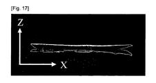
- a conceptual illustration is shown in Fig. 3, and actual photographs are shown in Figs. 15 to 17.
- the area and thickness of the above-described membrane may be the same as those of the above-described first embodiment (A).
- the biodegradable base layer and adhesion preventive layer in the second embodiment (B) may be layers produced by the same method as that for the biodegradable base layer and adhesion preventive layer in the above-described first embodiment (A), but are not limited thereto.
- tissue sandwiching part refers to one that sandwiches an edge of a tissue to protect the edge.
- normal line direction to a surface of a membrane refers to the arrow direction in Fig. 3 or the Z-axis directions in Figs. 15 to 17.
- the area of a tissue sandwiching part is about 0.5 to 90%, preferably 5 to 75%, more preferably about 10 to 50% but is not limited thereto.
- Examples of the method of producing the tissue sandwiching part of the present invention comprise:
- Each of the above-described lamination of a biodegradable base layer and an adhesion preventive layer, binder treatment, and suture may be performed in accordance with the above-described method described in the first embodiment (A), but is not limited thereto.
- the above-described slit may be accomplished by using a general-purpose instrument such as a microtome and knife.
- a particularly preferable example of the second embodiment (B) of an adhesion preventive kit of the present invention is a form that includes a membrane including a biodegradable base layer made of a collagen nonwoven fabric and an adhesion preventive layer made of a sponge of a mixture of collagen and hyaluronic acid, which membrane has an outermost surface constituted of the adhesion preventive layer and has a tissue sandwiching part at the surrounding part of the membrane.
- the adhesion preventive kit of the present invention may provide a novel method of preventing adhesion between an injured or deficient tissue and a surrounding tissue thereof.
- the method of preventing adhesion in the respective examples will be described with reference to drawings, but the present invention is not limited thereto.
- Fig. 1 shows an example of the method of preventing adhesion using a first embodiment (A) of the adhesion preventive kit of the present invention.
- a first membrane 1 covers an injured or deficient portion so that a biodegradable base layer 4 in the first membrane is faced to the injured or deficient tissue 6 to make prosthesis for the part.
- a second membrane 2 is used for prosthesis for the other surface of the injured or deficient tissue 6.
- the injured or deficient portion is sandwiched by the first membrane 1 and second membrane 2, which are arranged so that the injured or deficient portion is not exposed, so that the portion is not in physical contact with surrounding tissues 71 and 72, thereby providing a more efficient method of preventing adhesion than a conventional method.
- the positions of the first and second membranes may be reversed.
- Fig. 1 may be mainly applied to the case where a tissue such as pericardium, pleura, cerebral dura mater, diaphragm, stomach, esophagus, or digestive apparatus is injured or deficient, but is not limited thereto.
- a tissue such as pericardium, pleura, cerebral dura mater, diaphragm, stomach, esophagus, or digestive apparatus is injured or deficient, but is not limited thereto.
- Fig. 2 shows another example of the method of preventing adhesion using the first embodiment (A) of the adhesion preventive kit of the present invention.
- a second membrane 2 is arranged at a position facing the injured or deficient portion.
- a first membrane 1 covers the injured or deficient portion so that a biodegradable base layer 4 in the first membrane is faced to the injured or deficient tissue 6 to make prosthesis for the part. That is, the injured or deficient portion is not in physical contact with the surrounding tissues 71, 72, and the like, thereby providing a more efficient method of preventing adhesion than a conventional method.
- Fig. 2 may be mainly applied to the case where a tissue such as pericardium, pleura, diaphragm, or cerebral dura mater is injured or deficient, but is not limited thereto.
- a tissue such as pericardium, pleura, diaphragm, or cerebral dura mater is injured or deficient, but is not limited thereto.
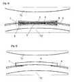
- Fig. 4 shows an example of the method of preventing adhesion using the second embodiment (B) of the adhesion preventive kit of the present invention.
- An injured or deficient tissue 6 is held by a tissue sandwiching part 8. That is, the injured or deficient portion is not exposed, so that the portion is not in physical contact with surrounding tissues 71, 72, and the like, thereby providing a more efficient method of preventing adhesion than a conventional method.
- a tissue such as pericardium, pleura, cerebral dura mater, diaphragm, stomach, esophagus, or a digestive apparatus is injured or deficient, but is not limited thereto.
- the method of preventing adhesion using adhesion preventive kits of the first embodiment (A) and the second embodiment (B) of the present invention or the use thereof is characterized by separating an injured or deficient tissue from a surrounding tissue by the adhesion preventive layer.
- Equal parts of an aqueous 1 wt% hydrofluoric acid solution and an aqueous 1 wt% collagen solution were mixed to give 250 mL of an equal part mixture of collagen and hydrofluoric acid.
- the mixture which was in acidic state, was neutralized with an aqueous solution of 0.1 N sodium hydroxide, and then the resultant was loaded into a metallic container having a rectangular space (about 10 cm square), followed by freezed at -20°C for about 12 hours.
- the frozen product was freeze-dried by using a lyophilizer (manufactured by EYELA Co.: FDU-830) under reduced pressure (0.1 torr or less) for about 24 hours and then compressed using a compressor (manufactured by Iuchi Seieido Co., Ltd.: 15 tons press machine) at a pressure of 100 kgf/cm 2 .
- a lyophilizer manufactured by EYELA Co.: FDU-830
- a compressor manufactured by Iuchi Seieido Co., Ltd.: 15 tons press machine
- an aqueous 1-wt% collagen solution was coated on the collagen nonwoven fabric as a binder treatment to fill the gaps between the filaments of the crosslinked laminated collagen nonwoven fabric, and then dried.
- a crosslinking reaction using thermal dehydration is performed in a vacuum drying oven at 120°C for 12 hours.
- the resultant product was neutralized with an aqueous 7.5 wt% sodium bicarbonate solution, and air-dried in a clean bench to give a biodegradable base layer (about 10 cm square) made of a collagen nonwoven fabric of a prosthetic membrane for tissue regeneration.
- Example 1 (1) By applying a neutralized solution containing a mixture of collagen and hyaluronic acid on the biodegradable base layer made of a collagen nonwoven fabric obtained in Example 1 (2), the adhesion preventive layer obtained in Example 1 (1) was bond to the biodegradable base layer.
- the resultant was air-dried in a clean bench and then performed a crosslinking reaction by thermal dehydration by heating it in a vacuum drying oven under reduced pressure (1 torr or less) at 110°C for 24 hours to give a first membrane.
- the adhesion preventive layer prepared in Example 1 (1) was used as a second membrane without any modification.
- the first membrane prepared in Example 1 (3) and the second membrane prepared in Example 1 (4) were subjected to gamma irradiation sterilization at an exposure dose of 25 kGy to give an adhesion preventive kit of the first embodiment of the present invention.
- Example 2 Adhesion prevention experiment using adhesion preventive kit of the present invention
- Example 7 the second membrane prepared in Example 1 (4) was trimmed to about 5 cm square and inserted through the pericardium-defective site, and the second membrane was arranged at a position facing the pericardium-defective site prepared in Example 2 (1) (Fig. 7).
- the first membrane prepared in Example 1 (3) was trimmed to about 5 cm square and immersed in sterilized distilled water for injection for about 5 to 10 minutes to soften sufficiently. Then, the collagen nonwoven fabric in the first membrane was faced to the pericardium-defective side prepared in Example 2 (1), and the membrane was fixed by continuous suture (Fig. 8). Thereafter, the thorax was closed, and follow-up was performed for three months.
- Comparative Example 1 Adhesion prevention experiment using only first membrane of the present invention
- Fig. 10 shows a photograph of the first membrane fixed on the pericardium-defective site by means of continuous suture in Comparative Example 1.
- ratio of adhesion area refers to a ratio of an area where adhesion occurred for the case that an area of a pericardium defect is expressed as 100%. Meanwhile, the degree of adhesion was judged by visual observation based on the determination criteria in Table 1.
- adhesion preventive kit of the present invention has an excellent ability to prevent adhesion and has a function to regenerate pericardium in a pericardium defect. Evaluation results of adhesion-preventive effect are shown in Table 2.
- a photograph of the adhesion state in Sample No. 1 of Example 2 is shown in Fig. 9. After the first and second membranes were degraded and absorbed, a pericardium-like membrane-like product was regenerated at the pericardium-defective site and there was no adhesion between the pericardium and heart.
- Photographs of adhesion states in Sample Nos. 1 and 4 of Comparative Example 1 are shown in Figs. 11 and 12, respectively.
- the first membrane was degraded and absorbed, and a pericardium-like membrane-like product was regenerated at the pericardium-defective site, but adhesions between the pericardium and heart were observed in some parts.
- Example 2 In the same way as in Example 1 (2), two laminated collagen nonwoven fabric were prepared, each of which had been subjected to a binder treatment with an aqueous 1-wt% collagen solution.
- the aqueous collagen solution was separately coated on the 2 cm square of central parts of the binder-treated laminated nonwoven fabrics, and then the fabrics are bonded each other. Then, crosslinking by thermal dehydration was performed in a vacuum drying oven under high vacuum (1 torr or less) at 120°C for 12 hours to give a biodegradable base layer.
- the layer was immersed in an aqueous 7.5-wt% sodium bicarbonate solution for 30 minutes to perform a neutralization treatment, and washed with distilled water and air-dried in a clean bench to give a biodegradable base layer (10 cm square) having a tissue sandwiching part.
- a neutralized solution containing a mixture of collagen and hyaluronic acid was applied on the both surfaces of the biodegradable base layer; and the adhesion preventive layers obtained in Example 1 (1) were separately laminated on the both surfaces of membranes of the two biodegradable base layers.
- the product was air-dried in a clean bench and heated in a vacuum drying oven under high vacuum (1 torr or less) at 110°C for 24 hours to perform a crosslinking reaction by thermal dehydration to prepare an adhesion preventive kit of a second embodiment (B) of the present invention. Photographs of the kit are shown in Figs. 15 to 17.
- the present invention is an innovative invention in surgery fields. Specifically, according to the present invention, the frequency and degree of adhesion are further reduced compared with a conventional adhesion preventive membrane, so that the time for an adhesion-peeling operation is drastically reduced in reoperation. That is, the labor of an operator required in the surgery is significantly reduced, and the blood loss of a patient is significantly suppressed in the surgery, thereby reducing patient burden.
Landscapes
- Health & Medical Sciences (AREA)
- Heart & Thoracic Surgery (AREA)
- Surgery (AREA)
- Vascular Medicine (AREA)
- Epidemiology (AREA)
- Life Sciences & Earth Sciences (AREA)
- Animal Behavior & Ethology (AREA)
- General Health & Medical Sciences (AREA)
- Public Health (AREA)
- Veterinary Medicine (AREA)
- Materials For Medical Uses (AREA)
Abstract
Description
- The present invention relates to an adhesion preventive kit for preventing adhesion in guided regeneration therapy for an injured tissue. The present invention relates to an adhesion preventive kit comprising: (A) a first membrane of at least two layers having a biodegradable base layer and an adhesion preventive layer provided respectively at outermost surfaces thereof and a second membrane of at least one layer having an adhesion preventive layer provided at an outermost surface thereof; or (B) an adhesion preventive membrane including a biodegradable base layer and an adhesion preventive layer, which membrane has an outermost surface constituted of the adhesion preventive layer and has a tissue sandwiching part.
- In various surgical operations, excision of affected parts and repair of injured sites are often performed. In particular, in the case where various organs, such as a lung, heart, liver, brain, digestive apparatus, and gallbladder, are targeted, a membrane-like material that covers the tissue of the organ must be filled or used for prosthesis on the cut surface, the deficient part, or the like. If the treatment is insufficiently done, the patient will die because of dysfunction of the organ in some cases. Also, even if the function of the organ itself is narrowly maintained, the body fluid, digestive juice, contents, etc. that exudes or leaks from the organ may cause infection, attacks or erosions to other organs, resulting in a crisis of life.
- Furthermore, adhesion between an injured or deficient portion in the above-described organ and a tissue located in the surrounding part thereof (hereinafter, also referred to as surrounding tissue) occurs in high frequencies. As a result, dysfunction of the organ may be induced with a lapse of time. In particular, in the cardiac surgery field, the pericardium is excised to perform a cardiac surgical operation, but reoperation may be required depending on conditions after the operation. In this case, adhesion of the pericardium to the heart and the surrounding tissue is caused, so that an operation of peeling the adhesion is required at the beginning. The operation is performed with the greatest care so as to minimize blood loss of the patient, so that it often takes about 3 to 5 hours to perform for this peeling operation.
- For the purpose of solving such various problems, prosthetic membranes to cover organs or tissues of the organs have been developed by using various materials. However, in the case where the above-described prosthetic membranes are made of a synthetic fiber or the like, insuficiency of biocompatibility causes various drawbacks such as excessive calcification, foreign body reaction, and inflammatory reaction. Even in the case of using a bioabsorbable material, such material as the material itself mediates adhesion between the injured or deficient tissue and another tissue corresponding thereto cannot be used.
- In addition, the above-described material must be a material having a function to prevent adhesion. Examples of a material that meets these requirements include hyaluronic acid and gelatin. Since those materials are mainly extracted from living bodies such as animals and purified, they have good biocompatibility and are already coming into practical use in various medical fields including medicines. Examples thereof include an adhesion preventive membrane using hyaluronic acid (
Patent Documents 1 to 4). - However, membranes made of the above-described biodegradable material lacked strength enough to resist sutures when used as prosthetic membranes. The inventors of the present invention have solved such problems and developed an adhesion preventive membrane having excellent suture strength in which all materials of a prosthetic membrane are composed of biodegradable and absorbable materials, and patent applications for the membrane have filed (
Patent Documents 5 to 9). - However, such adhesion preventive membranes were only used for prosthesis in the form shown in Fig. 5 for an injured or deficient portion. For example, in the case where injured or deficient portions of membrane-like, bag-like, and tube-like tissues are subjected to prosthesis, there is parts that directly come into contact with a surrounding tissue facing the opposite side of the surface subjected to prosthesis (A in Fig. 5), so that the edges of the injured tissue adhere to the surrounding tissue in some cases.
- Patent Document 1:
Japanese Laid-Open Patent Publication No. 61-234864
Patent Document 2:Japanese Laid-Open Patent Publication No. 06-073103
Patent Document 3:Japanese Laid-Open Patent Publication No. 08-157378
Patent Document 4:Japanese Laid-Open Patent Publication No. 08-333402
Patent Document 5:Japanese Laid-Open Patent Publication No. 2000-093497
Patent Document 6:Japanese Laid-Open Patent Publication No. 2000-210376
Patent Document 7:Japanese Laid-Open Patent Publication No. 2000-271207
Patent Document 8:Japanese Laid-Open Patent Publication No. 2003-235955
Patent Document 9:Japanese Laid-Open Patent Publication No. 2003-245351 - Therefore, it is required to develop an adhesion preventive kit and a method of preventing adhesion, which enable prevention of adhesion even in a surrounding part of an edge of an injured or deficient tissue.
- The present invention relates to the following:
- (1) an adhesion preventive kit comprising the following (A) or (B):
- (A) a first membrane of at least two layers having a biodegradable base layer and an adhesion preventive layer provided respectively at outermost surfaces thereof and a second membrane of at least one layer having an adhesion preventive layer provided at an outermost surface thereof; or
- (B) an adhesion preventive membrane including a biodegradable base layer and an adhesion preventive layer, which membrane has an outermost surface constituted of the adhesion preventive layer and has a tissue sandwiching part,
- (2) an adhesion preventive kit according to the item (1), wherein the biodegradable base layer contains collagen, polylactic acid, or polyglycolic acid,
- (3) an adhesion preventive kit according to the item (1), wherein the biodegradable base layer is composed of woven cloth, a nonwoven fabric, a sheet, or a sponge,
- (4) an adhesion preventive kit according to the item (1), wherein the biodegradable base layer is composed of a collagen nonwoven fabric,
- (5) an adhesion preventive kit according to the item (1), wherein the adhesion preventive layer contains hyaluronic acid, collagen, or gelatin,
- (6) an adhesion preventive kit according to the item (1), wherein the adhesion preventive layer is composed of a sheet or a sponge,
- (7) an adhesion preventive kit according to the item (1), wherein the adhesion preventive layer is composed of a sponge of a mixture of collagen and hyaluronic acid,
- (8) a method of producing an adhesion preventive kit which comprises (B) an adhesion preventive membrane including a biodegradable base layer and an adhesion preventive layer, which membrane has an outermost surface constituted of the adhesion preventive layer and has a tissue sandwiching part, wherein the method is characterized in that a surrounding part of the biodegradable base layer is branched in a normal line direction to a surface of the membrane,
- (9) a method of producing an adhesion preventive kit according to the item (8), characterized by comprising: preparing a membrane of at least two layers having a biodegradable base layer and an adhesion preventive layer provided respectively at outermost surfaces thereof; layering the two membranes so that the biodegradable base layers face each other; and bonding or sewing only a central part thereof,
- (10) a method of producing an adhesion preventive kit according to the item (8), characterized by comprising: layering two biodegradable base layers; bonding or sewing a central part thereof; and providing adhesion preventive layers at the outer surfaces of the biodegradable base layers,
- (11) a method of preventing adhesion characterized by preventing adhesion between an injured or deficient tissue and a surrounding tissue located in a surrounding part of the injured or deficient tissue using an adhesion preventive kit comprising the following (A) or (B):
- (A) a first membrane of at least two layers having a biodegradable base layer and an adhesion preventive layer provided respectively at outermost surfaces thereof and a second membrane of at least one layer having an adhesion preventive layer provided at an outermost surface thereof; or
- (B) an adhesion preventive membrane including a biodegradable base layer and an adhesion preventive layer, which membrane has an outermost surface constituted of the adhesion preventive layer and has a tissue sandwiching part,
- (12) a method of preventing adhesion according to the item (11), wherein the tissue is pericardium, pleura, diaphragm, cerebral dura mater, stomach, esophagus, or a digestive apparatus,
- (13) a method of preventing adhesion according to the item (11), wherein the tissue is pericardium, and the surrounding tissue is heart,
- (14) a method of preventing adhesion according to the item (11) characterized by separating the injured or deficient tissue from the surrounding tissue by at least the adhesion preventive layer, and
- (15) a use of an adhesion preventive kit comprising the following (A) or (B):
- (A) a first membrane of at least two layers having a biodegradable base layer and an adhesion preventive layer provided respectively at outermost surfaces thereof and a second membrane of at least one layer having an adhesion preventive layer provided at an outermost surface thereof; or
- (B) an adhesion preventive membrane including a biodegradable base layer and an adhesion preventive layer, which membrane has an outermost surface constituted of the adhesion preventive layer and has a tissue sandwiching part.
- According to the present invention, the frequency and degree of adhesion are further reduced compared with a conventional adhesion preventive membrane, so that the time for an adhesion-peeling operation is reduced in reoperation. That is, the labor of an operator required in the surgery is reduced, and the blood loss of a patient is significantly suppressed during the surgery, thereby reducing patient burden.
-
- [Fig. 1] A conceptional illustration of a surgical operation in the case of using an adhesion preventive kit of a first embodiment of the present invention.
- [Fig. 2] A conceptional illustration of a different surgical operation from that in Fig. 1 in the case of using an adhesion preventive kit of a first embodiment of the present invention.
- [Fig. 3] A conceptional illustration of an adhesion preventive kit of a second embodiment of the present invention.
- [Fig. 4] A conceptional illustration of a surgical operation mode in the case of using an adhesion preventive kit of a second embodiment of the present invention.
- [Fig. 5] A conceptional illustration of a surgical operation in the case of using a conventional adhesion preventive membrane.
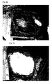
- [Fig. 6] A photograph of the pericardium-defective site of the beagle in Example 2.
- [Fig. 7] A photograph of the second membrane arranged on the surface of the heart of the beagle.
- [Fig. 8] A photograph of the first membrane sutured to the pericardium of the beagle.
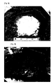
- [Fig. 9] A photograph of the portion between the pericardium and the heart after three months from embedment of an adhesion preventive kit of the present invention in the pericardium-defective site of the beagle.
- [Fig. 10] A photograph of the first membrane sutured to the pericardium of the male beagle in Comparative Example 1.
- [Fig. 11] A photograph of the portion between the pericardium and heart after three months from embedment of the first membrane in the pericardium-defective site of the male beagle in Comparative Example 1 (adhesion ratio: 80%).
- [Fig. 12] A photograph of the portion between the pericardium and the heart after three months from embedment of the first membrane in the pericardium-defective site of the male beagle in Comparative Example 1 (adhesion ratio: 50%).
- [Fig. 13] A photograph of the commercially available ePTFE membrane sutured to the pericardium of the male beagle in Comparative Example 2.
- [Fig. 14] A photograph of the portion between the pericardium and the heart after three months from application of the commercially available ePTFE membrane to the pericardium-defective site of the male beagle in Comparative Example 2.
- [Fig. 15] A photograph of the adhesion preventive kit of the second embodiment of the present invention when viewed from an angle (in tissue sandwiching parts, the portions parallel to the Y-axis are bent to the Z-axis direction).
- [Fig. 16] A photograph of an adhesion preventive kit of a second embodiment of the present invention in Fig. 15 when viewed from the XZ-plane.
- [Fig. 17] A photograph of the adhesion preventive kit in Fig. 16, in which, in tissue sandwiching parts, the portions parallel to the X-axis are bent to the Z-axis direction.
-
- 1
- First membrane of a first embodiment (A)
- 2
- Second membrane of a first embodiment (A)
- 3
- Membrane of a second embodiment (B)
- 4
- Biodegradable base layer
- 5
- Adhesion preventive layer
- 6
- Injured or deficient tissue
- 7
- Surrounding tissue
- 8
- Tissue sandwiching part
- 9
- Conventional adhesion preventive membrane
- The term "adhesion preventive kit" of the present invention refers to one to be used for preventing adhesion between an injured or deficient tissue and a surrounding tissue located in a position physically accessible thereto when an incised or excised tissue is intended for prosthesis or regeneration in regenerative or surgical operations for a tissue injured by an accident or the like.
- The term "tissue" as used in the present invention refers to an organ in a living body, a membrane tissue that cover the organ, and an apparatus. Examples of the organ include heart, liver, stomach, pancreas, gallbladder, and brain. Examples of the membrane tissue that covers the organ include pericardium, pleura, peritoneum, diaphragm, and cerebral dura mater. Examples of the apparatus include trachea, esophagus, and digestive apparatus.
- The term "injury" as used in the present invention refers to a state where a tissue is damaged, and examples thereof include, but are not limited to, accidental injury caused by a foreign matter from outside of a body and surgical injury due to incision or the like in surgical operations. The term "deficiency" as used refers to a state where part of a tissue is lacking, and examples thereof include, but are not limited to, surgical deficiency caused by excision of an affected part in surgical operations.
- The term "surrounding tissue" as used in the present invention refers to a tissue that is located in a surrounding part of the above-described injured or deficient tissue and is present at a position where adhesion may occur in regeneration of the above-described injured or deficient tissue. For example, in the case where the injured or deficient tissue is pericardium, the surrounding tissue thereof is heart, breastbone, and lung. In the case where the injured or deficient tissue is an apparatus such as trachea, esophagus, or digestive apparatus, the tissue also includes a wall surface facing an injured or deficient site in the apparatus.
- The adhesion preventive kit of the present invention comprises the following two embodiments (A) and (B), and a person skilled in the art may select both embodiments.
- (A) a first membrane of at least two layers having a biodegradable base layer and an adhesion preventive layer provided respectively at outermost surfaces thereof and a second membrane of at least one layer having an adhesion preventive layer provided at an outermost surface thereof; or
- (B) an adhesion preventive membrane including a biodegradable base layer and an adhesion preventive layer, which membrane has an outermost surface constituted of the adhesion preventive layer and has a tissue sandwiching part.
- The first embodiment (A) of an adhesion preventive kit of the present invention refers to a form that includes a first membrane of at least two layers having a biodegradable base layer and an adhesion preventive layer provided respectively at outermost surfaces thereof and a second membrane of at least one layer having an adhesion preventive layer provided at an outermost surface thereof. Depending on an injured or deficient tissue to be subjected to guided regeneration of the tissue, for example, in the case of guided regeneration of the pericardium excised in heart surgery operations, each of the above-described first and second membranes has an area of about 1 to 200 cm2, preferably about 15 to 150 cm2 and has a total thickness of about 0.1 to 30 mm, preferably about 0.5 to 8 mm, but the area and thickness are not limited thereto.
- The above-described first membrane refers to a membrane that includes at least a biodegradable base layer and an adhesion preventive layer and is a membrane of at least two layers having the biodegradable base layer and adhesion preventive layer provided respectively at outermost surfaces thereof, which has effects on preventing adhesion between an injured or deficient tissue and a surrounding tissue and on promoting regeneration of the tissue in the regeneration of the injured or deficient tissue.
- The above-described biodegradable base layer refers to a layer that has good biocompatibility when embedded in a living body, is made from a biodegradable and absorbable polymer to be degraded and absorbed after a certain period of time, and may enable guided regeneration of an injured or deficient tissue by using the biodegradable base layer as a scaffold. The term "biodegradable and absorbable polymer" refers to a material to be degraded and absorbed after a certain period of time from embedment in a living body. Examples thereof include collagen, polylactic acid, and polyglycolic acid. Collagen is preferable in view of excellent safety after embedded in a living body, adhesiveness to a cell, and a proliferative ability.
- For the above-described collagen, collagen that has been subjected to a treatment so as to be able to dissolve in a solvent may be selected. Examples thereof include solubilized collagens such as an enzyme-solubilized collagen, an acid-solubilized collagen, an alkali-solubilized collagen, and a neutral-solubilized collagen. Among them, in view of ease of handling, an acid-solubilized collagen is preferable. Moreover, in view of safety when embedded in a living body, the collagen is preferably atelocollagen that has been subjected to a removal treatment for telopeptide, which is an antigenic determinant, but is not limited thereto. The collagen is derived from an extract from skin, tendon, bone, cartilage, organs, and the like of animal species such as oxen, pigs, bird, fish, rabbits, sheep, mouse, or humans. In view of availability, the collagen is preferably a collagen derived from pig skin, but is not limited thereto. Furthermore, examples of the type of collagen include types I, II, and III, and in view of ease of handling, the type is preferably types I and III, but is not limited thereto.
- Examples of the form of a biodegradable base layer of the preset invention include a woven fabric, a nonwoven fabric, a sponge, and a sheet. Among them, in view of suture strength, a woven fabric and a nonwoven fabric are preferable. Moreover, in view of ease of production and cost, a nonwoven fabric is preferable, but is not limited thereto.
- In the case of using the above-described woven or nonwoven fabric, a strand-like material to constitute such a form is produced. In view of improvement of suture strength, the outer diameter of the strand-like material is about 0.01 to 1,000 µm, preferably about 0.05 to 200 µm, more preferably about 0.1 to 200 µm, but is not limited thereto.
- The above-described strand-like material may be produced in accordance with a conventional method. For example, it may be produced by continuously spinning it out of a solution of the above-described biodegradable and absorbable polymer. In view of strand strength, the concentration of the above-described solution is about 0.1 to 20 wt%, preferably about 1 to 10 wt%, but is not limited thereto. Examples of a device to be used for discharging the solution include a gear pump, a dispenser, and various types of extruders. To perform a uniform spinning, the device is preferably a dispenser in view of stable discharge of a fixed amount of the solution with less pulsation, but is not limited thereto.
- A solvent for a coagulation bath used in the wet spinning may be a solvent, suspension, emulsion, or solution that can coagulate the above-described biodegradable and absorbable polymer. In the case of using collagen as a raw material of a strand-like material, examples of the solvents include aqueous solutions of inorganic salts, inorganic salt-containing organic solvents, alcohols, and ketones. Examples of the aqueous solutions of inorganic salts include aqueous solutions of sodium sulfate, sodium chloride, ammonium sulfate, calcium chloride, and magnesium chloride. In particular, an aqueous solution of sodium chloride, sodium sulfate, or ammonium sulfate is preferable. A solvent obtained by dissolving or dispersing such inorganic salts in alcohols or acetones may also be used. Examples of the alcohols include methanol, ethanol, isopropanol, amyl alcohol, pentanol, hexanol, and ethylene glycol. Examples of the ketones include acetone and methyl ethyl ketone. Among them, in view of strength of a spun strand, ethanol, a solution of sodium chloride in ethanol, or a dispersion of sodium chloride in ethanol is preferably used, but the solvent is not limited thereto.
- In the case where a woven fabric is selected as a biodegradable base layer, the distance between strand-like materials is about 0.01 to 500 µm, preferably about 0.1 to 200 µm, and each distance between the strand-like materials is preferably uniform but is not limited thereto.
- The above-described woven fabric may be produced by a method using, for example, a weaving machine. Herein, in view of prevention of cutting in strand-like materials, the above-described strand-like materials are preferably modified into a twist yarn before use, but the form is not limited thereto.
- In the case where a nonwoven fabric is selected as a biodegradable base layer, in view of suture strength, the distance between the strand-like materials is about 0.01 to 500 µm, preferably about 0.1 to 200 µm, but the materials may partially be in contact each other.
- The above-described nonwoven fabric may have, for example, a form produced as a laminate that includes a first layer composed of a plurality of strand-like materials arranged substantially in parallel and a second layer composed of a plurality of strand-like materials arranged substantially in parallel such that the acute angle between the directions of arrangement of the strand-like materials is about 70° to 90°. Furthermore, the nonwoven fabric may have a form that includes a third layer composed of a plurality of strand-like materials arranged substantially in parallel on the second layer such that the acute angle between the directions of the arrangement of strand-like materials in the second layer and those in the third layer is about 70° to 90°. That is, in view of suture strength, the nonwoven fabric of the present invention preferably has a form that includes a layer composed of a plurality of strand-like materials arranged substantially in parallel (i.e., n layer; n is an integer of 2 or more) and upper and lower layers in contact with the n layer (i.e., n-1 layer or n+1 layer; n is an integer of 2 or more) such that the acute angle between the directions of the arranged strand-like materials in the n layer and those in the n-1 layer or n+1 layer is about 70° to 90°, but the form is not limited thereto. In view of suture strength and the membrane weight, the total number of these layered articles is about 2 to 20, preferably about 4 to 16, but is not limited thereto. The nonwoven fabric having the above-described layered form is referred to as a layered nonwoven fabric in the present invention.
- Moreover, the above-described sheet refers to a plane membrane having a surface that is nearly uniformly composed of the biodegradable and absorbable polymer. Examples of a method of producing them include extrusion molding, compression molding, and solvent casting methods, and in view of ease of production, the solvent casting method is preferable, but the method is not limited thereto.
- Meanwhile, the above-described sponge refers to a substance in a porous form in which sections having many pores with uniform or nonuniform sizes are observed to be continuously or discontinuously dispersed in visual determination or under a microscope. An example of a method of forming thereof includes a method that involves casting a solution of a biodegradable polymer into a mold having an intended shape of a injured site in a tissue, followed by forming the sponge by means of a method such as air-drying, vacuum drying, freeze-thaw, vacuum freeze-drying. Among them, in view of uniform formation, the sponge is preferably formed by means of vacuum freeze-drying, but the method is not limited thereto. Examples of the above-described vacuum freeze-drying include, but are not limited to, a method of drying an about 0.05 to 30 wt% collagen solution at about 0.08 Torr or less in view of ease of production. After freeze-drying, the resultant is removed form the mold to give a sponge. Furthermore, in view of suture strength, the above-described sponge is preferably compressed by a press machine, but the process is not limited thereto.
- In view of improvement of strength, the biodegradable base layer is preferably further immersed in a solution of a biodegradable and absorbable polymer and then air-dried, but the process is not limited thereto. Such a treatment is referred to as a binder treatment. In view of compatibility with the biodegradable base layer and strength, the biodegradable and absorbable polymer used in a binder treatment is preferably the same substance as that of the biodegradable base layer, but is not limited thereto. The binder treatment is performed by impregnating the above-described biodegradable base layer with a solution of a biodegradable and absorbable polymer or the like and drying it by an appropriate drying method such as air-drying, air-blast drying, drying under reduced pressure, drying at low temperature, or vacuum freeze-drying. A biodegradable base layer obtained by such binder treatment has drastically improved suture strength compared with an untreated biodegradable base layer. Meanwhile, in view of handling of the solution, the concentration of the biodegradable and absorbable polymer solution used in the binder treatment is about 0.05 to 30 wt%, preferably about 0.1 to 10 wt%, but is not limited thereto.
- The biodegradable base layer of the present invention is most preferably a nonwoven fabric made of collagen strand in view of ease of production, suture strength, degradation resistance, and safety after embedded in a living body.
- The term "adhesion preventive layer" as used in the present invention refers to a layer having a preventive effect on adhesion between an injured or deficient tissue and a surrounding tissue thereof in the regeneration of the tissue. An example of a raw material thereof includes a polymer having a preventive effect on adhesion. Examples thereof include collagen, gelatin, and hyaluronic acid, and the material is preferably hyaluronic acid having an excellent preventive effect on adhesion, and particularly preferably a mixture of collagen and hyaluronic acid that has improved degradation resistance.
- The above-described hyaluronic acid may be derived from an animal or microorganism. Moreover, it may be used as a salt of an alkaline metal (e.g., sodium, potassium) or the like. Among them, from the viewpoint that the hyaluronic acid may be embedded in a living body, medical grade hyaluronic acid is preferable.
- Meanwhile, in view of adhesion prevention and degradation resistance, a mixing ratio of collagen and hyaluronic acid is about 3:7 to 7:3, preferably 5:5, but is not limited thereto.
- Moreover, examples of the form of the above-described adhesion preventive layer include a sheet and a sponge. Among them, a sponge is preferable in view of ease in lamination on a biodegradable base layer. A method of producing a sheet or a sponge may be the same as the above-described method of producing a biodegradable base layer, but is not limited thereto.
- The adhesion preventive layer of the present invention is most preferably in the form of sponge made of a mixture of collagen and hyaluronic acid in view of ease of production, adhesion preventive effect, degradation resistance, and safety after embedded in a living body.
- Examples of the lamination method of a biodegradable base layer and an adhesion preventive layer in the above-described first membrane include a method of forming an adhesion preventive layer directly on a biodegradable base layer and a method of separately producing a biodegradable base layer and adhesion preventive layer and then laminating the layers. Among them, in view of ease of production, the method is preferably the method of separately producing a biodegradable base layer and adhesion preventive layer and then laminating the layers, but is not limited thereto.
- An example of the above-described method of forming an adhesion preventive layer directly on a biodegradable base layer includes, but are not limited to, a method of forming an adhesion preventive layer by immersing a biodegradable base layer made of a nonwoven fabric in a solution of a biodegradable and absorbable polymer, followed by freeze-drying.
- On the other hand, examples of the method of separately producing a biodegradable base layer and adhesion preventive layer and then laminating the layers include the above-described binder treatment and suture with thread. Among them, in view of ease of production, the method is preferably the above-described binder treatment using a biodegradable and absorbable polymer but is not limited thereto. The above-mentioned binder treatment may be performed in accordance with the above-described method, but is not limited thereto.
- Examples of the above-described suture include a method using a commercially available surgical thread, the above-described strand-like material made from a biodegradable and absorbable polymer, and the like. Among them, in view of safety when embedded in a living body, it is preferable to sew with the above-described strand-like material made from a biodegradable and absorbable polymer, but is not limited thereto. In view of membrane strength, the sewing distance is about 1 to 20 mm, preferably about 2 to 10 mm, and the pitch distance of a sewing machine is about 1 to 20 mm, preferably about 2 to 10 mm, but the distances are not limited thereto.
- The above-described biodegradable base layer and adhesion preventive layer are preferably further subjected to a crosslinking treatment, if necessary. The crosslinking treatment may enable timely control of the time to degrade the adhesion preventive layer in a living body. Examples of a crosslinking method include chemical crosslinking, gamma-irradiation, ultraviolet irradiation, electron beam irradiation, plasma irradiation, and crosslinking treatment by thermal dehydration. Among them, in the case where the above-described biodegradable base layer and adhesion preventive layer include collagen, in view of safety after embedded in a living body, the method is preferably the crosslinking treatment by thermal dehydration, but is not limited thereto. In the crosslinking treatment, degradability and absorbability in a living body can be controlled by crosslinking temperature and crosslinking time.
- The above-described second membrane includes at least an adhesion preventive layer. The adhesion preventive layer may be a layer produced by the same method as that for the adhesion preventive layer in the above-described first membrane. Among them, in view of production cost, the form of the second membrane is preferably the same as that of the adhesion preventive layer in the above-described first membrane, but is not limited thereto.
- In view of production cost, ease of production, membrane strength, safety when embedded in a living body, and the like, a preferable example of a first embodiment (A) of an adhesion preventive kit of the present invention is a form that includes a first membrane of at least two layers having a biodegradable base layer made of a collagen nonwoven fabric and an adhesion preventive layer made of sponge of a mixture of collagen and hyaluronic acid provided respectively at outermost surfaces thereof and a second membrane of at least one layer having an adhesion preventive layer made of sponge of a mixture of collagen and hyaluronic acid provided at an outermost surface thereof.
- The second embodiment (B) of an adhesion preventive kit of the present invention is a form that includes an adhesion preventive membrane including a biodegradable base layer and an adhesion preventive layer, which membrane has an outermost surface constituted of the adhesion preventive layer and has a tissue sandwiching part. A conceptual illustration is shown in Fig. 3, and actual photographs are shown in Figs. 15 to 17. The area and thickness of the above-described membrane may be the same as those of the above-described first embodiment (A).
- The biodegradable base layer and adhesion preventive layer in the second embodiment (B) may be layers produced by the same method as that for the biodegradable base layer and adhesion preventive layer in the above-described first embodiment (A), but are not limited thereto.
- The term "tissue sandwiching part" as used in the present invention refers to one that sandwiches an edge of a tissue to protect the edge. The term "normal line direction to a surface of a membrane" refers to the arrow direction in Fig. 3 or the Z-axis directions in Figs. 15 to 17. With regard to the size of the sandwiching part, in view of strength of the membrane and ease in sandwiching a tissue, for example, in the case where the area of the surface of an adhesion preventive membrane is expressed as 100%, the area of a tissue sandwiching part is about 0.5 to 90%, preferably 5 to 75%, more preferably about 10 to 50% but is not limited thereto.
- Examples of the method of producing the tissue sandwiching part of the present invention comprise:
- (i) a method that includes the steps of: laminating two membranes composed of two layers of a biodegradable base layer and an adhesion preventive layer so that the biodegradable base layers face each other; and bonding or sewing only the central part thereof,
- (ii) a method that includes the steps of: laminating two membranes composed of a biodegradable base layer; bonding or sewing the central part thereof; and laminating an adhesion preventive layer on the outer surface of the biodegradable base layer,
- (iii) a method that includes the steps of: separately preparing a membrane-like material composed of a biodegradable base layer and an adhesion preventive layer and a membrane-like material composed of an adhesion preventive layer; and laminating these membranes and bonding or sewing the central part thereof,
- (iv) a method that includes the steps of: preparing a membrane having laminated adhesion preventive layers on the both surfaces of a biodegradable base layer; and making slits in an adhesion preventive layer, in a biodegradable base layer, or between an adhesion preventive layer and a biodegradable base layer in parallel with the surface of the membrane, and
- (v) a method that includes the steps of: making slits in a biodegradable base layer in parallel with the surface of the membrane; and laminating an adhesion preventive layer on the outer surface of the biodegradable base layer. Among them, in view of membrane strength and ease of production, the method is preferably (ii) a method that includes the steps of: laminating two membranes composed of a biodegradable base layer; bonding or sewing the central part thereof; and laminating an adhesion preventive layer on the outer surface of the biodegradable base layer, but is not limited thereto. The above-described membrane-like material composed of two layers of a biodegradable base layer and an adhesion preventive layer has the same structure as that of the first membrane in the first embodiment (A) of the present invention.
- Each of the above-described lamination of a biodegradable base layer and an adhesion preventive layer, binder treatment, and suture may be performed in accordance with the above-described method described in the first embodiment (A), but is not limited thereto.
- Moreover, the above-described slit may be accomplished by using a general-purpose instrument such as a microtome and knife.
- In view of production cost, ease of production, membrane strength, safety when embedded in a living body, and the like, a particularly preferable example of the second embodiment (B) of an adhesion preventive kit of the present invention is a form that includes a membrane including a biodegradable base layer made of a collagen nonwoven fabric and an adhesion preventive layer made of a sponge of a mixture of collagen and hyaluronic acid, which membrane has an outermost surface constituted of the adhesion preventive layer and has a tissue sandwiching part at the surrounding part of the membrane.
- The adhesion preventive kit of the present invention may provide a novel method of preventing adhesion between an injured or deficient tissue and a surrounding tissue thereof. Hereinafter, the method of preventing adhesion in the respective examples will be described with reference to drawings, but the present invention is not limited thereto.
- Fig. 1 shows an example of the method of preventing adhesion using a first embodiment (A) of the adhesion preventive kit of the present invention. On one surface of an injured or
deficient tissue 6, afirst membrane 1 covers an injured or deficient portion so that abiodegradable base layer 4 in the first membrane is faced to the injured ordeficient tissue 6 to make prosthesis for the part. Subsequently, asecond membrane 2 is used for prosthesis for the other surface of the injured ordeficient tissue 6. That is, the injured or deficient portion is sandwiched by thefirst membrane 1 andsecond membrane 2, which are arranged so that the injured or deficient portion is not exposed, so that the portion is not in physical contact with surroundingtissues - The example shown in Fig. 1 may be mainly applied to the case where a tissue such as pericardium, pleura, cerebral dura mater, diaphragm, stomach, esophagus, or digestive apparatus is injured or deficient, but is not limited thereto.
- Fig. 2 shows another example of the method of preventing adhesion using the first embodiment (A) of the adhesion preventive kit of the present invention. In a surrounding
tissue 71 of an injured ordeficient tissue 6, asecond membrane 2 is arranged at a position facing the injured or deficient portion. Subsequently, on the surface opposite to the surroundingtissue 71 of the injured ordeficient tissue 6, afirst membrane 1 covers the injured or deficient portion so that abiodegradable base layer 4 in the first membrane is faced to the injured ordeficient tissue 6 to make prosthesis for the part. That is, the injured or deficient portion is not in physical contact with the surroundingtissues - The example shown in Fig. 2 may be mainly applied to the case where a tissue such as pericardium, pleura, diaphragm, or cerebral dura mater is injured or deficient, but is not limited thereto.
- Fig. 4 shows an example of the method of preventing adhesion using the second embodiment (B) of the adhesion preventive kit of the present invention. An injured or
deficient tissue 6 is held by atissue sandwiching part 8. That is, the injured or deficient portion is not exposed, so that the portion is not in physical contact with surroundingtissues - The example shown in Fig. 4 may be mainly applied to the case where a tissue such as pericardium, pleura, cerebral dura mater, diaphragm, stomach, esophagus, or a digestive apparatus is injured or deficient, but is not limited thereto.
- Therefore, the method of preventing adhesion using adhesion preventive kits of the first embodiment (A) and the second embodiment (B) of the present invention or the use thereof is characterized by separating an injured or deficient tissue from a surrounding tissue by the adhesion preventive layer.
- Hereinafter, the present invention will be described in detail by means of examples, but is not limited to these examples.
- Equal parts of an aqueous 1 wt% hydrofluoric acid solution and an aqueous 1 wt% collagen solution were mixed to give 250 mL of an equal part mixture of collagen and hydrofluoric acid. The mixture, which was in acidic state, was neutralized with an aqueous solution of 0.1 N sodium hydroxide, and then the resultant was loaded into a metallic container having a rectangular space (about 10 cm square), followed by freezed at -20°C for about 12 hours. Then, the frozen product was freeze-dried by using a lyophilizer (manufactured by EYELA Co.: FDU-830) under reduced pressure (0.1 torr or less) for about 24 hours and then compressed using a compressor (manufactured by Iuchi Seieido Co., Ltd.: 15 tons press machine) at a pressure of 100 kgf/cm2. In this way, an adhesion preventive layer (about 10 cm square) made of sponge of a mixture of collagen and hyaluronic acid was obtained.
- 150 mL of an aqueous 7 wt% acid-solubilized collagen solution was extruded into a coagulation bath containing 3 L of 99.5 vol% ethanol, dehydrated and coagulated, and then the resultant collagen strand was laminated in accordance with the method described in
Japanese Laid-Open Patent Publication No. 2000-93497 - By applying a neutralized solution containing a mixture of collagen and hyaluronic acid on the biodegradable base layer made of a collagen nonwoven fabric obtained in Example 1 (2), the adhesion preventive layer obtained in Example 1 (1) was bond to the biodegradable base layer. The resultant was air-dried in a clean bench and then performed a crosslinking reaction by thermal dehydration by heating it in a vacuum drying oven under reduced pressure (1 torr or less) at 110°C for 24 hours to give a first membrane.
- The adhesion preventive layer prepared in Example 1 (1) was used as a second membrane without any modification.
- The first membrane prepared in Example 1 (3) and the second membrane prepared in Example 1 (4) were subjected to gamma irradiation sterilization at an exposure dose of 25 kGy to give an adhesion preventive kit of the first embodiment of the present invention.
- The thorax of each of six beagles (average body weight: about 10.5 kg) was opened between the left ribs under continuous anesthesia to expose the heart, and then about 4 cm square of the pericardium covering the surface of the heart was excised to prepare pericardium defect (Fig. 6).
- Next, the second membrane prepared in Example 1 (4) was trimmed to about 5 cm square and inserted through the pericardium-defective site, and the second membrane was arranged at a position facing the pericardium-defective site prepared in Example 2 (1) (Fig. 7).
- The first membrane prepared in Example 1 (3) was trimmed to about 5 cm square and immersed in sterilized distilled water for injection for about 5 to 10 minutes to soften sufficiently. Then, the collagen nonwoven fabric in the first membrane was faced to the pericardium-defective side prepared in Example 2 (1), and the membrane was fixed by continuous suture (Fig. 8). Thereafter, the thorax was closed, and follow-up was performed for three months.
- All the same procedures as in Example 2 were performed except that the step of Example 2 (2) was not performed. That is, only the procedures of Example 2 (1) and (3) were performed. Fig. 10 shows a photograph of the first membrane fixed on the pericardium-defective site by means of continuous suture in Comparative Example 1.
- During the follow-up for three months, all the six beagles of Example 2 and Comparative Example 1 had no particular abnormalities and were confirmed to maintain healthy conditions. After three months, the thoraxes were opened again by median incision, and the ratios of pericardium-heart adhesion area and the degree of adhesion were evaluated. Herein, the term "ratio of adhesion area" refers to a ratio of an area where adhesion occurred for the case that an area of a pericardium defect is expressed as 100%. Meanwhile, the degree of adhesion was judged by visual observation based on the determination criteria in Table 1.
-
[Table 1] Degree of adhesion Grade No adhesion 0 Manually peelable adhesion 1 Adhesion peelable by an instrument or the like 2 Adhesion that cannot be peeled without incision by an instrument or the like 3 - As a result, in Example 2, adhesion was observed in 2/6 cases. In both the two cases, the degrees of adhesion were
grade 3, while the ratios of adhesion area were 10% in both the two cases. In other words, it may be said that an operator had little trouble in the adhesion peeling operation. On the other hand, in Comparative Example 1, adhesion was observed in 3 cases, and the degrees of adhesion weregrade 3 in all the cases. However, the ratios of adhesion area were 50, 80, and 100%, respectively. From the above results, it is revealed that the adhesion preventive kit of the present invention has an excellent ability to prevent adhesion and has a function to regenerate pericardium in a pericardium defect. Evaluation results of adhesion-preventive effect are shown in Table 2. -
[Table 2] Sample No. Example 2 Comparative Example 1 Ratio of adhesion area Degree of adhesion Ratio of adhesion area Degree of adhesion 1 0 0 80 3 2 0 0 0 0 3 10 3 100 3 4 10 3 50 3 5 0 0 0 0 6 0 0 0 0 Average 3.3 1 38.3 1.5 - Moreover, a photograph of the adhesion state in Sample No. 1 of Example 2 is shown in Fig. 9. After the first and second membranes were degraded and absorbed, a pericardium-like membrane-like product was regenerated at the pericardium-defective site and there was no adhesion between the pericardium and heart.
- Photographs of adhesion states in Sample Nos. 1 and 4 of Comparative Example 1 are shown in Figs. 11 and 12, respectively. The first membrane was degraded and absorbed, and a pericardium-like membrane-like product was regenerated at the pericardium-defective site, but adhesions between the pericardium and heart were observed in some parts.
- The same procedures as in Comparative Example 1 were performed except that a commercially available adhesion preventive membrane (registered trademark: GORE-TEX), trimmed to about 5 cm square and made of expanded polytetrafluoroethylene (ePTFE), was used instead of the first membrane in Comparative Example 1. A photograph of the commercially available adhesion preventive membrane fixed on the pericardium-defective site by means of continuous suture in Comparative Example 2 is shown in Fig. 13.
- During the follow-up for three months, the beagles had no particular abnormalities and were confirmed to maintain healthy conditions. After a lapse of three months, the thoraxes were opened again by median incision to perform evaluation, and GORE-TEX (registered trademark) was found to remain in each of the living bodies. Moreover, a severe degree of adhesion was formed between the pericardium and heart (Fig. 14).
- In the same way as in Example 1 (2), two laminated collagen nonwoven fabric were prepared, each of which had been subjected to a binder treatment with an aqueous 1-wt% collagen solution. The aqueous collagen solution was separately coated on the 2 cm square of central parts of the binder-treated laminated nonwoven fabrics, and then the fabrics are bonded each other. Then, crosslinking by thermal dehydration was performed in a vacuum drying oven under high vacuum (1 torr or less) at 120°C for 12 hours to give a biodegradable base layer. Subsequently, the layer was immersed in an aqueous 7.5-wt% sodium bicarbonate solution for 30 minutes to perform a neutralization treatment, and washed with distilled water and air-dried in a clean bench to give a biodegradable base layer (10 cm square) having a tissue sandwiching part. A neutralized solution containing a mixture of collagen and hyaluronic acid was applied on the both surfaces of the biodegradable base layer; and the adhesion preventive layers obtained in Example 1 (1) were separately laminated on the both surfaces of membranes of the two biodegradable base layers. The product was air-dried in a clean bench and heated in a vacuum drying oven under high vacuum (1 torr or less) at 110°C for 24 hours to perform a crosslinking reaction by thermal dehydration to prepare an adhesion preventive kit of a second embodiment (B) of the present invention. Photographs of the kit are shown in Figs. 15 to 17.
- The present invention is an innovative invention in surgery fields. Specifically, according to the present invention, the frequency and degree of adhesion are further reduced compared with a conventional adhesion preventive membrane, so that the time for an adhesion-peeling operation is drastically reduced in reoperation. That is, the labor of an operator required in the surgery is significantly reduced, and the blood loss of a patient is significantly suppressed in the surgery, thereby reducing patient burden.
Claims (15)
- An adhesion preventive kit, comprising:(A) a first membrane of at least two layers having a biodegradable base layer and an adhesion preventive layer provided respectively at outermost surfaces thereof and a second membrane of at least one layer having an adhesion preventive layer provided at an outermost surface thereof; or(B) an adhesion preventive membrane including a biodegradable base layer and an adhesion preventive layer, which membrane has an outermost surface constituted of the adhesion preventive layer and has a tissue sandwiching part.
- An adhesion preventive kit according to Claim 1, wherein the biodegradable base layer contains collagen, polylactic acid, or polyglycolic acid.
- An adhesion preventive kit according to Claim 1, wherein the biodegradable base layer is composed of woven cloth, a nonwoven fabric, a sheet, or a sponge.
- An adhesion preventive kit according to Claim 1, wherein the biodegradable base layer is composed of a collagen nonwoven fabric.
- An adhesion preventive kit according to Claim 1, wherein the adhesion preventive layer contains hyaluronic acid, collagen, or gelatin.
- An adhesion preventive kit according to Claim 1, wherein the adhesion preventive layer is composed of a sheet or a sponge.
- An adhesion preventive kit according to Claim 1, wherein the adhesion preventive layer is composed of a sponge of a mixture of collagen and hyaluronic acid.
- A method of producing an adhesion preventive kit which comprises (B) an adhesion preventive membrane including a biodegradable base layer and an adhesion preventive layer, which membrane has an outermost surface constituted of the adhesion preventive layer and has a tissue sandwiching part, wherein the method is characterized in that a surrounding part of the biodegradable base layer is branched in a normal line direction to a surface of the membrane.
- A method of producing an adhesion preventive kit according to Claim 8, characterized by comprising: preparing a membrane of at least two layers having a biodegradable base layer and an adhesion preventive layer provided respectively at outermost surfaces thereof; layering the two membranes so that the biodegradable base layers face each other; and bonding or sewing only a central part thereof.
- A method of producing an adhesion preventive kit according to Claim 8, characterized by comprising: layering two biodegradable base layers; bonding or sewing a central part thereof; and providing adhesion preventive layers at the outer surfaces of the biodegradable base layers.
- A method of preventing adhesion, characterized by preventing adhesion between an injured or deficient tissue and a surrounding tissue located in a surrounding part of the injured or deficient tissue using an adhesion preventive kit comprising the following (A) or (B):(A) a first membrane of at least two layers having a biodegradable base layer and an adhesion preventive layer provided respectively at outermost surfaces thereof and a second membrane of at least one layer having an adhesion preventive layer provided at an outermost surface thereof; or(B) an adhesion preventive membrane including a biodegradable base layer and an adhesion preventive layer, which membrane has an outermost surface constituted of the adhesion preventive layer and has a tissue sandwiching part.
- A method of preventing adhesion according to Claim 11, wherein the tissue is pericardium, pleura, diaphragm, cerebral dura mater, stomach, esophagus, or a digestive apparatus.
- A method of preventing adhesion according to Claim 11, wherein the tissue is pericardium, and the surrounding tissue is heart.
- A method of preventing adhesion according to Claim 11, characterized by separating the injured or deficient tissue from the surrounding tissue by at least the adhesion preventive layer.
- A use of an adhesion preventive kit comprising the following (A) or (B):(A) a first membrane of at least two layers having a biodegradable base layer and an adhesion preventive layer provided respectively at outermost surfaces thereof and a second membrane of at least one layer having an adhesion preventive layer provided at an outermost surface thereof; or(B) an adhesion preventive membrane including a biodegradable base layer and an adhesion preventive layer, which membrane has an outermost surface constituted of the adhesion preventive layer and has a tissue sandwiching part.
Applications Claiming Priority (3)
| Application Number | Priority Date | Filing Date | Title |
|---|---|---|---|
| JP2004102638 | 2004-03-31 | ||
| JP2004264902 | 2004-09-13 | ||
| PCT/JP2005/006369 WO2005094915A1 (en) | 2004-03-31 | 2005-03-31 | Antiadhesive kit, process for producing the same and method of adhesion prevention |
Publications (3)
| Publication Number | Publication Date |
|---|---|
| EP1731180A1 true EP1731180A1 (en) | 2006-12-13 |
| EP1731180A4 EP1731180A4 (en) | 2010-07-21 |
| EP1731180B1 EP1731180B1 (en) | 2017-10-11 |
Family
ID=35063550
Family Applications (1)
| Application Number | Title | Priority Date | Filing Date |
|---|---|---|---|
| EP05727265.0A Expired - Lifetime EP1731180B1 (en) | 2004-03-31 | 2005-03-31 | Antiadhesive kit, process for producing the same and method of adhesion prevention |
Country Status (4)
| Country | Link |
|---|---|
| US (2) | US20080241203A1 (en) |
| EP (1) | EP1731180B1 (en) |
| JP (1) | JP4840136B2 (en) |
| WO (1) | WO2005094915A1 (en) |
Cited By (1)
| Publication number | Priority date | Publication date | Assignee | Title |
|---|---|---|---|---|
| EP3135315A4 (en) * | 2014-04-24 | 2017-11-29 | Tokai University Educational System | Polymer laminate |
Families Citing this family (13)
| Publication number | Priority date | Publication date | Assignee | Title |
|---|---|---|---|---|
| WO2007103993A2 (en) * | 2006-03-07 | 2007-09-13 | Axle International | Bioactive scaffold for therapeutic and adhesion prevention applications |
| JP5143396B2 (en) * | 2006-10-30 | 2013-02-13 | 川澄化学工業株式会社 | Anti-adhesive material |
| AR069109A1 (en) | 2007-10-30 | 2009-12-30 | Baxter Int | USE OF A REGENERATIVE BIOFUNCTIONAL COLOGEN BIOMATRIZ TO TREAT VISCERAL OR PARIETAL DEFECTS |
| US9889230B2 (en) * | 2008-10-17 | 2018-02-13 | Covidien Lp | Hemostatic implant |
| JP2010279574A (en) * | 2009-06-05 | 2010-12-16 | Nipro Corp | Anti-adhesion membrane |
| WO2011081162A1 (en) * | 2009-12-28 | 2011-07-07 | 川澄化学工業株式会社 | Anti-adhesion material |
| DE102011004239A1 (en) * | 2011-02-16 | 2012-08-16 | Gelita Ag | Use of a medical implant as an adhesion barrier |
| ES2797484T3 (en) | 2013-03-15 | 2020-12-02 | Toray Industries | Laminated film using polylactic acid based resin |
| TWI547529B (en) * | 2014-02-14 | 2016-09-01 | 瀚醫生技股份有限公司 | Method for forming dual layer composite material and dual layer composite material thereby |
| WO2015194616A1 (en) | 2014-06-18 | 2015-12-23 | 東レ株式会社 | Laminate and production method therefor |
| WO2016093162A1 (en) | 2014-12-10 | 2016-06-16 | 東レ株式会社 | Laminated film and sheet for medical use |
| WO2016181977A1 (en) | 2015-05-11 | 2016-11-17 | 東レ株式会社 | Polymer film, and liquid dispersion and agglomerate using same |
| KR102157057B1 (en) | 2016-03-23 | 2020-09-17 | 도레이 카부시키가이샤 | Laminate |
Family Cites Families (17)
| Publication number | Priority date | Publication date | Assignee | Title |
|---|---|---|---|---|
| US4925924A (en) * | 1984-03-27 | 1990-05-15 | University Of Medicine And Dentistry Of New Jersey | Biocompatible synthetic and collagen compositions having a dual-type porosity for treatment of wounds and pressure ulcers and therapeutic methods thereof |
| MX163953B (en) * | 1984-03-27 | 1992-07-03 | Univ New Jersey Med | PROCEDURE FOR PREPARING A BIODEGRADABLE COLLAGEN MATRIX |
| US4600533A (en) * | 1984-12-24 | 1986-07-15 | Collagen Corporation | Collagen membranes for medical use |
| SE8501022L (en) * | 1985-03-01 | 1986-09-02 | Pharmacia Ab | FORMAT CREATES AND PROCEDURES FOR ITS PREPARATION |
| IT1198449B (en) * | 1986-10-13 | 1988-12-21 | F I D I Farmaceutici Italiani | ESTERS OF POLYVALENT ALCOHOLS OF HYALURONIC ACID |
| US4937270A (en) * | 1987-09-18 | 1990-06-26 | Genzyme Corporation | Water insoluble derivatives of hyaluronic acid |
| US5171273A (en) * | 1989-01-13 | 1992-12-15 | University Of Medicine And Dentistry Of New Jersey | Synthetic collagen orthopaedic structures such as grafts, tendons and other structures |
| GB2282328B (en) * | 1993-09-29 | 1997-10-08 | Johnson & Johnson Medical | Absorbable structures for ligament and tendon repair |
| TW501934B (en) * | 1996-11-20 | 2002-09-11 | Tapic Int Co Ltd | Collagen material and process for making the same |
| FR2759084B1 (en) * | 1997-02-06 | 1999-10-29 | Dev Des Utilisations Du Collag | COLLAGENIC MATERIAL USEFUL IN PARTICULAR FOR THE PREVENTION OF POST-OPERATIVE ADHESIONS |
| US6977231B1 (en) * | 1999-01-21 | 2005-12-20 | Nipro Corporation | Suturable adhesion-preventing membrane |
| JP3463596B2 (en) | 1999-03-29 | 2003-11-05 | ニプロ株式会社 | Suture-resistant adhesion prevention membrane |
| JP3536295B2 (en) * | 1999-01-21 | 2004-06-07 | ニプロ株式会社 | Sutureable tissue regeneration induced adhesion prevention membrane |
| US6773723B1 (en) * | 2000-08-30 | 2004-08-10 | Depuy Acromed, Inc. | Collagen/polysaccharide bilayer matrix |
| US20030114061A1 (en) * | 2001-12-13 | 2003-06-19 | Kazuhisa Matsuda | Adhesion preventive membrane, method of producing a collagen single strand, collagen nonwoven fabric and method and apparatus for producing the same |
| JP4595279B2 (en) * | 2001-12-13 | 2010-12-08 | ニプロ株式会社 | Anti-adhesion membrane |
| EP1545389B1 (en) * | 2002-07-31 | 2020-04-22 | Mast Biosurgery AG | Apparatus for preventing adhesions between an implant and surrounding tissues |
-
2005
- 2005-03-31 JP JP2006511824A patent/JP4840136B2/en not_active Expired - Fee Related
- 2005-03-31 US US10/594,801 patent/US20080241203A1/en not_active Abandoned
- 2005-03-31 WO PCT/JP2005/006369 patent/WO2005094915A1/en not_active Ceased
- 2005-03-31 EP EP05727265.0A patent/EP1731180B1/en not_active Expired - Lifetime
-
2015
- 2015-11-12 US US14/939,374 patent/US20160121031A1/en not_active Abandoned
Cited By (1)
| Publication number | Priority date | Publication date | Assignee | Title |
|---|---|---|---|---|
| EP3135315A4 (en) * | 2014-04-24 | 2017-11-29 | Tokai University Educational System | Polymer laminate |
Also Published As
| Publication number | Publication date |
|---|---|
| US20160121031A1 (en) | 2016-05-05 |
| EP1731180A4 (en) | 2010-07-21 |
| JPWO2005094915A1 (en) | 2008-02-14 |
| JP4840136B2 (en) | 2011-12-21 |
| EP1731180B1 (en) | 2017-10-11 |
| WO2005094915A1 (en) | 2005-10-13 |
| US20080241203A1 (en) | 2008-10-02 |
Similar Documents
| Publication | Publication Date | Title |
|---|---|---|
| US20160121031A1 (en) | Antiadhesive Kit and Method of Adhesion Prevention | |
| EP1319415B1 (en) | Adhesion preventive membrane, method of producing a collagen single strand, collagen nonwoven fabric and method and apparatus for producing the same | |
| US6977231B1 (en) | Suturable adhesion-preventing membrane | |
| DE69729314T2 (en) | COLLAGEN LAMINATE MATERIAL AND METHOD FOR THE PRODUCTION THEREOF | |
| US8932619B2 (en) | Dural repair material | |
| DE69431376T2 (en) | Use of a device for inhibiting postoperative adhesion | |
| US7084082B1 (en) | Collagen material and its production process | |
| TWI461227B (en) | Use of a regenerative biofunctional collagen biomatrix for treating visceral or parietal defects | |
| JPWO1998022157A1 (en) | Collagen material and its manufacturing method | |
| WO2007082295A2 (en) | Suturable dural and meningeal repair product comprising collagen matrix | |
| JP2000271207A (en) | Suture-resistant adhesion prevention membrane | |
| JP4968976B2 (en) | Collagen material and production method thereof | |
| JP3536295B2 (en) | Sutureable tissue regeneration induced adhesion prevention membrane | |
| AU2011244877A1 (en) | Hemostatic implant | |
| JP3726280B2 (en) | Medical collagen membrane | |
| JP4345296B2 (en) | Anti-adhesion membrane | |
| JP4595279B2 (en) | Anti-adhesion membrane | |
| WO2015049800A1 (en) | Bioadhesive medical film | |
| JP2010029684A (en) | Collagen material and process for producing the same | |
| US12059509B1 (en) | Method and matrix for tissue regeneration |
Legal Events
| Date | Code | Title | Description |
|---|---|---|---|
| PUAI | Public reference made under article 153(3) epc to a published international application that has entered the european phase |
Free format text: ORIGINAL CODE: 0009012 |
|
| 17P | Request for examination filed |
Effective date: 20061004 |
|
| AK | Designated contracting states |
Kind code of ref document: A1 Designated state(s): AT BE BG CH CY CZ DE DK EE ES FI FR GB GR HU IE IS IT LI LT LU MC NL PL PT RO SE SI SK TR |
|
| DAX | Request for extension of the european patent (deleted) | ||
| A4 | Supplementary search report drawn up and despatched |
Effective date: 20100617 |
|
| 17Q | First examination report despatched |
Effective date: 20101117 |
|
| REG | Reference to a national code |
Ref country code: DE Ref legal event code: R079 Ref document number: 602005052879 Country of ref document: DE Free format text: PREVIOUS MAIN CLASS: A61L0031000000 Ipc: A61L0031140000 |
|
| RIC1 | Information provided on ipc code assigned before grant |
Ipc: A61L 31/14 20060101AFI20160310BHEP |
|
| GRAP | Despatch of communication of intention to grant a patent |
Free format text: ORIGINAL CODE: EPIDOSNIGR1 |
|
| INTG | Intention to grant announced |
Effective date: 20160429 |
|
| GRAJ | Information related to disapproval of communication of intention to grant by the applicant or resumption of examination proceedings by the epo deleted |
Free format text: ORIGINAL CODE: EPIDOSDIGR1 |
|
| INTC | Intention to grant announced (deleted) | ||
| GRAP | Despatch of communication of intention to grant a patent |
Free format text: ORIGINAL CODE: EPIDOSNIGR1 |
|
| INTG | Intention to grant announced |
Effective date: 20170621 |
|
| GRAS | Grant fee paid |
Free format text: ORIGINAL CODE: EPIDOSNIGR3 |
|
| GRAA | (expected) grant |
Free format text: ORIGINAL CODE: 0009210 |
|
| AK | Designated contracting states |
Kind code of ref document: B1 Designated state(s): AT BE BG CH CY CZ DE DK EE ES FI FR GB GR HU IE IS IT LI LT LU MC NL PL PT RO SE SI SK TR |
|
| REG | Reference to a national code |
Ref country code: GB Ref legal event code: FG4D |
|
| REG | Reference to a national code |
Ref country code: CH Ref legal event code: EP |
|
| REG | Reference to a national code |
Ref country code: IE Ref legal event code: FG4D |
|
| REG | Reference to a national code |
Ref country code: AT Ref legal event code: REF Ref document number: 935458 Country of ref document: AT Kind code of ref document: T Effective date: 20171115 |
|
| REG | Reference to a national code |
Ref country code: DE Ref legal event code: R096 Ref document number: 602005052879 Country of ref document: DE |
|
| REG | Reference to a national code |
Ref country code: NL Ref legal event code: MP Effective date: 20171011 |
|
| REG | Reference to a national code |
Ref country code: LT Ref legal event code: MG4D |
|
| REG | Reference to a national code |
Ref country code: AT Ref legal event code: MK05 Ref document number: 935458 Country of ref document: AT Kind code of ref document: T Effective date: 20171011 |
|
| REG | Reference to a national code |
Ref country code: FR Ref legal event code: PLFP Year of fee payment: 14 |
|
| PG25 | Lapsed in a contracting state [announced via postgrant information from national office to epo] |
Ref country code: NL Free format text: LAPSE BECAUSE OF FAILURE TO SUBMIT A TRANSLATION OF THE DESCRIPTION OR TO PAY THE FEE WITHIN THE PRESCRIBED TIME-LIMIT Effective date: 20171011 |
|
| PG25 | Lapsed in a contracting state [announced via postgrant information from national office to epo] |
Ref country code: LT Free format text: LAPSE BECAUSE OF FAILURE TO SUBMIT A TRANSLATION OF THE DESCRIPTION OR TO PAY THE FEE WITHIN THE PRESCRIBED TIME-LIMIT Effective date: 20171011 Ref country code: FI Free format text: LAPSE BECAUSE OF FAILURE TO SUBMIT A TRANSLATION OF THE DESCRIPTION OR TO PAY THE FEE WITHIN THE PRESCRIBED TIME-LIMIT Effective date: 20171011 Ref country code: ES Free format text: LAPSE BECAUSE OF FAILURE TO SUBMIT A TRANSLATION OF THE DESCRIPTION OR TO PAY THE FEE WITHIN THE PRESCRIBED TIME-LIMIT Effective date: 20171011 Ref country code: SE Free format text: LAPSE BECAUSE OF FAILURE TO SUBMIT A TRANSLATION OF THE DESCRIPTION OR TO PAY THE FEE WITHIN THE PRESCRIBED TIME-LIMIT Effective date: 20171011 |
|
| PG25 | Lapsed in a contracting state [announced via postgrant information from national office to epo] |
Ref country code: GR Free format text: LAPSE BECAUSE OF FAILURE TO SUBMIT A TRANSLATION OF THE DESCRIPTION OR TO PAY THE FEE WITHIN THE PRESCRIBED TIME-LIMIT Effective date: 20180112 Ref country code: IS Free format text: LAPSE BECAUSE OF FAILURE TO SUBMIT A TRANSLATION OF THE DESCRIPTION OR TO PAY THE FEE WITHIN THE PRESCRIBED TIME-LIMIT Effective date: 20180211 Ref country code: BG Free format text: LAPSE BECAUSE OF FAILURE TO SUBMIT A TRANSLATION OF THE DESCRIPTION OR TO PAY THE FEE WITHIN THE PRESCRIBED TIME-LIMIT Effective date: 20180111 Ref country code: AT Free format text: LAPSE BECAUSE OF FAILURE TO SUBMIT A TRANSLATION OF THE DESCRIPTION OR TO PAY THE FEE WITHIN THE PRESCRIBED TIME-LIMIT Effective date: 20171011 |
|
| REG | Reference to a national code |
Ref country code: DE Ref legal event code: R097 Ref document number: 602005052879 Country of ref document: DE |
|
| PG25 | Lapsed in a contracting state [announced via postgrant information from national office to epo] |
Ref country code: SK Free format text: LAPSE BECAUSE OF FAILURE TO SUBMIT A TRANSLATION OF THE DESCRIPTION OR TO PAY THE FEE WITHIN THE PRESCRIBED TIME-LIMIT Effective date: 20171011 Ref country code: CZ Free format text: LAPSE BECAUSE OF FAILURE TO SUBMIT A TRANSLATION OF THE DESCRIPTION OR TO PAY THE FEE WITHIN THE PRESCRIBED TIME-LIMIT Effective date: 20171011 Ref country code: DK Free format text: LAPSE BECAUSE OF FAILURE TO SUBMIT A TRANSLATION OF THE DESCRIPTION OR TO PAY THE FEE WITHIN THE PRESCRIBED TIME-LIMIT Effective date: 20171011 Ref country code: EE Free format text: LAPSE BECAUSE OF FAILURE TO SUBMIT A TRANSLATION OF THE DESCRIPTION OR TO PAY THE FEE WITHIN THE PRESCRIBED TIME-LIMIT Effective date: 20171011 |
|
| PLBE | No opposition filed within time limit |
Free format text: ORIGINAL CODE: 0009261 |
|
| STAA | Information on the status of an ep patent application or granted ep patent |
Free format text: STATUS: NO OPPOSITION FILED WITHIN TIME LIMIT |
|
| PG25 | Lapsed in a contracting state [announced via postgrant information from national office to epo] |
Ref country code: PL Free format text: LAPSE BECAUSE OF FAILURE TO SUBMIT A TRANSLATION OF THE DESCRIPTION OR TO PAY THE FEE WITHIN THE PRESCRIBED TIME-LIMIT Effective date: 20171011 Ref country code: RO Free format text: LAPSE BECAUSE OF FAILURE TO SUBMIT A TRANSLATION OF THE DESCRIPTION OR TO PAY THE FEE WITHIN THE PRESCRIBED TIME-LIMIT Effective date: 20171011 Ref country code: IT Free format text: LAPSE BECAUSE OF FAILURE TO SUBMIT A TRANSLATION OF THE DESCRIPTION OR TO PAY THE FEE WITHIN THE PRESCRIBED TIME-LIMIT Effective date: 20171011 |
|
| 26N | No opposition filed |
Effective date: 20180712 |
|
| REG | Reference to a national code |
Ref country code: CH Ref legal event code: PL |
|
| PG25 | Lapsed in a contracting state [announced via postgrant information from national office to epo] |
Ref country code: SI Free format text: LAPSE BECAUSE OF FAILURE TO SUBMIT A TRANSLATION OF THE DESCRIPTION OR TO PAY THE FEE WITHIN THE PRESCRIBED TIME-LIMIT Effective date: 20171011 Ref country code: MC Free format text: LAPSE BECAUSE OF FAILURE TO SUBMIT A TRANSLATION OF THE DESCRIPTION OR TO PAY THE FEE WITHIN THE PRESCRIBED TIME-LIMIT Effective date: 20171011 |
|
| REG | Reference to a national code |
Ref country code: BE Ref legal event code: MM Effective date: 20180331 |
|
| REG | Reference to a national code |
Ref country code: IE Ref legal event code: MM4A |
|
| PG25 | Lapsed in a contracting state [announced via postgrant information from national office to epo] |
Ref country code: LU Free format text: LAPSE BECAUSE OF NON-PAYMENT OF DUE FEES Effective date: 20180331 |
|
| PG25 | Lapsed in a contracting state [announced via postgrant information from national office to epo] |
Ref country code: IE Free format text: LAPSE BECAUSE OF NON-PAYMENT OF DUE FEES Effective date: 20180331 |
|
| PG25 | Lapsed in a contracting state [announced via postgrant information from national office to epo] |
Ref country code: BE Free format text: LAPSE BECAUSE OF NON-PAYMENT OF DUE FEES Effective date: 20180331 Ref country code: LI Free format text: LAPSE BECAUSE OF NON-PAYMENT OF DUE FEES Effective date: 20180331 Ref country code: CH Free format text: LAPSE BECAUSE OF NON-PAYMENT OF DUE FEES Effective date: 20180331 |
|
| PGFP | Annual fee paid to national office [announced via postgrant information from national office to epo] |
Ref country code: DE Payment date: 20190321 Year of fee payment: 15 Ref country code: GB Payment date: 20190320 Year of fee payment: 15 Ref country code: FR Payment date: 20190322 Year of fee payment: 15 |
|
| PG25 | Lapsed in a contracting state [announced via postgrant information from national office to epo] |
Ref country code: TR Free format text: LAPSE BECAUSE OF FAILURE TO SUBMIT A TRANSLATION OF THE DESCRIPTION OR TO PAY THE FEE WITHIN THE PRESCRIBED TIME-LIMIT Effective date: 20171011 |
|
| PG25 | Lapsed in a contracting state [announced via postgrant information from national office to epo] |
Ref country code: PT Free format text: LAPSE BECAUSE OF FAILURE TO SUBMIT A TRANSLATION OF THE DESCRIPTION OR TO PAY THE FEE WITHIN THE PRESCRIBED TIME-LIMIT Effective date: 20171011 Ref country code: HU Free format text: LAPSE BECAUSE OF FAILURE TO SUBMIT A TRANSLATION OF THE DESCRIPTION OR TO PAY THE FEE WITHIN THE PRESCRIBED TIME-LIMIT; INVALID AB INITIO Effective date: 20050331 |
|
| PG25 | Lapsed in a contracting state [announced via postgrant information from national office to epo] |
Ref country code: CY Free format text: LAPSE BECAUSE OF FAILURE TO SUBMIT A TRANSLATION OF THE DESCRIPTION OR TO PAY THE FEE WITHIN THE PRESCRIBED TIME-LIMIT Effective date: 20171011 |
|
| REG | Reference to a national code |
Ref country code: DE Ref legal event code: R119 Ref document number: 602005052879 Country of ref document: DE |
|
| PG25 | Lapsed in a contracting state [announced via postgrant information from national office to epo] |
Ref country code: DE Free format text: LAPSE BECAUSE OF NON-PAYMENT OF DUE FEES Effective date: 20201001 Ref country code: FR Free format text: LAPSE BECAUSE OF NON-PAYMENT OF DUE FEES Effective date: 20200331 |
|
| GBPC | Gb: european patent ceased through non-payment of renewal fee |
Effective date: 20200331 |
|
| PG25 | Lapsed in a contracting state [announced via postgrant information from national office to epo] |
Ref country code: GB Free format text: LAPSE BECAUSE OF NON-PAYMENT OF DUE FEES Effective date: 20200331 |