EP1722835B1 - Hyaluronic acid derivative-based three-dimensional matrix - Google Patents
Hyaluronic acid derivative-based three-dimensional matrix Download PDFInfo
- Publication number
- EP1722835B1 EP1722835B1 EP05716808A EP05716808A EP1722835B1 EP 1722835 B1 EP1722835 B1 EP 1722835B1 EP 05716808 A EP05716808 A EP 05716808A EP 05716808 A EP05716808 A EP 05716808A EP 1722835 B1 EP1722835 B1 EP 1722835B1
- Authority
- EP
- European Patent Office
- Prior art keywords
- use according
- cartilage
- hyaluronic acid
- percentage
- class
- Prior art date
- Legal status (The legal status is an assumption and is not a legal conclusion. Google has not performed a legal analysis and makes no representation as to the accuracy of the status listed.)
- Expired - Lifetime
Links
Images

Classifications
-
- A—HUMAN NECESSITIES
- A61—MEDICAL OR VETERINARY SCIENCE; HYGIENE
- A61L—METHODS OR APPARATUS FOR STERILISING MATERIALS OR OBJECTS IN GENERAL; DISINFECTION, STERILISATION OR DEODORISATION OF AIR; CHEMICAL ASPECTS OF BANDAGES, DRESSINGS, ABSORBENT PADS OR SURGICAL ARTICLES; MATERIALS FOR BANDAGES, DRESSINGS, ABSORBENT PADS OR SURGICAL ARTICLES
- A61L27/00—Materials for grafts or prostheses or for coating grafts or prostheses
- A61L27/14—Macromolecular materials
- A61L27/20—Polysaccharides
-
- A—HUMAN NECESSITIES
- A61—MEDICAL OR VETERINARY SCIENCE; HYGIENE
- A61L—METHODS OR APPARATUS FOR STERILISING MATERIALS OR OBJECTS IN GENERAL; DISINFECTION, STERILISATION OR DEODORISATION OF AIR; CHEMICAL ASPECTS OF BANDAGES, DRESSINGS, ABSORBENT PADS OR SURGICAL ARTICLES; MATERIALS FOR BANDAGES, DRESSINGS, ABSORBENT PADS OR SURGICAL ARTICLES
- A61L27/00—Materials for grafts or prostheses or for coating grafts or prostheses
- A61L27/36—Materials for grafts or prostheses or for coating grafts or prostheses containing ingredients of undetermined constitution or reaction products thereof, e.g. transplant tissue, natural bone, extracellular matrix
- A61L27/3604—Materials for grafts or prostheses or for coating grafts or prostheses containing ingredients of undetermined constitution or reaction products thereof, e.g. transplant tissue, natural bone, extracellular matrix characterised by the human or animal origin of the biological material, e.g. hair, fascia, fish scales, silk, shellac, pericardium, pleura, renal tissue, amniotic membrane, parenchymal tissue, fetal tissue, muscle tissue, fat tissue, enamel
- A61L27/3633—Extracellular matrix [ECM]
-
- A—HUMAN NECESSITIES
- A61—MEDICAL OR VETERINARY SCIENCE; HYGIENE
- A61L—METHODS OR APPARATUS FOR STERILISING MATERIALS OR OBJECTS IN GENERAL; DISINFECTION, STERILISATION OR DEODORISATION OF AIR; CHEMICAL ASPECTS OF BANDAGES, DRESSINGS, ABSORBENT PADS OR SURGICAL ARTICLES; MATERIALS FOR BANDAGES, DRESSINGS, ABSORBENT PADS OR SURGICAL ARTICLES
- A61L27/00—Materials for grafts or prostheses or for coating grafts or prostheses
- A61L27/36—Materials for grafts or prostheses or for coating grafts or prostheses containing ingredients of undetermined constitution or reaction products thereof, e.g. transplant tissue, natural bone, extracellular matrix
- A61L27/3641—Materials for grafts or prostheses or for coating grafts or prostheses containing ingredients of undetermined constitution or reaction products thereof, e.g. transplant tissue, natural bone, extracellular matrix characterised by the site of application in the body
- A61L27/3645—Connective tissue
- A61L27/3654—Cartilage, e.g. meniscus
-
- A—HUMAN NECESSITIES
- A61—MEDICAL OR VETERINARY SCIENCE; HYGIENE
- A61L—METHODS OR APPARATUS FOR STERILISING MATERIALS OR OBJECTS IN GENERAL; DISINFECTION, STERILISATION OR DEODORISATION OF AIR; CHEMICAL ASPECTS OF BANDAGES, DRESSINGS, ABSORBENT PADS OR SURGICAL ARTICLES; MATERIALS FOR BANDAGES, DRESSINGS, ABSORBENT PADS OR SURGICAL ARTICLES
- A61L27/00—Materials for grafts or prostheses or for coating grafts or prostheses
- A61L27/36—Materials for grafts or prostheses or for coating grafts or prostheses containing ingredients of undetermined constitution or reaction products thereof, e.g. transplant tissue, natural bone, extracellular matrix
- A61L27/38—Materials for grafts or prostheses or for coating grafts or prostheses containing ingredients of undetermined constitution or reaction products thereof, e.g. transplant tissue, natural bone, extracellular matrix containing added animal cells
- A61L27/3804—Materials for grafts or prostheses or for coating grafts or prostheses containing ingredients of undetermined constitution or reaction products thereof, e.g. transplant tissue, natural bone, extracellular matrix containing added animal cells characterised by specific cells or progenitors thereof, e.g. fibroblasts, connective tissue cells, kidney cells
- A61L27/3817—Cartilage-forming cells, e.g. pre-chondrocytes
-
- A—HUMAN NECESSITIES
- A61—MEDICAL OR VETERINARY SCIENCE; HYGIENE
- A61L—METHODS OR APPARATUS FOR STERILISING MATERIALS OR OBJECTS IN GENERAL; DISINFECTION, STERILISATION OR DEODORISATION OF AIR; CHEMICAL ASPECTS OF BANDAGES, DRESSINGS, ABSORBENT PADS OR SURGICAL ARTICLES; MATERIALS FOR BANDAGES, DRESSINGS, ABSORBENT PADS OR SURGICAL ARTICLES
- A61L27/00—Materials for grafts or prostheses or for coating grafts or prostheses
- A61L27/36—Materials for grafts or prostheses or for coating grafts or prostheses containing ingredients of undetermined constitution or reaction products thereof, e.g. transplant tissue, natural bone, extracellular matrix
- A61L27/38—Materials for grafts or prostheses or for coating grafts or prostheses containing ingredients of undetermined constitution or reaction products thereof, e.g. transplant tissue, natural bone, extracellular matrix containing added animal cells
- A61L27/3839—Materials for grafts or prostheses or for coating grafts or prostheses containing ingredients of undetermined constitution or reaction products thereof, e.g. transplant tissue, natural bone, extracellular matrix containing added animal cells characterised by the site of application in the body
- A61L27/3843—Connective tissue
- A61L27/3852—Cartilage, e.g. meniscus
-
- A—HUMAN NECESSITIES
- A61—MEDICAL OR VETERINARY SCIENCE; HYGIENE
- A61P—SPECIFIC THERAPEUTIC ACTIVITY OF CHEMICAL COMPOUNDS OR MEDICINAL PREPARATIONS
- A61P19/00—Drugs for skeletal disorders
Definitions
- the present invention relates to the use of a biological material containing a three-dimensional matrix based on a hyaluronic acid derivative for the preparation of graft for the protection and repair of joint cartilage damaged by degenerative pathologies such as osteoarthritis, osteoarthrosis, rheumatoid arthritis and psoriatic arthritis.
- Osteoarthritis is a pathology that is characterised by the erosion of the joint cartilage associated with the remodelling of the subchondral bone component with the formation of osteophytes.
- the main causes of said pathology are the mechanical and biochemical changes affecting the joint as a whole.
- chondrocyte response manifested by the synthesis of enzymes that are responsible for degradation of the cartilage.
- IL-1 enhances the synthesis, secretion and activation of metalloproteins (MMP) by the chondrocytes, protein enzymes responsible for the degradation of the cartilage matrix, prevalently constituted by collagens and proteoglycans.
- MMP metalloproteins
- said cytokine also proves to inhibit the proliferation of chondrocytes, suppress the production of the natural inhibitors of these metalloproteins (TIMPs), stimulate the synthesis of high levels of nitric oxide (NO) by the chondrocytes themselves, while inhibiting the synthesis of type-II collagen and aggrecan, a major component of the proteoglycans that constitute cartilage ( Kafienah W. et al., Arthritis Rheum. 2003,48:709-718 ).
- IL-1 IL-1-1
- RA rheumatoid arthritis
- psoriatic arthritis Arend W.P. et al., Arthritis Rheum,1995, 38:151-160 ).
- Hyaluronic acid is one of the main molecules constituting the cartilage matrix, but it also represents the chief non-protein component of the synovial fluid.
- HA has been used to treat OA for over 30 years, and especially to treat the pain that accompanies the condition ( Ghosh P. et al., Semin Arthritis Rheum, 2002, 32: 10-37 ).
- a cartilage tissue prepared in vitro comprising a three-dimensional matrix constituted by a biocompatible polymer (such as collagen, gelatine, PGA or synthetic polymers), in which stromal cells such as chondrocytes or fibroblasts can adhere and proliferate.
- a biocompatible polymer such as collagen, gelatine, PGA or synthetic polymers
- EP 0907721 describes and claims a substrate for the growth of cells (such as chondrocytes), formed by a sponge prevalently constituted by an HA derivative.
- EP 1144459 describes and claims a composite, porous matrix composed of an HA derivative and gelatine, to be loaded with chondrocytes to form a tissue-engineered cartilage.
- EP 1232203 describes and claims the preparation of a matrix constituted by chitosan to be implanted in vivo.
- hyaluronic acid esters are completely biocompatible, biodegradable polymer ( Capoccia D. et al., Biomaterials, 1998, 19:2101-2127 ), that can induce and favour the adhesion, proliferation and re-differentiation of human joint chondrocytes previously expanded in vitro and then loaded onto a three-dimensional matrix formed by said derivative for the in vitro production of new cartilage containing, besides the cellular component, a new extracellular matrix ( Brun P. et al., J Biomed Mater Res, 1999, 46:337-346 ; Aigner J. et al., J Biomed Mater Res, 1998, 42:172-181 ).
- WO 03/07873 describes and claims a porous matrix (sponge) formed mainly by plasma proteins (such as fibrin), for the adhesion and proliferation of stromal cells such as chondrocytes.
- HA derivatives in the form of fibres ( European patent No. 0618817 B1 ) which, when made into a non-woven fabric, constitute a three-dimensional matrix (without a cell component) to be used in the field of dermatology; moreover, said three-dimensional structures may be loaded with mesenchymal cells and kept in vitro for as long as necessary for their proliferation and/or partial differentiation (European patent No. 0863776 B1 ), including differentiation into chondrocytes directed by specific trophic factors.
- WO02/053201 discloses the use of a biological material containing cells supported on a three dimensional scaffold formed by a hyaluronic acid derivative and an other polymer selected from natural, synthetic or semisynthetic polymers for the preparation of grafts suitable for implantation by arthroscopic techniques.
- cartilage tissues obtained in vitro by tissue engineering techniques using biomaterials constituted by natural, seminsynthetic or synthetic polymers
- biomaterials constituted by natural, seminsynthetic or synthetic polymers
- the present invention consists in the use of a biological material containing:
- said graft is an in vitro cartilage tissue to be surgically implanted in vivo inside the inflamed joint capsule in which one of said degenerative pathologies has been established with consequent degradation of the extracellular cartilage matrix.
- said in said in vitro cartilage tissue further comprise the extracellular matrix produced by said chondrocytes or mesenchymal cells partially or completely differentiated towards chondrocytes said extracellular matrix being both said in vitro cartilage tissue, and once in vivo surgically implanted, also inside the joint cartilage affected by one of said degenerative pathologies.
- the Applicant demonstrated how the three-dimensional matrix constituted by a hyaluronic acid derivative made into a biological material and preferably loaded in vitro with autologous and/or allogenic chondrocytes, protects the new cartilage tissue that is formed both in vitro and in vivo after implantation from degradation of the molecules that form the matrix by the pro-inflammatory cytokines that stimulate the production (by the chondrocytes themselves) of proteolytic enzymes such as metalloproteins.
- the present invention is therefore directed the use of the said three-dimensional matrix for the preparation of grafts to be surgically implanted into the joint cartilage as a new therapy:
- the grafts when surgically implanted in the early stage of one of said degenerative disease is able to delay the degradation of proteoglycans and collagen, and, when surgically implanted in the later stage of the disease into the progressively degraded joint capsule as new, tissue-engineered cartilage, it proves able both to cover the previously created cartilage lesion and to substitute the eroded extracellular matrix with new cartilage tissue, so that the matrix will not undergo any further degradation because it is protected by hyaluronic acid derivative from the erosive action of IL-1.
- HA is a hetero-polysaccharide composed of alternate residues of D-glucuronic acid and N-acetyl-D-glucosamine. It is a straight-chained polymer with a molecular weight that ranges between 50,000 and 13 x 10 6 Da, depending on the source from which it is obtained and the methods used to prepare it. It is present in nature in the pericellular gels, in the fundamental substance of the connective tissue of vertebrate organisms (of which it is one of the main components), in the synovial fluid of the joints, in the vitreous humor and in the umbilical cord.
- HA therefore plays a major role in biological organisms, especially as a mechanical support for many kinds of cells, such as those of the skin, tendons, muscles and cartilage.
- HA by its membrane receptor CD44, modulates many divers processes relative to cell physiology and biology, such as cell proliferation, migration and differentiation and angiogenesis, and that it has other functions such as tissue hydration and joint lubrication.
- the HA to be used in the present invention may come from various sources, for example, it can be obtained by extraction from rooster combs (European patent No. 0138572 B1 ), by fermentation(European patent No. 0716688 B1 ), or by technological means, and its molecular weight may range between 400 and 3x10 6 Da, in particular between 1x 10 5 Da and 1x 10 6 Da, and even more particularly between 200,000 and 750,000 Da.
- HA derivatives that can preferably be used to make three-dimensional matrices for the use according to the present invention, are listed below:
- the three-dimensional matrix to be used according to the present invention is preferably in the form of a non woven tissue, a tissue, microspheres, or a sponge.
- the three-dimensional matrix consists of a hyaluronic acid ester with benzyl alcohol having a percentage of esterification ranging from 75 to 100% and more preferably 100% esterification (HYAFF TM 11) in the form of a non woven tissue prepared as described in European Patent 0618817B1 ).
- Fragments of bovine cartilage were taken from five adult animals and cut into sections measuring 25mm X 3mm X 10mm.
- the sections thus obtained were placed in culture wells in 400 ⁇ l of DMEM culture medium containing Glutamine (2 mM), Penicillin-G (200 U/ml), Streptomycin (0.1 mg/ml) and HEPES (10 mM), without the presence of bovine foetal calf serum, in an incubator set at 37°C with 5% CO 2 , for a period of 4 weeks.
- the chondrocytes thus obtained were washed in PBS, centrifuged and resuspended in DMEM culture medium with FCS also containing FGF (1 ⁇ l/ml).
- the cells thus obtained were then seeded on culture dishes to enable them to proliferate for a period of 7 days, in an incubator set at 37°C with 5% CO 2 .
- the three-dimensional support represented by the non-woven fabric of HYAFF ® -11 was first hydrated with culture medium and then loaded with 15X10 6 million chondrocytes per scaffold. Each scaffold was then placed in a culture dish (to which a thin layer of agarose (1%) had previously been made to adhere (to facilitate adhesion of said matrix and prevent it from moving about on the dish) immersed in DMEM medium containing FCS and FGF, while continuously shaking, in an incubator set at 37°C for a period of 42 days. The trophic factor FGF was added only for the first 4 days, after which the medium was changed with fresh DMEM with FCS containing insulin (10 ⁇ l/ml) and ascorbic acid (50 ⁇ l/ml).
- FCS FCS is a metalloprotein enzyme inhibitor
- Glutamine 2 mM
- Penicillin-G 200 U/ml
- Streptomycin 0.1 mg/ml
- HEPES 10 mM
- IL-1 was added to the culture medium to make a final concentration of 3 nM.
- the medium was changed every 2-3 days, always at an IL-1 concentration of 3 nM.
- the experimental protocol provided for a treatment time with IL-1 of 2 or 4 weeks. All the medium eliminated each week was collected, divided by treatment week and stored at - 20°C for the final determinations.
- the culture medium of the explant and engineered matrix collected each week of treatment with IL-1, was exposed to specific enzymatic digestion with proteinase K (1 mg/ml) at 56°C for 15 hours, together with the residues of the explant and tissue collected at the end of the experiment.
- a specific ELISA test ( Hollander A.P. et al., J Clin. Invest., 1994, 93:1722-1732 ) was used to measure the quantity of type II collagen present both in the culture samples collected weekly (including the non-treated controls) and to determine the collagen left inside the explant and HYAFF ® -11 matrix after treatment with IL-1.
- Metalloprotein enzyme activity was determined on all the culture media collected weekly both for the explants and for the engineered tissues and relative non-treated controls.
- a fluorescent substrate was used to determine said enzymatic activity (7-methoxycoumarine-4-acetyl(MCA)-Pro-Leu-Gli-Leu-(3-(2;4-dinitrophenyl)-L-2,3-diamino-propionyl)(Dpa)-Ala-Arg-NH).
- the substrate and all the samples collected were first diluted in Tris-HCL buffer 0.1 M with a pH of 7.5, containing CaCO 3 10 Mm and 0.2% (v/v) of Triton X-100.
- the enzymatic activity of each sample was measured, by fluorometric reading two minutes after adding the substrate to the sample to be analysed and expressed as units/total ⁇ g of type-II collagen contained in each sample (and therefore determined both on the residue and on the culture medium) ( Kozaci L.D. et al., Arthritis & Rheumatism, 1997,40.164-174 ).
- the percentage of degradation of the proteoglycans was determined by calculating the concentration of the molecules in the culture medium for each week of treatment with IL-1 (duration of treatment: 2 weeks), and was expressed as the percentage of the total concentration of proteoglycans present both in the residue (that is, in the residue of the explant or in the residue of the engineered matrix) and in the corresponding culture medium.
- IL-1 induces a significant degradation of the proteoglycans in the treated cartilage explants, with a percentage of degradation of 86% within the first week of treatment ( Figure 3A ).
- IL-1 causes the degradation of proteoglycans in the engineered matrix too, but the level of degradation reaches about 70% only in the 2 nd week of treatment ( Figure 3B ).
- the degree of degradation of the type II collagen was determined by calculating the concentration of said protein in the culture medium for each week of treatment with IL-1 (duration of treatment: 4 weeks), subsequently expressed as the percentage of total concentration of type-II collagen present both in the residue (i.e. in the residue of the explant or in the residue of the engineered matrix) and in the corresponding culture medium.
- the results obtained demonstrate that IL-1 in the treated cartilage explant causes the total degradation of the collagen after four weeks of treatment ( Figure 4A ), while the level of degradation of the protein in the HYAFF ® -11-based tissue is negligible, reaching just 20% after four weeks of treatment ( Figure 4B ).
- MMP metalloprotein enzymes
- IL-1 induces a strong increase in the MMP enzymes in the treated cartilage explants (after both 3 and 4 weeks of treatment) but it does not determine any increase in said enzymatic activity in the engineered tissue based on HYAFF ® -11.
- All the above-reported results enable us to state that the biomaterial constituted by a derivative of hyaluronic acid, (and particularly by its benzyl ester HYAFF ® -11), made into a three-dimensional matrix (preferably non-woven), exercises a strong protective action on the molecules that make up the extracellular cartilage matrix when this last is exposed to the erosive action of pro-inflammatory cytokines that have been over-produced in phlogogenic situations such as those that occur in the course of OA, rheumatoid arthritis and psoriatic arthritis.
Landscapes
- Health & Medical Sciences (AREA)
- Life Sciences & Earth Sciences (AREA)
- Engineering & Computer Science (AREA)
- Biomedical Technology (AREA)
- Chemical & Material Sciences (AREA)
- Animal Behavior & Ethology (AREA)
- Medicinal Chemistry (AREA)
- General Health & Medical Sciences (AREA)
- Public Health (AREA)
- Veterinary Medicine (AREA)
- Oral & Maxillofacial Surgery (AREA)
- Dermatology (AREA)
- Transplantation (AREA)
- Epidemiology (AREA)
- Chemical Kinetics & Catalysis (AREA)
- Botany (AREA)
- Cell Biology (AREA)
- Zoology (AREA)
- Urology & Nephrology (AREA)
- Vascular Medicine (AREA)
- Rheumatology (AREA)
- Molecular Biology (AREA)
- Biophysics (AREA)
- Bioinformatics & Cheminformatics (AREA)
- Physical Education & Sports Medicine (AREA)
- General Chemical & Material Sciences (AREA)
- Nuclear Medicine, Radiotherapy & Molecular Imaging (AREA)
- Organic Chemistry (AREA)
- Pharmacology & Pharmacy (AREA)
- Pharmaceuticals Containing Other Organic And Inorganic Compounds (AREA)
- Materials For Medical Uses (AREA)
- Polysaccharides And Polysaccharide Derivatives (AREA)
- Cosmetics (AREA)
- Medicines Containing Material From Animals Or Micro-Organisms (AREA)
- Medicines That Contain Protein Lipid Enzymes And Other Medicines (AREA)
Abstract
Description
- The present invention relates to the use of a biological material containing a three-dimensional matrix based on a hyaluronic acid derivative for the preparation of graft for the protection and repair of joint cartilage damaged by degenerative pathologies such as osteoarthritis, osteoarthrosis, rheumatoid arthritis and psoriatic arthritis.
- Osteoarthritis (OA) is a pathology that is characterised by the erosion of the joint cartilage associated with the remodelling of the subchondral bone component with the formation of osteophytes.
- The main causes of said pathology are the mechanical and biochemical changes affecting the joint as a whole.
- These mechanical changes may be determined by irregularities in the joint system due to a number of possible causes, such as those listed below:
- □ Slackening of the joint capsule;
- □ Presence of loose bone matter within the joint;
- □ Breakage of the menisci;
- □ Joint trauma;
- □ Erosions and/or incrustations of the joint capsule, ligaments and/or menisci, due to aging of the cartilage;
- □ Inflammation of the joint system.
- Excessive and/or incorrect loading of the joint may trigger a chondrocyte response manifested by the synthesis of enzymes that are responsible for degradation of the cartilage.
- The biochemical changes resulting from cartilage degradation translate into macrophage recruitment and, hence, inflammation involving the synovial membrane too, where an inflammatory reaction often leads to the synthesis of pro-inflammatory cytokines (such as IL-1), which spread through the synovial fluid, inciting the chondrocytes to produce pro-inflammatory cytokines too (such as IL-1, TNF, IL-6).
- This super-expression of IL-1 is crucial to the pathogenesis of OA.
- Indeed, IL-1 enhances the synthesis, secretion and activation of metalloproteins (MMP) by the chondrocytes, protein enzymes responsible for the degradation of the cartilage matrix, prevalently constituted by collagens and proteoglycans.
- Moreover, said cytokine also proves to inhibit the proliferation of chondrocytes, suppress the production of the natural inhibitors of these metalloproteins (TIMPs), stimulate the synthesis of high levels of nitric oxide (NO) by the chondrocytes themselves, while inhibiting the synthesis of type-II collagen and aggrecan, a major component of the proteoglycans that constitute cartilage (Kafienah W. et al., Arthritis Rheum. 2003,48:709-718).
- The effect of IL-1 on joint cartilage has been amply documented by results obtained from in vivo experiments performed by infusing said interleukin into the joint, thus causing histological damage to the cartilage similar in all respects to that seen in OA (van Beuningen H.M. et al., Arthritis Rheum, 1991, 34:606-615).
- All the experimental data on the OA process therefore strongly support the hypothesis that IL-1 (in particular IL-1 β), and probably TNFα too, represent the main catabolic system involved in the destruction of joint tissues and, moreover, that they may constitute an endogenous source of the molecules responsible for the aforesaid cartilage damage.
- Indeed, it has been demonstrated that blocking the production and/or activation of IL-1 prevents and/or decreases destruction of the joint matrix (Caron J.P. et al., Arthritis Rheum, 1996, 39:1535-1544).
- High levels of IL-1 have also been detected in the synovial fluid of patients suffering from rheumatoid arthritis (RA) and psoriatic arthritis (Arend W.P. et al., Arthritis Rheum,1995, 38:151-160).
- Hyaluronic acid (HA) is one of the main molecules constituting the cartilage matrix, but it also represents the chief non-protein component of the synovial fluid.
- It is a strongly hydrophilic, viscoelastic molecule that imparts lubricating properties to the synovial fluid. For this reason, HA has been used to treat OA for over 30 years, and especially to treat the pain that accompanies the condition (Ghosh P. et al., Semin Arthritis Rheum, 2002, 32: 10-37).
- Various studies have provided data on the protective effect of HA in maintaining cartilage integrity during the pathological process of OA, by demonstrating how the polysaccharide lessens the disintegration of the joint tissue caused by IL-1 (Stove J. et al., Journal of Orthopaedic Research, 2002, 20:551-555).
- For over a decade, the direct implantation of autologous chondrocytes into damaged cartilage tissue has been used as a technique for treating joint defects, even though new tissue engineering techniques are now becoming more commonplace, although they involve the application of tissues to cartilage that has mostly been damaged by trauma, so they are not applicable to degenerative pathologies such as OA (Freed L.E. et al., J Biomed Mater Res, 1993, 27: 11-23).
-
US patent No. 5,902,741 describes and claims a cartilage tissue prepared in vitro, comprising a three-dimensional matrix constituted by a biocompatible polymer (such as collagen, gelatine, PGA or synthetic polymers), in which stromal cells such as chondrocytes or fibroblasts can adhere and proliferate. -
US patent No. 5,736,372 describes and claims a three-dimensional structure for the preparation of cartilage to be subsequently implanted in vivo, constituted by a synthetic biodegradable polymer (optionally also in combination with a second, non-biodegradable polymer), wherein chondrocytes can be grown. -
EP 0907721 describes and claims a substrate for the growth of cells (such as chondrocytes), formed by a sponge prevalently constituted by an HA derivative. -
EP 1144459 describes and claims a composite, porous matrix composed of an HA derivative and gelatine, to be loaded with chondrocytes to form a tissue-engineered cartilage. -
EP 1232203 describes and claims the preparation of a matrix constituted by chitosan to be implanted in vivo. - Numerous scientific studies have amply demonstrated that the hyaluronic acid esters are completely biocompatible, biodegradable polymer (Capoccia D. et al., Biomaterials, 1998, 19:2101-2127), that can induce and favour the adhesion, proliferation and re-differentiation of human joint chondrocytes previously expanded in vitro and then loaded onto a three-dimensional matrix formed by said derivative for the in vitro production of new cartilage containing, besides the cellular component, a new extracellular matrix (Brun P. et al., J Biomed Mater Res, 1999, 46:337-346; Aigner J. et al., J Biomed Mater Res, 1998, 42:172-181).
-
WO 03/07873 - Also known is the use of HA derivatives in the form of fibres (
European patent No. 0618817 B1 ) which, when made into a non-woven fabric, constitute a three-dimensional matrix (without a cell component) to be used in the field of dermatology; moreover, said three-dimensional structures may be loaded with mesenchymal cells and kept in vitro for as long as necessary for their proliferation and/or partial differentiation (European patent No.0863776 B1 ), including differentiation into chondrocytes directed by specific trophic factors. -
WO02/053201 - To date, all the cartilage tissues obtained in vitro by tissue engineering techniques using biomaterials (constituted by natural, seminsynthetic or synthetic polymers) have proved to be useful only for implantation to correct cartilage lesions that are not connected with a degenerative and/or inflammatory pathology such as osteoarthritis.
- Indeed, in an osteoarthritic lesion the main obstacle to implantation of a device such as those described above, lies in the high possibility/probability of inserting a tissue-engineered cartilage into a joint capsule that is rich in pro-inflammatory cytokines that would inevitably determine the onset of a slow degenerative pathology, rapidly affecting the new tissues too, with consequent slow degradation of the matrix molecules newly synthesised from the cartilage tissues introduced therein.
- This hypothesis has already been verified in vitro using a scaffold constituted by PGA loaded with chondrocytes of bovine origin (Kafienah W. et al., Arthritis Rheum. 2003, 48:709-718).
- The present invention consists in the use of a biological material containing:
- a) a three-dimensional matrix based on a hyaluronic acid derivative and optionally
- b) chondrocytes and/or mesenchymal cells partially or completely differentiated towards chondrocytes
for the preparation of a graft to be surgically implanted into a joint cartilage damaged by or to be protected against a degenerative and/or inflammatory pathology, such as osteoarthritis and/or osteoarthrosis, rheumatoid arthritis and psoriatic arthritis. - More preferably when the biological material contains the aforementioned cellular components (b) said graft is an in vitro cartilage tissue to be surgically implanted in vivo inside the inflamed joint capsule in which one of said degenerative pathologies has been established with consequent degradation of the extracellular cartilage matrix.
- In this case said in said in vitro cartilage tissue further comprise the extracellular matrix produced by said chondrocytes or mesenchymal cells partially or completely differentiated towards chondrocytes said extracellular matrix being both said in vitro cartilage tissue, and once in vivo surgically implanted, also inside the joint cartilage affected by one of said degenerative pathologies.
-
-
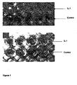
Fig. 1A: is a photo of a macroscopic image of the cartilage explants treated with IL-1 -
Fig. 1 B: is a photo of a macroscopic image of the tissue-engineered cartilage prepared in vitro with HYAFF®-11, after treatment with IL-1 -
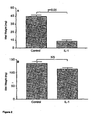
Figure 2 represents the effect of II-1 on the wet weights (Mean ± SEM; n = 5) of natural cartilage (A) and tissue engineered cartilage (B). In each experiment the cartilage was exposed to 3nM IL-1β or medium only (control) for 4 weeks. Statistical analysis was by the 2-tailed Mann-Whitney U-test. -
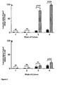
Figure 3 represents the effect of II-1 on the proteoglycan content (Mean ± SEM; n = 5) of natural cartilage (A) and tissue engineered cartilage (B). In each experiment the cartilage was exposed to 3nM IL-1β (hatched bars) or medium only (black bars) for 4 weeks. Culture medium was collected each week and residual cartilage at the end of the experiment for measurement of proteoglycan. Statistical analysis was by the 2-tailed Mann-Whitney U-test. -
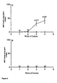
Figure 4 reports the effect of II-1 on the type II collagen content (Mean ± SEM; n = 5) of natural cartilage (A) and tissue engineered cartilage (B). In each experiment the cartilage was exposed to 3nM IL- β(hatched bars) or medium only (black bars) for 4 weeks. Culture medium was collected each week and residual cartilage at the end of the experiment for measurement of type II collagen. Statistical analysis was by the 2-tailed Mann-Whitney U-test. - Effect of IL-1 on the release of MMP activity (Mean ± SEM; n = 5) from natural cartilage (A) and tissue engineered cartilage (B). In each experiment the cartilage was exposed to 3nM IL-1β (hatched bars) or medium only (black bars) for 4 weeks. Culture medium was collected each week and residual cartilage at the end of the experiment for measurement of MMP activity (medium) and type II collagen (medium and cartilage). Statistical analysis was by the 2-tailed Mann-Whitney U-test.
- The Applicant demonstrated how the three-dimensional matrix constituted by a hyaluronic acid derivative made into a biological material and preferably loaded in vitro with autologous and/or allogenic chondrocytes, protects the new cartilage tissue that is formed both in vitro and in vivo after implantation from degradation of the molecules that form the matrix by the pro-inflammatory cytokines that stimulate the production (by the chondrocytes themselves) of proteolytic enzymes such as metalloproteins.
- The present invention is therefore directed the use of the said three-dimensional matrix for the preparation of grafts to be surgically implanted into the joint cartilage as a new therapy:
- □ in the early stages of one of said degenerative diseases and in particular osteoarthritis, at the beginning of the process of degradation of the molecules that make up the extracellular matrix of the cartilage;
- □ in the later stages of said pathology too, when moderately and/or badly damaged areas of cartilage can be seen.
- In fact the grafts when surgically implanted in the early stage of one of said degenerative disease is able to delay the degradation of proteoglycans and collagen, and, when surgically implanted in the later stage of the disease into the progressively degraded joint capsule as new, tissue-engineered cartilage, it proves able both to cover the previously created cartilage lesion and to substitute the eroded extracellular matrix with new cartilage tissue, so that the matrix will not undergo any further degradation because it is protected by hyaluronic acid derivative from the erosive action of IL-1.
- HA is a hetero-polysaccharide composed of alternate residues of D-glucuronic acid and N-acetyl-D-glucosamine. It is a straight-chained polymer with a molecular weight that ranges between 50,000 and 13 x 106 Da, depending on the source from which it is obtained and the methods used to prepare it. It is present in nature in the pericellular gels, in the fundamental substance of the connective tissue of vertebrate organisms (of which it is one of the main components), in the synovial fluid of the joints, in the vitreous humor and in the umbilical cord.
- HA therefore plays a major role in biological organisms, especially as a mechanical support for many kinds of cells, such as those of the skin, tendons, muscles and cartilage.
- Moreover, it is known that HA, by its membrane receptor CD44, modulates many divers processes relative to cell physiology and biology, such as cell proliferation, migration and differentiation and angiogenesis, and that it has other functions such as tissue hydration and joint lubrication.
- The HA to be used in the present invention may come from various sources, for example, it can be obtained by extraction from rooster combs (European patent No.
0138572 B1 ), by fermentation(European patent No.0716688 B1 ), or by technological means, and its molecular weight may range between 400 and 3x10⁶Da, in particular between1x 105Da and1x 106Da, and even more particularly between 200,000 and 750,000 Da. - The HA derivatives that can preferably be used to make three-dimensional matrices for the use according to the present invention, are listed below:
- A) HA salified with organic and/or inorganic bases; and more preferably said base is NaOH;
- B) HA esters with alcohols of the aliphatic, araliphatic, cycloaliphatic, aromatic, cyclic and heterocyclic series, with a percentage of esterification that may vary according to the type and length of the alcohol used, preferably between 50 and 100%, while the remaining percentage of unesterified HA may be salified with organic and/or inorganic bases (European patent No.
0216453 B1 ) more preferably with sodium hydroxide; these esters are commercially available with the name HYAFF®. - C) amides of HA with amines of the aliphatic, araliphatic, cycloaliphatic, aromatic, cyclic and heterocyclic series, with a percentage of amidation more preferably ranging between 0.1 and 50%, while the remaining percentage of HA that has not undergone amidation may be salified with organic and/or inorganic bases,more preferably with sodium hydroxide (European patent application, publication No.
1095064 ); these amides are commercially available with the name Hyadd™ - D) O-sulphated derivatives of HA having preferably from 1 to 4 -OSO3H per disaccharide unit (European patent No.
0702699 B1 ); - E) inner esters of HA with a percentage of esterification that does not exceed 20%, and is more preferably between 0.05 and 10%, while the remaining percentage of non-esterified HA may be salified with organic and/or inorganic bases, more preferably with sodium hydroxide (European patent No.
0341745 B1 ); these esters are commercially available with the name ACP®, - F) Deacetylated derivatives of HA: are obtained by the deacetylation of the N-acetyl-glucosamine group with a percentage of deacetylation preferably ranging between 0.1 and 30%, while all the carboxy groups of HA may be salified with organic and/or inorganic bases, more preferably with sodium hydroxide (European patent application No.
1313772 ); - G) percarboxylated derivatives of HA obtained by oxidising the primary hydroxyl of the N-acetyl-glucosamine unit with a degree of percarboxylation ranging between 0.1 and 100% and more preferably between 25 and 75%. All the carboxy groups of HA can be salified with organic and/or inorganic bases, more preferably with sodium hydroxide (European patent application No.
1339753 ) these products are commercially available with the name Hyoxx™. - The three-dimensional matrix to be used according to the present invention is preferably in the form of a non woven tissue, a tissue, microspheres, or a sponge.
- According to a more preferred embodiment the three-dimensional matrix consists of a hyaluronic acid ester with benzyl alcohol having a percentage of esterification ranging from 75 to 100% and more preferably 100% esterification (HYAFF™ 11) in the form of a non woven tissue prepared as described in European Patent
0618817B1 ). - In order to demonstrate, with relative documentation, that HYAFF®-11 has a specific protective action towards the main components of the extracellular matrix of the cartilage, the following experiments were planned and performed:
- □ Preparation of bovine cartilage explants and their culture as such;
- □ Preparation of cultures of chondrocytes from bovine cartilage and their expansion in vitro;
- □ Preparation of a three-dimensional matrix constituted by a non-woven fabric based on HYAFF®-11 loaded with bovine chondrocytes previously expanded in vitro (which represents the new tissue-engineered cartilage prepared in vitro);
- □ Treatment with IL-1 of both the explant and said matrix (previously loaded with chondrocytes), to determine the effect of pro-inflammatory cytokine on the matrix molecules and on the synthesis of protein enzymes;
- □ Measurement of the type-II collagen released from both the explant and from the engineered matrix by the action of IL-1;
- □ Measurement of the proteoglycans released by the action of IL-1 both from the explant and from the abovesaid matrix;
- □ Measurement of the enzymatic activity of the proteases after treatment of the explant and of the engineered tissue with IL-1.
- We report herein below in the following examples the results obtained by carrying out the aforementioned experiments.
- Fragments of bovine cartilage were taken from five adult animals and cut into sections measuring 25mm X 3mm X 10mm.
- The sections were then washed in phosphate buffer solution (PBS) containing the antibiotics Penicillin-G and Streptomycin and the anti-fungal agents Fungizone and
- Amphotericin.
- The sections thus obtained were placed in culture wells in 400µl of DMEM culture medium containing Glutamine (2 mM), Penicillin-G (200 U/ml), Streptomycin (0.1 mg/ml) and HEPES (10 mM), without the presence of bovine foetal calf serum, in an incubator set at 37°C with 5% CO2, for a period of 4 weeks.
- Fragments of bovine cartilage taken from 5 adult animals and cut into small sections were exposed to enzymatic digestion with hyaluronidase (1 mg/ml) at 37°C for 15 min., and subsequently with trypsin (0.25%) at 37°C for another 30 min. and lastly with bacterial collagenase (2 mg/ml) while continuously shaking overnight at room temperature. All the above said enzymes were prepared in DMEM containing 10 % foetal calf serum (FCS).
- The chondrocytes thus obtained were washed in PBS, centrifuged and resuspended in DMEM culture medium with FCS also containing FGF (1µl/ml).
- The cells thus obtained were then seeded on culture dishes to enable them to proliferate for a period of 7 days, in an incubator set at 37°C with 5% CO2.
- For purely descriptive, and not limitative, purposes, we report hereafter an example of the in vitro preparation of tissue-engineered cartilage.
- The three-dimensional support represented by the non-woven fabric of HYAFF®-11 was first hydrated with culture medium and then loaded with 15X106 million chondrocytes per scaffold. Each scaffold was then placed in a culture dish (to which a thin layer of agarose (1%) had previously been made to adhere (to facilitate adhesion of said matrix and prevent it from moving about on the dish) immersed in DMEM medium containing FCS and FGF, while continuously shaking, in an incubator set at 37°C for a period of 42 days. The trophic factor FGF was added only for the first 4 days, after which the medium was changed with fresh DMEM with FCS containing insulin (10µl/ml) and ascorbic acid (50µl/ml).
- This medium was changed every 2-3 days. After 42 days of culture, each scaffold was divided into two and all the pieces thus obtained where transferred to culture wells for another four weeks, where they were immersed in DMEM culture medium without FCS (FCS is a metalloprotein enzyme inhibitor), containing Glutamine (2 mM), Penicillin-G (200 U/ml), Streptomycin (0.1 mg/ml) and HEPES (10 mM), further supplemented with insulin/transferrin/selenium. All the experiments with IL-1 were performed with scaffolds made as described above.
- A quantity equal to half the sections of cartilage explant and scaffolds containing bovine chondrocytes (prepared in vitro as previously described), was used for the experiment with IL-1 while the other half was not exposed to any kind of treatment as it represented the untreated control. Treatment protocol: IL-1 was added to the culture medium to make a final concentration of 3 nM. the medium was changed every 2-3 days, always at an IL-1 concentration of 3 nM. The experimental protocol provided for a treatment time with IL-1 of 2 or 4 weeks. All the medium eliminated each week was collected, divided by treatment week and stored at - 20°C for the final determinations.
- The culture medium of the explant and engineered matrix, collected each week of treatment with IL-1, was exposed to specific enzymatic digestion with proteinase K (1 mg/ml) at 56°C for 15 hours, together with the residues of the explant and tissue collected at the end of the experiment.
- A specific ELISA test (Hollander A.P. et al., J Clin. Invest., 1994, 93:1722-1732) was used to measure the quantity of type II collagen present both in the culture samples collected weekly (including the non-treated controls) and to determine the collagen left inside the explant and HYAFF® -11 matrix after treatment with IL-1.
- The culture medium of both the explant and the engineered matrix collected each week of treatment with IL-1, was exposed to specific enzymatic digestion with proteinase K as previously described for the determination of collagen, together with the residues of the explant and the matrix collected at the end of the experiment.
- Using DBM (methylene blue), a specific dye, and a special colorimetric test, the concentrations of proteoglycans present in the analysed samples were subsequently determined (Handley C.J. et al., Methods Enzymol.,1995, 248:47-48).
- Metalloprotein enzyme activity was determined on all the culture media collected weekly both for the explants and for the engineered tissues and relative non-treated controls.
- A fluorescent substrate was used to determine said enzymatic activity (7-methoxycoumarine-4-acetyl(MCA)-Pro-Leu-Gli-Leu-(3-(2;4-dinitrophenyl)-L-2,3-diamino-propionyl)(Dpa)-Ala-Arg-NH). The substrate and all the samples collected were first diluted in Tris-HCL buffer 0.1 M with a pH of 7.5, containing
CaCO 3 10 Mm and 0.2% (v/v) of Triton X-100. - The enzymatic activity of each sample was measured, by fluorometric reading two minutes after adding the substrate to the sample to be analysed and expressed as units/total µg of type-II collagen contained in each sample (and therefore determined both on the residue and on the culture medium) (Kozaci L.D. et al., Arthritis & Rheumatism, 1997,40.164-174).
- As
Figure 1A clearly shows, the explants of bovine collagen are completely degraded after four weeks of treatment with IL-1, while the tissue-engineered cartilage constituted by HYAFF®-11 shows no macroscopic changes after four weeks of treatment (Figure 1B ). - These results are confirmed by determining the weight of the residues after treatment compared to the weight of the corresponding non-treated controls (
Figure 2 A-B ). - The percentage of degradation of the proteoglycans was determined by calculating the concentration of the molecules in the culture medium for each week of treatment with IL-1 (duration of treatment: 2 weeks), and was expressed as the percentage of the total concentration of proteoglycans present both in the residue (that is, in the residue of the explant or in the residue of the engineered matrix) and in the corresponding culture medium.
- The results obtained show that IL-1 induces a significant degradation of the proteoglycans in the treated cartilage explants, with a percentage of degradation of 86% within the first week of treatment (
Figure 3A ). IL-1 causes the degradation of proteoglycans in the engineered matrix too, but the level of degradation reaches about 70% only in the 2nd week of treatment (Figure 3B ). - The degree of degradation of the type II collagen was determined by calculating the concentration of said protein in the culture medium for each week of treatment with IL-1 (duration of treatment: 4 weeks), subsequently expressed as the percentage of total concentration of type-II collagen present both in the residue (i.e. in the residue of the explant or in the residue of the engineered matrix) and in the corresponding culture medium. The results obtained demonstrate that IL-1 in the treated cartilage explant causes the total degradation of the collagen after four weeks of treatment (
Figure 4A ), while the level of degradation of the protein in the HYAFF®-11-based tissue is negligible, reaching just 20% after four weeks of treatment (Figure 4B ). - The total enzymatic activity of the metalloprotein enzymes (MMP) produced as a reaction to treatment with IL-1 both by the cartilage explant and the engineered tissue made in vitro, are quantified in
Figure 5 A-B . - IL-1 induces a strong increase in the MMP enzymes in the treated cartilage explants (after both 3 and 4 weeks of treatment) but it does not determine any increase in said enzymatic activity in the engineered tissue based on HYAFF®-11. All the above-reported results enable us to state that the biomaterial constituted by a derivative of hyaluronic acid, (and particularly by its benzyl ester HYAFF®-11), made into a three-dimensional matrix (preferably non-woven), exercises a strong protective action on the molecules that make up the extracellular cartilage matrix when this last is exposed to the erosive action of pro-inflammatory cytokines that have been over-produced in phlogogenic situations such as those that occur in the course of OA, rheumatoid arthritis and psoriatic arthritis.
Claims (24)
- Use of a biological material containing:a) a three-dimensional matrix based on a hyaluronic acid derivative and optionallyb) chondrocytes and/or mesenchymal cells partially or completely differentiated towards chondrocytes,
for the preparation of a graft surgically implantable into a joint cartilage for the protection and/or recovery of said joint cartilage from a degenerative and/or inflammatory pathology, associated with the production of IL-1, selected from osteoarthrosis, psoriatic and rheumatoid arthritis. - The use according to claim 1, wherein, when the biological material contains the aforementioned cellular components (b) said graft is an in vitro cartilage tissue to be surgically implanted in vivo inside the joint capsule in which one of said degenerative pathologies has been established with consequent degradation of the extracellular cartilage matrix.
- The use according to claim 2 wherein in vitro cartilage tissue further comprise the extracellular matrix produced by said chondrocytes or mesenchymal cells partially or completely differentiated towards chondrocytes said extracellular matrix being both inside said in vitro cartilage tissue and once in vivo implanted also inside the joint cartilage affected by one of said degenerative pathologies.
- The use according to claim 3 wherein said grafts are surgically implantable at the beginning of the process of degradation of the molecules that make up the extracellular matrix of the cartilage.
- The use according to anyone of claims 1-4 wherein said grafts are surgically implantable in the later stages of said pathology too, when moderately and/or badly damaged areas of cartilage can be seen.
- The use according to anyone of claims 1-5 wherein the average molecular weight of hyaluronic acid in the hyaluronic acid derivative range between 1 x 10⁵ Da and 1 x 10⁶ Da.
- The use according to claim 6, wherein the average molecular weight of hyaluronic acid range between 200,000 and 750,000 Da.
- The use according to anyone of claims 1-7, wherein the hyaluronic acid derivative is selected from the class consisting of:A) HA salified with organic and/or inorganic bases,B) HA esters with alcohols of the aliphatic, araliphatic, cycloaliphatic, aromatic, cyclic and heterocyclic series,C) HA amides with amine of the aliphatic, araliphatic, cycloaliphatic, aromatic, cyclic and heterocyclic series.D) O-sulphated derivatives of HA,E) inner esters of HA with a percentage of esterification that does not exceed 20%,F) Deacetylated derivatives of HA obtained by the deacetylation of the N-acetyl-glucosamine fraction,G) percarboxylated derivatives of HA obtained by oxidising the primary hydroxyl of the N-acetyl-glucosamine fraction with a degree of percarboxylation ranging between 0.1 and 100%.
- The use according to claim 8, wherein the HA derivative belongs to class(A) it is obtained by treating hyaluronic acid with sodium hydroxide.
- The use according to claim 8, wherein, when the HA derivative belongs to class (B) it has a percentage of esterification ranging from 50 to 100%,and the remainining percentage of unesterified HA is salified with organic or inorganic base.
- The use according to claim 10, wherein said base is sodium hydroxide.
- The use according to claim 8, wherein when the HA derivative belongs to class (C) it has a percentage of amidation ranging between 0.1 and 50% and the remaining portion is salified with organic and/or inorganic bases.
- The use according to claim 12 wherein said base is sodium hydroxide.
- The use according to claim 8, wherein when the HA derivative belongs to class (D) it has from 1 to 4 -OSO3H group per saccharide unit.
- The use according to claim 8 wherein when the HA derivative belongs to class (E) it has a degree of esterification ranging from 0.05 to 10%, and the remaining percentage of non-esterified HA may be salified with organic and/or inorganic bases.
- The use according to claim 15, wherein said base is sodium hydroxide.
- The use according to claim 8, wherein when the HA derivative belongs to class (F) it has a percentage of deacetylation ranging between 0.1 and 30% and all the carboxy groups of HA are salified with organic and/or inorganic bases.
- The use according to claim 17, wherein said base is sodium hydroxide.
- The use according to claim 8, wherein, when the HA derivative belongs to class (G), it has a degree of percarboxylation ranging from 25 to 75% and all the carboxy groups are salified with organic and/or inorganic bases.
- The use according to claim 19, wherein the base is sodium hydroxide.
- The use according to anyone of claims 1-20, wherein said three-dimensional matrix is in a form selected from the group consisting of: a non-woven tissue, a tissue, microspheres, and a sponge.
- The use according to anyone of claims 1-21, wherein said HA derivative is a hyaluronic acid ester belonging to class (A).
- The use according to claim 22 wherein said HA ester is the benzyl ester having a percentage of esterification ranging from 75 to 100%.
- The use according to claim 23, wherein said benzylester has a percentage of esterification of 100% and is in the form of a non-woven tissue.
Priority Applications (3)
| Application Number | Priority Date | Filing Date | Title |
|---|---|---|---|
| SI200530812T SI1722835T1 (en) | 2004-02-27 | 2005-02-25 | Hyaluronic acid derivative-based three-dimensional matrix |
| PL05716808T PL1722835T3 (en) | 2004-02-27 | 2005-02-25 | Hyaluronic acid derivative-based three-dimensional matrix |
| CY20091101122T CY1109550T1 (en) | 2004-02-27 | 2009-10-29 | HYDRAULIC ACID PRODUCER WITH THREE DIMENSIONS |
Applications Claiming Priority (2)
| Application Number | Priority Date | Filing Date | Title |
|---|---|---|---|
| IT000053A ITPD20040053A1 (en) | 2004-02-27 | 2004-02-27 | BIOMATERIALS CONSTITUTED FROM HYALURONIC ACID DERIVATIVES AS A NEW CARE THERAPY FOR THE PROTECTION AND REPAIR OF DAMAGED ARTICULAR CARTILAGE FOR OSTEOARTHROSIS |
| PCT/EP2005/050817 WO2005082433A1 (en) | 2004-02-27 | 2005-02-25 | Hyaluronic acid derivative based three-dimensional matrix |
Publications (2)
| Publication Number | Publication Date |
|---|---|
| EP1722835A1 EP1722835A1 (en) | 2006-11-22 |
| EP1722835B1 true EP1722835B1 (en) | 2009-08-05 |
Family
ID=34897807
Family Applications (1)
| Application Number | Title | Priority Date | Filing Date |
|---|---|---|---|
| EP05716808A Expired - Lifetime EP1722835B1 (en) | 2004-02-27 | 2005-02-25 | Hyaluronic acid derivative-based three-dimensional matrix |
Country Status (15)
| Country | Link |
|---|---|
| US (2) | US7842487B2 (en) |
| EP (1) | EP1722835B1 (en) |
| JP (1) | JP2007524489A (en) |
| AT (1) | ATE438421T1 (en) |
| AU (1) | AU2005216648A1 (en) |
| CA (1) | CA2555559C (en) |
| CY (1) | CY1109550T1 (en) |
| DE (1) | DE602005015824D1 (en) |
| DK (1) | DK1722835T3 (en) |
| ES (1) | ES2329485T3 (en) |
| IT (1) | ITPD20040053A1 (en) |
| PL (1) | PL1722835T3 (en) |
| PT (1) | PT1722835E (en) |
| SI (1) | SI1722835T1 (en) |
| WO (1) | WO2005082433A1 (en) |
Families Citing this family (10)
| Publication number | Priority date | Publication date | Assignee | Title |
|---|---|---|---|---|
| ITPD20060219A1 (en) | 2006-05-31 | 2007-12-01 | Fidia Farmaceutici | PHARMACEUTICAL COMPOSITIONS CONTAINING HYALURONIC ACID SULFATED IN THE TREATMENT OF OSTEOARTHROSIS |
| ES2298052B1 (en) | 2006-08-03 | 2009-09-28 | Bioiberica S.A. | PROCEDURE FOR THE PROLIFERATION OF CELLS. |
| AU2013203054B2 (en) * | 2007-08-06 | 2017-05-18 | Mesoblast, Inc. | Methods of generating, repairing and/or maintaining connective tissue in vivo |
| CN103169731A (en) | 2007-08-06 | 2013-06-26 | 成血管细胞系统公司 | Methods of generating, repairing and/or maintaining connective tissue in vivo |
| KR20170104643A (en) | 2008-06-25 | 2017-09-15 | 메소블라스트, 아이엔씨. | Repair and/or Reconstitution of Invertebral Discs |
| JP2011041472A (en) * | 2009-08-19 | 2011-03-03 | Tokai Univ | Spheroid for cell transplantation treatment composed of cell mixture and method for preparing the same |
| EP2807271B1 (en) | 2012-01-24 | 2018-08-22 | CD Diagnostics, Inc. | System for detecting infection in synovial fluid |
| ITUA20164153A1 (en) * | 2016-06-07 | 2017-12-07 | Jointherapeutics S R L | Polysaccharide compositions usable in tissue repair |
| IT201700110784A1 (en) * | 2017-10-03 | 2019-04-03 | Fidia Farm Spa | Pharmaceutical compositions containing Hyaluronic acid and Carnosine and its use |
| CN111787886A (en) | 2018-01-02 | 2020-10-16 | 卡尔蒂希尔(2009)公司 | Implantation tools and protocols for optimized solid matrices to promote cell and tissue growth |
Family Cites Families (12)
| Publication number | Priority date | Publication date | Assignee | Title |
|---|---|---|---|---|
| IT1219587B (en) * | 1988-05-13 | 1990-05-18 | Fidia Farmaceutici | SELF-CROSS-LINKED CARBOXYLY POLYSACCHARIDES |
| US7789841B2 (en) * | 1997-02-06 | 2010-09-07 | Exogen, Inc. | Method and apparatus for connective tissue treatment |
| ITPD980149A1 (en) * | 1998-06-17 | 1999-12-17 | Fidia Advanced Biopolymers Srl | THREE-DIMENSIONAL PROSTHESES INCLUDING HYALURONIC ACID DERIVATIVES TO REPAIR OR REBUILD DAMAGED TISSUES AND PROCESS FOR THE |
| ITPD980169A1 (en) * | 1998-07-06 | 2000-01-06 | Fidia Advanced Biopolymers Srl | AMIDES OF HYALURONIC ACID AND ITS DERIVATIVES AND PROCESS FOR THEIR PREPARATION. |
| DE19855890A1 (en) * | 1998-12-03 | 2000-06-08 | Nerlich Michael | Porous composite matrix, its production and use |
| IT1302534B1 (en) | 1998-12-21 | 2000-09-05 | Fidia Advanced Biopolymers Srl | INJECTABLE, BIOCOMPATIBLE AND BIODEGRADABLE COMPOSITIONS INCLUDING AT LEAST A DERIVATIVE OF HYALURONIC ACID, CHONDROGENIC CELLS, FOR |
| IT1317358B1 (en) * | 2000-08-31 | 2003-06-16 | Fidia Advanced Biopolymers Srl | CROSS-LINKATED DERIVATIVES OF HYALURONIC ACID. |
| IT1317359B1 (en) * | 2000-08-31 | 2003-06-16 | Fidia Advanced Biopolymers Srl | PERCARBOXYLATE POLYSACCHARIDES, SUCH AS HYALURONIC ACID, PROCESS FOR THEIR PREPARATION AND USE IN THE PHARMACEUTICAL FIELD AND |
| ES2340141T3 (en) * | 2000-12-28 | 2010-05-31 | Fidia Advanced Biopolymers S.R.L. | USE OF BIOLOGICAL MATERIAL THAT CONTAINS TISSULAR MATRIXS TRISIMENSIONAL SUPPORT OF DERIVATIVES OF THE HIALURONIC ACID FOR THE PREPARATION OF IMPLANTS OF ARTHROSCOPY AND A KIT FOR INSTRUMENTS TO IMPLEMENT SUCH BIOLOGICAL MATERIAL THROUGH ARTHROSCOPY. |
| ITPD20010032A1 (en) * | 2001-02-09 | 2002-08-09 | Fidia Advanced Biopolymers Srl | ENGINEERED GRAFTES FOR THE REPAIR OF OSTEOCONDRAL DEFECTS |
| ITPD20020064A1 (en) * | 2002-03-12 | 2003-09-12 | Fidia Advanced Biopolymers Srl | FOREIGN DERIVATIVES OF HYALURONIC ACID FOR THE PREPARATION OF HYDROGELD FOR USE IN THE BIOMEDICAL, SANITARY AND SURGICAL FIELD AND AS A SYSTEM |
| US7931687B2 (en) * | 2002-05-13 | 2011-04-26 | Articular Engineering, Llc | Tissue engineered osteochondral implant |
-
2004
- 2004-02-27 IT IT000053A patent/ITPD20040053A1/en unknown
-
2005
- 2005-02-25 SI SI200530812T patent/SI1722835T1/en unknown
- 2005-02-25 DK DK05716808T patent/DK1722835T3/en active
- 2005-02-25 DE DE602005015824T patent/DE602005015824D1/en not_active Expired - Lifetime
- 2005-02-25 PT PT05716808T patent/PT1722835E/en unknown
- 2005-02-25 AT AT05716808T patent/ATE438421T1/en active
- 2005-02-25 WO PCT/EP2005/050817 patent/WO2005082433A1/en not_active Ceased
- 2005-02-25 AU AU2005216648A patent/AU2005216648A1/en not_active Abandoned
- 2005-02-25 US US10/590,250 patent/US7842487B2/en active Active
- 2005-02-25 JP JP2007500225A patent/JP2007524489A/en active Pending
- 2005-02-25 ES ES05716808T patent/ES2329485T3/en not_active Expired - Lifetime
- 2005-02-25 CA CA2555559A patent/CA2555559C/en not_active Expired - Lifetime
- 2005-02-25 EP EP05716808A patent/EP1722835B1/en not_active Expired - Lifetime
- 2005-02-25 PL PL05716808T patent/PL1722835T3/en unknown
-
2009
- 2009-10-29 CY CY20091101122T patent/CY1109550T1/en unknown
-
2010
- 2010-11-30 US US12/956,158 patent/US20110104284A1/en not_active Abandoned
Also Published As
| Publication number | Publication date |
|---|---|
| US20110104284A1 (en) | 2011-05-05 |
| HK1096047A1 (en) | 2007-05-25 |
| ITPD20040053A1 (en) | 2004-05-27 |
| CA2555559C (en) | 2013-03-12 |
| DK1722835T3 (en) | 2009-11-09 |
| AU2005216648A1 (en) | 2005-09-09 |
| US20080317808A1 (en) | 2008-12-25 |
| DE602005015824D1 (en) | 2009-09-17 |
| WO2005082433A1 (en) | 2005-09-09 |
| SI1722835T1 (en) | 2009-12-31 |
| CY1109550T1 (en) | 2014-08-13 |
| US7842487B2 (en) | 2010-11-30 |
| PT1722835E (en) | 2009-11-13 |
| PL1722835T3 (en) | 2010-01-29 |
| JP2007524489A (en) | 2007-08-30 |
| ES2329485T3 (en) | 2009-11-26 |
| CA2555559A1 (en) | 2005-09-09 |
| ATE438421T1 (en) | 2009-08-15 |
| EP1722835A1 (en) | 2006-11-22 |
Similar Documents
| Publication | Publication Date | Title |
|---|---|---|
| US20110104284A1 (en) | Hyaluronic acid derivative based three-dimensional matrix | |
| Suh et al. | Application of chitosan-based polysaccharide biomaterials in cartilage tissue engineering: a review | |
| EP2345435B1 (en) | Method for manufacturing a porous three-dimensional support using powder from animal tissue, and porous three-dimensional support manufactured by same | |
| EP2061527B1 (en) | Repair of cartilage tissue | |
| US20100119577A1 (en) | Therapeutic composite for cartilage disorder using extracellular matrix (ecm) scaffold | |
| Pan et al. | Fabrication of modified dextran–gelatin in situ forming hydrogel and application in cartilage tissue engineering | |
| Hofmann et al. | Cartilage-like tissue engineering using silk scaffolds and mesenchymal stem cells | |
| Shi et al. | Development and potential of a biomimetic chitosan/type II collagen scaffold for cartilage tissue engineering | |
| US20140193468A1 (en) | Methods for preparation of neo-cartilage constructs | |
| Wan et al. | BMSCs laden injectable amino-diethoxypropane modified alginate-chitosan hydrogel for hyaline cartilage reconstruction | |
| Teng et al. | Extracellular matrix powder from cultured cartilage-like tissue as cell carrier for cartilage repair | |
| Vaca‐González et al. | Anatomy, molecular structures, and hyaluronic acid–gelatin injectable hydrogels as a therapeutic alternative for hyaline cartilage recovery: a review | |
| WO2009017267A1 (en) | Method for differenciating mesenchymal stem cell and culturing chondrocytes using alginate coated fibrin/ha composite scaffold | |
| KR100926975B1 (en) | Differentiation of Mesenchymal Stem Cells and Culture of Chondrocytes Using Alginate Coated Fibrin / HA Mixture Scaffolds | |
| CN116159188A (en) | Chitosan-citric acid/polyvinyl alcohol double-network hydrogel bracket and preparation method and application thereof | |
| EP1254670A1 (en) | Type II collagen matrices for use in cartilage engineering | |
| US20190321451A1 (en) | Methods for preparation of neo-cartilage constructs | |
| HK1096047B (en) | Hyaluronic acid derivative-based three-dimensional matrix | |
| CN116899016B (en) | Cartilage extracellular matrix microcapsule, tissue engineering scaffold and preparation methods thereof | |
| DiCarlo et al. | Biomaterial effects in articular cartilage tissue engineering using polyglycolic acid, a novel marine origin biomaterial, IGF-I, and TGF-β1 | |
| CN114796616A (en) | Preparation method of hyaline cartilage acellular matrix composite scaffold | |
| CN120346378A (en) | Double-layer composite bracket, preparation method and application | |
| US20200282109A1 (en) | Devices and Methods for Repairing Cartilage and Osteochondral Defects | |
| Zhao | Biomaterial and Cell Based Cartilage Repair | |
| CN121550496A (en) | Cartilage extracellular matrix gel and preparation method thereof |
Legal Events
| Date | Code | Title | Description |
|---|---|---|---|
| PUAI | Public reference made under article 153(3) epc to a published international application that has entered the european phase |
Free format text: ORIGINAL CODE: 0009012 |
|
| 17P | Request for examination filed |
Effective date: 20060926 |
|
| AK | Designated contracting states |
Kind code of ref document: A1 Designated state(s): AT BE BG CH CY CZ DE DK EE ES FI FR GB GR HU IE IS IT LI LT LU MC NL PL PT RO SE SI SK TR |
|
| DAX | Request for extension of the european patent (deleted) | ||
| REG | Reference to a national code |
Ref country code: HK Ref legal event code: DE Ref document number: 1096047 Country of ref document: HK |
|
| GRAP | Despatch of communication of intention to grant a patent |
Free format text: ORIGINAL CODE: EPIDOSNIGR1 |
|
| GRAS | Grant fee paid |
Free format text: ORIGINAL CODE: EPIDOSNIGR3 |
|
| GRAA | (expected) grant |
Free format text: ORIGINAL CODE: 0009210 |
|
| AK | Designated contracting states |
Kind code of ref document: B1 Designated state(s): AT BE BG CH CY CZ DE DK EE ES FI FR GB GR HU IE IS IT LI LT LU MC NL PL PT RO SE SI SK TR |
|
| REG | Reference to a national code |
Ref country code: GB Ref legal event code: FG4D |
|
| REG | Reference to a national code |
Ref country code: CH Ref legal event code: EP |
|
| REG | Reference to a national code |
Ref country code: IE Ref legal event code: FG4D |
|
| REF | Corresponds to: |
Ref document number: 602005015824 Country of ref document: DE Date of ref document: 20090917 Kind code of ref document: P |
|
| REG | Reference to a national code |
Ref country code: CH Ref legal event code: NV Representative=s name: N&G PATENT SERVICES SA |
|
| REG | Reference to a national code |
Ref country code: RO Ref legal event code: EPE |
|
| REG | Reference to a national code |
Ref country code: DK Ref legal event code: T3 |
|
| REG | Reference to a national code |
Ref country code: PT Ref legal event code: SC4A Free format text: AVAILABILITY OF NATIONAL TRANSLATION Effective date: 20091105 |
|
| REG | Reference to a national code |
Ref country code: SE Ref legal event code: TRGR |
|
| REG | Reference to a national code |
Ref country code: ES Ref legal event code: FG2A Ref document number: 2329485 Country of ref document: ES Kind code of ref document: T3 |
|
| REG | Reference to a national code |
Ref country code: GR Ref legal event code: EP Ref document number: 20090402763 Country of ref document: GR |
|
| PG25 | Lapsed in a contracting state [announced via postgrant information from national office to epo] |
Ref country code: IS Free format text: LAPSE BECAUSE OF FAILURE TO SUBMIT A TRANSLATION OF THE DESCRIPTION OR TO PAY THE FEE WITHIN THE PRESCRIBED TIME-LIMIT Effective date: 20091205 |
|
| REG | Reference to a national code |
Ref country code: PL Ref legal event code: T3 |
|
| NLV1 | Nl: lapsed or annulled due to failure to fulfill the requirements of art. 29p and 29m of the patents act | ||
| PG25 | Lapsed in a contracting state [announced via postgrant information from national office to epo] |
Ref country code: NL Free format text: LAPSE BECAUSE OF FAILURE TO SUBMIT A TRANSLATION OF THE DESCRIPTION OR TO PAY THE FEE WITHIN THE PRESCRIBED TIME-LIMIT Effective date: 20090805 |
|
| PG25 | Lapsed in a contracting state [announced via postgrant information from national office to epo] |
Ref country code: BG Free format text: LAPSE BECAUSE OF FAILURE TO SUBMIT A TRANSLATION OF THE DESCRIPTION OR TO PAY THE FEE WITHIN THE PRESCRIBED TIME-LIMIT Effective date: 20091105 |
|
| REG | Reference to a national code |
Ref country code: HK Ref legal event code: GR Ref document number: 1096047 Country of ref document: HK |
|
| PG25 | Lapsed in a contracting state [announced via postgrant information from national office to epo] |
Ref country code: EE Free format text: LAPSE BECAUSE OF FAILURE TO SUBMIT A TRANSLATION OF THE DESCRIPTION OR TO PAY THE FEE WITHIN THE PRESCRIBED TIME-LIMIT Effective date: 20090805 |
|
| PGFP | Annual fee paid to national office [announced via postgrant information from national office to epo] |
Ref country code: CZ Payment date: 20100127 Year of fee payment: 6 Ref country code: DK Payment date: 20100121 Year of fee payment: 6 Ref country code: IE Payment date: 20100122 Year of fee payment: 6 Ref country code: LT Payment date: 20100111 Year of fee payment: 6 Ref country code: LU Payment date: 20100122 Year of fee payment: 6 Ref country code: MC Payment date: 20100125 Year of fee payment: 6 Ref country code: PT Payment date: 20100225 Year of fee payment: 6 Ref country code: RO Payment date: 20100225 Year of fee payment: 6 |
|
| PG25 | Lapsed in a contracting state [announced via postgrant information from national office to epo] |
Ref country code: SK Free format text: LAPSE BECAUSE OF FAILURE TO SUBMIT A TRANSLATION OF THE DESCRIPTION OR TO PAY THE FEE WITHIN THE PRESCRIBED TIME-LIMIT Effective date: 20090805 |
|
| PGFP | Annual fee paid to national office [announced via postgrant information from national office to epo] |
Ref country code: CY Payment date: 20100115 Year of fee payment: 6 Ref country code: FI Payment date: 20100122 Year of fee payment: 6 Ref country code: PL Payment date: 20100113 Year of fee payment: 6 Ref country code: SI Payment date: 20100121 Year of fee payment: 6 |
|
| PLBE | No opposition filed within time limit |
Free format text: ORIGINAL CODE: 0009261 |
|
| STAA | Information on the status of an ep patent application or granted ep patent |
Free format text: STATUS: NO OPPOSITION FILED WITHIN TIME LIMIT |
|
| PGFP | Annual fee paid to national office [announced via postgrant information from national office to epo] |
Ref country code: TR Payment date: 20100112 Year of fee payment: 6 |
|
| 26N | No opposition filed |
Effective date: 20100507 |
|
| PG25 | Lapsed in a contracting state [announced via postgrant information from national office to epo] |
Ref country code: IT Free format text: LAPSE BECAUSE OF NON-PAYMENT OF DUE FEES Effective date: 20100225 |
|
| PGRI | Patent reinstated in contracting state [announced from national office to epo] |
Ref country code: IT Effective date: 20110616 |
|
| REG | Reference to a national code |
Ref country code: PT Ref legal event code: MM4A Free format text: LAPSE DUE TO NON-PAYMENT OF FEES Effective date: 20110825 |
|
| LTLA | Lt: lapse of european patent or patent extension |
Effective date: 20110225 |
|
| PG25 | Lapsed in a contracting state [announced via postgrant information from national office to epo] |
Ref country code: MC Free format text: LAPSE BECAUSE OF NON-PAYMENT OF DUE FEES Effective date: 20110228 |
|
| REG | Reference to a national code |
Ref country code: DK Ref legal event code: EBP |
|
| REG | Reference to a national code |
Ref country code: SI Ref legal event code: KO00 Effective date: 20110929 |
|
| PG25 | Lapsed in a contracting state [announced via postgrant information from national office to epo] |
Ref country code: PT Free format text: LAPSE BECAUSE OF NON-PAYMENT OF DUE FEES Effective date: 20110825 Ref country code: LT Free format text: LAPSE BECAUSE OF NON-PAYMENT OF DUE FEES Effective date: 20110225 |
|
| REG | Reference to a national code |
Ref country code: IE Ref legal event code: MM4A |
|
| PG25 | Lapsed in a contracting state [announced via postgrant information from national office to epo] |
Ref country code: CZ Free format text: LAPSE BECAUSE OF NON-PAYMENT OF DUE FEES Effective date: 20110225 Ref country code: FI Free format text: LAPSE BECAUSE OF NON-PAYMENT OF DUE FEES Effective date: 20110225 Ref country code: CY Free format text: LAPSE BECAUSE OF NON-PAYMENT OF DUE FEES Effective date: 20110225 Ref country code: SI Free format text: LAPSE BECAUSE OF NON-PAYMENT OF DUE FEES Effective date: 20110226 |
|
| PG25 | Lapsed in a contracting state [announced via postgrant information from national office to epo] |
Ref country code: IE Free format text: LAPSE BECAUSE OF NON-PAYMENT OF DUE FEES Effective date: 20110225 |
|
| PG25 | Lapsed in a contracting state [announced via postgrant information from national office to epo] |
Ref country code: PL Free format text: LAPSE BECAUSE OF NON-PAYMENT OF DUE FEES Effective date: 20110225 |
|
| REG | Reference to a national code |
Ref country code: PL Ref legal event code: LAPE |
|
| PG25 | Lapsed in a contracting state [announced via postgrant information from national office to epo] |
Ref country code: HU Free format text: LAPSE BECAUSE OF FAILURE TO SUBMIT A TRANSLATION OF THE DESCRIPTION OR TO PAY THE FEE WITHIN THE PRESCRIBED TIME-LIMIT Effective date: 20100206 |
|
| PG25 | Lapsed in a contracting state [announced via postgrant information from national office to epo] |
Ref country code: LU Free format text: LAPSE BECAUSE OF NON-PAYMENT OF DUE FEES Effective date: 20110225 |
|
| PG25 | Lapsed in a contracting state [announced via postgrant information from national office to epo] |
Ref country code: TR Free format text: LAPSE BECAUSE OF NON-PAYMENT OF DUE FEES Effective date: 20110225 |
|
| REG | Reference to a national code |
Ref country code: FR Ref legal event code: PLFP Year of fee payment: 12 |
|
| REG | Reference to a national code |
Ref country code: FR Ref legal event code: PLFP Year of fee payment: 13 |
|
| REG | Reference to a national code |
Ref country code: FR Ref legal event code: PLFP Year of fee payment: 14 |
|
| PGFP | Annual fee paid to national office [announced via postgrant information from national office to epo] |
Ref country code: GR Payment date: 20240110 Year of fee payment: 20 |
|
| PGFP | Annual fee paid to national office [announced via postgrant information from national office to epo] |
Ref country code: ES Payment date: 20240305 Year of fee payment: 20 |
|
| PGFP | Annual fee paid to national office [announced via postgrant information from national office to epo] |
Ref country code: AT Payment date: 20240125 Year of fee payment: 20 |
|
| PGFP | Annual fee paid to national office [announced via postgrant information from national office to epo] |
Ref country code: DE Payment date: 20240109 Year of fee payment: 20 Ref country code: CH Payment date: 20240301 Year of fee payment: 20 Ref country code: GB Payment date: 20240115 Year of fee payment: 20 |
|
| PGFP | Annual fee paid to national office [announced via postgrant information from national office to epo] |
Ref country code: SE Payment date: 20240110 Year of fee payment: 20 Ref country code: IT Payment date: 20240129 Year of fee payment: 20 Ref country code: FR Payment date: 20240123 Year of fee payment: 20 Ref country code: BE Payment date: 20240119 Year of fee payment: 20 |
|
| REG | Reference to a national code |
Ref country code: DE Ref legal event code: R071 Ref document number: 602005015824 Country of ref document: DE |
|
| REG | Reference to a national code |
Ref country code: CH Ref legal event code: PL |
|
| REG | Reference to a national code |
Ref country code: ES Ref legal event code: FD2A Effective date: 20250304 |
|
| REG | Reference to a national code |
Ref country code: GB Ref legal event code: PE20 Expiry date: 20250224 |
|
| REG | Reference to a national code |
Ref country code: SE Ref legal event code: EUG |
|
| REG | Reference to a national code |
Ref country code: AT Ref legal event code: MK07 Ref document number: 438421 Country of ref document: AT Kind code of ref document: T Effective date: 20250225 |
|
| PG25 | Lapsed in a contracting state [announced via postgrant information from national office to epo] |
Ref country code: ES Free format text: LAPSE BECAUSE OF EXPIRATION OF PROTECTION Effective date: 20250226 |
|
| PG25 | Lapsed in a contracting state [announced via postgrant information from national office to epo] |
Ref country code: GB Free format text: LAPSE BECAUSE OF EXPIRATION OF PROTECTION Effective date: 20250224 |
|
| REG | Reference to a national code |
Ref country code: BE Ref legal event code: MK Effective date: 20250225 |