EP1721967A1 - Method of differentiating mesenchymal stem cells into steroid-producing cells - Google Patents
Method of differentiating mesenchymal stem cells into steroid-producing cells Download PDFInfo
- Publication number
- EP1721967A1 EP1721967A1 EP05719267A EP05719267A EP1721967A1 EP 1721967 A1 EP1721967 A1 EP 1721967A1 EP 05719267 A EP05719267 A EP 05719267A EP 05719267 A EP05719267 A EP 05719267A EP 1721967 A1 EP1721967 A1 EP 1721967A1
- Authority
- EP
- European Patent Office
- Prior art keywords
- cells
- mesenchymal stem
- stem cells
- producing
- steroid hormone
- Prior art date
- Legal status (The legal status is an assumption and is not a legal conclusion. Google has not performed a legal analysis and makes no representation as to the accuracy of the status listed.)
- Granted
Links
- 0 C*(C)C1C2(*)C=CC2C=C2*1C=CCC2 Chemical compound C*(C)C1C2(*)C=CC2C=C2*1C=CCC2 0.000 description 1
Images




Classifications
-
- C—CHEMISTRY; METALLURGY
- C12—BIOCHEMISTRY; BEER; SPIRITS; WINE; VINEGAR; MICROBIOLOGY; ENZYMOLOGY; MUTATION OR GENETIC ENGINEERING
- C12P—FERMENTATION OR ENZYME-USING PROCESSES TO SYNTHESISE A DESIRED CHEMICAL COMPOUND OR COMPOSITION OR TO SEPARATE OPTICAL ISOMERS FROM A RACEMIC MIXTURE
- C12P33/00—Preparation of steroids
-
- C—CHEMISTRY; METALLURGY
- C12—BIOCHEMISTRY; BEER; SPIRITS; WINE; VINEGAR; MICROBIOLOGY; ENZYMOLOGY; MUTATION OR GENETIC ENGINEERING
- C12N—MICROORGANISMS OR ENZYMES; COMPOSITIONS THEREOF; PROPAGATING, PRESERVING, OR MAINTAINING MICROORGANISMS; MUTATION OR GENETIC ENGINEERING; CULTURE MEDIA
- C12N5/00—Undifferentiated human, animal or plant cells, e.g. cell lines; Tissues; Cultivation or maintenance thereof; Culture media therefor
- C12N5/06—Animal cells or tissues; Human cells or tissues
- C12N5/0602—Vertebrate cells
- C12N5/0613—Cells from endocrine organs
- C12N5/0614—Adrenal gland
-
- C—CHEMISTRY; METALLURGY
- C12—BIOCHEMISTRY; BEER; SPIRITS; WINE; VINEGAR; MICROBIOLOGY; ENZYMOLOGY; MUTATION OR GENETIC ENGINEERING
- C12N—MICROORGANISMS OR ENZYMES; COMPOSITIONS THEREOF; PROPAGATING, PRESERVING, OR MAINTAINING MICROORGANISMS; MUTATION OR GENETIC ENGINEERING; CULTURE MEDIA
- C12N5/00—Undifferentiated human, animal or plant cells, e.g. cell lines; Tissues; Cultivation or maintenance thereof; Culture media therefor
- C12N5/06—Animal cells or tissues; Human cells or tissues
- C12N5/0602—Vertebrate cells
- C12N5/0681—Cells of the genital tract; Non-germinal cells from gonads
- C12N5/0683—Cells of the male genital tract, e.g. prostate, epididymis; Non-germinal cells from testis, e.g. Leydig cells, Sertoli cells
-
- A—HUMAN NECESSITIES
- A01—AGRICULTURE; FORESTRY; ANIMAL HUSBANDRY; HUNTING; TRAPPING; FISHING
- A01K—ANIMAL HUSBANDRY; AVICULTURE; APICULTURE; PISCICULTURE; FISHING; REARING OR BREEDING ANIMALS, NOT OTHERWISE PROVIDED FOR; NEW BREEDS OF ANIMALS
- A01K2217/00—Genetically modified animals
- A01K2217/05—Animals comprising random inserted nucleic acids (transgenic)
-
- C—CHEMISTRY; METALLURGY
- C12—BIOCHEMISTRY; BEER; SPIRITS; WINE; VINEGAR; MICROBIOLOGY; ENZYMOLOGY; MUTATION OR GENETIC ENGINEERING
- C12N—MICROORGANISMS OR ENZYMES; COMPOSITIONS THEREOF; PROPAGATING, PRESERVING, OR MAINTAINING MICROORGANISMS; MUTATION OR GENETIC ENGINEERING; CULTURE MEDIA
- C12N2501/00—Active agents used in cell culture processes, e.g. differentation
- C12N2501/01—Modulators of cAMP or cGMP, e.g. non-hydrolysable analogs, phosphodiesterase inhibitors, cholera toxin
-
- C—CHEMISTRY; METALLURGY
- C12—BIOCHEMISTRY; BEER; SPIRITS; WINE; VINEGAR; MICROBIOLOGY; ENZYMOLOGY; MUTATION OR GENETIC ENGINEERING
- C12N—MICROORGANISMS OR ENZYMES; COMPOSITIONS THEREOF; PROPAGATING, PRESERVING, OR MAINTAINING MICROORGANISMS; MUTATION OR GENETIC ENGINEERING; CULTURE MEDIA
- C12N2501/00—Active agents used in cell culture processes, e.g. differentation
- C12N2501/60—Transcription factors
-
- C—CHEMISTRY; METALLURGY
- C12—BIOCHEMISTRY; BEER; SPIRITS; WINE; VINEGAR; MICROBIOLOGY; ENZYMOLOGY; MUTATION OR GENETIC ENGINEERING
- C12N—MICROORGANISMS OR ENZYMES; COMPOSITIONS THEREOF; PROPAGATING, PRESERVING, OR MAINTAINING MICROORGANISMS; MUTATION OR GENETIC ENGINEERING; CULTURE MEDIA
- C12N2506/00—Differentiation of animal cells from one lineage to another; Differentiation of pluripotent cells
- C12N2506/13—Differentiation of animal cells from one lineage to another; Differentiation of pluripotent cells from connective tissue cells, from mesenchymal cells
- C12N2506/1346—Differentiation of animal cells from one lineage to another; Differentiation of pluripotent cells from connective tissue cells, from mesenchymal cells from mesenchymal stem cells
- C12N2506/1353—Differentiation of animal cells from one lineage to another; Differentiation of pluripotent cells from connective tissue cells, from mesenchymal cells from mesenchymal stem cells from bone marrow mesenchymal stem cells (BM-MSC)
-
- C—CHEMISTRY; METALLURGY
- C12—BIOCHEMISTRY; BEER; SPIRITS; WINE; VINEGAR; MICROBIOLOGY; ENZYMOLOGY; MUTATION OR GENETIC ENGINEERING
- C12N—MICROORGANISMS OR ENZYMES; COMPOSITIONS THEREOF; PROPAGATING, PRESERVING, OR MAINTAINING MICROORGANISMS; MUTATION OR GENETIC ENGINEERING; CULTURE MEDIA
- C12N2510/00—Genetically modified cells
Definitions
- the present invention relates to a method for differentiating mesenchymal stem cells into steroid-producing cells.
- mesenchymal stem cells have multiple differentiation potential in vivo beyond germ layers (references 1-3). It is expected that the mesenchymal stem cells will play an important role in the fields of development and regeneration, since these stem cells are easily obtained from adults and are easily subjected to established cell lines after differentiation in contrast to other embryo stem cells. The method for differentiating mesenchymal stem cells into adipocytes, chondrocytes or osteoblasts has been examined already (reference 4).
- the present inventors discovered a method for controlling the differentiation of mesenchymal stem cells derived from bone marrow into steroid-producing cells. The inventors believe that the method of the present invention will become very useful method in the field of development and regeneration.
- mice bone marrow mesenchymal stem cells were differentiated into Leydig cells, steroid hormone-producing cells, when transplanted into testis (Example 4). Furthermore, it was found that a limited portion of mouse bone marrow-derived mesenchymal stem cells KUM9 were spontaneously differentiated into steroid-producing cells during culturing, and at the same time, that a transcriptional factor SF-1 was expressed (Example 5). Therefore, the inventors prepared a KUM9-derived strain, wherein SF-1 is expressed constitutively, and added a stimulation by cAMP, then the inventor found that the KUM9-derived cells stably produced steroid hormone (Examples 1 and 2).
- SF-1 transcriptional factor
- the present invention is a method for differentiating bone marrow-derived mesenchymal stem cells into steroid hormone-producing cells, comprising stimulating the mesenchymal stem cells by a transcriptional factor (SF-1). Furthermore, said method may comprise further stimulating the mesenchymal stem cells by cAMP
- the mesenchymal stem cells are preferably derived from bone marrow and said mesenchymal stem cells are preferably human cells.
- the present invention is a method for producing steroid-producing cells, comprising producing steroid producing cells by implementing said method in vitro or by transplanting the mesenchymal stem cells into a mammalian reproductive organ.
- the present invention is steroid-producing cells or non-human animals containing said steroid-producing cells obtained by these method.
- the present invention provides, for the first time, a method for differentiating mesenchymal stem cells into steroid hormone-producing cells.
- differentiated cells It will be possible to obtain a large amount of differentiated cells by transplanting stem cells into a genital gland of a non-human animal such as swine, if immune difficulties were overcome.
- the most important issue in regeneration medicine and others is to maintain transplanted cells keeping the differentiated state and the present invention may be applied as a tool for the development of said method.
- Said differentiated cells may be used as model cells for the development of regeneration medicine.
- Mesenchymal stem cells are derived from various interstitial cells and are able to differentiate into various cells. Mesenchymal stem cells are present not only in bone marrow but also interstitial and other tissues (e.g. in kidney, articular membrane, amnion and cord blood) of mesoderm-derived tissues. All these mesenchymal stem cells can be used in the present invention.
- Mesenchymal stem cells derived from bone marrow are interstitial cells, which present in marrow stroma and maintain hematopoiesis, and said stem cells from even adult bear pluripotency of differentiating into various cells and tissues.
- Mesenchymal stem cells derived from bone marrow may be isolated by inserting an injection needle into an epiphysis for example, by feeding PBS into the epiphysis, by pulling out bone marrow cells, by seeding and subjecting the bone marrow cells to adhere to a plastic dish and by repeating the subculture.
- the transcriptional factor (SF-1), an inducing factor used in the method of the present invention, is an orphan intranuclear receptor, which is expressed in genital and adrenal gland-type steroid hormone-producing cells, and has been known to control the transcription of steroid hormone producing-enzymes ( Endocrine Reviews vol.18, No.3, 361-377 (1997) ; The FASEB Journal vol.10 1569-1577 (1996) ). Even if SF-1 is derived from different animal species, SF-1 binds to a common target DNA sequence in mesenchymal stem cells and the factor is expected to provide the same result.
- cAMP exists ubiquitously in all living organism, whose intra-cellular concentration is 10 -6 to 10 -7 M. cAMP participates in generation of specific enzymes and metabolic control in target cells and also participates in growth and differentiation of cells. cAMP is a second messenger of LH and ACTH, which induces the expression of steroid hormone production-related enzymes in genital and adrenal glands and enhances the production of steroid hormones.
- the mean to stimulate by the transcriptional factor (SF-1) and cAMP may include the direct contact of these factors with mesenchymal stem cells or the use of a vector expressing these factors.
- mesenchymal stem cells may be stimulated by an inducing factor in vitro or said cells may be transplanted into a mammalian reproductive organ.
- human mesenchymal stem cells may be cultured in DMEM (Dulbecco's Modified Eagle's Medium) containing 10% FBS and mouse stem cells may be cultured in IMDM (Iscove's Modified Dulbecco's Medium) or in DMEM containing 10% FBS.
- DMEM Dens Modified Eagle's Medium
- IMDM Iscove's Modified Dulbecco's Medium
- the above cells are cultured in an incubator with 5% CO 2 at 37°C as usual.
- the concentration of SF-1 is about 0.1 ⁇ 10 ⁇ g, preferably at 1 ⁇ g / 1x10 5 cells
- that of cAMP is about 0.5 ⁇ 2.0 mM, preferably at 1 mM / 1x10 5 cells.
- SF-1 SF-1
- cAMP is necessarily present in all kind of cells with a large variety of concentrations, intracellular concentration of cAMP might be raised by some reason without adding exogenous cAMP.
- steroid-producing cells may be obtained accordingly, then steroid hormone may be obtained from the steroid-producing cells.
- Steroid-producing cells derived from mesenchymal stem cells include adrenal cortical cells, ovarian granulosa cells, ovarian capsular cells, testis Leydig cells, testis, Sertoli cells and others.
- Obtained steroid hormones include any steroid hormone and the like, which are synthesized from cholesterol, such as progestin, androgen, estrogen, glucocorticoid and mineralcorticoid.
- the coding region (Nucleotides 1 ⁇ 1389 of SEQ ID NO: 17) of rat SF-1 gene (Gene Bank NM_053344, SEQ ID NO:17) was integrated in pIRES-puro2 vector (Clontech, Fig. 1).
- KUM9 cells obtained from Department of Reproductive Biology, National Research Institute for Child Health and Development
- an established adult mouse-derived mesenchymal stem cell line were transfected with said vector after linearized with Nru I and were selected in a medium containing puromycin (2 ⁇ g/ml) at 48 hrs after the transfection.
- the resistant colonies remained for 2 weeks in the medium were picked up and were cloned.
- Those clones transfected with parent vector pIRES-puro showed similar morphology to that of KUM9 without transfection (Fig. 2A).
- a large number of fat droplets, conceivably containing cholesterol, i.e. a precursor of steroid hormones, were observed intracellularly in clones (SF-7 and SF-9) expressing SF-1 (Fig. 2C).
- Proteins were extracted from each clone (SF-7 and SF-9), were examined the expression of SF-1 protein by Western blotting and SF-1 protein was detected (Fig. 3).
- SF-9 The cloned cells (SF-9), wherein expression of SF-1 was observed in example 1, were cultured for 7 days in the presence of cAMP (1 mM, Sigma). On 1,3,5 and 7 days after addition of cAMP, RNA was extracted from said cells and was examined the gene expression by RT-PCR. For comparison, the same procedures were performed for cloned cells using the parent vector pIRES as a negative control. The results show that the expression of p450scc was induced after 1 day, HSD3b1 was induced after 3 days and furthermore p450c17 was induced after 5 days (Fig. 4).
- SF-1 is an essential factor to differentiate mesenchymal stem cells into steroid hormone producing cells and cAMP is a cofactor.
- the output of steroid hormone was measured by radioimmuno assay (RIA) after recovery of culture medium on the 3rd day and 7th day. According to the results, progesterone and androgen were detected on the 3rd day and 7th day, respectively, in parallel to the results of expression of mRNA and proteins (Fig.6) in the group of cells added with cAMP Not only the induction of steroid synthesizing enzyme but also the synthesis of androstendione, a kind of androgenic hormone, was confirmed.
- hMSC cells stably expressing pIRES-puro-SF-1 did not show morphoological change as observed for KUM9 cells (Fig. 2A ⁇ C).
- analysis of gene expression by RT-PCR shows the induction of expression of a group of steroid hormone synthesizing genes such as StaR, P450scc and 3 ⁇ -HSD.
- addition of cAMP induced the expression of P450 c21 and P450 11b1, glucocorticoid producing enzymes in adrenal cortex (Fig. 8).
- bone marrow-derived mesenchymal stem cells were transplanted into testis.
- Bone marrow cells were isolated from epiphysial region of os longum from limbs of 5 w.o. male Green rat by insertion of a 26-gauge needle for syringe and by filling of PBS. After red blood cells were disrupted by osmotic shock, said cells were seeded at 5x 10⁷ / 100 ml dish containing ⁇ -MEM medium fortified with 20% FBS. After 24 hrs, the medium was changed to remove non-adherent cells. After 3 days, cells were trypsinized by 0.25% trypsin, subcultured, and cultured for further 1 week.
- testis was removed, observed under inverted microscopy and fixed by 4% para-formaldehyde. Frozen sections were prepared and used for immunohistochemical method. Double staining was performed using rabbit anti-rat P450scc antibody (CHEMICON) and mouse anti-GFP antibody (MBL) for the first antibody and Cy3-conjugated anti-rabbit IgG antibody (SIGMA) and FITC-conjugated anti-mouse IgG (SIGMA) as the second antibody.
- CHEMICON rabbit anti-rat P450scc antibody
- MBL mouse anti-GFP antibody
- SIGMA Cy3-conjugated anti-rabbit IgG antibody
- SIGMA FITC-conjugated anti-mouse IgG
- GFP positive cells possibly derived from Green rat bone marrow, were observed easily under inverted fluorescent microscopy and were detected in testis at 3 weeks after transplantation (Fig. 9A). Therefore, it was found that transplanted cells adhered to host testis. To examine whether these cells were differentiated into steroid hormone producing cells (Leydig cells), double immunostainig using anti-GFP (Fig. 9B) and anti-P450scc (Fig. 9C) was performed. Since most of P450scc positive cells were also GFP positive, Green rat bone marrow-derived cells were found to be differentiated into Leydig cells.
- KUM9 cells an established adult mouse-derived mesenchymal stem cells, has a potency to be differentiated in vitro into steroid hormone-producing cells.
- P450scc (a side chain cleavage enzyme) is an enzyme, which is localized on mitochondrial inner membrane and catalyzes a reaction degrading cholesterol into prenegnenolone. Since the above reaction is a necessary procedure in all steroid hormone production, P450scc is expressed in all steroid hormone-producing cells.
- the group of Chung et al. prepared and used transgenic mice with a visual reporter gene Lac Z and showed that 2.3 Kbp of 5' upstream region in human P450scc gene (Gene Bank M60421) controls the expression of P450scc gene in steroid hormone-producing cells in genital and adrenal gland ( Endocrinology, 140:5609 18, 1999 ). Therefore, preparation of stem cells, wherein the visual reporter gene expresses by the control of the said 5' upstream region, conceivably enables to detect the differentiation into steroid hormone-producing cells.
- P450scc gene (Gene Bank M60421, SEQ ID NO: 18) was integrated into the upstream of EGFP protein coding region of pEGFP-1 vector (Fig. 10, Clontech) and the vector was fused with pPUR vector (Fig. 11, Clontech) coding puromycin resistant gene.
- KUM9 cells (Gifted from Department of Reproductive Biology, National Research Institute for Child Health and Development), derived from mouse bone marrow mesenchymal stem cells by lipofection. Since fluorescent GFP expresses at the time of expression of P450scc in these cells, spontaneously differentiated KUM9-derived steroid-producing cells could be separated by flow cytometer using fluorescence as a probe.
- GFP positive cells were detected in 2 clones out of isolated 250 clones during growing phase of these cells under inverted fluorescence microscopic observation. GFP positive and negative cells were separated by flow cytometer for said 2 clones, wherein a lot of GFP positive cells were found, and GFP positive cells were 2 ⁇ 5% (Fig. 12).
- GFP positive (scc+) and negative (scc-) cells were separated by a cell sorter, total RNA was extracted from the separated cells and cDNA was prepared by reverse transcription reaction using reverse transcriptase. RT-PCR was performed using a specific primer for each gene. The expression of mRNA of P450scc gene was observed in GFP positive cells (scc+) (Fig 14). Additionally, the transcriptional factor SF-1 was expressed in said cells isolated. Still furthermore, examination of the expression of genes involved in steroid hormone production revealed that the expression of 38-HSD gene of both genital and adrenal gland type (HSD3b1) and placental type (HSD3b6) was observed.
- HSD3b1 genital and adrenal gland type
- HSD3b6 placental type
Landscapes
- Health & Medical Sciences (AREA)
- Engineering & Computer Science (AREA)
- Life Sciences & Earth Sciences (AREA)
- Chemical & Material Sciences (AREA)
- Organic Chemistry (AREA)
- Biomedical Technology (AREA)
- Wood Science & Technology (AREA)
- Zoology (AREA)
- Bioinformatics & Cheminformatics (AREA)
- Biotechnology (AREA)
- Genetics & Genomics (AREA)
- Microbiology (AREA)
- Biochemistry (AREA)
- General Engineering & Computer Science (AREA)
- General Health & Medical Sciences (AREA)
- Endocrinology (AREA)
- Cell Biology (AREA)
- Reproductive Health (AREA)
- General Chemical & Material Sciences (AREA)
- Urology & Nephrology (AREA)
- Gastroenterology & Hepatology (AREA)
- Chemical Kinetics & Catalysis (AREA)
- Micro-Organisms Or Cultivation Processes Thereof (AREA)
- Preparation Of Compounds By Using Micro-Organisms (AREA)
Abstract
Description
- The present invention relates to a method for differentiating mesenchymal stem cells into steroid-producing cells.
- It is well known that mesenchymal stem cells have multiple differentiation potential in vivo beyond germ layers (references 1-3). It is expected that the mesenchymal stem cells will play an important role in the fields of development and regeneration, since these stem cells are easily obtained from adults and are easily subjected to established cell lines after differentiation in contrast to other embryo stem cells. The method for differentiating mesenchymal stem cells into adipocytes, chondrocytes or osteoblasts has been examined already (reference 4).
- reference 1: J. Clin. Invest. 10, 697-705, 1999
- reference 2: Exp. Cell. Res. 288, 51-9, 2003
- reference 3: Biochem. Biophys. Res. Commun. 295, 354-61, 2002
- reference 4:
WO2002/022788 - The present inventors discovered a method for controlling the differentiation of mesenchymal stem cells derived from bone marrow into steroid-producing cells. The inventors believe that the method of the present invention will become very useful method in the field of development and regeneration.
- The present inventors discovered that mouse bone marrow mesenchymal stem cells were differentiated into Leydig cells, steroid hormone-producing cells, when transplanted into testis (Example 4). Furthermore, it was found that a limited portion of mouse bone marrow-derived mesenchymal stem cells KUM9 were spontaneously differentiated into steroid-producing cells during culturing, and at the same time, that a transcriptional factor SF-1 was expressed (Example 5). Therefore, the inventors prepared a KUM9-derived strain, wherein SF-1 is expressed constitutively, and added a stimulation by cAMP, then the inventor found that the KUM9-derived cells stably produced steroid hormone (Examples 1 and 2). Moreover, it was confirmed that not only a gonadal steroid hormone-producing enzymes but also adrenal steroid hormone-producing enzymes are induced in a human cell line (Example 3). It is expected that these results are fundamental data in using differentiation of stem cells for medical treatment of human steroid hormone deficiency and provide a useful method for regeneration treatment.
- From the above results, the inventors discovered that stimulation of mesenchymal stem cells by a transcriptional factor (SF-1), preferably with SF-1 and cAMP, enabled to differentiate the mesenchymal stem cells into steroid-producing cells and accomplished the present invention.
- Namely, the present invention is a method for differentiating bone marrow-derived mesenchymal stem cells into steroid hormone-producing cells, comprising stimulating the mesenchymal stem cells by a transcriptional factor (SF-1). Furthermore, said method may comprise further stimulating the mesenchymal stem cells by cAMP
- The mesenchymal stem cells are preferably derived from bone marrow and said mesenchymal stem cells are preferably human cells.
- Still furthermore, the present invention is a method for producing steroid-producing cells, comprising producing steroid producing cells by implementing said method in vitro or by transplanting the mesenchymal stem cells into a mammalian reproductive organ.
- Moreover the present invention is steroid-producing cells or non-human animals containing said steroid-producing cells obtained by these method.
- The present invention provides, for the first time, a method for differentiating mesenchymal stem cells into steroid hormone-producing cells.
- It will be possible to obtain a large amount of differentiated cells by transplanting stem cells into a genital gland of a non-human animal such as swine, if immune difficulties were overcome. The most important issue in regeneration medicine and others is to maintain transplanted cells keeping the differentiated state and the present invention may be applied as a tool for the development of said method. Said differentiated cells may be used as model cells for the development of regeneration medicine.
-
- Figure 1 shows pIRES-puro2 vector.
- Figure 2 shows KUM9 cells (A) and KUM9 cells stably transfected with parent vector (B) and with SF-1 expressing vector (C).
- Figure 3 shows the western blotting showing SF-1 protein detected in various cell lines. SF-7 and SF-9 are the 7th and 9th cell lines, respectively, which are established by transduction of SF-1.
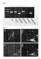
- Figure 4 shows the result of RT-PCR for RNA isolated from KUM9 cells, which are stably transduced with parent vector (pIRES) and SF-1 (SF-9) and are cultured for 7 days in the presence of cAMP. Days shown on the upper row are days until extraction of RNA after the addition of cAMP. HSD3b (3b-HSD) expresses on the 3rd day and P450c17 and P450c11b1 express on the 5th day.
- Figure 5 shows the western blotting for proteins extracted from the cells cultured under a condition similar to that described in Fig. 4. Proteins from MA10-derived cells originated from mouse Leydig cells are used as a positive control.
- Figure 6 shows quantification by RIA of secreted steroid hormone, which is obtained by the recovery of supernatant of the medium used for the cells cultured under a condition similar to that described in Fig.4. The amount of hormone produced within 24 hrs was determined by changing culture medium to fresh medium on the day before the each indicated day.
- Figure 7 shows immunostainig by anti-3β HSD I antibody of SF-9 cells, which are fixed before adding cAMP (-) or after culture for 7 days in the presence of cAMP (+). DAPI (4',6-Diamidino-2-phenylindole, dihydrochloride) is a dye marker for DNA and shows the intracellular localization of nucleus.
- Figure 8 shows the result of RT-PCR for RNA extracted from hMSC E6/E7 cells, which is derived from human bone marrow mesenchymal stem cells, are stably transduced with SF-1, and are cultured for 7 days in the presence of cAMP.
- Figure 9 shows testis transplanted with mesenchymal stem cells derived from bone marrow. ST shows seminiferous tubule.
- Figure 10 shows pEGFP-1 vector.
- Figure 11 shows pPUR vector.
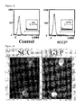
- Figure 12 shows the charts of flow cytometry for a GFP positive clone.
- Figure 13 shows immunostaining of P450scc in GFP positive cells.
- Figure 14 shows the results of RT-PCR of various enzymes in GFP positive (scc+) and negative (scc-) cell fractions.
- Mesenchymal stem cells are derived from various interstitial cells and are able to differentiate into various cells. Mesenchymal stem cells are present not only in bone marrow but also interstitial and other tissues (e.g. in kidney, articular membrane, amnion and cord blood) of mesoderm-derived tissues. All these mesenchymal stem cells can be used in the present invention.
- Mesenchymal stem cells derived from bone marrow are interstitial cells, which present in marrow stroma and maintain hematopoiesis, and said stem cells from even adult bear pluripotency of differentiating into various cells and tissues. Mesenchymal stem cells derived from bone marrow may be isolated by inserting an injection needle into an epiphysis for example, by feeding PBS into the epiphysis, by pulling out bone marrow cells, by seeding and subjecting the bone marrow cells to adhere to a plastic dish and by repeating the subculture.
- The transcriptional factor (SF-1), an inducing factor used in the method of the present invention, is an orphan intranuclear receptor, which is expressed in genital and adrenal gland-type steroid hormone-producing cells, and has been known to control the transcription of steroid hormone producing-enzymes (Endocrine Reviews vol.18, No.3, 361-377 (1997); The FASEB Journal vol.10 1569-1577 (1996)). Even if SF-1 is derived from different animal species, SF-1 binds to a common target DNA sequence in mesenchymal stem cells and the factor is expected to provide the same result.
- cAMP exists ubiquitously in all living organism, whose intra-cellular concentration is 10-6 to 10-7 M. cAMP participates in generation of specific enzymes and metabolic control in target cells and also participates in growth and differentiation of cells. cAMP is a second messenger of LH and ACTH, which induces the expression of steroid hormone production-related enzymes in genital and adrenal glands and enhances the production of steroid hormones.
- The mean to stimulate by the transcriptional factor (SF-1) and cAMP may include the direct contact of these factors with mesenchymal stem cells or the use of a vector expressing these factors.
- To differentiate mesenchymal stem cells into steroid hormone-producing cells, mesenchymal stem cells may be stimulated by an inducing factor in vitro or said cells may be transplanted into a mammalian reproductive organ. In the case of in vitro induction, for example, human mesenchymal stem cells may be cultured in DMEM (Dulbecco's Modified Eagle's Medium) containing 10% FBS and mouse stem cells may be cultured in IMDM (Iscove's Modified Dulbecco's Medium) or in DMEM containing 10% FBS. The above cells are cultured in an incubator with 5% CO2 at 37°C as usual. The concentration of SF-1 is about 0.1~10 µg, preferably at 1 µg / 1x10⁵ cells, and that of cAMP is about 0.5~2.0 mM, preferably at 1 mM / 1x10⁵ cells.
- When mesenchymal stem cells were transplanted into a mammalian reproductive organ, a certain factor activates the cell (in the case of organ transplantation, interaction with the surrounding cells) to express SF-1, which is considered to make these stem cells to differentiate into steroid hormone-producing cells.
- In the differentiation of mesenchymal stem cells, the role of cAMP is subsidiary. Furthermore, since cAMP is necessarily present in all kind of cells with a large variety of concentrations, intracellular concentration of cAMP might be raised by some reason without adding exogenous cAMP.
- As described above, steroid-producing cells may be obtained accordingly, then steroid hormone may be obtained from the steroid-producing cells.
- Steroid-producing cells derived from mesenchymal stem cells include adrenal cortical cells, ovarian granulosa cells, ovarian capsular cells, testis Leydig cells, testis, Sertoli cells and others.
- Obtained steroid hormones include any steroid hormone and the like, which are synthesized from cholesterol, such as progestin, androgen, estrogen, glucocorticoid and mineralcorticoid.
- The following examples further illustrate the present invention, but it is not intended to limit the scope of the present invention.
- Gene expression of steroid hormone synthesizing enzymes was examined by RT-PCR using the primers shown in Table 1.
Table 1 Symbol Enzyme Primer SEQ ID NO: StAR steroidogenic acute regulatory protein F-GAAGGAAAGCCAGCAGGAGAACG 1 R-CTCTGATGACACCACTCTGCTCC 2 P450scc cholesterol side chain cleavage enzyme F-CGCACAGTCCAGAACAACAAGCA 3 R-CGGTTAGAGAAGGCAGGATAGAG 4 HSD3b 3-β hydroxysteriod dehydrogenase F-GCAGACCATCCTAGATGTCAAT 5 R-TCATCATAGCTTTGGTGAGG 6 P450c17 17-alpha-hydroxylase/17,20-lyase F-AAAATAATAACACTGGGGAAGGC 7 R- TGGGTGTGGGTGTAATGAGATGG 8 P450c21 steroid 21-hydroxylase F-AGAGGATCCGCTTGGGGCTGC 9 R- GGAGAATTCOTTATGGATGGC 10 P45011b1 steroid 11-beta-hydroxylase F-TCACCAAATGTATCAAGAATGTGT 11 R-CCATOTGCACATCCTCTTTCTCTT 12 P45011b2 aldosterone synthase F-CCAACAGATGTATCTGGAAGGTGC 13 R-CCATCTGCACATOCTCTTDCCTCA 14 GAPDH glyceraldehyde phosphate dehydrogenase F-ACCACAGTCCATGCCATCAC 15 R-TCCACCACCCTGTTGCTGTA 16 - The coding region (
Nucleotides 1~1389 of SEQ ID NO: 17) of rat SF-1 gene (Gene Bank NM_053344, SEQ ID NO:17) was integrated in pIRES-puro2 vector (Clontech, Fig. 1). KUM9 cells (obtained from Department of Reproductive Biology, National Research Institute for Child Health and Development), an established adult mouse-derived mesenchymal stem cell line, were transfected with said vector after linearized with Nru I and were selected in a medium containing puromycin (2 µg/ml) at 48 hrs after the transfection. The resistant colonies remained for 2 weeks in the medium were picked up and were cloned. - Those clones transfected with parent vector pIRES-puro (Fig. 2B) showed similar morphology to that of KUM9 without transfection (Fig. 2A). In contrast, a large number of fat droplets, conceivably containing cholesterol, i.e. a precursor of steroid hormones, were observed intracellularly in clones (SF-7 and SF-9) expressing SF-1 (Fig. 2C).
- Proteins were extracted from each clone (SF-7 and SF-9), were examined the expression of SF-1 protein by Western blotting and SF-1 protein was detected (Fig. 3).
- Furthermore, gene expression of steroid hormone synthesizing enzymes in these cells was examined by RT-PCR and the expression of P450scc was confirmed.
- The cloned cells (SF-9), wherein expression of SF-1 was observed in example 1, were cultured for 7 days in the presence of cAMP (1 mM, Sigma). On 1,3,5 and 7 days after addition of cAMP, RNA was extracted from said cells and was examined the gene expression by RT-PCR. For comparison, the same procedures were performed for cloned cells using the parent vector pIRES as a negative control. The results show that the expression of p450scc was induced after 1 day, HSD3b1 was induced after 3 days and furthermore p450c17 was induced after 5 days (Fig. 4).
- The result of Western blotting shows that the expression of these enzyme proteins was induced similar to that of mRNA (Fig. 5).
- Moreover, according to these results, those cells without transfection with SF-1 (pIRES-puro) do not express steroid hormone-producing enzymes by the addition of cAMP and do not produce steroid hormone. These facts could be interpreted as that only cAMP could not induce differentiation into steroid hormone-producing cells.
- The interpretation depicts that SF-1 is an essential factor to differentiate mesenchymal stem cells into steroid hormone producing cells and cAMP is a cofactor.
- The output of steroid hormone was measured by radioimmuno assay (RIA) after recovery of culture medium on the 3rd day and 7th day. According to the results, progesterone and androgen were detected on the 3rd day and 7th day, respectively, in parallel to the results of expression of mRNA and proteins (Fig.6) in the group of cells added with cAMP Not only the induction of steroid synthesizing enzyme but also the synthesis of androstendione, a kind of androgenic hormone, was confirmed.
- Immunostaining of HSD3b1 shows that all cells are positive (Fig. 7).
- The results of examples 1 and 2 show that forced expression of SF-1 leads mesenchymal stem cells derived from bone marrow to differentiate into steroid hormone-producing cells.
- Induction of steroid hormone-producing cells for human cells was examined in an experiment similar to example 2 using human derived cells. Human bone marrow mesenchymal stem cells hMSCE6/E7 (gifted from Field of Clinical Application, Institute for frontier Medical Sciences, Kyoto University) were transfected with said pIRES-puro-SE-1 vector by calcium phosphate method. At 48 hrs after transfection, cells were selected in a medium containing puromycin (0.3 µg/ml), puromycin resistant colonies were picked up after 2 weeks and they were cloned. After 7 days culture in the presence of cAMP, RNA was isolated and gene expression was examined by RT-PCR.
- hMSC cells stably expressing pIRES-puro-SF-1 did not show morphoological change as observed for KUM9 cells (Fig. 2A~C). However, analysis of gene expression by RT-PCR shows the induction of expression of a group of steroid hormone synthesizing genes such as StaR, P450scc and 3β-HSD. Furthermore, addition of cAMP induced the expression of P450 c21 and P450 11b1, glucocorticoid producing enzymes in adrenal cortex (Fig. 8).
These results show that human bone marrow-derived mesenchymal stem cells were differentiated into steroid hormone-producing cells. - In this example, bone marrow-derived mesenchymal stem cells were transplanted into testis. Bone marrow cells were isolated from epiphysial region of os longum from limbs of 5 w.o. male Green rat by insertion of a 26-gauge needle for syringe and by filling of PBS. After red blood cells were disrupted by osmotic shock, said cells were seeded at 5x 10⁷ / 100 ml dish containing α-MEM medium fortified with 20% FBS. After 24 hrs, the medium was changed to remove non-adherent cells. After 3 days, cells were trypsinized by 0.25% trypsin, subcultured, and cultured for further 1 week. These procedures lead to concentrate mesenchymal stem cells among various bone marrow-derived cells. After confluent cells were trypsinized, 1 x 10⁶ cells were suspended in 50 µl PBS and were injected into testis of 3 w.o. male Sprague Dawley rat (Japan Charles River Co.).
- After 3 weeks, the testis was removed, observed under inverted microscopy and fixed by 4% para-formaldehyde. Frozen sections were prepared and used for immunohistochemical method. Double staining was performed using rabbit anti-rat P450scc antibody (CHEMICON) and mouse anti-GFP antibody (MBL) for the first antibody and Cy3-conjugated anti-rabbit IgG antibody (SIGMA) and FITC-conjugated anti-mouse IgG (SIGMA) as the second antibody.
- GFP positive cells, possibly derived from Green rat bone marrow, were observed easily under inverted fluorescent microscopy and were detected in testis at 3 weeks after transplantation (Fig. 9A). Therefore, it was found that transplanted cells adhered to host testis. To examine whether these cells were differentiated into steroid hormone producing cells (Leydig cells), double immunostainig using anti-GFP (Fig. 9B) and anti-P450scc (Fig. 9C) was performed. Since most of P450scc positive cells were also GFP positive, Green rat bone marrow-derived cells were found to be differentiated into Leydig cells.
- Stem cells could not completely be clonized by the method of example 4 because of the contamination of macrophage cells. Therefore, an experiment was performed to examine whether KUM9 cells, an established adult mouse-derived mesenchymal stem cells, has a potency to be differentiated in vitro into steroid hormone-producing cells.
- P450scc (a side chain cleavage enzyme) is an enzyme, which is localized on mitochondrial inner membrane and catalyzes a reaction degrading cholesterol into prenegnenolone. Since the above reaction is a necessary procedure in all steroid hormone production, P450scc is expressed in all steroid hormone-producing cells.
The group of Chung et al. prepared and used transgenic mice with a visual reporter gene Lac Z and showed that 2.3 Kbp of 5' upstream region in human P450scc gene (Gene Bank M60421) controls the expression of P450scc gene in steroid hormone-producing cells in genital and adrenal gland (Endocrinology, 140:5609 18, 1999). Therefore, preparation of stem cells, wherein the visual reporter gene expresses by the control of the said 5' upstream region, conceivably enables to detect the differentiation into steroid hormone-producing cells. - Therefore, P450scc gene (Gene Bank M60421, SEQ ID NO: 18) was integrated into the upstream of EGFP protein coding region of pEGFP-1 vector (Fig. 10, Clontech) and the vector was fused with pPUR vector (Fig. 11, Clontech) coding puromycin resistant gene.
- Said synthetic vector, after linearized with ApaL I, was transfected into KUM9 cells (Gifted from Department of Reproductive Biology, National Research Institute for Child Health and Development), derived from mouse bone marrow mesenchymal stem cells by lipofection. Since fluorescent GFP expresses at the time of expression of P450scc in these cells, spontaneously differentiated KUM9-derived steroid-producing cells could be separated by flow cytometer using fluorescence as a probe.
- At 48 hrs after the separation, cells were selected in the medium containing puromycin (2 µg/ml) and resistant colonies after 2 weeks was clonized by a limiting dilution method.
- GFP positive cells were detected in 2 clones out of isolated 250 clones during growing phase of these cells under inverted fluorescence microscopic observation. GFP positive and negative cells were separated by flow cytometer for said 2 clones, wherein a lot of GFP positive cells were found, and GFP positive cells were 2~5% (Fig. 12).
- Furthermore, immunostaining of P450scc for GFP positive cells confirmed that GFP positive cells were also P450scc positive (Fig. 13).
- Moreover, GFP positive (scc+) and negative (scc-) cells were separated by a cell sorter, total RNA was extracted from the separated cells and cDNA was prepared by reverse transcription reaction using reverse transcriptase. RT-PCR was performed using a specific primer for each gene. The expression of mRNA of P450scc gene was observed in GFP positive cells (scc+) (Fig 14). Additionally, the transcriptional factor SF-1 was expressed in said cells isolated. Still furthermore, examination of the expression of genes involved in steroid hormone production revealed that the expression of 38-HSD gene of both genital and adrenal gland type (HSD3b1) and placental type (HSD3b6) was observed.
- From these results, it was interpreted that the transplantation induced the expression of SF-1 in stem cells, and that these stem cells differentiated into steroid hormone-producing cells on contact with SF-1.
Claims (10)
- A method for differentiating bone marrow-derived mesenchymal stem cells into steroid hormone-producing cells, comprising stimulating the mesenchymal stem cells by a transcriptional factor (SF-1).
- A method of claim 1 comprising further stimulating the mesenchymal stem cells by cAMP
- A method of claim 1 or 2, wherein the mesenchymal stem cells are derived from bone marrow.
- A method of claim 3, wherein the mesenchymal cells are derived from human.
- A method for producing steroid hormone-producing cells, comprising implementing the method of any one of claims 1 to 4 in vitro.
- A method for producing steroid hormone-producing cells, comprising implementing the method of any one of claims 1 to 4 by transplanting the mesenchymal stem cells into a mammalian reproductive organ.
- Steroid hormone-producing cells obtained by the method of claim 5 or 6.
- A method for producing steroid hormone, comprising culturing the steroid hormone-producing cells of claim 7 and recovering steroid hormone from the culture medium.
- The method of claim 8, wherein said steroid hormones are progestin, androgen, estrogen, glucocorticoid or mineralcorticoid.
- Non-human animals carrying the steroid-producing cells of claim 7.
Applications Claiming Priority (2)
| Application Number | Priority Date | Filing Date | Title |
|---|---|---|---|
| JP2004058406 | 2004-03-03 | ||
| PCT/JP2005/002548 WO2005085425A1 (en) | 2004-03-03 | 2005-02-18 | Method of differentiating mesenchymal stem cells into steroid-producing cells |
Publications (3)
| Publication Number | Publication Date |
|---|---|
| EP1721967A1 true EP1721967A1 (en) | 2006-11-15 |
| EP1721967A4 EP1721967A4 (en) | 2008-06-18 |
| EP1721967B1 EP1721967B1 (en) | 2010-03-24 |
Family
ID=34917930
Family Applications (1)
| Application Number | Title | Priority Date | Filing Date |
|---|---|---|---|
| EP05719267A Expired - Lifetime EP1721967B1 (en) | 2004-03-03 | 2005-02-18 | Method of differentiating mesenchymal stem cells into steroid-producing cells |
Country Status (6)
| Country | Link |
|---|---|
| US (1) | US7879603B2 (en) |
| EP (1) | EP1721967B1 (en) |
| JP (1) | JP4548849B2 (en) |
| CA (1) | CA2557992C (en) |
| DE (1) | DE602005020120D1 (en) |
| WO (1) | WO2005085425A1 (en) |
Families Citing this family (9)
| Publication number | Priority date | Publication date | Assignee | Title |
|---|---|---|---|---|
| JP4548849B2 (en) | 2004-03-03 | 2010-09-22 | 独立行政法人科学技術振興機構 | Methods for differentiating mesenchymal stem cells into steroidogenic cells |
| JPWO2006038462A1 (en) * | 2004-10-01 | 2008-05-15 | 国立大学法人九州大学 | New steroidogenic cells |
| WO2007117472A2 (en) * | 2006-04-07 | 2007-10-18 | Los Angeles Biomedical Research Institute At Harbor-Ucla Medical Center | Adult bone marrow cell transplantation to testes creation of transdifferentiated testes germ cells, leydig cells and sertoli cells |
| WO2007136044A1 (en) * | 2006-05-19 | 2007-11-29 | Kyushu University, National University Corporation | Steroid hormone-producing cell |
| JP2011015630A (en) * | 2009-07-08 | 2011-01-27 | Univ Of Fukui | Method for inducing differentiation of pluripotent stem cell into steroid hormone producing cell |
| US11253549B2 (en) * | 2014-05-23 | 2022-02-22 | JangoBio, LLC | Methods to rebalance the hypothalamic-pituitary-gonadal axis |
| CN105255826B (en) * | 2015-11-27 | 2019-01-08 | 中山大学 | Method of inducing differentiation and application thereof of the people iPS cell to interstitial glands |
| WO2018147257A1 (en) * | 2017-02-07 | 2018-08-16 | 国立大学法人京都大学 | Method for producing steroidogenic cells |
| AU2022386630A1 (en) | 2021-11-09 | 2024-05-30 | Technische Universität Dresden | A method for producing induced steroidogenic cells and use thereof in cell therapy |
Family Cites Families (4)
| Publication number | Priority date | Publication date | Assignee | Title |
|---|---|---|---|---|
| JP4036596B2 (en) | 2000-03-03 | 2008-01-23 | サントリー株式会社 | Lipids containing 5,11,14-eicosatrienoic acid and / or 5,11,14,17-eicosatetraenoic acid and method for producing the same |
| EP1285057B1 (en) * | 2000-04-28 | 2006-05-31 | Children's Medical Center Corporation | Isolation of mesenchymal stem cells and use thereof |
| WO2002022788A1 (en) | 2000-09-12 | 2002-03-21 | Yukio Kato | Method of culturing mesenchymal stem cells |
| JP4548849B2 (en) | 2004-03-03 | 2010-09-22 | 独立行政法人科学技術振興機構 | Methods for differentiating mesenchymal stem cells into steroidogenic cells |
-
2005
- 2005-02-18 JP JP2006510634A patent/JP4548849B2/en not_active Expired - Fee Related
- 2005-02-18 CA CA2557992A patent/CA2557992C/en not_active Expired - Fee Related
- 2005-02-18 WO PCT/JP2005/002548 patent/WO2005085425A1/en not_active Ceased
- 2005-02-18 US US10/591,530 patent/US7879603B2/en not_active Expired - Fee Related
- 2005-02-18 DE DE602005020120T patent/DE602005020120D1/en not_active Expired - Lifetime
- 2005-02-18 EP EP05719267A patent/EP1721967B1/en not_active Expired - Lifetime
Also Published As
| Publication number | Publication date |
|---|---|
| US20080313752A1 (en) | 2008-12-18 |
| EP1721967B1 (en) | 2010-03-24 |
| EP1721967A4 (en) | 2008-06-18 |
| US7879603B2 (en) | 2011-02-01 |
| DE602005020120D1 (en) | 2010-05-06 |
| JPWO2005085425A1 (en) | 2007-12-13 |
| WO2005085425A1 (en) | 2005-09-15 |
| JP4548849B2 (en) | 2010-09-22 |
| CA2557992C (en) | 2011-12-20 |
| CA2557992A1 (en) | 2005-09-15 |
Similar Documents
| Publication | Publication Date | Title |
|---|---|---|
| Moscoso et al. | Differentiation “in vitro” of primary and immortalized porcine mesenchymal stem cells into cardiomyocytes for cell transplantation | |
| Zhang et al. | CRISPR/Cas9‐mediated sheep MSTN gene knockout and promote sSMSCs differentiation | |
| EP1983042B1 (en) | A method for purifying cardiomyocytes or programmed cardiomyocytes derived from stem cells or fetuses | |
| US20040092011A1 (en) | Adipocytic differentiated adipose derived adult stem cells and uses thereof | |
| CN1822846B (en) | Angiogenesis induced by perivascular mesenchymal precursor cells | |
| Wu et al. | In vitro culture and induced differentiation of sheep skeletal muscle satellite cells | |
| CA2465173C (en) | Endothelial cells derived from primate embryonic stem cells | |
| AU2002356896A1 (en) | Endothelial cells derived from primate embryonic stem cells | |
| EP1721967B1 (en) | Method of differentiating mesenchymal stem cells into steroid-producing cells | |
| US20150315545A1 (en) | Method of rapidly inducing large-scale and high-purity mesenchymal stem cells to transdetermine into hematopoietic stem cells | |
| Yamada et al. | Single-cell-derived mesenchymal stem cells overexpressing Csx/Nkx2. 5 and GATA4 undergo the stochastic cardiomyogenic fate and behave like transient amplifying cells | |
| WO2014163206A1 (en) | Use of functional melanocytes readily differentiated from multilineage-differentiating stress-enduring (Muse) cells, distinct stem cells in human fibroblasts | |
| WO2007010858A1 (en) | Pluripotent stem cell cloned from single cell derived from skeletal muscle tissue | |
| Camara et al. | Differentiation of canine adipose mesenchymal stem cells into insulin-producing cells: comparison of different culture medium compositions | |
| CN107056895A (en) | The artificial polypeptide and its biological products of inducing bone mesenchymal stem cell into hepatocyte differentiation | |
| AU2013225946A1 (en) | Method for guiding the derivation of endothelial cells from human pluripotent stem cells employing two-dimensional, feeder-free differentiation | |
| AU2012227337B2 (en) | A method for purifying cardiomyocytes or programmed cardiomyocytes derived from stem cells or fetuses | |
| Vidal et al. | Adipogenic differentiation of adult equine mesenchymal stromal cells | |
| WO2001048150A1 (en) | Cells capable of differentiating into heart muscle cells | |
| JPWO2006038462A1 (en) | New steroidogenic cells | |
| KR100973823B1 (en) | Induction of Differentiation of Embryonic Carcinoma Cells into Vascular Endothelial Cells | |
| TWI437097B (en) | Fluorescent protein-expressing swine bone marrow mesenchymal stem cells | |
| Ramkisoensing | Molecular and environmental cues in cardiac differentiation of mesenchymal stem | |
| JP2006122040A (en) | New steroidogenic cells |
Legal Events
| Date | Code | Title | Description |
|---|---|---|---|
| PUAI | Public reference made under article 153(3) epc to a published international application that has entered the european phase |
Free format text: ORIGINAL CODE: 0009012 |
|
| 17P | Request for examination filed |
Effective date: 20060825 |
|
| AK | Designated contracting states |
Kind code of ref document: A1 Designated state(s): CH DE FR GB LI NL SE |
|
| DAX | Request for extension of the european patent (deleted) | ||
| RBV | Designated contracting states (corrected) |
Designated state(s): CH DE FR GB LI NL SE |
|
| A4 | Supplementary search report drawn up and despatched |
Effective date: 20080519 |
|
| 17Q | First examination report despatched |
Effective date: 20090324 |
|
| GRAP | Despatch of communication of intention to grant a patent |
Free format text: ORIGINAL CODE: EPIDOSNIGR1 |
|
| GRAS | Grant fee paid |
Free format text: ORIGINAL CODE: EPIDOSNIGR3 |
|
| GRAA | (expected) grant |
Free format text: ORIGINAL CODE: 0009210 |
|
| AK | Designated contracting states |
Kind code of ref document: B1 Designated state(s): CH DE FR GB LI NL SE |
|
| REG | Reference to a national code |
Ref country code: GB Ref legal event code: FG4D |
|
| REG | Reference to a national code |
Ref country code: CH Ref legal event code: EP |
|
| REF | Corresponds to: |
Ref document number: 602005020120 Country of ref document: DE Date of ref document: 20100506 Kind code of ref document: P |
|
| REG | Reference to a national code |
Ref country code: NL Ref legal event code: T3 |
|
| REG | Reference to a national code |
Ref country code: SE Ref legal event code: TRGR |
|
| REG | Reference to a national code |
Ref country code: CH Ref legal event code: NV Representative=s name: LUCHS & PARTNER PATENTANWAELTE |
|
| PLBE | No opposition filed within time limit |
Free format text: ORIGINAL CODE: 0009261 |
|
| STAA | Information on the status of an ep patent application or granted ep patent |
Free format text: STATUS: NO OPPOSITION FILED WITHIN TIME LIMIT |
|
| 26N | No opposition filed |
Effective date: 20101228 |
|
| PGFP | Annual fee paid to national office [announced via postgrant information from national office to epo] |
Ref country code: CH Payment date: 20140212 Year of fee payment: 10 Ref country code: SE Payment date: 20140211 Year of fee payment: 10 Ref country code: NL Payment date: 20140110 Year of fee payment: 10 |
|
| REG | Reference to a national code |
Ref country code: FR Ref legal event code: PLFP Year of fee payment: 11 |
|
| PGFP | Annual fee paid to national office [announced via postgrant information from national office to epo] |
Ref country code: DE Payment date: 20150113 Year of fee payment: 11 |
|
| PGFP | Annual fee paid to national office [announced via postgrant information from national office to epo] |
Ref country code: FR Payment date: 20150113 Year of fee payment: 11 Ref country code: GB Payment date: 20150112 Year of fee payment: 11 |
|
| REG | Reference to a national code |
Ref country code: NL Ref legal event code: V1 Effective date: 20150901 |
|
| REG | Reference to a national code |
Ref country code: SE Ref legal event code: EUG |
|
| PG25 | Lapsed in a contracting state [announced via postgrant information from national office to epo] |
Ref country code: NL Free format text: LAPSE BECAUSE OF NON-PAYMENT OF DUE FEES Effective date: 20150901 |
|
| REG | Reference to a national code |
Ref country code: CH Ref legal event code: PL |
|
| PG25 | Lapsed in a contracting state [announced via postgrant information from national office to epo] |
Ref country code: CH Free format text: LAPSE BECAUSE OF NON-PAYMENT OF DUE FEES Effective date: 20150228 Ref country code: LI Free format text: LAPSE BECAUSE OF NON-PAYMENT OF DUE FEES Effective date: 20150228 |
|
| PG25 | Lapsed in a contracting state [announced via postgrant information from national office to epo] |
Ref country code: SE Free format text: LAPSE BECAUSE OF NON-PAYMENT OF DUE FEES Effective date: 20150219 |
|
| REG | Reference to a national code |
Ref country code: DE Ref legal event code: R119 Ref document number: 602005020120 Country of ref document: DE |
|
| GBPC | Gb: european patent ceased through non-payment of renewal fee |
Effective date: 20160218 |
|
| REG | Reference to a national code |
Ref country code: FR Ref legal event code: ST Effective date: 20161028 |
|
| PG25 | Lapsed in a contracting state [announced via postgrant information from national office to epo] |
Ref country code: FR Free format text: LAPSE BECAUSE OF NON-PAYMENT OF DUE FEES Effective date: 20160229 Ref country code: DE Free format text: LAPSE BECAUSE OF NON-PAYMENT OF DUE FEES Effective date: 20160901 Ref country code: GB Free format text: LAPSE BECAUSE OF NON-PAYMENT OF DUE FEES Effective date: 20160218 |