EP1697498B1 - Kinase suppressor of ras inactivation for therapy of ras mediated tumorigenesis - Google Patents
Kinase suppressor of ras inactivation for therapy of ras mediated tumorigenesis Download PDFInfo
- Publication number
- EP1697498B1 EP1697498B1 EP04812928A EP04812928A EP1697498B1 EP 1697498 B1 EP1697498 B1 EP 1697498B1 EP 04812928 A EP04812928 A EP 04812928A EP 04812928 A EP04812928 A EP 04812928A EP 1697498 B1 EP1697498 B1 EP 1697498B1
- Authority
- EP
- European Patent Office
- Prior art keywords
- ksr
- ras
- cells
- expression
- cancer
- Prior art date
- Legal status (The legal status is an assumption and is not a legal conclusion. Google has not performed a legal analysis and makes no representation as to the accuracy of the status listed.)
- Expired - Lifetime
Links
- 108010003046 KSR-1 protein kinase Proteins 0.000 title claims abstract description 487
- 102100021001 Kinase suppressor of Ras 1 Human genes 0.000 title abstract description 425
- 102000016914 ras Proteins Human genes 0.000 title abstract description 149
- 108010014186 ras Proteins Proteins 0.000 title abstract description 149
- 231100000504 carcinogenesis Toxicity 0.000 title abstract description 49
- 208000005623 Carcinogenesis Diseases 0.000 title abstract description 47
- 230000036952 cancer formation Effects 0.000 title abstract description 47
- 230000001404 mediated effect Effects 0.000 title abstract description 45
- 230000002779 inactivation Effects 0.000 title description 40
- 238000002560 therapeutic procedure Methods 0.000 title description 5
- 230000014509 gene expression Effects 0.000 claims abstract description 204
- 108091034117 Oligonucleotide Proteins 0.000 claims abstract description 197
- 150000007523 nucleic acids Chemical class 0.000 claims abstract description 81
- 239000000074 antisense oligonucleotide Substances 0.000 claims abstract description 80
- 238000012230 antisense oligonucleotides Methods 0.000 claims abstract description 80
- 102000039446 nucleic acids Human genes 0.000 claims abstract description 70
- 108020004707 nucleic acids Proteins 0.000 claims abstract description 70
- 210000004027 cell Anatomy 0.000 claims description 222
- 206010028980 Neoplasm Diseases 0.000 claims description 174
- 125000003729 nucleotide group Chemical group 0.000 claims description 84
- 239000002773 nucleotide Substances 0.000 claims description 82
- 230000000694 effects Effects 0.000 claims description 78
- 108020004999 messenger RNA Proteins 0.000 claims description 56
- 241000124008 Mammalia Species 0.000 claims description 41
- 102000005789 Vascular Endothelial Growth Factors Human genes 0.000 claims description 41
- 108010019530 Vascular Endothelial Growth Factors Proteins 0.000 claims description 41
- 108091026890 Coding region Proteins 0.000 claims description 39
- 201000011510 cancer Diseases 0.000 claims description 39
- 230000005865 ionizing radiation Effects 0.000 claims description 39
- 150000001413 amino acids Chemical class 0.000 claims description 38
- 230000002401 inhibitory effect Effects 0.000 claims description 37
- 230000000692 anti-sense effect Effects 0.000 claims description 33
- 210000004881 tumor cell Anatomy 0.000 claims description 25
- 230000033115 angiogenesis Effects 0.000 claims description 22
- 206010061902 Pancreatic neoplasm Diseases 0.000 claims description 21
- 201000002528 pancreatic cancer Diseases 0.000 claims description 20
- 208000015486 malignant pancreatic neoplasm Diseases 0.000 claims description 19
- 208000008443 pancreatic carcinoma Diseases 0.000 claims description 19
- 102000004190 Enzymes Human genes 0.000 claims description 12
- 108090000790 Enzymes Proteins 0.000 claims description 12
- 239000000126 substance Substances 0.000 claims description 11
- 230000007423 decrease Effects 0.000 claims description 9
- 239000003814 drug Substances 0.000 claims description 8
- RYYWUUFWQRZTIU-UHFFFAOYSA-K thiophosphate Chemical compound [O-]P([O-])([O-])=S RYYWUUFWQRZTIU-UHFFFAOYSA-K 0.000 claims description 8
- 239000003446 ligand Substances 0.000 claims description 6
- 230000002285 radioactive effect Effects 0.000 claims description 6
- 208000000453 Skin Neoplasms Diseases 0.000 claims description 5
- 238000004519 manufacturing process Methods 0.000 claims description 5
- 206010006187 Breast cancer Diseases 0.000 claims description 4
- 206010009944 Colon cancer Diseases 0.000 claims description 4
- 206010058467 Lung neoplasm malignant Diseases 0.000 claims description 4
- 206010029260 Neuroblastoma Diseases 0.000 claims description 4
- 208000024770 Thyroid neoplasm Diseases 0.000 claims description 4
- 208000032839 leukemia Diseases 0.000 claims description 4
- 201000007270 liver cancer Diseases 0.000 claims description 4
- 208000014018 liver neoplasm Diseases 0.000 claims description 4
- 230000014399 negative regulation of angiogenesis Effects 0.000 claims description 4
- 206010005003 Bladder cancer Diseases 0.000 claims description 3
- 208000026310 Breast neoplasm Diseases 0.000 claims description 3
- 208000000461 Esophageal Neoplasms Diseases 0.000 claims description 3
- 208000005016 Intestinal Neoplasms Diseases 0.000 claims description 3
- 206010025323 Lymphomas Diseases 0.000 claims description 3
- 206010030155 Oesophageal carcinoma Diseases 0.000 claims description 3
- 206010033128 Ovarian cancer Diseases 0.000 claims description 3
- 206010061535 Ovarian neoplasm Diseases 0.000 claims description 3
- 206010060862 Prostate cancer Diseases 0.000 claims description 3
- 208000000236 Prostatic Neoplasms Diseases 0.000 claims description 3
- 208000005718 Stomach Neoplasms Diseases 0.000 claims description 3
- 208000007097 Urinary Bladder Neoplasms Diseases 0.000 claims description 3
- 208000006593 Urologic Neoplasms Diseases 0.000 claims description 3
- 208000029742 colonic neoplasm Diseases 0.000 claims description 3
- 201000004101 esophageal cancer Diseases 0.000 claims description 3
- 206010017758 gastric cancer Diseases 0.000 claims description 3
- 201000010536 head and neck cancer Diseases 0.000 claims description 3
- 208000014829 head and neck neoplasm Diseases 0.000 claims description 3
- 201000002313 intestinal cancer Diseases 0.000 claims description 3
- 201000005202 lung cancer Diseases 0.000 claims description 3
- 208000020816 lung neoplasm Diseases 0.000 claims description 3
- 201000000849 skin cancer Diseases 0.000 claims description 3
- 201000011549 stomach cancer Diseases 0.000 claims description 3
- 201000002510 thyroid cancer Diseases 0.000 claims description 3
- 201000005112 urinary bladder cancer Diseases 0.000 claims description 3
- 238000000034 method Methods 0.000 abstract description 97
- 230000005764 inhibitory process Effects 0.000 abstract description 80
- 239000000203 mixture Substances 0.000 abstract description 41
- 230000000295 complement effect Effects 0.000 abstract description 33
- 108020000948 Antisense Oligonucleotides Proteins 0.000 abstract description 25
- 230000002068 genetic effect Effects 0.000 abstract description 16
- 238000013459 approach Methods 0.000 abstract description 15
- 206010027476 Metastases Diseases 0.000 abstract description 8
- 230000009401 metastasis Effects 0.000 abstract description 8
- 230000000903 blocking effect Effects 0.000 abstract description 5
- 238000011275 oncology therapy Methods 0.000 abstract description 2
- 229940046166 oligodeoxynucleotide Drugs 0.000 description 160
- 241000282414 Homo sapiens Species 0.000 description 74
- 238000011282 treatment Methods 0.000 description 74
- 108020004414 DNA Proteins 0.000 description 72
- 241000699670 Mus sp. Species 0.000 description 69
- JLCPHMBAVCMARE-UHFFFAOYSA-N [3-[[3-[[3-[[3-[[3-[[3-[[3-[[3-[[3-[[3-[[3-[[5-(2-amino-6-oxo-1H-purin-9-yl)-3-[[3-[[3-[[3-[[3-[[3-[[5-(2-amino-6-oxo-1H-purin-9-yl)-3-[[5-(2-amino-6-oxo-1H-purin-9-yl)-3-hydroxyoxolan-2-yl]methoxy-hydroxyphosphoryl]oxyoxolan-2-yl]methoxy-hydroxyphosphoryl]oxy-5-(5-methyl-2,4-dioxopyrimidin-1-yl)oxolan-2-yl]methoxy-hydroxyphosphoryl]oxy-5-(6-aminopurin-9-yl)oxolan-2-yl]methoxy-hydroxyphosphoryl]oxy-5-(6-aminopurin-9-yl)oxolan-2-yl]methoxy-hydroxyphosphoryl]oxy-5-(6-aminopurin-9-yl)oxolan-2-yl]methoxy-hydroxyphosphoryl]oxy-5-(6-aminopurin-9-yl)oxolan-2-yl]methoxy-hydroxyphosphoryl]oxyoxolan-2-yl]methoxy-hydroxyphosphoryl]oxy-5-(5-methyl-2,4-dioxopyrimidin-1-yl)oxolan-2-yl]methoxy-hydroxyphosphoryl]oxy-5-(4-amino-2-oxopyrimidin-1-yl)oxolan-2-yl]methoxy-hydroxyphosphoryl]oxy-5-(5-methyl-2,4-dioxopyrimidin-1-yl)oxolan-2-yl]methoxy-hydroxyphosphoryl]oxy-5-(5-methyl-2,4-dioxopyrimidin-1-yl)oxolan-2-yl]methoxy-hydroxyphosphoryl]oxy-5-(6-aminopurin-9-yl)oxolan-2-yl]methoxy-hydroxyphosphoryl]oxy-5-(6-aminopurin-9-yl)oxolan-2-yl]methoxy-hydroxyphosphoryl]oxy-5-(4-amino-2-oxopyrimidin-1-yl)oxolan-2-yl]methoxy-hydroxyphosphoryl]oxy-5-(4-amino-2-oxopyrimidin-1-yl)oxolan-2-yl]methoxy-hydroxyphosphoryl]oxy-5-(4-amino-2-oxopyrimidin-1-yl)oxolan-2-yl]methoxy-hydroxyphosphoryl]oxy-5-(6-aminopurin-9-yl)oxolan-2-yl]methoxy-hydroxyphosphoryl]oxy-5-(4-amino-2-oxopyrimidin-1-yl)oxolan-2-yl]methyl [5-(6-aminopurin-9-yl)-2-(hydroxymethyl)oxolan-3-yl] hydrogen phosphate Polymers Cc1cn(C2CC(OP(O)(=O)OCC3OC(CC3OP(O)(=O)OCC3OC(CC3O)n3cnc4c3nc(N)[nH]c4=O)n3cnc4c3nc(N)[nH]c4=O)C(COP(O)(=O)OC3CC(OC3COP(O)(=O)OC3CC(OC3COP(O)(=O)OC3CC(OC3COP(O)(=O)OC3CC(OC3COP(O)(=O)OC3CC(OC3COP(O)(=O)OC3CC(OC3COP(O)(=O)OC3CC(OC3COP(O)(=O)OC3CC(OC3COP(O)(=O)OC3CC(OC3COP(O)(=O)OC3CC(OC3COP(O)(=O)OC3CC(OC3COP(O)(=O)OC3CC(OC3COP(O)(=O)OC3CC(OC3COP(O)(=O)OC3CC(OC3COP(O)(=O)OC3CC(OC3COP(O)(=O)OC3CC(OC3COP(O)(=O)OC3CC(OC3CO)n3cnc4c(N)ncnc34)n3ccc(N)nc3=O)n3cnc4c(N)ncnc34)n3ccc(N)nc3=O)n3ccc(N)nc3=O)n3ccc(N)nc3=O)n3cnc4c(N)ncnc34)n3cnc4c(N)ncnc34)n3cc(C)c(=O)[nH]c3=O)n3cc(C)c(=O)[nH]c3=O)n3ccc(N)nc3=O)n3cc(C)c(=O)[nH]c3=O)n3cnc4c3nc(N)[nH]c4=O)n3cnc4c(N)ncnc34)n3cnc4c(N)ncnc34)n3cnc4c(N)ncnc34)n3cnc4c(N)ncnc34)O2)c(=O)[nH]c1=O JLCPHMBAVCMARE-UHFFFAOYSA-N 0.000 description 68
- 238000001994 activation Methods 0.000 description 61
- 230000004913 activation Effects 0.000 description 60
- 102000043136 MAP kinase family Human genes 0.000 description 56
- 108091054455 MAP kinase family Proteins 0.000 description 56
- 230000011664 signaling Effects 0.000 description 52
- 108091032973 (ribonucleotides)n+m Proteins 0.000 description 49
- 102000009024 Epidermal Growth Factor Human genes 0.000 description 45
- YOHYSYJDKVYCJI-UHFFFAOYSA-N n-[3-[[6-[3-(trifluoromethyl)anilino]pyrimidin-4-yl]amino]phenyl]cyclopropanecarboxamide Chemical compound FC(F)(F)C1=CC=CC(NC=2N=CN=C(NC=3C=C(NC(=O)C4CC4)C=CC=3)C=2)=C1 YOHYSYJDKVYCJI-UHFFFAOYSA-N 0.000 description 43
- 102000052116 epidermal growth factor receptor activity proteins Human genes 0.000 description 42
- 108091028043 Nucleic acid sequence Proteins 0.000 description 39
- 239000003795 chemical substances by application Substances 0.000 description 39
- 108700015053 epidermal growth factor receptor activity proteins Proteins 0.000 description 39
- 108010073929 Vascular Endothelial Growth Factor A Proteins 0.000 description 38
- 231100000590 oncogenic Toxicity 0.000 description 37
- 230000002246 oncogenic effect Effects 0.000 description 37
- 108090000623 proteins and genes Proteins 0.000 description 37
- 210000001519 tissue Anatomy 0.000 description 35
- 150000001875 compounds Chemical class 0.000 description 33
- 235000001014 amino acid Nutrition 0.000 description 32
- 229940024606 amino acid Drugs 0.000 description 31
- 101710113436 GTPase KRas Proteins 0.000 description 30
- 101500027983 Rattus norvegicus Octadecaneuropeptide Proteins 0.000 description 30
- 239000013598 vector Substances 0.000 description 30
- 238000001727 in vivo Methods 0.000 description 29
- 230000001225 therapeutic effect Effects 0.000 description 29
- 101100454128 Caenorhabditis elegans ksr-1 gene Proteins 0.000 description 28
- 230000037361 pathway Effects 0.000 description 27
- 238000001262 western blot Methods 0.000 description 27
- 230000006907 apoptotic process Effects 0.000 description 25
- 230000004663 cell proliferation Effects 0.000 description 25
- 230000001419 dependent effect Effects 0.000 description 25
- 230000035772 mutation Effects 0.000 description 24
- 230000006870 function Effects 0.000 description 23
- 102000004169 proteins and genes Human genes 0.000 description 23
- 238000000338 in vitro Methods 0.000 description 22
- 241000699666 Mus <mouse, genus> Species 0.000 description 21
- 235000018102 proteins Nutrition 0.000 description 21
- 239000002299 complementary DNA Substances 0.000 description 19
- 108020004705 Codon Proteins 0.000 description 18
- 230000009466 transformation Effects 0.000 description 18
- 239000012634 fragment Substances 0.000 description 17
- 230000007246 mechanism Effects 0.000 description 17
- 230000004044 response Effects 0.000 description 17
- 238000002474 experimental method Methods 0.000 description 16
- 230000035755 proliferation Effects 0.000 description 16
- 108700028369 Alleles Proteins 0.000 description 15
- 241000699660 Mus musculus Species 0.000 description 15
- 238000001802 infusion Methods 0.000 description 15
- 102100033479 RAF proto-oncogene serine/threonine-protein kinase Human genes 0.000 description 14
- 101710141955 RAF proto-oncogene serine/threonine-protein kinase Proteins 0.000 description 14
- 239000013604 expression vector Substances 0.000 description 14
- 108090000765 processed proteins & peptides Proteins 0.000 description 14
- 230000009467 reduction Effects 0.000 description 14
- 231100000673 dose–response relationship Toxicity 0.000 description 13
- 230000001965 increasing effect Effects 0.000 description 13
- 239000013612 plasmid Substances 0.000 description 13
- 210000003491 skin Anatomy 0.000 description 13
- 241001465754 Metazoa Species 0.000 description 12
- 238000011161 development Methods 0.000 description 12
- 230000018109 developmental process Effects 0.000 description 12
- 230000012010 growth Effects 0.000 description 12
- 229920001184 polypeptide Polymers 0.000 description 12
- 102000004196 processed proteins & peptides Human genes 0.000 description 12
- 230000005855 radiation Effects 0.000 description 12
- 230000000638 stimulation Effects 0.000 description 12
- 231100000419 toxicity Toxicity 0.000 description 12
- 230000001988 toxicity Effects 0.000 description 12
- 230000004614 tumor growth Effects 0.000 description 12
- -1 125I Chemical compound 0.000 description 11
- 238000003556 assay Methods 0.000 description 11
- 230000027455 binding Effects 0.000 description 11
- 229940088598 enzyme Drugs 0.000 description 11
- 230000004048 modification Effects 0.000 description 11
- 238000012986 modification Methods 0.000 description 11
- 238000011580 nude mouse model Methods 0.000 description 11
- 230000002018 overexpression Effects 0.000 description 11
- MTCFGRXMJLQNBG-REOHCLBHSA-N (2S)-2-Amino-3-hydroxypropansäure Chemical compound OC[C@H](N)C(O)=O MTCFGRXMJLQNBG-REOHCLBHSA-N 0.000 description 10
- YDNKGFDKKRUKPY-JHOUSYSJSA-N C16 ceramide Natural products CCCCCCCCCCCCCCCC(=O)N[C@@H](CO)[C@H](O)C=CCCCCCCCCCCCCC YDNKGFDKKRUKPY-JHOUSYSJSA-N 0.000 description 10
- DHMQDGOQFOQNFH-UHFFFAOYSA-N Glycine Chemical compound NCC(O)=O DHMQDGOQFOQNFH-UHFFFAOYSA-N 0.000 description 10
- 108020004511 Recombinant DNA Proteins 0.000 description 10
- 108700019146 Transgenes Proteins 0.000 description 10
- 230000015572 biosynthetic process Effects 0.000 description 10
- 230000004709 cell invasion Effects 0.000 description 10
- 229940106189 ceramide Drugs 0.000 description 10
- ZVEQCJWYRWKARO-UHFFFAOYSA-N ceramide Natural products CCCCCCCCCCCCCCC(O)C(=O)NC(CO)C(O)C=CCCC=C(C)CCCCCCCCC ZVEQCJWYRWKARO-UHFFFAOYSA-N 0.000 description 10
- 239000003085 diluting agent Substances 0.000 description 10
- 230000003463 hyperproliferative effect Effects 0.000 description 10
- 230000009545 invasion Effects 0.000 description 10
- VVGIYYKRAMHVLU-UHFFFAOYSA-N newbouldiamide Natural products CCCCCCCCCCCCCCCCCCCC(O)C(O)C(O)C(CO)NC(=O)CCCCCCCCCCCCCCCCC VVGIYYKRAMHVLU-UHFFFAOYSA-N 0.000 description 10
- 230000008685 targeting Effects 0.000 description 10
- 238000013519 translation Methods 0.000 description 10
- 238000011144 upstream manufacturing Methods 0.000 description 10
- 229920001817 Agar Polymers 0.000 description 9
- LFQSCWFLJHTTHZ-UHFFFAOYSA-N Ethanol Chemical compound CCO LFQSCWFLJHTTHZ-UHFFFAOYSA-N 0.000 description 9
- AYFVYJQAPQTCCC-GBXIJSLDSA-N L-threonine Chemical compound C[C@@H](O)[C@H](N)C(O)=O AYFVYJQAPQTCCC-GBXIJSLDSA-N 0.000 description 9
- CRJGESKKUOMBCT-VQTJNVASSA-N N-acetylsphinganine Chemical compound CCCCCCCCCCCCCCC[C@@H](O)[C@H](CO)NC(C)=O CRJGESKKUOMBCT-VQTJNVASSA-N 0.000 description 9
- 108091000080 Phosphotransferase Proteins 0.000 description 9
- FAPWRFPIFSIZLT-UHFFFAOYSA-M Sodium chloride Chemical compound [Na+].[Cl-] FAPWRFPIFSIZLT-UHFFFAOYSA-M 0.000 description 9
- 239000008272 agar Substances 0.000 description 9
- 238000009396 hybridization Methods 0.000 description 9
- 210000004072 lung Anatomy 0.000 description 9
- 230000002297 mitogenic effect Effects 0.000 description 9
- 102000020233 phosphotransferase Human genes 0.000 description 9
- 230000019491 signal transduction Effects 0.000 description 9
- 108090000994 Catalytic RNA Proteins 0.000 description 8
- 102000053642 Catalytic RNA Human genes 0.000 description 8
- OUYCCCASQSFEME-QMMMGPOBSA-N L-tyrosine Chemical compound OC(=O)[C@@H](N)CC1=CC=C(O)C=C1 OUYCCCASQSFEME-QMMMGPOBSA-N 0.000 description 8
- 230000000259 anti-tumor effect Effects 0.000 description 8
- 230000033228 biological regulation Effects 0.000 description 8
- 238000001516 cell proliferation assay Methods 0.000 description 8
- 230000008859 change Effects 0.000 description 8
- 239000003937 drug carrier Substances 0.000 description 8
- 210000003780 hair follicle Anatomy 0.000 description 8
- 238000002360 preparation method Methods 0.000 description 8
- 108091092562 ribozyme Proteins 0.000 description 8
- 230000004083 survival effect Effects 0.000 description 8
- RYVNIFSIEDRLSJ-UHFFFAOYSA-N 5-(hydroxymethyl)cytosine Chemical compound NC=1NC(=O)N=CC=1CO RYVNIFSIEDRLSJ-UHFFFAOYSA-N 0.000 description 7
- 102000053602 DNA Human genes 0.000 description 7
- WHUUTDBJXJRKMK-UHFFFAOYSA-N Glutamic acid Natural products OC(=O)C(N)CCC(O)=O WHUUTDBJXJRKMK-UHFFFAOYSA-N 0.000 description 7
- DCXYFEDJOCDNAF-REOHCLBHSA-N L-asparagine Chemical compound OC(=O)[C@@H](N)CC(N)=O DCXYFEDJOCDNAF-REOHCLBHSA-N 0.000 description 7
- WHUUTDBJXJRKMK-VKHMYHEASA-N L-glutamic acid Chemical compound OC(=O)[C@@H](N)CCC(O)=O WHUUTDBJXJRKMK-VKHMYHEASA-N 0.000 description 7
- ZDXPYRJPNDTMRX-VKHMYHEASA-N L-glutamine Chemical compound OC(=O)[C@@H](N)CCC(N)=O ZDXPYRJPNDTMRX-VKHMYHEASA-N 0.000 description 7
- KDXKERNSBIXSRK-YFKPBYRVSA-N L-lysine Chemical compound NCCCC[C@H](N)C(O)=O KDXKERNSBIXSRK-YFKPBYRVSA-N 0.000 description 7
- KDXKERNSBIXSRK-UHFFFAOYSA-N Lysine Natural products NCCCCC(N)C(O)=O KDXKERNSBIXSRK-UHFFFAOYSA-N 0.000 description 7
- 108010053099 Vascular Endothelial Growth Factor Receptor-2 Proteins 0.000 description 7
- 239000002253 acid Substances 0.000 description 7
- 230000001413 cellular effect Effects 0.000 description 7
- 230000036210 malignancy Effects 0.000 description 7
- 230000000144 pharmacologic effect Effects 0.000 description 7
- 230000026731 phosphorylation Effects 0.000 description 7
- 238000006366 phosphorylation reaction Methods 0.000 description 7
- 238000001959 radiotherapy Methods 0.000 description 7
- 239000011780 sodium chloride Substances 0.000 description 7
- 238000006467 substitution reaction Methods 0.000 description 7
- 238000003786 synthesis reaction Methods 0.000 description 7
- 238000013518 transcription Methods 0.000 description 7
- 230000035897 transcription Effects 0.000 description 7
- PEDCQBHIVMGVHV-UHFFFAOYSA-N Glycerine Chemical compound OCC(O)CO PEDCQBHIVMGVHV-UHFFFAOYSA-N 0.000 description 6
- WZUVPPKBWHMQCE-UHFFFAOYSA-N Haematoxylin Chemical compound C12=CC(O)=C(O)C=C2CC2(O)C1C1=CC=C(O)C(O)=C1OC2 WZUVPPKBWHMQCE-UHFFFAOYSA-N 0.000 description 6
- XUJNEKJLAYXESH-REOHCLBHSA-N L-Cysteine Chemical compound SC[C@H](N)C(O)=O XUJNEKJLAYXESH-REOHCLBHSA-N 0.000 description 6
- CKLJMWTZIZZHCS-REOHCLBHSA-N L-aspartic acid Chemical compound OC(=O)[C@@H](N)CC(O)=O CKLJMWTZIZZHCS-REOHCLBHSA-N 0.000 description 6
- COLNVLDHVKWLRT-QMMMGPOBSA-N L-phenylalanine Chemical compound OC(=O)[C@@H](N)CC1=CC=CC=C1 COLNVLDHVKWLRT-QMMMGPOBSA-N 0.000 description 6
- 229940124647 MEK inhibitor Drugs 0.000 description 6
- 206010027458 Metastases to lung Diseases 0.000 description 6
- 101710163270 Nuclease Proteins 0.000 description 6
- 108010029485 Protein Isoforms Proteins 0.000 description 6
- 102000001708 Protein Isoforms Human genes 0.000 description 6
- 102000001788 Proto-Oncogene Proteins c-raf Human genes 0.000 description 6
- 108010029869 Proto-Oncogene Proteins c-raf Proteins 0.000 description 6
- 238000002105 Southern blotting Methods 0.000 description 6
- 102100033177 Vascular endothelial growth factor receptor 2 Human genes 0.000 description 6
- 230000002238 attenuated effect Effects 0.000 description 6
- 230000022131 cell cycle Effects 0.000 description 6
- 230000010261 cell growth Effects 0.000 description 6
- 230000007547 defect Effects 0.000 description 6
- 230000002950 deficient Effects 0.000 description 6
- 238000009826 distribution Methods 0.000 description 6
- 239000001963 growth medium Substances 0.000 description 6
- UYTPUPDQBNUYGX-UHFFFAOYSA-N guanine Chemical compound O=C1NC(N)=NC2=C1N=CN2 UYTPUPDQBNUYGX-UHFFFAOYSA-N 0.000 description 6
- 239000006166 lysate Substances 0.000 description 6
- 208000002154 non-small cell lung carcinoma Diseases 0.000 description 6
- 208000003154 papilloma Diseases 0.000 description 6
- 108700042226 ras Genes Proteins 0.000 description 6
- 230000001105 regulatory effect Effects 0.000 description 6
- 239000000523 sample Substances 0.000 description 6
- BJHCYTJNPVGSBZ-YXSASFKJSA-N 1-[4-[6-amino-5-[(Z)-methoxyiminomethyl]pyrimidin-4-yl]oxy-2-chlorophenyl]-3-ethylurea Chemical compound CCNC(=O)Nc1ccc(Oc2ncnc(N)c2\C=N/OC)cc1Cl BJHCYTJNPVGSBZ-YXSASFKJSA-N 0.000 description 5
- 238000011740 C57BL/6 mouse Methods 0.000 description 5
- AGPKZVBTJJNPAG-WHFBIAKZSA-N L-isoleucine Chemical compound CC[C@H](C)[C@H](N)C(O)=O AGPKZVBTJJNPAG-WHFBIAKZSA-N 0.000 description 5
- ROHFNLRQFUQHCH-YFKPBYRVSA-N L-leucine Chemical compound CC(C)C[C@H](N)C(O)=O ROHFNLRQFUQHCH-YFKPBYRVSA-N 0.000 description 5
- FFEARJCKVFRZRR-BYPYZUCNSA-N L-methionine Chemical compound CSCC[C@H](N)C(O)=O FFEARJCKVFRZRR-BYPYZUCNSA-N 0.000 description 5
- QIVBCDIJIAJPQS-VIFPVBQESA-N L-tryptophane Chemical compound C1=CC=C2C(C[C@H](N)C(O)=O)=CNC2=C1 QIVBCDIJIAJPQS-VIFPVBQESA-N 0.000 description 5
- 108091026898 Leader sequence (mRNA) Proteins 0.000 description 5
- 108010076504 Protein Sorting Signals Proteins 0.000 description 5
- RYYWUUFWQRZTIU-UHFFFAOYSA-N Thiophosphoric acid Chemical class OP(O)(S)=O RYYWUUFWQRZTIU-UHFFFAOYSA-N 0.000 description 5
- 108091036066 Three prime untranslated region Proteins 0.000 description 5
- QIVBCDIJIAJPQS-UHFFFAOYSA-N Tryptophan Natural products C1=CC=C2C(CC(N)C(O)=O)=CNC2=C1 QIVBCDIJIAJPQS-UHFFFAOYSA-N 0.000 description 5
- 241000700605 Viruses Species 0.000 description 5
- 150000007513 acids Chemical class 0.000 description 5
- 239000004480 active ingredient Substances 0.000 description 5
- 125000000539 amino acid group Chemical group 0.000 description 5
- 238000004458 analytical method Methods 0.000 description 5
- 239000000872 buffer Substances 0.000 description 5
- 238000001514 detection method Methods 0.000 description 5
- HNDVDQJCIGZPNO-UHFFFAOYSA-N histidine Natural products OC(=O)C(N)CC1=CN=CN1 HNDVDQJCIGZPNO-UHFFFAOYSA-N 0.000 description 5
- 230000037417 hyperactivation Effects 0.000 description 5
- 230000006698 induction Effects 0.000 description 5
- 238000011835 investigation Methods 0.000 description 5
- 229930182817 methionine Natural products 0.000 description 5
- COLNVLDHVKWLRT-UHFFFAOYSA-N phenylalanine Natural products OC(=O)C(N)CC1=CC=CC=C1 COLNVLDHVKWLRT-UHFFFAOYSA-N 0.000 description 5
- 230000002062 proliferating effect Effects 0.000 description 5
- 230000002829 reductive effect Effects 0.000 description 5
- 230000002441 reversible effect Effects 0.000 description 5
- 150000003839 salts Chemical class 0.000 description 5
- 230000035945 sensitivity Effects 0.000 description 5
- 230000004936 stimulating effect Effects 0.000 description 5
- 238000007920 subcutaneous administration Methods 0.000 description 5
- 230000014621 translational initiation Effects 0.000 description 5
- 230000001173 tumoral effect Effects 0.000 description 5
- OUYCCCASQSFEME-UHFFFAOYSA-N tyrosine Natural products OC(=O)C(N)CC1=CC=C(O)C=C1 OUYCCCASQSFEME-UHFFFAOYSA-N 0.000 description 5
- 230000006711 vascular endothelial growth factor production Effects 0.000 description 5
- 239000003981 vehicle Substances 0.000 description 5
- XLYOFNOQVPJJNP-UHFFFAOYSA-N water Substances O XLYOFNOQVPJJNP-UHFFFAOYSA-N 0.000 description 5
- 102000040650 (ribonucleotides)n+m Human genes 0.000 description 4
- DCXYFEDJOCDNAF-UHFFFAOYSA-N Asparagine Natural products OC(=O)C(N)CC(N)=O DCXYFEDJOCDNAF-UHFFFAOYSA-N 0.000 description 4
- 102100031480 Dual specificity mitogen-activated protein kinase kinase 1 Human genes 0.000 description 4
- 101710146526 Dual specificity mitogen-activated protein kinase kinase 1 Proteins 0.000 description 4
- 108060006698 EGF receptor Proteins 0.000 description 4
- 102100039788 GTPase NRas Human genes 0.000 description 4
- 239000004471 Glycine Substances 0.000 description 4
- 101150037595 KSR1 gene Proteins 0.000 description 4
- ONIBWKKTOPOVIA-BYPYZUCNSA-N L-Proline Chemical compound OC(=O)[C@@H]1CCCN1 ONIBWKKTOPOVIA-BYPYZUCNSA-N 0.000 description 4
- QNAYBMKLOCPYGJ-REOHCLBHSA-N L-alanine Chemical compound C[C@H](N)C(O)=O QNAYBMKLOCPYGJ-REOHCLBHSA-N 0.000 description 4
- ROHFNLRQFUQHCH-UHFFFAOYSA-N Leucine Natural products CC(C)CC(N)C(O)=O ROHFNLRQFUQHCH-UHFFFAOYSA-N 0.000 description 4
- 239000004472 Lysine Substances 0.000 description 4
- 102000004232 Mitogen-Activated Protein Kinase Kinases Human genes 0.000 description 4
- 238000000636 Northern blotting Methods 0.000 description 4
- 108090000430 Phosphatidylinositol 3-kinases Proteins 0.000 description 4
- 102000003993 Phosphatidylinositol 3-kinases Human genes 0.000 description 4
- ONIBWKKTOPOVIA-UHFFFAOYSA-N Proline Natural products OC(=O)C1CCCN1 ONIBWKKTOPOVIA-UHFFFAOYSA-N 0.000 description 4
- 108091008611 Protein Kinase B Proteins 0.000 description 4
- 238000012228 RNA interference-mediated gene silencing Methods 0.000 description 4
- 240000004808 Saccharomyces cerevisiae Species 0.000 description 4
- MTCFGRXMJLQNBG-UHFFFAOYSA-N Serine Natural products OCC(N)C(O)=O MTCFGRXMJLQNBG-UHFFFAOYSA-N 0.000 description 4
- 108020004459 Small interfering RNA Proteins 0.000 description 4
- AYFVYJQAPQTCCC-UHFFFAOYSA-N Threonine Natural products CC(O)C(N)C(O)=O AYFVYJQAPQTCCC-UHFFFAOYSA-N 0.000 description 4
- 239000004473 Threonine Substances 0.000 description 4
- KZSNJWFQEVHDMF-UHFFFAOYSA-N Valine Natural products CC(C)C(N)C(O)=O KZSNJWFQEVHDMF-UHFFFAOYSA-N 0.000 description 4
- 108010053096 Vascular Endothelial Growth Factor Receptor-1 Proteins 0.000 description 4
- 102100033178 Vascular endothelial growth factor receptor 1 Human genes 0.000 description 4
- 235000004279 alanine Nutrition 0.000 description 4
- 230000004075 alteration Effects 0.000 description 4
- 238000010171 animal model Methods 0.000 description 4
- 239000002246 antineoplastic agent Substances 0.000 description 4
- 235000009582 asparagine Nutrition 0.000 description 4
- 229960001230 asparagine Drugs 0.000 description 4
- 235000003704 aspartic acid Nutrition 0.000 description 4
- OQFSQFPPLPISGP-UHFFFAOYSA-N beta-carboxyaspartic acid Natural products OC(=O)C(N)C(C(O)=O)C(O)=O OQFSQFPPLPISGP-UHFFFAOYSA-N 0.000 description 4
- 125000003178 carboxy group Chemical group [H]OC(*)=O 0.000 description 4
- 238000002512 chemotherapy Methods 0.000 description 4
- 210000000349 chromosome Anatomy 0.000 description 4
- 210000001072 colon Anatomy 0.000 description 4
- OPTASPLRGRRNAP-UHFFFAOYSA-N cytosine Chemical compound NC=1C=CNC(=O)N=1 OPTASPLRGRRNAP-UHFFFAOYSA-N 0.000 description 4
- 238000004925 denaturation Methods 0.000 description 4
- 230000036425 denaturation Effects 0.000 description 4
- 210000003527 eukaryotic cell Anatomy 0.000 description 4
- 210000002950 fibroblast Anatomy 0.000 description 4
- 230000009368 gene silencing by RNA Effects 0.000 description 4
- 238000009650 gentamicin protection assay Methods 0.000 description 4
- 235000013922 glutamic acid Nutrition 0.000 description 4
- 239000004220 glutamic acid Substances 0.000 description 4
- ZDXPYRJPNDTMRX-UHFFFAOYSA-N glutamine Natural products OC(=O)C(N)CCC(N)=O ZDXPYRJPNDTMRX-UHFFFAOYSA-N 0.000 description 4
- 230000001939 inductive effect Effects 0.000 description 4
- 239000003112 inhibitor Substances 0.000 description 4
- 230000000977 initiatory effect Effects 0.000 description 4
- AGPKZVBTJJNPAG-UHFFFAOYSA-N isoleucine Natural products CCC(C)C(N)C(O)=O AGPKZVBTJJNPAG-UHFFFAOYSA-N 0.000 description 4
- 229960000310 isoleucine Drugs 0.000 description 4
- 238000011813 knockout mouse model Methods 0.000 description 4
- 150000002632 lipids Chemical class 0.000 description 4
- 210000004962 mammalian cell Anatomy 0.000 description 4
- 239000000463 material Substances 0.000 description 4
- 230000001394 metastastic effect Effects 0.000 description 4
- 206010061289 metastatic neoplasm Diseases 0.000 description 4
- 239000002777 nucleoside Substances 0.000 description 4
- 230000000637 radiosensitizating effect Effects 0.000 description 4
- 102000005962 receptors Human genes 0.000 description 4
- 108020003175 receptors Proteins 0.000 description 4
- 238000010188 recombinant method Methods 0.000 description 4
- 230000010076 replication Effects 0.000 description 4
- 238000012216 screening Methods 0.000 description 4
- 230000001235 sensitizing effect Effects 0.000 description 4
- 239000012679 serum free medium Substances 0.000 description 4
- 239000004055 small Interfering RNA Substances 0.000 description 4
- 238000010186 staining Methods 0.000 description 4
- 235000000346 sugar Nutrition 0.000 description 4
- 230000009885 systemic effect Effects 0.000 description 4
- RWQNBRDOKXIBIV-UHFFFAOYSA-N thymine Chemical compound CC1=CNC(=O)NC1=O RWQNBRDOKXIBIV-UHFFFAOYSA-N 0.000 description 4
- 230000002103 transcriptional effect Effects 0.000 description 4
- 230000001131 transforming effect Effects 0.000 description 4
- 230000005740 tumor formation Effects 0.000 description 4
- 230000003442 weekly effect Effects 0.000 description 4
- CSCPPACGZOOCGX-UHFFFAOYSA-N Acetone Chemical compound CC(C)=O CSCPPACGZOOCGX-UHFFFAOYSA-N 0.000 description 3
- 239000004475 Arginine Substances 0.000 description 3
- 241000283707 Capra Species 0.000 description 3
- 102000004163 DNA-directed RNA polymerases Human genes 0.000 description 3
- 108090000626 DNA-directed RNA polymerases Proteins 0.000 description 3
- 101150039808 Egfr gene Proteins 0.000 description 3
- 238000012413 Fluorescence activated cell sorting analysis Methods 0.000 description 3
- 241000282412 Homo Species 0.000 description 3
- 101000744505 Homo sapiens GTPase NRas Proteins 0.000 description 3
- KZSNJWFQEVHDMF-BYPYZUCNSA-N L-valine Chemical compound CC(C)[C@H](N)C(O)=O KZSNJWFQEVHDMF-BYPYZUCNSA-N 0.000 description 3
- 108090000744 Mitogen-Activated Protein Kinase Kinases Proteins 0.000 description 3
- 241001529936 Murinae Species 0.000 description 3
- 108700020796 Oncogene Proteins 0.000 description 3
- 230000018199 S phase Effects 0.000 description 3
- 108091081024 Start codon Proteins 0.000 description 3
- 108700009124 Transcription Initiation Site Proteins 0.000 description 3
- 230000009471 action Effects 0.000 description 3
- 230000003321 amplification Effects 0.000 description 3
- 230000001772 anti-angiogenic effect Effects 0.000 description 3
- ODKSFYDXXFIFQN-UHFFFAOYSA-N arginine Natural products OC(=O)C(N)CCCNC(N)=N ODKSFYDXXFIFQN-UHFFFAOYSA-N 0.000 description 3
- 230000008827 biological function Effects 0.000 description 3
- 210000004556 brain Anatomy 0.000 description 3
- 238000004113 cell culture Methods 0.000 description 3
- 230000030833 cell death Effects 0.000 description 3
- 230000010307 cell transformation Effects 0.000 description 3
- 230000005754 cellular signaling Effects 0.000 description 3
- 238000012512 characterization method Methods 0.000 description 3
- 238000006243 chemical reaction Methods 0.000 description 3
- HVYWMOMLDIMFJA-DPAQBDIFSA-N cholesterol Chemical group C1C=C2C[C@@H](O)CC[C@]2(C)[C@@H]2[C@@H]1[C@@H]1CC[C@H]([C@H](C)CCCC(C)C)[C@@]1(C)CC2 HVYWMOMLDIMFJA-DPAQBDIFSA-N 0.000 description 3
- 238000003776 cleavage reaction Methods 0.000 description 3
- 230000005025 clonogenic survival Effects 0.000 description 3
- 230000006552 constitutive activation Effects 0.000 description 3
- XUJNEKJLAYXESH-UHFFFAOYSA-N cysteine Natural products SCC(N)C(O)=O XUJNEKJLAYXESH-UHFFFAOYSA-N 0.000 description 3
- 235000018417 cysteine Nutrition 0.000 description 3
- 230000004069 differentiation Effects 0.000 description 3
- 201000010099 disease Diseases 0.000 description 3
- 208000037265 diseases, disorders, signs and symptoms Diseases 0.000 description 3
- XQTWDDCIUJNLTR-CVHRZJFOSA-N doxycycline monohydrate Chemical compound O.O=C1C2=C(O)C=CC=C2[C@H](C)[C@@H]2C1=C(O)[C@]1(O)C(=O)C(C(N)=O)=C(O)[C@@H](N(C)C)[C@@H]1[C@H]2O XQTWDDCIUJNLTR-CVHRZJFOSA-N 0.000 description 3
- 229940079593 drug Drugs 0.000 description 3
- 239000012636 effector Substances 0.000 description 3
- 210000002257 embryonic structure Anatomy 0.000 description 3
- 230000002708 enhancing effect Effects 0.000 description 3
- 230000002922 epistatic effect Effects 0.000 description 3
- GNBHRKFJIUUOQI-UHFFFAOYSA-N fluorescein Chemical compound O1C(=O)C2=CC=CC=C2C21C1=CC=C(O)C=C1OC1=CC(O)=CC=C21 GNBHRKFJIUUOQI-UHFFFAOYSA-N 0.000 description 3
- 238000009472 formulation Methods 0.000 description 3
- 230000002962 histologic effect Effects 0.000 description 3
- 238000002513 implantation Methods 0.000 description 3
- 238000002372 labelling Methods 0.000 description 3
- 201000001441 melanoma Diseases 0.000 description 3
- 239000000178 monomer Substances 0.000 description 3
- 238000003199 nucleic acid amplification method Methods 0.000 description 3
- 150000003833 nucleoside derivatives Chemical class 0.000 description 3
- 210000004940 nucleus Anatomy 0.000 description 3
- 230000005853 oncogenic activation Effects 0.000 description 3
- 210000000056 organ Anatomy 0.000 description 3
- 230000036961 partial effect Effects 0.000 description 3
- 239000008194 pharmaceutical composition Substances 0.000 description 3
- 239000000546 pharmaceutical excipient Substances 0.000 description 3
- 239000012071 phase Substances 0.000 description 3
- 239000000843 powder Substances 0.000 description 3
- 230000008569 process Effects 0.000 description 3
- 230000003439 radiotherapeutic effect Effects 0.000 description 3
- 102000027426 receptor tyrosine kinases Human genes 0.000 description 3
- 108091008598 receptor tyrosine kinases Proteins 0.000 description 3
- 230000022532 regulation of transcription, DNA-dependent Effects 0.000 description 3
- 108091008146 restriction endonucleases Proteins 0.000 description 3
- 230000007017 scission Effects 0.000 description 3
- 206010041823 squamous cell carcinoma Diseases 0.000 description 3
- 238000012360 testing method Methods 0.000 description 3
- 230000009261 transgenic effect Effects 0.000 description 3
- 238000002054 transplantation Methods 0.000 description 3
- 239000004474 valine Substances 0.000 description 3
- 108020005345 3' Untranslated Regions Proteins 0.000 description 2
- FWMNVWWHGCHHJJ-SKKKGAJSSA-N 4-amino-1-[(2r)-6-amino-2-[[(2r)-2-[[(2r)-2-[[(2r)-2-amino-3-phenylpropanoyl]amino]-3-phenylpropanoyl]amino]-4-methylpentanoyl]amino]hexanoyl]piperidine-4-carboxylic acid Chemical compound C([C@H](C(=O)N[C@H](CC(C)C)C(=O)N[C@H](CCCCN)C(=O)N1CCC(N)(CC1)C(O)=O)NC(=O)[C@H](N)CC=1C=CC=CC=1)C1=CC=CC=C1 FWMNVWWHGCHHJJ-SKKKGAJSSA-N 0.000 description 2
- 229930024421 Adenine Natural products 0.000 description 2
- GFFGJBXGBJISGV-UHFFFAOYSA-N Adenine Chemical compound NC1=NC=NC2=C1N=CN2 GFFGJBXGBJISGV-UHFFFAOYSA-N 0.000 description 2
- 201000004384 Alopecia Diseases 0.000 description 2
- 108091093088 Amplicon Proteins 0.000 description 2
- 108090000672 Annexin A5 Proteins 0.000 description 2
- 102000004121 Annexin A5 Human genes 0.000 description 2
- 108020005544 Antisense RNA Proteins 0.000 description 2
- 241000894006 Bacteria Species 0.000 description 2
- 241000244203 Caenorhabditis elegans Species 0.000 description 2
- 101100454129 Caenorhabditis elegans ksr-2 gene Proteins 0.000 description 2
- 244000038022 Chenopodium capitatum Species 0.000 description 2
- 235000004391 Chenopodium capitatum Nutrition 0.000 description 2
- 108091035707 Consensus sequence Proteins 0.000 description 2
- 230000004543 DNA replication Effects 0.000 description 2
- 241000255581 Drosophila <fruit fly, genus> Species 0.000 description 2
- 102000001301 EGF receptor Human genes 0.000 description 2
- 108010042407 Endonucleases Proteins 0.000 description 2
- 241000588724 Escherichia coli Species 0.000 description 2
- 241000701959 Escherichia virus Lambda Species 0.000 description 2
- 108010040476 FITC-annexin A5 Proteins 0.000 description 2
- WSFSSNUMVMOOMR-UHFFFAOYSA-N Formaldehyde Chemical compound O=C WSFSSNUMVMOOMR-UHFFFAOYSA-N 0.000 description 2
- ZHNUHDYFZUAESO-UHFFFAOYSA-N Formamide Chemical compound NC=O ZHNUHDYFZUAESO-UHFFFAOYSA-N 0.000 description 2
- 102100029974 GTPase HRas Human genes 0.000 description 2
- 102000009465 Growth Factor Receptors Human genes 0.000 description 2
- 108010009202 Growth Factor Receptors Proteins 0.000 description 2
- 102100031573 Hematopoietic progenitor cell antigen CD34 Human genes 0.000 description 2
- HTTJABKRGRZYRN-UHFFFAOYSA-N Heparin Chemical compound OC1C(NC(=O)C)C(O)OC(COS(O)(=O)=O)C1OC1C(OS(O)(=O)=O)C(O)C(OC2C(C(OS(O)(=O)=O)C(OC3C(C(O)C(O)C(O3)C(O)=O)OS(O)(=O)=O)C(CO)O2)NS(O)(=O)=O)C(C(O)=O)O1 HTTJABKRGRZYRN-UHFFFAOYSA-N 0.000 description 2
- 241000238631 Hexapoda Species 0.000 description 2
- 101000584633 Homo sapiens GTPase HRas Proteins 0.000 description 2
- 101000777663 Homo sapiens Hematopoietic progenitor cell antigen CD34 Proteins 0.000 description 2
- 101001012157 Homo sapiens Receptor tyrosine-protein kinase erbB-2 Proteins 0.000 description 2
- 101000808011 Homo sapiens Vascular endothelial growth factor A Proteins 0.000 description 2
- 206010021143 Hypoxia Diseases 0.000 description 2
- DGAQECJNVWCQMB-PUAWFVPOSA-M Ilexoside XXIX Chemical compound C[C@@H]1CC[C@@]2(CC[C@@]3(C(=CC[C@H]4[C@]3(CC[C@@H]5[C@@]4(CC[C@@H](C5(C)C)OS(=O)(=O)[O-])C)C)[C@@H]2[C@]1(C)O)C)C(=O)O[C@H]6[C@@H]([C@H]([C@@H]([C@H](O6)CO)O)O)O.[Na+] DGAQECJNVWCQMB-PUAWFVPOSA-M 0.000 description 2
- 102100034343 Integrase Human genes 0.000 description 2
- 108091092195 Intron Proteins 0.000 description 2
- HNDVDQJCIGZPNO-YFKPBYRVSA-N L-histidine Chemical compound OC(=O)[C@@H](N)CC1=CN=CN1 HNDVDQJCIGZPNO-YFKPBYRVSA-N 0.000 description 2
- 102000019149 MAP kinase activity proteins Human genes 0.000 description 2
- 108040008097 MAP kinase activity proteins Proteins 0.000 description 2
- 101710135898 Myc proto-oncogene protein Proteins 0.000 description 2
- 102100038895 Myc proto-oncogene protein Human genes 0.000 description 2
- 206010029098 Neoplasm skin Diseases 0.000 description 2
- 108091005461 Nucleic proteins Chemical group 0.000 description 2
- 238000012408 PCR amplification Methods 0.000 description 2
- 208000034038 Pathologic Neovascularization Diseases 0.000 description 2
- 108091093037 Peptide nucleic acid Proteins 0.000 description 2
- 102000003992 Peroxidases Human genes 0.000 description 2
- 239000004952 Polyamide Substances 0.000 description 2
- 241000700159 Rattus Species 0.000 description 2
- 102100030086 Receptor tyrosine-protein kinase erbB-2 Human genes 0.000 description 2
- 108091028664 Ribonucleotide Proteins 0.000 description 2
- 206010070834 Sensitisation Diseases 0.000 description 2
- 241000251131 Sphyrna Species 0.000 description 2
- 101710150448 Transcriptional regulator Myc Proteins 0.000 description 2
- ISAKRJDGNUQOIC-UHFFFAOYSA-N Uracil Chemical compound O=C1C=CNC(=O)N1 ISAKRJDGNUQOIC-UHFFFAOYSA-N 0.000 description 2
- 230000003213 activating effect Effects 0.000 description 2
- 229960000643 adenine Drugs 0.000 description 2
- 208000009956 adenocarcinoma Diseases 0.000 description 2
- 231100000360 alopecia Toxicity 0.000 description 2
- 125000003277 amino group Chemical group 0.000 description 2
- 210000004102 animal cell Anatomy 0.000 description 2
- 238000000137 annealing Methods 0.000 description 2
- 230000002424 anti-apoptotic effect Effects 0.000 description 2
- 230000001028 anti-proliverative effect Effects 0.000 description 2
- 229940041181 antineoplastic drug Drugs 0.000 description 2
- 230000005735 apoptotic response Effects 0.000 description 2
- 238000003149 assay kit Methods 0.000 description 2
- 230000001580 bacterial effect Effects 0.000 description 2
- 230000008901 benefit Effects 0.000 description 2
- 229960000397 bevacizumab Drugs 0.000 description 2
- 210000002459 blastocyst Anatomy 0.000 description 2
- 210000004369 blood Anatomy 0.000 description 2
- 239000008280 blood Substances 0.000 description 2
- 210000004899 c-terminal region Anatomy 0.000 description 2
- 150000001720 carbohydrates Chemical class 0.000 description 2
- 235000014633 carbohydrates Nutrition 0.000 description 2
- 230000003915 cell function Effects 0.000 description 2
- 210000000170 cell membrane Anatomy 0.000 description 2
- 230000004700 cellular uptake Effects 0.000 description 2
- 239000013043 chemical agent Substances 0.000 description 2
- 230000000973 chemotherapeutic effect Effects 0.000 description 2
- 230000002759 chromosomal effect Effects 0.000 description 2
- 238000010293 colony formation assay Methods 0.000 description 2
- 239000003184 complementary RNA Substances 0.000 description 2
- 238000004590 computer program Methods 0.000 description 2
- 239000003636 conditioned culture medium Substances 0.000 description 2
- 229940104302 cytosine Drugs 0.000 description 2
- 230000006378 damage Effects 0.000 description 2
- 238000012217 deletion Methods 0.000 description 2
- 230000037430 deletion Effects 0.000 description 2
- 230000001687 destabilization Effects 0.000 description 2
- 238000010790 dilution Methods 0.000 description 2
- 239000012895 dilution Substances 0.000 description 2
- 230000003292 diminished effect Effects 0.000 description 2
- 229960003722 doxycycline Drugs 0.000 description 2
- 239000000975 dye Substances 0.000 description 2
- 239000003995 emulsifying agent Substances 0.000 description 2
- 210000002889 endothelial cell Anatomy 0.000 description 2
- YQGOJNYOYNNSMM-UHFFFAOYSA-N eosin Chemical compound [Na+].OC(=O)C1=CC=CC=C1C1=C2C=C(Br)C(=O)C(Br)=C2OC2=C(Br)C(O)=C(Br)C=C21 YQGOJNYOYNNSMM-UHFFFAOYSA-N 0.000 description 2
- 210000000981 epithelium Anatomy 0.000 description 2
- 210000003020 exocrine pancreas Anatomy 0.000 description 2
- 238000000855 fermentation Methods 0.000 description 2
- 230000004151 fermentation Effects 0.000 description 2
- 238000000684 flow cytometry Methods 0.000 description 2
- 239000000499 gel Substances 0.000 description 2
- 238000003205 genotyping method Methods 0.000 description 2
- 210000004602 germ cell Anatomy 0.000 description 2
- 239000011521 glass Substances 0.000 description 2
- 239000008187 granular material Substances 0.000 description 2
- 239000003102 growth factor Substances 0.000 description 2
- 210000004209 hair Anatomy 0.000 description 2
- 229960002897 heparin Drugs 0.000 description 2
- 229920000669 heparin Polymers 0.000 description 2
- 238000002744 homologous recombination Methods 0.000 description 2
- 230000006801 homologous recombination Effects 0.000 description 2
- 210000005260 human cell Anatomy 0.000 description 2
- 206010020718 hyperplasia Diseases 0.000 description 2
- 230000002390 hyperplastic effect Effects 0.000 description 2
- FDGQSTZJBFJUBT-UHFFFAOYSA-N hypoxanthine Chemical compound O=C1NC=NC2=C1NC=N2 FDGQSTZJBFJUBT-UHFFFAOYSA-N 0.000 description 2
- 238000003125 immunofluorescent labeling Methods 0.000 description 2
- 238000003364 immunohistochemistry Methods 0.000 description 2
- 238000001114 immunoprecipitation Methods 0.000 description 2
- 230000001771 impaired effect Effects 0.000 description 2
- 238000011534 incubation Methods 0.000 description 2
- 239000007924 injection Substances 0.000 description 2
- 238000002347 injection Methods 0.000 description 2
- 238000007913 intrathecal administration Methods 0.000 description 2
- 238000007914 intraventricular administration Methods 0.000 description 2
- 230000003902 lesion Effects 0.000 description 2
- 230000000670 limiting effect Effects 0.000 description 2
- 239000007788 liquid Substances 0.000 description 2
- 238000011068 loading method Methods 0.000 description 2
- 230000004807 localization Effects 0.000 description 2
- 230000003211 malignant effect Effects 0.000 description 2
- 210000001161 mammalian embryo Anatomy 0.000 description 2
- 108010082117 matrigel Proteins 0.000 description 2
- 239000002609 medium Substances 0.000 description 2
- 239000012528 membrane Substances 0.000 description 2
- 230000000394 mitotic effect Effects 0.000 description 2
- 238000010369 molecular cloning Methods 0.000 description 2
- 230000004660 morphological change Effects 0.000 description 2
- 210000005088 multinucleated cell Anatomy 0.000 description 2
- 230000009826 neoplastic cell growth Effects 0.000 description 2
- 230000001613 neoplastic effect Effects 0.000 description 2
- 238000010899 nucleation Methods 0.000 description 2
- 238000007899 nucleic acid hybridization Methods 0.000 description 2
- 238000007911 parenteral administration Methods 0.000 description 2
- 230000008506 pathogenesis Effects 0.000 description 2
- 108040007629 peroxidase activity proteins Proteins 0.000 description 2
- PHEDXBVPIONUQT-RGYGYFBISA-N phorbol 13-acetate 12-myristate Chemical compound C([C@]1(O)C(=O)C(C)=C[C@H]1[C@@]1(O)[C@H](C)[C@H]2OC(=O)CCCCCCCCCCCCC)C(CO)=C[C@H]1[C@H]1[C@]2(OC(C)=O)C1(C)C PHEDXBVPIONUQT-RGYGYFBISA-N 0.000 description 2
- 230000008488 polyadenylation Effects 0.000 description 2
- 229920002647 polyamide Polymers 0.000 description 2
- 239000013641 positive control Substances 0.000 description 2
- 125000002924 primary amino group Chemical group [H]N([H])* 0.000 description 2
- 230000000861 pro-apoptotic effect Effects 0.000 description 2
- 238000011160 research Methods 0.000 description 2
- 230000001177 retroviral effect Effects 0.000 description 2
- 238000003757 reverse transcription PCR Methods 0.000 description 2
- 239000002336 ribonucleotide Substances 0.000 description 2
- 125000002652 ribonucleotide group Chemical group 0.000 description 2
- 230000028327 secretion Effects 0.000 description 2
- 230000008313 sensitization Effects 0.000 description 2
- 210000002966 serum Anatomy 0.000 description 2
- 230000007727 signaling mechanism Effects 0.000 description 2
- 231100000444 skin lesion Toxicity 0.000 description 2
- 206010040882 skin lesion Diseases 0.000 description 2
- 239000011734 sodium Substances 0.000 description 2
- 229910052708 sodium Inorganic materials 0.000 description 2
- 239000007787 solid Substances 0.000 description 2
- 239000000243 solution Substances 0.000 description 2
- 241000894007 species Species 0.000 description 2
- 150000003408 sphingolipids Chemical class 0.000 description 2
- 210000000952 spleen Anatomy 0.000 description 2
- 230000010473 stable expression Effects 0.000 description 2
- 238000010561 standard procedure Methods 0.000 description 2
- 239000000758 substrate Substances 0.000 description 2
- 239000000725 suspension Substances 0.000 description 2
- MPLHNVLQVRSVEE-UHFFFAOYSA-N texas red Chemical compound [O-]S(=O)(=O)C1=CC(S(Cl)(=O)=O)=CC=C1C(C1=CC=2CCCN3CCCC(C=23)=C1O1)=C2C1=C(CCC1)C3=[N+]1CCCC3=C2 MPLHNVLQVRSVEE-UHFFFAOYSA-N 0.000 description 2
- 239000002562 thickening agent Substances 0.000 description 2
- 229940113082 thymine Drugs 0.000 description 2
- 230000017423 tissue regeneration Effects 0.000 description 2
- 238000001890 transfection Methods 0.000 description 2
- 238000011830 transgenic mouse model Methods 0.000 description 2
- GETQZCLCWQTVFV-UHFFFAOYSA-N trimethylamine Chemical compound CN(C)C GETQZCLCWQTVFV-UHFFFAOYSA-N 0.000 description 2
- 230000005747 tumor angiogenesis Effects 0.000 description 2
- 230000005751 tumor progression Effects 0.000 description 2
- 241000701161 unidentified adenovirus Species 0.000 description 2
- 241001430294 unidentified retrovirus Species 0.000 description 2
- 210000003932 urinary bladder Anatomy 0.000 description 2
- 230000002792 vascular Effects 0.000 description 2
- 210000003135 vibrissae Anatomy 0.000 description 2
- PGOHTUIFYSHAQG-LJSDBVFPSA-N (2S)-6-amino-2-[[(2S)-5-amino-2-[[(2S)-2-[[(2S)-2-[[(2S)-2-[[(2S)-4-amino-2-[[(2S)-2-[[(2S)-2-[[(2S)-2-[[(2S)-2-[[(2S)-5-amino-2-[[(2S)-5-amino-2-[[(2S)-2-[[(2S)-2-[[(2S)-2-[[(2S,3R)-2-[[(2S)-5-amino-2-[[(2S)-2-[[(2S)-2-[[(2S,3R)-2-[[(2S)-2-[[(2S)-2-[[(2S)-2-[[(2S)-2-[[(2S)-5-amino-2-[[(2S)-1-[(2S,3R)-2-[[(2S)-2-[[(2S)-2-[[(2R)-2-[[(2S)-2-[[(2S)-2-[[2-[[(2S)-2-[[(2S)-2-[[(2S)-2-[[(2S)-1-[(2S)-2-[[(2S)-2-[[(2S)-2-[[(2S)-2-amino-4-methylsulfanylbutanoyl]amino]-3-(1H-indol-3-yl)propanoyl]amino]-5-carbamimidamidopentanoyl]amino]propanoyl]pyrrolidine-2-carbonyl]amino]-3-methylbutanoyl]amino]-4-methylpentanoyl]amino]-4-methylpentanoyl]amino]acetyl]amino]-3-hydroxypropanoyl]amino]-4-methylpentanoyl]amino]-3-sulfanylpropanoyl]amino]-4-methylsulfanylbutanoyl]amino]-5-carbamimidamidopentanoyl]amino]-3-hydroxybutanoyl]pyrrolidine-2-carbonyl]amino]-5-oxopentanoyl]amino]-3-hydroxypropanoyl]amino]-3-hydroxypropanoyl]amino]-3-(1H-imidazol-5-yl)propanoyl]amino]-4-methylpentanoyl]amino]-3-hydroxybutanoyl]amino]-3-(1H-indol-3-yl)propanoyl]amino]-5-carbamimidamidopentanoyl]amino]-5-oxopentanoyl]amino]-3-hydroxybutanoyl]amino]-3-hydroxypropanoyl]amino]-3-carboxypropanoyl]amino]-3-hydroxypropanoyl]amino]-5-oxopentanoyl]amino]-5-oxopentanoyl]amino]-3-phenylpropanoyl]amino]-5-carbamimidamidopentanoyl]amino]-3-methylbutanoyl]amino]-4-methylpentanoyl]amino]-4-oxobutanoyl]amino]-5-carbamimidamidopentanoyl]amino]-3-(1H-indol-3-yl)propanoyl]amino]-4-carboxybutanoyl]amino]-5-oxopentanoyl]amino]hexanoic acid Chemical compound CSCC[C@H](N)C(=O)N[C@@H](Cc1c[nH]c2ccccc12)C(=O)N[C@@H](CCCNC(N)=N)C(=O)N[C@@H](C)C(=O)N1CCC[C@H]1C(=O)N[C@@H](C(C)C)C(=O)N[C@@H](CC(C)C)C(=O)N[C@@H](CC(C)C)C(=O)NCC(=O)N[C@@H](CO)C(=O)N[C@@H](CC(C)C)C(=O)N[C@@H](CS)C(=O)N[C@@H](CCSC)C(=O)N[C@@H](CCCNC(N)=N)C(=O)N[C@@H]([C@@H](C)O)C(=O)N1CCC[C@H]1C(=O)N[C@@H](CCC(N)=O)C(=O)N[C@@H](CO)C(=O)N[C@@H](CO)C(=O)N[C@@H](Cc1cnc[nH]1)C(=O)N[C@@H](CC(C)C)C(=O)N[C@@H]([C@@H](C)O)C(=O)N[C@@H](Cc1c[nH]c2ccccc12)C(=O)N[C@@H](CCCNC(N)=N)C(=O)N[C@@H](CCC(N)=O)C(=O)N[C@@H]([C@@H](C)O)C(=O)N[C@@H](CO)C(=O)N[C@@H](CC(O)=O)C(=O)N[C@@H](CO)C(=O)N[C@@H](CCC(N)=O)C(=O)N[C@@H](CCC(N)=O)C(=O)N[C@@H](Cc1ccccc1)C(=O)N[C@@H](CCCNC(N)=N)C(=O)N[C@@H](C(C)C)C(=O)N[C@@H](CC(C)C)C(=O)N[C@@H](CC(N)=O)C(=O)N[C@@H](CCCNC(N)=N)C(=O)N[C@@H](Cc1c[nH]c2ccccc12)C(=O)N[C@@H](CCC(O)=O)C(=O)N[C@@H](CCC(N)=O)C(=O)N[C@@H](CCCCN)C(O)=O PGOHTUIFYSHAQG-LJSDBVFPSA-N 0.000 description 1
- OIXLLKLZKCBCPS-RZVRUWJTSA-N (2s)-2-azanyl-5-[bis(azanyl)methylideneamino]pentanoic acid Chemical compound OC(=O)[C@@H](N)CCCNC(N)=N.OC(=O)[C@@H](N)CCCNC(N)=N OIXLLKLZKCBCPS-RZVRUWJTSA-N 0.000 description 1
- BHQCQFFYRZLCQQ-UHFFFAOYSA-N (3alpha,5alpha,7alpha,12alpha)-3,7,12-trihydroxy-cholan-24-oic acid Natural products OC1CC2CC(O)CCC2(C)C2C1C1CCC(C(CCC(O)=O)C)C1(C)C(O)C2 BHQCQFFYRZLCQQ-UHFFFAOYSA-N 0.000 description 1
- QGVQZRDQPDLHHV-DPAQBDIFSA-N (3s,8s,9s,10r,13r,14s,17r)-10,13-dimethyl-17-[(2r)-6-methylheptan-2-yl]-2,3,4,7,8,9,11,12,14,15,16,17-dodecahydro-1h-cyclopenta[a]phenanthrene-3-thiol Chemical compound C1C=C2C[C@@H](S)CC[C@]2(C)[C@@H]2[C@@H]1[C@@H]1CC[C@H]([C@H](C)CCCC(C)C)[C@@]1(C)CC2 QGVQZRDQPDLHHV-DPAQBDIFSA-N 0.000 description 1
- GZCWLCBFPRFLKL-UHFFFAOYSA-N 1-prop-2-ynoxypropan-2-ol Chemical compound CC(O)COCC#C GZCWLCBFPRFLKL-UHFFFAOYSA-N 0.000 description 1
- PRDFBSVERLRRMY-UHFFFAOYSA-N 2'-(4-ethoxyphenyl)-5-(4-methylpiperazin-1-yl)-2,5'-bibenzimidazole Chemical compound C1=CC(OCC)=CC=C1C1=NC2=CC=C(C=3NC4=CC(=CC=C4N=3)N3CCN(C)CC3)C=C2N1 PRDFBSVERLRRMY-UHFFFAOYSA-N 0.000 description 1
- MIJDSYMOBYNHOT-UHFFFAOYSA-N 2-(ethylamino)ethanol Chemical compound CCNCCO MIJDSYMOBYNHOT-UHFFFAOYSA-N 0.000 description 1
- QKNYBSVHEMOAJP-UHFFFAOYSA-N 2-amino-2-(hydroxymethyl)propane-1,3-diol;hydron;chloride Chemical compound Cl.OCC(N)(CO)CO QKNYBSVHEMOAJP-UHFFFAOYSA-N 0.000 description 1
- YVOOPGWEIRIUOX-UHFFFAOYSA-N 2-azanyl-3-sulfanyl-propanoic acid Chemical compound SCC(N)C(O)=O.SCC(N)C(O)=O YVOOPGWEIRIUOX-UHFFFAOYSA-N 0.000 description 1
- WEVYNIUIFUYDGI-UHFFFAOYSA-N 3-[6-[4-(trifluoromethoxy)anilino]-4-pyrimidinyl]benzamide Chemical compound NC(=O)C1=CC=CC(C=2N=CN=C(NC=3C=CC(OC(F)(F)F)=CC=3)C=2)=C1 WEVYNIUIFUYDGI-UHFFFAOYSA-N 0.000 description 1
- OSJPPGNTCRNQQC-UWTATZPHSA-N 3-phospho-D-glyceric acid Chemical compound OC(=O)[C@H](O)COP(O)(O)=O OSJPPGNTCRNQQC-UWTATZPHSA-N 0.000 description 1
- FWBHETKCLVMNFS-UHFFFAOYSA-N 4',6-Diamino-2-phenylindol Chemical compound C1=CC(C(=N)N)=CC=C1C1=CC2=CC=C(C(N)=N)C=C2N1 FWBHETKCLVMNFS-UHFFFAOYSA-N 0.000 description 1
- LQLQRFGHAALLLE-UHFFFAOYSA-N 5-bromouracil Chemical compound BrC1=CNC(=O)NC1=O LQLQRFGHAALLLE-UHFFFAOYSA-N 0.000 description 1
- JDBGXEHEIRGOBU-UHFFFAOYSA-N 5-hydroxymethyluracil Chemical compound OCC1=CNC(=O)NC1=O JDBGXEHEIRGOBU-UHFFFAOYSA-N 0.000 description 1
- ZLAQATDNGLKIEV-UHFFFAOYSA-N 5-methyl-2-sulfanylidene-1h-pyrimidin-4-one Chemical compound CC1=CNC(=S)NC1=O ZLAQATDNGLKIEV-UHFFFAOYSA-N 0.000 description 1
- LRSASMSXMSNRBT-UHFFFAOYSA-N 5-methylcytosine Chemical compound CC1=CNC(=O)N=C1N LRSASMSXMSNRBT-UHFFFAOYSA-N 0.000 description 1
- CKOMXBHMKXXTNW-UHFFFAOYSA-N 6-methyladenine Chemical compound CNC1=NC=NC2=C1N=CN2 CKOMXBHMKXXTNW-UHFFFAOYSA-N 0.000 description 1
- LOSIULRWFAEMFL-UHFFFAOYSA-N 7-deazaguanine Chemical compound O=C1NC(N)=NC2=C1CC=N2 LOSIULRWFAEMFL-UHFFFAOYSA-N 0.000 description 1
- 102100026802 72 kDa type IV collagenase Human genes 0.000 description 1
- 101710151806 72 kDa type IV collagenase Proteins 0.000 description 1
- LPXQRXLUHJKZIE-UHFFFAOYSA-N 8-azaguanine Chemical compound NC1=NC(O)=C2NN=NC2=N1 LPXQRXLUHJKZIE-UHFFFAOYSA-N 0.000 description 1
- 229960005508 8-azaguanine Drugs 0.000 description 1
- MSSXOMSJDRHRMC-UHFFFAOYSA-N 9H-purine-2,6-diamine Chemical compound NC1=NC(N)=C2NC=NC2=N1 MSSXOMSJDRHRMC-UHFFFAOYSA-N 0.000 description 1
- 208000030507 AIDS Diseases 0.000 description 1
- 102000013563 Acid Phosphatase Human genes 0.000 description 1
- 108010051457 Acid Phosphatase Proteins 0.000 description 1
- 241000251468 Actinopterygii Species 0.000 description 1
- 108010072151 Agouti Signaling Protein Proteins 0.000 description 1
- 102000006822 Agouti Signaling Protein Human genes 0.000 description 1
- 102000002260 Alkaline Phosphatase Human genes 0.000 description 1
- 108020004774 Alkaline Phosphatase Proteins 0.000 description 1
- QGZKDVFQNNGYKY-UHFFFAOYSA-O Ammonium Chemical compound [NH4+] QGZKDVFQNNGYKY-UHFFFAOYSA-O 0.000 description 1
- 241000272517 Anseriformes Species 0.000 description 1
- 108020004491 Antisense DNA Proteins 0.000 description 1
- 101100067974 Arabidopsis thaliana POP2 gene Proteins 0.000 description 1
- 208000032791 BCR-ABL1 positive chronic myelogenous leukemia Diseases 0.000 description 1
- 241000193830 Bacillus <bacterium> Species 0.000 description 1
- 102000051485 Bcl-2 family Human genes 0.000 description 1
- 108700038897 Bcl-2 family Proteins 0.000 description 1
- QCMYYKRYFNMIEC-UHFFFAOYSA-N COP(O)=O Chemical class COP(O)=O QCMYYKRYFNMIEC-UHFFFAOYSA-N 0.000 description 1
- FHFSVDXYQFWKPC-MQWKRIRWSA-N C[C@]1(C2)C2CCCC1 Chemical compound C[C@]1(C2)C2CCCC1 FHFSVDXYQFWKPC-MQWKRIRWSA-N 0.000 description 1
- 101100381481 Caenorhabditis elegans baz-2 gene Proteins 0.000 description 1
- 101100127890 Caenorhabditis elegans let-23 gene Proteins 0.000 description 1
- OYPRJOBELJOOCE-UHFFFAOYSA-N Calcium Chemical compound [Ca] OYPRJOBELJOOCE-UHFFFAOYSA-N 0.000 description 1
- 241000282472 Canis lupus familiaris Species 0.000 description 1
- 101710132601 Capsid protein Proteins 0.000 description 1
- 201000009030 Carcinoma Diseases 0.000 description 1
- 241000282693 Cercopithecidae Species 0.000 description 1
- 241000282552 Chlorocebus aethiops Species 0.000 description 1
- 239000004380 Cholic acid Substances 0.000 description 1
- 208000010833 Chronic myeloid leukaemia Diseases 0.000 description 1
- 101710094648 Coat protein Proteins 0.000 description 1
- 206010052358 Colorectal cancer metastatic Diseases 0.000 description 1
- 108020004635 Complementary DNA Proteins 0.000 description 1
- 241000484025 Cuniculus Species 0.000 description 1
- 230000004544 DNA amplification Effects 0.000 description 1
- 230000005971 DNA damage repair Effects 0.000 description 1
- 230000007023 DNA restriction-modification system Effects 0.000 description 1
- 230000006820 DNA synthesis Effects 0.000 description 1
- 230000004568 DNA-binding Effects 0.000 description 1
- 108010014303 DNA-directed DNA polymerase Proteins 0.000 description 1
- 102000016928 DNA-directed DNA polymerase Human genes 0.000 description 1
- AHCYMLUZIRLXAA-SHYZEUOFSA-N Deoxyuridine 5'-triphosphate Chemical compound O1[C@H](COP(O)(=O)OP(O)(=O)OP(O)(O)=O)[C@@H](O)C[C@@H]1N1C(=O)NC(=O)C=C1 AHCYMLUZIRLXAA-SHYZEUOFSA-N 0.000 description 1
- 241000702421 Dependoparvovirus Species 0.000 description 1
- 241000255601 Drosophila melanogaster Species 0.000 description 1
- 239000006144 Dulbecco’s modified Eagle's medium Substances 0.000 description 1
- KCXVZYZYPLLWCC-UHFFFAOYSA-N EDTA Chemical compound OC(=O)CN(CC(O)=O)CCN(CC(O)=O)CC(O)=O KCXVZYZYPLLWCC-UHFFFAOYSA-N 0.000 description 1
- 238000002965 ELISA Methods 0.000 description 1
- 241000196324 Embryophyta Species 0.000 description 1
- 102100031780 Endonuclease Human genes 0.000 description 1
- 102000004533 Endonucleases Human genes 0.000 description 1
- YQYJSBFKSSDGFO-UHFFFAOYSA-N Epihygromycin Natural products OC1C(O)C(C(=O)C)OC1OC(C(=C1)O)=CC=C1C=C(C)C(=O)NC1C(O)C(O)C2OCOC2C1O YQYJSBFKSSDGFO-UHFFFAOYSA-N 0.000 description 1
- 241000206602 Eukaryota Species 0.000 description 1
- 108060002716 Exonuclease Proteins 0.000 description 1
- 108010037362 Extracellular Matrix Proteins Proteins 0.000 description 1
- 102000010834 Extracellular Matrix Proteins Human genes 0.000 description 1
- 241000233866 Fungi Species 0.000 description 1
- 230000004668 G2/M phase Effects 0.000 description 1
- 102000013446 GTP Phosphohydrolases Human genes 0.000 description 1
- 108091006109 GTPases Proteins 0.000 description 1
- 108700039691 Genetic Promoter Regions Proteins 0.000 description 1
- WQZGKKKJIJFFOK-GASJEMHNSA-N Glucose Natural products OC[C@H]1OC(O)[C@H](O)[C@@H](O)[C@@H]1O WQZGKKKJIJFFOK-GASJEMHNSA-N 0.000 description 1
- 239000004366 Glucose oxidase Substances 0.000 description 1
- 108010015776 Glucose oxidase Proteins 0.000 description 1
- 102000053187 Glucuronidase Human genes 0.000 description 1
- 108010060309 Glucuronidase Proteins 0.000 description 1
- SXRSQZLOMIGNAQ-UHFFFAOYSA-N Glutaraldehyde Chemical compound O=CCCCC=O SXRSQZLOMIGNAQ-UHFFFAOYSA-N 0.000 description 1
- 102100021181 Golgi phosphoprotein 3 Human genes 0.000 description 1
- 238000010268 HPLC based assay Methods 0.000 description 1
- 102100030387 Hemoglobin subunit zeta Human genes 0.000 description 1
- 108091005905 Hemoglobin subunit zeta Proteins 0.000 description 1
- 101100118549 Homo sapiens EGFR gene Proteins 0.000 description 1
- 101001050288 Homo sapiens Transcription factor Jun Proteins 0.000 description 1
- 102000008100 Human Serum Albumin Human genes 0.000 description 1
- 108091006905 Human Serum Albumin Proteins 0.000 description 1
- UGQMRVRMYYASKQ-UHFFFAOYSA-N Hypoxanthine nucleoside Natural products OC1C(O)C(CO)OC1N1C(NC=NC2=O)=C2N=C1 UGQMRVRMYYASKQ-UHFFFAOYSA-N 0.000 description 1
- 206010061218 Inflammation Diseases 0.000 description 1
- 229930010555 Inosine Natural products 0.000 description 1
- UGQMRVRMYYASKQ-KQYNXXCUSA-N Inosine Chemical compound O[C@@H]1[C@H](O)[C@@H](CO)O[C@H]1N1C2=NC=NC(O)=C2N=C1 UGQMRVRMYYASKQ-KQYNXXCUSA-N 0.000 description 1
- 101710203526 Integrase Proteins 0.000 description 1
- 241000581650 Ivesia Species 0.000 description 1
- 108010025815 Kanamycin Kinase Proteins 0.000 description 1
- ODKSFYDXXFIFQN-BYPYZUCNSA-P L-argininium(2+) Chemical compound NC(=[NH2+])NCCC[C@H]([NH3+])C(O)=O ODKSFYDXXFIFQN-BYPYZUCNSA-P 0.000 description 1
- 102000002576 MAP Kinase Kinase 1 Human genes 0.000 description 1
- 108010068342 MAP Kinase Kinase 1 Proteins 0.000 description 1
- 108060006687 MAP kinase kinase kinase Proteins 0.000 description 1
- FYYHWMGAXLPEAU-UHFFFAOYSA-N Magnesium Chemical compound [Mg] FYYHWMGAXLPEAU-UHFFFAOYSA-N 0.000 description 1
- 101710125418 Major capsid protein Proteins 0.000 description 1
- 238000007807 Matrigel invasion assay Methods 0.000 description 1
- 241001599018 Melanogaster Species 0.000 description 1
- 208000033761 Myelogenous Chronic BCR-ABL Positive Leukemia Diseases 0.000 description 1
- 206010071972 N-ras gene mutation Diseases 0.000 description 1
- 229930193140 Neomycin Natural products 0.000 description 1
- 206010061309 Neoplasm progression Diseases 0.000 description 1
- 101710149086 Nuclease S1 Proteins 0.000 description 1
- 101710141454 Nucleoprotein Proteins 0.000 description 1
- 241000283973 Oryctolagus cuniculus Species 0.000 description 1
- 101150012394 PHO5 gene Proteins 0.000 description 1
- 108020002230 Pancreatic Ribonuclease Proteins 0.000 description 1
- 102000005891 Pancreatic ribonuclease Human genes 0.000 description 1
- 108091005804 Peptidases Proteins 0.000 description 1
- 102000004160 Phosphoric Monoester Hydrolases Human genes 0.000 description 1
- 108090000608 Phosphoric Monoester Hydrolases Proteins 0.000 description 1
- 239000002202 Polyethylene glycol Substances 0.000 description 1
- 108010021757 Polynucleotide 5'-Hydroxyl-Kinase Proteins 0.000 description 1
- 102000008422 Polynucleotide 5'-hydroxyl-kinase Human genes 0.000 description 1
- 241001505332 Polyomavirus sp. Species 0.000 description 1
- ZLMJMSJWJFRBEC-UHFFFAOYSA-N Potassium Chemical compound [K] ZLMJMSJWJFRBEC-UHFFFAOYSA-N 0.000 description 1
- 101710083689 Probable capsid protein Proteins 0.000 description 1
- 239000004365 Protease Substances 0.000 description 1
- 102000001253 Protein Kinase Human genes 0.000 description 1
- 102000004022 Protein-Tyrosine Kinases Human genes 0.000 description 1
- 108090000412 Protein-Tyrosine Kinases Proteins 0.000 description 1
- 108010067787 Proteoglycans Proteins 0.000 description 1
- 102000016611 Proteoglycans Human genes 0.000 description 1
- 241000589516 Pseudomonas Species 0.000 description 1
- 239000012083 RIPA buffer Substances 0.000 description 1
- 230000004570 RNA-binding Effects 0.000 description 1
- 108010092799 RNA-directed DNA polymerase Proteins 0.000 description 1
- 238000010240 RT-PCR analysis Methods 0.000 description 1
- 101100372762 Rattus norvegicus Flt1 gene Proteins 0.000 description 1
- 102100037486 Reverse transcriptase/ribonuclease H Human genes 0.000 description 1
- 101100123851 Saccharomyces cerevisiae (strain ATCC 204508 / S288c) HER1 gene Proteins 0.000 description 1
- 208000030695 Sparse hair Diseases 0.000 description 1
- 108010061312 Sphingomyelin Phosphodiesterase Proteins 0.000 description 1
- 241000187747 Streptomyces Species 0.000 description 1
- 238000000692 Student's t-test Methods 0.000 description 1
- 241000282887 Suidae Species 0.000 description 1
- 108020005038 Terminator Codon Proteins 0.000 description 1
- 102000002262 Thromboplastin Human genes 0.000 description 1
- 108010000499 Thromboplastin Proteins 0.000 description 1
- GYDJEQRTZSCIOI-UHFFFAOYSA-N Tranexamic acid Chemical compound NCC1CCC(C(O)=O)CC1 GYDJEQRTZSCIOI-UHFFFAOYSA-N 0.000 description 1
- 102100023132 Transcription factor Jun Human genes 0.000 description 1
- 102000004357 Transferases Human genes 0.000 description 1
- 108090000992 Transferases Proteins 0.000 description 1
- 108060008682 Tumor Necrosis Factor Proteins 0.000 description 1
- 108060008683 Tumor Necrosis Factor Receptor Proteins 0.000 description 1
- 102000000852 Tumor Necrosis Factor-alpha Human genes 0.000 description 1
- 102100040247 Tumor necrosis factor Human genes 0.000 description 1
- 102100031988 Tumor necrosis factor ligand superfamily member 6 Human genes 0.000 description 1
- 108050002568 Tumor necrosis factor ligand superfamily member 6 Proteins 0.000 description 1
- 108010046334 Urease Proteins 0.000 description 1
- 241000700618 Vaccinia virus Species 0.000 description 1
- 206010046865 Vaccinia virus infection Diseases 0.000 description 1
- 102000016549 Vascular Endothelial Growth Factor Receptor-2 Human genes 0.000 description 1
- 241000251539 Vertebrata <Metazoa> Species 0.000 description 1
- HMNZFMSWFCAGGW-XPWSMXQVSA-N [3-[hydroxy(2-hydroxyethoxy)phosphoryl]oxy-2-[(e)-octadec-9-enoyl]oxypropyl] (e)-octadec-9-enoate Chemical group CCCCCCCC\C=C\CCCCCCCC(=O)OCC(COP(O)(=O)OCCO)OC(=O)CCCCCCC\C=C\CCCCCCCC HMNZFMSWFCAGGW-XPWSMXQVSA-N 0.000 description 1
- SXEHKFHPFVVDIR-UHFFFAOYSA-N [4-(4-hydrazinylphenyl)phenyl]hydrazine Chemical compound C1=CC(NN)=CC=C1C1=CC=C(NN)C=C1 SXEHKFHPFVVDIR-UHFFFAOYSA-N 0.000 description 1
- 230000002159 abnormal effect Effects 0.000 description 1
- 230000005856 abnormality Effects 0.000 description 1
- 230000035508 accumulation Effects 0.000 description 1
- 238000009825 accumulation Methods 0.000 description 1
- 102000010126 acid sphingomyelin phosphodiesterase activity proteins Human genes 0.000 description 1
- 239000011149 active material Substances 0.000 description 1
- 239000013543 active substance Substances 0.000 description 1
- 239000000654 additive Substances 0.000 description 1
- 150000001298 alcohols Chemical class 0.000 description 1
- 125000001931 aliphatic group Chemical group 0.000 description 1
- 125000000217 alkyl group Chemical group 0.000 description 1
- 230000000172 allergic effect Effects 0.000 description 1
- WYTGDNHDOZPMIW-RCBQFDQVSA-N alstonine Natural products C1=CC2=C3C=CC=CC3=NC2=C2N1C[C@H]1[C@H](C)OC=C(C(=O)OC)[C@H]1C2 WYTGDNHDOZPMIW-RCBQFDQVSA-N 0.000 description 1
- 150000001408 amides Chemical group 0.000 description 1
- 125000005122 aminoalkylamino group Chemical group 0.000 description 1
- 238000011122 anti-angiogenic therapy Methods 0.000 description 1
- 230000001093 anti-cancer Effects 0.000 description 1
- 230000000118 anti-neoplastic effect Effects 0.000 description 1
- 239000003080 antimitotic agent Substances 0.000 description 1
- 229940045985 antineoplastic platinum compound Drugs 0.000 description 1
- 239000003816 antisense DNA Substances 0.000 description 1
- 230000001640 apoptogenic effect Effects 0.000 description 1
- 239000007864 aqueous solution Substances 0.000 description 1
- QVGXLLKOCUKJST-UHFFFAOYSA-N atomic oxygen Chemical compound [O] QVGXLLKOCUKJST-UHFFFAOYSA-N 0.000 description 1
- 208000010668 atopic eczema Diseases 0.000 description 1
- JPIYZTWMUGTEHX-UHFFFAOYSA-N auramine O free base Chemical compound C1=CC(N(C)C)=CC=C1C(=N)C1=CC=C(N(C)C)C=C1 JPIYZTWMUGTEHX-UHFFFAOYSA-N 0.000 description 1
- 230000003305 autocrine Effects 0.000 description 1
- 238000011888 autopsy Methods 0.000 description 1
- 238000000376 autoradiography Methods 0.000 description 1
- 229940120638 avastin Drugs 0.000 description 1
- 210000001142 back Anatomy 0.000 description 1
- 230000006399 behavior Effects 0.000 description 1
- 230000003542 behavioural effect Effects 0.000 description 1
- 230000009286 beneficial effect Effects 0.000 description 1
- WQZGKKKJIJFFOK-VFUOTHLCSA-N beta-D-glucose Chemical compound OC[C@H]1O[C@@H](O)[C@H](O)[C@@H](O)[C@@H]1O WQZGKKKJIJFFOK-VFUOTHLCSA-N 0.000 description 1
- 102000005936 beta-Galactosidase Human genes 0.000 description 1
- 108010005774 beta-Galactosidase Proteins 0.000 description 1
- 239000011230 binding agent Substances 0.000 description 1
- 230000003115 biocidal effect Effects 0.000 description 1
- 230000008512 biological response Effects 0.000 description 1
- 230000005540 biological transmission Effects 0.000 description 1
- 229960002685 biotin Drugs 0.000 description 1
- 239000011616 biotin Substances 0.000 description 1
- 230000000740 bleeding effect Effects 0.000 description 1
- 230000037396 body weight Effects 0.000 description 1
- 210000000481 breast Anatomy 0.000 description 1
- 244000144987 brood Species 0.000 description 1
- 239000011575 calcium Substances 0.000 description 1
- 229910052791 calcium Inorganic materials 0.000 description 1
- 238000004364 calculation method Methods 0.000 description 1
- 239000002775 capsule Substances 0.000 description 1
- 150000001718 carbodiimides Chemical class 0.000 description 1
- 230000003197 catalytic effect Effects 0.000 description 1
- 238000006555 catalytic reaction Methods 0.000 description 1
- 125000002091 cationic group Chemical group 0.000 description 1
- 230000012820 cell cycle checkpoint Effects 0.000 description 1
- 230000024245 cell differentiation Effects 0.000 description 1
- 230000022534 cell killing Effects 0.000 description 1
- 239000013592 cell lysate Substances 0.000 description 1
- 238000005119 centrifugation Methods 0.000 description 1
- 125000001549 ceramide group Chemical group 0.000 description 1
- 239000003153 chemical reaction reagent Substances 0.000 description 1
- 235000012000 cholesterol Nutrition 0.000 description 1
- BHQCQFFYRZLCQQ-OELDTZBJSA-N cholic acid Chemical compound C([C@H]1C[C@H]2O)[C@H](O)CC[C@]1(C)[C@@H]1[C@@H]2[C@@H]2CC[C@H]([C@@H](CCC(O)=O)C)[C@@]2(C)[C@@H](O)C1 BHQCQFFYRZLCQQ-OELDTZBJSA-N 0.000 description 1
- 235000019416 cholic acid Nutrition 0.000 description 1
- 229960002471 cholic acid Drugs 0.000 description 1
- 239000013611 chromosomal DNA Substances 0.000 description 1
- 238000010367 cloning Methods 0.000 description 1
- 230000000052 comparative effect Effects 0.000 description 1
- 230000004154 complement system Effects 0.000 description 1
- 239000012059 conventional drug carrier Substances 0.000 description 1
- 239000006071 cream Substances 0.000 description 1
- 239000013078 crystal Substances 0.000 description 1
- 210000000448 cultured tumor cell Anatomy 0.000 description 1
- 238000009109 curative therapy Methods 0.000 description 1
- 230000004734 cutaneous carcinogenesis Effects 0.000 description 1
- WZHCOOQXZCIUNC-UHFFFAOYSA-N cyclandelate Chemical compound C1C(C)(C)CC(C)CC1OC(=O)C(O)C1=CC=CC=C1 WZHCOOQXZCIUNC-UHFFFAOYSA-N 0.000 description 1
- 125000000753 cycloalkyl group Chemical group 0.000 description 1
- 208000031513 cyst Diseases 0.000 description 1
- 230000021953 cytokinesis Effects 0.000 description 1
- 230000001086 cytosolic effect Effects 0.000 description 1
- 229940127089 cytotoxic agent Drugs 0.000 description 1
- 230000034994 death Effects 0.000 description 1
- 230000003247 decreasing effect Effects 0.000 description 1
- 230000007812 deficiency Effects 0.000 description 1
- 230000006735 deficit Effects 0.000 description 1
- 230000001934 delay Effects 0.000 description 1
- KXGVEGMKQFWNSR-UHFFFAOYSA-N deoxycholic acid Natural products C1CC2CC(O)CCC2(C)C2C1C1CCC(C(CCC(O)=O)C)C1(C)C(O)C2 KXGVEGMKQFWNSR-UHFFFAOYSA-N 0.000 description 1
- 239000005547 deoxyribonucleotide Substances 0.000 description 1
- 125000002637 deoxyribonucleotide group Chemical group 0.000 description 1
- 239000008121 dextrose Substances 0.000 description 1
- 238000003745 diagnosis Methods 0.000 description 1
- 230000029087 digestion Effects 0.000 description 1
- 125000005442 diisocyanate group Chemical group 0.000 description 1
- 208000002173 dizziness Diseases 0.000 description 1
- 230000003828 downregulation Effects 0.000 description 1
- 239000003651 drinking water Substances 0.000 description 1
- 235000020188 drinking water Nutrition 0.000 description 1
- 239000006196 drop Substances 0.000 description 1
- 229940000406 drug candidate Drugs 0.000 description 1
- 230000002500 effect on skin Effects 0.000 description 1
- 230000008030 elimination Effects 0.000 description 1
- 238000003379 elimination reaction Methods 0.000 description 1
- 230000013020 embryo development Effects 0.000 description 1
- 230000004528 endothelial cell apoptotic process Effects 0.000 description 1
- 230000003511 endothelial effect Effects 0.000 description 1
- 238000005516 engineering process Methods 0.000 description 1
- 239000003623 enhancer Substances 0.000 description 1
- 230000007071 enzymatic hydrolysis Effects 0.000 description 1
- 238000006047 enzymatic hydrolysis reaction Methods 0.000 description 1
- 210000002615 epidermis Anatomy 0.000 description 1
- 210000003238 esophagus Anatomy 0.000 description 1
- 102000013165 exonuclease Human genes 0.000 description 1
- 239000003777 experimental drug Substances 0.000 description 1
- 210000002744 extracellular matrix Anatomy 0.000 description 1
- 238000009093 first-line therapy Methods 0.000 description 1
- 239000000796 flavoring agent Substances 0.000 description 1
- 239000007850 fluorescent dye Substances 0.000 description 1
- 238000001215 fluorescent labelling Methods 0.000 description 1
- 235000013355 food flavoring agent Nutrition 0.000 description 1
- IRSCQMHQWWYFCW-UHFFFAOYSA-N ganciclovir Chemical compound O=C1NC(N)=NC2=C1N=CN2COC(CO)CO IRSCQMHQWWYFCW-UHFFFAOYSA-N 0.000 description 1
- 229960002963 ganciclovir Drugs 0.000 description 1
- 230000002496 gastric effect Effects 0.000 description 1
- 230000030279 gene silencing Effects 0.000 description 1
- 238000010363 gene targeting Methods 0.000 description 1
- 229940116332 glucose oxidase Drugs 0.000 description 1
- 235000019420 glucose oxidase Nutrition 0.000 description 1
- 125000003827 glycol group Chemical group 0.000 description 1
- 230000002414 glycolytic effect Effects 0.000 description 1
- 230000003779 hair growth Effects 0.000 description 1
- 210000004919 hair shaft Anatomy 0.000 description 1
- 210000002216 heart Anatomy 0.000 description 1
- 230000002489 hematologic effect Effects 0.000 description 1
- 238000007490 hematoxylin and eosin (H&E) staining Methods 0.000 description 1
- 125000005842 heteroatom Chemical group 0.000 description 1
- 125000000623 heterocyclic group Chemical group 0.000 description 1
- 125000000592 heterocycloalkyl group Chemical group 0.000 description 1
- 239000000710 homodimer Substances 0.000 description 1
- 102000058223 human VEGFA Human genes 0.000 description 1
- 235000003642 hunger Nutrition 0.000 description 1
- 235000011167 hydrochloric acid Nutrition 0.000 description 1
- 150000004679 hydroxides Chemical class 0.000 description 1
- 230000007954 hypoxia Effects 0.000 description 1
- 230000001146 hypoxic effect Effects 0.000 description 1
- 210000001822 immobilized cell Anatomy 0.000 description 1
- 230000002519 immonomodulatory effect Effects 0.000 description 1
- 238000003018 immunoassay Methods 0.000 description 1
- 238000010166 immunofluorescence Methods 0.000 description 1
- 238000010820 immunofluorescence microscopy Methods 0.000 description 1
- 230000001976 improved effect Effects 0.000 description 1
- 238000000099 in vitro assay Methods 0.000 description 1
- 238000005462 in vivo assay Methods 0.000 description 1
- 230000000415 inactivating effect Effects 0.000 description 1
- 239000000411 inducer Substances 0.000 description 1
- 230000004054 inflammatory process Effects 0.000 description 1
- 239000004615 ingredient Substances 0.000 description 1
- 238000011221 initial treatment Methods 0.000 description 1
- 210000001208 inner root sheath cell Anatomy 0.000 description 1
- 150000007529 inorganic bases Chemical class 0.000 description 1
- 229960003786 inosine Drugs 0.000 description 1
- 238000003780 insertion Methods 0.000 description 1
- 230000037431 insertion Effects 0.000 description 1
- 239000000138 intercalating agent Substances 0.000 description 1
- 238000009830 intercalation Methods 0.000 description 1
- 230000002687 intercalation Effects 0.000 description 1
- 230000002452 interceptive effect Effects 0.000 description 1
- 230000004068 intracellular signaling Effects 0.000 description 1
- 238000010255 intramuscular injection Methods 0.000 description 1
- 239000007927 intramuscular injection Substances 0.000 description 1
- 238000007912 intraperitoneal administration Methods 0.000 description 1
- 239000007928 intraperitoneal injection Substances 0.000 description 1
- 238000001990 intravenous administration Methods 0.000 description 1
- JEIPFZHSYJVQDO-UHFFFAOYSA-N iron(III) oxide Inorganic materials O=[Fe]O[Fe]=O JEIPFZHSYJVQDO-UHFFFAOYSA-N 0.000 description 1
- YOBAEOGBNPPUQV-UHFFFAOYSA-N iron;trihydrate Chemical compound O.O.O.[Fe].[Fe] YOBAEOGBNPPUQV-UHFFFAOYSA-N 0.000 description 1
- JJWLVOIRVHMVIS-UHFFFAOYSA-N isopropylamine Chemical compound CC(C)N JJWLVOIRVHMVIS-UHFFFAOYSA-N 0.000 description 1
- 150000002540 isothiocyanates Chemical class 0.000 description 1
- 210000003734 kidney Anatomy 0.000 description 1
- 210000003292 kidney cell Anatomy 0.000 description 1
- 230000002045 lasting effect Effects 0.000 description 1
- 230000001665 lethal effect Effects 0.000 description 1
- 239000006193 liquid solution Substances 0.000 description 1
- 239000006194 liquid suspension Substances 0.000 description 1
- 239000006210 lotion Substances 0.000 description 1
- DLBFLQKQABVKGT-UHFFFAOYSA-L lucifer yellow dye Chemical compound [Li+].[Li+].[O-]S(=O)(=O)C1=CC(C(N(C(=O)NN)C2=O)=O)=C3C2=CC(S([O-])(=O)=O)=CC3=C1N DLBFLQKQABVKGT-UHFFFAOYSA-L 0.000 description 1
- 201000005296 lung carcinoma Diseases 0.000 description 1
- 239000012139 lysis buffer Substances 0.000 description 1
- 239000011777 magnesium Substances 0.000 description 1
- 229910052749 magnesium Inorganic materials 0.000 description 1
- 238000012423 maintenance Methods 0.000 description 1
- 230000014759 maintenance of location Effects 0.000 description 1
- 238000007726 management method Methods 0.000 description 1
- 238000013507 mapping Methods 0.000 description 1
- 230000013011 mating Effects 0.000 description 1
- 230000035800 maturation Effects 0.000 description 1
- 238000005259 measurement Methods 0.000 description 1
- 230000010534 mechanism of action Effects 0.000 description 1
- 210000002284 membrane microdomain Anatomy 0.000 description 1
- 150000007522 mineralic acids Chemical class 0.000 description 1
- 230000011278 mitosis Effects 0.000 description 1
- 239000003068 molecular probe Substances 0.000 description 1
- 125000004573 morpholin-4-yl group Chemical group N1(CCOCC1)* 0.000 description 1
- 230000000869 mutational effect Effects 0.000 description 1
- 108700024542 myc Genes Proteins 0.000 description 1
- 230000001338 necrotic effect Effects 0.000 description 1
- 230000035407 negative regulation of cell proliferation Effects 0.000 description 1
- 229960004927 neomycin Drugs 0.000 description 1
- 210000005170 neoplastic cell Anatomy 0.000 description 1
- 238000007857 nested PCR Methods 0.000 description 1
- 230000007935 neutral effect Effects 0.000 description 1
- 239000012457 nonaqueous media Substances 0.000 description 1
- 230000009871 nonspecific binding Effects 0.000 description 1
- 125000003835 nucleoside group Chemical group 0.000 description 1
- 239000002674 ointment Substances 0.000 description 1
- 238000002515 oligonucleotide synthesis Methods 0.000 description 1
- 230000006548 oncogenic transformation Effects 0.000 description 1
- 239000011022 opal Substances 0.000 description 1
- 150000007524 organic acids Chemical class 0.000 description 1
- 235000005985 organic acids Nutrition 0.000 description 1
- 150000007530 organic bases Chemical class 0.000 description 1
- 125000001181 organosilyl group Chemical group [SiH3]* 0.000 description 1
- 230000003204 osmotic effect Effects 0.000 description 1
- 210000002488 outer root sheath cell Anatomy 0.000 description 1
- 210000001672 ovary Anatomy 0.000 description 1
- 229910052760 oxygen Inorganic materials 0.000 description 1
- 239000001301 oxygen Substances 0.000 description 1
- 239000006179 pH buffering agent Substances 0.000 description 1
- 238000004806 packaging method and process Methods 0.000 description 1
- 210000000496 pancreas Anatomy 0.000 description 1
- 201000008129 pancreatic ductal adenocarcinoma Diseases 0.000 description 1
- 239000012188 paraffin wax Substances 0.000 description 1
- 239000002245 particle Substances 0.000 description 1
- 230000007170 pathology Effects 0.000 description 1
- 239000008188 pellet Substances 0.000 description 1
- 230000035699 permeability Effects 0.000 description 1
- 230000003285 pharmacodynamic effect Effects 0.000 description 1
- 230000009038 pharmacological inhibition Effects 0.000 description 1
- 238000002135 phase contrast microscopy Methods 0.000 description 1
- 125000001997 phenyl group Chemical group [H]C1=C([H])C([H])=C(*)C([H])=C1[H] 0.000 description 1
- 239000002644 phorbol ester Substances 0.000 description 1
- 150000004713 phosphodiesters Chemical group 0.000 description 1
- 150000003904 phospholipids Chemical class 0.000 description 1
- 235000011007 phosphoric acid Nutrition 0.000 description 1
- 150000003016 phosphoric acids Chemical class 0.000 description 1
- 230000004962 physiological condition Effects 0.000 description 1
- 239000006187 pill Substances 0.000 description 1
- 230000036470 plasma concentration Effects 0.000 description 1
- 150000003058 platinum compounds Chemical class 0.000 description 1
- 229920000768 polyamine Polymers 0.000 description 1
- 229920001223 polyethylene glycol Polymers 0.000 description 1
- 229920000642 polymer Polymers 0.000 description 1
- 230000029279 positive regulation of transcription, DNA-dependent Effects 0.000 description 1
- 239000011591 potassium Substances 0.000 description 1
- 229910052700 potassium Inorganic materials 0.000 description 1
- 230000003389 potentiating effect Effects 0.000 description 1
- 230000002265 prevention Effects 0.000 description 1
- MFDFERRIHVXMIY-UHFFFAOYSA-N procaine Chemical compound CCN(CC)CCOC(=O)C1=CC=C(N)C=C1 MFDFERRIHVXMIY-UHFFFAOYSA-N 0.000 description 1
- 229960004919 procaine Drugs 0.000 description 1
- 210000001236 prokaryotic cell Anatomy 0.000 description 1
- 230000009684 proliferation defect Effects 0.000 description 1
- 230000009696 proliferative response Effects 0.000 description 1
- 230000001737 promoting effect Effects 0.000 description 1
- 230000000644 propagated effect Effects 0.000 description 1
- XJMOSONTPMZWPB-UHFFFAOYSA-M propidium iodide Chemical compound [I-].[I-].C12=CC(N)=CC=C2C2=CC=C(N)C=C2[N+](CCC[N+](C)(CC)CC)=C1C1=CC=CC=C1 XJMOSONTPMZWPB-UHFFFAOYSA-M 0.000 description 1
- 108060006633 protein kinase Proteins 0.000 description 1
- 238000010379 pull-down assay Methods 0.000 description 1
- 230000002685 pulmonary effect Effects 0.000 description 1
- 238000000746 purification Methods 0.000 description 1
- 150000003230 pyrimidines Chemical class 0.000 description 1
- 238000000163 radioactive labelling Methods 0.000 description 1
- 238000009790 rate-determining step (RDS) Methods 0.000 description 1
- 230000006884 regulation of angiogenesis Effects 0.000 description 1
- 125000006853 reporter group Chemical group 0.000 description 1
- 230000001850 reproductive effect Effects 0.000 description 1
- 230000004043 responsiveness Effects 0.000 description 1
- 238000012552 review Methods 0.000 description 1
- PYWVYCXTNDRMGF-UHFFFAOYSA-N rhodamine B Chemical compound [Cl-].C=12C=CC(=[N+](CC)CC)C=C2OC2=CC(N(CC)CC)=CC=C2C=1C1=CC=CC=C1C(O)=O PYWVYCXTNDRMGF-UHFFFAOYSA-N 0.000 description 1
- 238000010187 selection method Methods 0.000 description 1
- 230000007781 signaling event Effects 0.000 description 1
- 238000002741 site-directed mutagenesis Methods 0.000 description 1
- 230000009645 skeletal growth Effects 0.000 description 1
- 238000002415 sodium dodecyl sulfate polyacrylamide gel electrophoresis Methods 0.000 description 1
- 238000010532 solid phase synthesis reaction Methods 0.000 description 1
- 230000009870 specific binding Effects 0.000 description 1
- 238000001228 spectrum Methods 0.000 description 1
- 239000007921 spray Substances 0.000 description 1
- 210000004085 squamous epithelial cell Anatomy 0.000 description 1
- 102000009076 src-Family Kinases Human genes 0.000 description 1
- 108010087686 src-Family Kinases Proteins 0.000 description 1
- 230000037351 starvation Effects 0.000 description 1
- 238000007619 statistical method Methods 0.000 description 1
- 210000000130 stem cell Anatomy 0.000 description 1
- 239000008223 sterile water Substances 0.000 description 1
- 210000002784 stomach Anatomy 0.000 description 1
- 239000007929 subcutaneous injection Substances 0.000 description 1
- 125000001424 substituent group Chemical group 0.000 description 1
- 150000008163 sugars Chemical class 0.000 description 1
- 230000008093 supporting effect Effects 0.000 description 1
- 239000000829 suppository Substances 0.000 description 1
- 230000001629 suppression Effects 0.000 description 1
- 230000002459 sustained effect Effects 0.000 description 1
- 229940124597 therapeutic agent Drugs 0.000 description 1
- 150000003568 thioethers Chemical class 0.000 description 1
- ZEMGGZBWXRYJHK-UHFFFAOYSA-N thiouracil Chemical compound O=C1C=CNC(=S)N1 ZEMGGZBWXRYJHK-UHFFFAOYSA-N 0.000 description 1
- 210000001541 thymus gland Anatomy 0.000 description 1
- 238000011200 topical administration Methods 0.000 description 1
- 230000000699 topical effect Effects 0.000 description 1
- 230000005030 transcription termination Effects 0.000 description 1
- 230000004565 tumor cell growth Effects 0.000 description 1
- 102000003298 tumor necrosis factor receptor Human genes 0.000 description 1
- 238000013042 tunel staining Methods 0.000 description 1
- 125000002948 undecyl group Chemical group [H]C([*])([H])C([H])([H])C([H])([H])C([H])([H])C([H])([H])C([H])([H])C([H])([H])C([H])([H])C([H])([H])C([H])([H])C([H])([H])[H] 0.000 description 1
- 229940035893 uracil Drugs 0.000 description 1
- 210000001635 urinary tract Anatomy 0.000 description 1
- VBEQCZHXXJYVRD-GACYYNSASA-N uroanthelone Chemical class C([C@@H](C(=O)N[C@H](C(=O)N[C@@H](CS)C(=O)N[C@@H](CC(N)=O)C(=O)N[C@@H](CS)C(=O)N[C@H](C(=O)N[C@@H]([C@@H](C)CC)C(=O)NCC(=O)N[C@@H](CC=1C=CC(O)=CC=1)C(=O)N[C@@H](CO)C(=O)NCC(=O)N[C@@H](CC(O)=O)C(=O)N[C@@H](CCCNC(N)=N)C(=O)N[C@@H](CS)C(=O)N[C@@H](CCC(N)=O)C(=O)N[C@@H]([C@@H](C)O)C(=O)N[C@@H](CCCNC(N)=N)C(=O)N[C@@H](CC(O)=O)C(=O)N[C@@H](CC(C)C)C(=O)N[C@@H](CCCNC(N)=N)C(=O)N[C@@H](CC=1C2=CC=CC=C2NC=1)C(=O)N[C@@H](CC=1C2=CC=CC=C2NC=1)C(=O)N[C@@H](CCC(O)=O)C(=O)N[C@@H](CC(C)C)C(=O)N[C@@H](CCCNC(N)=N)C(O)=O)C(C)C)[C@@H](C)O)NC(=O)[C@H](CO)NC(=O)[C@H](CC(O)=O)NC(=O)[C@H](CC(C)C)NC(=O)[C@H](CO)NC(=O)[C@H](CCC(O)=O)NC(=O)[C@@H](NC(=O)[C@H](CC=1NC=NC=1)NC(=O)[C@H](CCSC)NC(=O)[C@H](CS)NC(=O)[C@@H](NC(=O)CNC(=O)CNC(=O)[C@H](CC(N)=O)NC(=O)[C@H](CC(C)C)NC(=O)[C@H](CS)NC(=O)[C@H](CC=1C=CC(O)=CC=1)NC(=O)CNC(=O)[C@H](CC(O)=O)NC(=O)[C@H](CC=1C=CC(O)=CC=1)NC(=O)[C@H](CO)NC(=O)[C@H](CO)NC(=O)[C@H]1N(CCC1)C(=O)[C@H](CS)NC(=O)CNC(=O)[C@H]1N(CCC1)C(=O)[C@H](CC=1C=CC(O)=CC=1)NC(=O)[C@H](CO)NC(=O)[C@@H](N)CC(N)=O)C(C)C)[C@@H](C)CC)C1=CC=C(O)C=C1 VBEQCZHXXJYVRD-GACYYNSASA-N 0.000 description 1
- 210000004291 uterus Anatomy 0.000 description 1
- 208000007089 vaccinia Diseases 0.000 description 1
- 210000005166 vasculature Anatomy 0.000 description 1
- 239000002525 vasculotropin inhibitor Substances 0.000 description 1
- 230000003612 virological effect Effects 0.000 description 1
- 230000022211 vulval development Effects 0.000 description 1
- 230000004580 weight loss Effects 0.000 description 1
- 238000009736 wetting Methods 0.000 description 1
- 239000000080 wetting agent Substances 0.000 description 1
- 230000029663 wound healing Effects 0.000 description 1
- 238000012447 xenograft mouse model Methods 0.000 description 1
- 210000005253 yeast cell Anatomy 0.000 description 1
Images

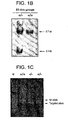


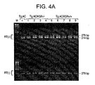
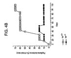
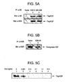
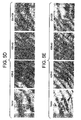
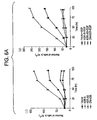
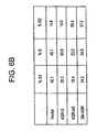


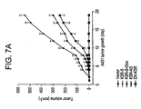
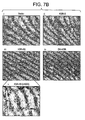
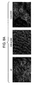
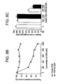
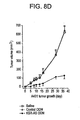
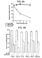

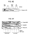
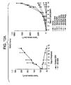


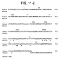
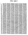
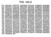
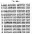
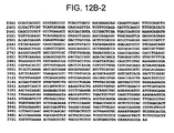
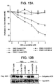
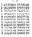
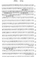

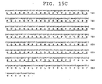


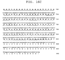

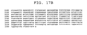
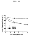
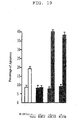
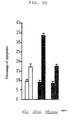

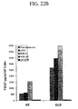

Classifications
-
- C—CHEMISTRY; METALLURGY
- C12—BIOCHEMISTRY; BEER; SPIRITS; WINE; VINEGAR; MICROBIOLOGY; ENZYMOLOGY; MUTATION OR GENETIC ENGINEERING
- C12N—MICROORGANISMS OR ENZYMES; COMPOSITIONS THEREOF; PROPAGATING, PRESERVING, OR MAINTAINING MICROORGANISMS; MUTATION OR GENETIC ENGINEERING; CULTURE MEDIA
- C12N15/00—Mutation or genetic engineering; DNA or RNA concerning genetic engineering, vectors, e.g. plasmids, or their isolation, preparation or purification; Use of hosts therefor
- C12N15/09—Recombinant DNA-technology
- C12N15/63—Introduction of foreign genetic material using vectors; Vectors; Use of hosts therefor; Regulation of expression
- C12N15/79—Vectors or expression systems specially adapted for eukaryotic hosts
- C12N15/85—Vectors or expression systems specially adapted for eukaryotic hosts for animal cells
- C12N15/8509—Vectors or expression systems specially adapted for eukaryotic hosts for animal cells for producing genetically modified animals, e.g. transgenic
-
- A—HUMAN NECESSITIES
- A01—AGRICULTURE; FORESTRY; ANIMAL HUSBANDRY; HUNTING; TRAPPING; FISHING
- A01K—ANIMAL HUSBANDRY; AVICULTURE; APICULTURE; PISCICULTURE; FISHING; REARING OR BREEDING ANIMALS, NOT OTHERWISE PROVIDED FOR; NEW BREEDS OF ANIMALS
- A01K67/00—Rearing or breeding animals, not otherwise provided for; New or modified breeds of animals
- A01K67/027—New or modified breeds of vertebrates
- A01K67/0275—Genetically modified vertebrates, e.g. transgenic
- A01K67/0276—Knock-out vertebrates
-
- A—HUMAN NECESSITIES
- A61—MEDICAL OR VETERINARY SCIENCE; HYGIENE
- A61P—SPECIFIC THERAPEUTIC ACTIVITY OF CHEMICAL COMPOUNDS OR MEDICINAL PREPARATIONS
- A61P35/00—Antineoplastic agents
-
- A—HUMAN NECESSITIES
- A61—MEDICAL OR VETERINARY SCIENCE; HYGIENE
- A61P—SPECIFIC THERAPEUTIC ACTIVITY OF CHEMICAL COMPOUNDS OR MEDICINAL PREPARATIONS
- A61P35/00—Antineoplastic agents
- A61P35/02—Antineoplastic agents specific for leukemia
-
- A—HUMAN NECESSITIES
- A61—MEDICAL OR VETERINARY SCIENCE; HYGIENE
- A61P—SPECIFIC THERAPEUTIC ACTIVITY OF CHEMICAL COMPOUNDS OR MEDICINAL PREPARATIONS
- A61P35/00—Antineoplastic agents
- A61P35/04—Antineoplastic agents specific for metastasis
-
- A—HUMAN NECESSITIES
- A61—MEDICAL OR VETERINARY SCIENCE; HYGIENE
- A61P—SPECIFIC THERAPEUTIC ACTIVITY OF CHEMICAL COMPOUNDS OR MEDICINAL PREPARATIONS
- A61P43/00—Drugs for specific purposes, not provided for in groups A61P1/00-A61P41/00
-
- C—CHEMISTRY; METALLURGY
- C12—BIOCHEMISTRY; BEER; SPIRITS; WINE; VINEGAR; MICROBIOLOGY; ENZYMOLOGY; MUTATION OR GENETIC ENGINEERING
- C12N—MICROORGANISMS OR ENZYMES; COMPOSITIONS THEREOF; PROPAGATING, PRESERVING, OR MAINTAINING MICROORGANISMS; MUTATION OR GENETIC ENGINEERING; CULTURE MEDIA
- C12N15/00—Mutation or genetic engineering; DNA or RNA concerning genetic engineering, vectors, e.g. plasmids, or their isolation, preparation or purification; Use of hosts therefor
- C12N15/09—Recombinant DNA-technology
- C12N15/11—DNA or RNA fragments; Modified forms thereof; Non-coding nucleic acids having a biological activity
- C12N15/113—Non-coding nucleic acids modulating the expression of genes, e.g. antisense oligonucleotides; Antisense DNA or RNA; Triplex- forming oligonucleotides; Catalytic nucleic acids, e.g. ribozymes; Nucleic acids used in co-suppression or gene silencing
-
- C—CHEMISTRY; METALLURGY
- C12—BIOCHEMISTRY; BEER; SPIRITS; WINE; VINEGAR; MICROBIOLOGY; ENZYMOLOGY; MUTATION OR GENETIC ENGINEERING
- C12N—MICROORGANISMS OR ENZYMES; COMPOSITIONS THEREOF; PROPAGATING, PRESERVING, OR MAINTAINING MICROORGANISMS; MUTATION OR GENETIC ENGINEERING; CULTURE MEDIA
- C12N15/00—Mutation or genetic engineering; DNA or RNA concerning genetic engineering, vectors, e.g. plasmids, or their isolation, preparation or purification; Use of hosts therefor
- C12N15/09—Recombinant DNA-technology
- C12N15/11—DNA or RNA fragments; Modified forms thereof; Non-coding nucleic acids having a biological activity
- C12N15/113—Non-coding nucleic acids modulating the expression of genes, e.g. antisense oligonucleotides; Antisense DNA or RNA; Triplex- forming oligonucleotides; Catalytic nucleic acids, e.g. ribozymes; Nucleic acids used in co-suppression or gene silencing
- C12N15/1137—Non-coding nucleic acids modulating the expression of genes, e.g. antisense oligonucleotides; Antisense DNA or RNA; Triplex- forming oligonucleotides; Catalytic nucleic acids, e.g. ribozymes; Nucleic acids used in co-suppression or gene silencing against enzymes
-
- A—HUMAN NECESSITIES
- A01—AGRICULTURE; FORESTRY; ANIMAL HUSBANDRY; HUNTING; TRAPPING; FISHING
- A01K—ANIMAL HUSBANDRY; AVICULTURE; APICULTURE; PISCICULTURE; FISHING; REARING OR BREEDING ANIMALS, NOT OTHERWISE PROVIDED FOR; NEW BREEDS OF ANIMALS
- A01K2217/00—Genetically modified animals
- A01K2217/07—Animals genetically altered by homologous recombination
- A01K2217/075—Animals genetically altered by homologous recombination inducing loss of function, i.e. knock out
-
- A—HUMAN NECESSITIES
- A01—AGRICULTURE; FORESTRY; ANIMAL HUSBANDRY; HUNTING; TRAPPING; FISHING
- A01K—ANIMAL HUSBANDRY; AVICULTURE; APICULTURE; PISCICULTURE; FISHING; REARING OR BREEDING ANIMALS, NOT OTHERWISE PROVIDED FOR; NEW BREEDS OF ANIMALS
- A01K2227/00—Animals characterised by species
- A01K2227/10—Mammal
- A01K2227/105—Murine
-
- A—HUMAN NECESSITIES
- A01—AGRICULTURE; FORESTRY; ANIMAL HUSBANDRY; HUNTING; TRAPPING; FISHING
- A01K—ANIMAL HUSBANDRY; AVICULTURE; APICULTURE; PISCICULTURE; FISHING; REARING OR BREEDING ANIMALS, NOT OTHERWISE PROVIDED FOR; NEW BREEDS OF ANIMALS
- A01K2267/00—Animals characterised by purpose
- A01K2267/03—Animal model, e.g. for test or diseases
- A01K2267/0331—Animal model for proliferative diseases
-
- A—HUMAN NECESSITIES
- A61—MEDICAL OR VETERINARY SCIENCE; HYGIENE
- A61K—PREPARATIONS FOR MEDICAL, DENTAL OR TOILETRY PURPOSES
- A61K38/00—Medicinal preparations containing peptides
-
- C—CHEMISTRY; METALLURGY
- C12—BIOCHEMISTRY; BEER; SPIRITS; WINE; VINEGAR; MICROBIOLOGY; ENZYMOLOGY; MUTATION OR GENETIC ENGINEERING
- C12N—MICROORGANISMS OR ENZYMES; COMPOSITIONS THEREOF; PROPAGATING, PRESERVING, OR MAINTAINING MICROORGANISMS; MUTATION OR GENETIC ENGINEERING; CULTURE MEDIA
- C12N2310/00—Structure or type of the nucleic acid
- C12N2310/10—Type of nucleic acid
- C12N2310/11—Antisense
-
- C—CHEMISTRY; METALLURGY
- C12—BIOCHEMISTRY; BEER; SPIRITS; WINE; VINEGAR; MICROBIOLOGY; ENZYMOLOGY; MUTATION OR GENETIC ENGINEERING
- C12N—MICROORGANISMS OR ENZYMES; COMPOSITIONS THEREOF; PROPAGATING, PRESERVING, OR MAINTAINING MICROORGANISMS; MUTATION OR GENETIC ENGINEERING; CULTURE MEDIA
- C12N2310/00—Structure or type of the nucleic acid
- C12N2310/10—Type of nucleic acid
- C12N2310/11—Antisense
- C12N2310/111—Antisense spanning the whole gene, or a large part of it
-
- C—CHEMISTRY; METALLURGY
- C12—BIOCHEMISTRY; BEER; SPIRITS; WINE; VINEGAR; MICROBIOLOGY; ENZYMOLOGY; MUTATION OR GENETIC ENGINEERING
- C12N—MICROORGANISMS OR ENZYMES; COMPOSITIONS THEREOF; PROPAGATING, PRESERVING, OR MAINTAINING MICROORGANISMS; MUTATION OR GENETIC ENGINEERING; CULTURE MEDIA
- C12N2310/00—Structure or type of the nucleic acid
- C12N2310/30—Chemical structure
- C12N2310/31—Chemical structure of the backbone
- C12N2310/315—Phosphorothioates
Definitions
- the present invention relates to methods and compositions for the specific inhibition of of kinase suppressor of Ras (KSR).
- KSR kinase suppressor of Ras
- the invention provides genetic approaches and nucleic acids for the specific inhibition of KSR, particularly of KSR expression.
- the invention relates to antisense oligonucleotides and the expression of nucleic acid complementary to KSR RNA to specifically inhibit KSR and blockgf Ras mediated tumorigenesis.
- Ras plays an essential role in oncogenic transformation and genesis.
- Oncogenic H-, K-, and N-Ras arise from point mutations limited to a small number of sites (amino acids 12, 13, 59 and 61).
- oncogenic ras proteins lack intrinsic GTPase activity and hence remain constitutively activated ( Trahey, M., and McCormick, F. (1987) Science 238: 542-5 ; Tabin, C. J. et al. (1982) Nature. 300: 143-9 ; Taparowsky, E. et al. (1982) Nature. 300: 762-5 ).
- the participation of oncogenic ras in human cancers is estimated to be 30% ( Almoguera, C. et al (1988) Cell. 53:549-54 ).
- K- ras is the most commonly mutated oncogene in human cancers, especially the codon-12 mutation. While oncogenic activation of H-, K-, and N-Ras arising from single nucleotide substitutions has been observed in 30% of human cancers ( Bos, J.L. (1989) Cancer Res 49, 4682-9 ), over 90% of human pancreatic cancer manifest the codon 12 K-ras mutation ( Almoguera, C. et al. (1988) Cell 53, 549-54 ; Smit, V.T. et al.
- Pancreatic ductal adenocarcinoma the most common cancer of the pancreas, is notorious for its rapid onset and resistance to treatment.
- the high frequency of K- ras mutations in human pancreatic tumors suggests that constitutive Ras activation plays a critical role during pancreatic oncogenesis.
- Adenocarcinoma of the exocrine pancreas represents the fourth-leading cause of cancer-related mortality in Western countries.
- K-ras mutations are present in 50% of the cancers of colon and lung ( Bos, J. L. et al. (1987) Nature. 327: 293-7 ; Rodenhuis, S. et al. (1988) Cancer Res. 48: 5738-41 ). In cancers of the urinary tract and bladder, mutations are primarily in the H- ras gene ( Fujita, J. et al. (1984) Nature. 309: 464-6 ; Visvanathan, K. V. et al. (1988) Oncogene Res. 3: 77-86 ). N- ras gene mutations are present in 30% of leukemia and liver cancer.
- Constitutive activation of Ras can be achieved through oncogenic mutations or via hyperactivated growth factor receptors such as the EGFRs. Elevated expression and/or amplification of the members of the EGFR family, especially the EGFR and HER2, have been implicated in various forms of human malignancies (as reviewed in Prenzel, N. et al. (2001) Endocr Relat Cancer. 8: 11-31 ). In some of these cancers (including pancreas, colon, bladder, lung), EGFR/HER2 overexpression is compounded by the presence of oncogenic Ras mutations.
- Abnormal activation of these receptors in tumors can be attributed to overexpression, gene amplification, constitutive activation mutations or autocrine growth factor loops ( Voldborg, B. R. et al. (1997) Ann Oncol. 8: 1197-206 ).
- growth factor receptors especially the EGFRs, amplification or /and overexpression of these receptors frequently occur in the cancers of the breast, ovary, stomach, esophagus, pancreatic, lung, colon neuroblastoma.
- the present invention relates to the use of antisense oligonucleotides for the specific inhibition of kinase suppressor of Ras (KSR).
- KSR kinase suppressor of Ras
- the uses of the present invention inhibit the expression and/or activity of KSR.
- the invention provides genetic approaches and nucleic acids for the specific inhibition of KSR. It is herein demonstrated that on specific inhibition of KSR the Ras pathway is disrupted and, specifically, Ras-mediated tumors and tumorigenesis is inhibited or blocked, existing tumors regress, metasasis is inhibited and proliferation of tumor or cancer cells is inhibited.
- the present invention further relates to the use of antisense oligonucleotides for radiosensitization, particularly of hyperproliferative, cancer or tumor cells, by specific inhibition of KSR expression and/or activity.
- the hyperproliferative, cancer or tumor cells are resistant to ionizing radiation (IR) induced apoptosis or cell death.
- IR ionizing radiation
- Inhibition of KSR, by administration or expression of antisense oligonucleotide complementary to KSR results in sensitization of cells to ionizing radiation, whereby the division or growth of cells is blocked or the cells are more effectively killed by IR than in the absence of the antisense oligonucleotide.
- the administration or expression of antisense oligonucleotide complementary to KSR facilitates and enhances cancer therapy, including radiation therapy.
- the present invention further relates to the use of antisense oligonucleotides for modulating angiogenesis, particularly by modulation of VEGF expression upon activation or inhibition of expression and/or activity of KSR.
- VEGF expression is modulated by specific inhibition or activation of KSR expression and/or activity.
- Activation or enhanced expression of KSR increases the amount or expression of VEGF in or by cells.
- activation or enhanced expression of KSR in a cell or tissue provides a method of stimulating angiogenesis.
- Inhibition, blockage or reduction of KSR decreases the amount or expression of VEGF, thereby having an anti-angiogenic effect.
- VEGF expression of cancer or tumor cells is blocked by inhibition of KSR, including by administration or expression of antisense oligonucleotide complementary to KSR, resulting in inhibition of angiogenesis in the cancer or tumor.
- inhibition of KSR including by administration or expression of antisense oligonucleotide complementary to KSR, provides a method of inhibiting angiogenesis in a tumor, tissue or cells expressing KSR, particularly a tissue or tumor expressing gf Ras or wherein the Ras pathway is hyperactivated or Ras is overexpressed or amplified.
- the present invention provides for the use of oligonucleotides and nucleic acids which specifically inhibit or block the expression and activity of KSR.
- antisense oligonucleotides and the expression of nucleic acid complementary to KSR RNA specifically inhibits expression of KSR and blocks gf Ras mediated tumorigenesis.
- the present invention provides for the use of an oligonucleotide which is specifically hybridizable to a region of KSR RNA, wherein said oligonucleotide inhibits the expression of KSR.
- the invention further provides an oligonucleotide which is specifically hybridizable to a nucleic acid encoding mammalian KSR.
- oligonucleotides are provided which are substantially complementary to nucleic acid encoding mammalian KSR, particularly human and mouse KSR.
- an oligonucleotide which is specifically hybridizable to a translation initiation site, 5' untranslated region, coding region or 3' untranslated region of mRNA encoding mammalian KSR.
- the invention thus provides for the use of an oligonucleotide which is specifically hybridizable to a translation initiation site, 5' untranslated region, coding region or 3' untranslated region of mRNA encoding human KSR as provided in FIGURE 14 and SEQ ID NO: 24.
- the invention provides for the use of an antisense oligonucleotide specifically hybridizable to the N-terminal coding region of mammalian KSR mRNA, particularly to the region of nucleotides 1 through 761 of the coding region of mammalian KSR mRNA.
- the invention includes the use of an antisense oligonucleotide comprising a sequence specifically hybridizable to the CA1 region of KSR.
- the invention provides for the use of oligonucleotides comprising a sequence specifically hybridizable to nucleotides encoding amino acids 33 to 72 of the sequence of human KSR, or a portion thereof.
- the invention includes the use of an antisense oligonucleotide comprising a sequence specifically hybridizable to nucleotides 97 to 216 of the coding sequence of human KSR (SEQ ID NO: 25), or a portion thereof, such nucleotides encoding amino acids 33 to 72 of human KSR (SEQ ID NO: 26) or a portion thereof.
- oligonucleotides of the invention includes the use of oligonucleotides comprising a sequence specifically hybridizable to nucleotides selected from the group of: nucleotides 124 to 141 of human KSR, corresponding to nucleotides 151 to 168 of mouse KSR (SEQ ID NO: 3); nucleotides 154 to 171 of human KSR (SEQ ID NO: 27), corresponding nearly identically (with a single base pair difference at the 5' most nucleotide) to nucleotides 181 to 198 of mouse KSR (SEQ ID NO:4); and nucleotides 187 to 204 of human KSR, corresponding to nucleotides 214 to 231 of mouse KSR (SEQ ID NO:5).
- the invention includes the use of antisense oligonucleotides comprising a sequence selected from the group of SEQ ID NOS: 6-8 and SEQ ID NOS: 29-38.
- the oligonucleotides for use in the present invention may be labeled with a detectable label.
- the label may be selected from enzymes, ligands, chemicals which fluoresce and radioactive elements.
- a radioactive label such as the isotopes 3 H, 14 C, 32 P, 35 S, 36 Cl, 51 Cr, 57 Co, 58 Co, 59 Fe, 90 Y, 125 I, 131 I, and 186 Re are used, known currently available counting procedures may be utilized.
- detection may be accomplished by any of the presently utilized colorimetric, spectrophotometric, fluorospectrophotometric, amperometric or gasometric techniques known in the art.
- nucleic acids and oligonucleotides for use in the present invention may be modified, either by manipulation of the chemical backbone of the nucleic acids or by covalent or non-covalent attachment of other moieties. In each or any case, such manipulation or attachment may serve to modify the stability, cellular, tissue or organ uptake, or otherwise enhance efficacy of the nucleic acids and oligonucleotides.
- the oligonucleotides for use in the present invention may be covalently linked to other molecules, including but not limited to polypeptides, carbohydrates, lipid or lipid-like moieties, ligands, chemical agents or compounds, which may serve to enhance the uptake, stability or to target the oligonucleotides.
- the oligonucleotides for use in the present invention are modified in their chemical backbone.
- the oligonucleotides comprise at least one phosphorothioate (P-S) linkage.
- Recombinant DNA molecules comprising a nucleic acid sequence which encodes on transcription an antisense RNA complementary to mammalian KSR RNA or a portion thereof are provided for use in the invention. Further, the recombinant DNA molecules comprise a nucleic acid sequence wherein said nucleic acid sequence is operatively linked to a transcription control sequence. Cell lines transfected with these recombinant DNA molecules are also included in the invention.
- an expression vector which is capable of expressing a nucleic acid which is specifically hybridizable to the coding sequence of KSR RNA, or a portion thereof, wherein said nucleic acid inhibits the expression of KSR.
- compositions of the nucleic acids and oligonucleotides for use in the invention includes a composition comprising an oligonucleotide which is specifically hybridizable to a region of KSR RNA and a pharmaceutically acceptable carrier or diluent.
- a pharmaceutical composition comprising a therapeutically effective amount of an oligonucleotide which is specifically hybridizable to a region of KSR RNA and a pharmaceutically acceptable carrier or diluent is disclosed.
- compositions comprising one or more chemotherapeutic or radiotherapeutic agent and an oligonucleotide which is targeted to a mRNA encoding mammalian KSR and which inhibits KSR expression.
- composition comprising an expression vector and a pharmaceutically acceptable carrier or diluent, wherein said expression vector is capable of expressing nucleic acid which is specifically hybridizable to the coding sequence of KSR RNA, or a portion thereof, wherein said nucleic acid inhibits the expression of KSR.
- Methods for inhibiting expression of KSR are provided.
- a method of inhibiting the expression of mammalian KSR comprising contacting cells which express KSR with an effective amount of a nucleic acid which is complementary to a portion of the mRNA encoding KSR is included.
- a method of inhibiting the expression of mammalian KSR comprising contacting cells which express KSR with an effective amount of the oligonucleotide for use in the present invention whereby expression of mammalian KSR is inhibited.
- a method of inhibiting expression of KSR wherein tissues or a tumor, particularly a tissue or tumor expressing gf Ras or wherein the Ras pathway is hyperactivated or Ras is overexpressed or amplified, is contacted with an effective amount of the oligonucleotide or nucleic acid for use in the present invention, thus inhibiting the expression of KSR.
- compositions and methods for the inhibition or blockage ofthe activity of KSR including the kinase or phosphorylation activity of KSR.
- a method of inhibiting expression of KSR is provided, wherein a tumor, tissue or cells expressing KSR, particularly a tissue or tumor expressing gf Ras or wherein the Ras pathway is hyperactivated or Ras is overexpressed or amplified, is contacted with an effective amount of the nucleic acid or composition for use in the present invention, thus inhibiting the activity of KSR.
- Disclosed herein is a method of treating or preventing a hyperproliferative condition associated with the expression of gf -Ras or heightened expression of Ras in a mammal comprising administering to said mammal a therapeutically effective amount of a compound or agent which inhibits the expression of mammalian KSR protein.
- said compound or agent is an antisense oligonucleotide which specifically hybridizes to a portion of the mRNA encoding KSR.
- a method of treating or preventing a hyperproliferative condition associated with the expression of gf-Ras or heightened expression or hyperactivation of Ras in a mammal comprising expressing in said mammal or administering to said mammal a therapeutically effective amount of a nucleic acid which is complementary to a portion of the mRNA encoding KSR.
- a method of treating or inhibiting the progression of cancer in a mammal comprising administering to a mammal a therapeutically effective amount of a compound or agent which inhibits the expression of mammalian KSR protein.
- Cancers which are susceptible to the invention's method include cancer selected from the group of pancreatic cancer, lung cancer, skin cancer, urinary tract cancer, bladder cancer, liver cancer, thyroid cancer, colon cancer, intestinal cancer, leukemia, lymphoma, neuroblastoma, head and neck cancer, breast cancer, ovarian cancer, stomach cancer, esophageal cancer and prostate cancer.
- a method for treating or inhibiting the progression of cancer in a mammal comprising administering to a mammal a therapeutically effective amount of one or more oligonucleotide of the present invention.
- the invention provides a method for sensitizing hyperproliferative, cancer or tumor cells in a mammal to ionizing radiation or other radiation or chemotherapy comprising administering to a mammal a therapeutically effective amount of one or more inhibitor of KSR expression and/or activity.
- the invention provides a method for sensitizing hyperproliferative, cancer or tumor cells in a mammal to ionizing radiation or other radiation or chemotherapy comprising administering to a mammal a therapeutically effective amount of one or more oligonucleotide of the present invention.
- the invention provides a method for modulating angiogenesis in a mammal by modulation of VEGF expression upon activation or inhibition of expression and/or activity of KSR. Inhibition, blockage or reduction of KSR decreases the amount or expression of VEGF, thereby having an anti-angiogenic effect.
- VEGF expression of cancer or tumor cells is blocked by inhibition of KSR, including by administration or expression of antisense oligonucleotide complementary to KSR, resulting in inhibition of angiogenesis in the cancer or tumor.
- the invention provides a method of inhibiting angiogenesis in a tumor, tissue or cell expressing KSR, particularly a tissue or tumor expressing gf Ras or wherein the Ras pathway is hyperactivated or Ras is overexpressed or amplified, comprising administering to said mammal an inhibitor of KSR, including a therapeutically effective amount of one or more antisense oligonucleotide complementary to KSR.
- an inhibitor of KSR including a therapeutically effective amount of one or more antisense oligonucleotide complementary to KSR.
- the activation or enhanced expression of KSR increases the amount or expression of VEGF in or by cells.
- a method of stimulating angiogenesis is provided comprising activating or enhancing expression of KSR in a cell or tissue.
- the invention includes additional compositions which can inhibit the expression of a protein, in particular KSR, at the transcriptional level by blocking translation of KSR mRNA or by facilitating destruction or destabilization of the RNA such that translation cannot efficiently take place.
- a protein in particular KSR
- the invention provides a ribozyme that cleaves KSR mRNA.
- the present invention naturally contemplates several means for preparation of the nucleic acids and oligonucleotides of the present invention, including as illustrated herein known recombinant techniques, and the invention is accordingly intended to cover such synthetic preparations within its scope.
- the knowledge of the cDNA and amino acid sequences of KSR as disclosed herein facilitates the preparation of the nucleic acids of the invention by such recombinant techniques, and accordingly, the invention extends to expression vectors prepared from the disclosed DNA sequences for expression in host systems by recombinant DNA techniques, and to the resulting transformed hosts.
- FIGURE 1 depicts targeted disruption of the ksr gene in mice.
- A Strategy for targeting the ksr allele. Simplified restriction maps of the 5' region of the wild-type ksr allele, the targeting vector, and the mutated allele are shown. Homologous recombination with endogenous ksr replaces an internal 1.1-kb Sma I- Spe I genomic fragment with a Neo cassette.
- B Southern blot analysis of an ES clone showing the correct insertion of the targeting construct. Genomic DNA isolated from ES cells was digested with Bgl II and Xho I and hybridized to the 5' probe located just outside the 5'arm of the ksr targeting region as shown in A.
- the wild-type allele yields a 5.7-kb fragment whereas the mutant allele yields a 3.1-kb fragment.
- C Genotyping of ksr -/- mice by PCR. The size of the PCR product is 493 bp for the wt allele and 312 bp for the mutated allele.
- D Expression of ksr in wild type mouse embryos. The sizes of the two transcripts are 6.4 kb and 7.4 kb.
- E Northern blot analysis of tissue ksr mRNAs.
- Poly-A + RNA isolated from different tissues of adult ksr +/+ , ksr +/- , and ksr -/- mice, was hybridized with a probe corresponding to domains CA2-CA4 in ksr cDNA. mRNA from NIH3T3 cells was used as control. F, KSR protein expression. Lysates prepared from wild-type and ksr -/- tissues were analyzed by western blot with a specific anti-KSR monoclonal antibody. Note that brain expresses the slightly shorter B-KSR1 isoform while lung and spleen express the longer KSR1 isoform. Lysates were also prepared from two independent sets of ksr +l+ and ksr -/- MEFs. Equal loading was confirmed by reprobing blots with an anti- ⁇ -tubulin antibody.
- FIGURE 2 depicts skin phenotype in newborn ksr -l- mice.
- Full thickness skin cuts of 10-day old ksr + / + , ksr -l- and egfr - / - mice were sectioned 4-6 ⁇ m thick, placed on glass slides, and stained with hematoxylin and eosin. s -serpentine, bl -blister, do-disoriented.
- FIGURE 3 depicts defects in EGF- and TPA- induced MAPK signaling and proliferation in ksr - / - MEFs.
- A Western blot analysis of MAPK activity upon EGF and TPA treatments.
- Low-passage MEFs derived from ksr + / + and ksr - / - were made quiescent by 48 h incubation in serum-free medium and stimulated with low doses of EGF for 3 min(upper panel) or with TPA for 10 min (lower panel).
- Cells were lysed in NP40 buffer and activation of the MAPK cascade was examined by western blot with anti-phospho specific antibodies for the activated forms of MAPK(ERK1/2). Shown are representative blots from one of four independent experiments.
- FIGURE 4 Disruption of ksr gene abrogated oncogenic Ras-mediated tumorigenesis in kst - / - mice.
- A RT-PCR detection of ⁇ -Ha- ras expression from total RNA isolated from the epidermis of Tg.AC/ ksr + / + and Tg.AC/ ksr - / - mice following TPA treatment. Intron spanning primers specific for the 3'UTR region of the ⁇ -Ha- ras transgene were used. The larger 279 bp amplicon, detected in the absence of reverse transcriptase [RT(-)], is derived from DNA and unspliced RNA.
- the smaller 214 bp amplicon is derived from spliced mRNA and is indicative of transgene expression.
- B Mice, grouped according to genotype (10/group), were treated with 5 ⁇ g of TPA twice a week for 15 weeks. Papillomas were counted weekly for 20 weeks.
- FIGURE 5 Inducible A431-Tet-Off-pTRE-KSR cells.
- a and B Western blot analysis of wild type Flag-KSR-S and Flag-DN-KSR expression (A), and inhibition of endogenous KSR1 expression by KSR-AS ( B).
- Flag-KSR-S and DN-KSR were immunoprecipitated (IP) with the monoclonal anti-Flag (M2) antibody and detected by WB. The identity of Flag-KSR was confirmed by re-probing with a monoclonal anti-KSR antibody (BD Biosciences). Endogenous KSR1 was immunoprecipitated as described in Methods and detected as above.
- C Dose-dependent inhibition of Flag-KSR-S expression by doxycycline.
- KSR-S cells were treated with indicated doses of Dox for 24 h and Flag-KSR-S expression after Dox treatment was determined by WB as above.
- D and E Inactivation of KSR1 by KSR-AS or DN-KSR leads to alterations in morphology ( D), and the development of a multinuclei phenotype ( E ).
- A431-pTRE cells were examined under the phase-contrast microscope and photographed at 20x magnification for cell morphology ( D) and 40x magnification for multinucleation ( E).
- FIGURE 6 Inactivation of KSR1 abolishes EGF-stimulated biological responses in A431 cells.
- A Cell proliferation assay without (i) and with (ii) EGF stimulation. Proliferation assays were performed as described in Methods.
- B Cell cycle distribution of A431-pTRE cells was determined by FACS analysis as described in Methods. The proportion of cells in the different phases of the cell cycle was calculated from the experimental fluorescence histograms.
- C Matrigel invasion assay in response to EGF stimulation. To optimize the stimulatory effect of overexpression of KSR-S on A431 cell invasion, the assay was terminated after 12 h (i).
- FIGURE 7 Inactivation of KSR1 prevents A431 tumorigenesis.
- A Growth curve of A431 tumors. 10 6 A431-pTRE cells were injected s.c. into nude mice as described in Methods. To determine the specificity of KSR-S on A431 tumorigenesis, Dox (100 mg/ml) was added to the drinking water of a group of KSR-S tumor-bearing mice (KSR-S + Dox) 3 days prior to tumor implantation and continued throughout the experiment to turn off KSR-S expression. Mice receiving KSR-AS and DN-KSR cells were monitored up to 120 days. These results represent one of three similar experiments. There were 5 mice in each experimental group. B, H&E staining of A431 tumors.
- FIGURE 8 Inactivation of KSR1 by AS-ODN attenuates A431 tumorigenesis.
- A Immunofluorescence staining of endogenous KSR1 expression after treatment with 1 mM Control- or AS-ODNs was performed as in Methods. Nuclei were counter stained with DAPI. To compare the intensity of fluorescence labeling, all images of KSR expression were taken with the same exposure time.
- B and C Dose-dependent inhibition of A431 cell proliferation ( B) and invasion ( C) by AS-ODN treatment. For the proliferation assay, 30% confluent A431 cells were treated with the indicated doses of Control- or AS-ODNs as in Fig. 3 . Cell proliferation after ODN treatment was calculated as percent of non-treated controls.
- Invasion assays were set up after 48 h of ODN treatment as above.
- D Attenuation of A431 tumorigenesis by continuous infusion of AS-ODN at 5 mg/kg/day.
- A431 seed tumor fragments freshly prepared as described in Methods, were transplanted s.c. into the right lateral flank of nude mice.
- Continuous infusion of ODNs was initiated 2 days prior to tumor transplantation. There were 5 mice in each treatment group.
- FIGURE 9 Inactivation of KSR1 by AS-ODN inhibits oncogenic K- ras signaling in vitro in PANC-1.
- A Dose-dependent inhibition of PANC-1 cell proliferation by AS-ODN treatment. PANC-1 cells were treated with the indicated doses of Control- or AS-ODNs and cell proliferation assays were performed as in FIGURE 7 .
- B AS-ODN treatment (5 ⁇ M) attenuated the proliferation of a panel of human pancreatic cancer cell lines. The seeding density for each cell lines was determined in preliminary studies so that all cell lines were 30-40% confluent when transfected with ODNs.
- C and D c-Raf-1 is epistatic to KSR1.
- PANC-1 cells were first treated with Sense- or AS-ODNs for 48 h and then transfected with the BXB-Raf as in Methods. 48 h after transfection, invasion and colony formation assays were set up as in FIGURE 7 .
- the inhibitory effect of AS-ODN on PANC-1 cell invasion ( C) and transformation (D ) was reversed by dominant positive BXB-Raf.
- AS-ODN treatment inhibited endogenous KSR1 expression in PANC-1 cells.
- Endogenous KSR1 was immunoprecipitated from non-treated (NT), Sense-ODN-treated or AS-ODN-treated PANC-1 cells, and KSR1 expression was determined by WB as described in Methods.
- FIGURE 10 AS-ODN treatment abolished PANC-1 and A549 tumorigenesis in vivo .
- A Continuous infusion of AS-ODN abolished PANC-1 tumor growth.
- a (i) established PANC tumors (approximately 100 mm 3 ) were treated with 10 mg/kg/day of Control- or AS-ODNs for 14 days. Mice bearing regressed AS-ODN-treated tumors were monitored up to 4 weeks.
- A(ii) freshly prepared PANC-1 seed tumor fragments were transplanted into nude mice as above.
- KSR AS-ODN treatment prevented A549 tumor growth (i) and inhibited lung metastases via systemic dissemination (ii).
- A549 seed tumor fragments freshly prepared as in Methods, were transplanted to nude mice.
- Treatment with control-or AS-ODNs were initiated when tumors reached 150 mm 3 and continued for additional 18 days ( i).
- animals were sacrificed at the end of the experiment, lungs were resected from control- or AS-ODN-treated mice and stained with Indian ink to visualize surface lung metastases derived via systemic dissemination (ii) .
- FIGURE 11 depicts a comparative alignment of the mouse KSR polypeptide sequence (SEQ ID NO: 9) and human KSR polypeptide sequence (SEQ ID NO: 10).
- FIGURE 12 depicts the nucleic acid coding (cDNA) sequence of mouse ksr (SEQ ID NO: 11).
- B depicts the partial nucleic acid coding (cDNA) sequence of human ksr (SEQ ID NO: 12).
- FIGURE 13 depicts the specific and dose-dependent inhibition of PANC-1 cell proliferation by AS-ODN treatment.
- A Dose-dependent inhibition of PANC-1 cell proliferation by AS-ODN treatment; proliferation of K562 cells is not inhibited by AS-ODN treatment.
- B Western blot analysis of endogenous KSR1 gene expression in K562 and PANC-1 cells. Treatment of K562 cells with 5 ⁇ M KSR AS-ODN-1 elicited comparable reduction of endogenous KSR1 gene expression (over 80%) to that observed in PANC-1 cells.
- FIGURE 14 depicts the human KSR1 full length mRNA sequence (SEQ ID NO:24).
- FIGURE 15 depicts the human KSR nucleic acid and protein sequence annotated with the locations of the CA1 to CA5 domains and the target sequences for AS-ODNs.
- the full length human KSR1 protein is predicted to have 866 amino acids.
- FIGURE 16 depicts the mouse KSR nucleic acid and protein sequence annotated with the locations of the CA1 to CA5 domains and the target sequences for the AS-ODNs.
- FIGURE 17 depicts the human KSR-1 full length mRNA sequence with the nucleic acid target sequences for human AS-ODN1 through AS-ODN12 depicted.
- FIGURE 18 depicts proliferation assay of PANC-1 cells treated with control ODN, AS-ODN2(181-198) (mouse) (AS-ODN2-old) and AS-ODN2(154-171) (human) (AS-ODN2-new).
- FIGURE 19 presents percentage of ionizing-radiation induced apoptosis as scored with Annexin V staining in A431 cells expressing vector, wild-type Flag-KSR (KSR-S), Flag-AS-KSR (KSR-AS) or dominant-negative Flag-Ki-KSR (Ki-KSR).
- KSR-S wild-type Flag-KSR
- KSR-AS Flag-AS-KSR
- Ki-KSR dominant-negative Flag-Ki-KSR
- FIGURE 20 presents percentage of ionizing-radiation induced apoptosis as scored with Annexin V staining in A431 cells treated with AS-ODN2(214-231), control ODN or expressing vector.
- FIGURE 21A and 21B depicts expression of KSR-AS radiosensitizes A431 cells to ionizing radiation.
- FIGURE 22A and 22B depicts inactivation of KSR (by KSR-AS expression) abrogates oth baseline and EGF-stimulated VEGF production in A431 cells.
- oligonucleotides refer to nucleic acid material including single or multiplenucleic acids, and extends to those oligonucleotides complementary to the nucleic acid sequences described herein, including as presented in FIGURE 12A, 12B and 14 and in SEQ ID NOS: 11, 12, and 24, including conserved and activity domains thereof as depicted in FIGURES 11 , 15 and 16 , and having the profile of activities set forth herein and in the Claims, particularly in being capable of inhibiting the expression of KSR.
- the oligonucleotides of the present invention may be specifically hybridizable to nucleic acid sequence specific to KSR, as provided in SEQ ID NO: 1 or SEQ ID NO:25, or to a portion thereof, as provided for example in SEQ ID NO: 3, 4, 5 and 27.
- Exemplary oligonucleotides include any of SEQ ID NOS 6-8 and SEQ ID NOS: 28-39.
- nucleic acids or analogs thereof displaying substantially equivalent or altered activity are likewise contemplated. These modifications may be deliberate, for example, such as modifications obtained through site-directed mutagenesis, or may be accidental, such as those obtained through mutations in hosts that are producers of the nucleic acids or of KSR.
- NH 2 refers to the free amino group present at the amino terminus of a polypeptide.
- COOH refers to the free carboxy group present at the carboxy terminus of a polypeptide.
- amino-acid residue sequences are represented herein by formulae whose left and right orientation is in the conventional direction of amino-terminus to carboxy-terminus. Furthermore, it should be noted that a dash at the beginning or end of an amino acid residue sequence indicates a peptide bond to a further sequence of one or more amino-acid residues.
- the above Table is presented to correlate the three-letter and one-letter notations which may appear alternately herein.
- a “replicon” is any genetic element (e.g., plasmid, chromosome, virus) that functions as an autonomous unit of DNA replication in vivo ; i.e., capable of replication under its own control.
- a “vector” is a replicon, such as plasmid, phage, virus, retrovirus or cosmid, to which another DNA segment may be attached so as to bring about the replication of the attached segment.
- a "DNA molecule” refers to the polymeric form of deoxyribonucleotides (adenine, guanine, thymine, or cytosine) in its either single stranded form, or a double-stranded helix. This term refers only to the primary and secondary structure of the molecule, and does not limit it to any particular tertiary forms. Thus, this term includes double-stranded DNA found, inter alia , in linear DNA molecules (e.g., restriction fragments), viruses, plasmids, and chromosomes.
- sequences may be described herein according to the normal convention of giving only the sequence in the 5' to 3' direction along the nontranscribed strand of DNA (i.e., the strand having a sequence homologous to the mRNA).
- An "origin of replication” refers to those DNA sequences that participate in DNA synthesis.
- a DNA "coding sequence” is a double-stranded DNA sequence which is transcribed and translated into a polypeptide in vivo when placed under the control of appropriate regulatory sequences. The boundaries of the coding sequence are determined by a start codon at the 5' (amino) terminus and a translation stop codon at the 3' (carboxyl) terminus.
- a coding sequence can include, but is not limited to, prokaryotic sequences, cDNA from eukaryotic mRNA, genomic DNA sequences from eukaryotic (e.g., mammalian) DNA, and even synthetic DNA sequences.
- a polyadenylation signal and transcription termination sequence will usually be located 3' to the coding sequence.
- Transcriptional and translational control sequences are DNA regulatory sequences, such as promoters, enhancers, polyadenylation signals, terminators, and the like, that provide for the expression of a coding sequence in a host cell.
- a “promoter sequence” is a DNA regulatory region capable of binding RNA polymerase in a cell and initiating transcription of a downstream (3' direction) coding sequence.
- the promoter sequence is bounded at its 3' terminus by the transcription initiation site and extends upstream (5' direction) to include the minimum number of bases or elements necessary to initiate transcription at levels detectable above background.
- a transcription initiation site (conveniently defined by mapping with nuclease S1), as well as protein binding domains (consensus sequences) responsible for the binding of RNA polymerase.
- Eukaryotic promoters will often, but not always, contain "TATA" boxes and "CAT” boxes.
- Prokaryotic promoters contain Shine-Dalgamo sequences in addition to the -10 and -35 consensus sequences.
- An “expression control sequence” is a DNA sequence that controls and regulates the transcription and translation of another DNA sequence.
- a coding sequence is "under the control" of transcriptional and translational control sequences in a cell when RNA polymerase transcribes the coding sequence into mRNA, which is then translated into the protein encoded by the coding sequence.
- a "signal sequence” can be included before the coding sequence. This sequence encodes a signal peptide, N-terminal to the polypeptide, that communicates to the host cell to direct the polypeptide to the cell surface or secrete the polypeptide into the media, and this signal peptide is clipped off by the host cell before the protein leaves the cell. Signal sequences can be found associated with a variety of proteins native to prokaryotes and eukaryotes.
- oligonucleotide as used herein in referring to a nucleic acid of the present invention, is defined as a molecule comprised of two or more ribonucleotides, preferably more than three. Its exact size will depend upon many factors which, in turn, depend upon the ultimate function and use of the oligonucleotide. In particular, and in accordance with the present invention, the oligonucleotide should particularly associate with the RNA encoding KSR and should be of the appropriate sequence and size or length so as to specifically and stably associate with the target RNA such that expression (i.e., translation) of the RNA is blocked or such that stability of the RNA is negatively affected.
- the antisense oligonucleotides of the present invention are from about 8 to about 50 nucleotides in length, particularly oligonucleotides from 10 to 30 nucleotides in length, particularly oligonucleotides from 15 to 25 nucleotides.
- primer refers to an oligonucleotide, whether occurring naturally as in a purified restriction digest or produced synthetically, which is capable of acting as a point of initiation of synthesis when placed under conditions in which synthesis of a primer extension product, which is complementary to a nucleic acid strand, is induced, i.e., in the presence of nucleotides and an inducing agent such as a DNA polymerase and at a suitable temperature and pH.
- the primer may be either single-stranded or double-stranded and must be sufficiently long to prime the synthesis of the desired extension product in the presence of the inducing agent.
- the exact length of the primer will depend upon many factors, including temperature, source of primer and use of the method. For example, for diagnostic applications, depending on the complexity of the target sequence, the oligonucleotide primer typically contains 15-25 or more nucleotides, although it may contain fewer nucleotides.
- the primers herein are selected to be “substantially" complementary to different strands of a particular target DNA sequence. This means that the primers must be sufficiently complementary to hybridize with their respective strands. Therefore, the primer sequence need not reflect the exact sequence of the template. For example, a non-complementary nucleotide fragment may be attached to the 5' end of the primer, with the remainder of the primer sequence being complementary to the strand. Alternatively, non-complementary bases or longer sequences can be interspersed into the primer, provided that the primer sequence has sufficient complementarity with the sequence of the strand to hybridize therewith and thereby form the template for the synthesis of the extension product.
- restriction endonucleases and “restriction enzymes” refer to bacterial enzymes, each of which cut double-stranded DNA at or near a specific nucleotide sequence.
- a cell has been "transformed” by exogenous or heterologous DNA when such DNA has been introduced inside the cell.
- the transforming DNA may or may not be integrated (covalently linked) into chromosomal DNA making up the genome of the cell.
- the transforming DNA may be maintained on an episomal element such as a plasmid.
- a stably transformed cell is one in which the transforming DNA has become integrated into a chromosome so that it is inherited by daughter cells through chromosome replication. This stability is demonstrated by the ability of the eukaryotic cell to establish cell lines or clones comprised of a population of daughter cells containing the transforming DNA.
- a "clone” is a population of cells derived from a single cell or common ancestor by mitosis.
- a "cell line” is a clone of a primary cell that is capable of stable growth in vitro for many generations.
- Two DNA sequences are "substantially homologous" when at least about 75% (preferably at least about 80%, and most preferably at least about 90 or 95%) of the nucleotides match over the defined length of the DNA sequences. Sequences that are substantially homologous can be identified by comparing the sequences using standard software available in sequence data banks, or in a Southern hybridization experiment under, for example, stringent conditions as defined for that particular system. Defining appropriate hybridization conditions is within the skill of the art. See, e.g., Maniatis et al., supra ; DNA Cloning, Vols. I & II, supra ; Nucleic Acid Hybridization, supra .
- DNA or nucleic acid sequences which code for KSR having the same amino acid sequence as the KSR sequences disclosed herein, including SEQ ID NO: 11 and SEQ ID NO: 12, but which are degenerate these sequences.
- degenerate to is meant that a different three-letter codon is used to specify a particular amino acid. It is well known in the art that the following codons can be used interchangeably to code for each specific amino acid:
- codons specified above are for RNA sequences.
- the corresponding codons for DNA have a T substituted for U.
- Mutations can be made in ksr such that a particular codon is changed to a codon which codes for a different amino acid. Such a mutation is generally made by making the fewest nucleotide changes possible.
- a substitution mutation of this sort can be made to change an amino acid in the resulting protein in a non-conservative manner (i.e., by changing the codon from an amino acid belonging to a grouping of amino acids having a particular size or characteristic to an amino acid belonging to another grouping) or in a conservative manner (i.e., by changing the codon from an amino acid belonging to a grouping of amino acids having a particular size or characteristic to an amino acid belonging to the same grouping).
- Such a conservative change generally leads to less change in the structure and function of the resulting protein.
- a non-conservative change is more likely to alter the structure, activity or function of the resulting protein.
- the present invention should be considered to include sequences containing conservative changes which do not significantly alter the activity or binding characteristics of the resulting protein.
- Amino acids with uncharged polar R groups Glycine, Serine, Threonine, Cysteine, Tyrosine, Asparagine, Glutamine
- Another grouping may be those amino acids with phenyl groups:
- Another grouping may be according to molecular weight (i.e., size of R groups): Glycine 75 Alanine 89 Serine 105 Proline 115 Valine 117 Threonine 119 Cysteine 121 Leucine 131 Isoleucine 131 Asparagine 132 Aspartic acid 133 Glutamine 146 Lysine 146 Glutamic acid 147 Methionine 149 Histidine (at pH 6.0) 155 Phenylalanine 165 Arginine 174 Tyrosine 181 Tryptophan 204
- Amino acid substitutions may also be introduced to substitute an amino acid with a particularly preferable property.
- a Cys may be introduced a potential site for disulfide bridges with another Cys.
- a His may be introduced as a particularly "catalytic" site (i.e., His can act as an acid or base and is the most common amino acid in biochemical catalysis).
- Pro may be introduced because of its particularly planar structure, which induces ⁇ -turns in the protein's structure.
- Two amino acid sequences are "substantially homologous" when at least about 70% of the amino acid residues (preferably at least about 80%, and most preferably at least about 90 or 95%) are identical, or represent conservative substitutions.
- a "heterologous" region of the DNA construct is an identifiable segment of DNA within a larger DNA molecule that is not found in association with the larger molecule in nature.
- the gene when the heterologous region encodes a mammalian gene, the gene will usually be flanked by DNA that does not flank the mammalian genomic DNA in the genome of the source organism.
- Another example of a heterologous coding sequence is a construct where the coding sequence itself is not found in nature (e.g., a cDNA where the genomic coding sequence contains introns, or synthetic sequences having codons different than the native gene). Allelic variations or naturally-occurring mutational events do not give rise to a heterologous region of DNA as defined herein.
- phrases “pharmaceutically acceptable” refers to molecular entities and compositions that are physiologically tolerable and do not typically produce an allergic or similar untoward reaction, such as gastric upset, dizziness and the like, when administered to a human.
- terapéuticaally effective amount is used herein to mean an amount sufficient to prevent, and preferably reduce by at least about 30 percent, more preferably by at least 50 percent, more preferably by at least 70 percent, most preferably by at least 90 percent, a clinically significant change in the mitotic activity of a target cellular mass, or other feature of pathology such as for example, reduced tumor mass, reduced tumor cell proliferation, reduction in metastatic capacity, or enhanced apoptosis, as may attend its presence and activity.
- pg means picogram
- ng means nanogram
- ug means microgram
- mg means milligram
- ul means microliter
- ml means milliliter
- 1 means liter.
- a DNA sequence is "operatively linked" to an expression control sequence when the expression control sequence controls and regulates the transcription and translation of that DNA sequence.
- the term "operatively linked” includes having an appropriate start signal (e.g., ATG) in front of the DNA sequence to be expressed and maintaining the correct reading frame to permit expression of the DNA sequence under the control of the expression control sequence and production of the desired product encoded by the DNA sequence. If a gene that one desires to insert into a recombinant DNA molecule does not contain an appropriate start signal, such a start signal can be inserted in front of the gene.
- standard hybridization conditions refers to salt and temperature conditions substantially equivalent to 5 x SSC and 65°C for both hybridization and wash. However, one skilled in the art will appreciate that such “standard hybridization conditions” are dependent on particular conditions including the concentration of sodium and magnesium in the buffer, nucleotide sequence length and concentration, percent mismatch, percent formamide, and the like. Also important in the determination of “standard hybridization conditions” is whether the two sequences hybridizing are RNA-RNA, DNA-DNA or RNA-DNA. Such standard hybridization conditions are easily determined by one skilled in the art according to well known formulae, wherein hybridization is typically 10-20°C below the predicted or determined T m with washes of higher stringency, if desired.
- the present invention relates to methods and compositions for the specific inhibition of expression and/or activity of kinase suppressor of Ras (KSR).
- KSR kinase suppressor of Ras
- the invention provides genetic approaches and nucleic acids for the specific inhibition of KSR. It is herein demonstrated that on specific inhibition of KSR the Ras pathway is disrupted and, specifically, that Ras-mediated tumors, tumorigenesis and metastasis regress, are inhibited, or are blocked.
- antisense oligonucleotides and the expression of nucleic acid complementary to KSR RNA specifically inhibits expression of KSR and blocks gf Ras mediated tumorigenesis.
- the present invention provides an oligonucleotide which is specifically hybridizable to a region of KSR RNA, wherein said oligonucleotide inhibits the expression of KSR.
- the invention further provides an oligonucleotide which is specifically hybridizable to a nucleic acid encoding mammalian KSR.
- an oligonucleotide is provided which is specifically hybridizable to a nucleic acid encoding human KSR.
- an oligonucleotide which is specifically hybridizable to a translation initiation site, 5' untranslated region, coding region or 3' untranslated region of mRNA encoding mammalian KSR.
- the invention thus provides an oligonucleotide which is specifically hybridizable to a translation initiation site, 5' untranslated region, coding region or 3' untranslated region of mRNA encoding human KSR as provided in FIGURE 14 and SEQ ID NO: 24.
- the invention provides an antisense oligonucleotide specifically hybridizable to the N-terminal coding region of mammalian KSR mRNA, particularly to the region ofnucleotides 1 through 761 of the coding region of mammalian KSR mRNA.
- the invention includes an antisense oligonucleotide comprising a sequence specifically hybridizable to the CA1 region of KSR (SEQ ID NO: 1 and SEQ ID NO:25).
- the invention provides oligonucleotides comprising a sequence specifically hybridizable to nucleotides encoding amino acids 33 to 72 of human KSR (SEQ ID NO:26) or 42 to 82 of mouse KSR (amino acids SEQ ID NO: 2), or a portion thereof, of the sequence of KSR.
- the invention includes an antisense oligonucleotide comprising a sequence specifically hybridizable to nucleotides 97 to 216 ofthe coding sequence of human KSR (SEQ ID NO:25) corresponding to nucleotides 124 to 243 of mouse KSR (SEQ ID NO: 1), or a portion thereof.
- the invention provides an oligonucleotide comprising a sequence specifically hybridizable to nucleotides encoding amino acids 33 to 72 of human KSR (SEQ ID NO:26) and 42 to 82 of mouse KSR (SEQ ID NO: 2) or a portion thereof.
- oligonucleotides of the invention include oligonucleotides comprising a sequence specifically hybridizable to nucleotides selected from the group of: nucleotides 124 to 141 of human KSR, corresponding to nucleotides 151 to 168 of mouse KSR (SEQ ID NO: 3); nucleotides 154 to 171 of human KSR (SEQ ID NO: 27), corresponding closely to nucleotides 181 to 189 of mouse KSR (SEQ ID NO: 4), with a single base change at the 5'-most nucleotide; and nucleotides 187 to 204 of human KSR, corresponding to nucleotides 214 to 231 of mouse KSR (SEQ ID NO: 5).
- the invention includes antisense oligonucleotides comprising a sequence selected from the group of SEQ ID NOS: 6-8 and SEQ ID NOS: 28-39.
- nucleic acids and oligonucleotides of the present invention may be modified, either by manipulation of the chemical backbone of the nucleic acids or by covalent or non-covalent attachment of other moieties.
- manipulation or attachment may serve to modify the stability, cellular, tissue or organ uptake, or otherwise enhance efficacy of the nucleic acids and covalently linked to other molecules, including but not limited to polypeptides, carbohydrates, lipid or lipid-like moieties, ligands, chemical agents or compounds, which may serve to enhance the uptake, stability or to target the oligonucleotides.
- the oligonucleotides of the present invention are modified in their chemical backbone.
- the oligonucleotides comprise at least one phosphorothioate linkage.
- the oligonucleotides of the present invention may be combined with oligonucleotides directed to other targets, by mixture or by non-covalent or covalent attachment.
- the KSR antisense oligonucleotides of the present invention may be combined with antisense directed to raf as described in U.S. Patent 6,391,636 or to other oncogenic or proliferative proteins.
- Recombinant DNA molecules comprising a nucleic acid sequence which encodes on transcription an antisense RNA complementary to mammalian KSR RNA or a portion thereof are provided by the invention. Further, the recombinant DNA molecules comprise a nucleic acid sequence wherein said nucleic acid sequence is operatively linked to a transcription control sequence. Cell lines transfected with these recombinant DNA molecules are also included in the invention.
- an expression vector which is capable of expressing a nucleic acid which is specifically hybridizable to the coding sequence of KSR RNA, or a portion thereof, wherein said nucleic acid inhibits the expression of KSR.
- this includes an expression vector capable of expressing an oligonucleotide which is specifically hybridizable to the CA1 region of the coding sequence of KSR RNA, or a portion thereof, wherein said oligonucleotide inhibits the expression of KSR.
- the invention provides a composition comprising an expression vector and a pharmaceutically acceptable carrier or diluent, wherein said expression vector is capable of expressing nucleic acid which is specifically hybridizable to the coding sequence of KSR RNA, or a portion thereof, wherein said nucleic acid inhibits the expression of KSR.
- a method of inhibiting the expression of mammalian KSR comprising contacting cells which express KSR with an effective amount of a nucleic acid which is complementary to a portion of the mRNA encoding KSR is included.
- a method of inhibiting the expression of mammalian KSR comprising contacting cells which express KSR with an effective amount of the oligonucleotide of the present invention whereby expression of mammalian KSR is inhibited.
- a method of modulating KSR activity comprising contacting cells which express KSR with an effective amount of an agent or compound which modulates KSR activity.
- a method of modulating KSR activity comprising contacting cells which express KSR with an effective amount of an agent or compound which blocks, inhibits or otherwise reduces the activity of KSR.
- Methods for inhibiting activity, including and preferably kinase or phosphorylation activity, of KSR are provided.
- a method of inhibiting the activity of mammalian KSR comprising contacting cells which express KSR with an effective amount of a compound or agent that inhibits or blocks KSR is included.
- a method of inhibiting the activity of mammalian KSR comprising contacting cells which express KSR with an effective amount of a compound, agent or composition of the present invention whereby activity of mammalian KSR is inhibited.
- a method for identifying compounds or agents which inhibit the activity, including kinase or phosphorylation activity, of KSR comprising the steps of:
- the present invention naturally contemplates several means for preparation of the nucleic acids and oligonucleotides of the present invention, including as illustrated herein known recombinant techniques, and the invention is accordingly intended to cover such synthetic preparations within its scope.
- the knowledge of the cDNA and amino acid sequences ofKSR as disclosed herein facilitates the preparation of the nucleic acids of the invention by such recombinant techniques, and accordingly, the invention extends to expression vectors prepared from the disclosed DNA sequences for expression in host systems by recombinant DNA techniques, and to the resulting transformed hosts.
- nucleic acids disclosed herein Another feature of this invention is the expression of the nucleic acids disclosed herein.
- nucleic acid or DNA sequences may be expressed by operatively linking them to an expression control sequence in an appropriate expression vector and employing that expression vector to transform an appropriate unicellular host.
- Such operative linking of a nucleic acid sequence of this invention to an expression control sequence includes, if not already part of the DNA sequence, the provision of an initiation codon, ATG, in the correct reading frame upstream of the DNA sequence.
- a wide variety of host/expression vector combinations may be employed in expressing the DNA sequences of this invention.
- Useful expression vectors may consist of segments of chromosomal, non-chromosomal and synthetic DNA sequences.
- Suitable vectors include derivatives of SV40 and known bacterial plasmids, e.g., E.
- viral and retroviral vectors including but not limited to adenovirus and adeno-associated virus, may be useful in such expression.
- useful expression control sequences include, for example, the early or late promoters of SV40, CMV, vaccinia, polyoma or adenovirus, the lac system, the trp system, the TAC system, the TRC system, the LTR system, the major operator and promoter regions of phage ⁇ , the control regions of fd coat protein, the promoter for 3-phosphoglycerate kinase or other glycolytic enzymes, the promoters of acid phosphatase (e.g., Pho5), the promoters of the yeast - mating factors, and other sequences known to control the expression of genes of prokaryotic or eukaryotic cells or their viruses, and various combinations thereof.
- a wide variety of unicellular host cells are also useful in expressing the DNA sequences ofthis invention.
- These hosts may include well known eukaryotic and prokaryotic hosts, such as strains of E. coli, Pseudomonas, Bacillus, S treptomyces , fungi such as yeasts, and animal cells, such as CHO, R1.1, B-W and L-M cells, African Green Monkey kidney cells (e.g., COS 1, COS 7, BSC1, BSC40, and BMT10), insect cells (e.g., Sf9), and tumor cells, transformed cells, human cells and plant cells in tissue culture.
- eukaryotic and prokaryotic hosts such as strains of E. coli, Pseudomonas, Bacillus, S treptomyces , fungi such as yeasts, and animal cells, such as CHO, R1.1, B-W and L-M cells, African Green Monkey kidney cells (e.g., COS 1, COS 7,
- an expression control sequence a variety of factors will normally be considered. These include, for example, the relative strength of the system, its controllability, and its compatibility with the particular DNA sequence or gene to be expressed, particularly as regards potential secondary structures. Suitable unicellular hosts will be selected by consideration of, e.g., their compatibility with the chosen vector, their secretion characteristics, their ability to fold proteins correctly, and their fermentation requirements, as well as the toxicity to the host of the product encoded by the DNA sequences to be expressed, and the ease of purification of the expression products. Considering these and other factors a person skilled in the art will be able to construct a variety of vector/expression control sequence/host combinations that will express the DNA sequences of this invention on fermentation or in large scale animal culture.
- the present invention further includes transgenic animals and animal models wherein KSR is knocked out or otherwise nullified (as in the ksr - / - animals described herein) or wherein KSR is overexpressed as further described herein.
- Such animal models include mammals for instance mice, rats, pigs, rabbits, dogs, monkeys, etc. and any other recognized vertebrate or invertebrate system for study, including ducks, fish, drosphila, C. elegans , etc.
- nullified KSR these animals are useful for the study of oncogenesis or the blockage of tumorigenesis in a KSR null background, including to identify other factors in tumorigenesis or metastasis.
- KSR overexpressors wherein tumorigenesis, cell proliferation and metastasis is enhanced
- these systems may be useful for the rapid study of tumor models and for evaluating potential anti-cancer compounds or agents, including those targeting KSR as well as other pathways.
- nucleic acids and oligonucleotides of the present invention can be prepared synthetically rather than cloned. In general, one will select preferred codons for the intended host if the sequence will be used for expression.
- the complete sequence is assembled from overlapping oligonucleotides prepared by standard methods and assembled into a complete coding sequence. See, e.g., Edge, Nature, 292:756 (1981 ); Nambair et al., Science, 223:1299 (1984 ); Jay et al., J. Biol. Chem., 259:6311 (1984 ).
- Antisense nucleic acids are DNA or RNA molecules that are complementary to at least a portion of a specific mRNA molecule. (See Weintraub, 1990; Marcus-Sekura, 1988.) In the cell, they hybridize to that mRNA, forming a double stranded molecule and interfering with the expression of mRNA into protein. Antisense methods have been used to inhibit the expression of many genes in vitro (Marcus-Sekura, 1988; Hambor et al., 1988).
- the antisense oligonucleotides of the invention are selected as specifically hybridizable to a region of KSR mRNA.
- the oligonucleotides of the present invention may be complementary to regions including but not limited to: a) the 5'-cap site of an mRNA molecule ( Ojala et al. (1997) Antisense Nucl. Drug Dev. 7:31-38 ); b) the transcription start site ( Monia et al. (1992) J. Biol. Chem. 267:19954-19962 ); c) the translation initiation codon ( Dean et al. (1994) Proc. Natl. Acad. Sci. U.S.A.
- Antisense oligonucleotides may be selected on the basis of secondary structure ( Wickstrom et al (1991) in Prospects for Antisense Nucleic Acid Therapy of Cancer and AIDS, Wickstrom, ed., Wiley-Liss, Inc., New York, pp. 7-24 ; Lima et al. (1992) Biochem. 31:12055-12061 ). Schmidt and Thompson (U.S.
- Patent 6416,951 describe a method for identifying a functional antisense agent comprising hybridizing an RNA with an oligonucleotide and measuring in real time the kinetics of hybridization by hybridizing in the presence of an intercalation dye or incorporating a label and measuring the spectroscopic properties of the dye or the label's signal in the presence of unlabelled oligonucleotide.
- any of a variety of computer programs may be utilized which predict suitable antisense oligonucleotide sequences or antisense targets utilizing various criteria recognized by the skilled artisan, including for example the absence of self-complementarity, the absence hairpin loops, the absence of stable homodimer and duplex formation (stability being assessed by predicted energy in kcal/mol).
- Examples of such computer programs are readily available and known to the skilled artisan and include the OLIGO 4 or OLIGO 6 program (Molecular Biology Insights, Inc., Cascade, CO) and the Oligo Tech program (Oligo Therapeutics Inc., Wilsonville, OR).
- antisense oligonucleotides suitable in the present invention may be identified by screening an oligonucleotide library, or a library of nucleic acid molecules, under hybridization conditions and selecting for those which hybridize to the target RNA or nucleic acid (see for example U.S. Patent 6,500,615 ). Mishra and Toulme have also developed a selection procedure based on selective amplification of oligonucleotides that bind target ( Mishra et al (1994) Life Sciences 317:977-982 ).
- Oligonucleotides may also be selected by their ability to mediate cleavage of target RNA by RNAse H, by selection and characterization of the cleavage fragments ( Ho et al (1996) Nucl Acids Res 24:1901-1907 ; Ho et al (1998) Nature Biotechnology 16:59-630 ). Generation and targeting of oligonucleotides to GGGA motifs of RNA molecules has also been described ( U.S. Patent 6,277,981 ).
- Inhibition of ksr gene expression can be measured in ways which are routine in the art, for example by Northern blot assay of mRNA expression or Western blot assay of protein expression as well known to the skilled artisan. Effects on cell proliferation or tumor cell growth can also be measured, in vitro or in vivo , in cell, tumor or animal model systems, by methods well known to the skilled artisan, including as taught in the examples of the instant application. Similarly, inhibition of KSR activity, particularly phosphorylation or kinase activity may be measured.
- Specifically hybridizable is used to indicate a sufficient degree of complementarity such that stable and specific binding occurs between the DNA or RNA target and the oligonucleotide or nucleic acid. It is understood that an oligonucleotide need not be 100% complementary to its target nucleic acid sequence to be specifically hybridizable.
- An oligonucleotide is specifically hybridizable when binding of the oligonucleotide to the target interferes with the normal function of the target molecule to cause a loss of utility or expression, and there is a sufficient degree of complementarity to avoid non-specific binding of the oligonucleotide to non-target sequences under physiological conditions in the case of in vivo assays or therapeutic treatment or, in the case of in vitro assays, under conditions in which the assays are conducted.
- oligonucleotide refers to an oligomer or polymer of nucleotide or nucleoside monomers consisting of naturally occurring bases, sugars and intersugar (backbone) linkages. Oligonucleotide includes oligomers comprising non-naturally occurring monomers, or portions thereof, which function similarly and such modified or substituted oligonucleotides may be preferred over native forms because of, for example, enhanced cellular uptake and increased stability against nucleases.
- the oligonucleotides of the present invention may contain two or more chemically distinct regions, each made up of at least one nucleotide, for instance, at least one region of modified nucleotides that confers one or more beneficial properties (for example, increased nuclease resistance, increased uptake into cells, increased binding affinity for the RNA target) and a region that is a substrate for enzymes capable of cleaving RNA:DNA or RNA:RNA hybrids (for example, RNase H - a cellular endonuclease which cleaves the RNA strand of an RNA:DNA duplex).
- beneficial properties for example, increased nuclease resistance, increased uptake into cells, increased binding affinity for the RNA target
- a region that is a substrate for enzymes capable of cleaving RNA:DNA or RNA:RNA hybrids for example, RNase H - a cellular endonuclease which cleaves the RNA strand of an RNA:DNA duplex.
- the region of the oligonucleotide which is modified to increase KSR mRNA binding affinity comprises at least one nucleotide modified at the 2' position of the sugar, most preferably a 2'-O-alkyl, 2'-O-alkyl-O-alkyl or 2'-fluoro-modified nucleotide.
- modifications are routinely incorporated into oligonucleotides and these oligonucleotides have been shown to have a higher Tm (i.e., higher target binding affinity) than 2'-deoxyoligonucleotides against a given target.
- the oligonucleotide is modified to enhance nuclease resistance.
- oligonucleotide modifications which enhance target binding affinity are also, independently, able to enhance nuclease resistance.
- oligonucleotides envisioned for this invention include those containing modified backbones, for example, phosphorothioates, phosphotriesters, methyl phosphonates, short chain alkyl or cycloalkyl intersugar linkages or short chain heteroatomic or heterocyclic intersugar linkages. Most preferred are oligonucleotides with phosphorothioate backbones and those with heteroatom backbones.
- the amide backbones disclosed by De Mesmaeker et al. (1995) Acc. Chem. Res. 28:366-374 ) are also preferred.
- oligonucleotides having morpholino backbone structures Summerton and Weller, U.S. Pat. No.
- the phosphodiester backbone of the oligonucleotide is replaced with a polyamide backbone, the nucleobases being bound directly or indirectly to the aza nitrogen atoms of the polyamide backbone ( Nielsen et al., Science, 1991, 254, 1497 ).
- Oligonucleotides may also contain one or more substituted sugar moieties.
- Preferred oligonucleotides comprise one of the following at the 2' position: OH, SH, SCH 3 , F, OCN, heterocycloalkyl; heterocycloalkaryl; aminoalkylamino; polyalkylamino; substituted silyl; an RNA cleaving group; a reporter group; an intercalator; a group for improving the pharmacokinetic properties of an oligonucleotide; or a group for improving the pharmacodynamic properties of an oligonucleotide and other substituents having similar properties. Similar modifications may also be made at other positions on the oligonucleotide, particularly the 3' position of the sugar on the 3' terminal nucleotide and the 5' position of 5' terminal nucleotide.
- Oligonucleotides may also include, additionally or alternatively base modifications or substitutions.
- "unmodified" or “natural” nucleobases include adenine (A), guanine (G), thymine (T), cytosine (C) and uracil (U).
- Modified nucleobases include nucleobases found only infrequently or transiently in natural nucleic acids, e.g., hypoxanthine, 6-methyladenine, 5-me pyrimidines, particularly 5-methylcytosine (5-me-C) ( Sanghvi, Y. S., in Crooke, S. T.
- HMC 5-hydroxymethylcytosine
- glycosyl HMC glycosyl HMC and gentobiosyl HMC
- synthetic nucleobases including but not limited to, 2-aminoadenine, 2-thiouracil, 2-thiothymine, 5-bromouracil, 5-hydroxymethyluracil, 8-azaguanine, 7-deazaguanine ( Kornberg, A., DNA Replication, W.H. Freeman & Co., San Francisco, 1980, pp75-77 ; Gebeyehu, G., et al., 1987, Nucl. Acids Res. 15:4513 ).
- a "universal" base known in the art, e.g., inosine, may be included.
- oligonucleotides of the invention involves chemically linking to the oligonucleotide one or more moieties or conjugates which enhance the activity or cellular uptake of the oligonucleotide.
- moieties include but are not limited to lipid moieties such as a cholesterol moiety, a cholesteryl moiety ( Letsinger et al. (1989) Proc. Natl. Acad. Sci. USA 86: 6553 ), cholic acid ( Manoharan et al. (1994) Bioorg. Med. Chem. Let. 4:1053 ), a thioether, for example, hexyl-S-tritylthiol ( Manoharan et al.
- Oligonucleotides comprising lipophilic moieties, and methods for preparing such oligonucleotides are known in the art, for example, U.S. Pat. Nos. 5,138,045 , 5,218,105 and 5,459,255 .
- oligonucleotides in accordance with this invention preferably are from about 8 to about 50 nucleotides in length. Particularly preferred oligonucleotides are from 10 to 30 nucleotides in length, particularly preferred are from 15 to 25 nucleotides. In the context of this invention it is understood that this encompasses non-naturally occurring oligomers as hereinbefore described, having 8 to 50 monomers.
- oligonucleotides used in accordance with this invention may be conveniently and routinely made through the well-known technique of solid phase synthesis. Equipment for such synthesis is sold by several vendors including Applied Biosystems. Any other means for such synthesis may also be employed; the actual synthesis of the oligonucleotides is well within the talents of the skilled artisan. It is also well known to use similar techniques to prepare other oligonucleotides such as the phosphorothioates and alkylated derivatives.
- the therapeutic possibilities that are raised by the methods and compositions, particularly oligonucleotides, of the present invention derive from the demonstration in the Examples herein that inactivation of KSR, including by genetic knockout, by inhibition of its expression utilizing antisense oligonucleotides, and by expression of reverse complement RNA or antisense DNA constructs, results in specific blockage of Ras-mediated tumorigenesis and cellular hyperproliferation, including via gf -Ras, and treatment or inhibition of progression of cancer.
- the present invention contemplates pharmaceutical intervention in the cascade of reactions in which overexpressed or amplified, hyperactivated or oncogenic Ras, including gf -Ras, is implicated, to regress, block, treat or inhibit the progression of Ras-mediated tumors, oncogenesis, metastasis and angiogenesis.
- Ras including gf -Ras
- the nucleic acids and oligonucleotides of the present invention could be introduced to block or inhibit the Ras pathway.
- the invention further includes a method of treating or preventing a hyperproliferative condition associated with the expression of gf -Ras or heightened expression or hyperactivation of Ras or the Ras pathway in a mammal comprising administering to said mammal a therapeutically effective amount of a compound or agent which inhibits the expression or activity of mammalian KSR protein.
- said compound or agent is an antisense oligonucleotide which specifically hybridizes to a portion of the mRNA encoding KSR.
- a method of treating or preventing a hyperproliferative condition associated with the expression of gf -Ras or heightened expression or hyperactivation of Ras in a mammal comprising expressing in said mammal or administering to said mammal a therapeutically effective amount of a nucleic acid which is complementary to a portion of the mRNA encoding KSR.
- the invention further provides a method for sensitizing hyperproliferative, cancer or tumor cells in a mammal to ionizing radiation or other radiation or chemotherapy comprising administering to a mammal a therapeutically effective amount of one or more inhibitor of KSR expression and/or activity.
- the invention provides a method for sensitizing hyperproliferative, cancer or tumor cells in a mammal to ionizing radiation or other radiation or chemotherapy comprising administering to a mammal a therapeutically effective amount of one or more oligonucleotide of the present invention.
- the present invention further provides a method for modulating angiogenesis, particularly by modulation of VEGF expression upon activation or inhibition of expression and/or activity of KSR.
- VEGF expression is modulated by specific inhibition or activation of KSR expression and/or activity.
- Activation or enhanced expression of KSR increases the amount or expression of VEGF in or by cells.
- activation or enhanced expression of KSR in a cell or tissue provides a method of stimulating angiogenesis.
- Inhibition, blockage or reduction of KSR decreases the amount or expression of VEGF, thereby having an anti-angiogenic effect.
- VEGF expression of cancer or tumor cells is blocked by inhibition of KSR, including by administration or expression of antisense oligonucleotide complementary to KSR, resulting in inhibition of angiogenesis in the cancer or tumor.
- Inhibition of KSR, including by administration or expression of antisense oligonucleotide complementary to KSR provides a method of inhibiting angiogenesis in a tumor, tissue or cells expressing KSR, particularly a tissue or tumor expressing gf Ras or wherein the Ras pathway is hyperactivated or Ras is overexpressed or amplified.
- a method of treating or inhibiting the progression of cancer in a mammal comprising administering to a mammal a therapeutically effective amount of a compound or agent which inhibits the expression or activity of mammalian KSR protein.
- Cancers which are susceptible to the invention's method include cancer selected from the group of pancreatic cancer, lung cancer, skin cancer, urinary tract cancer, bladder cancer, liver cancer, thyroid cancer, colon cancer, intestinal cancer, leukemia, lymphoma, neuroblastoma, head and neck cancer, breast cancer, ovarian cancer, stomach cancer, esophageal cancer and prostate cancer.
- a method of treating or inhibiting the progression of pancreatic cancer in a mammal comprising administering to a mammal a therapeutically effective amount of a compound or agent which inhibits the expression or activity of mammalian KSR protein.
- a method for treating or inhibiting the progression of cancer in a mammal comprising administering to a mammal a therapeutically effective amount of one or more oligonucleotide of the present invention.
- a method is provided for treating or inhibiting the progression of pancreatic cancer in a mammal comprising administering to a mammal a therapeutically effective amount of one or more oligonucleotide of the present invention.
- a subject therapeutic composition includes, in admixture, a pharmaceutically acceptable carrier (excipient) or diluent and one or more nucleic acid or oligonucleotide of the invention as described herein as an active ingredient.
- the composition comprises an oligonucleotide capable of inhibiting the expression of KSR.
- compositions ofthe nucleic acids and oligonucleotides are an additional aspect of the invention.
- the invention includes a composition comprising an oligonucleotide which is specifically hybridizable to a region of KSR RNA and a pharmaceutically acceptable carrier or diluent.
- the invention thus provides a pharmaceutical composition comprising a therapeutically effective amount of an oligonucleotide which is specifically hybridizable to a region of KSR RNA and a pharmaceutically acceptable carrier or diluent.
- compositions comprising one or more chemotherapeutic or radiotherapeutic agent and an oligonucleotide which is targeted to a mRNA encoding mammalian KSR and which inhibits KSR expression.
- compositions which contain nucleic acids, oligonucleotides, or analogs as active ingredients are well understood in the art.
- Such compositions can be prepared as injectables, either as liquid solutions or suspensions. Solid forms suitable for solution in, or suspension in, liquid prior to injection can also be prepared.
- the compositions may be prepared in solid pill form, including slow release formulations.
- the composition may be in a patch form for transdermal application, particularly in slow release format.
- the preparation can also be emulsified.
- the active therapeutic ingredient is often mixed with excipients which are pharmaceutically acceptable and compatible with the active ingredient. Suitable excipients are, for example, water, saline, dextrose, glycerol, ethanol, or the like and combinations thereof.
- the composition can contain minor amounts of auxiliary substances such as wetting or emulsifying agents, pH buffering agents which enhance the effectiveness of the active ingredient.
- a nucleic acid or oligonucleotide can be formulated into the therapeutic composition as neutralized pharmaceutically acceptable salt forms.
- Pharmaceutically acceptable salts include the acid addition salts (formed with the free amino groups of the polypeptide or antibody molecule) and which are formed with inorganic acids such as, for example, hydrochloric or phosphoric acids, or such organic acids as acetic, oxalic, tartaric, mandelic, and the like. Salts formed from the free carboxyl groups can also be derived from inorganic bases such as, for example, sodium, potassium, ammonium, calcium, or ferric hydroxides, and such organic bases as isopropylamine, trimethylamine, 2-ethylamino ethanol, histidine, procaine, and the like.
- the therapeutic nucleic acid-, oligonicleotide-, analog- or active fragment-containing compositions may be administered intravenously, as by injection of a unit dose, for example.
- unit dose when used in reference to a therapeutic composition of the present invention refers to physically discrete units suitable as unitary dosage for humans, each unit containing a predetermined quantity of active material calculated to produce the desired therapeutic effect in association with the required diluent; i.e., carrier, or vehicle.
- the therapeutic compositions may further include an effective amount of the nucleic acid or oligonucleotide, and one or more of the following active ingredients or agents: a chemotherapeutic agent, a radiotherapeutic agent, an immunomodulatory agent, an anti-mitotic agent.
- compositions of the present invention may be administered in a number of ways depending upon whether local or systemic treatment is desired and upon the area to be treated. Administration may be topical (including ophthalmic, vaginal, rectal, intranasal, transdermal), oral or parenteral. Parenteral administration includes intravenous drip or infusion, subcutaneous, intraperitoneal or intramuscular injection, pulmonary administration, e.g., by inhalation or insufflation, or intrathecal or intraventricular administration. For oral administration, it has been found that oligonucleotides with at least one 2'-substituted ribonucleotide are particularly useful because of their absortion and distribution characteristics.
- Formulations for topical administration may include transdermal patches, ointments, lotions, creams, gels, drops, suppositories, sprays, liquids and powders.
- Conventional pharmaceutical carriers, aqueous, powder or oily bases, thickeners and the like may be necessary or desirable.
- Coated condoms, gloves and the like may also be useful.
- Compositions for oral administration include powders or granules, suspensions or solutions in water or nonaqueous media, capsules, sachets or tablets. Thickeners, flavoring agents, diluents, emulsifiers, dispersing aids or binders may be desirable.
- compositions for parenteral, intrathecal or intraventricular administration may include sterile aqueous solutions which may also contain buffers, diluents and other suitable additives.
- cationic lipids may be included in the formulation to facilitate oligonucleotide uptake.
- One such composition shown to facilitate uptake is Lipofectin (BRL Bethesda Md.).
- Dosing is dependent on severity and responsiveness of the condition to be treated, with course of treatment lasting from several days to several months or until a cure is effected or a diminution of disease state is achieved.
- Optimal dosing schedules can be calculated from measurements of drug accumulation in the body. Persons of ordinary skill can easily determine optimum dosages, dosing methodologies and repetition rates.
- Optimum dosages may vary depending on the relative potency of individual oligonucleotides, and can generally be calculated based on IC50s or EC50s in in vitro and in vivo animal studies. For example, given the molecular weight of compound (derived from oligonucleotide sequence and chemical structure) and an effective dose such as an IC50, for example (derived experimentally), a dose in mg/kg is routinely calculated.
- oligonucleotides of the invention are also useful for detection and diagnosis of KSR expression.
- radiolabeled oligonucleotides can be prepared by radioactive (e.g. 32 P) labeling at the 5' end or 3' end (including with polynucleotide kinase), contacted with tissue or cell samples suspected of KSR expression or of gf-Ras and unbound oligonucleotide removed. Radioactivity remaining in the sample indicates bound oligonucleotide (which in turn indicates the presence of KSR or of gf-Ras) and can be quantitated using a scintillation counter or other routine means.
- radioactive e.g. 32 P
- Radioactivity remaining in the sample indicates bound oligonucleotide (which in turn indicates the presence of KSR or of gf-Ras) and can be quantitated using a scintillation counter or other routine means.
- Radiolabeled oligonucleotide can also be used to perform autoradiography of tissues to determine the localization, distribution and quantitation of KSR or gf-Ras expression for research, diagnostic or therapeutic purposes.
- the radiolabel may have a therapeutic effect in promoting cell death or blocking cellular proliferation.
- Analogous assays for fluorescent detection of raf expression can be developed using oligonucleotides of the invention which are conjugated with fluorescein or other fluorescent tag instead of radiolabeling.
- the oligonucleotides of the present invention may be labeled with a detectable label.
- the label may be selected from enzymes, chemicals which fluoresce and radioactive elements.
- a radioactive label such as the isotopes 3 H, 14 C, 32 p, 35 S, 36 Cl, 51 Cr, 57 Co, 58 Co, 59 Fe, 90 Y, 125 I, 131 I, and 186 Re are used, known currently available counting procedures may be utilized.
- detection may be accomplished by any of the presently utilized colorimetric, spectrophotometric, fluorospectrophotometric, amperometric or gasometric techniques known in the art.
- a number of fluorescent materials are known and can be utilized as labels. These include, for example, fluorescein, rhodamine, auramine, Texas Red, AMCA blue and Lucifer Yellow.
- a particular detecting material is anti-rabbit antibody prepared in goats and conjugated with fluorescein through an isothiocyanate. Enzyme labels are also useful, and can be detected by any of the presently utilized colorimetric, spectrophotometric, fluorospectrophotometric, amperometric or gasometric techniques.
- the enzyme is conjugated to the selected particle by reaction with bridging molecules such as carbodiimides, diisocyanates, glutaraldehyde and the like.
- the invention includes additional compositions which can inhibit the expression of a protein, in particular KSR, at the transcriptional level by blocking translation of KSR mRNA or by facilitating destruction or destabilization of the RNA such that translation cannot efficiently take place.
- a protein in particular KSR
- the invention provides a ribozyme that cleaves KSR mRNA.
- Ribozymes are RNA molecules possessing the ability to specifically cleave other single stranded RNA molecules in a manner somewhat analogous to DNA restriction endonucleases. Ribozymes were discovered from the observation that certain mRNAs have the ability to excise their own introns. By modifying the nucleotide sequence of these RNAs, researchers have been able to engineer molecules that recognize specific nucleotide sequences in an RNA molecule and cleave it (Cech, 1988.). Because they are sequence-specific, only mRNAs with particular sequences are inactivated.
- Tetrahymena -type ribozymes recognize four-base sequences, while "hammerhead”-type recognize eleven- to eighteen-base sequences. The longer the recognition sequence, the more likely it is to occur exclusively in the target mRNA species. Therefore, hammerhead-type ribozymes are preferable to Tetrahymena -type ribozymes for inactivating a specific mRNA species, and eighteen base recognition sequences are preferable to shorter recognition sequences.
- RNA interference refers to the silencing of genes specifically by double stranded RNA (dsRNA) ( Fine, A. et al (1998) Nature 391;806-811 ).
- dsRNA double stranded RNA
- siRNA short or small interfering RNA
- long double stranded RNA hairpins may be employed ( Tavemarakis, N.
- KSR mammalian KSR integrates signaling through the EGFR/Ras/MAPK signaling module. That EGFR, Ras and KSR are on the same signaling pathway in mammalian cells is supported by the unusual hair follicle phenotype manifested in EGFR knockout mice and recapitulated in the KSR knockout, by the attenuation of EGF-induced MAPK signaling in MEFs, and by the abrogation of EGFR- /Ras-mediated tumorigenesis in multiple experimental models. Further, genetic and pharmacologic approaches identified KSR as required for various aspects of tumorigenesis in vitro and in vivo.
- KSR loss of KSR function reduced proliferation of MEFs, A431 and MCF-7 cells, abrogated Ras-mediated MEF transformation, and attenuated A431 and MCF-7 cell invasion.
- in vivo inactivation of KSR antagonized v-Ha-Ras-mediated tumor formation and growth of an established EGFR-driven tumor that requires wild type Ras for neoplastic progression.
- KSR appears dispensable, for the most part, for normal development, but required when increased signaling through the EGFR/Ras pathway is necessary, as occurs acutely in response to EGF stimulation or chronically in Ras-mediated tumors. This suggests that pharmacologic inactivation might yield a therapeutic gain.
- KSR-AS ODNs ksr antisense oligonucleotides
- K inase S uppressor of R as (KSR) positively modulates Ras/ m itogen- a ctivated protein k inase (MAPK) signaling either upstream of or parallel to Raf 1-3 .
- MAPK Ras/ m itogen- a ctivated protein k inase
- ksr -/- mice are viable and without major developmental defects. Newborn mice, however, display a unique hair follicle phenotype previously observed in EGFR-deficient mice, providing genetic support for the notion that EGFR, Ras and KSR are on the same signaling pathway in mammals. Embryonic fibroblasts from ksr -/- animals were defective in EGF activation of the MAPK pathway, and displayed diminished proliferative potential and impaired Ras-dependent transformability. Tumor formation in Tg.AC mice, resulting from skin-specific v -Ha- ras expression, was abrogated in the ksr -/- background. Thus, evidence presented herein suggests KSR transduces EGFR-/Ras-mediated neoplasia, which may be potentially targeted by anti-KSR therapeutic strategies.
- KSR Kinase s uppressor of R as
- ksr -/- mice are viable and without major developmental defects. Newborn mice, however, display a unique hair follicle phenotype previously observed in EGFR-deficient mice.
- Mouse embryonic fibroblasts (MEFs) from ksr -/- animals displayed diminished proliferative potential and impaired oncogenic v -Ha-Ras-dependent transformation.
- EGF and TPA activated the MAPK cascade to a similar extent in MEFs, yet only c-Raf-1 activation by mitogenic doses of EGF depended on ksr.
- the KSR knockout mouse thus allows the delineation of KSR-dependent and independent mechanisms of c-Raf-1 activation. Further, tumor formation in Tg.AC mice resulting from skin-specific v -Ha- ras expression, which utilizes MAPK signaling for transformation (32), was abrogated in the ksr -/- background. These defects in proliferation, transformation and tumor formation suggest KSR transduces some forms of Ras-mediated neoplasia.
- mice deficient in KSR expression were generated by homologous recombination in embryonic stem (ES) cells using the pF9 targeting vector shown in FIGURE 1 a .
- the targeted region included the starting methionine (ATG codon at nt 83 in ksr cDNA) and the following 74 amino acids encompassing 85% of the KSR unique CA1 domain.
- Two targeted ES clones FIGURE 1 b ) were microinjected into C57BL/6 blastocysts and both resulted in chimeric mice that transmitted the mutated ksr allele through to the germline.
- Crosses of the ksr +/- mice generated progeny with genotypes of the expected Mendelian frequencies.
- a PCR-based screening strategy was developed to detect both the wild-type (wt) and mutated alleles from mouse genomic DNA ( FIGURE 1 c ).
- ksr -/- mice did not express detectable levels of either ksr mRNA in any tissue tested ( FIGURE 1 e ).
- KSR1 and B-KSR1 proteins were also not detected by Western blot analysis in tissues or in m ouse e mbryo f ibroblasts (MEFs) from ksr -/- mice ( FIGURE 1 f ).
- the lack of KSR was also confirmed by RT-PCR analysis with primers specific for the 3'-UTR of ksr cDNA (not shown). Our data thus suggest that replacement of the 5' region of ksr including the start coding site and most of the CA1 domain successfully abolished expression of both murine KSR forms.
- KSR knockout mice were viable and fertile, with no major developmental defects. No gross histologic abnormalities of the major organs were apparent in young mice or in adults up to one year of age. Animal weight, behavior and brood size were also unaffected in the KSR knockout.
- histologic examination of the skin of 10-day-old ksr -/- mice revealed noticeably fewer hair follicles, which were disorganized in dermal location (depth) and orientation (direction), and manifested asynchronous growth ( FIGURE 2 a vs. 2 b,c ) . Further, a significant proportion displayed a serpentine morphology ( FIGURE 2 b ).
- FIGURE 2c In other follicles, the inner root sheath separated from the hair shaft, resulting in formation of blisters or cysts ( FIGURE 2c ). Strikingly, this phenotype closely resembles that found in the skin of EGFR-deficient mice (13) ( FIGURE 2 d ). Grossly, egfr -/- mice display short, wavy pelage hair and curly whiskers during the first weeks of age, with pelage and vibrissa hairs becoming progressively sparser and atrophic over time, eventually leading to alopecia (13).
- FIGURE 3A For EGF stimulation, inhibition of MAPK activation was manifest at doses as low as 0.01 ng/ml (not shown), whereas at 100 ng/ml EGF, MAPK activation was partially restored ( FIGURE 3A , upper panel, Lane 8).
- Raf-1 activation under conditions of MAPK inhibition, endogenous Raf-1 was evenly immunoprecipitated from all MEF lysates (not shown) and activity assayed using kinase-inactive MEK (K97M) as a substrate. While Raf-1 activity was greatly inhibited (>90%) in ksr -/- MEFs in response to mitogenic doses of EGF ( FIGURE 3B , upper panel, lanes 4 and 6), no inhibition was observed when stimulated with 100 ng/ml EGF ( FIGURE 3B , upper panel, lane 8).
- KSR KSR-induced c-Raf-1 activation
- TPA-induced Raf-1 activation was not altered in ksr -/- MEFs ( FIGURE 3B , lower panel).
- the use of the KSR knockout MEFs allows for the definition of two mechanisms of c-Raf-1 activation, a KSR-dependent mechanism necessary for mitogenic EGF stimulation, and a KSR- independent mechanism used by TPA, and perhaps pharmacologic doses of EGF. Loss of KSR thuis can impact M APK activation by two mechanisms, via loss of c-Raf-1 activation as well as the MEK scaffolding function of KSR.
- c-Myc and Ha- ras V12 constructs were transduced into ksr + l + and ksr - / - early-passage MEFs using high-titer retroviruses, and the ability to grow as colonies in soft agar was assessed as described (15). While ksr +/+ fibroblasts did not form colonies in soft-agar, they did so in the presence of Myc and Ras oncogenes (not shown). In contrast, kst -/- MEFs could not be transformed by Ha- ras V12 , even though they were immortalized by c-Myc. Taken together, all these results show that inactivation of KSR by genetic deletion attenuates signaling through the EGFR/Ras/MAPK pathway.
- oncogenic ras in human cancers is estimated to be 30% (33) and approximately 25% of skin lesions in humans involve mutations of the Ha-Ras (25% for squamous cell carcinoma and 28% for melanomas) (34,35). Since ksr -/- mice showed a defect in normal development of the hair follicle, presumably via impairment of EGFR signaling, we examined the role of KSR in gain-of-function Ras in the skin. For these studies, we employed Tg.AC mice, which harbor oncogenic v -Ha- ras fused to the ⁇ -globin promoter (16-18), a standardized model for the study of two-stage skin carcinogenesis.
- the v -Ha- ras transgene of Tg.AC mice is transcriptionally silent until induced in latent neoplastic cells (putative stem cells) closely associated with the outer root sheath cells of the hair follicle (19), a site consistent with our localization of KSR in mouse skin (not shown).
- Tg.AC mice in FVB/N strain background
- ksr -/- mice in a mixed C57BL/6:129sv background.
- F1 offspring heterozygous for the ksr gene were then interbred to obtain F2 offspring carrying the v -Ha- ras transgene in the ksr +/+ and ksr -/- background.
- KSR appears dispensable, for the most part, for normal development, but required when increased signaling through the EGFR/Ras pathway is necessary and for some forms of oncogenic Ras-transduced MAPK-mediated tumorigenesis, as occurs acutely in response to EGF stimulation or chronically in Ras-mediated tumors, indicating that KSR inactivation could yield a therapeutic gain, particularly for selective abrogation of the Ras/MAPK signaling of human tumorigenesis.
- Mouse ksr genomic DNA clones were isolated by screening a ⁇ FixII phage library prepared from mouse strain 129/sv (Stratagene, La Jolla, CA) using the 5' coding region (nt 1-786) of mouse ksr cDNA (Genbank accession # U43585).
- the mouse ksr cDNA sequence is provided in FIGURE 12A .
- the targeting vector pF9 was constructed by inserting a 2.5-kb Spe I -Sma I fill-in fragment from the 5' end of the mouse ksr genomic clone into the Not I fill-in site of pPGK-NTK vector (a gift from Dr. Frank Sirotnak).
- This probe hybridizes to a 5.7-kb DNA fragment for the wt ksr allele and a 3.1-kb fragment from the disrupted allele.
- Heterozygous ES cells were microinjected into blastocyst-stage C57BL/6 mouse embryos at the Sloan-Kettering Institute's Transgenic Core Facility. Injected blastocysts were then transplanted into the uterus of pseudopregnant C57BL/6 mice. Chimeric males were crossed to C57BL/6 females. Germline transmission was monitored by Southern blot in agouti F1 offspring.
- genomic DNA was isolated from mouse tails with the DNeasy kit (Qiagen Inc., Valencia, CA) and was either digested with Bgl II and Xho I and examined by Southern blot as for ES cells, or analyzed by PCR amplification with two sets of primers.
- Primers for the wt allele were derived from the cDNA sequence of mouse ksr CA1 domain: upstream primer, 5'-TATCTCCATCGGCAGTCT-3' (SEQ ID NO:20), downstream primer, 5'- TCGACGCTCACACT TCAA-3' (SEQ ID NO:21).
- the primers for the mutant allele were from the sequence of the neomycin phosphotransferase gene: upstream primer, 5'-CTGACCGCTTCCTCGTG-3' (SEQ ID NO:22); downstream primer, 5'-ATAGAGCCCACCGCATCC-3'(SEQ ID NO:23).
- the size of the expected product is 493-bp for the wt and 312-bp for the disrupted allele.
- Standard PCR conditions were employed: initial denaturation of 5 min at 94°C, followed by 30 cycles with annealing at 56°C, extension at 72°C, and denaturation at 94°C, all for 30 sec.
- RNA was prepared from adult mouse tissues using the Oligotex kit from Qiagen Inc. (Valencia, CA). The blots were hybridized with a specific 32 P-labeled probe corresponding to the CA2-CA4 domains of murine ksr cDNA (1.47-kb). For embryonic tissues, we used a Mouse Embryo MTN Blot (BD Biosciences, San Diego, CA). Protein homogenates were prepared from ksr +/+ and ksr - / - tissues, or MEFs in RIPA buffer and fractionated by SDS-PAGE (100 ⁇ g protein/lane).
- KSR expression was detected by western blot with a mouse monoclonal anti-KSR antibody (BD Biosciences, San Diego, CA) or a goat polyclonal anti-KSR antibody generated to amino acids 855 to 871 of KSR (c-19, Santa Cruz Biotechnology, Santa Cruz, CA).
- MEK and MAPK activation in MEFs were detected by western blot with anti-phospho-MEK and anti-phospho-MAPK specific antibodies: polyclonal anti-MEK, polyclonal anti-p44/42 MAPK, monoclonal anti-phospho-p44/42 MAPK (Thr 202 /Tyr 204 ) and polyclonal anti-phosph-MEK1/2 (Ser 217 /Ser 221 ) (Cell Signaling, Beverly, CA).
- Skin tissues were collected from 10-day old ksr +/+ , ksr -/- and egfr -/- (kindly provided by Dr. Laura Hansen) mice and fixed for 15-18 hours in 10% neutral buffered formalin, washed 2 hours in 70% ethanol and embedded in paraffin blocks. The blocks were sectioned 4-6 ⁇ m thick, placed on glass slides and stained with hematoxylin and eosin.
- MEF studies MEFs, derived from ksr +/+ and ksr -/- day 12-13 embryos, were prepared as described 15 . 0.25 x 10 6 early passage MEFs (PDL ⁇ 6) were seeded in 6-well plates and grown in DMEM supplemented with 10% FBS for 24 h at 37°C.
- Tg.AC/ ksr - / - mice Homozygous male and female Tg.AC transgenic mice (16) were obtained at 3-4 week of age from Charles River Laboratories Inc. (Wilmington, MA). To produce the target population, ksr -l- mice were first bred to hemizygous Tg.AC mice containing the v -Ha- ras transgene. The resulting F1 females and males, heterozygous for ksr and hemizygous for the Tg.AC transgene, were then bred to obtain offspring in the ksr background. Nonresponder Tg.AC mice (17) were excluded from the study group.
- Presence of the Tg.AC transgene was determined by PCR amplification as follows: initial denaturation of 1 min 10 sec at 74°C, followed by 30 cycles with annealing at 55°C for 1 min, extension at 72°C for 3min, and denaturation at 94°C for 1 min.
- the sequence of the Forward Primer was 5'-GGAACCTTACTTCTGTGGTGTGAC-3' (SEQ ID NO:13)
- the sequence of the Reverse Primer was 5'-TAGCAGACACTCTATGCCTGTGTG-3' (SEQ ID NO: 14). PCR results were confirmed by Southern blot analysis as described (17).
- mice were treated twice weekly with 5 ⁇ g TPA (Sigma Chemical Company, St. Louis, Missouri) for 15 weeks and observed for papilloma development as described (16). Offspring from the original Tg.AC mice in the FVB/N background from Charles River Laboratory were used as controls. Papillomas were counted weekly for 20 weeks. v -Ha- ras transgene expression in skin after TPA treatment was assessed by nested PCR as described (24).
- TPA Sigma Chemical Company, St. Louis, Missouri
- g f Ras signaling was targeted indirectly via genetic or pharmacologic inactivation of Kinase Suppressor of Ras1 (KSR1), an immediate downstream effector selective for gf Ras.
- KSR1 inactivation abrogated gf Ras-mediated tumorigenesis induced by constitutively activated epidermal growth factor receptor or oncogenic K-Ras mutation in several human tumor cell lines and in nude mice xenografts.
- KSR antisense oligonucleotides prevented growth of oncogenic K- ras -dependent human PANC-1 pancreatic and A549 non-small-cell lung carcinoma (NSCLC) xenografts in nude mice, effected regression of established PANC-1 tumors, and inhibited A549 lung metastases, without apparent toxicity.
- NSCLC non-small-cell lung carcinoma
- Adenocarcinoma of the exocrine pancreas represents the fourth-leading cause of cancer-related mortality in Western countries. Treatment has had limited success and the five-year survival remains less than 5% with a mean survival of 4 months for patients with surgically unresectable tumors (1,2). While oncogenic activation of H-, K-, and N-Ras arising from single nucleotide substitutions has been observed in 30% of human cancers (5) ,over 90% of human pancreatic cancer manifest the codon 12 K- ras mutation (3-5).
- This point mutation can be identified early in the course of the disease when normal cuboidal pancreatic ductal epithelium progresses to a flat hyperplastic lesion, and is considered causative in the pathogenesis of pancreatic cancer (6,7) .
- the regulation of oncogenic K- ras signaling in human pancreatic cancer remains largely unknown. While various therapeutic strategies have been developed to inactivate key components of the Ras-Raf MAPK cascade, specific inhibition of gf Ras action has not been achieved clinically (8,9).
- KSR1 Kinase Suppressor of Ras
- KSR1 acts downstream of Ras, either upstream of or parallel to Raf in Drosophila and C. elegans (10-12).
- KSR loss-of-function reverts the gf Ras-mediated multivulva phenotype (10,11) caused by the same codon 13 mutation that confers oncogenic potential onto mammalian Ras.
- KSR AS-ODNs antisense-mediated inhibition of k sr1 gene expression via continuous infusion of KSR AS-ODNs prevented the oncogenic K- ras- dependent growth of human PANC-1 pancreatic and A549 non-small-cell lung carcinoma (NSCLC) xenografts in nude mice, elicited regression of established PANC-1 tumors, and inhibited A549 lung metastases without apparent toxicity.
- NSCLC non-small-cell lung carcinoma
- KSR1 regulates gf Ras signaling of vulval development, a pathway initiated through LET-23, the EGFR homolog (10,11) .
- A431 human epidermoid carcinoma tumor line in which tumor growth is driven through wild type Ras by a 100-fold excess of activated EGFR/HER1 (10 7 receptors/cell) (14).
- KSR-S wild type KSR1
- KSR-AS antisense KSR1
- DN-KSR dominant negative KSR1
- FIGURE 5E Phase-contrast microscopy reveals that while over 80% of KSR-AS cells contained multiple nuclei, multinucleated cells were rarely seen ( ⁇ 8%) in control or KSR-S cells. Similar morphologic changes were observed in DN-KSR cells ( FIGURE 5D and 5E ) indicating that inhibition of ks r1 gene expression in A431 cells might have profound effects on EGFR-mediated biological events in this tumor cell line.
- KSR significantly impacted cell cycle distribution as determined by FACS analysis ( FIGURE 6B ). While there was a significant elevation of S-phase cells in exponentially growing KSR-S cells, a sharp reduction in S-phase cells coupled with a concomitant increase of G2/M-phase cells was observed in KSR-AS cells compared to vector-transfected controls (p ⁇ 0.05 in each case). These observations were confirmed by Ki-67 staining (not shown). The specificity of KSR-S and KSR-AS in mediating stimulation and inhibition, respectively, of proliferation, invasion and transformation was confirmed by turning off the KSR-S and KSR-AS expression by Dox treatment (not shown). These observations demonstrate that while overexpression of KSR enhanced the neoplastic properties of A431 cells, inactivation of KSR by KSR-AS or DN-KSR rendered A431 cells less malignant.
- KSR-S 10 6 KSR-S, KSR-AS, DN-KSR or vector-transfected A431 cells were injected subcutaneously (s.c.) into the right flank of immunodeficient (nude) mice. While tumor take was 100% in mice receiving KSR-S and vector-transfected cells, KSR-S tumors had an earlier onset ( FIGURE 7A , left-shifted growth curve, p ⁇ 0.05), were 200% larger in size on day 25, and had 2.5-fold more Ki-67 positive cells than vector-transfected tumors of the comparable size (not shown). Examination of tumor specimens removed on day 25 revealed continued expression of Flag-KSR-S (not shown).
- KSR AS-ODN Similar to inactivation of KSR by stable expression of full-length KSR-AS, KSR AS-ODN treatment attenuated A431 cell proliferation ( FIGURE 8B ) and invasion ( FIGURE 8C ) in a dose-dependent fashion (p ⁇ 0.05). At 1 mM, KSR AS-ODN inhibited A431 cell proliferation and invasion by 80% and 70%, respectively. In contrast, Control-ODN ( FIGURE 8C ), which lacks homology to any mammalian gene (16), or Sense-ODN or mismatch AS-ODNs (not shown), were ineffective.
- AS-ODNs or control ODNs were delivered via continuous s.c. infusion to provide sustained tumor exposure. Infusion was initiated two days prior to tumor implantation in order to reach a steady state ODN plasma level. 10 6 A431 cells were injected s. c. into nude mice to obtain seed tumors of 400 mm 3 . Approximately 50 mg of the freshly prepared seed tumor fragments were then transplanted to AS-ODN- or Control ODN-treated mice.
- KSR AS-ODNs appeared to be achieved via selective inhibition of ks r1 gene expression with high specificity.
- FIGURE 9A Similar to A431 cells, treatment of PANC-1 cells in vitro with KSR AS-ODNs attenuated cell proliferation ( FIGURE 9A ), invasion ( FIGURE 9C ) and transformation ( FIGURE 9D ) in a dose-dependent fashion ( FIGURE 9 and not shown) (p ⁇ 0.05 in each case). Further, treatment with 5 ⁇ M AS-ODN led to a 90% reduction of endogenous KSR1 expression ( FIGURE 9E ).
- a panel of codon-12 K- ras mutated human pancreatic cancer cell lines were treated with 5 ⁇ M of KSR AS-ODN and assayed for cell proliferation. While cell growth was inhibited by 50 to 80% in all cell lines after AS-ODN treatment (p ⁇ 0.01 each), Sense-ODN had no apparent effect ( FIGURE 9B ).
- KSR1 activation is required for c-Raf-1 and subsequent MAPK activation in vitro in response to mitogenic doses of EGF stimulation (18,19).
- PANC-1 cells were treated with 5 ⁇ M AS-ODN, transfected with the dominant positive BXB-Raf-1 and assayed for cell invasion and transformation. If Raf-1 is downstream of KSR, gf Raf-1 (BXB-Raf-1) should reverse the inhibitory effect of KSR inactivation by AS-ODNs on PANC-1 cell invasion and transformation.
- MAPK and PI-3 kinase activation were assayed by Western blot analysis using phospho-MAPK and phospho-Akt specific antibodies. While AS-ODN treatment blocked EGF-induced MAPK activation ( FIGURE 9F , upper panel, lane 6 vs . lane 2), it had no apparent effect on Akt activation ( FIGURE 9F , lower panel, lane 6 vs . lane 2). Sense-ODN had no effect on either MAPK or Akt activation ( FIGURE 9F ). Moreover, the inhibitory effect of AS-ODN on MAPK activation could be completely reversed by expression of BXB-Raf-1 ( FIGURE 9F , upper panel, lane 4 vs. lane 2).
- the steady state plasma AS-ODN levels for the 5 and 10 mg/kg/day doses of infusion were determined by OliGreen and HPLC assays to be 63 and 123 ng/ml, respectively, consistent with that reported in the literature using similar doses (23).
- Infusion of AS-ODNs at 10 mg/kg/day for 14 days resulted in 40% reduction in tumor volume with a 100% response rate ( FIGURE 10A (i), p ⁇ 0.05 vs. Control-ODN).
- a group of AS-ODN treated tumors that had regressed were monitored for tumor re-growth after the treatment was discontinued.
- KSR1 is obligate for gf Ras signaling at he tissue level, and that inhibition of ksr1 expression leads to selective regression of gf Ras-dependent tumors.
- Previous clinical studies designed to treat gf Ras-dependent tumors by inhibition of elements of the Ras/Raf-1/MAPK signaling cascade have to date been largely unsuccessful (9). While toxicity for most agents has been acceptable, success of treatment has been limited by lack of specificity in inhibiting different Ras isoforms, which recent data suggest may have distinct biologic functions (24-26) and lack of selectivity towards gf versus physiologic Ras signaling (8,9). Similar problems exists for experimental drugs designed to inhibit elements of the Raf-1/MEK1/MAPK cascade (9).
- the present studies on the effects of KSR AS-ODNs provide an approach to specifically attenuate gf Ras signaling in the treatment of Ras-dependent human tumors.
- Ras and c-Raf-1 associate with sphingolipid enriched microdomains (also known as rafts) in the plasma membrane and with the bulk membrane fraction.
- Raft association at least for c-Raf-1, appears to involve binding to the sphingolipid ceramide (31).
- Ras forms may traffic between compartments (32), with gf Ras preferentially targeting the bulk membrane.
- KSR which some groups argue is ceramide-activated (20,33)
- AS-ODNs the phosphorothioate class of AS-ODNs, the most commonly used AS-ODNs, are generally well tolerated.
- Administration via continuous infusion in preclinical models and in human clinical trials have established that sequence-independent toxicities (activation of the complement system, prolongation of activated partial thromboplastin time and alterations of hematological parameters) are usually not encountered at doses at which pharmacologic antisense effects are achieved (34-36).
- KSR AS-ODNs may not be limited to oncogenic K- ra s-dependent human cancers, but might include a broader spectrum of tumor types, as our studies with KSR AS-ODNs were found effective against the tumor line A431, which is driven by hyperactivated wild type Ras.
- the therapeutic action of KSR AS-ODNs on established tumors in vivo likely involves both inhibition of tumor cell proliferation and induction of tumoral cell death.
- the anti-proliferative effect of KSR1 inactivation was evident by a decrease in cells in S phase and the induction of multinuclei phenotype in vitro and in vivo.
- KSR appears required for oncogenic Ras-mediated tumor metastatic progression.
- Inhibition of A549 lung metastases with KSR AS-ODN treatment is in agreement with our preliminary data that MMP-2 and 9 activities were increased in A431-KSR-S cells and inhibited in A431-KSR-AS cells (data not shown). Investigations are underway to elucidate the role of KSR in tumor progression.
- KSR AS-ODNs The effective use of KSR AS-ODNs also provides the potential for improved understanding of the regulation of critical downstream events involved in gf Ras signaling, which at the present time, are only partially known.
- Raf-MAPK and PI-3 kinase modules are two established downstream pathways mediating gf Ras signaling of tumorigenesis (38-41).
- KSR1 functions as a critical mediator of gf Ras likely via specific regulation of the Raf-1-MAPK signaling arm.
- Support for this notion is derived from recent studies demonstrating that MMTV-MT-dependent mammary tumor genesis, signaled primarily via src and PI-3 kinase via wild type Ras, was not affected in ksr -/- mice (13).
- the current study provides original observations supporting KSR1 as a new molecular target for the treatment of human malignancies dependent on gf Ras signaling.
- Retro-Tet-Off A431 cell lines Human epidermal carcinoma cell line A431, lung carcinoma cell line A549 and pancreatic cell lines PANC-1, Capan-2, PL-45, HPAF-II, AsPc-1 and MiapaPa-2 were obtained from ATCC (Manassas, VA).
- DN-KSR (D683A/D700A/R589M) was sub-cloned similarly.
- A431 cells were infected with medium collected from PT67 packaging cells transfected with KSR-S, KSR-AS, DN-KSR or the empty vector, and maintained under double selection (0.1 mg/ml neomycin and 0.1 mg/ml Hygromycin).
- KSR1 expression was determined by immunoprecipitation and WB analysis from 1 mg of total lysates or by immunofluorescence microscopy, using the monoclonal anti-KSR antibody (BD Biosciences, San Diego, CA) (1:100 dilution) and HRP- or Texas-Red-conjugated goat anti-mouse secondary antibodies, respectively (Molecular Probes, Eugene, Oregon). Histology and immunohistochemistry were performed on formalin-fixed, paraffin-embedded tumor or tissue specimens.
- the Soft Agar assay was set up in 35mm culture plates coated with 1.5 ml culture medium containing 0.5% agar and 5% FBS. 5 x 10 3 cells were suspended in 1.5 ml medium containing 0.1% agar and 5% FBS, and added to the agar pre-coated plates. Colonies consisting of more than 50 cells were scored after 14-21 days of incubation using a dissecting microscope. For EGF treatment, FBS was omitted from the culture medium.
- Cell cycle analysis Cell cycle distribution was determined by FACS analysis. For these studies, cell pellets collected from exponentially growing monolayers were washed twice with PBS containing 0.5% FBS and fixed with 100% ethanol for 15 min. Fixed cells were treated with RNase A (0.1 mg/ml) for 30 min at 37°C and stained with propidium iodide (0.05 mg/ml). The proportion of cells in the different phases of the cell cycle was calculated from the experimental fluorescence histograms.
- KSR1 AS-ODN (5'-CTTTGCCTCTAGGGTCCG-3') (SEQ ID NO: 8)(AS-ODN1(214-231)) and KSR sense-ODN (5'-CGGACCCTAGAGGCAAAG-3') (SEQ ID NO: 15) were generated as phosphorothioate derivatives against nucleotides 214 to 231 (SEQ ID NO: 1) of the unique CA1 domain (amino acids (AAs) 42-82) of KSR1 by Genelink Inc. (Hawthorne, NY).
- Control ODN (5'-CACGTCACGCGCGCACTATT-3') (SEQ ID NO: 16) was prepared similarly.
- ODNs were dissolved in sterile water and delivered to cells by Oligofectamine (Invitrogen, Carlsbad, CA) when cells were 30-40% confluent according to manufacturer's instructions. Cell proliferation was assayed at the indicated time points. 48 h after treatment, invasion and transformation assays were set up as above.
- Control- and AS-ODN-treated PANC-1 cells were transfected with the dominant positive RSV-Raf-BXB (kindly provided by Dr. Joseph Bruder, NCI).
- Tumor induction and in vivo treatment with KSR AS-ODN were transplanted subcutaneously into the right lateral flank of 6-8 wk old male athymic NCRnu (Germantown, NY). Tumor growth was measured every other day by calipers and tumor volume calculated as described (42).
- Dox-containing water 100 mg/ml
- Ras activation assay Ras activation status (GTP-Ras) in control ODNs or AS-ODN-treated PANC-1 cells or tumors was measured using the Ras activation assay kit (Upstate Biotechnology Inc., Lake Placid, NY) according to manufacturer's instructions as described (45).
- Antisense nucleotides and their sequences were selected on the basis of criteria (based on hybridization conditions) to have no stable homodimer formation, no hairpin loop formation , no self-complementary sequences, and to have no stable duplex formation ( ⁇ -6 kcal/mol. Sequences were selected using the Oligo 4 program (Molecular Biology Insights, Inc., Cascade, CO) and subsequently verified with the Oligo Tech program (Oligo Therapeutics Inc., Wilsonville, OR).
- Antisense oligonucleotides were generated against nucleotides 151 to 168 (AS-ODN3(151-168) (AS oligo sequence 5'-CAGCCCGCGCAGACTGCC-3') (SEQ ID NO: 6) and nucleotides 181 to 198 (AS-ODN2(181-198)) (AS oligo sequence 5'-GAGGTCGTTAGACACTGA-3') (SEQ ID NO: 7) of the KSR CA1 domain (both were P-S oligonucleotides).
- oligonucleotides were tested along with the AS-ODN oligonucleotide against nucleotides 214 to 231 (AS-ODN1(214-231)) (AS oligo sequence SEQ ID NO: 8) described in Example 2.
- AS-ODN1(214-231) AS oligo sequence SEQ ID NO: 8
- antisense oligonucleotides represent reverse complements of nucleic acids encoding the amino acid sequences GSLRGL (SEQ ID NO: 17), AVSNDL (SEQ ID NO: 18) and RTLEAK (SEQ ID NO: 19) of the CA1 domain of KSR, respectively.
- A431 cells were transfected with the indicated amount of KSR AS-ODNs and cell proliferation was assessed 72 hr after the treatment.
- K562 is Bcr-abl-driven and is therefore independent of gf Ras signaling.
- the specific and dose-dependent inhibition of PANC-1 cell proliferation by AS-ODN treatment is depicted in FIGURE 13 .
- PANC-1 and K562 cells were treated with the indicated doses of Control-or AS-ODNs and cell proliferation assays were performed ( FIGURE 13A ).
- AS-ODN-1 and AS-ODN-2 correspond to nucleotides 214-231 and 181-198 of ksr1 cDNA, respectively.
- K562 cell proliferation was unaffected by ODN treatment.
- Treatment of K562 cells with 5 ⁇ M KSR AS-ODN-1 elicited comparable reduction of endogenous KSR1 gene expression (over 80%) to that observed in PANC-1 cells, as determined by Western blot analysis ( FIGURE 13B ). Nonetheless, inhibition of proliferation was observed only in PANC-1 cells ( FIGURE 13A ).
- Equal loading of the gels was confirmed using total P44/42 MAPK. Note that purified Flag-KSR, which served as a positive control for the Western blot, migrates slightly slower than endogenous KSR due to the Flag-tag ( FIGURE 13B ).
- the full length mRNA sequence of human KSR1 has been determined. The sequence is depicted in FIGURE 14 (SEQ ID NO: 24). Antisense oligonucleotides, including those described above, were designed against the CA1 domain of human KSR nucleic acid. The human KSR antisense oligonucleotides are depicted on the annotated human KSR sequence in FIGURE 15 . Oligonucleotide AS-ODN1(187-204) (5'CTTTGCCTCTAGGGTCCG 3') (SEQ ID NO: 28) against nucleotides 187 to 204 of the human sequence corresponds in sequence to AS-ODN1(214-231) (SEQ ID NO:8) described above.
- AS-ODN1 is complementary to the nucleotides at positions 187-204 and 214-231 of the human and mouse cDNA, respectively.
- Oligonucleotide AS-ODN3(124-141) (5'CAGCCCGCGCAGACTGCC 3') (SEQ ID NO: 29) against nucleotides 124 to 141 of the human sequence corresponds in sequence to AS-ODNl(151-168) (SEQ ID NO: 6) described above.
- AS-ODN3 is complementary to the nucleotides at positions 124-141 and 151-168 of the human and mouse cDNA sequences, respectively.
- Oligonucleotide AS-ODN2(154-171) is designed against nucleotides 154 to 171 of the human sequence.
- AS-ODN2 is complementary to nucleotides at positions 154-171 and 181-198 of the human and mouse cDNA, respectively.
- the human sequence differs by a single base pair in the most 5' bp of the antisense sequence from the mouse sequence in the corresponding position, with the human AS-ODN2(154-171) sequence being 5'GAGGTCGTTAGACACTGC 3' (SEQ ID NO: 30) and the mouse sequence being 5'GAGGTCGTTAGACACTGA 3' (SEQ ID NO: 7) (the nucleotide difference is set out in bold).
- FIGURE 16 depicts the annotated mouse KSR cDNA sequence with antisense oligonucleotides indicated.
- ionizing radiation remains a primary treatment for human cancers, the failure to respond to radiation therapy limits the efficacy of this modality.
- Accumulating evidence supports the contention that a signal transduction pathway, analogous to that for cell growth and differentiation leads to resistance to IR. More specifically, constitutive activation of the EGFR/Ras pathway via g f Ras or hyperactivation of EGFRs lead to radioresistance in human tumor cells (1-27). The precise mechanisms underlying this action of EGFR and Ras are not well understood. Hence, identification of downstream elements of EGFR and Ras effector signaling in response to IR is critical to the development of mechanism-based therapeutic strategies for IR treatment.
- Ras-Raf-MAPK 10, 28-32
- Ras-PI3-Kinase 3,33,34
- the relative contribution of these two signaling pathways may be tumor-type specific.
- EGFR/Ras-mediated radioresistance include: increased capacity for DNA damage repair, accelerated repopulation of surviving tumor cells, resistance to IR-induced apoptosis and lack of IR-induced cell cycle check point delays.
- IR-induced EGFR/Ras activation results in a pronounced dose-dependent proliferative response via the MAPK signaling cascade, contributing at least in part, to the mechanism of accelerated proliferation, a cause contributing to the failure of radiotherapy (49,50).
- inactivation of KSR may increase the efficacy of IR therapy by abrogating the accelerated repopulation of surviving tumor cells.
- KSR may also transmit signals for the lipid second messenger ceramide.
- Emerging data suggest that IR acts directly on the plasma membrane of several cell types, activating acid sphingomyelinase, which generates ceramide by enzymatic hydrolysis of sphingomyelin. Ceramide then acts as a second messenger in initiating or inhibiting an apoptotic response that is cell-type and cell-context specific (13, 15, 35-38).
- ceramide may also directly activate KSR.
- KSR was originally identified by Kolesnick and co-workers as c eramide a ctivate p rotein k inase (CAPK) (39). While ceramide can signal apoptosis in some cell types through the c-Jun kinase and transcriptional regulation of gene products, such as Fas ligand or TNF ⁇ that mediate the death response (13, 14, 45), in other cells that contain the pro-apoptotic Bcl-2 family member BAD, ceramide induces apoptosis directly through a mechanism involving sequential Ras-dependent activation of KSR, c-Raf-1, and MEK1 (44, 46-48).
- CAPK ctivate p rotein k inase
- KSR may modulate radiosensitivity by regulation of ceramide-dependent and independent apoptotic responses in a cell-type specific manner.
- Polk and co-workers showed that inactivation of KSR in YAMC colon cells antagonizes the anti-apoptotic signals emanating from TNF receptor, converting TNF into an inducer of apoptosis in these cells (41,42).
- A431 cells stably-transfected with different KSR constructs were analyzed for their sensitivity to IR-induced apoptosis.
- A431 cells, due to the overexpression of activated EGFR, are known to be radioresistant (43).
- Serum-starved A431 cells were irradiated with a single dose of 20Gy and apoptosis was determined 24 hr post-IR by flow cytometry using Annexin V-FITC (Sigma).
- FIGURE 19 shows that A431 cells overexpressing wild-type Flag-KSR (KSR-S) displayed minimal IR-induced apoptosis.
- KSR-AS Flag-AS-KSR
- Ki-KSR dominant-negative Flag-Ki-KSR
- KSR AS-ODNs sensitize A431 cells to IR-induced apoptosis:
- control ODN had no significant effect on radiation sensitivity to IR-induced apoptosis. It should be noted that at the concentration of Oligofectamine used, no significant elevation of basal level of apoptosis was detected (not shown).
- KSR-S- and KSR-AS-transfected A431 cells were determined after increasing doses of radiation exposure.
- A431-pTRE, KSR-S and KSR-AS cells were plated at seeding density of 3.4 x 10 4 , 1.4 x 10 4 , or 7.0 x 10 3 cells/cm 2 , respectively in 60 mm dishes for 4 days to reach 85% confluency.
- the culture medium was replaced with serum-free growth medium containing 0.2% human albumin for 20-24 hr.
- Cultures were then subjected to 1-15Gy of radiation at a dose rate of 2.24Gy/minute, using a 60 Co source. The culture medium was then replaced again with fresh growth medium containing 10% Tet-approved FBS for additional 24 h. Thereafter, cells were harvested by trypsinization and a clonogenic survival assay was performed using a standard procedure. The cells were seeded at different dilutions to generate 25-80 colonies per 60 mm culture dish for each radiation dose, incubated at 37°C in 5% CO 2 for 12 days and colonies containing over 50 cells were stained with crystal violet to determine the surviving fractions.
- FIGURE 21 demonstrates that overexpression of KSR-S mildly enhanced radioresistance as compared to A431-pTRE controls, associated with an increase in the shoulder width (D q ) of the dose-survival curve, without a significant change in the slope (D 0 ).
- inactivation of KSR1 by KSR-AS significantly radiosensitized A431 cells, manifested by complete elimination of the dose-survival curve shoulder and a decrease of the D 0 .
- the dose modification factor (MDF) at 10% survival level (D 10 ) upon KSR inactivation was 2.44.
- Angiogenesis the formation of new vessels from pre-existing vasculature, plays a crucial role during normal development, tissue regeneration in wound healing and post-ischemic tissue repair.
- angiogenesis is central to tumor growth, progression and dissemination a,b,c,d .
- New vessel growth and maturation are highly complex and coordinated processes, requiring the sequential activation of a series of receptors by numerous ligands e,f .
- Vascular endothelial growth factor (VEGF) is a key regulator of angiogenesis.
- VEGF signaling mediated by two receptor tyrosine kinases VEGFR-1 and VEGFR-2, represents a critical rate-limiting step not only in physiologic angiogenesis such as embryogenesis, skeletal growth and reproductive functions, but also in pathological angiogenesis, such as that associated with tumor growth g .
- human VEGF gene gives rise to four major VEGF isoform (VEGF 121 , VEGF 165 , VEGF 189 , and VEGF 201 ) having respectively 121, 163, 189 and 201 amino acids after signal sequence cleavage h,i .
- VEGF 121 is a free diffusible protein
- VEGF 189 and EGF 206 have high affinity for heparin and are mostly bound to heparin-containing proteoglycans in the extracellular matrix (ECM) j,k .
- ECM extracellular matrix
- VEGF 165 the predominant isoform, is secreted yet with a significant portion remain bound to cell surface or ECM k .
- VEGFR-1 receptor tyrosine kinases
- VEGFR-2 is the chief mediator of VEGF signaling of endothelial cell mitogenesis and survival, angiogenesis and microvascular permeability.
- VEGFR-1 does not transmit mitogenic signal effectively. It inhibits VEGFR-2 activity by sequestering VEGF and prevents it from binding VEGFR-2.
- VEGFR-1 modulates the release of tissue-specific growth factors from vascular bed f .
- VEGF vascular endothelial growth factor
- HIF hypoxia-induced factor
- EGFR receptor tyrosine kinases
- Ras-Raf-1 confers vascular survival in response to intrinsic and extrinsic apoptotic stimuli o .
- VEGF vascular endothelial growth factor
- KSR1 is required for VEGF production in A431 cells
- KSR1 is an essential mediator of gain-of-function Ras signaling of tumorigenesis r,s .
- A431-pTRE-KSR cells were seeded at 6 x 10 5 cells (for pTRE and KSR-S cells) or 3 x 10 5 cells (for KSR-AS and DN-KSR cells), respectively in 6-well culture plates and let grow for 48 hr to 80% confluent.
- VEGF vascular endothelial growth factor
- conditioned medium containing secreted forms of VEGF was collected, cell debris cleared by centrifugation and stored at -80°C until further characterization.
- ELISA immunoassay (R&D Systems) was performed to measure soluble forms of VEGF in conditioned medium utilizing an antibody recognizing all isoforms of VEGF according to manufacturer's instructions.
- FIGURE 22 shows that approximately 400 pg/ml/10 5 cells VEGF were secreted by A431-pTRE cells over 24h, consistent with the reported value in the literature t .
- Overexpression of Flag-KSR1 in A431-KSR-S cells lead to a 3-fold increase of basal VEGF production compared to that of A431-pTRE cells.
- inactivation of KSR1 by either KSR-AS or a dominant negative KSR1 mutant (DN-KSR1) completely abolished baseline VEGF secretion at all time points examined.
- VEGF in non-transfected A431 cells or in A431-pTRE and KSR-S cells could be further increased by stimulation with 100 ng/ml EGF treatment
- A431-KSR1-AS and A431-DN-KSR1 cells failed to respond to EGF stimulation. Similar results were observed when stimulated with lower doses of EGF.
- KSR1 is obligate for baseline and EGF-induced VEGF ligands production in vitro and KSR1 may represent a novel target for anti-angiogenic therapy to treat conditions associated with pathological angiogenesis such as cancer. Future studies will evaluate the effectiveness of KSR1 AS-ODN in inhibiting VEGF expression in A431 cells in vitro and in A431 tumor xenografts in vivo .
Landscapes
- Health & Medical Sciences (AREA)
- Life Sciences & Earth Sciences (AREA)
- Engineering & Computer Science (AREA)
- Genetics & Genomics (AREA)
- Organic Chemistry (AREA)
- Biomedical Technology (AREA)
- Chemical & Material Sciences (AREA)
- Biotechnology (AREA)
- Zoology (AREA)
- Bioinformatics & Cheminformatics (AREA)
- Wood Science & Technology (AREA)
- General Health & Medical Sciences (AREA)
- General Engineering & Computer Science (AREA)
- Molecular Biology (AREA)
- Veterinary Medicine (AREA)
- Microbiology (AREA)
- Plant Pathology (AREA)
- Biochemistry (AREA)
- Biophysics (AREA)
- Physics & Mathematics (AREA)
- Animal Behavior & Ethology (AREA)
- Chemical Kinetics & Catalysis (AREA)
- Medicinal Chemistry (AREA)
- Nuclear Medicine, Radiotherapy & Molecular Imaging (AREA)
- Pharmacology & Pharmacy (AREA)
- General Chemical & Material Sciences (AREA)
- Public Health (AREA)
- Environmental Sciences (AREA)
- Biodiversity & Conservation Biology (AREA)
- Animal Husbandry (AREA)
- Oncology (AREA)
- Virology (AREA)
- Hematology (AREA)
- Pharmaceuticals Containing Other Organic And Inorganic Compounds (AREA)
- Medicines That Contain Protein Lipid Enzymes And Other Medicines (AREA)
- Investigating Or Analysing Biological Materials (AREA)
- Enzymes And Modification Thereof (AREA)
- Measuring Or Testing Involving Enzymes Or Micro-Organisms (AREA)
- Micro-Organisms Or Cultivation Processes Thereof (AREA)
- Medicines Containing Material From Animals Or Micro-Organisms (AREA)
Abstract
Description
- The present invention relates to methods and compositions for the specific inhibition of of kinase suppressor of Ras (KSR). In particular, the invention provides genetic approaches and nucleic acids for the specific inhibition of KSR, particularly of KSR expression. The invention relates to antisense oligonucleotides and the expression of nucleic acid complementary to KSR RNA to specifically inhibit KSR and blockgf Ras mediated tumorigenesis.
- Ras plays an essential role in oncogenic transformation and genesis. Oncogenic H-, K-, and N-Ras arise from point mutations limited to a small number of sites (
amino acids 12, 13, 59 and 61). Unlike normal Ras, oncogenic ras proteins lack intrinsic GTPase activity and hence remain constitutively activated (Trahey, M., and McCormick, F. (1987) Science 238: 542-5; Tabin, C. J. et al. (1982) Nature. 300: 143-9; Taparowsky, E. et al. (1982) Nature. 300: 762-5). The participation of oncogenic ras in human cancers is estimated to be 30% (Almoguera, C. et al (1988) Cell. 53:549-54). - Mutations are frequently limited to only one of the ras genes, and the frequency is tissue- and tumor type-specific. K-ras is the most commonly mutated oncogene in human cancers, especially the codon-12 mutation. While oncogenic activation of H-, K-, and N-Ras arising from single nucleotide substitutions has been observed in 30% of human cancers (Bos, J.L. (1989) Cancer Res 49, 4682-9), over 90% of human pancreatic cancer manifest the codon 12 K-ras mutation (Almoguera, C. et al. (1988) Cell 53, 549-54; Smit, V.T. et al. (1988) ; Bos, J.L. (1989) Cancer Res 49, 4682-9). Pancreatic ductal adenocarcinoma, the most common cancer of the pancreas, is notorious for its rapid onset and resistance to treatment. The high frequency of K-ras mutations in human pancreatic tumors suggests that constitutive Ras activation plays a critical role during pancreatic oncogenesis. Adenocarcinoma of the exocrine pancreas represents the fourth-leading cause of cancer-related mortality in Western countries. Treatment has had limited success and the five-year survival remains less than 5% with a mean survival of 4 months for patients with surgically unresectable tumors (Jemal, A et al (2002) CA Cancer J Clin 52, 23-47; Burris, H.A., 3rd et al. (1997) J Clin Oncol 15, 2403-13). This point mutation can be identified early in the course of the disease when normal cuboidal pancreatic ductal epithelium progresses to a flat hyperplastic lesion, and is considered causative in the pathogenesis of pancreatic cancer (Hruban, R.H. et al (2000) Clin Cancer Res 6, 2969-72; Tada, M. et al. (1996) Gastroenterology 110, 227-31). The regulation of oncogenic K-ras signaling in human pancreatic cancer, however, remains largely unknown.
- K-ras mutations are present in 50% of the cancers of colon and lung (Bos, J. L. et al. (1987) Nature. 327: 293-7; Rodenhuis, S. et al. (1988) Cancer Res. 48: 5738-41). In cancers of the urinary tract and bladder, mutations are primarily in the H-ras gene (Fujita, J. et al. (1984) Nature. 309: 464-6; Visvanathan, K. V. et al. (1988) Oncogene Res. 3: 77-86). N-ras gene mutations are present in 30% of leukemia and liver cancer. Approximately 25% of skin lesions in humans involve mutations of the Ha-Ras (25% for squamous cell carcinoma and 28% for melanomas) (Bos, J. L. (1989) Cancer Res. 49:4683-9; Migley, R. S. and Kerr, D. J. (2002) Crit Rev Oncol Hematol. 44:109-20).50-60% of thyroid carcinomas are unique in having mutations in all three genes (Adjei, A. A. (2001) J Natl Cancer Inst. 93: 1062-74).
- Constitutive activation of Ras can be achieved through oncogenic mutations or via hyperactivated growth factor receptors such as the EGFRs. Elevated expression and/or amplification of the members of the EGFR family, especially the EGFR and HER2, have been implicated in various forms of human malignancies (as reviewed in Prenzel, N. et al. (2001) Endocr Relat Cancer. 8: 11-31). In some of these cancers (including pancreas, colon, bladder, lung), EGFR/HER2 overexpression is compounded by the presence of oncogenic Ras mutations. Abnormal activation of these receptors in tumors can be attributed to overexpression, gene amplification, constitutive activation mutations or autocrine growth factor loops (Voldborg, B. R. et al. (1997) Ann Oncol. 8: 1197-206). For growth factor receptors, especially the EGFRs, amplification or /and overexpression of these receptors frequently occur in the cancers of the breast, ovary, stomach, esophagus, pancreatic, lung, colon neuroblastoma.
- While various therapeutic strategies have been developed to inactivate key components of the Ras-Raf-MAPK cascade, specific inhibition of gain-of-function or constitutive Ras (gfRas) action has not been achieved clinically (Adjei, A.A. (2001) J Natl Cancer Inst 93, 1062-74; Cox, A.D. & Der, C.J. (2002) Curr Opin Pharmacol 2,388-93). Inhibition of Kinase Suppressor of Ras (KSR) by antisense ODNs result in reduced tumour volume and metastasis (Xing, H.R. et al. (2003) Nat Med. 9: 1267-8).
- Therefore, in view of the aforementioned deficiencies attendant with prior art methods to inactivate or inhibit the Ras pathway, and particularly Ras-mediated cancers, it should be apparent that there still exists a need in the art for methods and compositions for specific inhibition of the Ras pathway and particularly for inhibition of gf Ras.
- The present invention relates to the use of antisense oligonucleotides for the specific inhibition of kinase suppressor of Ras (KSR). The uses of the present invention inhibit the expression and/or activity of KSR. In one aspect, the invention provides genetic approaches and nucleic acids for the specific inhibition of KSR. It is herein demonstrated that on specific inhibition of KSR the Ras pathway is disrupted and, specifically, Ras-mediated tumors and tumorigenesis is inhibited or blocked, existing tumors regress, metasasis is inhibited and proliferation of tumor or cancer cells is inhibited.
- The present invention further relates to the use of antisense oligonucleotides for radiosensitization, particularly of hyperproliferative, cancer or tumor cells, by specific inhibition of KSR expression and/or activity. In one such aspect, the hyperproliferative, cancer or tumor cells are resistant to ionizing radiation (IR) induced apoptosis or cell death. Inhibition of KSR, by administration or expression of antisense oligonucleotide complementary to KSR, results in sensitization of cells to ionizing radiation, whereby the division or growth of cells is blocked or the cells are more effectively killed by IR than in the absence of the antisense oligonucleotide. The administration or expression of antisense oligonucleotide complementary to KSR facilitates and enhances cancer therapy, including radiation therapy.
- The present invention further relates to the use of antisense oligonucleotides for modulating angiogenesis, particularly by modulation of VEGF expression upon activation or inhibition of expression and/or activity of KSR. VEGF expression is modulated by specific inhibition or activation of KSR expression and/or activity. Activation or enhanced expression of KSR increases the amount or expression of VEGF in or by cells. Thus, activation or enhanced expression of KSR in a cell or tissue provides a method of stimulating angiogenesis. Inhibition, blockage or reduction of KSR decreases the amount or expression of VEGF, thereby having an anti-angiogenic effect. In one such aspect, VEGF expression of cancer or tumor cells is blocked by inhibition of KSR, including by administration or expression of antisense oligonucleotide complementary to KSR, resulting in inhibition of angiogenesis in the cancer or tumor. In one aspect, inhibition of KSR, including by administration or expression of antisense oligonucleotide complementary to KSR, provides a method of inhibiting angiogenesis in a tumor, tissue or cells expressing KSR, particularly a tissue or tumor expressing gf Ras or wherein the Ras pathway is hyperactivated or Ras is overexpressed or amplified.
- The present invention provides for the use of oligonucleotides and nucleic acids which specifically inhibit or block the expression and activity of KSR. In particular, antisense oligonucleotides and the expression of nucleic acid complementary to KSR RNA specifically inhibits expression of KSR and blocks gf Ras mediated tumorigenesis. The present invention provides for the use of an oligonucleotide which is specifically hybridizable to a region of KSR RNA, wherein said oligonucleotide inhibits the expression of KSR. The invention further provides an oligonucleotide which is specifically hybridizable to a nucleic acid encoding mammalian KSR. In particular embodiments, oligonucleotides are provided which are substantially complementary to nucleic acid encoding mammalian KSR, particularly human and mouse KSR.
- In an aspect of the invention the use of an oligonucleotide is provided which is specifically hybridizable to a translation initiation site, 5' untranslated region, coding region or 3' untranslated region of mRNA encoding mammalian KSR. In one such aspect, the invention thus provides for the use of an oligonucleotide which is specifically hybridizable to a translation initiation site, 5' untranslated region, coding region or 3' untranslated region of mRNA encoding human KSR as provided in
FIGURE 14 and SEQ ID NO: 24. In a particular embodiment, the invention provides for the use of an antisense oligonucleotide specifically hybridizable to the N-terminal coding region of mammalian KSR mRNA, particularly to the region ofnucleotides 1 through 761 of the coding region of mammalian KSR mRNA. In one embodiment, the invention includes the use of an antisense oligonucleotide comprising a sequence specifically hybridizable to the CA1 region of KSR. The invention provides for the use of oligonucleotides comprising a sequence specifically hybridizable to nucleotides encoding amino acids 33 to 72 of the sequence of human KSR, or a portion thereof. - In a further embodiment, the invention includes the use of an antisense oligonucleotide comprising a sequence specifically hybridizable to nucleotides 97 to 216 of the coding sequence of human KSR (SEQ ID NO: 25), or a portion thereof, such nucleotides encoding amino acids 33 to 72 of human KSR (SEQ ID NO: 26) or a portion thereof. In particular, the use of oligonucleotides of the invention includes the use of oligonucleotides comprising a sequence specifically hybridizable to nucleotides selected from the group of:
nucleotides 124 to 141 of human KSR, corresponding to nucleotides 151 to 168 of mouse KSR (SEQ ID NO: 3); nucleotides 154 to 171 of human KSR (SEQ ID NO: 27), corresponding nearly identically (with a single base pair difference at the 5' most nucleotide) tonucleotides 181 to 198 of mouse KSR (SEQ ID NO:4); and nucleotides 187 to 204 of human KSR, corresponding tonucleotides 214 to 231 of mouse KSR (SEQ ID NO:5). The invention includes the use of antisense oligonucleotides comprising a sequence selected from the group of SEQ ID NOS: 6-8 and SEQ ID NOS: 29-38. - The oligonucleotides for use in the present invention may be labeled with a detectable label. In particular aspects, the label may be selected from enzymes, ligands, chemicals which fluoresce and radioactive elements. In the instance where a radioactive label, such as the isotopes 3H, 14C, 32P, 35S, 36Cl, 51Cr, 57Co, 58Co, 59Fe, 90Y, 125I, 131I, and 186Re are used, known currently available counting procedures may be utilized. In the instance where the label is an enzyme, detection may be accomplished by any of the presently utilized colorimetric, spectrophotometric, fluorospectrophotometric, amperometric or gasometric techniques known in the art.
- In a particular aspect, the nucleic acids and oligonucleotides for use in the present invention may be modified, either by manipulation of the chemical backbone of the nucleic acids or by covalent or non-covalent attachment of other moieties. In each or any case, such manipulation or attachment may serve to modify the stability, cellular, tissue or organ uptake, or otherwise enhance efficacy of the nucleic acids and oligonucleotides. In further aspects of the invention, the oligonucleotides for use in the present invention may be covalently linked to other molecules, including but not limited to polypeptides, carbohydrates, lipid or lipid-like moieties, ligands, chemical agents or compounds, which may serve to enhance the uptake, stability or to target the oligonucleotides.
- In further embodiments, the oligonucleotides for use in the present invention are modified in their chemical backbone. In a particular embodiment, the oligonucleotides comprise at least one phosphorothioate (P-S) linkage.
- Recombinant DNA molecules comprising a nucleic acid sequence which encodes on transcription an antisense RNA complementary to mammalian KSR RNA or a portion thereof are provided for use in the invention. Further, the recombinant DNA molecules comprise a nucleic acid sequence wherein said nucleic acid sequence is operatively linked to a transcription control sequence. Cell lines transfected with these recombinant DNA molecules are also included in the invention.
- Disclosed herein is an expression vector which is capable of expressing a nucleic acid which is specifically hybridizable to the coding sequence of KSR RNA, or a portion thereof, wherein said nucleic acid inhibits the expression of KSR. This includes an expression vector capable of expressing an oligonucleotide which is specifically hybridizable to the CA1 region ofthe coding sequence of KSR RNA, particularly of mouse or human KSR (SEQ ID NO: 1 or SEQ ID NO: 25), or a portion thereof, wherein said oligonucleotide inhibits the expression of KSR.
- Disclosed herein are compositions of the nucleic acids and oligonucleotides for use in the invention. This includes a composition comprising an oligonucleotide which is specifically hybridizable to a region of KSR RNA and a pharmaceutically acceptable carrier or diluent. A pharmaceutical composition comprising a therapeutically effective amount of an oligonucleotide which is specifically hybridizable to a region of KSR RNA and a pharmaceutically acceptable carrier or diluent is disclosed.
- Compositions are provided comprising one or more chemotherapeutic or radiotherapeutic agent and an oligonucleotide which is targeted to a mRNA encoding mammalian KSR and which inhibits KSR expression.
- Disclosed herein is a composition comprising an expression vector and a pharmaceutically acceptable carrier or diluent, wherein said expression vector is capable of expressing nucleic acid which is specifically hybridizable to the coding sequence of KSR RNA, or a portion thereof, wherein said nucleic acid inhibits the expression of KSR.
- Methods for inhibiting expression of KSR are provided. In one aspect, a method of inhibiting the expression of mammalian KSR comprising contacting cells which express KSR with an effective amount of a nucleic acid which is complementary to a portion of the mRNA encoding KSR is included. In particular, a method of inhibiting the expression of mammalian KSR is provided, comprising contacting cells which express KSR with an effective amount of the oligonucleotide for use in the present invention whereby expression of mammalian KSR is inhibited. In an additional aspect, a method of inhibiting expression of KSR is provided, wherein tissues or a tumor, particularly a tissue or tumor expressing gf Ras or wherein the Ras pathway is hyperactivated or Ras is overexpressed or amplified, is contacted with an effective amount of the oligonucleotide or nucleic acid for use in the present invention, thus inhibiting the expression of KSR.
- Disclosed herein are compositions and methods for the inhibition or blockage ofthe activity of KSR, including the kinase or phosphorylation activity of KSR. In an additional aspect, a method of inhibiting expression of KSR is provided, wherein a tumor, tissue or cells expressing KSR, particularly a tissue or tumor expressing gf Ras or wherein the Ras pathway is hyperactivated or Ras is overexpressed or amplified, is contacted with an effective amount of the nucleic acid or composition for use in the present invention, thus inhibiting the activity of KSR.
- Disclosed herein is a method of treating or preventing a hyperproliferative condition associated with the expression of gf-Ras or heightened expression of Ras in a mammal comprising administering to said mammal a therapeutically effective amount of a compound or agent which inhibits the expression of mammalian KSR protein. In one aspect of this method, said compound or agent is an antisense oligonucleotide which specifically hybridizes to a portion of the mRNA encoding KSR.
- Disclosed herein is a method of treating or preventing a hyperproliferative condition associated with the expression of gf-Ras or heightened expression or hyperactivation of Ras in a mammal is provided, comprising expressing in said mammal or administering to said mammal a therapeutically effective amount of a nucleic acid which is complementary to a portion of the mRNA encoding KSR.
- Disclosed herein is a method of treating or inhibiting the progression of cancer in a mammal is included, comprising administering to a mammal a therapeutically effective amount of a compound or agent which inhibits the expression of mammalian KSR protein. Cancers which are susceptible to the invention's method include cancer selected from the group of pancreatic cancer, lung cancer, skin cancer, urinary tract cancer, bladder cancer, liver cancer, thyroid cancer, colon cancer, intestinal cancer, leukemia, lymphoma, neuroblastoma, head and neck cancer, breast cancer, ovarian cancer, stomach cancer, esophageal cancer and prostate cancer. Thus, a method is provided for treating or inhibiting the progression of cancer in a mammal comprising administering to a mammal a therapeutically effective amount of one or more oligonucleotide of the present invention.
- The invention provides a method for sensitizing hyperproliferative, cancer or tumor cells in a mammal to ionizing radiation or other radiation or chemotherapy comprising administering to a mammal a therapeutically effective amount of one or more inhibitor of KSR expression and/or activity. The invention provides a method for sensitizing hyperproliferative, cancer or tumor cells in a mammal to ionizing radiation or other radiation or chemotherapy comprising administering to a mammal a therapeutically effective amount of one or more oligonucleotide of the present invention.
- The invention provides a method for modulating angiogenesis in a mammal by modulation of VEGF expression upon activation or inhibition of expression and/or activity of KSR. Inhibition, blockage or reduction of KSR decreases the amount or expression of VEGF, thereby having an anti-angiogenic effect. In a particular such method, VEGF expression of cancer or tumor cells is blocked by inhibition of KSR, including by administration or expression of antisense oligonucleotide complementary to KSR, resulting in inhibition of angiogenesis in the cancer or tumor. The invention provides a method of inhibiting angiogenesis in a tumor, tissue or cell expressing KSR, particularly a tissue or tumor expressing gf Ras or wherein the Ras pathway is hyperactivated or Ras is overexpressed or amplified, comprising administering to said mammal an inhibitor of KSR, including a therapeutically effective amount of one or more antisense oligonucleotide complementary to KSR. In a further method fopr modulating angiogenesis, the activation or enhanced expression of KSR increases the amount or expression of VEGF in or by cells. A method of stimulating angiogenesis is provided comprising activating or enhancing expression of KSR in a cell or tissue.
- In addition, a method is provided for identifying compounds or agents which inhibit the expression of KSR comprising the steps of:
- (a) incubating a cell expressing KSR in the presence and absence of a candidate compound or agent; and
- (b) detecting or measuring the expression of KSR in the presence and absence of a candidate compound or agent,
- The invention includes additional compositions which can inhibit the expression of a protein, in particular KSR, at the transcriptional level by blocking translation of KSR mRNA or by facilitating destruction or destabilization of the RNA such that translation cannot efficiently take place. In this aspect, the invention provides a ribozyme that cleaves KSR mRNA.
- The present invention naturally contemplates several means for preparation of the nucleic acids and oligonucleotides of the present invention, including as illustrated herein known recombinant techniques, and the invention is accordingly intended to cover such synthetic preparations within its scope. The knowledge of the cDNA and amino acid sequences of KSR as disclosed herein facilitates the preparation of the nucleic acids of the invention by such recombinant techniques, and accordingly, the invention extends to expression vectors prepared from the disclosed DNA sequences for expression in host systems by recombinant DNA techniques, and to the resulting transformed hosts.
- Other objects and advantages will become apparent to those skilled in the art from a review of the following description which proceeds with reference to the following illustrative drawings.
-
FIGURE 1 depicts targeted disruption of the ksr gene in mice. A, Strategy for targeting the ksr allele. Simplified restriction maps of the 5' region of the wild-type ksr allele, the targeting vector, and the mutated allele are shown. Homologous recombination with endogenous ksr replaces an internal 1.1-kb SmaI-SpeI genomic fragment with a Neo cassette. B, Southern blot analysis of an ES clone showing the correct insertion of the targeting construct. Genomic DNA isolated from ES cells was digested with BglII and XhoI and hybridized to the 5' probe located just outside the 5'arm of the ksr targeting region as shown in A. The wild-type allele yields a 5.7-kb fragment whereas the mutant allele yields a 3.1-kb fragment. C, Genotyping of ksr -/- mice by PCR. The size of the PCR product is 493 bp for the wt allele and 312 bp for the mutated allele. D, Expression of ksr in wild type mouse embryos. The sizes of the two transcripts are 6.4 kb and 7.4 kb. E, Northern blot analysis of tissue ksr mRNAs. Poly-A+ RNA, isolated from different tissues of adult ksr +/+, ksr +/-, and ksr -/- mice, was hybridized with a probe corresponding to domains CA2-CA4 in ksr cDNA. mRNA from NIH3T3 cells was used as control. F, KSR protein expression. Lysates prepared from wild-type and ksr -/- tissues were analyzed by western blot with a specific anti-KSR monoclonal antibody. Note that brain expresses the slightly shorter B-KSR1 isoform while lung and spleen express the longer KSR1 isoform. Lysates were also prepared from two independent sets of ksr+l+ and ksr -/- MEFs. Equal loading was confirmed by reprobing blots with an anti-α-tubulin antibody. -
FIGURE 2 depicts skin phenotype in newborn ksr-l- mice. Full thickness skin cuts of 10-day old ksr +/ +, ksr-l- and egfr -/ - mice were sectioned 4-6 µm thick, placed on glass slides, and stained with hematoxylin and eosin. s -serpentine, bl -blister, do-disoriented. -
FIGURE 3 depicts defects in EGF- and TPA- induced MAPK signaling and proliferation in ksr -/ - MEFs. A, Western blot analysis of MAPK activity upon EGF and TPA treatments. Low-passage MEFs derived from ksr +/ + and ksr -/ - were made quiescent by 48 h incubation in serum-free medium and stimulated with low doses of EGF for 3 min(upper panel) or with TPA for 10 min (lower panel). Cells were lysed in NP40 buffer and activation of the MAPK cascade was examined by western blot with anti-phospho specific antibodies for the activated forms of MAPK(ERK1/2). Shown are representative blots from one of four independent experiments. B, Activation of endogenous Raf-1 upon EGF (upper panel) and TPA (lower panel) treatments were determined by Raf-1 activity assay as described herein in Methods. MEK1 phosphorylation was examined by western blot with anti-phospho specific antibodies for the activated forms of MEK1. Shown are representative blots from one or four independent experiments. C, Proliferation of MEFs. 0.15 x 10⁶ ksr +/ + or ksr -/ - low-passage MEFs were seeded on 60 mm plates and grown as described in Methods. Cells were trypsinized every other day and counted by hemacytometer. Data (mean±SD) are compiled from three independent experiments. -
FIGURE 4 . Disruption of ksr gene abrogated oncogenic Ras-mediated tumorigenesis in kst -/ - mice. A, RT-PCR detection of ν-Ha-ras expression from total RNA isolated from the epidermis of Tg.AC/ksr +/ + and Tg.AC/ksr -/ - mice following TPA treatment. Intron spanning primers specific for the 3'UTR region of the ν-Ha-ras transgene were used. The larger 279 bp amplicon, detected in the absence of reverse transcriptase [RT(-)], is derived from DNA and unspliced RNA. The smaller 214 bp amplicon is derived from spliced mRNA and is indicative of transgene expression. B, Mice, grouped according to genotype (10/group), were treated with 5µg of TPA twice a week for 15 weeks. Papillomas were counted weekly for 20 weeks. -
FIGURE 5 . Inducible A431-Tet-Off-pTRE-KSR cells. A and B, Western blot analysis of wild type Flag-KSR-S and Flag-DN-KSR expression (A), and inhibition of endogenous KSR1 expression by KSR-AS (B). Flag-KSR-S and DN-KSR were immunoprecipitated (IP) with the monoclonal anti-Flag (M2) antibody and detected by WB. The identity of Flag-KSR was confirmed by re-probing with a monoclonal anti-KSR antibody (BD Biosciences). Endogenous KSR1 was immunoprecipitated as described in Methods and detected as above. C, Dose-dependent inhibition of Flag-KSR-S expression by doxycycline. KSR-S cells were treated with indicated doses of Dox for 24 h and Flag-KSR-S expression after Dox treatment was determined by WB as above. D and E, Inactivation of KSR1 by KSR-AS or DN-KSR leads to alterations in morphology (D), and the development of a multinuclei phenotype ( E). A431-pTRE cells were examined under the phase-contrast microscope and photographed at 20x magnification for cell morphology (D) and 40x magnification for multinucleation (E). -
FIGURE 6 . Inactivation of KSR1 abolishes EGF-stimulated biological responses in A431 cells. A, Cell proliferation assay without (i) and with (ii) EGF stimulation. Proliferation assays were performed as described in Methods. B, Cell cycle distribution of A431-pTRE cells was determined by FACS analysis as described in Methods. The proportion of cells in the different phases of the cell cycle was calculated from the experimental fluorescence histograms. C, Matrigel invasion assay in response to EGF stimulation. To optimize the stimulatory effect of overexpression of KSR-S on A431 cell invasion, the assay was terminated after 12 h (i). To maximize the inhibitory effect of KSR-AS and DN-KSR on A431 cell invasion, the assay was terminated after 18 h (ii). D, Soft agar colony formation assays in response to EGF stimulation were performed as in Methods. For each cell line or treatment, 4 plates were counted. These results represent one of four similar studies. -
FIGURE 7 . Inactivation of KSR1 prevents A431 tumorigenesis. A, Growth curve of A431 tumors. 106 A431-pTRE cells were injected s.c. into nude mice as described in Methods. To determine the specificity of KSR-S on A431 tumorigenesis, Dox (100 mg/ml) was added to the drinking water of a group of KSR-S tumor-bearing mice (KSR-S + Dox) 3 days prior to tumor implantation and continued throughout the experiment to turn off KSR-S expression. Mice receiving KSR-AS and DN-KSR cells were monitored up to 120 days. These results represent one of three similar experiments. There were 5 mice in each experimental group. B, H&E staining of A431 tumors. Formalin-fixed, paraffin-embedded and 5 mm-cut A431-pTRE tumor sections were stained with H&E as described in Methods. Black arrows in (i) and (ii) indicate squamous differentiation. Black arrows in (iii) and (iv) indicate multinucleated tumor cells, and (v) is the enlargement of the framed field in (iii) of a multinucleated cell. -
FIGURE 8 . Inactivation of KSR1 by AS-ODN attenuates A431 tumorigenesis. A, Immunofluorescence staining of endogenous KSR1 expression after treatment with 1 mM Control- or AS-ODNs was performed as in Methods. Nuclei were counter stained with DAPI. To compare the intensity of fluorescence labeling, all images of KSR expression were taken with the same exposure time. B and C, Dose-dependent inhibition of A431 cell proliferation (B) and invasion (C) by AS-ODN treatment. For the proliferation assay, 30% confluent A431 cells were treated with the indicated doses of Control- or AS-ODNs as inFig. 3 . Cell proliferation after ODN treatment was calculated as percent of non-treated controls. Invasion assays were set up after 48 h of ODN treatment as above. D, Attenuation of A431 tumorigenesis by continuous infusion of AS-ODN at 5 mg/kg/day. A431 seed tumor fragments freshly prepared as described in Methods, were transplanted s.c. into the right lateral flank of nude mice. Continuous infusion of ODNs was initiated 2 days prior to tumor transplantation. There were 5 mice in each treatment group. These results represent one of three similar experiments. -
FIGURE 9 . Inactivation of KSR1 by AS-ODN inhibits oncogenic K-ras signaling in vitro in PANC-1. A, Dose-dependent inhibition of PANC-1 cell proliferation by AS-ODN treatment. PANC-1 cells were treated with the indicated doses of Control- or AS-ODNs and cell proliferation assays were performed as inFIGURE 7 . B, AS-ODN treatment (5 µM) attenuated the proliferation of a panel of human pancreatic cancer cell lines. The seeding density for each cell lines was determined in preliminary studies so that all cell lines were 30-40% confluent when transfected with ODNs. C and D, c-Raf-1 is epistatic to KSR1. PANC-1 cells were first treated with Sense- or AS-ODNs for 48 h and then transfected with the BXB-Raf as in Methods. 48 h after transfection, invasion and colony formation assays were set up as inFIGURE 7 . The inhibitory effect of AS-ODN on PANC-1 cell invasion (C) and transformation (D) was reversed by dominant positive BXB-Raf. E, AS-ODN treatment inhibited endogenous KSR1 expression in PANC-1 cells. Endogenous KSR1 was immunoprecipitated from non-treated (NT), Sense-ODN-treated or AS-ODN-treated PANC-1 cells, and KSR1 expression was determined by WB as described in Methods. Purified Flag-KSR served as a positive control for the WB. F, MAPK and PI-3 kinase activation in AS-ODN-treated and BXB-Raf-1-transfected PANC-1 cells in response to EGF were determined by WB analysis using phospho-MAPK and phospho-Akt specific antibodies as described in Methods. Under these conditions, b-actin and total Akt were unchanged (not shown). These results represent one of three similar experiments. -
FIGURE 10 . AS-ODN treatment abolished PANC-1 and A549 tumorigenesis in vivo. A, Continuous infusion of AS-ODN abolished PANC-1 tumor growth. PANC-1 xenografts derived either from 106 PANC-1 cells (i), or from freshly harvested seed PANC-1 tumors (ii) were transplanted into nude mice as described in Methods. A (i), established PANC tumors (approximately 100 mm3 ) were treated with 10 mg/kg/day of Control- or AS-ODNs for 14 days. Mice bearing regressed AS-ODN-treated tumors were monitored up to 4 weeks. A(ii), freshly prepared PANC-1 seed tumor fragments were transplanted into nude mice as above. Infusion with ODNs was initiated two days prior to tumor implantation and continued for an additional 14 days. These results represent one of three similar experiments. There were 5 mice for each treatment group. B, AS-ODN treatment inhibited endogenous tumoral KSR1 expression. Tumoral KSR1 was immunoprecipitated from Saline-, Sense- or AS-ODN-treated PANC-1 tumors and its expression determined by WB as above. C, Inhibition of ksr1 had no effect of Ras activation in PANC-1 tumors. Ras activation status, measured by the amount of GTP-Ras in Saline-, Control-ODN-, Sense-ODN- or AS-ODN-treated PANC-1 tumors was determined using the Ras activation assay kit. D, KSR AS-ODN treatment prevented A549 tumor growth (i) and inhibited lung metastases via systemic dissemination (ii). A549 seed tumor fragments, freshly prepared as in Methods, were transplanted to nude mice. Treatment with control-or AS-ODNs were initiated when tumors reached 150 mm3 and continued for additional 18 days (i). When animals were sacrificed at the end of the experiment, lungs were resected from control- or AS-ODN-treated mice and stained with Indian ink to visualize surface lung metastases derived via systemic dissemination (ii). These results represent one of three similar experiments. There were 5 mice for each treatment group. -
FIGURE 11 depicts a comparative alignment of the mouse KSR polypeptide sequence (SEQ ID NO: 9) and human KSR polypeptide sequence (SEQ ID NO: 10). -
FIGURE 12 . A, depicts the nucleic acid coding (cDNA) sequence of mouse ksr (SEQ ID NO: 11). B, depicts the partial nucleic acid coding (cDNA) sequence of human ksr (SEQ ID NO: 12). -
FIGURE 13 depicts the specific and dose-dependent inhibition of PANC-1 cell proliferation by AS-ODN treatment. A, Dose-dependent inhibition of PANC-1 cell proliferation by AS-ODN treatment; proliferation of K562 cells is not inhibited by AS-ODN treatment. B, Western blot analysis of endogenous KSR1 gene expression in K562 and PANC-1 cells. Treatment of K562 cells with 5µM KSR AS-ODN-1 elicited comparable reduction of endogenous KSR1 gene expression (over 80%) to that observed in PANC-1 cells. -
FIGURE 14 depicts the human KSR1 full length mRNA sequence (SEQ ID NO:24). -
FIGURE 15 depicts the human KSR nucleic acid and protein sequence annotated with the locations of the CA1 to CA5 domains and the target sequences for AS-ODNs. The full length human KSR1 protein is predicted to have 866 amino acids. -
FIGURE 16 depicts the mouse KSR nucleic acid and protein sequence annotated with the locations of the CA1 to CA5 domains and the target sequences for the AS-ODNs. -
FIGURE 17 depicts the human KSR-1 full length mRNA sequence with the nucleic acid target sequences for human AS-ODN1 through AS-ODN12 depicted. -
FIGURE 18 depicts proliferation assay of PANC-1 cells treated with control ODN, AS-ODN2(181-198) (mouse) (AS-ODN2-old) and AS-ODN2(154-171) (human) (AS-ODN2-new). -
FIGURE 19 presents percentage of ionizing-radiation induced apoptosis as scored with Annexin V staining in A431 cells expressing vector, wild-type Flag-KSR (KSR-S), Flag-AS-KSR (KSR-AS) or dominant-negative Flag-Ki-KSR (Ki-KSR). -
FIGURE 20 presents percentage of ionizing-radiation induced apoptosis as scored with Annexin V staining in A431 cells treated with AS-ODN2(214-231), control ODN or expressing vector. -
FIGURE 21A and 21B depicts expression of KSR-AS radiosensitizes A431 cells to ionizing radiation. -
FIGURE 22A and22B depicts inactivation of KSR (by KSR-AS expression) abrogates oth baseline and EGF-stimulated VEGF production in A431 cells. - In accordance with the present invention there may be employed conventional molecular biology, microbiology, and recombinant DNA techniques within the skill of the art. Such techniques are explained fully in the literature. See, e.g., Sambrook et al, "Molecular Cloning: A Laboratory Manual" (1989); "Current Protocols in Molecular Biology" Volumes I-III [Ausubel, R. M., ed. (1994)]; "Cell Biology: A Laboratory Handbook" Volumes I-III [J. E. Celis, ed. (1994))]; "Current Protocols in Immunology" Volumes I-III [Coligan, J. E., ed. (1994)]; "Oligonucleotide Synthesis" (M.J. Gait ed. 1984); "Nucleic Acid Hybridization" [B.D. Hames & S.J. Higgins eds. (1985)]; "Transcription And Translation" [B.D. Hames & S.J. Higgins, eds. (1984)]; "Animal Cell Culture" [R.I. Freshney, ed. (1986)]; "Immobilized Cells And Enzymes" [IRL Press, (1986)]; B. Perbal, "A Practical Guide To Molecular Cloning" (1984).
- Therefore, if appearing herein, the following terms shall have the definitions set out below.
- The terms "oligonucleotides", "antisense", "antisense oligonucleotides", "KSR ODN", "KSR antisense" and any variants not specifically listed, may be used herein interchangeably, and as used throughout the present application and claims refer to nucleic acid material including single or multiplenucleic acids, and extends to those oligonucleotides complementary to the nucleic acid sequences described herein, including as presented in
FIGURE 12A, 12B and14 and in SEQ ID NOS: 11, 12, and 24, including conserved and activity domains thereof as depicted inFIGURES 11 ,15 and16 , and having the profile of activities set forth herein and in the Claims, particularly in being capable of inhibiting the expression of KSR. In particular, the oligonucleotides of the present invention may be specifically hybridizable to nucleic acid sequence specific to KSR, as provided in SEQ ID NO: 1 or SEQ ID NO:25, or to a portion thereof, as provided for example in SEQ ID NO: 3, 4, 5 and 27. Exemplary oligonucleotides include any of SEQ ID NOS 6-8 and SEQ ID NOS: 28-39. Accordingly, nucleic acids or analogs thereof displaying substantially equivalent or altered activity are likewise contemplated. These modifications may be deliberate, for example, such as modifications obtained through site-directed mutagenesis, or may be accidental, such as those obtained through mutations in hosts that are producers of the nucleic acids or of KSR. - NH2 refers to the free amino group present at the amino terminus of a polypeptide. COOH refers to the free carboxy group present at the carboxy terminus of a polypeptide. In keeping with standard polypeptide nomenclature, J. Biol. Chem., 243:3552-59 (1969), abbreviations for amino acid residues are shown in the following Table of Correspondence:
TABLE OF CORRESPONDENCE SYMBOL AMINO ACID 1-Letter 3-Letter Y Tyr tyrosine G Gly glycine F Phe phenylalanine M Met methionine A Ala alanine S Ser serine I Ile isoleucine L Leu leucine T Thr threonine V Val valine P Pro proline K Lys lysine H His histidine Q Gln glutamine E Glu glutamic acid W Trp tryptophan R Arg arginine D Asp aspartic acid N Asn asparagine C Cys cysteine - It should be noted that all amino-acid residue sequences are represented herein by formulae whose left and right orientation is in the conventional direction of amino-terminus to carboxy-terminus. Furthermore, it should be noted that a dash at the beginning or end of an amino acid residue sequence indicates a peptide bond to a further sequence of one or more amino-acid residues. The above Table is presented to correlate the three-letter and one-letter notations which may appear alternately herein.
- A "replicon" is any genetic element (e.g., plasmid, chromosome, virus) that functions as an autonomous unit of DNA replication in vivo; i.e., capable of replication under its own control.
- A "vector" is a replicon, such as plasmid, phage, virus, retrovirus or cosmid, to which another DNA segment may be attached so as to bring about the replication of the attached segment.
- A "DNA molecule" refers to the polymeric form of deoxyribonucleotides (adenine, guanine, thymine, or cytosine) in its either single stranded form, or a double-stranded helix. This term refers only to the primary and secondary structure of the molecule, and does not limit it to any particular tertiary forms. Thus, this term includes double-stranded DNA found, inter alia, in linear DNA molecules (e.g., restriction fragments), viruses, plasmids, and chromosomes. In discussing the structure of particular double-stranded DNA molecules, sequences may be described herein according to the normal convention of giving only the sequence in the 5' to 3' direction along the nontranscribed strand of DNA (i.e., the strand having a sequence homologous to the mRNA).
- An "origin of replication" refers to those DNA sequences that participate in DNA synthesis.
- A DNA "coding sequence" is a double-stranded DNA sequence which is transcribed and translated into a polypeptide in vivo when placed under the control of appropriate regulatory sequences. The boundaries of the coding sequence are determined by a start codon at the 5' (amino) terminus and a translation stop codon at the 3' (carboxyl) terminus. A coding sequence can include, but is not limited to, prokaryotic sequences, cDNA from eukaryotic mRNA, genomic DNA sequences from eukaryotic (e.g., mammalian) DNA, and even synthetic DNA sequences. A polyadenylation signal and transcription termination sequence will usually be located 3' to the coding sequence.
- Transcriptional and translational control sequences are DNA regulatory sequences, such as promoters, enhancers, polyadenylation signals, terminators, and the like, that provide for the expression of a coding sequence in a host cell.
- A "promoter sequence" is a DNA regulatory region capable of binding RNA polymerase in a cell and initiating transcription of a downstream (3' direction) coding sequence. For purposes of defining the present invention, the promoter sequence is bounded at its 3' terminus by the transcription initiation site and extends upstream (5' direction) to include the minimum number of bases or elements necessary to initiate transcription at levels detectable above background. Within the promoter sequence will be found a transcription initiation site (conveniently defined by mapping with nuclease S1), as well as protein binding domains (consensus sequences) responsible for the binding of RNA polymerase. Eukaryotic promoters will often, but not always, contain "TATA" boxes and "CAT" boxes. Prokaryotic promoters contain Shine-Dalgamo sequences in addition to the -10 and -35 consensus sequences.
- An "expression control sequence" is a DNA sequence that controls and regulates the transcription and translation of another DNA sequence. A coding sequence is "under the control" of transcriptional and translational control sequences in a cell when RNA polymerase transcribes the coding sequence into mRNA, which is then translated into the protein encoded by the coding sequence.
- A "signal sequence" can be included before the coding sequence. This sequence encodes a signal peptide, N-terminal to the polypeptide, that communicates to the host cell to direct the polypeptide to the cell surface or secrete the polypeptide into the media, and this signal peptide is clipped off by the host cell before the protein leaves the cell. Signal sequences can be found associated with a variety of proteins native to prokaryotes and eukaryotes.
- The term "oligonucleotide," as used herein in referring to a nucleic acid of the present invention, is defined as a molecule comprised of two or more ribonucleotides, preferably more than three. Its exact size will depend upon many factors which, in turn, depend upon the ultimate function and use of the oligonucleotide. In particular, and in accordance with the present invention, the oligonucleotide should particularly associate with the RNA encoding KSR and should be of the appropriate sequence and size or length so as to specifically and stably associate with the target RNA such that expression (i.e., translation) of the RNA is blocked or such that stability of the RNA is negatively affected. In one particular aspect of the invention, the antisense oligonucleotides of the present invention are from about 8 to about 50 nucleotides in length, particularly oligonucleotides from 10 to 30 nucleotides in length, particularly oligonucleotides from 15 to 25 nucleotides.
- The term "primer" as used herein refers to an oligonucleotide, whether occurring naturally as in a purified restriction digest or produced synthetically, which is capable of acting as a point of initiation of synthesis when placed under conditions in which synthesis of a primer extension product, which is complementary to a nucleic acid strand, is induced, i.e., in the presence of nucleotides and an inducing agent such as a DNA polymerase and at a suitable temperature and pH. The primer may be either single-stranded or double-stranded and must be sufficiently long to prime the synthesis of the desired extension product in the presence of the inducing agent. The exact length of the primer will depend upon many factors, including temperature, source of primer and use of the method. For example, for diagnostic applications, depending on the complexity of the target sequence, the oligonucleotide primer typically contains 15-25 or more nucleotides, although it may contain fewer nucleotides.
- The primers herein are selected to be "substantially" complementary to different strands of a particular target DNA sequence. This means that the primers must be sufficiently complementary to hybridize with their respective strands. Therefore, the primer sequence need not reflect the exact sequence of the template. For example, a non-complementary nucleotide fragment may be attached to the 5' end of the primer, with the remainder of the primer sequence being complementary to the strand. Alternatively, non-complementary bases or longer sequences can be interspersed into the primer, provided that the primer sequence has sufficient complementarity with the sequence of the strand to hybridize therewith and thereby form the template for the synthesis of the extension product.
- As used herein, the terms "restriction endonucleases" and "restriction enzymes" refer to bacterial enzymes, each of which cut double-stranded DNA at or near a specific nucleotide sequence.
- A cell has been "transformed" by exogenous or heterologous DNA when such DNA has been introduced inside the cell. The transforming DNA may or may not be integrated (covalently linked) into chromosomal DNA making up the genome of the cell. In prokaryotes, yeast, and mammalian cells for example, the transforming DNA may be maintained on an episomal element such as a plasmid. With respect to eukaryotic cells, a stably transformed cell is one in which the transforming DNA has become integrated into a chromosome so that it is inherited by daughter cells through chromosome replication. This stability is demonstrated by the ability of the eukaryotic cell to establish cell lines or clones comprised of a population of daughter cells containing the transforming DNA. A "clone" is a population of cells derived from a single cell or common ancestor by mitosis. A "cell line" is a clone of a primary cell that is capable of stable growth in vitro for many generations.
- Two DNA sequences are "substantially homologous" when at least about 75% (preferably at least about 80%, and most preferably at least about 90 or 95%) of the nucleotides match over the defined length of the DNA sequences. Sequences that are substantially homologous can be identified by comparing the sequences using standard software available in sequence data banks, or in a Southern hybridization experiment under, for example, stringent conditions as defined for that particular system. Defining appropriate hybridization conditions is within the skill of the art. See, e.g., Maniatis et al., supra; DNA Cloning, Vols. I & II, supra; Nucleic Acid Hybridization, supra.
- It should be appreciated that also within the scope of the present invention are DNA or nucleic acid sequences which code for KSR having the same amino acid sequence as the KSR sequences disclosed herein, including SEQ ID NO: 11 and SEQ ID NO: 12, but which are degenerate these sequences. By "degenerate to" is meant that a different three-letter codon is used to specify a particular amino acid. It is well known in the art that the following codons can be used interchangeably to code for each specific amino acid:
-
Phenylalanine (Phe or F) UUU or UUC Leucine (Leu or L) UUA or UUG or CUU or CUC or CUA or CUG Isoleucine (Ile or I) AUU or AUC or AUA Methionine (Met or M) AUG Valine (Val or V) GUU or GUC of GUA or GUG Serine (Ser or S) UCU or UCC or UCA or UCG or AGU or AGC Proline (Pro or P) CCU or CCC or CCA or CCG Threonine (Thr or T) ACU or ACC or ACA or ACG Alanine (Ala or A) GCU or GCG or GCA or GCG Tyrosine (Tyr or Y) UAU or UAC Histidine (His or H) CAU or CAC Glutamine (Gln or Q) CAA or CAG Asparagine (Asn or N) AAU or AAC Lysine (Lys or K) AAA or AAG Aspartic Acid (Asp or D) GAU or GAC Glutamic Acid (Glu or E) GAA or GAG Cysteine (Cys or C) UGU or UGC Arginine (Arg or R) CGU or CGC or CGA or CGG or AGA or AGG Glycine (Gly or G) GGU or GGC or GGA or GGG Tryptophan (Trp or W) UGG Termination codon UAA (ochre) or UAG (amber) or UGA (opal) - It should be understood that the codons specified above are for RNA sequences. The corresponding codons for DNA have a T substituted for U.
- Mutations can be made in ksr such that a particular codon is changed to a codon which codes for a different amino acid. Such a mutation is generally made by making the fewest nucleotide changes possible. A substitution mutation of this sort can be made to change an amino acid in the resulting protein in a non-conservative manner (i.e., by changing the codon from an amino acid belonging to a grouping of amino acids having a particular size or characteristic to an amino acid belonging to another grouping) or in a conservative manner (i.e., by changing the codon from an amino acid belonging to a grouping of amino acids having a particular size or characteristic to an amino acid belonging to the same grouping). Such a conservative change generally leads to less change in the structure and function of the resulting protein. A non-conservative change is more likely to alter the structure, activity or function of the resulting protein. The present invention should be considered to include sequences containing conservative changes which do not significantly alter the activity or binding characteristics of the resulting protein.
- The following is one example of various groupings of amino acids:
- Amino acids with nonpolar R groups
- Alanine, Valine, Leucine, Isoleucine, Proline, Phenylalanine, Tryptophan, Methionine
- Amino acids with uncharged polar R groups
Glycine, Serine, Threonine, Cysteine, Tyrosine, Asparagine, Glutamine - Amino acids with charged polar R groups (negatively charged at Ph 6.0)
Aspartic acid, Glutamic acid - Basic amino acids (positively charged at pH 6.0)
Lysine, Arginine, Histidine (at pH 6.0) - Another grouping may be those amino acids with phenyl groups:
- Phenylalanine, Tryptophan, Tyrosine
- Another grouping may be according to molecular weight (i.e., size of R groups):
Glycine 75 Alanine 89 Serine 105 Proline 115 Valine 117 Threonine 119 Cysteine 121 Leucine 131 Isoleucine 131 Asparagine 132 Aspartic acid 133 Glutamine 146 Lysine 146 Glutamic acid 147 Methionine 149 Histidine (at pH 6.0) 155 Phenylalanine 165 Arginine 174 Tyrosine 181 Tryptophan 204 - Particularly preferred substitutions are:
- Lys for Arg and vice versa such that a positive charge may be maintained;
- Glu for Asp and vice versa such that a negative charge may be maintained;
- Ser for Thr such that a free -OH can be maintained; and
- Gln for Asn such that a free NH2 can be maintained.
- Amino acid substitutions may also be introduced to substitute an amino acid with a particularly preferable property. For example, a Cys may be introduced a potential site for disulfide bridges with another Cys. A His may be introduced as a particularly "catalytic" site (i.e., His can act as an acid or base and is the most common amino acid in biochemical catalysis). Pro may be introduced because of its particularly planar structure, which induces β-turns in the protein's structure.
- Two amino acid sequences are "substantially homologous" when at least about 70% of the amino acid residues (preferably at least about 80%, and most preferably at least about 90 or 95%) are identical, or represent conservative substitutions.
- A "heterologous" region of the DNA construct is an identifiable segment of DNA within a larger DNA molecule that is not found in association with the larger molecule in nature. Thus, when the heterologous region encodes a mammalian gene, the gene will usually be flanked by DNA that does not flank the mammalian genomic DNA in the genome of the source organism. Another example of a heterologous coding sequence is a construct where the coding sequence itself is not found in nature (e.g., a cDNA where the genomic coding sequence contains introns, or synthetic sequences having codons different than the native gene). Allelic variations or naturally-occurring mutational events do not give rise to a heterologous region of DNA as defined herein.
- The phrase "pharmaceutically acceptable" refers to molecular entities and compositions that are physiologically tolerable and do not typically produce an allergic or similar untoward reaction, such as gastric upset, dizziness and the like, when administered to a human.
- The phrase "therapeutically effective amount" is used herein to mean an amount sufficient to prevent, and preferably reduce by at least about 30 percent, more preferably by at least 50 percent, more preferably by at least 70 percent, most preferably by at least 90 percent, a clinically significant change in the mitotic activity of a target cellular mass, or other feature of pathology such as for example, reduced tumor mass, reduced tumor cell proliferation, reduction in metastatic capacity, or enhanced apoptosis, as may attend its presence and activity.
- As used herein, "pg" means picogram, "ng" means nanogram, "ug" or "µg" mean microgram, "mg" means milligram, "ul" or "µl" mean microliter, "ml" means milliliter, "1" means liter.
- A DNA sequence is "operatively linked" to an expression control sequence when the expression control sequence controls and regulates the transcription and translation of that DNA sequence. The term "operatively linked" includes having an appropriate start signal (e.g., ATG) in front of the DNA sequence to be expressed and maintaining the correct reading frame to permit expression of the DNA sequence under the control of the expression control sequence and production of the desired product encoded by the DNA sequence. If a gene that one desires to insert into a recombinant DNA molecule does not contain an appropriate start signal, such a start signal can be inserted in front of the gene.
- The term "standard hybridization conditions" refers to salt and temperature conditions substantially equivalent to 5 x SSC and 65°C for both hybridization and wash. However, one skilled in the art will appreciate that such "standard hybridization conditions" are dependent on particular conditions including the concentration of sodium and magnesium in the buffer, nucleotide sequence length and concentration, percent mismatch, percent formamide, and the like. Also important in the determination of "standard hybridization conditions" is whether the two sequences hybridizing are RNA-RNA, DNA-DNA or RNA-DNA. Such standard hybridization conditions are easily determined by one skilled in the art according to well known formulae, wherein hybridization is typically 10-20°C below the predicted or determined Tm with washes of higher stringency, if desired.
- The present invention relates to methods and compositions for the specific inhibition of expression and/or activity of kinase suppressor of Ras (KSR). In particular, the invention provides genetic approaches and nucleic acids for the specific inhibition of KSR. It is herein demonstrated that on specific inhibition of KSR the Ras pathway is disrupted and, specifically, that Ras-mediated tumors, tumorigenesis and metastasis regress, are inhibited, or are blocked. In particular antisense oligonucleotides and the expression of nucleic acid complementary to KSR RNA specifically inhibits expression of KSR and blocks gf Ras mediated tumorigenesis.
- The present invention provides an oligonucleotide which is specifically hybridizable to a region of KSR RNA, wherein said oligonucleotide inhibits the expression of KSR. The invention further provides an oligonucleotide which is specifically hybridizable to a nucleic acid encoding mammalian KSR. In a particular embodiment, an oligonucleotide is provided which is specifically hybridizable to a nucleic acid encoding human KSR.
- In an aspect of the invention an oligonucleotide is provided which is specifically hybridizable to a translation initiation site, 5' untranslated region, coding region or 3' untranslated region of mRNA encoding mammalian KSR. In one such aspect, the invention thus provides an oligonucleotide which is specifically hybridizable to a translation initiation site, 5' untranslated region, coding region or 3' untranslated region of mRNA encoding human KSR as provided in
FIGURE 14 and SEQ ID NO: 24. In a particular embodiment, the invention provides an antisense oligonucleotide specifically hybridizable to the N-terminal coding region of mammalian KSR mRNA, particularly to the region ofnucleotides 1 through 761 of the coding region of mammalian KSR mRNA. In one embodiment, the invention includes an antisense oligonucleotide comprising a sequence specifically hybridizable to the CA1 region of KSR (SEQ ID NO: 1 and SEQ ID NO:25). The invention provides oligonucleotides comprising a sequence specifically hybridizable to nucleotides encoding amino acids 33 to 72 of human KSR (SEQ ID NO:26) or 42 to 82 of mouse KSR (amino acids SEQ ID NO: 2), or a portion thereof, of the sequence of KSR. - In a further embodiment, the invention includes an antisense oligonucleotide comprising a sequence specifically hybridizable to nucleotides 97 to 216 ofthe coding sequence of human KSR (SEQ ID NO:25) corresponding to
nucleotides 124 to 243 of mouse KSR (SEQ ID NO: 1), or a portion thereof. The invention provides an oligonucleotide comprising a sequence specifically hybridizable to nucleotides encoding amino acids 33 to 72 of human KSR (SEQ ID NO:26) and 42 to 82 of mouse KSR (SEQ ID NO: 2) or a portion thereof. In particular, oligonucleotides of the invention include oligonucleotides comprising a sequence specifically hybridizable to nucleotides selected from the group of:nucleotides 124 to 141 of human KSR, corresponding to nucleotides 151 to 168 of mouse KSR (SEQ ID NO: 3); nucleotides 154 to 171 of human KSR (SEQ ID NO: 27), corresponding closely tonucleotides 181 to 189 of mouse KSR (SEQ ID NO: 4), with a single base change at the 5'-most nucleotide; and nucleotides 187 to 204 of human KSR, corresponding tonucleotides 214 to 231 of mouse KSR (SEQ ID NO: 5). The invention includes antisense oligonucleotides comprising a sequence selected from the group of SEQ ID NOS: 6-8 and SEQ ID NOS: 28-39. - In a particular aspect, the nucleic acids and oligonucleotides of the present invention may be modified, either by manipulation of the chemical backbone of the nucleic acids or by covalent or non-covalent attachment of other moieties. In each or any case, such manipulation or attachment may serve to modify the stability, cellular, tissue or organ uptake, or otherwise enhance efficacy of the nucleic acids and covalently linked to other molecules, including but not limited to polypeptides, carbohydrates, lipid or lipid-like moieties, ligands, chemical agents or compounds, which may serve to enhance the uptake, stability or to target the oligonucleotides.
- In further embodiments, the oligonucleotides of the present invention are modified in their chemical backbone. In a particular embodiment, the oligonucleotides comprise at least one phosphorothioate linkage.
- The oligonucleotides of the present invention may be combined with oligonucleotides directed to other targets, by mixture or by non-covalent or covalent attachment. For instance, the KSR antisense oligonucleotides of the present invention may be combined with antisense directed to raf as described in
U.S. Patent 6,391,636 or to other oncogenic or proliferative proteins. - Recombinant DNA molecules comprising a nucleic acid sequence which encodes on transcription an antisense RNA complementary to mammalian KSR RNA or a portion thereof are provided by the invention. Further, the recombinant DNA molecules comprise a nucleic acid sequence wherein said nucleic acid sequence is operatively linked to a transcription control sequence. Cell lines transfected with these recombinant DNA molecules are also included in the invention.
- In a further aspect, an expression vector is provided which is capable of expressing a nucleic acid which is specifically hybridizable to the coding sequence of KSR RNA, or a portion thereof, wherein said nucleic acid inhibits the expression of KSR. In a particular aspect, this includes an expression vector capable of expressing an oligonucleotide which is specifically hybridizable to the CA1 region of the coding sequence of KSR RNA, or a portion thereof, wherein said oligonucleotide inhibits the expression of KSR.
- In an additional embodiment, the invention provides a composition comprising an expression vector and a pharmaceutically acceptable carrier or diluent, wherein said expression vector is capable of expressing nucleic acid which is specifically hybridizable to the coding sequence of KSR RNA, or a portion thereof, wherein said nucleic acid inhibits the expression of KSR.
- Methods for inhibiting expression of KSR are provided. In one aspect, a method of inhibiting the expression of mammalian KSR comprising contacting cells which express KSR with an effective amount of a nucleic acid which is complementary to a portion of the mRNA encoding KSR is included. In particular, a method of inhibiting the expression of mammalian KSR is provided, comprising contacting cells which express KSR with an effective amount of the oligonucleotide of the present invention whereby expression of mammalian KSR is inhibited. In a further aspect, a method of modulating KSR activity is provided comprising contacting cells which express KSR with an effective amount of an agent or compound which modulates KSR activity. In one such aspect, a method of modulating KSR activity is provided comprising contacting cells which express KSR with an effective amount of an agent or compound which blocks, inhibits or otherwise reduces the activity of KSR.
- In addition, a method is provided for identifying compounds or agents which inhibit the expression of KSR comprising the steps of:
- (a) incubating a cell expressing KSR in the presence and absence of a candidate compound or agent; and
- (b) detecting or measuring the expression of KSR in the presence and absence of a candidate compound or agent,
whereby a decrease in the expression of KSR in the presence of said candidate compound or agent versus in the absence of said candidate compound or agent indicates that said compound or agent inhibits the expression of KSR. - Methods for inhibiting activity, including and preferably kinase or phosphorylation activity, of KSR are provided. In one aspect, a method of inhibiting the activity of mammalian KSR comprising contacting cells which express KSR with an effective amount of a compound or agent that inhibits or blocks KSR is included. In particular, a method of inhibiting the activity of mammalian KSR is provided, comprising contacting cells which express KSR with an effective amount of a compound, agent or composition of the present invention whereby activity of mammalian KSR is inhibited.
- In addition, a method is provided for identifying compounds or agents which inhibit the activity, including kinase or phosphorylation activity, of KSR comprising the steps of:
- (a) incubating a cell expressing KSR in the presence and absence of a candidate compound or agent; and
- (b) detecting or measuring the activity of KSR in the presence and absence of a candidate compound or agent,
- The present invention naturally contemplates several means for preparation of the nucleic acids and oligonucleotides of the present invention, including as illustrated herein known recombinant techniques, and the invention is accordingly intended to cover such synthetic preparations within its scope. The knowledge of the cDNA and amino acid sequences ofKSR as disclosed herein facilitates the preparation of the nucleic acids of the invention by such recombinant techniques, and accordingly, the invention extends to expression vectors prepared from the disclosed DNA sequences for expression in host systems by recombinant DNA techniques, and to the resulting transformed hosts.
- Another feature of this invention is the expression of the nucleic acids disclosed herein. As is well known in the art, nucleic acid or DNA sequences may be expressed by operatively linking them to an expression control sequence in an appropriate expression vector and employing that expression vector to transform an appropriate unicellular host. Such operative linking of a nucleic acid sequence of this invention to an expression control sequence, of course, includes, if not already part of the DNA sequence, the provision of an initiation codon, ATG, in the correct reading frame upstream of the DNA sequence.
- A wide variety of host/expression vector combinations may be employed in expressing the DNA sequences of this invention. Useful expression vectors, for example, may consist of segments of chromosomal, non-chromosomal and synthetic DNA sequences. Suitable vectors include derivatives of SV40 and known bacterial plasmids, e.g., E. coli plasmids col El, pCR1, pBR322, pMB9 and their derivatives, plasmids such as RP4; phage DNAS, e.g., the numerous derivatives of phage λ, e.g., NM989, and other phage DNA, e.g., M13 and filamentous single stranded phage DNA; yeast plasmids such as the 2µ plasmid or derivatives thereof; vectors useful in eukaryotic cells, such as vectors useful in insect or mammalian cells; vectors derived from combinations of plasmids and phage DNAs, such as plasmids that have been modified to employ phage DNA or other expression control sequences; and the like. In addition, viral and retroviral vectors, including but not limited to adenovirus and adeno-associated virus, may be useful in such expression.
- Any of a wide variety of expression control sequences -- sequences that control the expression of a DNA sequence operatively linked to it -- may be used in these vectors to express the DNA sequences of this invention. Such useful expression control sequences include, for example, the early or late promoters of SV40, CMV, vaccinia, polyoma or adenovirus, the lac system, the trp system, the TAC system, the TRC system, the LTR system, the major operator and promoter regions of phage λ, the control regions of fd coat protein, the promoter for 3-phosphoglycerate kinase or other glycolytic enzymes, the promoters of acid phosphatase (e.g., Pho5), the promoters of the yeast - mating factors, and other sequences known to control the expression of genes of prokaryotic or eukaryotic cells or their viruses, and various combinations thereof.
- A wide variety of unicellular host cells are also useful in expressing the DNA sequences ofthis invention. These hosts may include well known eukaryotic and prokaryotic hosts, such as strains of E. coli, Pseudomonas, Bacillus, Streptomyces, fungi such as yeasts, and animal cells, such as CHO, R1.1, B-W and L-M cells, African Green Monkey kidney cells (e.g.,
COS 1,COS 7, BSC1, BSC40, and BMT10), insect cells (e.g., Sf9), and tumor cells, transformed cells, human cells and plant cells in tissue culture. - It will be understood that not all vectors, expression control sequences and hosts will function equally well to express the DNA sequences of this invention. Neither will all hosts function equally well with the same expression system. However, one skilled in the art will be able to select the proper vectors, expression control sequences, and hosts without undue experimentation to accomplish the desired expression without departing from the scope of this invention. For example, in selecting a vector, the host must be considered because the vector must function in it. The vector's copy number, the ability to control that copy number, and the expression of any other proteins encoded by the vector, such as antibiotic markers, will also be considered.
- In selecting an expression control sequence, a variety of factors will normally be considered. These include, for example, the relative strength of the system, its controllability, and its compatibility with the particular DNA sequence or gene to be expressed, particularly as regards potential secondary structures. Suitable unicellular hosts will be selected by consideration of, e.g., their compatibility with the chosen vector, their secretion characteristics, their ability to fold proteins correctly, and their fermentation requirements, as well as the toxicity to the host of the product encoded by the DNA sequences to be expressed, and the ease of purification of the expression products. Considering these and other factors a person skilled in the art will be able to construct a variety of vector/expression control sequence/host combinations that will express the DNA sequences of this invention on fermentation or in large scale animal culture.
- The present invention further includes transgenic animals and animal models wherein KSR is knocked out or otherwise nullified (as in the ksr -/ - animals described herein) or wherein KSR is overexpressed as further described herein. Such animal models include mammals for instance mice, rats, pigs, rabbits, dogs, monkeys, etc. and any other recognized vertebrate or invertebrate system for study, including ducks, fish, drosphila, C. elegans, etc. In the case of nullified KSR, these animals are useful for the study of oncogenesis or the blockage of tumorigenesis in a KSR null background, including to identify other factors in tumorigenesis or metastasis. In the case of KSR overexpressors, wherein tumorigenesis, cell proliferation and metastasis is enhanced, these systems may be useful for the rapid study of tumor models and for evaluating potential anti-cancer compounds or agents, including those targeting KSR as well as other pathways.
- As mentioned above, nucleic acids and oligonucleotides of the present invention can be prepared synthetically rather than cloned. In general, one will select preferred codons for the intended host if the sequence will be used for expression. The complete sequence is assembled from overlapping oligonucleotides prepared by standard methods and assembled into a complete coding sequence. See, e.g., Edge, Nature, 292:756 (1981); Nambair et al., Science, 223:1299 (1984); Jay et al., J. Biol. Chem., 259:6311 (1984).
- Antisense nucleic acids are DNA or RNA molecules that are complementary to at least a portion of a specific mRNA molecule. (See Weintraub, 1990; Marcus-Sekura, 1988.) In the cell, they hybridize to that mRNA, forming a double stranded molecule and interfering with the expression of mRNA into protein. Antisense methods have been used to inhibit the expression of many genes in vitro (Marcus-Sekura, 1988; Hambor et al., 1988).
- The antisense oligonucleotides of the invention are selected as specifically hybridizable to a region of KSR mRNA. The oligonucleotides of the present invention may be complementary to regions including but not limited to: a) the 5'-cap site of an mRNA molecule (Ojala et al. (1997) Antisense Nucl. Drug Dev. 7:31-38); b) the transcription start site (Monia et al. (1992) J. Biol. Chem. 267:19954-19962); c) the translation initiation codon (Dean et al. (1994) Proc. Natl. Acad. Sci. U.S.A. 91:11762-11766); d) the translation stop codon (Wang et al. (1995) Proc. Natl. Acad. Sci. USA 92:3318-3322); e) mRNA splice sites (Agrawal et al. (1988) Proc. Natl. Acad. Sci. U.S.A. 86:7790-7794; Colige et al. (1993) Biochem. 32:7-11); f) the 5'-untranslated region of mRNA molecules (Duff et al. (1995) J. Biol. Chem. 270:7161-7166; Yamagami et al. (1996) Blood 87:2878-2884); g) the 3'-untranslated region of mRNA molecules (Bennett et al. (1994) J. Immunol. 152:3530-3540; Dean et al. (1994) J. Biol. Chem. 269:16146-16424); and h) the coding region (Laptev et al. (1994) Biochem. 33:11033-11039; Yamagami et al. (1996) Blood 87:2878-2884).
- The skilled artisan can readily utilize any of several strategies to facilitate and simplify the selection process for nucleic acids and oligonucleotides effective in inhibition of KSR expression. Predictions of the binding energy or calculation of thermodynamic indices between an olionucleotide and a complementary sequence in an mRNA molecule may be utilized (Chiang et al. (1991) J. Biol. Chem. 266:18162-18171; Stull et al. (1992) Nucl. Acids Res. 20:3501-3508). Antisense oligonucleotides may be selected on the basis of secondary structure (Wickstrom et al (1991) in Prospects for Antisense Nucleic Acid Therapy of Cancer and AIDS, Wickstrom, ed., Wiley-Liss, Inc., New York, pp. 7-24; Lima et al. (1992) Biochem. 31:12055-12061).
Schmidt and Thompson (U.S. Patent 6416,951 ) describe a method for identifying a functional antisense agent comprising hybridizing an RNA with an oligonucleotide and measuring in real time the kinetics of hybridization by hybridizing in the presence of an intercalation dye or incorporating a label and measuring the spectroscopic properties of the dye or the label's signal in the presence of unlabelled oligonucleotide. In addition, any of a variety of computer programs may be utilized which predict suitable antisense oligonucleotide sequences or antisense targets utilizing various criteria recognized by the skilled artisan, including for example the absence of self-complementarity, the absence hairpin loops, the absence of stable homodimer and duplex formation (stability being assessed by predicted energy in kcal/mol). Examples of such computer programs are readily available and known to the skilled artisan and include theOLIGO 4 orOLIGO 6 program (Molecular Biology Insights, Inc., Cascade, CO) and the Oligo Tech program (Oligo Therapeutics Inc., Wilsonville, OR). - In addition, antisense oligonucleotides suitable in the present invention may be identified by screening an oligonucleotide library, or a library of nucleic acid molecules, under hybridization conditions and selecting for those which hybridize to the target RNA or nucleic acid (see for example
U.S. Patent 6,500,615 ). Mishra and Toulme have also developed a selection procedure based on selective amplification of oligonucleotides that bind target (Mishra et al (1994) Life Sciences 317:977-982). Oligonucleotides may also be selected by their ability to mediate cleavage of target RNA by RNAse H, by selection and characterization of the cleavage fragments (Ho et al (1996) Nucl Acids Res 24:1901-1907; Ho et al (1998) Nature Biotechnology 16:59-630). Generation and targeting of oligonucleotides to GGGA motifs of RNA molecules has also been described (U.S. Patent 6,277,981 ). - Inhibition of ksr gene expression can be measured in ways which are routine in the art, for example by Northern blot assay of mRNA expression or Western blot assay of protein expression as well known to the skilled artisan. Effects on cell proliferation or tumor cell growth can also be measured, in vitro or in vivo, in cell, tumor or animal model systems, by methods well known to the skilled artisan, including as taught in the examples of the instant application. Similarly, inhibition of KSR activity, particularly phosphorylation or kinase activity may be measured.
- "Specifically hybridizable" is used to indicate a sufficient degree of complementarity such that stable and specific binding occurs between the DNA or RNA target and the oligonucleotide or nucleic acid. It is understood that an oligonucleotide need not be 100% complementary to its target nucleic acid sequence to be specifically hybridizable. An oligonucleotide is specifically hybridizable when binding of the oligonucleotide to the target interferes with the normal function of the target molecule to cause a loss of utility or expression, and there is a sufficient degree of complementarity to avoid non-specific binding of the oligonucleotide to non-target sequences under physiological conditions in the case of in vivo assays or therapeutic treatment or, in the case of in vitro assays, under conditions in which the assays are conducted.
- In the context of this invention, the term "oligonucleotide" refers to an oligomer or polymer of nucleotide or nucleoside monomers consisting of naturally occurring bases, sugars and intersugar (backbone) linkages. Oligonucleotide includes oligomers comprising non-naturally occurring monomers, or portions thereof, which function similarly and such modified or substituted oligonucleotides may be preferred over native forms because of, for example, enhanced cellular uptake and increased stability against nucleases. The oligonucleotides of the present invention may contain two or more chemically distinct regions, each made up of at least one nucleotide, for instance, at least one region of modified nucleotides that confers one or more beneficial properties (for example, increased nuclease resistance, increased uptake into cells, increased binding affinity for the RNA target) and a region that is a substrate for enzymes capable of cleaving RNA:DNA or RNA:RNA hybrids (for example, RNase H - a cellular endonuclease which cleaves the RNA strand of an RNA:DNA duplex).
- In a preferred embodiment, the region of the oligonucleotide which is modified to increase KSR mRNA binding affinity comprises at least one nucleotide modified at the 2' position of the sugar, most preferably a 2'-O-alkyl, 2'-O-alkyl-O-alkyl or 2'-fluoro-modified nucleotide. Such modifications are routinely incorporated into oligonucleotides and these oligonucleotides have been shown to have a higher Tm (i.e., higher target binding affinity) than 2'-deoxyoligonucleotides against a given target. In another preferred embodiment, the oligonucleotide is modified to enhance nuclease resistance. Cells contain a variety of exo- and endo-nucleases which can degrade nucleic acids. A number of nucleotide and nucleoside modifications have been shown to confer relatively greater resistance to nuclease digestion. Oligonucleotides which contain at least one phosphorothioate modification are presently more preferred (Geary, R.S. et al (1997) Anticancer Drug Des 12:383-93; Henry, S. P. et al (1997) Anticancer Drug Des 12:395-408; Banerjee, D. (2001) Curr Opin Investig Drugs 2:574-80). In some cases, oligonucleotide modifications which enhance target binding affinity are also, independently, able to enhance nuclease resistance.
- Specific examples of some preferred oligonucleotides envisioned for this invention include those containing modified backbones, for example, phosphorothioates, phosphotriesters, methyl phosphonates, short chain alkyl or cycloalkyl intersugar linkages or short chain heteroatomic or heterocyclic intersugar linkages. Most preferred are oligonucleotides with phosphorothioate backbones and those with heteroatom backbones. The amide backbones disclosed by De Mesmaeker et al. (1995) Acc. Chem. Res. 28:366-374) are also preferred. Also preferred are oligonucleotides having morpholino backbone structures (
Summerton and Weller, U.S. Pat. No. 5,034,506 ). In other preferred embodiments, such as the peptide nucleic acid (PNA) backbone, the phosphodiester backbone of the oligonucleotide is replaced with a polyamide backbone, the nucleobases being bound directly or indirectly to the aza nitrogen atoms of the polyamide backbone (Nielsen et al., Science, 1991, 254, 1497). Oligonucleotides may also contain one or more substituted sugar moieties. Preferred oligonucleotides comprise one of the following at the 2' position: OH, SH, SCH3, F, OCN, heterocycloalkyl; heterocycloalkaryl; aminoalkylamino; polyalkylamino; substituted silyl; an RNA cleaving group; a reporter group; an intercalator; a group for improving the pharmacokinetic properties of an oligonucleotide; or a group for improving the pharmacodynamic properties of an oligonucleotide and other substituents having similar properties. Similar modifications may also be made at other positions on the oligonucleotide, particularly the 3' position of the sugar on the 3' terminal nucleotide and the 5' position of 5' terminal nucleotide. - Oligonucleotides may also include, additionally or alternatively base modifications or substitutions. As used herein, "unmodified" or "natural" nucleobases include adenine (A), guanine (G), thymine (T), cytosine (C) and uracil (U). Modified nucleobases include nucleobases found only infrequently or transiently in natural nucleic acids, e.g., hypoxanthine, 6-methyladenine, 5-me pyrimidines, particularly 5-methylcytosine (5-me-C) (Sanghvi, Y. S., in Crooke, S. T. and Lebleu, B., eds., Antisense Research and Applications, CRC Press, Boca Raton, 1993, pp. 276-278), 5-hydroxymethylcytosine (HMC), glycosyl HMC and gentobiosyl HMC, as well as synthetic nucleobases, including but not limited to, 2-aminoadenine, 2-thiouracil, 2-thiothymine, 5-bromouracil, 5-hydroxymethyluracil, 8-azaguanine, 7-deazaguanine (Kornberg, A., DNA Replication, W.H. Freeman & Co., San Francisco, 1980, pp75-77; Gebeyehu, G., et al., 1987, Nucl. Acids Res. 15:4513). A "universal" base known in the art, e.g., inosine, may be included.
- Another modification of the oligonucleotides of the invention involves chemically linking to the oligonucleotide one or more moieties or conjugates which enhance the activity or cellular uptake of the oligonucleotide. Such moieties include but are not limited to lipid moieties such as a cholesterol moiety, a cholesteryl moiety (Letsinger et al. (1989) Proc. Natl. Acad. Sci. USA 86: 6553), cholic acid (Manoharan et al. (1994) Bioorg. Med. Chem. Let. 4:1053), a thioether, for example, hexyl-S-tritylthiol (Manoharan et al. (1992) Ann. N.Y. Acad. Sci. 660: 306; Manoharan et al. (1993) Bioorg. Med. Chem. Let. 3: 2765), a thiocholesterol (Oberhauser et al. (1992) Nucl. Acids Res.20:533), an aliphatic chain, for example, dodecandiol or undecyl residues (Saison-Behmoaras et al. (1991) EMBO J. 10:111; Kabanov et al. (1990) FEBS Lett. 259:327; Svinarchuk et al. (1993) Biochimie 75:49), a phospholipid, a polyamine or a polyethylene glycol chain (Manoharan et al. (1995) Nucleosides & Nucleotides 14:969). Oligonucleotides comprising lipophilic moieties, and methods for preparing such oligonucleotides are known in the art, for example,
U.S. Pat. Nos. 5,138,045 ,5,218,105 and5,459,255 . -
Farrel and Kloster (U.S. Patent 6,310,047 ) describe the enhancement of delivery and of in vivo nuclease resistance of antisense oligonucleotides using high affinity DNA binding polynuclear platinum compounds. - It is not necessary for all positions in a given oligonucleotide to be uniformly modified, and more than one of the aforementioned modifications may be incorporated in a single oligonucleotide or even at a single nucleoside within an oligonucleotide.
- The oligonucleotides in accordance with this invention preferably are from about 8 to about 50 nucleotides in length. Particularly preferred oligonucleotides are from 10 to 30 nucleotides in length, particularly preferred are from 15 to 25 nucleotides. In the context of this invention it is understood that this encompasses non-naturally occurring oligomers as hereinbefore described, having 8 to 50 monomers.
- The oligonucleotides used in accordance with this invention may be conveniently and routinely made through the well-known technique of solid phase synthesis. Equipment for such synthesis is sold by several vendors including Applied Biosystems. Any other means for such synthesis may also be employed; the actual synthesis of the oligonucleotides is well within the talents of the skilled artisan. It is also well known to use similar techniques to prepare other oligonucleotides such as the phosphorothioates and alkylated derivatives.
- The therapeutic possibilities that are raised by the methods and compositions, particularly oligonucleotides, of the present invention derive from the demonstration in the Examples herein that inactivation of KSR, including by genetic knockout, by inhibition of its expression utilizing antisense oligonucleotides, and by expression of reverse complement RNA or antisense DNA constructs, results in specific blockage of Ras-mediated tumorigenesis and cellular hyperproliferation, including via gf-Ras, and treatment or inhibition of progression of cancer. The present invention contemplates pharmaceutical intervention in the cascade of reactions in which overexpressed or amplified, hyperactivated or oncogenic Ras, including gf-Ras, is implicated, to regress, block, treat or inhibit the progression of Ras-mediated tumors, oncogenesis, metastasis and angiogenesis. Thus, in instances where it is desired to reduce or inhibit Ras, including gf-Ras, the nucleic acids and oligonucleotides of the present invention could be introduced to block or inhibit the Ras pathway.
- The invention further includes a method of treating or preventing a hyperproliferative condition associated with the expression of gf-Ras or heightened expression or hyperactivation of Ras or the Ras pathway in a mammal comprising administering to said mammal a therapeutically effective amount of a compound or agent which inhibits the expression or activity of mammalian KSR protein. In one aspect of this method, said compound or agent is an antisense oligonucleotide which specifically hybridizes to a portion of the mRNA encoding KSR. A method of treating or preventing a hyperproliferative condition associated with the expression of gf-Ras or heightened expression or hyperactivation of Ras in a mammal is provided, comprising expressing in said mammal or administering to said mammal a therapeutically effective amount of a nucleic acid which is complementary to a portion of the mRNA encoding KSR.
- The invention further provides a method for sensitizing hyperproliferative, cancer or tumor cells in a mammal to ionizing radiation or other radiation or chemotherapy comprising administering to a mammal a therapeutically effective amount of one or more inhibitor of KSR expression and/or activity. The invention provides a method for sensitizing hyperproliferative, cancer or tumor cells in a mammal to ionizing radiation or other radiation or chemotherapy comprising administering to a mammal a therapeutically effective amount of one or more oligonucleotide of the present invention.
- The present invention further provides a method for modulating angiogenesis, particularly by modulation of VEGF expression upon activation or inhibition of expression and/or activity of KSR. VEGF expression is modulated by specific inhibition or activation of KSR expression and/or activity. Activation or enhanced expression of KSR increases the amount or expression of VEGF in or by cells. Thus, activation or enhanced expression of KSR in a cell or tissue provides a method of stimulating angiogenesis. Inhibition, blockage or reduction of KSR decreases the amount or expression of VEGF, thereby having an anti-angiogenic effect. In one such aspect, VEGF expression of cancer or tumor cells is blocked by inhibition of KSR, including by administration or expression of antisense oligonucleotide complementary to KSR, resulting in inhibition of angiogenesis in the cancer or tumor. Inhibition of KSR, including by administration or expression of antisense oligonucleotide complementary to KSR, provides a method of inhibiting angiogenesis in a tumor, tissue or cells expressing KSR, particularly a tissue or tumor expressing gf Ras or wherein the Ras pathway is hyperactivated or Ras is overexpressed or amplified.
- In a further aspect, a method of treating or inhibiting the progression of cancer in a mammal is included, comprising administering to a mammal a therapeutically effective amount of a compound or agent which inhibits the expression or activity of mammalian KSR protein. Cancers which are susceptible to the invention's method include cancer selected from the group of pancreatic cancer, lung cancer, skin cancer, urinary tract cancer, bladder cancer, liver cancer, thyroid cancer, colon cancer, intestinal cancer, leukemia, lymphoma, neuroblastoma, head and neck cancer, breast cancer, ovarian cancer, stomach cancer, esophageal cancer and prostate cancer. In one aspect, a method of treating or inhibiting the progression of pancreatic cancer in a mammal is provided, comprising administering to a mammal a therapeutically effective amount of a compound or agent which inhibits the expression or activity of mammalian KSR protein.
- Thus, a method is provided for treating or inhibiting the progression of cancer in a mammal comprising administering to a mammal a therapeutically effective amount of one or more oligonucleotide of the present invention. A method is provided for treating or inhibiting the progression of pancreatic cancer in a mammal comprising administering to a mammal a therapeutically effective amount of one or more oligonucleotide of the present invention.
- The present invention further contemplates therapeutic compositions useful in practicing the therapeutic methods of this invention. A subject therapeutic composition includes, in admixture, a pharmaceutically acceptable carrier (excipient) or diluent and one or more nucleic acid or oligonucleotide of the invention as described herein as an active ingredient. In a preferred embodiment, the composition comprises an oligonucleotide capable of inhibiting the expression of KSR.
- Compositions ofthe nucleic acids and oligonucleotides are an additional aspect of the invention. The invention includes a composition comprising an oligonucleotide which is specifically hybridizable to a region of KSR RNA and a pharmaceutically acceptable carrier or diluent. The invention thus provides a pharmaceutical composition comprising a therapeutically effective amount of an oligonucleotide which is specifically hybridizable to a region of KSR RNA and a pharmaceutically acceptable carrier or diluent.
- In a further aspect, compositions are provided comprising one or more chemotherapeutic or radiotherapeutic agent and an oligonucleotide which is targeted to a mRNA encoding mammalian KSR and which inhibits KSR expression.
- The preparation of therapeutic compositions which contain nucleic acids, oligonucleotides, or analogs as active ingredients is well understood in the art. Such compositions can be prepared as injectables, either as liquid solutions or suspensions. Solid forms suitable for solution in, or suspension in, liquid prior to injection can also be prepared. The compositions may be prepared in solid pill form, including slow release formulations. The composition may be in a patch form for transdermal application, particularly in slow release format. The preparation can also be emulsified. The active therapeutic ingredient is often mixed with excipients which are pharmaceutically acceptable and compatible with the active ingredient. Suitable excipients are, for example, water, saline, dextrose, glycerol, ethanol, or the like and combinations thereof. In addition, if desired, the composition can contain minor amounts of auxiliary substances such as wetting or emulsifying agents, pH buffering agents which enhance the effectiveness of the active ingredient.
- A nucleic acid or oligonucleotide can be formulated into the therapeutic composition as neutralized pharmaceutically acceptable salt forms. Pharmaceutically acceptable salts include the acid addition salts (formed with the free amino groups of the polypeptide or antibody molecule) and which are formed with inorganic acids such as, for example, hydrochloric or phosphoric acids, or such organic acids as acetic, oxalic, tartaric, mandelic, and the like. Salts formed from the free carboxyl groups can also be derived from inorganic bases such as, for example, sodium, potassium, ammonium, calcium, or ferric hydroxides, and such organic bases as isopropylamine, trimethylamine, 2-ethylamino ethanol, histidine, procaine, and the like.
- The therapeutic nucleic acid-, oligonicleotide-, analog- or active fragment-containing compositions may be administered intravenously, as by injection of a unit dose, for example. The term "unit dose" when used in reference to a therapeutic composition of the present invention refers to physically discrete units suitable as unitary dosage for humans, each unit containing a predetermined quantity of active material calculated to produce the desired therapeutic effect in association with the required diluent; i.e., carrier, or vehicle.
- The therapeutic compositions may further include an effective amount of the nucleic acid or oligonucleotide, and one or more of the following active ingredients or agents: a chemotherapeutic agent, a radiotherapeutic agent, an immunomodulatory agent, an anti-mitotic agent.
- The pharmaceutical compositions of the present invention may be administered in a number of ways depending upon whether local or systemic treatment is desired and upon the area to be treated. Administration may be topical (including ophthalmic, vaginal, rectal, intranasal, transdermal), oral or parenteral. Parenteral administration includes intravenous drip or infusion, subcutaneous, intraperitoneal or intramuscular injection, pulmonary administration, e.g., by inhalation or insufflation, or intrathecal or intraventricular administration. For oral administration, it has been found that oligonucleotides with at least one 2'-substituted ribonucleotide are particularly useful because of their absortion and distribution characteristics.
U.S. Pat. No. 5,591,721 (Agrawal et al. ) and may be suitable for oral administration. Formulations for topical administration may include transdermal patches, ointments, lotions, creams, gels, drops, suppositories, sprays, liquids and powders. Conventional pharmaceutical carriers, aqueous, powder or oily bases, thickeners and the like may be necessary or desirable. Coated condoms, gloves and the like may also be useful. Compositions for oral administration include powders or granules, suspensions or solutions in water or nonaqueous media, capsules, sachets or tablets. Thickeners, flavoring agents, diluents, emulsifiers, dispersing aids or binders may be desirable. Compositions for parenteral, intrathecal or intraventricular administration may include sterile aqueous solutions which may also contain buffers, diluents and other suitable additives. In addition to such pharmaceutical carriers, cationic lipids may be included in the formulation to facilitate oligonucleotide uptake. One such composition shown to facilitate uptake is Lipofectin (BRL Bethesda Md.). - Dosing is dependent on severity and responsiveness of the condition to be treated, with course of treatment lasting from several days to several months or until a cure is effected or a diminution of disease state is achieved. Optimal dosing schedules can be calculated from measurements of drug accumulation in the body. Persons of ordinary skill can easily determine optimum dosages, dosing methodologies and repetition rates. Optimum dosages may vary depending on the relative potency of individual oligonucleotides, and can generally be calculated based on IC50s or EC50s in in vitro and in vivo animal studies. For example, given the molecular weight of compound (derived from oligonucleotide sequence and chemical structure) and an effective dose such as an IC50, for example (derived experimentally), a dose in mg/kg is routinely calculated.
- The oligonucleotides of the invention are also useful for detection and diagnosis of KSR expression. For example, radiolabeled oligonucleotides can be prepared by radioactive (e.g. 32P) labeling at the 5' end or 3' end (including with polynucleotide kinase), contacted with tissue or cell samples suspected of KSR expression or of gf-Ras and unbound oligonucleotide removed. Radioactivity remaining in the sample indicates bound oligonucleotide (which in turn indicates the presence of KSR or of gf-Ras) and can be quantitated using a scintillation counter or other routine means. Radiolabeled oligonucleotide can also be used to perform autoradiography of tissues to determine the localization, distribution and quantitation of KSR or gf-Ras expression for research, diagnostic or therapeutic purposes. In addition, the radiolabel may have a therapeutic effect in promoting cell death or blocking cellular proliferation. Analogous assays for fluorescent detection of raf expression can be developed using oligonucleotides of the invention which are conjugated with fluorescein or other fluorescent tag instead of radiolabeling.
- The oligonucleotides of the present invention may be labeled with a detectable label. In particular aspects, the label may be selected from enzymes, chemicals which fluoresce and radioactive elements. In the instance where a radioactive label, such as the isotopes 3H, 14C, 32p, 35S, 36Cl, 51Cr, 57Co, 58Co, 59Fe, 90Y, 125I, 131I, and 186Re are used, known currently available counting procedures may be utilized. In the instance where the label is an enzyme, detection may be accomplished by any of the presently utilized colorimetric, spectrophotometric, fluorospectrophotometric, amperometric or gasometric techniques known in the art. A number of fluorescent materials are known and can be utilized as labels. These include, for example, fluorescein, rhodamine, auramine, Texas Red, AMCA blue and Lucifer Yellow. A particular detecting material is anti-rabbit antibody prepared in goats and conjugated with fluorescein through an isothiocyanate. Enzyme labels are also useful, and can be detected by any of the presently utilized colorimetric, spectrophotometric, fluorospectrophotometric, amperometric or gasometric techniques. The enzyme is conjugated to the selected particle by reaction with bridging molecules such as carbodiimides, diisocyanates, glutaraldehyde and the like. Many enzymes which can be used in these procedures are known , including but not limited to peroxidase, β-glucuronidase, β-D-glucosidase, β-D-galactosidase, urease, glucose oxidase plus peroxidase and alkaline phosphatase.
U.S. Patent Nos. 3,654,090 ;3,850,752 ; and4,016,043 are referred to by way of example for their disclosure of alternate labeling material and methods. - The invention includes additional compositions which can inhibit the expression of a protein, in particular KSR, at the transcriptional level by blocking translation of KSR mRNA or by facilitating destruction or destabilization of the RNA such that translation cannot efficiently take place. In this aspect, the invention provides a ribozyme that cleaves KSR mRNA.
- Ribozymes are RNA molecules possessing the ability to specifically cleave other single stranded RNA molecules in a manner somewhat analogous to DNA restriction endonucleases. Ribozymes were discovered from the observation that certain mRNAs have the ability to excise their own introns. By modifying the nucleotide sequence of these RNAs, researchers have been able to engineer molecules that recognize specific nucleotide sequences in an RNA molecule and cleave it (Cech, 1988.). Because they are sequence-specific, only mRNAs with particular sequences are inactivated.
- Investigators have identified two types of ribozymes, Tetrahymena-type and "hammerhead"-type. (Hasselhoff and Gerlach, 1988) Tetrahymena-type ribozymes recognize four-base sequences, while "hammerhead"-type recognize eleven- to eighteen-base sequences. The longer the recognition sequence, the more likely it is to occur exclusively in the target mRNA species. Therefore, hammerhead-type ribozymes are preferable to Tetrahymena-type ribozymes for inactivating a specific mRNA species, and eighteen base recognition sequences are preferable to shorter recognition sequences.
- The use of RNA interference strategies to inhibit the expression of KSR is further embodied in the invention. Thus, methods of RNA interference and small interfering RNA compositions are included in the methods and compositions of the present invention. RNA interference refers to the silencing of genes specifically by double stranded RNA (dsRNA) (Fine, A. et al (1998) Nature 391;806-811). In one embodiment, short or small interfering RNA (siRNA) is utilized (Elbashir, S.M. et al (2001) Nature 411:494-498). In addition, long double stranded RNA hairpins may be employed (Tavemarakis, N. et al (2000) Nature Genet 24:180-183; Chuang, C.F. and Meyerowitz, E.M. (2000) PNAS USA 97:4985-90; Smith, NA et al (2000) Nature 407:319-20). Virus-mediated RNA interference against K-Ras has been described (B rummelkamp, T.R. et al (2002) Cancer Cell 2:243-247).
- The invention may be better understood by reference to the following non-limiting Examples, which are provided as exemplary of the invention. The following examples are presented in order to more fully illustrate the preferred embodiments of the invention and should in no way be construed, however, as limiting the broad scope of the invention.
- These studies demonstrate that mammalian KSR integrates signaling through the EGFR/Ras/MAPK signaling module. That EGFR, Ras and KSR are on the same signaling pathway in mammalian cells is supported by the unusual hair follicle phenotype manifested in EGFR knockout mice and recapitulated in the KSR knockout, by the attenuation of EGF-induced MAPK signaling in MEFs, and by the abrogation of EGFR- /Ras-mediated tumorigenesis in multiple experimental models. Further, genetic and pharmacologic approaches identified KSR as required for various aspects of tumorigenesis in vitro and in vivo. In vitro, loss of KSR function reduced proliferation of MEFs, A431 and MCF-7 cells, abrogated Ras-mediated MEF transformation, and attenuated A431 and MCF-7 cell invasion. In vivo, inactivation of KSR antagonized v-Ha-Ras-mediated tumor formation and growth of an established EGFR-driven tumor that requires wild type Ras for neoplastic progression. As in C. elegans 2,3, KSR appears dispensable, for the most part, for normal development, but required when increased signaling through the EGFR/Ras pathway is necessary, as occurs acutely in response to EGF stimulation or chronically in Ras-mediated tumors. This suggests that pharmacologic inactivation might yield a therapeutic gain. Indeed, the results presented herein, including in vivo models of Ras-mediated tumorigenesis, show significant inhibition of cell proliferation and cell invasion on inactivation of ksr. These studies demonstrate the use of ksr antisense oligonucleotides (KSR-AS ODNs) as a therapeutic approach in cancer and tumorigenesis, particularly K-Ras mediated tumorigenesis.
- In Drosophila melanogaster and Caenorhabditis elegans, Kinase Suppressor of Ras (KSR) positively modulates Ras/mitogen-activated protein kinase (MAPK) signaling either upstream of or parallel to Raf1-3. The precise signaling mechanism of mammalian KSR, and its role in Ras-mediated transformation, however, remains uncertain. Utilizing cells markedly overexpressing recombinant KSR, some groups reported KSR inhibits MAPK activation and Ras-induced transformation4-6 while others observed enhancing effects7-10. Evidence suggests these discrepancies reflect gene dosage effects11. To gain insight into KSR function in vivo, we generated mice homozygous null for KSR. ksr -/- mice are viable and without major developmental defects. Newborn mice, however, display a unique hair follicle phenotype previously observed in EGFR-deficient mice, providing genetic support for the notion that EGFR, Ras and KSR are on the same signaling pathway in mammals. Embryonic fibroblasts from ksr -/- animals were defective in EGF activation of the MAPK pathway, and displayed diminished proliferative potential and impaired Ras-dependent transformability. Tumor formation in Tg.AC mice, resulting from skin-specific v-Ha-ras expression, was abrogated in the ksr -/- background. Thus, evidence presented herein suggests KSR transduces EGFR-/Ras-mediated neoplasia, which may be potentially targeted by anti-KSR therapeutic strategies.
- Kinase suppressor of Ras (KSR) was identified in Drosophiula melanogaster and Caenorhabditis elegans as a positive modulator of Ras/mitogen-activated protein kinase (MAPK) signaling either upstream of or parallel to Raf (1-3). Although an intensive effort has been directed at elucidating the biochemical properties of mammalian KSR, its precise signaling mechanism remains uncertain. In particular, its role in Ras-mediated transformation has not been addressed convincingly. Some groups reported KSR inhibits MAPK activation and Ras-induced transformation (4-6) while others observed enhancing effects (8-10). These experiments utilized cell systems overexpressing recombinant KSR to levels far beyond endogenous KSR, and evidence suggests these discrepancies might reflect gene dosage (11). While we and others argue the necessity of both the kinase and scaffolding functions of KSR for its optimal activation of the Raf-MAPK cascade (26-30), others believe that KSR signals solely via its scaffolding function (9, 12, 31).
- To gain insight into the in vivo function of KSR, we generated a mouse homozygous null for KSR. ksr -/- mice are viable and without major developmental defects. Newborn mice, however, display a unique hair follicle phenotype previously observed in EGFR-deficient mice. Mouse embryonic fibroblasts (MEFs) from ksr -/-animals displayed diminished proliferative potential and impaired oncogenic v-Ha-Ras-dependent transformation. Moreover, EGF and TPA activated the MAPK cascade to a similar extent in MEFs, yet only c-Raf-1 activation by mitogenic doses of EGF depended on ksr. The KSR knockout mouse thus allows the delineation of KSR-dependent and independent mechanisms of c-Raf-1 activation. Further, tumor formation in Tg.AC mice resulting from skin-specific v-Ha-ras expression, which utilizes MAPK signaling for transformation (32), was abrogated in the ksr -/- background. These defects in proliferation, transformation and tumor formation suggest KSR transduces some forms of Ras-mediated neoplasia.
- To investigate the in vivo function of KSR in mammals, we targeted the mouse ksr locus to obtain mice deficient in KSR expression. ksr -/ - mice were generated by homologous recombination in embryonic stem (ES) cells using the pF9 targeting vector shown in
FIGURE 1 a. The targeted region included the starting methionine (ATG codon at nt 83 in ksr cDNA) and the following 74 amino acids encompassing 85% of the KSR unique CA1 domain. Two targeted ES clones (FIGURE 1 b) were microinjected into C57BL/6 blastocysts and both resulted in chimeric mice that transmitted the mutated ksr allele through to the germline. Crosses of the ksr +/- mice generated progeny with genotypes of the expected Mendelian frequencies. A PCR-based screening strategy was developed to detect both the wild-type (wt) and mutated alleles from mouse genomic DNA (FIGURE 1 c). - As previously reported (12), Northern blot analysis revealed wt KSR transcripts of 6.4 and 7.4 kb. The smaller transcript was detected by
embryonic day 7, while the larger transcript was observed fromday 11 on (FIGURE 1 d). In the adult, numerous tissues expressed ksr transcripts including heart, spleen, lung, thymus, and brain (FIGURE 1 e) . Kidney displayed little if any ksr mRNA, while the larger transcript was restricted to brain. The existence of this larger mRNA was recently reported by Morrison and co-workers to represent a splice variant of murine KSR1, named B-KSR1 (12). Importantly, ksr -/- mice did not express detectable levels of either ksr mRNA in any tissue tested (FIGURE 1 e). KSR1 and B-KSR1 proteins were also not detected by Western blot analysis in tissues or in mouse embryo fibroblasts (MEFs) from ksr -/- mice (FIGURE 1 f). The lack of KSR was also confirmed by RT-PCR analysis with primers specific for the 3'-UTR of ksr cDNA (not shown). Our data thus suggest that replacement of the 5' region of ksr including the start coding site and most of the CA1 domain successfully abolished expression of both murine KSR forms. - KSR knockout mice were viable and fertile, with no major developmental defects. No gross histologic abnormalities of the major organs were apparent in young mice or in adults up to one year of age. Animal weight, behavior and brood size were also unaffected in the KSR knockout. However, histologic examination of the skin of 10-day-old ksr -/- mice revealed noticeably fewer hair follicles, which were disorganized in dermal location (depth) and orientation (direction), and manifested asynchronous growth (
FIGURE 2 a vs. 2b,c). Further, a significant proportion displayed a serpentine morphology (FIGURE 2 b). In other follicles, the inner root sheath separated from the hair shaft, resulting in formation of blisters or cysts (FIGURE 2c ). Strikingly, this phenotype closely resembles that found in the skin of EGFR-deficient mice (13) (FIGURE 2 d). Grossly, egfr -/- mice display short, wavy pelage hair and curly whiskers during the first weeks of age, with pelage and vibrissa hairs becoming progressively sparser and atrophic over time, eventually leading to alopecia (13). Although these gross phenotypes were not seen in ksr -/ - mice, increased alopecia and sparse hair growth were observed following treatment with the phorbol ester 12-O-tetradecanoylphorbol 13-acetate (TPA) compared to similarly treated ksr +l+ controls (not shown). The manifestation of this unique hair follicle phenotype by both knockouts supports the contention that EGFR and KSR might be on the same pathway in mice. - To further elucidate the effect of KSR disruption on activation of the EGFR/MAPK pathway, we generated MEFs from ksr +/+ and ksr -/- littermates and evaluated their response to low, mitogenic doses of EGF and TPA, two growth stimuli known to activate the MAPK cascade. After 48 hr of serum starvation, MAPK activation in response to various doses of EGF (0.01-100ng/ml) or TPA (10nM-1uM) was determined by Western blot analysis using the monoclonal and anti-phospho-p44/42 MAPK (Thr202/Tyr204) antibody. ksr -/- MEFs displayed a significant reduction in EGF- and TPA-induced MAPK (ERK1/2) activation at all doses examined, while total MAPK content remained largely unchanged (
FIGURE 3A ). For EGF stimulation, inhibition of MAPK activation was manifest at doses as low as 0.01 ng/ml (not shown), whereas at 100 ng/ml EGF, MAPK activation was partially restored (FIGURE 3A , upper panel, Lane 8). - To examine Raf-1 activation under conditions of MAPK inhibition, endogenous Raf-1 was evenly immunoprecipitated from all MEF lysates (not shown) and activity assayed using kinase-inactive MEK (K97M) as a substrate. While Raf-1 activity was greatly inhibited (>90%) in ksr -/- MEFs in response to mitogenic doses of EGF (
FIGURE 3B , upper panel,lanes 4 and 6), no inhibition was observed when stimulated with 100 ng/ml EGF (FIGURE 3B , upper panel, lane 8). Thus, partial inhibition of MAPK activation in response to 100 ng/ml EGF in ksr -/- MEFs is independent of Raf-1 activation, likely resulting from the known MAK scaffolding function of KSR. These results indicate that EGF-stimulated Raf-1 activation in MEFs is dose-dependent and may occur via KSR-dependent and independent mechanisms, consistent with our previous findings (28). - The requirement for KSR for TPA-induced c-Raf-1 activation differed from that of mitogenic doses of EGF. In contrast to complete inhibition of c-Raf-1 activation after stimulation with mitogenic doses of EGF upon deletion of ksr, TPA-induced Raf-1 activation was not altered in ksr -/- MEFs (
FIGURE 3B , lower panel). Thus, the use of the KSR knockout MEFs allows for the definition of two mechanisms of c-Raf-1 activation, a KSR-dependent mechanism necessary for mitogenic EGF stimulation, and a KSR- independent mechanism used by TPA, and perhaps pharmacologic doses of EGF. Loss of KSR thuis can impact M APK activation by two mechanisms, via loss of c-Raf-1 activation as well as the MEK scaffolding function of KSR. - To examine the biologic consequence of MAPK inhibition on cell proliferation in vivo, a proliferation assay was performed using MEFs in the exponential phase of cell growth. Consistent with reduction in signaling through the MAPK mitogenic pathway, which provides proliferative signals, a 50% reduction in growth rate in ksr -/- MEFS was observed (
FIGURE 3C ). - To determine the potential impact of KSR inactivation in Ras-mediated transformation, c-Myc and Ha-rasV12 constructs were transduced into ksr +l+ and ksr -/- early-passage MEFs using high-titer retroviruses, and the ability to grow as colonies in soft agar was assessed as described (15). While ksr +/+ fibroblasts did not form colonies in soft-agar, they did so in the presence of Myc and Ras oncogenes (not shown). In contrast, kst -/- MEFs could not be transformed by Ha-rasV12, even though they were immortalized by c-Myc. Taken together, all these results show that inactivation of KSR by genetic deletion attenuates signaling through the EGFR/Ras/MAPK pathway.
- The participation of oncogenic ras in human cancers is estimated to be 30% (33) and approximately 25% of skin lesions in humans involve mutations of the Ha-Ras (25% for squamous cell carcinoma and 28% for melanomas) (34,35). Since ksr -/- mice showed a defect in normal development of the hair follicle, presumably via impairment of EGFR signaling, we examined the role of KSR in gain-of-function Ras in the skin. For these studies, we employed Tg.AC mice, which harbor oncogenic v-Ha-ras fused to the ζ-globin promoter (16-18), a standardized model for the study of two-stage skin carcinogenesis. The v-Ha-ras transgene of Tg.AC mice is transcriptionally silent until induced in latent neoplastic cells (putative stem cells) closely associated with the outer root sheath cells of the hair follicle (19), a site consistent with our localization of KSR in mouse skin (not shown). Tg.AC mice (in FVB/N strain background) were crossed with ksr -/- mice (in a mixed C57BL/6:129sv background). F1 offspring heterozygous for the ksr gene were then interbred to obtain F2 offspring carrying the v-Ha-ras transgene in the ksr +/+ and ksr -/- background. To determine if disruption of ksr might influence tumorigenesis in this model, we topically treated the dorsum of F2 mice twice weekly for 15 weeks with vehicle (acetone), or with 5 µg of TPA. Animals were monitored for development of skin malignancies for 20 weeks.
- Initial control studies using RT-PCR to detect the v-Ha-ras transgene mRNA showed that loss of KSR function in ksr -/- mice had no impact on TPA-induced expression of the oncogenic v-Ha-ras transgene in the skin (
FIGURE 4A ). However, 70% of Tg.AC transgenic mice in a ksr +/+ background developed papillomas, while only 10% in a ksr-l- background displayed papillomas (FIGURE 4B ). The average number of papillomas in our study was 2-4 per mouse in each group. These studies with Tg.AC mice demonstrate that genetic inactivation of KSR prevents EGFR-/Ras-mediated skin tumorigenesis. - In summary, these studies demonstrate that mammalian KSR integrates signaling through the EGFR/Ras/MAPK signaling module. That EGFR, Ras and KSR are on the same signaling pathway in mammalian cells is supported by the unusual hair follicle phenotype manifested in EGFR knockout mice and recapitulated in the KSR knockout, by the attenuation of EGF-induced MAPK signaling in MEFs, and by the abrogation of EGFR-/Ras-mediated tumorigenesis in multiple experimental models (see also Example 2). These studies further demonstrate that Raf-1 activation may occur by KSR-dependent and independent mechanisms. We believe this observation may help to resolve some of the questions regarding upstream elements of the Ras/Raf-1-MAPK module and provides new targets and reagents for additional investigation. In C. elegans (2,3), KSR appears dispensable, for the most part, for normal development, but required when increased signaling through the EGFR/Ras pathway is necessary and for some forms of oncogenic Ras-transduced MAPK-mediated tumorigenesis, as occurs acutely in response to EGF stimulation or chronically in Ras-mediated tumors, indicating that KSR inactivation could yield a therapeutic gain, particularly for selective abrogation of the Ras/MAPK signaling of human tumorigenesis.
- Gene targeting. Mouse ksr genomic DNA clones were isolated by screening a λFixII phage library prepared from mouse strain 129/sv (Stratagene, La Jolla, CA) using the 5' coding region (nt 1-786) of mouse ksr cDNA (Genbank accession # U43585). The mouse ksr cDNA sequence is provided in
FIGURE 12A . The targeting vector pF9 was constructed by inserting a 2.5-kb SpeI-SmaI fill-in fragment from the 5' end of the mouse ksr genomic clone into the NotI fill-in site of pPGK-NTK vector (a gift from Dr. Frank Sirotnak). A 6.3-kb SpeI-HindIII fill-in fragment from the 3' downstream region of the mouse ksr genomic clone was inserted into the vector at the ClaI fill-in site. The resulting plasmid was linearized with KpnI and electroporated into 129/Sv-derived W9.5 ES cells (Chrysalis DNX Transgenic Sciences, Princeton, New Jersey). Two hundred G418/Gancyclovir-resistant ES cell clones were analyzed by Southern blot using a 0.6 kb BglII-SpeI probe derived from genomic sequences located immediately outside (5') those present in pF9. This probe hybridizes to a 5.7-kb DNA fragment for the wt ksr allele and a 3.1-kb fragment from the disrupted allele. Heterozygous ES cells were microinjected into blastocyst-stage C57BL/6 mouse embryos at the Sloan-Kettering Institute's Transgenic Core Facility. Injected blastocysts were then transplanted into the uterus of pseudopregnant C57BL/6 mice. Chimeric males were crossed to C57BL/6 females. Germline transmission was monitored by Southern blot in agouti F1 offspring. For mouse genotyping, genomic DNA was isolated from mouse tails with the DNeasy kit (Qiagen Inc., Valencia, CA) and was either digested with BglII and XhoI and examined by Southern blot as for ES cells, or analyzed by PCR amplification with two sets of primers. Primers for the wt allele were derived from the cDNA sequence of mouse ksr CA1 domain: upstream primer, 5'-TATCTCCATCGGCAGTCT-3' (SEQ ID NO:20), downstream primer, 5'- TCGACGCTCACACT TCAA-3' (SEQ ID NO:21). The primers for the mutant allele were from the sequence of the neomycin phosphotransferase gene: upstream primer, 5'-CTGACCGCTTCCTCGTG-3' (SEQ ID NO:22); downstream primer, 5'-ATAGAGCCCACCGCATCC-3'(SEQ ID NO:23). The size of the expected product is 493-bp for the wt and 312-bp for the disrupted allele. Standard PCR conditions were employed: initial denaturation of 5 min at 94°C, followed by 30 cycles with annealing at 56°C, extension at 72°C, and denaturation at 94°C, all for 30 sec. - Northern and western blot analysis. Poly A+ RNA was prepared from adult mouse tissues using the Oligotex kit from Qiagen Inc. (Valencia, CA). The blots were hybridized with a specific 32P-labeled probe corresponding to the CA2-CA4 domains of murine ksr cDNA (1.47-kb). For embryonic tissues, we used a Mouse Embryo MTN Blot (BD Biosciences, San Diego, CA). Protein homogenates were prepared from ksr +/+ and ksr -/ - tissues, or MEFs in RIPA buffer and fractionated by SDS-PAGE (100 µg protein/lane). KSR expression was detected by western blot with a mouse monoclonal anti-KSR antibody (BD Biosciences, San Diego, CA) or a goat polyclonal anti-KSR antibody generated to amino acids 855 to 871 of KSR (c-19, Santa Cruz Biotechnology, Santa Cruz, CA). MEK and MAPK activation in MEFs were detected by western blot with anti-phospho-MEK and anti-phospho-MAPK specific antibodies: polyclonal anti-MEK, polyclonal anti-p44/42 MAPK, monoclonal anti-phospho-p44/42 MAPK (Thr202/Tyr204) and polyclonal anti-phosph-MEK1/2 (Ser217/Ser221) (Cell Signaling, Beverly, CA).
- Histology. Skin tissues were collected from 10-day old ksr +/+, ksr -/- and egfr -/-(kindly provided by Dr. Laura Hansen) mice and fixed for 15-18 hours in 10% neutral buffered formalin, washed 2 hours in 70% ethanol and embedded in paraffin blocks. The blocks were sectioned 4-6 µm thick, placed on glass slides and stained with hematoxylin and eosin.
- MEF studies. MEFs, derived from ksr +/+ and ksr -/- day 12-13 embryos, were prepared as described15. 0.25 x 10⁶ early passage MEFs (PDL<6) were seeded in 6-well plates and grown in DMEM supplemented with 10% FBS for 24 h at 37°C. After 48 h in serum-free medium, cells were stimulated with 0.05-1.0 ng/ml EGF for 3 min, washed with PBS and lysed in 0.2 ml of NP-40 lysis buffer (20 mM Tris-HCl, 137 mM NaCl, 2 mM EDTA, 10% Glycerol, 1% Nonidet P-40 plus protease and phosphatase inhibitors). Raf-1 activity was performed as previously described (27). Briefly, 300 ug of total lysate was immunoprecipitated with a polyclonal anti-Raf-1 antibody (Upstate Biotechnology, Lake Placid, NY), washed with NP-40 buffer containing 0.5M NaCl and incubated with the kinase-dead GST-MEK-1 (K97M). Activated MEK-1 was visualized by Western blot with a polyclonal anti-phospho-MEK antibody (Cell Signaling, Beverly, CA). To analyze cell proliferation, 0.15 x 10⁶ ksr +/+ or ksr -/- low-passage MEFs were seeded on 60 mm plates and counted at the indicated time points by hemacytometer. Data (mean+/- SD) are compiled from three independent experiments. To assess transformation capacity, MEFs from ksr +/+ and ksr -/ - mice were transduced sequentially with retroviral plasmids pWZL-Hygro-c-myc and pBabe-Puro-H-RasV12 (kindly provided by Scott Lowe, Cold Spring Harbor Laboratories), resuspended in 0.3% noble agar and seeded in 60 mm plates as described (15). Colonies consisting of at least 50 cells were counted after 3 weeks.
- Generation of Tg.AC/ksr -/- mice. Homozygous male and female Tg.AC transgenic mice (16) were obtained at 3-4 week of age from Charles River Laboratories Inc. (Wilmington, MA). To produce the target population, ksr-l- mice were first bred to hemizygous Tg.AC mice containing the v-Ha-ras transgene. The resulting F1 females and males, heterozygous for ksr and hemizygous for the Tg.AC transgene, were then bred to obtain offspring in the ksr background. Nonresponder Tg.AC mice (17) were excluded from the study group. Presence of the Tg.AC transgene was determined by PCR amplification as follows: initial denaturation of 1
min 10 sec at 74°C, followed by 30 cycles with annealing at 55°C for 1 min, extension at 72°C for 3min, and denaturation at 94°C for 1 min. The sequence of the Forward Primer was 5'-GGAACCTTACTTCTGTGGTGTGAC-3' (SEQ ID NO:13), and the sequence of the Reverse Primer was 5'-TAGCAGACACTCTATGCCTGTGTG-3' (SEQ ID NO: 14). PCR results were confirmed by Southern blot analysis as described (17). - Skin tumor experiments. Mice were treated twice weekly with 5 µg TPA (Sigma Chemical Company, St. Louis, Missouri) for 15 weeks and observed for papilloma development as described (16). Offspring from the original Tg.AC mice in the FVB/N background from Charles River Laboratory were used as controls. Papillomas were counted weekly for 20 weeks. v-Ha-ras transgene expression in skin after TPA treatment was assessed by nested PCR as described (24).
-
- 1. Therrien, M. et al. KSR, a novel protein kinase required for RAS signal transduction. ).
- 2. Kornfeld, K., Hom, D. B. & Horvitz, H. R. The ksr-1 gene encodes a novel protein kinase involved in Ras-mediated signaling in C. elegans. ).
- 3. Sundaram, M. & Han, M. The C. elegans ksr-1 gene encodes a novel Raf-related kinase involved in Ras-mediated signal transduction. ).
- 4. Denouel-Galy, A. et al. Murine Ksr interacts with MEK and inhibits Ras-induced transformation. ).
- 5. Yu, W., Fantl, W. J., Harrowe, G. & Williams, L. T. Regulation of the MAP kinase pathway by mammalian Ksr through direct interaction with MEK and ERK. ).
- 6. Joneson, T. et al. Kinase suppressor of Ras inhibits the activation of extracellular ligand-regulated (ERK) mitogen-activated protein (MAP) kinase by growth factors, activated Ras, and Ras effectors. J Biol Chem 273, 7743-8 (1998).
- 7. Zhang, Y. et al. Kinase suppressor of Ras is ceramide-activated protein kinase. Cell 89, 63-72. (1997).
- 8. Xing, H., Kornfeld, K. & Muslin, A. J. The protein kinase KSR interacts with 14-3-3 protein and Raf. ).
- 9. Michaud, N. R. et al. KSR stimulates Raf-1 activity in a kinase-independent manner. Proc Natl Acad Sci U S A 94, 12792-6 (1997).
- 10. Basu, S., Bayoumy, S., Zhang, Y., Lozano, J. & Kolesnick, R. BAD enables ceramide to signal apoptosis via Ras and Ras-1. J Biol Chem 273, 30419-26 (1998).
- 11. Cacace, A. M. et al. Identification of constitutive and ras-inducible phosphorylation sites of KSR: implications for 14-3-3 binding, mitogen-activated protein kinase binding, and KSR overexpression. Mol Cell Biol 19, 229-40 (1999).
- 12. Muller, J., Cacace, A. M., Lyons, W. E., McGill, C. B. & Morrison, D. K. Identification of B-KSR1, a novel brain-specific isoform of KSR1 that functions in neuronal signaling. ).
- 13. Hansen, L. A. et al. Genetically null mice reveal a central role for epidermal growth factor receptor in the differentiation of the hair follicle and normal hair development. ).
- 14. Wennstrom, S. & Downward, J. Role of phosphoinositide 3-kinase in activation of ras and mitogen-activated protein kinase by epidermal growth factor. Mol Cell Biol 19, 4279-88 (1999).
- 15. Serrano, M., Lin, A. W., McCurrach, M. E., Beach, D. & Lowe, S. W. Oncogenic ras provokes premature cell senescence associated with accumulation of p53 and p16INK4a. Cell 88, 593-602 (1997).
- 16. Leder, A., Kuo, A., Cardiff, R. D., Sinn, E. & Leder, P. v-Ha-ras transgene abrogates the initiation step in mouse skin tumorigenesis: effects ofphorbol esters and retinoic acid. Proc Natl Acad Sci U S A 87, 9178-82 (1990).
- 17. Kantz, D. C., Lacks, G. D. & Cannon, R. E. Chemiluminescence-based method for genotyping Tg.AC responder mice. Biotechniques 27, 278-80 (1999).
- 18. Humble, M. C. et al. Radial transformation-associated recombination cloning from the mouse genome: isolation of Tg.AC transgene with flanking DNAs. ).
- 19. Hansen, L. A. & Tennant, R. W. Follicular origin of epidermal papillomas in v-Ha-ras transgenic TG.AC mouse skin. Proc Natl Acad Sci U S A 91, 7822-6. (1994).
- 20. Merlino, G. T. et al. Amplification and enhanced expression of the epidermal growth factor receptor gene in A431 human carcinoma cells. Science 224, 417-9. (1984).
- 21. Ogiso, Y., Sakai, N., Watari, H., Yokoyama, T. & Kuzumaki, N. Suppression of various human tumor cell lines by a dominant negative H-ras mutant. ).
- 22. Cotter, F. E. et al. Antisense oligonucleotides suppress B-cell lymphoma growth in a SCID-hu mouse model. ).
- 23. Agrawal, S., Temsamani, J. & Tang, J. Y. Pharmacokinetics, biodistribution, and stability of oligodeoxynucleotide phosphorothioates in mice. Proc Natl Acad Sci U S A 88, 7595-9. (1991).
- 24. Battalora, M. S. et al. Age-dependent skin tumorigenesis and transgene expression in the Tg.AC (v-Ha-ras) transgenic mouse. Carcinogenesis 22, 651-9. (2001).
- 25. Xing, R. H., Mazar, A., Henkin, J. & Rabbani, S. A. Prevention of breast cancer growth, invasion, and metastasis by antiestrogen tamoxifen alone or in combination with urokinase inhibitor B-428. Cancer Res 57, 3585-93. (1997).
- 26. Wang, X and Studzinski, G.P, Phosphorylation of raf-1 by kinase suppressor of ras is inhibited by "MEK-specific" inhibitors PD 098059 and U 0126 in differentiating HL60 cells, Exp Cell Res. 268:294-300, 2001.
- 27. Xing, H, R., Lozano, J., and Kolesnick, R. Epidermal growth factor treatment enhances the kinase activity of kinase suppressor of Ras, J Biol Chem. 275:17267-80, 2000.
- 28. Xing, H.R. and Kolesnick R. Kinase suppressor of Ras signals through Thr269 of c-Raf-1, J Biol Chem. 276:9733-41., 2001.
- 29. Yan, F., John, S. K., and Polk, D. B. Kinase suppressor of Ras determines survival of intestinal epithelial cells exposed to tumor necrosis factor, Cancer Res. 61:8668-75., 2001.
- 30. Yan, F. and Polk, D. B. Kinase suppressor of ras is necessary for tumor necrosis factor alpha activation of extracellular signal-regulated kinase/mitogen-activated protein kinase in intestinal epithelial cells, Cancer Res. 16:963-9., 2001.
- 31. Roy, F., Laberge, G., Douziech, M., Ferland-McCollough, D., and Therrien, M. KSR is a scaffold required for activation of the ERK/MAPK module, Genes Dev. 16:427-38., 2002.
- 32. Hamilton, M. and Wolfman, A. Oncogenic Ha-Ras-dependent mitogen-activated protein kinase activity requires signaling through the epidermal growth factor receptor, J Biol Chem. 273:28155-62., 1998.
- 33. Almoguera, C., Shibata, D., Forrester, K., Martin, J., Arnheim, N., and Perucho, M. Most human carcinomas of the exocrine pancreas contain mutant c-K-ras genes, Cell. 53:549-54., 1988.
- 34. Bos, J., L. ras oncogenes in human cancer: a review, Cancer Res. 49:4683-9., 1989.
- 35. Migley, R. S. and Kerr, D. J. Ras as a target in cancer therapy, Crit Rev Oncol Hematol. 44:109-20., 2002.
- Given the prevalence of oncogenic ras mutations in human cancers, selective inactivation of gain-of-function (gf) Ras signaling represents a highly attractive therapeutic approach, although it has not been achieved clinically. Here, gf Ras signaling was targeted indirectly via genetic or pharmacologic inactivation of Kinase Suppressor of Ras1 (KSR1), an immediate downstream effector selective for gf Ras. KSR1 inactivation abrogated gf Ras-mediated tumorigenesis induced by constitutively activated epidermal growth factor receptor or oncogenic K-Ras mutation in several human tumor cell lines and in nude mice xenografts. Inhibition of ksr1 via continuous infusion of KSR antisense oligonucleotides (AS-ODNs) prevented growth of oncogenic K-ras-dependent human PANC-1 pancreatic and A549 non-small-cell lung carcinoma (NSCLC) xenografts in nude mice, effected regression of established PANC-1 tumors, and inhibited A549 lung metastases, without apparent toxicity. These studies suggest KSR AS-ODNs as a treatment for gf Ras-dependent human malignancies, in particular pancreatic cancer for which there is presently no effective curative therapy.
- Adenocarcinoma of the exocrine pancreas represents the fourth-leading cause of cancer-related mortality in Western countries. Treatment has had limited success and the five-year survival remains less than 5% with a mean survival of 4 months for patients with surgically unresectable tumors (1,2). While oncogenic activation of H-, K-, and N-Ras arising from single nucleotide substitutions has been observed in 30% of human cancers (5) ,over 90% of human pancreatic cancer manifest the codon 12 K-ras mutation (3-5). This point mutation can be identified early in the course of the disease when normal cuboidal pancreatic ductal epithelium progresses to a flat hyperplastic lesion, and is considered causative in the pathogenesis of pancreatic cancer (6,7) . The regulation of oncogenic K-ras signaling in human pancreatic cancer, however, remains largely unknown. While various therapeutic strategies have been developed to inactivate key components of the Ras-Raf MAPK cascade, specific inhibition of gf Ras action has not been achieved clinically (8,9).
- Recent studies demonstrated that Kinase Suppressor of Ras (KSR1) positively modulates the Ras/MAPK signaling arm of the EGFR/Ras pathway. KSR1 acts downstream of Ras, either upstream of or parallel to Raf in Drosophila and C. elegans (10-12). Mammalian forms of KSR1, identified on the basis of sequence homology (12), suggest that KSR signaling is evolutionarily conserved. The precise mechanisms of mammalian KSR signaling and its biological functions, however, remain largely unknown. Genetic studies from ksr1 -deficient C. elegans and mice demonstrate that while ksr1 is dispensable for normal development (10,11,13) , it may be obligate for gf Ras signaling through the MAPK cascade (see Example 1 above). In C. elegans, KSR loss-of-function (lf) reverts the gf Ras-mediated multivulva phenotype (10,11) caused by the same codon 13 mutation that confers oncogenic potential onto mammalian Ras.
- To elucidate the role of KSR1 in Ras-mediated human malignancies, especially in pancreatic cancer, and to explore the feasibility of employing KSR1 as a therapeutic target, antisense approaches were employed to genetically and pharmacologically inactivate mammalian ksr1. We report here that both approaches to KSR1 inactivation abrogated gf Ras signaling of tumorigenesis, either via constitutively activated EGFR or oncogenic K-Ras mutation. Further, antisense-mediated inhibition of ksr1 gene expression via continuous infusion of KSR AS-ODNs prevented the oncogenic K-ras-dependent growth of human PANC-1 pancreatic and A549 non-small-cell lung carcinoma (NSCLC) xenografts in nude mice, elicited regression of established PANC-1 tumors, and inhibited A549 lung metastases without apparent toxicity. These studies demonstrate that KSR AS-ODNs might represent a tumor-specific therapeutic agent for the treatment of oncogenic K-ras-dependent human malignancies.
- Inhibition of ksr1 gene expression induces morphologic changes in A431 cells. In C. elegans, KSR1 regulates gf Ras signaling of vulval development, a pathway initiated through LET-23, the EGFR homolog (10,11) . To explore the role of mammalian KSR1 in EGFR-mediated tumorigenesis, we employed the A431 human epidermoid carcinoma tumor line in which tumor growth is driven through wild type Ras by a 100-fold excess of activated EGFR/HER1 (107 receptors/cell) (14). We generated A431 cell lines stably expressing inducible forms of wild type KSR1 (KSR-S), antisense KSR1 (KSR-AS) and dominant negative KSR1 (DN-KSR) using the Retro-Tet-Off system. While Flag-tagged KSR-S and DN-KSR were expressed to similar levels (
FIGURE 5A ), stable expression of the KSR-AS resulted in a 60% reduction of endogenous KSR expression (FIGURE 5B ). Further, doxycycline (Dox) treatment elicited a dose-dependent inhibition of KSR-S expression (FIGURE 5C ), and Dox withdrawal following its addition effectively restored KSR-S expression (not shown). Similar results were found in the DN-KSR and KSR-AS cells (not shown). These observations indicate that the KSR-Tet-Off system is tightly regulated by Dox. - The effect of manipulating KSR1 levels on the morphology of stably transfected A431 cells was examined first. While non-transfected (not shown), vector-transfected and KSR-S-transfected A431 cells displayed the similar cobblestone morphology of poorly differentiated squamous epithelial cells (
FIGURE 5D ), abrogation of ksr1 expression by KSR-AS produced a marked change in cell morphology. The KSR-AS cell somata gradually enlarged and flattened, cytoplasmic processes retracted, and cells grew in a more scattered pattern (FIGURE 5D ). Further, these cells became multinucleated (FIGURE 5E ), indicative of the failure to complete cytokinesis with a resultant proliferation defect (see below) (15). Phase-contrast microscopy reveals that while over 80% of KSR-AS cells contained multiple nuclei, multinucleated cells were rarely seen (<8%) in control or KSR-S cells. Similar morphologic changes were observed in DN-KSR cells (FIGURE 5D and 5E ) indicating that inhibition of ksr1 gene expression in A431 cells might have profound effects on EGFR-mediated biological events in this tumor cell line. - Inhibition of ksr1 gene expression attenuates A431 tumorigenesis. To assess the consequences of KSR1 inhibition on the malignant properties of A431 cells and their response to EGF in vitro, cell proliferation, invasion and transformation assays were performed. When KSR-S was overexpressed by A431 cells, both baseline and EGF-stimulated proliferation (
FIGURE 6A ), invasion (FIGURE 6C ) and transformation (FIGURE 6D ) were markedly enhanced. In contrast, depletion of ksr1 expression in KSR-AS cells resulted a significant inhibition of baseline proliferation, invasion and transformation (FIGURE 6 , p<0.05 in each case), and the abrogation of EGF responses (FIGURE 6 ). The DN-KSR effect was similar to that observed with KSR-AS (FIGURE 6 ). - Consistent with the observed alterations in cell growth, KSR significantly impacted cell cycle distribution as determined by FACS analysis (
FIGURE 6B ). While there was a significant elevation of S-phase cells in exponentially growing KSR-S cells, a sharp reduction in S-phase cells coupled with a concomitant increase of G2/M-phase cells was observed in KSR-AS cells compared to vector-transfected controls (p<0.05 in each case). These observations were confirmed by Ki-67 staining (not shown). The specificity of KSR-S and KSR-AS in mediating stimulation and inhibition, respectively, of proliferation, invasion and transformation was confirmed by turning off the KSR-S and KSR-AS expression by Dox treatment (not shown). These observations demonstrate that while overexpression of KSR enhanced the neoplastic properties of A431 cells, inactivation of KSR by KSR-AS or DN-KSR rendered A431 cells less malignant. - To elucidate whether KSR1 down-regulation might have a similar anti-proliferative effect in vivo, 106 KSR-S, KSR-AS, DN-KSR or vector-transfected A431 cells were injected subcutaneously (s.c.) into the right flank of immunodeficient (nude) mice. While tumor take was 100% in mice receiving KSR-S and vector-transfected cells, KSR-S tumors had an earlier onset (
FIGURE 7A , left-shifted growth curve, p<0.05), were 200% larger in size onday 25, and had 2.5-fold more Ki-67 positive cells than vector-transfected tumors of the comparable size (not shown). Examination of tumor specimens removed onday 25 revealed continued expression of Flag-KSR-S (not shown). The specificity of KSR-S in mediating these effects was confirmed by feeding a group of KSR-S tumor-bearing mice with Dox-containing water which shut off tumoral KSR-S expression efficiently (not shown), and almost completely prevented the growth stimulatory effect of KSR-S on A431 tumors (FIGURE 7A , KSR-S vs. KSR-S + Dox, p<0.01). In contrast, mice injected with 106 A431 KSR-AS or DN-KSR cells failed to develop any tumors when observed up to 120 days (FIGURE 7A and not shown). When the inocula size was increased to 10 x 106 and prepared in 50% Matrigel, only 1 out of 20 mice in each case developed a late onset (day 42 for KSR-AS and day 36 for DN-KSR) slow growing tumor. Further, squamous differentiation was evident in both the vector- and KSR-S tumors (FIGURE 7B (i) and (ii), black arrows), although KSR-S tumors had less kertohyalin granules and a higher mitotic index (not shown). In contrast, squamous differentiation was absent from KSR-AS and DN-KSR tumors (FIGURE 7B (iii) and (iv)). Moreover, consistent with our observations in vitro, 25% of the KSR-AS and 18% of the DN-KSR tumor cells were multinucleated in vivo (FIGURE 7B (iii) and (iv), black arrows, and (v)). - These observations demonstrate that inhibition of A431 tumorigenesis by KSR-AS involves attenuation of proliferation and induction of multinucleation. To confirm that prevention of A431 tumorigenesis by KSR-AS is due to inhibition of ksr1 expression by KSR-AS, we designed phosphorothioate AS-ODNs against the unique CA1 domain (SEQ ID NO: 1) (amino acids 42-82 (SEQ ID NO:2)) of KSR1, which is conserved between the mouse and human (12), to inactivate ksr1 expression pharmacologically. Among the AS-ODNs tested, the AS-ODN against
nucleotides 214 to 231 (SEQ ID NO:5) of KSR1 (designated AS-ODN1(214-231)), which has no sequence homology to any other mammalian gene, exhibited the most potent and specific antisense effect, and was chosen for further characterization. In vitro treatment of A431 cells with 1 µM KSR AS-ODN (SEQ ID NO: 8) for 24 h resulted in a 90% reduction of endogenous KSR1 expression as determined by immunofluorescence staining (FIGURE 8A ) and Western blotting (not shown), while control ODNs had no apparent effect (FIGURE 8A and not shown). Moreover, expression of other cellular proteins including the EGFR, H-Ras, c-Raf-1 and MAPK was not altered by treatment with KSR AS-ODN or control ODNs (not shown), indicating that the antisense effect was specific for KSR. Similar to inactivation of KSR by stable expression of full-length KSR-AS, KSR AS-ODN treatment attenuated A431 cell proliferation (FIGURE 8B ) and invasion (FIGURE 8C ) in a dose-dependent fashion (p<0.05). At 1 mM, KSR AS-ODN inhibited A431 cell proliferation and invasion by 80% and 70%, respectively. In contrast, Control-ODN (FIGURE 8C ), which lacks homology to any mammalian gene (16), or Sense-ODN or mismatch AS-ODNs (not shown), were ineffective. - To assess the antitumor activity of KSR AS-ODNs in vivo, AS-ODNs or control ODNs were delivered via continuous s.c. infusion to provide sustained tumor exposure. Infusion was initiated two days prior to tumor implantation in order to reach a steady state ODN plasma level. 106 A431 cells were injected s. c. into nude mice to obtain seed tumors of 400 mm3. Approximately 50 mg of the freshly prepared seed tumor fragments were then transplanted to AS-ODN- or Control ODN-treated mice. Treatment with KSR AS-ODN at a low dose of 5 mg/kg/day effectively reduced tumoral KSR1 levels by 85% and attenuated A431 tumor growth by 80% (
FIGURE 8D , p<0.01), without apparent toxicity, consistent with the known lack of toxicity of this therapeutic approach (17). In contrast, no antitumor effects were observed following treatment with vehicle alone (saline) or with identical doses of the Control-ODN, or Sense-ODN (FIGURE 8D and not shown). Similar results were obtained when treatment was initiated using mice with established A431 tumors of 150 mm3 (not shown). Collectively, these results demonstrate that KSR1 is obligate for EGFR signaling of A431 tumorigenesis in vivo via hyperactivated wild type Ras. Further, the antitumor activity of KSR AS-ODNs appeared to be achieved via selective inhibition of ksr1 gene expression with high specificity. These studies suggest that it might be feasible to use KSR AS-ODN to abrogate EGFR/Ras signaling of human tumorigenesis. - Inhibition of ksr1 expression abrogates oncogenic K-ras-mediated human pancreatic tumorigenesis via specific attenuation of Ras/Raf-MAPK signaling. To elucidate the importance of KSR1 in mediating oncogenic Ras signaling of human tumorigenesis and to explore the therapeutic potential of KSR AS-ODNs, we employed the human pancreatic cancer PANC-1 xenograft mouse model. This tumor manifests the oncogenic codon 12-mutation of K-ras. Similar to A431 cells, treatment of PANC-1 cellsin vitro with KSR AS-ODNs attenuated cell proliferation (
FIGURE 9A ), invasion (FIGURE 9C ) and transformation (FIGURE 9D ) in a dose-dependent fashion (FIGURE 9 and not shown) (p<0.05 in each case). Further, treatment with 5 µM AS-ODN led to a 90% reduction of endogenous KSR1 expression (FIGURE 9E ). To confirm the effectiveness of KSR AS-ODN in inhibiting oncogenic K-ras function, a panel of codon-12 K-ras mutated human pancreatic cancer cell lines were treated with 5 µM of KSR AS-ODN and assayed for cell proliferation. While cell growth was inhibited by 50 to 80% in all cell lines after AS-ODN treatment (p<0.01 each), Sense-ODN had no apparent effect (FIGURE 9B ). - We previously demonstrated that KSR1 activation is required for c-Raf-1 and subsequent MAPK activation in vitro in response to mitogenic doses of EGF stimulation (18,19). To molecularly order KSR1 and Raf-1 in oncogenic K-ras signaling, PANC-1 cells were treated with 5 µM AS-ODN, transfected with the dominant positive BXB-Raf-1 and assayed for cell invasion and transformation. If Raf-1 is downstream of KSR, gf Raf-1 (BXB-Raf-1) should reverse the inhibitory effect of KSR inactivation by AS-ODNs on PANC-1 cell invasion and transformation. Indeed, while BXB-Raf-1 had no effect on endogenous KSR1 expression (
FIGURE 9E ), it completely reversed the inhibitory effect of AS-ODN on PANC-1 cell invasion (FIGURE 9C ) and transformation (FIGURE 9D ). These observations indicate that c-Raf-1 is epistatic to KSR1, consistent with our in vitro findings and with the current literature (19-22). Additional studies were performed to examine the mechanism by which KSR1 inactivation affected oncogenic Ras-mediated intracellular signaling. For these studies, AS-ODN-treated and BXB-Raf-1-transfected PANC-1 cells were serum-depleted for 48 h and stimulated with 1 ng/ml of EGF. MAPK and PI-3 kinase activation were assayed by Western blot analysis using phospho-MAPK and phospho-Akt specific antibodies. While AS-ODN treatment blocked EGF-induced MAPK activation (FIGURE 9F , upper panel,lane 6 vs. lane 2), it had no apparent effect on Akt activation (FIGURE 9F , lower panel,lane 6 vs. lane 2). Sense-ODN had no effect on either MAPK or Akt activation (FIGURE 9F ). Moreover, the inhibitory effect of AS-ODN on MAPK activation could be completely reversed by expression of BXB-Raf-1 (FIGURE 9F , upper panel,lane 4 vs. lane 2). Total MAPK and Akt content were largely unaffected by treatment with ODNs or transfection with BXB-Raf-1 (FIGURE 9F and not shown). These results suggest that abrogation of oncogenic K-Ras signaling in pancreatic cancer cells by KSR AS-ODN is likely achieved by specific inhibition of the Ras-Raf-MAPK cascade. To test the therapeutic potential of KSR AS-ODNs to treat human pancreatic cancer, PANC-1 xenografts either derived from 106 cultured PANC-1 cells (FIGURE 10A (i)), or from freshly harvested seed PANC-1 tumors (prepared as described above) (FIGURE 10A (ii)), were transplanted into nude mice. The steady state plasma AS-ODN levels for the 5 and 10 mg/kg/day doses of infusion were determined by OliGreen and HPLC assays to be 63 and 123 ng/ml, respectively, consistent with that reported in the literature using similar doses (23). For PANC-1 tumors arising from the injected cells, tumors were allowed to reach 100 mm3 prior to the initiation of AS-ODN treatment. Infusion of AS-ODNs at 10 mg/kg/day for 14 days resulted in 40% reduction in tumor volume with a 100% response rate (FIGURE 10A (i), p<0.05 vs. Control-ODN). A group of AS-ODN treated tumors that had regressed were monitored for tumor re-growth after the treatment was discontinued. Only 1 of 5 tumors exhibited re-growth while the rest remained regressed and stable for up to 4 weeks (not shown). For PANC-1 xenografts propagated via serial passage in vivo, continuous infusion of KSR AS-ODNs, initiated 2 days prior to tumor transplantation, attenuated the growth of PANC-1 tumors in a dose-dependent fashion (FIGURE 10A (ii)). No apparent toxicity (weight loss, behavioral alteration, organomegaly, inflammation, bleeding) was observed at any dose and was confirmed by histologic examination of numerous tissues at autopsy (not shown). At 75 mg/kg/day, PANC-1 tumor growth was completely abolished and all mice remain tumor-free up to 4 weeks after the treatment was discontinued (FIGURE 10A (ii) and not shown). In contrast, treatment with vehicle alone (saline), Control-ODN, or Sense-ODN exhibited no antitumor effects at all doses examined (FIGURE 10A and not shown). These observations support the conclusion that the antitumor effects observed for KSR AS-ODNs occur through an antisense mechanism of action. Similar anti-neoplastic effects of KSR AS-ODN were observed in PANC-1 tumors transplanted orthotopically under the pancreatic capsular tissue (not shown). - To confirm the specificity of KSR in mediating K-ras signaling of pancreatic tumorigenesis, we examined endogenous ksr1 gene expression in saline-, Sense-ODN and AS-ODN-infused PANC-1 tumors. KSR1 expression was inhibited by 90% in all AS-ODN-treated animals examined, while it was largely unchanged by saline or Sense-ODN infusion (
FIGURE 10B ), confirming a sequence-specific target effect. As an additional control, the effect of AS-ODN treatment on Ras activation in vivo was measured by determining the amount of GTP-Ras in PANC-1 tumors using the GST-RBD-Raf-1 pull down assay as described in Methods. Consistent with the data of A431 and PANC-1 cells in culture (not shown), AS-ODN treatment had no apparent effect on Ras activation (FIGURE 10C ), indicating that signaling events upstream of Ras activation were intact and inactivation of oncogenic K-ras signaling in PANC-1 cells by KSR depletion occurs downstream of Ras. - To confirm the effectiveness of KSR AS-ODNs in hindering other oncogenic K-ras-dependent human tumors, the codon 12 K-ras mutated A549 human non-small-cell lung carcinoma model (NSCLC) was selected. For these studies, 50 mg A549 seed tumor fragments, prepared similarly to A431 and PANC-1 seed tumors as above, were transplanted s. c. into nude mice. Treatment with KSR AS-ODN was initiated when A549 tumors reached 150 mm3. At 10 mg/kg/day, KSR AS-ODN completely inhibited the growth of the established A549 tumors while Saline, Control-ODN or Sense-ODN had no apparent effect (
FIGURE 10D (i)). When animals were sacrificed at the end of the experiment, lungs were resected from Sense-ODN- or AS-ODN-treated mice and stained with Indian ink to visualize surface lung metastases derived via systemic dissemination. Control-ODN-treated lungs had an average of 8-11 metastatic foci. AS-ODN treatment elicited a dose-dependent inhibition of A549 lung metastasis (FIGURE 10D (ii), p<0.05). These observations suggest that while KSR1 is obligate for K-ras-dependent primary tumor growth, it may also play an essential role in the metastatic progression of these tumors. Further, KSR AS-ODN could be an effective agent in the management of K-ras-dependent human malignancies. - The present studies provide evidence that KSR1 is obligate for gf Ras signaling at he tissue level, and that inhibition of ksr1 expression leads to selective regression of gf Ras-dependent tumors. Previous clinical studies designed to treat gf Ras-dependent tumors by inhibition of elements of the Ras/Raf-1/MAPK signaling cascade have to date been largely unsuccessful (9). While toxicity for most agents has been acceptable, success of treatment has been limited by lack of specificity in inhibiting different Ras isoforms, which recent data suggest may have distinct biologic functions (24-26) and lack of selectivity towards gf versus physiologic Ras signaling (8,9). Similar problems exists for experimental drugs designed to inhibit elements of the Raf-1/MEK1/MAPK cascade (9). The present studies on the effects of KSR AS-ODNs provide an approach to specifically attenuate gf Ras signaling in the treatment of Ras-dependent human tumors.
- While the targeting of DNA sequences with AS-ODN technology represents an attractive therapeutic approach to the treatment of cancer (27), a principle problem with this approach has been the designation of specificity of the AS-ODN effect for the gene of interest. We provide a number of different lines of evidence to support the notion that the inhibition of tumorigenesis observed with KSR AS-ODNs is due to selective inactivation of gf K-ras signaling via inhibition of ksr1. Our data show that genetic inhibition of ksr1 expression by the KSR-AS Tet-Off construct yielded comparable antisense-mediated effects in vitro and in vivo as KSR AS-ODNs. Further, various ODNs, designed to control for sequence-dependent and sequence-independent non-antisense artifacts, had no effects on ksr1 gene expression, or on tumor growth in cell culture or in vivo. In addition to ODN sequence specificity, the effects of KSR AS-ODN on ksr1 expression were specific for the intended target as expression of other genes of the EGFR-Ras-MAPK pathway was unaffected. Finally, conditional overexpression of KSR-S by A431 cells delivered a phenotype opposite to KSR inactivation by KSR-AS, and both KSR-S and KSR-AS effects were reversible in the Tet-Off system by turning off expression by Dox treatment. These results collectively attest that the antitumor effects of KSR AS-ODN are achieved by an antisense mechanism.
- The lack of normal tissue toxicity in animals treated with KSR AS-ODNs is consistent with recent reports that ksr1 is dispensable for normal development in C. elegans and mice (10,11,13, and see Example 1 above). As recent investigations have uncovered a second ksr allele, ksr2 in C. elegans (28) and in mice (29), the lack of tissue toxicity after depletion of ksr1 by KSR AS-ODN might be due to compensation by ksr2 for normal cellular functions. Alternately, the lack of toxicity may reflect topological distribution of KSR. Recent evidence suggests that elements of the Ras/Raf-1/MAPK pathway are compartmentalized in more than one type of membrane microdomains (26,30), and that compartmentalization is associated with regulation of activity. In this regard, Ras and c-Raf-1 associate with sphingolipid enriched microdomains (also known as rafts) in the plasma membrane and with the bulk membrane fraction. Raft association, at least for c-Raf-1, appears to involve binding to the sphingolipid ceramide (31). Further, depending on activation status, Ras forms may traffic between compartments (32), with gf Ras preferentially targeting the bulk membrane.
- Whether KSR, which some groups argue is ceramide-activated (20,33), plays a specific role in the gf Ras activation process, and hence its inactivation would marginally affect normal cellular function, will be the topic of future investigation. Lastly, the apparent lack of toxicity of our KSR ODN is not surprising as the phosphorothioate class of AS-ODNs, the most commonly used AS-ODNs, are generally well tolerated. Administration via continuous infusion in preclinical models and in human clinical trials have established that sequence-independent toxicities (activation of the complement system, prolongation of activated partial thromboplastin time and alterations of hematological parameters) are usually not encountered at doses at which pharmacologic antisense effects are achieved (34-36).
- These studies also suggest that the therapeutic benefit of KSR AS-ODNs may not be limited to oncogenic K-ras-dependent human cancers, but might include a broader spectrum of tumor types, as our studies with KSR AS-ODNs were found effective against the tumor line A431, which is driven by hyperactivated wild type Ras. The therapeutic action of KSR AS-ODNs on established tumors in vivo likely involves both inhibition of tumor cell proliferation and induction of tumoral cell death. The anti-proliferative effect of KSR1 inactivation was evident by a decrease in cells in S phase and the induction of multinuclei phenotype in vitro and in vivo. Additionally, AS-ODN-treated A431 and PANC-1 tumors contained large necrotic areas (60-80% of the surface of the cut section). The mechanism of the latter effect, however, remains unknown. Previous studies demonstrated that significant microvascular endothelial apoptosis might also contribute to the anti-tumor effect as a result of ras inactivation (37). However, in our models, only sporadic endothelial cell apoptosis was detected by CD34 and TUNEL staining in PANC-1 tumors treated with KSR AS-ODNs (not shown). The role of KSR in angiogenesis must await further investigation in more relevant models of tumor angiogenesis.
- Another important finding emerging from the present study is that KSR appears required for oncogenic Ras-mediated tumor metastatic progression. Inhibition of A549 lung metastases with KSR AS-ODN treatment is in agreement with our preliminary data that MMP-2 and 9 activities were increased in A431-KSR-S cells and inhibited in A431-KSR-AS cells (data not shown). Investigations are underway to elucidate the role of KSR in tumor progression.
- The effective use of KSR AS-ODNs also provides the potential for improved understanding of the regulation of critical downstream events involved in gf Ras signaling, which at the present time, are only partially known. Raf-MAPK and PI-3 kinase modules are two established downstream pathways mediating gf Ras signaling of tumorigenesis (38-41). Here we provided evidence that KSR1 functions as a critical mediator of gf Ras likely via specific regulation of the Raf-1-MAPK signaling arm. Support for this notion is derived from recent studies demonstrating that MMTV-MT-dependent mammary tumor genesis, signaled primarily via src and PI-3 kinase via wild type Ras, was not affected in ksr -/- mice (13). In contrast, tumor genesis of oncogenic v-Ha-Ras-mediated epidermal skin tumors, signaled through the c-Raf-1/MAPK cascade, was abrogated in ksr1 -/- mice (Lozano and Kolesnick, unpublished). Further, the present studies with KSR antisense support the molecular ordering of c-Raf-1 as epistatic to mammalian KSR1, which is consistent with genetic results from Drosophila and C. elegans (10,11). We believe these observations may help to resolve some of the disputes regarding upstream elements of the Ras/Raf-1/MAPK. module.
- In summary, the current study provides original observations supporting KSR1 as a new molecular target for the treatment of human malignancies dependent on gf Ras signaling.
- Cell culture and generation of Retro-Tet-Off A431 cell lines. Human epidermal carcinoma cell line A431, lung carcinoma cell line A549 and pancreatic cell lines PANC-1, Capan-2, PL-45, HPAF-II, AsPc-1 and MiapaPa-2 were obtained from ATCC (Manassas, VA). The full-length wild type mouse ksr1 cDNA, which is over 90% identical to human ksr1 (12 ), was cloned in both sense (KSR-S) and antisense (KSR-AS) orientations into the pRetor-TRE under a doxycycline-inducible promoter in pRetro-Tet-Off (Clontech, Palo Alto, CA). DN-KSR (D683A/D700A/R589M) was sub-cloned similarly. A431 cells were infected with medium collected from PT67 packaging cells transfected with KSR-S, KSR-AS, DN-KSR or the empty vector, and maintained under double selection (0.1 mg/ml neomycin and 0.1 mg/ml Hygromycin).
- Western blot, immunofluorescence and immunohistochemistry. Total cell lysates and tumor lysates were prepared in NP-40 buffer as described (18,42). Immunoprecipitation (IP) or Western blotting (WB) was performed according to the manufacturer's protocols with the following antibodies: monoclonal anti-Flag M2 antibody from Sigma (St Louis, MO), polyclonal anti-p44/42 MAPK, monoclonal anti-phospho-p44/42 MAPK (Thr202/Tyr204), polyclonal anti-phospho-MEK1/2 (Ser217 /Ser221) and polyclonal anti-phospho-Akt (Ser 473) antibodies from Cell Signaling (Beverly, CA), and polyclonal anti-c-Raf-1 antibody from Upstate Biotechnology Inc. (Lake Placid, NY). Endogenous KSR1 expression was determined by immunoprecipitation and WB analysis from 1 mg of total lysates or by immunofluorescence microscopy, using the monoclonal anti-KSR antibody (BD Biosciences, San Diego, CA) (1:100 dilution) and HRP- or Texas-Red-conjugated goat anti-mouse secondary antibodies, respectively (Molecular Probes, Eugene, Oregon). Histology and immunohistochemistry were performed on formalin-fixed, paraffin-embedded tumor or tissue specimens. 5 mm-cut sections were deparaffinized, rehydrated in graded alcohols, and H & E stained or immunostained using the avidin-biotin immunoperoxidase (Vector Laboratories, Burlingame, CA) method (43). The following primary antibodies were used: rat anti-mouse CD34 (1:50) antibody from PharMingen (San Diego, CA) and polyclonal anti-human Ki67 antibody (1:100) (Vector Laboratories). Diaminobenzidine was used as the chromogen and hematoxylin as the nuclear counterstain as described (43). Apoptosis were assessed by terminal deoxy transferase-mediated deoxyuridine triphosphate nick end labeling (TUNEL) (Roch, Mannheim, Germany) as described (43).
- Proliferation, Matrigel invasion and soft agar transformation assays. 2 x 10⁴ A431 cells or 1-3
x 104 human pancreatic cells were plated in 6-well plates. The total number of cells/well was counted at the indicated time points to construct cell growth curves. For EGF treatment, 1.0 ng/ml of EGF was added to the culture and replaced every other day. The invasion assay was performed as described (42). Cells on the underside of the filters were counted in 10 randomly chosen fields (40X magnification) and reported as an average number of cells invaded per field. For EGF treatment, cells were replaced with serum-free medium for 2 h prior to the experiment. The Soft Agar assay was set up in 35mm culture plates coated with 1.5 ml culture medium containing 0.5% agar and 5% FBS. 5 x 10³ cells were suspended in 1.5 ml medium containing 0.1% agar and 5% FBS, and added to the agar pre-coated plates. Colonies consisting of more than 50 cells were scored after 14-21 days of incubation using a dissecting microscope. For EGF treatment, FBS was omitted from the culture medium. - Cell cycle analysis. Cell cycle distribution was determined by FACS analysis. For these studies, cell pellets collected from exponentially growing monolayers were washed twice with PBS containing 0.5% FBS and fixed with 100% ethanol for 15 min. Fixed cells were treated with RNase A (0.1 mg/ml) for 30 min at 37°C and stained with propidium iodide (0.05 mg/ml). The proportion of cells in the different phases of the cell cycle was calculated from the experimental fluorescence histograms.
- In vitro treatment with KSR AS-ODN. KSR1 AS-ODN (5'-CTTTGCCTCTAGGGTCCG-3') (SEQ ID NO: 8)(AS-ODN1(214-231)) and KSR sense-ODN (5'-CGGACCCTAGAGGCAAAG-3') (SEQ ID NO: 15) were generated as phosphorothioate derivatives against
nucleotides 214 to 231 (SEQ ID NO: 1) of the unique CA1 domain (amino acids (AAs) 42-82) of KSR1 by Genelink Inc. (Hawthorne, NY). Control ODN (5'-CACGTCACGCGCGCACTATT-3') (SEQ ID NO: 16) was prepared similarly. For in vitro studies, ODNs were dissolved in sterile water and delivered to cells by Oligofectamine (Invitrogen, Carlsbad, CA) when cells were 30-40% confluent according to manufacturer's instructions. Cell proliferation was assayed at the indicated time points. 48 h after treatment, invasion and transformation assays were set up as above. For some studies, Control- and AS-ODN-treated PANC-1 cells were transfected with the dominant positive RSV-Raf-BXB (kindly provided by Dr. Joseph Bruder, NCI). - Tumor induction and in vivo treatment with KSR AS-ODN. For tumor induction, 106 cultured tumor cells suspended in 0.1 ml of PBS, or 50 mg of tumor fragments freshly harvested from serial passaged seed tumors, were transplanted subcutaneously into the right lateral flank of 6-8 wk old male athymic NCRnu (Germantown, NY). Tumor growth was measured every other day by calipers and tumor volume calculated as described (42). To determine the specificity of KSR-S on A431 tumorigenesis, a group of KSR-S tumor-bearing mice were fed with Dox-containing water (100 mg/ml). To determine the antitumor activity of KSR-AS ODN in vivo, infusion with Sense-, Control-or AS-ODNs via Alzet osmotic minipumps was initiated either 2 days prior to tumor transplantation or when tumor reached 100-150 mm3. A 5.0 - 75 mg/kg body weight/day dose range of ODN was chosen based on similar AS studies in vivo (34,44)
- Ras activation assay . Ras activation status (GTP-Ras) in control ODNs or AS-ODN-treated PANC-1 cells or tumors was measured using the Ras activation assay kit (Upstate Biotechnology Inc., Lake Placid, NY) according to manufacturer's instructions as described (45).
- Statistical analysis. All data were evaluated by the Student's t test (two-tailed) with p < 0.05 considered significant.
-
- 1. Jemal, A., Thomas, A., Murray, T. & Thun, M. Cancer Statistics, 2002. CA Cancer J Clin 52, 23-47. (2002).
- 2. Burris, H.A., 3rd et al. Improvements in survival and clinical benefit with gemcitabine as first- line therapy for patients with advanced pancreas cancer: a randomized trial. ).
- 3. Almoguera, C. et al. Most human carcinomas of the exocrine pancreas contain mutant c-K-ras genes. Cell 53, 549-54. (1988).
- 4. Smit, V.T. et al. ).
- 5. Bos, J.L. ras oncogenes in human cancer: a review. Cancer Res 49, 4682-9. (1989).
- 6. Hruban, R.H., Goggins, M., Parsons, J. & Kern, S.E. Progression model for pancreatic cancer. ).
- 7. Tada, M. et al. Analysis of K-ras gene mutation in hyperplastic duct cells of the pancreas without pancreatic disease. Gastroenterology 110, 227-31. (1996).
- 8. Adjei, A.A. Blocking oncogenic Ras signaling for cancer therapy. J Natl Cancer Inst 93, 1062-74. (2001).
- 9. Cox, A.D. & Der, C.J. Famesyltransferase inhibitors: promises and realities. ).
- 10. Kornfeld, K., Hom, D.B. & Horvitz, H.R. The ksr-1 gene encodes a novel protein kinase involved in Ras-mediated signaling in C. elegans. ).
- 11. Sundaram, M. & Han, M. The C. elegans ksr-1 gene encodes a novel Raf-related kinase involved in Ras-mediated signal transduction. ).
- 12. Therrien, M. et al. KSR, a novel protein kinase required for RAS signal transduction. ).
- 13. Nguyen, A. et al. Kinase suppressor ofRas (KSR) is a scaffold which facilitates mitogen- activated protein kinase activation in vivo. Mol Cell Biol 22, 3035-45 (2002).
- 14. Merlino, G.T. et al. Amplification and enhanced expression of the epidermal growth factor receptor gene in A431 human carcinoma cells. Science 224, 417-9 (1984).
- 15. Chang, W.P. & Little, J.B. Delayed reproductive death in X-irradiated Chinese hamster ovary cells. Int ).
- 16. Cotter, F.E. et al. Antisense oligonucleotides suppress B-cell lymphoma growth in a SCID-hu mouse model. ).
- 17. Agrawal, S., Temsamani, J. & Tang, J.Y. Pharmacokinetics, biodistribution, and stability of oligodeoxynucleotide phosphorothioates in mice. Proc Natl Acad Sci U S A 88, 7595-9. (1991).
- 18. Xing, H.R., Lozano, J. & Kolesnick, R. Epidermal growth factor treatment enhances the kinase activity of kinase suppressor of Ras. J Biol Chem 275, 17276-80 (2000).
- 19. Xing, H.R. & Kolesnick, R. Kinase suppressor of Ras signals through Thr269 of c-Raf-1. J Biol Chem 276, 9733-41. (2001).
- 20. Yan, F., John, S.K. & Polk, D.B. Kinase suppressor of Ras determines survival of intestinal epithelial cells exposed to tumor necrosis factor. ).
- 21. Anselmo, A.N., Bumeister, R., Thomas, J.M. & White, M.A. Critical contribution of linker proteins to Raf kinase activation. ).
- 22. Wang, X. & Studzinski, G.P. Phosphorylation of raf-1 by kinase suppressor of ras is inhibited by MEK-specific inhibitors PD 098059 and U0126 in differentiating HL60 cells. Exp Cell Res 268, 294-300. (2001).
- 23. Gray, G.D. & Wickstrom, E. Rapid measurement of modified oligonucleotide levels in plasma samples with a fluorophore specific for single-stranded DNA. Antisense Nucleic ).
- 24. Roy, S., Wyse, B. & Hancock, J.F. H-Ras signaling and K-Ras signaling are differentially dependent on endocytosis. Mol Cell Biol 22, 5128-40. (2002).
- 25. Koera, K. et al. K-ras is essential for the development of the mouse embryo. ).
- 26. Wolfman, A. Ras isoform-specific signaling: location, location, location. Sci STKE 2001, E2. (2001).
- 27. Kushner, D.M. & Silverman, R.H. Antisense cancer therapy: the state of the science. ).
- 28. Ohmachi, M. et al. C. elegans ksr-1 and ksr-2 have both unique and redundant functions and are required for MPK-1 ERK phosphorylation. ).
- 29. Roy, F., Laberge, G., Douziech, M., Ferland-McCollough, D. & Therrien, M. KSR is a scaffold required for activation of the ERK/MAPK module. ).
- 30. Prior, I.A. et al. GTP-dependent segregation of H-ras from lipid rafts is required for biological activity. ).
- 31. Hekman, M. et al. Associations of B- and C-Rafwith cholesterol, phosphatidylserine, and lipid second messengers: preferential binding of Raf to artificial lipid rafts. J Biol Chem 277, 24090-102. (2002).
- 32. Trahey, M. & McCormick, F. A cytoplasmic protein stimulates normal N-ras p21 GTPase, but does not affect oncogenic mutants. Science 238, 542-5. (1987).
- 33. Zhang, Y. et al. Kinase suppressor of Ras is ceramide-activated protein kinase. Cell 89, 63-72 (1997).
- 34. Geary, R.S., Leeds, J.M., Henry, S.P., Monteith, D.K. & Levin, A.A. Antisense oligonucleotide inhibitors for the treatment of cancer: 1. Pharmacokinetic properties of phosphorothioate oligodeoxynucleotides. ).
- 35. Henry, S.P., Monteith, D. & Levin, A.A. Antisense oligonucleotide inhibitors for the treatment of cancer: 2. Toxicological properties ofphosphorothioate oligodeoxynucleotides. ).
- 36. Banerjee, D. Genasense (Genta Inc). Curr ).
- 37. Chin, L. et al. Essential role for oncogenic Ras in tumour maintenance. ).
- 38. Stokoe, D., Macdonald, S.G., Cadwallader, K., Symons, M. & Hancock, J.F. Activation of Raf as a result of recruitment to the plasma membrane. Science 264, 1463-7. (1994).
- 39. Howe, L.R. et al. Activation of the MAP kinase pathway by the protein kinase raf. Cell 71, 335-42 (1992).
- 40. Rodriguez-Viciana, P. et al. Phosphatidylinositol-3-OH kinase as a direct target of Ras. Nature 370, 527-32 (1994).
- 41. Rodriguez-Viciana, P. et al. Role of phosphoinositide 3-OH kinase in cell transformation and control of the actin cytoskeleton by Ras. Cell 89, 457-67. (1997).
- 42. Xing, R.H., Mazar, A., Henkin, J. & Rabbani, S.A. Prevention ofbreast cancer growth, invasion, and metastasis by antiestrogen tamoxifen alone or in combination with urokinase inhibitor B-428. Cancer Res 57, 3585-93 (1997).
- 43. Paris, F. et al. Endothelial apoptosis as the primary lesion initiating intestinal radiation damage in mice. Science 293, 293-7. (2001).
- 44. Agrawal, S. & Zhao, Q. Antisense therapeutics. Curr ).
- 45. Taylor, S.J. & Shalloway, D. Cell cycle-dependent activation of Ras. ).
- Additional antisense oligonucleotides were synthesized and tested by proliferation assay in A431 cells as indicated in TABLE 1. Antisense nucleotides and their sequences were selected on the basis of criteria (based on hybridization conditions) to have no stable homodimer formation, no hairpin loop formation , no self-complementary sequences, and to have no stable duplex formation (<-6 kcal/mol. Sequences were selected using the
Oligo 4 program (Molecular Biology Insights, Inc., Cascade, CO) and subsequently verified with the Oligo Tech program (Oligo Therapeutics Inc., Wilsonville, OR). Antisense oligonucleotides were generated against nucleotides 151 to 168 (AS-ODN3(151-168) (AS oligo sequence 5'-CAGCCCGCGCAGACTGCC-3') (SEQ ID NO: 6) andnucleotides 181 to 198 (AS-ODN2(181-198)) (AS oligo sequence 5'-GAGGTCGTTAGACACTGA-3') (SEQ ID NO: 7) of the KSR CA1 domain (both were P-S oligonucleotides). These oligonucleotides were tested along with the AS-ODN oligonucleotide againstnucleotides 214 to 231 (AS-ODN1(214-231)) (AS oligo sequence SEQ ID NO: 8) described in Example 2. These antisense oligonucleotides represent reverse complements of nucleic acids encoding the amino acid sequences GSLRGL (SEQ ID NO: 17), AVSNDL (SEQ ID NO: 18) and RTLEAK (SEQ ID NO: 19) of the CA1 domain of KSR, respectively. A431 cells were transfected with the indicated amount of KSR AS-ODNs and cell proliferation was assessed 72 hr after the treatment. The effect of AS-ODN on A431 cell proliferation was presented as percent of inhibition of vehicle-treated (Oligofectamine alone) controls. This is a representation of one of four similar studies.TABLE 1.Screening of KSR AS-ODNs by proliferation assay in A431 cells Concentration of ODN (nM) AS151-168 (% inhibition) AS181-198 (% inhibition) AS214-231 (% inhibition) Control ODN (% inhibition) 10 0 0 8 2 50 0 5 20 3 100 3 15 25 2 200 16 23 42 0 400 24 42 67 4 800 38 58 87 3 - To establish the specificity of KSR1 in mediating gf Ras signaling, the well-characterized human chronic myeloid leukemia cell line K562 was employed. K562 is Bcr-abl-driven and is therefore independent of gf Ras signaling. The specific and dose-dependent inhibition of PANC-1 cell proliferation by AS-ODN treatment is depicted in
FIGURE 13 . PANC-1 and K562 cells were treated with the indicated doses of Control-or AS-ODNs and cell proliferation assays were performed (FIGURE 13A ). AS-ODN-1 and AS-ODN-2 correspond to nucleotides 214-231 and 181-198 of ksr1 cDNA, respectively. While PANC-1 cell proliferation was inhibited as expected, K562 cell proliferation was unaffected by ODN treatment. Treatment of K562 cells with 5 µM KSR AS-ODN-1 elicited comparable reduction of endogenous KSR1 gene expression (over 80%) to that observed in PANC-1 cells, as determined by Western blot analysis (FIGURE 13B ). Nonetheless, inhibition of proliferation was observed only in PANC-1 cells (FIGURE 13A ). Equal loading of the gels was confirmed using total P44/42 MAPK. Note that purified Flag-KSR, which served as a positive control for the Western blot, migrates slightly slower than endogenous KSR due to the Flag-tag (FIGURE 13B ). These results provide evidence that gf Ras signaling is specifically coupled to KSR1. - The full length mRNA sequence of human KSR1 has been determined. The sequence is depicted in
FIGURE 14 (SEQ ID NO: 24). Antisense oligonucleotides, including those described above, were designed against the CA1 domain of human KSR nucleic acid. The human KSR antisense oligonucleotides are depicted on the annotated human KSR sequence inFIGURE 15 . Oligonucleotide AS-ODN1(187-204) (5'CTTTGCCTCTAGGGTCCG 3') (SEQ ID NO: 28) against nucleotides 187 to 204 of the human sequence corresponds in sequence to AS-ODN1(214-231) (SEQ ID NO:8) described above. Thus, AS-ODN1 is complementary to the nucleotides at positions 187-204 and 214-231 of the human and mouse cDNA, respectively. Oligonucleotide AS-ODN3(124-141) (5'CAGCCCGCGCAGACTGCC 3') (SEQ ID NO: 29) againstnucleotides 124 to 141 of the human sequence corresponds in sequence to AS-ODNl(151-168) (SEQ ID NO: 6) described above. Thus, AS-ODN3 is complementary to the nucleotides at positions 124-141 and 151-168 of the human and mouse cDNA sequences, respectively. Oligonucleotide AS-ODN2(154-171) is designed against nucleotides 154 to 171 of the human sequence. AS-ODN2 is complementary to nucleotides at positions 154-171 and 181-198 of the human and mouse cDNA, respectively. The human sequence differs by a single base pair in the most 5' bp of the antisense sequence from the mouse sequence in the corresponding position, with the human AS-ODN2(154-171) sequence being 5'GAGGTCGTTAGACACTGC 3' (SEQ ID NO: 30) and the mouse sequence being 5'GAGGTCGTTAGACACTGA 3' (SEQ ID NO: 7) (the nucleotide difference is set out in bold).FIGURE 16 depicts the annotated mouse KSR cDNA sequence with antisense oligonucleotides indicated. We have compared the original AS-ODN2(181-198) to the revised human AS-ODN2(154-171) and found they inhibited proliferation of PANC-1 cells, which are oncogenic K-ras-dependent human pancreatic cells, nearly identically (FIGURE 18 ). - We have designed additional potential AS-ODNs against other nucleotides of human KSR1. These ODNs (AS-ODN4 to AS-ODN12) are marked and annotated on human KSR1 nucleotide sequences in
FIGURE 17 . TABLE 2 is a list of these newly designed ODNs with corresponding human nucleotide target sequence.TABLE 2 AS-ODN ID # Target Sequence (nt) (5'-3') Sequence (5' to 3') AS-ODN4 ATGGGAGAGAAGGAGGGC (1-18) GCCCTCCTTCTCTCCCAT (SEQIDNO: 31) AS-ODN5 CTGGTCCGTTACATTTGT (205-222) ACAAATGTAACGGACCAG (SEQ ID NO: 32) AS-ODN6 GTGGCTCCCGGTGAGAGG (247-264) CCTCTCACCGGGAGCCAC (SEQ ID NO: 33) AS-ODN7 GACTGGCTGTACACTTTC (298-315) GAAAGTGTACAGCCAGTC (SEQ ID NO: 34) AS-ODN8 GAGGCCGGAGGTGGTGCA (321-338) TGCACCACCTCCGGCCTC (SEQ ID NO: 35) AS-ODN9 AGATCCCCCGAGACCTCA (351-368) TGAGGTCTCGGGGGATCT (SEQ ID NO: 36) AS-ODN10 ATGAATGAGGCCAAGGTG (379-396) CACCTTGGCCTCATTCAT (SEQ ID NO: 37) AS-ODN11 AGTTGGAGTTCATTGGAT (511-528) ATCCAATGAACTCCAACT (SEQ ID NO: 38) AS-ODN12 GCGGCGGGAAAGTGGCTC (531-548) GAGCCACTTTCCCGCCGC (SEQ ID NO: 39) - Although ionizing radiation (IR) remains a primary treatment for human cancers, the failure to respond to radiation therapy limits the efficacy of this modality. Accumulating evidence supports the contention that a signal transduction pathway, analogous to that for cell growth and differentiation leads to resistance to IR. More specifically, constitutive activation of the EGFR/Ras pathway via gf Ras or hyperactivation of EGFRs lead to radioresistance in human tumor cells (1-27). The precise mechanisms underlying this action of EGFR and Ras are not well understood. Hence, identification of downstream elements of EGFR and Ras effector signaling in response to IR is critical to the development of mechanism-based therapeutic strategies for IR treatment. Of the EGFR and Ras effector signaling pathways, the Ras-Raf-MAPK (10, 28-32) and Ras-PI3-Kinase (3,33,34) pathways have been implicated to mediate EGFR/Ras regulation of radiosensitivity to IR. The relative contribution of these two signaling pathways may be tumor-type specific.
- Potential mechanisms underlying EGFR/Ras-mediated radioresistance include: increased capacity for DNA damage repair, accelerated repopulation of surviving tumor cells, resistance to IR-induced apoptosis and lack of IR-induced cell cycle check point delays. IR-induced EGFR/Ras activation results in a pronounced dose-dependent proliferative response via the MAPK signaling cascade, contributing at least in part, to the mechanism of accelerated proliferation, a cause contributing to the failure of radiotherapy (49,50). As an obligate mediator of the gf EGFR/Ras-Raf-1-MAPK signaling of tumor cell proliferation (51), inactivation of KSR may increase the efficacy of IR therapy by abrogating the accelerated repopulation of surviving tumor cells.
- In addition to potentially mediating radiation resistance through EGFR and Ras, KSR may also transmit signals for the lipid second messenger ceramide. Emerging data suggest that IR acts directly on the plasma membrane of several cell types, activating acid sphingomyelinase, which generates ceramide by enzymatic hydrolysis of sphingomyelin. Ceramide then acts as a second messenger in initiating or inhibiting an apoptotic response that is cell-type and cell-context specific (13, 15, 35-38). In addition to the tyrosine kinase based mechanism that the EGFR utilizes for KSR activation, ceramide may also directly activate KSR. In this regard, KSR was originally identified by Kolesnick and co-workers as ceramide activate protein kinase (CAPK) (39). While ceramide can signal apoptosis in some cell types through the c-Jun kinase and transcriptional regulation of gene products, such as Fas ligand or TNFα that mediate the death response (13, 14, 45), in other cells that contain the pro-apoptotic Bcl-2 family member BAD, ceramide induces apoptosis directly through a mechanism involving sequential Ras-dependent activation of KSR, c-Raf-1, and MEK1 (44, 46-48). In this case, the availability of a single target, such as BAD (44), converts the MAPK cascade, which is usually proliferative and/or anti-apoptotic, into a pro-apoptotic signaling pathway. Therefore, KSR may modulate radiosensitivity by regulation of ceramide-dependent and independent apoptotic responses in a cell-type specific manner. In support of the notion that KSR may prevent some forms of apoptosis from ensuing, Polk and co-workers showed that inactivation of KSR in YAMC colon cells antagonizes the anti-apoptotic signals emanating from TNF receptor, converting TNF into an inducer of apoptosis in these cells (41,42).
- In the present example, we confine our studies to inactivation of the EGFR/Ras pathway as a proof-in-principle that this strategy can lead to radiosensitization. To explore this venue, we synthesized KSR specific AS-ODNs to pharmacologically inhibit KSR function (as described herein above) and employed A431 cells, which are driven through wild type Ras by an 100-fold overexpression of activated EGFR. When tested in vitro, AS-ODNs were specifically taken up into the nucleus, resulting in decreased endogenous KSR gene expression and inhibition of A431 proliferation, invasion, transformation and tumorigenesis (see Example 2). Moreover, here we demonstrate that inactivation of KSR via genetic (expression of KSR-AS) or pharmacological (AS-ODNs) approaches yields sensitization of A431 cells to IR-induced apoptosis in vitro.
- To test the hypothesis that inactivation of KSR might enhance the sensitivity to IR-induced cell killing, A431 cells stably-transfected with different KSR constructs were analyzed for their sensitivity to IR-induced apoptosis. A431 cells, due to the overexpression of activated EGFR, are known to be radioresistant (43). Serum-starved A431 cells were irradiated with a single dose of 20Gy and apoptosis was determined 24 hr post-IR by flow cytometry using Annexin V-FITC (Sigma).
FIGURE 19 shows that A431 cells overexpressing wild-type Flag-KSR (KSR-S) displayed minimal IR-induced apoptosis. Expression of Flag-AS-KSR (KSR-AS) or dominant-negative Flag-Ki-KSR (Ki-KSR) radiosensitized A431 cells, resulting in a substantial increase in IR-induced apoptosis (n=3). Nearly 40% of A431- KSR-AS and Ki-KSR cells underwentapoptosis 24 hr after IR. These results, although preliminary, strongly suggest that KSR may play a key role in cellular sensitivity to IR. Comparable results were obtained when apoptosis was scored with Bisbenzimide staining. - As described above, abrogation of KSR function by a genetic approach (AS-KSR and DN-Ki-KSR) sensitized A431 cells to IR-induced apoptosis. To test the effectiveness of pharmacologic inhibition of KSR1 gene expression by AS-ODN on radiosensitivity, A431 cells were treated with 200 nM of AS-214231 for 36 hr prior to IR. Apoptosis was quantitated by flow cytometry using Annexin V-FITC. As shown in
FIGURE 20 , AS-214231 sensitized A431 cells, leading to a 2-fold increase in IR-induced apoptosis after 72 hours. The magnitude of this effect is comparable to that observed with overexpression of AS-KSR. In contrast, control ODN had no significant effect on radiation sensitivity to IR-induced apoptosis. It should be noted that at the concentration of Oligofectamine used, no significant elevation of basal level of apoptosis was detected (not shown). These results indicate that the radiosensitizing effect of AS-214231 is KSR sequence specific and that it might be feasible to use KSR AS-ODNs as radiosensitizers. - Inactivation of KSR1 by KSR-AS inhibits A431 cells to IR-induced clonogenic survival: To assess whether KSR1 is involved in the lethal effects of ionizing radiation on A431 cells, the clonogenic survival of KSR-S- and KSR-AS-transfected A431 cells was determined after increasing doses of radiation exposure. A431-pTRE, KSR-S and KSR-AS cells were plated at seeding density of 3.4 x 10⁴, 1.4 x 10⁴, or 7.0 x 10³ cells/cm2, respectively in 60 mm dishes for 4 days to reach 85% confluency. Prior to radiation, the culture medium was replaced with serum-free growth medium containing 0.2% human albumin for 20-24 hr. Cultures were then subjected to 1-15Gy of radiation at a dose rate of 2.24Gy/minute, using a 60Co source. The culture medium was then replaced again with fresh growth medium containing 10% Tet-approved FBS for additional 24 h. Thereafter, cells were harvested by trypsinization and a clonogenic survival assay was performed using a standard procedure. The cells were seeded at different dilutions to generate 25-80 colonies per 60 mm culture dish for each radiation dose, incubated at 37°C in 5% CO2 for 12 days and colonies containing over 50 cells were stained with crystal violet to determine the surviving fractions.
FIGURE 21 demonstrates that overexpression of KSR-S mildly enhanced radioresistance as compared to A431-pTRE controls, associated with an increase in the shoulder width (Dq) of the dose-survival curve, without a significant change in the slope (D0). In contrast, inactivation of KSR1 by KSR-AS significantly radiosensitized A431 cells, manifested by complete elimination of the dose-survival curve shoulder and a decrease of the D0. The dose modification factor (MDF) at 10% survival level (D10) upon KSR inactivation was 2.44. These results indicate that KSR1 is obligate for radiation-induced cell kill and that KSR1 represent a new molecular target for radiosensitization. -
- 1. Bemhard, E. J., Stanbridge, E. J., Gupta, S., Gupta, A. K., Soto, D., Bakanauskas, V. J., Cerniglia, G. J., Muschel, R. J., and McKenna, W. G. (2000). Direct evidence for the contribution of activated N-ras and K-ras oncogenes to increased intrinsic radiation resistance in human tumor cell lines. Cancer Res. 60: 6597-600.
- 2. Grant, M. L., Bruton, R. K., Byrd, P. J., Gallimore, P. H., Steele, J. C., Taylor, A. M., and Grand, R. J. (1990). Sensitivity to ionising radiation of transformed human cells containing mutant ras genes. Oncogene. 5: 1159-64.
- 3. Gupta, A. K., Bakanauskas, V. J., Cerniglia, G. J., Cheng, Y., Bernhard, E. J., Muschel, R. J., and McKenna, W. G. (2001). The Ras radiation resistance pathway. Cancer Res. 61: 4278-82.
- 4. Dent, P., Reardon, D. B., Park, J. S., Bowers, G., Logsdon, C., Valerie, K., and Schmidt-Ullrich, R. (1999). Radiation-induced release of transforming growth factor alpha activates the epidermal growth factor receptor and mitogen-activated protein kinase pathway in carcinoma cells, leading to increased proliferation and protection from radiation-induced cell death. Mol Biol Cell. 10: 2493-506.
- 5. Huang, S. M., and Harari, P. M. (2000). Modulation of radiation response after epidermal growth factor receptor blockade in squamous cell carcinomas: inhibition of damage repair, cell cycle kinetics, and tumor angiogenesis. Clin Cancer Res. 6: 2166-74.
- 6. Lammering, G., Valerie, K., Lin, P. S., Mikkelsen, R. B., Contessa, J. N., Feden, J. P., Farnsworth, J., Dent, P., and Schmidt-Ullrich, R. K. (2001). Radiosensitization of malignant glioma cells through overexpression of dominant-negative epidermal growth factor receptor. Clin Cancer Res. 7: 682-90.
- 7. Ling, C. C., and Endlich, B. (1989). Radioresistance induced by oncogenic transformation. Radiat Res. 120: 267-79.
- 8. McKenna, W. G., Weiss, M. C., Endlich, B., Ling, C. C., Bakanauskas, V. J., Kelsten, M. L., and Muschel, R. J. (1990). Synergistic effect of the v-myc oncogene with H-ras on radioresistance. Cancer Res. 50: 97-102.
- 9. Schmidt-Ullrich, R. K., Dent, P., Grant, S., Mikkelsen, R. B., and Valerie, K. (2000). Signal transduction and cellular radiation responses. Radiat Res. 153: 245-57.
- 10. Kasid, U., Suy, S., Dent, P., Ray, S., Whiteside, T. L., and Sturgill, T. W. (1996). Activation of Raf by ionizing radiation. Nature. 382: 813-6.
- 11. Wollman, R., Yahalom, J., Maxy, R., Pinto, J., and Fuks, Z. (1994). Effect of epidermal growth factor on the growth and radiation sensitivity of human breast cancer cells in vitro. Int J Radiat Oncol Biol Phys. 30: 91-8.
- 12. Carter, S., Auer, K. L., Reardon, D. B., Birrer, M., Fisher, P. B., Valerie, K., Schmidt-Ullrich, R., Mikkelsen, R., and Dent, P. (1998). Inhibition of the mitogen activated protein (MAP) kinase cascade potentiates cell killing by low dose ionizing radiation in A431 human squamous carcinoma cells. Oncogene. 16: 2787-96.
- 13. Santana, P., Pena, L. A., Haimovitz-Friedman, A., Martin, S., Green, D., McLoughlin, M., Cordon-Cardo, C., Schuchman, E. H., Fuks, Z., and Kolesnick, R. (1996). Acid sphingomyelinase-deficient human lymphoblasts and mice are defective in radiation-induced apoptosis. Cell. 86: 189-99
- 14. Xia, F., Wang, X., Wang, Y. H., Tsang, N. M., Yandell, D. W., Kelsey, K. T., and Liber, H. L. (1995). Altered p53 status correlates with differences in sensitivity to radiation-induced mutation and apoptosis in two closely related human lymphoblast lines. Cancer Res. 55: 12-5.
- 15. Chmura, S. J., Mauceri, H. J., Advani, S., Heimann, R., Beckett, M. A., Nodzenski, E., Quintans, J., Kufe, D. W., and Weichselbaum, R. R. (1997). Decreasing the apoptotic threshold of tumor cells through protein kinase C inhibition and sphingomyelinase activation increases tumor killing by ionizing radiation. Cancer Res. 57: 4340-7.
- 16. Pirollo, K. F., Tong, Y. A., Villegas, Z., Chen, Y., and Chang, E. H. (1993). Oncogene- transformed NIH 3T3 cells display radiation resistance levels indicative of a signal transduction pathway leading to the radiation-resistant phenotype. Radiat Res. 135: 234-43.
- 17. Fuks, Z., Persaud, R. S., Alfieri, A., McLoughlin, M., Ehleiter, D., Schwartz, J. L., Seddon, A. P., Cordon-Cardo, C., and Haimovitz-Friedman, A. (1994). Basic fibroblast growth factor protects endothelial cells against radiation-induced programmed cell death in vitro and in vivo. Cancer Res. 54: 2582-90.
- 18. Pirollo, K. F., Hao, Z., Rait, A., Ho, C. W., and Chang, E. H. (1997). Evidence supporting a signal transduction pathway leading to the radiation-resistant phenotype in human tumor cells. Biochem Biophys Res Commun. 230: 196-201.
- 19. Miller, A. C., Kariko, K., Myers, C. E., Clark, E. P., and Samid, D. (1993). Increased radioresistance of EJras-transformed human osteosarcoma cells and its modulation by lovastatin, an inhibitor of p21 ras isoprenylation. Int J Cancer. 53: 302-7.
- 20. McKenna, W. G., Weiss, M. C., Bakanauskas, V. J., Sandler, H., Kelsten, M. L., Biaglow, J., Tuttle, S. W., Endlich, B., Ling, C. C., and Muschel, R. J. (1990). The role of the H-ras oncogene in radiation resistance and metastasis. Int J Radiat Oncol Biol Phys. 18: 849-59.
- 21. Bemhard, E. J., McKenna, W. G., Hamilton, A. D., Sebti, S. M., Qian, Y., Wu, J. M., and Muschel, R. J. (1998). Inhibiting Ras prenylation increases the radiosensitivity of human tumor cell lines with activating mutations of ras oncogenes. Cancer Res. 58: 1754-61.
- 22. Bemhard, E. J., Kao, G., Cox, A. D., Sebti, S. M., Hamilton, A. D., Muschel, R. J., and McKenna, W. G. (1996). The farnesyltransferase inhibitor FTI-277 radiosensitizes H-ras-transformed rat embryo fibroblasts. Cancer Res. 56: 1727-30.
- 23. Kavanagh, B. D., Dent, P., Schmidt-Ullrich, R. K., Chen, P., and Mikkelsen, R. B. (1998). Calcium-dependent stimulation of mitogen-activated protein kinase activity in A431 cells by low doses of ionizing radiation. Radiat Res. 149: 579-87.
- 24. Schmidt-Ullrich, R. K., Mikkelsen, R. B., Dent, P., Todd, D. G., Valerie, K., Kavanagh, B. D., Contessa, J. N., Rorrer, W. K., and Chen, P. B. (1997). Radiation-induced proliferation of the human A431 squamous carcinoma cells is dependent on EGFR tyrosine phosphorylation. Oncogene. 15: 1191-7.
- 25. Balaban, N., Moni, J., Shannon, M., Dang, L., Murphy, E., and Goldkom, T. (1996). The effect of ionizing radiation on signal transduction: antibodies to EGF receptor sensitize A431 cells to radiation. Biochim Biophys Acta. 1314: 147-56.
- 26. Todd, D. G., Mikkelsen, R. B., Rorrer, W. K., Valerie, K., and Schmidt-Ullrich, R. K. (1999). Ionizing radiation stimulates existing signal transduction pathways involving the activation of epidermal growth factor receptor and ERBB-3, and changes of intracellular calcium in A431 human squamous carcinoma cells. J Recept Signal Transduct Res. 19: 885-908.
- 27. Reardon, D. B., Contessa, J. N., Mikkelsen, R. B., Valerie, K., Amir, C., Dent, P., and Schmidt-Ullrich, R. K. (1999). Dominant negative EGFR-CD533 and inhibition of MAPK modify JNK1 activation and enhance radiation toxicity of human mammary carcinoma cells. Oncogene. 18: 4756-66.
- 28. Kasid, U., Pfeifer, A., Brennan, T., Beckett, M., Weichselbaum, R. R., Dritschilo, A., and Mark, G. E. (1989). Effect of antisense c-raf-1 on tumorigenicity and radiation sensitivity of a human squamous carcinoma. Science. 243: 1354-6.
- 29. Hagan, M., Wang, L., Hanley, J. R., Park, J. S., and Dent, P. (2000). Ionizing radiation-induced mitogen-activated protein (MAP) kinase activation in DU145 prostate carcinoma cells: MAP kinase inhibition enhances radiation-induced cell killing and G2/M-phase arrest. Radiat Res. 153: 371-83.
- 30. Kasid, U. (2001). Raf-1 protein kinase, signal transduction, and targeted intervention of radiation response. Exp Biol Med (Maywood). 226: 624-5.
- 31. Gokhale, P. C., McRae, D., Monia, B. P., Bagg, A., Rahman, A., Dritschilo, A., and Kasid, U. (1999). Antisense raf oligodeoxyribonucleotide is a radiosensitizer in vivo. Antisense Nucleic Acid Drug Dev. 9: 191-201.
- 32. Park, J. S., Carter, S., Reardon, D. B., Schmidt-Ullrich, R., Dent, P., and Fisher, P. B. (1999). Roles for basal and stimulated p21(Cip-1/WAF1/MDA6) expression and mitogen-activated protein kinase signaling in radiation-induced cell cycle checkpoint control in carcinoma cells. Mol Biol Cell. 10: 4231-46.
- 33. Gupta, A. K., Bernhard, E. J., Bakanauskas, V. J., Wu, J., Muschel, R. J., and McKenna, W. G. (2000). RAS-Mediated radiation resistance is not linked to MAP kinase activation in two bladder carcinoma cell lines. Radiat Res. 154: 64-72.
- 34. Jones, H. A., Hahn, S. M., Bernhard, E., and McKenna, W. G. (2001). Ras inhibitors and radiation therapy. Semin Radiat Oncol. 11: 328-37.
- 35. Chmura, S. J., Nodzenski, E., Kharbanda, S., Pandey, P., Quintans, J., Kufe, D. W., and Weichselbaum, R. R. (2000). Down-regulation of ceramide production abrogates ionizing radiation-induced cytochrome c release and apoptosis. Mol Pharmacol. 57: 792-6.
- 36. Chmura, S. J., Nodzenski, E., Beckett, M. A., Kufe, D. W., Quintans, J., and Weichselbaum, R. R. (1997). Loss of ceramide production confers resistance to radiation-induced apoptosis. Cancer Res. 57: 1270-5.
- 37. Verheij, M., Ruiter, G. A., Zerp, S. F., van Blitterswijk, W. J., Fuks, Z., Haimovitz-Friedman, A., and Bartelink, H. (1998). The role of the stress-activated protein kinase (SAPK/JNK) signaling pathway in radiation-induced apoptosis. Radiother Oncol. 47: 225-32.
- 38. Rosette, C., and Karin, M. (1996). Ultraviolet light and osmotic stress: activation of the JNK cascade through multiple growth factor and cytokine receptors. Science. 274: 1194-7.
- 39. Zhang, Y., Yao, B., Delikat, S., Bayoumy, S., Lin, X. H., Basu, S., McGinley, M., Chan-Hui, P. Y., Lichenstein, H., and Kolesnick, R. (1997). Kinase suppressor of Ras is ceramide-activated protein kinase. Cell. 89: 63-72
- 40. Yao, B., Zhang, Y., Delikat, S., Mathias, S., Basu, S., and Kolesnick, R. (1995). Phosphorylation of Raf by ceramide-activated protein kinase. Nature. 378: 307-10.
- 41. Yan, F., and Polk, D. B. (2001). Kinase suppressor of ras is necessary for tumor necrosis factor alpha activation of extracellular signal-regulated kinase/mitogen-activated protein kinase in intestinal epithelial cells. Cancer Res. 61: 963-9.
- 42. Yan, F., John, S. K., and Polk, D. B. (2001). Kinase suppressor of Ras determines survival of intestinal epithelial cells exposed to tumor necrosis factor. Cancer Res. 61: 8668-75.
- 43. Kwok, T. T., and Sutherland, R. M. (1992). Cell cycle dependence of epidermal growth factor induced radiosensitization. Int J Radiat Oncol Biol Phys. 22: 525-7.
- 44. Basu S, Bayoumy S, Zhang Y, Lozano J, and Kolesnick R. (1998). BAD enables ceramide to signal apoptosis via Ras and Raf-1. J. Biol. Chem. 273: 30419-26.
- 45. Herr I., Wilhelm, D., Böhler T, Angel P. and Debatin K. (1997). Activation of CD95 (APO-1/Fas) signaling by ceramide mediates cancer therapy-induced apoptosis. EMBO J. 16:6200-08.
- 46. Susin, S. A., Zamzami, N., Castedo, M., Daugas, E., Wang, H. G., Geley, S., Fassy, F., Reed, J. C., and Kroemer, G. (1997) J. Exp. Med. 186, 25-37.
- 47. Smyth, M. J., Perry, D. K., Zhang, J., Poirier, G. G., Hannun, Y. A., and Obeid, L. M. (1996) Biochem. J. 316, 25-28.
- 48. Farschon, D. M., Couture, C., Mustelin, T., and Newmeyer, D. D. (1997) J. Cell Biol. 137, 1117-1125.
- 49. Tubiana, M. (1988). Repopulation in human tumors. A biological background for fractionation in radiotherapy. Acta Oncol. 27: 83-8.
- 50. Begg, A. C. (1994). Prediction of repopulation rates and radiosensitivity in human tumours. Int J Radiat Biol. 65: 103-8.
- 51. Xing, H. R., Cordon-Cardo, C., Deng, X., Tong, W., Campodonico, L., Fuks, Z., and Kolesnick, R. (2003). Pharmacologic inactivation of kinase suppressor ofras-1 abrogates Ras-mediated pancreatic cancer. Nat. Med. 9: 1267-8.
- Background: Angiogenesis, the formation of new vessels from pre-existing vasculature, plays a crucial role during normal development, tissue regeneration in wound healing and post-ischemic tissue repair. In addition, angiogenesis is central to tumor growth, progression and disseminationa,b,c,d. New vessel growth and maturation are highly complex and coordinated processes, requiring the sequential activation of a series of receptors by numerous ligandse,f. Vascular endothelial growth factor (VEGF) is a key regulator of angiogenesis. VEGF signaling, mediated by two receptor tyrosine kinases VEGFR-1 and VEGFR-2, represents a critical rate-limiting step not only in physiologic angiogenesis such as embryogenesis, skeletal growth and reproductive functions, but also in pathological angiogenesis, such as that associated with tumor growthg.
- Through alternative mRNA splicing, human VEGF gene gives rise to four major VEGF isoform (VEGF121, VEGF 165, VEGF 189, and VEGF 201) having respectively 121, 163, 189 and 201 amino acids after signal sequence cleavage h,i. While VEGF121 is a free diffusible protein, VEGF189, and EGF206 have high affinity for heparin and are mostly bound to heparin-containing proteoglycans in the extracellular matrix (ECM)j,k. VEGF 165, the predominant isoform, is secreted yet with a significant portion remain bound to cell surface or ECMk. The biological functions of VEGF are mediated by two receptor tyrosine kinases VEGFR-1 (also known as Flt-1) and VEGFR-2 (also known as Flk-1 or KDR). VEGFR-2 is the chief mediator of VEGF signaling of endothelial cell mitogenesis and survival, angiogenesis and microvascular permeability. In contrast, VEGFR-1 does not transmit mitogenic signal effectively. It inhibits VEGFR-2 activity by sequestering VEGF and prevents it from binding VEGFR-2. In addition, VEGFR-1 modulates the release of tissue-specific growth factors from vascular bed f .
- The gene expression of VEGF is tightly regulated. VEGF mRNA expression is up regulated through hypoxia-induced factor (HIF) in response to hypoxic or oxygen tension1. In addition, hyperactivation of receptor tyrosine kinases such as EGFR or oncogenic activation of Ras leads to transcriptional upregulation of VEGF expressiong,m. Further, DePinho and colleagues recently demonstrate that oncogenic H-Ras signaling is obligate for melanoma tumor maintenance via regulation of VEGF production and endothelial cell survivaln. Moreover, importance of Ras-Raf-1 in angiogenesis is strengthened by the observations of Cheresh and co-workers that Raf-1 confers vascular survival in response to intrinsic and extrinsic apoptotic stimulio.
- The importance of VEGF in angiogenesis has established it as a therapeutic target for cancer treatment and led to the clinical development of a variety of VEGF inhibitors. In 1993, it was shown that a monoclonal antibody that targeted VEGF results in a dramatic suppression of tumor growth in vivop , which led to the development of bevacizumab (Avastin; Genentech), a humanized variant of this anti-VEGF antibody, as an anticancer agent. The recent approval of bevacizumab by the US FDA as a first-line therapy for metastatic colorectal cancer validates the ideas that VEGF is a key mediator of tumor angiogenesis and that blocking angiogenesis is an effective strategy to treat human cancerq.
- Since KSR1 is an essential mediator of gain-of-function Ras signaling of tumorigenesisr,s, we evaluated the involvement of KSR1 in EGF-stimulated VEGF production in A431-pTRE-Tet-Off-KSR cell lines that we have previously characterizeds. For these studies, A431-pTRE-KSR cells were seeded at 6 x 10⁵ cells (for pTRE and KSR-S cells) or 3 x 10⁵ cells (for KSR-AS and DN-KSR cells), respectively in 6-well culture plates and let grow for 48 hr to 80% confluent. Cells were then washed twice with PBS and incubated in 1ml serum-free medium in the presence or absence of EGF (1-100 ng/ml) for additional 24 or 48 hr. At the indicated time points, conditioned medium containing secreted forms of VEGF was collected, cell debris cleared by centrifugation and stored at -80°C until further characterization. ELISA immunoassay (R&D Systems) was performed to measure soluble forms of VEGF in conditioned medium utilizing an antibody recognizing all isoforms of VEGF according to manufacturer's instructions.
-
FIGURE 22 shows that approximately 400 pg/ml/105cells VEGF were secreted by A431-pTRE cells over 24h, consistent with the reported value in the literaturet. Overexpression of Flag-KSR1 in A431-KSR-S cells lead to a 3-fold increase of basal VEGF production compared to that of A431-pTRE cells. In contrast, inactivation of KSR1 by either KSR-AS or a dominant negative KSR1 mutant (DN-KSR1) completely abolished baseline VEGF secretion at all time points examined. Furthermore, while VEGF in non-transfected A431 cells or in A431-pTRE and KSR-S cells could be further increased by stimulation with 100 ng/ml EGF treatment, A431-KSR1-AS and A431-DN-KSR1 cells failed to respond to EGF stimulation. Similar results were observed when stimulated with lower doses of EGF. These results suggest that KSR1 is obligate for baseline and EGF-induced VEGF ligands production in vitro and KSR1 may represent a novel target for anti-angiogenic therapy to treat conditions associated with pathological angiogenesis such as cancer. Future studies will evaluate the effectiveness of KSR1 AS-ODN in inhibiting VEGF expression in A431 cells in vitro and in A431 tumor xenografts in vivo. - a Folkman, J. Nat. Med. 1, 27-31, 1995.
- a Carmellet, P. Nat. Med 9, 653-660, 2003.
- a Bergers, G et al. Science 284, 808-812, 1999.
- a Kerbel, R and Folkman, J. .
- a Yancopoulos, GD et al., Nature 407, 242-248, 2000.
- a Ferrara, N and Alitalo, K. Nat. Med. 9, 669-676, 2003.
- a Ferrara, N and Davis-Smith, D. Endocr. Rev. 18, 4-25, 1997.
- a Houck, KA et al. Mol. Endocrinol. 5, 1806-1814, 1991.
- a Tischer, A et al. J. Biol. Chem. 266, 11947-11954, 1991.
- a Houck, KA et al. J. Biol. Chem. 267, 26031-26037, 1992.
- a Park, JE et al. .
- a Semenza, Z. Biochem. Pharmacol. 64, 993-998, 2002.
- a Okada, F et al. pro. Natl. Acad. Sci. USA 95, 3609-3614, 1998.
-
- <110> Memorial Sloan-Kettering Cancer Center
- <120> KINASE SUPPRESSOR OF RAS INACTIVATION FOR THERAPY OF RAS MEDIATED TUMORIGENESIS
- <130> P044068EP
- <140>
EP 04812928.2
<141> 2004-12-03 - <150>
US 10/727,358
<151> 2003-12-03 - <160> 39
- <170> PatentIn version 3.1
- <210> 1
<211> 120
<212> DNA
<213> Homo sapiens - <400> 1

- <210> 2
<211> 41
<212> PRT
<213> Homo sapiens - <400> 2

- <210> 3
<211> 18
<212> DNA
<213> Homo sapiens - <400> 3
ggcagtctgc gcgggctg 19 - <210> 4
<211> 18
<212> DNA
<213> Homo sapiens - <400> 4
tcagtgtcta acgacctc 18 - <210> 5
<211> 18
<212> DNA
<213> Homo sapiens - <400> 5
cggaccctag aggcaaag 18 - <210> 6
<211> 19
<212> DNA
<213> Artificial Sequence - <220>
<223> antisense oligonucleotide - <400> 6
cagcccgcgc agactgccg 19 - <210> 7
<211> 18
<212> DNA
<213> Artificial Sequence - <220>
<223> antisense oligonucleotide - <400> 7
gaggtcgtta gacactga 18 - <210> 8
<211> 16
<212> DNA
<213> Artificial Sequence - <220>
<223> antisense oligonucleotide - <400> 8
ctttgcctct agggtc 16 - <210> 9
<211> 873
<212> PRT
<213> Mus musculus - <400> 9


- <210> 10
<211> 866
<212> PRT
<213> Homo sapiens - <400> 10


- <210> 11
<211> 4094
<212> DNA
<213> Mus musculus - <400> 11


- <210> 12
<211> 3772
<212> DNA
<213> Homo sapiens - <400> 12
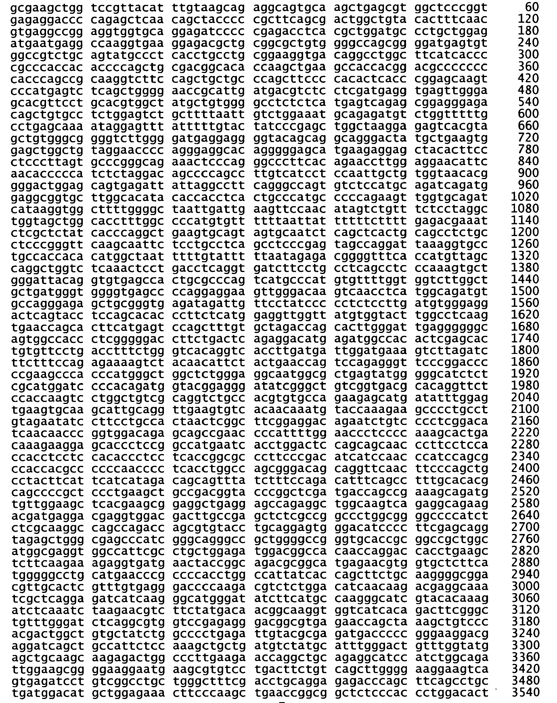

- <210> 13
<211> 24
<212> DNA
<213> Artificial Sequence - <220>
<223> primer - <400> 13
ggaaccttac ttctgtggtg tgac 24 - <210> 14
<211> 24
<212> DNA
<213> Artificial Sequence - <220>
<223> primer - <400> 14
tagcagacac tctatgcctg tgtg 24 - <210> 15
<211> 18
<212> DNA
<213> Artificial Sequence - <220>
<223> sense oligonucleotide - <400> 15
cggaccctag aggcaaag 18 - <210> 16
<211> 20
<212> DNA
<213> Artificial Sequence - <220>
<223> control oligonucleotide - <400> 16
cacgtcacgc gcgcactatt 20 - <210> 17
<211> 6
<212> PRT
<213> Homo sapiens - <400> 17

- <210> 18
<211> 6
<212> PRT
<213> Homo sapiens - <400> 18

- <210> 19
<211> 6
<212> PRT
<213> Homo sapiens - <400> 19

- <210> 20
<211> 18
<212> DNA
<213> Artificial Sequence - <220>
<223> primer - <400> 20 18
tatctccatc ggcagtct 18 - <210> 21
<211> 18
<212> DNA
<213> Artificial Sequence - <220>
<223> primer - <400> 21
tcgacgctca cacttcaa 18 - <210> 22
<211> 17
<212> DNA
<213> Artificial Sequence - <220>
<223> primer - <400> 22 17
ctgaccgctt cctcgtg 17 - <210> 23
<211> 18
<212> DNA
<213> Artificial Sequence - <220>
<223> primer - <400> 23 18
atagagccca ccgcatcc 18 - <210> 24
<211> 2601
<212> DNA
<213> Homo sapiens - <400> 24

- <210> 25
<211> 120
<212> DNA
<213> Homo sapiens - <400> 25

- <210> 26
<211> 40
<212> PRT
<213> Homo sapiens - <400> 26

- <210> 27
<211> 18
<212> DNA
<213> Homo sapiens - <400> 27
gcagtgtcta acgacctc 18 - <210> 28
<211> 18
<212> DNA
<213> Artificial Sequence - <220>
<223> antisense oligonucleotide - <400> 28
ctttgcctct agggtccg 18 - <210> 29
<211> 18
<212> DNA
<213> Artificial Sequence - <220>
<223> antisense oligonucleotide - <400> 29
cagcccgcgc agactgcc 18 - <210> 30
<211> 18
<212> DNA
<213> Artificial sequence - <220>
<223> Antisense oligonucleotide - <400> 30
gaggtcgtta gacactgc 18 - <210> 31
<211> 18
<212> DNA
<213> Artificial Sequence - <220>
<223> antisense oligonucleotide - <400> 31
gccctccttc tctcccat 18 - <210> 32
<211> 18
<212> DNA
<213> Artificial Sequence - <220>
<223> antisense oligonucleotide - <400> 32
acaaatgtaa cggaccag 18 - <210> 33
<211> 18
<212> DNA
<213> Artificial Sequence - <220>
<223> antisense oligonucleotide - <400> 33
cctctcaccg ggagccac 18 - <210> 34
<211> 18
<212> DNA
<213> Artificial Sequence - <220>
<223> antisense oligonucleotide - <400> 34
gaaagtgtac agccagtc 18 - <210> 35
<211> 18
<212> DNA
<213> Artificial Sequence - <220>
<223> antisense oligonucleotide - <400> 35
tgcaccacct ccggcctc 18 - <210> 36
<211> 18
<212> DNA
<213> Artificial Sequence - <220>
<223> antisense oligonucleotide - <400> 36
tgaggtctcg ggggatct 18 - <210> 37
<211> 18
<212> DNA
<213> Artificial Sequence - <220>
<223> antisense oligonucleotide - <400> 37
caccttggcc tcattcat 18 - <210> 38
<211> 18
<212> DNA
<213> Artificial Sequence - <220>
<223> antisense oligonucleotide - <400> 38
atccaatgaa ctccaact 18 - <210> 39
<211> 18
<212> DNA
<213> Artificial Sequence - <220>
<223> antisense oligonucleotide - <400> 39
gagccacttt cccgccgc 18
Claims (19)
- Use of a therapeutically effective amount of an antisense nucleotide which is specifically hybridizable to a portion of the coding region of mRNA encoding human KSR (SEQ ID NO: 24) for the manufacture of a medicament for conferring radiosensitivity to ionizing radiation in tumour cells in a mammal.
- An antisense oligonucleotide for use in conferring radiosensitivity to ionizing radiation in tumor cells in a mammal, wherein the antisense nucleotide is specifically hybridizable to a portion of the coding region of mRNA encoding human KSR (SEQ ID NO: 24).
- The use of claim 1 or the antisense oligonucleotide of claim 2 wherein the tumor cells are cancer cells selected from the group of pancreatic cancer, lung cancer, skin cancer, urinary tract cancer, bladder cancer, liver cancer, thyroid cancer, colon cancer, intestinal cancer, leukemia, lymphoma, neuroblastoma, head and neck cancer, breast cancer, ovarian cancer, stomach cancer, esophageal cancer and prostate cancer.
- Use of a therapeutically effective amount of an antisense nucleotide which is specifically hybridizable to a portion of the coding region of mRNA encoding human KSR (SEQ ID NO: 24) for the manufacture of a medicament for inhibiting angiogenesis in a mammal.
- An antisense oligonucleotide for inhibiting angiogenesis in a mammal, wherein the antisense nucleotide is specifically hybridizable to a portion of the coding region of mRNA encoding human KSR (SEQ ID NO: 24).
- Use of a therapeutically effective amount of an antisense nucleotide which is specifically hybridizable to a portion of the coding region of mRNA encoding human KSR (SEQ ID NO: 24) for the manufacture of a medicament for inhibiting or reducing VEGF expression or activity in a mammal.
- An antisense oligonucleotide for inhibiting or reducing VEGF expression or activity in a mammal, wherein the antisense nucleotide is specifically hybridizable to a portion of the coding region of mRNA encoding human KSR (SEQ ID NO: 24).
- The use of claim 4 or the oligonucleotide of claim 5, wherein the inhibition of angiogenesis is determined by a decrease in the expression of VEGF.
- The use of or the antisense oligonucleotide of any one of the preceding claims, wherein the antisense oligonucleotide comprises a sequence specifically hybridizable to nucleic acid encoding the CA1 region, amino acids 33-72 of human KSR (SEQ ID NO: 26), or a portion thereof.
- The use or an antisense oligonucleotide of any one of claims 1-8, wherein the antisense oligonucleotide comprises a sequence specifically hybridizable to nucleotides 97 to 216 of human KSR (SEQ ID NO: 25), or a portion thereof.
- The use or an antisense oligonucleotide of claim 10, wherein the antisense oligonucleotide comprises a sequence specifically hybridizable to nucleotides 124-141 (SEQ ID NO: 3), 154-171 (SEQID NO: 27), or 187-204 (SEQ ID NO: 5) of the sequence of human KSR.
- The use or an antisense oligonucleotide of any one of claims 1-8, wherein the antisense oligonucleotide comprises a sequence specifically hybridizable to a portion of the coding region of human KSR (SEQ ID NO: 24)selected from:a. Nucleotides 1 to 18 (ATGGGAGAGAAGGAGGGC);b. Nucleotides 205 to 222 (CTGGTCCGTTACATTTGT);c. Nucleotides 247 to 264 (GTGGCTCCCGGTGAGAGG);d. Nucleotides 298 to 315 (GACTGGCTGTACACTTTC);e. Nucleotides 321 to 338 (GAGGCCGGAGGTGGTGCA);f. Nucleotides 351 to 368 (AGATCCCCCGAGACCTCA);g. Nucleotides 379 to 396 (ATGAATGAGGCCAAGGTG);h. Nucleotides 511 to 528 (AGTTGGAGTTCATTGGAT); andi. Nucleotides 531 to 548 (GCGGCGGGAAAGTGGCTC).
- The use or the antisense oligonucleotide of any one of claims 1-8, wherein the antisense oligonucleotide comprises a sequence selected from the group of SEQ ID NOS: 28-39.
- The use or the antisense oligonucleotide of claim 13 wherein the antisense oligonucleotide comprises a sequence of SEQ ID NO: 28.
- The use or the antisense nucleotide of any one of the preceding claims, wherein the antisense oligonucleotide is labelled with a detectable label.
- The use or the antisense oligonucleotide of claim 13 wherein the label is selected from enzymes, ligands, chemicals which fluoresce, and radioactive elements.
- The use or the antisense oligonucleotide of any one of the preceding claims, wherein the antisense oligonucleotide is modified in its chemical backbone.
- The use or the antisense nucleotide of claim 17, wherein the antisense oligonucleotide comprises at least one phosphorothioate linkage.
- The use of or antisense nucleotide of claim 18, wherein the antisense oligonucleotide is a phosphorothioate deoxynucleotide.
Applications Claiming Priority (2)
| Application Number | Priority Date | Filing Date | Title |
|---|---|---|---|
| US10/727,358 US7301017B2 (en) | 2002-05-30 | 2003-12-03 | Kinase suppressor of Ras inactivation for therapy of Ras mediated tumorigenesis |
| PCT/US2004/040506 WO2005056756A2 (en) | 2003-12-03 | 2004-12-03 | Kinase suppressor of ras inactivation for therapy of ras mediated tumorigenesis |
Publications (3)
| Publication Number | Publication Date |
|---|---|
| EP1697498A2 EP1697498A2 (en) | 2006-09-06 |
| EP1697498A4 EP1697498A4 (en) | 2007-10-31 |
| EP1697498B1 true EP1697498B1 (en) | 2010-02-10 |
Family
ID=34677113
Family Applications (1)
| Application Number | Title | Priority Date | Filing Date |
|---|---|---|---|
| EP04812928A Expired - Lifetime EP1697498B1 (en) | 2003-12-03 | 2004-12-03 | Kinase suppressor of ras inactivation for therapy of ras mediated tumorigenesis |
Country Status (8)
| Country | Link |
|---|---|
| US (3) | US7301017B2 (en) |
| EP (1) | EP1697498B1 (en) |
| JP (1) | JP4855265B2 (en) |
| AT (1) | ATE457351T1 (en) |
| AU (1) | AU2004297594B2 (en) |
| CA (1) | CA2549384A1 (en) |
| DE (1) | DE602004025499D1 (en) |
| WO (1) | WO2005056756A2 (en) |
Families Citing this family (10)
| Publication number | Priority date | Publication date | Assignee | Title |
|---|---|---|---|---|
| US7301017B2 (en) * | 2002-05-30 | 2007-11-27 | Kolesnick Richard N | Kinase suppressor of Ras inactivation for therapy of Ras mediated tumorigenesis |
| US8927598B2 (en) * | 2008-01-30 | 2015-01-06 | Duke University | Treating oncogenic Ras driven cancers |
| US8258111B2 (en) | 2008-05-08 | 2012-09-04 | The Johns Hopkins University | Compositions and methods related to miRNA modulation of neovascularization or angiogenesis |
| WO2011009218A1 (en) * | 2009-07-24 | 2011-01-27 | Université de Montréal | Raf dimers and uses thereof |
| US8900845B2 (en) | 2011-02-08 | 2014-12-02 | Washington University | Methods and uses of KSR kinase, and mutations thereof |
| US9526709B2 (en) * | 2011-04-06 | 2016-12-27 | Chemo-Enhanced Llc | Compositions and methods for treating cancer |
| WO2014110555A1 (en) | 2013-01-14 | 2014-07-17 | Chemo-Enchanced Llc | Compositions and methods for treating cancer |
| ES2879287T3 (en) * | 2015-09-15 | 2021-11-22 | Us Health | T-cell receptors that recognize mutated HLA-cw8-restricted KRAS |
| KR101881874B1 (en) * | 2016-04-29 | 2018-07-26 | 한국수력원자력 주식회사 | Methods for Preventing the Malignization of Normal Cells by Low Dose Radiation |
| KR101875111B1 (en) * | 2016-04-29 | 2018-07-09 | 한국수력원자력 주식회사 | Inhibition of Ras-induced malignization by low-dose radiation |
Family Cites Families (23)
| Publication number | Priority date | Publication date | Assignee | Title |
|---|---|---|---|---|
| US591721A (en) | 1897-10-12 | Fan attachment | ||
| US4016043A (en) * | 1975-09-04 | 1977-04-05 | Akzona Incorporated | Enzymatic immunological method for the determination of antigens and antibodies |
| US6582908B2 (en) * | 1990-12-06 | 2003-06-24 | Affymetrix, Inc. | Oligonucleotides |
| US6261834B1 (en) * | 1991-11-08 | 2001-07-17 | Research Corporation Technologies, Inc. | Vector for gene therapy |
| CA2143326A1 (en) * | 1992-08-26 | 1994-03-03 | Guerard Byrne | Tetracycline repressor-mediated binary regulation system for control of gene expression in transgenic animals |
| US5624803A (en) * | 1993-10-14 | 1997-04-29 | The Regents Of The University Of California | In vivo oligonucleotide generator, and methods of testing the binding affinity of triplex forming oligonucleotides derived therefrom |
| US5792638A (en) * | 1994-05-24 | 1998-08-11 | The United States Of America As Represented By The Department Of Health And Human Services | Human ras-related oncogenes unmasked by expression cDNA cloning |
| US5591721A (en) * | 1994-10-25 | 1997-01-07 | Hybridon, Inc. | Method of down-regulating gene expression |
| US5700675A (en) * | 1995-12-13 | 1997-12-23 | Regents Of The University Of California | Protein kinase required for Ras signal transduction |
| US5786213A (en) * | 1996-04-18 | 1998-07-28 | Board Of Regents, The University Of Texas System | Inhibition of endogenous gastrin expression for treatment of colorectal cancer |
| GB9620749D0 (en) * | 1996-10-04 | 1996-11-20 | Brax Genomics Ltd | Identifying antisense oligonucleotides |
| JP2001505428A (en) * | 1996-12-06 | 2001-04-24 | ニールズ パリスガード | Detection of chromosomal abnormalities |
| WO1999001139A1 (en) * | 1997-07-03 | 1999-01-14 | Thomas Jefferson University | An improved method for design and selection of efficacious antisense oligonucleotides |
| US6849334B2 (en) | 2001-08-17 | 2005-02-01 | Neophotonics Corporation | Optical materials and optical devices |
| GB2335035B (en) * | 1998-03-03 | 2003-05-28 | Brax Genomics Ltd | Screening for functional antisense agents |
| CA2395926A1 (en) * | 1999-12-28 | 2001-07-05 | Curagen Corporation | Nucleic acids containing single nucleotide polymorphisms and methods of use thereof |
| US6329203B1 (en) * | 2000-09-08 | 2001-12-11 | Isis Pharmaceuticals, Inc. | Antisense modulation of glioma-associated oncogene-1 expression |
| US20030051267A1 (en) * | 2001-05-10 | 2003-03-13 | Pfizer Inc. | Targeted disruption of kinase suppressor of RAS |
| US20030109466A1 (en) | 2001-09-20 | 2003-06-12 | Isis Pharmaceuticals Inc. | Antisense modulation of KSR expression |
| US7301017B2 (en) * | 2002-05-30 | 2007-11-27 | Kolesnick Richard N | Kinase suppressor of Ras inactivation for therapy of Ras mediated tumorigenesis |
| DE60333365D1 (en) * | 2002-05-30 | 2010-08-26 | Sloan Kettering Inst Cancer | KINASESUPPRESSOR OF RAS INACTIVATION FOR THE THERAPY OF RAS-ASSOCIATED TUMORGENESIS |
| US7250496B2 (en) * | 2002-11-14 | 2007-07-31 | Rosetta Genomics Ltd. | Bioinformatically detectable group of novel regulatory genes and uses thereof |
| US20040265961A1 (en) * | 2003-03-28 | 2004-12-30 | Wei Liu | Novel proteins homologous to kinase suppressor of Ras |
-
2003
- 2003-12-03 US US10/727,358 patent/US7301017B2/en not_active Expired - Fee Related
-
2004
- 2004-12-03 AU AU2004297594A patent/AU2004297594B2/en not_active Ceased
- 2004-12-03 DE DE602004025499T patent/DE602004025499D1/en not_active Expired - Lifetime
- 2004-12-03 AT AT04812928T patent/ATE457351T1/en not_active IP Right Cessation
- 2004-12-03 JP JP2006542782A patent/JP4855265B2/en not_active Expired - Fee Related
- 2004-12-03 EP EP04812928A patent/EP1697498B1/en not_active Expired - Lifetime
- 2004-12-03 WO PCT/US2004/040506 patent/WO2005056756A2/en not_active Ceased
- 2004-12-03 CA CA002549384A patent/CA2549384A1/en not_active Abandoned
-
2007
- 2007-10-12 US US11/974,473 patent/US8258108B2/en not_active Expired - Fee Related
- 2007-10-12 US US11/974,481 patent/US8283459B2/en not_active Expired - Fee Related
Also Published As
| Publication number | Publication date |
|---|---|
| US20080096840A1 (en) | 2008-04-24 |
| JP2007512844A (en) | 2007-05-24 |
| EP1697498A4 (en) | 2007-10-31 |
| JP4855265B2 (en) | 2012-01-18 |
| DE602004025499D1 (en) | 2010-03-25 |
| US20050037455A1 (en) | 2005-02-17 |
| US8283459B2 (en) | 2012-10-09 |
| ATE457351T1 (en) | 2010-02-15 |
| US7301017B2 (en) | 2007-11-27 |
| CA2549384A1 (en) | 2005-06-23 |
| WO2005056756A3 (en) | 2006-08-03 |
| AU2004297594A1 (en) | 2005-06-23 |
| US20090143320A1 (en) | 2009-06-04 |
| EP1697498A2 (en) | 2006-09-06 |
| WO2005056756A2 (en) | 2005-06-23 |
| AU2004297594B2 (en) | 2009-12-17 |
| US8258108B2 (en) | 2012-09-04 |
Similar Documents
| Publication | Publication Date | Title |
|---|---|---|
| US8283459B2 (en) | Kinase suppressor of Ras inactivation for therapy of Ras mediated tumorigenesis | |
| US7528116B2 (en) | Kinase suppressor of Ras inactivation for therapy of Ras mediated tumorigenesis | |
| Mercer et al. | Expression of endogenous oncogenic V600EB-raf induces proliferation and developmental defects in mice and transformation of primary fibroblasts | |
| Fisher et al. | Induction and apoptotic regression of lung adenocarcinomas by regulation of a K-Ras transgene in the presence and absence of tumor suppressor genes | |
| US7858593B2 (en) | Inhibitors of prolyl-hydroxylase-1 for the treatment of skeletal muscle degeneration | |
| Suga et al. | Transgenic mice expressing a mutant form of loricrin reveal the molecular basis of the skin diseases, Vohwinkel syndrome and progressive symmetric erythrokeratoderma | |
| KR102378462B1 (en) | Animal model of non-alcoholic liver disease and compositions of diagnosis, prevention or treatment for non-alcoholic liver disease | |
| EP2274324B1 (en) | Substances and compositions for enhancing dna repair and methods of use | |
| EP1599573B1 (en) | Model for studying the role of genes in tumor resistance to chemotherapy | |
| US20090186839A1 (en) | Model for studying the role of genes in chemoresistance | |
| US20120208750A1 (en) | Use of protein kinase c delta (pkcd) inhibitors to treat diabetes, obesity, and hepatic steatosis | |
| Park et al. | Early embryonic lethality caused by targeted disruption of the TRAF-interacting protein (TRIP) gene | |
| WO2011011061A2 (en) | Method of regulating angiogenesis and lymphangiogenesis, and a pharmaceutical composition for effecting anti-angiogenic and anti-lymphangiogenic cancer therapy | |
| Minowada et al. | Overexpression of Sprouty 2 in mouse lung epithelium inhibits urethane-induced tumorigenesis | |
| JP2013094071A (en) | TRANSGENIC MOUSE EXPRESSING HUMAN TGFβ1 | |
| US20130041009A1 (en) | Methods and means for increasing resistance to cell damage | |
| JP5737702B2 (en) | Arf6 gene function loss animal and method of using the same | |
| WO2009022119A1 (en) | Murine endometriosis modelled by k-ras activation of menstruating endometrium | |
| WO2000044766A1 (en) | Inhibition of cationic amino acid transporter protein and uses thereof | |
| WO2002009736A1 (en) | Methods and agents for treating persistent pain | |
| Johnson et al. | Nrf Transcription Factors in Keratinocytes | |
| AU2004213822A1 (en) | Model for studying the role of genes in tumor resistance to chemotherapy |
Legal Events
| Date | Code | Title | Description |
|---|---|---|---|
| PUAI | Public reference made under article 153(3) epc to a published international application that has entered the european phase |
Free format text: ORIGINAL CODE: 0009012 |
|
| 17P | Request for examination filed |
Effective date: 20060609 |
|
| AK | Designated contracting states |
Kind code of ref document: A2 Designated state(s): AT BE BG CH CY CZ DE DK EE ES FI FR GB GR HU IE IS IT LI LT LU MC NL PL PT RO SE SI SK TR |
|
| AX | Request for extension of the european patent |
Extension state: AL BA HR LV MK YU |
|
| RIC1 | Information provided on ipc code assigned before grant |
Ipc: C07H 21/04 20060101AFI20060911BHEP |
|
| DAX | Request for extension of the european patent (deleted) | ||
| RIC1 | Information provided on ipc code assigned before grant |
Ipc: C07H 21/02 20060101ALI20070710BHEP Ipc: C12N 15/11 20060101AFI20070710BHEP Ipc: A61K 31/7125 20060101ALI20070710BHEP Ipc: C07H 21/04 20060101ALI20070710BHEP |
|
| A4 | Supplementary search report drawn up and despatched |
Effective date: 20071002 |
|
| 17Q | First examination report despatched |
Effective date: 20080317 |
|
| GRAP | Despatch of communication of intention to grant a patent |
Free format text: ORIGINAL CODE: EPIDOSNIGR1 |
|
| GRAS | Grant fee paid |
Free format text: ORIGINAL CODE: EPIDOSNIGR3 |
|
| GRAA | (expected) grant |
Free format text: ORIGINAL CODE: 0009210 |
|
| AK | Designated contracting states |
Kind code of ref document: B1 Designated state(s): AT BE BG CH CY CZ DE DK EE ES FI FR GB GR HU IE IS IT LI LT LU MC NL PL PT RO SE SI SK TR |
|
| REG | Reference to a national code |
Ref country code: GB Ref legal event code: FG4D |
|
| REG | Reference to a national code |
Ref country code: CH Ref legal event code: EP |
|
| REG | Reference to a national code |
Ref country code: IE Ref legal event code: FG4D |
|
| REF | Corresponds to: |
Ref document number: 602004025499 Country of ref document: DE Date of ref document: 20100325 Kind code of ref document: P |
|
| REG | Reference to a national code |
Ref country code: NL Ref legal event code: VDEP Effective date: 20100210 |
|
| LTIE | Lt: invalidation of european patent or patent extension |
Effective date: 20100210 |
|
| PG25 | Lapsed in a contracting state [announced via postgrant information from national office to epo] |
Ref country code: PT Free format text: LAPSE BECAUSE OF FAILURE TO SUBMIT A TRANSLATION OF THE DESCRIPTION OR TO PAY THE FEE WITHIN THE PRESCRIBED TIME-LIMIT Effective date: 20100611 Ref country code: LT Free format text: LAPSE BECAUSE OF FAILURE TO SUBMIT A TRANSLATION OF THE DESCRIPTION OR TO PAY THE FEE WITHIN THE PRESCRIBED TIME-LIMIT Effective date: 20100210 Ref country code: IS Free format text: LAPSE BECAUSE OF FAILURE TO SUBMIT A TRANSLATION OF THE DESCRIPTION OR TO PAY THE FEE WITHIN THE PRESCRIBED TIME-LIMIT Effective date: 20100610 Ref country code: ES Free format text: LAPSE BECAUSE OF FAILURE TO SUBMIT A TRANSLATION OF THE DESCRIPTION OR TO PAY THE FEE WITHIN THE PRESCRIBED TIME-LIMIT Effective date: 20100521 |
|
| PG25 | Lapsed in a contracting state [announced via postgrant information from national office to epo] |
Ref country code: PL Free format text: LAPSE BECAUSE OF FAILURE TO SUBMIT A TRANSLATION OF THE DESCRIPTION OR TO PAY THE FEE WITHIN THE PRESCRIBED TIME-LIMIT Effective date: 20100210 Ref country code: FI Free format text: LAPSE BECAUSE OF FAILURE TO SUBMIT A TRANSLATION OF THE DESCRIPTION OR TO PAY THE FEE WITHIN THE PRESCRIBED TIME-LIMIT Effective date: 20100210 Ref country code: AT Free format text: LAPSE BECAUSE OF FAILURE TO SUBMIT A TRANSLATION OF THE DESCRIPTION OR TO PAY THE FEE WITHIN THE PRESCRIBED TIME-LIMIT Effective date: 20100210 Ref country code: SI Free format text: LAPSE BECAUSE OF FAILURE TO SUBMIT A TRANSLATION OF THE DESCRIPTION OR TO PAY THE FEE WITHIN THE PRESCRIBED TIME-LIMIT Effective date: 20100210 |
|
| PG25 | Lapsed in a contracting state [announced via postgrant information from national office to epo] |
Ref country code: SE Free format text: LAPSE BECAUSE OF FAILURE TO SUBMIT A TRANSLATION OF THE DESCRIPTION OR TO PAY THE FEE WITHIN THE PRESCRIBED TIME-LIMIT Effective date: 20100210 Ref country code: CY Free format text: LAPSE BECAUSE OF FAILURE TO SUBMIT A TRANSLATION OF THE DESCRIPTION OR TO PAY THE FEE WITHIN THE PRESCRIBED TIME-LIMIT Effective date: 20100210 Ref country code: BE Free format text: LAPSE BECAUSE OF FAILURE TO SUBMIT A TRANSLATION OF THE DESCRIPTION OR TO PAY THE FEE WITHIN THE PRESCRIBED TIME-LIMIT Effective date: 20100210 Ref country code: EE Free format text: LAPSE BECAUSE OF FAILURE TO SUBMIT A TRANSLATION OF THE DESCRIPTION OR TO PAY THE FEE WITHIN THE PRESCRIBED TIME-LIMIT Effective date: 20100210 Ref country code: NL Free format text: LAPSE BECAUSE OF FAILURE TO SUBMIT A TRANSLATION OF THE DESCRIPTION OR TO PAY THE FEE WITHIN THE PRESCRIBED TIME-LIMIT Effective date: 20100210 Ref country code: GR Free format text: LAPSE BECAUSE OF FAILURE TO SUBMIT A TRANSLATION OF THE DESCRIPTION OR TO PAY THE FEE WITHIN THE PRESCRIBED TIME-LIMIT Effective date: 20100511 Ref country code: RO Free format text: LAPSE BECAUSE OF FAILURE TO SUBMIT A TRANSLATION OF THE DESCRIPTION OR TO PAY THE FEE WITHIN THE PRESCRIBED TIME-LIMIT Effective date: 20100210 |
|
| PG25 | Lapsed in a contracting state [announced via postgrant information from national office to epo] |
Ref country code: SK Free format text: LAPSE BECAUSE OF FAILURE TO SUBMIT A TRANSLATION OF THE DESCRIPTION OR TO PAY THE FEE WITHIN THE PRESCRIBED TIME-LIMIT Effective date: 20100210 Ref country code: CZ Free format text: LAPSE BECAUSE OF FAILURE TO SUBMIT A TRANSLATION OF THE DESCRIPTION OR TO PAY THE FEE WITHIN THE PRESCRIBED TIME-LIMIT Effective date: 20100210 Ref country code: BG Free format text: LAPSE BECAUSE OF FAILURE TO SUBMIT A TRANSLATION OF THE DESCRIPTION OR TO PAY THE FEE WITHIN THE PRESCRIBED TIME-LIMIT Effective date: 20100510 |
|
| PLBE | No opposition filed within time limit |
Free format text: ORIGINAL CODE: 0009261 |
|
| STAA | Information on the status of an ep patent application or granted ep patent |
Free format text: STATUS: NO OPPOSITION FILED WITHIN TIME LIMIT |
|
| 26N | No opposition filed |
Effective date: 20101111 |
|
| PG25 | Lapsed in a contracting state [announced via postgrant information from national office to epo] |
Ref country code: DK Free format text: LAPSE BECAUSE OF FAILURE TO SUBMIT A TRANSLATION OF THE DESCRIPTION OR TO PAY THE FEE WITHIN THE PRESCRIBED TIME-LIMIT Effective date: 20100210 |
|
| PG25 | Lapsed in a contracting state [announced via postgrant information from national office to epo] |
Ref country code: MC Free format text: LAPSE BECAUSE OF NON-PAYMENT OF DUE FEES Effective date: 20101231 |
|
| REG | Reference to a national code |
Ref country code: CH Ref legal event code: PL |
|
| PG25 | Lapsed in a contracting state [announced via postgrant information from national office to epo] |
Ref country code: IE Free format text: LAPSE BECAUSE OF NON-PAYMENT OF DUE FEES Effective date: 20101203 Ref country code: CH Free format text: LAPSE BECAUSE OF NON-PAYMENT OF DUE FEES Effective date: 20101231 Ref country code: LI Free format text: LAPSE BECAUSE OF NON-PAYMENT OF DUE FEES Effective date: 20101231 |
|
| PG25 | Lapsed in a contracting state [announced via postgrant information from national office to epo] |
Ref country code: HU Free format text: LAPSE BECAUSE OF FAILURE TO SUBMIT A TRANSLATION OF THE DESCRIPTION OR TO PAY THE FEE WITHIN THE PRESCRIBED TIME-LIMIT Effective date: 20100811 Ref country code: LU Free format text: LAPSE BECAUSE OF NON-PAYMENT OF DUE FEES Effective date: 20101203 |
|
| PG25 | Lapsed in a contracting state [announced via postgrant information from national office to epo] |
Ref country code: TR Free format text: LAPSE BECAUSE OF FAILURE TO SUBMIT A TRANSLATION OF THE DESCRIPTION OR TO PAY THE FEE WITHIN THE PRESCRIBED TIME-LIMIT Effective date: 20100210 |
|
| PGFP | Annual fee paid to national office [announced via postgrant information from national office to epo] |
Ref country code: IT Payment date: 20121217 Year of fee payment: 9 Ref country code: GB Payment date: 20121220 Year of fee payment: 9 |
|
| PGFP | Annual fee paid to national office [announced via postgrant information from national office to epo] |
Ref country code: FR Payment date: 20130123 Year of fee payment: 9 Ref country code: DE Payment date: 20121219 Year of fee payment: 9 |
|
| REG | Reference to a national code |
Ref country code: DE Ref legal event code: R119 Ref document number: 602004025499 Country of ref document: DE |
|
| GBPC | Gb: european patent ceased through non-payment of renewal fee |
Effective date: 20131203 |
|
| REG | Reference to a national code |
Ref country code: FR Ref legal event code: ST Effective date: 20140829 |
|
| REG | Reference to a national code |
Ref country code: DE Ref legal event code: R119 Ref document number: 602004025499 Country of ref document: DE Effective date: 20140701 |
|
| PG25 | Lapsed in a contracting state [announced via postgrant information from national office to epo] |
Ref country code: DE Free format text: LAPSE BECAUSE OF NON-PAYMENT OF DUE FEES Effective date: 20140701 |
|
| PG25 | Lapsed in a contracting state [announced via postgrant information from national office to epo] |
Ref country code: GB Free format text: LAPSE BECAUSE OF NON-PAYMENT OF DUE FEES Effective date: 20131203 Ref country code: FR Free format text: LAPSE BECAUSE OF NON-PAYMENT OF DUE FEES Effective date: 20131231 |
|
| PG25 | Lapsed in a contracting state [announced via postgrant information from national office to epo] |
Ref country code: IT Free format text: LAPSE BECAUSE OF NON-PAYMENT OF DUE FEES Effective date: 20131231 |
|
| PG25 | Lapsed in a contracting state [announced via postgrant information from national office to epo] |
Ref country code: IT Free format text: LAPSE BECAUSE OF NON-PAYMENT OF DUE FEES Effective date: 20131203 |