EP1687442B1 - Nucleic acid detection - Google Patents
Nucleic acid detection Download PDFInfo
- Publication number
- EP1687442B1 EP1687442B1 EP03819025A EP03819025A EP1687442B1 EP 1687442 B1 EP1687442 B1 EP 1687442B1 EP 03819025 A EP03819025 A EP 03819025A EP 03819025 A EP03819025 A EP 03819025A EP 1687442 B1 EP1687442 B1 EP 1687442B1
- Authority
- EP
- European Patent Office
- Prior art keywords
- nucleic acid
- nucleic acids
- label
- probe
- target nucleic
- Prior art date
- Legal status (The legal status is an assumption and is not a legal conclusion. Google has not performed a legal analysis and makes no representation as to the accuracy of the status listed.)
- Expired - Lifetime
Links
- 150000007523 nucleic acids Chemical class 0.000 title claims abstract description 181
- 108020004707 nucleic acids Proteins 0.000 title claims abstract description 122
- 102000039446 nucleic acids Human genes 0.000 title claims abstract description 122
- 238000001514 detection method Methods 0.000 title claims abstract description 29
- 108091028043 Nucleic acid sequence Proteins 0.000 claims abstract description 47
- 239000000523 sample Substances 0.000 claims abstract description 44
- 239000000203 mixture Substances 0.000 claims abstract description 40
- 238000000034 method Methods 0.000 claims abstract description 35
- 239000007791 liquid phase Substances 0.000 claims abstract description 6
- 238000000926 separation method Methods 0.000 claims description 12
- 230000000295 complement effect Effects 0.000 claims description 9
- 239000002773 nucleotide Substances 0.000 claims description 7
- 125000003729 nucleotide group Chemical group 0.000 claims description 7
- 230000005855 radiation Effects 0.000 claims description 4
- 239000000427 antigen Substances 0.000 claims description 2
- 108091007433 antigens Proteins 0.000 claims description 2
- 102000036639 antigens Human genes 0.000 claims description 2
- 230000029918 bioluminescence Effects 0.000 claims description 2
- 238000005415 bioluminescence Methods 0.000 claims description 2
- 230000002285 radioactive effect Effects 0.000 claims description 2
- 230000000063 preceeding effect Effects 0.000 claims 2
- 108020004414 DNA Proteins 0.000 description 18
- 239000012528 membrane Substances 0.000 description 15
- 108020004711 Nucleic Acid Probes Proteins 0.000 description 14
- 239000002853 nucleic acid probe Substances 0.000 description 14
- 108091034117 Oligonucleotide Proteins 0.000 description 12
- JLCPHMBAVCMARE-UHFFFAOYSA-N [3-[[3-[[3-[[3-[[3-[[3-[[3-[[3-[[3-[[3-[[3-[[5-(2-amino-6-oxo-1H-purin-9-yl)-3-[[3-[[3-[[3-[[3-[[3-[[5-(2-amino-6-oxo-1H-purin-9-yl)-3-[[5-(2-amino-6-oxo-1H-purin-9-yl)-3-hydroxyoxolan-2-yl]methoxy-hydroxyphosphoryl]oxyoxolan-2-yl]methoxy-hydroxyphosphoryl]oxy-5-(5-methyl-2,4-dioxopyrimidin-1-yl)oxolan-2-yl]methoxy-hydroxyphosphoryl]oxy-5-(6-aminopurin-9-yl)oxolan-2-yl]methoxy-hydroxyphosphoryl]oxy-5-(6-aminopurin-9-yl)oxolan-2-yl]methoxy-hydroxyphosphoryl]oxy-5-(6-aminopurin-9-yl)oxolan-2-yl]methoxy-hydroxyphosphoryl]oxy-5-(6-aminopurin-9-yl)oxolan-2-yl]methoxy-hydroxyphosphoryl]oxyoxolan-2-yl]methoxy-hydroxyphosphoryl]oxy-5-(5-methyl-2,4-dioxopyrimidin-1-yl)oxolan-2-yl]methoxy-hydroxyphosphoryl]oxy-5-(4-amino-2-oxopyrimidin-1-yl)oxolan-2-yl]methoxy-hydroxyphosphoryl]oxy-5-(5-methyl-2,4-dioxopyrimidin-1-yl)oxolan-2-yl]methoxy-hydroxyphosphoryl]oxy-5-(5-methyl-2,4-dioxopyrimidin-1-yl)oxolan-2-yl]methoxy-hydroxyphosphoryl]oxy-5-(6-aminopurin-9-yl)oxolan-2-yl]methoxy-hydroxyphosphoryl]oxy-5-(6-aminopurin-9-yl)oxolan-2-yl]methoxy-hydroxyphosphoryl]oxy-5-(4-amino-2-oxopyrimidin-1-yl)oxolan-2-yl]methoxy-hydroxyphosphoryl]oxy-5-(4-amino-2-oxopyrimidin-1-yl)oxolan-2-yl]methoxy-hydroxyphosphoryl]oxy-5-(4-amino-2-oxopyrimidin-1-yl)oxolan-2-yl]methoxy-hydroxyphosphoryl]oxy-5-(6-aminopurin-9-yl)oxolan-2-yl]methoxy-hydroxyphosphoryl]oxy-5-(4-amino-2-oxopyrimidin-1-yl)oxolan-2-yl]methyl [5-(6-aminopurin-9-yl)-2-(hydroxymethyl)oxolan-3-yl] hydrogen phosphate Polymers Cc1cn(C2CC(OP(O)(=O)OCC3OC(CC3OP(O)(=O)OCC3OC(CC3O)n3cnc4c3nc(N)[nH]c4=O)n3cnc4c3nc(N)[nH]c4=O)C(COP(O)(=O)OC3CC(OC3COP(O)(=O)OC3CC(OC3COP(O)(=O)OC3CC(OC3COP(O)(=O)OC3CC(OC3COP(O)(=O)OC3CC(OC3COP(O)(=O)OC3CC(OC3COP(O)(=O)OC3CC(OC3COP(O)(=O)OC3CC(OC3COP(O)(=O)OC3CC(OC3COP(O)(=O)OC3CC(OC3COP(O)(=O)OC3CC(OC3COP(O)(=O)OC3CC(OC3COP(O)(=O)OC3CC(OC3COP(O)(=O)OC3CC(OC3COP(O)(=O)OC3CC(OC3COP(O)(=O)OC3CC(OC3COP(O)(=O)OC3CC(OC3CO)n3cnc4c(N)ncnc34)n3ccc(N)nc3=O)n3cnc4c(N)ncnc34)n3ccc(N)nc3=O)n3ccc(N)nc3=O)n3ccc(N)nc3=O)n3cnc4c(N)ncnc34)n3cnc4c(N)ncnc34)n3cc(C)c(=O)[nH]c3=O)n3cc(C)c(=O)[nH]c3=O)n3ccc(N)nc3=O)n3cc(C)c(=O)[nH]c3=O)n3cnc4c3nc(N)[nH]c4=O)n3cnc4c(N)ncnc34)n3cnc4c(N)ncnc34)n3cnc4c(N)ncnc34)n3cnc4c(N)ncnc34)O2)c(=O)[nH]c1=O JLCPHMBAVCMARE-UHFFFAOYSA-N 0.000 description 12
- 239000000499 gel Substances 0.000 description 11
- 238000001502 gel electrophoresis Methods 0.000 description 10
- 238000000636 Northern blotting Methods 0.000 description 8
- 238000002105 Southern blotting Methods 0.000 description 8
- 238000012546 transfer Methods 0.000 description 6
- 238000009396 hybridization Methods 0.000 description 5
- LFQSCWFLJHTTHZ-UHFFFAOYSA-N Ethanol Chemical compound CCO LFQSCWFLJHTTHZ-UHFFFAOYSA-N 0.000 description 4
- 230000000903 blocking effect Effects 0.000 description 4
- 238000006243 chemical reaction Methods 0.000 description 4
- 238000010438 heat treatment Methods 0.000 description 4
- 108090000623 proteins and genes Proteins 0.000 description 4
- 108091032973 (ribonucleotides)n+m Proteins 0.000 description 3
- HEMHJVSKTPXQMS-UHFFFAOYSA-M Sodium hydroxide Chemical compound [OH-].[Na+] HEMHJVSKTPXQMS-UHFFFAOYSA-M 0.000 description 3
- 239000008280 blood Substances 0.000 description 3
- 210000004369 blood Anatomy 0.000 description 3
- 239000003153 chemical reaction reagent Substances 0.000 description 3
- 239000000975 dye Substances 0.000 description 3
- 239000000463 material Substances 0.000 description 3
- 229920002401 polyacrylamide Polymers 0.000 description 3
- 229920000936 Agarose Polymers 0.000 description 2
- 102000053602 DNA Human genes 0.000 description 2
- 102000004594 DNA Polymerase I Human genes 0.000 description 2
- 108010017826 DNA Polymerase I Proteins 0.000 description 2
- 239000004677 Nylon Substances 0.000 description 2
- 229910019142 PO4 Inorganic materials 0.000 description 2
- VMHLLURERBWHNL-UHFFFAOYSA-M Sodium acetate Chemical compound [Na+].CC([O-])=O VMHLLURERBWHNL-UHFFFAOYSA-M 0.000 description 2
- 239000007984 Tris EDTA buffer Substances 0.000 description 2
- 238000003491 array Methods 0.000 description 2
- 238000000376 autoradiography Methods 0.000 description 2
- 238000007796 conventional method Methods 0.000 description 2
- 238000001962 electrophoresis Methods 0.000 description 2
- ZMMJGEGLRURXTF-UHFFFAOYSA-N ethidium bromide Chemical compound [Br-].C12=CC(N)=CC=C2C2=CC=C(N)C=C2[N+](CC)=C1C1=CC=CC=C1 ZMMJGEGLRURXTF-UHFFFAOYSA-N 0.000 description 2
- 229960005542 ethidium bromide Drugs 0.000 description 2
- 238000002372 labelling Methods 0.000 description 2
- 239000003068 molecular probe Substances 0.000 description 2
- 229920001778 nylon Polymers 0.000 description 2
- 235000021317 phosphate Nutrition 0.000 description 2
- 239000001632 sodium acetate Substances 0.000 description 2
- 235000017281 sodium acetate Nutrition 0.000 description 2
- 239000000243 solution Substances 0.000 description 2
- YBJHBAHKTGYVGT-ZKWXMUAHSA-N (+)-Biotin Chemical compound N1C(=O)N[C@@H]2[C@H](CCCCC(=O)O)SC[C@@H]21 YBJHBAHKTGYVGT-ZKWXMUAHSA-N 0.000 description 1
- 241000972773 Aulopiformes Species 0.000 description 1
- 235000000638 D-biotin Nutrition 0.000 description 1
- 239000011665 D-biotin Substances 0.000 description 1
- 108020003215 DNA Probes Proteins 0.000 description 1
- 239000003298 DNA probe Substances 0.000 description 1
- 101710088194 Dehydrogenase Proteins 0.000 description 1
- KCXVZYZYPLLWCC-UHFFFAOYSA-N EDTA Chemical compound OC(=O)CN(CC(O)=O)CCN(CC(O)=O)CC(O)=O KCXVZYZYPLLWCC-UHFFFAOYSA-N 0.000 description 1
- 239000000020 Nitrocellulose Substances 0.000 description 1
- WDVSHHCDHLJJJR-UHFFFAOYSA-N Proflavine Chemical compound C1=CC(N)=CC2=NC3=CC(N)=CC=C3C=C21 WDVSHHCDHLJJJR-UHFFFAOYSA-N 0.000 description 1
- 108020004518 RNA Probes Proteins 0.000 description 1
- 239000003391 RNA probe Substances 0.000 description 1
- XUIMIQQOPSSXEZ-UHFFFAOYSA-N Silicon Chemical compound [Si] XUIMIQQOPSSXEZ-UHFFFAOYSA-N 0.000 description 1
- 239000007983 Tris buffer Substances 0.000 description 1
- DPKHZNPWBDQZCN-UHFFFAOYSA-N acridine orange free base Chemical compound C1=CC(N(C)C)=CC2=NC3=CC(N(C)C)=CC=C3C=C21 DPKHZNPWBDQZCN-UHFFFAOYSA-N 0.000 description 1
- 239000011543 agarose gel Substances 0.000 description 1
- DZBUGLKDJFMEHC-UHFFFAOYSA-N benzoquinolinylidene Natural products C1=CC=CC2=CC3=CC=CC=C3N=C21 DZBUGLKDJFMEHC-UHFFFAOYSA-N 0.000 description 1
- 230000015572 biosynthetic process Effects 0.000 description 1
- 239000007853 buffer solution Substances 0.000 description 1
- 238000005251 capillar electrophoresis Methods 0.000 description 1
- 238000003271 compound fluorescence assay Methods 0.000 description 1
- 238000001816 cooling Methods 0.000 description 1
- 230000002079 cooperative effect Effects 0.000 description 1
- 230000001419 dependent effect Effects 0.000 description 1
- 238000013461 design Methods 0.000 description 1
- 238000005516 engineering process Methods 0.000 description 1
- 235000013861 fat-free Nutrition 0.000 description 1
- 230000002349 favourable effect Effects 0.000 description 1
- 239000007850 fluorescent dye Substances 0.000 description 1
- 239000011521 glass Substances 0.000 description 1
- MNQZXJOMYWMBOU-UHFFFAOYSA-N glyceraldehyde Chemical compound OCC(O)C=O MNQZXJOMYWMBOU-UHFFFAOYSA-N 0.000 description 1
- 230000003993 interaction Effects 0.000 description 1
- 239000000138 intercalating agent Substances 0.000 description 1
- 238000009830 intercalation Methods 0.000 description 1
- 239000003550 marker Substances 0.000 description 1
- 239000011159 matrix material Substances 0.000 description 1
- 235000013336 milk Nutrition 0.000 description 1
- 239000008267 milk Substances 0.000 description 1
- 210000004080 milk Anatomy 0.000 description 1
- 238000002156 mixing Methods 0.000 description 1
- 229920001220 nitrocellulos Polymers 0.000 description 1
- 239000008188 pellet Substances 0.000 description 1
- NBIIXXVUZAFLBC-UHFFFAOYSA-K phosphate Chemical compound [O-]P([O-])([O-])=O NBIIXXVUZAFLBC-UHFFFAOYSA-K 0.000 description 1
- 239000010452 phosphate Substances 0.000 description 1
- 150000003013 phosphoric acid derivatives Chemical class 0.000 description 1
- 238000001556 precipitation Methods 0.000 description 1
- 230000037452 priming Effects 0.000 description 1
- 229960000286 proflavine Drugs 0.000 description 1
- 108091008146 restriction endonucleases Proteins 0.000 description 1
- 108020004418 ribosomal RNA Proteins 0.000 description 1
- 235000019515 salmon Nutrition 0.000 description 1
- 239000012488 sample solution Substances 0.000 description 1
- 229910052710 silicon Inorganic materials 0.000 description 1
- 239000010703 silicon Substances 0.000 description 1
- 238000010186 staining Methods 0.000 description 1
- 238000003786 synthesis reaction Methods 0.000 description 1
- 238000012360 testing method Methods 0.000 description 1
- 210000001519 tissue Anatomy 0.000 description 1
Images


Classifications
-
- C—CHEMISTRY; METALLURGY
- C12—BIOCHEMISTRY; BEER; SPIRITS; WINE; VINEGAR; MICROBIOLOGY; ENZYMOLOGY; MUTATION OR GENETIC ENGINEERING
- C12Q—MEASURING OR TESTING PROCESSES INVOLVING ENZYMES, NUCLEIC ACIDS OR MICROORGANISMS; COMPOSITIONS OR TEST PAPERS THEREFOR; PROCESSES OF PREPARING SUCH COMPOSITIONS; CONDITION-RESPONSIVE CONTROL IN MICROBIOLOGICAL OR ENZYMOLOGICAL PROCESSES
- C12Q1/00—Measuring or testing processes involving enzymes, nucleic acids or microorganisms; Compositions therefor; Processes of preparing such compositions
- C12Q1/68—Measuring or testing processes involving enzymes, nucleic acids or microorganisms; Compositions therefor; Processes of preparing such compositions involving nucleic acids
- C12Q1/6813—Hybridisation assays
- C12Q1/6816—Hybridisation assays characterised by the detection means
-
- Y—GENERAL TAGGING OF NEW TECHNOLOGICAL DEVELOPMENTS; GENERAL TAGGING OF CROSS-SECTIONAL TECHNOLOGIES SPANNING OVER SEVERAL SECTIONS OF THE IPC; TECHNICAL SUBJECTS COVERED BY FORMER USPC CROSS-REFERENCE ART COLLECTIONS [XRACs] AND DIGESTS
- Y10—TECHNICAL SUBJECTS COVERED BY FORMER USPC
- Y10T—TECHNICAL SUBJECTS COVERED BY FORMER US CLASSIFICATION
- Y10T436/00—Chemistry: analytical and immunological testing
- Y10T436/14—Heterocyclic carbon compound [i.e., O, S, N, Se, Te, as only ring hetero atom]
- Y10T436/142222—Hetero-O [e.g., ascorbic acid, etc.]
- Y10T436/143333—Saccharide [e.g., DNA, etc.]
Definitions
- the present invention relates to detection of a nucleic acid sequence in a mixture of different nucleic acids and a kit therefor.
- WO 0157258 A discloses a direct detection and quantitation of RNA or DNA contained in a sample obtained by capillary electrophoresis of the RNA or DNA hybridized to a DNA or RNA probe of complementary sequence stabilized by the combination of a fluorophore terminally conjugated to the probe and a dye intercalating the RNA-DNA or DNA-DNA hybrid so formed.
- EP 1186673 A discloses calibrating different types of signals scanned from a molecular array, or calibrating signals scanned from different molecular arrays, by employing calibrating probes that generate signals proportional to the total concentrations of labeled target molecules to which the molecular array probes are directed over an entire range of sample solutions, and molecular arrays incorporating sets of calibrating probes.
- nucleic acid sequences are the southern blotting technique for DNA and the northern blotting technique for RNA.
- the nucleic acid mixtures are separated in nucleic acids of different mass using gel electrophoresis, for example in an agarose or polyacrylamide gel.
- the different nucleic acids are preferably converted to single stranded nucleic acids.
- the single stranded nucleic acids are then transferred onto a microcellulose or a nylon filter and are crosslinked with the membrane using heat or UV radiation. The membrane is then blocked with a blocking reagent to saturize all unspecific binding sites of the membrane.
- the nucleic acids fixed on the membrane are hybridized with a labeled nucleic acid probe, which includes a primary sequence complementary to the primary sequence of a target nucleic acid sequence.
- the label of the nucleic acid probe often contains 32 P-labeled phosphates, which can be detected due to their radioactivity (see for example Figure 1 ).
- the northern or southern blotting techniques therefore involve lots of different steps, e.g. gel electrophoresis, blotting onto a membrane and detection by hybridization, which are very time consuming and complicated to carry out. For the southern and northern blotting techniques different media (gels for gel electrophoresis and membranes for the blotting) are used, so that lots of different and at least partially expensive materials are used.
- the present invention meets these needs by providing a method for detection of a target nucleic acid sequence according to the base claim 1.
- Favorable embodiments of the method of the invention and a kit for the detection of the target nucleic acid sequence are subjects of further claims.
- Embodiments of the invention provide a fast and easy-to-handle procedure for detection of a target nucleic acid sequence, wherein the hybridizing of the target nucleic acid sequence with the probe takes place in liquid phase.
- the procedure in A) therefore avoids the complicated, time-consuming and also material-consuming stepproceedure of transfer of the nucleic acids onto a membrane.
- an operator carrying out the embodiments of the invention usually needs less skill than an operator carrying out conventional Northern or Southern blot techniques.
- the method for detection requires the hybridizing of the probe with the target nucleic acid sequence prior to separating the nucleic acids. This sequence of the method is reversed in comparison to the conventional northern and southern blotting techniques, where the nucleic acids are separated first and then hybridized with a labeled probe.
- the additional binding sites are hybridized with single-stranded nucleic acids having random primary sequences in liquid phase.
- the additional binding sites of the nucleic acids, which are still present after A) are often comprised of unpaired bases in single stranded areas of the nucleic acids.
- the single-stranded nucleic acids can basepair with single-stranded parts of the different nucleic acids in the nucleic acid mixture and if present also with single-stranded parts of the target nucleic acid sequence, forming nucleic acid double strands. Therefore after A1) the nucleic acids in the nucleic acid mixture are mainly double-stranded, simplifying the separation of the different nucleic acids in the subsequent B). Due to A1) no retardation of the double stranded hybrid between the probe and the target nucleic acid sequence in comparison to the other nucleic acids occurs during the separation procedure in B).
- a retardation of the double stranded hybrid during gel electrophoresis normally takes place, when single stranded nucleic acids are still present in the nucleic acid mixture and are also subjected to gel electrophoresis, so that the important information about the size of the target sequence is lost.
- the information about the size of the target sequence is often used as a control for the correct hybridization between the target sequence and the probe.
- nucleic acids with a random primary sequence having a length of 6 to 14 nucleotides are provided in A1) for conversion of the single-stranded parts of the nucleic acid mixture into double-stranded parts.
- These short oligonucleotides are easy to synthesize and can easily be handled during A1). Due to their small size, these oligonucleotides reliably interact with single-stranded areas in the nucleic acid mixture.
- the hybridizing in A1) is carried out at roughly room temperature and the hybridizing of the probe with the target sequence in A) is carried out at a temperature between 30°C to 72°C, preferably 56°C to 72°C.
- a temperature between 30°C to 48°C can also be useful.
- a further preferred condition for hybridizing in A) is a pH range between 6 to 8.5, preferably slightly alkaline, for example pH 7.5 (e.g. TRIS EDTA buffer pH 7.5).
- a nucleic acid having a length of at least 2 times the length of the oligonucleotides with the random primary sequence can be used as a probe.
- the probe is large compared to the oligonucleotides with the random sequence, it is possible to carry out A1) and A) simultaneously. Due to cooperative effects, the large probe is still able to interact with the correct target sequence and can also replace short oligonucleotides with the random primary sequence, which already have bound to the target nucleic acid sequence.
- This variant therefore provides the hybridization of the target nucleic acid sequence with the probe and the conversion of the single-stranded areas of the nucleic acid into double stranded nucleic acids in one go. This procedure therefore reduces the number of method sequences, allowing a faster and easier detection of the target nucleic acid sequence.
- nucleic acids labeled with a second label are used for hybridizing, the second label being different from the first label.
- the amount and the size of the target nucleic acid sequence and the amount of the total nucleic acids in the mixture can be determined using different detection methods.
- nucleic acids with the random primary sequence used for hybridizing in A1) are labeled with a second label after A1), the second again being different from the first label.
- a subsequent labeling of the nucleic acids can, for example, be carried out using dyes like ethidiumbromide, acridine orange, proflavin or Sybr Green®. These intercalating agents are normally used to stain double- or single-stranded nucleic acids.
- oligonucleotides with the random primary sequence used in A1) by a random-oligonucleotide labeling method, developed by Feinberg and Vogelstein ( Feinberg, A.P., Vogelstein, B., Anal Biochem 137, 266 - 267, 1984 ).
- random decanucleotide primers can be used for synthesis of complementary strands of template nucleic acids.
- the complementary strands are synthesized from the 3'-end of the random decanucleotide primers using, for example Klenow fragment of DNA polymerase I.
- labeled oligonucleotides for A1) are synthesized.
- Denaturing advantageously converts the nucleic acids, which might be double-stranded into single-stranded nucleic acids, so that the hybridization in subsequent A) can occur without major difficulties. Denaturing might be carried out, for example, by heating the nucleic acid mixture to high temperatures, for example 90°C to 99°C, preferably 95°C to 99°C for a certain time, e.g. five minutes and immediately reducing the temperature afterwards e.g. by chilling on ice.
- a nucleic acid is used as a probe, having a stretch of 18 to 25 nucleotides being able to hybridize with the target nucleic acid sequence, this stretch having at least 80% sequence homology to the complementary sequence of the target nucleic acid sequence.
- the nucleic acid probe can have at least 12 continuos nucleotides, complementary to the target nucleic acid sequence in order to ensure a good and reliable hybridization between the target nucleic acid sequence and the probe.
- Such nucleic acid probes can selectively detect the target nucleic acid even within a mixture of other different nucleic acids.
- the nucleic acids are separated according to their mass in B) by using a gel electrophoresis.
- the gel electrophoresis can, for example, be carried out in a polyacrylamide gel or an agarose gel. This separation technique is especially suited to separate the nucleic acids in a reliable manner and in a short time.
- a microfluidic chip having capillaries suitable for nucleic acids electrophoresis is used for separation.
- the microfluidic chip can comprise, for example, a glass or silicon chip in which capillaries are etched.
- the capillaries can be filled with a electrophoresis medium, for example polyacrylamide or agarose and the nucleic acid mixture can be driven through the capillaries using electrophoretic and electro-osmotic forces.
- electrophoresis medium for example polyacrylamide or agarose
- the nucleic acid mixture can be driven through the capillaries using electrophoretic and electro-osmotic forces.
- These kind of labels are especially suited to label nucleic acids and can easily be monitored using standard detection methods like autoradiography, fluorescence assays or antibodies.
- fluorescent markers are used as the first and if present second label, wherein the fluorescent markers of the first and second label emit radiation of different wavelengths.
- the detection of both the target nucleic acid sequence and the other different nucleic acids in the mixture can easily by carried out.
- both fluorescent markers emitting radiation of different wavelengths
- the amount and the size of the target nucleic acid and the amount of the other different nucleic acids in the mixture can be determined via the first and second label using a spectrometer for the detection of both labels in C).
- This embodiment allows a simultaneous detection of both the target nucleic acid sequence and the other nucleic acids in the mixture by simply using a spectrometer e.g. a bioanalyzer instrument.
- nucleic acid probes can be synthesize different kinds of nucleic acid probes, depending on the target nucleic acid sequence, which has to be detected. If for example the HI-virus has to be detected in a human tissue sample, a nucleic acid probe can be designed by a person of ordinary skill in the art, which shows a high degree of complementarity in a well-conserved stretch of the HIV genome. This nucleic acid probe might still allow some mismatches in the base pairing, for example in regions of high variability within different HIV subtypes in order to allow a detection of HIV independent from its subtypes. Furthermore, additional nucleic acid probes for HIV detection can be designed by a person of ordinary skill in the art, having a high degree of complementarity even in regions of high variability in the HIV genome, therefore allowing to distinguish different subtypes of HIV.
- nucleic acids are separated in the line 100 of the gel 50 by gel electrophoresis (shown on the left side of the page).
- a DNA ladder 60 might simultaneously be applied on the gel in line 110 in order to simplify the determination of the size of the nucleic acid in the mixture.
- ethidiumbromide like the two bands 70, representing ribosomal RNA.
- the nucleic acids are transferred onto a nitrocellulose or a nylon filter 80 and are cross-linked with the membrane, as shown in the middle of Fig. 1 .
- the transfer normally also involves the treatment of the gel with NaOH in order to convert the double stranded nucleic acids into single-stranded nucleic acids, which able to hybridize with a nucleic acid probe.
- the transfer of the nucleic acids from the gel onto the membrane is normally very time-consuming and also requires lots of material, for example buffer solutions.
- the membrane with the single-stranded nucleic acids is frequently blocked with a blocking reagent in order to saturize all unspecific binding sites on the membrane.
- This blocking normally takes place by incubating the membrane with commercially available blocking reagents, e. g. Denharts solution, non-fat milk or salmon sperm DNA. Afterwards the membrane is brought into contact with a solution containing a nucleic acid probe 15 with a label 20, as shown on the left side. Normally a 32 P-label is used for northern or southern blotting techniques. The labeled nucleic acid probe can hybridize with the target nucleic acid sequence, the membrane is washed and the signal is detected, e. g. by autoradiography.
- blocking reagents e. g. Denharts solution, non-fat milk or salmon sperm DNA.
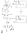
- FIG. 2 one embodiment is shown.
- the subsequent procesing of the method is depicted in Figure 2 from left to right.
- a nucleic acid mixture is used, containing different double-stranded nucleic acids 5 and also a double-stranded nucleic acid containing the target nucleic acid sequence 1A and its complementary sequence 1 B, both shown in boldfaced representation.
- the double-stranded nucleic acids in the nucleic acid mixture are converted to single-stranded nucleic acids, for example by heating the nucleic acid mixture to a high temperature for a short time (for example 95°C for five minutes).
- the nucleic acid mixture contains mainly single-stranded nucleic acids and also the single-stranded target nucleic acid sequence 1A.
- a single-stranded nucleic acid probe 15 with a first label 20 is added in A) and the probe 15 can hybridize with the single-stranded target nucleic acid 1A to form a hybrid between the probe 15 and the target nucleic acid sequence 1A.
- the major advantage of this embodiment in comparison to conventional methods is that A2) and A) are carried out in liquid phase and are therefore much easier to perform than the standard blotting transfer techniques shown in Figure 1 .
- the nucleic acid mixture can be separated, for example in a gel 50 in B) by gel electrophoresis.
- the hybrid 1A, 15 between the target nucleic acid sequence 1A and the probe 15 can be detected, for example by using a spectrometer with the wavelength ⁇ 1 if a fluorescent marker is used as a label.
- the nucleic acid band containing the hybrid 1A, 15 lights up and can therefore be detected.
- the denaturing of the nucleic acid mixture in A2) is compulsory, when a double stranded DNA target nucleic acid sequence has to be detected within a mixture of other double-stranded DNA molecules. It can readily be seen in Fig. 2 , that the separation of the nucleic acid mixture in B) and the detection of the target nucleic acid sequence in C) are both carried out in the gel 50, so that no transfer onto a membrane is necessary.
- the nucleic acid mixture contains single-stranded nucleic acids 5 having additional binding sites 10 and also a single-stranded target nucleic acid sequence 1A, shown again in boldfaced representation. In other cases it might be preferred to denature even single-stranded nucleic acid mixtures in order to ensure a good base pairing between the probe and the target nucleic acid sequence.
- the single-stranded target nucleic acid sequence 1A is hybridized with the nucleic acid probe 15, which is labeled with a first label 20.
- oligonucleotides 25 with a random primary sequence, having a second label 30 are incubated with the single-stranded nucleic acids 5 in the mixture in order to bind to the additional binding sites 10, thereby converting nearly all single-stranded nucleic acids 5 into double-stranded nucleic acids.
- multiple oligonucleotides 25 can bind to one single-stranded nucleic acid 5 converting this nucleic acid into a double-stranded form. Additionally the oligonucleotides 25 might also bind to single-stranded regions of the hybrid between the probe 15 and the single-stranded nucleic acid target sequence 1A.
- A1) converts almost all of the single-stranded nucleic acids into double-stranded nucleic acids, allowing a precise mass-dependent separation of the double-stranded nucleic acids in subsequent B).
- the separation again might be carried out in a gel 50. Due to the conversion of A1) no shift of the signal band comprising the hybrid 1A, 25, 15 occurs during the separation of the nucleic acids in B), allowing the determination of the size of the target nucleic acid sequence.
- the probe 15 and the oligonucleotides 25 might be simultaneously detected with the other nucleic acids 5 by using a spectrometer with different wavelengths ⁇ 1 and ⁇ 2 in C).
- This special embodiment allows the determination of the amount and the size of the target nucleic acid sequence as well as the determination of the amount of the other nucleic acids 5 in the nucleic acid mixture.
- a detection was carried out, detecting the gene for the human glycerin aldehyde phosphate dehydrogenase (GADPH) in human female blood total DNA.
- GADPH human glycerin aldehyde phosphate dehydrogenase
- the human female blood total DNA was digested using the restriction enzyme Dra I. Afterwards the DNA was concentrated by using a sodium acetate precipitation. 15 ⁇ l of sodium acetate 5 M and 175 ⁇ l ethanol were added and mixed. Afterwards the DNA was precipitated by incubating the mixture for one hour on ice. The DNA was pelleted by centrifuging 50 minutes at full speed and the DNA pellet was washed in 70% ethanol, dried and resuspended in 5 ⁇ l TE buffer (10 mM TRIS, 0.01 mM EDTA). Subsequently the digested DNA was denatured in A2) in order to convert the DNA molecules into single stranded nucleic acids by heating at 99°C for 5 minutes.
- the probe for the GADPH gene which was labeled with the fluorescent dye BODIPY® 650/665 (available from molecular probes) and decamers with random primary sequence (available from Ambion) were both denatured in separate tubes by heating at 99°C for 5 minutes and chilling on ice. Subsequently A) was carried out by mixing the human female blood total DNA and the labeled probe, incubating for 5 minutes at 99°C, cooling down to 65°C and incubating for five minutes at 65°C. Afterwards the mixture was chilled on ice. Subsequently the decamers with the random primary sequence were added and incubated with the DNA and the labeled probe for five minutes and put on ice again in A1).
- BODIPY® 650/665 available from molecular probes
- decamers with random primary sequence available from Ambion
- the labeled GADPH probe consisted of a mixture of different probes having a medium size of 200 to 500 nucleotides, mostly being complementary to the GADPH gene and spanning the whole gene.
- the probes were synthesized by the random priming reaction of Feinberg and Vogelstein by using hexanucleotides as random primers.
- the separation of the nucleic acids in B) was carried out by transferring the nucleic acid mixture onto a DNA 12000 microfluidic chip (Agilent Technologies, Waldbronn, Germany) with 20 ⁇ M SYTO 16® (Molecular probes, Eugene, OR, USA) as a nucleic acid specific dye in the gel matrix of the chip as a second label.
- SYTO 16® Molecular probes, Eugene, OR, USA
Landscapes
- Chemical & Material Sciences (AREA)
- Organic Chemistry (AREA)
- Life Sciences & Earth Sciences (AREA)
- Zoology (AREA)
- Wood Science & Technology (AREA)
- Proteomics, Peptides & Aminoacids (AREA)
- Health & Medical Sciences (AREA)
- Engineering & Computer Science (AREA)
- Microbiology (AREA)
- Immunology (AREA)
- Physics & Mathematics (AREA)
- Molecular Biology (AREA)
- Biotechnology (AREA)
- Biophysics (AREA)
- Analytical Chemistry (AREA)
- Biochemistry (AREA)
- Bioinformatics & Cheminformatics (AREA)
- General Engineering & Computer Science (AREA)
- General Health & Medical Sciences (AREA)
- Genetics & Genomics (AREA)
- Measuring Or Testing Involving Enzymes Or Micro-Organisms (AREA)
Abstract
Description
- The present invention relates to detection of a nucleic acid sequence in a mixture of different nucleic acids and a kit therefor.
-
WO 0157258 A -
EP 1186673 A , by the same applicant, discloses calibrating different types of signals scanned from a molecular array, or calibrating signals scanned from different molecular arrays, by employing calibrating probes that generate signals proportional to the total concentrations of labeled target molecules to which the molecular array probes are directed over an entire range of sample solutions, and molecular arrays incorporating sets of calibrating probes. - Widespread conventional techniques for the detection of nucleic acid sequences are the southern blotting technique for DNA and the northern blotting technique for RNA. At the beginning of these procedures the nucleic acid mixtures are separated in nucleic acids of different mass using gel electrophoresis, for example in an agarose or polyacrylamide gel. Following the gel electrophoresis, the different nucleic acids are preferably converted to single stranded nucleic acids. The single stranded nucleic acids are then transferred onto a microcellulose or a nylon filter and are crosslinked with the membrane using heat or UV radiation. The membrane is then blocked with a blocking reagent to saturize all unspecific binding sites of the membrane. Subsequently the nucleic acids fixed on the membrane are hybridized with a labeled nucleic acid probe, which includes a primary sequence complementary to the primary sequence of a target nucleic acid sequence. The label of the nucleic acid probe often contains 32P-labeled phosphates, which can be detected due to their radioactivity (see for example
Figure 1 ). The northern or southern blotting techniques therefore involve lots of different steps, e.g. gel electrophoresis, blotting onto a membrane and detection by hybridization, which are very time consuming and complicated to carry out. For the southern and northern blotting techniques different media (gels for gel electrophoresis and membranes for the blotting) are used, so that lots of different and at least partially expensive materials are used. - Therefore there is a need for a new method for detection of a target nucleic acid sequence in a mixture of different nucleic acids, which allows a fast and reliable detection of a target nucleic acid without the necessity to use blotting techniques. The present invention meets these needs by providing a method for detection of a target nucleic acid sequence according to the base claim 1. Favorable embodiments of the method of the invention and a kit for the detection of the target nucleic acid sequence are subjects of further claims.
- Embodiments of the invention provide a fast and easy-to-handle procedure for detection of a target nucleic acid sequence, wherein the hybridizing of the target nucleic acid sequence with the probe takes place in liquid phase. The procedure in A) therefore avoids the complicated, time-consuming and also material-consuming stepproceedure of transfer of the nucleic acids onto a membrane. Furthermore an operator carrying out the embodiments of the invention usually needs less skill than an operator carrying out conventional Northern or Southern blot techniques. After the hybridizing of the target nucleic acid sequence with the labeled probe in stepA), forming a at least partially double stranded hybrid strand between the target nucleic acid and the probe, the different nucleic acids and the target nucleic acid sequence are separated in subsequent B), allowing a detection of the target nucleic acid sequence in following C). Therefore the method for detection requires the hybridizing of the probe with the target nucleic acid sequence prior to separating the nucleic acids. This sequence of the method is reversed in comparison to the conventional northern and southern blotting techniques, where the nucleic acids are separated first and then hybridized with a labeled probe.
- In A1), the additional binding sites are hybridized with single-stranded nucleic acids having random primary sequences in liquid phase. The additional binding sites of the nucleic acids, which are still present after A) are often comprised of unpaired bases in single stranded areas of the nucleic acids.
- The single-stranded nucleic acids can basepair with single-stranded parts of the different nucleic acids in the nucleic acid mixture and if present also with single-stranded parts of the target nucleic acid sequence, forming nucleic acid double strands. Therefore after A1) the nucleic acids in the nucleic acid mixture are mainly double-stranded, simplifying the separation of the different nucleic acids in the subsequent B). Due to A1) no retardation of the double stranded hybrid between the probe and the target nucleic acid sequence in comparison to the other nucleic acids occurs during the separation procedure in B). A retardation of the double stranded hybrid during gel electrophoresis normally takes place, when single stranded nucleic acids are still present in the nucleic acid mixture and are also subjected to gel electrophoresis, so that the important information about the size of the target sequence is lost. In practice the information about the size of the target sequence is often used as a control for the correct hybridization between the target sequence and the probe.
- Advantageously short nucleic acids with a random primary sequence having a length of 6 to 14 nucleotides are provided in A1) for conversion of the single-stranded parts of the nucleic acid mixture into double-stranded parts. These short oligonucleotides are easy to synthesize and can easily be handled during A1). Due to their small size, these oligonucleotides reliably interact with single-stranded areas in the nucleic acid mixture.
- In another variant , the hybridizing in A1) is carried out at roughly room temperature and the hybridizing of the probe with the target sequence in A) is carried out at a temperature between 30°C to 72°C, preferably 56°C to 72°C. A temperature between 30°C to 48°C can also be useful. A further preferred condition for hybridizing in A) is a pH range between 6 to 8.5, preferably slightly alkaline, for example pH 7.5 (e.g. TRIS EDTA buffer pH 7.5).
- Due to the low temperature during hybridizing in A1) mismatches in the base pairing do not impair interaction between single-stranded areas of the different nucleic acids in the mixtures and the oligonucleotides with the random primary sequence. In contrast to the low temperature in A1) a higher temperature in A) is used in order to provide a more stringent condition for hybridizing, therefore enabling a good selectivity during the detection of the target nucleic acid sequence by the probe, reducing false signals.
- A nucleic acid having a length of at least 2 times the length of the oligonucleotides with the random primary sequence can be used as a probe. When the probe is large compared to the oligonucleotides with the random sequence, it is possible to carry out A1) and A) simultaneously. Due to cooperative effects, the large probe is still able to interact with the correct target sequence and can also replace short oligonucleotides with the random primary sequence, which already have bound to the target nucleic acid sequence. This variant therefore provides the hybridization of the target nucleic acid sequence with the probe and the conversion of the single-stranded areas of the nucleic acid into double stranded nucleic acids in one go. This procedure therefore reduces the number of method sequences, allowing a faster and easier detection of the target nucleic acid sequence.
- Advantageously, in A1) nucleic acids labeled with a second label are used for hybridizing, the second label being different from the first label.
- Due to the different labels for the probe and for the nucleic acids having random primary sequences, the amount and the size of the target nucleic acid sequence and the amount of the total nucleic acids in the mixture can be determined using different detection methods.
- It is also possible that the nucleic acids with the random primary sequence used for hybridizing in A1) are labeled with a second label after A1), the second again being different from the first label. Such a subsequent labeling of the nucleic acids can, for example, be carried out using dyes like ethidiumbromide, acridine orange, proflavin or Sybr Green®. These intercalating agents are normally used to stain double- or single-stranded nucleic acids.
- It is also possible to label the oligonucleotides with the random primary sequence used in A1) by a random-oligonucleotide labeling method, developed by Feinberg and Vogelstein (Feinberg, A.P., Vogelstein, B., Anal Biochem 137, 266 - 267, 1984). Using this method random decanucleotide primers can be used for synthesis of complementary strands of template nucleic acids. The complementary strands are synthesized from the 3'-end of the random decanucleotide primers using, for example Klenow fragment of DNA polymerase I. In the presence of nucleotides, which are marked with a label, for example biotine or 32P, labeled oligonucleotides for A1) are synthesized.
- Favorably, in A2) prior to A) the mixture of different nucleic acids is denatured.
- Denaturing advantageously converts the nucleic acids, which might be double-stranded into single-stranded nucleic acids, so that the hybridization in subsequent A) can occur without major difficulties. Denaturing might be carried out, for example, by heating the nucleic acid mixture to high temperatures, for example 90°C to 99°C, preferably 95°C to 99°C for a certain time, e.g. five minutes and immediately reducing the temperature afterwards e.g. by chilling on ice.
- Preferably in A) a nucleic acid is used as a probe, having a stretch of 18 to 25 nucleotides being able to hybridize with the target nucleic acid sequence, this stretch having at least 80% sequence homology to the complementary sequence of the target nucleic acid sequence. Alternatively the nucleic acid probe can have at least 12 continuos nucleotides, complementary to the target nucleic acid sequence in order to ensure a good and reliable hybridization between the target nucleic acid sequence and the probe. Such nucleic acid probes can selectively detect the target nucleic acid even within a mixture of other different nucleic acids.
- In another embodiment the nucleic acids are separated according to their mass in B) by using a gel electrophoresis. The gel electrophoresis can, for example, be carried out in a polyacrylamide gel or an agarose gel. This separation technique is especially suited to separate the nucleic acids in a reliable manner and in a short time.
- Preferably in B) a microfluidic chip having capillaries suitable for nucleic acids electrophoresis is used for separation. The microfluidic chip can comprise, for example, a glass or silicon chip in which capillaries are etched. The capillaries can be filled with a electrophoresis medium, for example polyacrylamide or agarose and the nucleic acid mixture can be driven through the capillaries using electrophoretic and electro-osmotic forces. Using these microfluidic chips, small volumes can be analyzed very quickly. Therefore microfluidic chips are well-suited to save working time and also reduce the expenses for material.
- Preferably the first and the second label are being selected from the following group:
- radioactive labels, fluorescent markers, chemoluminescence, bioluminescence, magnetic labels and antigen labels.
- These kind of labels are especially suited to label nucleic acids and can easily be monitored using standard detection methods like autoradiography, fluorescence assays or antibodies.
- Preferably fluorescent markers are used as the first and if present second label, wherein the fluorescent markers of the first and second label emit radiation of different wavelengths. Using this variant the detection of both the target nucleic acid sequence and the other different nucleic acids in the mixture can easily by carried out.
- When fluorescent markers are used as the first and second label, both fluorescent markers emitting radiation of different wavelengths, the amount and the size of the target nucleic acid and the amount of the other different nucleic acids in the mixture can be determined via the first and second label using a spectrometer for the detection of both labels in C). This embodiment allows a simultaneous detection of both the target nucleic acid sequence and the other nucleic acids in the mixture by simply using a spectrometer e.g. a bioanalyzer instrument.
- A person of ordinary skill in the art can synthesize different kinds of nucleic acid probes, depending on the target nucleic acid sequence, which has to be detected. If for example the HI-virus has to be detected in a human tissue sample, a nucleic acid probe can be designed by a person of ordinary skill in the art, which shows a high degree of complementarity in a well-conserved stretch of the HIV genome. This nucleic acid probe might still allow some mismatches in the base pairing, for example in regions of high variability within different HIV subtypes in order to allow a detection of HIV independent from its subtypes. Furthermore, additional nucleic acid probes for HIV detection can be designed by a person of ordinary skill in the art, having a high degree of complementarity even in regions of high variability in the HIV genome, therefore allowing to distinguish different subtypes of HIV.
- In the following embodiments of the invention will be explained in more detail. All figures are just simplified schematic representations presented for illustration purposes only.
-
-
Figure 1 shows the course of separation and detection of a target nucleic acid sequence during a standard northern or southern blotting technique. - The
Figures 2 and3 depict a schematic course of subsequent method proceedure during different embodiments of the invention. - Referring to
Figure 1 the course of subsequent method steps of a conventional southern or northern blot is shown from left to right. At the beginning of a standard detection method for nucleic acids, the nucleic acids are separated in theline 100 of thegel 50 by gel electrophoresis (shown on the left side of the page). ADNA ladder 60 might simultaneously be applied on the gel inline 110 in order to simplify the determination of the size of the nucleic acid in the mixture. Normally only highly abundant nucleic acids are visible after staining, e. g. with ethidiumbromide, like the twobands 70, representing ribosomal RNA. After the separation of the nucleic acids the nucleic acids are transferred onto a nitrocellulose or anylon filter 80 and are cross-linked with the membrane, as shown in the middle ofFig. 1 . The transfer normally also involves the treatment of the gel with NaOH in order to convert the double stranded nucleic acids into single-stranded nucleic acids, which able to hybridize with a nucleic acid probe. The transfer of the nucleic acids from the gel onto the membrane is normally very time-consuming and also requires lots of material, for example buffer solutions. After transfer, the membrane with the single-stranded nucleic acids is frequently blocked with a blocking reagent in order to saturize all unspecific binding sites on the membrane. This blocking normally takes place by incubating the membrane with commercially available blocking reagents, e. g. Denharts solution, non-fat milk or salmon sperm DNA. Afterwards the membrane is brought into contact with a solution containing anucleic acid probe 15 with alabel 20, as shown on the left side. Normally a 32P-label is used for northern or southern blotting techniques. The labeled nucleic acid probe can hybridize with the target nucleic acid sequence, the membrane is washed and the signal is detected, e. g. by autoradiography. - Turning now to
Figure 2 , one embodiment is shown. The subsequent procesing of the method is depicted inFigure 2 from left to right. At the beginning a nucleic acid mixture is used, containing different double-strandednucleic acids 5 and also a double-stranded nucleic acid containing the targetnucleic acid sequence 1A and itscomplementary sequence 1 B, both shown in boldfaced representation. In A2) the double-stranded nucleic acids in the nucleic acid mixture are converted to single-stranded nucleic acids, for example by heating the nucleic acid mixture to a high temperature for a short time (for example 95°C for five minutes). Afterwards the nucleic acid mixture contains mainly single-stranded nucleic acids and also the single-stranded targetnucleic acid sequence 1A. Subsequently a single-strandednucleic acid probe 15 with afirst label 20 is added in A) and theprobe 15 can hybridize with the single-stranded targetnucleic acid 1A to form a hybrid between theprobe 15 and the targetnucleic acid sequence 1A. The major advantage of this embodiment in comparison to conventional methods is that A2) and A) are carried out in liquid phase and are therefore much easier to perform than the standard blotting transfer techniques shown inFigure 1 . Subsequently the nucleic acid mixture can be separated, for example in agel 50 in B) by gel electrophoresis. During C) thehybrid nucleic acid sequence 1A and theprobe 15 can be detected, for example by using a spectrometer with the wavelength λ1 if a fluorescent marker is used as a label. In this case the nucleic acid band containing the hybrid 1A, 15 lights up and can therefore be detected. - The denaturing of the nucleic acid mixture in A2) is compulsory, when a double stranded DNA target nucleic acid sequence has to be detected within a mixture of other double-stranded DNA molecules. It can readily be seen in
Fig. 2 , that the separation of the nucleic acid mixture in B) and the detection of the target nucleic acid sequence in C) are both carried out in thegel 50, so that no transfer onto a membrane is necessary. - Referring now to
Figure 3 another embodiment is shown from left to right. In this case the nucleic acid mixture contains single-strandednucleic acids 5 having additionalbinding sites 10 and also a single-stranded targetnucleic acid sequence 1A, shown again in boldfaced representation. In other cases it might be preferred to denature even single-stranded nucleic acid mixtures in order to ensure a good base pairing between the probe and the target nucleic acid sequence. In A) of this embodiment the single-stranded targetnucleic acid sequence 1A is hybridized with thenucleic acid probe 15, which is labeled with afirst label 20. In subsequent A1) oligonucleotides 25 with a random primary sequence, having asecond label 30 are incubated with the single-strandednucleic acids 5 in the mixture in order to bind to the additionalbinding sites 10, thereby converting nearly all single-strandednucleic acids 5 into double-stranded nucleic acids. During A1)multiple oligonucleotides 25 can bind to one single-strandednucleic acid 5 converting this nucleic acid into a double-stranded form. Additionally theoligonucleotides 25 might also bind to single-stranded regions of the hybrid between theprobe 15 and the single-stranded nucleicacid target sequence 1A. A1) converts almost all of the single-stranded nucleic acids into double-stranded nucleic acids, allowing a precise mass-dependent separation of the double-stranded nucleic acids in subsequent B). The separation again might be carried out in agel 50. Due to the conversion of A1) no shift of the signal band comprising thehybrid first label 20 and thesecond label 30 the hybrid between the targetnucleic acid sequence 1A, theprobe 15 and theoligonucleotides 25 might be simultaneously detected with the othernucleic acids 5 by using a spectrometer with different wavelengths λ1 and λ2 in C). This special embodiment allows the determination of the amount and the size of the target nucleic acid sequence as well as the determination of the amount of the othernucleic acids 5 in the nucleic acid mixture. - In order to test the feasibility of embodiments of the invention a detection was carried out, detecting the gene for the human glycerin aldehyde phosphate dehydrogenase (GADPH) in human female blood total DNA.
- At the beginning the human female blood total DNA was digested using the restriction enzyme Dra I. Afterwards the DNA was concentrated by using a sodium acetate precipitation. 15 µl of sodium acetate 5 M and 175 µl ethanol were added and mixed. Afterwards the DNA was precipitated by incubating the mixture for one hour on ice. The DNA was pelleted by centrifuging 50 minutes at full speed and the DNA pellet was washed in 70% ethanol, dried and resuspended in 5 µl TE buffer (10 mM TRIS, 0.01 mM EDTA). Subsequently the digested DNA was denatured in A2) in order to convert the DNA molecules into single stranded nucleic acids by heating at 99°C for 5 minutes. Then the mixture was chilled on ice. The probe for the GADPH gene, which was labeled with the fluorescent dye BODIPY® 650/665 (available from molecular probes) and decamers with random primary sequence (available from Ambion) were both denatured in separate tubes by heating at 99°C for 5 minutes and chilling on ice. Subsequently A) was carried out by mixing the human female blood total DNA and the labeled probe, incubating for 5 minutes at 99°C, cooling down to 65°C and incubating for five minutes at 65°C. Afterwards the mixture was chilled on ice. Subsequently the decamers with the random primary sequence were added and incubated with the DNA and the labeled probe for five minutes and put on ice again in A1). The labeled GADPH probe consisted of a mixture of different probes having a medium size of 200 to 500 nucleotides, mostly being complementary to the GADPH gene and spanning the whole gene. The probes were synthesized by the random priming reaction of Feinberg and Vogelstein by using hexanucleotides as random primers. After A1) the separation of the nucleic acids in B) was carried out by transferring the nucleic acid mixture onto a DNA 12000 microfluidic chip (Agilent Technologies, Waldbronn, Germany) with 20 µM SYTO 16® (Molecular probes, Eugene, OR, USA) as a nucleic acid specific dye in the gel matrix of the chip as a second label. Using a spectrometer signals for the hybrid between the probe and the target nucleic acid sequence as well as signals for the other nucleic acids could be determined.
- The scope of the invention is not limited to the embodiments shown in the figures. Indeed, variations especially concerning the combination of the different optional method features and variations concerning the design of the nucleic acid probe are possible.
Claims (11)
- A method for detection of a target nucleic acid sequence (1A) in a mixture of different nucleic acids (5) having additional binding sites (10), the method comprising:A) hybridizing the target nucleic acid sequence with a probe (15) in liquid phase, the probe having a first label (20),A1) hybridizing the additional binding sites (10) with single stranded nucleic acids (25) having random primary sequences in liquid phase,B) separating the different nucleic acids (1A, 5),C) detecting the target nucleic acid (1A) by using the labeled probe (15).
- Method according to claim 1,- wherein short nucleic acids having a length of 6 to 12 nucleotides are provided in A1) for hybridizing.
- Method according to claim 1 or any one of the above claims,- wherein hybridizing in A1) is carried out at roughly room temperature, and- hybridizing in A) is carried out at a temperature between 56°C to 72°C.
- Method according to claim 1 or any one of the above claims,- wherein a nucleic acid with a length of at least 10-times the length of the single stranded nucleic acids (25) with random primary sequence is used as a probe (15),- wherein A1) and A) are carried out simultaneously.
- Method according to claim 2 or any one of the above claims,- wherein in A1) nucleic acids (25) labeled with a second label (30) are used for hybridizing,- the second label (30) being different from the first label (20).
- Method according to claim 2 or any one of the above claims,- wherein the nucleic acids (25) used for hybridizing in A1) are subsequently labeled with a second label (30) after A1),- the second label being different from the first label.
- Method according to claim 1 or any of the above claims, comprising at least one of:- prior to A) the mixture of different nucleic acids is denatured in a A2);- in A) a nucleic acid is used as a probe (15), having a stretch of 18 to 25 nucleotides being able to hybridize with the target nucleic acid sequence (1A), this stretch having at least 80% sequence homology to the complementary sequence of the target nucleic acid sequence.
- Method according to claim 1 or any one of the above claims, comprising at least one of:- in B) the nucleic acids are separated according to their mass by using a gel electrophorese;- in B) a microfluidic chip having capillaries suitable for nucleic acid electrophorese is used for separation.
- Method according to claim 1 or any one of the above claims,- wherein a first and if present a second label is used, each being selected from the following group:- radioactive labels, fluorescent markers, chemoluminescence, bioluminescence, magnetic labels and antigen labels.
- Method according to the preceeding claim,- wherein fluorescent markers are used as the first and if present second label,- the fluorescent markers of the first and second label emitting radiation of different wavelengths.
- Method according to the preceeding claim,- wherein in C) the amount and the size of the hybrid strand of the target nucleic acid (1A) and the probe (15) is determined via the first label (20) and in case the second label (30) is present, the amount of the other different nucleic acids (5) in the mixture is determined via the second label (30),- using a spectrometer for the detection of both labels.
Applications Claiming Priority (1)
| Application Number | Priority Date | Filing Date | Title |
|---|---|---|---|
| PCT/EP2003/050844 WO2005049859A1 (en) | 2003-11-17 | 2003-11-17 | Nucleic acid detection |
Publications (2)
| Publication Number | Publication Date |
|---|---|
| EP1687442A1 EP1687442A1 (en) | 2006-08-09 |
| EP1687442B1 true EP1687442B1 (en) | 2008-04-09 |
Family
ID=34610026
Family Applications (1)
| Application Number | Title | Priority Date | Filing Date |
|---|---|---|---|
| EP03819025A Expired - Lifetime EP1687442B1 (en) | 2003-11-17 | 2003-11-17 | Nucleic acid detection |
Country Status (5)
| Country | Link |
|---|---|
| US (1) | US20080311668A1 (en) |
| EP (1) | EP1687442B1 (en) |
| AU (1) | AU2003300569A1 (en) |
| DE (1) | DE60320319T2 (en) |
| WO (1) | WO2005049859A1 (en) |
Family Cites Families (5)
| Publication number | Priority date | Publication date | Assignee | Title |
|---|---|---|---|---|
| US5104792A (en) * | 1989-12-21 | 1992-04-14 | The United States Of America As Represented By The Department Of Health And Human Services | Method for amplifying unknown nucleic acid sequences |
| US6720166B2 (en) * | 1994-02-14 | 2004-04-13 | Abbott Laboratories | Non-a, non-b, non-c, non-c, non-d, non-e hepatitis reagents and methods for their use |
| US6261781B1 (en) * | 1997-08-05 | 2001-07-17 | Wisconsin Alumni Research Foundation | Direct detection and mutation analysis of low copy number nucleic acids |
| US6013442A (en) * | 1997-08-05 | 2000-01-11 | Wisconsin Alumni Res Found | Direct quantitation of low copy number RNA |
| EP1186673A3 (en) * | 2000-09-11 | 2003-03-26 | Agilent Technologies, Inc. (a Delaware corporation) | Calibration of molecular array data |
-
2003
- 2003-11-17 AU AU2003300569A patent/AU2003300569A1/en not_active Abandoned
- 2003-11-17 EP EP03819025A patent/EP1687442B1/en not_active Expired - Lifetime
- 2003-11-17 US US10/573,438 patent/US20080311668A1/en not_active Abandoned
- 2003-11-17 DE DE60320319T patent/DE60320319T2/en not_active Expired - Lifetime
- 2003-11-17 WO PCT/EP2003/050844 patent/WO2005049859A1/en not_active Ceased
Also Published As
| Publication number | Publication date |
|---|---|
| DE60320319D1 (en) | 2008-05-21 |
| DE60320319T2 (en) | 2008-07-10 |
| WO2005049859A1 (en) | 2005-06-02 |
| AU2003300569A1 (en) | 2005-06-08 |
| EP1687442A1 (en) | 2006-08-09 |
| US20080311668A1 (en) | 2008-12-18 |
Similar Documents
| Publication | Publication Date | Title |
|---|---|---|
| JP3066984B2 (en) | General-purpose spacer / energy transition dye | |
| US6072043A (en) | Optimally fluorescent oligonucleotides | |
| EP0743987B1 (en) | Probes labelled with energy transfer coupled dyes | |
| EP2837695B1 (en) | Nucleic acid quantification method, detection probe, detection probe set, and nucleic acid detection method | |
| EP1046717A3 (en) | Method and probes for determining a concentration of target nucleic acid molecules and method for analyzing data obtained by the method | |
| EP2443254A2 (en) | Improved methods for forensic dna quantitation | |
| WO2003033741A1 (en) | Universal e-tag primer and probe compositions and methods | |
| AU2012316129B2 (en) | Methods of co-detecting mRNA and small non-coding RNA | |
| US6063572A (en) | Method of assay of nucleic acid sequences | |
| CA2448211A1 (en) | Nucleic acid detection method | |
| CA2655518C (en) | Method for analyzing amplified nucleic acids | |
| US20080194804A1 (en) | Method of nucleic acid hybridization | |
| Lehmusvuori et al. | Homogeneous duplex polymerase chain reaction assay using switchable lanthanide fluorescence probes | |
| AU2002226448B2 (en) | A method and test kit for quantitative determination of variations in polynucleotide amounts in cell or tissue samples | |
| US20080311668A1 (en) | Nucleic Acid Detection | |
| CN119979711A (en) | A single molecule detection kit for multiple non-coding RNAs in human breast tissue using a programmable automatic cascade mechanism and its use | |
| JP2003527866A (en) | Methods for detecting polynucleotide kinase and their use as labels | |
| US20040121359A1 (en) | Fluorescence polarisation | |
| AU2002226448A1 (en) | A method and test kit for quantitative determination of variations in polynucleotide amounts in cell or tissue samples | |
| JP7756503B2 (en) | Labeling of nucleic acid molecules with interstrand cross-linked double-stranded DNA | |
| CA2381037A1 (en) | Amplification method for detection of target nucleic acids involving fluorescence energy transfer | |
| US20050064481A1 (en) | Method for reducing background contamination | |
| Bortolin et al. | Time-resolved immunofluorometric determination of specific mRNA sequences amplified by the polymerase chain reaction | |
| WO2006073449A2 (en) | Multiplex systems, methods, and kits for detecting and identifying nucleic acids | |
| US20020155456A1 (en) | Method of amplification for increasing the sensitivity of detecting nucleic acid-probe target hybrids |
Legal Events
| Date | Code | Title | Description |
|---|---|---|---|
| PUAI | Public reference made under article 153(3) epc to a published international application that has entered the european phase |
Free format text: ORIGINAL CODE: 0009012 |
|
| 17P | Request for examination filed |
Effective date: 20060619 |
|
| AK | Designated contracting states |
Kind code of ref document: A1 Designated state(s): CH DE FR GB LI |
|
| RAP1 | Party data changed (applicant data changed or rights of an application transferred) |
Owner name: AGILENT TECHNOLOGIES, INC. |
|
| DAX | Request for extension of the european patent (deleted) | ||
| RBV | Designated contracting states (corrected) |
Designated state(s): CH DE FR GB LI |
|
| 17Q | First examination report despatched |
Effective date: 20070427 |
|
| GRAP | Despatch of communication of intention to grant a patent |
Free format text: ORIGINAL CODE: EPIDOSNIGR1 |
|
| GRAS | Grant fee paid |
Free format text: ORIGINAL CODE: EPIDOSNIGR3 |
|
| GRAA | (expected) grant |
Free format text: ORIGINAL CODE: 0009210 |
|
| AK | Designated contracting states |
Kind code of ref document: B1 Designated state(s): CH DE FR GB LI |
|
| REG | Reference to a national code |
Ref country code: GB Ref legal event code: FG4D |
|
| REG | Reference to a national code |
Ref country code: CH Ref legal event code: EP |
|
| REF | Corresponds to: |
Ref document number: 60320319 Country of ref document: DE Date of ref document: 20080521 Kind code of ref document: P |
|
| RIN2 | Information on inventor provided after grant (corrected) |
Inventor name: LUEDKE, GERD |
|
| EN | Fr: translation not filed | ||
| PGFP | Annual fee paid to national office [announced via postgrant information from national office to epo] |
Ref country code: CH Payment date: 20081130 Year of fee payment: 6 |
|
| PLBE | No opposition filed within time limit |
Free format text: ORIGINAL CODE: 0009261 |
|
| STAA | Information on the status of an ep patent application or granted ep patent |
Free format text: STATUS: NO OPPOSITION FILED WITHIN TIME LIMIT |
|
| 26N | No opposition filed |
Effective date: 20090112 |
|
| PG25 | Lapsed in a contracting state [announced via postgrant information from national office to epo] |
Ref country code: FR Free format text: LAPSE BECAUSE OF FAILURE TO SUBMIT A TRANSLATION OF THE DESCRIPTION OR TO PAY THE FEE WITHIN THE PRESCRIBED TIME-LIMIT Effective date: 20090130 |
|
| REG | Reference to a national code |
Ref country code: CH Ref legal event code: PL |
|
| PG25 | Lapsed in a contracting state [announced via postgrant information from national office to epo] |
Ref country code: CH Free format text: LAPSE BECAUSE OF NON-PAYMENT OF DUE FEES Effective date: 20091130 Ref country code: LI Free format text: LAPSE BECAUSE OF NON-PAYMENT OF DUE FEES Effective date: 20091130 |
|
| PGFP | Annual fee paid to national office [announced via postgrant information from national office to epo] |
Ref country code: DE Payment date: 20181106 Year of fee payment: 16 |
|
| PGFP | Annual fee paid to national office [announced via postgrant information from national office to epo] |
Ref country code: GB Payment date: 20181114 Year of fee payment: 16 |
|
| REG | Reference to a national code |
Ref country code: DE Ref legal event code: R119 Ref document number: 60320319 Country of ref document: DE |
|
| GBPC | Gb: european patent ceased through non-payment of renewal fee |
Effective date: 20191117 |
|
| PG25 | Lapsed in a contracting state [announced via postgrant information from national office to epo] |
Ref country code: GB Free format text: LAPSE BECAUSE OF NON-PAYMENT OF DUE FEES Effective date: 20191117 Ref country code: DE Free format text: LAPSE BECAUSE OF NON-PAYMENT OF DUE FEES Effective date: 20200603 |