EP1674077A1 - Mucoadhesive pharmaceutical compositions comprising chemoattractants - Google Patents
Mucoadhesive pharmaceutical compositions comprising chemoattractants Download PDFInfo
- Publication number
- EP1674077A1 EP1674077A1 EP04106995A EP04106995A EP1674077A1 EP 1674077 A1 EP1674077 A1 EP 1674077A1 EP 04106995 A EP04106995 A EP 04106995A EP 04106995 A EP04106995 A EP 04106995A EP 1674077 A1 EP1674077 A1 EP 1674077A1
- Authority
- EP
- European Patent Office
- Prior art keywords
- composition
- cells
- chemoattractant
- hpv
- treatment
- Prior art date
- Legal status (The legal status is an assumption and is not a legal conclusion. Google has not performed a legal analysis and makes no representation as to the accuracy of the status listed.)
- Withdrawn
Links
- 239000002975 chemoattractant Substances 0.000 title claims abstract description 27
- 230000003232 mucoadhesive effect Effects 0.000 title claims abstract description 5
- 239000008194 pharmaceutical composition Substances 0.000 title claims abstract description 4
- 239000000203 mixture Substances 0.000 claims abstract description 38
- 241000701806 Human papillomavirus Species 0.000 claims abstract description 20
- 208000025157 Oral disease Diseases 0.000 claims abstract description 11
- 208000030194 mouth disease Diseases 0.000 claims abstract description 11
- 229920000642 polymer Polymers 0.000 claims abstract description 9
- 102100039620 Granulocyte-macrophage colony-stimulating factor Human genes 0.000 claims description 22
- 102100021943 C-C motif chemokine 2 Human genes 0.000 claims description 20
- 108010017213 Granulocyte-Macrophage Colony-Stimulating Factor Proteins 0.000 claims description 20
- 101710155857 C-C motif chemokine 2 Proteins 0.000 claims description 19
- 101500025408 Homo sapiens Neutrophil defensin 2 Proteins 0.000 claims description 13
- 101500028710 Homo sapiens Neutrophil defensin 2 Proteins 0.000 claims description 13
- 238000000034 method Methods 0.000 claims description 8
- 210000004877 mucosa Anatomy 0.000 claims description 4
- 229920002994 synthetic fiber Polymers 0.000 claims description 4
- 210000002540 macrophage Anatomy 0.000 claims description 3
- 239000003814 drug Substances 0.000 claims description 2
- 210000003714 granulocyte Anatomy 0.000 claims description 2
- 238000004519 manufacturing process Methods 0.000 claims description 2
- 108010032806 molgramostim Proteins 0.000 claims description 2
- 229960003063 molgramostim Drugs 0.000 claims description 2
- 229920001206 natural gum Polymers 0.000 claims description 2
- 210000004443 dendritic cell Anatomy 0.000 description 64
- 210000002510 keratinocyte Anatomy 0.000 description 27
- 210000004027 cell Anatomy 0.000 description 26
- 239000000499 gel Substances 0.000 description 22
- 102000019034 Chemokines Human genes 0.000 description 21
- 108010012236 Chemokines Proteins 0.000 description 21
- 102000000541 Defensins Human genes 0.000 description 20
- 108010002069 Defensins Proteins 0.000 description 20
- 230000008595 infiltration Effects 0.000 description 18
- 238000001764 infiltration Methods 0.000 description 18
- 101000884714 Homo sapiens Beta-defensin 4A Proteins 0.000 description 16
- 102000049262 human DEFB4A Human genes 0.000 description 16
- 229920000148 Polycarbophil calcium Polymers 0.000 description 15
- 229950005134 polycarbophil Drugs 0.000 description 15
- 230000003399 chemotactic effect Effects 0.000 description 13
- 239000001963 growth medium Substances 0.000 description 12
- 230000035605 chemotaxis Effects 0.000 description 8
- 210000003819 peripheral blood mononuclear cell Anatomy 0.000 description 8
- 230000035755 proliferation Effects 0.000 description 8
- IQFYYKKMVGJFEH-XLPZGREQSA-N Thymidine Chemical compound O=C1NC(=O)C(C)=CN1[C@@H]1O[C@H](CO)[C@@H](O)C1 IQFYYKKMVGJFEH-XLPZGREQSA-N 0.000 description 7
- 210000000981 epithelium Anatomy 0.000 description 7
- 238000009472 formulation Methods 0.000 description 7
- 230000005012 migration Effects 0.000 description 7
- 238000013508 migration Methods 0.000 description 7
- QTBSBXVTEAMEQO-UHFFFAOYSA-N Acetic acid Chemical compound CC(O)=O QTBSBXVTEAMEQO-UHFFFAOYSA-N 0.000 description 6
- 102000008186 Collagen Human genes 0.000 description 6
- 108010035532 Collagen Proteins 0.000 description 6
- HEMHJVSKTPXQMS-UHFFFAOYSA-M Sodium hydroxide Chemical compound [OH-].[Na+] HEMHJVSKTPXQMS-UHFFFAOYSA-M 0.000 description 6
- 229920001436 collagen Polymers 0.000 description 6
- 238000002474 experimental method Methods 0.000 description 6
- 239000002609 medium Substances 0.000 description 6
- -1 parabens Chemical compound 0.000 description 6
- 102000004388 Interleukin-4 Human genes 0.000 description 5
- 108090000978 Interleukin-4 Proteins 0.000 description 5
- 238000009630 liquid culture Methods 0.000 description 5
- XLYOFNOQVPJJNP-UHFFFAOYSA-N water Chemical compound O XLYOFNOQVPJJNP-UHFFFAOYSA-N 0.000 description 5
- 102100031573 Hematopoietic progenitor cell antigen CD34 Human genes 0.000 description 4
- 101000777663 Homo sapiens Hematopoietic progenitor cell antigen CD34 Proteins 0.000 description 4
- 229920002125 Sokalan® Polymers 0.000 description 4
- 102000046299 Transforming Growth Factor beta1 Human genes 0.000 description 4
- 101800002279 Transforming growth factor beta-1 Proteins 0.000 description 4
- 239000003636 conditioned culture medium Substances 0.000 description 4
- 230000001085 cytostatic effect Effects 0.000 description 4
- 210000005081 epithelial layer Anatomy 0.000 description 4
- 210000002950 fibroblast Anatomy 0.000 description 4
- 238000010348 incorporation Methods 0.000 description 4
- 230000003902 lesion Effects 0.000 description 4
- 238000007799 mixed lymphocyte reaction assay Methods 0.000 description 4
- 239000008213 purified water Substances 0.000 description 4
- 230000007115 recruitment Effects 0.000 description 4
- 239000000243 solution Substances 0.000 description 4
- 238000012360 testing method Methods 0.000 description 4
- LENZDBCJOHFCAS-UHFFFAOYSA-N tris Chemical compound OCC(N)(CO)CO LENZDBCJOHFCAS-UHFFFAOYSA-N 0.000 description 4
- 229960000281 trometamol Drugs 0.000 description 4
- 108091003079 Bovine Serum Albumin Proteins 0.000 description 3
- KCXVZYZYPLLWCC-UHFFFAOYSA-N EDTA Chemical compound OC(=O)CN(CC(O)=O)CCN(CC(O)=O)CC(O)=O KCXVZYZYPLLWCC-UHFFFAOYSA-N 0.000 description 3
- 229920002153 Hydroxypropyl cellulose Polymers 0.000 description 3
- 208000009608 Papillomavirus Infections Diseases 0.000 description 3
- 108060008682 Tumor Necrosis Factor Proteins 0.000 description 3
- 102000000852 Tumor Necrosis Factor-alpha Human genes 0.000 description 3
- 230000001464 adherent effect Effects 0.000 description 3
- 239000000427 antigen Substances 0.000 description 3
- 102000036639 antigens Human genes 0.000 description 3
- 108091007433 antigens Proteins 0.000 description 3
- 238000003556 assay Methods 0.000 description 3
- 230000004663 cell proliferation Effects 0.000 description 3
- 238000005119 centrifugation Methods 0.000 description 3
- 239000003795 chemical substances by application Substances 0.000 description 3
- 201000010099 disease Diseases 0.000 description 3
- 208000037265 diseases, disorders, signs and symptoms Diseases 0.000 description 3
- 230000009036 growth inhibition Effects 0.000 description 3
- 239000001863 hydroxypropyl cellulose Substances 0.000 description 3
- 235000010977 hydroxypropyl cellulose Nutrition 0.000 description 3
- 229940071676 hydroxypropylcellulose Drugs 0.000 description 3
- 238000011534 incubation Methods 0.000 description 3
- 239000007788 liquid Substances 0.000 description 3
- 230000035515 penetration Effects 0.000 description 3
- 239000011148 porous material Substances 0.000 description 3
- YBJHBAHKTGYVGT-ZKWXMUAHSA-N (+)-Biotin Chemical compound N1C(=O)N[C@@H]2[C@H](CCCCC(=O)O)SC[C@@H]21 YBJHBAHKTGYVGT-ZKWXMUAHSA-N 0.000 description 2
- CHHHXKFHOYLYRE-UHFFFAOYSA-M 2,4-Hexadienoic acid, potassium salt (1:1), (2E,4E)- Chemical compound [K+].CC=CC=CC([O-])=O CHHHXKFHOYLYRE-UHFFFAOYSA-M 0.000 description 2
- WLAMNBDJUVNPJU-UHFFFAOYSA-N 2-methylbutyric acid Chemical compound CCC(C)C(O)=O WLAMNBDJUVNPJU-UHFFFAOYSA-N 0.000 description 2
- CSCPPACGZOOCGX-UHFFFAOYSA-N Acetone Chemical compound CC(C)=O CSCPPACGZOOCGX-UHFFFAOYSA-N 0.000 description 2
- NIXOWILDQLNWCW-UHFFFAOYSA-N Acrylic acid Chemical compound OC(=O)C=C NIXOWILDQLNWCW-UHFFFAOYSA-N 0.000 description 2
- 206010008342 Cervix carcinoma Diseases 0.000 description 2
- 102000004127 Cytokines Human genes 0.000 description 2
- 108090000695 Cytokines Proteins 0.000 description 2
- WZUVPPKBWHMQCE-UHFFFAOYSA-N Haematoxylin Chemical compound C12=CC(O)=C(O)C=C2CC2(O)C1C1=CC=C(O)C(O)=C1OC2 WZUVPPKBWHMQCE-UHFFFAOYSA-N 0.000 description 2
- 229920000663 Hydroxyethyl cellulose Polymers 0.000 description 2
- 239000004354 Hydroxyethyl cellulose Substances 0.000 description 2
- 206010028980 Neoplasm Diseases 0.000 description 2
- 239000004698 Polyethylene Substances 0.000 description 2
- 239000012980 RPMI-1640 medium Substances 0.000 description 2
- 208000032124 Squamous Intraepithelial Lesions Diseases 0.000 description 2
- 230000024932 T cell mediated immunity Effects 0.000 description 2
- 210000001744 T-lymphocyte Anatomy 0.000 description 2
- 208000006105 Uterine Cervical Neoplasms Diseases 0.000 description 2
- 210000000612 antigen-presenting cell Anatomy 0.000 description 2
- 229940098773 bovine serum albumin Drugs 0.000 description 2
- 201000011510 cancer Diseases 0.000 description 2
- 238000004113 cell culture Methods 0.000 description 2
- 201000010881 cervical cancer Diseases 0.000 description 2
- 238000011161 development Methods 0.000 description 2
- 230000000694 effects Effects 0.000 description 2
- DNJIEGIFACGWOD-UHFFFAOYSA-N ethyl mercaptane Natural products CCS DNJIEGIFACGWOD-UHFFFAOYSA-N 0.000 description 2
- 210000004700 fetal blood Anatomy 0.000 description 2
- JYGXADMDTFJGBT-VWUMJDOOSA-N hydrocortisone Chemical compound O=C1CC[C@]2(C)[C@H]3[C@@H](O)C[C@](C)([C@@](CC4)(O)C(=O)CO)[C@@H]4[C@@H]3CCC2=C1 JYGXADMDTFJGBT-VWUMJDOOSA-N 0.000 description 2
- 239000000017 hydrogel Substances 0.000 description 2
- 235000019447 hydroxyethyl cellulose Nutrition 0.000 description 2
- 229940071826 hydroxyethyl cellulose Drugs 0.000 description 2
- 238000000338 in vitro Methods 0.000 description 2
- NOESYZHRGYRDHS-UHFFFAOYSA-N insulin Chemical compound N1C(=O)C(NC(=O)C(CCC(N)=O)NC(=O)C(CCC(O)=O)NC(=O)C(C(C)C)NC(=O)C(NC(=O)CN)C(C)CC)CSSCC(C(NC(CO)C(=O)NC(CC(C)C)C(=O)NC(CC=2C=CC(O)=CC=2)C(=O)NC(CCC(N)=O)C(=O)NC(CC(C)C)C(=O)NC(CCC(O)=O)C(=O)NC(CC(N)=O)C(=O)NC(CC=2C=CC(O)=CC=2)C(=O)NC(CSSCC(NC(=O)C(C(C)C)NC(=O)C(CC(C)C)NC(=O)C(CC=2C=CC(O)=CC=2)NC(=O)C(CC(C)C)NC(=O)C(C)NC(=O)C(CCC(O)=O)NC(=O)C(C(C)C)NC(=O)C(CC(C)C)NC(=O)C(CC=2NC=NC=2)NC(=O)C(CO)NC(=O)CNC2=O)C(=O)NCC(=O)NC(CCC(O)=O)C(=O)NC(CCCNC(N)=N)C(=O)NCC(=O)NC(CC=3C=CC=CC=3)C(=O)NC(CC=3C=CC=CC=3)C(=O)NC(CC=3C=CC(O)=CC=3)C(=O)NC(C(C)O)C(=O)N3C(CCC3)C(=O)NC(CCCCN)C(=O)NC(C)C(O)=O)C(=O)NC(CC(N)=O)C(O)=O)=O)NC(=O)C(C(C)CC)NC(=O)C(CO)NC(=O)C(C(C)O)NC(=O)C1CSSCC2NC(=O)C(CC(C)C)NC(=O)C(NC(=O)C(CCC(N)=O)NC(=O)C(CC(N)=O)NC(=O)C(NC(=O)C(N)CC=1C=CC=CC=1)C(C)C)CC1=CN=CN1 NOESYZHRGYRDHS-UHFFFAOYSA-N 0.000 description 2
- 210000001821 langerhans cell Anatomy 0.000 description 2
- 239000012528 membrane Substances 0.000 description 2
- 229920001983 poloxamer Polymers 0.000 description 2
- 239000004584 polyacrylic acid Substances 0.000 description 2
- 239000004417 polycarbonate Substances 0.000 description 2
- 229920000515 polycarbonate Polymers 0.000 description 2
- 229920000573 polyethylene Polymers 0.000 description 2
- 229920001451 polypropylene glycol Polymers 0.000 description 2
- 239000004302 potassium sorbate Substances 0.000 description 2
- 235000010241 potassium sorbate Nutrition 0.000 description 2
- 229940069338 potassium sorbate Drugs 0.000 description 2
- 238000002360 preparation method Methods 0.000 description 2
- DAEPDZWVDSPTHF-UHFFFAOYSA-M sodium pyruvate Chemical compound [Na+].CC(=O)C([O-])=O DAEPDZWVDSPTHF-UHFFFAOYSA-M 0.000 description 2
- 238000007619 statistical method Methods 0.000 description 2
- 210000001519 tissue Anatomy 0.000 description 2
- DGVVWUTYPXICAM-UHFFFAOYSA-N β‐Mercaptoethanol Chemical compound OCCS DGVVWUTYPXICAM-UHFFFAOYSA-N 0.000 description 2
- SMZOUWXMTYCWNB-UHFFFAOYSA-N 2-(2-methoxy-5-methylphenyl)ethanamine Chemical compound COC1=CC=C(C)C=C1CCN SMZOUWXMTYCWNB-UHFFFAOYSA-N 0.000 description 1
- JKMHFZQWWAIEOD-UHFFFAOYSA-N 2-[4-(2-hydroxyethyl)piperazin-1-yl]ethanesulfonic acid Chemical compound OCC[NH+]1CCN(CCS([O-])(=O)=O)CC1 JKMHFZQWWAIEOD-UHFFFAOYSA-N 0.000 description 1
- HSTOKWSFWGCZMH-UHFFFAOYSA-N 3,3'-diaminobenzidine Chemical compound C1=C(N)C(N)=CC=C1C1=CC=C(N)C(N)=C1 HSTOKWSFWGCZMH-UHFFFAOYSA-N 0.000 description 1
- 229930024421 Adenine Natural products 0.000 description 1
- GFFGJBXGBJISGV-UHFFFAOYSA-N Adenine Chemical compound NC1=NC=NC2=C1N=CN2 GFFGJBXGBJISGV-UHFFFAOYSA-N 0.000 description 1
- 206010059313 Anogenital warts Diseases 0.000 description 1
- 208000019751 Anorectal disease Diseases 0.000 description 1
- 108090001008 Avidin Proteins 0.000 description 1
- DWRXFEITVBNRMK-UHFFFAOYSA-N Beta-D-1-Arabinofuranosylthymine Natural products O=C1NC(=O)C(C)=CN1C1C(O)C(O)C(CO)O1 DWRXFEITVBNRMK-UHFFFAOYSA-N 0.000 description 1
- 238000007809 Boyden Chamber assay Methods 0.000 description 1
- 102100025074 C-C chemokine receptor-like 2 Human genes 0.000 description 1
- 102000000905 Cadherin Human genes 0.000 description 1
- 108050007957 Cadherin Proteins 0.000 description 1
- 229920002134 Carboxymethyl cellulose Polymers 0.000 description 1
- 108010083700 Chemokine CCL20 Proteins 0.000 description 1
- 102000006432 Chemokine CCL20 Human genes 0.000 description 1
- 102000009016 Cholera Toxin Human genes 0.000 description 1
- 108010049048 Cholera Toxin Proteins 0.000 description 1
- 208000000907 Condylomata Acuminata Diseases 0.000 description 1
- 239000006144 Dulbecco’s modified Eagle's medium Substances 0.000 description 1
- 102400001368 Epidermal growth factor Human genes 0.000 description 1
- 101800003838 Epidermal growth factor Proteins 0.000 description 1
- 238000012413 Fluorescence activated cell sorting analysis Methods 0.000 description 1
- 102100020715 Fms-related tyrosine kinase 3 ligand protein Human genes 0.000 description 1
- 101710162577 Fms-related tyrosine kinase 3 ligand protein Proteins 0.000 description 1
- 108010010803 Gelatin Proteins 0.000 description 1
- 101000716068 Homo sapiens C-C chemokine receptor type 6 Proteins 0.000 description 1
- 101000713099 Homo sapiens C-C motif chemokine 20 Proteins 0.000 description 1
- 101000946889 Homo sapiens Monocyte differentiation antigen CD14 Proteins 0.000 description 1
- 101000918983 Homo sapiens Neutrophil defensin 1 Proteins 0.000 description 1
- 101000766306 Homo sapiens Serotransferrin Proteins 0.000 description 1
- 108010001336 Horseradish Peroxidase Proteins 0.000 description 1
- 241000341655 Human papillomavirus type 16 Species 0.000 description 1
- 102000004877 Insulin Human genes 0.000 description 1
- 108090001061 Insulin Proteins 0.000 description 1
- 239000007836 KH2PO4 Substances 0.000 description 1
- ZDXPYRJPNDTMRX-VKHMYHEASA-N L-glutamine Chemical compound OC(=O)[C@@H](N)CCC(N)=O ZDXPYRJPNDTMRX-VKHMYHEASA-N 0.000 description 1
- 229930182816 L-glutamine Natural products 0.000 description 1
- 101710091439 Major capsid protein 1 Proteins 0.000 description 1
- 102100035877 Monocyte differentiation antigen CD14 Human genes 0.000 description 1
- 208000022034 Penile disease Diseases 0.000 description 1
- 108700020962 Peroxidase Proteins 0.000 description 1
- 102000003992 Peroxidases Human genes 0.000 description 1
- BELBBZDIHDAJOR-UHFFFAOYSA-N Phenolsulfonephthalein Chemical compound C1=CC(O)=CC=C1C1(C=2C=CC(O)=CC=2)C2=CC=CC=C2S(=O)(=O)O1 BELBBZDIHDAJOR-UHFFFAOYSA-N 0.000 description 1
- 208000015815 Rectal disease Diseases 0.000 description 1
- 239000006146 Roswell Park Memorial Institute medium Substances 0.000 description 1
- 230000018199 S phase Effects 0.000 description 1
- 108010090804 Streptavidin Proteins 0.000 description 1
- AUYYCJSJGJYCDS-LBPRGKRZSA-N Thyrolar Chemical compound IC1=CC(C[C@H](N)C(O)=O)=CC(I)=C1OC1=CC=C(O)C(I)=C1 AUYYCJSJGJYCDS-LBPRGKRZSA-N 0.000 description 1
- 229960000643 adenine Drugs 0.000 description 1
- 230000000735 allogeneic effect Effects 0.000 description 1
- APKFDSVGJQXUKY-INPOYWNPSA-N amphotericin B Chemical compound O[C@H]1[C@@H](N)[C@H](O)[C@@H](C)O[C@H]1O[C@H]1/C=C/C=C/C=C/C=C/C=C/C=C/C=C/[C@H](C)[C@@H](O)[C@@H](C)[C@H](C)OC(=O)C[C@H](O)C[C@H](O)CC[C@@H](O)[C@H](O)C[C@H](O)C[C@](O)(C[C@H](O)[C@H]2C(O)=O)O[C@H]2C1 APKFDSVGJQXUKY-INPOYWNPSA-N 0.000 description 1
- 208000025009 anogenital human papillomavirus infection Diseases 0.000 description 1
- 201000004201 anogenital venereal wart Diseases 0.000 description 1
- 239000003242 anti bacterial agent Substances 0.000 description 1
- 229940088710 antibiotic agent Drugs 0.000 description 1
- 230000030741 antigen processing and presentation Effects 0.000 description 1
- 238000013459 approach Methods 0.000 description 1
- IQFYYKKMVGJFEH-UHFFFAOYSA-N beta-L-thymidine Natural products O=C1NC(=O)C(C)=CN1C1OC(CO)C(O)C1 IQFYYKKMVGJFEH-UHFFFAOYSA-N 0.000 description 1
- 238000001574 biopsy Methods 0.000 description 1
- 229960002685 biotin Drugs 0.000 description 1
- 235000020958 biotin Nutrition 0.000 description 1
- 239000011616 biotin Substances 0.000 description 1
- 229960001631 carbomer Drugs 0.000 description 1
- 239000001768 carboxy methyl cellulose Substances 0.000 description 1
- 235000010948 carboxy methyl cellulose Nutrition 0.000 description 1
- 239000008112 carboxymethyl-cellulose Substances 0.000 description 1
- 230000022131 cell cycle Effects 0.000 description 1
- 230000012292 cell migration Effects 0.000 description 1
- 230000006041 cell recruitment Effects 0.000 description 1
- 230000001413 cellular effect Effects 0.000 description 1
- 208000019065 cervical carcinoma Diseases 0.000 description 1
- 210000003679 cervix uteri Anatomy 0.000 description 1
- 238000012512 characterization method Methods 0.000 description 1
- 230000010002 chemokinesis Effects 0.000 description 1
- 239000000512 collagen gel Substances 0.000 description 1
- 238000012258 culturing Methods 0.000 description 1
- 238000000432 density-gradient centrifugation Methods 0.000 description 1
- 230000000994 depressogenic effect Effects 0.000 description 1
- 230000004069 differentiation Effects 0.000 description 1
- 238000010790 dilution Methods 0.000 description 1
- 239000012895 dilution Substances 0.000 description 1
- 230000002500 effect on skin Effects 0.000 description 1
- 238000001493 electron microscopy Methods 0.000 description 1
- 230000007613 environmental effect Effects 0.000 description 1
- 229940116977 epidermal growth factor Drugs 0.000 description 1
- 210000002919 epithelial cell Anatomy 0.000 description 1
- 238000011067 equilibration Methods 0.000 description 1
- 239000012894 fetal calf serum Substances 0.000 description 1
- 238000000684 flow cytometry Methods 0.000 description 1
- 239000012737 fresh medium Substances 0.000 description 1
- 239000008273 gelatin Substances 0.000 description 1
- 229920000159 gelatin Polymers 0.000 description 1
- 235000019322 gelatine Nutrition 0.000 description 1
- 235000011852 gelatine desserts Nutrition 0.000 description 1
- 239000008187 granular material Substances 0.000 description 1
- 230000012010 growth Effects 0.000 description 1
- 239000003102 growth factor Substances 0.000 description 1
- 102000045734 human CCL20 Human genes 0.000 description 1
- 102000018475 human neutrophil peptide 2 Human genes 0.000 description 1
- 230000004727 humoral immunity Effects 0.000 description 1
- 229960000890 hydrocortisone Drugs 0.000 description 1
- 239000001866 hydroxypropyl methyl cellulose Substances 0.000 description 1
- 229920003088 hydroxypropyl methyl cellulose Polymers 0.000 description 1
- 235000010979 hydroxypropyl methyl cellulose Nutrition 0.000 description 1
- UFVKGYZPFZQRLF-UHFFFAOYSA-N hydroxypropyl methyl cellulose Chemical compound OC1C(O)C(OC)OC(CO)C1OC1C(O)C(O)C(OC2C(C(O)C(OC3C(C(O)C(O)C(CO)O3)O)C(CO)O2)O)C(CO)O1 UFVKGYZPFZQRLF-UHFFFAOYSA-N 0.000 description 1
- 238000010191 image analysis Methods 0.000 description 1
- 230000028993 immune response Effects 0.000 description 1
- 208000026278 immune system disease Diseases 0.000 description 1
- 230000036039 immunity Effects 0.000 description 1
- 238000002991 immunohistochemical analysis Methods 0.000 description 1
- 230000002055 immunohistochemical effect Effects 0.000 description 1
- 238000003364 immunohistochemistry Methods 0.000 description 1
- 238000012744 immunostaining Methods 0.000 description 1
- 230000001024 immunotherapeutic effect Effects 0.000 description 1
- 238000001727 in vivo Methods 0.000 description 1
- 230000001939 inductive effect Effects 0.000 description 1
- 229940125396 insulin Drugs 0.000 description 1
- 208000024312 invasive carcinoma Diseases 0.000 description 1
- 238000002955 isolation Methods 0.000 description 1
- 210000000265 leukocyte Anatomy 0.000 description 1
- 210000004698 lymphocyte Anatomy 0.000 description 1
- 238000002826 magnetic-activated cell sorting Methods 0.000 description 1
- 230000003211 malignant effect Effects 0.000 description 1
- 239000000463 material Substances 0.000 description 1
- 239000011159 matrix material Substances 0.000 description 1
- 230000001404 mediated effect Effects 0.000 description 1
- 210000004379 membrane Anatomy 0.000 description 1
- 229920000609 methyl cellulose Polymers 0.000 description 1
- YLGXILFCIXHCMC-JHGZEJCSSA-N methyl cellulose Chemical compound COC1C(OC)C(OC)C(COC)O[C@H]1O[C@H]1C(OC)C(OC)C(OC)OC1COC YLGXILFCIXHCMC-JHGZEJCSSA-N 0.000 description 1
- 239000004292 methyl p-hydroxybenzoate Substances 0.000 description 1
- 235000010270 methyl p-hydroxybenzoate Nutrition 0.000 description 1
- 239000001923 methylcellulose Substances 0.000 description 1
- 235000010981 methylcellulose Nutrition 0.000 description 1
- LXCFILQKKLGQFO-UHFFFAOYSA-N methylparaben Chemical compound COC(=O)C1=CC=C(O)C=C1 LXCFILQKKLGQFO-UHFFFAOYSA-N 0.000 description 1
- 229960002216 methylparaben Drugs 0.000 description 1
- 238000010232 migration assay Methods 0.000 description 1
- 210000005087 mononuclear cell Anatomy 0.000 description 1
- 229910000402 monopotassium phosphate Inorganic materials 0.000 description 1
- 230000000877 morphologic effect Effects 0.000 description 1
- 230000001613 neoplastic effect Effects 0.000 description 1
- GRZXCHIIZXMEPJ-HTLKCAKFSA-N neutrophil peptide-2 Chemical compound C([C@H]1C(=O)N[C@H]2CSSC[C@H]3C(=O)N[C@H](C(N[C@@H](C)C(=O)NCC(=O)N[C@@H](CCC(O)=O)C(=O)N[C@@H](CCCNC(N)=N)C(=O)N[C@@H](CCCNC(N)=N)C(=O)N[C@@H](CC=4C=CC(O)=CC=4)C(=O)NCC(=O)N[C@H](C(=O)N[C@@H](CSSC[C@H](NC(=O)[C@H](CC=4C=CC(O)=CC=4)NC(=O)[C@@H](N)CSSC[C@H](NC2=O)C(O)=O)C(=O)N[C@@H](CCCNC(N)=N)C(=O)N[C@@H]([C@@H](C)CC)C(=O)N2[C@@H](CCC2)C(=O)N[C@@H](C)C(=O)N3)C(=O)N[C@H](C(=O)N[C@@H](CC=2C=CC(O)=CC=2)C(=O)N[C@@H](CCC(N)=O)C(=O)NCC(=O)N[C@@H](CCCNC(N)=N)C(=O)N[C@@H](CC(C)C)C(=O)N[C@@H](CC=2C3=CC=CC=C3NC=2)C(=O)N[C@@H](C)C(=O)N1)[C@@H](C)CC)[C@@H](C)O)=O)[C@@H](C)CC)C1=CC=CC=C1 GRZXCHIIZXMEPJ-HTLKCAKFSA-N 0.000 description 1
- 239000000546 pharmaceutical excipient Substances 0.000 description 1
- 239000002831 pharmacologic agent Substances 0.000 description 1
- 229960003531 phenolsulfonphthalein Drugs 0.000 description 1
- 229920003023 plastic Polymers 0.000 description 1
- 239000004033 plastic Substances 0.000 description 1
- 239000013641 positive control Substances 0.000 description 1
- GNSKLFRGEWLPPA-UHFFFAOYSA-M potassium dihydrogen phosphate Chemical compound [K+].OP(O)([O-])=O GNSKLFRGEWLPPA-UHFFFAOYSA-M 0.000 description 1
- 230000003389 potentiating effect Effects 0.000 description 1
- 239000002243 precursor Substances 0.000 description 1
- 239000003755 preservative agent Substances 0.000 description 1
- 230000002335 preservative effect Effects 0.000 description 1
- 230000009696 proliferative response Effects 0.000 description 1
- 239000004405 propyl p-hydroxybenzoate Substances 0.000 description 1
- 235000010232 propyl p-hydroxybenzoate Nutrition 0.000 description 1
- QELSKZZBTMNZEB-UHFFFAOYSA-N propylparaben Chemical compound CCCOC(=O)C1=CC=C(O)C=C1 QELSKZZBTMNZEB-UHFFFAOYSA-N 0.000 description 1
- XNSAINXGIQZQOO-SRVKXCTJSA-N protirelin Chemical compound NC(=O)[C@@H]1CCCN1C(=O)[C@@H](NC(=O)[C@H]1NC(=O)CC1)CC1=CN=CN1 XNSAINXGIQZQOO-SRVKXCTJSA-N 0.000 description 1
- 238000004445 quantitative analysis Methods 0.000 description 1
- 238000011158 quantitative evaluation Methods 0.000 description 1
- 238000000926 separation method Methods 0.000 description 1
- 210000002966 serum Anatomy 0.000 description 1
- 244000000033 sexually transmitted pathogen Species 0.000 description 1
- AJPJDKMHJJGVTQ-UHFFFAOYSA-M sodium dihydrogen phosphate Chemical compound [Na+].OP(O)([O-])=O AJPJDKMHJJGVTQ-UHFFFAOYSA-M 0.000 description 1
- 229910000162 sodium phosphate Inorganic materials 0.000 description 1
- 229940054269 sodium pyruvate Drugs 0.000 description 1
- 238000001228 spectrum Methods 0.000 description 1
- 239000003381 stabilizer Substances 0.000 description 1
- 229910001220 stainless steel Inorganic materials 0.000 description 1
- 239000010935 stainless steel Substances 0.000 description 1
- 210000000130 stem cell Anatomy 0.000 description 1
- 238000013517 stratification Methods 0.000 description 1
- 229960005322 streptomycin Drugs 0.000 description 1
- 239000000758 substrate Substances 0.000 description 1
- 239000000725 suspension Substances 0.000 description 1
- 208000024891 symptom Diseases 0.000 description 1
- 230000008719 thickening Effects 0.000 description 1
- 229940104230 thymidine Drugs 0.000 description 1
- 238000012546 transfer Methods 0.000 description 1
- 231100000588 tumorigenic Toxicity 0.000 description 1
- 230000000381 tumorigenic effect Effects 0.000 description 1
- 238000012762 unpaired Student’s t-test Methods 0.000 description 1
- VBEQCZHXXJYVRD-GACYYNSASA-N uroanthelone Chemical compound C([C@@H](C(=O)N[C@H](C(=O)N[C@@H](CS)C(=O)N[C@@H](CC(N)=O)C(=O)N[C@@H](CS)C(=O)N[C@H](C(=O)N[C@@H]([C@@H](C)CC)C(=O)NCC(=O)N[C@@H](CC=1C=CC(O)=CC=1)C(=O)N[C@@H](CO)C(=O)NCC(=O)N[C@@H](CC(O)=O)C(=O)N[C@@H](CCCNC(N)=N)C(=O)N[C@@H](CS)C(=O)N[C@@H](CCC(N)=O)C(=O)N[C@@H]([C@@H](C)O)C(=O)N[C@@H](CCCNC(N)=N)C(=O)N[C@@H](CC(O)=O)C(=O)N[C@@H](CC(C)C)C(=O)N[C@@H](CCCNC(N)=N)C(=O)N[C@@H](CC=1C2=CC=CC=C2NC=1)C(=O)N[C@@H](CC=1C2=CC=CC=C2NC=1)C(=O)N[C@@H](CCC(O)=O)C(=O)N[C@@H](CC(C)C)C(=O)N[C@@H](CCCNC(N)=N)C(O)=O)C(C)C)[C@@H](C)O)NC(=O)[C@H](CO)NC(=O)[C@H](CC(O)=O)NC(=O)[C@H](CC(C)C)NC(=O)[C@H](CO)NC(=O)[C@H](CCC(O)=O)NC(=O)[C@@H](NC(=O)[C@H](CC=1NC=NC=1)NC(=O)[C@H](CCSC)NC(=O)[C@H](CS)NC(=O)[C@@H](NC(=O)CNC(=O)CNC(=O)[C@H](CC(N)=O)NC(=O)[C@H](CC(C)C)NC(=O)[C@H](CS)NC(=O)[C@H](CC=1C=CC(O)=CC=1)NC(=O)CNC(=O)[C@H](CC(O)=O)NC(=O)[C@H](CC=1C=CC(O)=CC=1)NC(=O)[C@H](CO)NC(=O)[C@H](CO)NC(=O)[C@H]1N(CCC1)C(=O)[C@H](CS)NC(=O)CNC(=O)[C@H]1N(CCC1)C(=O)[C@H](CC=1C=CC(O)=CC=1)NC(=O)[C@H](CO)NC(=O)[C@@H](N)CC(N)=O)C(C)C)[C@@H](C)CC)C1=CC=C(O)C=C1 VBEQCZHXXJYVRD-GACYYNSASA-N 0.000 description 1
- 238000005406 washing Methods 0.000 description 1
- 230000003442 weekly effect Effects 0.000 description 1
Images



Classifications
-
- A—HUMAN NECESSITIES
- A61—MEDICAL OR VETERINARY SCIENCE; HYGIENE
- A61K—PREPARATIONS FOR MEDICAL, DENTAL OR TOILETRY PURPOSES
- A61K9/00—Medicinal preparations characterised by special physical form
- A61K9/0012—Galenical forms characterised by the site of application
- A61K9/0034—Urogenital system, e.g. vagina, uterus, cervix, penis, scrotum, urethra, bladder; Personal lubricants
-
- A—HUMAN NECESSITIES
- A61—MEDICAL OR VETERINARY SCIENCE; HYGIENE
- A61K—PREPARATIONS FOR MEDICAL, DENTAL OR TOILETRY PURPOSES
- A61K47/00—Medicinal preparations characterised by the non-active ingredients used, e.g. carriers or inert additives; Targeting or modifying agents chemically bound to the active ingredient
- A61K47/06—Organic compounds, e.g. natural or synthetic hydrocarbons, polyolefins, mineral oil, petrolatum or ozokerite
- A61K47/08—Organic compounds, e.g. natural or synthetic hydrocarbons, polyolefins, mineral oil, petrolatum or ozokerite containing oxygen, e.g. ethers, acetals, ketones, quinones, aldehydes, peroxides
- A61K47/10—Alcohols; Phenols; Salts thereof, e.g. glycerol; Polyethylene glycols [PEG]; Poloxamers; PEG/POE alkyl ethers
-
- A—HUMAN NECESSITIES
- A61—MEDICAL OR VETERINARY SCIENCE; HYGIENE
- A61K—PREPARATIONS FOR MEDICAL, DENTAL OR TOILETRY PURPOSES
- A61K47/00—Medicinal preparations characterised by the non-active ingredients used, e.g. carriers or inert additives; Targeting or modifying agents chemically bound to the active ingredient
- A61K47/30—Macromolecular organic or inorganic compounds, e.g. inorganic polyphosphates
- A61K47/34—Macromolecular compounds obtained otherwise than by reactions only involving carbon-to-carbon unsaturated bonds, e.g. polyesters, polyamino acids, polysiloxanes, polyphosphazines, copolymers of polyalkylene glycol or poloxamers
-
- A—HUMAN NECESSITIES
- A61—MEDICAL OR VETERINARY SCIENCE; HYGIENE
- A61K—PREPARATIONS FOR MEDICAL, DENTAL OR TOILETRY PURPOSES
- A61K47/00—Medicinal preparations characterised by the non-active ingredients used, e.g. carriers or inert additives; Targeting or modifying agents chemically bound to the active ingredient
- A61K47/30—Macromolecular organic or inorganic compounds, e.g. inorganic polyphosphates
- A61K47/36—Polysaccharides; Derivatives thereof, e.g. gums, starch, alginate, dextrin, hyaluronic acid, chitosan, inulin, agar or pectin
- A61K47/38—Cellulose; Derivatives thereof
-
- A—HUMAN NECESSITIES
- A61—MEDICAL OR VETERINARY SCIENCE; HYGIENE
- A61K—PREPARATIONS FOR MEDICAL, DENTAL OR TOILETRY PURPOSES
- A61K9/00—Medicinal preparations characterised by special physical form
- A61K9/0012—Galenical forms characterised by the site of application
- A61K9/0031—Rectum, anus
-
- A—HUMAN NECESSITIES
- A61—MEDICAL OR VETERINARY SCIENCE; HYGIENE
- A61K—PREPARATIONS FOR MEDICAL, DENTAL OR TOILETRY PURPOSES
- A61K9/00—Medicinal preparations characterised by special physical form
- A61K9/0012—Galenical forms characterised by the site of application
- A61K9/0053—Mouth and digestive tract, i.e. intraoral and peroral administration
- A61K9/006—Oral mucosa, e.g. mucoadhesive forms, sublingual droplets; Buccal patches or films; Buccal sprays
-
- A—HUMAN NECESSITIES
- A61—MEDICAL OR VETERINARY SCIENCE; HYGIENE
- A61P—SPECIFIC THERAPEUTIC ACTIVITY OF CHEMICAL COMPOUNDS OR MEDICINAL PREPARATIONS
- A61P1/00—Drugs for disorders of the alimentary tract or the digestive system
-
- A—HUMAN NECESSITIES
- A61—MEDICAL OR VETERINARY SCIENCE; HYGIENE
- A61P—SPECIFIC THERAPEUTIC ACTIVITY OF CHEMICAL COMPOUNDS OR MEDICINAL PREPARATIONS
- A61P1/00—Drugs for disorders of the alimentary tract or the digestive system
- A61P1/02—Stomatological preparations, e.g. drugs for caries, aphtae, periodontitis
-
- A—HUMAN NECESSITIES
- A61—MEDICAL OR VETERINARY SCIENCE; HYGIENE
- A61P—SPECIFIC THERAPEUTIC ACTIVITY OF CHEMICAL COMPOUNDS OR MEDICINAL PREPARATIONS
- A61P15/00—Drugs for genital or sexual disorders; Contraceptives
-
- A—HUMAN NECESSITIES
- A61—MEDICAL OR VETERINARY SCIENCE; HYGIENE
- A61P—SPECIFIC THERAPEUTIC ACTIVITY OF CHEMICAL COMPOUNDS OR MEDICINAL PREPARATIONS
- A61P17/00—Drugs for dermatological disorders
-
- A—HUMAN NECESSITIES
- A61—MEDICAL OR VETERINARY SCIENCE; HYGIENE
- A61P—SPECIFIC THERAPEUTIC ACTIVITY OF CHEMICAL COMPOUNDS OR MEDICINAL PREPARATIONS
- A61P31/00—Antiinfectives, i.e. antibiotics, antiseptics, chemotherapeutics
- A61P31/12—Antivirals
-
- A—HUMAN NECESSITIES
- A61—MEDICAL OR VETERINARY SCIENCE; HYGIENE
- A61P—SPECIFIC THERAPEUTIC ACTIVITY OF CHEMICAL COMPOUNDS OR MEDICINAL PREPARATIONS
- A61P31/00—Antiinfectives, i.e. antibiotics, antiseptics, chemotherapeutics
- A61P31/12—Antivirals
- A61P31/20—Antivirals for DNA viruses
Definitions
- the present invention relates to a composition for the treatment of a anogenital or oral disease, especially such a disease caused by a human papillomavirus.
- HPV Human Papillomaviruses
- SIL squamous intraepithelial lesions
- a mucoadhesive pharmaceutical composition comprising a polymer and a chemoattractant wherein the pH of the composition is greater than 6.
- a chemoattractant significantly enhanced the chemotaxis of an antigen presenting cell and that the inclusion of a chemoattractant in a mucoadhesive composition having a pH greater than 6 stimulated the infiltration of an antigen presenting cellinto a HPV-transformed epithelium. Therefore, the composition according to the invention will be useful in the treatment of a anogenital or oral disease, particularly an anogenital or oral disease caused by the human papillomavirus.
- a further useful feature of the invention is that the chemoattractant did not interfere with the antigen presenting ability of the antigen presenting cell.
- a chemoattractant is generally understood to be a pharmacological agent which modulates the recruitment of cells such as a dendritic and/or Langerhans cell.
- the chemoattractant used in the invention is preferably selected from the group consisting of MIP3 ⁇ (macrophage inflamatory proteine 3 ⁇ ), H ⁇ D2 (human ⁇ defensin 2), MCP-1 (monocyte chemotactic protein-1) and molgramostim.
- MIP3 ⁇ macrophage inflamatory proteine 3 ⁇
- H ⁇ D2 human ⁇ defensin 2
- MCP-1 monocyte chemotactic protein-1
- molgramostim molgramostim
- two or more chemoattractants are used, e.g. MIP3 ⁇ and H ⁇ D2.
- composition according to the invention preferably contains an auxiliary chemoattractant which is selected from the group consisting of granulocyte macrophage - colony recruiting factor (GM-CSF) and HNP2 (human neutrophil defensin 2).
- GM-CSF granulocyte macrophage - colony recruiting factor
- HNP2 human neutrophil defensin 2
- a preferred combination of chemoattractant and auxiliary chemoattractant for use in the composition according to the invention is MCP-1 and HNP2.
- the composition according to the invention is preferably in the form of a continuous structure, preferably having a solid-like property.
- the composition is optionally either clear or opaque.
- the polymer is a natural gum (e.g. xanthane), a semi-synthetic material (e.g. methylcellulose, carboxymethylcellulose, hydroxyethyl-cellulose hydroxypropylmethyl-cellulose or hydroxypropyl-cellulose) or a synthetic material (e.g. a carbomer, polycarbophil and/or a carboxyvinyl polymer).
- the polymer is preferably an acrylic acid containing polymer, more preferably it is a polycarbophil.
- the composition according to the invention is preferably in the form of a hydrogel.
- the pH of the composition is preferably from 6 to 8. More preferably it is about 6.9.
- composition according to the invention optionally further comprises a preservative, buffer (especially an isotonic buffer) and/or stabiliser such as methylparahydroxybenzoate, parabens, EDTA, potassium sorbate and/or propylparahydroxybenzoate.
- a suitable carrier, excipient and/or other agent may be included in the composition according to the invention, e.g. to provide improved transfer and/or delivery.
- composition according to the invention for use in the treatment of a squamous mucosa, preferably in the treatment of an anogenital or oral disease, especially human papilloma virus.
- anogenital disease is meant vulvar, vaginal, cervical and/or penile and/or anorectal disease.
- composition according to the invention in the manufacture of a medicament for use in the treatment of a squamous mucosa, preferably in the treatment of an anogenital or oral disease, especially human papillomavirus.
- a method of treating an anogenital and/or oral disease comprises administering a therapeutically effective amount of a composition according to the invention to a patient in need of such treatment.
- treatment with direct administration is made daily, weekly or monthly for a period of time sufficient to reduce, prevent or ameliorate one or more symptoms.
- the amount of chemoattractant used in the composition is from 10ng to 500 ⁇ g, preferably from 100ng to 100 ⁇ g, depending on the chemoattractant used.
- a person of skill in the art will be able to determine a suitable amount.
- a gel formulation according to the invention was prepared in the usual manner from a therapeutically effective amount of MCP-1, MIP3 ⁇ or H ⁇ D2, 3 wt.% hydroxyethylcellulose (TYLOSE® H4000), 0.3 wt.% of potassium sorbate, 0.3 wt.% of NaH 2 PO 4 and purified water q.s. to 100 wt.%.
- a gel formulation according to the invention suitable for oral or anal delivery was prepared in the usual manner from a therapeutically effective amount of MCP-1, MIP3 ⁇ or H ⁇ D2 with 7g of hydroxypropylcellulose (Klucel GF) and an isotonic buffer to 100g.
- the isotonic buffer contains 1.36 g of KH 2 PO 4 , 200 ml of purified water and 4N NaOH to give a pH of 6.9.
- a gel formulation according to the invention suitable for oral or anal delivery was prepared in the usual manner from a therapeutically effective amount of MCP-1, MIP3 ⁇ or H ⁇ D2 with 20 g of polyethylene polyoxypropylene block polymer (Lutrol F127) and an isotonic buffer as defined in Example 2 to 100g.
- a gel formulation according to the invention suitable for oral or anal delivery was prepared in the usual manner from a therapeutically effective amount of MCP-1, MIP3 ⁇ or H ⁇ D2 with 1.5g of polyacrylic acid (Carbopol 974P), 0.5g of EDTA, trometamol to a pH of 6.9 and purified water to 100g.
- a gel formulation according to the invention suitable for oral or anal delivery was prepared in the usual manner from a therapeutically effective amount of MCP-1, MIP3 ⁇ or H ⁇ D2 with: Hydroxypropylcellulose (Klucel GF) 7g Parabens q.s. Isotonic buffer (as in Example 2) to 100g
- a gel formulation according to the invention suitable for oral or anal delivery was prepared in the usual manner from a therapeutically effective amount of MCP-1, MIP3 ⁇ or H ⁇ D2 with: Polyethylene polyoxypropylene block polymer (Lutrol F127) 20 g Parabens q.s. Isotonic buffer (as in Example 2) to 100g
- a gel formulation according to the invention suitable for oral or anal delivery was prepared in the usual manner from a therapeutically effective amount of MCP-1, MIP3 ⁇ or H ⁇ D2 with: Polyacrylic acid (Carbopol 974P) 1.5 g EDTA 0.5 Parabens q.s. Purified water to 100g Trometamol to pH 6.9
- SiHa and CasKi are tumorigenic cervical carcinoma-derived keratinocyte cell lines (Friedl et al, 1970; Pater et al, 1985; Auersperg et al, 1962). SiHa cell line contains one copy and CasKi contains approximately 600 copies of integrated HPV-16 DNA.
- HPV-transformed keratinocyte cell lines were cultured in growth medium composed of 1/3 mixture of HAM F12 (GIBCO BRL, Nord Island, NY, USA)/Dulbecco's modified Eagle's medium (GIBCO BRL), supplemented with 0.4 ⁇ g/ml hydrocortisone (Sigma Chemical Co., St Louis, MO), 2 ng/ml epidermal growth factor (Sigma), 10 % fetal calf serum (Life Sciences International, Zelik, Belgium), 2 mmol/L L-glutamine (GIBCO BRL), 10 mmol/L Hepes (GIBCO BRL), 1 ⁇ g/ml fungizone (GIBCO BRL), 1 mmol/L sodium pyruvate (GIBCO BRL), 3000 U/ml penicillin-streptomycin (GIBCO BRL), 10 -10 mol/L cholera toxin (Sigma), 5 ⁇ g/ml insulin (Sigma), 20 ⁇ g/ml
- Dendritic cells were generated by culturing adherent fraction of human PBMC as previously described (Hubert et al, 1998; Sallusto and Lanzavecchia, 1994). Briefly, Peripheral Blood Mononuclear Cells (PBMC) were isolated from leukocyte-enriched buffy-coats by centrifugation on Ficoll-Hypaque. After washings at low centrifugation speed to discard a maximum of platelets, PBMC were plated in six-well dishes (Nunclon Multidishes, NUNC, Denmark) at a density of 10 ⁇ 10 6 PBMC per well in 3 ml of medium without FCS.
- PBMC Peripheral Blood Mononuclear Cells
- CB MNC Cord blood mononuclear cells
- LymphoprepTM Ned Pharma As, Oslo, Norway
- CD34 + cells were isolated from CB MNC using the MACS Direct CD34 Progenitor Cell Isolation Kit (Miltenyi Biotec GmBH, Bergisch Gladbach, Germany) and MiniMACS separation columns (Miltenyi Biotec) according to the manufacturer's protocol.
- CD34 + cells Fifteen 10 4 CD34 + cells were seeded in T25 flasks (Sarstedt, Inc, Newton, NC) in 10 ml of RPMI 1640 medium supplemented with 10% FCS, antibiotics and 50 ⁇ M mercaptoethanol (all from GIBCO-BRL).
- GM-CSF and IL4 were obtained from Amoytop (Amoytop Biotech, Xiamen, China) and Biosource (Nivelles, Belgium), respectively.
- the cells were cultured at 37°C in a humidified atmosphere and in the presence of 5% CO 2 .
- the cellular density was adjusted to 2.10 4 per cm 2 and the cells were fed, at days 7 and 14, with GM-CSF, IL4, TNF ⁇ and TGF- ⁇ 1 at the same concentrations as those used at the start of the culture, except for TGF- ⁇ 1 at day 14 which was 12.5 ng/ml for all the cultures.
- cells were collected from cultures by vigorous pipetting to prepare single cell populations (Hubert et al, in press).
- chemotaxis microchamber technique 48-well Boyden microchamber; Neuroprobe, Cabin John, MD,USA.
- the lower wells of the chemotaxis chamber was filled with 27 of non conditioned medium, human fibroblasts derived cell-conditioned medium or the differents chemokines studied.
- Nonconditioned medium was used as control for random migration. Each condition was repeated six times.
- DC were harvested and 55 ⁇ l of DC suspension (2x10 6 cells/ml) were applied to the upper wells of the chamber, with a polyvinylpyrollidone-free polycarbonate membrane 8- ⁇ m pore filter (Poretics Corp., Livermore, CA) separating the lower wells.
- Polycarbophil Noveon AA1
- Noveon AA1 Polycarbophil (Noveon AA1) was supplied from Noveon (Brussels, Belgium).
- the polycarbophil gel (1.5% w/w) was prepared by dispersing the Noveon AA1 in keratinocyte growth medium. The mixture was stirred until thickening occurred and then neutralized by dropwise addition of 40% (w/w) trometamol, until a transparent gel appeared. The quantity of trometamol was adjusted to achieve pH 7.
- collagen/fibroblast solution was poured into 24-well plates (Nunclon Multidishes, Nunc, Roskilde, Denmark) and allowed to solidify at 37°C for 1 hour.
- the final concentrations of collagen and fibroblasts were 3.2 mg/ml and 5x10 4 cells/ml, respectively.
- 30.10 4 HPV-transformed keratinocytes resuspended in 50 ⁇ l of growth medium were seeded on top of the gels and maintained submerged for 48 hours.
- the collagen rafts were raised in 25-mm tissue culture insert (8- ⁇ m pore size; Nunc) and placed onto stainless-steel grids at the interface between air and liquid culture medium.
- Epithelial cells were then allowed to stratify over 20 days. After stratification of keratinocytes, DC were seeded on top of the in vitro-formed epithelium at a concentration of 4.10 5 cells/50 ⁇ l of keratinocyte growth medium.
- the liquid culture medium or the polycarbophil gel was supplemented or not with the different chemokines/defensins at the following concentrations: 500 ng/ml of Monocyte Chemotactic Protein 1 (MCP-1, PeproTech, Rocky Hill, NJ), 750 ng/ml of Human Neutrophil Peptide-2 (HNP2, Sigma-Aldrich, St Louis, MO), 750 ng/ml of Human ⁇ Defensin-2 (H ⁇ D2, PeproTech, Rocky Hill, NJ) or 500 ng/ml of Macrophage Inflammatory Protein 3 ⁇ (MIP3 ⁇ , PeproTech, Rocky Hill, NJ). After 48 hours at 37°C, the collagen rafts were harvested. The cultures were then embedded in OCT compound (Tissue Tek, Sakura, The Netherlands) at -70°C and sectioned with a cryostat microtome for the immunohistochemical analysis.
- MCP-1 Monocyte Chemotactic Protein 1
- HNP2 Human Neutrophil Peptide-2
- the density of DC migrating into the epithelial layer was assessed by the avidin-biotin-peroxidase technique (Vectastain ABC kit, Vector Laboratories, Burlingame, CA) with an anti-CD1a monoclonal antibody (clone NA1/34 from Dako, Glostrup, Denmark). Eight-micron frozen sections were fixed in cold acetone for 3 minutes, and endogenous peroxidases were blocked with 0.1% H 2 O 2 for 30 minutes.
- Sections were then incubated sequentially with anti-CD1a antibody (at a 1/40 dilution in PBS containing 2% bovine serum albumin (BSA)) for 1 hour, with a biotinylated mouse anti-Ig antibody for 30 minutes, and with streptavidin/horseradish peroxidase/avidin/biotin complex for another 30 minutes. Positive cells were visualized by a 3,3'-diaminobenzidine substrate (DAB). The sections were counterstained with hematoxylin.
- anti-CD1a antibody at a 1/40 dilution in PBS containing 2% bovine serum albumin (BSA)
- BSA bovine serum albumin
- DAB 3,3'-diaminobenzidine substrate
- the DC/LC infiltration in the organotypic cultures was evaluated by measuring the distance between the top of the epithelium and individual DC with a computerized system of image analysis (CAS, Becton Dikinson, Erembodegem, Belgium). The ratio between the infiltration depth and the thickness of the culture was then calculated. The percentage ratio was 0% for DC/LC staying on the top of the culture and 100% for DC/LC reaching the bottom of the epithelium.
- HPV-transformed keratinocyte cell lines (5x10 3 cells/well) with or without DC (40000 DC/well) were cultured in 96-well plates (Nunclon Surface, NUNC) with or without chemokines/defensins. Proliferation was measured after 48h at 37°C, following an 18 h incubation with 0.4 ⁇ Ci/well of [ 3 H]thymidine (6,7Ci/mmol, Moravek Biochemicals, Brea, CA). [ 3 H]thymidine can incorporate DNA during the S phase of dividing cell cycle.
- % proliferation ( test cpm / control cpm ) ⁇ 100 where test cpm is [ 3 H]thymidine incoporation by keratinocytes cultured with DC in the presence or not of chemokines/defensins and control cpm is [ 3 H]thymidine incorporation by keratinocytes without DC but with or without chemokines/defensins.
- Stimulator populations were harvested and irradiated with 2500 rads. After a wash and a centrifugation, the cells were adjusted to 50000 DC/100 ⁇ l in RPMI-1640 medium containing 5% human pooled AB serum. Stimulator cells (50000 DC/well) were then added to roundbottomed 96-well plates (Nunclon) containing 1x10 5 allogeneic PBMC per well and the differents chemokines/defensins. The tests were performed in quadruplate.
- a proliferative response was measured after 7 days of culture by adding 0.4 ⁇ Ci [ 3 H]thymidine (6,7Ci/mmol, Moravek Biochemicals, Brea, CA) to each well. DNA was harvested 18h later by an automated sample harvester (Packar, Canberra, Tilburg, The Netherlands) and counted in a liquid scintillation counter (Top Count, Packard, Australia).
- HNP2 and MCP-1 induce the chemotaxis of DC
- DC generated for this study were judged to be 90% pure based on several criteria, including morphology, forward-scatter and side-scatter values by flow cytometry, and surface phenotype (data not shown).
- LC were generated from CD34 + cord blood progenitors cultivated in the presence of hematopo ⁇ etic growth factors (TPO, SCF, Flt3L), cytokines (GM-CSF, TNF ⁇ , IL4) and TGF- ⁇ 1(Hubert et al, in press). They exhibited morphological, immunohistochemical (CD1a + , CD207 + , E-cadherin + , CLA + and CCR6 + ) and ultrastructural features (Birbeck granules) of LC as verified by FACS analysis and electron microscopy (data not shown).
- TPO hematopo ⁇ etic growth factors
- SCF hematopo ⁇ etic growth factors
- Flt3L cytokines
- TGF- ⁇ 1(Hubert et al, in press) TGF- ⁇ 1(Hubert et al, in press). They exhibited morphological, immunohistochemical (CD1a + , CD207 + , E-cadherin + ,
- GM-CSF (10ng/ml) induced a significantly higher LC migration than the nonconditioned medium.
- the mobility of LC in the presence of MIP3 ⁇ 500 ng/ml) or H ⁇ D2 (750 ng/ml) was identical to that observed with GM-CSF ( Figure 2).
- Chemokines and defensins included in a polycarbophil gel stimulate the infiltration of DC/LC into organotypic cultures of HPV-transformed keratinocytes. It was investigated whether the addition of MIP3 ⁇ and H ⁇ D-2 or MCP-1 and HNP2 could modulate the ability of LC and DC respectively to infiltrate an in vitro -formed (pre)neoplastic epithelium, reminiscent of a cervical high-grade lesion observed in vivo . After 20 days of culture, the HPV-transformed keratinocyte cell lines grown on a collagen gel at the air/liquid interface, produced an epithelial layer of up to 10-15 cells in thickness. These cells appeared disorganized and highly atypical throughout the full thickness of the epithelium as observed in high-grade lesion biopsies.
- DC or LC were layered on the top of these cultures in the presence or in the absence of chemoattractant molecules.
- chemokines and defensins were included in a polycarbophil gel.
- the ability of chemotactic molecules to influence the migration of DC/LC was determined by evaluating the density of CD1a + cells in sections of organotypic cultures at 48 hours following chemoattractant addition.
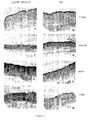
- Figure 3 illustrates representative experiments showing CD1a-labeled DC in HPV-transformed organotypic cultures incubated or not with chemotactic molecules included or not in polycarbophil gel.
- Figure 4 represents similar experiments performed with LC.
- the cytostatic activity of DC against HPV+ keratinocytes is not modified in the presence of chemokines/defensins. Since DC are able to inhibit the growth of HPV-transformed keratinocytes (Hubert et al, 2001) in vitro, we wanted to determine the influence of chemotactic molecules on this cytostatic activity.
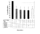
- DC exhibited a significant growth inhibition effect on HPV + keratinocytes since DC reduced the proliferation of HPV + keratinocytes to 56% compared to cultures without DC. Neither defensins nor chemokines modified the cytostatic activity of DC against HPV + keratinocytes.
- Chemokines and defensins do not affect the antigen presentation function of DC in a Mixed lymphocyte reaction (MLR)
- MLR Mixed lymphocyte reaction
Landscapes
- Health & Medical Sciences (AREA)
- Chemical & Material Sciences (AREA)
- Life Sciences & Earth Sciences (AREA)
- Animal Behavior & Ethology (AREA)
- Veterinary Medicine (AREA)
- Medicinal Chemistry (AREA)
- Public Health (AREA)
- General Health & Medical Sciences (AREA)
- Pharmacology & Pharmacy (AREA)
- Epidemiology (AREA)
- Chemical Kinetics & Catalysis (AREA)
- General Chemical & Material Sciences (AREA)
- Engineering & Computer Science (AREA)
- Nuclear Medicine, Radiotherapy & Molecular Imaging (AREA)
- Organic Chemistry (AREA)
- Bioinformatics & Cheminformatics (AREA)
- Inorganic Chemistry (AREA)
- Reproductive Health (AREA)
- Virology (AREA)
- Oil, Petroleum & Natural Gas (AREA)
- Communicable Diseases (AREA)
- Urology & Nephrology (AREA)
- Gynecology & Obstetrics (AREA)
- Nutrition Science (AREA)
- Physiology (AREA)
- Oncology (AREA)
- Proteomics, Peptides & Aminoacids (AREA)
- Dermatology (AREA)
- Endocrinology (AREA)
- Biotechnology (AREA)
- Molecular Biology (AREA)
- Medicinal Preparation (AREA)
- Medicines That Contain Protein Lipid Enzymes And Other Medicines (AREA)
Abstract
Description
- The present invention relates to a composition for the treatment of a anogenital or oral disease, especially such a disease caused by a human papillomavirus.
- Human Papillomaviruses (HPV) are common sexually transmitted pathogens inducing a spectrum of diseases ranging from benign genital warts to invasive carcinomas. Some types of HPV have been shown to be directly involved in the development of cervical cancer and its precursors (squamous intraepithelial lesions (SIL)) (Bosch et al, 1995). Squamous intraepithelial lesions refer to a group of premalignant changes in the epithelium of the uterine cervix that precede the onset of invasive cancer.
- Additional environmental and/or host factors are probably involved in malignant progression as suggested by the small number of infected individuals developing cervical cancer and the relatively long latency period before cancer emergence. The role of the intrinsic immunity in controlling HPV infection and the subsequent development of SIL is shown indirectly by the increased frequency of HPV-associated lesions in patients with depressed cell-mediated immunity (Petry et al, 1994 ; Ellerbrock et al, 2000). The nature of an effective immune response to HPV infections is not well understood, although cell-mediated immunity is thought to be more important than humoral immunity (Thivolet et al, 1982; Wu et al, 1994). Several studies have described a localized immune dysfunction accompanying cervical HPV infections and associated cervical lesions.
- A way of improving the treatment of such conditions has been sought.
- According to the invention there is provided a mucoadhesive pharmaceutical composition comprising a polymer and a chemoattractant wherein the pH of the composition is greater than 6.
- It has surprisingly been found that a chemoattractant significantly enhanced the chemotaxis of an antigen presenting cell and that the inclusion of a chemoattractant in a mucoadhesive composition having a pH greater than 6 stimulated the infiltration of an antigen presenting cellinto a HPV-transformed epithelium. Therefore, the composition according to the invention will be useful in the treatment of a anogenital or oral disease, particularly an anogenital or oral disease caused by the human papillomavirus. A further useful feature of the invention is that the chemoattractant did not interfere with the antigen presenting ability of the antigen presenting cell.
- A chemoattractant is generally understood to be a pharmacological agent which modulates the recruitment of cells such as a dendritic and/or Langerhans cell. The chemoattractant used in the invention is preferably selected from the group consisting of MIP3α (macrophage inflamatory proteine 3α), HβD2 (human β defensin 2), MCP-1 (monocyte chemotactic protein-1) and molgramostim. Preferably two or more chemoattractants are used, e.g. MIP3α and HβD2.
- The composition according to the invention preferably contains an auxiliary chemoattractant which is selected from the group consisting of granulocyte macrophage - colony recruiting factor (GM-CSF) and HNP2 (human neutrophil defensin 2). A preferred combination of chemoattractant and auxiliary chemoattractant for use in the composition according to the invention is MCP-1 and HNP2.
- The composition according to the invention is preferably in the form of a continuous structure, preferably having a solid-like property. The composition is optionally either clear or opaque. In the simplest composition according to the invention, the polymer is a natural gum (e.g. xanthane), a semi-synthetic material (e.g. methylcellulose, carboxymethylcellulose, hydroxyethyl-cellulose hydroxypropylmethyl-cellulose or hydroxypropyl-cellulose) or a synthetic material (e.g. a carbomer, polycarbophil and/or a carboxyvinyl polymer).
- The polymer is preferably an acrylic acid containing polymer, more preferably it is a polycarbophil. The composition according to the invention is preferably in the form of a hydrogel.
- The pH of the composition is preferably from 6 to 8. More preferably it is about 6.9.
- The composition according to the invention optionally further comprises a preservative, buffer (especially an isotonic buffer) and/or stabiliser such as methylparahydroxybenzoate, parabens, EDTA, potassium sorbate and/or propylparahydroxybenzoate. A suitable carrier, excipient and/or other agent may be included in the composition according to the invention, e.g. to provide improved transfer and/or delivery.
- According to the invention there is also provided a composition according to the invention for use in the treatment of a squamous mucosa, preferably in the treatment of an anogenital or oral disease, especially human papilloma virus.
- By anogenital disease is meant vulvar, vaginal, cervical and/or penile and/or anorectal disease.
- According to the invention there is further provided use of a composition according to the invention in the manufacture of a medicament for use in the treatment of a squamous mucosa, preferably in the treatment of an anogenital or oral disease, especially human papillomavirus.
- According to the invention there is also provided a method of treating an anogenital and/or oral disease which method comprises administering a therapeutically effective amount of a composition according to the invention to a patient in need of such treatment. Typically, treatment with direct administration is made daily, weekly or monthly for a period of time sufficient to reduce, prevent or ameliorate one or more symptoms.
- For a typical composition having a volume of about 100ml, the amount of chemoattractant used in the composition is from 10ng to 500µg, preferably from 100ng to 100µg, depending on the chemoattractant used. A person of skill in the art will be able to determine a suitable amount.
- The invention is illustrated with reference to the Figures of the accompanying drawings in which:
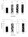
- Figure 1 is a graph showing chemotactic activity of different molecules on DC wherein NC indicates non-conditioned medium; the results are the mean ± SD of three experiments and asterisks indicate statistically significant differences: **P<0.01;
- Figure 2 is a graph showing chemotactic activity of different molecules on LC wherein NC indicates non-conditioned medium; results are the mean ± SD of seven experiments and asterisks indicate statistically significant differences: *P<0.05;
- Figure 3 shows the correlation between the penetration of DC into organotypic cultures and the addition of exogenous GM-CSF(C,D); MCP-1(E,F); and HNP2(G,H), shown by CD1a+ immunolabeled sections of organotypic culture of HPV+ keratinocytes wherein photographs A and B show DC infiltration in the absence of chemotactic molecules and C-H show DC recruitment in the presence of chemotactic molecules in liquid culture medium (C, E, G) or included in the polycarbophil gel (D, F, H);
- Figure 4 shows the correlation between the penetration of LC into organotypic cultures and the addition of exogenous GM-CSF (C,D); MIP3α (E,F); and HβD2 (G,H), shown by CD1a+ immunolabeled sections of organotypic culture of HPV+ keratinocytes wherein photographs A and B show DC infiltration in the absence of chemotactic molecules and C-H show DC recruitment in the presence of chemotactic molecules in liquid culture medium (C, E, G) or included in the polycarbophil gel (D, F, H);
- Figure 5 shows quantitative evaluation of DC and LC infiltration into organotypic cultures of HPV-transformed keratinocytes wherein the penetration of DC (A) is tested in the absence or in the presence of GM-CSF (used as control), MCP-1 and HNP2 included or not in polycarbophil gel; and the infiltration of LC (B) is evaluated in the absence or in the presence of GM-CSF, MIP3α and HβD2 included or not in the polycarbophil gel. DC and LC were detected by immunolabeling with anti-CD1a antibody. Results are expressed as infiltration depth in percentage of epithelial sheet thickness (n=4 for each culture condition). Asterisks indicate statistically significant differences: *P<0.05; **P<0.01; ***P<0.001;
- Figure 6 shows that DC-mediated cytostatic activity is maintained in the presence of chemokines and defensins. HPV+ keratinocytes were cultured in 96-well plates at 5x10³ cells/well. These cells were cultured with DC added at 4x10⁴ cell/well. After an incubation of 48h in the presence or in the absence of chemotactic molecules, cell proliferation was measured by 3H-TdR incorporation. The results are expressed as percentages of HPV+ keratinocyte proliferation in the coculture compared to keratinocytes cultured alone, in the presence or in the absence of chemokines or defensins. The results are presented as means ± SD of 4 experiments using DC from different donors; and
- Figure 7 shows that Chemokines and defensins did not alter the ability of DC to stimulate a potent mixed lymphocyte reaction. 5x10⁴ DC irradiated with 2500 rads were cultured with 1x10⁵ PBMC in the presence of chemokines or defensins, during 7 days. The cell proliferation was measured by 3H-TdR incorporation. Proliferation index was obtained by comparing the cell proliferation in the mixed DC-PBMC cultures added or not with chemokines/defensins, with PBMC cultured alone with or without chemokines/defensins. These results are presented as means ± SD of 3 experiments using DC from different donors.
- The invention will now be illustrated with reference to the following Examples which are not intended to limit the scope of the invention defined herein.
- A gel formulation according to the invention was prepared in the usual manner from a therapeutically effective amount of MCP-1, MIP3α or HβD2, 3 wt.% hydroxyethylcellulose (TYLOSE® H4000), 0.3 wt.% of potassium sorbate, 0.3 wt.% of NaH2PO4 and purified water q.s. to 100 wt.%.
- A gel formulation according to the invention suitable for oral or anal delivery was prepared in the usual manner from a therapeutically effective amount of MCP-1, MIP3α or HβD2 with 7g of hydroxypropylcellulose (Klucel GF) and an isotonic buffer to 100g. The isotonic buffer contains 1.36 g of KH2PO4, 200 ml of purified water and 4N NaOH to give a pH of 6.9.
- A gel formulation according to the invention suitable for oral or anal delivery was prepared in the usual manner from a therapeutically effective amount of MCP-1, MIP3α or HβD2 with 20 g of polyethylene polyoxypropylene block polymer (Lutrol F127) and an isotonic buffer as defined in Example 2 to 100g.
- A gel formulation according to the invention suitable for oral or anal delivery was prepared in the usual manner from a therapeutically effective amount of MCP-1, MIP3α or HβD2 with 1.5g of polyacrylic acid (Carbopol 974P), 0.5g of EDTA, trometamol to a pH of 6.9 and purified water to 100g.
- A gel formulation according to the invention suitable for oral or anal delivery was prepared in the usual manner from a therapeutically effective amount of MCP-1, MIP3α or HβD2 with:
Hydroxypropylcellulose (Klucel GF) 7g Parabens q.s. Isotonic buffer (as in Example 2) to 100g - A gel formulation according to the invention suitable for oral or anal delivery was prepared in the usual manner from a therapeutically effective amount of MCP-1, MIP3α or HβD2 with:
Polyethylene polyoxypropylene block polymer (Lutrol F127) 20 g Parabens q.s. Isotonic buffer (as in Example 2) to 100g - A gel formulation according to the invention suitable for oral or anal delivery was prepared in the usual manner from a therapeutically effective amount of MCP-1, MIP3α or HβD2 with:
Polyacrylic acid (Carbopol 974P) 1.5 g EDTA 0.5 Parabens q.s. Purified water to 100g Trometamol to pH 6.9 - SiHa and CasKi are tumorigenic cervical carcinoma-derived keratinocyte cell lines (Friedl et al, 1970; Pater et al, 1985; Auersperg et al, 1962). SiHa cell line contains one copy and CasKi contains approximately 600 copies of integrated HPV-16 DNA. These HPV-transformed keratinocyte cell lines were cultured in growth medium composed of 1/3 mixture of HAM F12 (GIBCO BRL, Nord Island, NY, USA)/Dulbecco's modified Eagle's medium (GIBCO BRL), supplemented with 0.4 µg/ml hydrocortisone (Sigma Chemical Co., St Louis, MO), 2 ng/ml epidermal growth factor (Sigma), 10 % fetal calf serum (Life Sciences International, Zelik, Belgium), 2 mmol/L L-glutamine (GIBCO BRL), 10 mmol/L Hepes (GIBCO BRL), 1 µg/ml fungizone (GIBCO BRL), 1 mmol/L sodium pyruvate (GIBCO BRL), 3000 U/ml penicillin-streptomycin (GIBCO BRL), 10-10 mol/L cholera toxin (Sigma), 5 µg/ml insulin (Sigma), 20 µg/ml adenine (Sigma), 5 µg/ml human transferrin (Sigma), and 15.10-4 µg/
ml 3,3',5-triiodo-L-thyronine (Sigma). - Dendritic cells (DC) were generated by culturing adherent fraction of human PBMC as previously described (Hubert et al, 1998; Sallusto and Lanzavecchia, 1994). Briefly, Peripheral Blood Mononuclear Cells (PBMC) were isolated from leukocyte-enriched buffy-coats by centrifugation on Ficoll-Hypaque. After washings at low centrifugation speed to discard a maximum of platelets, PBMC were plated in six-well dishes (Nunclon Multidishes, NUNC, Denmark) at a density of 10×106 PBMC per well in 3 ml of medium without FCS. After 18h at 37°C, non-adherent cells were discarded by two gentle washes and the plastic adherent fraction was cultured with 800 U/ml human recombinant GM-CSF (Amoytop Biotech, Xiamen, China) and 40 U/ml IL4 (ImmunoTools, Friesoythe, Germany) in 3 ml of RPMI 10% FCS-50µM Mercaptoethanol. Cultures were fed every 3 days with fresh medium containing cytokines and harvested with PBS-EDTA 1mM at day 7.
- Cord blood mononuclear cells (CB MNC) were recovered after discontinuous density gradient centrifugation using Lymphoprep™ (Nycomed Pharma As, Oslo, Norway) within 24 hours after collection. CD34+ cells were isolated from CB MNC using the MACS Direct CD34 Progenitor Cell Isolation Kit (Miltenyi Biotec GmBH, Bergisch Gladbach, Germany) and MiniMACS separation columns (Miltenyi Biotec) according to the manufacturer's protocol. Fifteen 104 CD34+ cells were seeded in T25 flasks (Sarstedt, Inc, Newton, NC) in 10 ml of RPMI 1640 medium supplemented with 10% FCS, antibiotics and 50 µM mercaptoethanol (all from GIBCO-BRL). Cultures were supplemented with previously optimized concentrations of the following human molecules: SCF (20 ng/ml, specific activity (SA) > 5 105 U/mg), TPO (10 U/ml, SA >1 106 U/mg), F1T3-L (25 ng/ml, SA >2 105 U/mg), GM-CSF (200 U/ml, SA: 11,1 106 U/mg), TNFα (50 U/ml, SA >2 107 U/mg), IL4 (100 U/ml, SA >2 106 U/ml) and TGF-β1 (5 ng/ml or 12.5 ng/ml, SA >2 108 U/mg). All these agents were purchased from PeproTech (Rockey Hill, NJ), except for GM-CSF and IL4 which were obtained from Amoytop (Amoytop Biotech, Xiamen, China) and Biosource (Nivelles, Belgium), respectively. The cells were cultured at 37°C in a humidified atmosphere and in the presence of 5% CO2. At day 7, the cellular density was adjusted to 2.104 per cm2 and the cells were fed, at days 7 and 14, with GM-CSF, IL4, TNFα and TGF-β1 at the same concentrations as those used at the start of the culture, except for TGF-β1 at day 14 which was 12.5 ng/ml for all the cultures. At day 18, cells were collected from cultures by vigorous pipetting to prepare single cell populations (Hubert et al, in press).
- Cell migration was evaluated using a chemotaxis microchamber technique (48-well Boyden microchamber; Neuroprobe, Cabin John, MD,USA). The lower wells of the chemotaxis chamber was filled with 27 of non conditioned medium, human fibroblasts derived cell-conditioned medium or the differents chemokines studied. Nonconditioned medium was used as control for random migration. Each condition was repeated six times. After phenotypic characterisation, DC were harvested and 55µl of DC suspension (2x10⁶ cells/ml) were applied to the upper wells of the chamber, with a polyvinylpyrollidone-free polycarbonate membrane 8-µm pore filter (Poretics Corp., Livermore, CA) separating the lower wells. These membranes were coated by incubation with 100µg/ml gelatin in 0.1% acetic acid solution. The chamber was incubated for 5h at 37°C in a 5% CO2/95% air atmosphere. The cells having migrated to the underside of the filter were fixed and stained with Diff Quick Stain set (Baxter Diagnostics AG, Düdingen, Switzerland). The upper side of the filter was scraped to remove residual non migrating cells. One random field was counted per well using an eyepiece with a calibrated grid to evaluate the number of fully migrated cells.
- The same tests were realized for LC using standard 5-µm pore polyvinylpyrollidone-free polycarbonate filters to separate the upper and the lower wells.
- Polycarbophil (Noveon AA1) was supplied from Noveon (Brussels, Belgium). The polycarbophil gel (1.5% w/w) was prepared by dispersing the Noveon AA1 in keratinocyte growth medium. The mixture was stirred until thickening occurred and then neutralized by dropwise addition of 40% (w/w) trometamol, until a transparent gel appeared. The quantity of trometamol was adjusted to achieve pH 7.
- Organotypic cultures of HPV-transformed keratinocyte cell lines were prepared by procedures slightly modified from those described previously (Delvenne et al, 1995; Merrick et al, 1992). For the preparation of dermal equivalents, a collagen matrix solution was made with 32 mg of collagen (cellagen solution AC-5, type I, ICN, Biomedical, Asse-Relegen, Belgium) mixed on ice with 1.6 ml of 0.1% acetic acid, 1ml of chilled 10-fold concentrated Hanks' buffer supplemented with phenol red and 1N NaOH to give a pH of 7.2. One milliliter of FCS containing 5x10⁵ normal human fibroblasts was then added. One milliliter of collagen/fibroblast solution was poured into 24-well plates (Nunclon Multidishes, Nunc, Roskilde, Denmark) and allowed to solidify at 37°C for 1 hour. The final concentrations of collagen and fibroblasts were 3.2 mg/ml and 5x10⁴ cells/ml, respectively. After gel equilibration with 1ml of growth medium overnight at 37°C, 30.104 HPV-transformed keratinocytes resuspended in 50µl of growth medium were seeded on top of the gels and maintained submerged for 48 hours. The collagen rafts were raised in 25-mm tissue culture insert (8-µm pore size; Nunc) and placed onto stainless-steel grids at the interface between air and liquid culture medium. Epithelial cells were then allowed to stratify over 20 days. After stratification of keratinocytes, DC were seeded on top of the in vitro-formed epithelium at a concentration of 4.105 cells/50µl of keratinocyte growth medium. The liquid culture medium or the polycarbophil gel was supplemented or not with the different chemokines/defensins at the following concentrations: 500 ng/ml of Monocyte Chemotactic Protein 1 (MCP-1, PeproTech, Rocky Hill, NJ), 750 ng/ml of Human Neutrophil Peptide-2 (HNP2, Sigma-Aldrich, St Louis, MO), 750 ng/ml of Human β Defensin-2 (HβD2, PeproTech, Rocky Hill, NJ) or 500 ng/ml of Macrophage Inflammatory Protein 3α (MIP3α, PeproTech, Rocky Hill, NJ). After 48 hours at 37°C, the collagen rafts were harvested. The cultures were then embedded in OCT compound (Tissue Tek, Sakura, The Netherlands) at -70°C and sectioned with a cryostat microtome for the immunohistochemical analysis.
- The density of DC migrating into the epithelial layer was assessed by the avidin-biotin-peroxidase technique (Vectastain ABC kit, Vector Laboratories, Burlingame, CA) with an anti-CD1a monoclonal antibody (clone NA1/34 from Dako, Glostrup, Denmark). Eight-micron frozen sections were fixed in cold acetone for 3 minutes, and endogenous peroxidases were blocked with 0.1% H2O2 for 30 minutes. Sections were then incubated sequentially with anti-CD1a antibody (at a 1/40 dilution in PBS containing 2% bovine serum albumin (BSA)) for 1 hour, with a biotinylated mouse anti-Ig antibody for 30 minutes, and with streptavidin/horseradish peroxidase/avidin/biotin complex for another 30 minutes. Positive cells were visualized by a 3,3'-diaminobenzidine substrate (DAB). The sections were counterstained with hematoxylin.
- Assessment of CD1a+ Cell Infiltration in Organotypic Cultures.
- The DC/LC infiltration in the organotypic cultures was evaluated by measuring the distance between the top of the epithelium and individual DC with a computerized system of image analysis (CAS, Becton Dikinson, Erembodegem, Belgium). The ratio between the infiltration depth and the thickness of the culture was then calculated. The percentage ratio was 0% for DC/LC staying on the top of the culture and 100% for DC/LC reaching the bottom of the epithelium.
- HPV-transformed keratinocyte cell lines (5x10³ cells/well) with or without DC (40000 DC/well) were cultured in 96-well plates (Nunclon Surface, NUNC) with or without chemokines/defensins. Proliferation was measured after 48h at 37°C, following an 18 h incubation with 0.4µCi/well of [3H]thymidine (6,7Ci/mmol, Moravek Biochemicals, Brea, CA). [3H]thymidine can incorporate DNA during the S phase of dividing cell cycle. DNA was harvested by an automated sample harvester (Packar, Canberra, Tilburg, The Netherlands) and thymidine incorporation was analysed by using a liquid scintillation counter (Top Count, Packard, Canberra). The data are presented as the percentage of proliferation calculated by using the following formula:

where test cpm is [3H]thymidine incoporation by keratinocytes cultured with DC in the presence or not of chemokines/defensins and control cpm is [3H]thymidine incorporation by keratinocytes without DC but with or without chemokines/defensins. - Stimulator populations (dendritic cells) were harvested and irradiated with 2500 rads. After a wash and a centrifugation, the cells were adjusted to 50000 DC/100µl in RPMI-1640 medium containing 5% human pooled AB serum. Stimulator cells (50000 DC/well) were then added to roundbottomed 96-well plates (Nunclon) containing 1x10⁵ allogeneic PBMC per well and the differents chemokines/defensins. The tests were performed in quadruplate. A proliferative response was measured after 7 days of culture by adding 0.4 µCi [3H]thymidine (6,7Ci/mmol, Moravek Biochemicals, Brea, CA) to each well. DNA was harvested 18h later by an automated sample harvester (Packar, Canberra, Tilburg, The Netherlands) and counted in a liquid scintillation counter (Top Count, Packard, Canberra).
- Statistical analysis was performed by using the Unpaired Student t test (Instat Mac 2.01 software; Graph-Pad Software, San Diego, CA). Differences were considered statistically significant when P<0.05.
- To identify molecules that could influence the migration of DC, we conducted a migration assay using a Boyden microchemotaxis chamber. DC generated for this study were judged to be 90% pure based on several criteria, including morphology, forward-scatter and side-scatter values by flow cytometry, and surface phenotype (data not shown).
- As shown in Figure 1, the addition of GM-CSF (1ng/ml) known to be chemoattractant for DC (Hubert et al, 1999) significantly enhanced the chemotaxis of DC, compared with the nonconditioned medium used to assess the chemokinesis. Interestingly, MCP-1 and HNP2 increased the migration of DC at respectively 500 ng/ml and 750 ng/ml which were previously determined in a preliminary work to be the optimal concentrations. Besides, the chemotactic activity of MCP-1 and HNP2 was similar to the level of chemotaxis induced by GM-CSF.
- In order to determine whether LC could be attracted by chemotactic molecules, Boyden chamber assay was performed with in vitro generated LC. LC were generated from CD34+ cord blood progenitors cultivated in the presence of hematopoïetic growth factors (TPO, SCF, Flt3L), cytokines (GM-CSF, TNFα, IL4) and TGF-β1(Hubert et al, in press). They exhibited morphological, immunohistochemical (CD1a+, CD207+, E-cadherin+, CLA+ and CCR6+) and ultrastructural features (Birbeck granules) of LC as verified by FACS analysis and electron microscopy (data not shown).
- As observed for DC, GM-CSF (10ng/ml) induced a significantly higher LC migration than the nonconditioned medium. The mobility of LC in the presence of MIP3α 500 ng/ml) or HβD2 (750 ng/ml) was identical to that observed with GM-CSF (Figure 2).
- Chemokines and defensins, included in a polycarbophil gel stimulate the infiltration of DC/LC into organotypic cultures of HPV-transformed keratinocytes.
It was investigated whether the addition of MIP3α and HβD-2 or MCP-1 and HNP2 could modulate the ability of LC and DC respectively to infiltrate an in vitro-formed (pre)neoplastic epithelium, reminiscent of a cervical high-grade lesion observed in vivo. After 20 days of culture, the HPV-transformed keratinocyte cell lines grown on a collagen gel at the air/liquid interface, produced an epithelial layer of up to 10-15 cells in thickness. These cells appeared disorganized and highly atypical throughout the full thickness of the epithelium as observed in high-grade lesion biopsies. - DC or LC were layered on the top of these cultures in the presence or in the absence of chemoattractant molecules. In order to evaluate the possibility to topically apply these molecules in an immunotherapeutic approach, chemokines and defensins were included in a polycarbophil gel. The ability of chemotactic molecules to influence the migration of DC/LC was determined by evaluating the density of CD1a+ cells in sections of organotypic cultures at 48 hours following chemoattractant addition. Figure 3 illustrates representative experiments showing CD1a-labeled DC in HPV-transformed organotypic cultures incubated or not with chemotactic molecules included or not in polycarbophil gel. Figure 4 represents similar experiments performed with LC.
- In HPV-transformed keratinocyte organotypic cultures, LC/DC poorly infiltrated the epithelial layer in the absence of chemoattractant (Figure 3, A-B and figure 4, A-B). In contrast, addition of GM-CSF, the positive control, caused a significant increase in the density of LC/DC observed in the epithelial layer (Figure 3, C-D and Figure 4, C-D). The addition of chemokines and defensins in growth medium induced a recruitment of DC/LC similar to that observed with GM-CSF (Figure 3 C,E,G and Figure 4 C,E,G). Interestingly, this infiltration was also present when the chemoattractants were included in the polycarbophil gel (Figure 3 D, F, H and Figure 4 D, F, H).
- Quantitative analysis of DC/LC infiltration was perfomed by evaluating the infiltration depth of all CD1a+ cells throughout the full thickness of organotypic cultures (Figure 5).
- When the medium of organotypic culture was supplemented with MCP1 or HNP2, the infiltration of DC increased compared to the basal infiltration without chemoattractant, and reached an infiltration level equivalent to that obtained with GM-CSF (Figure 5, A). The inclusion of chemoattractant molecules in the polycarbophil gel did not modify the DC infiltration compared with that observed in the presence of chemoattractants in the culture medium.
- The infiltration of LC was also increased in the presence of MIP3α or HβD-2 (Figure 5, B). When the medium of organotypic cultures was supplemented with HβD-2, the migration of LC was slightly lower compared with that observed in the presence of GM-CSF. The addition of MIP3α induced a recruitment similar to that obtained with GM-CSF. The chemoattractants included in liquid culture medium or in polycarbophil gel recruited LC in a similar manner.
- To determine the potential influence of chemokines/defensins on DC/LC differentiation, we performed a double-immunostaining (CD1a/CD14) of organotypic cultures. The phenotype of DC/LC infiltrating organotypic cultures in the presence of chemokines/defensins was not altered compared with that established before the migration (data not shown).
- The cytostatic activity of DC against HPV+ keratinocytes is not modified in the presence of chemokines/defensins.
Since DC are able to inhibit the growth of HPV-transformed keratinocytes (Hubert et al, 2001) in vitro, we wanted to determine the influence of chemotactic molecules on this cytostatic activity. We performed a 48h growth-inhibition assay by coculturing HPV+ keratinocytes with DC in the presence or not of different chemoattractants. - DC exhibited a significant growth inhibition effect on HPV+ keratinocytes since DC reduced the proliferation of HPV+ keratinocytes to 56% compared to cultures without DC. Neither defensins nor chemokines modified the cytostatic activity of DC against HPV+ keratinocytes.
- Chemokines and defensins do not affect the antigen presentation function of DC in a Mixed lymphocyte reaction (MLR)
The lymphocyte proliferation was not changed in the presence of chemokines or defensins only (data not shown). - As shown in Figure 7, DC in the presence or in the absence of chemotactic molecules did not differ significantly in their ability to stimulate the T lymphocyte proliferation. Consequently, the capacity of DC to present antigen to T lymphocytes was not affected.
-
- 1.
- Albert ML, Sauter B, Bhardwaj N. Dendritic cell acquire antigen from apoptotic cells and induce class I restricted CTLs. Nature 1998, 392:86-9.
- 2.
- A1-Saleh W, Delvenne P, Anesse JE, Nikkels A, Pierard GE, and Boniver J. Inverse modulation of intraepithelial Langerhans cell and stromal macrophage /dendrocyte populations in human papillomavirus-associated squamous intraepithelial lesions of the cervix. Virchows Arch 1995, 427:41-48.
- 3.
- Al-Saleh W, Giannini SL, Jacobs N, Moutschen N, Doyen J, Boniver J, and Delvenne P. Correlation of T-helper secretory differentiation and types of antigen-presenting cells in squamous intraepithelial lesions of the uterine cervix. J Pathol 1998, 184:283-290.
- 4.
- Auersperg N, Hawryluk AF. Chromosome observations on three epithelial cell cultures derived from carcinomas of the uterine cervix. J Natl Cancer Inst 1962, 28:605-627.
- 5.
- Bell D, Chomarat P, Broyles D, et al. In breast carcinoma tissue, immature dendritic cells reside within the tumor, whereas mature dendritic cells are located in peritumoral areas. J Exp Med 1999, 190:1417-26.
- 6.
- Blanton RA, Perez-Reyes N, Merrick DT, McDougall JK. Epithelial cells immortalized by human papillomaviruses have premalignant characteristics in organotypic cultures. Am J Pathol 1991, 138:673-685.
- 7.
- Bosch FX, Manos MM, Munoz N, Sherman M, Jansen AM, Peto J.
Prevalence of human papillomavirus in cervical cancer : a worldwide perspective. International Biological Study on cervical cancer 5IBBCC) study Group. J Natl Cancer Inst 1995, 87:796-802. - 8.
- Charbonnier AS, Kohrgruber N, Kriehuber E, et al. Macrophage
inflammatory protein 3 alpha is involved in the constitute trafficking of epidermal Langerhans cells. J Exp Med 1999, 190:1755-68. - 9.
- Delvenne P, Al-Saleh W, Gilles C, Thiry A, Boniver J. Inhibition of growth of normal and human papillomavirus-transformed keratinocytes in monolayer and organotypic cultures by interferon―γ and tumor necrosis factor-α. Am J Pathol 1995, 146:589-598.
- 10.
- Ellerbrock TV, Chiasson MA, Bush TJ, et al. Incidence of cervical squamous intraepithelial lesions in HIV-infected women. JAMA 2000, 283:1031-7.
- 11.
- Friedl F, Kimura I, Osato T, Ito Y. Studies on a new human cell line (SiHa) derived from carcinoma of uterus. I. Its establishment and morphology. Proc Soc Exp Biol Med 1970, 135:543-545.
- 12.
- Furukawa T, Watanabe S, Kodama T, Sato Y, Shimosat Y, and Suematsu K. T-zone histiocytes in adenocarcinoma of the lung in relation to postoperative prognosis. Cancer 1985, 56:2651-2656.
- 13.
- Giannini SL, Al-Saleh W, Piron H, Jacobs N, Doyen J, Boniver J, and Delvenne P. Cytokines expression in squamous intraepithelial lesions of the uterine cervix : implications for the generation of local immunosuppression. Clin Exp Immunol 1998, 113:183-189.
- 14.
- Giannini SL, Hubert P, Doyen J, Boniver J, and Delvenne P. Influence of the mucosal epithelium microenvironnement on Langerhans'cells : intraepithelial lesions of the cervix. Int J Cancer 2002, 10:654-659.
- 15.
- Granelli-Piperno A, Moser B, Pope M, Chen D, Wei Y, Isdell F, O'Doherty U, Paxton W, Koup R, Mosjov S. Efficient interaction of HIV-1 with purified dendritic cells via multiple chemokines receptors. J Exp Med 1996, 184:2433-2438.
- 16.
- Hubert P, Greimers R, Franzen-Detrooz E, Doyen J, Delanaye P, Boniver J, Delvenne P. In vivo propagated dendritic cells from patients with human-papillomavirus-associated preneoplastic lesions of the uterine cervix : use of Flt3 ligand. Cancer Immunol Immunother 1998, 47:81-89.
- 17.
- Hubert P, van den Brûle F, Giannini SL, Franzen-detrooz E, Boniver J, and Delvenne P. Colonization of in vitro-formed cervical human papillomavirus-associated (pre) neoplastic lesions with dendritic cells : role of granulocyte/macrophage colony-stimulating factor. Am J Pathol 1999, 154:775-784.
- 18.
- Hubert P, Giannini SL, Vanderplasschen A, Franzen-detrooz E, Jacobs N, Boniver J, and Delvenne P. Dendritic cells induce the death of human papillomavirus transformed keratinocytes. FASEB J 2001, 15:2521-2523.
- 19.
- Hubert P, Evrard B, Maillard C, Franzen-detrooz E, Delattre L, Foidart JM, Noël A, Boniver J, and Delvenne P. A bioadhesive hydrogel of GM-CSF stimulates the migration of dendritic cells in models of HPV-associated (pre)neoplastic epithelium. Antimicrob Agents Chemother 2004, in press.
- 20.
- Hubert P, Bousarghin L, Greimers R, Franzen-detrooz E, Boniver J, and Delvenne P. Production of large numbers of Langerhans cells with intraepithelial migration ability in vitro. Exp Dermatol 2004, in press.
- 21.
- Hui W, Yoko NS, Toshikazy K, Mariko A, and Naofumi M. Potential involvement of monocyte chemoattractant protein (MCP)-1/CCL2 in IL-4-mediated tumor immunity through inducing dendritic cell migration into the draining lymph nodes. Int Immunopharmacol 2003, 3:627-642.
- 22.
- Jacobs N, Giannini SL, Doyen J, Baptista A, Moutshen M, Boniver J, and Delvenne P. Inverse modulation of IL10 and IL12 in the blood of women with preneoplastic lesions of the uterine cervix. Clin Exp Immunol 1998, 111:219-224.
- 23.
- Jonuleit H, Knop J, Enk AH. Cytokines and their effects on maturation, differentiation and migration of dendritic cells. Arch Dermatol Res 1999, 289:1-8.
- 24.
- Kaplan G. Recent advances in cytokine therapy in leprosy. J Infect Dis 1993, 167:S18-S22.
- 25.
- Kleine-Lowinski K, Rheinwald JG, Fichorova RN, Anderson DJ, Basile J, Munger K, Daly CM, Rosl F, Rollins BJ. Selective suppression of monocyte chemoattractant protein-1 expression by human papillomavirus E6 and E7oncoproteins in human cervical epithelial and epidermal cells.Int J Cancer 2003, 107:407-15.
- 26.
- Knuth K, Amiji M, and Robinson J. Hydrogel delivery system for cervico-vaginal and oral applications: formulations and biological consideration Adv Drug Deliv Rev 1993, 11:137-167.
- 27.
- Macatonia SE, Gompels M, Pinchin AJ, et al. Antigen presentation by macrophages but not by dendritic cells in human immunodeficiency virus (HIV) infection. Immunology 1992, 75:576-581.
- 28.
- Merrick DT, Blanton RA, Gow AM, McDougall JK. Altered expression of proliferation and differentiation markers in human papillomavirus 16 and 18 immortalized epithelial cells grown in organotypic cultures. Am J Pathol 1992, 140:167-177.
- 29.
- Morelli AE, Sananes C,Di Paola G, Paredes A, Frainboim L. Relationship between types of human papillomavirus and Langerhans'cells in cervical condyloma and intraepithelial neoplasia. Am J Clin Pathol 1993, 99:200-207.
- 30.
- Müller CA, Markovic-Lipkovski J, Klatt T, Gamper J, Schwarz G, Beck H, Deeg M, Kalbacher H, Widmann S, Wessels JT, Becker V, Müller GA, and Flad T. Human □-defensins HNPs-1, -2, and -3 in renal cell carcinoma. Am J Pathol 2002, 160:1311-1324.
- 31.
- Nakamura R, Williams I, and kupper T. Keratinocyte-derived monocyte chemoattractant protein 1 (MCP-1) : analysis in a transgenic model demonstrates MCP-1 can recruit dendritic and Langerhans cells to skin. J Invest Dermatol 1995, 105:635-643.
- 32.
- Nakano T, Oka K, Arai T, Morita S, and Tsunemoto H. Prognostic significance of Langerhans' cell infiltration in radiation therapy for squamous cell carcinoma of the uterine cervix. Arch Pathol Lab Med 1989,113:507-511.
- 33.
- Nakano T, Oka K, Takahashi T, Morita S, and Arai T. Roles of Langerhans'cells and T-lymphocytes infiltrating cancer tissues in patients treated by radiation therapy for cervical cancer. Cancer 1992, 70:2839-44.
- 34.
- Oka K, Nakano T, and Arai T. Adenocarcinoma of the cervix treated with radiation alone: Prognostic significance of S-100 protein and vimentin immunostaining. Obstet Gynecol 1992, 79:347-50.
- 35.
- Parkin D, Lääza E, Muir CS. Estimates of the worlwide frequency of sixteen major cancers in 1980. Int J Cancer 1988, 41:184-197.
- 36.
- Parody TR, Stone MJ. High level expression, activation, and antagonism of CC chemokines receptors CCR2 and CCR3 in chinese hamster ovary cells. Cytokines 2004, 27:38-46.
- 37.
- Pater M, Pater A. Human papillomavirus types 16 and 18 sequences in carcinoma cell lines of the cervix. Virology 1985, 145:313-318.
Claims (8)
- A mucoadhesive pharmaceutical composition comprising a polymer and a chemoattractant wherein the pH of the composition is greater than 6.
- A composition as defined in claim 1 wherein the chemoattractant is selected from the group consisting of MIP3α, HβD2, MCP-1 and molgramostim.
- A composition as defined in claim 1 or claim 2 which contains an auxiliary chemoattractant which is selected from the group consisting of granulocyte macrophage ― colony recruiting factor (GM-CSF) and HNP2 (human neutrophil defensin 2).
- A composition as defined in any one of the preceding claims wherein the polymer is selected from the group consisting of a natural gum, a semi-synthetic material, and a synthetic material.
- A composition as defined in any one of the preceding claims wherein the pH of the composition is from 6 to 8, preferably the pH of the composition is about 6.9.
- A composition as defined in any one of the preceding claims for use in the treatment of a squamous mucosa, preferably in the treatment of an anogenital or oral disease, especially human papillomavirus.
- Use of a composition as defined in any one of the preceding claims in the manufacture of a medicament for use in the treatment of a squamous mucosa, preferably in the treatment of an anogenital or oral disease, especially human papillomavirus.
- A method of treating an anogenital and/or oral disease which method comprises administering a therapeutically effective amount of a composition as defined in any one of claims 1 to 5 to a patient in need of such treatment.
Priority Applications (6)
| Application Number | Priority Date | Filing Date | Title |
|---|---|---|---|
| EP04106995A EP1674077A1 (en) | 2004-12-27 | 2004-12-27 | Mucoadhesive pharmaceutical compositions comprising chemoattractants |
| PCT/EP2005/056709 WO2006069911A2 (en) | 2004-12-27 | 2005-12-12 | Mucoadhesive pharmaceutical compositions comprising chemoattractants |
| JP2007548800A JP2008525516A (en) | 2004-12-27 | 2005-12-12 | Improvement of chemical attractant or improvement on chemical attractant |
| MX2007007876A MX2007007876A (en) | 2004-12-27 | 2005-12-12 | Mucoadhesive pharmaceutical compositions comprising chemoattractants. |
| EP05826365A EP1835887A2 (en) | 2004-12-27 | 2005-12-12 | Mucoadhesive pharmaceutical compositions comprising chemoattractants |
| US11/794,146 US20080293630A1 (en) | 2004-12-27 | 2005-12-12 | Mucoadhesive Pharmaceutical Compositions Comprising Chemoattractants |
Applications Claiming Priority (1)
| Application Number | Priority Date | Filing Date | Title |
|---|---|---|---|
| EP04106995A EP1674077A1 (en) | 2004-12-27 | 2004-12-27 | Mucoadhesive pharmaceutical compositions comprising chemoattractants |
Publications (1)
| Publication Number | Publication Date |
|---|---|
| EP1674077A1 true EP1674077A1 (en) | 2006-06-28 |
Family
ID=34930173
Family Applications (2)
| Application Number | Title | Priority Date | Filing Date |
|---|---|---|---|
| EP04106995A Withdrawn EP1674077A1 (en) | 2004-12-27 | 2004-12-27 | Mucoadhesive pharmaceutical compositions comprising chemoattractants |
| EP05826365A Withdrawn EP1835887A2 (en) | 2004-12-27 | 2005-12-12 | Mucoadhesive pharmaceutical compositions comprising chemoattractants |
Family Applications After (1)
| Application Number | Title | Priority Date | Filing Date |
|---|---|---|---|
| EP05826365A Withdrawn EP1835887A2 (en) | 2004-12-27 | 2005-12-12 | Mucoadhesive pharmaceutical compositions comprising chemoattractants |
Country Status (5)
| Country | Link |
|---|---|
| US (1) | US20080293630A1 (en) |
| EP (2) | EP1674077A1 (en) |
| JP (1) | JP2008525516A (en) |
| MX (1) | MX2007007876A (en) |
| WO (1) | WO2006069911A2 (en) |
Families Citing this family (1)
| Publication number | Priority date | Publication date | Assignee | Title |
|---|---|---|---|---|
| WO2006084131A2 (en) * | 2005-02-01 | 2006-08-10 | The Government Of The United States Of America, As Represented By The Secretary, Department Of Health And Human Services | Furin inhibitors and alpha-defensins for the treatment or prevention of papillomavirus infection |
Citations (2)
| Publication number | Priority date | Publication date | Assignee | Title |
|---|---|---|---|---|
| WO1998009642A2 (en) * | 1996-09-06 | 1998-03-12 | The United States Of America, Represented By The Secretary, Department Of Health And Human Services | Therapeutic chemokine antagonists |
| WO2002036141A2 (en) * | 2000-11-02 | 2002-05-10 | Immunex Corporation | Method of enhancing lymphocyte-mediated immune responses |
Family Cites Families (4)
| Publication number | Priority date | Publication date | Assignee | Title |
|---|---|---|---|---|
| US5942253A (en) * | 1995-10-12 | 1999-08-24 | Immunex Corporation | Prolonged release of GM-CSF |
| IT1298575B1 (en) * | 1998-02-06 | 2000-01-12 | Vectorpharma Int | PHARMACEUTICAL COMPOSITIONS IN THE FORM OF NANOPARTICLES INCLUDING LIPID SUBSTANCES AND ANTIPHILIC SUBSTANCES AND RELATED PROCESS OF |
| AU2001291049A1 (en) * | 2000-09-15 | 2002-03-26 | The Government Of The United States Of America, As Represented By The Secretary, Department Of Health And Human Services, National Institutes Of Health, Office Of Technology Transfer | Defensin-antigen fusion proteins |
| GB0317999D0 (en) * | 2003-07-31 | 2003-09-03 | Univ Liege | Improvements in or relating to drug delivery systems |
-
2004
- 2004-12-27 EP EP04106995A patent/EP1674077A1/en not_active Withdrawn
-
2005
- 2005-12-12 MX MX2007007876A patent/MX2007007876A/en not_active Application Discontinuation
- 2005-12-12 EP EP05826365A patent/EP1835887A2/en not_active Withdrawn
- 2005-12-12 WO PCT/EP2005/056709 patent/WO2006069911A2/en not_active Ceased
- 2005-12-12 US US11/794,146 patent/US20080293630A1/en not_active Abandoned
- 2005-12-12 JP JP2007548800A patent/JP2008525516A/en active Pending
Patent Citations (2)
| Publication number | Priority date | Publication date | Assignee | Title |
|---|---|---|---|---|
| WO1998009642A2 (en) * | 1996-09-06 | 1998-03-12 | The United States Of America, Represented By The Secretary, Department Of Health And Human Services | Therapeutic chemokine antagonists |
| WO2002036141A2 (en) * | 2000-11-02 | 2002-05-10 | Immunex Corporation | Method of enhancing lymphocyte-mediated immune responses |
Non-Patent Citations (1)
| Title |
|---|
| OH Y-K ET AL: "Enhanced mucosal and systemic immune responses to a vaginal vaccine coadministered with RANTES-expressing plasmid DNA using in situ-gelling mucoadhesive delivery system", VACCINE, BUTTERWORTH SCIENTIFIC. GUILDFORD, GB, vol. 21, no. 17-18, 16 May 2003 (2003-05-16), pages 1989 - 1997, XP004421114, ISSN: 0264-410X * |
Also Published As
| Publication number | Publication date |
|---|---|
| EP1835887A2 (en) | 2007-09-26 |
| US20080293630A1 (en) | 2008-11-27 |
| JP2008525516A (en) | 2008-07-17 |
| WO2006069911A3 (en) | 2006-09-08 |
| WO2006069911A2 (en) | 2006-07-06 |
| MX2007007876A (en) | 2007-09-07 |
Similar Documents
| Publication | Publication Date | Title |
|---|---|---|
| EP1658046B1 (en) | Mucoadhesive composition comprising polyacrylate and chemoattractant | |
| Majewski et al. | Natural cell-mediated cytotoxicity against various target cells in patients with epidermodysplasia verruciformis | |
| Hubert et al. | Colonization of in vitro-formed cervical human papillomavirus-associated (pre) neoplastic lesions with dendritic cells: role of granulocyte/macrophage colony-stimulating factor | |
| Carr et al. | Human papillomavirus: epidemiology, transmission, and pathogenesis | |
| Rs | The immune microenvironment in human papilloma virus-induced cervical lesions—evidence for estrogen as an immunomodulator | |
| Pater et al. | Oncogenic transformation by human papillomavirus type 16 deoxyribonucleic acid in the presence of progesterone or progestins from oral contraceptives | |
| Purdie et al. | Malignant transformation of cutaneous lesions in renal allograft patients: a role for human papillomavirus? | |
| EP2542058B1 (en) | Effective sensitizing dose of a gelled immunomodulating topical composition | |
| Severson et al. | Human papillomavirus infections: epidemiology, pathogenesis, and therapy | |
| Hubert et al. | Defensins induce the recruitment of dendritic cells in cervical human papillomavirus‐associated (pre) neoplastic lesions formed in vitro and transplanted in vivo | |
| Schaefer et al. | IL-1β-mediated proinflammatory responses are inhibited by estradiol via down-regulation of IL-1 receptor type I in uterine epithelial cells | |
| Worda et al. | Prevalence of cervical and intrauterine human papillomavirus infected in the third trimester in asymptomatic women | |
| CN111375051A (en) | Application of polypeptide in preparation of preparation for preventing and treating human papilloma virus infection | |
| Wadler et al. | A phase II trial of interleukin-12 in patients with advanced cervical cancer: clinical and immunologic correlates: Eastern Cooperative Oncology Group study E1E96 | |
| WO2009143309A2 (en) | Female reproductive tract and anal prophylaxes | |
| Majewski et al. | Immunology of HPV infection and HPV-associated tumors. | |
| Minjarez et al. | Abnormal uterine bleeding in adolescents | |
| Behbahani et al. | Proinflammatory and type 1 cytokine expression in cervical mucosa during HIV-1 and human papillomavirus infection | |
| Gissmann | Linking HPV to cancer | |
| WO2019238120A1 (en) | Medicament for treating diseases caused by persistent hpv infection, preparation method therefor and use thereof | |
| US20080293630A1 (en) | Mucoadhesive Pharmaceutical Compositions Comprising Chemoattractants | |
| Montgomery Rice et al. | Differential responses of granulosa cells from small and large follicles to follicle stimulating hormone (FSH) during the menstrual cycle and acyclicity: effects of tumour necrosis factor-alpha. | |
| Schneider et al. | Efficacy trial of topically administered interferon gamma-1β gel in comparison to laser treatment in cervical intraepithelial neoplasia | |
| Hubert et al. | Delivery of granulocyte-macrophage colony-stimulating factor in bioadhesive hydrogel stimulates migration of dendritic cells in models of human papillomavirus-associated (pre) neoplastic epithelial lesions | |
| Ney et al. | Squamous Epitheliuai in the Trigone of the Human Female Urinary Bladder: With a Note on Cystoscopic Observations During Estrogen Therapy |
Legal Events
| Date | Code | Title | Description |
|---|---|---|---|
| PUAI | Public reference made under article 153(3) epc to a published international application that has entered the european phase |
Free format text: ORIGINAL CODE: 0009012 |
|
| AK | Designated contracting states |
Kind code of ref document: A1 Designated state(s): AT BE BG CH CY CZ DE DK EE ES FI FR GB GR HU IE IS IT LI LT LU MC NL PL PT RO SE SI SK TR |
|
| AX | Request for extension of the european patent |
Extension state: AL BA HR LV MK YU |
|
| 17P | Request for examination filed |
Effective date: 20060801 |
|
| AKX | Designation fees paid |
Designated state(s): AT BE BG CH CY CZ DE DK EE ES FI FR GB GR HU IE IS IT LI LT LU MC NL PL PT RO SE SI SK TR |
|
| 17Q | First examination report despatched |
Effective date: 20081229 |
|
| STAA | Information on the status of an ep patent application or granted ep patent |
Free format text: STATUS: THE APPLICATION IS DEEMED TO BE WITHDRAWN |
|
| 18D | Application deemed to be withdrawn |
Effective date: 20090509 |