EP1669453A1 - Novel nerve stem cell marker - Google Patents
Novel nerve stem cell marker Download PDFInfo
- Publication number
- EP1669453A1 EP1669453A1 EP04787861A EP04787861A EP1669453A1 EP 1669453 A1 EP1669453 A1 EP 1669453A1 EP 04787861 A EP04787861 A EP 04787861A EP 04787861 A EP04787861 A EP 04787861A EP 1669453 A1 EP1669453 A1 EP 1669453A1
- Authority
- EP
- European Patent Office
- Prior art keywords
- cells
- nerve
- gene
- stem cells
- nerve stem
- Prior art date
- Legal status (The legal status is an assumption and is not a legal conclusion. Google has not performed a legal analysis and makes no representation as to the accuracy of the status listed.)
- Withdrawn
Links
Images

Classifications
-
- G—PHYSICS
- G01—MEASURING; TESTING
- G01N—INVESTIGATING OR ANALYSING MATERIALS BY DETERMINING THEIR CHEMICAL OR PHYSICAL PROPERTIES
- G01N33/00—Investigating or analysing materials by specific methods not covered by groups G01N1/00 - G01N31/00
- G01N33/48—Biological material, e.g. blood, urine; Haemocytometers
- G01N33/50—Chemical analysis of biological material, e.g. blood, urine; Testing involving biospecific ligand binding methods; Immunological testing
- G01N33/5005—Chemical analysis of biological material, e.g. blood, urine; Testing involving biospecific ligand binding methods; Immunological testing involving human or animal cells
- G01N33/5008—Chemical analysis of biological material, e.g. blood, urine; Testing involving biospecific ligand binding methods; Immunological testing involving human or animal cells for testing or evaluating the effect of chemical or biological compounds, e.g. drugs, cosmetics
- G01N33/5044—Chemical analysis of biological material, e.g. blood, urine; Testing involving biospecific ligand binding methods; Immunological testing involving human or animal cells for testing or evaluating the effect of chemical or biological compounds, e.g. drugs, cosmetics involving specific cell types
- G01N33/5058—Neurological cells
-
- G—PHYSICS
- G01—MEASURING; TESTING
- G01N—INVESTIGATING OR ANALYSING MATERIALS BY DETERMINING THEIR CHEMICAL OR PHYSICAL PROPERTIES
- G01N33/00—Investigating or analysing materials by specific methods not covered by groups G01N1/00 - G01N31/00
- G01N33/48—Biological material, e.g. blood, urine; Haemocytometers
- G01N33/50—Chemical analysis of biological material, e.g. blood, urine; Testing involving biospecific ligand binding methods; Immunological testing
- G01N33/5005—Chemical analysis of biological material, e.g. blood, urine; Testing involving biospecific ligand binding methods; Immunological testing involving human or animal cells
- G01N33/5008—Chemical analysis of biological material, e.g. blood, urine; Testing involving biospecific ligand binding methods; Immunological testing involving human or animal cells for testing or evaluating the effect of chemical or biological compounds, e.g. drugs, cosmetics
-
- G—PHYSICS
- G01—MEASURING; TESTING
- G01N—INVESTIGATING OR ANALYSING MATERIALS BY DETERMINING THEIR CHEMICAL OR PHYSICAL PROPERTIES
- G01N33/00—Investigating or analysing materials by specific methods not covered by groups G01N1/00 - G01N31/00
- G01N33/48—Biological material, e.g. blood, urine; Haemocytometers
- G01N33/50—Chemical analysis of biological material, e.g. blood, urine; Testing involving biospecific ligand binding methods; Immunological testing
- G01N33/5005—Chemical analysis of biological material, e.g. blood, urine; Testing involving biospecific ligand binding methods; Immunological testing involving human or animal cells
- G01N33/5008—Chemical analysis of biological material, e.g. blood, urine; Testing involving biospecific ligand binding methods; Immunological testing involving human or animal cells for testing or evaluating the effect of chemical or biological compounds, e.g. drugs, cosmetics
- G01N33/502—Chemical analysis of biological material, e.g. blood, urine; Testing involving biospecific ligand binding methods; Immunological testing involving human or animal cells for testing or evaluating the effect of chemical or biological compounds, e.g. drugs, cosmetics for testing non-proliferative effects
- G01N33/5023—Chemical analysis of biological material, e.g. blood, urine; Testing involving biospecific ligand binding methods; Immunological testing involving human or animal cells for testing or evaluating the effect of chemical or biological compounds, e.g. drugs, cosmetics for testing non-proliferative effects on expression patterns
-
- G—PHYSICS
- G01—MEASURING; TESTING
- G01N—INVESTIGATING OR ANALYSING MATERIALS BY DETERMINING THEIR CHEMICAL OR PHYSICAL PROPERTIES
- G01N33/00—Investigating or analysing materials by specific methods not covered by groups G01N1/00 - G01N31/00
- G01N33/48—Biological material, e.g. blood, urine; Haemocytometers
- G01N33/50—Chemical analysis of biological material, e.g. blood, urine; Testing involving biospecific ligand binding methods; Immunological testing
- G01N33/5005—Chemical analysis of biological material, e.g. blood, urine; Testing involving biospecific ligand binding methods; Immunological testing involving human or animal cells
- G01N33/5008—Chemical analysis of biological material, e.g. blood, urine; Testing involving biospecific ligand binding methods; Immunological testing involving human or animal cells for testing or evaluating the effect of chemical or biological compounds, e.g. drugs, cosmetics
- G01N33/5044—Chemical analysis of biological material, e.g. blood, urine; Testing involving biospecific ligand binding methods; Immunological testing involving human or animal cells for testing or evaluating the effect of chemical or biological compounds, e.g. drugs, cosmetics involving specific cell types
- G01N33/5073—Stem cells
-
- C—CHEMISTRY; METALLURGY
- C12—BIOCHEMISTRY; BEER; SPIRITS; WINE; VINEGAR; MICROBIOLOGY; ENZYMOLOGY; MUTATION OR GENETIC ENGINEERING
- C12Q—MEASURING OR TESTING PROCESSES INVOLVING ENZYMES, NUCLEIC ACIDS OR MICROORGANISMS; COMPOSITIONS OR TEST PAPERS THEREFOR; PROCESSES OF PREPARING SUCH COMPOSITIONS; CONDITION-RESPONSIVE CONTROL IN MICROBIOLOGICAL OR ENZYMOLOGICAL PROCESSES
- C12Q1/00—Measuring or testing processes involving enzymes, nucleic acids or microorganisms; Compositions therefor; Processes of preparing such compositions
- C12Q1/68—Measuring or testing processes involving enzymes, nucleic acids or microorganisms; Compositions therefor; Processes of preparing such compositions involving nucleic acids
- C12Q1/6876—Nucleic acid products used in the analysis of nucleic acids, e.g. primers or probes
-
- G—PHYSICS
- G01—MEASURING; TESTING
- G01N—INVESTIGATING OR ANALYSING MATERIALS BY DETERMINING THEIR CHEMICAL OR PHYSICAL PROPERTIES
- G01N2500/00—Screening for compounds of potential therapeutic value
Definitions
- This invention relates to a method for detecting nerve stem cells/progenitor cells among a tissue or a mass of cells as constituted of a plurality of cell species.
- the invention also relates to a method for screening for a drug or procedure, for example, by which nerve stem cells/progenitor cells are allowed to maintain their characters when a tissue or a mass of cells constituted of a plurality of cell species is subjected to a process or procedure for differentiation induction or by which nerve stem cells/progenitor cells can be induced in a mass of differentiated nerve cells.
- nerve stem cells have also made it possible to screen for drugs acting on nerve stem cells and causing them to differentiate into functioning nerve cells such as neurons or glial cells (Roy, N. et al. (2000) "In vitro neurogenesis by neural progenitor cells isolated from the adult human hippocampus.” Nat. Med. 6:271-77).
- the NC1 gene though first identified as a gene specifically expressed in the pancreas of patients with persistent hyperinsulinemic hypoglycemia of infancy (PHHI), has been shown to be a gene specifically expressed in the brain among normal tissues.
- the present inventors investigated the functions of the NC1 gene in further detail and found, by using the techniques described below, that this gene functions as a means for detecting nerve stem cells/progenitor cells.
- Nerve stem cells are defined as undifferentiated nerve system cells capable of proliferating and being repeatedly passaged (self-replicating ability) and, at the same time, capable of generating cells, such as neurons and glial cells, which constitute the central nerve system (multipotency).
- a selective method (neurosphere method) for cultivating nerve stem cells/progenitor cells has been established by Weiss et al. (Science, 255, 1707 (1992)).
- a group of central nerve system cells including nerve stem cells/progenitor cells, obtained from the murine embryonic spinal cord or the corpus striatum are cultured in a serum-free medium supplemented with EGF (epidermal growth factor) or FGF2 (fibroblast growth factor 2).
- EGF epidermal growth factor
- FGF2 fibroblast growth factor 2
- the present inventors have already established a system for forming neurospheres from cells prepared from the fetal human spinal cord or the forebrain and cultivating the same in the presence of serum for differentiation thereof into neurons or glial cells.
- the expression of the NC1 gene was investigated using this system and, this time, it was found for the first time, that the expression thereof is high in the state of neurospheres, namely in nerve stem cells/progenitor cells and the expression decreases with an advance of differentiation into neurons or glial cells, as described hereinafter.
- the present invention has been completed which makes it possible to detect nerve stem cells/progenitor cells and/or sorting nerve stem cells/progenitor cells utilizing the NC1 gene as a novel nerve stem cell marker and using the expression of the NC1 gene as an indicator.
- the present invention relates to a DNA comprising one of the base sequences shown under SEQ ID NO:1 to SEQ ID NO:3 which can be used for the detection of the expression of the NC1 gene.
- DNA is a DNA comprising the whole or a part of the base sequence shown under SEQ ID NO:1; or a DNA comprising the base sequence shown under SEQ ID NO:2 or SEQ ID NO:3.
- the invention also relates to a peptide comprising one of the amino acid sequences shown under SEQ ID NO:4 to SEQ ID NO:6 which serves as an epitope against an antibody specifically reacting with the NC1 gene product and capable of being used for the detection of the expression of the NC1 gene.
- the invention thus relates to a peptide comprising the amino acid sequence shown under SEQ ID NO:4; a peptide comprising the amino acid sequence shown under SEQ ID NO:5; a peptide comprising the amino acid sequence shown under SEQ ID NO:6; an antibody obtained by using one of the peptides mentioned above as an antigen; the antibody mentioned above which is a polyclonal antibody; the antibody mentioned above which is a monoclonal antibody.
- the invention further relates to a method for detecting the expression of the NC1 gene by using at least one species selected from the group consisting of the above-mentioned DNAs, peptides and antibodies and to a method for screening for a drug functioning as a nerve differentiation factor or the like by using the expression of the NC1 gene as an indicator.
- the invention provides a method for detecting nerve stem cells/progenitor cells which uses at least one species selected from the group consisting of the above-mentioned DNAs, peptides and antibodies; and a method for screening for a drug functioning as a nerve differentiation factor or a nerve differentiation inhibitor which uses the above-mentioned detection method.
- the method for detecting nerve stem cells/progenitor cells uses at least one of DNAs comprising one of the base sequence shown under SEQ ID NO:1 to 3, peptides comprising one of the amino acid sequences shown under SEQ ID NO:4 to SEQ ID NO:6, and antibodies obtained by using such the peptide as an antigen.
- the nerve stem cells/progenitor cells which are the targets of the present invention, are not particularly restricted with respect to the animal species, morphology, developmental stage and the like, provided that they can differentiate into functional nerve cells, such as neurons or glial cells, in vitro or in vivo.
- Mammal-derived cells are desirable and, from the clinical application viewpoint, human-derived ones are more desirable.
- the Northern blotting for instance, is available as a method for detecting the expression of a target gene in a certain tissue or cells.
- the NC1 gene is highly expressed in nerve stem cells/progenitor cells and, therefore, it is possible to detect nerve stem cells/progenitor cells in a living tissue or a mass of cells as constituted of a plurality of cell species using the Northern blotting with the DNA comprising the whole or a part of the base sequence shown under SEQ ID NO:1, for instance, as a probe.
- the DNA comprising the base sequence shown under SEQ ID NO:1 can be obtained by PCR, for instance.
- the DNA to be used as a probe is not limited to the DNA comprising the base sequence shown under SEQ ID NO:1 but all DNAs specifically recognizing the NC1 gene-derived mRNA can be used each as a probe.
- the substance for labeling the probe includes, but is not particularly limited to, radioactive substances such as 32 P and 35 S; enzymes such as alkaline phosphatase and horseradish peroxidase; fluorescent substances such as fluorescein isothiocyanate; and the like.
- the method for labeling the probe includes, but is not particularly limited to, an enzymatic binding method and a chemical binding method and, further, such in vitro transcription-based method as used in Example 1, as described later herein.
- the method for detecting the expression of a target gene is not limited to the Northern blotting but, for example, RT-PCR or the primer extension technique may also be used for the same purpose.
- a DNA comprising the base sequence shown under SEQ ID NO:2 and a DNA comprising the base sequence shown under SEQ ID NO:3 can be used as primers.
- the DNAs comprising the base sequence shown under SEQ ID NO:2 and 3, respectively, can be obtained by a DNA synthesis.
- the primers to be used are not limited to those but all DNAs useful in amplifying a DNA fragment comprising a base sequence regarded as an NC1 gene-derived base sequence or specifically recognizing the NC1 gene transcript can be utilized as primers.
- the NC1 gene product is a protein composed of 247 amino acid residues, and the production of the protein to serve as the antigen requires a somewhat complicated production process even if genetic engineering techniques are utilized. Therefore, the present inventors decided to use a peptide, which can be prepared with ease, as an antigen and selected those peptides which comprise the amino acid sequences shown under SEQ ID NO:4 to SEQ ID NO:6 as the peptides comprising an amino acid sequence possibly serving as epitopes in view of the hydrophobicity and basicity of their amino acids.
- the antigens to be used for producing antibodies reacting with the NC1 gene product are not limited to the peptides described herein but the whole or a part of the NC1 gene product composed of 247 amino acid residues may also be used.
- a protein or peptide further containing an amino acid sequence other than the NC1 gene product for example the FLAG or Myc peptide, which can function as a tag sequence.
- the peptides used as antigens for example the peptides comprising the amino acid sequences shown under SEQ ID NO:4 to SEQ ID NO:6, were synthesized by the solid method using a peptide synthesizer, a method or a mode of synthesis, an apparatus therefor and the like, are not particularly restricted.
- the antibody reacting with the above NC1 gene product is not particularly restricted but may be a polyclonal antibody or a monoclonal antibody. Those methods known in the art can be used in producing the polyclonal and monoclonal antibodies.
- the histoimmunostaining method for the detection of the occurrence of the target protein.
- These methods are also usable in detecting nerve stem cells/progenitor cells among a living tissue or a mass of cells as constituted of a plurality of cell species using an antibody recognizing the NC1 gene product.
- the method for screening for a drug functioning as a nerve differentiation factor or a nerve differentiation inhibitor according to the invention uses the above-mentioned method for detecting nerve stem cells/progenitor cells.
- Neurospheres comprising nerve stem cells/progenitor cells, upon being stimulated with serum or the like, differentiate into nerve cells, such as neurons and glial cells. It is possible to screen for the drug having the desired activity by adding, to such a nerve cell differentiation-causing culture system, a substance, which is a candidate for the drug functioning as a nerve differentiation factor, a nerve differentiation inhibitor or the like, or an extract or the like possibly containing such a substance and evaluating the number of nerve stem cells contained in the culture system, the intensity of the NC1 gene expression serving as a nerve stem cell marker, or the like by using the above detection method in the present invention.
- a neurosphere culture system is most appropriate.
- any of other culture systems in which the level of the NC1 gene expression changes as the progress of differentiation may be used without any particular limitation.
- the substance serving as the candidate for the drug functioning as a nerve differentiation factor or a nerve differentiation inhibitor, or the extract or the like possibly containing such a substance includes, but is not limited to, chemically synthesized compounds, microbial or cell culture fluids, extracts from individuals, tissues of organisms and the like.
- NC1 gene and NC1 protein of the present invention are considered to be involved in the process of differentiation of nerve system cells and can be applied as nerve stem cell markers with which nerve stem cells/progenitor cells can be detected or sorted out.
- the probe to be used in detecting the gene expression was constructed in an in vitro transcription system using a vector containing a DNA fragment corresponding to the NC1 gene as inserted therein. Specifically, the following procedure was followed. The oligonucleotides resulting from addition of an XhoI site to each of the base sequence shown under SEQ ID No: 2 and the base sequence shown under SEQ ID No: 3 were used as primers. A part of a human embryonic cDNA library obtained from Clontech Laboratories, Inc. was subjected to heat treatment, and the supernatant obtained by separating the sediment by centrifugation was used as the template.
- RNA probe labeled with DIG-11-UTP was prepared in an in vitro transcription system.
- Tissue sections were collected from the human embryo forebrain and spinal cord, and neurospheres comprising human nerve stem cells/progenitor cells were cultivated using the neurosphere cultivation technique (Reynolds, B. A. et al., (1992) J. Neurosci, 12:4565-74; Vescovi, A. L. et al., (1993) Neuron 11:951-66; Reynolds, B. A. and Weiss, S., (1996) Dev. Biol. 175:1-13).
- the procedure of Kanemura et al. was followed.
- DIG Easy Hyb Buffer product of Roche Molecular Biochemicals
- the membrane was washed and then reacted with Anti-Digoxigenin-AP Fab Fragment (product of Roche Molecular Biochemicals) and subjected to the NC1 gene expression detection using CDP-Start chemiluminescent substrate (product of Amersham Biosciences K.K.).
- Fig. 1 shows the results of the analyses.
- NC1 indicates the position corresponding to the mobility of the NC1 gene-derived mRNA
- 28S rRNA indicates the position corresponding to the mobility of the 28S ribosome RNA
- BXPC3 indicates the human pancreatic carcinoma-derived cell line BXPC3, "NTERA-2” the human embryonal carcinoma-derived cell line NTERA-2, "Jurkat” the human T cell line Jurkat, and "U251" the human glioblastoma-derived cell line U251.
- the neurospheres prepared from the fetal human tissues comprise nerve stem cells/progenitor cells.
- the cells are composed of tubulin B III-positive cells having the characters of neurons and GFAP (glial fibrillary acidic protein)-positive cells having the characters of astrocytes and, in the differentiation-induced group B, almost all cells are GFAP-positive cells.
- the expression of the NC1 gene was markedly high in neurospheres, and only low levels of the expression were observed in both the differentiation-induced groups A and B (Fig. 1).
- These results indicate that the expression of the NC1 gene is high in nerve stem cells/progenitor cells and decreases with the progress of differentiation into neurons and/or astrocytes.
- this gene can be applied as a nerve stem cell marker.
- An antibody responsive to the NC1 gene product was produced by using, as an antigen, a partial peptide of the protein encoded by the NC1 gene.
- a partial peptide of the NC1 gene product namely a peptide having the amino acid sequence shown under SEQ ID NO:4, was synthesized, and a rabbit was immunized therewith to give antiserum. It was confirmed by ELISA that this antiserum reacts with the original peptide at a titer of at least ten thousand times. Thus, this was used as antiserum containing an antibody against the NC1 gene product.
- Each sample prepared as described above in (2) was heated at 95°C for 5 minutes and then fractionated by SDS polyacrylamide gel electrophoresis, followed by transfer to a nitrocellulose membrane.
- the membrane was blocked with a blocking solution (Tris buffer containing 5% of skimmed milk and 0.1% of Tween 20) and, then, antiserum prepared as described above in (1) was added.
- the membrane was washed and then reacted with a horseradish peroxidase-labeled anti-rabbit IgG antibody (product of Amersham Biosciences K.K.) for detecting any reactive fraction using ECL (product of Amersham Biosciences K.K.).
- the antiserum mentioned above did not react with the Jurkat cells incapable of expressing the NC1 gene (Fig. 2, lane 1) but reacted with the transformant (Jurkat-NC1) derived from Jurkat cells and forcedly caused to express the NC1 gene, giving a band corresponding to the NC1 gene product (Fig. 2, lane 2). This indicates that antiserum produced as described above under (1) recognizes the NC1 gene product and that the product can be detected at least by the Western blotting.
- NC1 gene and the NC1 protein of the invention are considered to be involved in the process of differentiation of nerve system cells and can be applied as nerve stem cell markers with which nerve stem cells/progenitor cells can be detected or sorted out.
Landscapes
- Health & Medical Sciences (AREA)
- Life Sciences & Earth Sciences (AREA)
- Engineering & Computer Science (AREA)
- Biomedical Technology (AREA)
- Immunology (AREA)
- Chemical & Material Sciences (AREA)
- Hematology (AREA)
- Urology & Nephrology (AREA)
- Molecular Biology (AREA)
- Cell Biology (AREA)
- Analytical Chemistry (AREA)
- General Physics & Mathematics (AREA)
- Biotechnology (AREA)
- Toxicology (AREA)
- Bioinformatics & Cheminformatics (AREA)
- Food Science & Technology (AREA)
- Medicinal Chemistry (AREA)
- Physics & Mathematics (AREA)
- Tropical Medicine & Parasitology (AREA)
- Biochemistry (AREA)
- General Health & Medical Sciences (AREA)
- Microbiology (AREA)
- Pathology (AREA)
- Neurology (AREA)
- Neurosurgery (AREA)
- Developmental Biology & Embryology (AREA)
- Measuring Or Testing Involving Enzymes Or Micro-Organisms (AREA)
- Preparation Of Compounds By Using Micro-Organisms (AREA)
- Peptides Or Proteins (AREA)
- Micro-Organisms Or Cultivation Processes Thereof (AREA)
- Investigating Or Analysing Biological Materials (AREA)
Abstract
Description
- This invention relates to a method for detecting nerve stem cells/progenitor cells among a tissue or a mass of cells as constituted of a plurality of cell species. The invention also relates to a method for screening for a drug or procedure, for example, by which nerve stem cells/progenitor cells are allowed to maintain their characters when a tissue or a mass of cells constituted of a plurality of cell species is subjected to a process or procedure for differentiation induction or by which nerve stem cells/progenitor cells can be induced in a mass of differentiated nerve cells.
- It has so far been considered that the central nerve system, once damaged, cannot be restored in its functions since neurons in charge of the nerve functions have themselves no division potential. In recent years, however, it has been revealed that nerve stem cells having both self-replicating ability and multipotency are present in the central nerve system of human adults and other animal adults as well, so that the so-called regenerative therapy, which comprises causing stem cells to differentiate into functioning cells or tissues to complement the damaged tissue or organ even in the central nerve system, has taken on a tough of real possibility (Reynolds, B. A. et al. (1992) "A multipotent EGF-responsive striatal embryonic progenitor cell produces neurons and astrocytes." J. Neurosci. 12:4565-74). The discovery of nerve stem cells has also made it possible to screen for drugs acting on nerve stem cells and causing them to differentiate into functioning nerve cells such as neurons or glial cells (Roy, N. et al. (2000) "In vitro neurogenesis by neural progenitor cells isolated from the adult human hippocampus." Nat. Med. 6:271-77).
- Various therapies have so far been tried for such central nerve system diseases as spinal cord injury, Parkinson's disease and Alzheimer' s disease. However, any therapy leading to complete recovery from these diseases has not yet been found out. A regenerative therapy applicable to the central nerve system and a drug capable of inducing differentiation of nerve stem cells into functioning nerve cells, if available, will bring about a radical therapy for such central nerve system diseases. For realizing such therapy or drug, it is essential to isolate nerve stem cells using some or other marker. Several nerve stem cell marker genes, for example the Nestin,
Musashi 1 and α-tubulin genes, have already been identified in the central nerve system (Keyoung, H. M. et al. (2001) "High-yield selection and extraction of two promoter-defined phenotypes of neural stem cells from the fetal brain." Nat. Biotech. 19:843-50; and Miller, F. et al. (1987) "Isotypes of alpha-tubulin are differentially regulated during neural maturation." J. Cell. Biol. 105:3065-73). Fundamentally, it is possible to use these genes to identify nerve stem cells/progenitor cells; however, each of those genes has both merits and demerits in its function as a marker. - It is an object of the present invention to find out a novel nerve stem cell marker and provide a method for detecting nerve stem cells/progenitor cells among a tissue or a mass of cells as constituted of a plurality of cell species and, further, provide a method for sorting out nerve stem cells/progenitor cells.
- For solving the above problems, the present inventors paid their attention to the NC1 gene. The NC1 gene, though first identified as a gene specifically expressed in the pancreas of patients with persistent hyperinsulinemic hypoglycemia of infancy (PHHI), has been shown to be a gene specifically expressed in the brain among normal tissues. The present inventors investigated the functions of the NC1 gene in further detail and found, by using the techniques described below, that this gene functions as a means for detecting nerve stem cells/progenitor cells.
- Nerve stem cells are defined as undifferentiated nerve system cells capable of proliferating and being repeatedly passaged (self-replicating ability) and, at the same time, capable of generating cells, such as neurons and glial cells, which constitute the central nerve system (multipotency). A selective method (neurosphere method) for cultivating nerve stem cells/progenitor cells has been established by Weiss et al. (Science, 255, 1707 (1992)). According to the method of Weiss et al., a group of central nerve system cells, including nerve stem cells/progenitor cells, obtained from the murine embryonic spinal cord or the corpus striatum are cultured in a serum-free medium supplemented with EGF (epidermal growth factor) or FGF2 (fibroblast growth factor 2). On that occasion, many cells are damaged in the serum-free environment but nerve stem cells/progenitor cells proliferate under the culture conditions and form floating cell agglomerates (neurospheres).
- The present inventors have already established a system for forming neurospheres from cells prepared from the fetal human spinal cord or the forebrain and cultivating the same in the presence of serum for differentiation thereof into neurons or glial cells. The expression of the NC1 gene was investigated using this system and, this time, it was found for the first time, that the expression thereof is high in the state of neurospheres, namely in nerve stem cells/progenitor cells and the expression decreases with an advance of differentiation into neurons or glial cells, as described hereinafter. Based on this finding, the present invention has been completed which makes it possible to detect nerve stem cells/progenitor cells and/or sorting nerve stem cells/progenitor cells utilizing the NC1 gene as a novel nerve stem cell marker and using the expression of the NC1 gene as an indicator.
- Thus, the present invention relates to a DNA comprising one of the base sequences shown under SEQ ID NO:1 to SEQ ID NO:3 which can be used for the detection of the expression of the NC1 gene. Such DNA is a DNA comprising the whole or a part of the base sequence shown under SEQ ID NO:1; or a DNA comprising the base sequence shown under SEQ ID NO:2 or SEQ ID NO:3.
- The invention also relates to a peptide comprising one of the amino acid sequences shown under SEQ ID NO:4 to SEQ ID NO:6 which serves as an epitope against an antibody specifically reacting with the NC1 gene product and capable of being used for the detection of the expression of the NC1 gene. The invention thus relates to a peptide comprising the amino acid sequence shown under SEQ ID NO:4; a peptide comprising the amino acid sequence shown under SEQ ID NO:5; a peptide comprising the amino acid sequence shown under SEQ ID NO:6; an antibody obtained by using one of the peptides mentioned above as an antigen; the antibody mentioned above which is a polyclonal antibody; the antibody mentioned above which is a monoclonal antibody.
- The invention further relates to a method for detecting the expression of the NC1 gene by using at least one species selected from the group consisting of the above-mentioned DNAs, peptides and antibodies and to a method for screening for a drug functioning as a nerve differentiation factor or the like by using the expression of the NC1 gene as an indicator. Thus, the invention provides a method for detecting nerve stem cells/progenitor cells which uses at least one species selected from the group consisting of the above-mentioned DNAs, peptides and antibodies; and a method for screening for a drug functioning as a nerve differentiation factor or a nerve differentiation inhibitor which uses the above-mentioned detection method.
- First, the method for detecting nerve stem cells/progenitor cells according to the invention is described.
- The method for detecting nerve stem cells/progenitor cells according to the invention uses at least one of DNAs comprising one of the base sequence shown under SEQ ID NO:1 to 3, peptides comprising one of the amino acid sequences shown under SEQ ID NO:4 to SEQ ID NO:6, and antibodies obtained by using such the peptide as an antigen.
- The nerve stem cells/progenitor cells, which are the targets of the present invention, are not particularly restricted with respect to the animal species, morphology, developmental stage and the like, provided that they can differentiate into functional nerve cells, such as neurons or glial cells, in vitro or in vivo. Mammal-derived cells are desirable and, from the clinical application viewpoint, human-derived ones are more desirable.
- The Northern blotting, for instance, is available as a method for detecting the expression of a target gene in a certain tissue or cells.
- As described later herein, the NC1 gene is highly expressed in nerve stem cells/progenitor cells and, therefore, it is possible to detect nerve stem cells/progenitor cells in a living tissue or a mass of cells as constituted of a plurality of cell species using the Northern blotting with the DNA comprising the whole or a part of the base sequence shown under SEQ ID NO:1, for instance, as a probe. The DNA comprising the base sequence shown under SEQ ID NO:1 can be obtained by PCR, for instance.
- The DNA to be used as a probe is not limited to the DNA comprising the base sequence shown under SEQ ID NO:1 but all DNAs specifically recognizing the NC1 gene-derived mRNA can be used each as a probe.
- The substance for labeling the probe includes, but is not particularly limited to, radioactive substances such as 32P and 35S; enzymes such as alkaline phosphatase and horseradish peroxidase; fluorescent substances such as fluorescein isothiocyanate; and the like.
- The method for labeling the probe includes, but is not particularly limited to, an enzymatic binding method and a chemical binding method and, further, such in vitro transcription-based method as used in Example 1, as described later herein.
- The method for detecting the expression of a target gene is not limited to the Northern blotting but, for example, RT-PCR or the primer extension technique may also be used for the same purpose.
- When RT-PCR or the primer extension method is employed, a DNA comprising the base sequence shown under SEQ ID NO:2 and a DNA comprising the base sequence shown under SEQ ID NO:3 can be used as primers. The DNAs comprising the base sequence shown under SEQ ID NO:2 and 3, respectively, can be obtained by a DNA synthesis.
- The primers to be used are not limited to those but all DNAs useful in amplifying a DNA fragment comprising a base sequence regarded as an NC1 gene-derived base sequence or specifically recognizing the NC1 gene transcript can be utilized as primers.
- Further available as a method for detecting an occurrence of the target protein in a certain cell species is the Western blotting, for instance. It is possible, however, to detect nerve stem cells/progenitors cells among a living tissue or a mass of cells as constituted of a plurality of cell species by using an antibody specifically reacting with the NC1 gene product.
- For producing such an antibody, it is necessary to prepare a protein or a peptide to serve as an antigen. The NC1 gene product is a protein composed of 247 amino acid residues, and the production of the protein to serve as the antigen requires a somewhat complicated production process even if genetic engineering techniques are utilized. Therefore, the present inventors decided to use a peptide, which can be prepared with ease, as an antigen and selected those peptides which comprise the amino acid sequences shown under SEQ ID NO:4 to SEQ ID NO:6 as the peptides comprising an amino acid sequence possibly serving as epitopes in view of the hydrophobicity and basicity of their amino acids.
- The antigens to be used for producing antibodies reacting with the NC1 gene product are not limited to the peptides described herein but the whole or a part of the NC1 gene product composed of 247 amino acid residues may also be used.
- Furthermore, for facilitating the antigen production and purification, it is also possible to employ, as the antigen, a protein or peptide further containing an amino acid sequence other than the NC1 gene product, for example the FLAG or Myc peptide, which can function as a tag sequence.
- While, in an embodiment of the invention, the peptides used as antigens, for example the peptides comprising the amino acid sequences shown under SEQ ID NO:4 to SEQ ID NO:6, were synthesized by the solid method using a peptide synthesizer, a method or a mode of synthesis, an apparatus therefor and the like, are not particularly restricted.
- The antibody reacting with the above NC1 gene product is not particularly restricted but may be a polyclonal antibody or a monoclonal antibody. Those methods known in the art can be used in producing the polyclonal and monoclonal antibodies.
- In addition to the Western blotting, there are further available the histoimmunostaining method, the immunoprecipitation method and the like methods for the detection of the occurrence of the target protein. These methods are also usable in detecting nerve stem cells/progenitor cells among a living tissue or a mass of cells as constituted of a plurality of cell species using an antibody recognizing the NC1 gene product.
- The screening method of the present invention is now described.
- The method for screening for a drug functioning as a nerve differentiation factor or a nerve differentiation inhibitor according to the invention uses the above-mentioned method for detecting nerve stem cells/progenitor cells.
- Thus, by utilizing the above method for detecting nerve stem cells/progenitor cells according to the invention, it is possible to screen for the drug functioning as the nerve differentiation factor, the nerve differentiation inhibitor or the like, for example, by the following method.
- Neurospheres comprising nerve stem cells/progenitor cells, upon being stimulated with serum or the like, differentiate into nerve cells, such as neurons and glial cells. It is possible to screen for the drug having the desired activity by adding, to such a nerve cell differentiation-causing culture system, a substance, which is a candidate for the drug functioning as a nerve differentiation factor, a nerve differentiation inhibitor or the like, or an extract or the like possibly containing such a substance and evaluating the number of nerve stem cells contained in the culture system, the intensity of the NC1 gene expression serving as a nerve stem cell marker, or the like by using the above detection method in the present invention.
- As for the nerve cell differentiation-causing culture system, a neurosphere culture system is most appropriate. However, any of other culture systems in which the level of the NC1 gene expression changes as the progress of differentiation may be used without any particular limitation.
- The substance serving as the candidate for the drug functioning as a nerve differentiation factor or a nerve differentiation inhibitor, or the extract or the like possibly containing such a substance includes, but is not limited to, chemically synthesized compounds, microbial or cell culture fluids, extracts from individuals, tissues of organisms and the like.
- The NC1 gene and NC1 protein of the present invention are considered to be involved in the process of differentiation of nerve system cells and can be applied as nerve stem cell markers with which nerve stem cells/progenitor cells can be detected or sorted out.
-
- Fig. 1 is a representation of the results of the NC1 gene expression analysis by the Northern blotting. "NC1" indicates the position corresponding to the mobility of the NC1 gene-derived mRNA.
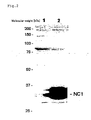
- Fig. 2 is a representation of the results of detection, by the Western blotting, of the NC1 gene product forcedly expressed in Jurkat cells. In
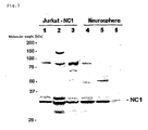
Lane 1, the Jurkat cell-derived whole RNA was electrophoresed and, inLane 2, the whole RNA derived from Jurkat cells in which the NC1 gene had been forcedly expressed was electrophoresed. - Fig. 3 is a representation of the results of detection, by the Western blotting, the NC1 gene product forcedly expressed in Jurkat cells and the NC1 gene product occurring in the neurospheres. In
Lanes Lanes Lanes - The following examples illustrate the present invention more specifically. They are, however, by no means limitative of the scope of the invention. It is to be noted that various variations and modifications can be made without departing from the spirit of the invention.
- The following experiments were carried out for detecting the expression of the NC1 gene by the Northern blotting.
- The probe to be used in detecting the gene expression was constructed in an in vitro transcription system using a vector containing a DNA fragment corresponding to the NC1 gene as inserted therein. Specifically, the following procedure was followed. The oligonucleotides resulting from addition of an XhoI site to each of the base sequence shown under SEQ ID No: 2 and the base sequence shown under SEQ ID No: 3 were used as primers. A part of a human embryonic cDNA library obtained from Clontech Laboratories, Inc. was subjected to heat treatment, and the supernatant obtained by separating the sediment by centrifugation was used as the template. Using the above-mentioned primers and template, the DNA fragment containing an NC1 gene-specific sequence was amplified by PCR. The amplification product was cleaved with XhoI, followed by subcloning into a vector (pGEM-T Easy, product of Promega K.K.) . This vector thus contained the base sequence shown under SEQ ID NO:1 as a probe fragment. Using this vector, an RNA probe labeled with DIG-11-UTP (product of Roche Molecular Biochemicals) was prepared in an in vitro transcription system.
- Tissue sections were collected from the human embryo forebrain and spinal cord, and neurospheres comprising human nerve stem cells/progenitor cells were cultivated using the neurosphere cultivation technique (Reynolds, B. A. et al., (1992) J. Neurosci, 12:4565-74; Vescovi, A. L. et al., (1993) Neuron 11:951-66; Reynolds, B. A. and Weiss, S., (1996) Dev. Biol. 175:1-13). As for the details of the cultivation technique, the procedure of Kanemura et al. (Kanemura, Y. et al., (2002) J. Neurosci. Res. 69:869-79) was followed.
- In each of cell culture flasks with a base area of 75 cm2, 3 x 10⁶ cells were precultured for 4 days and, then, cells were recovered by centrifugation and resuspended in a medium supplemented with retinoic acid and 1% of fetal calf serum. On the 7th day after the start of cultivation, the medium was exchanged for a fresh one and the cultivation was continued until the 14th day. This group of cells was designated as "differentiation-induced group A". The group of cells obtained by growing, similarly after 4 days of precultivation, the cells in a medium supplemented with fetal calf serum to a final concentration of 10% and continuing the cultivation until a confluent state after two passages was designated as "differentiation-induced group B".
- Total RNA was extracted from neurosphere-constituting cells, and from each of differentiation-induced groups A and B. 1-µg portion of each extract was developed on an agarose gel containing 1% of formamide, followed by transfer to a nylon membrane (Hybond-N; product of Amersham Biosciences K.K.). The membrane with transferred RNA and the labeled RNA probe prepared as described above in (1) were subjected to overnight hybridization in DIG Easy Hyb Buffer (product of Roche Molecular Biochemicals) at 68°C. After hybridization, the membrane was washed and then reacted with Anti-Digoxigenin-AP Fab Fragment (product of Roche Molecular Biochemicals) and subjected to the NC1 gene expression detection using CDP-Start chemiluminescent substrate (product of Amersham Biosciences K.K.).
- Using the materials and methods described above under (1) to (3), the expression of the NC1 gene was investigated in human nerve stem cells/progenitor cells and in each of the differentiation-induced groups of cells. Fig. 1 shows the results of the analyses. In Fig. 1, "NC1" indicates the position corresponding to the mobility of the NC1 gene-derived mRNA, and "28S rRNA" indicates the position corresponding to the mobility of the 28S ribosome RNA. "BXPC3" indicates the human pancreatic carcinoma-derived cell line BXPC3, "NTERA-2" the human embryonal carcinoma-derived cell line NTERA-2, "Jurkat" the human T cell line Jurkat, and "U251" the human glioblastoma-derived cell line U251.
- The neurospheres prepared from the fetal human tissues comprise nerve stem cells/progenitor cells. In the differentiation-induced group A, the cells are composed of tubulin B III-positive cells having the characters of neurons and GFAP (glial fibrillary acidic protein)-positive cells having the characters of astrocytes and, in the differentiation-induced group B, almost all cells are GFAP-positive cells. In these culture systems, the expression of the NC1 gene was markedly high in neurospheres, and only low levels of the expression were observed in both the differentiation-induced groups A and B (Fig. 1). These results indicate that the expression of the NC1 gene is high in nerve stem cells/progenitor cells and decreases with the progress of differentiation into neurons and/or astrocytes. Thus, it was shown that since the NC1 gene highly expresses specifically in nerve stem cells/progenitor cells, this gene can be applied as a nerve stem cell marker.
- The following experiments were carried out for detecting the NC1 gene product by the Western blotting.
- An antibody responsive to the NC1 gene product was produced by using, as an antigen, a partial peptide of the protein encoded by the NC1 gene. In practice, a partial peptide of the NC1 gene product, namely a peptide having the amino acid sequence shown under SEQ ID NO:4, was synthesized, and a rabbit was immunized therewith to give antiserum. It was confirmed by ELISA that this antiserum reacts with the original peptide at a titer of at least ten thousand times. Thus, this was used as antiserum containing an antibody against the NC1 gene product.
- An attempt was made to detect, by the Western blotting using antiserum prepared as described above under (1), the NC1 gene product in the neurospheres prepared from the fetal human nerve tissue in which the expression of the NC1 gene was detected in Example 1. Attempts were also made to detect, by the Western blotting, the NC1 gene product in Jurkat cells in which the expression of the NC1 gene was not detected and in a transformant strain derived from Jurkat cells by introduction of the NC1 gene expression vector. Whole cell extracts were prepared from these cell species and, further, cytoplasmic fractions and nuclear fractions were prepared using N-XTRACT Kit (product of Sigma).
- Each sample prepared as described above in (2) was heated at 95°C for 5 minutes and then fractionated by SDS polyacrylamide gel electrophoresis, followed by transfer to a nitrocellulose membrane. The membrane was blocked with a blocking solution (Tris buffer containing 5% of skimmed milk and 0.1% of Tween 20) and, then, antiserum prepared as described above in (1) was added. The membrane was washed and then reacted with a horseradish peroxidase-labeled anti-rabbit IgG antibody (product of Amersham Biosciences K.K.) for detecting any reactive fraction using ECL (product of Amersham Biosciences K.K.).
- Using the materials and methods described above in (1) to (3), attempts were made to detect the NC1 gene product by means of antiserum prepared. The results are shown in Figs. 2 and 3. Referring to Fig. 2, the Jurkat cell-derived whole RNA was electrophoresed in
Lane 1, and the whole RNA derived from Jurkat cells in which the NC1 gene had been forcedly expressed was electrophoreses inLane 2. Referring to Fig. 3, inLane 1, the whole cell extract derived from Jurkat cells in which the NC1 gene had been forcedly expressed was electrophoresed; inlane 2, the cytoplasmic fraction derived from Jurkat cells in which the NC1 gene had been forcedly expressed was electrophoresed; inLane 3, the nuclear fraction derived from Jurkat cells in which the NC1 gene had been forcedly expressed was electrophoresed; inLane 4, the whole cell extract from the neurospheres was electrophoresed; inLane 5, the cytoplasmic fraction of the neurospheres was electrophoresed; and, inLane 6, the nuclear fraction of the neurospheres was electrophoresed. - The antiserum mentioned above did not react with the Jurkat cells incapable of expressing the NC1 gene (Fig. 2, lane 1) but reacted with the transformant (Jurkat-NC1) derived from Jurkat cells and forcedly caused to express the NC1 gene, giving a band corresponding to the NC1 gene product (Fig. 2, lane 2). This indicates that antiserum produced as described above under (1) recognizes the NC1 gene product and that the product can be detected at least by the Western blotting.
- When the fetal human nerve tissue-derived neurospheres were subjected to Western blotting, bands reacting with antiserum were detected at the same position as that detected in Jurkat-NC1 forcedly caused to express the NC1 gene (Fig. 3) . Furthermore, in the case of Jurkat NC1, antiserum reacted with both the cytoplasmic and nuclear fractions but, in the case of the neurospheres, the cytoplasmic fraction alone showed reactivity. Therefore, the occurrence of the NC1 gene product in nerve stem cells/progenitor cells, such as neurospheres, can be detected by Western blotting using this antiserum. Since this antiserum reacts not only with the whole cell extract but also with the fractionated extract, it is also possible to reveal the intracellular localization of the NC1 gene product.
- The NC1 gene and the NC1 protein of the invention are considered to be involved in the process of differentiation of nerve system cells and can be applied as nerve stem cell markers with which nerve stem cells/progenitor cells can be detected or sorted out.


Claims (10)
- A DNA which comprises the whole or a part of the base sequence shown under SEQ ID NO:1.
- A DNA which comprises the base sequence shown under SEQ ID NO:2 or SEQ ID NO:3.
- A peptide which comprises the amino acid sequence shown under SEQ ID NO:4.
- A peptide which comprises the amino acid sequence shown under SEQ ID NO:5.
- A peptide which comprises the amino acid sequence shown under SEQ ID NO:6.
- An antibody which is obtained by using one of the peptides according to any one of Claims 3 to 5 as an antigen.
- The antibody according to Claim 6
wherein the antibody is a polyclonal antibody. - The antibody according to Claim 6
wherein the antibody is a monoclonal antibody. - A method for detecting nerve stem cells/progenitor cells which uses at least one species selected from the group consisting of the DNAs according to Claims 1 and 2, peptides according to Claims 3 to 5 and antibodies according to Claims 6 to 8.
- A method for screening for a drug functioning as a nerve differentiation factor or nerve differentiation inhibitor using the method according to Claim 9.
Applications Claiming Priority (2)
| Application Number | Priority Date | Filing Date | Title |
|---|---|---|---|
| JP2003321564A JP2005087018A (en) | 2003-09-12 | 2003-09-12 | Novel neural stem cell marker |
| PCT/JP2004/013221 WO2005030961A1 (en) | 2003-09-12 | 2004-09-10 | Novel nerve stem cell marker |
Publications (2)
| Publication Number | Publication Date |
|---|---|
| EP1669453A1 true EP1669453A1 (en) | 2006-06-14 |
| EP1669453A4 EP1669453A4 (en) | 2007-12-12 |
Family
ID=34385936
Family Applications (1)
| Application Number | Title | Priority Date | Filing Date |
|---|---|---|---|
| EP04787861A Withdrawn EP1669453A4 (en) | 2003-09-12 | 2004-09-10 | NEW STERILE NERVE CELL MARKER |
Country Status (4)
| Country | Link |
|---|---|
| US (1) | US20070031894A1 (en) |
| EP (1) | EP1669453A4 (en) |
| JP (2) | JP2005087018A (en) |
| WO (1) | WO2005030961A1 (en) |
Families Citing this family (5)
| Publication number | Priority date | Publication date | Assignee | Title |
|---|---|---|---|---|
| JP4792290B2 (en) * | 2005-12-27 | 2011-10-12 | 財団法人 東京都医学総合研究所 | Method for producing neural stem cells |
| JP2012029684A (en) * | 2010-06-30 | 2012-02-16 | Cell Aid Kenkyusho:Kk | Method for producing cell |
| WO2012068170A2 (en) * | 2010-11-15 | 2012-05-24 | Jau-Nan Lee | Generation of neural stem cells from human trophoblast stem cells |
| GB2523696B (en) | 2012-11-30 | 2017-12-27 | Accelerated Biosciences Corp | Methods of differentiating stem cells to pancreatic progenitor cells by modulating miR-124 |
| SG11201704291QA (en) | 2014-11-26 | 2017-06-29 | Accelerated Biosciences Corp | Induced hepatocytes and uses thereof |
Family Cites Families (3)
| Publication number | Priority date | Publication date | Assignee | Title |
|---|---|---|---|---|
| US6682888B1 (en) * | 2000-05-05 | 2004-01-27 | Incyte Corporation | Genes expressed in alzheimer's disease |
| US20030028904A1 (en) * | 2001-04-19 | 2003-02-06 | Gumienny Tina L. | Genes involved in engulfment of dying cells and cell migration |
| US20060057645A1 (en) * | 2002-03-15 | 2006-03-16 | Hideo Niwa | Novel genes |
-
2003
- 2003-09-12 JP JP2003321564A patent/JP2005087018A/en active Pending
-
2004
- 2004-09-10 EP EP04787861A patent/EP1669453A4/en not_active Withdrawn
- 2004-09-10 JP JP2005514168A patent/JPWO2005030961A1/en active Pending
- 2004-09-10 WO PCT/JP2004/013221 patent/WO2005030961A1/en not_active Ceased
- 2004-09-10 US US10/571,268 patent/US20070031894A1/en not_active Abandoned
Non-Patent Citations (2)
| Title |
|---|
| DATABASE EMBL [Online] 9 November 1996 (1996-11-09), "Homo sapiens mRNA for KIAA0281 gene, partial cds." XP002455954 retrieved from EBI accession no. EMBL:D87457 Database accession no. D87457 * |
| See also references of WO2005030961A1 * |
Also Published As
| Publication number | Publication date |
|---|---|
| JP2005087018A (en) | 2005-04-07 |
| EP1669453A4 (en) | 2007-12-12 |
| WO2005030961A1 (en) | 2005-04-07 |
| JPWO2005030961A1 (en) | 2008-02-14 |
| US20070031894A1 (en) | 2007-02-08 |
Similar Documents
| Publication | Publication Date | Title |
|---|---|---|
| Britanova et al. | Novel transcription factor Satb2 interacts with matrix attachment region DNA elements in a tissue‐specific manner and demonstrates cell‐type‐dependent expression in the developing mouse CNS | |
| Behar et al. | cis-acting signals and trans-acting proteins are involved in tau mRNA targeting into neurites of differentiating neuronal cells | |
| CA2190365A1 (en) | Pigment epithelium-derived factor: characterization, genomic organization and sequence of the pedf gene | |
| AU2002306374B2 (en) | Genes with ES cell-specific expression | |
| JP5711149B2 (en) | Non-embryonic stem cells and uses thereof | |
| RU2002121771A (en) | Compositions and methods for the treatment and diagnosis of prostate cancer | |
| Boyd et al. | Proteomic evaluation reveals that olfactory ensheathing cells but not Schwann cells express calponin | |
| Maurer et al. | Comprehensive proteome expression profiling of undifferentiated versus differentiated neural stem cells from adult rat hippocampus | |
| Rapp et al. | Identification of T cell target antigens in glioblastoma stem-like cells using an integrated proteomics-based approach in patient specimens | |
| Tang et al. | A lncRNA Dleu2-encoded peptide relieves autoimmunity by facilitating Smad3-mediated Treg induction | |
| US20070031894A1 (en) | Novel nerve stem cell marker | |
| Skalnikova et al. | A proteomic approach to studying the differentiation of neural stem cells | |
| DE69534705D1 (en) | DICKDARM SPECIFIC GENES AND PROTEINS | |
| EP1333099B1 (en) | Method of searching for gene encoding nuclear transport protein | |
| EP1399584A2 (en) | Cancer-linked genes as targets for chemotherapy | |
| CN114736298B (en) | Anti-shrom 2 autoantibody for detecting autoimmune system diseases, and product and application thereof | |
| CN115724997B (en) | Anti-CD 97 chimeric antigen receptor, gene, expression vector, T cell and application | |
| KR102787403B1 (en) | Techniques for producing immortalized pig alveolar macrophages cell lines using IKKε and providing ASFV-infected cell lines | |
| CN101891813A (en) | Leukemia drug-resistant cell membrane protein and its use as drug-resistant target protein | |
| Das et al. | Genetic identity and differential expression of p38. 5 (Haymaker) in human malignant and nonmalignant cells | |
| KR101818819B1 (en) | Method of providing the information for predicting survival of brain cancer patients | |
| Vidovic et al. | Analysis of EphB receptors and their ligands in the developing retinocollicular system of the wallaby reveals dynamic patterns of expression in the retina | |
| US20140018304A1 (en) | Method for screening for a cancer treatment agent using the interaction between pauf and a binding partner thereof | |
| US11920159B2 (en) | Methods for expanding hematopoietic stem cells using revitalized mesenchymal stem cells | |
| CN121824725A (en) | Application of MP48 and its inhibitors in the preparation of drugs for treating glioblastoma |
Legal Events
| Date | Code | Title | Description |
|---|---|---|---|
| PUAI | Public reference made under article 153(3) epc to a published international application that has entered the european phase |
Free format text: ORIGINAL CODE: 0009012 |
|
| 17P | Request for examination filed |
Effective date: 20060323 |
|
| AK | Designated contracting states |
Kind code of ref document: A1 Designated state(s): AT BE BG CH CY CZ DE DK EE ES FI FR GB GR HU IE IT LI LU MC NL PL PT RO SE SI SK TR |
|
| RAP1 | Party data changed (applicant data changed or rights of an application transferred) |
Owner name: FOUNDATION FOR BIOMEDICAL RESEARCH AND INNOVATION Owner name: KANEKA CORPORATION |
|
| DAX | Request for extension of the european patent (deleted) | ||
| A4 | Supplementary search report drawn up and despatched |
Effective date: 20071112 |
|
| STAA | Information on the status of an ep patent application or granted ep patent |
Free format text: STATUS: THE APPLICATION IS DEEMED TO BE WITHDRAWN |
|
| 18D | Application deemed to be withdrawn |
Effective date: 20080210 |