EP1658855B1 - Use of substance p for mobilization or proliferation of mesenchymal stem cells and for wound healing - Google Patents
Use of substance p for mobilization or proliferation of mesenchymal stem cells and for wound healing Download PDFInfo
- Publication number
- EP1658855B1 EP1658855B1 EP05023367A EP05023367A EP1658855B1 EP 1658855 B1 EP1658855 B1 EP 1658855B1 EP 05023367 A EP05023367 A EP 05023367A EP 05023367 A EP05023367 A EP 05023367A EP 1658855 B1 EP1658855 B1 EP 1658855B1
- Authority
- EP
- European Patent Office
- Prior art keywords
- substance
- wound
- mscs
- healing
- cells
- Prior art date
- Legal status (The legal status is an assumption and is not a legal conclusion. Google has not performed a legal analysis and makes no representation as to the accuracy of the status listed.)
- Expired - Lifetime
Links
Images




Classifications
-
- A—HUMAN NECESSITIES
- A61—MEDICAL OR VETERINARY SCIENCE; HYGIENE
- A61K—PREPARATIONS FOR MEDICAL, DENTAL OR TOILETRY PURPOSES
- A61K35/00—Medicinal preparations containing materials or reaction products thereof with undetermined constitution
- A61K35/12—Materials from mammals; Compositions comprising non-specified tissues or cells; Compositions comprising non-embryonic stem cells; Genetically modified cells
- A61K35/28—Bone marrow; Haematopoietic stem cells; Mesenchymal stem cells of any origin, e.g. adipose-derived stem cells
-
- A—HUMAN NECESSITIES
- A61—MEDICAL OR VETERINARY SCIENCE; HYGIENE
- A61K—PREPARATIONS FOR MEDICAL, DENTAL OR TOILETRY PURPOSES
- A61K38/00—Medicinal preparations containing peptides
- A61K38/04—Peptides having up to 20 amino acids in a fully defined sequence; Derivatives thereof
- A61K38/046—Tachykinins, e.g. eledoisins, substance P; Related peptides
-
- A—HUMAN NECESSITIES
- A61—MEDICAL OR VETERINARY SCIENCE; HYGIENE
- A61P—SPECIFIC THERAPEUTIC ACTIVITY OF CHEMICAL COMPOUNDS OR MEDICINAL PREPARATIONS
- A61P17/00—Drugs for dermatological disorders
- A61P17/02—Drugs for dermatological disorders for treating wounds, ulcers, burns, scars, keloids, or the like
-
- A—HUMAN NECESSITIES
- A61—MEDICAL OR VETERINARY SCIENCE; HYGIENE
- A61P—SPECIFIC THERAPEUTIC ACTIVITY OF CHEMICAL COMPOUNDS OR MEDICINAL PREPARATIONS
- A61P43/00—Drugs for specific purposes, not provided for in groups A61P1/00-A61P41/00
Definitions
- the present invention relates to Substance-P for use in a method of wound-healing or facilitating wound-healing, as defined in the claims. Further, the invention relates to a composition comprising as effective ingredients Substance-P and mesenchymal stem cells (MSCs), as defined in the claims.
- MSCs mesenchymal stem cells
- Substance-P is neuropeptide consisted of 11 amino acids the tachykinin family that is expressed in sensory neurons, macrophages, eosinophils, endothelial cells, and corneal cells such as epithelial cells and keratocytes as well as granulation tissue.
- Several reports have suggested implication of Substance-P in neuro-immune communication on hematopoietic modulation.
- the bone marrow stroma was innervated by Substance-P nerve fibers and Substance-P stimulates marrow stromal cells through their surface receptor NK-1 to produce the stem cell factor and interleukin-1, which may be favorable for hematopoietic stimulation as feeders.
- NK-1 surface receptor NK-1
- a wound itself creates a unique and specific microenvironment that is composed of growth factors, cytokines, neurohormones, and extracellular matrix, all of which are secreted from the neighboring cells, blood born cells, and sensory neurons. Some factors in the wound microenvironment may last sufficiently long to be diffused into the peripheral blood, and in turn affect the stem cells in the bone marrow, to induce mobilization of the bone marrow cells into peripheral blood, supply of the bone marrow cells into the wound site, and participation of the bone marrow cells in the wound healing.
- the cornea is consisted of transparent, avascular and heavily innervated tissue, whose integrity may be broken in case of corneal damage and disease thereof.
- a corneal wound stimulates lateral movement of the corneal epithelial cells most likely provided by the limbal stem cells, infiltration of the inflammatory cells, and neovascularization in the wound stroma, all of which may be stimulated by the unique corneal wound microenvironment.
- Previous reports suggest that corneal denervation is a cause of substantial delay in the wound healing process, and the lower level of Substance-P in diabetic patients than non-diabetic patients is also a cause of delayed re-epithelialization and delayed healing.
- Substance P Since the cornea surface is extensively innervated by trigeminal ganglion neurons, and endogenous Substance P is expressed in the corneal epithelial cells and keratocytes, it is expected for Substance P to involve in the wound microenvironment and participate in cornea repair.
- Substance-P is a first neuropeptide which is elevated in eye and peripheral blood in a mouse with corneal alkali burn. Also, they discovered that even though Substance P was injected intravenously into the non-corneal wound mice, many CD29+ MSCs were mobilized into peripheral blood. Further, it was revealed in 3-D collagen gel in vitro that Substance-P stimulates migration of human MSC with induction of matrix degrading enzymes and inhibition of their inhibitor. In addition, they discovered that Substance P stimulates cell proliferation of MSCs, nuclear translocation of ⁇ -catenin, and expression of its target genes, VEGF and fibronectin, and so Substance P may play a role in the bone marrow repopulation of MSCs after their mobilization.
- Di 1 labeled MSCs which were transfused into the ear vein of the alkali burned rabbits, are successfully mobilized at the wound bed and facilitates cornea wound healing with improved corneal transparency and visual recovery. Further, they showed that intravenously-injected Substance-P can facilitate wound healing of alkali burn rabbit eye.
- the first object of the present invention is to provide Substance-P for use in a method of wound-healing or facilitating wound-healing, wherein the Substance-P is administered by intravenous injection, as defined in the claims.
- the second object of the present invention is to provide a composition comprising as effective ingredients Substance-P and mesenchymal stem cells (MSCs) which have been mobilized or proliferated from the bone marrow by treatment of Substance-P for use in a method of wound-healing or facilitating wound-healing, as defined in the claims.
- the medicament is a cellular therapeutic agent.
- a use of Substance-P for the manufacture of a medicament for mobilization or proliferation of Mesenchymal stem cells (MSCs) from the bone marrow, or facilitating said mobilization or proliferation is described.
- Substance-P for the manufacture of a medicament for wound-healing or facilitating wound-healing is described.
- a wound healing or facilitating wound healing agent containing MSCs as an effective ingredient, wherein the MSCs are mobilized or proliferated from the bone marrow by treatment of Substance-P, is described.
- a method of healing wound or facilitating wound-healing comprising administering a therapeutically effective amount of MSCs, wherein the MSCs are mobilized or proliferated from the bone marrow by treatment of Substance-P, is described.
- Substance-P for the manufacture of a medicament for mobilization or proliferation of Mesenchymal stem cells (MSCs) from the bone marrow, or facilitating said mobilization or proliferation is described. It is contemplated that said medicament may comprise MSCs. It is also contemplated that said medicament may be a cellular therapeutic agent.
- MSCs Mesenchymal stem cells
- the present invention includes a use of Substance-P as a medicament for wound-healing or facilitating wound-healing, for example, cornea and skin wound.
- said medicament may comprise MSCs.
- said medicament may be a cellular therapeutic agent.
- the present invention relates to Substance-P for use in a method of wound-healing or facilitating wound-healing, wherein the Substance-P is administered by intravenous injection, as defined in the claims.
- Substance-P as an effective ingredient, for example, for cornea and skin wound.
- a wound healing or facilitating wound healing agent containing MSCs as an effective ingredient, wherein the MSCs are mobilized or proliferated from the bone marrow by treatment of Substance-P is described.
- the invention provides a composition comprising as effective ingredients Substance-P and MSCs which have been mobilized or proliferated from the bone marrow by treatment of Substance-P for use in a method of wound-heating or facilitating wound healing, as defined in the claims.
- a method of healing wound or facilitating wound-healing comprising administering a therapeutically effective amount of MSCs, wherein the MSCs are mobilized or proliferated from the bone marrow by treatment of Substance-P is described.
- the present inventors used alkali burn cornea animal model which has advantages in examining systemic participation of stem cells during the wound healing process over other wound model since cornea has transparency and avascularization.
- Substance-P is the first neuropeptide whose expression is elevated in the cornea wound as well as in the peripheral blood of the mice with corneal alkali burn. At this time, Substance-P was first increased in cornea and later in peripheral blood.
- Substance-P is provided by the corneal resident cells such as the sensory neurons, epithelial cells, keratocytes, and endothelial cells, not by infiltration of neutrophils or macrophages.
- the increase of Substance-P concentration in blood may be from systemic diffusion of Substance-P.
- lack of Substance-P immunoreactivity in the neutrophils, earlier induction and termination of Substance P than duration of the inflammatory phase (up to 5 days after wound), and the weak Substance-P staining in the epithelial cells and fibroblastic cells at the later stage of wound healing (7-10 days after wound) supports an assumption that Substance-P is provided by nociceptive stimulation of the sensory neurons.
- the present inventors examined whether or not intravenously injected MSCs arrive at damaged tissue and are involved in repair of the cornea, based on the corneal transparency and vision recovery scores. As a result, they confirmed that Di 1-labeled MSCs, which were transfused into the ear vein of alkali burned rabbits, are successfully supplied to the wound bed and improve corneal transparency and visual recovery, thereby facilitating cornea wound healing.
- Substance-P can facilitate corneal wound healing, based on macroscopic examination, H&E staining, and immunohistochemical staining. As a result, compared with the control group (Substance-P non-injected group), healing of the corneal wound in Substance-P-injected group was progressed rapidly.
- Substance-P is first elevated in the cornea wound and the peripheral blood, and facilitates mobilization of MSCs to peripheral blood, and promotes healing of the corneal wound and proliferation of MSCs in the bone marrow.
- Substance-P plays a role as wound signaling initiator to involve in mobilization and supply of MSCs from the bone marrow to the wound site. Also, it was identified that MSCs mobilize to wound site and are involved in wound healing.
- MSCs can be used as wound healing or facilitating wound healing medicament, or cellular treatment medicament.
- Substance-P can be used as wound healing or facilitating wound healing agent, or cellular treatment medicament, or mobilization, or proliferation medicament, or facilitating mobilization or proliferation medicament, of MSCs.
- the effective dosage of Substance-P is 0.1 ⁇ g/kg to 100 ⁇ g/kg
- the effective dosage of MSCs is 3 ⁇ 10 4 cells/kg to 3 ⁇ 10 7 cells/kg, particularly, 1 ⁇ 10 5 cells/kg to 1 ⁇ 10 7 cells/kg.
- these dosages can be modified depending on weight, age, sex or extent of wound of patients.
- a formulation according to the present invention can be administered into human bodies parentally or locally, for example, by intravenous injection, subcutaneous injection, endodermis injection, and muscular injection. Preferably, they are administered by intravenous injection.
- the effective ingredients are suspended or dissolved in a pharmacologically acceptable carrier, preferably, water-soluble carrier.
- Example 1 Identification of elevation of c-kit positive stem cells in peripheral blood and their supply to the would bed after corneal alkali burn
- New Zealand white rabbits weighing 2-3kg were purchased from Samtako BioKorea. All these animal experiments were approved by Ethical Committee, Korea Institute of Radiological and Medical Sciences, and Chung-Ang University, and conducted in accordance with the ARVO Statement for the Use of Animal in Ophthalmic and Vision Research.
- the rabbit eyes were placed in contact with a 6mm piece of circular Whatman filter paper soaked in 1N NaOH for 30 seconds.
- rabbit eyes and whole blood were isolated.
- the smears of blood and cornea section were stained with Anti-C kit antibody, and this result was shown in Figure 1 .
- Intact cornea is avascular tissue, and so extensive damage thereto might require vascular supply of inflammatory cells and other stem cells for the corneal repair.
- Figure 1 when the alkali burn was done on the rabbit cornea, the number of c-kit positive cells was increased in the peripheral blood, compared with the normal control, which was also detected in the corneal wound bed at 5th day after the alkali burn. This result confirms that stem cells originating from the bone marrow may actively participate in the repair of the cornea.
- Example 2 Identification of role of Substance-P as wound signaling initiator induced by cornea alkali burn
- mice weighing 30-40g were purchased from Jackson Lab (West Grove, PA).
- the mouse eyes were placed in contact with a 3mm piece of circular Whatman filter paper soaked in 1N NaOH for 10 seconds.
- the mouse eyes and whole blood were isolated.
- Tissue damage itself may constitute a unique wound microenvironment to induce a systemic response such as inflammation and stem cell mobilization from the bone marrow to repair the damaged tissue.
- RT-PCR was used to analyze candidate factors retaining these functions in the wound microenvironment of alkali burned cornea. Specifically,
- ELISA R&D system
- Fig. 2b ELISA (R&D system) to the peripheral blood was performed.
- the level of Substance-P in the serum was elevated by approximately 3.2. times at 1 day, 4.4 times at 3 day, and 1.3 times at 5 day.
- pre-inflammatory cytokines, TNF-a, and IL-1 induction were detected at 5 day and 3 day after the alkali burn, respectively, and VEGF was induced from 7 day much later than them.
- Hematoxylin and Eosin (H&E) staining was performed for corneal wound bed, and immunohistochemical staining was performed with Substance-P antibody (Santa Cruz Biotechnology: Cat # sc-9758, 1:200).
- the mouse eyes were fixed with 4% of paraformaldehyde for 48h.
- the paraffin-embedded specimens were cut longitudinally into 4- ⁇ m sections, and transferred to poly-D lysine-coated slides. Then, Hematoxylin and Eosin (H&E) staining was performed for the histological examination.
- Fig. 2c The result is shown in Fig. 2c .
- the inflammatory neutrophils were sustained for up to 5 days, the migrating epithelial cells were discernible at 5 day, fibroblastic infiltration was obvious at 7 day, and most macrophages had disappeared at 14 day.
- the immuno-reactive Substance-P was most strongly detected in the wound bed after 1 day and became weaker till 3rd day.
- Substance-P was barely detected in the fibroblastic cells and migrating epithelial cells, but was not detected in the inflammatory cells, which suggests that the early inflammatory cells may not secrete Substance-P. Therefore, the cornea wound itself appears to create a Substance-P rich microenvironment possibly provided by cornea resident cells, which may in turn induce the Substance-P elevation in the serum.
- Example 3 Identification of mobilization of MSCs from the marrow into peripheral blood by intravenous injection of Substance-P
- Substance-P was examined to determine whether it is responsible for stem cell mobilization into the blood stream or whether some other unidentified factors in the wound microenvironment are involved in stem cell mobilization.
- Substance-P (0.1 nmole/g of body weight) (Calbiochem) was injected intravenously through tail vein in Balb-c mice without causing alkali bums to the eye, and then the whole blood was collected after 1 day. After removing red blood cells by percoll gradient centrifugation, the attached cells were cultured for 48 h in order to remove lymphocytes, and then, in order to distinguish MSCs from the lymphocytes, they were immune-stained with antibodies against CD-29 ( ⁇ 1 integrin) which is not expressed in the macrophages.
- CD-29 ⁇ 1 integrin
- Figs. 3a and 3b The result is shown in Figs. 3a and 3b .
- the intravenous injection of Substance-P strongly stimulated mobilization of CD-29 positive MSCs into the peripheral blood approximately by 15 times more than in the non-injected mice ( Figure 3b ).
- Substance-P was first examined to determine whether it stimulates migration of human MSCs cultured on top of the 3-D collagen gel.
- the type I collagen gel matrix was made within a 12 mm millicell membrane (milipore: 12- ⁇ m pore size) according to the manufacturer's specifications (Nitta gelatin, Japan) and coated overnight with type IV collagen (Nitta).
- the MSCs were plated on top of the collagen gel, and the outer chamber was filled with Substance-P-containing MSCGM (Cambrex Bio Science). Clearance of the collagen gel was monitored by using the phase contrast microscopy (Olympus).
- the millicell insert and outer dishes were fixed and stained with hematoxylin, and then the culture supernatants were stored for gelatin zymography. This result is shown in Fig. 4a .
- Substance-P applied into the bottom dishes stimulated degradation of the collagen gel and MSC migration in a dose dependent manner.
- the culture supernatants were examined for gelatin zymography.
- the eletrophoresis was performed for the culture supernatant in which sample buffer without mercaptoethanol was added thereto, in 8 % SDS-PAGE.
- the gel was renatured with 2.5% of Triton x-100 in order to remove the SDS, and then was incubated with a developing buffer (50 mM Tris-HCl, pH 7.2 100 mM NaCl, 20 mM CaCl 2 ) at 37°C overnight, and the gel was stained with Coomassie blue.
- a developing buffer 50 mM Tris-HCl, pH 7.2 100 mM NaCl, 20 mM CaCl 2
- gelatin zymography of the culture medium revealed strong induction of MMP-9 and MMP-2 activities during MSC migration.
- the effect of Substance-P on the biosynthesis of MMPs (matrix metalloproteinases) and their inhibitors was examined by immunoprecipitation of the culture medium and the cell lysates of MSCs which were labeled with 35 S-methionine.
- the MSCs were treated with Substance-P and labeled with 50 ⁇ Ci/ml 35 S-methionine (Amersham) for 16h.
- the cell lysate was prepared with a lysis buffer (1% of NP40, 10 mM Tris-HCl, pH 7.4, 150 mM NaCl, 1 mM EDTA, 2 mM PMSF).
- MMP-1, MMP-2, MMP-9, TIMP-1 and TIMP-2 were incubated with their specific antibodies [anti-MMP-1 (chemicon; Cat # MAB3307, 1:100), anti-MMP-2 (Chemicon; Cat # MAB13405, 1:200), anti-MMP-9 (Calbiochem; Cat # 444236, 1:100), anti-TIMP-1 (Calbiochem; Cat # IM32L, 1:250) and anti-TIMP-2 (Calbiochem; Cat # IM11L, 1:250)] for 2h under constant rotation, and the antibodies were collected by protein-G sepharose-4B (Biorad).
- the pellet was washed three times with an immunoprecipitation buffer (150 mM NaOH, 50 mM Tris-HCl pH 7.4, 0.2% SDS and 0.5% Nadeoxycholate), once with a high salt buffer (10 mM Tris-HCl, pH 7.4, 0.5 M NaCl), and once with a low salt buffer (10 mM Tris-HCl, pH 7.4).
- an immunoprecipitation buffer 150 mM NaOH, 50 mM Tris-HCl pH 7.4, 0.2% SDS and 0.5% Nadeoxycholate
- a high salt buffer (10 mM Tris-HCl, pH 7.4, 0.5 M NaCl
- a low salt buffer 10 mM Tris-HCl, pH 7.4
- Substance-P stimulated MSC migration in 3-D collagen gel, induction of MMPs, and inhibition of their inhibitors, TIMPs.
- Example 5 Identification for facilitation of MSC repopulation by Substance-P
- Substance-P only has capacity of MSC mobilization from the bone marrow, the bone marrow stroma may become empty in the MSC pool after the mobilization.
- the effect of Substance-P was again examined about its cell proliferation and self-renewal capacity.
- Substance-P increased the number of viable cells, 1.7 times at 1nM, 2.1 times at 10 nM, and 2.2 times at 100 nM, compared with the control group.
- ⁇ -catenin which is the downstream effecter of the wnt signaling pathway.
- the MSCs were fixed with 4% of paraformaldehyde and permeabilized with 0.3% of Triton X-100. After blocking nonspecific antigen with 20% normal goat serum which was diluted in phosphate buffer for 1h, the coverslips were incubated with antibodies against ⁇ -catenin (BD Biosciencs; Cat # 6010153, 1:100) for 90min, and then with the FITC-conjugated secondary antibodies for 60 min(Vector). The cells were counterstained with DAPI, and examined by using confocal microscopy (Leica). This result is shown in Fig. 5b .
- ⁇ -catenin was mainly observed in the cytoplasm of human MSCs, but only a small number of MSCs at 100 nM showed nuclear localization of ⁇ -catenin. However, at 48h after Substance-P treatment, the nuclear translocation of ⁇ -catenin was more prominent, and particularly at 100 nM of Substance-P treatment, all the MSCs showed nuclear translocation of ⁇ -catenin.
- Substance-P induced VEGF and fibronectin. This proves that Substance-P plays a role in repopulation of the remaining MSCs by the stimulation of cell proliferation or self-renewal via well-known canonical wnt signaling pathway.
- Substance-P stimulated cell proliferation, translocation of ⁇ -catenin, and induction of ⁇ -catenin downstream gene, VEGF and fibronectin, all of which suggest that Substance-P plays a role in repopulation of the bone marrow.
- Example 6 Identification for supply of I. V.-injected MSCs to the cornea wound bed and improvement in the vision recovery
- the MSCs were isolated from the tibia of a 1 month old allogenic rabbit by bone marrow irrigation and aspiration, and cultured with MSCGM up to passage 3.
- the attached cells were identified as MSCs by the expression of c-kit, STRO-1, and ⁇ -smooth muscle (SM) actin, and were used for the cell transfusion experiment.
- the trypsinized cells were incubated with a Di 1 solution [CelltrackerTM CM-Di I(Cat# C-7000); Molecular Probes] for 5min at 37°C and 15min at 4°C. After labeling and subsequent washing with PBS three times, the MSCs (1x10 6 cells /ml) were resuspended in fresh serum free medium and injected into the ear vein of 10 rabbits at 1 day after the corneal alkali burn.
- the epithelial healing time was determined by closure of the epithelial defect with photo-slit microscopic examination and photo-documentation.
- the corneal opacity was graded according to the following criteria: grade 0 - no opacity, grade 1 - the iris structure is visible with mild opacity, grade 2 - the fine iris structure is not visible, grade 3 - only the presence of the iris is noted as brown color, and grade 4 - totally opaque. This result is shown in Table 1.
- the MSCs-injected rabbits showed 2-fold shortening of the epithelial healing time, better corneal transparency, and less corneal neovascularization than the non-injected group.
- Fig. 6b the Di 1-labeled MSCs were detected at the epithelium level as well as at the stroma level. Di 1-labeled MSCs were detected at the wound bed as early as 1 day after the intravenous transfusion, and their presence was detected even after 3 months from the transfusion though the intensity of Di 1 was lower.
- Example 7 Identification for facilitation of cornea wound healing by intravenous injection of Substance-P
- intravenous injection of Substance-P was sufficient to faciliate the mobilization of mouse CD-29+ MSCs into the peripheral blood, and as shown in Fig. 6 , intravenous injection of Substance-P improved corneal vision recovery.
- Substance-P only can stimulate corneal wound healing.
- Substance-P at dosage 6.5ug/kg was intravenously injected firstly immediately after the injury, and then secondly on 2nd day after the wound.
- cornea recovery was estimated by naked eyes with taking digital picture. This result is shown in Fig. 7a .
- Fig. 7a As shown in Fig.
- the corneal regeneration was much faster in the Substance-P-injected rabbit than the non-injected control group.
- the intravenous injection of Substance-P strongly expedited the cornea healing as shown by clearance of corneal opaqueness and disappearance of hemorrhagic area, compared with the non-injected control rabbit.
- Fig. 7b The result of H&E staining is shown in Fig. 7b .
- the Substance-P-injected group showed full thickness coverage of epithelium, well organized vasculature, and ordered array of dense collagen bundles, which are similar to those of the non-injured collateral side except the still presence of macrophages and polymorphonuclear giant cells beneath the epithelium.
- the non-injected rabbit on the same day showed only thin coverage of epithelium, active infiltration of large fibroblast like cells, and loose unorganized collagen fibrils in the stroma, which are similar to those observed on 5 day in the Substance-P injected group.
- Fig. 7c The result of CD29 staining is shown in Fig. 7c .
- the non-injected group showed undifferentiated mesenchymal stem cell (CD29-expressing cells) but, the Substance-P-injected group showed CD 29+cells of small and spindle shape inside of collagen fibers, which is similar to normal cornea stroma.
- SP IV injection speeded up the healing rate faster than the control group.
- Substance-P is initially elevated in the alkali burned cornea, and mobilized into the bone marrow through blood, and then stimulates mesenchymal stem cell in the bone marrow, and induces repopulation of MSCs in the bone marrow.
- Substance-P can be utilized as wound healing or facilitating wound-healing agent, mobilization or proliferation or facilitating mobilization or proliferation agent, of MSCs.
- MSCs are confirmed to directly involve in wound healing.
- MSCs can be utilized as wound healing or facilitating wound healing agent, or cellular therapeutic agent.
Landscapes
- Health & Medical Sciences (AREA)
- Life Sciences & Earth Sciences (AREA)
- Immunology (AREA)
- Veterinary Medicine (AREA)
- Public Health (AREA)
- Chemical & Material Sciences (AREA)
- Medicinal Chemistry (AREA)
- Pharmacology & Pharmacy (AREA)
- Engineering & Computer Science (AREA)
- Animal Behavior & Ethology (AREA)
- General Health & Medical Sciences (AREA)
- Developmental Biology & Embryology (AREA)
- Cell Biology (AREA)
- Epidemiology (AREA)
- Bioinformatics & Cheminformatics (AREA)
- Hematology (AREA)
- Biomedical Technology (AREA)
- Biotechnology (AREA)
- Virology (AREA)
- Zoology (AREA)
- Gastroenterology & Hepatology (AREA)
- Proteomics, Peptides & Aminoacids (AREA)
- Organic Chemistry (AREA)
- Chemical Kinetics & Catalysis (AREA)
- General Chemical & Material Sciences (AREA)
- Nuclear Medicine, Radiotherapy & Molecular Imaging (AREA)
- Dermatology (AREA)
- Medicines Containing Material From Animals Or Micro-Organisms (AREA)
- Medicines That Contain Protein Lipid Enzymes And Other Medicines (AREA)
- Micro-Organisms Or Cultivation Processes Thereof (AREA)
Abstract
Description
- The present invention relates to Substance-P for use in a method of wound-healing or facilitating wound-healing, as defined in the claims. Further, the invention relates to a composition comprising as effective ingredients Substance-P and mesenchymal stem cells (MSCs), as defined in the claims.
- Substance-P is neuropeptide consisted of 11 amino acids the tachykinin family that is expressed in sensory neurons, macrophages, eosinophils, endothelial cells, and corneal cells such as epithelial cells and keratocytes as well as granulation tissue. Several reports have suggested implication of Substance-P in neuro-immune communication on hematopoietic modulation. The bone marrow stroma was innervated by Substance-P nerve fibers and Substance-P stimulates marrow stromal cells through their surface receptor NK-1 to produce the stem cell factor and interleukin-1, which may be favorable for hematopoietic stimulation as feeders. However, the role of Substance-P in the systemic MSC mobilization and MSC repopulation in the bone marrow has not been reported yet.
- A wound itself creates a unique and specific microenvironment that is composed of growth factors, cytokines, neurohormones, and extracellular matrix, all of which are secreted from the neighboring cells, blood born cells, and sensory neurons. Some factors in the wound microenvironment may last sufficiently long to be diffused into the peripheral blood, and in turn affect the stem cells in the bone marrow, to induce mobilization of the bone marrow cells into peripheral blood, supply of the bone marrow cells into the wound site, and participation of the bone marrow cells in the wound healing.
- Recent cell transplantation experiments with bone marrow stem cells and cultured MSCs show that they are involved in the tissue repair of lung, gastrointestinal tract, and infarcted myocardium. However, in a normal physiological state without any wound, MSCs are detected in tissues such as fat tissue and pterygium except the bone marrow, but barely in the peripheral blood. Therefore, it is believed that there may be a certain system to mobilize MSCs from the bone marrow into other peripheral tissue during tissue repair and pathological progression.
- The cornea is consisted of transparent, avascular and heavily innervated tissue, whose integrity may be broken in case of corneal damage and disease thereof. A corneal wound stimulates lateral movement of the corneal epithelial cells most likely provided by the limbal stem cells, infiltration of the inflammatory cells, and neovascularization in the wound stroma, all of which may be stimulated by the unique corneal wound microenvironment. Previous reports suggest that corneal denervation is a cause of substantial delay in the wound healing process, and the lower level of Substance-P in diabetic patients than non-diabetic patients is also a cause of delayed re-epithelialization and delayed healing. Since the cornea surface is extensively innervated by trigeminal ganglion neurons, and endogenous Substance P is expressed in the corneal epithelial cells and keratocytes, it is expected for Substance P to involve in the wound microenvironment and participate in cornea repair.
- The present inventors discovered that Substance-P is a first neuropeptide which is elevated in eye and peripheral blood in a mouse with corneal alkali burn. Also, they discovered that even though Substance P was injected intravenously into the non-corneal wound mice, many CD29+ MSCs were mobilized into peripheral blood. Further, it was revealed in 3-D collagen gel in vitro that Substance-P stimulates migration of human MSC with induction of matrix degrading enzymes and inhibition of their inhibitor. In addition, they discovered that Substance P stimulates cell proliferation of MSCs, nuclear translocation of β-catenin, and expression of its target genes, VEGF and fibronectin, and so Substance P may play a role in the bone marrow repopulation of MSCs after their mobilization. Furthermore, the present inventors established that Di 1 labeled MSCs, which were transfused into the ear vein of the alkali burned rabbits, are successfully mobilized at the wound bed and facilitates cornea wound healing with improved corneal transparency and visual recovery. Further, they showed that intravenously-injected Substance-P can facilitate wound healing of alkali burn rabbit eye.
- Therefore, the first object of the present invention is to provide Substance-P for use in a method of wound-healing or facilitating wound-healing, wherein the Substance-P is administered by intravenous injection, as defined in the claims. The second object of the present invention is to provide a composition comprising as effective ingredients Substance-P and mesenchymal stem cells (MSCs) which have been mobilized or proliferated from the bone marrow by treatment of Substance-P for use in a method of wound-healing or facilitating wound-healing, as defined in the claims. In an embodiment, the medicament is a cellular therapeutic agent. Further, a use of Substance-P for the manufacture of a medicament for mobilization or proliferation of Mesenchymal stem cells (MSCs) from the bone marrow, or facilitating said mobilization or proliferation is described.
- Also, a use of Substance-P for the manufacture of a medicament for wound-healing or facilitating wound-healing is described.
- Furthermore, an agent for mobilization or proliferation of MSCs from the bone marrow, or facilitating said mobilization or proliferation, containing Substance-P as an effective ingredient is described.
- Also, a wound healing or facilitating wound healing agent, containing Substance-P as an effective ingredient is described.
- In addition, a wound healing or facilitating wound healing agent, containing MSCs as an effective ingredient, wherein the MSCs are mobilized or proliferated from the bone marrow by treatment of Substance-P, is described.
- Also, a method for separating MSCs, comprising a step of mobilizing MSCs from the bone marrow by Substance-P treatment, is described.
- Further, a method of proliferating MSCs, comprising a step of proliferating MSCs in the presence of Substance-P, is described.
- Also, a method of healing wound or facilitating wound-healing, comprising administering a therapeutically effective amount of Substance-P is described.
- Furthermore, a method of healing wound or facilitating wound-healing, comprising administering a therapeutically effective amount of MSCs, wherein the MSCs are mobilized or proliferated from the bone marrow by treatment of Substance-P, is described.
-
-
Figure 1 shows elevation of c-kit+ stem cells in the peripheral blood and wound bed of a rabbit after corneal alkali burn (c-kit positive cells at the wound bed are shown by arrows). -
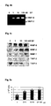
Figure 2a shows results of RT-PCR analysis of the mouse eyes after corneal alkali burn. -
Figure 2b shows results of ELISA analysis of the peripheral blood of the mice collected at 0, 1, 3, 5, 7, 10, 14 days after corneal alkali burn. -
Figure 2c shows results of Hematoxylin &Eosin staining, and immunohistochemical staining with Substance-P antibodies in the wound bed after the alkali burn in the mouse eye. -
Figure 2d shows results of immunohistochemical staining with c-kit, CD-29, and α-SM actin antibodies in the wound bed after the alkali burn in the mouse eye. -
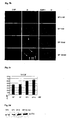
Figure 3a shows results of immunocytochemical staining with CD-29 antibodies in the attached cells isolated from the mouse blood after intravenous injection of Substance-P. -
Figure 3b is a graph showing mean numbers of CD-29+ cells in the blood before or after intravenous injection of Substance-P. -
Figure 4a shows the results of transmigration and collagen degradation of human MSCs on the 3-D collagen gel in response to Substance-P treatment. -
Figure 4b shows results of gelatin zymography of culture supernatants collected from human MSC culture on the 3-D collagen gel after the treatment of Substance-P (0-100 nM). -
Figure 4c shows results of immunoprecipitation with antibodies against MMP-1, MMP-2, MMP-9, TIMP-1 and TIMP-2 in the culture supernatant collected from human MSCs after the treatment of Substance-P (0-100nM). -
Figure 5a is a graph to show numbers of viable human MSCs after the Substance-P treatment (n=4, mean number ±S.D). -
Figure 5b shows result of immunofluorescence staining with β-catenin antibody in human MSCs cultured in the presence of Substance-P (0-100nM). -
Figure 5c is a graph of ELISA analysis result of VEGF collected from culture supernatants of Substance-P treated human MSCs. -
Figure 5d shows results of Western blot analysis with monoclonal antibody against human fibronectin from the culture supernatant of Substance-P treated human MSCs. -
Figure 6a shows a photo of slit-lamp examination result of the rabbit eye at 14, 30 days after corneal alkali burn with or without transfusion ofDi 1 labeled rabbit MSCs. -
Figure 6b shows a photo of existence ofDi 1 labeled MSCs in cornea observed by flat mount examination with fluorescent stereoscopic microscope. -
Figure 7a shows a photo of the corneal wound recovery after corneal alkali burn in rabbit with or without intravenous injection of Substance-P. -
Figure 7b shows a photo of H&E staining result of the tissue section after corneal alkali burn with or without intravenous injection of Substance-P -
Figure 7c shows a photo of immunohistochemical staining with CD-29 antibody in the tissue section after corneal alkali burn with or without intravenous injection of Substance-P. - A use of Substance-P for the manufacture of a medicament for mobilization or proliferation of Mesenchymal stem cells (MSCs) from the bone marrow, or facilitating said mobilization or proliferation is described. It is contemplated that said medicament may comprise MSCs. It is also contemplated that said medicament may be a cellular therapeutic agent.
- First, the present invention includes a use of Substance-P as a medicament for wound-healing or facilitating wound-healing, for example, cornea and skin wound. In an embodiment, said medicament may comprise MSCs. In another embodiment, said medicament may be a cellular therapeutic agent.
- Specifically, the present invention relates to Substance-P for use in a method of wound-healing or facilitating wound-healing, wherein the Substance-P is administered by intravenous injection, as defined in the claims.
- Also, an agent for mobilization or proliferation of MSCs from the bone marrow, or facilitating said mobilization or proliferation, containing Substance-P as an effective ingredient is described.
- Further, a wound healing or facilitating wound healing agent, containing Substance-P as an effective ingredient, for example, for cornea and skin wound, is described.
- Also, a wound healing or facilitating wound healing agent, containing MSCs as an effective ingredient, wherein the MSCs are mobilized or proliferated from the bone marrow by treatment of Substance-P is described.
- Specifically, the invention provides a composition comprising as effective ingredients Substance-P and MSCs which have been mobilized or proliferated from the bone marrow by treatment of Substance-P for use in a method of wound-heating or facilitating wound healing, as defined in the claims.
- Further, a method for separating MSCs, comprising a step of mobilizing MSCs from the bone marrow by Substance-P treatment is described.
- Also, a method of proliferating MSCs, comprising a step of proliferating MSCs in the presence of Substance-P is described.
- Further, a method of healing wound or facilitating wound-healing, comprising administering a therapeutically effective amount of Substance-P is described.
- Also, a method of healing wound or facilitating wound-healing, comprising administering a therapeutically effective amount of MSCs, wherein the MSCs are mobilized or proliferated from the bone marrow by treatment of Substance-P is described.
- Below, the present invention is described in detail.
- The present inventors used alkali burn cornea animal model which has advantages in examining systemic participation of stem cells during the wound healing process over other wound model since cornea has transparency and avascularization.
- After observing advent of c-kit+ cells in cornea and peripheral blood of rabbit with corneal alkali burn, the expression profiles of the cytokines and other factors in the wound microenvironment and the peripheral blood in the mice with a corneal alkali burn were examined by using RT-PCR analysis and ELISA. As a result, it was revealed that Substance-P is the first neuropeptide whose expression is elevated in the cornea wound as well as in the peripheral blood of the mice with corneal alkali burn. At this time, Substance-P was first increased in cornea and later in peripheral blood. It shows that Substance-P is provided by the corneal resident cells such as the sensory neurons, epithelial cells, keratocytes, and endothelial cells, not by infiltration of neutrophils or macrophages. The increase of Substance-P concentration in blood may be from systemic diffusion of Substance-P. Also, lack of Substance-P immunoreactivity in the neutrophils, earlier induction and termination of Substance P than duration of the inflammatory phase (up to 5 days after wound), and the weak Substance-P staining in the epithelial cells and fibroblastic cells at the later stage of wound healing (7-10 days after wound) supports an assumption that Substance-P is provided by nociceptive stimulation of the sensory neurons.
- Subsequently, in order to determine the systemic effect of Substance P separately from the other factors that are found in the wound microenvironment, Substance P was injected intravenously into the mice without wound, and the mobilization of CD29+ MSCs into peripheral blood was examined. As a result, it was observed that approximately 15 times more CD29+ MSCs are mobilized into the peripheral blood in mice into which Substance-P was injected intravenously, than in the non-injected mice. Here, it is believed that MSCs in non-wound mice were mobilized to peripheral blood because there was no wound site for MSCs to mobilize in the absence of wound bed.
- Also, to determine the action mechanism of Substance P in MSC mobilization from the endosteal surface of the bone marrow, an in vitro cell migration assay was used, and the effect of Substance-P on the MSC migration, dynamics of matrix degrading enzyme, cell proliferation and β-catenin localization were examined. As a result, it was revealed that in 3-D collagen gel in vitro, Substance-P induces matrix degrading enzymes and inhibits their inhibitors, thereby stimulating migration of human MSCs. This result further supports the role of Substance-P in MSCs migration. Also, the inventors discovered that Substance-P stimulates cell proliferation and nuclear translocation of β-catenin, thereby promoting the bone marrow repopulation of MSCs after the mobilization of MSCs.
- Furthermore, the present inventors examined whether or not intravenously injected MSCs arrive at damaged tissue and are involved in repair of the cornea, based on the corneal transparency and vision recovery scores. As a result, they confirmed that Di 1-labeled MSCs, which were transfused into the ear vein of alkali burned rabbits, are successfully supplied to the wound bed and improve corneal transparency and visual recovery, thereby facilitating cornea wound healing.
- Finally, the present inventors examined whether or not intravenous injection of Substance-P can facilitate corneal wound healing, based on macroscopic examination, H&E staining, and immunohistochemical staining. As a result, compared with the control group (Substance-P non-injected group), healing of the corneal wound in Substance-P-injected group was progressed rapidly.
- In conclusion, it was discovered that Substance-P is first elevated in the cornea wound and the peripheral blood, and facilitates mobilization of MSCs to peripheral blood, and promotes healing of the corneal wound and proliferation of MSCs in the bone marrow.
- Thus, it was concluded that Substance-P plays a role as wound signaling initiator to involve in mobilization and supply of MSCs from the bone marrow to the wound site. Also, it was identified that MSCs mobilize to wound site and are involved in wound healing.
- Considering the above results, MSCs can be used as wound healing or facilitating wound healing medicament, or cellular treatment medicament. Also, Substance-P can be used as wound healing or facilitating wound healing agent, or cellular treatment medicament, or mobilization, or proliferation medicament, or facilitating mobilization or proliferation medicament, of MSCs.
- In the present invention, the effective dosage of Substance-P is 0.1 µg/kg to 100 µg/kg, and the effective dosage of MSCs is 3× 10⁴cells/kg to 3× 10⁷cells/kg, particularly, 1 × 10⁵cells/kg to 1× 10⁷cells/kg. However, these dosages can be modified depending on weight, age, sex or extent of wound of patients.
- A formulation according to the present invention can be administered into human bodies parentally or locally, for example, by intravenous injection, subcutaneous injection, endodermis injection, and muscular injection. Preferably, they are administered by intravenous injection. For this purpose, the effective ingredients are suspended or dissolved in a pharmacologically acceptable carrier, preferably, water-soluble carrier.
- Below, the present invention is more specifically explained by the following Examples, but it is not intended to limit the scope of the present invention in any way.
- New Zealand white rabbits weighing 2-3kg were purchased from Samtako BioKorea. All these animal experiments were approved by Ethical Committee, Korea Institute of Radiological and Medical Sciences, and Chung-Ang University, and conducted in accordance with the ARVO Statement for the Use of Animal in Ophthalmic and Vision Research. For the alkali burn, the rabbit eyes were placed in contact with a 6mm piece of circular Whatman filter paper soaked in 1N NaOH for 30 seconds. At 1, 3, 5, 7, 10 and 14 day after the alkali burn, rabbit eyes and whole blood were isolated. The smears of blood and cornea section were stained with Anti-C kit antibody, and this result was shown in
Figure 1 . - Intact cornea is avascular tissue, and so extensive damage thereto might require vascular supply of inflammatory cells and other stem cells for the corneal repair. As shown in
Figure 1 , when the alkali burn was done on the rabbit cornea, the number of c-kit positive cells was increased in the peripheral blood, compared with the normal control, which was also detected in the corneal wound bed at 5th day after the alkali burn. This result confirms that stem cells originating from the bone marrow may actively participate in the repair of the cornea. - Balb/c mice weighing 30-40g were purchased from Jackson Lab (West Grove, PA). For the alkali burn, the mouse eyes were placed in contact with a 3mm piece of circular Whatman filter paper soaked in 1N NaOH for 10 seconds. At 1, 3, 5, 7, 10, and 14 day after the alkali burn, the mouse eyes and whole blood were isolated.
- Tissue damage itself may constitute a unique wound microenvironment to induce a systemic response such as inflammation and stem cell mobilization from the bone marrow to repair the damaged tissue. RT-PCR was used to analyze candidate factors retaining these functions in the wound microenvironment of alkali burned cornea. Specifically,
- RNA was isolated from the alkali burned eye by Trizol (Invitrogen). Total 1µg of RNA was reverse transcribed (RT) by using reverse transcription-polymerase kit (Takara), followed by PCR by using mouse gene-specific primers as follows:


- The result of PCT is shown by
Figure 2a . As shown inFigure 2a , it was found that Substance-P, IL-1, TNF-α and VEGF are induced in the eye after the cornea wound. - In order to further monitor the systemic profile of these factors and assess their possible roles in stem cell mobilization from the bone marrow, ELISA (R&D system) to the peripheral blood was performed. The result is shown in
Fig. 2b . As shown inFig. 2b , compared with the non-wound state, the level of Substance-P in the serum was elevated by approximately 3.2. times at 1 day, 4.4 times at 3 day, and 1.3 times at 5 day. However, pre-inflammatory cytokines, TNF-a, and IL-1 induction were detected at 5 day and 3 day after the alkali burn, respectively, and VEGF was induced from 7 day much later than them. - For the histological examination, Hematoxylin and Eosin (H&E) staining was performed for corneal wound bed, and immunohistochemical staining was performed with Substance-P antibody (Santa Cruz Biotechnology: Cat # sc-9758, 1:200). The mouse eyes were fixed with 4% of paraformaldehyde for 48h. The paraffin-embedded specimens were cut longitudinally into 4-µm sections, and transferred to poly-D lysine-coated slides. Then, Hematoxylin and Eosin (H&E) staining was performed for the histological examination. For immunohistochemical staining of Substance-P, the endogenous peroxidase activity was blocked by incubation with 0.5% of H2O2 for 10 min. The tissues were then permeabilized with 0.3% of Triton x-100 for 5 min, and incubated with the primary antibodies, the remaining procedures were followed by the manufacturer's specifications (ABC kit, Vector), and the cells were counterstained with fast red.
- The result is shown in
Fig. 2c . As a result of H&E staining, the inflammatory neutrophils were sustained for up to 5 days, the migrating epithelial cells were discernible at 5 day, fibroblastic infiltration was obvious at 7 day, and most macrophages had disappeared at 14 day. The immuno-reactive Substance-P was most strongly detected in the wound bed after 1 day and became weaker till 3rd day. At 5th day, Substance-P was barely detected in the fibroblastic cells and migrating epithelial cells, but was not detected in the inflammatory cells, which suggests that the early inflammatory cells may not secrete Substance-P. Therefore, the cornea wound itself appears to create a Substance-P rich microenvironment possibly provided by cornea resident cells, which may in turn induce the Substance-P elevation in the serum. - In order to determine whether the MSCs derived from bone marrow are supplied to a wound site, immunohistochemical staining was performed with the MSC markers, anti-CD-29 (Santa Cruz Biotechnology; Cat # sc-6622, 1:200), anti-c-kit (Santa Cruz Biotechnology; Cat # sc-1493, 1:100), and anti-a-SM actin antibodies (Progen, Cat # 61001, 1:200). The result is shown in
Fig. 2d . As known fromFig. 2d , the CD-29, c-kit, and α-SM-actin expressing fibroblastic cells were not detected at the early inflammatory phase until 5th day. They were detected at the wound bed from 7th day to 10th day corresponding to the fibroplasias phase, and had mostly disappeared after 14th day corresponding to the remodeling phase. This result is correlated with the absence of fibroblastic cells in the wound bed at the early phase of wound healing and the first emergence of fibroblastic cells in the wound bed at 7th day. Since only a few fibroblastic cells were present in the normal corneal stroma (Fig. 2c ) and the resident fibroblastic cells were absent at the inflammatory phase before 7 day, the CD-29, α-SM-actin, and c-kit positive cells in the 7th day wound bed were probably MSCs that were imported from the blood. - Substance-P was examined to determine whether it is responsible for stem cell mobilization into the blood stream or whether some other unidentified factors in the wound microenvironment are involved in stem cell mobilization.
- In order to distinguish effects of the others factors in the wound microenvironment, Substance-P (0.1 nmole/g of body weight) (Calbiochem) was injected intravenously through tail vein in Balb-c mice without causing alkali bums to the eye, and then the whole blood was collected after 1 day. After removing red blood cells by percoll gradient centrifugation, the attached cells were cultured for 48 h in order to remove lymphocytes, and then, in order to distinguish MSCs from the lymphocytes, they were immune-stained with antibodies against CD-29 (
β 1 integrin) which is not expressed in the macrophages. - The result is shown in
Figs. 3a and 3b . Within 1 day, the intravenous injection of Substance-P strongly stimulated mobilization of CD-29 positive MSCs into the peripheral blood approximately by 15 times more than in the non-injected mice (Figure 3b ). This shows a new role of Substance-P which may be manifested during early phase of the wound healing process, to mobilize MSC from the bone marrow into the systemic blood, to supply MSCs to the cornea wound site, and to facilitate the cornea repair. - In order to determine the mechanism of Substance-P in the mobilization of MSCs, which are strongly attached onto the endosteal surface of bone marrow, Substance-P was first examined to determine whether it stimulates migration of human MSCs cultured on top of the 3-D collagen gel. The type I collagen gel matrix was made within a 12 mm millicell membrane (milipore: 12-µm pore size) according to the manufacturer's specifications (Nitta gelatin, Japan) and coated overnight with type IV collagen (Nitta). The MSCs were plated on top of the collagen gel, and the outer chamber was filled with Substance-P-containing MSCGM (Cambrex Bio Science). Clearance of the collagen gel was monitored by using the phase contrast microscopy (Olympus). At 72h, the millicell insert and outer dishes were fixed and stained with hematoxylin, and then the culture supernatants were stored for gelatin zymography. This result is shown in
Fig. 4a . As shown inFig. 4a , Substance-P applied into the bottom dishes stimulated degradation of the collagen gel and MSC migration in a dose dependent manner. - The culture supernatants were examined for gelatin zymography. The eletrophoresis was performed for the culture supernatant in which sample buffer without mercaptoethanol was added thereto, in 8 % SDS-PAGE. The gel was renatured with 2.5% of Triton x-100 in order to remove the SDS, and then was incubated with a developing buffer (50 mM Tris-HCl, pH 7.2 100 mM NaCl, 20 mM CaCl2) at 37°C overnight, and the gel was stained with Coomassie blue. This result is shown in
Fig. 4b . As shown inFig. 4b , gelatin zymography of the culture medium revealed strong induction of MMP-9 and MMP-2 activities during MSC migration. - The effect of Substance-P on the biosynthesis of MMPs (matrix metalloproteinases) and their inhibitors was examined by immunoprecipitation of the culture medium and the cell lysates of MSCs which were labeled with 35S-methionine. The MSCs were treated with Substance-P and labeled with 50 µCi/ml 35S-methionine (Amersham) for 16h. The cell lysate was prepared with a lysis buffer (1% of NP40, 10 mM Tris-HCl, pH 7.4, 150 mM NaCl, 1 mM EDTA, 2 mM PMSF). For immunoprecipitation of the secreted MMP-1, MMP-2, MMP-9, TIMP-1 and TIMP-2, the culture supernatants were incubated with their specific antibodies [anti-MMP-1 (chemicon; Cat # MAB3307, 1:100), anti-MMP-2 (Chemicon; Cat # MAB13405, 1:200), anti-MMP-9 (Calbiochem; Cat # 444236, 1:100), anti-TIMP-1 (Calbiochem; Cat # IM32L, 1:250) and anti-TIMP-2 (Calbiochem; Cat # IM11L, 1:250)] for 2h under constant rotation, and the antibodies were collected by protein-G sepharose-4B (Biorad). The pellet was washed three times with an immunoprecipitation buffer (150 mM NaOH, 50 mM Tris-HCl pH 7.4, 0.2% SDS and 0.5% Nadeoxycholate), once with a high salt buffer (10 mM Tris-HCl, pH 7.4, 0.5 M NaCl), and once with a low salt buffer (10 mM Tris-HCl, pH 7.4). After SDS-PAGE, the gel was soaked in 2 M sodium salicylate, dried, and exposed to the X-ray film for 2 weeks. For immunoprecipitation of MMP-9, the cell lysate was incubated with MMP-9 antibodies, and the remaining procedures were the same as described above. This result is shown in
Fig. 4c . - The biosynthesis of MMP-1, MMP-2, MMP-9, and MT1-MMP was stimulated in a dose dependent manner. In contrast, the biosynthesis of their inhibitors, TIMP-1 and TIMP-2, was inhibited by Substance-P.
- As a result, Substance-P stimulated MSC migration in 3-D collagen gel, induction of MMPs, and inhibition of their inhibitors, TIMPs.
- If Substance-P only has capacity of MSC mobilization from the bone marrow, the bone marrow stroma may become empty in the MSC pool after the mobilization. The effect of Substance-P was again examined about its cell proliferation and self-renewal capacity.
- At 3day after the treatment of Substance-P, the triphan blue excluded viable cells were counted. The result is shown in
Fig. 5a . - Substance-P increased the number of viable cells, 1.7 times at 1nM, 2.1 times at 10 nM, and 2.2 times at 100 nM, compared with the control group. In order to determine whether Substance-P affects the self-renewal capacity of human MSCs, β-catenin, which is the downstream effecter of the wnt signaling pathway, was examined. MSCs (Cambrex Bio Science) were plated in the density of 1×10⁴ /well, and incubated with MSCGM (Cambrex Bio Science). After adhesion of the cells, they were treated with Substance-P in each concentration, and incubated for 16h and 48h. The MSCs were fixed with 4% of paraformaldehyde and permeabilized with 0.3% of Triton X-100. After blocking nonspecific antigen with 20% normal goat serum which was diluted in phosphate buffer for 1h, the coverslips were incubated with antibodies against β-catenin (BD Biosciencs; Cat # 6010153, 1:100) for 90min, and then with the FITC-conjugated secondary antibodies for 60 min(Vector). The cells were counterstained with DAPI, and examined by using confocal microscopy (Leica). This result is shown in
Fig. 5b . - At 16h after the Substance-P treatment, β-catenin was mainly observed in the cytoplasm of human MSCs, but only a small number of MSCs at 100 nM showed nuclear localization of β-catenin. However, at 48h after Substance-P treatment, the nuclear translocation of β-catenin was more prominent, and particularly at 100 nM of Substance-P treatment, all the MSCs showed nuclear translocation of β-catenin.
- ELISA and Western blot analysis of fibronectin and VEGF, which are down stream genes of Tcf/β-catenin, were carried out. These results are shown in
Figs. 5c and 5d . - As shown in
Figs. 5c and 5d , Substance-P induced VEGF and fibronectin. This proves that Substance-P plays a role in repopulation of the remaining MSCs by the stimulation of cell proliferation or self-renewal via well-known canonical wnt signaling pathway. - In conclusion, Substance-P stimulated cell proliferation, translocation of β-catenin, and induction of β-catenin downstream gene, VEGF and fibronectin, all of which suggest that Substance-P plays a role in repopulation of the bone marrow.
- As shown in
Fig. 3 , intravenous injection of Substance-P was sufficient to stimulate mobilization of mouse CD-29+ MSCs into the peripheral blood. In order to examine whether or not the mobilized MSCs can actually participate in cornea wound healing, Di 1-labeled MSCs were transfused to the ear vein of the non-irradiated rabbit at 24 h after the cornea alkali burn to minimize the MSC homing to the bone marrow and to maximize the supply into wound site. - The MSCs were isolated from the tibia of a 1 month old allogenic rabbit by bone marrow irrigation and aspiration, and cultured with MSCGM up to
passage 3. The attached cells were identified as MSCs by the expression of c-kit, STRO-1, and α-smooth muscle (SM) actin, and were used for the cell transfusion experiment. In order to trace the transfused MSCs, the trypsinized cells were incubated with aDi 1 solution [Celltracker™ CM-Di I(Cat# C-7000); Molecular Probes] for 5min at 37°C and 15min at 4°C. After labeling and subsequent washing with PBS three times, the MSCs (1x10⁶ cells /ml) were resuspended in fresh serum free medium and injected into the ear vein of 10 rabbits at 1 day after the corneal alkali burn. - In 2 weeks and 4 weeks after the alkali burn wound, the eyes of the non-injected group and MSC-injected group labeled with Di I were examined by slit lamp. This result is shown in
Fig. 6a . The corneal visual recovery was better in the MSC injected group than in the non-injected group. - The epithelial healing time was determined by closure of the epithelial defect with photo-slit microscopic examination and photo-documentation. The corneal opacity was graded according to the following criteria: grade 0 - no opacity, grade 1 - the iris structure is visible with mild opacity, grade 2 - the fine iris structure is not visible, grade 3 - only the presence of the iris is noted as brown color, and grade 4 - totally opaque. This result is shown in Table 1.
Table 1 the epithelial healing time (day) corneal opacity corneal neovascularization control group (n=10) 10.25±2.36 3.43±0.79 3.57±0.79 MSC IV (n=10) 5.50±0.58 * 1.50±0.65* 1.71±0.75* *: p<0.05 - As shown in Table 1, the MSCs-injected rabbits showed 2-fold shortening of the epithelial healing time, better corneal transparency, and less corneal neovascularization than the non-injected group.
- FLAT mount was performed in order to confirm whether or not the transfused
Di 1 labeled-MSCs are localized to the wound bed. In order to identify theDi 1 labeled-MSCs in the cornea, FLAT mounts at 1 day and in 2 weeks and 3 months were examined by fluorescence stereomicroscopy (Leica). This result is shown inFig. 6b . As shown inFig. 6b , the Di 1-labeled MSCs were detected at the epithelium level as well as at the stroma level. Di 1-labeled MSCs were detected at the wound bed as early as 1 day after the intravenous transfusion, and their presence was detected even after 3 months from the transfusion though the intensity ofDi 1 was lower. - As shown in
Fig. 3 , intravenous injection of Substance-P was sufficient to faciliate the mobilization of mouse CD-29+ MSCs into the peripheral blood, and as shown inFig. 6 , intravenous injection of Substance-P improved corneal vision recovery. Thus, it was examined whether or not Substance-P only can stimulate corneal wound healing. To estimate the effect of Substance-P intravenous injection on acceleration of the wound healing, Substance-P at dosage 6.5ug/kg was intravenously injected firstly immediately after the injury, and then secondly on 2nd day after the wound. At day 7 after the intravenous injection, cornea recovery was estimated by naked eyes with taking digital picture. This result is shown inFig. 7a . As shown inFig. 7a , the corneal regeneration was much faster in the Substance-P-injected rabbit than the non-injected control group. The intravenous injection of Substance-P strongly expedited the cornea healing as shown by clearance of corneal opaqueness and disappearance of hemorrhagic area, compared with the non-injected control rabbit. - To observe the wound healing degree in the histological level, rabbit eyes were isolated and fixed, and then, H&E staining and immunostaining for CD29 were carried out. The result of H&E staining is shown in
Fig. 7b . As shown inFig. 7b , the Substance-P-injected group showed full thickness coverage of epithelium, well organized vasculature, and ordered array of dense collagen bundles, which are similar to those of the non-injured collateral side except the still presence of macrophages and polymorphonuclear giant cells beneath the epithelium. In contrast, the non-injected rabbit on the same day showed only thin coverage of epithelium, active infiltration of large fibroblast like cells, and loose unorganized collagen fibrils in the stroma, which are similar to those observed on 5 day in the Substance-P injected group. - The result of CD29 staining is shown in
Fig. 7c . As shown inFig. 7c , the non-injected group showed undifferentiated mesenchymal stem cell (CD29-expressing cells) but, the Substance-P-injected group showedCD 29+cells of small and spindle shape inside of collagen fibers, which is similar to normal cornea stroma. Thus, comparing the histological observation between the Substance P-injected and the non injected cornea during the entire period, it is confirmed that SP IV injection speeded up the healing rate faster than the control group. These results mean that injection of Substance-P is effective to corneal wound healing. - According to the present invention, Substance-P is initially elevated in the alkali burned cornea, and mobilized into the bone marrow through blood, and then stimulates mesenchymal stem cell in the bone marrow, and induces repopulation of MSCs in the bone marrow.
- Thus, Substance-P can be utilized as wound healing or facilitating wound-healing agent, mobilization or proliferation or facilitating mobilization or proliferation agent, of MSCs.
- Also, MSCs are confirmed to directly involve in wound healing. Thus, MSCs can be utilized as wound healing or facilitating wound healing agent, or cellular therapeutic agent.
Claims (3)
- Substance-P for use in a method of wound-healing or facilitating wound-healing, wherein the Substance-P is administered by intravenous injection.
- A composition comprising as effective ingredients Substance-P and mesenchymal stem cells (MSCs) which have been mobilized or proliferated from the bone marrow by treatment of Substance-P for use in a method of wound-healing or facilitating wound-healing.
- The composition of claim 2, wherein said medicament is a cellular therapeutic agent.
Applications Claiming Priority (1)
| Application Number | Priority Date | Filing Date | Title |
|---|---|---|---|
| KR1020040086359A KR100593397B1 (en) | 2004-10-27 | 2004-10-27 | Wound healing or wound healing promoters containing mesodermal stem cells and / or P substances, or cell therapeutics |
Publications (3)
| Publication Number | Publication Date |
|---|---|
| EP1658855A2 EP1658855A2 (en) | 2006-05-24 |
| EP1658855A3 EP1658855A3 (en) | 2006-05-31 |
| EP1658855B1 true EP1658855B1 (en) | 2009-02-25 |
Family
ID=36013350
Family Applications (1)
| Application Number | Title | Priority Date | Filing Date |
|---|---|---|---|
| EP05023367A Expired - Lifetime EP1658855B1 (en) | 2004-10-27 | 2005-10-26 | Use of substance p for mobilization or proliferation of mesenchymal stem cells and for wound healing |
Country Status (7)
| Country | Link |
|---|---|
| US (2) | US8551470B2 (en) |
| EP (1) | EP1658855B1 (en) |
| JP (1) | JP5134772B2 (en) |
| KR (1) | KR100593397B1 (en) |
| CN (1) | CN1824289B (en) |
| AT (1) | ATE423565T1 (en) |
| DE (1) | DE602005012905D1 (en) |
Families Citing this family (28)
| Publication number | Priority date | Publication date | Assignee | Title |
|---|---|---|---|---|
| EP2055308B1 (en) * | 2006-10-30 | 2017-05-10 | Genomix Co., Ltd. | Pharmaceutical for promoting functional regeneration of damaged tissue |
| KR20080068950A (en) * | 2007-01-22 | 2008-07-25 | 경희대학교 산학협력단 | Composition for preventing and treating bone marrow damage |
| US8114668B2 (en) * | 2007-05-14 | 2012-02-14 | Cardiac Pacemakers, Inc. | Composition for cold storage of stem cells |
| WO2009017655A2 (en) * | 2007-07-27 | 2009-02-05 | Immuneregen Biosciences, Inc. | Methods and compositions for stimulating the proliferation or differentiation of stem cells with substance p or an analog thereof |
| KR20090035279A (en) * | 2007-10-05 | 2009-04-09 | 경희대학교 산학협력단 | Composition for preventing and treating gastrointestinal damage |
| WO2009065093A2 (en) * | 2007-11-17 | 2009-05-22 | University Of Medicine And Dentistry Of New Jersey | Use of stem cells for wound healing |
| JP5660889B2 (en) | 2008-04-30 | 2015-01-28 | 株式会社ジェノミックス | Bone marrow-derived pluripotent stem cell mobilization to peripheral circulation |
| KR20110017371A (en) * | 2008-04-30 | 2011-02-21 | 가부시키가이샤 제노믹스 | High efficiency sampling of functional cells in vivo |
| JP5814549B2 (en) * | 2008-04-30 | 2015-11-17 | 株式会社ジェノミックス | Drugs for promoting functional regeneration of damaged tissues |
| KR101175175B1 (en) | 2008-07-28 | 2012-08-20 | 서울대학교병원 | Method for separating high activity stem cells form human stem cells and high activity stem cells separated by the method |
| KR101590558B1 (en) * | 2009-03-06 | 2016-02-01 | 경희대학교 산학협력단 | Bone forming agent using P substance and method for producing the same |
| KR20120135190A (en) | 2009-10-28 | 2012-12-12 | 고꾸리쯔 다이가꾸 호우징 오사까 다이가꾸 | Tussue-regeneration promoter using recruitment of bone marrow mesenchymal stem cells and/or pluripotent stem cells in blood |
| JP5852773B2 (en) | 2010-06-01 | 2016-02-03 | ピアス株式会社 | Method for producing attractant for bone marrow mesenchymal stem cells, method for attracting bone marrow mesenchymal stem cells, and use for producing an attractant for bone marrow mesenchymal stem cells |
| US20130089529A1 (en) * | 2010-06-11 | 2013-04-11 | University-Industry Cooperation Group Of Kyunghee University | Agent for osteogenesis using substance-p and preparation process thereof |
| KR102113809B1 (en) | 2011-04-26 | 2020-05-21 | 가부시키가이샤 스템림 | Peptide for inducing regeneration of tissue and use thereof |
| ES2673861T3 (en) | 2012-10-25 | 2018-06-26 | Genomix Co., Ltd. | Novel method to treat spinal cord damage using the hmgb1 fragment |
| CA2889275C (en) | 2012-10-25 | 2021-06-15 | Katsuto Tamai | Novel method for treating cardiac infarction using hmgb1 fragment |
| JP6113133B2 (en) | 2014-11-06 | 2017-04-12 | 日本メナード化粧品株式会社 | Stem cell undifferentiated state maintenance agent and growth promoter |
| US10967006B2 (en) | 2016-01-21 | 2021-04-06 | Abt Holding Company | Stem cells for wound healing |
| KR101825041B1 (en) * | 2016-04-07 | 2018-02-02 | 주식회사 바이오솔루션 | Skin External composition for treating a wound comprising substance P |
| PL3556378T3 (en) | 2017-01-27 | 2025-06-09 | StemRIM Inc. | Therapeutic agent for cardiomyopathy, old myocardial infarction and chronic heart failure |
| US11298403B2 (en) | 2017-12-01 | 2022-04-12 | StemRIM Inc. | Therapeutic agent for inflammatory bowel disease |
| CN118460462A (en) | 2017-12-01 | 2024-08-09 | 斯特姆里姆有限公司 | Exo-embryonic interlayer mesenchymal stem cells and method for producing same |
| WO2019156137A1 (en) | 2018-02-08 | 2019-08-15 | 株式会社ステムリム | Therapeutic agent for psoriasis |
| EP3856203A4 (en) | 2018-09-27 | 2022-02-23 | Biocardia, Inc. | BONE MARROW DERIVED NEUROKININ 1 RECEPTOR POSITIVE (NK1R+) PROGENIOR CELLS FOR THERAPEUTIC APPLICATIONS |
| US12304933B2 (en) | 2018-10-05 | 2025-05-20 | StemRIM Inc. | Disease treatment drug based on mesenchymal-stem-cell mobilization |
| CN110732017B (en) * | 2019-11-08 | 2021-08-27 | 中山大学 | Use of SP in preparing medicine for treating male sterility |
| KR102398968B1 (en) | 2020-07-13 | 2022-05-17 | (주) 엘피스셀테라퓨틱스 | Combination therapy of Substance P for hematopoietic stem cell mobilization |
Family Cites Families (11)
| Publication number | Priority date | Publication date | Assignee | Title |
|---|---|---|---|---|
| US5616562A (en) | 1990-04-27 | 1997-04-01 | Murphy; Christopher J. | Methods and compositions using substance P to promote wound healing |
| US5545617A (en) * | 1993-11-12 | 1996-08-13 | The Schepens Eye Research Institute, Inc. | Therapeutic regulation of abnormal conjunctival goblet cell mucous secretion |
| US5945508A (en) | 1996-07-23 | 1999-08-31 | Witten; Mark L. | Substance P treatment for immunostimulation |
| ES2237089T3 (en) | 1998-03-13 | 2005-07-16 | Osiris Therapeutics, Inc. | USES FOR NON-AUTOMATIC HUMAN MESENQUIMATE MOTHER CELLS |
| CA2418976C (en) * | 2000-08-10 | 2011-08-02 | Santen Pharmaceutical Co., Ltd. | Skin wound healing promoters |
| JP4096115B2 (en) * | 2000-08-10 | 2008-06-04 | 輝夫 西田 | Skin wound healing promoter |
| WO2002102835A2 (en) | 2000-11-21 | 2002-12-27 | New England Medical Center | Novel antimicrobial compounds |
| US6437093B1 (en) | 2000-11-28 | 2002-08-20 | Milkhaus Laboratory, Inc. | Methods of treatment comprising administration of Substance P |
| EP1492525A2 (en) * | 2001-08-16 | 2005-01-05 | Probiodrug AG | Use of inhibitors of proline endopeptidase to modulate inositol (1,4,5) triphosphate concentration dependent on intracellular signal cascades |
| US7119071B2 (en) | 2002-05-21 | 2006-10-10 | University Of Medicine And Dentistry Of New Jersey | Amino terminal substance P compositions and methods for using the same |
| AR047712A1 (en) | 2002-09-07 | 2006-02-15 | Royal Veterinary College | TREATMENT METHOD OF A NATURAL SOFT SKELETTIC TISSUE INJURY MANAGING A COMPOSITION OF MESENQUIMATOSE MOTHER CELLS |
-
2004
- 2004-10-27 KR KR1020040086359A patent/KR100593397B1/en not_active Expired - Lifetime
-
2005
- 2005-10-25 US US11/259,550 patent/US8551470B2/en not_active Expired - Fee Related
- 2005-10-26 AT AT05023367T patent/ATE423565T1/en not_active IP Right Cessation
- 2005-10-26 EP EP05023367A patent/EP1658855B1/en not_active Expired - Lifetime
- 2005-10-26 DE DE602005012905T patent/DE602005012905D1/en not_active Expired - Lifetime
- 2005-10-27 CN CN2005101380225A patent/CN1824289B/en not_active Expired - Fee Related
- 2005-10-27 JP JP2005313174A patent/JP5134772B2/en not_active Expired - Fee Related
-
2013
- 2013-09-05 US US14/018,772 patent/US9254305B2/en not_active Expired - Fee Related
Also Published As
| Publication number | Publication date |
|---|---|
| KR20060037176A (en) | 2006-05-03 |
| JP5134772B2 (en) | 2013-01-30 |
| US9254305B2 (en) | 2016-02-09 |
| EP1658855A2 (en) | 2006-05-24 |
| US20060127373A1 (en) | 2006-06-15 |
| KR100593397B1 (en) | 2006-06-28 |
| EP1658855A3 (en) | 2006-05-31 |
| JP2006124389A (en) | 2006-05-18 |
| CN1824289A (en) | 2006-08-30 |
| CN1824289B (en) | 2011-08-31 |
| DE602005012905D1 (en) | 2009-04-09 |
| US8551470B2 (en) | 2013-10-08 |
| US20140065109A1 (en) | 2014-03-06 |
| ATE423565T1 (en) | 2009-03-15 |
Similar Documents
| Publication | Publication Date | Title |
|---|---|---|
| US9254305B2 (en) | Methods of administration of Substance P for wound healing | |
| ES2400916T3 (en) | A cellular therapy for ocular degeneration | |
| Azar | Corneal angiogenic privilege: angiogenic and antiangiogenic factors in corneal avascularity, vasculogenesis, and wound healing (an American Ophthalmological Society thesis) | |
| Yam et al. | Safety and feasibility of intrastromal injection of cultivated human corneal stromal keratocytes as cell-based therapy for corneal opacities | |
| WO2020210248A1 (en) | Processes for making and using a mesenchymal stem cell derived secretome | |
| KR101539700B1 (en) | A composition for treating diseases related to angiogenesis and cornea or conjunctiva transplant using chondrocyte-derived extracellular matrix membrane | |
| CN110025765B (en) | Extracellular matrix-derived peptide of chondrocyte | |
| JP2025181979A (en) | Compositions and methods for preventing and treating corneal opacification and scarring - Patent Application 20070122997 | |
| Na et al. | The combined effect of epidermal growth factor and keratinocyte growth factor delivered by hyaluronic acid hydrogel on corneal wound healing | |
| Izumi et al. | Involvement of insulin-like growth factor-I and insulin-like growth factor binding protein-3 in corneal fibroblasts during corneal wound healing | |
| CN117018165A (en) | Application of Reelin protein or Reln gene in cornea injury treatment medicine | |
| Maeda et al. | Immunoconfocal localization of gelatinase B expressed by migrating intrastromal epithelial cells after deep annular excimer keratectomy | |
| US20250025508A1 (en) | Compositions and methods for treating exposure of surgical mesh | |
| US20230077811A1 (en) | Activation of neuropeptide receptors on plasmacytoid dendritic cells to treat or prevent ocular diseases associated with neovascularization and inflammation | |
| US20070218105A1 (en) | Topical Treatment with NGF and DHA in Damaged Corneas | |
| US20150045296A1 (en) | Method for treating diabetic retinopathy | |
| WO2025151812A1 (en) | Processes for making and using a mesenchyme stem cell secretome for limbal stem cell deficiency (lscd) treatment | |
| US20200038355A1 (en) | Antioxidants for use in ophthalmic surgery | |
| Pao | Topical administration of orbital fat-derived stem cells | |
| Gordon | Matrix metalloproteinases: Roles and regulation in ocular surface regeneration | |
| 김수현 | Development of air assisted lamellar keratectomy for corneal haze model and deep anterior lamellar keratoplasty in dogs | |
| Hong et al. | Novel role of substance-p as a systemic wound messenger to mobilize mesenchymal stem cell from bone marrow and be engaged in the wound healing epithelial layer |
Legal Events
| Date | Code | Title | Description |
|---|---|---|---|
| PUAI | Public reference made under article 153(3) epc to a published international application that has entered the european phase |
Free format text: ORIGINAL CODE: 0009012 |
|
| PUAL | Search report despatched |
Free format text: ORIGINAL CODE: 0009013 |
|
| 17P | Request for examination filed |
Effective date: 20051026 |
|
| AK | Designated contracting states |
Kind code of ref document: A2 Designated state(s): AT BE BG CH CY CZ DE DK EE ES FI FR GB GR HU IE IS IT LI LT LU LV MC NL PL PT RO SE SI SK TR |
|
| AX | Request for extension of the european patent |
Extension state: AL BA HR MK YU |
|
| AK | Designated contracting states |
Kind code of ref document: A3 Designated state(s): AT BE BG CH CY CZ DE DK EE ES FI FR GB GR HU IE IS IT LI LT LU LV MC NL PL PT RO SE SI SK TR |
|
| AX | Request for extension of the european patent |
Extension state: AL BA HR MK YU |
|
| RTI1 | Title (correction) |
Free format text: USE OF SUBSTANCE P FOR MOBILIZATION OR PROLIFERATION OF MESENCHYMAL STEM CELLS AND FOR WOUND HEALING |
|
| 17Q | First examination report despatched |
Effective date: 20061117 |
|
| AKX | Designation fees paid |
Designated state(s): AT BE BG CH CY CZ DE DK EE ES FI FR GB GR HU IE IS IT LI LT LU LV MC NL PL PT RO SE SI SK TR |
|
| GRAP | Despatch of communication of intention to grant a patent |
Free format text: ORIGINAL CODE: EPIDOSNIGR1 |
|
| RIC1 | Information provided on ipc code assigned before grant |
Ipc: A61K 38/04 20060101AFI20080825BHEP Ipc: A61P 17/02 20060101ALI20080825BHEP |
|
| GRAS | Grant fee paid |
Free format text: ORIGINAL CODE: EPIDOSNIGR3 |
|
| GRAA | (expected) grant |
Free format text: ORIGINAL CODE: 0009210 |
|
| RAP1 | Party data changed (applicant data changed or rights of an application transferred) |
Owner name: KOREA INSTITUTE OF RADIOLOGICAL & MEDICAL SCIENCES Owner name: CHUNG-AUG UNIVERSITY INDUSTRY-ACADEMY COOPERATION |
|
| AK | Designated contracting states |
Kind code of ref document: B1 Designated state(s): AT BE BG CH CY CZ DE DK EE ES FI FR GB GR HU IE IS IT LI LT LU LV MC NL PL PT RO SE SI SK TR |
|
| REG | Reference to a national code |
Ref country code: GB Ref legal event code: FG4D |
|
| REG | Reference to a national code |
Ref country code: CH Ref legal event code: EP |
|
| REG | Reference to a national code |
Ref country code: IE Ref legal event code: FG4D |
|
| REF | Corresponds to: |
Ref document number: 602005012905 Country of ref document: DE Date of ref document: 20090409 Kind code of ref document: P |
|
| PG25 | Lapsed in a contracting state [announced via postgrant information from national office to epo] |
Ref country code: LT Free format text: LAPSE BECAUSE OF FAILURE TO SUBMIT A TRANSLATION OF THE DESCRIPTION OR TO PAY THE FEE WITHIN THE PRESCRIBED TIME-LIMIT Effective date: 20090225 Ref country code: NL Free format text: LAPSE BECAUSE OF FAILURE TO SUBMIT A TRANSLATION OF THE DESCRIPTION OR TO PAY THE FEE WITHIN THE PRESCRIBED TIME-LIMIT Effective date: 20090225 Ref country code: SI Free format text: LAPSE BECAUSE OF FAILURE TO SUBMIT A TRANSLATION OF THE DESCRIPTION OR TO PAY THE FEE WITHIN THE PRESCRIBED TIME-LIMIT Effective date: 20090225 Ref country code: FI Free format text: LAPSE BECAUSE OF FAILURE TO SUBMIT A TRANSLATION OF THE DESCRIPTION OR TO PAY THE FEE WITHIN THE PRESCRIBED TIME-LIMIT Effective date: 20090225 |
|
| NLV1 | Nl: lapsed or annulled due to failure to fulfill the requirements of art. 29p and 29m of the patents act | ||
| PG25 | Lapsed in a contracting state [announced via postgrant information from national office to epo] |
Ref country code: IS Free format text: LAPSE BECAUSE OF FAILURE TO SUBMIT A TRANSLATION OF THE DESCRIPTION OR TO PAY THE FEE WITHIN THE PRESCRIBED TIME-LIMIT Effective date: 20090625 Ref country code: SE Free format text: LAPSE BECAUSE OF FAILURE TO SUBMIT A TRANSLATION OF THE DESCRIPTION OR TO PAY THE FEE WITHIN THE PRESCRIBED TIME-LIMIT Effective date: 20090525 Ref country code: PL Free format text: LAPSE BECAUSE OF FAILURE TO SUBMIT A TRANSLATION OF THE DESCRIPTION OR TO PAY THE FEE WITHIN THE PRESCRIBED TIME-LIMIT Effective date: 20090225 Ref country code: AT Free format text: LAPSE BECAUSE OF FAILURE TO SUBMIT A TRANSLATION OF THE DESCRIPTION OR TO PAY THE FEE WITHIN THE PRESCRIBED TIME-LIMIT Effective date: 20090225 Ref country code: LV Free format text: LAPSE BECAUSE OF FAILURE TO SUBMIT A TRANSLATION OF THE DESCRIPTION OR TO PAY THE FEE WITHIN THE PRESCRIBED TIME-LIMIT Effective date: 20090225 |
|
| PG25 | Lapsed in a contracting state [announced via postgrant information from national office to epo] |
Ref country code: BE Free format text: LAPSE BECAUSE OF FAILURE TO SUBMIT A TRANSLATION OF THE DESCRIPTION OR TO PAY THE FEE WITHIN THE PRESCRIBED TIME-LIMIT Effective date: 20090225 |
|
| PG25 | Lapsed in a contracting state [announced via postgrant information from national office to epo] |
Ref country code: PT Free format text: LAPSE BECAUSE OF FAILURE TO SUBMIT A TRANSLATION OF THE DESCRIPTION OR TO PAY THE FEE WITHIN THE PRESCRIBED TIME-LIMIT Effective date: 20090812 Ref country code: ES Free format text: LAPSE BECAUSE OF FAILURE TO SUBMIT A TRANSLATION OF THE DESCRIPTION OR TO PAY THE FEE WITHIN THE PRESCRIBED TIME-LIMIT Effective date: 20090605 Ref country code: CZ Free format text: LAPSE BECAUSE OF FAILURE TO SUBMIT A TRANSLATION OF THE DESCRIPTION OR TO PAY THE FEE WITHIN THE PRESCRIBED TIME-LIMIT Effective date: 20090225 Ref country code: DK Free format text: LAPSE BECAUSE OF FAILURE TO SUBMIT A TRANSLATION OF THE DESCRIPTION OR TO PAY THE FEE WITHIN THE PRESCRIBED TIME-LIMIT Effective date: 20090225 Ref country code: EE Free format text: LAPSE BECAUSE OF FAILURE TO SUBMIT A TRANSLATION OF THE DESCRIPTION OR TO PAY THE FEE WITHIN THE PRESCRIBED TIME-LIMIT Effective date: 20090225 |
|
| PG25 | Lapsed in a contracting state [announced via postgrant information from national office to epo] |
Ref country code: SK Free format text: LAPSE BECAUSE OF FAILURE TO SUBMIT A TRANSLATION OF THE DESCRIPTION OR TO PAY THE FEE WITHIN THE PRESCRIBED TIME-LIMIT Effective date: 20090225 Ref country code: RO Free format text: LAPSE BECAUSE OF FAILURE TO SUBMIT A TRANSLATION OF THE DESCRIPTION OR TO PAY THE FEE WITHIN THE PRESCRIBED TIME-LIMIT Effective date: 20090225 |
|
| PLBE | No opposition filed within time limit |
Free format text: ORIGINAL CODE: 0009261 |
|
| STAA | Information on the status of an ep patent application or granted ep patent |
Free format text: STATUS: NO OPPOSITION FILED WITHIN TIME LIMIT |
|
| PG25 | Lapsed in a contracting state [announced via postgrant information from national office to epo] |
Ref country code: BG Free format text: LAPSE BECAUSE OF FAILURE TO SUBMIT A TRANSLATION OF THE DESCRIPTION OR TO PAY THE FEE WITHIN THE PRESCRIBED TIME-LIMIT Effective date: 20090525 |
|
| 26N | No opposition filed |
Effective date: 20091126 |
|
| PG25 | Lapsed in a contracting state [announced via postgrant information from national office to epo] |
Ref country code: MC Free format text: LAPSE BECAUSE OF NON-PAYMENT OF DUE FEES Effective date: 20091031 |
|
| REG | Reference to a national code |
Ref country code: CH Ref legal event code: PL |
|
| REG | Reference to a national code |
Ref country code: IE Ref legal event code: MM4A |
|
| REG | Reference to a national code |
Ref country code: FR Ref legal event code: ST Effective date: 20100630 |
|
| PG25 | Lapsed in a contracting state [announced via postgrant information from national office to epo] |
Ref country code: FR Free format text: LAPSE BECAUSE OF NON-PAYMENT OF DUE FEES Effective date: 20091102 |
|
| PG25 | Lapsed in a contracting state [announced via postgrant information from national office to epo] |
Ref country code: GR Free format text: LAPSE BECAUSE OF FAILURE TO SUBMIT A TRANSLATION OF THE DESCRIPTION OR TO PAY THE FEE WITHIN THE PRESCRIBED TIME-LIMIT Effective date: 20090526 Ref country code: CH Free format text: LAPSE BECAUSE OF NON-PAYMENT OF DUE FEES Effective date: 20091031 Ref country code: IE Free format text: LAPSE BECAUSE OF NON-PAYMENT OF DUE FEES Effective date: 20091026 Ref country code: LI Free format text: LAPSE BECAUSE OF NON-PAYMENT OF DUE FEES Effective date: 20091031 |
|
| PG25 | Lapsed in a contracting state [announced via postgrant information from national office to epo] |
Ref country code: IT Free format text: LAPSE BECAUSE OF FAILURE TO SUBMIT A TRANSLATION OF THE DESCRIPTION OR TO PAY THE FEE WITHIN THE PRESCRIBED TIME-LIMIT Effective date: 20090225 |
|
| PG25 | Lapsed in a contracting state [announced via postgrant information from national office to epo] |
Ref country code: LU Free format text: LAPSE BECAUSE OF NON-PAYMENT OF DUE FEES Effective date: 20091026 |
|
| PG25 | Lapsed in a contracting state [announced via postgrant information from national office to epo] |
Ref country code: HU Free format text: LAPSE BECAUSE OF FAILURE TO SUBMIT A TRANSLATION OF THE DESCRIPTION OR TO PAY THE FEE WITHIN THE PRESCRIBED TIME-LIMIT Effective date: 20090826 |
|
| PG25 | Lapsed in a contracting state [announced via postgrant information from national office to epo] |
Ref country code: TR Free format text: LAPSE BECAUSE OF FAILURE TO SUBMIT A TRANSLATION OF THE DESCRIPTION OR TO PAY THE FEE WITHIN THE PRESCRIBED TIME-LIMIT Effective date: 20090225 |
|
| PG25 | Lapsed in a contracting state [announced via postgrant information from national office to epo] |
Ref country code: CY Free format text: LAPSE BECAUSE OF FAILURE TO SUBMIT A TRANSLATION OF THE DESCRIPTION OR TO PAY THE FEE WITHIN THE PRESCRIBED TIME-LIMIT Effective date: 20090225 |
|
| PGFP | Annual fee paid to national office [announced via postgrant information from national office to epo] |
Ref country code: GB Payment date: 20160921 Year of fee payment: 12 |
|
| PGFP | Annual fee paid to national office [announced via postgrant information from national office to epo] |
Ref country code: DE Payment date: 20160921 Year of fee payment: 12 |
|
| REG | Reference to a national code |
Ref country code: DE Ref legal event code: R119 Ref document number: 602005012905 Country of ref document: DE |
|
| GBPC | Gb: european patent ceased through non-payment of renewal fee |
Effective date: 20171026 |
|
| PG25 | Lapsed in a contracting state [announced via postgrant information from national office to epo] |
Ref country code: GB Free format text: LAPSE BECAUSE OF NON-PAYMENT OF DUE FEES Effective date: 20171026 Ref country code: DE Free format text: LAPSE BECAUSE OF NON-PAYMENT OF DUE FEES Effective date: 20180501 |