EP1652122B1 - Automatic registration of intra-modality medical volume images using affine transformation - Google Patents
Automatic registration of intra-modality medical volume images using affine transformation Download PDFInfo
- Publication number
- EP1652122B1 EP1652122B1 EP04744057.3A EP04744057A EP1652122B1 EP 1652122 B1 EP1652122 B1 EP 1652122B1 EP 04744057 A EP04744057 A EP 04744057A EP 1652122 B1 EP1652122 B1 EP 1652122B1
- Authority
- EP
- European Patent Office
- Prior art keywords
- diagnostic
- points
- images
- image
- diagnostic images
- Prior art date
- Legal status (The legal status is an assumption and is not a legal conclusion. Google has not performed a legal analysis and makes no representation as to the accuracy of the status listed.)
- Expired - Lifetime
Links
Images


Classifications
-
- G—PHYSICS
- G06—COMPUTING OR CALCULATING; COUNTING
- G06T—IMAGE DATA PROCESSING OR GENERATION, IN GENERAL
- G06T7/00—Image analysis
- G06T7/30—Determination of transform parameters for the alignment of images, i.e. image registration
- G06T7/33—Determination of transform parameters for the alignment of images, i.e. image registration using feature-based methods
-
- G—PHYSICS
- G06—COMPUTING OR CALCULATING; COUNTING
- G06T—IMAGE DATA PROCESSING OR GENERATION, IN GENERAL
- G06T7/00—Image analysis
- G06T7/30—Determination of transform parameters for the alignment of images, i.e. image registration
- G06T7/38—Registration of image sequences
-
- G—PHYSICS
- G06—COMPUTING OR CALCULATING; COUNTING
- G06V—IMAGE OR VIDEO RECOGNITION OR UNDERSTANDING
- G06V10/00—Arrangements for image or video recognition or understanding
- G06V10/20—Image preprocessing
- G06V10/24—Aligning, centring, orientation detection or correction of the image
-
- G—PHYSICS
- G06—COMPUTING OR CALCULATING; COUNTING
- G06T—IMAGE DATA PROCESSING OR GENERATION, IN GENERAL
- G06T2207/00—Indexing scheme for image analysis or image enhancement
- G06T2207/30—Subject of image; Context of image processing
- G06T2207/30004—Biomedical image processing
- G06T2207/30061—Lung
-
- G—PHYSICS
- G06—COMPUTING OR CALCULATING; COUNTING
- G06V—IMAGE OR VIDEO RECOGNITION OR UNDERSTANDING
- G06V2201/00—Indexing scheme relating to image or video recognition or understanding
- G06V2201/03—Recognition of patterns in medical or anatomical images
Definitions
- the present invention relates to the diagnostic imaging arts. It finds particular application in conjunction with CT oncological studies of the lungs and will be described with particular reference thereto. However, it is to be appreciated that the present invention is applicable to a wide range of diagnostic imaging modalities and to the study of a variety of organs for a variety of reasons.
- the lungs are periodically re-inspected, such as with a CT scanner.
- the oncologist compares the current images from the CT scanner with the images of the subject that were taken at an earlier time. Based on this comparison, the oncologist can determine the rate of progression of the disease or whether it is in remission.
- the oncologist calls up both the current image data set and the prior image data set.
- the oncologist displays a transverse slice through the lungs of one of the image sets at the top of the video monitor and a transverse slice of the patient's lungs from the other data set at the bottom of the monitor.
- the oncologist manually steps through the two data sets independently and manually determines corresponding slices. Often, several thinner slices are fused into a thicker slice.
- the data from the two data sets may have been generated with different imaging parameters, with a different placement of the patient relative to the center of the scan circle, and the like.
- the two data sets may have slices of different thickness or skewed at different angles.
- the images may be shifted relative to the center of the scan circle. There may be differences in the field of view or scale of the images.
- the lungs may have been imaged at a different pulmonary phase.
- fiducials or imageable markers are affixed to the patient closely adjacent the region of interest so that they are in a fixed relationship. These markers are readily aligned in the two images.
- fiducials are used less commonly for internal organs in the torso because the patient surface moves in relation to most internal organs with respiration, body position, and the like.
- Images have also been aligned using anatomical markers. That is, characteristic portions of the body around the region of interest, such as unique locations on the skull or the vertebrae, have been used instead of externally applied fiducials. However, distinct anatomical markers are not readily available for lungs and other organs which can move relative to them. Consequently, most fiducial and anatomical marker alignment techniques have been used in rigid portions of the body, such as the head inside the rigid skull or along the vertebrae.
- the present application contemplates a new and improved automated registration technique which overcomes the above-referenced problems and others.
- Betke et al "Automatic 3-D Registration of Lung Surfaces in Computed Tomography Scans," LNCS 2208, pp. 725-733 , discloses an automated system that registers chest CT images temporally.
- the registration method matches corresponding anatomical landmarks to obtain initial registration parameters.
- the initial point-to-point registration is then generalized to an iterative surface-to-surface registration method.
- a "goodness-of-fit" measure is evaluated at each step in the iterative scheme until the registration performance is sufficient.
- an apparatus for diagnostic imaging is disclosed.
- a first memory stores a first diagnostic image of the lungs of a patient.
- a second memory stores a second diagnostic image of the lungs of the patient.
- a registering means automatically registers the first and second diagnostic images from the first and second image memories without operator assistance.
- the registering means includes an affine transformation determining means for determining an affine transformation representative of misalignment of the first and second diagnostic images.
- the apparatus comprises a means for operating on one of the first and second diagnostic images in accordance with the determined affine transform to register the first and second diagnostic images.
- a means concurrently displays corresponding slices of the first and second registered diagnostic images.
- a means concurrently steps the displayed slice to corresponding regions of the first and second images.
- a means replaces the displayed corresponding pair of slices of the first and second diagnostic images with another corresponding pair of slices of the first and second diagnostic images.
- the affine transformation determining means further includes a number of points reducing means arranged for selectively and non-uniformly reducing a number of points to be used in determining said affine transformation, wherein points which influence the registration along a longitudinal axis are favored in said reducing of points, wherein a higher percentage of points are selected along the top and bottom of the lungs.
- a method of diagnostic imaging is disclosed.
- a first and a second diagnostic images of the lungs of a patient are stored.
- the first and second diagnostic images are automatically registered without operator assistance.
- the registering includes determining an affine transformation representative of misalignment of the first and second diagnostic images.
- the method comprises operating on one of the first and second diagnostic images in accordance with the determined affine transform to register the first and second diagnostic images.
- the affine transformation determining includes selectively and non-uniformly reducing a number of points to be used in determining the affine transformation, wherein points which influence the registration along a longitudinal axis are favored in said reducing of points, wherein a higher percentage of points are selected along the top and bottom of the lungs.
- Corresponding slices of the first and second registered diagnostic images are concurrently displayed. The displayed slices are concurrently stepped to corresponding regions of the first and second images.
- One advantage of the present invention is that is quick, easy to use, and simple.
- Another advantage of the present invention is that it is completely automatic.
- Another advantage resides in its precision.
- Another advantage resides in the coordinated viewing and stepping through the images of both scans in a single action.
- the invention may take form in various components and arrangements of components, and in various steps and arrangements of steps.
- the drawings are only for purposes of illustrating the preferred embodiments and are not be construed as limiting the invention.
- a patient is positioned in a diagnostic imager 10, such as a CT scanner, for a follow-up examination.
- the generated data is reconstructed by a reconstruction processor 12 and stored in a 3D, volumetric image memory 14.
- Various image enhancement operations as are known in the art are also performed.
- Image data from the hospital archive or from another storage medium 16 of the same region of the same patient is retrieved and stored in an archived 3D volumetric image memory 18.
- archived 3D image memory 18 Both the current and archive 3D image memories may be parts of a common storage medium.
- a registering means 20 retrieves the current and archived images and automatically register the two images for a concurrent display on a monitor 22.
- a surface image generating means 24 generates surface images from the current archived images.
- a segmenting means 26 extracts the important body parts for the registration, then a feature extraction means 28 extracts common features for registration.
- the segmentation includes a thresholding means 30 thresholds the images. More specifically to the lung embodiment, the thresholding means 30 sets the gray scale value of air in the lungs to one extreme value, e.g., black or white, and all tissue in the image to the other.
- the feature extraction means 28 includes a surface voxel extracting means 32 which extracts the single layer of voxels which lies along the interface between the black and white regions.
- the voxels which represent the tissue surface rather than the air surface are selected.
- a feature exclusion means 34 removes part of the features of the 3D image that is defective or artifacted, if any. Looking specifically to lung images, portions of the lung closely adjacent to the heart often suffer motion artifacts. To simplify the remaining operations, the feature exclusion means 32 optionally removes a common artifacted region from both image representations.
- the voxels are stored in feature or surface image memories or memory sections 36, 36'.
- a scaling means 40 scales the two surface images in accordance with known differences in scanning parameters. For example, the images are adjusted for differences in the slice thickness of the scans, for the field of view, for magnification differences, and the like.
- a normalizing means 42 normalizes the two surface images. More specifically to the preferred embodiment, the extreme points along the z-axis of each image are labeled -1 in one direction and +1 in the other direction. Points in between are labeled proportionately. The same scaling is carried out with respect to the x and y-axes. Different normalization methods are also available, for example, computing the mean or median and standard deviation of the data and setting the central point to the mean or median and the length of one is set to the standard deviation from the mean or median. Of course, other coordinate systems are contemplated.
- the two scaled and normalized three-dimensional surface images are stored in scaled and normalized volume image memories or memory sections 44, 44'.
- an affine transform means 50 determines the twelve values of the affine transform that defines the registration error between the current and archived scaled and normalized 3D images. Specifically, the affine transform means 50 determines nine rotational components about three orthogonal axes and three translational components along the three axes. Optionally, a scaling parameter can also be determined.
- the affine transform means 50 includes a selective or non-uniform random feature or point reducing means 52. That is, with normal CT resolution, the two surface images are still hundreds of thousands of voxels.
- the selective (based on prior knowledge) point reducing means 52 or a non-uniform random point reducing means 52 preferably selectively (based on prior knowledge) randomly selects a fraction of the points, e.g., 1%, from both images. Because the diagnosing oncologist typically is most concerned with registration along the z- or longitudinal axis, the nonuniformity with which the points are selected is altered to favor points, which influence the z-registration. In the preferred embodiment, a higher percentage of points are selected at the top and bottom of the lungs.
- a matching means 54 matches the non-uniform randomly selected points from the one surface image with a corresponding point in the other surface image.
- a K-D tree matching technique or processor 56 is utilized. In each iteration, a non-uniformly random sample of the points from the current image, to be matched to the corresponding point in the other surface image, is taken to be matched to the other set of points from the previous image. For each point in the sample, a matching point is found using the K-D tree. For each matching pair, the coordinates or location of each voxel of the pair and the surface normal are determined. In the matching operation, if more than one candidate point has an equally good spatial match to the reference point, the point or voxel with the closest normal is selected to generate a set of points and normals which are stored in a point and normal memory means 58.
- a bad pair removal means 60 removes matched pairs of points that fail to meet preselected criteria.
- these criteria include a physical separation distance greater than a preselected minimum, badly misaligned normals, and points near to the cut surface, if any.
- An error estimating means 62 estimates the error between the points.
- the error estimating means 62 calculates a weighted distance using the differences in x, y, z, and the normals. Each coordinate is assigned with different weight so that the differences in specific parts will be favored over other parts. Looking specifically to lung images, the differences in the z-coordinate have higher weight than the x and y-coordinates, and higher than the normal differences.
- an error minimizing or affine registration means 70 finds an affine transform that minimizes the error between the pairs of points.
- the error minimizing means 70 uses a Levenberg-Marquardt minimization technique or processor 72. Using the weighted error estimate and the divergence of the weighted error estimate, the error minimizing means finds 70 a set of affine transformation parameters that gives the minimum of error for the set of sample pairs. Twelve affine transform coordinates and optionally the scaling correction are loaded into a correction transform memory 80.
- a transform processor 82 transforms one of the current and archived scaled and normalized surface images from memories 44, 44' in accordance with the determined correction transform.
- the affine transform means 50 repeats the registration technique re-registering the volume images, with one of them subject to the transform, which was predicted, to transform them into registration. This process generates the twelve affine transform coordinates (and optionally scaling) of a correction transform to the first correction transform.
- An end criteria determining means 84 determines whether the two images have been aligned within acceptable tolerances by monitoring the correction transforms.
- the end criteria determining means 84 determines whether the global translation is less than a preselected threshold, whether the change in the correction transform is below a preselected threshold, or whether a preselected number of iterations have been performed. If any of these criteria are met, a correction transform combining means 86 combines the first correction transform and each of the subsequent correction transforms to provide a single registration transform which is stored in a registration transform memory or buffer 88.
- a transform operator 90 operates on one of the current and 3D archive images, preferably the archived image to register it with the current 3D volume image.
- the transformed archived image is stored in a transformed 3D image volume memory 92.
- An operator through a keyboard or other input device 100 controls a stepping means 102 which causes a display processor 104 to withdraw and display corresponding slices from the current 3D image memory 14 and the transformed 3D image memory 92 on a monitor 22.
- the transverse slice from the current image memory 14 is displayed on the top half of the screen and the corresponding slice of the transformed memory 92 is displayed on the bottom half of the screen.
- corresponding orthogonal views are displayed adjacent the larger longitudinal slice.
- the operator input device 100 also controls a slice fusing means 106 that works with the display processor 104 to sum a preselected number of adjacent slices in each of the two images.
- the operator input means 100 is also connected with a single image slice stepping means 108, which can control the display processor 104 to step through slices of only one of the two 3D images independently of the other.
- 3-5 slices are fused together for display on monitor 22.
- a global registration algorithm which registers the two three-dimensional volume images within ⁇ 2 slices is considered accurate registration due to the fusing of the number of adjacent images.
- this technique is also applicable to magnetic resonance images, PET images, SPECT images, and other three-dimensional diagnostic images.
- the images being registered may be from mixed modalities.
- a CT image can be registered using this technique with a PET image.
- this technique is also applicable to studies of other organs such as the colon, the liver, and other non-rigid organs.
- this technique is also applicable to rigid portions of the body such as the head.
Landscapes
- Engineering & Computer Science (AREA)
- Physics & Mathematics (AREA)
- General Physics & Mathematics (AREA)
- Theoretical Computer Science (AREA)
- Computer Vision & Pattern Recognition (AREA)
- Multimedia (AREA)
- Apparatus For Radiation Diagnosis (AREA)
- Magnetic Resonance Imaging Apparatus (AREA)
- Image Processing (AREA)
- Image Analysis (AREA)
Description
- The present invention relates to the diagnostic imaging arts. It finds particular application in conjunction with CT oncological studies of the lungs and will be described with particular reference thereto. However, it is to be appreciated that the present invention is applicable to a wide range of diagnostic imaging modalities and to the study of a variety of organs for a variety of reasons.
- When a patient is undergoing treatment for lung cancer, the lungs are periodically re-inspected, such as with a CT scanner. The oncologist compares the current images from the CT scanner with the images of the subject that were taken at an earlier time. Based on this comparison, the oncologist can determine the rate of progression of the disease or whether it is in remission.
- Typically, the oncologist calls up both the current image data set and the prior image data set. The oncologist displays a transverse slice through the lungs of one of the image sets at the top of the video monitor and a transverse slice of the patient's lungs from the other data set at the bottom of the monitor. The oncologist manually steps through the two data sets independently and manually determines corresponding slices. Often, several thinner slices are fused into a thicker slice.
- One of the difficulties resides in the matching of corresponding slices. The matching is both time consuming and subjective.
- Moreover, the data from the two data sets may have been generated with different imaging parameters, with a different placement of the patient relative to the center of the scan circle, and the like. The two data sets may have slices of different thickness or skewed at different angles. The images may be shifted relative to the center of the scan circle. There may be differences in the field of view or scale of the images. The lungs may have been imaged at a different pulmonary phase. These and other such factors all contribute to the difficulty of manual image alignment and increase the prospect for subjectivity or human error.
- Previously, technique have been developed that use fiducials to align two images. That is, fiducials or imageable markers are affixed to the patient closely adjacent the region of interest so that they are in a fixed relationship. These markers are readily aligned in the two images.
- However, when the images are taken some duration apart, maybe months, maintaining the fiducials attached to the patient's exterior for that duration is inconvenient. Moreover, fiducials are used less commonly for internal organs in the torso because the patient surface moves in relation to most internal organs with respiration, body position, and the like.
- Images have also been aligned using anatomical markers. That is, characteristic portions of the body around the region of interest, such as unique locations on the skull or the vertebrae, have been used instead of externally applied fiducials. However, distinct anatomical markers are not readily available for lungs and other organs which can move relative to them. Consequently, most fiducial and anatomical marker alignment techniques have been used in rigid portions of the body, such as the head inside the rigid skull or along the vertebrae.
- The present application contemplates a new and improved automated registration technique which overcomes the above-referenced problems and others.
- Betke et al, "Automatic 3-D Registration of Lung Surfaces in Computed Tomography Scans," LNCS 2208, pp. 725-733, discloses an automated system that registers chest CT images temporally. The registration method matches corresponding anatomical landmarks to obtain initial registration parameters. The initial point-to-point registration is then generalized to an iterative surface-to-surface registration method. A "goodness-of-fit" measure is evaluated at each step in the iterative scheme until the registration performance is sufficient.
- Rusinkiewicz et al, "Efficient variants of the ICP algorithm" in 3-D Digital Imaging and Modeling, Proceedings of the third international conference on 28 May - 1 June 2001, pp 145 - 152, discloses an enumeration and classification of variants of the Iterative Closest Point (ICP) algorithm, which is widely used for geometric alignment of three-dimensional models when an initial estimate of the relative pose is known. The effect of these variants was evaluated on the speed with which the correct
alignment is reached. A variant based on uniform sampling of the space of normal was disclosed. - In accordance with one aspect of the present invention an apparatus for diagnostic imaging is disclosed. A first memory stores a first diagnostic image of the lungs of a patient. A second memory stores a second diagnostic image of the lungs of the patient. A registering means automatically registers the first and second diagnostic images from the first and second image memories without operator assistance. The registering means includes an affine transformation determining means for determining an affine transformation representative of misalignment of the first and second diagnostic images. The apparatus comprises a means for operating on one of the first and second diagnostic images in accordance with the determined affine transform to register the first and second diagnostic images. A means concurrently displays corresponding slices of the first and second registered diagnostic images. A means concurrently steps the displayed slice to corresponding regions of the first and second images. A means replaces the displayed corresponding pair of slices of the first and second diagnostic images with another corresponding pair of slices of the first and second diagnostic images. The affine transformation determining means further includes a number of points reducing means arranged for selectively and non-uniformly reducing a number of points to be used in determining said affine transformation, wherein points which influence the registration along a longitudinal axis are favored in said reducing of points, wherein a higher percentage of points are selected along the top and bottom of the lungs.
- In accordance with another aspect of the present invention a method of diagnostic imaging is disclosed. A first and a second diagnostic images of the lungs of a patient are stored. The first and second diagnostic images are automatically registered without operator assistance. The registering includes determining an affine transformation representative of misalignment of the first and second diagnostic images. The method comprises operating on one of the first and second diagnostic images in accordance with the determined affine transform to register the first and second diagnostic images. The affine transformation determining includes selectively and non-uniformly reducing a number of points to be used in determining the affine transformation, wherein points which influence the registration along a longitudinal axis are favored in said reducing of points, wherein a higher percentage of points are selected along the top and bottom of the lungs. Corresponding slices of the first and second registered diagnostic images are concurrently displayed. The displayed slices are concurrently stepped to corresponding regions of the first and second images.
- One advantage of the present invention is that is quick, easy to use, and simple.
- Another advantage of the present invention is that it is completely automatic.
- Another advantage resides in its precision.
- Another advantage resides in the coordinated viewing and stepping through the images of both scans in a single action.
- Yet other advantages reside in the ease of follow-up examinations of the same patient.
- Still further advantages and benefits of the present invention will become apparent to those of ordinary skill in the art upon reading and understanding the following detailed description of the preferred embodiments.
- The invention may take form in various components and arrangements of components, and in various steps and arrangements of steps. The drawings are only for purposes of illustrating the preferred embodiments and are not be construed as limiting the invention.
-
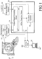
FIGURE 1 is a diagrammatic illustration of an apparatus which automatically registers 3D images of a whole body part; -
FIGURE 2 is an expanded diagrammatic illustration of a part an apparatus ofFIGURE 1 which automatically registers 3D images of a whole body part; -
FIGURE 3 is a diagrammatic illustration of the affine transform means ofFIGURE 1 and associated parts of the apparatus; -
FIGURE 4 is an expanded diagrammatic illustration of a part the apparatus ofFIGURE 1 which automatically registers 3D images of a whole body part and displays them on the monitor. - With reference to
FIGURE 1 , a patient is positioned in adiagnostic imager 10, such as a CT scanner, for a follow-up examination. The generated data is reconstructed by areconstruction processor 12 and stored in a 3D,volumetric image memory 14. Various image enhancement operations as are known in the art are also performed. - Image data from the hospital archive or from another
storage medium 16 of the same region of the same patient is retrieved and stored in an archived 3Dvolumetric image memory 18. Of course, both the current andarchive 3D image memories may be parts of a common storage medium. A registering means 20 retrieves the current and archived images and automatically register the two images for a concurrent display on amonitor 22. - With continuing reference to
FIGURE 1 and further reference toFIGURE 2 , a surface image generating means 24 generates surface images from the current archived images. A segmenting means 26 extracts the important body parts for the registration, then a feature extraction means 28 extracts common features for registration. Looking specifically to lung images, the segmentation includes a thresholding means 30 thresholds the images. More specifically to the lung embodiment, the thresholding means 30 sets the gray scale value of air in the lungs to one extreme value, e.g., black or white, and all tissue in the image to the other. Specifically to lung images, the feature extraction means 28 includes a surfacevoxel extracting means 32 which extracts the single layer of voxels which lies along the interface between the black and white regions. Preferably, the voxels which represent the tissue surface rather than the air surface are selected. A feature exclusion means 34 removes part of the features of the 3D image that is defective or artifacted, if any. Looking specifically to lung images, portions of the lung closely adjacent to the heart often suffer motion artifacts. To simplify the remaining operations, the feature exclusion means 32 optionally removes a common artifacted region from both image representations. The voxels are stored in feature or surface image memories ormemory sections 36, 36'. - A scaling means 40 scales the two surface images in accordance with known differences in scanning parameters. For example, the images are adjusted for differences in the slice thickness of the scans, for the field of view, for magnification differences, and the like. A normalizing
means 42 normalizes the two surface images. More specifically to the preferred embodiment, the extreme points along the z-axis of each image are labeled -1 in one direction and +1 in the other direction. Points in between are labeled proportionately. The same scaling is carried out with respect to the x and y-axes. Different normalization methods are also available, for example, computing the mean or median and standard deviation of the data and setting the central point to the mean or median and the length of one is set to the standard deviation from the mean or median. Of course, other coordinate systems are contemplated. The two scaled and normalized three-dimensional surface images are stored in scaled and normalized volume image memories ormemory sections 44, 44'. - With continuing reference to
FIGURES 1 and2 and further reference toFIGURE 3 , an affine transform means 50 determines the twelve values of the affine transform that defines the registration error between the current and archived scaled and normalized 3D images. Specifically, the affine transform means 50 determines nine rotational components about three orthogonal axes and three translational components along the three axes. Optionally, a scaling parameter can also be determined. - The affine transform means 50 includes a selective or non-uniform random feature or point reducing
means 52. That is, with normal CT resolution, the two surface images are still hundreds of thousands of voxels. To reduce the computational load, the selective (based on prior knowledge) point reducing means 52 or a non-uniform random point reducing means 52 preferably selectively (based on prior knowledge) randomly selects a fraction of the points, e.g., 1%, from both images. Because the diagnosing oncologist typically is most concerned with registration along the z- or longitudinal axis, the nonuniformity with which the points are selected is altered to favor points, which influence the z-registration. In the preferred embodiment, a higher percentage of points are selected at the top and bottom of the lungs. A matching means 54 matches the non-uniform randomly selected points from the one surface image with a corresponding point in the other surface image. In the preferred embodiment, a K-D tree matching technique orprocessor 56 is utilized. In each iteration, a non-uniformly random sample of the points from the current image, to be matched to the corresponding point in the other surface image, is taken to be matched to the other set of points from the previous image. For each point in the sample, a matching point is found using the K-D tree. For each matching pair, the coordinates or location of each voxel of the pair and the surface normal are determined. In the matching operation, if more than one candidate point has an equally good spatial match to the reference point, the point or voxel with the closest normal is selected to generate a set of points and normals which are stored in a point and normal memory means 58. - A bad pair removal means 60 removes matched pairs of points that fail to meet preselected criteria. In the preferred embodiment, these criteria include a physical separation distance greater than a preselected minimum, badly misaligned normals, and points near to the cut surface, if any. An error estimating means 62 estimates the error between the points. The error estimating means 62 calculates a weighted distance using the differences in x, y, z, and the normals. Each coordinate is assigned with different weight so that the differences in specific parts will be favored over other parts. Looking specifically to lung images, the differences in the z-coordinate have higher weight than the x and y-coordinates, and higher than the normal differences.
- With continuing reference to
FIGURE 3 , an error minimizing or affine registration means 70 finds an affine transform that minimizes the error between the pairs of points. In the preferred embodiment, the error minimizing means 70 uses a Levenberg-Marquardt minimization technique orprocessor 72. Using the weighted error estimate and the divergence of the weighted error estimate, the error minimizing means finds 70 a set of affine transformation parameters that gives the minimum of error for the set of sample pairs. Twelve affine transform coordinates and optionally the scaling correction are loaded into acorrection transform memory 80. - With continuing reference to
FIGURE 3 , atransform processor 82 transforms one of the current and archived scaled and normalized surface images frommemories 44, 44' in accordance with the determined correction transform. The affine transform means 50 repeats the registration technique re-registering the volume images, with one of them subject to the transform, which was predicted, to transform them into registration. This process generates the twelve affine transform coordinates (and optionally scaling) of a correction transform to the first correction transform. An endcriteria determining means 84 determines whether the two images have been aligned within acceptable tolerances by monitoring the correction transforms. In the preferred embodiment, the endcriteria determining means 84 determines whether the global translation is less than a preselected threshold, whether the change in the correction transform is below a preselected threshold, or whether a preselected number of iterations have been performed. If any of these criteria are met, a correction transform combining means 86 combines the first correction transform and each of the subsequent correction transforms to provide a single registration transform which is stored in a registration transform memory orbuffer 88. - With reference to
FIGURE 1 again and further reference toFIGURE 4 , atransform operator 90 operates on one of the current and 3D archive images, preferably the archived image to register it with the current 3D volume image. The transformed archived image is stored in a transformed 3Dimage volume memory 92. - An operator, through a keyboard or
other input device 100 controls a stepping means 102 which causes adisplay processor 104 to withdraw and display corresponding slices from the current3D image memory 14 and the transformed3D image memory 92 on amonitor 22. In the preferred embodiment, the transverse slice from thecurrent image memory 14 is displayed on the top half of the screen and the corresponding slice of the transformedmemory 92 is displayed on the bottom half of the screen. Preferably, corresponding orthogonal views are displayed adjacent the larger longitudinal slice. Theoperator input device 100 also controls a slice fusing means 106 that works with thedisplay processor 104 to sum a preselected number of adjacent slices in each of the two images. The operator input means 100 is also connected with a single image slice stepping means 108, which can control thedisplay processor 104 to step through slices of only one of the two 3D images independently of the other. - Typically, 3-5 slices are fused together for display on
monitor 22. A global registration algorithm which registers the two three-dimensional volume images within ±2 slices is considered accurate registration due to the fusing of the number of adjacent images. - Although described with particular reference to CT scanner imaging, it is to be appreciated that this technique is also applicable to magnetic resonance images, PET images, SPECT images, and other three-dimensional diagnostic images. Moreover, the images being registered may be from mixed modalities. For example, a CT image can be registered using this technique with a PET image. When mixing modalities, care should be taken to assure that the features are defined in both imaging modalities or appropriate adjustment made. Further, although described with particular reference to the lungs, it is to be appreciated that this technique is also applicable to studies of other organs such as the colon, the liver, and other non-rigid organs. Moreover, this technique is also applicable to rigid portions of the body such as the head.
- The invention has been described with reference to the preferred embodiments. Modifications and alterations will occur to others upon a reading and understanding of the preceding detailed description. It is intended that the invention be construed as including all such modifications and alterations insofar as they come within the scope of the appended claims or the equivalents thereof.
Claims (10)
- An apparatus for diagnostic imaging comprising:- a first memory means (14) for storing a first diagnostic image;- a second memory means (18) for storing a second diagnostic image;- a registering means (20) for automatically registering the first and second diagnostic images from the first and second image memories without operator assistance, the registering means (20) further including:an affine transformation determining means (50) for determining an affine transformation representative of misalignment of the first and second diagnostic images;- a means (90) for operating on one of the first and second diagnostic images in accordance with the determined affine transform to register the first and second diagnostic images;- a means (104, 22) for concurrently displaying a corresponding pair of slices of the registered first and second diagnostic images- a means (102) for replacing the displayed corresponding pair of slices of the first and second diagnostic images with another corresponding pair of slices of the first and second diagnostic images,characterized in that:said first diagnostic image is a first diagnostic image of the lungs of a patient,said second diagnostic image is a second diagnostic image of the lungs of said patient,and said affine transformation determining means (50) further includes a number of points reducing means (52) arranged for selectively and non-uniformly reducing a number of points to be used in determining said affine transformation, wherein points which influence the registration along a longitudinal axis are favored in said reducing of points, wherein a higher percentage of points are selected along the top and bottom of the lungs.
- The apparatus as set forth in claim 1, wherein the affine transform determining means (50) includes:- a means (54) for matching pairs of points in the first and second diagnostic images;- a means (58) for determining differences between locations and surface normal of the matched points; and- a means (70) for determining an affine transform which minimizes the deviation between the locations of the matched points.
- The apparatus as set forth in claim 2, wherein the point matching means (54) includes:- a processing means (56) which implements a K-D tree matching algorithm.
- The apparatus as set forth in claim 3, wherein the deviation minimizing means (70) includes:- a processor (72) which performs a Levenberg-Marquardt error minimization algorithm.
- The apparatus as set forth in claim 2, further including- a means (60) for removing matched pairs of points which fail to meet preselected criteria.
- The apparatus as set forth in claim 2, wherein the registering means (20) further includes:- a means (24) for converting the first and second diagnostic images into feature image representations, the affine transform determining means (50) operating on the first and second feature representations to determine the affine transform.
- The apparatus as set forth in claim 6, wherein the feature generating means (24) includes:- a segmentation means (26), which segments appropriate target organs in the diagnostic images; and- a feature extraction means (34) that extracts a set of features to be matched in the diagnostic images.
- The apparatus as set forth in claim 7, wherein the feature image generating means (24) includes:- a thresholding segmentation means (26), which segments lungs in the diagnostic images using a predetermined threshold and the features defined as the surface points of the lungs extracted by assigning a tissue on one side of a boundary of an organ of interest a first value and a tissue or air on another side of the boundary of the organ of interest a first value and a tissue or air on another side of the boundary of the organ of interest a second value, distinct from the first value; and- a means (28) for extracting a boundary layer of voxels of the first value which adjoin voxels of the second value.
- The apparatus as set forth in claim 1, further including:- means (106) for combining an operator selected plurality of slices in each of the displayed slice images.
- A computer-implemented
method of diagnostic imaging comprising:- storing a first diagnostic image;- storing a second diagnostic image;- automatically registering the first and second diagnostic images from the first and second image memories without operator assistance, the registering further including:- determining an affine transformation representative of misalignment of the first and second diagnostic images;- operating on one of the first and second diagnostic images in accordance with the determined affine transform to register the first and second diagnostic images;- concurrently displaying a corresponding pair of slices of the registered first and second diagnostic images- replacing the displayed corresponding pair of slices of the first and second diagnostic images with another corresponding pair of slices of the first and second diagnostic images, characterized in that:said first diagnostic image is a first diagnostic image of the lungs of a patient,said second diagnostic image is a second diagnostic image of the lungs of said patient,and said affine transformation determining further includes selectively and non-uniformly reducing a number of points to be used in determining said affine transformation, wherein points which influence the registration along a longitudinal axis are favored in said reducing of points, wherein a higher percentage of points are selected along the top and bottom of the lungs.
Applications Claiming Priority (2)
| Application Number | Priority Date | Filing Date | Title |
|---|---|---|---|
| US49104503P | 2003-07-30 | 2003-07-30 | |
| PCT/IB2004/002399 WO2005010793A2 (en) | 2003-07-30 | 2004-07-16 | Automatic registration of intra-modality medical volume images using affine transformation |
Publications (2)
| Publication Number | Publication Date |
|---|---|
| EP1652122A2 EP1652122A2 (en) | 2006-05-03 |
| EP1652122B1 true EP1652122B1 (en) | 2018-03-14 |
Family
ID=34103005
Family Applications (1)
| Application Number | Title | Priority Date | Filing Date |
|---|---|---|---|
| EP04744057.3A Expired - Lifetime EP1652122B1 (en) | 2003-07-30 | 2004-07-16 | Automatic registration of intra-modality medical volume images using affine transformation |
Country Status (4)
| Country | Link |
|---|---|
| US (1) | US7627158B2 (en) |
| EP (1) | EP1652122B1 (en) |
| JP (1) | JP5296981B2 (en) |
| WO (1) | WO2005010793A2 (en) |
Families Citing this family (17)
| Publication number | Priority date | Publication date | Assignee | Title |
|---|---|---|---|---|
| JP2007515714A (en) * | 2003-12-08 | 2007-06-14 | コーニンクレッカ フィリップス エレクトロニクス エヌ ヴィ | Point-based elastic image matching is adaptive |
| JP5676840B2 (en) * | 2004-11-17 | 2015-02-25 | コーニンクレッカ フィリップス エヌ ヴェ | Improved elastic image registration function |
| JP2008532612A (en) * | 2005-03-10 | 2008-08-21 | コーニンクレッカ フィリップス エレクトロニクス エヌ ヴィ | Image processing system and method for alignment of two-dimensional information with three-dimensional volume data during medical treatment |
| US7518619B2 (en) * | 2005-11-07 | 2009-04-14 | General Electric Company | Method and apparatus for integrating three-dimensional and two-dimensional monitors with medical diagnostic imaging workstations |
| EP1881453A3 (en) | 2006-07-18 | 2009-07-22 | Kabushiki Kaisha Toshiba | A medical image-processing apparatus and a method for processing medical images |
| US7848592B2 (en) * | 2006-07-31 | 2010-12-07 | Carestream Health, Inc. | Image fusion for radiation therapy |
| US20080186378A1 (en) * | 2007-02-06 | 2008-08-07 | Feimo Shen | Method and apparatus for guiding towards targets during motion |
| JP5159301B2 (en) * | 2007-12-28 | 2013-03-06 | 株式会社東芝 | Medical image display apparatus and image display method |
| JP5279291B2 (en) * | 2008-02-19 | 2013-09-04 | 株式会社東芝 | Medical image display apparatus and image display method |
| CA2748234A1 (en) * | 2008-12-25 | 2010-07-01 | Medic Vision - Imaging Solutions Ltd. | Denoising medical images |
| US20130345559A1 (en) * | 2012-03-28 | 2013-12-26 | Musc Foundation For Reseach Development | Quantitative perfusion analysis for embolotherapy |
| US9886546B2 (en) | 2012-11-20 | 2018-02-06 | General Electric Company | Methods and apparatus to label radiology images |
| JP2016539701A (en) * | 2013-12-11 | 2016-12-22 | コーニンクレッカ フィリップス エヌ ヴェKoninklijke Philips N.V. | Volumetric image data processing based on three-dimensional (3D) prescan |
| DE102015201271A1 (en) * | 2014-09-17 | 2016-03-17 | Friedrich-Alexander-Universität Erlangen - Nürnberg | Method and system for determining the local quality of surface data extracted from volume data |
| US10991069B2 (en) * | 2014-10-08 | 2021-04-27 | Samsung Electronics Co., Ltd. | Method and apparatus for registration of medical images |
| CA2985308C (en) * | 2015-05-08 | 2020-10-27 | Synaptive Medical (Barbados) Inc. | Magnetic resonance visible labels and markers for encoding information |
| CN114219702B (en) * | 2021-11-15 | 2025-06-17 | 宁波舜宇仪器有限公司 | Image matching and display method of pathological sections with different staining |
Family Cites Families (11)
| Publication number | Priority date | Publication date | Assignee | Title |
|---|---|---|---|---|
| US5800353A (en) * | 1996-02-12 | 1998-09-01 | Mclaurin, Jr.; Robert L. | Automatic image registration of magnetic resonance imaging scans for localization, 3-dimensional treatment planning, and radiation treatment of abnormal lesions |
| JPH10137231A (en) * | 1996-11-13 | 1998-05-26 | Toshiba Iyou Syst Eng Kk | Medical image processing equipment |
| US6363163B1 (en) * | 1998-02-23 | 2002-03-26 | Arch Development Corporation | Method and system for the automated temporal subtraction of medical images |
| JP2000149020A (en) * | 1998-11-06 | 2000-05-30 | Nippon Avionics Co Ltd | Pattern alignment method |
| JP4129896B2 (en) * | 1999-02-12 | 2008-08-06 | 松下電器産業株式会社 | Optical three-dimensional measuring apparatus and optical three-dimensional measuring method |
| FR2797978B1 (en) | 1999-08-30 | 2001-10-26 | Ge Medical Syst Sa | AUTOMATIC IMAGE RECORDING PROCESS |
| US7206462B1 (en) * | 2000-03-17 | 2007-04-17 | The General Hospital Corporation | Method and system for the detection, comparison and volumetric quantification of pulmonary nodules on medical computed tomography scans |
| AU2001251539A1 (en) * | 2000-04-11 | 2001-10-23 | Cornell Research Foundation Inc. | System and method for three-dimensional image rendering and analysis |
| US6714810B2 (en) * | 2000-09-07 | 2004-03-30 | Cbyon, Inc. | Fluoroscopic registration system and method |
| US6728424B1 (en) | 2000-09-15 | 2004-04-27 | Koninklijke Philips Electronics, N.V. | Imaging registration system and method using likelihood maximization |
| US7106891B2 (en) * | 2001-10-15 | 2006-09-12 | Insightful Corporation | System and method for determining convergence of image set registration |
-
2004
- 2004-07-16 JP JP2006521692A patent/JP5296981B2/en not_active Expired - Fee Related
- 2004-07-16 EP EP04744057.3A patent/EP1652122B1/en not_active Expired - Lifetime
- 2004-07-16 US US10/566,136 patent/US7627158B2/en not_active Expired - Fee Related
- 2004-07-16 WO PCT/IB2004/002399 patent/WO2005010793A2/en not_active Ceased
Also Published As
| Publication number | Publication date |
|---|---|
| JP5296981B2 (en) | 2013-09-25 |
| US20060204064A1 (en) | 2006-09-14 |
| US7627158B2 (en) | 2009-12-01 |
| WO2005010793A3 (en) | 2005-07-21 |
| WO2005010793A2 (en) | 2005-02-03 |
| JP2007500029A (en) | 2007-01-11 |
| EP1652122A2 (en) | 2006-05-03 |
Similar Documents
| Publication | Publication Date | Title |
|---|---|---|
| US8787648B2 (en) | CT surrogate by auto-segmentation of magnetic resonance images | |
| US8953856B2 (en) | Method and system for registering a medical image | |
| US8942455B2 (en) | 2D/3D image registration method | |
| US7117026B2 (en) | Physiological model based non-rigid image registration | |
| US8326086B2 (en) | Elastic image registration | |
| Li et al. | Establishing a normative atlas of the human lung: intersubject warping and registration of volumetric CT images | |
| US8553961B2 (en) | Automatic pre-alignment for registration of medical images | |
| US7397934B2 (en) | Registration of thoracic and abdominal imaging modalities | |
| JP6537981B2 (en) | Segmentation of large objects from multiple 3D views | |
| US7627158B2 (en) | Automatic registration of intra-modality medical volume images using affine transformation | |
| EP1815432B1 (en) | Improved elastic image registration functionality | |
| JP2016539701A (en) | Volumetric image data processing based on three-dimensional (3D) prescan | |
| Studholme et al. | Estimating tissue deformation between functional images induced by intracranial electrode implantation using anatomical MRI | |
| CN108430376B (en) | Providing a projection data set | |
| Alam et al. | Evaluation of medical image registration techniques based on nature and domain of the transformation | |
| Longuefosse et al. | Lung ct synthesis using gans with conditional normalization on registered ultrashort echo-time mri | |
| Li et al. | 3D intersubject warping and registration of pulmonary CT images for a human lung model | |
| Li et al. | Automatic inter-subject registration of whole body images | |
| Rösch et al. | Robust 3D deformation field estimation by template propagation | |
| CN120604264A (en) | Obtain a medical image at the target plane | |
| KAWATA et al. | Visualization of interval changes of pulmonary nodules using high-resolution CT images | |
| Hong et al. | Automatic segmentation and registration for the lung nodule matching in temporal chest CT scans | |
| Deniz | Automatic registration of medical image volumes of different modalities | |
| Prasai | Multimodality Image Registration |
Legal Events
| Date | Code | Title | Description |
|---|---|---|---|
| PUAI | Public reference made under article 153(3) epc to a published international application that has entered the european phase |
Free format text: ORIGINAL CODE: 0009012 |
|
| 17P | Request for examination filed |
Effective date: 20060228 |
|
| AK | Designated contracting states |
Kind code of ref document: A2 Designated state(s): AT BE BG CH CY CZ DE DK EE ES FI FR GB GR HU IE IT LI LU MC NL PL PT RO SE SI SK TR |
|
| 17Q | First examination report despatched |
Effective date: 20060703 |
|
| DAX | Request for extension of the european patent (deleted) | ||
| RAP1 | Party data changed (applicant data changed or rights of an application transferred) |
Owner name: KONINKLIJKE PHILIPS N.V. |
|
| GRAP | Despatch of communication of intention to grant a patent |
Free format text: ORIGINAL CODE: EPIDOSNIGR1 |
|
| INTG | Intention to grant announced |
Effective date: 20171002 |
|
| GRAS | Grant fee paid |
Free format text: ORIGINAL CODE: EPIDOSNIGR3 |
|
| GRAA | (expected) grant |
Free format text: ORIGINAL CODE: 0009210 |
|
| AK | Designated contracting states |
Kind code of ref document: B1 Designated state(s): AT BE BG CH CY CZ DE DK EE ES FI FR GB GR HU IE IT LI LU MC NL PL PT RO SE SI SK TR |
|
| REG | Reference to a national code |
Ref country code: GB Ref legal event code: FG4D |
|
| REG | Reference to a national code |
Ref country code: CH Ref legal event code: EP Ref country code: AT Ref legal event code: REF Ref document number: 979535 Country of ref document: AT Kind code of ref document: T Effective date: 20180315 |
|
| REG | Reference to a national code |
Ref country code: IE Ref legal event code: FG4D |
|
| REG | Reference to a national code |
Ref country code: DE Ref legal event code: R096 Ref document number: 602004052464 Country of ref document: DE |
|
| REG | Reference to a national code |
Ref country code: DE Ref legal event code: R084 Ref document number: 602004052464 Country of ref document: DE |
|
| REG | Reference to a national code |
Ref country code: NL Ref legal event code: MP Effective date: 20180314 |
|
| REG | Reference to a national code |
Ref country code: FR Ref legal event code: PLFP Year of fee payment: 15 |
|
| PG25 | Lapsed in a contracting state [announced via postgrant information from national office to epo] |
Ref country code: CY Free format text: LAPSE BECAUSE OF FAILURE TO SUBMIT A TRANSLATION OF THE DESCRIPTION OR TO PAY THE FEE WITHIN THE PRESCRIBED TIME-LIMIT Effective date: 20180314 Ref country code: ES Free format text: LAPSE BECAUSE OF FAILURE TO SUBMIT A TRANSLATION OF THE DESCRIPTION OR TO PAY THE FEE WITHIN THE PRESCRIBED TIME-LIMIT Effective date: 20180314 Ref country code: FI Free format text: LAPSE BECAUSE OF FAILURE TO SUBMIT A TRANSLATION OF THE DESCRIPTION OR TO PAY THE FEE WITHIN THE PRESCRIBED TIME-LIMIT Effective date: 20180314 |
|
| REG | Reference to a national code |
Ref country code: AT Ref legal event code: MK05 Ref document number: 979535 Country of ref document: AT Kind code of ref document: T Effective date: 20180314 |
|
| PG25 | Lapsed in a contracting state [announced via postgrant information from national office to epo] |
Ref country code: SE Free format text: LAPSE BECAUSE OF FAILURE TO SUBMIT A TRANSLATION OF THE DESCRIPTION OR TO PAY THE FEE WITHIN THE PRESCRIBED TIME-LIMIT Effective date: 20180314 Ref country code: BG Free format text: LAPSE BECAUSE OF FAILURE TO SUBMIT A TRANSLATION OF THE DESCRIPTION OR TO PAY THE FEE WITHIN THE PRESCRIBED TIME-LIMIT Effective date: 20180614 Ref country code: GR Free format text: LAPSE BECAUSE OF FAILURE TO SUBMIT A TRANSLATION OF THE DESCRIPTION OR TO PAY THE FEE WITHIN THE PRESCRIBED TIME-LIMIT Effective date: 20180615 |
|
| PG25 | Lapsed in a contracting state [announced via postgrant information from national office to epo] |
Ref country code: PL Free format text: LAPSE BECAUSE OF FAILURE TO SUBMIT A TRANSLATION OF THE DESCRIPTION OR TO PAY THE FEE WITHIN THE PRESCRIBED TIME-LIMIT Effective date: 20180314 Ref country code: NL Free format text: LAPSE BECAUSE OF FAILURE TO SUBMIT A TRANSLATION OF THE DESCRIPTION OR TO PAY THE FEE WITHIN THE PRESCRIBED TIME-LIMIT Effective date: 20180314 Ref country code: IT Free format text: LAPSE BECAUSE OF FAILURE TO SUBMIT A TRANSLATION OF THE DESCRIPTION OR TO PAY THE FEE WITHIN THE PRESCRIBED TIME-LIMIT Effective date: 20180314 Ref country code: RO Free format text: LAPSE BECAUSE OF FAILURE TO SUBMIT A TRANSLATION OF THE DESCRIPTION OR TO PAY THE FEE WITHIN THE PRESCRIBED TIME-LIMIT Effective date: 20180314 Ref country code: EE Free format text: LAPSE BECAUSE OF FAILURE TO SUBMIT A TRANSLATION OF THE DESCRIPTION OR TO PAY THE FEE WITHIN THE PRESCRIBED TIME-LIMIT Effective date: 20180314 |
|
| REG | Reference to a national code |
Ref country code: DE Ref legal event code: R079 Ref document number: 602004052464 Country of ref document: DE Free format text: PREVIOUS MAIN CLASS: G06F0019000000 Ipc: G16Z0099000000 |
|
| PG25 | Lapsed in a contracting state [announced via postgrant information from national office to epo] |
Ref country code: SK Free format text: LAPSE BECAUSE OF FAILURE TO SUBMIT A TRANSLATION OF THE DESCRIPTION OR TO PAY THE FEE WITHIN THE PRESCRIBED TIME-LIMIT Effective date: 20180314 Ref country code: CZ Free format text: LAPSE BECAUSE OF FAILURE TO SUBMIT A TRANSLATION OF THE DESCRIPTION OR TO PAY THE FEE WITHIN THE PRESCRIBED TIME-LIMIT Effective date: 20180314 Ref country code: AT Free format text: LAPSE BECAUSE OF FAILURE TO SUBMIT A TRANSLATION OF THE DESCRIPTION OR TO PAY THE FEE WITHIN THE PRESCRIBED TIME-LIMIT Effective date: 20180314 |
|
| REG | Reference to a national code |
Ref country code: DE Ref legal event code: R097 Ref document number: 602004052464 Country of ref document: DE |
|
| PG25 | Lapsed in a contracting state [announced via postgrant information from national office to epo] |
Ref country code: PT Free format text: LAPSE BECAUSE OF FAILURE TO SUBMIT A TRANSLATION OF THE DESCRIPTION OR TO PAY THE FEE WITHIN THE PRESCRIBED TIME-LIMIT Effective date: 20180716 |
|
| PLBE | No opposition filed within time limit |
Free format text: ORIGINAL CODE: 0009261 |
|
| STAA | Information on the status of an ep patent application or granted ep patent |
Free format text: STATUS: NO OPPOSITION FILED WITHIN TIME LIMIT |
|
| PG25 | Lapsed in a contracting state [announced via postgrant information from national office to epo] |
Ref country code: DK Free format text: LAPSE BECAUSE OF FAILURE TO SUBMIT A TRANSLATION OF THE DESCRIPTION OR TO PAY THE FEE WITHIN THE PRESCRIBED TIME-LIMIT Effective date: 20180314 |
|
| 26N | No opposition filed |
Effective date: 20181217 |
|
| PG25 | Lapsed in a contracting state [announced via postgrant information from national office to epo] |
Ref country code: SI Free format text: LAPSE BECAUSE OF FAILURE TO SUBMIT A TRANSLATION OF THE DESCRIPTION OR TO PAY THE FEE WITHIN THE PRESCRIBED TIME-LIMIT Effective date: 20180314 |
|
| REG | Reference to a national code |
Ref country code: CH Ref legal event code: PL |
|
| GBPC | Gb: european patent ceased through non-payment of renewal fee |
Effective date: 20180716 |
|
| PG25 | Lapsed in a contracting state [announced via postgrant information from national office to epo] |
Ref country code: LU Free format text: LAPSE BECAUSE OF NON-PAYMENT OF DUE FEES Effective date: 20180716 Ref country code: MC Free format text: LAPSE BECAUSE OF FAILURE TO SUBMIT A TRANSLATION OF THE DESCRIPTION OR TO PAY THE FEE WITHIN THE PRESCRIBED TIME-LIMIT Effective date: 20180314 |
|
| REG | Reference to a national code |
Ref country code: BE Ref legal event code: MM Effective date: 20180731 |
|
| REG | Reference to a national code |
Ref country code: IE Ref legal event code: MM4A |
|
| PG25 | Lapsed in a contracting state [announced via postgrant information from national office to epo] |
Ref country code: CH Free format text: LAPSE BECAUSE OF NON-PAYMENT OF DUE FEES Effective date: 20180731 Ref country code: GB Free format text: LAPSE BECAUSE OF NON-PAYMENT OF DUE FEES Effective date: 20180716 Ref country code: IE Free format text: LAPSE BECAUSE OF NON-PAYMENT OF DUE FEES Effective date: 20180716 Ref country code: LI Free format text: LAPSE BECAUSE OF NON-PAYMENT OF DUE FEES Effective date: 20180731 |
|
| PG25 | Lapsed in a contracting state [announced via postgrant information from national office to epo] |
Ref country code: BE Free format text: LAPSE BECAUSE OF NON-PAYMENT OF DUE FEES Effective date: 20180731 |
|
| PGFP | Annual fee paid to national office [announced via postgrant information from national office to epo] |
Ref country code: FR Payment date: 20190726 Year of fee payment: 16 |
|
| PGFP | Annual fee paid to national office [announced via postgrant information from national office to epo] |
Ref country code: DE Payment date: 20190930 Year of fee payment: 16 |
|
| PG25 | Lapsed in a contracting state [announced via postgrant information from national office to epo] |
Ref country code: TR Free format text: LAPSE BECAUSE OF FAILURE TO SUBMIT A TRANSLATION OF THE DESCRIPTION OR TO PAY THE FEE WITHIN THE PRESCRIBED TIME-LIMIT Effective date: 20180314 |
|
| PG25 | Lapsed in a contracting state [announced via postgrant information from national office to epo] |
Ref country code: HU Free format text: LAPSE BECAUSE OF FAILURE TO SUBMIT A TRANSLATION OF THE DESCRIPTION OR TO PAY THE FEE WITHIN THE PRESCRIBED TIME-LIMIT; INVALID AB INITIO Effective date: 20040716 |
|
| REG | Reference to a national code |
Ref country code: DE Ref legal event code: R119 Ref document number: 602004052464 Country of ref document: DE |
|
| PG25 | Lapsed in a contracting state [announced via postgrant information from national office to epo] |
Ref country code: FR Free format text: LAPSE BECAUSE OF NON-PAYMENT OF DUE FEES Effective date: 20200731 |
|
| PG25 | Lapsed in a contracting state [announced via postgrant information from national office to epo] |
Ref country code: DE Free format text: LAPSE BECAUSE OF NON-PAYMENT OF DUE FEES Effective date: 20210202 |