EP1650299A1 - Method of detecting reaction of dna and dna-binding protein - Google Patents
Method of detecting reaction of dna and dna-binding protein Download PDFInfo
- Publication number
- EP1650299A1 EP1650299A1 EP04746326A EP04746326A EP1650299A1 EP 1650299 A1 EP1650299 A1 EP 1650299A1 EP 04746326 A EP04746326 A EP 04746326A EP 04746326 A EP04746326 A EP 04746326A EP 1650299 A1 EP1650299 A1 EP 1650299A1
- Authority
- EP
- European Patent Office
- Prior art keywords
- transcription factor
- double
- stranded dna
- dna
- cell nucleus
- Prior art date
- Legal status (The legal status is an assumption and is not a legal conclusion. Google has not performed a legal analysis and makes no representation as to the accuracy of the status listed.)
- Withdrawn
Links
- 108020004414 DNA Proteins 0.000 title claims abstract description 335
- 238000000034 method Methods 0.000 title claims abstract description 42
- 238000006243 chemical reaction Methods 0.000 title claims description 74
- 102000052510 DNA-Binding Proteins Human genes 0.000 title claims description 35
- 101710096438 DNA-binding protein Proteins 0.000 title claims description 35
- 238000009739 binding Methods 0.000 claims abstract description 126
- 238000009792 diffusion process Methods 0.000 claims abstract description 122
- 230000027455 binding Effects 0.000 claims abstract description 99
- 238000002060 fluorescence correlation spectroscopy Methods 0.000 claims abstract description 85
- 102000006290 Transcription Factor TFIID Human genes 0.000 claims abstract description 45
- 108010083268 Transcription Factor TFIID Proteins 0.000 claims abstract description 45
- 108090000941 Transcription factor TFIIB Proteins 0.000 claims abstract description 30
- 102000004408 Transcription factor TFIIB Human genes 0.000 claims abstract description 30
- 238000002156 mixing Methods 0.000 claims abstract description 25
- 102000053602 DNA Human genes 0.000 claims description 244
- 210000003855 cell nucleus Anatomy 0.000 claims description 131
- 102100023132 Transcription factor Jun Human genes 0.000 claims description 103
- 108010018242 Transcription Factor AP-1 Proteins 0.000 claims description 101
- 108060008682 Tumor Necrosis Factor Proteins 0.000 claims description 66
- 239000000243 solution Substances 0.000 claims description 64
- 210000004027 cell Anatomy 0.000 claims description 51
- 108010057466 NF-kappa B Proteins 0.000 claims description 48
- 102000040945 Transcription factor Human genes 0.000 claims description 35
- 108091023040 Transcription factor Proteins 0.000 claims description 35
- MDDIUTVUBYEEEM-UHFFFAOYSA-N azane;pyrrolidine-1-carbodithioic acid Chemical compound N.SC(=S)N1CCCC1 MDDIUTVUBYEEEM-UHFFFAOYSA-N 0.000 claims description 32
- 239000007850 fluorescent dye Substances 0.000 claims description 27
- 239000011259 mixed solution Substances 0.000 claims description 18
- 239000000126 substance Substances 0.000 claims description 18
- 239000002245 particle Substances 0.000 claims description 10
- 102100040247 Tumor necrosis factor Human genes 0.000 claims 5
- QGZKDVFQNNGYKY-UHFFFAOYSA-O Ammonium Chemical compound [NH4+] QGZKDVFQNNGYKY-UHFFFAOYSA-O 0.000 claims 1
- 238000005259 measurement Methods 0.000 abstract description 41
- 238000001514 detection method Methods 0.000 abstract description 11
- 238000001962 electrophoresis Methods 0.000 abstract description 7
- 239000007787 solid Substances 0.000 abstract description 7
- 239000000758 substrate Substances 0.000 abstract description 7
- 238000002474 experimental method Methods 0.000 description 135
- 102000000852 Tumor Necrosis Factor-alpha Human genes 0.000 description 61
- 239000000047 product Substances 0.000 description 57
- 230000000638 stimulation Effects 0.000 description 50
- 102000003945 NF-kappa B Human genes 0.000 description 43
- PEDCQBHIVMGVHV-UHFFFAOYSA-N Glycerine Chemical compound OCC(O)CO PEDCQBHIVMGVHV-UHFFFAOYSA-N 0.000 description 24
- 239000007795 chemical reaction product Substances 0.000 description 19
- 210000004940 nucleus Anatomy 0.000 description 18
- 239000012148 binding buffer Substances 0.000 description 16
- TWRXJAOTZQYOKJ-UHFFFAOYSA-L Magnesium chloride Chemical compound [Mg+2].[Cl-].[Cl-] TWRXJAOTZQYOKJ-UHFFFAOYSA-L 0.000 description 12
- 108090000623 proteins and genes Proteins 0.000 description 12
- 102000004169 proteins and genes Human genes 0.000 description 11
- 239000000523 sample Substances 0.000 description 11
- 230000004913 activation Effects 0.000 description 9
- 239000000872 buffer Substances 0.000 description 9
- 102100029075 Exonuclease 1 Human genes 0.000 description 8
- FAPWRFPIFSIZLT-UHFFFAOYSA-M Sodium chloride Chemical compound [Na+].[Cl-] FAPWRFPIFSIZLT-UHFFFAOYSA-M 0.000 description 8
- 229920001577 copolymer Polymers 0.000 description 8
- 239000002953 phosphate buffered saline Substances 0.000 description 8
- 230000002860 competitive effect Effects 0.000 description 7
- LOKCTEFSRHRXRJ-UHFFFAOYSA-I dipotassium trisodium dihydrogen phosphate hydrogen phosphate dichloride Chemical compound P(=O)(O)(O)[O-].[K+].P(=O)(O)([O-])[O-].[Na+].[Na+].[Cl-].[K+].[Cl-].[Na+] LOKCTEFSRHRXRJ-UHFFFAOYSA-I 0.000 description 7
- KCXVZYZYPLLWCC-UHFFFAOYSA-N EDTA Chemical compound OC(=O)CN(CC(O)=O)CCN(CC(O)=O)CC(O)=O KCXVZYZYPLLWCC-UHFFFAOYSA-N 0.000 description 6
- 108010007577 Exodeoxyribonuclease I Proteins 0.000 description 6
- 230000000295 complement effect Effects 0.000 description 6
- 229910001629 magnesium chloride Inorganic materials 0.000 description 6
- 239000000203 mixture Substances 0.000 description 6
- 241000588724 Escherichia coli Species 0.000 description 5
- 238000002360 preparation method Methods 0.000 description 5
- XLYOFNOQVPJJNP-UHFFFAOYSA-N water Chemical compound O XLYOFNOQVPJJNP-UHFFFAOYSA-N 0.000 description 5
- QKNYBSVHEMOAJP-UHFFFAOYSA-N 2-amino-2-(hydroxymethyl)propane-1,3-diol;hydron;chloride Chemical compound Cl.OCC(N)(CO)CO QKNYBSVHEMOAJP-UHFFFAOYSA-N 0.000 description 4
- 229940124158 Protease/peptidase inhibitor Drugs 0.000 description 4
- 238000003556 assay Methods 0.000 description 4
- 230000000694 effects Effects 0.000 description 4
- 230000005764 inhibitory process Effects 0.000 description 4
- 239000000137 peptide hydrolase inhibitor Substances 0.000 description 4
- 239000001103 potassium chloride Substances 0.000 description 4
- WCUXLLCKKVVCTQ-UHFFFAOYSA-M potassium chloride Inorganic materials [Cl-].[K+] WCUXLLCKKVVCTQ-UHFFFAOYSA-M 0.000 description 4
- 235000011164 potassium chloride Nutrition 0.000 description 4
- 239000011780 sodium chloride Substances 0.000 description 4
- DGVVWUTYPXICAM-UHFFFAOYSA-N β‐Mercaptoethanol Chemical compound OCCS DGVVWUTYPXICAM-UHFFFAOYSA-N 0.000 description 4
- MZOFCQQQCNRIBI-VMXHOPILSA-N (3s)-4-[[(2s)-1-[[(2s)-1-[[(1s)-1-carboxy-2-hydroxyethyl]amino]-4-methyl-1-oxopentan-2-yl]amino]-5-(diaminomethylideneamino)-1-oxopentan-2-yl]amino]-3-[[2-[[(2s)-2,6-diaminohexanoyl]amino]acetyl]amino]-4-oxobutanoic acid Chemical compound OC[C@@H](C(O)=O)NC(=O)[C@H](CC(C)C)NC(=O)[C@H](CCCN=C(N)N)NC(=O)[C@H](CC(O)=O)NC(=O)CNC(=O)[C@@H](N)CCCCN MZOFCQQQCNRIBI-VMXHOPILSA-N 0.000 description 3
- 108091003079 Bovine Serum Albumin Proteins 0.000 description 3
- HDOWHFYKZWZAAE-UHFFFAOYSA-M Cl.Cl.Cl.[Cl-].[K+] Chemical compound Cl.Cl.Cl.[Cl-].[K+] HDOWHFYKZWZAAE-UHFFFAOYSA-M 0.000 description 3
- 101001050288 Homo sapiens Transcription factor Jun Proteins 0.000 description 3
- 102100023050 Nuclear factor NF-kappa-B p105 subunit Human genes 0.000 description 3
- 102000006467 TATA-Box Binding Protein Human genes 0.000 description 3
- 108010044281 TATA-Box Binding Protein Proteins 0.000 description 3
- 238000004458 analytical method Methods 0.000 description 3
- VHJLVAABSRFDPM-QWWZWVQMSA-N dithiothreitol Chemical compound SC[C@@H](O)[C@H](O)CS VHJLVAABSRFDPM-QWWZWVQMSA-N 0.000 description 3
- ABZLKHKQJHEPAX-UHFFFAOYSA-N tetramethylrhodamine Chemical compound C=12C=CC(N(C)C)=CC2=[O+]C2=CC(N(C)C)=CC=C2C=1C1=CC=CC=C1C([O-])=O ABZLKHKQJHEPAX-UHFFFAOYSA-N 0.000 description 3
- 238000013518 transcription Methods 0.000 description 3
- 230000035897 transcription Effects 0.000 description 3
- 230000002103 transcriptional effect Effects 0.000 description 3
- IERJLWIWZLIVQA-UHFFFAOYSA-N 2-[2-[bis(carboxymethyl)amino]ethyl-(carboxymethyl)amino]acetic acid;2-sulfanylethanol Chemical compound OCCS.OC(=O)CN(CC(O)=O)CCN(CC(O)=O)CC(O)=O IERJLWIWZLIVQA-UHFFFAOYSA-N 0.000 description 2
- JKMHFZQWWAIEOD-UHFFFAOYSA-N 2-[4-(2-hydroxyethyl)piperazin-1-yl]ethanesulfonic acid Chemical compound OCC[NH+]1CCN(CCS([O-])(=O)=O)CC1 JKMHFZQWWAIEOD-UHFFFAOYSA-N 0.000 description 2
- NKDFYOWSKOHCCO-YPVLXUMRSA-N 20-hydroxyecdysone Chemical compound C1[C@@H](O)[C@@H](O)C[C@]2(C)[C@@H](CC[C@@]3([C@@H]([C@@](C)(O)[C@H](O)CCC(C)(O)C)CC[C@]33O)C)C3=CC(=O)[C@@H]21 NKDFYOWSKOHCCO-YPVLXUMRSA-N 0.000 description 2
- 108010039627 Aprotinin Proteins 0.000 description 2
- VGGGPCQERPFHOB-MCIONIFRSA-N Bestatin Chemical compound CC(C)C[C@H](C(O)=O)NC(=O)[C@@H](O)[C@H](N)CC1=CC=CC=C1 VGGGPCQERPFHOB-MCIONIFRSA-N 0.000 description 2
- VGGGPCQERPFHOB-UHFFFAOYSA-N Bestatin Natural products CC(C)CC(C(O)=O)NC(=O)C(O)C(N)CC1=CC=CC=C1 VGGGPCQERPFHOB-UHFFFAOYSA-N 0.000 description 2
- PHEDXBVPIONUQT-UHFFFAOYSA-N Cocarcinogen A1 Natural products CCCCCCCCCCCCCC(=O)OC1C(C)C2(O)C3C=C(C)C(=O)C3(O)CC(CO)=CC2C2C1(OC(C)=O)C2(C)C PHEDXBVPIONUQT-UHFFFAOYSA-N 0.000 description 2
- 102000004127 Cytokines Human genes 0.000 description 2
- 108090000695 Cytokines Proteins 0.000 description 2
- 239000006144 Dulbecco’s modified Eagle's medium Substances 0.000 description 2
- 102000004190 Enzymes Human genes 0.000 description 2
- 108090000790 Enzymes Proteins 0.000 description 2
- 239000007995 HEPES buffer Substances 0.000 description 2
- 101000611183 Homo sapiens Tumor necrosis factor Proteins 0.000 description 2
- GDBQQVLCIARPGH-UHFFFAOYSA-N Leupeptin Natural products CC(C)CC(NC(C)=O)C(=O)NC(CC(C)C)C(=O)NC(C=O)CCCN=C(N)N GDBQQVLCIARPGH-UHFFFAOYSA-N 0.000 description 2
- 108010010677 Phosphodiesterase I Proteins 0.000 description 2
- VSWDORGPIHIGNW-UHFFFAOYSA-N Pyrrolidine dithiocarbamic acid Chemical compound SC(=S)N1CCCC1 VSWDORGPIHIGNW-UHFFFAOYSA-N 0.000 description 2
- 238000000137 annealing Methods 0.000 description 2
- 238000013459 approach Methods 0.000 description 2
- 229960004405 aprotinin Drugs 0.000 description 2
- 229940098773 bovine serum albumin Drugs 0.000 description 2
- 238000006731 degradation reaction Methods 0.000 description 2
- 239000008367 deionised water Substances 0.000 description 2
- 229910021641 deionized water Inorganic materials 0.000 description 2
- 238000000605 extraction Methods 0.000 description 2
- 102000057041 human TNF Human genes 0.000 description 2
- 230000003301 hydrolyzing effect Effects 0.000 description 2
- 238000011534 incubation Methods 0.000 description 2
- ZPNFWUPYTFPOJU-LPYSRVMUSA-N iniprol Chemical compound C([C@H]1C(=O)NCC(=O)NCC(=O)N[C@H]2CSSC[C@H]3C(=O)N[C@@H](CCCCN)C(=O)N[C@@H](C)C(=O)N[C@@H](CCCNC(N)=N)C(=O)N[C@H](C(N[C@H](C(=O)N[C@@H](CCCNC(N)=N)C(=O)N[C@@H](CC=4C=CC(O)=CC=4)C(=O)N[C@@H](CC=4C=CC=CC=4)C(=O)N[C@@H](CC=4C=CC(O)=CC=4)C(=O)N[C@@H](CC(N)=O)C(=O)N[C@@H](C)C(=O)N[C@@H](CCCCN)C(=O)N[C@@H](C)C(=O)NCC(=O)N[C@@H](CC(C)C)C(=O)N[C@@H](CSSC[C@H](NC(=O)[C@H](CC(O)=O)NC(=O)[C@H](CCC(O)=O)NC(=O)[C@H](C)NC(=O)[C@H](CO)NC(=O)[C@H](CCCCN)NC(=O)[C@H](CC=4C=CC=CC=4)NC(=O)[C@H](CC(N)=O)NC(=O)[C@H](CC(N)=O)NC(=O)[C@H](CCCNC(N)=N)NC(=O)[C@H](CCCCN)NC(=O)[C@H](C)NC(=O)[C@H](CCCNC(N)=N)NC2=O)C(=O)N[C@@H](CCSC)C(=O)N[C@@H](CCCNC(N)=N)C(=O)N[C@@H]([C@@H](C)O)C(=O)N[C@@H](CSSC[C@H](NC(=O)[C@H](CC=2C=CC=CC=2)NC(=O)[C@H](CC(O)=O)NC(=O)[C@H]2N(CCC2)C(=O)[C@@H](N)CCCNC(N)=N)C(=O)N[C@@H](CC(C)C)C(=O)N[C@@H](CCC(O)=O)C(=O)N2[C@@H](CCC2)C(=O)N2[C@@H](CCC2)C(=O)N[C@@H](CC=2C=CC(O)=CC=2)C(=O)N[C@@H]([C@@H](C)O)C(=O)NCC(=O)N2[C@@H](CCC2)C(=O)N3)C(=O)NCC(=O)NCC(=O)N[C@@H](C)C(O)=O)C(=O)N[C@@H](CCC(N)=O)C(=O)N[C@H](C(=O)N[C@@H](CC=2C=CC=CC=2)C(=O)N[C@H](C(=O)N1)C(C)C)[C@@H](C)O)[C@@H](C)CC)=O)[C@@H](C)CC)C1=CC=C(O)C=C1 ZPNFWUPYTFPOJU-LPYSRVMUSA-N 0.000 description 2
- GDBQQVLCIARPGH-ULQDDVLXSA-N leupeptin Chemical compound CC(C)C[C@H](NC(C)=O)C(=O)N[C@@H](CC(C)C)C(=O)N[C@H](C=O)CCCN=C(N)N GDBQQVLCIARPGH-ULQDDVLXSA-N 0.000 description 2
- 108010052968 leupeptin Proteins 0.000 description 2
- 239000002609 medium Substances 0.000 description 2
- 239000002773 nucleotide Substances 0.000 description 2
- 125000003729 nucleotide group Chemical group 0.000 description 2
- 230000003204 osmotic effect Effects 0.000 description 2
- 108010091212 pepstatin Proteins 0.000 description 2
- FAXGPCHRFPCXOO-LXTPJMTPSA-N pepstatin A Chemical compound OC(=O)C[C@H](O)[C@H](CC(C)C)NC(=O)[C@H](C)NC(=O)C[C@H](O)[C@H](CC(C)C)NC(=O)[C@H](C(C)C)NC(=O)[C@H](C(C)C)NC(=O)CC(C)C FAXGPCHRFPCXOO-LXTPJMTPSA-N 0.000 description 2
- PHEDXBVPIONUQT-RGYGYFBISA-N phorbol 13-acetate 12-myristate Chemical compound C([C@]1(O)C(=O)C(C)=C[C@H]1[C@@]1(O)[C@H](C)[C@H]2OC(=O)CCCCCCCCCCCCC)C(CO)=C[C@H]1[C@H]1[C@]2(OC(C)=O)C1(C)C PHEDXBVPIONUQT-RGYGYFBISA-N 0.000 description 2
- 102000040430 polynucleotide Human genes 0.000 description 2
- 108091033319 polynucleotide Proteins 0.000 description 2
- 239000002157 polynucleotide Substances 0.000 description 2
- 238000002731 protein assay Methods 0.000 description 2
- 239000012557 regeneration buffer Substances 0.000 description 2
- 239000012085 test solution Substances 0.000 description 2
- 229950009811 ubenimex Drugs 0.000 description 2
- 238000002604 ultrasonography Methods 0.000 description 2
- 101100369157 Arabidopsis thaliana TFIIB2 gene Proteins 0.000 description 1
- 102000007260 Deoxyribonuclease I Human genes 0.000 description 1
- 108010008532 Deoxyribonuclease I Proteins 0.000 description 1
- -1 Dichlorodiphenyltrichloroethane Glycerol Chemical compound 0.000 description 1
- 108091028043 Nucleic acid sequence Proteins 0.000 description 1
- NBIIXXVUZAFLBC-UHFFFAOYSA-N Phosphoric acid Chemical compound OP(O)(O)=O NBIIXXVUZAFLBC-UHFFFAOYSA-N 0.000 description 1
- 101710159322 Transcription initiation factor IIB 2 Proteins 0.000 description 1
- 238000002835 absorbance Methods 0.000 description 1
- 239000012472 biological sample Substances 0.000 description 1
- 230000015572 biosynthetic process Effects 0.000 description 1
- 230000015556 catabolic process Effects 0.000 description 1
- 239000003795 chemical substances by application Substances 0.000 description 1
- 238000012875 competitive assay Methods 0.000 description 1
- 238000005100 correlation spectroscopy Methods 0.000 description 1
- 230000003247 decreasing effect Effects 0.000 description 1
- 239000000539 dimer Substances 0.000 description 1
- 239000012153 distilled water Substances 0.000 description 1
- 229940079593 drug Drugs 0.000 description 1
- 239000003814 drug Substances 0.000 description 1
- DEFVIWRASFVYLL-UHFFFAOYSA-N ethylene glycol bis(2-aminoethyl)tetraacetic acid Chemical compound OC(=O)CN(CC(O)=O)CCOCCOCCN(CC(O)=O)CC(O)=O DEFVIWRASFVYLL-UHFFFAOYSA-N 0.000 description 1
- 239000011536 extraction buffer Substances 0.000 description 1
- 239000000710 homodimer Substances 0.000 description 1
- 239000012139 lysis buffer Substances 0.000 description 1
- 238000012423 maintenance Methods 0.000 description 1
- 150000007523 nucleic acids Chemical group 0.000 description 1
- 239000008188 pellet Substances 0.000 description 1
- 238000000746 purification Methods 0.000 description 1
- 238000011002 quantification Methods 0.000 description 1
- 229920006395 saturated elastomer Polymers 0.000 description 1
- 238000012216 screening Methods 0.000 description 1
- 230000004936 stimulating effect Effects 0.000 description 1
- 239000006228 supernatant Substances 0.000 description 1
- 239000000725 suspension Substances 0.000 description 1
- 238000012360 testing method Methods 0.000 description 1
- 230000036962 time dependent Effects 0.000 description 1
Images







Classifications
-
- G—PHYSICS
- G01—MEASURING; TESTING
- G01N—INVESTIGATING OR ANALYSING MATERIALS BY DETERMINING THEIR CHEMICAL OR PHYSICAL PROPERTIES
- G01N33/00—Investigating or analysing materials by specific methods not covered by groups G01N1/00 - G01N31/00
- G01N33/48—Biological material, e.g. blood, urine; Haemocytometers
- G01N33/50—Chemical analysis of biological material, e.g. blood, urine; Testing involving biospecific ligand binding methods; Immunological testing
- G01N33/68—Chemical analysis of biological material, e.g. blood, urine; Testing involving biospecific ligand binding methods; Immunological testing involving proteins, peptides or amino acids
- G01N33/6872—Intracellular protein regulatory factors and their receptors, e.g. including ion channels
-
- C—CHEMISTRY; METALLURGY
- C12—BIOCHEMISTRY; BEER; SPIRITS; WINE; VINEGAR; MICROBIOLOGY; ENZYMOLOGY; MUTATION OR GENETIC ENGINEERING
- C12Q—MEASURING OR TESTING PROCESSES INVOLVING ENZYMES, NUCLEIC ACIDS OR MICROORGANISMS; COMPOSITIONS OR TEST PAPERS THEREFOR; PROCESSES OF PREPARING SUCH COMPOSITIONS; CONDITION-RESPONSIVE CONTROL IN MICROBIOLOGICAL OR ENZYMOLOGICAL PROCESSES
- C12Q1/00—Measuring or testing processes involving enzymes, nucleic acids or microorganisms; Compositions therefor; Processes of preparing such compositions
- C12Q1/68—Measuring or testing processes involving enzymes, nucleic acids or microorganisms; Compositions therefor; Processes of preparing such compositions involving nucleic acids
- C12Q1/6804—Nucleic acid analysis using immunogens
Definitions
- the present invention relates to a method of detecting a reaction of a DNA and a DNA-binding protein, more particularly, a method of detecting the reaction using fluorescence correlation spectroscopy (FCS).
- FCS fluorescence correlation spectroscopy
- Patent Publication 1 Japanese Patent No. 3169610 describes a method of screening a molecule which can bind to a test sequence selected in a double-stranded DNA.
- Patent Publication 2 Japanese Patent No. 2953783 describes an effective method of identifying a drug which is active at a gene transcription level.
- Patent Publication 3 Jpn. Pat. Appln. KOKAI Publication No. 2001-321199 describes a method of quantitating a DNA-binding protein in a biological sample.
- Patent Publication 4 Jpn. Pat. Appln. KOKAI Publication No. 2003-88369 describes a method of detecting DNA endonuclease activity utilizing fluorescence correlation spectroscopy (FCS), and a relationship between a size of a molecular weight and a length of a translational diffusion time.
- FCS fluorescence correlation spectroscopy
- Patent Publication 5 Jpn. Pat. Appln. KOKAI Publication No. 2002-543414 describes a method of characterizing a fluorescent molecule or other particle in a sample, and describes that a translational diffusion time is obtained from fluorescence intensity multiple distribution analysis (FIMDA) and fluorescence autoconvoluted intensity distribution analysis (FACID).
- FIMDA fluorescence intensity multiple distribution analysis
- FACID fluorescence autoconvoluted intensity distribution analysis
- Patent Publication 2 Japanese Patent No. 2953783
- Patent Publication 3 Japanese Patent No. 2953783
- Patent Publication 4 Japanese Patent Publication 4
- Patent Publication 4 Japanese Patent No. 2003-88369
- Patent Publication 4 Jpn. Pat. Appln. KOKAI Publication No. 2003-88369
- Patent Publication 5 Jpn. Pat. Appln. KOKAI Publication No. 2002-543414
- Patent Publication 4 Jpn. Pat. Appln. KOKAI Publication No. 2003-88369
- Patent Publication 5 Jpn. Pat. Appln. KOKAI Publication No. 2002-543414
- An object of the present invention is to provide a method of detecting a reaction of a DNA and a DNA-binding protein simply and in a short time.
- a feature of the present invention which attains the aforementioned object is to mix a solution containing a fluorescently labeled double-stranded DNA with a transcription factor TFIID, and to obtain a translational diffusion time of a substance having a fluorescent label in the mixed solution by fluorescence correlation spectroscopy.
- information for obtaining a translational diffusion time is obtained by mixing a solution and measuring by fluorescence correlation spectroscopy.
- a reaction of a double-stranded DNA and a transcription factor TFIID which is a DNA-binding protein can be simply detected in a short time, without performing troublesome operation such as utilization of a radioisotope and selection by an electrophoresis method or immobilization of a molecule on a solid substrate.
- the particle number in a confocal volume becomes 2 to 5 at the time of FCS measurement.
- FCS measurement can be performed precisely, and thereby a binding reaction between a double-stranded DNA and a DNA-binding protein can be detected precisely.
- the detection method of the present invention is suitable also when a large amount of a double-stranded DNA or a DNA-binding protein can not be prepared, or when detection result is desired with only a small amount of a sample.
- Another feature of the present invention is to mix a solution containing a fluorescently labeled double-stranded DNA, a transcription factor TFIID and a transcription factor TFIIB, and to obtain a translational diffusion time of a substance having a fluorescent label in the mixed solution by fluorescence correlation spectroscopy.
- a translational diffusion time of a reaction product is extended by adding a transcription factor TFIIB, and therefore the binding can be detected more precisely.
- an anti-TFIIB antibody is further added to the mixed solution, a translational diffusion time of a reaction product is further extended, and therefore the binding can be detected more precisely.
- a fluorescent label is attached to a double-stranded DNA, and any method of claims 1 to 3 is performed. If binding is detected, this results in that a transcription factor TFIID-binding sequence is contained in a double-stranded DNA.
- the test solution and a fluorescently labeled double-stranded DNA having a transcription factor TFIID-binding sequence are mixed, and a translational diffusion time of a substance having a fluorescent label in the mixed solution is obtained by fluorescence correlation spectroscopy.
- the presence of a transcription factor TFIID can be detected more precisely, by adding either a transcription factor TFIIB, or adding a transcription factor TFIIB and an anti-TFIIB antibody.
- Another feature of the present invention is to mix a solution to be detected with a solution containing a fluorescently labeled double-stranded DNA having a transcription factor AP-1-binding sequence, and to obtain a translational diffusion time of a substance having a fluorescent label in a mixed solution by fluorescence correlation spectroscopy.
- information for obtaining a translational diffusion time is obtained by mixing a solution and measuring by fluorescence correlation spectroscopy.
- a reaction of a double-stranded DNA and a transcription factor AP-1 which is a DNA-binding protein can be simply detected in a short time, without performing troublesome operation such as utilization of a radioisotope and selection by an electrophoresis method or immobilization of a molecule on a solid substrate.
- Another feature of the present invention is to mix a solution containing a double-stranded DNA to be detected, a transcription factor AP-1, and a fluorescently labeled double-stranded DNA having a transcription factor AP-1-binding sequence, and to obtain a translational diffusion time of a substance having a fluorescent label in a mixed solution by fluorescence correlation spectroscopy.
- information for obtaining a translational diffusion time is obtained by mixing a solution and measuring by fluorescence correlation spectroscopy. Therefore, according to this feature, a reaction of a double-stranded DNA and a transcription factor AP-1 which is a DNA-binding protein can be simply detected in a short time, without performing troublesome operation such as utilization of a radioisotope and selection by an electrophoresis method or immobilization of a molecule on a solid substrate.
- Another feature of the present invention is to stepwise increase an amount of a solution to be detected, when the solution to be detected is mixed with a fluorescently labeled double-stranded DNA having a transcription factor-binding sequence, and to obtain a translational diffusion time of a substance having a fluorescent label in the mixed solution by fluorescence correlation spectroscopy at each stage.
- a transcription factor AP-1 is detected in a cell nucleus extract
- a double-stranded DNA having a transcription factor AP-1-binding site is used as a fluorescently labeled double-stranded DNA, and an amount of a cell nucleus extract solution is increased stepwise.
- a transcription factor NF- ⁇ B is detected in a cell nucleus extract
- a double-stranded DNA having a transcription factor NF- ⁇ B-binding site is used as a fluorescently labeled double-stranded DNA
- an amount of a cell nucleus extract solution is increased stepwise.
- Another feature of the present invention is to extract a cell nucleus after adding a tumor necrosis factor TNF- ⁇ to the cell, and then mix the obtained cell nucleus extract and a fluorescently labeled double-stranded DNA, and to obtain a translational diffusion time of a substance having a fluorescent label in a mixed solution by fluorescence correlation spectroscopy.
- stimulation may be performed for 30 minutes with 50 ng/ml of a tumor necrosis factor TNF- ⁇ .
- stimulation may be performed for 15 minutes.
- the stimulation with a tumor necrosis factor TNF- ⁇ activates a transcription factor in a cell nucleus extract, and makes it easy to bind the activated transcription factor and the double-stranded DNA, and as a result, a transcription factor can be detected precisely.
- Another feature of the present invention is to extract a cell nucleus after adding APDC and then a tumor necrosis factor TNF- ⁇ to the cell, and mix the obtained cell nucleus extract and a fluorescently labeled double-stranded DNA having a transcription factor AP-1-binding sequence, and to obtain a translational diffusion time of a substance having a fluorescent label in a mixed solution by fluorescence correlation spectroscopy.
- the stimulation with APDC and a tumor necrosis factor TNF- ⁇ activates a transcription factor AP-1 in a cell nucleus extract, and makes it easy to bind the activated transcription factor and the double-stranded DNA, and as a result, a transcription factor AP-1 can be detected precisely.
- a time for stimulation with APDC may be 2 hours, and a time for stimulation with a tumor necrosis factor TNF- ⁇ may be 30 minutes.
- Another feature of the present invention is to compare a translational diffusion time of a reaction product in the case of stimulation with only a tumor necrosis factor TNF- ⁇ , with a translational diffusion time of a reaction product in the case of stimulation with both APDC (Ammonium pyrrolidinedithiocarbamate) and a tumor necrosis factor TNF- ⁇ , when a transcription factor NF- ⁇ B is detected in a cell nucleus extract, based on a translational diffusion time of a reaction product resulting from the reaction of a transcription factor NF- ⁇ B and a fluorescently labeled double-stranded DNA having a transcription factor NF- ⁇ B-binding sequence.
- APDC Ammonium pyrrolidinedithiocarbamate
- a tumor necrosis factor TNF- ⁇ activates a transcription factor NF- ⁇ B, and APDC suppresses activation of a transcription factor NF- ⁇ B. Therefore, if a translational diffusion time is greater in the case of the stimulation with only a tumor necrosis factor TNF- ⁇ , it can be the that a transcription factor NF- ⁇ B is contained in a cell nucleus extract. According to this feature, a transcription factor NF- ⁇ B can be detected precisely.
- FCS measurement can be performed precisely, and thus a binding reaction between a double-stranded DNA and a DNA-binding protein can be detected precisely.
- the detection method of the present invention is suitable also when a large amount of a double-stranded DNA or a DNA-binding protein can not be prepared, or when detection result is desired with only a small amount of a sample.
- a binding reaction between a double-stranded DNA and a transcription factor can be detected precisely.
- a reaction product resulting from an inherent double-stranded DNA and a transcription factor can be detected without preventing a binding reaction between them, even in the presence of a double-stranded DNA which is different, only by one base or two bases, from the inherent double-stranded DNA to which a transcription factor inherently binds. Therefore, a measuring method by FCS according to the present invention is useful for detecting a particular DNA-binding protein (such as transcription factor or intranuclear receptor) present in a crude sample such as a cell nucleus extract.
- a particular DNA-binding protein such as transcription factor or intranuclear receptor
- various experiments are performed for detecting binding of a DNA and a protein which binds to a particular sequence part in a DNA (hereinafter, referred to as DNA binding protein). Embodiments will be explained as an example by each purpose of experiments. In experiments, after a process of a reaction of a biomolecule, FCS measurement is performed, and a translational diffusion time of the biomolecule is obtained.
- a magnitude of a translational diffusion time shows a magnitude of a molecular weight
- increase or decrease in a molecular weight is seen by comparing a translationial diffusion time before and after a reaction.
- Increase in a molecular weight shows a binding reaction between biomolecules
- decrease in a molecular weight shows a degradation reaction of a biomolecule
- maintenance of a molecular weight shows that there was neither binding nor degradation in a biomolecule.
- a binding reaction between a DNA and a DNA-binding protein can be detected by detecting increase in a translational diffusion time of a DNA after a reaction of a DNA and a DNA-binding protein, compared with a translational diffusion time of the DNA before the reaction.
- information for obtaining a translational diffusion time is obtained by mixing solutions and measuring by fluorescence correlation spectroscopy.
- a reaction of a DNA and a DNA-binding protein can be detected simply and in a short time, without performing troublesome operation such as utilization of a radioisotope and selection by an electrophoresis method, and immobilization of a molecule on a solid substrate.
- a binding reaction of a double-stranded DNA and a transcription factor can be detected precisely.
- a reaction product resulting from an inherent double-stranded DNA and a transcription factor can be detected without preventing a binding reaction between them, even in the presence of a double-stranded DNA which is different, only by one base or two bases, from the inherent double-stranded DNA to which a transcription factor inherently binds. Therefore, a measuring method by FCS according to the present invention is useful for detecting a particular DNA-binding protein (such as transcription factor or intranuclear receptor) present in a crude sample such as a cell nucleus extract.
- a particular DNA-binding protein such as transcription factor or intranuclear receptor
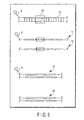
- a double-stranded DNA 10 shown in FIG. 5 is used as a DNA.
- the double-stranded DNA 10 consists of an oligo DNA 11 which has been labeled with a fluorescent label 4 (TAMRA label) at a 5'-terminal, and an oligo DNA 12 having a nucleotide sequence complementary to that of the oligo DNA 11.
- the double-stranded DNA 10 has a TATAA sequence (a complementary sequence is an ATATT sequence), which is a binding site 16 to which a transcription factor TFIID and a transcription factor TFIIB bind.
- a transcription factor TFIID 1 containing TBP (TATA binding protein) and a transcription factor TFIIB 2 are used. Further, as an antibody which binds to a transcription factor TFIIB2, an anti-TFIIB antibody 3 is used.
- a transcription factor TFIID 1 As a transcription factor TFIID 1, a transcription factor TFIIB 2 and an anti-TFIIB antibody 3, the following shown in Table 5 are used.
- a length of a double-stranded DNA 10 is suitably 100 bp or less, particularly desirably 10 to 50 bp.
- result of FCS measurement is obtained precisely, because an incremental ratio of a molecular weight of the double-stranded DNA is sufficiently great in the case where the double-stranded DNA binds to a transcription factor.
- Table 5 Transcription factor/antibody Concentration English name and others Transcription Transcription factor TFIID 1 50 ng/ ⁇ l TFIID(TBP) (manufactured by Santa Cruze Biotechnology.
- TFIIB 2 Transcription factor 170 ng/ ⁇ l TFIIB human recombinant (manufactured by Promega E3790)
- Anti-TFIIB antibody 3 200 ⁇ g/ml
- Anti-Human TFIIB IgG mAb manufactured by Promega E3838A
- a double-stranded DNA 10 used in detection experiment is prepared.
- the following shown in Table 6 are used.
- Table 6 Concentration Amount English name and others Oligo DNA 11 (labeled with a fluorescent label) 100 nM Oligo DNA 12 100 nM STE buffer (NaCl/TE) 100 mM 3' ⁇ 5' Exonuclease I 1 ⁇ l relative to 100 ⁇ l of reaction solution Exonuclease I (EPICENTRE X40501K) , Polynucleotide terminal hydrolytic enzyme MgCl 2 1M 1 ⁇ l relative to 100 ⁇ l reaction solution dW Suitable amount Deionized water
- the oligo DNA 11 and the oligo DNA 12 are reacted in the STE buffer, and the reaction is heated at 95°C for 5 minutes, and slowly cooled to 20°C. Then, the reaction solution is allowed to stand in a PCR instrument or at room temperature to cause annealing of the oligo DNAs. Exonuclease I and MgCl 2 are added to the annealed reaction solution, followed by a reaction at 37°C for 1 hour. Added amounts of Exonuclease I and MgCl 2 are 1 ⁇ l relative to 100 ⁇ l of the reaction solution, respectively. Thereafter, the reaction solution is purified with a purification kit MERmaid SPIN (BIO 101), and a double-stranded DNA 10 consisting of an oligo DNA 11 and an oligo DNA 12 is extracted with dW.
- a purification kit MERmaid SPIN BIO 101
- the extracted double-stranded DNA 10 is measured by fluorescence correlation spectroscopy (FCS), and diluted with dW so as to be around 20 of the particle number (n), thereby preparing a solution of a double-stranded DNA 10 (a concentration of the double-stranded DNA 10 in the solution is 2 nM to 5 nM).
- FCS fluorescence correlation spectroscopy
- FCS measurement is performed precisely, and a binding reaction of a double-stranded DNA and a DNA-binding protein can be detected precisely.
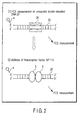
- FIG. 1 shows outline of a procedure and a product of Experiment 1-1. FCS measurement was performed five times after every reaction under the condition of irradiation of laser light at a wavelength of 543 nm and an output of 100 ⁇ W for 15 seconds per one time.
- Table 7 Concentration Amount (total 20 ⁇ l) Fluorescently labeled double-stranded DNA 10 5 ⁇ l 5 ⁇ binding buffer 4 ⁇ l dW 10 ⁇ l Buffer for TFIID 1 ⁇ l
- Table 8 Composition of 5 ⁇ binding buffer Concentration English name and others Tris-hydrochloride pH 7.5 250 mM Tris-HCl Potassium chloride 350 mM KCl EDTA 5 mM Ethylenediaminetetraacetic acid 2-Mercaptoethanol 5 mM 2-Mercaptoethanol BSA 1 mg/ml Bovine serum albumin Glycerol 50% concentration Glycerol
- Table 9 Composition of buffer for TFIID Concentration English name and others DTT 5 mM Dithiothreitol Glycerol
- a DNA-binding protein is not added at this stage, and thus a DNA-protein complex is not formed, and a double-stranded DNA 10 remains unreacted.
- Table 10 The following solution shown in Table 10 is placed in a container, stirred gently with a tip of a chip, slightly tapped, and allowed to stand at room temperature for 30 minutes. Then, FCS measurement is performed to obtain a translational diffusion time. A transcription factor TFIID has been adjusted with a buffer for TFIID to 25 ng/ ⁇ l in advance. Table 10 Concentration Amount (total 20 ⁇ l) Fluorescently labeled double-stranded DNA 10 5 ⁇ l Transcription factor TFIID 1 25 ng/ ⁇ l 1 ⁇ l dW 10 ⁇ l 5 ⁇ binding buffer 4 ⁇ l
- the obtained product is a DNA-protein complex in which a transcription factor TFIID 1 is bound to a binding site 16 of a double-stranded DNA 10.
- the product is a DNA-protein complex in which a transcription factor TFIID 1 and a transcription factor TFIIB 2 are bound to a binding site 16 of a double-stranded DNA 10.
- the product is a DNA-protein complex in which a transcription factor TFIID 1 and a transcription factor TFIIB 2 are bound to a binding site 16 of a double-stranded DNA 10, and an anti-TFIIB antibody 3 is further bound to a transcription factor to TFIIB 2.
- a translational diffusion time of a DNA-protein complex in which all of three proteins of a transcriptional factor TFIID 1, a transcription factor TFIIB 2 and an anti-TFIIB antibody 3 are bound is remarkably greater than a translational diffusion time of other DNA-protein complex.
- the presence of a binding site 16 can be detected precisely by adding all of a transcription factor TFIID 1, a transcription factor TFIIB 2 and an anti-TFIIB antibody 3 to a DNA containing solution, and measuring a DNA-protein complex by FCS to obtain a translational diffusion time.
- a transcription factor TFIID 1 in the case of detecting whether or not a transcription factor TFIID 1 is present in a solution containing some kind of DNA-binding protein, the presence of a transcription factor TFIID 1 can be detected precisely by adding a transcription factor TFIIB 2 and an anti-TFIIB antibody 3 together with a fluorescently labeled double-stranded DNA 10 having a binding site 16 to a reaction solution, and measuring a DNA-protein complex by FCS to obtain a translational diffusion time.
- a double-stranded DNA 20 shown in FIG. 6 is used as a DNA.
- the double-stranded DNA 20 consists of an oligo DNA 21 having a fluorescent label 4 (TAMRA label) at a 5'-terminal, and an oligo DNA 22 having a nucleotide sequence complementary to that of the oligo DNA 21.
- the double-stranded DNA 20 has a TGAGTCA sequence (a complementary sequence is an ACTCAGT sequence), which is a binding site 26 to which a transcription factor AP-1 binds.
- a length of the double-stranded DNA 20 is suitably 100 bp or less, particularly desirably 10 to 50 bp.
- transcription factor AP-1 5 As a DNA-binding protein, the following transcription factor AP-1 5 shown in Table 11 is used. Table 11 Transcription factor/antibody Molecular weight English name and others Transcription factor AP-1 5 40 kDa 80 kDa due to formation of dimer when bound to DNA AP-1 (c-Jun,human) (manufactured by Promega E3061)
- a double-stranded DNA 20 used in detection experiment is prepared.
- the following shown in Table 12 are used.
- Table 12 Concentration Amount English name and others Oligo DNA 21 (labeled with Fluorescent label) 100 nM Oligo DNA 22 100 nM STE buffer (NaCl/TE) 100 mM 3' ⁇ 5' Exonuclease I 1 ⁇ l relative to 100 ⁇ l of reaction solution Exonuclease I(EPICENTRE X40501K)
- Polynucleotide terminal hydrolytic enzyme MgCl 2 1M 1 ⁇ l relative to 100 ⁇ l of reaction solution dW Suitable amount Deionized water
- the oligo DNA 21 and the oligo DNA 22 are reacted in the STE buffer, and the reaction is heated at 95°C for 5 minutes, and slowly cooled to 20°C. Then, the reaction solution is allowed to stand in a PCR instrument or at room temperature to cause annealing of the oligo DNAs. Exonuclease I and MgCl 2 are added to the annealed reaction solution, followed by a reaction at 37°C for 1 hour. Added amounts of Exonuclease I and MgCl 2 are 1 ⁇ l relative to 100 ⁇ l of the reaction solution, respectively. Thereafter, the reaction solution is purified by MERmaid SPIN (BIO 101), and a double-stranded DNA 20 consisting of an oligo DNA 21 and an oligo DNA 22 is extracted with dW.
- MERmaid SPIN BIO 101
- the extracted double-stranded DNA 20 is measured by fluorescence correlation spectroscopy (FCS), and diluted with dW so as to be around 20 to 30 of the particle number (n), thereby preparing a solution of a double-stranded DNA 20.
- FCS fluorescence correlation spectroscopy
- An unlabeled double-stranded DNA 24 is prepared and extracted in the same way as the double-stranded DNA 20, by using an oligo DNA which has the same sequence but is not fluorescently labeled, in place of the oligo DNA 21 (labeled with a fluorescent label 4).
- An absorbance (OD260 value) of the extracted double-stranded DNA 24 is measured, and the extract is diluted with dW to an OD260 value of 20, thereby preparing a solution of a double-stranded DNA 24.
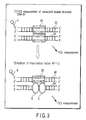
- FIG. 2 shows outline of a procedure and a product of Experiment 2-1.
- the following solution shown in Table 13 is placed in a container, gently stirred with a tip of a chip, slightly tapped, and reacted at 34°C for 1 hour. Then, FCS measurement is performed to obtain a translational diffusion time. FCS measurement was performed five times after each reaction under the condition of irradiation of laser light at a wavelength of 543 nm and an output of 100 ⁇ W for 15 seconds per one time.
- a translational diffusion time of the product was greatly extended, and concentration dependency was recognized.
- a difference in a translational diffusion time at a concentration of a transcription factor AP-1 from 600 ng/ul to 1500 ng/ ⁇ l is small, as compared with a difference in a translational diffusion time at a concentration of up to 300 ng/ ⁇ l. Accordingly it can be said that binding of a double-stranded DNA 20 and a transcription factor AP-1 5 approaches saturation at a concentration of 600 ng/ ⁇ l (see FIG. 9).
- an existence ratio of an unreacted double-stranded DNA 20 (K1) having a translational diffusion time of 432 ⁇ s and the product (K2) having a translational diffusion time of 862.4 ⁇ s is approximately constant at a concentration of a transcription factor AP-1 of 600 ng/ ⁇ l or higher. Therefore, a binding reaction is sufficiently generated at a concentration of 600 ng/ ⁇ l.
- a concentration of a transcription factor AP-1 is 1500 ng/ ⁇ l, it is thought that binding of a transcription factor AP-1 with a double-stranded DNA 20 having a constant concentration is in the saturated state (see FIG. 10).
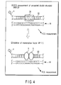
- FIG. 3 shows outline of a procedure and a product of Experiment 2-2.
- a double-stranded DNA 24 which has the same sequence as that of the double-stranded DNA 20 but is not labeled is used.
- a concentration of a transcription factor AP-1 5 is 600 ng/ ⁇ l at which a binding reaction was sufficiently generated in Experiment 2-1.
- FCS measurement is performed to obtain a translational diffusion time. FCS measurement was performed five times after each reaction under the condition of irradiation of laser light at a wavelength of 543 nm and an output of 100 ⁇ W for 15 seconds per one time.
- FIG. 4 shows outline of a procedure and a product of Experiment 2-3.
- a double-stranded DNA 18 which has an Escherichia coli -derived double-stranded random sequence having no AP-1-binding sequence and which has not been fluorescently labeled is used.
- a concentration of a transcription factor AP-1 5 is 600 ng/ ⁇ l, at which a binding reaction is sufficiently generated in Experiment 2-1.
- FCS measurement is performed to obtain a translational diffusion time. FCS measurement was performed five times after each reaction under the condition of irradiation of laser light at a wavelength of 543 nm and an output of 100 ⁇ W for 15 seconds per one time.
- the presence of a transcription factor AP-1 can be detected by stepwise adding a protein containing solution to a solution containing a fluorescently labeled double-stranded DNA, and measuring the product by FCS to obtain a translational diffusion time.
- Example 2 regarding a binding reaction of a DNA and a transcription factor in a cell nucleus extract, dependency on a concentration of a cell nucleus extract is examined.
- a DNA the double-stranded DNA 20 used in Example 2 is used.
- Table 18 For preparing a nucleus extract from a cell, the following shown in Table 18 are used. Table 18 English name and others HeLa cell Medium D-MEM Dulbecco's Modified Eagle's Medium (manufactured by Sigma D5796) Phosphate buffered saline PBS (-) phosphate buffered saline (GIBCO 21600-010) Fetal bovine serum Fetal Bovine Serum(GIBCO) Recombinant human tumor necrosis factor TNF- ⁇ Recombinant Human TNF- ⁇ (STARTHMANN BIOTEC AG) DC protein Assay Kit DC Protein Assay Kit (BIORAD 500-0116) Protease inhibitor cocktail Protease Inhibitor Cocktail (manufactured by Sigma P-1860)
- HeLa cells are put into the confluent state in a 10 cm dish. After the cells are washed with PBS(-) two times, a medium is exchanged with 0.5% FCS/DMEM, followed by stimulation with 100 ng/ml TNF- ⁇ . Cells after two hours from stimulation are washed with PBS(-) two times, and the following operation is performed to obtain an nucleus extract.
- a copolymer is added to the nucleus extract so that substances other than a target protein are not adhered to a double-stranded DNA 20.
- Poly dI-dC manufactured by Sigma, P-4929
- the copolymer is prepared as follows:
- FCS measurement is performed to obtain a translational diffusion time. FCS measurement was performed five times after each reaction under the condition of irradiation of laser light at a wavelength of 543 nm and an output of 100 ⁇ W for 15 seconds per one time.
- a translational diffusion time of a DNA-protein complex is obtained to be 1113.0 ⁇ s from a molecular weight of a DNA-protein complex, because a translational diffusion time is proportion to a cubic root of a molecular weight.
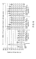
- a sum of an existence ratio of the reaction product having a translational diffusion of time of 1113.0 ⁇ s and an existence ratio of an unreacted double-stranded DNA 20 having a translational diffusion time of 658.5 ⁇ s is approximately 100%. Therefore, it can be said that the reaction product obtained in this experiment is a complex in which a transcription factor AP-1 in a cell nucleus extract is bound to a double-stranded DNA 20 (see FIG. 13).
- a double-stranded DNA 30 shown in FIG. 7 is used as a DNA.
- the double-stranded DNA 30 consists of an oligo DNA 31 having a fluorescent label 4 (TAMRA label) at a 5'-terminal, and an oligo DNA 32 having a nucleic acid sequence complementary to that of the oligo DNA 31.
- the double-stranded DNA 30 has a GGGGCTTT sequence (a complementary sequence is a CCCCTGAAA sequence), which is a binding site 33 to which a transcription factor NF- ⁇ B binds.
- the double-stranded DNA 30 is prepared in the same way as the double-stranded DNA 20, using 100 nM of a (fluorescently labeled) oligo DNA 31, and 100 nM of an oligo DNA 32 (see Example 2).
- a DNA-binding protein is not added in a reaction using a cell nucleus extract having a concentration of 0 ⁇ g, and thus a DNA-protein complex is not present, and a double-stranded DNA 30 remains unreacted.
- the product after the reaction is a complex in which a DNA-binding protein contained in a cell nucleus extract is bound to a double-stranded DNA 30.
- results of FCS measurement are shown in Table 23.
- Table 23 Results of FCS measurement of DNA-NF- ⁇ B complex in Experiment 3-2 Protein contained in cell nucleus extract [ ⁇ g] Difft.
- a translational diffusion time of the reaction product is 1468.7 ⁇ s.
- An existence ratio of the reaction product having a translational diffusion time of 1468.7 ⁇ s and an existence ratio of an unreacted double-stranded DNA 30 having a translational diffusion time of 568.7 ⁇ s are shown in FIG. 15. A sum of respective existence ratios is approximately 100%. Therefore, it can be said that the reaction product obtained in this experiment is a complex in which a transcription factor NF- ⁇ B in a cell nucleus extract is bound to a double-stranded DNA 30.
- the presence of a transcription factor can be detected, by stepwise adding a cell nucleus extract to a solution containing a fluorescently labeled double-stranded DNA having a binding site to which a transcription factor binds, and measuring the product by FCS to obtain a translational diffusion time.
- a transcription factor can be detected from the result of FCS measurement, and a molecular weight of a DNA-protein complex and a molecular weight of an unreacted double-stranded DNA.
- a fluorescently labeled double-stranded DNA 20 having a binding site 26 to which a transcription factor AP-1 binds is used as a double-stranded DNA.
- a fluorescently labeled double-stranded DNA 30 having a binding site 33 to which a transcription factor NF- ⁇ B binds is used as a double-stranded DNA.
- a transcription factor is contained in a cell nucleus extract, if the following results is obtained: as an addition amount of a cell nucleus extract is increased, a translational diffusion time is extended, and after a maximum value is taken, a translational diffusion time is decreased.
- a binding reaction of a DNA and a transcription factor AP-1 in a cell nucleus extract is detected by performing competitive assay in which a competitive DNA is added.
- a double-stranded DNA 24 which has the same sequence as that of the double-stranded DNA 20 but has not been fluorescently labeled is used.
- a concentration of a cell nucleus extract is set to be 30 ⁇ g at which a binding reaction was sufficiently generated in Experiment 3-1.
- FCS measurement is performed to obtain a translational diffusion time. FCS measurement was performed five times after each reaction under the condition of irradiation of laser light at a wavelength of 543 nm and an output of 100 ⁇ W for 15 seconds per one time.
- a double-stranded DNA 25 having a sequence, a part of which is different from a double-stranded DNA 20, and which has not been fluorescently labeled is used as the (unlabeled) double-stranded DNA to be added.
- the double-stranded DNA 25 is different from a double-stranded DNA 20 in a part of a binding site 26 by only two bases (see FIG. 6).
- Experiment 4-2 was performed using the double-stranded DNA 25 in the same way as Experiment 4-1.
- an unlabeled double-stranded DNA 18 having an Escherichia coli -derived double-stranded random sequence having no AP-1-binding sequence is used as the (unlabeled) double-stranded DNA to be added.
- Experiment 4-3 was performed using the double-stranded DNA 18 in the same way as Experiment 4-1.
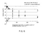
- Results of Experiments 4-1 to 4-3 are shown in FIG. 16. From the results of Experiment 4-1, a translational diffusion time of the product which can be confirmed by a fluorescent label (that is, a DNA-protein complex obtained by binding of a double-stranded DNA 20 and a DNA-binding protein in a cell nucleus extract) was not greatly extended, as compared with a translational diffusion time of an unreacted double-stranded DNA 20. Therefore, it was confirmed that binding of a double-stranded DNA 20 and a transcription factor AP-1 5 in a cell nucleus extract is inhibited by adding a double-stranded DNA 24.
- a fluorescent label that is, a DNA-protein complex obtained by binding of a double-stranded DNA 20 and a DNA-binding protein in a cell nucleus extract
- a translational diffusion time of the binding product of a double-stranded DNA 20 and a cell nucleus extract is approximately the same between the case where a double-stranded DNA 25 is added and the case where such competitive DNA 25 is not added. Therefore, regarding a reaction of a double-stranded DNA 20 and a transcription factor AP-1 5 in a cell nucleus extract, binding inhibition was not recognized by adding a double-stranded DNA 25 having a sequence which is different from that of a double-stranded DNA 20 by only two bases.
- a translational diffusion time of the binding product of a double-stranded DNA 20 and a cell nucleus extract is approximately the same between the case where a double-stranded DNA 18 is added and the case where such competitive DNA 18 is not added. Therefore, regarding a reaction of a double-stranded DNA 20 and a transcription factor AP-1 5 in a cell nucleus extract, binding inhibition was not recognized by adding a double-stranded DNA 18.
- a binding reaction of a DNA and a transcription factor NF- ⁇ B in a cell nucleus extract is detected by performing competition assay in which a competitive DNA is added.
- a double-stranded DNA 34 which has the same sequence as that of a double-stranded DNA 30 but has not been fluorescently labeled is used.
- a concentration of a cell nucleus extract is set to be 30 ⁇ g at which a binding reaction was sufficiently generated in Experiment 3-1.
- FCS measurement is performed to obtain a translational diffusion time. FCS measurement was performed five times after each reaction under the condition of irradiation of laser at a wavelength of 543 nm and an output of 100 ⁇ W for 15 seconds per one time.
- a double-stranded DNA 35 which has a sequence, a part of which is different from a double-stranded DNA 30, and which has not been fluorescently labeled is used.
- the double-stranded DNA 35 is different from a double-stranded DNA 30 in a part of a binding site 33 by only one base (see FIG. 7).
- Experiment 5-2 was performed using the double-stranded DNA 35 in the same way as Experiment 5-1.
- a double-stranded DNA 18 which has an Escherichia coli -derived double-stranded random sequence having no NF- ⁇ B-binding sequence and which has not been fluorescently labeled is used.
- Experiment 5-3 was performed using the double-stranded DNA 18 in the same way as Experiment 5-1.
- FIG. 17 shows results of Experiments 5-1 to 5-3. From the results of Experiment 5-1, a translational diffusion time of the product which can be confirmed by a fluorescent label (that is, a DNA-protein complex obtained by binding of a double-stranded DNA 30 and a DNA-binding protein in a cell nucleus extract) was not greatly extended, as compared with a translational diffusion time of an unreacted double-stranded DNA 30. Therefore, it was confirmed that binding of a double-stranded DNA 30 and a transcription factor AP-1 5 in a cell nucleus extract is inhibited by adding a competitive double-stranded DNA 34.
- a fluorescent label that is, a DNA-protein complex obtained by binding of a double-stranded DNA 30 and a DNA-binding protein in a cell nucleus extract
- a translational diffusion time of the binding product of a double-stranded DNA 30 and a cell nucleus extract is approximately the same between the case where a double-stranded DNA 35 is added and the case where nothing is added. Therefore, regarding a reaction of a double-stranded DNA 35 and a transcription factor NF-KB in a cell nucleus extract, binding inhibition was not recognized by adding a double-stranded DNA 35 having a sequence different from that of a double-stranded DNA 30 by only one base.
- a translational diffusion time of the binding product of a double-stranded DNA 30 and a cell nucleus extract is approximately the same between the case where a double-stranded DNA 18 is added and the case where such competitive DNA 18 is not added. Therefore, regarding a reaction of a double-stranded DNA 30 and a transcription factor NF- ⁇ B in a cell nucleus extract, binding inhibition was not recognized by adding a double stranded DNA 18.
- a transcription factor AP-1 and a transcription factor NF- ⁇ B have activity to an extent in a nucleus of HeLa cell, even when the cell is not stimulated with PMA (phorbol 12-myristate 13-acetate), cytokine or the like.
- a tumor necrosis factor TNF- ⁇ 50 ng/ml of a tumor necrosis factor TNF- ⁇ was added to HeLa cell, and the HeLa cell was stimulated for 0, 15, 30, 60, 120 or 180 minutes. Then, a nucleus extract was prepared from each HeLa cell (for preparation method, see Example 3). A concentration of a nucleus extract was set to be 20 ⁇ g.
- Table 26 Concentration Amount (total 20 ⁇ l) Fluorescently labeled double-stranded DNA 20 2 ⁇ l Cell nucleus extract (which is prepared under the condition of 0, 15, 30, 60, 120 or 180 minutes of a time length of stimulation with tumor necrosis factor TNF- ⁇ ) 20 ⁇ g relative to total amount of 20 ⁇ l Amount corresponding to 20 ⁇ g Poly dI-dC 1 mg/ml 2 ⁇ l dW Residual amount 5 ⁇ binding buffer (for transcription factor AP-1) 4 ⁇ l
- FIG. 18 shows a translational diffusion time of the product relative to a stimulation time.
- a translational diffusion time of the binding reaction product of a (labeled) double-stranded DNA 20 and a cell nucleus extract which has been stimulated with a tumor necrosis factor TNF- ⁇ for 30 minutes is greatest.
- the greatest value is approximately the same as a translational diffusion time of the reaction product obtained when a concentration of a nucleus extract is set to be 20 ⁇ g in Experiment 3-1. Therefore, it can be said that a transcription factor AP-1 in a cell nucleus extract is sufficiently activated by a stimulation time of 30 minutes.
- Experiment 6-2 Experiment of a reaction of a (fluorescently labeled) DNA having an NF- ⁇ B-binding sequence and a cell nucleus extract. The Experiment was performed using a cell nucleus extract which is extracted under the condition of a different time length of stimulation with a tumor necrosis factor TNF- ⁇ .
- Table 27 Concentration Amount (total 20 ⁇ l) Fluorescently labeled double-stranded DNA 30 2 ⁇ l Cell nucleus extract (which is prepared under the condition of 0, 15, 30, 60, 120 or 180 minutes of a time length of stimulation with tumor necrosis factor TNF- ⁇ ) 20 ⁇ g relative to total amount of 20 ⁇ l Amount corresponding to 20 ⁇ g Poly dI-dC 1 mg/ml 2 ⁇ l dW Residual amount 5 ⁇ binding buffer (for transcription factor NF- ⁇ B) 4 ⁇ l
- FIG. 19 shows a translational diffusion time of the product relative to a stimulation time.
- a translational diffusion time of the binding reaction product of a (labeled) double-stranded DNA 30 and a cell nucleus extract which has been stimulated with a tumor necrosis factor TNF- ⁇ for 15 minutes is greatest.
- the greatest value is approximately the same as a translational diffusion time of the reaction product obtained when a concentration of a nucleus extract is set to be 20 ⁇ g in Experiment 3-2. Therefore, it can be said that a transcription factor NF- ⁇ B in a cell nucleus extract was sufficiently activated by a stimulation time of 15 minutes.
- the presence of a transcription factor can be detected by adding 50 ng/ml of a tumor necrosis factor TNF- ⁇ to a cell to stimulate the cell for 30 minutes; extracting a cell nucleus from the cell; reacting the cell nucleus extract with a double-stranded DNA 20; and performing FCS measurement to obtain a translational diffusion time.
- 50 ng/ml of a tumor necrosis factor TNF- ⁇ is preferably added to a cell to stimulate the cell for 15 minutes.
- a tumor necrosis factor TNF- ⁇ is added to stimulate HeLa cell for 30 minutes, from which a nucleus extract is prepared.
- a nucleus extract having a concentration of 5, 10 or 20 ⁇ g is prepared. Stimulation on a cell is performed in the following two cases.
- FIG. 20 shows a translational diffusion time of the product relative to each stimulation condition. Stimulation dependency of a translational diffusion time of the product was seen depending on a concentration of a tumor necrosis factor TNF- ⁇ and a concentration of APDC.
- a concentration of APDC is 100 ⁇ M and 200 ⁇ M relative to 50 ng/ml of a tumor necrosis factor TNF- ⁇
- a translational diffusion time of the product is great. Therefore, it can be said that a transcription factor AP-1 in a cell nucleus extract is most activated at a combination of these concentrations.
- FIG. 21 shows a translational diffusion time of the product relative to each stimulating condition. Stimulation dependency of a translational diffusion time of the product was seen depending on a concentration of a tumor necrosis factor TNF- ⁇ and a concentration of APDC. In particular, when stimulation with only a tumor necrosis factor TNF- ⁇ is performed, and a concentration of a tumor necrosis factor TNF- ⁇ is 50 ng/ml, a translational diffusion time of the product is great. Therefore, it can be said that a transcription factor NF- ⁇ B in a cell nucleus extract is most activated at the concentration.
- APDC promotes activation of a transcription factor AP-1 by a tumor necrosis factor TNF- ⁇ , and suppresses activation of a transcription factor NF- ⁇ B by a tumor necrosis factor TNF- ⁇ .
- the presence of a transcription factor can be detected by adding a tumor necrosis factor TNF- ⁇ to a cell to stimulate the cell for 30 minutes, two hours after addition of APDC; extracting a cell nucleus from the cell; reacting the cell nucleus extract with a double-stranded DNA 20; and performing FCS measurement to obtain a translational diffusion time.
- a concentration of APDC is preferably not lower than 100 ⁇ M and not higher than 200 ⁇ M, relative to 50 ng/ml of a tumor necrosis factor TNF- ⁇ .
- a transcription factor NF- ⁇ B in the case of detecting whether or not a transcription factor NF- ⁇ B is contained in a cell nucleus extract, a transcription factor NF- ⁇ B can be detected precisely, by comparing a translational diffusion time obtained when stimulation is performed with only a tumor necrosis factor TNF- ⁇ and a translational diffusion time obtained when stimulation is performed with both APDC and a tumor necrosis factor TNF- ⁇ . If a translational diffusion time obtained when stimulation is performed with only a tumor necrosis factor TNF- ⁇ is greater than the other translational diffusion time, it can be said that a transcription factor NF- ⁇ B is contained in a cell nucleus extract.
- FCS fluorescence correlation spectroscopy
- the measuring method by FCS according to the present invention is useful for detecting a particular DNA-binding protein (such as transcription factor or intranuclear receptor) present in a crude sample such as a cell nucleus extract.
- a particular DNA-binding protein such as transcription factor or intranuclear receptor
- the effect of stimulation with a cytokine on a time-dependent amount of expression of a transcription factor or the effect of APDC can be also detected precisely.
- FCS fluorescent correlation spectroscopy
Landscapes
- Life Sciences & Earth Sciences (AREA)
- Health & Medical Sciences (AREA)
- Chemical & Material Sciences (AREA)
- Engineering & Computer Science (AREA)
- Immunology (AREA)
- Molecular Biology (AREA)
- Analytical Chemistry (AREA)
- Organic Chemistry (AREA)
- Proteomics, Peptides & Aminoacids (AREA)
- Biochemistry (AREA)
- Cell Biology (AREA)
- Microbiology (AREA)
- Urology & Nephrology (AREA)
- Physics & Mathematics (AREA)
- Wood Science & Technology (AREA)
- Zoology (AREA)
- Pathology (AREA)
- Hematology (AREA)
- Biomedical Technology (AREA)
- General Health & Medical Sciences (AREA)
- Biotechnology (AREA)
- Genetics & Genomics (AREA)
- General Engineering & Computer Science (AREA)
- Bioinformatics & Cheminformatics (AREA)
- Biophysics (AREA)
- Food Science & Technology (AREA)
- Medicinal Chemistry (AREA)
- General Physics & Mathematics (AREA)
- Measuring Or Testing Involving Enzymes Or Micro-Organisms (AREA)
- Investigating Or Analysing Biological Materials (AREA)
- Investigating Or Analysing Materials By The Use Of Chemical Reactions (AREA)
- Investigating, Analyzing Materials By Fluorescence Or Luminescence (AREA)
Abstract
Description
- The present invention relates to a method of detecting a reaction of a DNA and a DNA-binding protein, more particularly, a method of detecting the reaction using fluorescence correlation spectroscopy (FCS).
- Patent Publication 1 (Japanese Patent No. 3169610) describes a method of screening a molecule which can bind to a test sequence selected in a double-stranded DNA.
- Patent Publication 2 (Japanese Patent No. 2953783) describes an effective method of identifying a drug which is active at a gene transcription level.
- Patent Publication 3 (Jpn. Pat. Appln. KOKAI Publication No. 2001-321199) describes a method of quantitating a DNA-binding protein in a biological sample.
- Patent Publication 4 (Jpn. Pat. Appln. KOKAI Publication No. 2003-88369) describes a method of detecting DNA endonuclease activity utilizing fluorescence correlation spectroscopy (FCS), and a relationship between a size of a molecular weight and a length of a translational diffusion time.
- Patent Publication 5 (Jpn. Pat. Appln. KOKAI Publication No. 2002-543414) describes a method of characterizing a fluorescent molecule or other particle in a sample, and describes that a translational diffusion time is obtained from fluorescence intensity multiple distribution analysis (FIMDA) and fluorescence autoconvoluted intensity distribution analysis (FACID).
- Among the aforementioned prior art, the detecting methods described in Patent Publication 2 (Japanese Patent No. 2953783), Patent Publication 3 (Jpn. Pat. Appln. KOKAI Publication No. 2001-321199) and Patent Publication 4 (Jpn. Pat. Appln. KOKAI Publication No. 2003-88369) use a radioisotope, or perform selection by an electrophoresis method or immobilization of a molecule on a solid substrate, when a particular sequence or a molecule having a particular sequence is detected, and therefore there is a problem that operation is troublesome and it takes a time for obtaining detection results.
- Patent Publication 4 (Jpn. Pat. Appln. KOKAI Publication No. 2003-88369) and Patent Publication 5 (Jpn. Pat. Appln. KOKAI Publication No. 2002-543414) do not describe a method of detecting a reaction of a DNA and a DNA-binding protein.
- An object of the present invention is to provide a method of detecting a reaction of a DNA and a DNA-binding protein simply and in a short time.
- A feature of the present invention which attains the aforementioned object is to mix a solution containing a fluorescently labeled double-stranded DNA with a transcription factor TFIID, and to obtain a translational diffusion time of a substance having a fluorescent label in the mixed solution by fluorescence correlation spectroscopy. According to the present invention, information for obtaining a translational diffusion time is obtained by mixing a solution and measuring by fluorescence correlation spectroscopy. Therefore, according to the present invention, a reaction of a double-stranded DNA and a transcription factor TFIID which is a DNA-binding protein can be simply detected in a short time, without performing troublesome operation such as utilization of a radioisotope and selection by an electrophoresis method or immobilization of a molecule on a solid substrate.
- In addition, when each experiment is performed using a solution having a double-stranded DNA concentration in a sample of 2 nM to 5 nM, the particle number in a confocal volume becomes 2 to 5 at the time of FCS measurement. As a result, FCS measurement can be performed precisely, and thereby a binding reaction between a double-stranded DNA and a DNA-binding protein can be detected precisely. In particular, even when the particle number in a confocal volume is 2 to 3, precise detection is possible. Therefore, the detection method of the present invention is suitable also when a large amount of a double-stranded DNA or a DNA-binding protein can not be prepared, or when detection result is desired with only a small amount of a sample.
- (2) Another feature of the present invention is to mix a solution containing a fluorescently labeled double-stranded DNA, a transcription factor TFIID and a transcription factor TFIIB, and to obtain a translational diffusion time of a substance having a fluorescent label in the mixed solution by fluorescence correlation spectroscopy. In this case, a translational diffusion time of a reaction product is extended by adding a transcription factor TFIIB, and therefore the binding can be detected more precisely. In addition, when an anti-TFIIB antibody is further added to the mixed solution, a translational diffusion time of a reaction product is further extended, and therefore the binding can be detected more precisely.
- In the case of detecting whether or not a transcription factor TFIID-binding sequence is contained in a double-stranded DNA, a fluorescent label is attached to a double-stranded DNA, and any method of
claims 1 to 3 is performed. If binding is detected, this results in that a transcription factor TFIID-binding sequence is contained in a double-stranded DNA. - In the case of detecting whether or not a transcription factor TFIID is contained in a test solution, the test solution and a fluorescently labeled double-stranded DNA having a transcription factor TFIID-binding sequence are mixed, and a translational diffusion time of a substance having a fluorescent label in the mixed solution is obtained by fluorescence correlation spectroscopy. In this case, the presence of a transcription factor TFIID can be detected more precisely, by adding either a transcription factor TFIIB, or adding a transcription factor TFIIB and an anti-TFIIB antibody.
- Another feature of the present invention is to mix a solution to be detected with a solution containing a fluorescently labeled double-stranded DNA having a transcription factor AP-1-binding sequence, and to obtain a translational diffusion time of a substance having a fluorescent label in a mixed solution by fluorescence correlation spectroscopy. According to this feature, information for obtaining a translational diffusion time is obtained by mixing a solution and measuring by fluorescence correlation spectroscopy. Therefore, according to this feature, a reaction of a double-stranded DNA and a transcription factor AP-1 which is a DNA-binding protein can be simply detected in a short time, without performing troublesome operation such as utilization of a radioisotope and selection by an electrophoresis method or immobilization of a molecule on a solid substrate.
- Another feature of the present invention is to mix a solution containing a double-stranded DNA to be detected, a transcription factor AP-1, and a fluorescently labeled double-stranded DNA having a transcription factor AP-1-binding sequence, and to obtain a translational diffusion time of a substance having a fluorescent label in a mixed solution by fluorescence correlation spectroscopy.
- According to this feature, information for obtaining a translational diffusion time is obtained by mixing a solution and measuring by fluorescence correlation spectroscopy. Therefore, according to this feature, a reaction of a double-stranded DNA and a transcription factor AP-1 which is a DNA-binding protein can be simply detected in a short time, without performing troublesome operation such as utilization of a radioisotope and selection by an electrophoresis method or immobilization of a molecule on a solid substrate.
- Another feature of the present invention is to stepwise increase an amount of a solution to be detected, when the solution to be detected is mixed with a fluorescently labeled double-stranded DNA having a transcription factor-binding sequence, and to obtain a translational diffusion time of a substance having a fluorescent label in the mixed solution by fluorescence correlation spectroscopy at each stage. In the case where a transcription factor AP-1 is detected in a cell nucleus extract, a double-stranded DNA having a transcription factor AP-1-binding site is used as a fluorescently labeled double-stranded DNA, and an amount of a cell nucleus extract solution is increased stepwise. In the case where a transcription factor NF-κB is detected in a cell nucleus extract, a double-stranded DNA having a transcription factor NF-κB-binding site is used as a fluorescently labeled double-stranded DNA, an amount of a cell nucleus extract solution is increased stepwise.
- Another feature of the present invention is to extract a cell nucleus after adding a tumor necrosis factor TNF-α to the cell, and then mix the obtained cell nucleus extract and a fluorescently labeled double-stranded DNA, and to obtain a translational diffusion time of a substance having a fluorescent label in a mixed solution by fluorescence correlation spectroscopy. In the case where a transcriptional factor AP-1 is detected in a nucleus extract of HeLa cell, stimulation may be performed for 30 minutes with 50 ng/ml of a tumor necrosis factor TNF-α. In the case where a transcription factor AP-1 is detected in a nucleus extract of HeLa cell, stimulation may be performed for 15 minutes. The stimulation with a tumor necrosis factor TNF-α activates a transcription factor in a cell nucleus extract, and makes it easy to bind the activated transcription factor and the double-stranded DNA, and as a result, a transcription factor can be detected precisely.
- Another feature of the present invention is to extract a cell nucleus after adding APDC and then a tumor necrosis factor TNF-α to the cell, and mix the obtained cell nucleus extract and a fluorescently labeled double-stranded DNA having a transcription factor AP-1-binding sequence, and to obtain a translational diffusion time of a substance having a fluorescent label in a mixed solution by fluorescence correlation spectroscopy. According to the present invention, the stimulation with APDC and a tumor necrosis factor TNF-α activates a transcription factor AP-1 in a cell nucleus extract, and makes it easy to bind the activated transcription factor and the double-stranded DNA, and as a result, a transcription factor AP-1 can be detected precisely. A time for stimulation with APDC may be 2 hours, and a time for stimulation with a tumor necrosis factor TNF-α may be 30 minutes.
- Another feature of the present invention is to compare a translational diffusion time of a reaction product in the case of stimulation with only a tumor necrosis factor TNF-α, with a translational diffusion time of a reaction product in the case of stimulation with both APDC (Ammonium pyrrolidinedithiocarbamate) and a tumor necrosis factor TNF-α, when a transcription factor NF-κB is detected in a cell nucleus extract, based on a translational diffusion time of a reaction product resulting from the reaction of a transcription factor NF-κB and a fluorescently labeled double-stranded DNA having a transcription factor NF-κB-binding sequence. A tumor necrosis factor TNF-α activates a transcription factor NF-κB, and APDC suppresses activation of a transcription factor NF-κB. Therefore, if a translational diffusion time is greater in the case of the stimulation with only a tumor necrosis factor TNF-α, it can be the that a transcription factor NF-κB is contained in a cell nucleus extract. According to this feature, a transcription factor NF-κB can be detected precisely.
- When the particle number in a confocal volume is 2 to 5 at the time of FCS measurement, FCS measurement can be performed precisely, and thus a binding reaction between a double-stranded DNA and a DNA-binding protein can be detected precisely. In particular, even when the particle number in a confocal volume is 2 to 3, precise detection is possible. Therefore, the detection method of the present invention is suitable also when a large amount of a double-stranded DNA or a DNA-binding protein can not be prepared, or when detection result is desired with only a small amount of a sample.
- In addition, according to the present invention, even when a crude sample such as an unpurified cell nucleus extract is used, a binding reaction between a double-stranded DNA and a transcription factor can be detected precisely. Further, according to the present invention, a reaction product resulting from an inherent double-stranded DNA and a transcription factor can be detected without preventing a binding reaction between them, even in the presence of a double-stranded DNA which is different, only by one base or two bases, from the inherent double-stranded DNA to which a transcription factor inherently binds. Therefore, a measuring method by FCS according to the present invention is useful for detecting a particular DNA-binding protein (such as transcription factor or intranuclear receptor) present in a crude sample such as a cell nucleus extract.
-
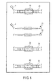
- FIG. 1 shows outline of a procedure and a product of Experiment 1-1. (1) FCS measurement of an unreacted double-stranded DNA (reference numeral 10) is performed. (2) A transcription factor TFIID (reference numeral 1) is added. (3) A transcription factor TFIIB (reference numeral 2) is added. (4) An anti-TFIIB antibody (reference numeral 3) is added.
- FIG. 2 shows outline of a procedure and a product of Experiment 2-1. (1) FCS measurement of an unreacted double-stranded DNA (reference numeral 20) is performed. (2) A transcription factor AP-1 (reference numeral 5) is added.
- FIG. 3 shows outline of a procedure and a product of Experiment 2-2. (1) FCS measurement of an unreacted double-stranded DNA (reference numeral 20) is performed. (2) A transcription factor AP-1 (reference numeral 5) is added.
- FIG. 4 shows outline of a procedure and a product of Experiment 2-3. (1) FCS measurement of an unreacted double-stranded DNA (reference numeral 20) is performed. (2) A transcription factor AP-1 (reference numeral 5) is added.
- FIG. 5 shows a DNA used in each experiment.
- FIG. 6 shows a DNA used in each experiment.
- FIG. 7 shows a vicinity of an
objective lens 11 in Example 2. - FIG. 8 is a graph showing a translational diffusion time of a product (DNA-protein complex) after each reaction in Experiments 1-1 and 1-2. Reference numeral (1) denotes a translational diffusion time of a double-stranded DNA before a reaction; reference numeral (2) denotes the case where a transcription factor TFIID is added; reference numeral (3) denotes the case where a transcription factor TFIID and a transcription factor TFIIB are added; and reference numeral (4) denotes the case where a transcription factor TFIID, a transcription factor TFIIB and an anti-TFIIB antibody are added.
- FIG. 9 is a graph of a translational diffusion time of a product (DNA-AP-1 complex) relative to a concentration of a transcription factor AP-1 in Experiment 2-1.
- FIG. 10 is a graph showing an existence ratio of an unreacted product (unreacted DNA: K1) and a reaction product (DNA-AP-1 complex: K2) relative to a concentration of a transcription factor AP-1 in Experiment 2-1.
- FIG. 11 is a graph showing a translational diffusion time of a product (DNA-AP-1 complex) relative to a concentration of an (unlabeled) double-stranded DNA in Experiment 2-2, and a translational diffusion time of a product (DNA-AP-1 complex) relative to a concentration of an (unlabeled) DNA having no AP-1-binding sequence in Experiment 2-3.
- FIG. 12 is a graph showing a translational diffusion time of a product (DNA-AP-1 complex) relative to a concentration of a cell nucleus extract in Experiment 3-1.
- FIG. 13 is a graph showing an existence ratio of an unreacted product (unreacted DNA) and a product (DNA-AP-1 complex) relative to a concentration of a cell nucleus extract in Experiment 3-1.
- FIG. 14 is a graph showing a translational diffusion time of a product (DNA-NF-κB complex) relative to a concentration of a cell nucleus extract in Experiment 3-2.
- FIG. 15 is a graph showing an existence ratio of an unreacted product (unreacted DNA) and a product (DNA-NF-κB complex) relative to a concentration of a cell nucleus extract in Experiment 3-2.
- FIG. 16 is a graph showing a translational diffusion time of a product (DNA-protein complex) relative to a concentration of (unlabeled) each double-stranded DNA in Experiments 4-1 to 4-3.
- FIG. 17 is a graph showing a translational diffusion time of a product (DNA-protein complex) relative to a concentration of (unlabeled) each double-stranded DNA in Experiments 5-1 to 5-3.
- FIG. 18 is a graph showing a translational diffusion time of a product (DNA-AP-1 complex) relative to a stimulation time in Experiment 6-1.
- FIG. 19 is a graph showing a translational diffusion time of a product (DNA-NF-κB complex) relative to a stimulation time in Experiment 6-2.
- FIG. 20 is a graph showing a translational diffusion time of a product (DNA-AP-1 complex) relative to a concentration of added TNF-α and a concentration of added APDC in Experiment 7-1. (i) Stimulation with only a tumor necrosis factor TNF-α (concentration is 0 ng/mL). (ii) Stimulation with only a tumor necrosis factor TNF-α (concentration is 10 ng/mL). (iii) Stimulation with only a tumor necrosis factor TNF-α (concentration is 25 ng/mL). (iv) Stimulation with only a tumor necrosis factor TNF-α (concentration is 50 ng/mL). (v) Stimulation with APDC (concentration is 10 µM) and a tumor necrosis factor TNF-α (concentration is 50 ng/mL). (vi) Stimulation with APDC (concentration is 100 µM) and a tumor necrosis factor TNF-α (concentration is 50 ng/mL). (vii) Stimulation with APDC (concentration is 200 µM) and a tumor necrosis factor TNF-α (concentration is 50 ng/mL).
- FIG. 21 is a graph showing a translational diffusion time of a product (DNA-NF-κB complex) relative to a concentration of added TNF-α and a concentration of added APDC in Experiment 7-2. (i) Stimulation with only a tumor necrosis factor TNF-α (concentration is 0 ng/mL). (ii) Stimulation with only a tumor necrosis factor TNF-α (concentration is 10 ng/mL). (iii) Stimulation with only a tumor necrosis factor TNF-α (concentration is 25 ng/mL). (iv) Stimulation with only a tumor necrosis factor TNF-α (concentration is 50 ng/mL). (v) Stimulation with APDC (concentration is 10 µM) and a tumor necrosis factor TNF-α (concentration is 50 ng/mL). (vi) Stimulation with APDC (concentration is 100 µM) and a tumor necrosis factor TNF-α (concentration is 50 ng/mL). (vii) Stimulation with APDC (concentration is 200 µM) and a tumor necrosis factor TNF-α (concentration is 50 ng/mL).
- Reference numerals in the figures denotes as follows:
- 1
- transcription factor TFIID;
- 2
- transcription factor TFIIB;
- 3
- anti-TFIIB antibody;
- 4
- fluorescent label;
- 5
- transcription factor AP-1;
- 10
- double-stranded DNA;
- 11
- oligo DNA;
- 12
- oligo DNA;
- 16
- binding site;
- 17
- double-stranded DNA;
- 18
- double-stranded DNA;
- 20
- double-stranded DNA;
- 21
- oligo DNA;
- 22
- oligo DNA;
- 24
- double-stranded DNA;
- 25
- double-stranded DNA;
- 26
- binding site;
- 26'
- binding site different by two bases;
- 30
- double-stranded DNA;
- 31
- oligo DNA;
- 32
- oligo DNA;
- 33
- binding site;
- 33'
- binding site different by one base;
- 34
- double-stranded DNA;
- 35
- double-stranded DNA.
- In embodiments of the present invention, various experiments are performed for detecting binding of a DNA and a protein which binds to a particular sequence part in a DNA (hereinafter, referred to as DNA binding protein). Embodiments will be explained as an example by each purpose of experiments. In experiments, after a process of a reaction of a biomolecule, FCS measurement is performed, and a translational diffusion time of the biomolecule is obtained.
- Since a magnitude of a translational diffusion time shows a magnitude of a molecular weight, increase or decrease in a molecular weight is seen by comparing a translationial diffusion time before and after a reaction. Increase in a molecular weight shows a binding reaction between biomolecules, decrease in a molecular weight shows a degradation reaction of a biomolecule, and maintenance of a molecular weight shows that there was neither binding nor degradation in a biomolecule.
- Therefore, a binding reaction between a DNA and a DNA-binding protein can be detected by detecting increase in a translational diffusion time of a DNA after a reaction of a DNA and a DNA-binding protein, compared with a translational diffusion time of the DNA before the reaction.
- According to the present invention, information for obtaining a translational diffusion time is obtained by mixing solutions and measuring by fluorescence correlation spectroscopy. As a result, a reaction of a DNA and a DNA-binding protein can be detected simply and in a short time, without performing troublesome operation such as utilization of a radioisotope and selection by an electrophoresis method, and immobilization of a molecule on a solid substrate.
- In addition, according to the present invention, even when a crude sample such as a cell nucleus extract which has not been purified is used, a binding reaction of a double-stranded DNA and a transcription factor can be detected precisely. Further, according to the present invention, a reaction product resulting from an inherent double-stranded DNA and a transcription factor can be detected without preventing a binding reaction between them, even in the presence of a double-stranded DNA which is different, only by one base or two bases, from the inherent double-stranded DNA to which a transcription factor inherently binds. Therefore, a measuring method by FCS according to the present invention is useful for detecting a particular DNA-binding protein (such as transcription factor or intranuclear receptor) present in a crude sample such as a cell nucleus extract.
- Summary of experimental contents and experimental results of each Example is shown in Tables 1 to 4, and detail of each Example will be explained later.


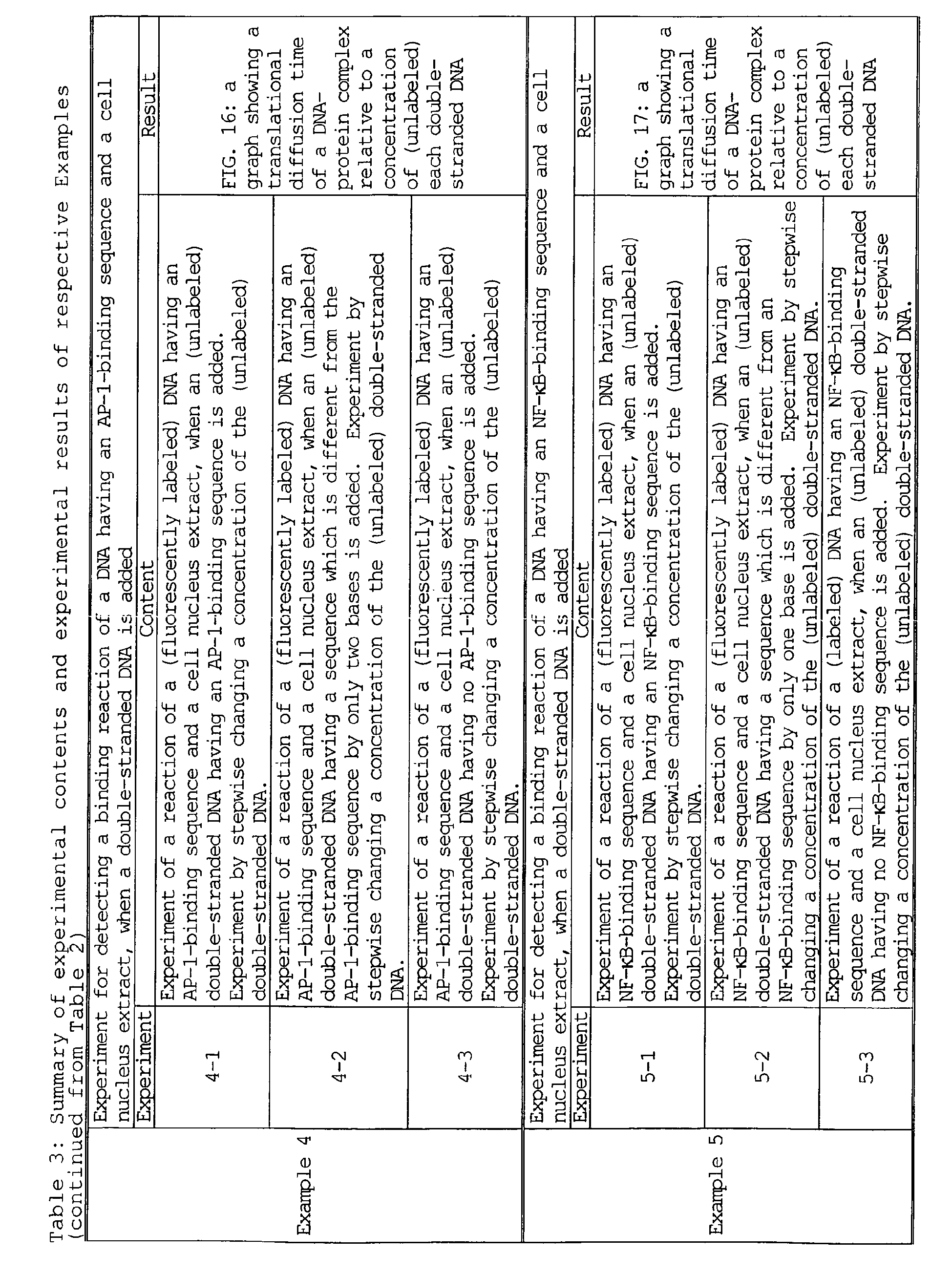

- In the present Example, a binding reaction of a DNA, and a transcription factor TFIID and a transcription factor TFIIB which are a DNA-binding protein is detected.
- As a DNA, a double-stranded
DNA 10 shown in FIG. 5 is used. The double-strandedDNA 10 consists of anoligo DNA 11 which has been labeled with a fluorescent label 4 (TAMRA label) at a 5'-terminal, and anoligo DNA 12 having a nucleotide sequence complementary to that of theoligo DNA 11. The double-strandedDNA 10 has a TATAA sequence (a complementary sequence is an ATATT sequence), which is abinding site 16 to which a transcription factor TFIID and a transcription factor TFIIB bind. - As a DNA-binding protein, a
transcription factor TFIID 1 containing TBP (TATA binding protein) and atranscription factor TFIIB 2 are used. Further, as an antibody which binds to a transcription factor TFIIB2, ananti-TFIIB antibody 3 is used. - As a
transcription factor TFIID 1, atranscription factor TFIIB 2 and ananti-TFIIB antibody 3, the following shown in Table 5 are used. A length of a double-strandedDNA 10 is suitably 100 bp or less, particularly desirably 10 to 50 bp. When a length of a double-stranded DNA is in this range, result of FCS measurement is obtained precisely, because an incremental ratio of a molecular weight of the double-stranded DNA is sufficiently great in the case where the double-stranded DNA binds to a transcription factor.Table 5 Transcription factor/antibody Concentration English name and others Transcription Transcription factor TFIID 150 ng/µl TFIID(TBP) (manufactured by Santa Cruze Biotechnology. Inc sc-4000) Transcription factor TFIIB 2170 ng/µl TFIIB human recombinant (manufactured by Promega E3790) Anti-TFIIB antibody 3200 µg/ml Anti-Human TFIIB IgG mAb (manufactured by Promega E3838A) - First, a double-stranded
DNA 10 used in detection experiment is prepared. For preparing the double-strandedDNA 10, the following shown in Table 6 are used.Table 6 Concentration Amount English name and others Oligo DNA 11 (labeled with a fluorescent label) 100 nM Oligo DNA 12 100 nM STE buffer (NaCl/TE) 100 mM 3' → 5' Exonuclease I 1 µl relative to 100 µl of reaction solution Exonuclease I (EPICENTRE X40501K) , Polynucleotide terminal hydrolytic enzyme MgCl2 1M 1 µl relative to 100 µl reaction solution dW Suitable amount Deionized water - The
oligo DNA 11 and theoligo DNA 12 are reacted in the STE buffer, and the reaction is heated at 95°C for 5 minutes, and slowly cooled to 20°C. Then, the reaction solution is allowed to stand in a PCR instrument or at room temperature to cause annealing of the oligo DNAs. Exonuclease I and MgCl2 are added to the annealed reaction solution, followed by a reaction at 37°C for 1 hour. Added amounts of Exonuclease I and MgCl2 are 1 µl relative to 100 µl of the reaction solution, respectively. Thereafter, the reaction solution is purified with a purification kit MERmaid SPIN (BIO 101), and a double-strandedDNA 10 consisting of anoligo DNA 11 and anoligo DNA 12 is extracted with dW. - The extracted double-stranded
DNA 10 is measured by fluorescence correlation spectroscopy (FCS), and diluted with dW so as to be around 20 of the particle number (n), thereby preparing a solution of a double-stranded DNA 10 (a concentration of the double-strandedDNA 10 in the solution is 2 nM to 5 nM). When each experiment is performed using the solution of a double-stranded DNA having the particle number (n) of around 20, the particle number in a confocal volume is 2 to 5 at the time of FCS measurement. As a result, FCS measurement is performed precisely, and a binding reaction of a double-stranded DNA and a DNA-binding protein can be detected precisely. - FIG. 1 shows outline of a procedure and a product of Experiment 1-1. FCS measurement was performed five times after every reaction under the condition of irradiation of laser light at a wavelength of 543 nm and an output of 100 µW for 15 seconds per one time.
- The following solution shown in Table 7 is placed in a container, stirred gently with a tip of a chip, slightly tapped, and allowed to stand at room temperature for 30 minutes. Then, FCS measurement is performed to obtain a translational diffusion time.
Table 7 Concentration Amount (total 20 µl) Fluorescently labeled double-stranded DNA 105 µl 5 × binding buffer 4 µl dW 10 µl Buffer for TFIID 1 µl Table 8: Composition of 5 × binding buffer Concentration English name and others Tris-hydrochloride pH 7.5 250 mM Tris-HCl Potassium chloride 350 mM KCl EDTA 5 mM Ethylenediaminetetraacetic acid 2- Mercaptoethanol 5 mM 2- Mercaptoethanol BSA 1 mg/ml Bovine serum albumin Glycerol 50% concentration Glycerol Table 9: Composition of buffer for TFIID Concentration English name and others DTT 5 mM Dithiothreitol Glycerol/ PBS 50% concentration Glycerol/PBS (phosphate buffered saline) - A DNA-binding protein is not added at this stage, and thus a DNA-protein complex is not formed, and a double-stranded
DNA 10 remains unreacted. - The following solution shown in Table 10 is placed in a container, stirred gently with a tip of a chip, slightly tapped, and allowed to stand at room temperature for 30 minutes. Then, FCS measurement is performed to obtain a translational diffusion time. A transcription factor TFIID has been adjusted with a buffer for TFIID to 25 ng/µl in advance.
Table 10 Concentration Amount (total 20 µl) Fluorescently labeled double-stranded DNA 105 µl Transcription factor TFIID 125 ng/ µl 1 µl dW 10 µl 5 × binding buffer 4 µl - The obtained product is a DNA-protein complex in which a
transcription factor TFIID 1 is bound to abinding site 16 of a double-strandedDNA 10. - 1 µl of 85 ng/µl
transcription factor TFIIB 2 is added to 20 µl of the reacted solution (2), gently mixed with a tip of a chip, slightly tapped, and allowed to stand at room temperature for 20 minutes. Then, FCS measurement is performed to obtain a translational diffusion time. - The product is a DNA-protein complex in which a
transcription factor TFIID 1 and atranscription factor TFIIB 2 are bound to abinding site 16 of a double-strandedDNA 10. - 1 µl of an
anti-TFIIB antibody 3 is added to 20 µl of the reacted solution of (3), gently mixed with a tip of a chip, slightly tapped, and allowed to stand at room temperature for 20 minutes. Then, FCS measurement is performed to obtain a translational diffusion time. - The product is a DNA-protein complex in which a
transcription factor TFIID 1 and atranscription factor TFIIB 2 are bound to abinding site 16 of a double-strandedDNA 10, and ananti-TFIIB antibody 3 is further bound to a transcription factor toTFIIB 2. - The same reaction experiment as Experiment 1-1 (1) to (4) was performed using an Escherichia coli-derived double-stranded
random sequence DNA 17, which dose not contain abinding site 16 to which a transcription factor TFIID binds, in place of a double-strandedDNA 10. - Translational diffusion times of the products obtained in Experiments 1-1 and 1-2 are shown in FIG. 8. In Experiment 1-1 using a double stranded
DNA 10, a translational diffusion time of a DNA-protein complex after each reaction is greater as compared with a translational diffusion time of a double-strandedDNA 10 before the reaction. The result shows that the double-strandedDNA 10 is bound to atranslational factor TFIID 1, atranscription factor TFIIB 2 and ananti-TFIIB antibody 3. On the other hand, in Experiment 1-2 using an Escherichia coli-derived double-strandedrandom sequence DNA 17, a change in a translational diffusion time is little in any case, and binding of the DNA with a DNA-binding protein could not be confirmed. In particular, a translational diffusion time of a DNA-protein complex in which all of three proteins of atranscriptional factor TFIID 1, atranscription factor TFIIB 2 and ananti-TFIIB antibody 3 are bound is remarkably greater than a translational diffusion time of other DNA-protein complex. - Therefore, in the case of detecting whether or not a
binding site 16 for atranscription factor TFIID 1 and atranscription factor TFIIB 2 is present in a fluorescently labeled DNA, the presence of abinding site 16 can be detected precisely by adding all of atranscription factor TFIID 1, atranscription factor TFIIB 2 and ananti-TFIIB antibody 3 to a DNA containing solution, and measuring a DNA-protein complex by FCS to obtain a translational diffusion time. - In addition, in the case of detecting whether or not a
transcription factor TFIID 1 is present in a solution containing some kind of DNA-binding protein, the presence of atranscription factor TFIID 1 can be detected precisely by adding atranscription factor TFIIB 2 and ananti-TFIIB antibody 3 together with a fluorescently labeled double-strandedDNA 10 having a bindingsite 16 to a reaction solution, and measuring a DNA-protein complex by FCS to obtain a translational diffusion time. - In the present Example, regarding a binding reaction of a DNA and a transcription factor AP-1 which is a DNA-binding protein, dependency on a concentration of a transcription factor AP-1 is examined.
- As a DNA, a double-stranded
DNA 20 shown in FIG. 6 is used. The double-strandedDNA 20 consists of anoligo DNA 21 having a fluorescent label 4 (TAMRA label) at a 5'-terminal, and anoligo DNA 22 having a nucleotide sequence complementary to that of theoligo DNA 21. The double-strandedDNA 20 has a TGAGTCA sequence (a complementary sequence is an ACTCAGT sequence), which is abinding site 26 to which a transcription factor AP-1 binds. A length of the double-strandedDNA 20 is suitably 100 bp or less, particularly desirably 10 to 50 bp. When a length of a double-stranded DNA is in this range, result of FCS measurement is obtained precisely, because an increment ratio of a molecular weight of a double-stranded DNA is great in the case where a double-stranded DNA is bound to a transcription factor or the like. - As a DNA-binding protein, the following transcription factor AP-1 5 shown in Table 11 is used.
Table 11 Transcription factor/antibody Molecular weight English name and others Transcription factor AP-1 5 40 kDa 80 kDa due to formation of dimer when bound to DNA AP-1 (c-Jun,human) (manufactured by Promega E3061) - First, a double-stranded
DNA 20 used in detection experiment is prepared. For preparing the double-strandedDNA 20, the following shown in Table 12 are used.Table 12 Concentration Amount English name and others Oligo DNA 21 (labeled with Fluorescent label) 100 nM Oligo DNA 22 100 nM STE buffer (NaCl/TE) 100 mM 3' → 5' Exonuclease I 1 µl relative to 100 µl of reaction solution Exonuclease I(EPICENTRE X40501K) Polynucleotide terminal hydrolytic enzyme MgCl2 1M 1 µl relative to 100 µl of reaction solution dW Suitable amount Deionized water - The
oligo DNA 21 and theoligo DNA 22 are reacted in the STE buffer, and the reaction is heated at 95°C for 5 minutes, and slowly cooled to 20°C. Then, the reaction solution is allowed to stand in a PCR instrument or at room temperature to cause annealing of the oligo DNAs. Exonuclease I and MgCl2 are added to the annealed reaction solution, followed by a reaction at 37°C for 1 hour. Added amounts of Exonuclease I and MgCl2 are 1 µl relative to 100 µl of the reaction solution, respectively. Thereafter, the reaction solution is purified by MERmaid SPIN (BIO 101), and a double-strandedDNA 20 consisting of anoligo DNA 21 and anoligo DNA 22 is extracted with dW. - The extracted double-stranded
DNA 20 is measured by fluorescence correlation spectroscopy (FCS), and diluted with dW so as to be around 20 to 30 of the particle number (n), thereby preparing a solution of a double-strandedDNA 20. - An unlabeled double-stranded
DNA 24 is prepared and extracted in the same way as the double-strandedDNA 20, by using an oligo DNA which has the same sequence but is not fluorescently labeled, in place of the oligo DNA 21 (labeled with a fluorescent label 4). An absorbance (OD260 value) of the extracted double-strandedDNA 24 is measured, and the extract is diluted with dW to an OD260 value of 20, thereby preparing a solution of a double-strandedDNA 24. - FIG. 2 shows outline of a procedure and a product of Experiment 2-1. The following solution shown in Table 13 is placed in a container, gently stirred with a tip of a chip, slightly tapped, and reacted at 34°C for 1 hour. Then, FCS measurement is performed to obtain a translational diffusion time. FCS measurement was performed five times after each reaction under the condition of irradiation of laser light at a wavelength of 543 nm and an output of 100 µW for 15 seconds per one time.
Table 13 Concentration Amount (total 20 µl) Fluorescently labeled double-stranded DNA 202 µl Transcription factor AP-1 5 0, 10, 25, 50, 100, 150, 300, 600, 900 or 1500 ng relative to total amount of 20 µl 5 µl dW 9 µl 5 × binding buffer (for transcription factor AP-1) 4 µl Table 14: Composition of 5 × binding buffer (for transcription factor AP-1) Concentration English name and others Tris-HCl pH 7.5 250 mM Tris-HCl Potassium chloride 350 mM KCl EDTA 5 mM Ethylenediaminetetraacetic acid 2- Mercaptoethanol 5 mM 2- Mercaptoethanol BSA 1 mg/ml Bovine serum albumin Glycerol 40% concentration Glycerol - In a reaction using a transcription factor AP-1 5 having a concentration of 0 ng/µl, a DNA-binding protein is not added, and thus a DNA-protein complex is not present, and a double-stranded
DNA 20 remains unreacted (see FIG. 2 (1)). The product obtained after the reaction is a complex in which a transcription factor AP-1 5 is bound to abinding site 26 of a double-stranded DNA 20 (see FIG. 2 (2)). Regarding an unreacted double-strandedDNA 20 and products obtained at various concentration of transcription factor AP-1 5, results of FCS measurement are shown in Table 15.
- At a concentration of a transcription factor AP-1 of 600 ng/µl, a translational diffusion time of the product was greatly extended, and concentration dependency was recognized. A difference in a translational diffusion time at a concentration of a transcription factor AP-1 from 600 ng/ul to 1500 ng/µl is small, as compared with a difference in a translational diffusion time at a concentration of up to 300 ng/µl. Accordingly it can be said that binding of a double-stranded
DNA 20 and a transcription factor AP-1 5 approaches saturation at a concentration of 600 ng/µl (see FIG. 9). - In addition, an existence ratio of an unreacted double-stranded DNA 20 (K1) having a translational diffusion time of 432 µs and the product (K2) having a translational diffusion time of 862.4 µs is approximately constant at a concentration of a transcription factor AP-1 of 600 ng/µl or higher. Therefore, a binding reaction is sufficiently generated at a concentration of 600 ng/µl. When a concentration of a transcription factor AP-1 is 1500 ng/µl, it is thought that binding of a transcription factor AP-1 with a double-stranded
DNA 20 having a constant concentration is in the saturated state (see FIG. 10). - FIG. 3 shows outline of a procedure and a product of Experiment 2-2. As the (unlabeled) double-stranded DNA to be added, a double-stranded
DNA 24 which has the same sequence as that of the double-strandedDNA 20 but is not labeled is used. A concentration of a transcription factor AP-1 5 is 600 ng/µl at which a binding reaction was sufficiently generated in Experiment 2-1. - The following solution shown in Table 16 is placed in a container, gently stirred with a tip of a chip, slightly tapped, and reacted at 34°C for 1 hour. Then, FCS measurement is performed to obtain a translational diffusion time. FCS measurement was performed five times after each reaction under the condition of irradiation of laser light at a wavelength of 543 nm and an output of 100 µW for 15 seconds per one time.
Table 16 Concentration Amount (total 20 µl) Fluorescently labeled double-stranded DNA 202 µl Transcription factor AP-1 5 600 ng relative to total amount of 20 µl 5 µl Unlabeled double-stranded DNA 24 diluted withdW 0, 10, 25, 50, 100, 150, 300, 600, 900 or 1500 ng relative to total amount of 20 µl 10 µl 5 × binding buffer (for transcription factor AP-1) 4 µl - FIG. 4 shows outline of a procedure and a product of Experiment 2-3. As the (unlabeled) DNA to be added, a double-stranded
DNA 18 which has an Escherichia coli-derived double-stranded random sequence having no AP-1-binding sequence and which has not been fluorescently labeled is used. A concentration of a transcription factor AP-1 5 is 600 ng/µl, at which a binding reaction is sufficiently generated in Experiment 2-1. - The following solution shown in Table 17 is placed in a container, gently stirred with a tip of a chip, slightly tapped, and reacted at 34°C for 1 hour. Then, FCS measurement is performed to obtain a translational diffusion time. FCS measurement was performed five times after each reaction under the condition of irradiation of laser light at a wavelength of 543 nm and an output of 100 µW for 15 seconds per one time.
Table 17 Concentration Amount (total 20 µl) Fluorescently labeled double-stranded DNA 202 µl Transcription factor AP-1 5 600 ng relative to total amount of 20 µl Amount corresponding to 5 µl Unlabeled double-stranded DNA 18 diluted withdW 25, 50, 100, 200, 300 or 600 ng relative to total amount of 20 µl 10 µl 5 × binding buffer (for transcription factor AP-1) 4 µl - From results of Experiment 2-2, a translational diffusion time of a DNA-protein complex of a double-stranded
DNA 20 and a transcription factor AP-1 5 approached a transcription diffusion time of an unreacted DNA, by adding an unlabeled double-strandedDNA 24. Therefore, it was confirmed that binding of a double-strandedDNA 20 and a transcription factor AP-1 5 is inhibited (see FIG. 3 (2) and FIG. 11). - In addition, from results of Experiment 2-3, a transcriptional diffusion time of a DNA-protein complex of a double-stranded
DNA 20 and a transcription factor AP-1 5 did not approach a translational diffusion time of an unreacted DNA, even when an unlabeled double-strandedDNA 18 was added (see FIG. 4 (2) and FIG. 11). - From results of competition assay performed in the above Experiment 2-2 and Experiment 2-3, it was demonstrated that a transcription factor AP-1 binds to a specific sequence of a double-stranded DNA.
- In the case of detecting whether or not a transcription factor AP-1 is contained in a protein containing solution, the presence of a transcription factor AP-1 can be detected by stepwise adding a protein containing solution to a solution containing a fluorescently labeled double-stranded DNA, and measuring the product by FCS to obtain a translational diffusion time.
- In the present Example, regarding a binding reaction of a DNA and a transcription factor in a cell nucleus extract, dependency on a concentration of a cell nucleus extract is examined. As a DNA, the double-stranded
DNA 20 used in Example 2 is used. - For preparing a nucleus extract from a cell, the following shown in Table 18 are used.
Table 18 English name and others HeLa cell Medium D-MEM Dulbecco's Modified Eagle's Medium (manufactured by Sigma D5796) Phosphate buffered saline PBS (-) phosphate buffered saline (GIBCO 21600-010) Fetal bovine serum Fetal Bovine Serum(GIBCO) Recombinant human tumor necrosis factor TNF-α Recombinant Human TNF-α (STARTHMANN BIOTEC AG) DC protein Assay Kit DC Protein Assay Kit (BIORAD 500-0116) Protease inhibitor cocktail Protease Inhibitor Cocktail (manufactured by Sigma P-1860) - HeLa cells are put into the confluent state in a 10 cm dish. After the cells are washed with PBS(-) two times, a medium is exchanged with 0.5% FCS/DMEM, followed by stimulation with 100 ng/ml TNF-α. Cells after two hours from stimulation are washed with PBS(-) two times, and the following operation is performed to obtain an nucleus extract.
- (i) Cells are recovered in 1 ml of an ice-cooled low osmotic pressure lysis buffer with a scraper. The buffer contains 10 mM of HEPES at pH 7.4, 10 mM of KCl, 1.5 mM of dithiothreitol, E-64-containing protease inhibitor cocktail, leupeptin, pepstatin A, bestatin, and aprotinin.
- (ii) After 15 minutes, incubation is performed at an ice temperature, and 0.05% NP-40 is added. Thereafter, it is centrifuged at 1000g at 4°C for 1 minute.
- (iii) Nucleus pellets are collected, and resuspended in 100 µl of a high osmotic pressure extraction buffer. The buffer contains 20 mM of HEPES at pH 7.4, 0.4 mM of NaCl, 1 mM of EDTA, 1 mM of EGTA, 10% glycerol, 0.5 mM of dithiothreitol, E-64-containing protease inhibitor cocktail, leupeptin, pepstatin A, bestatin, and aprotinin.
- (iv) After 15 minutes, incubation is performed at an ice temperature, and it is centrifuged at 1000g at 4°C for 1 minute.
- (v) The supernatant is obtained as a nucleus extract, and an amount of a protein contained in the nucleus extract is quantified using a protein quantification kit. The nucleus extract is dispensed at a small scale, and is stored at -80°C or lower.
- A copolymer is added to the nucleus extract so that substances other than a target protein are not adhered to a double-stranded
DNA 20. As a copolymer, Poly dI-dC (manufactured by Sigma, P-4929) is used. The copolymer is prepared as follows: - (i) A copolymer is suspended in a regeneration buffer to a concentration of 10 mg/ml, and the suspension is placed in a microcentrifuge tube having a volume of 1.5 ml. The regeneration buffer contains 50 mM of NaCl, 10 mM of Tris-HCl at pH 8.0, and 1 mM of EDTA at pH 8.0.
- (ii) The copolymer is treated with ultrasound for 20 seconds at ultrasound setting 4, so as to obtain a uniform length of copolymer.
- (iii) The copolymer is heated at 90°C for 10 minutes, and is cooled to room temperature slowly.
- (iv) Each 5 µl is dispensed, and is stored at -20°C.
- (v) Before use, 45 µl of distilled water is added to adjust it to a concentration of 1 mg/ml.
- The following solution shown in Table 19 is placed in a container, gently stirred with a tip of a chip, slightly tapped, and reacted at 34°C for 1 hour. Then, FCS measurement is performed to obtain a translational diffusion time. FCS measurement was performed five times after each reaction under the condition of irradiation of laser light at a wavelength of 543 nm and an output of 100 µW for 15 seconds per one time.
Table 19 Concentration Amount (total 20 µl) Fluorescently Labeled double-stranded DNA 202 µl Cell nucleus extract 0, 5, 10, 20, 30 or 40 µg of contained protein weight relative to total amount of 20 µl Amount corresponding to 0, 5, 10, 20, 30 or 40 µg Poly dI- dC 1 mg/ml Addition of 1 µg (= 1 µl) relative to 10 µg of cell extract dW Residual amount or suitable amount 5 × binding buffer (for transcription factor AP-1) 4 µl - In a reaction using 0 ng of a cell nucleus extract, a DNA-binding protein is not added, and thus a DNA-protein complex is not present, and a double-stranded
DNA 20 remains unreacted. The product obtained after a reaction is a complex in which a DNA-binding protein contained in a cell nucleus extract is bound to a double-strandedDNA 20. Regarding an unreacted double-strandedDNA 20 and products obtained at each concentration of a cell nucleus extract, results of FCS measurement are shown in Table 20.Table 20: Results of FCS measurement of DNA-AP-1 complex in Experiment 3-1 Protein contained in cell nucleus extract [µg] Difft. K1 [µs] SD Difft. K1 CPP [kHz] CR [kHz] SD Countrate [kHz] n 0 658.5 21.83 9.6 36.9 0.24 3.8 5 794 21.49 8.2 36.3 0.4 4.4 10 929.4 11.54 7.1 37 0.37 5.2 20 1052.6 139.7 7.4 38.2 0.95 5.1 30 1213.4 26.1 9.2 40.9 0.28 4.5 40 1113 61.05 11 42.3 0.44 3.8 - As a concentration of a cell nucleus extract is increased, a translational diffusion time of the product is increased. Concentration dependency was recognized in a range of a concentration of a cell nucleus extract of up to 30 µg (see FIG. 12).
- In addition, under the presumption that a transcription factor AP-1 is bound to a double-stranded
DNA 20 in a c-Jun/c-Jun homodimer to form a DNA-protein complex, a translational diffusion time of a DNA-protein complex is obtained to be 1113.0 µs from a molecular weight of a DNA-protein complex, because a translational diffusion time is proportion to a cubic root of a molecular weight. In this experiment, a sum of an existence ratio of the reaction product having a translational diffusion of time of 1113.0 µs and an existence ratio of an unreacted double-strandedDNA 20 having a translational diffusion time of 658.5 µs is approximately 100%. Therefore, it can be said that the reaction product obtained in this experiment is a complex in which a transcription factor AP-1 in a cell nucleus extract is bound to a double-stranded DNA 20 (see FIG. 13). - As a DNA, a double-stranded
DNA 30 shown in FIG. 7 is used. The double-strandedDNA 30 consists of anoligo DNA 31 having a fluorescent label 4 (TAMRA label) at a 5'-terminal, and anoligo DNA 32 having a nucleic acid sequence complementary to that of theoligo DNA 31. The double-strandedDNA 30 has a GGGGCTTT sequence (a complementary sequence is a CCCCTGAAA sequence), which is abinding site 33 to which a transcription factor NF-κB binds. - The double-stranded
DNA 30 is prepared in the same way as the double-strandedDNA 20, using 100 nM of a (fluorescently labeled)oligo DNA - In this Experiment, a binding reaction experiment is performed in the same way as Experiment 3-1, using the following shown in Table 21, and the reaction product is measured by FCS to obtain a translational diffusion time.
Table 21 Concentration Amount (total 20 µl) Fluorescently labeled double-stranded DNA 302 µl Cell nucleus extract 0, 5, 10, 20, 30 or 40 µg of contained protein weight relative to total amount of 20 µl Amount corresponding to 0, 5, 10, 20, 30 or 40 µg Poly dI- dC 1 mg/ml Addition of 1 µg (= 1 µl) relative to 10 µg of cell extract dW Residual amount or suitable amount 5 × binding buffer (for transcription factor NF-κB) 4 µl Table 22: Composition of 5 × binding buffer (for transcription factor NF-κB) Concentration English name and others Tris-HCl pH 7.5 50 mM Tris-HCl Potassium chloride 250 mM KCl EDTA 1 mM Ethylenediaminetetraacetic acid DTT 2.5 mM Dichlorodiphenyltrichloroethane Glycerol 10% concentration Glycerol - As in the case of Experiment 3-1, a DNA-binding protein is not added in a reaction using a cell nucleus extract having a concentration of 0 µg, and thus a DNA-protein complex is not present, and a double-stranded
DNA 30 remains unreacted. The product after the reaction is a complex in which a DNA-binding protein contained in a cell nucleus extract is bound to a double-strandedDNA 30. Regarding an unreacted double-strandedDNA 30 and the products obtained at each concentration of a cell nucleus extract, results of FCS measurement are shown in Table 23.Table 23: Results of FCS measurement of DNA-NF-κB complex in Experiment 3-2 Protein contained in cell nucleus extract [µg] Difft. K1 [µs] SD Difft. K1 CPP [kHz] CR [kHz] SD Countrate [kHz] n 0 568.7 16.87 24.9 48.1 0.53 1.9 5 815.7 26.96 24.3 52 0.95 2.1 10 964.4 35.48 21 53.8 0.95 2.6 20 1327.7 59.23 17.9 47.3 0.71 2.6 30 1468.7 70.69 17 47.3 0.74 2.8 40 1355.7 39.14 15.9 42.5 0.28 2.7 - Also in the present experiment as well as in Experiment 3-1, as a concentration of a cell nucleus extract is increased, a translational diffusion time of the product is extended. A translational diffusion time is maximum at 30 µg of a concentration of a cell nucleus extract, and concentration dependency was recognized (see FIG. 14).
- In addition, under the presumption that the reaction product is a product obtained by binding a double-stranded
DNA 30 and a transcription factor NF-κB, and that binding by *p50/p50 and binding by p50/p65 are averagely present, a translational diffusion time of the reaction product is 1468.7 µs. An existence ratio of the reaction product having a translational diffusion time of 1468.7 µs and an existence ratio of an unreacted double-strandedDNA 30 having a translational diffusion time of 568.7 µs are shown in FIG. 15. A sum of respective existence ratios is approximately 100%. Therefore, it can be said that the reaction product obtained in this experiment is a complex in which a transcription factor NF-κB in a cell nucleus extract is bound to a double-strandedDNA 30. - In the case of detecting whether or not a transcription factor is contained in a cell nucleus extract, as explained in Example 2, the presence of a transcription factor can be detected, by stepwise adding a cell nucleus extract to a solution containing a fluorescently labeled double-stranded DNA having a binding site to which a transcription factor binds, and measuring the product by FCS to obtain a translational diffusion time. In addition an approximate existence ratio of a DNA-protein complex and an unreacted double-stranded DNA can be detected from the result of FCS measurement, and a molecular weight of a DNA-protein complex and a molecular weight of an unreacted double-stranded DNA.
- In the case of detecting whether or not a transcription factor AP-1 is contained in a cell nucleus extract, a fluorescently labeled double-stranded
DNA 20 having a bindingsite 26 to which a transcription factor AP-1 binds is used as a double-stranded DNA. In the case of detecting whether or not a transcription factor NF-κB is contained in a cell nucleus extract, a fluorescently labeled double-strandedDNA 30 having a bindingsite 33 to which a transcription factor NF-κB binds is used as a double-stranded DNA. It can be said that a transcription factor is contained in a cell nucleus extract, if the following results is obtained: as an addition amount of a cell nucleus extract is increased, a translational diffusion time is extended, and after a maximum value is taken, a translational diffusion time is decreased. - In the present Example, a binding reaction of a DNA and a transcription factor AP-1 in a cell nucleus extract is detected by performing competitive assay in which a competitive DNA is added.
- As the (unlabeled) double-stranded DNA to be added, a double-stranded
DNA 24 which has the same sequence as that of the double-strandedDNA 20 but has not been fluorescently labeled is used. A concentration of a cell nucleus extract is set to be 30 µg at which a binding reaction was sufficiently generated in Experiment 3-1. - The following solution shown in Table 24 is placed in a container, gently stirred with a tip of a chip, slightly tapped, and reacted at 34°C for 1 hour. Then, FCS measurement is performed to obtain a translational diffusion time. FCS measurement was performed five times after each reaction under the condition of irradiation of laser light at a wavelength of 543 nm and an output of 100 µW for 15 seconds per one time.
Table 24 Concentration Amount (total 20 µl) Fluorescently labeled double-stranded DNA 202 µl Cell nucleus extract (when cell nucleus extract is not added, only extractant for cell nucleus is added) 30 µg relative to total amount of 20 µl Amount corresponding to 30 µg Poly dI- dC 1 mg/ ml 3 µl Unlabeled double-stranded DNA 24 diluted withdW 0, 10, 25, 50, 100, 200 or 400 ng relative to total amount of 20 µl 6.7 µl 5 × binding buffer (for transcription factor AP-1) 4 µl - As the (unlabeled) double-stranded DNA to be added, a double-stranded
DNA 25 having a sequence, a part of which is different from a double-strandedDNA 20, and which has not been fluorescently labeled is used. The double-strandedDNA 25 is different from a double-strandedDNA 20 in a part of abinding site 26 by only two bases (see FIG. 6). Experiment 4-2 was performed using the double-strandedDNA 25 in the same way as Experiment 4-1. - As the (unlabeled) double-stranded DNA to be added, an unlabeled double-stranded
DNA 18 having an Escherichia coli-derived double-stranded random sequence having no AP-1-binding sequence is used. Experiment 4-3 was performed using the double-strandedDNA 18 in the same way as Experiment 4-1. - Results of Experiments 4-1 to 4-3 are shown in FIG. 16. From the results of Experiment 4-1, a translational diffusion time of the product which can be confirmed by a fluorescent label (that is, a DNA-protein complex obtained by binding of a double-stranded
DNA 20 and a DNA-binding protein in a cell nucleus extract) was not greatly extended, as compared with a translational diffusion time of an unreacted double-strandedDNA 20. Therefore, it was confirmed that binding of a double-strandedDNA 20 and a transcription factor AP-1 5 in a cell nucleus extract is inhibited by adding a double-strandedDNA 24. - In addition, from the results of Experiment 4-2, a translational diffusion time of the binding product of a double-stranded
DNA 20 and a cell nucleus extract is approximately the same between the case where a double-strandedDNA 25 is added and the case where suchcompetitive DNA 25 is not added. Therefore, regarding a reaction of a double-strandedDNA 20 and a transcription factor AP-1 5 in a cell nucleus extract, binding inhibition was not recognized by adding a double-strandedDNA 25 having a sequence which is different from that of a double-strandedDNA 20 by only two bases. - In addition, from the results of Experiment 4-3, a translational diffusion time of the binding product of a double-stranded
DNA 20 and a cell nucleus extract is approximately the same between the case where a double-strandedDNA 18 is added and the case where suchcompetitive DNA 18 is not added. Therefore, regarding a reaction of a double-strandedDNA 20 and a transcription factor AP-1 5 in a cell nucleus extract, binding inhibition was not recognized by adding a double-strandedDNA 18. - From the results of competition assay performed in the above Experiments 4-1 to 4-3, it was demonstrated that a transcription factor AP-1 in a cell nucleus extract binds to a specific sequence of a DNA contained in a cell nucleus extract.
- In the present Example, a binding reaction of a DNA and a transcription factor NF-κB in a cell nucleus extract is detected by performing competition assay in which a competitive DNA is added.
- As the (unlabeled) double-stranded DNA to be added, a double-stranded
DNA 34 which has the same sequence as that of a double-strandedDNA 30 but has not been fluorescently labeled is used. A concentration of a cell nucleus extract is set to be 30 µg at which a binding reaction was sufficiently generated in Experiment 3-1. - The following solution shown in Table 25 is placed in a container, gently stirred with a tip of a chip, slightly tapped, and reacted at 34°C for 1 hour. Then, FCS measurement is performed to obtain a translational diffusion time. FCS measurement was performed five times after each reaction under the condition of irradiation of laser at a wavelength of 543 nm and an output of 100 µW for 15 seconds per one time.
Table 25 Concentration Amount (total 20 µl) Fluorescently labeled double-stranded DNA 302 µl Cell nucleus extract (when cell nucleus extract is not added, only extractant for cell nucleus is added) 30 µg is added relative to total amount of 20 µl Amount corresponding to 30 µg Poly dI- dC 1 mg/ ml 3 µl Unlabeled competitive double-stranded DNA 34 diluted withdW 0, 10, 25, 50, 100, 200 or 400 ng relative to total amount of 20 µl 6.7 µl 5 × binding buffer (for transcription factor NF-κB) 4 µl - As the (unlabeled) double-stranded DNA to be added, a double-stranded
DNA 35 which has a sequence, a part of which is different from a double-strandedDNA 30, and which has not been fluorescently labeled is used. The double-strandedDNA 35 is different from a double-strandedDNA 30 in a part of abinding site 33 by only one base (see FIG. 7). Experiment 5-2 was performed using the double-strandedDNA 35 in the same way as Experiment 5-1. - As the (unlabeled) double-stranded DNA to be added, a double-stranded
DNA 18 which has an Escherichia coli-derived double-stranded random sequence having no NF-κB-binding sequence and which has not been fluorescently labeled is used. Experiment 5-3 was performed using the double-strandedDNA 18 in the same way as Experiment 5-1. - FIG. 17 shows results of Experiments 5-1 to 5-3. From the results of Experiment 5-1, a translational diffusion time of the product which can be confirmed by a fluorescent label (that is, a DNA-protein complex obtained by binding of a double-stranded
DNA 30 and a DNA-binding protein in a cell nucleus extract) was not greatly extended, as compared with a translational diffusion time of an unreacted double-strandedDNA 30. Therefore, it was confirmed that binding of a double-strandedDNA 30 and a transcription factor AP-1 5 in a cell nucleus extract is inhibited by adding a competitive double-strandedDNA 34. - In addition, from the results of Experiment 5-2, a translational diffusion time of the binding product of a double-stranded
DNA 30 and a cell nucleus extract is approximately the same between the case where a double-strandedDNA 35 is added and the case where nothing is added. Therefore, regarding a reaction of a double-strandedDNA 35 and a transcription factor NF-KB in a cell nucleus extract, binding inhibition was not recognized by adding a double-strandedDNA 35 having a sequence different from that of a double-strandedDNA 30 by only one base. - In addition, from the results of Experiment 5-3, a translational diffusion time of the binding product of a double-stranded
DNA 30 and a cell nucleus extract is approximately the same between the case where a double-strandedDNA 18 is added and the case where suchcompetitive DNA 18 is not added. Therefore, regarding a reaction of a double-strandedDNA 30 and a transcription factor NF-κB in a cell nucleus extract, binding inhibition was not recognized by adding a double strandedDNA 18. - From the results of competition assay performed in the above Experiments 5-1 to 5-3, it was demonstrated that a transcription factor NF-κB in a cell nucleus extract binds to a specific sequence of a DNA contained in a cell nucleus extract.
- In the present Example, regarding activation of a transcription factor in a cell nucleus, dependency on stimulation with a tumor necrosis factor TNF-α at the time of extraction of cell nucleus is examined. Specifically, HeLa cell is stimulated using a tumor necrosis factor TNF-α, and a degree of activation of a transcription factor AP-1 and a transcription factor NF-κB in a cell nucleus is detected with the lapse of time. It is known that a transcription factor AP-1 and a transcription factor NF-κB have activity to an extent in a nucleus of HeLa cell, even when the cell is not stimulated with PMA (phorbol 12-myristate 13-acetate), cytokine or the like.
- 50 ng/ml of a tumor necrosis factor TNF-α was added to HeLa cell, and the HeLa cell was stimulated for 0, 15, 30, 60, 120 or 180 minutes. Then, a nucleus extract was prepared from each HeLa cell (for preparation method, see Example 3). A concentration of a nucleus extract was set to be 20 µg.
- The following shown in Table 26 are used to perform a binding reaction experiment in the same way as Experiment 3-1.
Table 26 Concentration Amount (total 20 µl) Fluorescently labeled double-stranded DNA 202 µl Cell nucleus extract (which is prepared under the condition of 0, 15, 30, 60, 120 or 180 minutes of a time length of stimulation with tumor necrosis factor TNF-α) 20 µg relative to total amount of 20 µl Amount corresponding to 20 µg Poly dI- dC 1 mg/ ml 2 µl dW Residual amount 5 × binding buffer (for transcription factor AP-1) 4 µl - FIG. 18 shows a translational diffusion time of the product relative to a stimulation time. A translational diffusion time of the binding reaction product of a (labeled) double-stranded
DNA 20 and a cell nucleus extract which has been stimulated with a tumor necrosis factor TNF-α for 30 minutes is greatest. The greatest value is approximately the same as a translational diffusion time of the reaction product obtained when a concentration of a nucleus extract is set to be 20 µg in Experiment 3-1. Therefore, it can be said that a transcription factor AP-1 in a cell nucleus extract is sufficiently activated by a stimulation time of 30 minutes. [Experiment 6-2] Experiment of a reaction of a (fluorescently labeled) DNA having an NF-κB-binding sequence and a cell nucleus extract. The Experiment was performed using a cell nucleus extract which is extracted under the condition of a different time length of stimulation with a tumor necrosis factor TNF-α. - In the same way as Experiment 6-1, 50 ng/ml of a tumor necrosis factor TNF-α was added to HeLa cell, and the HeLa cell was stimulated for 0, 15, 30, 60, 120 or 180 minutes. Then, a nucleus extract was prepared from each HeLa cell.
- The following shown in Table 27 are used to perform a binding reaction experiment in the same way as Experiment 3-2.
Table 27 Concentration Amount (total 20 µl) Fluorescently labeled double-stranded DNA 302 µl Cell nucleus extract (which is prepared under the condition of 0, 15, 30, 60, 120 or 180 minutes of a time length of stimulation with tumor necrosis factor TNF-α) 20 µg relative to total amount of 20 µl Amount corresponding to 20 µg Poly dI- dC 1 mg/ ml 2 µl dW Residual amount 5 × binding buffer (for transcription factor NF-κB) 4 µl - FIG. 19 shows a translational diffusion time of the product relative to a stimulation time. A translational diffusion time of the binding reaction product of a (labeled) double-stranded
DNA 30 and a cell nucleus extract which has been stimulated with a tumor necrosis factor TNF-α for 15 minutes is greatest. The greatest value is approximately the same as a translational diffusion time of the reaction product obtained when a concentration of a nucleus extract is set to be 20 µg in Experiment 3-2. Therefore, it can be said that a transcription factor NF-κB in a cell nucleus extract was sufficiently activated by a stimulation time of 15 minutes. - In the case of detecting whether or not a transcription factor AP-1 is contained in a cell nucleus extract, the presence of a transcription factor can be detected by adding 50 ng/ml of a tumor necrosis factor TNF-α to a cell to stimulate the cell for 30 minutes; extracting a cell nucleus from the cell; reacting the cell nucleus extract with a double-stranded
DNA 20; and performing FCS measurement to obtain a translational diffusion time. In addition, in the case of detecting whether or not a transcription factor NF-κB is contained in a cell nucleus extract, 50 ng/ml of a tumor necrosis factor TNF-α is preferably added to a cell to stimulate the cell for 15 minutes. - In the present Example, regarding activation of a transcription factor in a cell nucleus, dependency on stimulation with APDC (Ammonium pyrrolidinedithiocarbamate) and a tumor necrosis factor TNF-α at the time of extraction of cell nucleus is examined. Specifically, HeLa cell is stimulated using APDC and a tumor necrosis factor TNF-α, and a degree of activation of a transcription factor AP-1 and a transcription factor NF-κB in a cell nucleus is detected. It is said that APDC promotes activation of a transcription factor AP-1 and, on the other hand, suppresses activation of a transcription factor NF-κB.
- Two hours after addition of APDC, a tumor necrosis factor TNF-α is added to stimulate HeLa cell for 30 minutes, from which a nucleus extract is prepared. A nucleus extract having a concentration of 5, 10 or 20 µg is prepared. Stimulation on a cell is performed in the following two cases.
- (I) The case where stimulation with only a tumor necrosis factor TNF-α is performed for 30 minutes. A concentration of a tumor necrosis factor TNF-α is 0, 10, 25 or 50 ng/ml.
- (II) The case where stimulation with a tumor necrosis factor TNF-α is performed for 30 minutes, two hours after addition of APDC. A concentration of APDC is 10, 100 or 200 µM relative to a 50 ng/ml of a tumor necrosis factor TNF-α.
- The following shown in Table 28 are used to perform a binding reaction experiment in the same way as Experiment 3-1.
Table 28 Concentration Amount (total 20 µl) Fluorescently labeled double-stranded DNA 202 µl Cell nucleus extract 5, 10 or 20 µg of an extract from a cell stimulated by (i) or 5, 10 or 20 µg of an extract from a cell stimulated by (ii), relative to total amount of 20 µl Amount corresponding to 5, 10 or 20 µg (when an amount does not attain 20 µg, an equivalent amount of an extractant for a cell is added) Poly dI- dC 1 mg/ml Addition of 1 µg (= 1 µl) relative to 10 µg of cell extract dW Residual amount 5 × binding buffer (for transcription factor AP-1) 4 µl - FIG. 20 shows a translational diffusion time of the product relative to each stimulation condition. Stimulation dependency of a translational diffusion time of the product was seen depending on a concentration of a tumor necrosis factor TNF-α and a concentration of APDC. In particular, when both of stimulation with APDC and stimulation with a tumor necrosis factor TNF-α are performed, and a concentration of APDC is 100 µM and 200 µM relative to 50 ng/ml of a tumor necrosis factor TNF-α, a translational diffusion time of the product is great. Therefore, it can be said that a transcription factor AP-1 in a cell nucleus extract is most activated at a combination of these concentrations.
- The following shown in Table 29 are used to perform a binding reaction experiment in the same way as Experiment 3-2.
Table 29 Concentration Amount (total 20 µl) Fluorescently labeled double-stranded DNA 302 µl Cell nucleus extract 5, 10 or 20 µg of an extract from a cell stimulated by (i), or 5, 10 or 20 µg of an extract from a cell stimulated by (ii), relative to total amount of 20 µl Amount corresponding to 5, 10 or 20 µg (when an amount does not attain 20 µg, an equivalent amount of an extractant for a cell is added) Poly dI- dC 1 mg/ml Addition of 1 µg (= 1 µl) relative to 10 µg of cell extract dW Residual amount 5 × binding buffer (for transcription factor NF-κB) 4 µl - FIG. 21 shows a translational diffusion time of the product relative to each stimulating condition. Stimulation dependency of a translational diffusion time of the product was seen depending on a concentration of a tumor necrosis factor TNF-α and a concentration of APDC. In particular, when stimulation with only a tumor necrosis factor TNF-α is performed, and a concentration of a tumor necrosis factor TNF-α is 50 ng/ml, a translational diffusion time of the product is great. Therefore, it can be said that a transcription factor NF-κB in a cell nucleus extract is most activated at the concentration.
- From the results of Experiments 7-1 and 7-2, it was demonstrated that APDC promotes activation of a transcription factor AP-1 by a tumor necrosis factor TNF-α, and suppresses activation of a transcription factor NF-κB by a tumor necrosis factor TNF-α.
- In the case of detecting whether or not a transcription factor AP-1 is contained in a cell nucleus extract, the presence of a transcription factor can be detected by adding a tumor necrosis factor TNF-α to a cell to stimulate the cell for 30 minutes, two hours after addition of APDC; extracting a cell nucleus from the cell; reacting the cell nucleus extract with a double-stranded
DNA 20; and performing FCS measurement to obtain a translational diffusion time. A concentration of APDC is preferably not lower than 100 µM and not higher than 200 µM, relative to 50 ng/ml of a tumor necrosis factor TNF-α. - In addition, in the case of detecting whether or not a transcription factor NF-κB is contained in a cell nucleus extract, a transcription factor NF-κB can be detected precisely, by comparing a translational diffusion time obtained when stimulation is performed with only a tumor necrosis factor TNF-α and a translational diffusion time obtained when stimulation is performed with both APDC and a tumor necrosis factor TNF-α. If a translational diffusion time obtained when stimulation is performed with only a tumor necrosis factor TNF-α is greater than the other translational diffusion time, it can be said that a transcription factor NF-κB is contained in a cell nucleus extract.
- As shown in the aforementioned respective Examples, information for obtaining a translational diffusion time is obtained by mixing of a sample and measurement by fluorescence correlation spectroscopy (FCS). As a result, result of detection can be obtained simply and in a short time without performing troublesome operation such as utilization of a radioisotope and selection by an electrophoresis method or immobilization of a molecule on a solid substrate. Further, even when a crude sample such as an unpurified cell nucleus extract is used, a DNA-protein complex of a double-stranded DNA and a transcription factor can be detected precisely. Further, even in the presence of a double-stranded DNA which is different, by only 1 base or 2 bases, from an inherent double-stranded DNA to which a transcription factor inherently binds, a DNA-protein complex resulting from the inherent double-stranded DNA and a transcription factor can be detected without preventing a binding reaction between them. Therefore, the measuring method by FCS according to the present invention is useful for detecting a particular DNA-binding protein (such as transcription factor or intranuclear receptor) present in a crude sample such as a cell nucleus extract. In addition, the effect of stimulation with a cytokine on a time-dependent amount of expression of a transcription factor or the effect of APDC can be also detected precisely.
- In the aforementioned respective Examples, fluorescent correlation spectroscopy (FCS) was used in order to obtain a translational diffusion time of the product, but Fluorescence Intensity Multiple Distribution Analysis may be used instead of the fluorescence correlation spectroscopy (FCS).


Claims (11)
- A method of detecting binding of a transcription factor TFIID and a double-stranded DNA, characterized by comprising:a step of mixing a solution containing a fluorescently labeled double-stranded DNA with a transcription factor TFIID; anda step of obtaining a translational diffusion time of a substance having a fluorescent label in the mixed solution by fluorescence correlation spectroscopy.
- The method of detecting binding of a transcription factor TFIID and a double-stranded DNA according to claim 1, characterized in that the step of mixing comprises mixing further a transcription factor TFIIB.
- The method of detecting binding of a transcription factor TFIID and a double-stranded DNA according to claim 1, characterized in that the step of mixing comprises mixing further a transcription factor TFIIB and an anti-TFIIB antibody.
- A method of detecting a transcription factor AP-1 in a solution, characterized by comprising:a step of mixing a solution to be detected with a solution containing a fluorescently labeled double-stranded DNA having a transcription factor AP-1-binding sequence; anda step of obtaining a translational diffusion time of a substance having a fluorescent label in the mixed solution by fluorescence correlation spectroscopy.
- A method of detecting a double-stranded DNA having a transcription factor AP-1-binding site, characterized by comprising:a step of mixing a solution containing a double-stranded DNA to be detected with a transcription factor AP-1 and a fluorescently labeled double-stranded DNA having a transcription factor AP-1-binding sequence; anda step of obtaining a translational diffusion time of a substance having a fluorescent label in the mixed solution by fluorescence correlation spectroscopy.
- A method of detecting a transcription factor in a solution, characterized by comprising:a step of mixing a solution to be detected with a fluorescently labeled double-stranded DNA having a transcription factor-binding sequence; anda step of obtaining a translational diffusion time of a substance having a fluorescent label in the mixed solution by fluorescence correlation spectroscopy,wherein the mixing step is performed by stepwise increasing an amount of the solution to be detected, and, the step of obtaining a translational diffusion time is performed at each stage after mixing each increased amount of the solution to be detected.
- A method of detecting a transcription factor AP-1 in a cell nucleus extract, characterized by comprising:a step of adding a tumor necrosis factor TNF-α to a cell, and then extracting a cell nucleus from the cell;a step of mixing the extracted cell nucleus with a solution containing a fluorescently labeled double-stranded DNA having a transcription factor AP-1-binding sequence; anda step of obtaining a translational diffusion time of a substance having a fluorescent label in the mixed solution by fluorescence correlation spectroscopy.
- A method of detecting a transcription factor NF-κB in a cell nucleus extract, characterized by comprising:a step of adding a tumor necrosis factor TNF-α to a cell, and then extracting a cell nucleus from the cell;a step of mixing the extracted cell nucleus with a solution containing a fluorescently labeled double-stranded DNA having a transcription factor NF-κB-binding sequence; anda step of obtaining a translational diffusion time of a substance having a fluorescent label in the mixed solution by fluorescence correlation spectroscopy.
- A method of detecting a transcription factor AP-1 in a cell nucleus extract, characterized by comprising:a step of adding APDC (Ammonium pyrrolidinedithiocarbomate) and then a tumor necrosis factor TNF-α to a cell, and thereafter, extracting a cell nucleus from the cell;a step of mixing the extracted cell nucleus with a solution containing a fluorescently double-stranded DNA having a transcription factor AP-1-binding sequence; anda step of obtaining a translational diffusion time of a substance having a fluorescent label in the mixed solution by fluorescence correlation spectroscopy.
- A method of detecting a transcription factor NF-κB in a cell nucleus extract, characterized by comprising:a first mixing step of mixing an extracted cell nucleus with a solution containing a fluorescently labeled double-stranded DNA having a transcription factor NF-κB-binding sequence, wherein the extracted cell nucleus is obtained by adding a tumor necrosis factor TNF-α to a cell, and thereafter extracting a cell nucleus from the cell;a second mixing step of mixing an extracted cell nucleus with a solution containing a fluorescently labeled double-stranded DNA having a transcription factor NF-κB-binding sequence, wherein the extracted cell nucleus is obtained by adding APDC and then a tumor necrosis factor TNF-α to a cell, and thereafter extracting a cell nucleus from the cell;a first calculating step of obtaining a translational diffusion time of a substance having a fluorescent label in the mixed solution obtained in the first mixing step by fluorescence correlation spectroscopy;a second calculating step of obtaining a translational diffusion time of a substance having a fluorescent label in the mixed solution obtained in the second mixing step by fluorescence correlation spectroscopy; anda step of comparing the translational diffusion time obtained in the first calculating step and the translational diffusion time obtained in the second calculating step.
- The method of detecting a reaction between a DNA and a DNA-binding protein according to claim 1, 4, 5, 6, 7, 8, 9 or 10, characterized in that the particle number of the fluorescently labeled double-stranded DNA in a confocal volume of the fluorescence correlation spectroscopy is 2 to 5.
Applications Claiming Priority (2)
| Application Number | Priority Date | Filing Date | Title |
|---|---|---|---|
| JP2003175186A JP4274856B2 (en) | 2003-06-19 | 2003-06-19 | Method for detecting reaction between DNA and DNA-binding protein |
| PCT/JP2004/008857 WO2004113529A1 (en) | 2003-06-19 | 2004-06-17 | Method of detecting reaction between dna and dna binding proteins |
Publications (2)
| Publication Number | Publication Date |
|---|---|
| EP1650299A1 true EP1650299A1 (en) | 2006-04-26 |
| EP1650299A4 EP1650299A4 (en) | 2006-12-20 |
Family
ID=33534812
Family Applications (1)
| Application Number | Title | Priority Date | Filing Date |
|---|---|---|---|
| EP04746326A Withdrawn EP1650299A4 (en) | 2003-06-19 | 2004-06-17 | Method of detecting reaction of dna and dna-binding protein |
Country Status (4)
| Country | Link |
|---|---|
| US (1) | US7560235B2 (en) |
| EP (1) | EP1650299A4 (en) |
| JP (1) | JP4274856B2 (en) |
| WO (1) | WO2004113529A1 (en) |
Families Citing this family (6)
| Publication number | Priority date | Publication date | Assignee | Title |
|---|---|---|---|---|
| JP4338590B2 (en) | 2004-06-03 | 2009-10-07 | 独立行政法人科学技術振興機構 | Rapid detection and / or measurement of antigen by fluorescence correlation spectroscopy. |
| JP2009278866A (en) * | 2006-09-04 | 2009-12-03 | Olympus Corp | Method of detecting binding site to dna-binding protein in dna |
| JP2009008608A (en) | 2007-06-29 | 2009-01-15 | Sysmex Corp | Method and kit for determining amount of deoxyribonucleic acid (dna)-binding protein |
| CN103901006B (en) * | 2014-03-26 | 2016-04-13 | 华南师范大学 | Reagent and the method for cadmium ion is detected based on ZnO quantum dot |
| CN116391042A (en) * | 2020-10-02 | 2023-07-04 | 新加坡国立大学 | DNA assembly mixtures and methods of use thereof |
| WO2023039491A2 (en) * | 2021-09-09 | 2023-03-16 | Proof Diagnostics, Inc. | Coronavirus rapid diagnostics |
Family Cites Families (4)
| Publication number | Priority date | Publication date | Assignee | Title |
|---|---|---|---|---|
| US5804374A (en) * | 1980-12-05 | 1998-09-08 | Massachusetts Insti. Technology | Nuclear factors associates with transcriptional regulation |
| AU658257B2 (en) * | 1991-10-31 | 1995-04-06 | Matritech, Inc. | Nuclear matrix protein fluid assay |
| DE19533092A1 (en) * | 1995-09-07 | 1997-03-13 | Basf Ag | Device for parallelized two-photon fluorescence correlation spectroscopy (TPA-FCS) and its use for drug screening |
| GB0120441D0 (en) * | 2001-08-22 | 2001-10-17 | Novartis Forschungsstiftung | Model of autoimmune disease and methods for identifying anti autoimmune disease disorders |
-
2003
- 2003-06-19 JP JP2003175186A patent/JP4274856B2/en not_active Expired - Fee Related
-
2004
- 2004-06-17 WO PCT/JP2004/008857 patent/WO2004113529A1/en not_active Ceased
- 2004-06-17 EP EP04746326A patent/EP1650299A4/en not_active Withdrawn
-
2005
- 2005-12-19 US US11/311,827 patent/US7560235B2/en not_active Expired - Fee Related
Also Published As
| Publication number | Publication date |
|---|---|
| JP4274856B2 (en) | 2009-06-10 |
| US7560235B2 (en) | 2009-07-14 |
| WO2004113529A1 (en) | 2004-12-29 |
| JP2005006566A (en) | 2005-01-13 |
| EP1650299A4 (en) | 2006-12-20 |
| US20060166237A1 (en) | 2006-07-27 |
Similar Documents
| Publication | Publication Date | Title |
|---|---|---|
| Khandjian | UV crosslinking of RNA to nylon membrane enhances hybridization signals | |
| Ekker et al. | Differential DNA sequence recognition is a determinant of specificity in homeotic gene action. | |
| Palen et al. | Phosphorylation of ribosomal protein S6 by cAMP-dependent protein kinase and mitogen-stimulated S6 kinase differentially alters translation of globin mRNA. | |
| Kroes et al. | The identification of novel therapeutic targets for the treatment of malignant brain tumors | |
| Dolganov et al. | A novel method of gene transcript profiling in airway biopsy homogenates reveals increased expression of a Na+-K+-Cl− cotransporter (NKCC1) in asthmatic subjects | |
| Chikaraishi | Complexity of cytoplasmic polyadenylated and nonpolyadenylated rat brain ribonucleic acids | |
| EP2479268B1 (en) | Dissociation method and dissociation agent for avidin and biotinderivatives | |
| Wölfl et al. | Transcription of the human corticotropin-releasing hormone gene in NPLC cells is correlated with Z-DNA formation. | |
| WO1996032504A3 (en) | Solid phase sequencing of biopolymers | |
| Thomas et al. | Suppression of c-myc oncogene expression by a polyamine-complexed triplex forming oligonucleotide in MCF-7 breast cancer cells | |
| WO2006085616A1 (en) | Method for amplification of nucleotide sequence | |
| Xie et al. | Progression through the spliceosome cycle requires Prp38p function for U4/U6 snRNA dissociation | |
| CN101538613B (en) | Molecular probe related to disease and method for producing the same | |
| AU2018351132A1 (en) | MSI from liquid biopsies | |
| Cmarik et al. | Tumor promoter induces high mobility group HMG-Y protein expression in transformation-sensitive but not-resistant cells | |
| US7560235B2 (en) | Method of detecting reaction of DNA and DNA-binding protein | |
| EP2182357A1 (en) | Cell monitoring and molecular analysis | |
| O'Neill et al. | Deposition of histone H1 onto reconstituted nucleosome arrays inhibits both initiation and elongation of transcripts by 17 RNA polymerase | |
| KR20040024588A (en) | Homogeneous Assay of Biopolymer Binding by Means of Multiple Measurements Under Varied Conditions | |
| US6858390B2 (en) | Aptamers containing sequences of nucleic acid or nucleic acid analogues bound homologously, or in novel complexes | |
| AU743338B2 (en) | Selective technique for rapid identification of proteins and genes and uses thereof | |
| Tse et al. | A fluorescent intercalator displacement assay for establishing DNA binding selectivity and affinity | |
| Zahradka et al. | Transcription factors mediate rRNA synthesis during myogenesis | |
| AU767583B2 (en) | Method for the affinity isolation of newly synthesized RNA | |
| Uddin et al. | A methodology for quantitation and characterization of oligonucleotides in albumin microspheres |
Legal Events
| Date | Code | Title | Description |
|---|---|---|---|
| PUAI | Public reference made under article 153(3) epc to a published international application that has entered the european phase |
Free format text: ORIGINAL CODE: 0009012 |
|
| 17P | Request for examination filed |
Effective date: 20060103 |
|
| AK | Designated contracting states |
Kind code of ref document: A1 Designated state(s): DE FR GB |
|
| DAX | Request for extension of the european patent (deleted) | ||
| RBV | Designated contracting states (corrected) |
Designated state(s): DE FR GB |
|
| RIC1 | Information provided on ipc code assigned before grant |
Ipc: C12Q 1/68 20060101AFI20061106BHEP |
|
| A4 | Supplementary search report drawn up and despatched |
Effective date: 20061117 |
|
| 17Q | First examination report despatched |
Effective date: 20070329 |
|
| STAA | Information on the status of an ep patent application or granted ep patent |
Free format text: STATUS: THE APPLICATION IS DEEMED TO BE WITHDRAWN |
|
| 18D | Application deemed to be withdrawn |
Effective date: 20100105 |