EP1637598B1 - Cd47 partal peptide and anti-shps-1 monoclonal antibody - Google Patents
Cd47 partal peptide and anti-shps-1 monoclonal antibody Download PDFInfo
- Publication number
- EP1637598B1 EP1637598B1 EP04735526.8A EP04735526A EP1637598B1 EP 1637598 B1 EP1637598 B1 EP 1637598B1 EP 04735526 A EP04735526 A EP 04735526A EP 1637598 B1 EP1637598 B1 EP 1637598B1
- Authority
- EP
- European Patent Office
- Prior art keywords
- shps
- cells
- monoclonal antibody
- protein
- cell
- Prior art date
- Legal status (The legal status is an assumption and is not a legal conclusion. Google has not performed a legal analysis and makes no representation as to the accuracy of the status listed.)
- Expired - Lifetime
Links
Images




Classifications
-
- C—CHEMISTRY; METALLURGY
- C07—ORGANIC CHEMISTRY
- C07K—PEPTIDES
- C07K16/00—Immunoglobulins [IG], e.g. monoclonal or polyclonal antibodies
- C07K16/18—Immunoglobulins [IG], e.g. monoclonal or polyclonal antibodies against material from animals or humans
-
- A—HUMAN NECESSITIES
- A61—MEDICAL OR VETERINARY SCIENCE; HYGIENE
- A61P—SPECIFIC THERAPEUTIC ACTIVITY OF CHEMICAL COMPOUNDS OR MEDICINAL PREPARATIONS
- A61P35/00—Antineoplastic agents
-
- A—HUMAN NECESSITIES
- A61—MEDICAL OR VETERINARY SCIENCE; HYGIENE
- A61P—SPECIFIC THERAPEUTIC ACTIVITY OF CHEMICAL COMPOUNDS OR MEDICINAL PREPARATIONS
- A61P35/00—Antineoplastic agents
- A61P35/04—Antineoplastic agents specific for metastasis
-
- A—HUMAN NECESSITIES
- A61—MEDICAL OR VETERINARY SCIENCE; HYGIENE
- A61P—SPECIFIC THERAPEUTIC ACTIVITY OF CHEMICAL COMPOUNDS OR MEDICINAL PREPARATIONS
- A61P9/00—Drugs for disorders of the cardiovascular system
- A61P9/10—Drugs for disorders of the cardiovascular system for treating ischaemic or atherosclerotic diseases, e.g. antianginal drugs, coronary vasodilators, drugs for myocardial infarction, retinopathy, cerebrovascula insufficiency, renal arteriosclerosis
-
- C—CHEMISTRY; METALLURGY
- C07—ORGANIC CHEMISTRY
- C07K—PEPTIDES
- C07K14/00—Peptides having more than 20 amino acids; Gastrins; Somatostatins; Melanotropins; Derivatives thereof
- C07K14/435—Peptides having more than 20 amino acids; Gastrins; Somatostatins; Melanotropins; Derivatives thereof from animals; from humans
- C07K14/705—Receptors; Cell surface antigens; Cell surface determinants
- C07K14/70596—Molecules with a "CD"-designation not provided for elsewhere
Definitions
- the invention of this application relates to uses of a fusion protein of a CD47 partial peptide and an immunoglobin Fc fragment, and optionally an anti-SHPS-1 monoclonal antibody. More specifically, the invention of this application relates to uses of a fusion protein of a CD47 partial peptide and an immunoglobin Fc fragment, and optionally an anti-SHPS-1 monoclonal antibody capable of acting on a function of cell response mediated by SHPS-1.
- tyrosine phosphatase having an SH2 domain plays an important role in activating a Ras low-molecular G protein by stimulation of a growth factor (Non-patent Document 1). Further, they have found a receptor-type protein SHPS-1 (SH2-containing Protein Tyrosine Phosphatase Substrate-1) belonging to a immunoglobulin super family (Non-patent Document 2) or a docking protein Gab-1 as a dephosphorylation substrate protein of SHP-2.
- SHPS-1 SH2-containing Protein Tyrosine Phosphatase Substrate-1
- a docking protein Gab-1 as a dephosphorylation substrate protein of SHP-2.
- an SHPS-1/SHP-2 system or a Gab-1/SHP-2 system interacts with an integrin system which is a receptor molecule on a cell surface functioning in adhering cells to an extracellular matrix or a cadherin system being a molecular group necessary for adhering cells to each other, so that cell movement is also controlled through activation of an Rho low-molecular G protein.
- CD47-SHPS-1 system a receptor-type molecule CD47 as a physiological ligand of its extracellular region to form an intercellular signal transduction system
- CD47-SHPS-1 system a receptor-type molecule CD47 as a physiological ligand of its extracellular region to form an intercellular signal transduction system
- CD47-SHPS-1 system CD47-SHPS-1 system
- CD47-SHPS-1 system vascular endothelial cells. Consequently, it is considered that CD47 of vascular endothelial cells interacts with SHPS-1 on macrophages to negatively control a macrophage function.
- minute clarification of this CD47-SHPS-1 system can greatly contribute to elucidation of the foregoing "control mechanism of growth and adhesion" or "control mechanism of macrophage functions". As a result, it can be expected to contribute to, for example, inhibition of metastasis of cancer cells expressing SHPS-1 and prevention or treatment of arteriosclerosis.
- WO 02/092784 A2 concerns polypeptides that can modulate SIRP ⁇ -CD47 functions and methods of use of the polypeptides are presented.
- CD47-SHPS-1 Although a possibility that the CD47-SHPS-1 system is involved in "control mechanism of growth and adhesion" and “control mechanism of macrophage functions” is suggested and these are considered to be applied to prevention or treatment of various diseases, CD47 is a membrane penetration-type protein and it is difficult to allow it to act as a ligand in view of time and quantitative determination. Thus, there was no effective method for controlling functions of SHPS-1.
- the invention of this application aims to provide, upon solving the existing problems, the use of a fusion protein of a CD47 partial peptide and an immunoglobin Fc fragment and optionally an anti-SHPS-1 monoclonal antibody capable of acting on a function of cell response mediated by SHPS-1.
- the invention of this application provides, as a means for solving the foregoing problems, an in vitro or ex vivo method to inhibit movement of cells by controlling a function of cell response mediated by SHPS-1, which comprises contacting SHPS-1 on the cell with a fusion protein of a CD47 partial peptide and an immunoglobulin Fc fragment, which fusion protein has an amino acid sequence of SEQ ID No. 4;
- the invention further provides a fusion protein of a CD47 partial peptide and an immunoglobulin Fc fragment, which has an amino acid sequence of SEQ ID No. 4, for use in a method for the treatment of cancers expressing SHPS-1, wherein the treatment is achieved by inhibiting metastasis of cancer cells.
- Said CD47 partial peptide has an amino acid sequence constituting an extracellular region having an immunoglobulin-like structure of a CD47 protein and which can specifically bind to an N-terminal immunoglobulin-like structure of a dephosphorylation substrate protein SHPS-1 of an SH2 domain-containing protein.
- Said CD47 partial peptide has the amino acid sequence of SEQ ID No. 2.
- the CD47 partial peptide used in accordance with the invention has an immunoglobulin Fc fragment bound and has an amino acid sequence of SEQ ID No. 4, which is optionally an expression product of a polynucleotide having a base sequence of SEQ ID No. 3.
- polynucleotide having a base sequence of SEQ ID No. 3, and an expression vector carrying said polynucleotide.
- Also disclosed herein is a transformant cell obtained with said expression vector.
- the method and use of the present invention may further comprise addition of an anti-SHPS-1 monoclonal antibody which specifically recognizes and is capable of binding to a dephosphorylation substrate protein SHPS-1 of an SH2 domain-containing protein.
- Also disclosed herein is a hybridoma cell strain producing said antibody.
- composition characterized by containing, as an active ingredient, at least one of the CD47 partial peptide as described herein and the anti-SHPS-1 monoclonal antibody along with a pharmacological component.
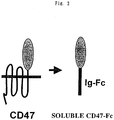
- CD47 protein disclosed herein (for example, GenBank Accession No.: BT00697) was identified as a molecule associated with an integrin being a cell adhesion molecule, it is also called IAP (Integrin-Associated Protein), and is a five-time transmembrane-type protein universally expressed in tissues. As shown in Figs. 1 and 2 , its extracellular region has one immunoglobulin-like structure which can bind to an immunoglobulin-like structure present on an N-terminal side of SHPS-1.
- IAP Integrin-Associated Protein
- the CD47 partial peptide of the fusion protein used in accordance with the method and use of the invention is a polypeptide which has an amino acid sequence constituting an extracellular region having an immunoglobulin-like structure of a CD47 protein and which is specifically bound to an N-terminal immunoglobulin-like structure of a dephosphorylation substrate protein SHPS-1 of an SH2 domain-containing protein.
- the amino acid sequence of SEQ ID No. 2 disclosed herein is an amino acid sequence constituting the extracellular region having the immunoglobulin-like structure of the CD47 protein.
- This CD47 partial peptide is a partial sequence (1st to 142nd amino acids) of a known CD47 protein (for example, GenBank Accession No.: BT00697), and it can be cloned from a human cell-derived cDNA library.
- the cDNA library can be synthesized by a known method (for example, Okayama, H., et al., Mol. Cell. Biol. 2; 161-170, 1982 ) using poly(A)+RNA extracted from human cells or the like as a template, and a commercially available cDNA library can also be used.
- the CD47 partial peptide can be cloned from the cDNA library by synthesizing an oligonucleotide on the basis of a base sequence of any portion in the CD47 protein, as described above, and conducting screening through colony hybridization or plaque hybridization by a known method using this oligonucleotide as a probe. Further, a CD47 partial peptide cDNA can be obtained by synthesizing an oligonucleotide to be hybridized on both terminals of a desired polynucleotide and conducting RT-PCR from mRNA isolated from cells using this as a primer.
- a polynucleotide encoding an amino acid sequence of SEQ ID No. 2 (namely, a polynucleotide having a base sequence of SEQ ID No. 1) can be prepared by digesting this cDNA with an appropriate restriction enzyme.
- a fusion protein (hereinafter sometimes referred to as CD47-Fc) which may be obtained by binding a CD47 partial peptide to an immunoglobulin Fc fragment (hereinafter sometimes referred to as Ig-Fc), which may result from digestion of an antibody with a protease such as papain.
- Said fusion protein has an amino acid sequence of SEQ ID No. 4 which is an amino acid sequence encoding CD47-Fc, a fusion protein of the CD47 partial peptide and Ig-Fc.
- Said fusion protein is optionally an expression product of a polynucleotide having a base sequence of SEQ ID No. 3. Also disclosed herein is the polynucleotide having the base sequence of SEQ ID No. 3.
- CD47 partial peptide constituting CD47-Fc having the amino acid of SEQ ID No. 4 encodes 1st to 142nd amino acids, and Ig-Fc encodes 143rd to 371st amino acids.
- an effect of CD47-Fc or an anti-SHPS-1 monoclonal antibody on accumulation and crosslinking of SHPS-1 is enhanced by addition of a secondary antibody to increase an inhibitory effect mediated by a function of SHPS-1.
- This secondary antibody is an IgG antibody to antibody series of a primary antibody, and it is not particularly limited. For example, when it is an antibody obtained by immunizing a mouse, an antibody is a mouse IgG, and an anti-mouse IgG antibody is therefore used as a secondary antibody. Further, in case of a human Fc fragment protein, an anti-human IgGFc antibody is used.
- the expression vector for the fusion protein used in accordance with the method and use of the invention may be obtained by treating a polynucleotide encoding the CD47 partial peptide and a polynucleotide encoding Ig-Fc respectively with appropriate restriction enzymes and binding them through ligation and introducing the resulting fragment (namely, a polynucleotide comprising a base sequence of SEQ ID No. 3). CD47-Fc may be expressed using this expression vector.
- Examples of the available expression vector can include pTracerCMV, pKA1, pCDM8, pSVK3, pMSG, pSVL, pBK-CMV, pBK-RSV, EBV vector, pRS, pcDNA3, pMSG, pYES2 and the like.
- the CD47 partial peptide can be utilized as a structure (monovalent) of the CD47 partial peptide only.
- the Ig-Fc fragment is formed into a fusion protein by arbitrarily employing another peptide (for example, His-tag (histidine-tag) or GST (glutathione-S-transferase)).
- the fusion protein used in accordance with the method and use of the present invention may be obtained by transforming the expression vector carrying the foregoing polynucleotide comprising the base sequence of SEQ ID NO. 3 in any culture of cells.
- the cultured cells are transformant cells that stably express CD47-Fc, and they have been established by selective culture in a cell culture medium containing various drugs (for example, G418) adapted to drug resistance genes that the expression vector carries.
- CD47-Fc can be obtained by recovering and purifying a cell supernatant of the transformant cell strain.
- the culture cells are not particularly limited, and various cells including Hela cells, COS7 cells and CHO cells can be used.
- a method for recovering cells generally, cells are recovered from an incubator along with a medium, the medium containing the cells is treated by low-speed centrifugation, and the cells precipitated are suspended in an appropriate buffer solution.
- the buffer solution can include a Tris buffer solution, a HEPES buffer solution, a phosphate buffer solution and the like.
- the buffer solution can properly be selected according to the type of cells and the experimental purpose.
- the cell suspension can be mixed, as required, with a cell storage solution such as DMSO or a commercial storage solution kit (for example, CellvationTM; manufactured by Celox Laboratories) to store the mixture in liquid nitrogen or a freezer of not higher than -80° for a long period of time.
- the cell supernatant can be purified by employing, for example, affinity column or gel filtration.
- affinity column Especially in case of CD47-Fc fusion protein used in accordance with the method and use of the invention of this application, the fusion protein with the Fc fragment is formed. Accordingly, an affinity column using a Protein-A agarose having high to the Fc fragment is preferably utilized.
- the origin of the CD47 partial peptide constituting CD47-Fc or Ig-Fc is preferably mammals. Examples thereof can include a sheep, a goat, a rat, a mouse, a rabbit, a horse, cattle, humans and the like. Humans are especially preferable for the purpose of inhibition of cancer cell metastasis of humans, prevention or treatment of arteriosclerosis of humans or the like.
- CD47-Fc can be bound to SHPS-1 as a type of a ligand of SHPS-1, and a function of SHPS-1 can be controlled by adjusting the use amount, the use timing or the like of CD47-Fc at this time.
- This SHPS-1 is a receptor-type protein as a dephosphorylation substrate protein of a tyrosine phosphatase SHP-2 having an SH2 domain. It is a protein having three immunoglobulin-like structures in an extracellular region and carrying plural tyrosine residues that undergo phosphorylation in an intracellular region.
- CD47-Fc an immunoglobulin-like structure on an N-terminal side of SHPS-1 and one immunoglobulin-like structure in a CD47 extracellular region are bound to provide interaction with SHPS-1 (namely, a CD47-SHPS-1 system).
- This CD47-SHPS-1 system is involved in negative control of universal cell movement.
- Fig. 3 it is considered that the CD47-SHPS-1 system inhibits cell movement bidirectionally by contact of cells expressing a CD47 molecule and an SHPS-1 molecule respectively and that the CD47-SHPS-1 system is greatly involved in a molecular mechanism of contact inhibition.
- the anti-SHPS-1 monoclonal antibody which is optionally additionally used in accordance with the method and use of the invention of this application is an anti-SHPS-1 monoclonal antibody which specifically recognizes and is capable of binding to a dephosphorylation substrate protein SHPS-1 of an SH2 domain-containing protein. Accordingly, the function of SHPS-1 can be controlled, similarly to CD47-Fc, by adjusting the use amount, the use timing or the like of the anti-SHPS-1 monoclonal antibody.
- SHPS-1 which is its target is preferably mammalian. Examples thereof include a sheep, a goat, a rat, a mouse, a rabbit, a horse, cattle, humans and the like. In view of the purpose of inhibition of cancer cell metastasis of humans, prevention or treatment of arteriosclerosis of humans or the like, humans are especially preferable similarly to the CD47 partial peptide.
- the anti-SHPS-1 monoclonal antibody which may optionally additionally be used in accordance with the method and use of the invention can be produced from the hybridoma cell strain disclosed herein according to a prescribed method.
- a peptide SHPS-1 GenBank Accession No.: JC5287 (protein), GenBank Accession No.: E15703A (nucleic acid), Yamamoto, T., et al., Biochem. Biophys. Res. Commun., 231: 61-67, 1997 and etc
- a peptide SHPS-1 GenBank Accession No.: JC5287 (protein), GenBank Accession No.: E15703A (nucleic acid), Yamamoto, T., et al., Biochem. Biophys. Res. Commun., 231: 61-67, 1997 and etc
- Fc specific antibody Fc specific antibody
- SHPS-1 is administered to an animal to be immunized (for example, a mouse, a rat or a rabbit) as an immunogen along with any carrier (for example, bovine serum albumin (FCS)) for immunization.
- any carrier for example, bovine serum albumin (FCS)
- FCS bovine serum albumin
- antibody-productive cells such as lymphocytes
- myeloma cells prepared from the spleen or the lymph node of the immunized animal are fused with myeloma cells according to a known method (for example, a method for cell fusion in a solution of a polymer such as polyethylene glycol at a high concentration or a method for cell fusion by electric stimulation) to prepare a hybridoma cell strain that produces an anti-SHPS-1 monoclonal antibody, and its cell supernatant is purified.
- a known method for example, a method for cell fusion in a solution of a polymer such as polyethylene glycol at a high concentration or a method for cell fusion by electric stimulation
- the myeloma cells are not particularly limited. In consideration of convenience in selecting a hybridoma from fusion cells, an HGPRT (Hypoxanthine-guanine Phosphoribosyl Transferase)-deficient strain in which the selection method has been established is preferably used.
- Examples thereof can include X63-AG8(X63), NS-1Ag4/1(NS-1), P3X63-AG8.UI (P3UI), X63-Ag8.653(X63.653), SP2/0-AG14(SP2/0), Fo and the like which are derived from mice.
- Fusion cells and non-fusion cells can be selected by, for example, a known HAT (hypoxanthine ⁇ aminopterin ⁇ thymidine) selection method.
- This method is a method which is effective in case of producing fusion cells using myeloma cells of an HGPRT-deficient strain that cannot be alive in the presence of aminopterin.
- only fusion cells having resistance to aminopterin can selectively be cultured by culturing fusion cells and non-fusion cells in a HAT medium.
- the screening of hybridoma cells producing the desired monoclonal antibody can be conducted by a known method such as EIA (enzyme immunoassay), RIA (radioimmunoassay) or a fluorescent antibody method.
- the hybridoma cell strain obtained in the foregoing manner can be stored, similarly to the CD47-Fc production cell strain, in liquid nitrogen or a freezer of not higher than -80°C by being mixed with a cell storage solution such as DMSO or a storage liquid kit (for example, CellvationTM; manufactured by Celox Laboratories) as required.
- a cell storage solution such as DMSO or a storage liquid kit (for example, CellvationTM; manufactured by Celox Laboratories) as required.
- the fusion protein used in accordance with the use of the present invention can be in a composition optionally containing, as an active ingredient, the anti-SHPS-1 monoclonal antibody, along with a pharmacological component.
- This composition can be applied to inhibition of cancer cell metastasis, prevention or treatment of arteriosclerosis and the like.
- Said composition may contain said fusion protein and the anti-SHPS-1 monoclonal antibody along with the pharmacological component.
- the "pharmacological component” first means various carriers used in the ordinary drug production.
- the “carrier” can properly be selected from the wide range according to the type of target diseases or the dosage form of the drug.
- Said composition is preferably in a unit dosage form capable of oral administration or administration by injection. Especially, in case of administration by injection, local injection, intraperitoneal administration, selective intravenous injection, intravenous injection, subcutaneous injection, organ perfusate injection and the like can be employed.
- Oral liquid preparations such as a suspension and a syrup can be produced using water, saccharides such as sucrose, sorbitol and fructose, glycols such as polyethylene glycol, oils such as sesame oil and soybean oil, preservatives such as alkyl p-hydroxybenzoate, flavors such as a strawberry flavor and a peppermint, and the like.
- saccharides such as sucrose, sorbitol and fructose
- glycols such as polyethylene glycol
- oils such as sesame oil and soybean oil
- preservatives such as alkyl p-hydroxybenzoate
- flavors such as a strawberry flavor and a peppermint, and the like.
- Powders, pills, capsules and tablets can be formed using excipients such as lactose, glucose, sucrose and mannitol, disintegrants such as starch and sodium alginate, lubricants such as magnesium stearate and talc, binders such as polyvinyl alcohol, hydroxypropyl cellulose and gelatin, surfactants such as a fatty acid ester, plasticizers such as glycerin, and the like. Tablets and capsules are preferably in a unit dosage form in the composition used in this invention because of easy administration. In producing tablets or capsules, solid production carriers are used.
- An injection solution can be formed using a carrier comprising a salt solution, a glucose solution or a mixture of salt water and a glucose solution, various buffer solutions and the like. It is also possible that formulation is conducted in a powdery state, and the powder is mixed with the liquid carrier in use to prepare an injection solution.
- the pharmacological component is secondly a component for rendering the CD47-Fc fusion protein and/or the anti-SHPS-1 monoclonal antibody in a dosage form introducible into cells.
- a composition can be formed by mixing the polypeptide with a pharmacologically acceptable solution without changing the structure or the function of the polypeptide.
- Such a composition can be introduced into target cells by a method for introduction into cells via microinjection, a method for introduction into cells using lipids or the like.
- the anti-CD47 monoclonal antibody used in this Example was produced as an anti-human CD47 monoclonal antibody (CC2C6), and this was utilized.
- a CD47 protein was administered to a mouse as an immunogen along with bovine serum albumin (FCS)) as a carrier for immunization.
- FCS bovine serum albumin
- additional immunization was properly conducted after a prescribed period of time to fully sensitize the mouse.
- Antibody-productive cells such as lymphocytes, produced from the spleen or the lymph node of this mouse were fused with mouse myeloma cells P3U according to a known method to produce a CD47 antibody-productive hybridoma cell strain, and the cell supernatant was purified. In this manner, the anti-CD47 monoclonal antibody was obtained.
- An anti-SHPS-1 monoclonal antibody (SE12C3) was produced and purified by the same process as in (i) except that a peptide SHPS-1 was used as an immunogen.
- This SHPS-1 was obtained by conducting purification through immunoprecipitation using an Fc specific antibody.
- CD47 and SHPS-1 were confirmed by an immunofluorescence labeling analysis method using human malignant melanoma cells WM239a (M. Herlyn (provided by Wistar Institute, Philadelphia, PA)) as melanoma cells.
- a cDNA of human CD47 was obtained by being provided from Mr. F. P. Lindberg (Washington University, St Louis, MO).
- a cDNA encoding an Fc protein of human IgG1 was obtained by a known method ( Liu, Y. C., et al., Proc. Natl. Acad. Sci. 90; 8957-8961, 1993 ) using PCR. This PCR product was digested with SpeI and NotI. pEFneo vector (manufactured by Invitrogen) was ligated in sites homologous to SpeI and NotI to produce pEFneoFc76.
- a cDNA encoding a human CD47 partial peptide (1 to 142 amino acids) was obtained by PCR using a cDNA of a full-length human CD47 protein as a template.
- 5'-TAATACTAGTCTGCTGCTCCAGAC ACCTGCG-3' was used as a sense primer, and 5'-TAGCTCTAGAATTTTCA TTTGGAGAAAACCATGAAAC-3' as an antisense primer.
- the resulting PCR product was digested with XbaI and SpeI, and introduced into a SpeI site of pEFneoFc76.
- pTracerCMV-hCD47-Fc was transfected with CHO-Ras cells, and selective culture was conducted according to a prescribed method using Zeocin.
- Judgement of a cell strain that expresses and secretes the CD47-Fc fusion protein was performed by an immunoblotting method using an anti-human IgG polyclonal antibody in a cell supernatant.
- the supernatant was purified by column chromatography with rProtein A-Sepharose 4FF (manufactured by Amersham Pharmacia Biotech).
- the action on malignant melanoma cells WM239a was examined by measurement of cell movement.
- the cell movement was measured by a known method using a Transwell apparatus (manufactured by Corning) ( Longo, N., et al., Blood 98; 3717-3726, 2001 ).
- An anti-mouse IgG was used as a secondary antibody in this Example.
- CHO-IR-SHPS-1-WT cells carrying a rat SHPS-1 normal type and CHO-IR-rSHPS-1-4F cells carrying a rat SHPS-1 variant type with all four tyrosine residues substituted by phenylalanine residues were used.
- an anti-rat SHPS-1 monoclonal antibody (2F34) and an anti-rat SHPS-1 monoclonal antibody (9F10) were used as an antibody.
- Example 2 Verification in a macrophage function
- SHPS-1 KO mouse A gene-deficient mouse which is deficient in an intracellular region of SHPS-1 (hereinafter sometimes referred to as SHPS-1 KO mouse) was produced according to a known method (for example, supervised by Nomura Tatsuji, compiled by Kachiki Gen, et al., Hassei Kogaku Jikken Manuaru, Kodansha Scientific, 1987 , Capecchi, MR., Science 244: 1288-1292, 1989 , and the like).
- MGDF as its growth stimulation factor was administered, and the reaction was examined. However, no difference was found between a wild-type mouse and an SHPS-1 KO mouse.
- Fig. 9 (A) the number of platelets after 15 days from the administration was 182 ⁇ 13.7 x 10 4 / ⁇ l in the wild-type mouse (WT), whereas it was 117 ⁇ 41.2 x 10 4 / ⁇ l in the SHPS-1 KO mouse (KO) to find a difference.
- Fig. 9(B) an in-vivo half-life of biotin-labeled platelets was measured.
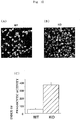
- Intraperitoneal macrophages were isolated from an SHPS-1 KO mouse by thioglycote intraperitoneal injection, and in-vitro phagocytic activity was analyzed. Phagocytic activity of mouse red cells was examined using the macrophages. Then, as shown in Figs. 10 (A), (B) and (C) respectively, significant enhancement of phagocytic activity to platelets was observed in macrophages derived from the SHPS-1 KO mouse (KO) in comparison with macrophages derived from the wild-type mouse (WT). From this fact, it can be understood that the decrease in number of platelets and shortening of life of platelets are attributable to the phagocytic activity of macrophages.
- CD47 on platelets is bound to SHPS-1 of macrophages to negatively control the phagocytosis process of platelets by macrophages. That is, the anti-SHPS-1 monoclonal antibody as a ligand of SHPS-1 or CD47-Fc acts to improve the phagocytic activity of macrophages to platelets involved in blood coagulation or the like, which can be applied to treatment of arteriosclerosis or the like.
- the CD47 partial peptide and the anti-SHPS-1 monoclonal antibody which can act on the function of cell response mediated by SHPS-1 to contribute to inhibition of cancer cell metastasis, improvement of arteriosclerosis or the like are provided.
Landscapes
- Health & Medical Sciences (AREA)
- Chemical & Material Sciences (AREA)
- Organic Chemistry (AREA)
- Life Sciences & Earth Sciences (AREA)
- General Health & Medical Sciences (AREA)
- Medicinal Chemistry (AREA)
- Genetics & Genomics (AREA)
- Molecular Biology (AREA)
- Proteomics, Peptides & Aminoacids (AREA)
- Biophysics (AREA)
- Biochemistry (AREA)
- Immunology (AREA)
- Chemical Kinetics & Catalysis (AREA)
- Veterinary Medicine (AREA)
- Pharmacology & Pharmacy (AREA)
- Nuclear Medicine, Radiotherapy & Molecular Imaging (AREA)
- Public Health (AREA)
- General Chemical & Material Sciences (AREA)
- Animal Behavior & Ethology (AREA)
- Engineering & Computer Science (AREA)
- Bioinformatics & Cheminformatics (AREA)
- Cell Biology (AREA)
- Toxicology (AREA)
- Zoology (AREA)
- Gastroenterology & Hepatology (AREA)
- Cardiology (AREA)
- Heart & Thoracic Surgery (AREA)
- Oncology (AREA)
- Vascular Medicine (AREA)
- Urology & Nephrology (AREA)
- Preparation Of Compounds By Using Micro-Organisms (AREA)
- Peptides Or Proteins (AREA)
- Medicines Containing Antibodies Or Antigens For Use As Internal Diagnostic Agents (AREA)
- Micro-Organisms Or Cultivation Processes Thereof (AREA)
- Medicinal Preparation (AREA)
- Medicines That Contain Protein Lipid Enzymes And Other Medicines (AREA)
Description
- The invention of this application relates to uses of a fusion protein of a CD47 partial peptide and an immunoglobin Fc fragment, and optionally an anti-SHPS-1 monoclonal antibody. More specifically, the invention of this application relates to uses of a fusion protein of a CD47 partial peptide and an immunoglobin Fc fragment, and optionally an anti-SHPS-1 monoclonal antibody capable of acting on a function of cell response mediated by SHPS-1.
- It has been so far clarified that on a control mechanism of growth and adhesion of cells, minute crosstalk acts between both of them. For example, when the skin is damaged by injury or the like, cells of the skin begin to grow for covering a wound, and when cells are contacted with each other, the growth of cells stops (contact inhibition). In many cancer cells, this mechanism of contact inhibition is lost, which is considered to cause acquirement of indefinite growth activity or metastatic activity of cancer cells. For this reason, clarification of a mechanism of controlling growth and adhesion of cells is important in understanding life phenomenon such as organogenesis, and also important in understanding disorders such as metastasis of cancer cells.
- In our country, the development of a new method for preventing or treating arteriosclerosis which often causes serious diseases has been an important issue in health medical care for persons of middle or advanced age. Recently, in a lesion of arteriosclerosis, a relation between immunocompetent cells, mainly macrophages and chronic inflammation has attracted much interest. Accordingly, it is quite important to clarify a minute control mechanism of macrophage functions on a molecular level for understanding the disease of arteriosclerosis in more detail and studying and developing more effective therapeutic strategies.
- The present inventors have clarified that tyrosine phosphatase having an SH2 domain plays an important role in activating a Ras low-molecular G protein by stimulation of a growth factor (Non-patent Document 1). Further, they have found a receptor-type protein SHPS-1 (SH2-containing Protein Tyrosine Phosphatase Substrate-1) belonging to a immunoglobulin super family (Non-patent Document 2) or a docking protein Gab-1 as a dephosphorylation substrate protein of SHP-2. It has also been clarified that an SHPS-1/SHP-2 system or a Gab-1/SHP-2 system interacts with an integrin system which is a receptor molecule on a cell surface functioning in adhering cells to an extracellular matrix or a cadherin system being a molecular group necessary for adhering cells to each other, so that cell movement is also controlled through activation of an Rho low-molecular G protein.
- The present inventors have lately obtained the results of study suggesting the possibility that SHPS-1 interacts with a receptor-type molecule CD47 as a physiological ligand of its extracellular region to form an intercellular signal transduction system (CD47-SHPS-1 system) (Non-patent Document 3). Further, CD47 is also expressed in vascular endothelial cells. Consequently, it is considered that CD47 of vascular endothelial cells interacts with SHPS-1 on macrophages to negatively control a macrophage function. It is considered that minute clarification of this CD47-SHPS-1 system can greatly contribute to elucidation of the foregoing "control mechanism of growth and adhesion" or "control mechanism of macrophage functions". As a result, it can be expected to contribute to, for example, inhibition of metastasis of cancer cells expressing SHPS-1 and prevention or treatment of arteriosclerosis.
- Non-patent Document 1: Noguchi, T., Matozaki, T., et al.: Mol. Cell. Biol., 14: 6674-6682, 1994
- Non-patent Document 2: Fujioka, Y., Matozaki, T., et al.: Mol. Cell. Biol., 16: 6887-6899, 1996
- Non-patent Document 3: Yamao, T., Noguchi, T., et al.: J. Biol. Chem., 277: 39833-39839, 2002
- Seiffert et al. (Blood, Vol 94, No 11, December 1 1999, pages 3633-3643) discloses that human signal-regulatory protein is expressed on normal, but not on subsets of leukemic myeloid cells and mediates cellular adhesion involving its counter receptor CD47.
-
WO 02/092784 A2 - Although a possibility that the CD47-SHPS-1 system is involved in "control mechanism of growth and adhesion" and "control mechanism of macrophage functions" is suggested and these are considered to be applied to prevention or treatment of various diseases, CD47 is a membrane penetration-type protein and it is difficult to allow it to act as a ligand in view of time and quantitative determination. Thus, there was no effective method for controlling functions of SHPS-1.
- In production of an anti-SHPS-1 antibody, a base sequence encoding SHPS-1 as an immunogen at this time has been identified for the first time by the present inventors (GenBank Accession No.: JC5287 (protein), GenBank Accession No.: E15703A (nucleic acid) Yamamoto, T., et al., Biochem. Biophys. Res. Commun., 231: 61-67, 1997). However, they have not come to produce the antibody.
- Under the foregoing circumstances, the invention of this application has been made, and it aims to provide, upon solving the existing problems, the use of a fusion protein of a CD47 partial peptide and an immunoglobin Fc fragment and optionally an anti-SHPS-1 monoclonal antibody capable of acting on a function of cell response mediated by SHPS-1.
- The invention of this application provides, as a means for solving the foregoing problems, an in vitro or ex vivo method to inhibit movement of cells by controlling a function of cell response mediated by SHPS-1, which comprises contacting SHPS-1 on the cell with a fusion protein of a CD47 partial peptide and an immunoglobulin Fc fragment, which fusion protein has an amino acid sequence of SEQ ID No. 4;
- The invention further provides a fusion protein of a CD47 partial peptide and an immunoglobulin Fc fragment, which has an amino acid sequence of SEQ ID No. 4, for use in a method for the treatment of cancers expressing SHPS-1, wherein the treatment is achieved by inhibiting metastasis of cancer cells.
- Said CD47 partial peptide has an amino acid sequence constituting an extracellular region having an immunoglobulin-like structure of a CD47 protein and which can specifically bind to an N-terminal immunoglobulin-like structure of a dephosphorylation substrate protein SHPS-1 of an SH2 domain-containing protein.
- Said CD47 partial peptide has the amino acid sequence of SEQ ID No. 2.
- The CD47 partial peptide used in accordance with the invention has an immunoglobulin Fc fragment bound and has an amino acid sequence of SEQ ID No. 4, which is optionally an expression product of a polynucleotide having a base sequence of SEQ ID No. 3.
- Also disclosed herein is a polynucleotide having a base sequence of SEQ ID No. 3, and
an expression vector carrying said polynucleotide. - Also disclosed herein is a transformant cell obtained with said expression vector.
- The method and use of the present invention may further comprise addition of an anti-SHPS-1 monoclonal antibody which specifically recognizes and is capable of binding to a dephosphorylation substrate protein SHPS-1 of an SH2 domain-containing protein.
- Also disclosed herein is a hybridoma cell strain producing said antibody.
- Also disclosed herein is a composition characterized by containing, as an active ingredient, at least one of the CD47 partial peptide as described herein and the anti-SHPS-1 monoclonal antibody along with a pharmacological component.
-
-
Fig. 1 is a schematic view showing a state of adding a CD47 protein and/or an anti-SHPS-1 monoclonal antibody to SHPS-1 expressed in melanoma cells. -
Fig. 2 is a schematic view showing CD47-Fc produced by fusing an extracellular region of 5-time transmembrane-type CD47 present on a cell membrane with IG-Fc. -
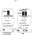
Fig. 3 is a schematic view showing a mechanism of inhibiting cell movement in a CD47-SHPS-1 system by addition of CD47-Fc. -
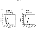
Fig. 4 is graphs showing confirmation of expression of SHPS-1 and CD47 in melanoma cells (WM239a). (A) shows results of confirming SHPS-1 expression, and (B) shows results of confirming CD47 expression. -
Fig. 5 is a graph showing an effect of CD47-Fc and an anti-SHPS-1 monoclonal antibody on inhibiting cell movement. -
Fig. 6 is a graph showing an effect on inhibiting cell movement in case of further adding a secondary antibody to the anti-SHPS-1 monoclonal antibody. -
Fig. 7 is a graph showing an effect of the anti-SHPS-1 monoclonal antibody on inhibiting cell movement in CHO-Ras cells expressing SHPS-1. -
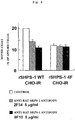
Fig. 8 is a graph showing an inhibitory effect of the anti-SHPS-1 monoclonal antibody on CHO-IR cells expressing SHPS-1 and variant CHO-Ras cells not expressing SHPS-1. -
Fig. 9 is graphs showing a platelet life in a wild-type mouse (WT) and an SHPS-1-deficient-type mouse (KO). (A) shows the number of days after administration of an anticancer agent, and (B) its half-life by biotin labeling. -
Fig. 10 is a view showing the increase in phagocytic activity of macrophages. (A) shows wild-type macrophages (WT), (B) SHPS-1-deficient-type macrophages (KO), and (C) a graph that compares a wild type (WT) with a SHPS-1-deficient type (KO). - The invention of this application has the foregoing characteristics, and embodiments thereof are described in detail below.
- Since the CD47 protein disclosed herein (for example, GenBank Accession No.: BT00697) was identified as a molecule associated with an integrin being a cell adhesion molecule, it is also called IAP (Integrin-Associated Protein), and is a five-time transmembrane-type protein universally expressed in tissues. As shown in
Figs. 1 and2 , its extracellular region has one immunoglobulin-like structure which can bind to an immunoglobulin-like structure present on an N-terminal side of SHPS-1. - The CD47 partial peptide of the fusion protein used in accordance with the method and use of the invention is a polypeptide which has an amino acid sequence constituting an extracellular region having an immunoglobulin-like structure of a CD47 protein and which is specifically bound to an N-terminal immunoglobulin-like structure of a dephosphorylation substrate protein SHPS-1 of an SH2 domain-containing protein. The amino acid sequence of SEQ ID No. 2 disclosed herein, is an amino acid sequence constituting the extracellular region having the immunoglobulin-like structure of the CD47 protein. This CD47 partial peptide is a partial sequence (1st to 142nd amino acids) of a known CD47 protein (for example, GenBank Accession No.: BT00697), and it can be cloned from a human cell-derived cDNA library. The cDNA library can be synthesized by a known method (for example, Okayama, H., et al., Mol. Cell. Biol. 2; 161-170, 1982) using poly(A)+RNA extracted from human cells or the like as a template, and a commercially available cDNA library can also be used.
- The CD47 partial peptide can be cloned from the cDNA library by synthesizing an oligonucleotide on the basis of a base sequence of any portion in the CD47 protein, as described above, and conducting screening through colony hybridization or plaque hybridization by a known method using this oligonucleotide as a probe. Further, a CD47 partial peptide cDNA can be obtained by synthesizing an oligonucleotide to be hybridized on both terminals of a desired polynucleotide and conducting RT-PCR from mRNA isolated from cells using this as a primer. A polynucleotide encoding an amino acid sequence of SEQ ID No. 2 (namely, a polynucleotide having a base sequence of SEQ ID No. 1) can be prepared by digesting this cDNA with an appropriate restriction enzyme.
- The method and use of the invention of this application use a fusion protein (hereinafter sometimes referred to as CD47-Fc) which may be obtained by binding a CD47 partial peptide to an immunoglobulin Fc fragment (hereinafter sometimes referred to as Ig-Fc), which may result from digestion of an antibody with a protease such as papain. Said fusion protein has an amino acid sequence of SEQ ID No. 4 which is an amino acid sequence encoding CD47-Fc, a fusion protein of the CD47 partial peptide and Ig-Fc. Said fusion protein is optionally an expression product of a polynucleotide having a base sequence of SEQ ID No. 3. Also disclosed herein is the polynucleotide having the base sequence of SEQ ID No. 3.
- The CD47 partial peptide constituting CD47-Fc having the amino acid of SEQ ID No. 4 encodes 1st to 142nd amino acids, and Ig-Fc encodes 143rd to 371st amino acids.
- Since the above-obtained CD47-Fc is soluble, antibody-like and divalent, an SHPS-1 crosslinking function is obtained, which is more preferable for realizing an effect of inhibiting cell movement or the like.
- Further, an effect of CD47-Fc or an anti-SHPS-1 monoclonal antibody on accumulation and crosslinking of SHPS-1 is enhanced by addition of a secondary antibody to increase an inhibitory effect mediated by a function of SHPS-1. This secondary antibody is an IgG antibody to antibody series of a primary antibody, and it is not particularly limited. For example, when it is an antibody obtained by immunizing a mouse, an antibody is a mouse IgG, and an anti-mouse IgG antibody is therefore used as a secondary antibody. Further, in case of a human Fc fragment protein, an anti-human IgGFc antibody is used.
- The expression vector for the fusion protein used in accordance with the method and use of the invention may be obtained by treating a polynucleotide encoding the CD47 partial peptide and a polynucleotide encoding Ig-Fc respectively with appropriate restriction enzymes and binding them through ligation and introducing the resulting fragment (namely, a polynucleotide comprising a base sequence of SEQ ID No. 3). CD47-Fc may be expressed using this expression vector. Examples of the available expression vector can include pTracerCMV, pKA1, pCDM8, pSVK3, pMSG, pSVL, pBK-CMV, pBK-RSV, EBV vector, pRS, pcDNA3, pMSG, pYES2 and the like.
- Also disclosed herein, the CD47 partial peptide can be utilized as a structure (monovalent) of the CD47 partial peptide only. Disclosed herein, it is possible that the Ig-Fc fragment is formed into a fusion protein by arbitrarily employing another peptide (for example, His-tag (histidine-tag) or GST (glutathione-S-transferase)).
- The fusion protein used in accordance with the method and use of the present invention may be obtained by transforming the expression vector carrying the foregoing polynucleotide comprising the base sequence of SEQ ID NO. 3 in any culture of cells. The cultured cells are transformant cells that stably express CD47-Fc, and they have been established by selective culture in a cell culture medium containing various drugs (for example, G418) adapted to drug resistance genes that the expression vector carries. CD47-Fc can be obtained by recovering and purifying a cell supernatant of the transformant cell strain. The culture cells are not particularly limited, and various cells including Hela cells, COS7 cells and CHO cells can be used.
- In a method for recovering cells, generally, cells are recovered from an incubator along with a medium, the medium containing the cells is treated by low-speed centrifugation, and the cells precipitated are suspended in an appropriate buffer solution. Examples of the buffer solution can include a Tris buffer solution, a HEPES buffer solution, a phosphate buffer solution and the like. The buffer solution can properly be selected according to the type of cells and the experimental purpose. The cell suspension can be mixed, as required, with a cell storage solution such as DMSO or a commercial storage solution kit (for example, Cellvation™; manufactured by Celox Laboratories) to store the mixture in liquid nitrogen or a freezer of not higher than -80° for a long period of time.
- As a method for purifying an intended expressed protein such as CD47-Fc, the cell supernatant can be purified by employing, for example, affinity column or gel filtration. Especially in case of CD47-Fc fusion protein used in accordance with the method and use of the invention of this application, the fusion protein with the Fc fragment is formed. Accordingly, an affinity column using a Protein-A agarose having high to the Fc fragment is preferably utilized.
- The origin of the CD47 partial peptide constituting CD47-Fc or Ig-Fc is preferably mammals. Examples thereof can include a sheep, a goat, a rat, a mouse, a rabbit, a horse, cattle, humans and the like. Humans are especially preferable for the purpose of inhibition of cancer cell metastasis of humans, prevention or treatment of arteriosclerosis of humans or the like.
- CD47-Fc can be bound to SHPS-1 as a type of a ligand of SHPS-1, and a function of SHPS-1 can be controlled by adjusting the use amount, the use timing or the like of CD47-Fc at this time. This SHPS-1 is a receptor-type protein as a dephosphorylation substrate protein of a tyrosine phosphatase SHP-2 having an SH2 domain. It is a protein having three immunoglobulin-like structures in an extracellular region and carrying plural tyrosine residues that undergo phosphorylation in an intracellular region.
- It is considered, as stated above, that in CD47-Fc, an immunoglobulin-like structure on an N-terminal side of SHPS-1 and one immunoglobulin-like structure in a CD47 extracellular region are bound to provide interaction with SHPS-1 (namely, a CD47-SHPS-1 system). This CD47-SHPS-1 system is involved in negative control of universal cell movement. As shown in
Fig. 3 , it is considered that the CD47-SHPS-1 system inhibits cell movement bidirectionally by contact of cells expressing a CD47 molecule and an SHPS-1 molecule respectively and that the CD47-SHPS-1 system is greatly involved in a molecular mechanism of contact inhibition. - The anti-SHPS-1 monoclonal antibody which is optionally additionally used in accordance with the method and use of the invention of this application is an anti-SHPS-1 monoclonal antibody which specifically recognizes and is capable of binding to a dephosphorylation substrate protein SHPS-1 of an SH2 domain-containing protein. Accordingly, the function of SHPS-1 can be controlled, similarly to CD47-Fc, by adjusting the use amount, the use timing or the like of the anti-SHPS-1 monoclonal antibody.
- In the anti-SHPS-1 monoclonal antibody, SHPS-1 which is its target is preferably mammalian. Examples thereof include a sheep, a goat, a rat, a mouse, a rabbit, a horse, cattle, humans and the like. In view of the purpose of inhibition of cancer cell metastasis of humans, prevention or treatment of arteriosclerosis of humans or the like, humans are especially preferable similarly to the CD47 partial peptide.
- The anti-SHPS-1 monoclonal antibody which may optionally additionally be used in accordance with the method and use of the invention can be produced from the hybridoma cell strain disclosed herein according to a prescribed method. First, a peptide SHPS-1 (GenBank Accession No.: JC5287 (protein), GenBank Accession No.: E15703A (nucleic acid), Yamamoto, T., et al., Biochem. Biophys. Res. Commun., 231: 61-67, 1997 and etc) can be obtained by purifying this SHPS-1 through immunoprecipitation using an Fc specific antibody. It may be produced in Escherichia coli, yeast or the like as a fusion protein with GST (glutathione-S-transferase) or as a fusion protein with immunoglobulin Fc. The resulting SHPS-1 is administered to an animal to be immunized (for example, a mouse, a rat or a rabbit) as an immunogen along with any carrier (for example, bovine serum albumin (FCS)) for immunization. As required, additional immunization is properly conducted after a prescribed period of time to fully sensitize this immunized animal. Subsequently, antibody-productive cells such as lymphocytes, prepared from the spleen or the lymph node of the immunized animal are fused with myeloma cells according to a known method (for example, a method for cell fusion in a solution of a polymer such as polyethylene glycol at a high concentration or a method for cell fusion by electric stimulation) to prepare a hybridoma cell strain that produces an anti-SHPS-1 monoclonal antibody, and its cell supernatant is purified. In this manner, the anti-SHPS-1 monoclonal antibody can be obtained. ("Tan Kuron Kotai", Iwasaki Tatsuo, Ando Tamie, et al., Kodansha, 1987; Seiffert, M., et al., Blood 94; 3633-3643, 1999, Seiffert, M., et al., Blood 97; 2741-2749, 2001, and the like). The myeloma cells are not particularly limited. In consideration of convenience in selecting a hybridoma from fusion cells, an HGPRT (Hypoxanthine-guanine Phosphoribosyl Transferase)-deficient strain in which the selection method has been established is preferably used. Examples thereof can include X63-AG8(X63), NS-1Ag4/1(NS-1), P3X63-AG8.UI (P3UI), X63-Ag8.653(X63.653), SP2/0-AG14(SP2/0), Fo and the like which are derived from mice.
- Fusion cells and non-fusion cells can be selected by, for example, a known HAT (hypoxanthine·aminopterin·thymidine) selection method. This method is a method which is effective in case of producing fusion cells using myeloma cells of an HGPRT-deficient strain that cannot be alive in the presence of aminopterin. In this method, only fusion cells having resistance to aminopterin can selectively be cultured by culturing fusion cells and non-fusion cells in a HAT medium.
- The screening of hybridoma cells producing the desired monoclonal antibody can be conducted by a known method such as EIA (enzyme immunoassay), RIA (radioimmunoassay) or a fluorescent antibody method.
- The hybridoma cell strain obtained in the foregoing manner can be stored, similarly to the CD47-Fc production cell strain, in liquid nitrogen or a freezer of not higher than -80°C by being mixed with a cell storage solution such as DMSO or a storage liquid kit (for example, Cellvation™; manufactured by Celox Laboratories) as required.
- The fusion protein used in accordance with the use of the present invention can be in a composition optionally containing, as an active ingredient, the anti-SHPS-1 monoclonal antibody, along with a pharmacological component. This composition can be applied to inhibition of cancer cell metastasis, prevention or treatment of arteriosclerosis and the like. Said composition may contain said fusion protein and the anti-SHPS-1 monoclonal antibody along with the pharmacological component.
- The "pharmacological component" first means various carriers used in the ordinary drug production. The "carrier" can properly be selected from the wide range according to the type of target diseases or the dosage form of the drug. Said composition is preferably in a unit dosage form capable of oral administration or administration by injection. Especially, in case of administration by injection, local injection, intraperitoneal administration, selective intravenous injection, intravenous injection, subcutaneous injection, organ perfusate injection and the like can be employed.
- Oral liquid preparations such as a suspension and a syrup can be produced using water, saccharides such as sucrose, sorbitol and fructose, glycols such as polyethylene glycol, oils such as sesame oil and soybean oil, preservatives such as alkyl p-hydroxybenzoate, flavors such as a strawberry flavor and a peppermint, and the like.
- Powders, pills, capsules and tablets can be formed using excipients such as lactose, glucose, sucrose and mannitol, disintegrants such as starch and sodium alginate, lubricants such as magnesium stearate and talc, binders such as polyvinyl alcohol, hydroxypropyl cellulose and gelatin, surfactants such as a fatty acid ester, plasticizers such as glycerin, and the like. Tablets and capsules are preferably in a unit dosage form in the composition used in this invention because of easy administration. In producing tablets or capsules, solid production carriers are used.
- An injection solution can be formed using a carrier comprising a salt solution, a glucose solution or a mixture of salt water and a glucose solution, various buffer solutions and the like. It is also possible that formulation is conducted in a powdery state, and the powder is mixed with the liquid carrier in use to prepare an injection solution.
- The pharmacological component is secondly a component for rendering the CD47-Fc fusion protein and/or the anti-SHPS-1 monoclonal antibody in a dosage form introducible into cells. For example, a composition can be formed by mixing the polypeptide with a pharmacologically acceptable solution without changing the structure or the function of the polypeptide. Such a composition can be introduced into target cells by a method for introduction into cells via microinjection, a method for introduction into cells using lipids or the like.
- The invention of this application is specifically described in more detail below by referring to Examples. Of course, the invention of this application is not limited by the following Examples.
- The anti-CD47 monoclonal antibody used in this Example was produced as an anti-human CD47 monoclonal antibody (CC2C6), and this was utilized.
- With respect to a production process, production and purification were conducted by a known method ("Tan Kuron Kotai", Iwasaki Tatsuo, Ando Tamie, et al., Kodansha, 1987; Seiffert, M., et al. Blood 94; 3633-3643, 1999, Seiffert, M., et al. Blood 97; 2741-2749, 2001, and the like).
- Specifically, a CD47 protein was administered to a mouse as an immunogen along with bovine serum albumin (FCS)) as a carrier for immunization. As required, additional immunization was properly conducted after a prescribed period of time to fully sensitize the mouse. Antibody-productive cells such as lymphocytes, produced from the spleen or the lymph node of this mouse were fused with mouse myeloma cells P3U according to a known method to produce a CD47 antibody-productive hybridoma cell strain, and the cell supernatant was purified. In this manner, the anti-CD47 monoclonal antibody was obtained.
- An anti-SHPS-1 monoclonal antibody (SE12C3) was produced and purified by the same process as in (i) except that a peptide SHPS-1 was used as an immunogen.
- This SHPS-1 was obtained by conducting purification through immunoprecipitation using an Fc specific antibody.
- Expression of CD47 and SHPS-1 was confirmed by an immunofluorescence labeling analysis method using human malignant melanoma cells WM239a (M. Herlyn (provided by Wistar Institute, Philadelphia, PA)) as melanoma cells.
- Consequently, as shown in
Fig. 4 , it was confirmed that expression was observed in both of SHPS-1 and CD47. In (A), an anti-SHPS-1 monoclonal antibody (SE12C3) was incubated with WM239a, and in (B), an anti-CD47 monoclonal antibody (CC2C6) was incubated with WM239a. After the incubation, detection was conducted by using an FITC-labeled anti-mouse IgG polyclonal antibody and by flow cytometry (thick line). A negative control was provided by replacing the anti-SHPS-1 monoclonal antibody or the anti-CD47 monoclonal antibody with a normal mouse IgG (thin line). - A cDNA of human CD47 was obtained by being provided from Mr. F. P. Lindberg (Washington University, St Louis, MO).
- First, a cDNA encoding an Fc protein of human IgG1 was obtained by a known method (Liu, Y. C., et al., Proc. Natl. Acad. Sci. 90; 8957-8961, 1993) using PCR. This PCR product was digested with SpeI and NotI. pEFneo vector (manufactured by Invitrogen) was ligated in sites homologous to SpeI and NotI to produce pEFneoFc76.
- Subsequently, a cDNA encoding a human CD47 partial peptide (1 to 142 amino acids) was obtained by PCR using a cDNA of a full-length human CD47 protein as a template. At this time, 5'-TAATACTAGTCTGCTGCTCCAGAC ACCTGCG-3' was used as a sense primer, and 5'-TAGCTCTAGAATTTTCA TTTGGAGAAAACCATGAAAC-3' as an antisense primer. The resulting PCR product was digested with XbaI and SpeI, and introduced into a SpeI site of pEFneoFc76. The resulting vector was digested with PstI and NotI, and the thus-obtained fragment was introduced into positions homologous to PstI and NotI sites of pTracerCMV to form plasmid pTracerCMV-hCD47-Fc. pTracerCMV-hCD47-Fc was transfected with CHO-Ras cells, and selective culture was conducted according to a prescribed method using Zeocin. Judgement of a cell strain that expresses and secretes the CD47-Fc fusion protein was performed by an immunoblotting method using an anti-human IgG polyclonal antibody in a cell supernatant. In the purification of CD47-Fc, the supernatant was purified by column chromatography with rProtein A-Sepharose 4FF (manufactured by Amersham Pharmacia Biotech).
- The action on malignant melanoma cells WM239a was examined by measurement of cell movement. The cell movement was measured by a known method using a Transwell apparatus (manufactured by Corning) (Longo, N., et al., Blood 98; 3717-3726, 2001).
- As shown in
Fig. 5 , an effect of inhibiting cell movement of malignant melanoma cells WM239a was confirmed in both of the anti-SHPS-1 monoclonal antibody and CD47-Fc. That is, the SHPS-1 ligand bound to CD47 (namely, anti-SHPS-1 monoclonal antibody and CD47-Fc) can inhibit cell movement, and inhibition of metastasis of cancer cells can be expected. - With respect to the effect of inhibiting cell movement in 4, it was confirmed, as shown in
Fig. 6 , that inhibition of cell movement was enhanced by addition of a secondary antibody. - It is considered that an effect of the fused protein CD47-Fc or the anti-SHPS-1 monoclonal antibody on SHPS-1 accumulation and crosslinking is enhanced to thereby increase an inhibitory effect mediated by the activity of SHPS-1.
- An anti-mouse IgG was used as a secondary antibody in this Example.
- An action on CHO-Ras cells (provided by S. Shirahata (Kyushu University, Fukuoka, Japan)) stably expressing H-Ras was examined by measurement of cell movement. The measurement of cell movement was conducted, as in 4, by a known method using a Transwell apparatus (manufactured by Corning) (Longo, N., et al., Blood 98; 3717-3726, 2001).
- In this Example, examination was conducted using two types of the anti-SHPS-1 monoclonal antibody, an anti-human SHPS-1 monoclonal antibody (SE12C3) and an anti-rat SHPS-monoclonal antibody (2F34).
- As shown in
Fig. 7 , an effect of inhibiting cell movement of CHO-Ras cells was confirmed in both of the anti-human SHPS-1 monoclonal antibody (SE12C3) and the anti-rat SHPS-1 monoclonal antibody (2F34). Especially, it was confirmed that the anti-human SHPS-1 monoclonal antibody acted more effectively on human SHPS-1 expression cells, whereas the anti-rat SHPS-1 monoclonal antibody acted more effectively on rat SHPS-1 expression cells. Incidentally, normal mouse IgG was used as a control. - With respect to CHO-IR cells stably expressing a human insulin receptor, CHO-IR-SHPS-1-WT cells carrying a rat SHPS-1 normal type and CHO-IR-rSHPS-1-4F cells carrying a rat SHPS-1 variant type with all four tyrosine residues substituted by phenylalanine residues were used. Further, as an antibody, two types, an anti-rat SHPS-1 monoclonal antibody (2F34) and an anti-rat SHPS-1 monoclonal antibody (9F10) were used.
- Consequently, as shown in
Fig. 8 , in CHO-IR-rSHPS-1-4F cells, no difference was found among the control, the anti-rat SHPS-1 monoclonal antibody (2F34) and the anti-rat SHPS-1 monoclonal antibody (9F10). Meanwhile, in CHO-IR-SHPS-1-WT cells, no effect was found in the control. However, the inhibitory effect was confirmed in the anti-rat SHPS-1 monoclonal antibody (2F34) and the anti-rat SHPS-1 monoclonal antibody (9F10). That is, it can be understood that the SHP-2 binding motif of SHPS-1 is necessary for inhibition of cell movement with the anti-SHPS-1 monoclonal antibody. - A gene-deficient mouse which is deficient in an intracellular region of SHPS-1 (hereinafter sometimes referred to as SHPS-1 KO mouse) was produced according to a known method (for example, supervised by Nomura Tatsuji, compiled by Kachiki Gen, et al., Hassei Kogaku Jikken Manuaru, Kodansha Scientific, 1987, Capecchi, MR., Science 244: 1288-1292, 1989, and the like).
- The peripheral blood of this SHPS-1 KO mouse was analyzed by flow cytometry using each blood cell ingredient specific antibody. Then, the number of platelets was reduced by up to about 70% in comparison with a wild-type mouse (Table 1).
Table 1 +/- male -/- male +/- female -/- female RBC (x104/ml) 925+/-25 924+/-20 973+/-27 932+/-8 WBC (x102/ml) 14.6+/-1.4 25.0+/-1.6 23.2+/-1.8 17.2+/-0.7 Hb (g/dL) 15.7+/-0.33 14.8+/-0.44 16.6+/-0.54 15.7+/-0.26 MCV (fL) 57.6+/-0.67 54.6+/-0.68 57.2+/-0.53 55.6+/-0.57 PLT (x104/ml) 98.2+/-4.1* 75.6+/-3.5* 64.3+/-4.3* 64.9+/-2.3* MPV (fL) 5.56+/-0.07 5.76+/-0.05 5.70+/-0.12 5.52+/-0.13 *Student's test, P< .005 - It was confirmed that the change in number of platelets was observed in 4 weeks from birth and continued for at least 13 weeks.
- With respect to verification of a cause of decrease in number of platelets, growth activity and differentiation activity of megakaryocytes obtained from an SHPS-1 KO mouse were examined by in-vitro culture, and recovery of number of platelets after administration of an anticancer agent 5FU was conducted with a flow cytometer.
- Specifically, regarding megakaryocyte colony formability in the femur, MGDF as its growth stimulation factor was administered, and the reaction was examined. However, no difference was found between a wild-type mouse and an SHPS-1 KO mouse.
- Meanwhile, in the platelet life, a recovery process of the number of platelets after inhibition of myeloma by administration of 5-FU as an anticancer agent was analyzed. Consequently, as shown in
Fig. 9 (A) , the number of platelets after 15 days from the administration was 182 ± 13.7 x 10⁴/µl in the wild-type mouse (WT), whereas it was 117 ± 41.2 x 10⁴/µl in the SHPS-1 KO mouse (KO) to find a difference. Further, inFig. 9(B) , an in-vivo half-life of biotin-labeled platelets was measured. Consequently, the half-life of the SHPS-1 KO mouse (KO) was confirmed to be significantly shortened in comparison with that of the wild-type mouse (WT). Thus, although hindrance of recovery with 5FU was observed, the result of the increase in disappearance speed of biotinized platelets was confirmed. Thus, it was identified that it does not mean hindrance in production process but increase in consumption process. - Intraperitoneal macrophages were isolated from an SHPS-1 KO mouse by thioglycote intraperitoneal injection, and in-vitro phagocytic activity was analyzed. Phagocytic activity of mouse red cells was examined using the macrophages. Then, as shown in
Figs. 10 (A), (B) and (C) respectively, significant enhancement of phagocytic activity to platelets was observed in macrophages derived from the SHPS-1 KO mouse (KO) in comparison with macrophages derived from the wild-type mouse (WT). From this fact, it can be understood that the decrease in number of platelets and shortening of life of platelets are attributable to the phagocytic activity of macrophages. That is, CD47 on platelets is bound to SHPS-1 of macrophages to negatively control the phagocytosis process of platelets by macrophages. That is, the anti-SHPS-1 monoclonal antibody as a ligand of SHPS-1 or CD47-Fc acts to improve the phagocytic activity of macrophages to platelets involved in blood coagulation or the like, which can be applied to treatment of arteriosclerosis or the like. - As has been thus far described in detail, the CD47 partial peptide and the anti-SHPS-1 monoclonal antibody which can act on the function of cell response mediated by SHPS-1 to contribute to inhibition of cancer cell metastasis, improvement of arteriosclerosis or the like are provided.
-
- <110> Japan Science And Technology Agency
- <120> CD47 partial peptide (1-142 amino acid)
- <130> 04F029PCT
- <150>
JP2003-157287
<151> 2003-06-02 - <160> 4
- <170> PatentIn version 3.1
- <210> 1
<211> 426
<212> DNA
<213> Homo sapience - <220>
<221> CDS
<222> (1).. (426)
<223> - <400> 1


- <210> 2
<211> 142
<212> PRT
<213> Homo sapience - <400> 2


- <210> 3
<211> 1113
<212> DNA
<213> Artificial - <220>
<223> Fusion of CD47 part(1-142 amino acid) and Ig-Fc - <220>
<221> CDS
<222> (1).. (1113)
<223> - <400> 3


- <210> 4
<211> 371
<212> PRT
<213> Artificial - <220>
<223> Fusion of CD47 part(1-142 amino acid) and Ig-Fc - <400> 4


Claims (5)
- An in vitro or ex vivo method to inhibit movement of cells by controlling a function of cell response mediated by SHPS-1, which comprises contacting SHPS-1 on the cell with a fusion protein of a CD47 partial peptide and an immunoglobulin Fc fragment, which fusion protein has an amino acid sequence of SEQ ID No. 4.
- The method of claim 1, which further comprises the addition of an anti-SHPS-1 monoclonal antibody which specifically recognizes and is capable of binding to SHPS-1.
- The method of claim 1 or 2, wherein the method is used to inhibit movement of cancer cells.
- A fusion protein of a CD47 partial peptide and an immunoglobulin Fc fragment, which has an amino acid sequence of SEQ ID No. 4 for use in a method for the treatment of cancers expressing SHPS-1, wherein the treatment is achieved by inhibiting metastasis of cancer cells.
- The fusion protein for use according to claim 4, which is in a composition which further contains, as an active ingredient, an anti-SHPS-1 monoclonal antibody which specifically recognizes and is capable of binding to SHPS-1.
Applications Claiming Priority (2)
| Application Number | Priority Date | Filing Date | Title |
|---|---|---|---|
| JP2003157287A JP3936673B2 (en) | 2003-06-02 | 2003-06-02 | CD47 partial peptide and anti-SHPS-1 monoclonal antibody |
| PCT/JP2004/007871 WO2004108923A1 (en) | 2003-06-02 | 2004-05-31 | Cd47 partal peptdie and anti-shps-1 monoclonal antibody |
Publications (3)
| Publication Number | Publication Date |
|---|---|
| EP1637598A1 EP1637598A1 (en) | 2006-03-22 |
| EP1637598A4 EP1637598A4 (en) | 2008-07-09 |
| EP1637598B1 true EP1637598B1 (en) | 2017-07-05 |
Family
ID=33508339
Family Applications (1)
| Application Number | Title | Priority Date | Filing Date |
|---|---|---|---|
| EP04735526.8A Expired - Lifetime EP1637598B1 (en) | 2003-06-02 | 2004-05-31 | Cd47 partal peptide and anti-shps-1 monoclonal antibody |
Country Status (4)
| Country | Link |
|---|---|
| US (1) | US20060135749A1 (en) |
| EP (1) | EP1637598B1 (en) |
| JP (1) | JP3936673B2 (en) |
| WO (1) | WO2004108923A1 (en) |
Families Citing this family (22)
| Publication number | Priority date | Publication date | Assignee | Title |
|---|---|---|---|---|
| US20080131431A1 (en) * | 2006-05-15 | 2008-06-05 | Viral Logic Systems Technology Corp. | CD47 related compositions and methods for treating immunological diseases and disorders |
| US8377448B2 (en) * | 2006-05-15 | 2013-02-19 | The Board Of Trustees Of The Leland Standford Junior University | CD47 related compositions and methods for treating immunological diseases and disorders |
| EP2573112A1 (en) | 2007-10-11 | 2013-03-27 | The Hospital For Sick Children | Modulation of sirpa - cd47 interaction for increasing human hematopoietic stem cell engraftment and compounds therefor |
| PT2242512T (en) | 2008-01-15 | 2016-07-18 | Univ Leland Stanford Junior | METHODS FOR MANIPULATING FAGOCYTOSIS MEDIATED BY CD47 |
| US20110105380A1 (en) * | 2009-03-10 | 2011-05-05 | The Trustees Of The University Of Pennsylvania | Protection of Nano-Scale Particles from Immune Cell Uptake |
| EP4311863A3 (en) | 2009-05-15 | 2024-04-10 | University Health Network | Compositions and methods for treating hematologic cancers targeting the sirp alpha- cd47 interaction |
| JP2013514795A (en) | 2009-12-22 | 2013-05-02 | ノバルティス アーゲー | Tetravalent CD47 antibody constant region fusion protein for use in therapy |
| US20110206696A1 (en) * | 2010-01-25 | 2011-08-25 | The Washington University | Methods for inhibiting bone loss and bone metastasis |
| CN102070719A (en) * | 2010-11-24 | 2011-05-25 | 中国人民解放军第四军医大学 | Soluble leukemia stem cell targeting proteins TrxHis-hCD47 |
| KR101258457B1 (en) | 2011-01-13 | 2013-04-26 | 강원대학교산학협력단 | Pharmaceutical composition comprising peptide having inhibitory activities against transendothelial migration of leucocytes and growth or metastasis of cancer cells |
| JP2014519338A (en) | 2011-06-16 | 2014-08-14 | ノバルティス アーゲー | Soluble proteins used as therapeutic agents |
| FR2977587B1 (en) * | 2011-07-08 | 2013-08-30 | Univ Reims Champagne Ardenne | ANTAGONIST PEPTIDE OF THE BINDING BETWEEN CD47 AND A PROTEIN BELONGING TO THE THROMBOSPONDIN FAMILY |
| HRP20192013T1 (en) | 2012-12-17 | 2020-02-07 | Trillium Therapeutics Inc. | TREATMENT OF CD47 + DISEASE CELLS WITH SIRP ALFA-FC FUSIONS |
| EP3012271A1 (en) | 2014-10-24 | 2016-04-27 | Effimune | Method and compositions for inducing differentiation of myeloid derived suppressor cell to treat cancer and infectious diseases |
| HRP20241316T1 (en) | 2016-04-14 | 2024-12-20 | Ose Immunotherapeutics | New anti-sirpa antibodies and their therapeutic applications |
| JP7461741B2 (en) | 2016-06-20 | 2024-04-04 | カイマブ・リミテッド | Anti-PD-L1 and IL-2 Cytokines |
| WO2018008470A1 (en) * | 2016-07-05 | 2018-01-11 | 国立大学法人神戸大学 | Antitumor agent |
| PL3658589T3 (en) | 2017-07-26 | 2024-03-18 | Forty Seven, Inc. | Anti-sirp-alpha antibodies and related methods |
| US11401329B2 (en) | 2017-08-02 | 2022-08-02 | Phanes Therapeutics, Inc. | Anti-CD47 antibodies and uses thereof |
| US11713356B2 (en) | 2017-10-13 | 2023-08-01 | Ose Immunotherapeutics | Modified bifunctional anti-human signal regulatory protein alpha (SIRPa) antibody and method of use thereof for treating cancer |
| TWI831759B (en) | 2017-12-01 | 2024-02-11 | 美商思進公司 | Cd47 antibodies and uses thereof for treating cancer |
| WO2019175218A1 (en) | 2018-03-13 | 2019-09-19 | Ose Immunotherapeutics | Use of anti-human sirpa v1 antibodies and method for producing anti-sirpa v1 antibodies |
Family Cites Families (6)
| Publication number | Priority date | Publication date | Assignee | Title |
|---|---|---|---|---|
| MX9709447A (en) * | 1995-06-07 | 1998-02-28 | Immunex Corp | Novel cd40l mutein. |
| WO1997027873A1 (en) * | 1996-01-30 | 1997-08-07 | Brigham & Women's Hospital, Inc. | Antibodies for modulating cd47-mediated neutrophil transmigration |
| CA2275183A1 (en) * | 1996-12-20 | 1998-07-02 | Amgen Inc. | Ob fusion protein compositions and methods |
| GB9930706D0 (en) * | 1999-12-24 | 2000-02-16 | Medical Res Council | Composition for inhibiting macrophage activity |
| AU2002315052A1 (en) * | 2001-05-15 | 2002-11-25 | Emory University | Polynucleotides and polypeptides relating to the modulation of sirp alpha-cd47 |
| US20040213792A1 (en) * | 2003-04-24 | 2004-10-28 | Clemmons David R. | Method for inhibiting cellular activation by insulin-like growth factor-1 |
-
2003
- 2003-06-02 JP JP2003157287A patent/JP3936673B2/en not_active Expired - Lifetime
-
2004
- 2004-05-31 WO PCT/JP2004/007871 patent/WO2004108923A1/en not_active Ceased
- 2004-05-31 EP EP04735526.8A patent/EP1637598B1/en not_active Expired - Lifetime
- 2004-05-31 US US10/559,237 patent/US20060135749A1/en not_active Abandoned
Non-Patent Citations (1)
| Title |
|---|
| None * |
Also Published As
| Publication number | Publication date |
|---|---|
| WO2004108923A1 (en) | 2004-12-16 |
| EP1637598A1 (en) | 2006-03-22 |
| JP2004357530A (en) | 2004-12-24 |
| EP1637598A4 (en) | 2008-07-09 |
| JP3936673B2 (en) | 2007-06-27 |
| US20060135749A1 (en) | 2006-06-22 |
Similar Documents
| Publication | Publication Date | Title |
|---|---|---|
| EP1637598A1 (en) | Cd47 partal peptdie and anti-shps-1 monoclonal antibody | |
| Pricci et al. | Role of galectin-3 as a receptor for advanced glycosylation end products | |
| Cai et al. | AND-34, a novel p130Cas-binding thymic stromal cell protein regulated by adhesion and inflammatory cytokines | |
| CA2395242A1 (en) | Composition for inhibiting macrophage activity | |
| US20020173461A1 (en) | Methods for enhancing the efficacy of cancer therapy | |
| EP1235934B1 (en) | Screening method for candidate drugs | |
| Bakiri et al. | Role of heterodimerization of c-Fos and Fra1 proteins in osteoclast differentiation | |
| JP2001515344A (en) | Integrin-linked kinase, inhibitors thereof, methods of medical treatment using these inhibitors, gene therapy and pseudosubstrate inhibitors | |
| JP2008522162A (en) | Mer diagnostic and therapeutic agents | |
| Eshel et al. | The GPI-linked Ly-6 antigen E48 regulates expression levels of the FX enzyme and of E-selectin ligands on head and neck squamous carcinoma cells | |
| US20040028684A1 (en) | Cancer diagnosis and assays for screening anti-cancer agents | |
| US8263822B2 (en) | TREM-2 gene and protein as inhibitors of expression of GA733-2, and transgenic animals comprising the same and uses thereof | |
| WO2005074556A2 (en) | Netrin compositions and methods of use thereof | |
| EP1666600B1 (en) | Methods for enhancing the efficacy of cancer therapy | |
| AU2003205615B2 (en) | A novel splice variant of MyD88 and uses thereof | |
| JP3914996B2 (en) | Anti-SHPS-1 monoclonal antibody | |
| EP1444890B1 (en) | RS1 deficient transgenic animal and uses thereof | |
| US20050113284A1 (en) | Drug for regenerating tissue and vessel and method therefor | |
| US8058009B2 (en) | Target protein and target gene in drug designing and screening method | |
| JPWO2007026969A1 (en) | Drug discovery target protein and target gene, and screening method | |
| EP1739186B1 (en) | Method of screening compound capable of accelerating or inhibiting apoptosis, apoptosis accelerator and apoptosis inhibitor | |
| EP0939815A1 (en) | IDENTIFICATION OF Bap-1, A PROTEIN THAT BINDS TO INTEGRIN AND IS INVOLVED IN INTEGRIN-MEDIATED SIGNAL TRANSDUCTION | |
| Avraham | Effects of Csk Homologous Kinase Overexpression on HER2/Neu-Mediated Signal Transduction Pathways in Breast Cancer Cells | |
| Cunningham et al. | Workshop 2: Neuroendocrine tumors: Genetics, tumor biology and treatment | |
| AU1868102A (en) | Identification of Bap-1, a protein that binds to integrin and is involved in integrin-mediated signal transduction |
Legal Events
| Date | Code | Title | Description |
|---|---|---|---|
| PUAI | Public reference made under article 153(3) epc to a published international application that has entered the european phase |
Free format text: ORIGINAL CODE: 0009012 |
|
| 17P | Request for examination filed |
Effective date: 20051227 |
|
| AK | Designated contracting states |
Kind code of ref document: A1 Designated state(s): CH DE FR GB LI |
|
| DAX | Request for extension of the european patent (deleted) | ||
| RBV | Designated contracting states (corrected) |
Designated state(s): CH DE FR GB LI |
|
| A4 | Supplementary search report drawn up and despatched |
Effective date: 20080606 |
|
| 17Q | First examination report despatched |
Effective date: 20090623 |
|
| RAP1 | Party data changed (applicant data changed or rights of an application transferred) |
Owner name: NATIONAL UNIVERSITY CORPORATION GUNMA UNIVERSITY |
|
| GRAP | Despatch of communication of intention to grant a patent |
Free format text: ORIGINAL CODE: EPIDOSNIGR1 |
|
| INTG | Intention to grant announced |
Effective date: 20170210 |
|
| GRAS | Grant fee paid |
Free format text: ORIGINAL CODE: EPIDOSNIGR3 |
|
| GRAA | (expected) grant |
Free format text: ORIGINAL CODE: 0009210 |
|
| RAP1 | Party data changed (applicant data changed or rights of an application transferred) |
Owner name: NATIONAL UNIVERSITY CORPORATION GUNMA UNIVERSITY |
|
| AK | Designated contracting states |
Kind code of ref document: B1 Designated state(s): CH DE FR GB LI |
|
| REG | Reference to a national code |
Ref country code: GB Ref legal event code: FG4D |
|
| REG | Reference to a national code |
Ref country code: CH Ref legal event code: EP |
|
| REG | Reference to a national code |
Ref country code: DE Ref legal event code: R096 Ref document number: 602004051491 Country of ref document: DE |
|
| REG | Reference to a national code |
Ref country code: DE Ref legal event code: R097 Ref document number: 602004051491 Country of ref document: DE |
|
| PLBE | No opposition filed within time limit |
Free format text: ORIGINAL CODE: 0009261 |
|
| STAA | Information on the status of an ep patent application or granted ep patent |
Free format text: STATUS: NO OPPOSITION FILED WITHIN TIME LIMIT |
|
| REG | Reference to a national code |
Ref country code: FR Ref legal event code: PLFP Year of fee payment: 15 |
|
| 26N | No opposition filed |
Effective date: 20180406 |
|
| PGFP | Annual fee paid to national office [announced via postgrant information from national office to epo] |
Ref country code: DE Payment date: 20190521 Year of fee payment: 16 |
|
| PGFP | Annual fee paid to national office [announced via postgrant information from national office to epo] |
Ref country code: FR Payment date: 20190523 Year of fee payment: 16 |
|
| PGFP | Annual fee paid to national office [announced via postgrant information from national office to epo] |
Ref country code: CH Payment date: 20190521 Year of fee payment: 16 |
|
| PGFP | Annual fee paid to national office [announced via postgrant information from national office to epo] |
Ref country code: GB Payment date: 20190521 Year of fee payment: 16 |
|
| REG | Reference to a national code |
Ref country code: DE Ref legal event code: R119 Ref document number: 602004051491 Country of ref document: DE |
|
| PG25 | Lapsed in a contracting state [announced via postgrant information from national office to epo] |
Ref country code: CH Free format text: LAPSE BECAUSE OF NON-PAYMENT OF DUE FEES Effective date: 20200531 Ref country code: LI Free format text: LAPSE BECAUSE OF NON-PAYMENT OF DUE FEES Effective date: 20200531 |
|
| GBPC | Gb: european patent ceased through non-payment of renewal fee |
Effective date: 20200531 |
|
| PG25 | Lapsed in a contracting state [announced via postgrant information from national office to epo] |
Ref country code: GB Free format text: LAPSE BECAUSE OF NON-PAYMENT OF DUE FEES Effective date: 20200531 Ref country code: FR Free format text: LAPSE BECAUSE OF NON-PAYMENT OF DUE FEES Effective date: 20200531 |
|
| PG25 | Lapsed in a contracting state [announced via postgrant information from national office to epo] |
Ref country code: DE Free format text: LAPSE BECAUSE OF NON-PAYMENT OF DUE FEES Effective date: 20201201 |