EP1630229B1 - Melanoma antigens - Google Patents
Melanoma antigens Download PDFInfo
- Publication number
- EP1630229B1 EP1630229B1 EP05018103A EP05018103A EP1630229B1 EP 1630229 B1 EP1630229 B1 EP 1630229B1 EP 05018103 A EP05018103 A EP 05018103A EP 05018103 A EP05018103 A EP 05018103A EP 1630229 B1 EP1630229 B1 EP 1630229B1
- Authority
- EP
- European Patent Office
- Prior art keywords
- seq
- peptide
- amino acid
- melanoma
- unknown
- Prior art date
- Legal status (The legal status is an assumption and is not a legal conclusion. Google has not performed a legal analysis and makes no representation as to the accuracy of the status listed.)
- Expired - Lifetime
Links
Images



Classifications
-
- C—CHEMISTRY; METALLURGY
- C07—ORGANIC CHEMISTRY
- C07K—PEPTIDES
- C07K14/00—Peptides having more than 20 amino acids; Gastrins; Somatostatins; Melanotropins; Derivatives thereof
- C07K14/435—Peptides having more than 20 amino acids; Gastrins; Somatostatins; Melanotropins; Derivatives thereof from animals; from humans
- C07K14/46—Peptides having more than 20 amino acids; Gastrins; Somatostatins; Melanotropins; Derivatives thereof from animals; from humans from vertebrates
- C07K14/47—Peptides having more than 20 amino acids; Gastrins; Somatostatins; Melanotropins; Derivatives thereof from animals; from humans from vertebrates from mammals
-
- A—HUMAN NECESSITIES
- A61—MEDICAL OR VETERINARY SCIENCE; HYGIENE
- A61K—PREPARATIONS FOR MEDICAL, DENTAL OR TOILETRY PURPOSES
- A61K39/00—Medicinal preparations containing antigens or antibodies
- A61K39/0005—Vertebrate antigens
- A61K39/0011—Cancer antigens
- A61K39/00119—Melanoma antigens
-
- A—HUMAN NECESSITIES
- A61—MEDICAL OR VETERINARY SCIENCE; HYGIENE
- A61K—PREPARATIONS FOR MEDICAL, DENTAL OR TOILETRY PURPOSES
- A61K39/00—Medicinal preparations containing antigens or antibodies
- A61K39/0005—Vertebrate antigens
- A61K39/0011—Cancer antigens
- A61K39/00119—Melanoma antigens
- A61K39/001191—Melan-A/MART
-
- A—HUMAN NECESSITIES
- A61—MEDICAL OR VETERINARY SCIENCE; HYGIENE
- A61K—PREPARATIONS FOR MEDICAL, DENTAL OR TOILETRY PURPOSES
- A61K39/00—Medicinal preparations containing antigens or antibodies
- A61K39/0005—Vertebrate antigens
- A61K39/0011—Cancer antigens
- A61K39/00119—Melanoma antigens
- A61K39/001192—Glycoprotein 100 [Gp100]
-
- A—HUMAN NECESSITIES
- A61—MEDICAL OR VETERINARY SCIENCE; HYGIENE
- A61K—PREPARATIONS FOR MEDICAL, DENTAL OR TOILETRY PURPOSES
- A61K40/00—Cellular immunotherapy
- A61K40/10—Cellular immunotherapy characterised by the cell type used
- A61K40/11—T-cells, e.g. tumour infiltrating lymphocytes [TIL] or regulatory T [Treg] cells; Lymphokine-activated killer [LAK] cells
-
- A—HUMAN NECESSITIES
- A61—MEDICAL OR VETERINARY SCIENCE; HYGIENE
- A61K—PREPARATIONS FOR MEDICAL, DENTAL OR TOILETRY PURPOSES
- A61K40/00—Cellular immunotherapy
- A61K40/40—Cellular immunotherapy characterised by antigens that are targeted or presented by cells of the immune system
- A61K40/41—Vertebrate antigens
- A61K40/42—Cancer antigens
- A61K40/4271—Melanoma antigens
-
- A—HUMAN NECESSITIES
- A61—MEDICAL OR VETERINARY SCIENCE; HYGIENE
- A61K—PREPARATIONS FOR MEDICAL, DENTAL OR TOILETRY PURPOSES
- A61K40/00—Cellular immunotherapy
- A61K40/40—Cellular immunotherapy characterised by antigens that are targeted or presented by cells of the immune system
- A61K40/41—Vertebrate antigens
- A61K40/42—Cancer antigens
- A61K40/4271—Melanoma antigens
- A61K40/4272—Melan-A/MART
-
- A—HUMAN NECESSITIES
- A61—MEDICAL OR VETERINARY SCIENCE; HYGIENE
- A61K—PREPARATIONS FOR MEDICAL, DENTAL OR TOILETRY PURPOSES
- A61K40/00—Cellular immunotherapy
- A61K40/40—Cellular immunotherapy characterised by antigens that are targeted or presented by cells of the immune system
- A61K40/41—Vertebrate antigens
- A61K40/42—Cancer antigens
- A61K40/4271—Melanoma antigens
- A61K40/4273—Glycoprotein 100 [Gp100]
-
- A—HUMAN NECESSITIES
- A61—MEDICAL OR VETERINARY SCIENCE; HYGIENE
- A61P—SPECIFIC THERAPEUTIC ACTIVITY OF CHEMICAL COMPOUNDS OR MEDICINAL PREPARATIONS
- A61P35/00—Antineoplastic agents
-
- A—HUMAN NECESSITIES
- A61—MEDICAL OR VETERINARY SCIENCE; HYGIENE
- A61P—SPECIFIC THERAPEUTIC ACTIVITY OF CHEMICAL COMPOUNDS OR MEDICINAL PREPARATIONS
- A61P37/00—Drugs for immunological or allergic disorders
- A61P37/02—Immunomodulators
- A61P37/04—Immunostimulants
-
- C—CHEMISTRY; METALLURGY
- C07—ORGANIC CHEMISTRY
- C07K—PEPTIDES
- C07K14/00—Peptides having more than 20 amino acids; Gastrins; Somatostatins; Melanotropins; Derivatives thereof
- C07K14/435—Peptides having more than 20 amino acids; Gastrins; Somatostatins; Melanotropins; Derivatives thereof from animals; from humans
- C07K14/46—Peptides having more than 20 amino acids; Gastrins; Somatostatins; Melanotropins; Derivatives thereof from animals; from humans from vertebrates
- C07K14/47—Peptides having more than 20 amino acids; Gastrins; Somatostatins; Melanotropins; Derivatives thereof from animals; from humans from vertebrates from mammals
- C07K14/4701—Peptides having more than 20 amino acids; Gastrins; Somatostatins; Melanotropins; Derivatives thereof from animals; from humans from vertebrates from mammals not used
- C07K14/4748—Tumour specific antigens; Tumour rejection antigen precursors [TRAP], e.g. MAGE
-
- G—PHYSICS
- G01—MEASURING; TESTING
- G01N—INVESTIGATING OR ANALYSING MATERIALS BY DETERMINING THEIR CHEMICAL OR PHYSICAL PROPERTIES
- G01N33/00—Investigating or analysing materials by specific methods not covered by groups G01N1/00 - G01N31/00
- G01N33/48—Biological material, e.g. blood, urine; Haemocytometers
- G01N33/50—Chemical analysis of biological material, e.g. blood, urine; Testing involving biospecific ligand binding methods; Immunological testing
- G01N33/53—Immunoassay; Biospecific binding assay; Materials therefor
- G01N33/575—Immunoassay; Biospecific binding assay; Materials therefor for cancer
- G01N33/5751—Immunoassay; Biospecific binding assay; Materials therefor for cancer of the skin, e.g. melanoma
-
- A—HUMAN NECESSITIES
- A61—MEDICAL OR VETERINARY SCIENCE; HYGIENE
- A61K—PREPARATIONS FOR MEDICAL, DENTAL OR TOILETRY PURPOSES
- A61K39/00—Medicinal preparations containing antigens or antibodies
- A61K2039/51—Medicinal preparations containing antigens or antibodies comprising whole cells, viruses or DNA/RNA
- A61K2039/525—Virus
- A61K2039/5256—Virus expressing foreign proteins
-
- A—HUMAN NECESSITIES
- A61—MEDICAL OR VETERINARY SCIENCE; HYGIENE
- A61K—PREPARATIONS FOR MEDICAL, DENTAL OR TOILETRY PURPOSES
- A61K39/00—Medicinal preparations containing antigens or antibodies
- A61K2039/51—Medicinal preparations containing antigens or antibodies comprising whole cells, viruses or DNA/RNA
- A61K2039/53—DNA (RNA) vaccination
-
- A—HUMAN NECESSITIES
- A61—MEDICAL OR VETERINARY SCIENCE; HYGIENE
- A61K—PREPARATIONS FOR MEDICAL, DENTAL OR TOILETRY PURPOSES
- A61K2239/00—Indexing codes associated with cellular immunotherapy of group A61K40/00
- A61K2239/31—Indexing codes associated with cellular immunotherapy of group A61K40/00 characterized by the route of administration
-
- A—HUMAN NECESSITIES
- A61—MEDICAL OR VETERINARY SCIENCE; HYGIENE
- A61K—PREPARATIONS FOR MEDICAL, DENTAL OR TOILETRY PURPOSES
- A61K2239/00—Indexing codes associated with cellular immunotherapy of group A61K40/00
- A61K2239/38—Indexing codes associated with cellular immunotherapy of group A61K40/00 characterised by the dose, timing or administration schedule
-
- A—HUMAN NECESSITIES
- A61—MEDICAL OR VETERINARY SCIENCE; HYGIENE
- A61K—PREPARATIONS FOR MEDICAL, DENTAL OR TOILETRY PURPOSES
- A61K38/00—Medicinal preparations containing peptides
-
- A—HUMAN NECESSITIES
- A61—MEDICAL OR VETERINARY SCIENCE; HYGIENE
- A61K—PREPARATIONS FOR MEDICAL, DENTAL OR TOILETRY PURPOSES
- A61K39/00—Medicinal preparations containing antigens or antibodies
Definitions
- This invention is in the field of prevention and treatment of human cancers. More specifically, this invention relates to genes encoding melanoma antigens recognized by T-Cells and their corresponding proteins and to preventative, diagnostic and therapeutic applications which employ these genes or proteins.
- Melanomas are aggressive, frequently metastatic tumors derived from either melanocytes or melanocyte related nevus cells (" Cellular and Molecular Immunology” (1991) (eds) Abbas A.K., Lechtman, A.H., Pober, J.S.; W.B. Saunders Company, Philadelphia: pages 340-341 ). Melanomas make up approximately three percent of all skin cancers and the worldwide increase in melanoma is unsurpassed by any other neoplasm with the exception of lung cancer in women (" Cellular and "Molecular immunology” (1991) (eds) Abbas, A.K., Lechtiman, A.H., Pober, J.S.; W.B.
- T cells play an important role in tumor regression in most murine tumor models.
- Tumor infiltrating lymphocytes that recognize unique cancer antigens can be isolated from many murine tumors.
- the adoptive transfer of these TIL plus interleukin-2 can mediate the regression of established lung and liver metastases ( Rosenberg, S.A., et al., (1986) Science 233:1318-1321 ).
- the secretion of IFN- ⁇ by injected TIL significantly correlates with in vivo regression of murine tumors suggesting activation of T-cells by the tumor antigens.
- T cell receptors on CD8 + T cells recognize a complex consisting of an antigenic peptide (9-10 amino acids for HLA-A2), ß-2 microglobulin and class I major histocompatibility complex (MHC) heavy chain (HLA-A, B, C, in humans).
- MHC major histocompatibility complex
- Peptides generated by digestion of endogenously synthesized proteins are transported into the endoplastic reticulum, bound to class I MHC heavy chain and ß2 microglobulin, and finally expressed in the cell surface in the groove of the class I MHC molecule. Therefore, T cells can detect molecules that originate from proteins inside cells, in contrast to antibodies that detect intact molecules expressed on the cell surface. Therefore, antigens recognized by T cells may be more useful than antigens recognized by antibodies.
- lymphocytes within melanoma deposits. These lymphocytes, when isolated, are capable of recognizing specific tumor antigens on autologous and allogeneic melanomas in an MHC restricted fashion.
- TIL from patients with metastatic melanoma recognize shared antigens including melanocyte-melanoma lineage specific tissue antigens in vitro ( Kawakami, Y., et al., (1993) J. Immunother.
- Anti-melanoma T cells appear to be enriched in TIL probably as a consequence of clonal expansion and accumulation at the tumor site in vivo ( Sensi, M., et al., (1993) J. Exp. Med. 178:1231-1246 ).
- TAA tumor associated antigens
- Peripheral blood lymphocytes have been used to identify potential melanoma tumor antigens.
- Van Der Bruggen et al. (1991) Science 254: 1643-1647 has characterized a gene coding for a melanoma antigen, designated MAGE-1, using T cell clones established from the peripheral blood of patients who were repetitively immunized in vivo with mutagenized tumor cells.
- Cytotoxic T-cells derived from the peripheral blood lymphocytes of patients with melanoma were used to identify a potential antigenic peptide encoding MAGE-1 ( Traversari, C., et al. (1992) J. Exp. Med. 176:1453-1457 ).
- Med. 178:489-495 has also characterized a gene encoding a melanoma antigen designated tyrosinase using peripheral blood lymphocytes from patients who were sensitized by repetitive in vitro stimulation with tumor. Further support for the therapeutic potential of melanoma antigens is provided by Brown et al. (United States Patent No. 5,262,177 ). Brown et al. (United States Patent Number 5,262,177 ) relates to a recombinant vaccinia virus-based melanoma vaccine where the melanoma antigen p97 is reported to show a protective effect from tumor cell challenge in a murine model.
- WO 94/21126 regarded as an Article 54(3) EPC document, describes a tumor rejection antigen precursor, "TRAP". Characterization of additional melanoma antigens is important for the development of new strategies for cancer immunotherapy, in particular for melanoma.
- This invention provides an isolated immunogenic peptide of SEQ ID NO: 121, which peptide stimulates TIL, selected from the group consisting of:
- Another aspect is to describe a method of identifying candidate immunogenic peptides derived from gp100 sequences for use in immunotherapy.
- Nucleic acid sequences include. DNA, RNA or cDNA.
- Nucleic acid sequence as used herein refers to an isolated and purified nucleic acid sequence.
- Substantially homologous as used herein refers to substantial correspondence between the nucleic acid sequence of MART-1 shown in Figure 1 (SEQ ID NO:1) and that of any other nucleic acid sequence.
- Substantially homologous means about 50-100% homologous homology, about 70-100% homology, and even about 90-100% homology between the MART-1 sequence and that of any other nucleic acid sequence.
- substantially homologous as used herein also refers to substantial correspondences between the amino acid sequence of the MART-1 antigen shown in Figure 1 (SEQ ID NO:2) and that of any other amino acid sequence.
- MHC Major Histocompatibility Complex
- HLA human leucocyte antigens
- melanoma includes, melanomas, metastatic melanomas, melanomas derived from either melanocytes or melanocytes related nevus cells, melanocarcinomas, melanoepitheliomas, melanosarcomas, melanoma in situ , superficial spreading melanoma, nodular melanoma, lentigo maligna melanoma, acral lentiginous melanoma, invasive melanoma or familial atypical mole and melanoma (FAM-M) syndrome.
- FAM-M familial atypical mole and melanoma
- Such melanomas in mammals may be caused by, chromosomal abnormalities, degenerative growth and developmental disorders, mitogenic agents, ultraviolet radiation (UV), viral infections, inappropriate tissue expression of gene, alterations in expression of a gene, or carcinogenic agents.
- UV ultraviolet radiation
- the aforementioned melanomas can be diagnosed, assessed or treated by methods described in the present application.
- atypical mole we mean a mole with features that are abnormal and may be precancerous.
- immunogenic peptide we mean a peptide derived from a gp100 protein sequence capable of causing a cellular or humoral immune response in a mammal. Such peptides may be reactive with antibodies from an animal immunized with the peptides. Such peptides may be about 5-20 amino acid in length or about 8 to 15 amino acid in length. Peptides of the invention are preferably about 9-10 amino acids in length.
- bioassays described herein may be used in the analysis of biological samples or tissues from any vertebrate species.
- mammalian biological samples or tissues are analyzed.
- Tissue includes single cells, whole organs and portions thereof.
- Biological samples include, tissues, primary cultures of mammalian tissues, biopsy specimens; pathology specimens, and necropsy specimens.
- Mammal includes humans, monkeys, dogs, cats, mice, rats, pigs, cows, pigs, horses, sheep and goats.
- Substantially homologous means about 50-100% homology, preferably about 70-100% homology, and most preferably about 90-100% homology between the MART-1 and any another amino acid sequence or protein or peptide.
- conservative substitutions include the substitution of one non-polar (hydrophobic) residue such as isoleucine, valine, leucine or methionine for another, the substitution of one polar (hydrophilic) residue for another such as between arginine and-lysine, between glutamine and asparagine, between glycine and serine, the substitution of one basic residue such as lysine, arginine or histidine for another, or the substitution of one acidic residue, such as aspartic acid or glutamic acid or another.
- “conservative substitution” also includes the use of a chemically derivatized residue in place of a non-derivatized residue.
- “Chemical derivative” refers to a subject polypeptide having one or more residues chemically derivatized by reaction of a functional side group, Examples of such derivatized molecules include for example, those molecules in which free amino groups have been derivatized to form amine hydrochlorides, p-toluene sulfonyl groups, carbobenzoxy groups, t-butyloxycarbonyl groups, chloroacetyl groups or formyl groups. Free carboxyl groups may be derivatized to form salts, methyl and ethyl esters or other types of esters or hydrazides.
- Free hydroxyl groups may be derivatized to form O-acyl or O-alkyl derivatives.
- the imidazole nitrogen of histidine may be derivatized to form N-im-benzylhistidine.
- chemical derivatives are those proteins or peptides which contain one or more naturally-occurring amino acid derivatives of the twenty standard amino acids. For examples: 4-hydroxyproline may be substituted for proline; 5-hydroxylysine may be substituted for lysine; 3-methylhistidine may be substituted for histidine; homoserine may be substituted for serine; and ornithine may be substituted for lysine.
- Expression vectors suitable for use in the present invention may comprise at least one expression control element operationally linked to the nucleic acid sequence.
- the expression control elements are inserted in the vector to control and regulate the expression of the nucleic acid sequence.
- Examples of expression control elements include, but are not limited to, lac system, operator and promoter regions of phage lambda, yeast promoters and promoters derived from polyoma, adenovirus, retrovirus or SV40. Additional operational elements may include leader sequence, termination codons, polyadenylation signals and any other sequences necessary for the appropriate transcription and subsequent translation of the nucleic acid sequence in the host system. It will be understood by one skilled in the art the correct combination of such expression control elements will depend on the host system chosen.
- the expression vector should contain additional elements necessary for the transfer and subsequent replication of the expression vector containing the nucleic acid sequence in the host system.
- additional elements include origins of replication and selectable markers.
- vectors are easily constructed using conventional methods ( Ausubel et al., (1987) in "Current Protocols in Molecular Biology", John Wiley and Sons, New York, New York ) or commercially available.
- the host cells transformed with e.g. the MART-1 nucleic acid sequence may include eukaryotes, such as animal, plant, insect and yeast cells and prokaryotes, such as E. coli .
- the means by which the vector carrying the gene may be introduced into the cell may include microinjection, electroporation, transduction, or transfection using DEAE-dextran, lipofection, calcium phosphate or other procedures known to one skilled in the art ( Sambrook et al.' (1989) in "Molecular Cloning. A Laboratory Manual", Cold Spring Harbor Press, Plainview, New York ).
- eukaryotic expression vectors that function in eukaryotic cells may be used.
- examples of such vectors include retroviral vectors, vaccinia virus vectors, adenovirus vectors, herpes virus vector., fowl pox virus vector, bacterial expression vectors, plasmid, such as pcDNA3 (Invitrogen, San Diego, CA) or the baculovirus transfer vectors.
- Eukaryotic cell lines include COS cells, CHO cells, HeLa cells, NIH/3T3 cells, 293 cells (ATCC# CRL1573), T2 cells, dendritic cells, or monocytes. embodiment the recombinant. The choice of an appropriate cell is within the skill of a person in the art.
- the recombinant protein expressed by the host cells can be obtained as a crude lysate or can be purified by standard protein purification procedures known in the art which may include differential precipitation, molecular sieve chromatography, ionexchange chromatography, isoelectric focusing, gel electrophoresis, affinity, and immunoaffinity chromatography and the like. ( Ausubel et. al., (1987) in "Current Protocols in Molecular Biology” John Wiley and Sons, New York, New York ).
- RNA can be isolated as whole cell RNA or as poly (A) RNA.
- Whole cell RNA can be isolated by a variety of methods known to those skilled in the art. ( Ausubel et al., (1987) on “Current Protocols in Molecular Biology", John Wiley and Sons, New York ). Such methods include extraction of RNA by differential precipitation ( Birnboim, H.C. (1988) Nucleic Acids Res., 16:1487-1497 ), extraction of RNA by organic solvents ( Chomczynski, P. et al. (1987) Anal. Biochem., 162:156-159 ) and the extraction of RNA with strong denaturants ( Chirgwin, J.M. et al.
- Poly(A) + RNA can be selected from whole cell RNA by affinity chromatography on oligo-d(T) columns ( Aviv, H. et al. (1972) Proc. Natl, Acad. Sci., 69:1408-1412 ). Examples of methods for determining cellular messenger mRNA levels may include Northern blotting ( Alwine, J.C. et al. (1977) Proc. Natl. Acad. Sci., 74:5350-5354 ), dot and slot hybridization ( Kafatos, F.C. et al. (1979) Nucleic Acids Res., 7:1541-1522 ), filter hybridization ( Hollander, M.C.
- Detection of complexes in bioassays can also be carried out by a variety of techniques. Detection of the complexes by signal amplification can be achieved by several conventional labelling techniques including radiolabels and enzymes ( Sambrook et. al., (1989) in “Molecular Cloning, A Laboratory Manual", Cold Spring Harbor Press, Plainview, New York ; Ausubel et al., (1987) in "Current Protocols in Molecular Biology, John Wiley and Sons, New York New York ). Radiolabelling kits are also commercially available.
- the nucleic acid sequence used as a probe in the bioassay may be RNA or DNA.
- Methods of labelling the DNA sequences are with 32 P using Klenow enzyme or polynuclectide kinase.
- Methods of labeling RNA or riboprobe sequences are with 32 P or 35 S using RNA polymerases.
- non-radioactive techniques for signal amplification including methods for attaching chemical moieties to pyrimidine and purine rings ( Dale, R.N.K. et al. (1973) Proc. Natl. Acad. Sci., 70:2238-2242 ; Heck, R.F. (1968) S. Am. Chem. Soc., 90:5518-5523 ), methods which allow detection by chemiluminescence ( Barton, S.K. et al.
- biological samples examples include, primary mammalian cultures, continuous mammalian cell lines, such as melanocyte cell lines, mammalian organs such as skin or retina, tissues, biopsy specimens, neoplasms, pathology specimens, and necropsy specimens.
- MART-1 nucleic acid probes can be utilized in detecting MART-1 RNA or alterations in the level of MART-1 mRNA in Biological sample isolated from a mammal afflicted with a disease. Examples of such diseases, include melanomas.
- alterations in the level of MART-1 mRNA we mean an increase or decrease in the level of an RNA relative to a control sample or the appearance or disappearance of the MART-1 mRNA relative to a control sample. Detection in the alterations of MART-1 mRNA will allow for diagnosis or the assessment of the diseased state. Therefore, alterations in the level of MART-1 mRNA may be predictive of the prognosis for the afflicted mammal.
- the nucleic acid described can be used in in situ hybridization on mammalian tissues to determine the precise site or subcellular site of expression of e.g. the MART-1 gene within a tissue.
- Conventional methods f or preparation of tissues for in situ, synthesis of probes and detection of signal can be found in Ausubel et. al., (eds) (1987) in. "Current Protocols in Molecular Biology,” John Wiley and Sons, New York, New York Chapter 14 and Vander Ploeg, M., Raap A.K. (1988) In “New Frontiers in Cytology” Goerttler, K., Feichter, GE, Witte. S. (eds) pp 13-21 Springer-Verlag, New York .
- tissue sections are then contacted with mammalian tissue sections and in situ analyses performed by conventional methods.
- tissues that can be used include, mammalian non-human embryos, adult mammalian tissues, such as skin, lymph nodes and retina, biopsy specimens, pathology specimens and necropsy specimens.
- Nucleic acid sequence may be used to generate transgenic non-human animals.
- the gene is introduced into an animal or an ancestor of the animal at an embryonic stage, preferably at the one cell stage and generally not later than about the eight cell stage.
- transgenic animals carrying a gene can be made.
- One method involves the use of retroviruses carrying all or part of the sequence.
- the retroviruses containing the transgene may be introduced into the non-human embryonic animal by transfection.
- Transgenic animals may be useful as biological models for the study of melanoma and to evaluate diagnostic or therapeutic methods for melanoma.
- MART-1 protein or peptides or modified peptides or analogs thereof having the amino acid sequence defined in Figure 1 (SEQ ID NO: 2) or a unique portion thereof.
- the antibodies are monoclonal or polyclonal in origin.
- MART-1 protein or peptides used to generate the antibodies may be from natural or recombinant sources or generated by chemical synthesis.
- Natural MART-1 proteins can be isolated from mammalian biological samples. Biological samples may include mammalian tissues such as fresh melanoma, skin, retina, primary or continuous cultures of mammalian cells such as melanoma cultures or cultured melanocytes.
- the natural MART-1 proteins may be isolated by the same methods described above for recombinant proteins.
- Recombinant MART-1 proteins or peptides may be produced and purified by conventional methods.
- Synthetic MART-1 peptides may be custom ordered or commercially made based on the predicted amino acid sequence. ( Figure 1 ; SEQ ID:2) or synthesized by methods known to one skilled in the art ( Merrifield, R.B. (1963) J. Amer. Soc. 85:2149 ).
- MART-1 peptides are AAGIGILTV (M9-2; SEQ ID NO: 4), EAAGIGILTV (M10-3; SEQ ID NO: 17) and AAGIGILTVI (M10-4; SEQ ID NO: 18) (peptides are presented in single letter amino acid code).
- peptides derived form e.g. the MART-1 protein sequence may modified to increase their immunogenicity by enhancing binding of the peptide to the MHC molecules in which the peptide is presented.
- modified MART-1 peptides examples include e.g. ALGIGILTV (M9-2-2L) (SEQ ID NO: 50), WAGIGILTV (M9-2-1W) (SEQ ID NO:53), FAGIGILTV (M9-2-1F) (SEQ ID NO:54) and AAYIGILTV (M9-2-3Y) (SEQ ID NO:58).
- the peptide or modified peptide may be conjugated to a carrier molecule to enhance the antigenicity of the peptide.
- carrier molecules include human albumin, bovine albumin, lipoprotein and keyhole limpet hemo-cyanin (" Basic and Clinical Immunology” (1991) Stites, D.P. and Terrr A.I. (eds) Appleton and Lange, Norwalk Connecticut, San Mateo, California ).
- Exemplary antibody molecules for use in the detection methods described may be intact immunoglobulin molecules, substantially intact immunoglobulin molecules or those portions of an immunoglobulin molecule that contain the antigen binding site, including those portions of immunoglobulin molecules known in the art as F(ab), F (ab') ; F (ab) 2 and F (v).
- Polyclonal or monoclonal antibodies may be produced by methods known in the art. ( Kohler and Milstein (1975) Nature 256, 495-497 ; Campbell "Monoclonal Antibody Technology, the Production and Characterization of Rodent and Human Hybridomas" in Burdon et al.
- the antibodies or antigen binding fragments may also be produced by genetic engineering.
- the technology f or expression of both heavy and light chain genes in E. coli is the subject of the PCT patent applications: publication number WO 901443 , WO 901443 and WO 9014424 and in Huse et al. (1989) Science 246:1275-1281 .
- the antibodies described may react with e.g. native or denatured MART-1 protein, peptides or analogs thereof, or modified peptides an analogs thereof.
- the specific immunoassay in which the antibodies are to be used will dictate which antibodies are desirable.
- Antibodies may be raised against e.g. the MART-1 protein or portions thereof or against synthetic peptides homologous to the MART-1 amino acid sequence.
- Antibodies described may be used in immunoassays to deteect e.g. the novel MART-1 protein in biological samples. In this method the antibodies are contacted with a biological sample and the formation of a complex between the MART-1 antigen and antibody is detected.
- Immunoassays may be radioimmunoassay, Western blot assay, immunofluorescent assay, enzyme immunoassay, chemiluminescent assay, immunohistochemical assay and the like. (In " Principles and Practice of Immunoassay” (1991) Christopher P. Price and David J. Neoman (eds), Stockton Press, New York, New York ; Ausubel et al.
- Bio samples appropriate for such detection assays may include mammalian tissues, melanoma and melanocyte cell lines, skin, retina, lymph nodes, pathology specimens, necropsy specimens, and biopsy specimens. Proteins may be isolated from biological samples by conventional methods described in ( Ausubel et al., (eds) (1987) in "Current Protocols in Molecular Biology” John Wiley and Sons, New York, New York ).
- the antibodies described can therefore be used in immunoassays to detect e.g. MART-1 antigen or alteration in the level of expression of the MART-1 antigen in biological samples isolated from mammals afflicted with a disease or disorder.
- biological samples include, mammalian tissues, biopsy tissue samples, melanoma and lymph node biopsy samples, pathology and tissue samples.
- diseases that can be assessed by these immunoassays, include, but are not limited to, melanomas and tissues which are secondary sites for melanoma metastasis.
- alteration in level of expression we mean an increase or decrease of e.g. the MART protein or portions thereof relative to a control sample. Alteration is also meant to encompass substitution, deletion or addition mutants of e.g.
- Such mutations can be determined by using the antibodies described known to react with specific epitopes of e.g. the MART-1 protein and determining which epitopes are present relative to a control.
- the antibodies described can therefore be used in an immunoassay to diagnose, assess or prognoses a mammal afflicted with the disease.
- the MART-1 antibodies can be used to assess the presence of the MART-1 antigen from a tissue biopsy of a mammal afflicted with melanoma using immunocytochemistry. Such assessment of the delineation of the MART-1 antigen in a diseased tissue can be used to prognose the progression of the disease in a mammal afflicted with the disease.
- the MART-1 antibodies can be used to characterize the radial and vertical growth phases of the melanoma lesion. Conventional methods for immunohistochemistry are described in ( Harlow and Lane (eds) (1988) In “Antibodies A Laboratory Manual", Cold Spinning Harbor Press, Cold Spring Harbor, New York ; Ausbel et al. (eds) (1987). In Current Protocols In Molecular Biology, Jonn Wiley and Sons (New York, New York ).
- Antibodies described may be used to purify e.g. the MART-1 protein or portions thereof.
- Immunoaffixity chromatography can be performed by conventional methods known to one skilled in the art ( Ausubel et al. (eds) (1987) in "Current Protocols in Molecular Biology” John Wiley and Sons, New York, New York ).
- Rabbit antisera containing antibodies which specifically recognize e.g. the MART-1 porotein may be used to detect said protein in Western Blot Analysis. Such antisera is directed to all, or a part or parts of the MART-1 protein or synthetic peptides derived from the MART-1 protein sequence. Alternatively, e.g. modified MART-1 peptides may be used.
- the peptide is synthesized by standard methods on an automated peptide synthesizer and purified by high pressure liquid chromatography (HPLC) as described in Example 2.
- HPLC high pressure liquid chromatography
- the purified peptide may be conjugated to a carrier as described in ( M. Bodanszky (1984) "Principles of Peptide Synthesis," Springer Verlag, New York, New York ).
- rabbits may be immunized with e.g. the MART-1 protein or peptide conjugated to carriers.
- About 0.1 to about 10 (mg) of antigen in adjuvant may be used, e.g. about 1 mg of antigen in adjuvant may be used.
- the animal receives similar booster doses and antisera titer is assessed by ELISA assay. Satisfactory levels of antisera are obtained when the anti-peptide antibody titer reaches a plateau.
- This antibody can be used in the standard immunoassays described above.
- T-lymphocytes recognize antigen in association with Class I or Class II MHC molecules in the form of a peptide fragment bound to an MHC molecule.
- the degree of peptide binding to a given MHC allele is based on amino acids at particular positions within the peptide ( Parker et al: (1992) Journal of Immunology 149:3580 ; Kubo, et al. (1994) Journal of Immunology 52 :3913-3924 ; Ruppert J. et al. (1993) Cell 74:929-937 ; Falk et al. (1991) Nature 351:290-296 ).
- the HLA-A2 allele binds peptides of nine or ten amino acids.
- positions within the peptide that may be altered to enhance binding include the first position, the second position, the third position and the last position of the peptide. Any amino acid may be used to substitute or replace these positions within the immunogenic peptide sequence.
- the amino acid at the second position of the peptide is preferably a hydrophobic aliphatic amino acid. Examples of amino acids that may be used at the second position include, but are not limited to, leucine, methionine, alanine, isoleucine, valine, threonine or glycine.
- leucine or methionine is found at the second position of the peptide.
- the last amino acid of the peptide is preferably a hydrophobic aliphatic amino acid.
- amino acids that may be used in the last position of the peptide include, but are not limited to, valine, methionine, leucine, alanine, isoleucine, threonine or glycine.
- valine is found at the last position in the peptide.
- the amino acids at the first and third positions in the peptide may also be modified to enhance binding of the peptide to the MHC Class I molecule.
- the amino acids at the first and third positions in the peptide may be any amino acid.
- the amino acids at the first and third positions are hydrophobic aliphatic amino acids or aromatic amino acids.
- Examples of amino acids that maybe used at these positions include', but are not limited to, leucine, methionine, valine, alanine, isoleucine, threonine, glycine, tryptophan, phenylalanine, tyrosine, serine, aspartic acid, or lysine.
- the recombinant or natural protein, peptides, or analogs thereof, or modified peptides, or analogs thereof as described herein may be used as a vaccine either prophylactically or therapeutically.
- the vaccine is provided in advance of any evidence of melanoma.
- the prophylactic administration of a vaccine should serve to prevent or attenuate melanoma in a mammal. Mammals, e.g. human, at high risk for melanoma may be prophylactically treated with the vaccines described.
- the vaccine is provided to enhance the patient's own immune response to the tumor antigen present on the melanoma or metastatic melanoma.
- the vaccine which acts as an immunogen, may be a cell, cell lysate from cells transfected with a recombinant expression vector, cell lysates from cells transfected with a recombinant expression vector, or a culture supernatant containing the expressed protein.
- the immunogen described may be a partially or substantially purified recombinant protein, peptide or analog thereof or modified peptides or analogs thereof.
- the proteins or peptides may be conjugated with lipoprotein or administered in liposomal form or with adjuvant.
- the immunogen While it is possible for the immunogen to be administered in a pure or substantially pure form, it may be presented as a pharmaceutical composition, formulation or preparation.
- formulations described in the specification may, comprise an immunogen as described above, together with one or more pharmaceutically acceptable carriers and, optionally, other therapeutic ingredients.
- the carrier(s) must be "acceptable” in the sense of being compatible with the other ingredients of the formulation and not deleterious to the recipient thereof.
- the formulations may conveniently be presented in unit dosage form and may be prepared by any method well-known in the pharmaceutical art.
- All methods may include the step of bringing into association the active ingredient with the carrier which constitutes one or more accessory ingredients.
- the formulations are prepared by uniformly and intimately bringing into association the active ingredient with liquid carriers or finely divided solid carriers or both, and then, if necessary, shaping the product into the desired formulation.
- Formulations suitable for intravenous intramuscular, subcutaneous, or intraperitoneal administration conveniently comprise sterile aqueous solutions of the active ingredient with solutions which are preferably isotonic with the blood of the recipient.
- Such formulations may be conveniently prepared by dissolving solid active ingredient in water containing physiologically compatible substances such as sodium chloride (e.g. 0.1-2.0M), glycine, and the like, and having a buffered pH compatible with physiological conditions to produce an aqueous solution, and rendering said solution sterile.
- physiologically compatible substances such as sodium chloride (e.g. 0.1-2.0M), glycine, and the like
- physiologically compatible substances such as sodium chloride (e.g. 0.1-2.0M), glycine, and the like
- pH compatible substances such as sodium chloride (e.g. 0.1-2.0M), glycine, and the like
- These may be present in unit or multi-dose containers, for example, sealed ampoules or vials.
- the formulations described may incorporate a stabilizer.
- Illustrative stabilizers are polyethylene glycol, proteins, saccharides, amino acids, inorganic acids, and organic acids which may be used either on their own or as admixtures. These stabilizers may be incorporated in an amount of 0.11-10,000 parts by weight per part by weight of immunogen. If two or more stabilizers are to be used, their total amount is preferably within the range specified above. These stabilizers are used in aqueous solutions at the appropriate concentration and pH. The specific osmotic pressure of such aqueous solutions is generally in the range of 0.1-3.0 osmoles, or in the range of 0.8-1.2. The pH of the aqueous solution is adjusted to be within the range of 5.0-9.0, or within the range of 6-8. In formulating the immunogen described anti-adsorption agent may be used.
- Controlled release preparations may be achieved through the use of polymer to complex or absorb the proteins or their derivatives.
- the controlled.delivery may be exercised by selecting appropriate macromolecules (for example polyester, polyamino acids, polyvinyl, pyrrolidone, ethylenevinylacetate, methylcellulose, carboxymethylcellulose, or protamine sulfate) and the concentration of macromolecules as well as the methods of incorporation in order to control release.
- Another possible method to control the duration of action by controlled-release preparations may be to incorporate the e.g.
- MART-1 protein, peptides and analogs thereof into particles of a polymeric material such as polyesters, polyamino acids, hydrogels, poly(lactic acid) or ethylene vinylacetate copolymers.
- a polymeric material such as polyesters, polyamino acids, hydrogels, poly(lactic acid) or ethylene vinylacetate copolymers.
- these agents instead of incorporating these agents into polymeric particles, it is possible to entrap these material in microcapsules prepared, for example, by coacervation techniques or by interfacial polymerization, for example, hydroxy-methylcellulose or gelatin-microcapsules and poly(methylmethacylate) microcapsules, respectively, or in colloidal drug delivery systems, for example, liposomes, albumin microspheres, microemulsions, nanoparticles, and nanocapsules or in macroemulsions.
- compositions may be combined with typical carriers, such as lactose, sucrose, starch, talc magnesium stearate, crystalline cellulose, methyl cellulose, carboxymethyl cellulose, glycerin, sodium alginate or gum arabic among others.
- typical carriers such as lactose, sucrose, starch, talc magnesium stearate, crystalline cellulose, methyl cellulose, carboxymethyl cellulose, glycerin, sodium alginate or gum arabic among others.
- the proteins described herein may be supplied in the form of a kit, alone, or in the form of a pharmaceutical composition as described above.
- Vaccination can be conducted by conventional methods.
- the immunogen can be used in a suitable diluent such as saline or water, or complete or incomplete adjuvants.
- the immunogen may or may not be bound to a carrier to make the protein immunogenic.
- carrier molecules include but are not limited to bovine serum albumin (BSA), keyhole limpet hemocyanin (KLH), tetanus toxoid, and the like.
- BSA bovine serum albumin
- KLH keyhole limpet hemocyanin
- the immunogen also may be coupled with lipoproteins or administered in liposomal form or with adjuvants.
- the immunogen can be administered by any route appropriate for antibody production such as intravenous, intraperitoneal, intramuscular, subcutaneous, and the like.
- the immunogen may be administered once or at periodic intervals until a significant titer of for example anti-MART-1 immune cells or anti-MART-1 antibody is produced.
- the presence of e.g. anti-MART-1 immune cells may be assessed by measuring the frequency of precursor CTL (cytoxic T-lymphocytes) against MART-1 antigen prior to and after immunization by a CTL precursor analysis assay ( Coulie, P. et al., (1992) International Journal Of Cancer 50:289-297 ).
- the antibody may be detected in the serum using the immunoassay described above.
- the administration of the vaccine or immunogen as described in the specification may be for either a prophylactic or therapeutic purpose.
- the immunogen is provided in advance of any evidence or in advance of any symptom due to melanoma.
- the prophylactic administration of the immunogen serves to prevent or attenuate melanoma in a mammal.
- the immunogen is provided at (or shortly after) the onset of the disease or at the onset of any symptom of the disease.
- the therapeutic administration of the immunogen serves to attenuate the disease.
- a genetic sequence which encodes for all or part of e.g. the MART-1 nucleic acid sequence is inserted into a expression vector, as described above, and introduced into the mammal to be imminized.
- vectors that may be used in the aforementioned vaccines include defective retroviral vectors, adenoviral vectors vaccinia viral vectors, fowl pox viral vectors, or other viral vectors ( Mulligan, R.C., (1993) Science 260:926-932 ).
- the viral vectors carrying all or part of the MART-1 nucleic sequence can be introduced into a mammal either prior to any evidence of melanoma or to mediate regression of the disease in a mammal afflicted with melanoma.
- methods for administering the viral vector into the mammals include, exposure of cells to the virus ex vivo , or injection of the retrovirus or a producer cell line of the virus into the affected tissue or intravenous administration of the virus.
- the viral vector carrying all or part of e.g. the MART-1 nucleic acid sequence may be administered locally by direct injection into the melanoma lesion or topical application in a pharmaceutically acceptable carrier.
- MART-1 nucleic acid sequence, to be administered is based on the titer of virus particles.
- a range of the immunogen to be administered may be about 10 6 to about 10 11 virus particles per mammal, preferably a human.
- the efficacy of the vaccine can be assessed by production of antibodies or immune cells that recognize the antigen, as assessed by specific lytic activity or specific cytokine production or by tumor regression.
- One skilled in the art would know the conventional methods to assess the aforementioned parameters. If the mammal to be immunized is already afflicted with melanoma or metastatic melanoma the vaccine can be administered it conjunction with other therapeutic treatments.
- MART-1 protein may be administered as a vaccine in a pharmaceutically acceptable carrier.
- Ranges of MART-1 protein that may be administered are about 0.001 to about 100 mg per patient, or doses are about 0.01 to about 100mg per patient.
- the MART-1 peptide AAGIGILTV (SEQ ID NO: 4) (presented in single letter code) or analogs thereof may be administered therapeutically or prophylactically to a mammal in need of such treatment.
- modified MART-1 peptides examples of which are presented in Table 14 may be used.
- Doses may be about 0.001 mg to about 100 mg, most preferred are about 0.01 mg to about 100 mg.
- the peptide may be synthetically or recombinantly produced. Immunization is repeated as necessary, until a sufficient titer of anti-immunogen antibody or immune cells has been obtained.
- a viral vector such as a retroviral vector
- mammalian cells into which the retroviral vector can be introduced may include, primary mammalian cultures or continuous mammalian cultures, COS cells, NIH3T3, or 293 cells (ATTC #CRL 1573).
- the means by which the vector carrying the gene may be introduced into a cell may include microinjection, electroporation, transfection or transfection using DEAE dextran, lipofection, calcium phosphate or other procedures known to one skilled in the art ( Sambrook et al. (EDS) (1989) in "Molecular Cloning. A laboratory manual", Cold Spring Harbor Press Plainview, New York ).
- the mammalian cells expressing e.g. the MART-1 antigen can be administered to mammals and serve as a vaccine or immunogen.
- Examples of how the cells expressing MART-1 antigens can be administered may include intravenous, intraperitoneal or intralesional.
- the part of the MART- 1 nucleic acid sequence corresponding to the peptide AAGIGILTV (SEQ ID NO: 4) can be inserted into the MART-1 expression vector and introduced into the mammalian cells.
- a nucleic acid sequence corresponding to MART-1 peptides which have been modified to enhance their binding to MHC molecules may be used.
- the nucleic acid sequences encoding the modified peptides shown in Table 14 may be inserted into an expressions vector and introduced with mammalian cells.
- the vaccine formulation described comprise an immunogen that induces an immune response directed against the melanoma associated antigens such as the melanoma associated MART-1-antigen.
- the vaccine formulations may be evaluated first in animal models, initially rodents, and in nonhuman primates and finally in humans. The safety of the immunization procedures is determined by looking for the effect of immunization on the general health of the immunized animal (weight change, fever, appetite behaviour etc.) and looking for pathological changes on autopsies. After initial testing in animals, melanoma cancer patients can be tested. Conventional methods would be used to evaluate the immune response of the patient to determine the efficiency of the vaccine.
- all part, or parts of e.g. the MART-1 protein or MART-1 peptides or analogs thereof, or modified MART-1 peptides or analogs thereof, may be exposed to dendritic cells cultured in vitro .
- the cultured dendritic cells provide a means of producing T-cell dependent antigens comprised of dendritic cell modified antigen or dendritic cells pulsed with antigen, in which the antigen is processed and expressed on the antigen activated dendritic cell.
- the MART-1 antigen activated dendritic cells or processed dendritic cell antigens may be used as immunogens for vaccines or for the treatment of melanoma.
- the dendritic cells should be exposed to antigen for sufficient time to allow the antigens to be internalized and presented on the dendritic cells surface.
- the resulting dendritic cells or the dendritic cell process antigens may be administered to an individual in need of therapy.
- Such methods are described in Steinman et al. (WO93/208185 ) and in Banchereau et al. (EPO Application 0563485A1 ).
- T-cells isolated from individuals can be exposed to e.g. the MART-1 protein or portions thereof, or MART-1 peptides or analogs thereof or MART-1 modified peptides or analogs thereof in vitro and then administered to a patient in need of such treatment in a therapeutically effective amount.
- T-lymphocytes may include peripheral blood cells lymphocytes (PBL), lymph nodes, or tumor infiltrating lymphocytes (TIL).
- PBL peripheral blood cells lymphocytes
- TIL tumor infiltrating lymphocytes
- lymphocytes can be isolated from the individual to be treated or from a donor by methods known in the art and cultured in vitro ( Kawakami, Y. et al. (1989) J. Immunol . 142: 2453-3461 ).
- Lymphocytes are cultured in media such as RPMI or RPMI 1640 or AIM V for 1-10 weeks. Viability is assessed by trypan blue dye exclusion assay.
- the lymphocytes are exposed to all or part of e.g. the MART-1 protein for part or all of the culture duration.
- the lymphocytes can be exposed to the AAGIGILTV (SEQ ID NO: 4) peptide (presented in single letter code) at a concentration of about 1 to about 10 micrograms(ug)/ml per 10 7 cells for all or part of the duration of lymphocyte culture. After being sensitized to the peptide the T-lymphocytes are administered to the mammal in need of such treatment.
- the modified MART-1 peptides shown in Table 14 may be exposed to lymphocytes.
- Examples of how these sensitized T-cells can be administered to the mammal may include intravenously, intraperitoneally or intralesionally.
- Parameters that may be assessed to determine the efficacy of these sensitized T-lymphocytes include, but are not limited to production of immune cells in the mammal being treated or tumor regression. Conventional methods are used to assess these parameters. Such treatment can be given in conjunction with cytokines or gene modified cells ( Rosenberg, S.A. et al. (1992) Human Gene Therapy, 3: 75-90 ; Rosenberg, S.A. et al. (1992) Human Gene Therapy, 3: 57-73 ).
- the compositions can be used to prepare antibodies to e.g. MART-1 antigen, peptides or analogs thereof, or modified MART-1 peptides and analogs thereof.
- the antibodies can be used directly as anti-melanoma agents.
- a host animal is immunized using e.g. the MART-1 protein, peptides or analogs thereof, or modified peptides or analogs thereof as the immunogen and bound to a carrier as described above for vaccines.
- the host serum or plasma is collected following an appropriate time interval to provide a compositions comprising antibodies reactive with the immunogen.
- the gamma globulin fraction or the IgG antibodies can be obtained, for example, by use of saturated ammonium sulfate or DEAE Sephadex, or other techniques known to those skilled in the art.
- the antibodies are substantially free of many of the adverse side effects which may be associated with other anti-cancer agents such as chemotherapy.
- the antibody compositions can be made even more compatible with the host system by minimizing potential adverse immune system responses. This is accomplished by removing all or a portion of the FC portion of a foreign species antibody or using an antibody of the same species as the host animal, for example, the use of antibodies from human/human hybridomas.
- Humanized antibodies i.e., nonimmunogenic in a human
- Such chimeric antibodies may contain the reactive or antigen binding portion of an antibody from one species and the Fc portion of an antibody (nonimmunogenic) from a different species.
- chimeric antibodies nonhuman mammal-human chimeras, rodent-human chimeras, murine-human and rat-human chimeras (Robinson et al., International Patent Application 184,187 ; Taniguchi M., European Patent Application 171,496 ; Morrison et al., European Patent Application 173, 494 ; Neuberger et al., PCT Application WO 86/01533 ; Cabilly et al., 1987 Proc. Natl. Acad. Sci. USA 84:3439 ; Nishimura et al., 1987 Canc. Res. 47:999 ; Wood et al., 1985 Nature 314:446 ; Shaw et al., 1988 J. Natl. Cancer Inst. 80: 15553 ).
- Suitable "humanized” antibodies can be alternatively produced by CDR or CEA substitution ( Jones et al., 1986 Nature 321:552 ; Verhoeyan et al., 1988 Science 239:1534 ; Biedleret al. 1988 J. Immunol. 141:4053 ).
- the antibodies or antigen binding fragments may also be produced by genetic engineering.
- the technology for expression of both heavy and light cain genes in E. coli is the subject the following PCT patent applications ; publication number WO 901443 , WO901443 , and WO 9014424 and in Huse et al., 1989 Science 246:1275-1281 .
- the antibodies can also be used as a means of enhancing the immune response.
- the antibodies can be administered in amounts similar to those used for other therapeutic administrations of antibody.
- pooled gamma globulin is administered at a range of about 1mg to about 100mg per patient.
- antibodies reactive with e.g. the MART-1 antigen can be passively administered alone or in conjunction with other anti-cancer therapies to a mammal afflicted with melanoma.
- anti-cancer therapies include, but are not limited to, chemotherapy, radiation therapy, adoptive immunotherapy therapy with TIL.
- anti MART-1 antigen antibodies can be induced by administering anti-idiotype antibodies as immunogens.
- a purified anti-MART-1 antibody preparation prepared as described above is used to induce anti-idiotype antibody in a host animal.
- the composition is administered to the host animal in a suitable diluent. Following administration, usually repeated administration, the host produces anti-idiotype antibody.
- antibodies produced by the same species as the host animal can be used or the Fc region of the administered antibodies can be removed.
- serum or plasma is removed to provide an antibody composition.
- composition. can be purified as described above for anti-MART-1 antibodies, or by affinity chromatography using anti-MART-1 antibodies bound to the affinity matrix.
- the anti-idiotype antibodies produced are similar in conformation to the authentic MART-1-antigen and may be used to prepare e.g. an MART-1 melanoma antigen vaccine rather than using the MART-1 protein, peptides analogs or portions thereof.
- the manner of injecting the antibody is the same as for vaccination purposes, namely intramuscularly, intraperitoneally, subcutaneously, interlesionally, or the like in an effective concentration in a physiologically suitable diluent with or without adjuvant.
- One or more booster injections may be desirable.
- peptides or modified peptides described are also intended for use in producing antiserum designed for pre- or post-disease prophylaxis.
- peptides or analogs thereof, or modified peptides or analogs thereof can be formulated with a suitable adjuvant and may be and be administered by injection to human volunteers, according to known methods for producing human antisera.
- Antibody response to the injected proteins can be monitored, during a several-week period following immunization, by periodic serum sampling to detect the presence of serum antibodies, using an immunoassay as described herein.
- the antiserum from immunized individuals may be administered as a prophylactic measure for individuals who are at risk of developing melanoma.
- the antiserum may also useful in treating an individual afflicted with melanoma for pose-disease prophylaxis.
- Monoclonal can be produced as follows. The spleen or lymphocytes from an immunized animal are removed and immortalized or used to prepare hybridomas by methods known to those skilled in the art. ( Goding, J.W. 1983. Monoclonal Antibodies: Principles and Practice, Pladermic Press, Inc., NY, NY, pp. 56-97 ). To produce a human-human hybridoma, a human lymphocyte donor is selected. A donor known to have a melanoma carrying e.g. MART-1 antigen may serve as a suitable lymphocyte donor.
- Lymphocytes can be isolated from a peripheral blood sample or spleen cells may be used if the donor is subject to splenectomy.
- Epstein-Barr virus (EBV) can be used to immortalize human lymphocytes or a human fusion partner can be used to produce human-human hybridomas.
- Primary in vitro immunization with peptides can also be used in the generation of human monoclonal antibodies.
- MART-1 peptides are AAGIGILTV (SEQ ID NO: 4), EAAGIGILTV (SEQ ID NO: 17) and AAGIGILTVI (SEQ ID NO: 18) (peptides are presented in single letter amino acid code).
- AAGIGILTV (SEQ ID NO: 4) can be used as the immunogen.
- peptides derived from the MART-1 amino acid sequence and modified to enhance binding of the peptide to a MHC Class I molecule may also be used.
- the modified peptides shown in Table 14 may be used as the immunogen.
- Antibodies secreted by the immortalized cells are screened to determine the clones that secrete antibodies of the desired specificity. For example, for monoclonal MART-1 antigen or peptide antibodies, the antibodies must bind to MART-1 antigen or peptide. For monoclonal anti-idiotype antibodies, the antibodies must bind to anti-MART-1 antibodies. Cells producing antibodies of the desired specificity are selected.
- the antibodies or chimeric antibodies described herein may also be coupled to toxin molecules radioisotopes and drugs by conventional methods ( Vitetta et al. (1991) in "Biologic Therapy of Cancer” De Vita VT, Hellman S., Rosenberg, S.A. (eds) J.B. Lippincott Co. Philadelphia ; Larson, S.M. et al. (1991) in "Biological Therapy of Cancer” De Vita V.T., Hellman S., Rosenberg, S.A. (eds) J.B. Lippincott Co., Philadelphia ).
- Examples of toxins to which the antibodies may be coupled to include ricin or diphtheria toxin.
- drugs or chemotherapeutic agents include, cyclophosphamide or doxorubcin.
- radioisotopes include, but are not limited to, 131 I.
- Antibodies covalently conjugated to the aforementioned agents can be used in cancer immunotherapy for treating melanoma.
- Local administration to the afflicted site may be accomplished through means known in the art, including, but not limited to, topical application, injection, and implantation of a porous device containing cells recombinantly expressing the infusion, implantation of a porous device in which (e.g. the MART-1 antibodies or chimeric antibodies, antibodies coupled to toxins, drugs or radiolabels or portions thereof are contained.
- a porous device e.g. the MART-1 antibodies or chimeric antibodies, antibodies coupled to toxins, drugs or radiolabels or portions thereof are contained.
- the above described antibodies and antigen binding fragments thereof may be supplied in kit form alone, or as a pharmaceutical composition for in vivo use.
- the antibodies may be used for therapeutic uses, diagnostic use in immunoassays or as an immunoaffinity agent.
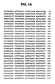
- the specification describes a substantially purified and isolated nucleic acid sequence, designated c (complementary) DNA25 ( Figures 4A and 4B ; SEQ ID NO: 26) which encodes a second melanoma recognized by tumor infiltrating lymphocytes.
- c complementary DNA25
- Figures 4A and 4B SEQ ID NO: 26
- the TIL which recognize the melanoma antigen encoded by cDNA25 are associated with in vivo tumor rejection.
- the TIL recognized the melanoma antigen encoded by cDNA25 in the context of HLA-A2.
- the cDNA25 sequence provided herein differs from the previously reported gp100 sequence in Genbank (Genbank Accession No. M32295) by two nucleotides, from the Pmel 17 sequence ( Kwon et al. (1991) Proc. Natl. Acad. Sciences (USA) 88: 9228-9232 ) by three nucleotides and a twenty one base pair deletion, and from the ME20 sequence ( Maresh et al. (1994) DNA and Cell Biology 13:87-95 ) by a single nucleotide difference.
- the protein encoded by cDNA25 differs from the gp100 in GenBank (GenBank Accession # M32295) by one amino acid at position 162, by a two amino acid difference at positions 162 and 274, compared to Pmel 17 and did not contain 7 amino acids that existed in Pmel 17 at positions 588-594. Therefore, cDNA25 appears to encode for a different form of the gene for gyp100.
- the differences between the cDNA25 nucleic acid sequence ( Figures 4A and 4B ; SEQ ID NO: 26) and amino acid sequence ( Figure 5A ; SEQ ID NO: 27) and previously reported gp100 sequences maybe due to polymorphisms, allelic variations, or to mutations within the tumor. Experiments with mouse tumors have shown that new antigens recognized by T-cells can result from point mutation in the coding region of the inactive gene ( Boon, T (1992) Advances in Cancer Research 58:177-210 ).
- This invention also provides as defined in the claims immunogenic peptides derived from gp100 protein sequences provided herein or analogs thereof. ( Figure 5A and Figure 7A ; SEQ ID NOS: 27 and 121). These immunogenic peptides represent antigenic portions of the gp100 protein ( Figures 5A and 7A ; SEQ ID NOS: 27 and 121) recognized by TIL.
- immunogenic peptides examples include, ALDGGNKHFL (peptide G10-22; SEQ ID NO: 35), VLKRCLLHL (peptide G9-19 SEQ ID NO: 36), , SVSVSQLRA (peptide G9-216; SEQ ID NO:39), LNVSLADTN (peptide G10-400 ; SEQ ID NO:41), ITDQVPFSV (peptide G9 209 , SEQ ID NO:48 ; Figure 7A ; amino acids 209 to 217) and TITDQVPFSV (peptide G10 208 ; SEQ ID NO: 49; Figure 7A ; amino acids 208 to 217).
- This invention further includes as defined in the claims analogs of these immunogenic peptides derived from gp100 amino acid sequence ( Figures 5A and 7A ; SEQ ID NOS: 27 and 121).
- the term analog includes any peptide which displays the functional aspects of these immunogenic peptides.
- the term analog also includes conservative substitution or chemical derivative of the peptides as described above.
- These immunogenic peptides may be synthetically or recombinantly produced in the same manner or fashion as described above for MART-1.
- immunogenic peptides derived from gp100 sequences are modified to increase immunogenicity by enhancing the binding of the peptide to MHC molecule with which the peptide is associated when presented to T-cells.
- modifications may include the substitution, deletion or addition, of one or more amino acids within the immunogenic peptide sequence, or insertion of amino acids within the given immunogenic peptide sequence or derivitization of existing amino acids within the given immunogenic peptide sequence or mutation of the amino acids within the given immunogenic peptide sequence.
- one amino acid is substituted or replaced in the given immunogenic peptide sequence.
- Any amino acid composing the given immunogenic peptide sequence may be modified. Any amino acid may be used to substitute or replace a given amino acid within the immunogenic peptide sequence . Modification may occur at any amino acid position within the immunogenic gp100 peptide. Modified gp100 peptides may include any modified immunogenic gp100 peptide exhibiting enhanced binding with the MHC molecule with which it is associated when presented to the T-cell.
- peptides recognized by T cells in the context of HLA-A2 are 9 to 10 amino acids in length.
- the second position and last position in the peptide are hydrophobic amino acids preferably aliphatic hydrophobic amino acids.
- the second position may be any aliphatic hydrophobic amino acid such as, but not limited to, leucine, methionine, isoleucine, valine, threonine, glycine or alanine.
- the last position of the peptide may be any aliphatic hydrophobic amino acid, such as but not limited to valine, leucine, alanine, leucine, isoleucine, glycine, methionine, valine, or threonine.
- the first and third positions of the immunogenic, peptide may be substituted or replaced with any amino acid, preferably, hydrophobic aliphatic amino acids, or aromatic amino acids.
- amino acids that may be used at the first or third position of the peptide include, but are not limited to, alanine, leucine, lysine, isoleucine, glycine, methionine, valine, threonine, tryptophan, phenylalanine, serine, lysine or tyrosine.
- gp100 peptides examples include LLDGTATLRL (peptide G10-4; SEQ ID NO: 33), VLYRYGSFSV (peptide G10-5; SEQ ID NO: 34), ALDGGNKHFL (peptide G10-22; SEQ ID NO: 35), VLKRCLLHL (peptide G9-19 SEQ ID NO: 36), VLPSPACQLV (peptide G10-8; SEQ ID NO: 37) SLADTNSLAV (peptide G10-9 ; SEQ ID NO:38), SVSVSQLRA (peptide G9-216; SEQ ID NO:39), YLEPGPVTA (peptide G9-280 ; SEQ ID NO:40), LNVSLADTN (peptide G10-400 ; SEQ ID NO:41), KTWGQYWQV (peptide G9 154 ; SEQ ID NO:46; Figure 7A ; amino acids 154 to 162), KTWGQYWQVL (peptide G10 154 ; SEQ ID NO:
- modified gp100 peptides derived from the immunogenic gp100 peptide KTWGQYWQV may have the formula X 1 X 2 X 3 GQYWQX 4 (SEQ ID NO:123) wherein:
- modified peptides are shown in Table 15.
- a preferred modified peptide is KIWGQYWQV (G9-154-2I) (SEQ ID NO:70).
- immunogenic gp100 ITDQVPFSV (G9-209; SEQ ID NO:48) may be modified, such modified peptides may have the general formula X 1 X 2 X 3 QVPFSX 4 (SEQ ID NO:124) wherein:
- modified peptides derived from ITDQVPFSV are shown in Table 16.
- the peptide FLDQVPFSV peptide G9-209-1F2L is used.
- modified gp100 peptides derived from the immunogenic gp100 peptide YLEPGPVTA may also be modified to enhance binding to MHC Class I molecules, preferably HLA-A2 or subtypes thereof.
- the modified peptides may have the general formula X 1 X 2 X 3 PGPVTX 4 (SEQ ID NO:125) wherein:
- modified peptides derived from YLEPGPVTA (G9-280; SEQ ID NO:40) are shown in Table 17.
- a preferred modified peptide is YLEPGPVTV (G9-280-9V) (SEQ ID NO:104).
- This invention further includes analogs as defined in the claims of these modified peptides derived from the gp-100 sequences disclosed herein ( Figure 5A ; SEQ ID NO:27 and FIGURE 7A SEQ ID NO:121).
- the term analog is intended to include any peptide which displays the functional aspects of these modified peptides as described above.
- These modified peptides may be synthetically or recombinantly provided by conventional methods.
- the immunogenic peptides derived from gp100 amino acid sequences or modified gp100 peptides as shown in Tables 15-17 or analogs thereof may be used as a vaccine either therapeutically or prophylactically.
- the vaccine is provided in advance of any evidence of melanoma.
- the prophylactic administration of these peptides should serve to prevent or attenuate melanoma in a mammal.
- the specification describes that, mammals, humans, at high risk for melanoma may be prophylactically treated with these vaccines.
- the vaccine may be provided therapeutically to enhance the patients own immune response to the tumor antigen prescribed on the melanoma or metastatic melanoma.
- the vaccine which acts as an immunogen, may be a cell, cell lysate from cells transfected with a recombinant expression vector carrying a nucleic acid sequences encoding gp100 immunogenic peptide or a culture supernatant containing the expressed protein. Expression vectors into which nucleic acid sequences encoding these immunogenic peptides may be introduced are the same as those described above for MART-1.
- the immunogen may be a partially or substantially purified recombinant gp100 peptide or analog thereof.
- the immunogen While it is possible for the immunogen to be administered in a pure or substantially pure form, it is intented to present it as pharmaceutical compositions, formulations or preparations as described above for MART-1. Vaccination can be conducted by conventional methods previously described above for MART-1.
- the gp100 immunogenic peptides and nucleic acids sequences encoding them may be used in bioassays, or to generate antibodies in the same manner or fashion as described above for MART-1.
- multivalent vaccines against one or more melanoma antigens may comprise all or part of the MART-1 protein peptides or modified peptides or gp100 peptides or modified peptides or combinations thereof.
- the method may comprise the following steps: (a) isolating tumor infiltrating lymphocytes from the tumor of a mammal afflicted with melanoma; (b) introducing a melanoma cDNA library into a mammalian cell line; (c) exposing said mammalian cells to said TIL; (d) screening for expression of an antigen encoded by said cDNA in said mammalian cells recognized by said TIL; and (e) isolating said cDNA corresponding to said antigen.
- Examples of cells that may be used to prepare the cDNA library used in step (b), include fresh or cultured melanoma cells.
- the cDNA library may be introduced into mammalian cells not expressing melanoma antigens. If non human mammalian cells or human cells not expressing the desired HLA haplotype for recognition by the TIL are used in step (b), such cells can be cotransfected with an HLA gene as described below.
- Examples of cells which can be used in step (b), may include tumor cell lines, such as breast cancer cell line MDA 231 (ATCC # HTB26), or COS 7 cells (ATCC #CRL 1651).
- Examples of MHC genes which can be used include HLA-A, HLA-B, and HLA-C genes, HLA-A2 and subtypes thereof ( Zemmour, J. et al. (1992) Tissue Antigens 40:221-228 ).
- the appropriate MHC gene to be used is determined by the haplotype of the tumor cells which were the source for the cDNA library. Standard methods can be used to determine the haplotype recognized by the TIL isolated ( ASHI Laboratory Manual (2nd Edition 1990 ).
- Examples of how to evaluate recognition of the cells containing the cDNA clone expressing an antigen recognized by the TIL includes, but is not limited to, ⁇ -interferon assays, TNF secretion ( Van de Bruggen et al., (1991) Science 254:1643-1647 ) or lysis of cells transfected with cDNA encoding for the recognized antigen. Such assays are performed by conventional methods known to one skilled in the art. Melanoma antigens can be isolated by or rescued by PCR using primer specific to flanking site of vector containing the cDNA. Examples of how to isolate the cDNA corresponding to the antigen recognized by the TIL may include, PCR.
- the next step is to determine the antigenic portion or epitope of the protein encoded by these genes. Therefore, in another aspect a method is described for assessing the immunogenicity of peptides derived from the predicted amino acid sequences of the gp100 protein ( Figure 5A and Figure 7A ; SEQ ID NO: 27 and SEQ ID NO: 121).
- the method may comprises the steps of: (a) preparing a plurality of peptides based on the gp100 ( Figure 5A and Figure 7A ; SEQ ID NO: 27 and SEQ ID NO: 121) amino acid sequence; (b) incubating at least one of said peptides with a mammalian cell line; (c) exposing said mammalian cells incubated with said peptide to tumor infiltrating lymphocytes (TIL); and (d) screening for recognition of TIL with said cells incubated with said peptide.
- TIL tumor infiltrating lymphocytes
- Peptides of about 25 to 5 amino acids can be used, or 20 to 10 amino acids or 9-10 amino acids.
- Examples of cells that may be used in step (b) include to, T2 cells, ( Cerundolo, V.
- T cells recognize antigen complexed with MHC Class 1 molecules.
- the MHC locus in all mammalian species contains numerous genes and is highly polymorphic. Different MHC molecules or haplotypes types bind different antigens.
- the HLA complex contains the HLA-A, HLA-3 and HLA-C gene loci which encode class I molecules. Lymphocytes will recognize tumor antigens on the context of HLA Class 1 molecule. If the cells containing e.g. the recombinant MART-1 expression vector are to be screened by the TIL but are not human cells, such as COS cells, or do not express a desired haplotype an expression vector containing an MHC Class 1 gene may also be introduced into the cells. (See Example 1) This, represents yet another alternative.
- MHC Class I genes that may be used include HLA-A, HLA-B and HLA-C genes.
- MHC specificities or restriction types include, HLA-A1, HLA-A2, such as the HLA-A2.1 subtype, or HLA-A24 ( Zemmour, J. et al. (1992) Tissue Antigens 40:221-228 ). In particular the HLA-A2.1 gene.
- compositions and therapeutic applications are also referred to by the compositions and therapeutic applications described herein.
- CTL cytotoxic T lymphocytes
- TIL501 and TIL1235 were predominantly CD8 + and were derived from the tumor specimens of patients with advanced metastatic melanoma.
- the CD8 + T cell clone, TIL501.A42, was established by limiting dilution methods and cultured with 120 IU/ml of IL-2 plus irradiated (once a week for 4 to 6 times) autologous tumor cells.
- NHEM483, NHEM493, NHEM527, NHEM529, NHEM530, NHEM533, NHEM616 and NHEM680 were purchased from Clonetics, San Diego, CA, FM725, FM801, FM902 were provided by M. Herlyn, Wistar Institute, Philadelphia PA, HA002 was provided by R. Halaban, Yale university, New Haven, CT and cultured in melanocyte growth medium (MGM, Clonetics).
- Melanoma cell lines C32, RPMI7951, WM115, A375, HS695T, Maline3M, colon cancer cell lines, Collo, SW480, WiDr, breast cancer cell lines, MDA231, MCF7, HS578, ZR75, neuroblastoma cell line, SK-N-SH, glioma cell lines, U138MG, HS683, H4, sarcoma cell line 143B, embryonal kidney cell line 293 transformed with adenovirus type 5 were purchased from ATCC, Rockville, MD. Renal cancer cell lines, UOK108 and UOK117 were provided M. Linehan NIH, Bethesda, MD.
- the small cell lung cancer cell line, H1092 was provided by J.D.Minna, University Texas Southwestern, Dallas, TX. Ewing's sarcoma cell lines, TC71, RD-ES, 6647 were provided by M. Tsokos, NIH, Bethesda, MD.
- the neuroblastoma cell line, SK-N-AS was provided by O.M. El Badry, NIH, Bethesda, MD.
- the plasmacytoma cell line, HMY-C1R and the M1 fibroblast cell line were provided by W.Biddison, NIH, Bethesda, MD. Kidney epithelial cells, KAM, WLC were provided by D. J. Hazen-Martin and D.A.Sens, Medical University of South Carolina, Charleston, SC.
- 51 Cr release assays were performed as described in Kawakami, Y., et al., (1988) J. Exp. Med. 168:2183-2191 . Briefly, 5000 target cells labeled with 51 Cr were mixed with varying numbers of effector cells and incubated for 5 hours(h). Then supernatants were collected, radioactivity was measured and the percent specific lysis was calculated.
- a cDNA library was constructed from poly A RNA from the HLA-A2 + melanoma cell line, 501mel as described in ( Miki, T., et al., (1989) Gene; 83:137-146 Miki et al. (1991) Proc. Nat. Acad. Sci. USA 88:5167-5171 ). Briefly, first strand cDNA was synthesized with a linker primer GGACAGGCCGAGGCGGCC(T) 40 (SEQ ID NO:42) followed by second strand cDNA synthesis.
- an SfiI adaptor consisting of two oligonucleotides, CCAATCGCGACC (SEQ ID NO:43) and GGTCGCGATTGGTAA (SEQ ID NO:44) was ligated to the end of the cDNA.
- the cDNA was digested with SfiI and the digested fragment was isolated by passing through a spun column.
- the cDNA was then mixed with bacteriophage ⁇ pCEV27 ( Miki, T. et al., (1991) Proc. Natl. Acad. Science (USA) 88: 5167-5771 ) vector arms prepared by SfiI digestion and in vitro packaging was performed.
- PCR Polymerase chain reaction
- the pcDNA3 containing the cloned genes were transiently transfected into the COS7 cell line by the DEAE dextran method ( Seed, B. and Aruffo, A. (1987) Proc._ Natl. Acad. Sci. (USA) 84:3365-3369 ).
- the 293 cell line was transiently transfected using lipofectamine (BRL, Gaithersburg, MD) according to the manufacturer's recommendation. After incubation, the ability of the transfected COS7 or 293 cells to mediate IFN- ⁇ release from TIL was assessed. The expression of the HLA-A2 gene was tested by flow cytometry. Stable transfectants were made by the calcium-phosphate method and individual colonies and pooled transfectants were tested for reactivity to TIL by cytotoxicity and IFN- ⁇ release assays.
- DNA sequencing of the cloned genes was performed by dideoxy chain germination method with dGTP and 7-deaza-dGTP.
- DNA and protein sequences were analyzed by the GCG program with GeneBank, and EMBL Data Library nucleotide databases and SWISS-PROT, PIR, GenPept, Brookhaven Protein Data Bank protein databases.
- Hybridization with the probe was performed according to the QuikHyb ® protocol (Stratagene) at 68 C for 2-16 h. Membranes were washed two times with 2XSSC/0.1%SDS at 60 C for 15 minutes (min) and once with 0.1XSSC at 60 C at for 30 min and then autoradiography was performed.
- TIL lines were established from KLA-A2* melanoma patients and tested for lysis of melanoma cell lines from HLA-A2* and HLA-A2* patients.
- HLA typing of patients is performed by conventional HLA typing techniques.
- HLA-A2 was selected because it is the most frequently expressed class I MHC antigen (about 50% of individuals) and has been shown to be a dominant restriction element for the recognition of melanoma antigens ( Crowley, N.J., et al. (1991), J. Immunol. 146:1692-1694 ).
- TIL501, TIL1235 and TIL1200 exhibited specific recognition of shared melanoma antigens in an HLA-A2 restricted fashion.
- TIL501.A42 was a T cell clone established from TIL501 by limiting dilution. These TIL caused lysis or released cytokines including IFN- ⁇ , TNF ⁇ and GM-CSF when cocultured with a variety of KLA-A2* melanoma or melanocyte cell lines but not HLA-A2* melanoma lines or HLA-A2* non-melanoma cell lines including the breast cancer cell line, MDA 231. Two representative experiments are shown in Table 1. Thus, these CTL seemed to recognize a non-mutated peptide derived from a melanocyte lineage specific antigen.
- a cDNA library from the HLA-A2* 501mel melanoma cell line was transfected into two highly transfectable HLA-A2.1* cancer cell lines, MDA231 and A375. These cell lines were not lysed by melanoma specific TIL but were lysed by HLA-A2 restricted influenza M1 specific CTL after incubation with the MI 58-66 peptide (GILGFVFTL; single letter code (SEQ ID NO:45) derived the influenze matrix protein or infection with a recombinant vaccinia virus containing the M1 gene (data not shown).
- GILGFVFTL single letter code
- these cell lines exhibited normal antigen processing and presenting ability but were not lysed by these melanoma-specific TIL because of the lack of expression of the relevant melanoma antigens.
- G418 After selection with G418, approximately 6700 transfected clones from each cell line were isolated and grown in microplates. Using the IFN- ⁇ release assay, 21 MDA231 and 27 A375 positive clones were isolated and rescreened. Of these clones, eight MDA231 and seven A375 clones were positive in a second screening assay.
- PCR using genomic DNA from these positive transfectants was performed with SP6 and T7 primers flanking the insert genes.
- the 1.6Kb band detected by Northern blot analysis with the cDNA 22 probe suggested that this fragment was a full length cDNA.
- Transient transfection of the expression vector pcDNA3 containing the cDNA from clones 22 or 23 into either COS7 or 293 cells along with the HLA-A2.1 gene conferred reactivity to TIL1235 and TIL501.A42 as demonstrated by the specific release of IFN- ⁇ (Table 2, Experiments 1 and 2). Stable transfection of these cDNA fragments into MDA 231 or A375mel cell lines also conferred reactivity to TIL1235 and TIL501.42 (Table 2, Experiment 3). TIL501.A42 could lyse MDA231 stably transfected with cDNA 22 (data not shown).
- the cDNA sequence of clones 22 and 23 were identical except at a single base that was believed to be a change introduced by PCR. Two other independently amplified fragments were also sequenced to clarify this region and the consensus sequence is shown in Figure 1 .
- the longest open reading frame in this gene consists of 354 bases corresponding to a 118 amino acid protein of 13kd. This sequence did not show significant similarity to any complete nucleotide or protein sequences in established databases.
- Amino acids 27-47 consist of a hydrophobic region that may contain the HLA-A2 binding peptides ( Falk, K., et al. (1991), Nature 351:290-296 ; Hunt, D.F., et al.
- TIL501.A42 and TIL 1235 When Cultured with HLA-A2 + Cell Lines Transfected with the Gene 22 or 23 Stimulator Cell Transfected Gene HLA-A2 TIL501.A42 TIL1235 Exp. 1 pg/ml 501mel none + 1009* 1076 397mel none - ⁇ 50 ⁇ 50 COS7 none - ⁇ 50 ⁇ 50 COS7 HLA-A2.1 + ⁇ 50 ⁇ 50 COS7 22 - ⁇ 50 ⁇ 50 COS7 HLA-A2.1+22 + 771 1049 Exp.
- IFN- ⁇ was secreted only when TIL were incubated with HLA-A2 + cell lines transfected with the cDNA 22 or 23.
- IFN- ⁇ secreted by TIL alone without stimulator ( ⁇ 50 pg/ml) was subtracted. *Not done Table 3.
- TIL 501.A42 which recognized the MART-1 Ag lysed 13 of 17 (76%) HLA-A2 + melanoma cell lines tested (data not shown).
- MART-1 is a previously undescribed antigen expressed on melanocyte lineage cells from skin and retina that is also expressed on melanoma cells.
- MART-1 Figure 1 ; SEQ ID NO.1
- gp100 a gene encoding melanoma antigens
- HLA-A2.1 restricted TIL HLA-A2.1 restricted TIL.
- the MART-1 antigen is a 118 amino acid proteins of approximately 13 kd. Neither the gene nor the amino acid sequence for MART-1 have been previously described.
- MART-1 RNA was expressed in 11 of 14 HLA-A2.1 positive or negative melanoma lines, and 7 of 7 melanocyte lines. With the exception of retinal tissue no MART-1 expression was found on any normal tissue tested, T-cell lines, B-cell lines, kidney epithelial lines, a fibroblast line or 19 tumor cell lines from cancers of the colon, breast, brain, kidney, lung or bone.
- MAGE-1 Another melanoma antigen, MAGE-1, has been described that is recognized by T cells derived from peripheral blood lymphocytes following repetitive in vivo or in vitro immunization ( Van Der Bruggen, et al. (1991), Science 254:1643-1647 ).
- TIL with anti melanocyte-melanoma reactivities have been administered to patients with advanced melanoma ( Rosenberg, S.A., et al. (1988), N Engl J Med 319:1676-1680 : Rosenberg S.A., J. Clin. Oncol. 10:180-199 ) and although sporadic vitiligo has been seen in these patients, no adverse ophthalmologic effects related to the possible expression of these melanocyte antigens on retinal cells has been observed.
- HLA-A2 is present in about 50% of individuals and the HLA-A2 restricted MART-1 antigen also appears to be widely expressed on melanomas, immunization with the MART-1 antigen may be particularly useful for the development of active immunotherapies.
- tyrosinase related protein (gp75) in pCEV27 plasmid was isolated from 501mel melanoma cDNA library using a probe generated by PCR based on the reported sequence of gp75 ( Cohen et al. (1980) Nucleic Acids Research 18:2807 ).
- Peptides were synthesized by a solid phase method using a Gilson AMS 422 multiple peptide synthesizer.
- the peptides were purified by HPLC on a Vydac C-4 column with 0.05% TFA/water-acetonitrile.
- TIL lysis of T2 cell lines preincubated for 2h with each peptide was measured using a 51 Cr release cytotoxicity assay.
- HLA-A2 restricted melanoma specific CTL lines and a clone, A42 were established from lymphocytes infiltrating into tumors of 10 melanoma patients.
- These TIL recognized autologous and most allogeneic fresh or cultured melanoma cells expressing HLA-A2, but did not recognize HLA-A2 - melanomas or HLA-A2 + non-melanoma cell lines ( Kawakami et al. (1992) J. Immunol 148:638 ). They also recognized HLA-A2 + normal cultured melanocytes derived from neonatal skin (see Example 1 and Kawakami, Y. et al. (1993), J. Immunotherapy 14:88 ). Thus, these TIL recognized non-mutated self-peptides derived from proteins expressed in melanoma and melanocytes in association with HLA-A2.
- TIL1200 which is a relatively oligoclonal CTL line ( Shilyansky, J. et al., (1994) Proc. Natl. Acad. Sci. (USA) 91:2829 ) did not respond to this COS transfectant.
- TIL 620, 660, 1143, 1200
- TIL1200 secreted large amounts of IFN- ⁇ compared to TIL620, 660, and 1143, suggesting that only a small subset of T-cells in these latter 3 TIL lines recognized gp100. None of these TIL recognized gp75 using this assay.
- MART-1 is a common melanoma antigen recognized by most HLA-A2 restricted TIL derived from melanoma patients.
- TIL1200 secreted IFN- ⁇ when cultured with COS7 cotransfected with cDNAs encoding MART-1 and HLA-A2.1.
- TIL620, 660, 1143 and 1200 secreted IFN- ⁇ when cultured with COS7 cotransfected with the cDNAs encoding gp100 and HLA-A2.1.
- cell line was lysed well by all 4 HLA-A2 restricted melanoma specific TIL tested when preincubated with either peptides M9-2, M10-3, or M10-4.
- Both 10 amino acid peptides, M10-3 and M10-4 contain the M9-2 sequence, with M10-3 having an additional glutamic acid at its N-terminus and M10-4 having an extra isoleucine at its C-terminal end. These peptides are located in a hydrophobic putative transmembrane domain in MART-1. The same lysis was observed when other HLA-A2 + cells incubated with these peptides were used as targets including the K4B (provided by Dr. William Biddson, NIH; Storkus. W et al. (1993) J. of Immunology 151:3719-3727 ) and 501EBVB Epstein-Barr virus transformed B cells ( Topalian et al. (1989) J.
- the peptides, M9-1, M9-2, M9-3, M10-2, M10-3, M10-4 and M10-5 were further purified and titrated in order to evaluate their relative ability to sensitize T2 cells to lysis by MART-1 reactive TIL1235 or T cell clone A42 ( Figure 2 ).
- the purified peptides M9-2, M10-3 and M10-4 were required in minimum concentrations of 1ng/ml, 100ng/ml an 1000ng/ml, respectively.
- the purified M10-4 was not recognized by TIL clone A42 even at 10ug/ml as shown in Figure 2 .
- M9-1, M9-3, M10-2, and M10-5 peptides were not recognized by either A42 or TIL1235.
- TIL620 and TIL1088 demonstrated low level of non-specific lysis of T2 cells without peptides or after the addition of irrelevant peptides, but showed significant increase of lysis of T2 cells preincubated with M9-2, M10-3, and M10-5 peptides.
- the recognition of M10-4 differed among the TIL, but was similar to the different reactivity to M10-4 by the T-cell clone A42 or the T-cell line TIL1235 ( Figures 2A and 2B ). Higher concentrations (1ug/ml) of M10-4 were required for lysis than were required for M9-2 or M10-3.
- TIL and clone A42 also secreted cytokines including IFN- ⁇ , GM-CSF and TNF- ⁇ when incubated with T2 cells preincubated with M9-2 or M10-3 (data not shown). Therefore, M9-2 or M10-3 are common epitopes recognized by a majority of HLA-A2 restricted melanoma specific TIL. Table 6.
- MART-1 is a 118 amino acid protein containing a single transmembrane domain and is expressed in most melanoma cells as well as cultured melanocytes and retina similar to the expression pattern of the DNA for a form of gp100 described in Example 3.
- the gp100 is recognized by 4 of 10 TIL.
- peptide M9-2 most effectively sensitized T2 cells for lysis ( Figure 2 ) suggesting that this peptide may be naturally processed and presented on tumor cells.
- the T-cells recognizing M9-2 may react with peptide M10-3 or M10-4 because the latter 10-mer peptides contain the 9 amino acid sequence of peptide M9-2.
- M10-4 was poorly recognized by the T-cell clone A42, but was well recognized by some TIL lines, although a higher concentration of M10-4 was necessary to observe the lysis.
- TIL lines may contain different T-cell clones which only recognize either M9-2 or M10-4.
- Peptides M10-3 and M10-4 may also be naturally processed and presented by tumor cells.
- the existence of multiple melanoma antigens presented by HLA-A2 has previously been suggested by analyzing the recognition of melanoma cell clones by a variety of T-cell clones ( Knuth, A. et al. (1989), Proc. Natl. Acad. Sci. (USA) 86:2804 , Wolfel, T. et al., 1989 J . Exp. Med.
- the cDNA25 clone encoding a form of the melanoma antigen designated gp100 was cloned by techniques similar to those described in Example 1 and in Miki, T., et al. (1991) Proc. Natl. Acad. Sci. (USA) 88:5167-5171 . Briefly, a breast cancer cell line, MDA231 (ATCC #HTB26), transfected with a cDNA library in ⁇ pCEV27 made from the 501mel melanoma cell line was screened for antigen positivity by measuring interferon- ⁇ (IFN- ⁇ ) secretion when cocultured with TIL1200.
- TIL1200 was generated as described in Kawakami, Y., (1988), J. Exp. Med. 168, 2183-2191 .
- the integrated cDNA was recovered from the genomic DNA of positive transfectants by PCR and cloned into the mammalian expression plasmid pCDNA3 (Invitrogen, San Diego. CA).
- the full length cDNA for cDNA25 was isolated from the 501mel ⁇ pCEV27 library using the cDNA25 probe.
- the ⁇ phage containing the full length cDNA25 was digested with XhoI, and then self-ligated with T4 DNA ligase to make the plasmid pCEV27-FL25.
- a full length cDNA25 isolated by PCR using the specific primers designed for gp100 was cloned-in pCRII(Invitrogen), and then cloned intopcDNA3(pcDNA3-FL25).
- pcDNA3-FL25 a full length cDNA25 isolated by PCR using the specific primers designed for gp100
- DNA sequence of the plasmid clone pCEV27-FL-25 was determined with an automated DNA sequencer (Model 373A; Applied Biosystems, Inc.), using the Taq DyeDeoxy terminator cycle sequencing kit (Applied Biosystems, Inc.) using the manufacturer's instructions.
- Peptides were synthesized by a solid phase method using a Gilson AMS 422 multiple peptide synthesizer.
- the peptides were purified by HPLC on a Vydac C-4 column with 0.05% TFA/water-acetonitrile.
- TIL lysis of T2 RET-cells preincubated with peptides for 2 hour (h) was measured using a 51 Cr release cytotoxicity assay.
- TIL 1200 Treatment of a patient with metastatic melanoma using TIL 1200
- a 29 year old male patient, designated patient number 1200, with a widely metastatic melanoma who had previously failed chemotherapy and radiation therapy was treated witt a single preparatory dose of 25 mg/Kg cyclophosphamide followed by the intravenous infusion of 1.6 x 10 11 TIL (including 9.1 x 10 9 Indium-111 labeled TIL) plus 7 doses of IL-2 art 720,000 IU/Kg given every 8 hours.
- a second cycle of treatment with TIL and IL-2 was given three weeks later. Radionuclide scans showed localization of TIL in tumour deposits ( Figure 3A ).
- Biopsy of subcutaneous tumors on days 8 and 11 after treatment showed significant localization of TIL to tumor (ratios of injectate per grat in tumor compared to normal tissue were 14.9 and 14.0 respectively).
- the patient's cancer regressed rapidly following the first course of treatment.
- 2 of 3 liver lesions had disappeared and a third lesion shrank by 50%.
- Multiple subcutaneous metastases regressed completely as shown in Figure 3B (the product of perpendicular diameters of individual lesions are shown).
- TIL lines established from HLA-A2 + melanoma patients lysed melanoma cell lines in a class I MHC-restricted fashion ( Kawakami, Y., et al. (1992) J. Immunol, 146; 638-643 ), and were shown to release IFN ⁇ , tumor necrosis factor-alpha (TNF ⁇ ) or granulocyte-macrophage colony stimulating factor (GM-CSF) when cocultured with the same tumor cell lines ( Hom, S.S., et al. (1993) J. Immunother. 13; 18-30 ).
- TNF ⁇ tumor necrosis factor-alpha
- GM-CSF granulocyte-macrophage colony stimulating factor
- a CD8 + CTL line, TIL1200 established from a metastatic subcutaneous tumor mass of patient 1200, lysed fresh autologous melanoma cells as well as 10 of 15 HLA-A2 + allogeneic melanoma cell lines, but did not lyse 16 of 18 HLA-A2 - melanoma cell lines or 6 of 8 HLA-A2 - non-melanoma cell lines ( Shilyansky, J., et al. (1993) Proc. Natl. Acad. Sci. USA, 91, 2829-2833 , unpublished data).
- Table 7 shows a cytotoxicity assay against 5 representative HLA-A2 + melanoma cell lines that were lysed by TIL1200, 4 representative HLA-A2 + melanoma cell lines that were not lysed by TIL1200, and one HLA-A2 - melanoma cell line.
- TIL1200 also secreted IFN- ⁇ when cocultured with HLA-A2 + normal cultured melanocytes established from neonatal foreskin as well as HLA-A2 + melanoma cell lines (Table 8). Therefore, TIL1200 appeared to recognize a non-mutated self peptide expressed in most melanomas and cultured neonatal melanocytes in an HLA-A2 restricted fashion.
- G418 resistant cells were selected and approximately 6700 individual transfectants from each cell line were isolated and screened based on their ability to stimulate IFN- ⁇ secretion from TIL1200.
- TIL1200 Specificity of Antigen Recognition by TIL1200: Recognition of HLA-a-a2 + Neonatal Melanocytes Stimulator HLA-A2 TIL1200 TIL888 (pgIFN- ⁇ /ml) 501mel + 562 0 624mel + 439 0 397mel - 0 0 888mel - 0 1970 NHEM493 - 441 0 NHEM527 + 418 0 NHEM530 + 164 0 NHEM616 + 53 0 FM725 + 107 0 FM801 + 250 343 THEM483 - 0 0 NHEM680 - 0 0 HA002 - 0 0 The IFN- ⁇ secretion by TIL was measured by ELISA as previously described in Example 1.
- TIL888 is a class I MHC restricted melanoma specific CTL, not restricted by HLA-A2.
- NHEM, FM, and HA refer to normal cultured melanocyte cell lines, all others are melanoma cell lines.
- Table 9 Transfection of cDNA 25 into A375 and COS7 Stimulator cells Transfected genes HLA-A2 Secretion by TIL1200 Exp. 1 (pgIFN- ⁇ /ml) 501 mel none + 987 397mel none - 0 A375 none + 0 A375 pcDNA3-25 + 230 Exp.
- pcDNA3 containing full length cDNA25 (pcDNA3-FL25) or pCEV27 containing full length cDNA25 (pCEV27-FL25) (Exp. 3) along with peDNA3 containing HLA-A2.1 (HLA-A2.1).
- HLA-A2 expression was determined by flow cytometry and interferon-gamma secretion was measured by ELISA.
- the cDNA 25 differed from the gp100 in GenBank (Accession No. M77348, also known as gp95) by two nucleotides, from the PMEL 17 sequence ( Kwon et al. (1991) Proc. Natl. Acad. Sciences (USA) 58: 9228-9232 ) by 3 bases and a 21 base pair deletion. ( Figure 5B ).
- the full length cDNA25 (FL25) was isolated in two plasmids, pCEV27-FL25 or pCDNA3-FL25. Transfection of either plasmid into COS7 along with pcDNA3-HLA-A2.1 conferred to COS7 the ability to induce IFN- ⁇ secretion by TIL1200.
- the amount of IFN- ⁇ secretion stimulated by COS7 transfected with the full length DNA plus HLA-A2.1 was similar to that stimulated by 501mel and was higher than that stimulated by COS7 transfected with the truncated cDNA25 possibly due to improved translation starting at the normal AUG initiation codon (Table 9, Exp. 2 and 3).
- the 5' region missing from the truncated cDNA25 may contain other epitopes recognized by clones in TIL1200.
- nucleotide and corresponding amino acid sequences of the truncated cDNA 25 and the full length cDNA25 cloned from the 501mel cDNA library by screening with the cDNA25 probe were compared with the GenBank sequences of Pmel17 isolated from normal melanocytes and gp100 isolated from the melanoma cell line MEL-1. ( Figure 5B ).
- the full length cDNA25 differed from the gp100 amino acid sequence at position 162. This amino acid difference is possibly caused by polymorphism or mutation in the tumor.
- cDNA25 had 2 amino acid differences at positions 162 and 274, compared to Pmel17 and did not contain 7 amino acids that existed in Pmel17 at positions 588-594.
- the amino acid sequence of the truncated cDNA25 that was isolated from the original MDA231 transfectant has a different sequence at the 3' end (from position 649 to the end) due to a frame shift caused by one extra cytidylic acid. It is not clear whether this difference was due to a true allelic difference or to la mutation that occurred during manipulation of the DNA. Nevertheless, TIL1200 appeared to recognize non-mutated peptides located between position 236 and 648.
- cDNA25 also had 87% similarity in amino acid sequence to cDNA RPE1 ( Kim, R., and Wistow, G.J. (1992) Exp. Eye Res.
- a gp100 protein was known to be recognized by monoclonal antibody HMB45 ( Adema et al., (1993) Am. J. Pathology, 143: 1579-1585 ).
- COS7 cells transfected with the full length cDNA25 were evaluated by flow cytometry using this monoclonal antibody. After transient expression of either pCEV27-FL25 or pcDNA3-FL25, COS7 expressed the antigen detected by HMB45 (data not shown).
- T-cell T-cell
- B-cells 501EBVB, 836EBVB
- fibroblast M1
- 20 non-melanoma tumor cell lines colon cancer, Collo, SW480, WiDr; breast cancer, MDA231, MCF7, HS578, ZR75; neuroblastoma, SK-N-AS, SK-N-SH; Ewing sarcoma, TC75, RD-ES, 6647; sarcoma 143B
- glioma U138MG, HS683
- renal cell cancer UOK108, UOK117, small cell lung cancer, H1092
- Burkitt's lymphoma Daudi
- myeloma HMY myeloma HMY
- this gene appeared to be specifically expressed in melanocyte lineage cells, consistent with the expression pattern of previously isolated forms of gp100 when analyzed using monoclonal antibodies, HMB45, NKI/betab, or HMB-50 ( Adema, G.J., et al. (1993) Am J Pathology 143: 1579-1585 ; Gown, A.M., et al., (1986) Am J Pathol 123:195-203 ; Colombari, R., et al. (1988) Virchows Archiv A Pathol Anat. 413:17-24 ); Vennegoor, C., et al. (1988) Am. J. Pathol.
- TIL TIL 1200
- TIL TIL1200
- TIL1200 which was a TIL line containing multiple CTL species recognized a tumor antigen in the context of HLA-A2, which is the most frequently expressed class I MHC antigen (about 50% of individuals) and has been shown to be a dominant restriction element for the induction of melanoma specific CTL. ( Crowley, N.J., et al. (1991) J. Immunol. 146, 1692-1699 ).
- a cDNA encoding another form of gp100 was also isolated by screening with a rabbit polyclonal antiserum against gp100 ( Adema, G.J., et al. (1993) Am J Pathology 143:1579-1585 ) and TIL1200 also lysed HLA-A2 + cell lines transfected with this cDNA clone ( Bakker, A.B.H. et al. (1994) J. Exp. Med. 179:1005-1009 ).
- T cells reactive to the self-antigen gp100 in tumors and the possible enrichment of these T cells at the tumor site as a possible consequence of the specific accumulation and expansion of antigen reactive cells ( Sensi, M. et. al. (1993) J. Exp. Med 178:1231-1246 ) raises important questions about the nature of the immune response to self antigens on growing cancers and about the mechanisms of immunologic tolerance to self-antigens.
- gp100 on melanoma cells relative to that in melanocytes demonstrated by Northern blot analysis or the unique inflammatory conditions that might exist at the tumor site, which may be associated with the secretion of cycokines and expression of costimulatory molecules on the cell surface, could break tolerance to gp100.
- Depigmentation has been reported to be associated with a good prognosis ( Nordlund, J.J., et al. (1983) J. Am. Acad. Dermatol. 9:689-695 ; Bystryn, J-C, et al. (1987) Arch.
- the gp100 protein ( Figure 5A ; SEQ ID NO: 27) and the ten amino acid peptide identified may represent a human tumor rejection antigen since the transfer into patient 1200 of TIL1200 plus IL2 was associated with cancer regression.
- the traffic of TIL1200 to tumor deposits in vivo and the rapidity of the antitumor response are characteristics of the response to TIL therapy, although IL2 may also have been involved in the tumor rejection.
- Adoptive transfer of 3 other TIL lines which recognized gp100 as well as MART-1 also mediated tumor regression (data not shown).
- Tyrosinase ( Brichard, V., et al. (1993) J. Exp. Med. 178, 489-495 ) and MART-1 (see Example 1) have been identified as melanoma antigens recognized by HLA-A2 restricted CTL.
- Another antigen, MAGE-1 is recognized by HLA-A1 restricted melanoma-specific CTL and is expressed on a variety of cancer cells as well as testis ( Van Der Bruggen, P. et al. (1991) Science, 254:1643-1647 ).
- HLA-A2 restricted TIL none of the ten HLA-A2 restricted TIL recently developed appeared to recognize MAGE-1 ( Zakut, R., et al. (1993) Cancer Res. 53: 5-8 ).
- gp100 proteins in melanomas the recognition of a peptide by T cells infiltrating into tumor, its restriction by HLA-A2, present in 50% of individuals, and the association of anti gp100 reactivity with cancer regression in patient 1200 imply that the gp100 antigen in particular the novel immunogenic peptides derived from the gp100 amino acid sequence ( Figure 5A ; SEQ ID NO: 27) may be particularly useful for the development of active immunotherapies for patients with melanoma.
- Peptides were synthesized by a solid phase method using a peptide synthesizer (model AMS 422;Gilson Co.Inc., Worthington, OH) (>90% purity).
- the peptides to be synthesized were selected from the reported human sequence of gp100 based on HLA-A2.1 binding motifs ( Falk, K., (1991) Nature 351:290 ; Hunt, D. F., et al, (1992) Science 255:1261 ; Ruppert, J., et al., (1993) Cell 74:929 ; Kubo, RT, et al. (1994) J Immunol. 152:3913 ).
- Possible epitopes identified in the first screening were further purified by HPLC on a C-4 column (VYDAC, Hesperia, CA) (>98% purity) and the molecular weights of the peptides were verified by mass spectrometry measurement as previously described (Example 3; Kawakami, Y., et al., (1994) J.Exp.Med. 180-:347 ; Kawakami, Y., et al., (1994) Proc Natl Acad Sci USA) 91:6458 ).
- Soluble HLA-A2.1 heavy chain, human beta 2-microglobulin, radiolabeled peptide HBc 18-27 (FLPSDYFPSV) and various concentrations of the sample peptides were coincubated in the presence of protease inhibitors for 2 days at room temperature as previously described ( Ruppert, J., et al., (1993) Cell 74:929 ; Kubo, RT, et al., (1994). J Immunol. 152:3913 ; Sett A., et al., (1994). Molecular Immunol. 31:813 ).
- the percentage of labeled peptide bound to HLA-A2.1 was calculated after separation by gel filtration and the concentration of the sample peptide necessary, to inhibit 50% of the binding of the labeled peptide was calculated.
- the relative affinity of peptides to HLA-A2.1 were also calculated as a ratio (concentration of the standard HBc 18-27 peptide to inhibit 50% of the binding of the labeled peptide / concentration of the sample peptide to inhibit 50% of the binding of the labeled peptide) as previously described ( Sett A., et al., (1994) Molecular Immunol. 31:813 ).
- Peptide binding was defined as high (50% inhibition at ⁇ 50nM, ratio >0.1), intermediate (50-500nM, ratio 0.1-0.01) or weak (>500nM, ratio ⁇ 0.01) ( Ruppert, J., et al., (1993) Cell 74:929 ; Kubo, RT, et al., (1994) J Immunol. 152:3913 ; Sett A., et al., (1994) Molecular Immunol. 31:813 ).
- the pcDNA3 plasmid containing the full length gp100 cDNA (Example 3; Kawakami, Y., et al., (1994)). Proc Natl Acad Sci (USA) 91:6458 ) was digested with Xho I and Xba I. After incorporation of alpha-phosphorothioate deoxynucleoside triphosphate into the Xba I site, a standard exonuclease III nested deletion was performed using the Exo Size Deletion Kit (New England Biolabs, inc., Beverly, MA). The deleted clones were self-ligated and amplified. The exact deletion for each clone was conformed by DNA sequencing.
- pcDNA3 plasmids (Invitrogen, San Diego CA) containing the cDNA fragments (D3, D5, D4, C3) generated by the sequential deletion with exonuclease from the 3' end of the full length gp100 cDNA as well as the truncated gp100 cDNA lacking the 5'-coding region (25TR) (Example 63; Kawakami, Y.
- a 51 Cr release assay or an IFN- ⁇ release assay were performed as previously described (Examples 1 and 2; Kawakami, Y., et al., (1994)). Proc Natl Acad Sci (USA) 91:3515 ; Kawakami, Y., et al., (1988). J. Exp. Med. 168:2183 ). Either COS7 cells transfected with cDNA encoding melanoma antigens and HLA-A2.1 cDNA, or T2 cells preincubated with peptides were used as stimulators for the IFN- ⁇ release assay. T2 cells pulsed with peptides were also used as targets for cytotoxicity assays ( Kawakami, Y., (1994) J._Exp, Med. 180:347 ).
- TIL620 (620-1, 620-2) or TIL660 (660-1, 660-2, 660-3) were grown from the TIL culture that was administered into the autologous patient, but they were separately cultured and had slightly different specificities likely due to the in vitro expansion of different clones.
- G9 209 (ITDQVPFSV) (SEQ ID NO:4B)
- G10 201 (TTTDQVPFSV) (SEQ ID NO:49), which has an extra threonine at the N-terminus of G9 209 , were recognized only by TIL620.
- G9 154 (KTWGQYWQV) (SEQ ID NO:46) and G10 154 (KTWGQYWQVL) (SEQ ID NO:47), which has an extra leucine at the C-terminus of G9 154 , were recognized by TIL1200, TIL620-2 and TIL660-2.
- G10-4 (LLDGTATLRL) (SEQ ID NO:33) was recognized by TIL1200 as demonstrated (Example 3).
- the peptide G9 280 (YLEPGPVTA) (SEQ ID NO:40) was recognized by TIL660 and TIL1143.
- TIL660-3 also recognized G10-5 (VLYRYGSFSV) (SEQ ID NO:34) as well as G9 280 . Lysis of T2 cells preincubated with G10-5 was repeatedly low, possibly because a small subset of T-cell clones was specific for this epitope.
- TIL 1200 recognized COS cells transfected with the fragments, 25TR, D5, D4 or C4, but not with D3, suggesting that at least 2 epitopes existed in the regions of amino acid residues 146-163 and 236-661.
- G9 154 and G10 154 were the only peptides which contained HLA-A2.1 binding motifs in the region 146-163 and both were recognized by TIL1200.
- G10-4 was located in the region 236-661 and was recognized by TIL1200.
- TIL620-1 recognized COS cells transfected with C4 but not with D3, D5, D4 or 25TR, suggesting that the epitope existed within residues 187-270.
- G9 209 and G10 208 which were recognized by TIL620-1 were located in this region.
- TIL620-2 another subculture of TIL620, also recognized COS cells transfected with D5 and D4, but not D3, and recognized G9 154 and G10 154 in the region 147-163, also recognized by TIL1200.
- TIL660-1 and TIL1143 recognized COS cells transfected with C4 or 25TR, but not with D3, D5, or D4, suggesting that epitopes existed in the 2 regions 187-270 and 236-661.
- G10-5 appeared to be inhibitory to the cytotoxic activity of CTL at concentration greater than 10ng/ml since lysis of T2 cells incubated with G10-5 at more than 10ng/ml was repeatedly lower than at 1-10ng/ml in this assay condition in which the peptide was present in the medium during entire 4h cytotoxicity assay ( Figure 8D ).
- the relative binding affinity of these epitopes to HLA-A2.1 was also measured using an in vitro competitive binding assay (Table 13).
- G9 154 had an higher binding affinity (50% inhibition of the standard peptide at 11nM) to the HLA-A2.1 molecule than G10 154 (1010nM) which contains an extra leucine at the C-terminus of G9 154 , and could sensitize T2 cells at lower concentrations than G10 154 ( Figure 8A ).
- G9 209 also bound to HLA-A2.1 with higher affinity (84nM) than G10 208 (2080nM) which contains an extra threonine, at the N-terminus, and could sensitize T2 cells at lower concentrations of peptide than G10 208 ( Figure 8B ).
- the 9-mer peptides were superior to the corresponding 10 mer peptides in the sensitization of T2 cells to CTL lysis, and they also had higher binding affinities to HLA-A2.1. This was also the case for the identified MART-1 9 and 10 amino acid peptides (M9-2, M10-3, M10-4) (Example 2; Kawakami, Y., et al., (1994). J. Exp. Med. 180:347 ). The results of the peptide titration in the T2 cell lysis assay correlated with the results of the HLA-A2.1 binding affinity as measured by the in vitro binding assay.
- the other gp100 epitopes, G9 280 , G10-4, or G10-5 had binding affinities for HLA-A2.1 with 50% inhibition at 95nM, 483nM, or 13nM, respectively.
- the HLA-A2.1 binding affinities of the previously identified HLA-A2 restricted melanoma epitopes in MART-1 (Example 2; Kawakami, Y., et al., (1994) J.Exp.Med. 180:347 ) and tyrosinase ( Wolfel, T., (1994) Eur.J. Immunol.
- the MART-1 peptide M9-2 may also be designated M9 27
- the MART-1 peptide M10-3 may also be designated M10 26
- the MART-1 peptide M10-4 may also be designated M10 27
- the gp100 peptide G10-4 may also be designated G10 457 and the gp100 peptide G10-5 may also be designated G10 476 .
- G9 154 (KTWGQYWQV), G10 154 (KTWGQYWQVL), G9 209 (ITDQVPFSV) GID 208 (TITDQVPFSV), G9 280 (YLEPGPVTA), G10 - 4 LLDGTATLRL), G10-5 (VLYRYGSFSV) at 1ug/ml (* 1ng/ml), was measured by 4h- 51 Cr release assays.
- TIL620-1, -2 or TIL660-1, -2, -3 were grown from the same TIL which was administered into the autologous patient, but were separately cultured.
- Peptides were synthesized by a solid phase method using a multiple peptide synthesizer and purified by HPLC, as previously described ( Rivoltini, L et al. (1995) Journal of Immunology Volume 154:2257-2265 ).
- test peptides ranging from 100 ⁇ M to 1 nM
- 5nM radiolabeled Hbc 18-27 FLPSDYFPSV
- HLA-A2.1 heavy chain and ⁇ 2-microglobulin 5nM radiolabeled Hbc 18-27
- the percentage of MHC-bound radioactivity was determined by gel filtration and the 50% inhibitory dose was calculated for each peptide.
- PBMC peripheral blood of HLA-A2+ melanoma patients and normal donors by centrifugation on Ficoll-Hypaque gradients and used as fresh or cryopreserved samples.
- Peptide specific CTL lines were generated as follows: at day 0, PBMC were plated at a concentration of 1.5x10 6 /ml in 24-well plates (2ml/well) in Iscove's medium containing 10% human AB serum, L-glutamine, antibiotics (CM) and in the presence of 1 ug/ml peptide. Two days later, 12 IU/ml interleukin 2 (IL-2) (Chiron Co., Emeryville, CA) were, added to the cultures.
- IL-2 interleukin 2
- Lymphocytes were then restimulated weekly as follows: responder cells were harvested, washed once and replated in 24-well plates at a concentration of 2.5x10 5 cells/ml in CM. Autologous PBMC were thawed, washed twice in PBS, resuspended at 5-8x10 6 cells/ml in CM and pulsed with 1ug/ml peptide in 15-ml conical tubes (5ml/tube) for 3 hours at 37°C. These PBMC (stimulators) were then irradiated at 3000 rads, washed once in PBS and added to the responder cells at responder:stimulator ratios ranging between 1:3 and 1:10.
- IL-2 12 IU/ml IL-2 were added to the cultures.
- the activity of these CTL was tested by cytotoxicity assays after at least 2 rounds (14 days) of peptide stimulation.
- the dissociated tumor suspension were cultured for 1-2 days in 10% FCS RPMI-1640 medium to allow tumor cell adherence.
- the lymphocytes, recovered from the non-adherent fraction, were used for the induction of peptide specific CTL as described above.
- 51 Cr release cytotoxic assays were performed to detect the recognition of peptide and melanoma cells by CTL.
- T2 cell lines were preincubated for 2 h at 37°C with 1ug/ml peptide, washed and used as target cells in a 51 Cr release cytotoxic assay.
- the melanoma lines 624mel was established in our laboratory (See Example 1).
- HLA-A2.1 binding peptides contained leucine or methionine at the 2and major anchor position and valine at the last major anchor position (dominant anchor amino acids) and had high binding affinity to HLA-A2.1, the isolated MART-1 or gp100.
- melanoma epitopes contain non-dominant amino acid at major anchor position such as alanine (the 2nd position of M9-2, the 9th position of G9-280) and threonine (the 2nd position of G9-154 and G9-209).
- the M9-2, G9-209 and G9-280 are not high affinity binders.
- modified M9-2 G9-280, G9-209, G9-154 peptides, M9-2-2L, M9-2-1F. M9-2-3Y, G9-280-9V, G9-280-9L. G9-280-9I.
- G9-280-1F, G9-209-2L, G9-209-2M, G9-209-2I, G9-209-1F, G9-209-1Y, G9-209-1W2L, G9-209-1F2L, G9-209-1Y2L have higher binding affinity and were recognized by the original melanoma reactive T-cells. (Tables 14, 15, 16 and 17) PBL stimulated with autologous PBMC pulsed with G9-154-2I.
- G9-209-1F2L, or G9-280-9V recognized and lysed not only the original epitopes but also melanoma tumor cells (624mel) better than PBL stimulated with natural epitopes (G9-154, G9-209, G9-280).
- modified peptides could be used for induction of anti-tumor T-cells instead of natural epitopes.
- Other peptides which were not recognized by the particular T-cells used in our study, but have higher binding affinity to HLA-A2.1 may induce a different set of T-cells capable of recognizing the original melanoma epitopes in in vitro or in vivo .
- These modified peptides may be used for induction of anti-melanoma T-cells in vitro and immunization of patients for the treatment of patients with melanoma or for the prevention of melanoma.
- Table 14 14.
- Peptides are defined as high (50 % inhibition at ⁇ 50nM), intermediate (50-500nM) and weak (> 500nM) binding peptides. (see example 4) Table 17. Modified gp100 G9-280 peptides Peptide Sequence Binding affinity to HLA-A2.1 (nM) Recognition by G9-280 reactive T-cells G9-280 parent YLEPGPVTA (SEQ ID NO:40) 455 + G9-280-9V YLEPGPVTV (SEQ ID NO:104) 48 + G9-280-9L YLEPGPVTL (SEQ ID NO:105) 88 + G9-280-9I YLEPGPVTI (SEQ ID NO:106) 65 + G9-280-1F FLEPGPVTA (SEQ ID NO:107) 125 + G9-280-1W WLEPGPVTA (SEQ ID NO:108) 833 + G9-280-3Y YLYPGPVTA (SEQ ID NO:109) 17 -
- MART-1 vaccines may be efficacious in treating mammals afflicted with melanomas.
- MART-1 vaccines may be administered to individuals.
- Mammals can be immunized with the MART-1 proteins, peptides or modified peptides described herein in ranges of about 1 mg-to about 100mg.
- mammals, preferably humans may be immunized with the MART-1 nucleic acid sequence inserted into a viral vector, such was vaccinia virus, adenovirus or fowl pox virus
- a range of about 10 6 to about 10 11 viral particles carrying the MART-1 nucleic acid sequences corresponding to immunogenic MART-1 peptides or modified peptides or analogs thereof, may be administered per mammal, preferably a human.
- the mammals will be monitored for antibodies to the immunogen or increase in cytotoxic lymphocytes (CTL) recognizing the immunogen by conventional methods or alleviation of clinical signs and symptoms of the active disease.
- CTL cytotoxic lymphocytes
- Specific parameters to be assessed include production of immune cells that recognize the vaccine antigen or tumor regression.
- Such vaccines may be administered either prophylactically or therapeutically.
- Mammals may also be immunized with the gp-100 nucleic acid sequence inserted into a retroviral vector or GP-100 immunogenic peptides or modified peptides or analogs thereof.
- Suggested dose ranges of the antigen in retroviruses that may be used are about 10 6 to about 10 11 viral particles per mammal, preferably a human. Response and efficacy of the retroviral vaccines will be assessed as described above.
- T-lymphocytes presensitized to the melanoma antigen may be effective in therapeutically treating mammals afflicted with melanoma.
- the T-lymphocytes will be isolated from peripheral blood lymphocytes or tumor infiltrating lymphocytes and exposed in vitro to the MART-1 protein or peptide.
- T-lymphocytes are isolated from peripheral blood or melanoma tumor suspensions and cultured in vitro ( Kawakami, Y. et al. (1988) J. Exp. Med. 168: 2183-2191 ).
- the T-lymphocytes are exposed to the MART-1 peptide AAGIGILTV for a period of about to 1-16 hours at a concentration of about 1 to about 10mg/ml.
- T-lymphocytes exposed to the antigen will be administered to the mammal, preferably a human at about 10 9 to about 10 12 lymphocytes per mammal.
- the T-lymphocytes may be exposed to the modified MART-1 peptides.
- the lymphocytes may be administered either intravenously, intraperitoneally or intralesionally. This treatment may be administered concurrently with other therapeutic treatments such as cytokines, radiotherapy, surgical excision of melanoma lesions and chemotherapeutic drugs, adoptive T lymphocyte therapy.
- the T-lymphocytes may be exposed to the gp100 immunogenic peptides or modified immunogenic peptides described herein.
Landscapes
- Health & Medical Sciences (AREA)
- Life Sciences & Earth Sciences (AREA)
- Chemical & Material Sciences (AREA)
- General Health & Medical Sciences (AREA)
- Immunology (AREA)
- Veterinary Medicine (AREA)
- Public Health (AREA)
- Animal Behavior & Ethology (AREA)
- Epidemiology (AREA)
- Medicinal Chemistry (AREA)
- Organic Chemistry (AREA)
- Molecular Biology (AREA)
- Microbiology (AREA)
- Pharmacology & Pharmacy (AREA)
- Engineering & Computer Science (AREA)
- Biochemistry (AREA)
- Mycology (AREA)
- Oncology (AREA)
- Proteomics, Peptides & Aminoacids (AREA)
- Hematology (AREA)
- Genetics & Genomics (AREA)
- Toxicology (AREA)
- Gastroenterology & Hepatology (AREA)
- Zoology (AREA)
- Biomedical Technology (AREA)
- Urology & Nephrology (AREA)
- Biophysics (AREA)
- Physics & Mathematics (AREA)
- Chemical Kinetics & Catalysis (AREA)
- Biotechnology (AREA)
- Food Science & Technology (AREA)
- Nuclear Medicine, Radiotherapy & Molecular Imaging (AREA)
- Analytical Chemistry (AREA)
- General Physics & Mathematics (AREA)
- Pathology (AREA)
- General Chemical & Material Sciences (AREA)
- Cell Biology (AREA)
- Bioinformatics & Cheminformatics (AREA)
- Peptides Or Proteins (AREA)
- Micro-Organisms Or Cultivation Processes Thereof (AREA)
Description
- This invention is in the field of prevention and treatment of human cancers. More specifically, this invention relates to genes encoding melanoma antigens recognized by T-Cells and their corresponding proteins and to preventative, diagnostic and therapeutic applications which employ these genes or proteins.
- Melanomas are aggressive, frequently metastatic tumors derived from either melanocytes or melanocyte related nevus cells ("Cellular and Molecular Immunology" (1991) (eds) Abbas A.K., Lechtman, A.H., Pober, J.S.; W.B. Saunders Company, Philadelphia: pages 340-341). Melanomas make up approximately three percent of all skin cancers and the worldwide increase in melanoma is unsurpassed by any other neoplasm with the exception of lung cancer in women ("Cellular and "Molecular immunology" (1991) (eds) Abbas, A.K., Lechtiman, A.H., Pober, J.S.; W.B. Saunders Company Philadelphia pages: 340-342; Kirkwood and Agarwala (1993) Principles and Practice of Oncology 7:1-16). Even when melanoma is apparently localized to the skin, up to 30% of the patients' will develop systemic metastasis and the majority will die. (Kirkwood and Agarwala (1993) Principles and Practice of Oncology 7:1-16). Classic modalities of treating melanoma include surgery, radiation and chemotherapy. In the past decade immunotherapy and gene therapy have emerged as new and promising methods for treating melanoma.
- T cells play an important role in tumor regression in most murine tumor models. Tumor infiltrating lymphocytes (TIL) that recognize unique cancer antigens can be isolated from many murine tumors. The adoptive transfer of these TIL plus interleukin-2 can mediate the regression of established lung and liver metastases (Rosenberg, S.A., et al., (1986) Science 233:1318-1321). In addition, the secretion of IFN-γ by injected TIL significantly correlates with in vivo regression of murine tumors suggesting activation of T-cells by the tumor antigens. (Barth, R.J., et al., (1991) J. Exp. Med. 173:647-658). The known ability of tumor TIL to mediate the regression of metastatic cancer in 35 to 40% of melanoma patients when adoptively transferred into patients with metastatic melanoma attests to the clinical importance of the antigens recognized (Rosenberg, S.A., et al., (1988) N Engl J Med 319:1676-1680; Rosenberg S.A. (1992) J. Clin. Oncol. 10:180-199).
- T cell receptors on CD8+ T cells recognize a complex consisting of an antigenic peptide (9-10 amino acids for HLA-A2), ß-2 microglobulin and class I major histocompatibility complex (MHC) heavy chain (HLA-A, B, C, in humans). Peptides generated by digestion of endogenously synthesized proteins are transported into the endoplastic reticulum, bound to class I MHC heavy chain and ß2 microglobulin, and finally expressed in the cell surface in the groove of the class I MHC molecule. Therefore, T cells can detect molecules that originate from proteins inside cells, in contrast to antibodies that detect intact molecules expressed on the cell surface. Therefore, antigens recognized by T cells may be more useful than antigens recognized by antibodies.
- Strong evidence that an immune response to cancer exists in humans is provided by the existence of lymphocytes within melanoma deposits. These lymphocytes, when isolated, are capable of recognizing specific tumor antigens on autologous and allogeneic melanomas in an MHC restricted fashion. (Itoh, K. et al. (1986), Cancer Res, 46: 3011-3017; Muul, L.M., et al. (1987), J. Immunol. 138:989-995); Topalian, S.L., et al., (1989) J. Immunol. 142: 3714-37725; Darrow, T.L., et al., (1989) J. Immunol. 142: 3329-3335; Horn, 3.., et al., (1991) J. Immunother. 10:153-164; Kawakami, Y., et al., (1992) J. Immunol. 148: 638-643; Horn, S.S., et al., (1993) J. Immunother. 13:18-30; O'Neil, B.H., et al., (1993), J. Immunol. 151: 1410-1418). TIL from patients with metastatic melanoma recognize shared antigens including melanocyte-melanoma lineage specific tissue antigens in vitro (Kawakami, Y., et al., (1993) J. Immunother. 14: 88-93; Anichini, A. et al., (1993) et al., J. Exp. Med. 177: 989-998). Anti-melanoma T cells appear to be enriched in TIL probably as a consequence of clonal expansion and accumulation at the tumor site in vivo (Sensi, M., et al., (1993) J. Exp. Med. 178:1231-1246). The fact that many melanoma patients mount cellular and humoral responses against these tumors and that melanomas express both MHC antigens and tumor associated antigens (TAA) suggests that identification and characterization of additional melanoma antigens will be important for immunotherapy of patients with melanoma.
- Peripheral blood lymphocytes have been used to identify potential melanoma tumor antigens. Van Der Bruggen et al. (1991) Science 254: 1643-1647 has characterized a gene coding for a melanoma antigen, designated MAGE-1, using T cell clones established from the peripheral blood of patients who were repetitively immunized in vivo with mutagenized tumor cells. Cytotoxic T-cells derived from the peripheral blood lymphocytes of patients with melanoma were used to identify a potential antigenic peptide encoding MAGE-1 (Traversari, C., et al. (1992) J. Exp. Med. 176:1453-1457). Brichard et al. (1993) J. Exp. Med. 178:489-495 has also characterized a gene encoding a melanoma antigen designated tyrosinase using peripheral blood lymphocytes from patients who were sensitized by repetitive in vitro stimulation with tumor. Further support for the therapeutic potential of melanoma antigens is provided by Brown et al. (United States Patent No.
5,262,177 ). Brown et al. (United States Patent Number5,262,177 ) relates to a recombinant vaccinia virus-based melanoma vaccine where the melanoma antigen p97 is reported to show a protective effect from tumor cell challenge in a murine model.WO 94/21126 - This invention provides an isolated immunogenic peptide of SEQ ID NO: 121, which peptide stimulates TIL, selected from the group consisting of:
- D3: amino acids 1-154 of SEQ ID NO: 121;
- D5: amino acids 1-163 of SEQ ID NO: 121;
- D4: amino acids 1-195 of SEQ ID NO: 121;
- C4: amino acids 1-270 of SEQ ID NO: 121;
- 25TR: amino acids 236-661 of SEQ ID NO: 121;
- SEQ ID NO: 35;
- SEQ ID NO: 36;
- SEQ ID NO: 39;
- SEQ ID NO: 41;
- SEQ ID NO: 48; and
- SEQ ID NO: 49.
- It is a further object of this invention to provide immunogenic peptides derived from gp100 melanoma antigen protein sequences for use in vaccines.
- It is yet another object of this invention to provide peptides derived from gp100 melanoma antigen sequences which have been modified to increase their immunogenicity or enhance induction of antimelanoma immune response by enhancing binding to MHC molecules for uses as described herein.
- A method of prophylactic or therapeutic immunization for melanoma using the vaccines described herein is further described.
- Further described is a method of identifying melanoma antigens that would constitute potential targets for immunotherapy.
- Another aspect is to describe a method of identifying candidate immunogenic peptides derived from gp100 sequences for use in immunotherapy.
-
-
Figure 1 shows the nucleotide and predicted amino acid sequence of the cDNA encoding the MART-1 antigen. The hydrophobic region is underlined. -
Figures 2 and 2B show titration of MART-1 peptides for recognition by TIL. T2 cells were incubated with varied concentrations of the purified MART-1 peptides, M9-1, M9-2, M9-3, M10-2, M10-3, M10-4 and M10-5, and lysis by TIL clone A42 (Figure 2A ) or TIL line TIL1235 (Figure 2B ) was measured by 4h-51Cr release cytotoxicity assay at an E (EFFECTOR) :T (TARGET) ratio of 20:1 for A42 and 40:1 for TIL1235. Peptide M9-2 sensitized T2 cells at a concentration of 1ng/ml. The purified peptide M10-4 was recognized by TIL1235, but not by A42. (M9-1 |-|, M9-2 ●-●, M9-3 ■-■ , M10-2 ▲-▲ , M10-3 ▼-▼, M10-4 ■-■ , M10-5 +-+). -
Figure 3A shows a radionuclide scan of patient 1200 with metastatic melanoma after receiving the adoptive transfer of autologous 111In labeled TIL1200. The arrow indicates one of the areas of TIL accumulation corresponding to a metastatic lesion in the left thigh. -
Figure 3B shows regression of subcutaneous metastatic tumors following treatment with TIL1200 plus IL-2. Treatment began onday 0. -
Figures 4A and4B show the nucleic acid sequence of the full length cDNA25. The start and stop codons are underlined. -
Figure 5A shows amino acid sequence of the full length cDNA25. The antigenic peptide is underlined. -
Figure 5B shows comparison of the amino acid sequence of the full length cDNA25 (cDNA25FL), the truncated form of cDNA25 (cDNA25TR), Pmel17, ME20 and gp100. (●indicates deletion; - indicates identity). -
Figures 6A, 6B and 6C show northern blot analysis of melanoma (Figure 6A ) and neonatal melanocyte cell lines (Figure 6B ) and various fresh tissues (Figure 6C ) (10-20ug of total RNA) with a cDNA25 probe(the Sal I digested fragment of pCRII-cDNA25) and the ß-actin probe(Clontech). C32, 586mel melanoma cell lines and NHEM529, NHEM530 neonatal melanocyte cell lines were very weak positive. -
Figures 7A-7B show the location of gp100 epitopes and the DNA fragments tested for epitope analysis and recognition by CTL.Figure 7A . Five DNA fragments (D3, D5, D4, C4, 25TR) tested for epitope analysis are shown (- --,identical amino acid). Locations of the identified epitopes are underlined.Figure 7B . Recognition by CTL (620-1, 620-2, 660-1, 1143, 1200) ofCOS 7 cells transfected with each DNA fragment in pcDNA3 plasmid along with HLA-A2.1 cDNA by IFN-γ secretion assays are shown (+, recognized; -, not recognized). - Figures BA-BD show titration of gp100 epitopes by sensitization of HLA-A2.1+ T2 cells for CTL lysis. Lysis of T2 cells preincubated with peptides was tested in a 4h 51Cr release cytocoxicity assay.
Figure 8A , Lysis by TIL1200 of T2 cells incubated with G9154 (■) or G10154(●).Figure 8B . Lysis by TIL620 of T2 cells incubated with G9209(■) or G10201(●). Figure BC. Lysis by TIL660-1 of T2 cells incubated with G9280(■).Figure 8D . Lysis by TIL660-2 of T2 cells incubated with G10-5(■). - For the purpose of a more complete understanding of the invention, the following definitions are described herein. Nucleic acid sequences include. DNA, RNA or cDNA. Nucleic acid sequence as used herein refers to an isolated and purified nucleic acid sequence. Substantially homologous as used herein refers to substantial correspondence between the nucleic acid sequence of MART-1 shown in
Figure 1 (SEQ ID NO:1) and that of any other nucleic acid sequence. Substantially homologous means about 50-100% homologous homology, about 70-100% homology, and even about 90-100% homology between the MART-1 sequence and that of any other nucleic acid sequence. In addition, substantially homologous as used herein also refers to substantial correspondences between the amino acid sequence of the MART-1 antigen shown inFigure 1 (SEQ ID NO:2) and that of any other amino acid sequence. - Major Histocompatibility Complex (MHC) is a generic designation meant to encompass the histo-compatibility antigen systems- described in different species including the human leucocyte antigens (HLA).
- The term melanoma includes, melanomas, metastatic melanomas, melanomas derived from either melanocytes or melanocytes related nevus cells, melanocarcinomas, melanoepitheliomas, melanosarcomas, melanoma in situ, superficial spreading melanoma, nodular melanoma, lentigo maligna melanoma, acral lentiginous melanoma, invasive melanoma or familial atypical mole and melanoma (FAM-M) syndrome. Such melanomas in mammals may be caused by, chromosomal abnormalities, degenerative growth and developmental disorders, mitogenic agents, ultraviolet radiation (UV), viral infections, inappropriate tissue expression of gene, alterations in expression of a gene, or carcinogenic agents. The aforementioned melanomas can be diagnosed, assessed or treated by methods described in the present application.
- By atypical mole we mean a mole with features that are abnormal and may be precancerous.
- By immunogenic peptide we mean a peptide derived from a gp100 protein sequence capable of causing a cellular or humoral immune response in a mammal. Such peptides may be reactive with antibodies from an animal immunized with the peptides. Such peptides may be about 5-20 amino acid in length or about 8 to 15 amino acid in length. Peptides of the invention are preferably about 9-10 amino acids in length.
- One skilled in the art will understand that the bioassays described herein may be used in the analysis of biological samples or tissues from any vertebrate species. In particular, mammalian biological samples or tissues are analyzed.
- Tissue includes single cells, whole organs and portions thereof. Biological samples include, tissues, primary cultures of mammalian tissues, biopsy specimens; pathology specimens, and necropsy specimens. Mammal includes humans, monkeys, dogs, cats, mice, rats, pigs, cows, pigs, horses, sheep and goats.
- Substantially homologous means about 50-100% homology, preferably about 70-100% homology, and most preferably about 90-100% homology between the MART-1 and any another amino acid sequence or protein or peptide.
- Examples of conservative substitutions include the substitution of one non-polar (hydrophobic) residue such as isoleucine, valine, leucine or methionine for another, the substitution of one polar (hydrophilic) residue for another such as between arginine and-lysine, between glutamine and asparagine, between glycine and serine, the substitution of one basic residue such as lysine, arginine or histidine for another, or the substitution of one acidic residue, such as aspartic acid or glutamic acid or another.
- The phrase "conservative substitution" also includes the use of a chemically derivatized residue in place of a non-derivatized residue. "Chemical derivative" refers to a subject polypeptide having one or more residues chemically derivatized by reaction of a functional side group, Examples of such derivatized molecules include for example, those molecules in which free amino groups have been derivatized to form amine hydrochlorides, p-toluene sulfonyl groups, carbobenzoxy groups, t-butyloxycarbonyl groups, chloroacetyl groups or formyl groups. Free carboxyl groups may be derivatized to form salts, methyl and ethyl esters or other types of esters or hydrazides. Free hydroxyl groups may be derivatized to form O-acyl or O-alkyl derivatives. The imidazole nitrogen of histidine may be derivatized to form N-im-benzylhistidine. Also included as chemical derivatives are those proteins or peptides which contain one or more naturally-occurring amino acid derivatives of the twenty standard amino acids. For examples: 4-hydroxyproline may be substituted for proline; 5-hydroxylysine may be substituted for lysine; 3-methylhistidine may be substituted for histidine; homoserine may be substituted for serine; and ornithine may be substituted for lysine.
- Expression vectors suitable for use in the present invention may comprise at least one expression control element operationally linked to the nucleic acid sequence. The expression control elements are inserted in the vector to control and regulate the expression of the nucleic acid sequence. Examples of expression control elements include, but are not limited to, lac system, operator and promoter regions of phage lambda, yeast promoters and promoters derived from polyoma, adenovirus, retrovirus or SV40. Additional operational elements may include leader sequence, termination codons, polyadenylation signals and any other sequences necessary for the appropriate transcription and subsequent translation of the nucleic acid sequence in the host system. It will be understood by one skilled in the art the correct combination of such expression control elements will depend on the host system chosen. It will further be understood that the expression vector should contain additional elements necessary for the transfer and subsequent replication of the expression vector containing the nucleic acid sequence in the host system. Examples of such elements include origins of replication and selectable markers. It will further be understood by one skilled in the art that such vectors are easily constructed using conventional methods (Ausubel et al., (1987) in "Current Protocols in Molecular Biology", John Wiley and Sons, New York, New York) or commercially available.
- Another aspect described relates to a non-human host organism into which recombinant expression vector containing all or part of e.g. the MART-1 nucleic acid sequence has been inserted. The host cells transformed with e.g. the MART-1 nucleic acid sequence may include eukaryotes, such as animal, plant, insect and yeast cells and prokaryotes, such as E. coli. The means by which the vector carrying the gene may be introduced into the cell may include microinjection, electroporation, transduction, or transfection using DEAE-dextran, lipofection, calcium phosphate or other procedures known to one skilled in the art (Sambrook et al.' (1989) in "Molecular Cloning. A Laboratory Manual", Cold Spring Harbor Press, Plainview, New York).
- In an aspect, eukaryotic expression vectors that function in eukaryotic cells may be used. Examples of such vectors include retroviral vectors, vaccinia virus vectors, adenovirus vectors, herpes virus vector., fowl pox virus vector, bacterial expression vectors, plasmid, such as pcDNA3 (Invitrogen, San Diego, CA) or the baculovirus transfer vectors. Eukaryotic cell lines include COS cells, CHO cells, HeLa cells, NIH/3T3 cells, 293 cells (ATCC# CRL1573), T2 cells, dendritic cells, or monocytes. embodiment the recombinant. The choice of an appropriate cell is within the skill of a person in the art.
- The recombinant protein expressed by the host cells can be obtained as a crude lysate or can be purified by standard protein purification procedures known in the art which may include differential precipitation, molecular sieve chromatography, ionexchange chromatography, isoelectric focusing, gel electrophoresis, affinity, and immunoaffinity chromatography and the like. (Ausubel et. al., (1987) in "Current Protocols in Molecular Biology" John Wiley and Sons, New York, New York).
- RNA can be isolated as whole cell RNA or as poly (A) RNA. Whole cell RNA can be isolated by a variety of methods known to those skilled in the art. (Ausubel et al., (1987) on "Current Protocols in Molecular Biology", John Wiley and Sons, New York). Such methods include extraction of RNA by differential precipitation (Birnboim, H.C. (1988) Nucleic Acids Res., 16:1487-1497), extraction of RNA by organic solvents (Chomczynski, P. et al. (1987) Anal. Biochem., 162:156-159) and the extraction of RNA with strong denaturants (Chirgwin, J.M. et al. (1979) Biochemistry, 18:5294-5299). Poly(A)+ RNA can be selected from whole cell RNA by affinity chromatography on oligo-d(T) columns (Aviv, H. et al. (1972) Proc. Natl, Acad. Sci., 69:1408-1412). Examples of methods for determining cellular messenger mRNA levels may include Northern blotting (Alwine, J.C. et al. (1977) Proc. Natl. Acad. Sci., 74:5350-5354), dot and slot hybridization (Kafatos, F.C. et al. (1979) Nucleic Acids Res., 7:1541-1522), filter hybridization (Hollander, M.C. et al. (1990) Biotecchniques; 9:174-179), RNase protection (Sambrook et. al., (1989) in "Molecular Cloning, A Laboratory Manual", Cold Spring Harbor Press, Plainview, NY), polymerase chain reaction (Watson, J.D. et al. (1992) in "Recombinant DNA" Second Edition, W.H. Freeman and Company, New York) and nuclear run-off assays (Ausubel et. al., (1987) in "Current Protocols in Molecular Biology" Supplement 9 (1990); John Wiley and Sons, New York, New York).
- Detection of complexes in bioassays can also be carried out by a variety of techniques. Detection of the complexes by signal amplification can be achieved by several conventional labelling techniques including radiolabels and enzymes (Sambrook et. al., (1989) in "Molecular Cloning, A Laboratory Manual", Cold Spring Harbor Press, Plainview, New York; Ausubel et al., (1987) in "Current Protocols in Molecular Biology, John Wiley and Sons, New York New York). Radiolabelling kits are also commercially available. The nucleic acid sequence used as a probe in the bioassay may be RNA or DNA. Methods of labelling the DNA sequences are with 32P using Klenow enzyme or polynuclectide kinase. Methods of labeling RNA or riboprobe sequences are with 32P or 35S using RNA polymerases. In addition, there are known non-radioactive techniques for signal amplification including methods for attaching chemical moieties to pyrimidine and purine rings (Dale, R.N.K. et al. (1973) Proc. Natl. Acad. Sci., 70:2238-2242; Heck, R.F. (1968) S. Am. Chem. Soc., 90:5518-5523), methods which allow detection by chemiluminescence (Barton, S.K. et al. (1992) J. Am. Chem. Soc., 114:8736-8740) and methods utilizing biotinylated nucleic acid probes (Johnson, T.K. et al. (1983) Anal. Biochem., 133:125-131; Erickson, P.F. et al. (1982) J. of Immunology Methods, 51:241-249; Matthaei, F.S. et al (1986) Anal. Biochem., 157:123-128) and methods which allow detection by fluorescence using commercially available products. Non-radioactive labelling kits are also commercially available.
- Examples of biological samples that may be used in this bioassay include, primary mammalian cultures, continuous mammalian cell lines, such as melanocyte cell lines, mammalian organs such as skin or retina, tissues, biopsy specimens, neoplasms, pathology specimens, and necropsy specimens.
- Standard methods for isolating DNA from a biological sample, detecting alterations in a gene and detecting complex between the nucleic acid probe and genomic DNA sequences are provided in manuals such as Sambrook et al., (eds) (1989) "Molecular Cloning, A Laboratory Mineral", Cold Spring Harbor Press, Plainview, New York and in Ausubel et al., (eds) (1987) in "Current Protocols in Molecular Biology" John Wiley and Sons, New York, New York.
- Conventional methods for nucleic acid hybridization, construction of libraries and cloning techniques are described in Sambrook et al., (eds) (1989) In "Molecular Cloning A Laboratory Manual" Cold Spring Harbor Press, Plainview, New York and Ausubel et al., (eds) in "Current Protocols in Molecular Biology" (1997), John Wiley and Sons, New York, New York.
- It is known that all or parts thereof of the MART-1 protein is an antigen present on melanoma cells. It is therefore described that MART-1 nucleic acid probes can be utilized in detecting MART-1 RNA or alterations in the level of MART-1 mRNA in Biological sample isolated from a mammal afflicted with a disease. Examples of such diseases, include melanomas. By alterations in the level of MART-1 mRNA we mean an increase or decrease in the level of an RNA relative to a control sample or the appearance or disappearance of the MART-1 mRNA relative to a control sample. Detection in the alterations of MART-1 mRNA will allow for diagnosis or the assessment of the diseased state. Therefore, alterations in the level of MART-1 mRNA may be predictive of the prognosis for the afflicted mammal.
- The nucleic acid described can be used in in situ hybridization on mammalian tissues to determine the precise site or subcellular site of expression of e.g. the MART-1 gene within a tissue. Conventional methods f or preparation of tissues for in situ, synthesis of probes and detection of signal can be found in Ausubel et. al., (eds) (1987) in. "Current Protocols in Molecular Biology," John Wiley and Sons, New York, and Vander Ploeg, M., Raap A.K. (1988) In "New Frontiers in Cytology" Goerttler, K., Feichter, GE, Witte. S. (eds) pp 13-21 Springer-Verlag, New York. The probe is then contacted with mammalian tissue sections and in situ analyses performed by conventional methods. Examples of tissues that can be used include, mammalian non-human embryos, adult mammalian tissues, such as skin, lymph nodes and retina, biopsy specimens, pathology specimens and necropsy specimens.
- Nucleic acid sequence may be used to generate transgenic non-human animals. The gene is introduced into an animal or an ancestor of the animal at an embryonic stage, preferably at the one cell stage and generally not later than about the eight cell stage. There are several means by which transgenic animals carrying a gene can be made. One method involves the use of retroviruses carrying all or part of the sequence. The retroviruses containing the transgene may be introduced into the non-human embryonic animal by transfection. Transgenic animals may be useful as biological models for the study of melanoma and to evaluate diagnostic or therapeutic methods for melanoma.
- This description further describes an antibody or antibodies reactive with e.g. the MART-1 protein or peptides or modified peptides or analogs thereof having the amino acid sequence defined in
Figure 1 (SEQ ID NO: 2) or a unique portion thereof. In this regard the antibodies are monoclonal or polyclonal in origin. As an example, MART-1 protein or peptides used to generate the antibodies may be from natural or recombinant sources or generated by chemical synthesis. Natural MART-1 proteins can be isolated from mammalian biological samples. Biological samples may include mammalian tissues such as fresh melanoma, skin, retina, primary or continuous cultures of mammalian cells such as melanoma cultures or cultured melanocytes. The natural MART-1 proteins may be isolated by the same methods described above for recombinant proteins. Recombinant MART-1 proteins or peptides may be produced and purified by conventional methods. Synthetic MART-1 peptides may be custom ordered or commercially made based on the predicted amino acid sequence. (Figure 1 ; SEQ ID:2) or synthesized by methods known to one skilled in the art (Merrifield, R.B. (1963) J. Amer. Soc. 85:2149). Examples of MART-1 peptides are AAGIGILTV (M9-2; SEQ ID NO: 4), EAAGIGILTV (M10-3; SEQ ID NO: 17) and AAGIGILTVI (M10-4; SEQ ID NO: 18) (peptides are presented in single letter amino acid code). - Alternatively, peptides derived form e.g. the MART-1 protein sequence may modified to increase their immunogenicity by enhancing binding of the peptide to the MHC molecules in which the peptide is presented. Examples of such modified MART-1 peptides that may used are shown, in Table 14. By way of example, the modified peptides are e.g. ALGIGILTV (M9-2-2L) (SEQ ID NO: 50), WAGIGILTV (M9-2-1W) (SEQ ID NO:53), FAGIGILTV (M9-2-1F) (SEQ ID NO:54) and AAYIGILTV (M9-2-3Y) (SEQ ID NO:58). The peptide or modified peptide may be conjugated to a carrier molecule to enhance the antigenicity of the peptide. Examples of carrier molecules, include human albumin, bovine albumin, lipoprotein and keyhole limpet hemo-cyanin ("Basic and Clinical Immunology" (1991) Stites, D.P. and Terrr A.I. (eds) Appleton and Lange, Norwalk Connecticut, San Mateo, California).
- Exemplary antibody molecules for use in the detection methods described may be intact immunoglobulin molecules, substantially intact immunoglobulin molecules or those portions of an immunoglobulin molecule that contain the antigen binding site, including those portions of immunoglobulin molecules known in the art as F(ab), F (ab') ; F (ab)2 and F (v). Polyclonal or monoclonal antibodies may be produced by methods known in the art. (Kohler and Milstein (1975) Nature 256, 495-497; Campbell "Monoclonal Antibody Technology, the Production and Characterization of Rodent and Human Hybridomas" in Burdon et al. (eds.) (1985) "Laboratory Techniques in Biochemistry and Molecular Biology," am). The antibodies or antigen binding fragments may also be produced by genetic engineering. The technology f or expression of both heavy and light chain genes in E. coli is the subject of the PCT patent applications: publication number
WO 901443 WO 901443 WO 9014424 - The antibodies described may react with e.g. native or denatured MART-1 protein, peptides or analogs thereof, or modified peptides an analogs thereof. The specific immunoassay in which the antibodies are to be used will dictate which antibodies are desirable. Antibodies may be raised against e.g. the MART-1 protein or portions thereof or against synthetic peptides homologous to the MART-1 amino acid sequence.
- Antibodies described may be used in immunoassays to deteect e.g. the novel MART-1 protein in biological samples. In this method the antibodies are contacted with a biological sample and the formation of a complex between the MART-1 antigen and antibody is detected. Immunoassays may be radioimmunoassay, Western blot assay, immunofluorescent assay, enzyme immunoassay, chemiluminescent assay, immunohistochemical assay and the like. (In "Principles and Practice of Immunoassay" (1991) Christopher P. Price and David J. Neoman (eds), Stockton Press, New York, New York; Ausubel et al. (eds) (1987) in "Current Protocols in Molecular Biology" John Wiley and Sons, New York, New York). Standard techniques known in the art for ELISA are described in Methods in Immunodiagnosis, 2nd Edition, Rose and Bigazzi, eds., John Wiley and Sons, New York 1980 and Campbell et al., Methods of Immunology, W.A. Benjamin, Inc., 1964). Such assays may be direct, indirect, competitive, or noncompetitive immunoassays as described in the art (In "Principles and Practice of Immunoassay" (1991) Christopher P. Price and David J. Neoman (eds), Stockton Pres, NY, NY; Oellirich, M. 1984. J. Clin. Chem, Clin. Biochem. 22: 895-904) Biological samples appropriate for such detection assays may include mammalian tissues, melanoma and melanocyte cell lines, skin, retina, lymph nodes, pathology specimens, necropsy specimens, and biopsy specimens. Proteins may be isolated from biological samples by conventional methods described in (Ausubel et al., (eds) (1987) in "Current Protocols in Molecular Biology" John Wiley and Sons, New York, New York).
- The antibodies described can therefore be used in immunoassays to detect e.g. MART-1 antigen or alteration in the level of expression of the MART-1 antigen in biological samples isolated from mammals afflicted with a disease or disorder. Examples of biological samples include, mammalian tissues, biopsy tissue samples, melanoma and lymph node biopsy samples, pathology and tissue samples. Examples of diseases that can be assessed by these immunoassays, include, but are not limited to, melanomas and tissues which are secondary sites for melanoma metastasis. By alteration in level of expression, we mean an increase or decrease of e.g. the MART protein or portions thereof relative to a control sample. Alteration is also meant to encompass substitution, deletion or addition mutants of e.g. the MART-1 protein. Such mutations can be determined by using the antibodies described known to react with specific epitopes of e.g. the MART-1 protein and determining which epitopes are present relative to a control. The antibodies described can therefore be used in an immunoassay to diagnose, assess or prognoses a mammal afflicted with the disease.
- By way of example, the MART-1 antibodies can be used to assess the presence of the MART-1 antigen from a tissue biopsy of a mammal afflicted with melanoma using immunocytochemistry. Such assessment of the delineation of the MART-1 antigen in a diseased tissue can be used to prognose the progression of the disease in a mammal afflicted with the disease. Specifically the MART-1 antibodies can be used to characterize the radial and vertical growth phases of the melanoma lesion. Conventional methods for immunohistochemistry are described in (Harlow and Lane (eds) (1988) In "Antibodies A Laboratory Manual", Cold Spinning Harbor Press, Cold Spring Harbor, New York; Ausbel et al. (eds) (1987). In Current Protocols In Molecular Biology, Jonn Wiley and Sons (New York, New York).
- Antibodies described may be used to purify e.g. the MART-1 protein or portions thereof. Immunoaffixity chromatography can be performed by conventional methods known to one skilled in the art (Ausubel et al. (eds) (1987) in "Current Protocols in Molecular Biology" John Wiley and Sons, New York, New York).
- Rabbit antisera containing antibodies which specifically recognize e.g. the MART-1 porotein may be used to detect said protein in Western Blot Analysis. Such antisera is directed to all, or a part or parts of the MART-1 protein or synthetic peptides derived from the MART-1 protein sequence. Alternatively, e.g. modified MART-1 peptides may be used. The peptide is synthesized by standard methods on an automated peptide synthesizer and purified by high pressure liquid chromatography (HPLC) as described in Example 2. The purified peptide may be conjugated to a carrier as described in (M. Bodanszky (1984) "Principles of Peptide Synthesis," Springer Verlag, New York, New York). Using conventional methods, rabbits may be immunized with e.g. the MART-1 protein or peptide conjugated to carriers. About 0.1 to about 10 (mg) of antigen in adjuvant may be used, e.g. about 1 mg of antigen in adjuvant may be used. The animal receives similar booster doses and antisera titer is assessed by ELISA assay. Satisfactory levels of antisera are obtained when the anti-peptide antibody titer reaches a plateau. This antibody can be used in the standard immunoassays described above.
- T-lymphocytes recognize antigen in association with Class I or Class II MHC molecules in the form of a peptide fragment bound to an MHC molecule. The degree of peptide binding to a given MHC allele is based on amino acids at particular positions within the peptide (Parker et al: (1992) Journal of Immunology 149:3580; Kubo, et al. (1994) Journal of Immunology 52 :3913-3924; Ruppert J. et al. (1993) Cell 74:929-937; Falk et al. (1991) Nature 351:290-296).
- By way of example, the HLA-A2 allele binds peptides of nine or ten amino acids. Examples of positions within the peptide that may be altered to enhance binding include the first position, the second position, the third position and the last position of the peptide. Any amino acid may be used to substitute or replace these positions within the immunogenic peptide sequence. For enhanced binding to HLA-A2 the amino acid at the second position of the peptide is preferably a hydrophobic aliphatic amino acid. Examples of amino acids that may be used at the second position include, but are not limited to, leucine, methionine, alanine, isoleucine, valine, threonine or glycine. Preferably leucine or methionine is found at the second position of the peptide. The last amino acid of the peptide (either the 9th or 10th amino acid depending on the length of the peptide) is preferably a hydrophobic aliphatic amino acid. Examples of amino acids that may be used in the last position of the peptide include, but are not limited to, valine, methionine, leucine, alanine, isoleucine, threonine or glycine. Preferably valine is found at the last position in the peptide. The amino acids at the first and third positions in the peptide may also be modified to enhance binding of the peptide to the MHC Class I molecule. The amino acids at the first and third positions in the peptide may be any amino acid. Preferably, the amino acids at the first and third positions are hydrophobic aliphatic amino acids or aromatic amino acids. Examples of amino acids that maybe used at these positions include', but are not limited to, leucine, methionine, valine, alanine, isoleucine, threonine, glycine, tryptophan, phenylalanine, tyrosine, serine, aspartic acid, or lysine.
- The recombinant or natural protein, peptides, or analogs thereof, or modified peptides, or analogs thereof as described herein may be used as a vaccine either prophylactically or therapeutically. When provided prophylactically the vaccine is provided in advance of any evidence of melanoma. The prophylactic administration of a vaccine should serve to prevent or attenuate melanoma in a mammal. Mammals, e.g. human, at high risk for melanoma may be prophylactically treated with the vaccines described. Examples of such mammals include humans with a family history of melanoma, humans with a history of atypical moles, humans with a history of FAM-M syndrome or humans afflicted with melanoma previously resected and therefore at risk for reoccurrence. When provided therapeutically, the vaccine is provided to enhance the patient's own immune response to the tumor antigen present on the melanoma or metastatic melanoma. The vaccine, which acts as an immunogen, may be a cell, cell lysate from cells transfected with a recombinant expression vector, cell lysates from cells transfected with a recombinant expression vector, or a culture supernatant containing the expressed protein. Alternatively, the immunogen described may be a partially or substantially purified recombinant protein, peptide or analog thereof or modified peptides or analogs thereof. The proteins or peptides may be conjugated with lipoprotein or administered in liposomal form or with adjuvant.
- While it is possible for the immunogen to be administered in a pure or substantially pure form, it may be presented as a pharmaceutical composition, formulation or preparation.
- The formulations described in the specification, both for veterinary and for human use, may, comprise an immunogen as described above, together with one or more pharmaceutically acceptable carriers and, optionally, other therapeutic ingredients. The carrier(s) must be "acceptable" in the sense of being compatible with the other ingredients of the formulation and not deleterious to the recipient thereof. The formulations may conveniently be presented in unit dosage form and may be prepared by any method well-known in the pharmaceutical art.
- All methods may include the step of bringing into association the active ingredient with the carrier which constitutes one or more accessory ingredients. In general, the formulations are prepared by uniformly and intimately bringing into association the active ingredient with liquid carriers or finely divided solid carriers or both, and then, if necessary, shaping the product into the desired formulation.
- Formulations suitable for intravenous intramuscular, subcutaneous, or intraperitoneal administration conveniently comprise sterile aqueous solutions of the active ingredient with solutions which are preferably isotonic with the blood of the recipient. Such formulations may be conveniently prepared by dissolving solid active ingredient in water containing physiologically compatible substances such as sodium chloride (e.g. 0.1-2.0M), glycine, and the like, and having a buffered pH compatible with physiological conditions to produce an aqueous solution, and rendering said solution sterile. These may be present in unit or multi-dose containers, for example, sealed ampoules or vials.
- The formulations described may incorporate a stabilizer. Illustrative stabilizers are polyethylene glycol, proteins, saccharides, amino acids, inorganic acids, and organic acids which may be used either on their own or as admixtures. These stabilizers may be incorporated in an amount of 0.11-10,000 parts by weight per part by weight of immunogen. If two or more stabilizers are to be used, their total amount is preferably within the range specified above. These stabilizers are used in aqueous solutions at the appropriate concentration and pH. The specific osmotic pressure of such aqueous solutions is generally in the range of 0.1-3.0 osmoles, or in the range of 0.8-1.2. The pH of the aqueous solution is adjusted to be within the range of 5.0-9.0, or within the range of 6-8. In formulating the immunogen described anti-adsorption agent may be used.
- Additional pharmaceutical methods may be employed to control the duration of action. Controlled release preparations may be achieved through the use of polymer to complex or absorb the proteins or their derivatives. The controlled.delivery may be exercised by selecting appropriate macromolecules (for example polyester, polyamino acids, polyvinyl, pyrrolidone, ethylenevinylacetate, methylcellulose, carboxymethylcellulose, or protamine sulfate) and the concentration of macromolecules as well as the methods of incorporation in order to control release. Another possible method to control the duration of action by controlled-release preparations may be to incorporate the e.g. MART-1 protein, peptides and analogs thereof into particles of a polymeric material such as polyesters, polyamino acids, hydrogels, poly(lactic acid) or ethylene vinylacetate copolymers. Alternatively, instead of incorporating these agents into polymeric particles, it is possible to entrap these material in microcapsules prepared, for example, by coacervation techniques or by interfacial polymerization, for example, hydroxy-methylcellulose or gelatin-microcapsules and poly(methylmethacylate) microcapsules, respectively, or in colloidal drug delivery systems, for example, liposomes, albumin microspheres, microemulsions, nanoparticles, and nanocapsules or in macroemulsions.
- When oral preparations are desired, the compositions may be combined with typical carriers, such as lactose, sucrose, starch, talc magnesium stearate, crystalline cellulose, methyl cellulose, carboxymethyl cellulose, glycerin, sodium alginate or gum arabic among others.
- The proteins described herein may be supplied in the form of a kit, alone, or in the form of a pharmaceutical composition as described above.
- Vaccination can be conducted by conventional methods. For example, the immunogen can be used in a suitable diluent such as saline or water, or complete or incomplete adjuvants. Further, the immunogen may or may not be bound to a carrier to make the protein immunogenic. Examples of such carrier molecules include but are not limited to bovine serum albumin (BSA), keyhole limpet hemocyanin (KLH), tetanus toxoid, and the like. The immunogen also may be coupled with lipoproteins or administered in liposomal form or with adjuvants. The immunogen can be administered by any route appropriate for antibody production such as intravenous, intraperitoneal, intramuscular, subcutaneous, and the like. The immunogen may be administered once or at periodic intervals until a significant titer of for example anti-MART-1 immune cells or anti-MART-1 antibody is produced. The presence of e.g. anti-MART-1 immune cells may be assessed by measuring the frequency of precursor CTL (cytoxic T-lymphocytes) against MART-1 antigen prior to and after immunization by a CTL precursor analysis assay (Coulie, P. et al., (1992) International Journal Of Cancer 50:289-297). The antibody may be detected in the serum using the immunoassay described above.
- The administration of the vaccine or immunogen as described in the specification may be for either a prophylactic or therapeutic purpose. When to be provided prophylactically, the immunogen is provided in advance of any evidence or in advance of any symptom due to melanoma. The prophylactic administration of the immunogen serves to prevent or attenuate melanoma in a mammal. When to be provided therapeutically, the immunogen is provided at (or shortly after) the onset of the disease or at the onset of any symptom of the disease. The therapeutic administration of the immunogen serves to attenuate the disease.
- For example, to provide a vaccine to an individual a genetic sequence which encodes for all or part of e.g. the MART-1 nucleic acid sequence is inserted into a expression vector, as described above, and introduced into the mammal to be imminized. Examples of vectors that may be used in the aforementioned vaccines include defective retroviral vectors, adenoviral vectors vaccinia viral vectors, fowl pox viral vectors, or other viral vectors (Mulligan, R.C., (1993) Science 260:926-932). The viral vectors carrying all or part of the MART-1 nucleic sequence can be introduced into a mammal either prior to any evidence of melanoma or to mediate regression of the disease in a mammal afflicted with melanoma. Examples of methods for administering the viral vector into the mammals include, exposure of cells to the virus ex vivo, or injection of the retrovirus or a producer cell line of the virus into the affected tissue or intravenous administration of the virus. Alternatively the viral vector carrying all or part of e.g. the MART-1 nucleic acid sequence may be administered locally by direct injection into the melanoma lesion or topical application in a pharmaceutically acceptable carrier. The quantity of viral vector, carrying all or part of the e.g. MART-1 nucleic acid sequence, to be administered is based on the titer of virus particles. A range of the immunogen to be administered may be about 106 to about 1011 virus particles per mammal, preferably a human. After immunization the efficacy of the vaccine can be assessed by production of antibodies or immune cells that recognize the antigen, as assessed by specific lytic activity or specific cytokine production or by tumor regression. One skilled in the art would know the conventional methods to assess the aforementioned parameters. If the mammal to be immunized is already afflicted with melanoma or metastatic melanoma the vaccine can be administered it conjunction with other therapeutic treatments. Examples of other therapeutic treatments include adoptive T cell immunotherapy, coadministration of cytokines or other therapeutic drugs for melanoma. As an alternatively example all or parts thereof of a substantially or partially purified e.g. MART-1 protein may be administered as a vaccine in a pharmaceutically acceptable carrier. Ranges of MART-1 protein that may be administered are about 0.001 to about 100 mg per patient, or doses are about 0.01 to about 100mg per patient. For example, the MART-1 peptide AAGIGILTV (SEQ ID NO: 4) (presented in single letter code) or analogs thereof may be administered therapeutically or prophylactically to a mammal in need of such treatment. Alternatively, modified MART-1 peptides, examples of which are presented in Table 14 may be used. Doses may be about 0.001 mg to about 100 mg, most preferred are about 0.01 mg to about 100 mg. The peptide may be synthetically or recombinantly produced. Immunization is repeated as necessary, until a sufficient titer of anti-immunogen antibody or immune cells has been obtained.
- In yet another alternative example a viral vector, such as a retroviral vector, can be introduced into mammalian cells. Examples of mammalian cells into which the retroviral vector can be introduced may include, primary mammalian cultures or continuous mammalian cultures, COS cells, NIH3T3, or 293 cells (ATTC #CRL 1573). The means by which the vector carrying the gene may be introduced into a cell may include microinjection, electroporation, transfection or transfection using DEAE dextran, lipofection, calcium phosphate or other procedures known to one skilled in the art (Sambrook et al. (EDS) (1989) in "Molecular Cloning. A laboratory manual", Cold Spring Harbor Press Plainview, New York). The mammalian cells expressing e.g. the MART-1 antigen can be administered to mammals and serve as a vaccine or immunogen. Examples of how the cells expressing MART-1 antigens can be administered may include intravenous, intraperitoneal or intralesional. For example, the part of the MART- 1 nucleic acid sequence corresponding to the peptide AAGIGILTV (SEQ ID NO: 4) can be inserted into the MART-1 expression vector and introduced into the mammalian cells. Alternatively, a nucleic acid sequence corresponding to MART-1 peptides which have been modified to enhance their binding to MHC molecules may be used. By way of example, the nucleic acid sequences encoding the modified peptides shown in Table 14 may be inserted into an expressions vector and introduced with mammalian cells.
- The vaccine formulation described comprise an immunogen that induces an immune response directed against the melanoma associated antigens such as the melanoma associated MART-1-antigen. The vaccine formulations may be evaluated first in animal models, initially rodents, and in nonhuman primates and finally in humans. The safety of the immunization procedures is determined by looking for the effect of immunization on the general health of the immunized animal (weight change, fever, appetite behaviour etc.) and looking for pathological changes on autopsies. After initial testing in animals, melanoma cancer patients can be tested. Conventional methods would be used to evaluate the immune response of the patient to determine the efficiency of the vaccine.
- In yet another example, all part, or parts of e.g. the MART-1 protein or MART-1 peptides or analogs thereof, or modified MART-1 peptides or analogs thereof, may be exposed to dendritic cells cultured in vitro. The cultured dendritic cells provide a means of producing T-cell dependent antigens comprised of dendritic cell modified antigen or dendritic cells pulsed with antigen, in which the antigen is processed and expressed on the antigen activated dendritic cell. The MART-1 antigen activated dendritic cells or processed dendritic cell antigens may be used as immunogens for vaccines or for the treatment of melanoma. The dendritic cells should be exposed to antigen for sufficient time to allow the antigens to be internalized and presented on the dendritic cells surface. The resulting dendritic cells or the dendritic cell process antigens may be administered to an individual in need of therapy. Such methods are described in
Steinman et al. (WO93/208185 Banchereau et al. (EPO Application 0563485A1 ). - In yet another example T-cells isolated from individuals can be exposed to e.g. the MART-1 protein or portions thereof, or MART-1 peptides or analogs thereof or MART-1 modified peptides or analogs thereof in vitro and then administered to a patient in need of such treatment in a therapeutically effective amount. Examples of where T-lymphocytes can be isolated, may include peripheral blood cells lymphocytes (PBL), lymph nodes, or tumor infiltrating lymphocytes (TIL). Such lymphocytes can be isolated from the individual to be treated or from a donor by methods known in the art and cultured in vitro (Kawakami, Y. et al. (1989) J. Immunol . 142: 2453-3461). Lymphocytes are cultured in media such as RPMI or RPMI 1640 or AIM V for 1-10 weeks. Viability is assessed by trypan blue dye exclusion assay. The lymphocytes are exposed to all or part of e.g. the MART-1 protein for part or all of the culture duration. The lymphocytes can be exposed to the AAGIGILTV (SEQ ID NO: 4) peptide (presented in single letter code) at a concentration of about 1 to about 10 micrograms(ug)/ml per 107 cells for all or part of the duration of lymphocyte culture. After being sensitized to the peptide the T-lymphocytes are administered to the mammal in need of such treatment. Or the modified MART-1 peptides shown in Table 14 may be exposed to lymphocytes. Examples of how these sensitized T-cells can be administered to the mammal may include intravenously, intraperitoneally or intralesionally. Parameters that may be assessed to determine the efficacy of these sensitized T-lymphocytes include, but are not limited to production of immune cells in the mammal being treated or tumor regression. Conventional methods are used to assess these parameters. Such treatment can be given in conjunction with cytokines or gene modified cells (Rosenberg, S.A. et al. (1992) Human Gene Therapy, 3: 75-90; Rosenberg, S.A. et al. (1992) Human Gene Therapy, 3: 57-73).
- In addition to use as a vaccine, the compositions can be used to prepare antibodies to e.g. MART-1 antigen, peptides or analogs thereof, or modified MART-1 peptides and analogs thereof. The antibodies can be used directly as anti-melanoma agents. To prepare antibodies, a host animal is immunized using e.g. the MART-1 protein, peptides or analogs thereof, or modified peptides or analogs thereof as the immunogen and bound to a carrier as described above for vaccines. The host serum or plasma is collected following an appropriate time interval to provide a compositions comprising antibodies reactive with the immunogen. The gamma globulin fraction or the IgG antibodies can be obtained, for example, by use of saturated ammonium sulfate or DEAE Sephadex, or other techniques known to those skilled in the art. The antibodies are substantially free of many of the adverse side effects which may be associated with other anti-cancer agents such as chemotherapy.
- The antibody compositions can be made even more compatible with the host system by minimizing potential adverse immune system responses. This is accomplished by removing all or a portion of the FC portion of a foreign species antibody or using an antibody of the same species as the host animal, for example, the use of antibodies from human/human hybridomas. Humanized antibodies (i.e., nonimmunogenic in a human) may be produced, for example, by replacing an immunogenic portion of an antibody with a corresponding, but nonimmunogenic portion (i.e., chimeric antibodies). Such chimeric antibodies may contain the reactive or antigen binding portion of an antibody from one species and the Fc portion of an antibody (nonimmunogenic) from a different species. Examples of chimeric antibodies nonhuman mammal-human chimeras, rodent-human chimeras, murine-human and rat-human chimeras (Robinson et al., International Patent Application
184,187 171,496 173, 494 Neuberger et al., PCT Application WO 86/01533 - General reviews of "humanized" Chimeric antibodies are provided by Morrison S., 1985 Science 229:1202 and by Oi et al., 1986 BioTechniques 4:214.
- Suitable "humanized" antibodies can be alternatively produced by CDR or CEA substitution (Jones et al., 1986 Nature 321:552; Verhoeyan et al., 1988 Science 239:1534; Biedleret al. 1988 J. Immunol. 141:4053).
- The antibodies or antigen binding fragments may also be produced by genetic engineering. The technology for expression of both heavy and light cain genes in E. coli is the subject the following PCT patent applications ; publication number
WO 901443 WO901443 WO 9014424 - The antibodies can also be used as a means of enhancing the immune response. The antibodies can be administered in amounts similar to those used for other therapeutic administrations of antibody. For example, pooled gamma globulin is administered at a range of about 1mg to about 100mg per patient. Thus, antibodies reactive with e.g. the MART-1 antigen can be passively administered alone or in conjunction with other anti-cancer therapies to a mammal afflicted with melanoma. Examples of anti-cancer therapies include, but are not limited to, chemotherapy, radiation therapy, adoptive immunotherapy therapy with TIL.
- For example, anti MART-1 antigen antibodies can be induced by administering anti-idiotype antibodies as immunogens. Conveniently, a purified anti-MART-1 antibody preparation prepared as described above is used to induce anti-idiotype antibody in a host animal. The composition is administered to the host animal in a suitable diluent. Following administration, usually repeated administration, the host produces anti-idiotype antibody. To eliminate an immunogenic response to the Fc region, antibodies produced by the same species as the host animal can be used or the Fc region of the administered antibodies can be removed. Following induction of anti-idiotype antibody in the host animal, serum or plasma is removed to provide an antibody composition. The composition.can be purified as described above for anti-MART-1 antibodies, or by affinity chromatography using anti-MART-1 antibodies bound to the affinity matrix. The anti-idiotype antibodies produced are similar in conformation to the authentic MART-1-antigen and may be used to prepare e.g. an MART-1 melanoma antigen vaccine rather than using the MART-1 protein, peptides analogs or portions thereof.
- When used as a means of inducing e.g. anti-MART-1 antibodies in an animal, the manner of injecting the antibody is the same as for vaccination purposes, namely intramuscularly, intraperitoneally, subcutaneously, interlesionally, or the like in an effective concentration in a physiologically suitable diluent with or without adjuvant. One or more booster injections may be desirable.
- The peptides or modified peptides described are also intended for use in producing antiserum designed for pre- or post-disease prophylaxis. Here peptides or analogs thereof, or modified peptides or analogs thereof can be formulated with a suitable adjuvant and may be and be administered by injection to human volunteers, according to known methods for producing human antisera. Antibody response to the injected proteins can be monitored, during a several-week period following immunization, by periodic serum sampling to detect the presence of serum antibodies, using an immunoassay as described herein.
- The antiserum from immunized individuals may be administered as a prophylactic measure for individuals who are at risk of developing melanoma. The antiserum may also useful in treating an individual afflicted with melanoma for pose-disease prophylaxis.
- For both in vivo use of antibodies and diagnostic use, it may be intended to use monoclonal antibodies. Monoclonal can be produced as follows. The spleen or lymphocytes from an immunized animal are removed and immortalized or used to prepare hybridomas by methods known to those skilled in the art. (Goding, J.W. 1983. Monoclonal Antibodies: Principles and Practice, Pladermic Press, Inc., NY, NY, pp. 56-97). To produce a human-human hybridoma, a human lymphocyte donor is selected. A donor known to have a melanoma carrying e.g. MART-1 antigen may serve as a suitable lymphocyte donor. Lymphocytes can be isolated from a peripheral blood sample or spleen cells may be used if the donor is subject to splenectomy. Epstein-Barr virus (EBV) can be used to immortalize human lymphocytes or a human fusion partner can be used to produce human-human hybridomas. Primary in vitro immunization with peptides can also be used in the generation of human monoclonal antibodies. Examples of MART-1 peptides, are AAGIGILTV (SEQ ID NO: 4), EAAGIGILTV (SEQ ID NO: 17) and AAGIGILTVI (SEQ ID NO: 18) (peptides are presented in single letter amino acid code). AAGIGILTV (SEQ ID NO: 4) can be used as the immunogen. Alternatively, peptides derived from the MART-1 amino acid sequence and modified to enhance binding of the peptide to a MHC Class I molecule may also be used. By way of example the modified peptides shown in Table 14 may be used as the immunogen.
- Antibodies secreted by the immortalized cells are screened to determine the clones that secrete antibodies of the desired specificity. For example, for monoclonal MART-1 antigen or peptide antibodies, the antibodies must bind to MART-1 antigen or peptide. For monoclonal anti-idiotype antibodies, the antibodies must bind to anti-MART-1 antibodies. Cells producing antibodies of the desired specificity are selected.
- The antibodies or chimeric antibodies described herein may also be coupled to toxin molecules radioisotopes and drugs by conventional methods (Vitetta et al. (1991) in "Biologic Therapy of Cancer" De Vita VT, Hellman S., Rosenberg, S.A. (eds) J.B. Lippincott Co. Philadelphia; Larson, S.M. et al. (1991) in "Biological Therapy of Cancer" De Vita V.T., Hellman S., Rosenberg, S.A. (eds) J.B. Lippincott Co., Philadelphia). Examples of toxins to which the antibodies may be coupled to include ricin or diphtheria toxin. Examples of drugs or chemotherapeutic agents include, cyclophosphamide or doxorubcin. Examples of radioisotopes, include, but are not limited to, 131I. Antibodies covalently conjugated to the aforementioned agents can be used in cancer immunotherapy for treating melanoma.
- Local administration to the afflicted site may be accomplished through means known in the art, including, but not limited to, topical application, injection, and implantation of a porous device containing cells recombinantly expressing the infusion, implantation of a porous device in which (e.g. the MART-1 antibodies or chimeric antibodies, antibodies coupled to toxins, drugs or radiolabels or portions thereof are contained.
- The above described antibodies and antigen binding fragments thereof may be supplied in kit form alone, or as a pharmaceutical composition for in vivo use. The antibodies may be used for therapeutic uses, diagnostic use in immunoassays or as an immunoaffinity agent.
- The specification describes a substantially purified and isolated nucleic acid sequence, designated c (complementary) DNA25 (
Figures 4A and4B ; SEQ ID NO: 26) which encodes a second melanoma recognized by tumor infiltrating lymphocytes. The TIL which recognize the melanoma antigen encoded by cDNA25 are associated with in vivo tumor rejection. The TIL recognized the melanoma antigen encoded by cDNA25 in the context of HLA-A2. Comparison of the cDNA25 nucleic acid sequence (Figures 4A and4B ; SEQ ID NO: 26) with the nucleic acid sequences for genes encoding a melanocyte-melanoma specific protein gp100 shows this sequence to be similar, but distinct, from the previously identified sequences for gp100. Previously identified sequences for gp100 include gp100 (GenBank Accession No. M32295; also designated gp95), Pmel 17 (GenBank Accession No. M77348 ; Kwon et al., (1991) Proc. Natl. Acad. Sciences (USA) 88:9228-9232) and ME20 (Maresh et al. (1994) DNA and Cell Biology 13:87-95). - The cDNA25 sequence provided herein (
Figures 4A and4B ; SEQ ID NO 26) differs from the previously reported gp100 sequence in Genbank (Genbank Accession No. M32295) by two nucleotides, from thePmel 17 sequence (Kwon et al. (1991) Proc. Natl. Acad. Sciences (USA) 88: 9228-9232) by three nucleotides and a twenty one base pair deletion, and from the ME20 sequence (Maresh et al. (1994) DNA and Cell Biology 13:87-95) by a single nucleotide difference. At the amino acid level, the protein encoded by cDNA25 differs from the gp100 in GenBank (GenBank Accession # M32295) by one amino acid atposition 162, by a two amino acid difference atpositions Pmel 17 and did not contain 7 amino acids that existed inPmel 17 at positions 588-594. Therefore, cDNA25 appears to encode for a different form of the gene for gyp100. The differences between the cDNA25 nucleic acid sequence (Figures 4A and4B ; SEQ ID NO: 26) and amino acid sequence (Figure 5A ; SEQ ID NO: 27) and previously reported gp100 sequences maybe due to polymorphisms, allelic variations, or to mutations within the tumor. Experiments with mouse tumors have shown that new antigens recognized by T-cells can result from point mutation in the coding region of the inactive gene (Boon, T (1992) Advances in Cancer Research 58:177-210). - This invention also provides as defined in the claims immunogenic peptides derived from gp100 protein sequences provided herein or analogs thereof. (
Figure 5A andFigure 7A ; SEQ ID NOS: 27 and 121). These immunogenic peptides represent antigenic portions of the gp100 protein (Figures 5A and7A ; SEQ ID NOS: 27 and 121) recognized by TIL. Examples of immunogenic peptides include, ALDGGNKHFL (peptide G10-22; SEQ ID NO: 35), VLKRCLLHL (peptide G9-19 SEQ ID NO: 36), , SVSVSQLRA (peptide G9-216; SEQ ID NO:39), LNVSLADTN (peptide G10-400 ; SEQ ID NO:41), ITDQVPFSV (peptide G9209, SEQ ID NO:48 ;Figure 7A ; amino acids 209 to 217) and TITDQVPFSV (peptide G10208; SEQ ID NO: 49;Figure 7A ; amino acids 208 to 217). This invention further includes as defined in the claims analogs of these immunogenic peptides derived from gp100 amino acid sequence (Figures 5A and7A ; SEQ ID NOS: 27 and 121). The term analog includes any peptide which displays the functional aspects of these immunogenic peptides. The term analog also includes conservative substitution or chemical derivative of the peptides as described above. These immunogenic peptides may be synthetically or recombinantly produced in the same manner or fashion as described above for MART-1. - In yet another embodiment of this invention and as defined in the claims immunogenic peptides derived from gp100 sequences (
Figure 5A andFigure 7A ; SEQ ID NOS: 27 and 121) are modified to increase immunogenicity by enhancing the binding of the peptide to MHC molecule with which the peptide is associated when presented to T-cells. By way of example, modifications may include the substitution, deletion or addition, of one or more amino acids within the immunogenic peptide sequence, or insertion of amino acids within the given immunogenic peptide sequence or derivitization of existing amino acids within the given immunogenic peptide sequence or mutation of the amino acids within the given immunogenic peptide sequence. In a preferred modification one amino acid is substituted or replaced in the given immunogenic peptide sequence. Any amino acid composing the given immunogenic peptide sequence may be modified. Any amino acid may be used to substitute or replace a given amino acid within the immunogenic peptide sequence . Modification may occur at any amino acid position within the immunogenic gp100 peptide. Modified gp100 peptides may include any modified immunogenic gp100 peptide exhibiting enhanced binding with the MHC molecule with which it is associated when presented to the T-cell. - By way of example peptides recognized by T cells in the context of HLA-A2 are 9 to 10 amino acids in length. Preferably for enhanced binding of the peptide to HLA-A2 the second position and last position in the peptide are hydrophobic amino acids preferably aliphatic hydrophobic amino acids. The second position may be any aliphatic hydrophobic amino acid such as, but not limited to, leucine, methionine, isoleucine, valine, threonine, glycine or alanine. The last position of the peptide (
position - The first and third positions of the immunogenic, peptide may be substituted or replaced with any amino acid, preferably, hydrophobic aliphatic amino acids, or aromatic amino acids. Examples of amino acids that may be used at the first or third position of the peptide include, but are not limited to, alanine, leucine, lysine, isoleucine, glycine, methionine, valine, threonine, tryptophan, phenylalanine, serine, lysine or tyrosine.
- Examples of gp100 peptides that may be modified are LLDGTATLRL (peptide G10-4; SEQ ID NO: 33), VLYRYGSFSV (peptide G10-5; SEQ ID NO: 34), ALDGGNKHFL (peptide G10-22; SEQ ID NO: 35), VLKRCLLHL (peptide G9-19 SEQ ID NO: 36), VLPSPACQLV (peptide G10-8; SEQ ID NO: 37) SLADTNSLAV (peptide G10-9 ; SEQ ID NO:38), SVSVSQLRA (peptide G9-216; SEQ ID NO:39), YLEPGPVTA (peptide G9-280 ; SEQ ID NO:40), LNVSLADTN (peptide G10-400 ; SEQ ID NO:41), KTWGQYWQV (peptide G9154; SEQ ID NO:46;
Figure 7A ; amino acids 154 to 162), KTWGQYWQVL (peptide G10154; SEQ ID NO:47;Figure 7A ; amino acids 154 to 163), ITDQVPFSV (peptide G9209, SEQ ID NO:48;Figure 7A ; amino acids 209 to 217) and TITDQVPFSV (peptide G10208; SEQ ID NO: 49;Figure 7A ; amino acids 208 to 217). - By way of example modified gp100 peptides derived from the immunogenic gp100 peptide KTWGQYWQV (SEQ ID NO:46) may have the formula X1X2X3GQYWQX4 (SEQ ID NO:123) wherein:
- X1 may be any amino acid, preferably any hydrophobic aliphatic amino acid, or aromatic amino acid. Examples of amino acids that may be used include, alanine, leucine, lysine, isoleucine, glycine, methionine, valine, threonine, tryptophan, phenylalanine, lysine or serine, aspartic acid or tyrosine;
- X1 may be any hydrophobic amino acid, preferably any aliphatic hydrophobic amino acid. Examples of amino acids that may be used include, leucine, methionine, isoleucine, alanine, threonine, glycine, or valine. Most preferably leucine, methionine or isoleucine.
- X3 may be any amino acid, preferably any hydrophobic aliphatic amino acid or aromatic amino acid. Examples of amino acids that may be used include, alanine, leucin, lysine, isoleucine, glycine, methianine, valine, threonine, tryptophan, phenylalanine, serine, lysine, or tyrosine;
- X4 may be any hydrophobic amino acid, preferably an aliphatic hydrophobic amino acid. Examples of amino acids that may be used include, valine, leucine, isoleucine, methionine, alanine, threonine, or glycine.
- Examples of modified peptides are shown in Table 15. A preferred modified peptide is KIWGQYWQV (G9-154-2I) (SEQ ID NO:70).
- Alternatively, the immunogenic gp100 ITDQVPFSV (G9-209; SEQ ID NO:48) may be modified, such modified peptides may have the general formula X1X2X3QVPFSX4 (SEQ ID NO:124) wherein:
- X1 may be any amino acid, preferably any hydrophobic aliphatic amino acid, or aromatic amino acid. Examples of amino acids that may be used include, leucine, methionine, alanine, isoleucine, valine, threanine, glycine, lysine, phenylalanine, tryptophan, or tyrosine, aspartic acid or serine;
- X1 may be any hydrophobic amino acid, preferably a hydrophobic aliphatic amino acid. Examples of amino acids that may be used include, but are not limited to, leucine, methionine, alanine, isoleucine, valine, threanine, or glycine;
- X3 may be any amino acid, preferably any hydrophobic, aliphatic amino acid or aromatic amino acid. Examples of amino acids that may be used include, leucine, methionine, alanine, isoleucine, valine, threonine, glycine, lysine, phenylalanine, trypcophan, tyrosine, aspartic acid or serine;
- X4 may be any hydrophobic amino acid, preferably any hydrophobic aliphatic amino acid. Examples of amino acids that may be used include, leucine, methionine, alanine, isoleucine, valine, or threonine,
- Examples of modified peptides derived from ITDQVPFSV are shown in Table 16. Preferably the peptide FLDQVPFSV (peptide G9-209-1F2L) is used.
- By way of example modified gp100 peptides derived from the immunogenic gp100 peptide YLEPGPVTA (G9-280; SEQ ID NO:40) may also be modified to enhance binding to MHC Class I molecules, preferably HLA-A2 or subtypes thereof.
- The modified peptides may have the general formula X1X2X3PGPVTX4 (SEQ ID NO:125) wherein:
- X1 may be any amino acid, preferably a hydrophobic aliphatic amino acid or aromatic amino acid. Examples of amino acids that may be used include, leucine, methionine, alanine, isoleucine, valine, threanine, glycine, lysine, phenylalanine, tryptophan, or tyrosine, aspartic acid or serine;
- X2 may be any hydrophobic amino acid, preferably an aliphatic hydrophobic amino acid. Examples of amino acids that may be used include, leucine, methionine, alanine, isoleucine, valine, threonine, or glycine;
- X3 may be any amino acid, preferably any hydrophobic aliphatic amino acid, or aromatic amino acid. Examples of amino acids that may be used include, leucine, methionine, alanine, isoleucine, valine, threanine, glycine, lysine, phenylalanine, tryptophan, tyrosine, aspartic acid or serine;
- X4 may be any hydrophobic amino acid preferably an aliphatic hydrophobic amino acid. Examples of amino acids that may be used include leucine, methionine, alanine, isoleucine, valine, threonine, or glycine.
- Examples of modified peptides derived from YLEPGPVTA (G9-280; SEQ ID NO:40) are shown in Table 17. A preferred modified peptide is YLEPGPVTV (G9-280-9V) (SEQ ID NO:104).
- This invention further includes analogs as defined in the claims of these modified peptides derived from the gp-100 sequences disclosed herein (
Figure 5A ; SEQ ID NO:27 andFIGURE 7A SEQ ID NO:121). The term analog is intended to include any peptide which displays the functional aspects of these modified peptides as described above. These modified peptides may be synthetically or recombinantly provided by conventional methods. - In another embodiment the immunogenic peptides derived from gp100 amino acid sequences or modified gp100 peptides as shown in Tables 15-17 or analogs thereof, may be used as a vaccine either therapeutically or prophylactically. When provided, prophylactically the vaccine is provided in advance of any evidence of melanoma. The prophylactic administration of these peptides should serve to prevent or attenuate melanoma in a mammal.
- The specification describes that, mammals, humans, at high risk for melanoma may be prophylactically treated with these vaccines. Alternatively, the vaccine may be provided therapeutically to enhance the patients own immune response to the tumor antigen prescribed on the melanoma or metastatic melanoma. The vaccine, which acts as an immunogen, may be a cell, cell lysate from cells transfected with a recombinant expression vector carrying a nucleic acid sequences encoding gp100 immunogenic peptide or a culture supernatant containing the expressed protein. Expression vectors into which nucleic acid sequences encoding these immunogenic peptides may be introduced are the same as those described above for MART-1. Alternatively, the immunogen may be a partially or substantially purified recombinant gp100 peptide or analog thereof.
- While it is possible for the immunogen to be administered in a pure or substantially pure form, it is intented to present it as pharmaceutical compositions, formulations or preparations as described above for MART-1. Vaccination can be conducted by conventional methods previously described above for MART-1.
- The gp100 immunogenic peptides and nucleic acids sequences encoding them may be used in bioassays, or to generate antibodies in the same manner or fashion as described above for MART-1.
- Describes are multivalent vaccines against one or more melanoma antigens. Such multivalent vaccines may comprise all or part of the MART-1 protein peptides or modified peptides or gp100 peptides or modified peptides or combinations thereof.
- Previous identification of genes encoding melanoma antigens have utilized P3L isolated from melanoma patients immunized or pretreated with antigens (Van Der Bruggen et al. (1991) Science 254: 1643-1647; Brichard et al. (1993) J. Exp. Med. 178: 489-495; Traversari, C., et al. (1992) J. Exp. Med. 176: 1453-1457). A strategy is to identify genes coding for tumor antigens that are recognized by TIL from tumor-bearing patients, in the absence of immunization of said patients. Such a strategy enhances the possibility that the genes identified code for antigens involved in the natural immune response against the growing cancer. Thus, described is a method of identifying genes encoding melanoma antigens utilizing cDNA expression cloning using tumors infiltrating lymphocytes isolated (TIL) from the tumor of patients afflicted with melanoma. The method may comprise the following steps: (a) isolating tumor infiltrating lymphocytes from the tumor of a mammal afflicted with melanoma; (b) introducing a melanoma cDNA library into a mammalian cell line; (c) exposing said mammalian cells to said TIL; (d) screening for expression of an antigen encoded by said cDNA in said mammalian cells recognized by said TIL; and (e) isolating said cDNA corresponding to said antigen. The tumor infiltrating lymphocytes in step (a) may be isolated from patients afflicted with melanoma including the melanoma lesion, subcutaneous tissue c= visceral organs. Examples of cells that may be used to prepare the cDNA library used in step (b), include fresh or cultured melanoma cells. The cDNA library may be introduced into mammalian cells not expressing melanoma antigens. If non human mammalian cells or human cells not expressing the desired HLA haplotype for recognition by the TIL are used in step (b), such cells can be cotransfected with an HLA gene as described below. Examples of cells which can be used in step (b), may include tumor cell lines, such as breast cancer cell line MDA 231 (ATCC # HTB26), or
COS 7 cells (ATCC #CRL 1651). Examples of MHC genes which can be used include HLA-A, HLA-B, and HLA-C genes, HLA-A2 and subtypes thereof (Zemmour, J. et al. (1992) Tissue Antigens 40:221-228). The appropriate MHC gene to be used is determined by the haplotype of the tumor cells which were the source for the cDNA library. Standard methods can be used to determine the haplotype recognized by the TIL isolated (ASHI Laboratory Manual (2nd Edition 1990). Examples of how to evaluate recognition of the cells containing the cDNA clone expressing an antigen recognized by the TIL includes, but is not limited to, γ-interferon assays, TNF secretion (Van de Bruggen et al., (1991) Science 254:1643-1647) or lysis of cells transfected with cDNA encoding for the recognized antigen. Such assays are performed by conventional methods known to one skilled in the art. Melanoma antigens can be isolated by or rescued by PCR using primer specific to flanking site of vector containing the cDNA. Examples of how to isolate the cDNA corresponding to the antigen recognized by the TIL may include, PCR. - Once the genes or nucleic acid sequences encoding melanoma antigens are identified, the next step is to determine the antigenic portion or epitope of the protein encoded by these genes. Therefore, in another aspect a method is described for assessing the immunogenicity of peptides derived from the predicted amino acid sequences of the gp100 protein (
Figure 5A andFigure 7A ; SEQ ID NO: 27 and SEQ ID NO: 121). The method may comprises the steps of: (a) preparing a plurality of peptides based on the gp100 (Figure 5A andFigure 7A ; SEQ ID NO: 27 and SEQ ID NO: 121) amino acid sequence; (b) incubating at least one of said peptides with a mammalian cell line; (c) exposing said mammalian cells incubated with said peptide to tumor infiltrating lymphocytes (TIL); and (d) screening for recognition of TIL with said cells incubated with said peptide. Peptides of about 25 to 5 amino acids can be used, or 20 to 10 amino acids or 9-10 amino acids. Examples of cells that may be used in step (b) include to, T2 cells, (Cerundolo, V. et al. (1990) Nature, 345: 449-452) or EBV transformed B cell lines (Topalian et al. (1989) J. Immunol. 142: 3714-3725). Examples of how to assess recognition of the cells incubated with peptide include 51CR release cytotoxicity assay (Cérundolo, v. et al. (1990) Nature 345:449-452.) or lymphokine assays such as γ-IFN or TNF secretion. (Schwartzentruber, D. et al., (1991) J. of Immunology 146:3674-3681). - T cells recognize antigen complexed with
MHC Class 1 molecules. The MHC locus in all mammalian species contains numerous genes and is highly polymorphic. Different MHC molecules or haplotypes types bind different antigens. In humans the HLA complex contains the HLA-A, HLA-3 and HLA-C gene loci which encode class I molecules. Lymphocytes will recognize tumor antigens on the context ofHLA Class 1 molecule. If the cells containing e.g. the recombinant MART-1 expression vector are to be screened by the TIL but are not human cells, such as COS cells, or do not express a desired haplotype an expression vector containing anMHC Class 1 gene may also be introduced into the cells. (See Example 1) This, represents yet another alternative. Cells expressing e.g. MART-1 antigens and HLA antigens can by screened with TIL to detect the presence of tumor antigens in the context of aspecific MHC Class 1 restriction types The appropriate haplotype is determined by the haplotype of the tumor from which the library is derived. Examples of MHC Class I genes that may be used include HLA-A, HLA-B and HLA-C genes. Examples of MHC specificities or restriction types include, HLA-A1, HLA-A2, such as the HLA-A2.1 subtype, or HLA-A24 (Zemmour, J. et al. (1992) Tissue Antigens 40:221-228). In particular the HLA-A2.1 gene. - Veterinary uses are also referred to by the compositions and therapeutic applications described herein.
- CTL were generated from excised tumor specimens by culturing a suspension of cells with 6000IU/ml of IL-2 (Cetus-Oncology Division, Chiron Corp. Emeryville, CA) for 30-70 days as described in Kawakami, Y., et al. (1988) J . Exp. Med. 168:2183-2191. TIL501 and TIL1235 were predominantly CD8+ and were derived from the tumor specimens of patients with advanced metastatic melanoma. The CD8+ T cell clone, TIL501.A42, was established by limiting dilution methods and cultured with 120 IU/ml of IL-2 plus irradiated (once a week for 4 to 6 times) autologous tumor cells.
- Melanoma cell lines, 397mel, 501mel, 526mel, 537mel, 624mel, 888mel, 952mel, and Epstein-Barr virus (EBV) transformed B cell lines, 501EBVB, 836EBVB were established in our laboratory and cultured in RPMI1640 (GIBCO/Lifetechnologies, Grand Island N.Y.) medium containing 10% fetal calf serum (FCS) (Biofluids, Rockville MD). (Topalian et al., (1989) J. Immunol. 142: 3714-3725) Norman cultured melanocytes, NHEM483, NHEM493, NHEM527, NHEM529, NHEM530, NHEM533, NHEM616 and NHEM680 were purchased from Clonetics, San Diego, CA, FM725, FM801, FM902 were provided by M. Herlyn, Wistar Institute, Philadelphia PA, HA002 was provided by R. Halaban, Yale university, New Haven, CT and cultured in melanocyte growth medium (MGM, Clonetics). Melanoma cell lines, C32, RPMI7951, WM115, A375, HS695T, Maline3M, colon cancer cell lines, Collo, SW480, WiDr, breast cancer cell lines, MDA231, MCF7, HS578, ZR75, neuroblastoma cell line, SK-N-SH, glioma cell lines, U138MG, HS683, H4, sarcoma cell line 143B, embryonal kidney cell line 293 transformed with adenovirus type 5 were purchased from ATCC, Rockville, MD. Renal cancer cell lines, UOK108 and UOK117 were provided M. Linehan NIH, Bethesda, MD. The small cell lung cancer cell line, H1092 was provided by J.D.Minna, University Texas Southwestern, Dallas, TX. Ewing's sarcoma cell lines, TC71, RD-ES, 6647 were provided by M. Tsokos, NIH, Bethesda, MD. The neuroblastoma cell line, SK-N-AS was provided by O.M. El Badry, NIH, Bethesda, MD. The plasmacytoma cell line, HMY-C1R and the M1 fibroblast cell line were provided by W.Biddison, NIH, Bethesda, MD. Kidney epithelial cells, KAM, WLC were provided by D. J. Hazen-Martin and D.A.Sens, Medical University of South Carolina, Charleston, SC. The monkey kidney cell line, COS7, was provided by W. Leonard, NIH, Bethesda, MD.
- 51Cr release assays were performed as described in Kawakami, Y., et al., (1988) J. Exp. Med. 168:2183-2191. Briefly, 5000 target cells labeled with 51Cr were mixed with varying numbers of effector cells and incubated for 5 hours(h). Then supernatants were collected, radioactivity was measured and the percent specific lysis was calculated.
- Fifty to one hundred thousand responder cells and 4x-104-105 stimulator cells were mixed in 300ul of AIM-V medium containing 120IU/ml IL-2 per well in a 96 flat-well microplate. After incubation for 20h, 100ul of supernatants were collected and added to an enzyme-linked immunosorbent assay (ELISA) plate (Immunoplate MaxiSorp, Nunc, Denmark) coated with anti-human IFN-y monoclonal antibody (mAb) (Biosource, Camerillo, CA). After overnight incubation at 4°C, the plates were washed 3 times and 100ul of a 1:2000 dilution of rabbit anti-human IFN-γ polyclonal antibody (Ab) (Biosource, Camerillo, CA) was added and incubated at 37°C for 2 h. Plates were washed 3 times, and 100ul of a 1:2000 dilution of alkaline phosphatase labelled goat anti-rabbit IgG polyclonal antibody (Ab) (Boehringer Mannheim, Indianapolis, IN) was added. After a 1 h incubation at 37° C, 100ul of 4mg/ml p-Nitrophenyl phosphate (Sigma, St Louis, MO) was added, incubated for 10-20 min at room temperature in the dark, and 25ul of 1N NaOH was added to stop the reaction. Optical density was measured at 405nm wave length and the concentration of IFN-γ was calculated in comparison to recombinant IFN-γ standards (Biogen, Cambridge, MA) measured in the same assay.
- A cDNA library was constructed from poly A RNA from the HLA-A2+ melanoma cell line, 501mel as described in (Miki, T., et al., (1989) Gene; 83:137-146 Miki et al. (1991) Proc. Nat. Acad. Sci. USA 88:5167-5171). Briefly, first strand cDNA was synthesized with a linker primer GGACAGGCCGAGGCGGCC(T)40 (SEQ ID NO:42) followed by second strand cDNA synthesis. After treatment with T4 DNA ligase, an SfiI adaptor consisting of two oligonucleotides, CCAATCGCGACC (SEQ ID NO:43) and GGTCGCGATTGGTAA (SEQ ID NO:44) was ligated to the end of the cDNA. The cDNA was digested with SfiI and the digested fragment was isolated by passing through a spun column. The cDNA was then mixed with bacteriophage λpCEV27 (Miki, T. et al., (1991) Proc. Natl. Acad. Science (USA) 88: 5167-5771) vector arms prepared by SfiI digestion and in vitro packaging was performed.
- To screen for melanoma antigens, 10ug of the amplified cDNA library containing approximately 107 clones was transfected into the HLA-A2+ antigen non-expressing cell lines,
MDA231 clone 7 and A375 clone 1-4 using a modified calcium-phosphate method (Mammalian Transfection Kit, Stratagene). After G418 (BRL, Gaithersburg, MD) selection, individual colonies were isolated and cultured in 96 well microplates and replica plates were made. A mixture of 5x10⁴ TIL1200 and 5x10⁴ TIL1235 was added to the wells of the microplates containing the growing transfectants that were near confluence and incubated for 20 h. Supernatants were collected and IFN-γ was measured by ELISA. - Polymerase chain reaction (PCR) was performed to rescue the transfected genes from the genomic DNA of positive transfectants using SP6 and T7 primers which flank the inserted genes. The amplified products were cloned in the pCRII vector (Invitrogen, San Diego, CA). For
cDNA clones 22 and 23, a Hind III and XhoI fragment containing the full length cDNA was subcloned into the expression vector pcDNA3 (Invitrogen, San Diego, Ca). - To test whether the cloned cDNAs encode tumor antigens, the pcDNA3 containing the cloned genes were transiently transfected into the COS7 cell line by the DEAE dextran method (Seed, B. and Aruffo, A. (1987) Proc._ Natl. Acad. Sci. (USA) 84:3365-3369). Briefly, 3x10⁵ cells per well in 6 well plates were incubated at 37° C for 4 hours (h) in 0.75 ml DMEM containing 100ug of DEAE dextran (Sigma), 0.1mM chloroquine and 1ug of the pcDNA3, containing the cloned genes and/or the pcDNA-HLA-A2.1 (Zemmour, J. et al. (1992) Tissue Antigens 40: 221-228). After medium was removed, 10% DMSO solution in HBSS buffer was added and incubated for 2 min. The cells were washed once with PBS and incubated in 7.5% FCS DMEM for 2 days. The 293 cell line was transiently transfected using lipofectamine (BRL, Gaithersburg, MD) according to the manufacturer's recommendation. After incubation, the ability of the transfected COS7 or 293 cells to mediate IFN-γ release from TIL was assessed. The expression of the HLA-A2 gene was tested by flow cytometry. Stable transfectants were made by the calcium-phosphate method and individual colonies and pooled transfectants were tested for reactivity to TIL by cytotoxicity and IFN-γ release assays.
- DNA sequencing of the cloned genes was performed by dideoxy chain germination method with dGTP and 7-deaza-dGTP. DNA and protein sequences were analyzed by the GCG program with GeneBank, and EMBL Data Library nucleotide databases and SWISS-PROT, PIR, GenPept, Brookhaven Protein Data Bank protein databases.
- Total RNA was isolated by the guanidine-isothiocyanate-cesium chloride centrifugation method. (Chirgwin, J.M. et al. (1979) Biochemistry 18: 5294). Total RNA from normal tissue was purchased from Clontech, (Palo Alto, CA). Ten to twenty micrograms of total RNA was subjected to electrophoresis in a 1% agarose formaldehyde gel and transferred to a nylon membrane (Duralon-UV™ membranes, Stratagene, La Jolla, CA) The Sal I digested fragment containing the full length cDNA from
clone 22 and the ß-actin cDNA (Clontech) were labeled by random priming and used as a probe. Hybridization with the probe was performed according to the QuikHyb® protocol (Stratagene) at 68 C for 2-16 h. Membranes were washed two times with 2XSSC/0.1%SDS at 60 C for 15 minutes (min) and once with 0.1XSSC at 60 C at for 30 min and then autoradiography was performed. - Multiple TIL lines were established from KLA-A2* melanoma patients and tested for lysis of melanoma cell lines from HLA-A2* and HLA-A2* patients. HLA typing of patients is performed by conventional HLA typing techniques. HLA-A2 was selected because it is the most frequently expressed class I MHC antigen (about 50% of individuals) and has been shown to be a dominant restriction element for the recognition of melanoma antigens (Crowley, N.J., et al. (1991), J. Immunol. 146:1692-1694). TIL501, TIL1235 and TIL1200 exhibited specific recognition of shared melanoma antigens in an HLA-A2 restricted fashion. TIL501.A42 was a T cell clone established from TIL501 by limiting dilution. These TIL caused lysis or released cytokines including IFN-γ, TNFα and GM-CSF when cocultured with a variety of KLA-A2* melanoma or melanocyte cell lines but not HLA-A2* melanoma lines or HLA-A2* non-melanoma cell lines including the breast cancer cell line, MDA 231. Two representative experiments are shown in Table 1. Thus, these CTL seemed to recognize a non-mutated peptide derived from a melanocyte lineage specific antigen.
- A cDNA library from the HLA-A2* 501mel melanoma cell line was transfected into two highly transfectable HLA-A2.1* cancer cell lines, MDA231 and A375. These cell lines were not lysed by melanoma specific TIL but were lysed by HLA-A2 restricted influenza M1 specific CTL after incubation with the MI58-66 peptide (GILGFVFTL; single letter code (SEQ ID NO:45) derived the influenze matrix protein or infection with a recombinant vaccinia virus containing the M1 gene (data not shown). Thus, these cell lines exhibited normal antigen processing and presenting ability but were not lysed by these melanoma-specific TIL because of the lack of expression of the relevant melanoma antigens. After selection with G418, approximately 6700 transfected clones from each cell line were isolated and grown in microplates. Using the IFN-γ release assay, 21 MDA231 and 27 A375 positive clones were isolated and rescreened. Of these clones, eight MDA231 and seven A375 clones were positive in a second screening assay.
- In order to rescue the integrated genes, PCR using genomic DNA from these positive transfectants was performed with SP6 and T7 primers flanking the insert genes. Eight genes that were amplified from the seven transfectants which showed 1 to 2 sharp bands, including a 1.6Kb band from MDA-22 and MDA-23 transfectants, were subcloned into the pCRII cloning vector, and then further cloned into the pcDNA3 eukaryotic expression vector. The 1.6Kb band detected by Northern blot analysis with the
cDNA 22 probe suggested that this fragment was a full length cDNA. - Transient transfection of the expression vector pcDNA3 containing the cDNA from
clones 22 or 23 into either COS7 or 293 cells along with the HLA-A2.1 gene conferred reactivity to TIL1235 and TIL501.A42 as demonstrated by the specific release of IFN-γ (Table 2,Experiments 1 and 2). Stable transfection of these cDNA fragments into MDA 231 or A375mel cell lines also conferred reactivity to TIL1235 and TIL501.42 (Table 2, Experiment 3). TIL501.A42 could lyse MDA231 stably transfected with cDNA 22 (data not shown). These results indicated that these cDNAs encode a melanoma antigen recognized by HLA-A2 restricted TIL from melanoma patients. Transfection of another clone, MDA-25 stimulated the release of interferon-γ only fromTIL 1200.Table 1. Specificity of TIL 501.A42 and TIL 1235 for Melanomas and Melanocytes A. Lytic Specificity** B. Cytokine Release Specificity** Target* HLA-A2 TIL501.A42 TIL1235 LAK+ Stimulator- Cells HLA-A2 TIL501.A42 TIL1235 TIL586++ % Lynis pg IFN- γ/ml 501mel + 54 51 78 501mel + 647 219 <50 526mel + 25 33 74 586mel - <50 <50 1034 624mel + 23 27 75 NHEM493 + 1835 850 <50 952mel + 10 11 75 NHEM527 + 1638 749 <50 Malme3M + 36 41 70 NHM530 + 1224 2532 <50 C32 + 17 23 82 MHEM533 + 300 251 <50 RPMI7951 + 1 6 67 NHEM616 + 635 423 <50 WM115 + -2 3 68 FM725 + 5975 1471 <50 HS695T + 1 2 87 FM801 + 1375 893 62 397mel -1 0 70 NHEM483 - <50 <50 <50 MDA231 + 0 3 94 NHEM680 - <50 <50 548 HA002 - <50 <50 <50 TIL 501.A42 and TIL1235 lysed most HLA-A2 melanoma cell lines and secreted IFN-γ when cultured with HLA-A2 melanomas and melanocytes.
*51Cr release assay was performed at E:T = 20:1 for TIL501.A42, at 40:1 for TIL1235. All targets were melanoma cell lines except for MDA231 which was a breast cancer cell line.
**IFN-γ in the supernatant was measured after TIL and stimulator cells were coincubated for 20 hr. 501mel and 586mel are melanoma cell lines. All others were normal melanocyte cell lines.
*LAK: Lymphokine activated killer cells.
**TIL586 is class I MHC restricted melanoma specific TIL, not restricted by HLA-A2. - The cDNA sequence of
clones 22 and 23 were identical except at a single base that was believed to be a change introduced by PCR. Two other independently amplified fragments were also sequenced to clarify this region and the consensus sequence is shown inFigure 1 . The longest open reading frame in this gene consists of 354 bases corresponding to a 118 amino acid protein of 13kd. This sequence did not show significant similarity to any complete nucleotide or protein sequences in established databases. Amino acids 27-47 consist of a hydrophobic region that may contain the HLA-A2 binding peptides (Falk, K., et al. (1991), Nature 351:290-296; Hunt, D.F., et al. (1992), Science 255:1261-1263; Ruppert, J., et al. (1993), Cell 74:929-937; Nijman. H.W., et al. (1993), Eur. J. Immunol. 23:1215-1219).The antigen encoded by thecDNA 22 and 23, was designated the MART-1 antigen (Melanoma Antigen Recognized by T cells-1). Of the ten HLA-A2 restricted TIL lines generated nine recognized MART-1, and four recognized a form of gp100 isolated and described herein (see Example 3) and none appeared to recognize MAGE-1 (Zakut, R., et al. (1993), Cancer Res 53:5+8.; data not shown).Table 2. Interferon Gamma Secretion by TIL501.A42 and TIL 1235 When Cultured with HLA-A2+ Cell Lines Transfected with the Gene 22 or 23Stimulator Cell Transfected Gene HLA-A2 TIL501.A42 TIL1235 Exp. 1 pg/ml 501mel none + 1009* 1076 397mel none - <50 <50 COS7 none - <50 <50 COS7 HLA-A2.1 + <50 <50 COS7 22 - <50 <50 COS7 HLA-A2.1+22 + 771 1049 Exp. 2 501mel none + ND+ 1051 397mel none - ND <50 293 HLA-A2.1 + ND <50 293 22 - ND <50 293 HL11-A2.1+22 + ND 255 Exp. 3 501mel none + 1073 1056 397mel none - <50 <50 MDA231 none + <50 <50 HDA231 23 + 674 725 A375 none + <50 <50 A375 23 + 264 131 IFN-γ in the supernatant was measured after TIL were coincubated for 20 hr with COS7 or 293 cell lines transiently transfected with the pcDNA3 containing the HLA-A2.1 and/or cDNA 22 by the DEAE-dextran method (Exp.
1 & 2), or with the A375 or MDA231 cell lines stably transfected with cDNA 23 (Exp. 3). IFN-γ was secreted only when TIL were incubated with HLA-A2+ cell lines transfected with thecDNA 22 or 23.
*IFN-γ secreted by TIL alone without stimulator (<50 pg/ml) was subtracted.
*Not doneTable 3. Expression of the MART-1 Gene in a Variety of Tissue and Cell Lines Melanoma Normal fresh tissue Colon cancer Collo - HLA-A2+ Brain - SW480 - Retina + WiDr - 501mel + Adrenal gland - 526mel + Lung - Breast cancer 624mel + Liver - MDA231 - Malme3M + Kidney - MCF7 - 952mel + Spleen - HS578 - 697mel + Thymus - ZR75 - C32 + Testis - RPMI7951 - Fetal liver - Neuroblastoma WM115 - SK-N-AS - A375 - Non-melanoma cell lines SK-H-SH - HLA-A2- T-cell Ewing sarcoma TILA - TC75 - 397mel + TILB - RD-ES - 888mel + 6647 537mel + B-cell 586mel + Daudi - Sarcoma HMY-C1R - 143B - Melanocyte 501EBVB - 836EBVB - Glioma NHEM483 + U138MG NHEM493 + Fibroblast HS683 - NHEM529 + M1 fibroblast - NHEM530 + Renal cell cancer FM902 + Kidney epithelial cell U0K108 - FM906 + KAM - U0K117 - HA002 + WLC - Small cell lung cancer H1092 - Northern blot analysis with 10-20 ug of total RNA was probed with the full length cDNA of the gene 22. THe RNA from most melanomas, all melanocyte cell lines tested and retina were positive. - Northern blot analysis of a variety of cell lines including melanoma, melanocyte and nonmelanoma cancer cell lines and normal tissues was performed to evaluate the expression of the gene coding for MART-1 (Table 3). Seven of ten HLA-A2+ melanoma cell lines, all four HLA-A2- melanoma cell lines, and all seven melanocyte cell lines tested were positive for MART-1 RNA expression. In this Northern analysis, all HLA-A2+ melanoma cell lines recently established in our laboratory expressed MART-1 RNA. There was a perfect correlation between MART-1 expression and lysis by TIL501.A42 in the 10 HLA-A2+ melanoma lines shown in Table 3. TIL 501.A42 which recognized the MART-1 Ag lysed 13 of 17 (76%) HLA-A2+ melanoma cell lines tested (data not shown). Of ten normal human tissues examined for mRNA expression by Northern blot analysis only retina was positive. No positivity was seen in any cell lines from T cells, B cells, kidney epithelial cells or fibroblasts or in 19 nonmelanoma tumors. It thus appears that MART-1 is a previously undescribed antigen expressed on melanocyte lineage cells from skin and retina that is also expressed on melanoma cells.
- Studies using a panel of T cell clones and immunoselected melanoma clones (Knuth, A., et al., (1989), Proc. Natl. Acad. sci. (USA) 86:2804-2808; Wolfel, T., et al. (1987), J. Exp. Med. 170:797-810), as well as studies analyzing HPLC fractionated peptides from melanoma cells (Slingluff, C.L., et al. (1993), J. Immunol. 150:2955-2963; Storkus, W.J., et al. (1993), J. Immunol. 151:3719-3727) suggest that multiple antigenic peptides that can provoke an immune response exist on melanomas. By DNA cloning, two genes encoding melanoma antigens have been identified; MART-1 (
Figure 1 ; SEQ ID NO.1) and a gp100 gene (see Example 3;Figures 4A and4B ; SEQ ID NO.26). MART-1 and the form of gp100 identified herein (Figures 4 &5A ; SEQ ID NOS: 26 and 27), both are recognized by HLA-A2.1 restricted TIL. The MART-1 antigen is a 118 amino acid proteins of approximately 13 kd. Neither the gene nor the amino acid sequence for MART-1 have been previously described. - MART-1 RNA was expressed in 11 of 14 HLA-A2.1 positive or negative melanoma lines, and 7 of 7 melanocyte lines. With the exception of retinal tissue no MART-1 expression was found on any normal tissue tested, T-cell lines, B-cell lines, kidney epithelial lines, a fibroblast line or 19 tumor cell lines from cancers of the colon, breast, brain, kidney, lung or bone.
- Another melanoma antigen, MAGE-1, has been described that is recognized by T cells derived from peripheral blood lymphocytes following repetitive in vivo or in vitro immunization (Van Der Bruggen, et al. (1991), Science 254:1643-1647).
- The identification of genes associated with melanoma tumor antigens opens new possibilities for active specific immunization approaches to the immunotherapy of patients with cancer based on the introduction of these genes into viral or bacterial vector systems. The possibility exists that immune reactions induced against melanocyte-melanoma lineage antigens such as MART-1 may be generated against normal cells. Vitiligo, probably resulting from anti-melanocyte immune reactions, has been reported to be associated with a favorable prognosis in patients with melanoma (Nordlund, J.J., et al. (1943), J. Am. Acad. Dermatol. 9:689-695); Bystryn, J-C, et al. (1987), Arch._ Dermatol. 123:1053-1055), and has also been reported in patients responding to chemoimmunotherapy (Richards, J.M., et al. (1992), J. Clin. Oncol. 10:1338-1343). TIL with anti melanocyte-melanoma reactivities have been administered to patients with advanced melanoma (Rosenberg, S.A., et al. (1988), N Engl J Med 319:1676-1680: Rosenberg S.A., J. Clin. Oncol. 10:180-199) and although sporadic vitiligo has been seen in these patients, no adverse ophthalmologic effects related to the possible expression of these melanocyte antigens on retinal cells has been observed.
- Because HLA-A2 is present in about 50% of individuals and the HLA-A2 restricted MART-1 antigen also appears to be widely expressed on melanomas, immunization with the MART-1 antigen may be particularly useful for the development of active immunotherapies.
- Melanoma specific CTL lines were generated by culturing a single cell suspension made from metastatic melanoma with 6000U/ml of IL2 (Cetus-Oncology Division, Chiron Corp. Emeryville, CA) as previously reported (Kawakami, Y. et al., (1988) J. Exp. Med. 168:2183) A T-cell clone, A42 was established by limiting dilution methods from patient, 501.
- 51Cr release cytotoxicity assays and cytokine release assays using ELISA to measure IFN-γ, GM-CSF and TNF-α were performed to analyze the reactivity of TIL as described in Kawakami, Y. et al. (1988), J. Exp. Med. 168:218 (see Example 1). Melanoma cell lines were established in the laboratory. For analysis of the recognition of known antigens by TIL, the COS7 cell line transfected with cDNAs encoding either; MART-1, gp100, or tyrosinase related protein (gp75) (Cohen, T. et al., (1990) Nucleic Acids Research 18:2807) along with HLA-A2.1 cDNA were incubated with TIL for 20h and the amount of IFN-γ secreted into the supernatant was measured by ELISA as described in Example 1. The cDNA encoding MART-1 (see Example 1) or gp100 (see Example 3) in plasmid pcDNA3 (Invitrogen, San Diego, CA) was cloned from a 501mel melanoma cDNA library by screening with TIL1235 or TIL1200, respectively (see Example 1). The cDNA encoding tyrosinase related protein (gp75) in pCEV27 plasmid was isolated from 501mel melanoma cDNA library using a probe generated by PCR based on the reported sequence of gp75 (Cohen et al. (1980) Nucleic Acids Research 18:2807).
- Peptides were synthesized by a solid phase method using a Gilson AMS 422 multiple peptide synthesizer. The peptides were purified by HPLC on a Vydac C-4 column with 0.05% TFA/water-acetonitrile. To identify the antigenic peptides, TIL lysis of T2 cell lines preincubated for 2h with each peptide was measured using a 51Cr release cytotoxicity assay.
- HLA-A2 restricted melanoma specific CTL lines and a clone, A42, were established from lymphocytes infiltrating into tumors of 10 melanoma patients. These TIL recognized autologous and most allogeneic fresh or cultured melanoma cells expressing HLA-A2, but did not recognize HLA-A2- melanomas or HLA-A2+ non-melanoma cell lines (Kawakami et al. (1992) J. Immunol 148:638). They also recognized HLA-A2+ normal cultured melanocytes derived from neonatal skin (see Example 1 and Kawakami, Y. et al. (1993), J. Immunotherapy 14:88). Thus, these TIL recognized non-mutated self-peptides derived from proteins expressed in melanoma and melanocytes in association with HLA-A2.
- To evaluate the frequency of recognition of 4 isolated melanoma proteins including MART-1, a form of gp100 (
Figure 5A ; SEQ ID NO: 26, see Example 3), and tyrosinase related protein(gp75), the reactivity of TIL to COS7 was tested on cell lines transfected with cDNAs encoding these 3 proteins with or without the cDNA encoding HLA-A2.1. One of several experiments with 9 TIL is shown in Table 4. Eight of the nine HLA-A2 restricted melanoma specific TIL secreted IFN-γ when incubated with COS7 cotransfected with MART-1 and HLA-A2.1. Only TIL1200 which is a relatively oligoclonal CTL line (Shilyansky, J. et al., (1994) Proc. Natl. Acad. Sci. (USA) 91:2829) did not respond to this COS transfectant. Four TIL (620, 660, 1143, 1200) recognized gp100 when transfected along with HLA-A2.1, TIL1200 secreted large amounts of IFN-γ compared to TIL620, 660, and 1143, suggesting that only a small subset of T-cells in these latter 3 TIL lines recognized gp100. None of these TIL recognized gp75 using this assay. Thus, MART-1 is a common melanoma antigen recognized by most HLA-A2 restricted TIL derived from melanoma patients. - To identify the MART-1 epitopes for these TIL, 23 peptides were selected based on the known peptide binding motifs to HLA-A2.1 (Falk, K. et al., (1993) Nature, 351:290; Hunt, D. F. et al. (1992), Science, 255:1261; Ruppert, J. et al., (1993) Cell 74:929), synthesized (>90% purity) and screened by testing lysis of the HLA-A2.1+ T2 cell line by TIL after incubation of the T2 line with each peptide (Table 5), The T2 cells (Cerundolo, V. et al.;
Table 4. Recognition of melanoma antigens by HLA-A2 restricted melanoma specific TIL Stimulator Cell line Transfected cDNA HLA- A2 TIL 501 TIL 620 TIL 660 TIL 1074 TIL 1088 TIL 1128 TIL 1143TIL 1200TIL 1235 IFN-γ secretion(pg/ml) 501mel none + 93 720 530 670 491 272 354 736 750 397mel none - 0 0 0 0 0 0 0 0 0 COS7 none - 0 0 0 0 0 0 14 0 0 COS7 HLA-A2.1 + 0 0 0 0 0 0 0 0 0 COS7 MART-1 - 0 0 0 0 0 0 12 0 0 COS7 gp100 - 0 0 0 0 0 0 14 0 0 COS7 gp75 - 0 0 0 0 0 0 0 0 0 COS7 HLA-A2.1+MART-1 + 270 196 131 625 328 52 184 0 743 COS7 HLA-A2.1+gp100 + 0 89 17 0 0 0 41 391 8 COS7 HLA-A2.1+gp75 + 0 0 0 11 0 0 7 0 0 IFN-γ was measured in the supernatants after HLA-A2 restricted melanoma specific TIL were coincubated with COS7 cells cotransfected with cDNAs encoding proteins expressed in melanoma with or without HLA-A2.1 cDNA. All TIL except TIL1200 secreted IFN-γ when cultured with COS7 cotransfected with cDNAs encoding MART-1 and HLA-A2.1. TIL620, 660, 1143 and 1200 secreted IFN-γ when cultured with COS7 cotransfected with the cDNAs encoding gp100 and HLA-A2.1. - The peptides, M9-1, M9-2, M9-3, M10-2, M10-3, M10-4 and M10-5 were further purified and titrated in order to evaluate their relative ability to sensitize T2 cells to lysis by MART-1 reactive TIL1235 or T cell clone A42 (
Figure 2 ). The purified peptides M9-2, M10-3 and M10-4 were required in minimum concentrations of 1ng/ml, 100ng/ml an 1000ng/ml, respectively. The purified M10-4 was not recognized by TIL clone A42 even at 10ug/ml as shown inFigure 2 . M9-1, M9-3, M10-2, and M10-5 peptides were not recognized by either A42 or TIL1235.Table 5. Lysis of T2 cells preincubated with synthetic MART-1 peptides Target Peptide A42 TIL 1235 TIL 660 TIL 1074 % specific lysis 501mel none 47 30 31 41 397mel none 1 0 1 2 T2 none -2 -3 -1 1 T2 M9-1 TTAEEAAGI -10 -5 -5 -4 T2 M9-2 AAGIGILTV 64 80 40 56 T2 M9-3 GIGILTVIL 18 20 0 10 T2 M9-4 GILTVILGV 1 -1 -3 2 T2 M9-5 ILTVILGVL -2 -1 -5 -1 T2 M9-6 LTVILGVLL 1 0 1 0 T2 M9-7 TVILGVLLL -2 -3 -2 1 T2 M9-8 VILGVILLI 1 5 -2 -2 T2 M9-9 ALMDKSLHV -1 -4 -8 0 T2 M9-10 SLHVGTQCA -1 1 -8 4 T2 M9-11 PVVPNAPPA - 2 0 4 -1 T2 M9-12 NAPPAYEKL 1 -2 0 6 T2 M10-1 YTTAEEAAGI -4 -2 -3 3 T2 M10-2 TAEEAAGIGI 7 11 12 15 T2 M10-3 EAAGIGILTV 55 66 31 51 T2 M10-4 AAGIGILTVI 34 68 29 21 T2 M10-5 GILTVILGVL -1 2 7 10 T2 M10-6 ILTVILGVLL 1 6 6 7 T2 M10-7 LTVILGVLLL -2 -1 -1 2 T2 M10-8 TVILGVLLLI -6 -1 -1 11 T2 M10-9 RALMDKSLHV 3 5 8 11 T2 M10-10 SLHVGTQCAL -2 -8 2 9 T2 M10-11 SLQEKNCEPV 3 2 2 9 Twenty-three peptides (SEQ ID NOS:3-25) (12 9 -mers and 11 10-mers) (>90% purity) were synthesized and the lysability by TIL clone A42, TIL lines TIL1235, TIL660, and TIL1074 derived from different patients was tested against HLA-A2+ T2 cells preincubated with each peptide (10ug/ml) in a 4h-51Cr release cytotoxicity assay at E:T ratio of 20:1 for A42 and 40:1 for other TIL lines. T2 cells were lysed well when incubated with M9-2, M10-3 and M10-4. M10-3 and M10-4 contain the entire M9-2 sequence (underlined). - To evaluate whether a variety of HLA-A2 restricted MART-1 specific TIL recognized the same or different epitopes in the MART-1 antigen, lysis of T2 cells (Cerundolo V., et al. (1990) Nature 345: 449-452) preincubated with each peptide was tested with TIL derived from 10 melanoma patients. A representative experiment with 10 TIL is shown in Table 6. M9-2 and M10-3 were recognized by 9 of 10 TIL (only TIL1200 were negative) as well as the A42 clone with the same pattern of lysis as COS7 cells cotransfected with cDNAs encoding MART-1 and HLA-A2.1. Only TIL620 and TIL1088 demonstrated low level of non-specific lysis of T2 cells without peptides or after the addition of irrelevant peptides, but showed significant increase of lysis of T2 cells preincubated with M9-2, M10-3, and M10-5 peptides. The recognition of M10-4 differed among the TIL, but was similar to the different reactivity to M10-4 by the T-cell clone A42 or the T-cell line TIL1235 (
Figures 2A and 2B ). Higher concentrations (1ug/ml) of M10-4 were required for lysis than were required for M9-2 or M10-3. These 10 TIL and clone A42 also secreted cytokines including IFN-γ, GM-CSF and TNF-α when incubated with T2 cells preincubated with M9-2 or M10-3 (data not shown). Therefore, M9-2 or M10-3 are common epitopes recognized by a majority of HLA-A2 restricted melanoma specific TIL.Table 6. Recognition of MART-1 peptides by HLA-A2 restricted melanoma specific TIL Target Peptide (ug/ml) TIL 501 TIL 620 TIL 660 TIL 1074 TIL 1088 TIL 1128 TIL 1143 TIL 1200 TIL 1235 TIL 1363 clone A42 % specific lysis 501mel none 42 49 35 32 31 19 24 41 32 43 41 397mel none 3 16 6 1 1 4 3 3 3 4 1 T2 none 0 7 -3 -6 7 -6 -7 .6 7 -6 T2 M9-1 (1) 4 15 -4 1 31 1 -5 -1 1 4 3 T2 M9-2 (1) 86 75 73 79 98 30 36 2 92 82 91 T2 M9-2 (0.001) 52 49 23 32 81 9 6 1 10 41 63 T2 M9-3 (1) 5 25 0 1 19 0 1 .2 0 -2 -4 T2 M10-2 (1) 10 22 5 8 21 8 3 7 7 7 6 T2 M10-3 (1) 84 68 68 73 79 24 27 1 42 67 62 T2 M10-3 (0.001) 91 50 33 25 86 13 14 0 14 39 1 T2 M10-4 (1) 83 47 16 35 80 6 3 1 14 53 0 T2 H10-4 (0.001) 0 11 3 0 14 4 -1 -1 2 -3 -3 T2 M10-5 (1) 4 14 1 4 13 2 3 0 3 0 2 Lysability by TIL clone A42 and TIL lines derived from 10 patients of T2 cells preincubated with the purified peptides M9-1, M9-2, M9-3, M10-2, M10-3, M10-4, and M10-5 was tested in a 4h-51Cr release assay at an B:T ratio of 20:1 for A42 and 40:1 for other TIL lines. Nine of ten TIL lysed T2 cells incubated with peptides M9-2 or M10-3, Seven of ten TIL lysed T2 incubated with peptide M10-4 at a concentration of 1ug/ml. - The relative frequency of recognition of known melanoma proteins by T-cells derived from the TIL of ten melanoma patients has been examined. The common epitopes, M9-2 and M10-3 in the MART-1 antigen that were dominantly recognized by nine of these TIL have also been identified. The cDNA encoding MART-1 was isolated by cDNA expression cloning using TIL1235 in screening assays (See Example 1). MART-1 is a 118 amino acid protein containing a single transmembrane domain and is expressed in most melanoma cells as well as cultured melanocytes and retina similar to the expression pattern of the DNA for a form of gp100 described in Example 3. The gp100 is recognized by 4 of 10 TIL.
- Based on dose response analysis, peptide M9-2 most effectively sensitized T2 cells for lysis (
Figure 2 ) suggesting that this peptide may be naturally processed and presented on tumor cells. The T-cells recognizing M9-2 may react with peptide M10-3 or M10-4 because the latter 10-mer peptides contain the 9 amino acid sequence of peptide M9-2. There is some difference in recognition of these 3 peptides by different TIL. For example, M10-4 was poorly recognized by the T-cell clone A42, but was well recognized by some TIL lines, although a higher concentration of M10-4 was necessary to observe the lysis. This may be due to the variation of TCR affinity for the M9-2 and M10-4 peptides in the context of HLA-A2, or alternatively, TIL lines may contain different T-cell clones which only recognize either M9-2 or M10-4. Peptides M10-3 and M10-4 may also be naturally processed and presented by tumor cells. The existence of multiple melanoma antigens presented by HLA-A2 has previously been suggested by analyzing the recognition of melanoma cell clones by a variety of T-cell clones (Knuth, A. et al. (1989), Proc. Natl. Acad. Sci. (USA) 86:2804, Wolfel, T. et al., 1989 J . Exp. Med. 170:797) or by analyzing HPLC peptide fractions that were isolated from HLA-A2 melanoma cells (Slingluff, C. L. Jr. et al., (1993) J. Immunol. 150:2955, Storkus; W. J. et al., (1993) J. Immunol. 151:3719). - The observation that most HLA-A2 restricted TIL from melanoma patients recognize common MART-1 peptides but not gp75 suggests that the M9-2 or M10-3 MART-1 peptides may be more immunogenic in inducing T-cell responses in vivo than other known melanoma antigens. Some of the TIL used in this study were injected along with IL2 into autologous patients, and interestingly, all 4 TIL (620, 660, 1074, 1200) that recognize a gp100 protein (
Figure 5A ; SEQ ID NO: 27) effectively induced tumor regression (more than 50% reduction of tumor). All but TIL1200 also recognized MART-1. - The cDNA25 clone encoding a form of the melanoma antigen designated gp100 was cloned by techniques similar to those described in Example 1 and in Miki, T., et al. (1991) Proc. Natl. Acad. Sci. (USA) 88:5167-5171. Briefly, a breast cancer cell line, MDA231 (ATCC #HTB26), transfected with a cDNA library in λpCEV27 made from the 501mel melanoma cell line was screened for antigen positivity by measuring interferon-λ(IFN-γ) secretion when cocultured with TIL1200. TIL1200 was generated as described in Kawakami, Y., (1988), J. Exp. Med. 168, 2183-2191. The integrated cDNA was recovered from the genomic DNA of positive transfectants by PCR and cloned into the mammalian expression plasmid pCDNA3 (Invitrogen, San Diego. CA). The full length cDNA for cDNA25 was isolated from the 501mel λpCEV27 library using the cDNA25 probe. The λphage containing the full length cDNA25 was digested with XhoI, and then self-ligated with T4 DNA ligase to make the plasmid pCEV27-FL25. Alternatively, a full length cDNA25 isolated by PCR using the specific primers designed for gp100 was cloned-in pCRII(Invitrogen), and then cloned intopcDNA3(pcDNA3-FL25). To test whether this cDNA encoded a melanoma antigen it was retransfected into COS7, A375 or MDA231 and the resulting transfectants were tested for stimulation of TIL1200. DNA sequence of the plasmid clone pCEV27-FL-25 was determined with an automated DNA sequencer (Model 373A; Applied Biosystems, Inc.), using the Taq DyeDeoxy terminator cycle sequencing kit (Applied Biosystems, Inc.) using the manufacturer's instructions.
- Peptides were synthesized by a solid phase method using a Gilson AMS 422 multiple peptide synthesizer. The peptides were purified by HPLC on a Vydac C-4 column with 0.05% TFA/water-acetonitrile. To identify antigenic peptides, TIL lysis of T2 RET-cells preincubated with peptides for 2 hour (h) was measured using a 51Cr release cytotoxicity assay.
- A 29 year old male patient, designated
patient number 1200, with a widely metastatic melanoma who had previously failed chemotherapy and radiation therapy was treated witt a single preparatory dose of 25 mg/Kg cyclophosphamide followed by the intravenous infusion of 1.6 x 10¹¹ TIL (including 9.1 x 10⁹ Indium-111 labeled TIL) plus 7 doses of IL-2 art 720,000 IU/Kg given every 8 hours. A second cycle of treatment with TIL and IL-2 was given three weeks later. Radionuclide scans showed localization of TIL in tumour deposits (Figure 3A ). Biopsy of subcutaneous tumors ondays treatment 2 of 3 liver lesions had disappeared and a third lesion shrank by 50%. Multiple subcutaneous metastases regressed completely as shown inFigure 3B (the product of perpendicular diameters of individual lesions are shown). - A number of TIL lines established from HLA-A2+ melanoma patients lysed melanoma cell lines in a class I MHC-restricted fashion (Kawakami, Y., et al. (1992) J. Immunol, 146; 638-643), and were shown to release IFNγ, tumor necrosis factor-alpha (TNFα) or granulocyte-macrophage colony stimulating factor (GM-CSF) when cocultured with the same tumor cell lines (Hom, S.S., et al. (1993) J. Immunother. 13; 18-30). A CD8+ CTL line, TIL1200, established from a metastatic subcutaneous tumor mass of
patient 1200, lysed fresh autologous melanoma cells as well as 10 of 15 HLA-A2+ allogeneic melanoma cell lines, but did not lyse 16 of 18 HLA-A2- melanoma cell lines or 6 of 8 HLA-A2- non-melanoma cell lines (Shilyansky, J., et al. (1993) Proc. Natl. Acad. Sci. USA, 91, 2829-2833, unpublished data). Table 7 shows a cytotoxicity assay against 5 representative HLA-A2+ melanoma cell lines that were lysed by TIL1200, 4 representative HLA-A2+ melanoma cell lines that were not lysed by TIL1200, and one HLA-A2- melanoma cell line. TIL1200 also secreted IFN-γ when cocultured with HLA-A2+ normal cultured melanocytes established from neonatal foreskin as well as HLA-A2+ melanoma cell lines (Table 8). Therefore, TIL1200 appeared to recognize a non-mutated self peptide expressed in most melanomas and cultured neonatal melanocytes in an HLA-A2 restricted fashion. - A cDNA library in λ pCEV27 from the HLA-A2+ 501mel melanoma cell line, which was lysed by most HLA-A2 restricted melanoma specific TIL, was stably transfected into the highly transfectable HLA-A2+ melanoma antigen
negative MDA231 clone 7 or A375 clone 1-4. G418 resistant cells were selected and approximately 6700 individual transfectants from each cell line were isolated and screened based on their ability to stimulate IFN-γ secretion from TIL1200. Six DNA fragments were isolated by PCR using, SP6/T7 primers flanking the integrated DNA from four MDA231 and one A375 transfectants that were positive in a second screening and were cloned into the mammalian expression vector pcDNA3 (Invitrogen). - These fragments in the pcDNA3 vector were transiently expressed in the COS7 cells with or without pcDNA3-HLA-A2.1. Transfection into COS7 of one of the cDNAs tested, cDNA25, along with HLA-A2.1 reproducibly conferred the ability to stimulate secretion of IFN-γ from TIL1200. The stable transfection of cDNA25 into A375 also stimulated IFN-γ release from TIL1200 (Table 9, Exp. 1 and Exp. 2). A 2.2 Kb band detected by Northern blot analysis of the melanoma using the cDNA25 probe suggested that the cloned 1.6Kb fragment was not a full length cDNA. Comparison with the GenBank database of the consensus DNA sequence of
Table 7. Specificity of Antigen Recognition by TIL1200: Lysis of XLA-A2+, gp100+ Melanoma Cell Lines Target HLA-A2 gp100 TIL1200 LAK FACS Northern (% specific lysis) 501mel + + + 46 78 526mel + + + 39 74 624mel + + + 33 76 952mel + + + 25 76 Malme3M + + + 43 70 C32 + - -/+* 6 82 RPMI7951 + - - 9 67 WM115 + - - 5 68 HS695T + - - 3 87 397mel - + + 0 70 5 hour (h) 51Cr release assay was performed to measure cellular cytotoxicity at an effector: target ratio of 40:1 as previously described(Kawakami, Y. et al. (1988) J. Exp. Med. 168:2183-2191). The expression of HLA-A2 and gp100 recognized by monoclonal antibody HMB45 (Enzo Diagnostics, New York, NY) was measured by flow cytometry (FACS). The expression of gp100 RNA was analyzed by Northern blot with a cDNA25 probe.
*-/+ indicates very weak positive.Table 8. Specificity of Antigen Recognition by TIL1200: Recognition of HLA-a-a2+ Neonatal Melanocytes Stimulator HLA-A2 TIL1200 TIL888 (pgIFN-γ/ml) 501mel + 562 0 624mel + 439 0 397mel - 0 0 888mel - 0 1970 NHEM493 - 441 0 NHEM527 + 418 0 NHEM530 + 164 0 NHEM616 + 53 0 FM725 + 107 0 FM801 + 250 343 THEM483 - 0 0 NHEM680 - 0 0 HA002 - 0 0 Table 9. Transfection of cDNA 25 into A375 and COS7Stimulator cells Transfected genes HLA-A2 Secretion by TIL1200 Exp. 1 (pgIFN-γ/ml) 501 mel none + 987 397mel none - 0 A375 none + 0 A375 pcDNA3-25 + 230 Exp. 2 501mel none + 662 397mel none - 0 COS7 none - 0 COS7 HLA-A2.1 + 0 COS7 pcDNA3-25 - 0 COS7 KLA-A2.1 +pcDNA3-25 + 310 Exp. 3 501mel none + 908 397mel none - 0 COS7 none - 0 COS7 HLA-A2.1 + 0 COS7 pCEV27-FL25 - 0 COS7 HLA-A2.1 +pCEV27-FL25 + 742 COS7 pcDNA3-FL25 - 0 COS7 HLA-A2.1+pcDNA3-FL25 + 801 TIL 1200 secreted IFN-γ when coincubated with HLA-A2+ A375 stably transfected with pcDNA3 containing truncated cDNA25 (pcDNA3-25) (Exp.1) or COS7 transiently transfected with either pcDNA3-25 (Exp. 2). pcDNA3 containing full length cDNA25 (pcDNA3-FL25) or pCEV27 containing full length cDNA25 (pCEV27-FL25) (Exp. 3) along with peDNA3 containing HLA-A2.1 (HLA-A2.1). HLA-A2 expression was determined by flow cytometry and interferon-gamma secretion was measured by ELISA.cDNA 25 differed from the gp100 in GenBank (Accession No. M77348, also known as gp95) by two nucleotides, from thePMEL 17 sequence (Kwon et al. (1991) Proc. Natl. Acad. Sciences (USA) 58: 9228-9232) by 3 bases and a 21 base pair deletion. (Figure 5B ). - The full length cDNA25 (FL25) was isolated in two plasmids, pCEV27-FL25 or pCDNA3-FL25. Transfection of either plasmid into COS7 along with pcDNA3-HLA-A2.1 conferred to COS7 the ability to induce IFN-γ secretion by TIL1200. The amount of IFN-γ secretion stimulated by COS7 transfected with the full length DNA plus HLA-A2.1 was similar to that stimulated by 501mel and was higher than that stimulated by COS7 transfected with the truncated cDNA25 possibly due to improved translation starting at the normal AUG initiation codon (Table 9, Exp. 2 and 3). Alternatively, the 5' region missing from the truncated cDNA25 may contain other epitopes recognized by clones in TIL1200. The requirement for HLA-A2.1 expression for IFN-γ release from TIL1200 and the fact that transfected cells did not stimulate IFN-γ secretion from irrelevant TIL (data not shown) demonstrated that the cDNA25 encoded an antigen recognized by TIL1200 in the context of HLA-A2.1 and did not encode a molecule that non-specifically induced IFN-γ release from T cells.
- The nucleotide and corresponding amino acid sequences of the truncated
cDNA 25 and the full length cDNA25 cloned from the 501mel cDNA library by screening with the cDNA25 probe (Figure 5A ) were compared with the GenBank sequences of Pmel17 isolated from normal melanocytes and gp100 isolated from the melanoma cell line MEL-1. (Figure 5B ). The full length cDNA25 differed from the gp100 amino acid sequence atposition 162. This amino acid difference is possibly caused by polymorphism or mutation in the tumor. cDNA25 had 2 amino acid differences atpositions position 649 to the end) due to a frame shift caused by one extra cytidylic acid. It is not clear whether this difference was due to a true allelic difference or to la mutation that occurred during manipulation of the DNA. Nevertheless, TIL1200 appeared to recognize non-mutated peptides located betweenposition 236 and 648. cDNA25 also had 87% similarity in amino acid sequence to cDNA RPE1 (Kim, R., and Wistow, G.J. (1992) Exp. Eye Res. 55 : 657-662) specifically expressed in bovine retinal pigment epithelium and 60% similarity to cDNA MMP115 that encoded a melanosomal matrix protein isolated from chicken pigmented epithelial cells (Shilyansky, J., et al. (1993) Proc. Natl. Acad. Sci, USA, 91, 2829-2833). - A gp100 protein was known to be recognized by monoclonal antibody HMB45 (Adema et al., (1993) Am. J. Pathology, 143: 1579-1585). COS7 cells transfected with the full length cDNA25 were evaluated by flow cytometry using this monoclonal antibody. After transient expression of either pCEV27-FL25 or pcDNA3-FL25, COS7 expressed the antigen detected by HMB45 (data not shown).
- Northern, blot analysis was performed with the cDNA25 probe to evaluate the tissue specific expression of this gene. Ten of 15 melanoma cell lines and 6 of 6 melanocyte cell lines were positive for cDNA 25 (
Figures 6A and 6B ). Of many normal tissues tested only retina was positive (Figure 6C ). Seven cell lines from T-cell (TILA, B), B-cells (501EBVB, 836EBVB) and fibroblast (M1) and 20 non-melanoma tumor cell lines (colon cancer, Collo, SW480, WiDr; breast cancer, MDA231, MCF7, HS578, ZR75; neuroblastoma, SK-N-AS, SK-N-SH; Ewing sarcoma, TC75, RD-ES, 6647; sarcoma 143B; glioma, U138MG, HS683; renal cell cancer, UOK108, UOK117, small cell lung cancer, H1092; Burkitt's lymphoma, Daudi; myeloma HMY) were all negative for cDNA25 (data not shown). Therefore, this gene appeared to be specifically expressed in melanocyte lineage cells, consistent with the expression pattern of previously isolated forms of gp100 when analyzed using monoclonal antibodies, HMB45, NKI/betab, or HMB-50 (Adema, G.J., et al. (1993) Am J Pathology 143: 1579-1585; Gown, A.M., et al., (1986) Am J Pathol 123:195-203; Colombari, R., et al. (1988) Virchows Archiv A Pathol Anat. 413:17-24); Vennegoor, C., et al. (1988) Am. J. Pathol. 130:1,79-192; Vogel, A.M., and Esclamado R.M. (1988) Cancer Res. 48:1286-1294). The levels of expression of the RNA detected by the DNA25 probe in cultured neonatal melanocyte cell lines was significantly lower than that in melanoma cell lines. There was a perfect correlation between gp100 expression detected by Northern blot analysis with cDNA25 and flow cytometry using HMB45 antibody and melanoma lysis by TIL1200 in the 10 HLA-A2+ melanoma cell lines as shown in Table 7. - Based on a comparison of the amino acid sequence of the truncated form of
cDNA 25 to known binding motifs of HLA-A2.1 (Falk, K., et al. (1992) Nature 351:290-296; Hunt, D.F., et al. (1992) Science 255:1261-1263; Ruppert, J., et al. (1993) Cell 74:929-9937;) 30 peptides of 9 or 10 amino acids in length from cDNA25 were synthesized. TIL1200 lysed the HLA-A2+ cell line, T2, only when incubated with the peptide LLDGTATLRL ((SEQ ID NO: 27 residues 457-486.Figure 5A ; SEQ ID NO: 33) but not when incubated with the other 29 peptides (Table 10,Figure 5A ). Only peptide LLDGTATLRL (SEQ ID NO: 33) was able to also stimulate IFN-γ secretion by TIL 1200 (data not shown). - Many melanoma-specific CTL derived from TIL appear to recognize non-mutated self peptides derived from melanocyte-melanoma lineage specific proteins, since these TIL recognize most Melanoma cell lines and normal cultured melanocytes sharing the appropriate restriction element. (Anichini, A., et al. (1993) J. Exp. Med. 177:989-998; Kawakami, Y., et al. (1993) J. Immunother. 14:88-93). In an attempt to isolate and identify melanoma antigens of value in the immunotherapy of melanoma patients, TIL, TIL1200 were used that, when transferred into a patient with metastatic cancer, localized to the tumor site and was associated with a dramatic tumor regression. It has been shown that, in contrast to non-activated lymphocytes and lymphokine activated killer cells, autologous TIL localize to tumor sites. This localization correlated with the ability of these TIL to mediate tumor regression (data not shown). TIL1200 which was a TIL line containing multiple CTL species recognized a tumor antigen in the context of HLA-A2, which is the most frequently expressed class I MHC antigen (about 50% of individuals) and has been shown to be a dominant restriction element for the induction of melanoma specific CTL. (Crowley, N.J., et al. (1991) J. Immunol. 146, 1692-1699).
- By cDNA expression cloning using T cell recognition for screening, a cDNA (
Figures 4A and4B ; SEQ ID NO: 26) encoding an antigen recognized by TIL1200 and identified as a form of gp100, a membrane glycoprotein also recognized by monoclonal antibodies, HMB45, HMB50 or NKI/betab has been identified. (Adema, G.J., et al. (1993) Am J Pathology 143, 1579-1585. Gown, A.M., et al. (1986) Am J Pathol 123, 195-203. Colombari, R., et al. (1988) Virchows Archiv A Pathol Anat. 413, 17-24; Vennegoor, C., et al. (1988) Am. J. Pathol. 130, 179-192; Vogel, A.M., and Esclamado R.M. (1988) Cancer Res. 48, 1286-1294). These antibodies are highly specific for melanocyte lineage tissues and strongly stain most melanoma cells. NKI/betab also reacts with adult melanocytes in normal skin (Vennegoor, C., et al. (1988)Table 10. TIL1200 Lysis of the T2 HLA-A2+ Cell Line Pulsed with the Peptide, LLDGTATLRL Target HLA-A2 Peptide* TIL1200 TIL1235+ (ug/ml) (% specific lysis)** 501mel + 0 66 51 397mel - 0 1 0 T2 + 0 2 1 T2 + 40 28 ND++ T2 + 10 32 0 T2 + 1 24 ND T2 + 0.1 6 ND T2 + 0.01 0 ND T2 + 0.001 2 ND * TIL1200 lysed T2 cells pulsed with the 10-mer peptide, LLDGTATLRL (457-466), but not other 29 peptides SEQ ID NO: 27 (residues 273-2B1, 297-306, 373-381, 399-407, 399-408, 409-418, 456-464, 463-471, 465-473, 476-485, 511-520, 519-528, 544-552, 544-553, 570-579, 576-584, 576-585, 585-593, 592-600, 597-605, 597-606, 602-610, 602-611, 603-611, 605-614, 606-614, 606-615, 619-827, 629-638)
+ TIL1235 is an HLA-A2 restricted melanoma specific CTL that does not recognize gp100.
** E:T of 50:1
++ ND, not done. - The existence of T cells reactive to the self-antigen gp100 in tumors and the possible enrichment of these T cells at the tumor site as a possible consequence of the specific accumulation and expansion of antigen reactive cells (Sensi, M. et. al. (1993) J. Exp. Med 178:1231-1246) raises important questions about the nature of the immune response to self antigens on growing cancers and about the mechanisms of immunologic tolerance to self-antigens. The increased expression of gp100 on melanoma cells relative to that in melanocytes demonstrated by Northern blot analysis or the unique inflammatory conditions that might exist at the tumor site, which may be associated with the secretion of cycokines and expression of costimulatory molecules on the cell surface, could break tolerance to gp100. Depigmentation has been reported to be associated with a good prognosis (Nordlund, J.J., et al. (1983) J. Am. Acad. Dermatol. 9:689-695; Bystryn, J-C, et al. (1987) Arch. Dermatol., 123:1053-1055) and with clinical response to chemoimmunotherapy (Richards, J.M., et al. (1992) J. Clin. Oncol. 10:1338-1343) in melanoma patients. Sporadic vitiligo in has been seen patients receiving melanoma-specific TIL but adverse ophthalmologic effects that might be related to melanocyte destruction has not been observed.
Patient 1200 did not develop vitiligo or any ophthalmologic side-effects. - The gp100 protein (
Figure 5A ; SEQ ID NO: 27) and the ten amino acid peptide identified may represent a human tumor rejection antigen since the transfer intopatient 1200 of TIL1200 plus IL2 was associated with cancer regression. The traffic of TIL1200 to tumor deposits in vivo and the rapidity of the antitumor response are characteristics of the response to TIL therapy, although IL2 may also have been involved in the tumor rejection. Adoptive transfer of 3 other TIL lines which recognized gp100 as well as MART-1 also mediated tumor regression (data not shown). - Tyrosinase (Brichard, V., et al. (1993) J. Exp. Med. 178, 489-495) and MART-1 (see Example 1) have been identified as melanoma antigens recognized by HLA-A2 restricted CTL. Another antigen, MAGE-1 is recognized by HLA-A1 restricted melanoma-specific CTL and is expressed on a variety of cancer cells as well as testis (Van Der Bruggen, P. et al. (1991) Science, 254:1643-1647). However, none of the ten HLA-A2 restricted TIL recently developed appeared to recognize MAGE-1 (Zakut, R., et al. (1993) Cancer Res. 53: 5-8).
- The wide expression of gp100 proteins in melanomas, the recognition of a peptide by T cells infiltrating into tumor, its restriction by HLA-A2, present in 50% of individuals, and the association of anti gp100 reactivity with cancer regression in
patient 1200 imply that the gp100 antigen in particular the novel immunogenic peptides derived from the gp100 amino acid sequence (Figure 5A ; SEQ ID NO: 27) may be particularly useful for the development of active immunotherapies for patients with melanoma. - Melanoma specific CTL were induced and expanded from TIL in media containing 6000IU/ml of IL2 as previously described (Kawakami, et al., (1988) J. Exp. Med. 168:2183). All available HLA-A2 restricted melanoma specific CTL which were administered to autologous patients in the Surgery Branch, NCI, were used in this study. TIL were administered intravenously along with IL2 into autologous patients with metastatic melanoma as previously reported (Rosenberg, S. A., et al., (1988) N Engl J Med 319:1676; Rosenberg S.A., et al., (1994) J. NCI. 86:1159). Fisher's exact test was used to determine the association of gp100 recognition by TIL with clinical response to TIL treatment; likewise with MART-1 recognition.
- Peptides were synthesized by a solid phase method using a peptide synthesizer (model AMS 422;Gilson Co.Inc., Worthington, OH) (>90% purity). The peptides to be synthesized were selected from the reported human sequence of gp100 based on HLA-A2.1 binding motifs (Falk, K., (1991) Nature 351:290; Hunt, D. F., et al, (1992) Science 255:1261; Ruppert, J., et al., (1993) Cell 74:929; Kubo, RT, et al. (1994) J Immunol. 152:3913). The following peptides were tested: Eight 8-mer peptides (with residues starting at -199, 212, 218, 237, 266, 267, 268, 269; see
Figure 7A ), eighty-four 9-mer peptides with residues starting at - 2, 4, 11, 18, 154, 162, 169, 171, 178, 199, 205, 209, 216, 241, 248, 250, 255, 262, 266, 267, 268, 273, 278, 280, 273, 286, 287, 298, 290, 309, 316, 332, 335, 350, 354, 358, 361, 371, 373, 384, 389, 397, 399, 400, 402, 407, 408, 420, 423, 425, 446, 449, 450, 456, 463, 465, 485, 488, 501, 512, 536, 544, 563, 570, 571, 576, 577, 578, 583, 585, 590, 592, 595, 598, 599, 601, 602, 603, 604, 606, 607, 613, 619, 648; seeFigure 7A ) and seventy-seven, 10-mer peptides with residues starting at - 9, 17, 57, 87, 96, 154, 161, 169, 177, 197, 199, 200, 208, 216, 224, 232, 240, 243, 250, 266, 267, 268, 272, 285, 287, 289, 297, 318, 323, 331, 342, 350, 355, 357, 365, 380, 383, 388, 391, 395, 399, 400, 406, 407, 409, 415, 432, 449, 453, 457, 462, 476, 484, 489, 492, 511, 519, 536, 543, 544, 548, 568, 570, 571, 576, 577, 584, 590, 595, 598, 599, 601, 602, 603, 605, 611, 629; seeFigure 7A ) were synthesized. Possible epitopes identified in the first screening were further purified by HPLC on a C-4 column (VYDAC, Hesperia, CA) (>98% purity) and the molecular weights of the peptides were verified by mass spectrometry measurement as previously described (Example 3; Kawakami, Y., et al., (1994) J.Exp.Med. 180-:347; Kawakami, Y., et al., (1994) Proc Natl Acad Sci USA) 91:6458). - Soluble HLA-A2.1 heavy chain, human beta 2-microglobulin, radiolabeled peptide HBc18-27 (FLPSDYFPSV) and various concentrations of the sample peptides were coincubated in the presence of protease inhibitors for 2 days at room temperature as previously described (Ruppert, J., et al., (1993) Cell 74:929; Kubo, RT, et al., (1994). J Immunol. 152:3913; Sett A., et al., (1994). Molecular Immunol. 31:813). The percentage of labeled peptide bound to HLA-A2.1 was calculated after separation by gel filtration and the concentration of the sample peptide necessary, to inhibit 50% of the binding of the labeled peptide was calculated. The relative affinity of peptides to HLA-A2.1 were also calculated as a ratio (concentration of the standard HBc18-27 peptide to inhibit 50% of the binding of the labeled peptide / concentration of the sample peptide to inhibit 50% of the binding of the labeled peptide) as previously described (Sett A., et al., (1994) Molecular Immunol. 31:813). Peptide binding was defined as high (50% inhibition at <50nM, ratio >0.1), intermediate (50-500nM, ratio 0.1-0.01) or weak (>500nM, ratio <0.01) (Ruppert, J., et al., (1993) Cell 74:929; Kubo, RT, et al., (1994) J Immunol. 152:3913; Sett A., et al., (1994) Molecular Immunol. 31:813).
- The pcDNA3 plasmid containing the full length gp100 cDNA (Example 3; Kawakami, Y., et al., (1994)). Proc Natl Acad Sci (USA) 91:6458) was digested with Xho I and Xba I. After incorporation of alpha-phosphorothioate deoxynucleoside triphosphate into the Xba I site, a standard exonuclease III nested deletion was performed using the Exo Size Deletion Kit (New England Biolabs, inc., Beverly, MA). The deleted clones were self-ligated and amplified. The exact deletion for each clone was conformed by DNA sequencing. To identify the region containing epitopes, pcDNA3 plasmids (Invitrogen, San Diego CA) containing the cDNA fragments (D3, D5, D4, C3) generated by the sequential deletion with exonuclease from the 3' end of the full length gp100 cDNA as well as the truncated gp100 cDNA lacking the 5'-coding region (25TR) (Example 63; Kawakami, Y. (1994) Proc Natl Acad Sci (USA) 91:6458), were transfected into COS7 cells along with the HLA-A2.1 cDNA and the recognition of the transfected COS cells by TIL was evaluated using IFN-γ release assays (Example 1; Kawakami, Y., (1994) Proc Natl Acad Sci (USA) 91:3515).
- To assess antigen recognition by T-cells, a 51Cr release assay or an IFN-γ release assay were performed as previously described (Examples 1 and 2; Kawakami, Y., et al., (1994)). Proc Natl Acad Sci (USA) 91:3515; Kawakami, Y., et al., (1988). J. Exp. Med. 168:2183). Either COS7 cells transfected with cDNA encoding melanoma antigens and HLA-A2.1 cDNA, or T2 cells preincubated with peptides were used as stimulators for the IFN-γ release assay. T2 cells pulsed with peptides were also used as targets for cytotoxicity assays (Kawakami, Y., (1994) J._Exp, Med. 180:347).
- Four of 14 HLA-A2 restricted melanoma specific CTL derived from TIL recognized gp100 while 13 recognized MART-1 (3 recognized both gp100 and MART-1). None recognized tyrosinase or gp75 as assessed by the reactivity of TIL against COS7 cells transfected with the cDNA encoding these melanoma antigens along with HLA-A2.1 cDNA (Example 2 ; Kawakami, Y et al. (1994) J. Exp. Med. 180:347). The HLA-A2 restriction and the recognition specificity of these 4 gp100 reactive CTL has been previously demonstrated (Examples 1-3; Kawakami, Y., et al., (1994) Proc Natl Acad Sci (USA) 91:6458; Kawakami, Y., et al., (1992) J Immunol 148:638; O'Neil, B. H., et al., (1993) J Immunol 1410:1418 ; Shilyansky, J., et al., (1994) Proc, Natl. Acad. Sci. (USA) 91:2829). Ten of these 14 CTL were administered into the autologous patients along with IL2. As summarized in Table 11, all 4 patients treated with CTL capable of recognizing gp100 resulted in an objective partial response (>50% tumor regression). Clinical response to TIL therapy associated with reactivity of TIL to gp100 (p=0.0048) but not to MART-1 (p=0.4). These data suggested that gp100 may contain epitopes capable of mediating in vivo tumor regression.
- To identify the epitopes recognized by these 4 gp100 reactive CTL, a 169 peptides which contained HLA-A2.1 binding motifs were synthesized. Peptide recognition was evaluated by testing the reactivity of these CTL against HLA-A2.1+ T2 cells preincubated with each peptide using both cytotoxicity and IFN-γ release assays. As shown in Table 12, 7 peptides were recognized by gp100 reactive TIL in the cytotoxicity assays. The results of the IFN-γ release assays performed at the same time were consistent with that of the cytotoxicity assays. The different subcultures of TIL620 (620-1, 620-2) or TIL660 (660-1, 660-2, 660-3) were grown from the TIL culture that was administered into the autologous patient, but they were separately cultured and had slightly different specificities likely due to the in vitro expansion of different clones. G9209 (ITDQVPFSV) (SEQ ID NO:4B) and G10201 (TTTDQVPFSV) (SEQ ID NO:49), which has an extra threonine at the N-terminus of G9209, were recognized only by TIL620. G9154 (KTWGQYWQV) (SEQ ID NO:46) and G10154 (KTWGQYWQVL) (SEQ ID NO:47), which has an extra leucine at the C-terminus of G9154, were recognized by TIL1200, TIL620-2 and TIL660-2. G10-4 (LLDGTATLRL) (SEQ ID NO:33) was recognized by TIL1200 as demonstrated (Example 3). The peptide G9280 (YLEPGPVTA) (SEQ ID NO:40) was recognized by TIL660 and TIL1143. TIL660-3 also recognized G10-5 (VLYRYGSFSV) (SEQ ID NO:34) as well as G9280. Lysis of T2 cells preincubated with G10-5 was repeatedly low, possibly because a small subset of T-cell clones was specific for this epitope.
- To complement the epitope identification using the known HLA-A2.1 binding motifs, another method was also used to identify regions possibly containing epitopes. Five gp100 cDNA fragments, 4 generated by exonuclease deletion from the 3'-end of the cDNA (D3, D4, D5, C4) as well as a partial cDNA clone lacking the first 705 base pairs of the 5'-coding region (25TR), were inserted into the pcDNA3 plasmid and transfected into COS7 cells along with the HLA-A2.1 cDNA. The locations of the fragments are shown in
Figure 7A . The recognition of these transfectants by the 4 gp100 reactive TIL was evaluated using an IFN-γ release assay (Figure 7B ).TIL 1200 recognized COS cells transfected with the fragments, 25TR, D5, D4 or C4, but not with D3, suggesting that at least 2 epitopes existed in the regions of amino acid residues 146-163 and 236-661. G9154 and G10154 were the only peptides which contained HLA-A2.1 binding motifs in the region 146-163 and both were recognized by TIL1200. G10-4 was located in the region 236-661 and was recognized by TIL1200. TIL620-1 recognized COS cells transfected with C4 but not with D3, D5, D4 or 25TR, suggesting that the epitope existed within residues 187-270. G9209 and G10208 which were recognized by TIL620-1 were located in this region. TIL620-2, another subculture of TIL620, also recognized COS cells transfected with D5 and D4, but not D3, and recognized G9154 and G10154 in the region 147-163, also recognized by TIL1200. TIL660-1 and TIL1143 recognized COS cells transfected with C4 or 25TR, but not with D3, D5, or D4, suggesting that epitopes existed in the 2 regions 187-270 and 236-661. G9280 located in the fragment 25TR, but not in the fragment C4, was recognized by TIL660 and TIL1143. - With the exception of G10-4, which required a concentration of 1ug/ml to sensitize T2 cells for CTL lysis (Example 3; Kawakami, Y., et al., (1994) Proc Natl Acad Sci (USA) 91:6458), all gp100 epitopes identified in this study could sensitize T2 cells for CTL lysis at a concentration of 1ng/ml (
Figures 8A-8D ). G10-5 appeared to be inhibitory to the cytotoxic activity of CTL at concentration greater than 10ng/ml since lysis of T2 cells incubated with G10-5 at more than 10ng/ml was repeatedly lower than at 1-10ng/ml in this assay condition in which the peptide was present in the medium during entire 4h cytotoxicity assay (Figure 8D ). The relative binding affinity of these epitopes to HLA-A2.1 was also measured using an in vitro competitive binding assay (Table 13). G9154 had an higher binding affinity (50% inhibition of the standard peptide at 11nM) to the HLA-A2.1 molecule than G10154 (1010nM) which contains an extra leucine at the C-terminus of G9154, and could sensitize T2 cells at lower concentrations than G10154 (Figure 8A ). G9209 also bound to HLA-A2.1 with higher affinity (84nM) than G10208 (2080nM) which contains an extra threonine, at the N-terminus, and could sensitize T2 cells at lower concentrations of peptide than G10208 (Figure 8B ). Thus, the 9-mer peptides were superior to the corresponding 10 mer peptides in the sensitization of T2 cells to CTL lysis, and they also had higher binding affinities to HLA-A2.1. This was also the case for the identified MART-1 9 and 10 amino acid peptides (M9-2, M10-3, M10-4) (Example 2; Kawakami, Y., et al., (1994). J. Exp. Med. 180:347). The results of the peptide titration in the T2 cell lysis assay correlated with the results of the HLA-A2.1 binding affinity as measured by the in vitro binding assay. The other gp100 epitopes, G9280, G10-4, or G10-5 had binding affinities for HLA-A2.1 with 50% inhibition at 95nM, 483nM, or 13nM, respectively. The HLA-A2.1 binding affinities of the previously identified HLA-A2 restricted melanoma epitopes in MART-1 (Example 2; Kawakami, Y., et al., (1994) J.Exp.Med. 180:347) and tyrosinase (Wolfel, T., (1994) Eur.J. Immunol. 24:759; SEQ ID NOS: 31 and 32) were also measured (M9-2 (397nM), M10-3 (2272nM), M10-4 (5555nM) , T9, (333nM), T9369 (40nM)). Except for the 10mer peptides (G10154, G10208, M10-3, M10-4), for which overlapping 9-mer epitopes (G9154, G9209, M9-2) existed, all melanoma epitopes had either high (G9154, G10-5, T9369) or intermediate (G9209, G9280, G10-4, M9-2, T91) binding affinities to HLA-A2.1. - Multiple epitopes in the gp100 human melanoma antigen recognized by 4 TIL which were associated with tumor regression when adoptively transferred to the autologous patients have been identified in this study. Among the 5 epitopes described in this study, G9154 or G10154 appeared to be the most commonly recognized, since these were recognized by 3 of 4 gp100 reactive TIL derived from different patients. Although the G9280 peptide was reported to be recognized by all 5 CTL derived from PBL of different patients (Cox, A.L., et al., (1994)). Science 264:716), it was only recognized by 2 of 4 gp100 reactive TIL in this study. This difference may be due to the sources of T-cells (TIL vs PBL) used.
- It will be appreciated that the MART-1 peptide M9-2 may also be designated M927, the MART-1 peptide M10-3 may also be designated M1026, and the MART-1 peptide M10-4 may also be designated M1027 . It will also be appreciated that the gp100 peptide G10-4 may also be designated G10457 and the gp100 peptide G10-5 may also be designated G10476 .
Table 11. Summary of antigen recognition by HLA-A2 restricted melanoma specific TIL TIL 1200 620 660 1143 1074 1088 1235 1318 1363 1399 Clinical* Response PR PR PR PR NR NR NR NR NR NR Antigen (epitope) gp100 + (G9154) (G10-4) + (G9209) (G9154) + (G90280) (G9154) (G10-5) + (G9280) - - - - - - MART-1 + + (M29-2) + (M9-2) + (M9-2) + (M9-2) + (M9-2) + (M9-2) + (M9-2) + (M9-2) + (M9-2) tyrosinase - - - - - - - - - - gp75 - - - - - - - - - - G9 (10) gp100-9(10)-mer peptides, M9-2, MART-127-35 peptide
Recognition of gp100 by TIL is significantly (p<0.001) correlated with clinical response for adoptive immunotherapy with HLA-A2 restricted TIL.
* PR, partial response (>50% reduction in the sum of the product of perpendicular tumor diameters), NR, no response (<50 % reduction)Table 12. Recognition of gp100 peptides by TIL Target cells Peptide TIL
(% specific lysis at E:T=40:1)Exp. 1 620-1 620-2 660-1 1143 1200 1235 624mel none 32 36 47 20 77 11 397mel none 2 3 0 0 0 0 T2 none 0 5 3 1 0 2 T2 M9-2 19 84 69 49 1 86 T2 G9154 0 21 4 0 100 0 T2 G10154 3 19 7 4 75 2 T2 G9209 45 21 0 3 0 0 T2 G10208 42 36 7 4 2 3 T2 G9280 2 7 43 11 0 0 T2 G10-4 0 7 6 0 15 0 T2 G10-5 2 7 2 1 7 0 Exp. 2 620-1 620-2 660-2 1143 1200 1235 624mel none 60 65 74 49 82 18 397mel none 2 6 0 0 0 0 T2 none 1 12 1 0 1 2 T2 M9-2 36 85 50 39 0 60 T2 G9154 5 27 32 1 78 5 T2 G10154 4 31 30 2 85 3 T2 G9209 22 74 5 4 1 3 T2 G10208 35 80 7 10 1 5 T2 G9280* 2 9 75 34 1 2 Exp. 3 660-3 1143 1200 1235 624mel none 52 15 66 40 397mel none 5 3 7 4 T2 none 7 3 7 4 T2 M9-2 50 62 4 94 T2 G9280 99 37 9 5 T2 G10-4 0 0 50 0 T2 G10-5 14 2 6 5 Lysis Dy TIL of T2 cells preincubated with MART1 epitope, M9-2 (AAGIGILTV! and gp100 epitopes, G9154 (KTWGQYWQV), G10154 (KTWGQYWQVL), G9209(ITDQVPFSV) GID208 (TITDQVPFSV), G9280 (YLEPGPVTA), G10-4 LLDGTATLRL), G10-5 (VLYRYGSFSV) at 1ug/ml (* 1ng/ml), was measured by 4h- 51Cr release assays. TIL620-1, -2 or TIL660-1, -2, -3 were grown from the same TIL which was administered into the autologous patient, but were separately cultured. 624mel, HLA-A2+ gp100+, MART-1+ melanoma cell line, 397mel, HLA-A2- melanoma cell line. T2 cells, HLA-A2+ T cell-B-cell hybridoma. Bold: statistically significant lysis Table 13. The relative binding affinity of the human melanoma epitopes to HLA-A2.1 Protein Peptide Sequence 50% inhibition (nM)a Ratio to Standard.b gp100 G9154 KTWGQYWQV 11 0.45 G10154 KTWGQYWQVL 1010 0.005 G9209 ITDQVPFSV 84 0.06 G10208 TITDQVPFSV 2080 0.0024 G9280 YLEPGPVTA 95 0.053 G10-4 LLDGTATLRL 483 0.01 G10-5 VLYRYGSFSV 13 0.38 MART-1 M9-2 AAGIGILTV 395 0.013 M10-3 EAAGIGILTV 2272 0.0022 M10-4 AAGIGILTVI 5555 0.0009 Tyrosinase T91 MLLAVLYCL 333 0.015 T9369 YMNGTMSQV 40 0.13 a Concentration of sample peptide required for 50% inhibition of the standard radiolabeled peptide HBC18-27.
b Ratio of the binding affinity of the sample peptide to that of the standard peptide (50% inhibition at 5nM). Peptides are defined as high (50% inhibition at <50nM, ratio >0.1), intermediate (50-500nM, ratio 0.1-0.01) and weak (>500nM, ratio <0.01) binding peptides. - Peptides were synthesized by a solid phase method using a multiple peptide synthesizer and purified by HPLC, as previously described (Rivoltini, L et al. (1995) Journal of Immunology Volume 154:2257-2265). The relative binding of peptides to HLA-A2.1, based on the inhibition of binding of a radiolabeled standard peptide to detergent-solubilized MHC molecules, was performed as previously described (Rivoltini, L et al. (1995) Journal of Immunology Volume 154:2257-2265). Briefly, various doses of the test peptides (ranging from 100 µM to 1 nM) were coincubated together with the 5nM radiolabeled Hbc 18-27 (FLPSDYFPSV) (SEQ ID NO: 126) peptide and HLA-A2.1 heavy chain and β2-microglobulin for 2 days at room temperature in the presence of protease inhibitors. The percentage of MHC-bound radioactivity was determined by gel filtration and the 50% inhibitory dose was calculated for each peptide.
- Induction of peptide specific CTL PBMC were separated from peripheral blood of HLA-A2+ melanoma patients and normal donors by centrifugation on Ficoll-Hypaque gradients and used as fresh or cryopreserved samples. Peptide specific CTL lines were generated as follows: at
day 0, PBMC were plated at a concentration of 1.5x10⁶/ml in 24-well plates (2ml/well) in Iscove's medium containing 10% human AB serum, L-glutamine, antibiotics (CM) and in the presence of 1 ug/ml peptide. Two days later, 12 IU/ml interleukin 2 (IL-2) (Chiron Co., Emeryville, CA) were, added to the cultures. Lymphocytes were then restimulated weekly as follows: responder cells were harvested, washed once and replated in 24-well plates at a concentration of 2.5x10⁵ cells/ml in CM. Autologous PBMC were thawed, washed twice in PBS, resuspended at 5-8x10⁶cells/ml in CM and pulsed with 1ug/ml peptide in 15-ml conical tubes (5ml/tube) for 3 hours at 37°C. These PBMC (stimulators) were then irradiated at 3000 rads, washed once in PBS and added to the responder cells at responder:stimulator ratios ranging between 1:3 and 1:10. The next day, 12 IU/ml IL-2 were added to the cultures. The activity of these CTL was tested by cytotoxicity assays after at least 2 rounds (14 days) of peptide stimulation. To generate CTL from TIL cultures, the dissociated tumor suspension were cultured for 1-2 days in 10% FCS RPMI-1640 medium to allow tumor cell adherence. The lymphocytes, recovered from the non-adherent fraction, were used for the induction of peptide specific CTL as described above. - Assessment of antigen recognition by CTL. 51Cr release cytotoxic assays were performed to detect the recognition of peptide and melanoma cells by CTL. To analyze peptide recognition, T2 cell lines were preincubated for 2 h at 37°C with 1ug/ml peptide, washed and used as target cells in a 51Cr release cytotoxic assay. The melanoma lines 624mel was established in our laboratory (See Example 1).
- In order is make more immunogenic peptides for induction of anti melanoma T-cells than natural melanoma epitopes, a variety of peptides in which at least 1 amino acid was changed based on consensus motifs in peptides binding to a specific MHC Class I allele Falk, et al. (1991) Nature 351:290; Kubo et al. (1994) J. Immunol 152:3913; Parker, K. et al. (1992) Journal of Immunology 149:3580; Ruppert, J. et al. (1993) Cell 74:929-937) (Tables 14, 15, 16, and 17). Although most of the previously isolated viral epitopes and the naturally processed HLA-A2.1 binding peptides contained leucine or methionine at the 2and major anchor position and valine at the last major anchor position (dominant anchor amino acids) and had high binding affinity to HLA-A2.1, the isolated MART-1 or gp100. melanoma epitopes contain non-dominant amino acid at major anchor position such as alanine (the 2nd position of M9-2, the 9th position of G9-280) and threonine (the 2nd position of G9-154 and G9-209). The M9-2, G9-209 and G9-280 are not high affinity binders. By changing amino acid at the 1st , 2nd 3rd or 9th positions which are important for HLA-A2 binding to the peptide, but less important for recognition by T-cell receptors, artificial peptides which can bind to HLA-A2.1 with higher affinity and still be recognized by natural epitope specific T-cells may be generated.
- Among modified M9-2, G9-280, G9-209, G9-154 peptides, M9-2-2L, M9-2-1F. M9-2-3Y, G9-280-9V, G9-280-9L. G9-280-9I. G9-280-1F, G9-209-2L, G9-209-2M, G9-209-2I, G9-209-1F, G9-209-1Y, G9-209-1W2L, G9-209-1F2L, G9-209-1Y2L have higher binding affinity and were recognized by the original melanoma reactive T-cells. (Tables 14, 15, 16 and 17) PBL stimulated with autologous PBMC pulsed with G9-154-2I. G9-209-1F2L, or G9-280-9V (Tables 18, 19 and 20) recognized and lysed not only the original epitopes but also melanoma tumor cells (624mel) better than PBL stimulated with natural epitopes (G9-154, G9-209, G9-280).
- These results demonstrated that modified peptides could be used for induction of anti-tumor T-cells instead of natural epitopes. Other peptides which were not recognized by the particular T-cells used in our study, but have higher binding affinity to HLA-A2.1 may induce a different set of T-cells capable of recognizing the original melanoma epitopes in in vitro or in vivo, These modified peptides may be used for induction of anti-melanoma T-cells in vitro and immunization of patients for the treatment of patients with melanoma or for the prevention of melanoma.
Table 14. Modified MART-1 M9-2 peptides Peptide Sequence Binding affinity to HLA-A2.1 (nM) Recognition by M9-2 reactive T-cell M9-2 parent AAGIGILTV (SEQ ID NO:4) 1064 + M9-2-2L ALGIGILTV (SEQ ID NO:50) 10 + M9-2-2M AMGIGILTV (SEQ ID NO:51) 14 - M9-2-2I AIGIGELTV (SEQ ID NO:52) 77 - M9-2-1W WAGIGILTV (SEQ ID NO:53) 1351 + M9-2-1F FAGIGILTV (SEQ ID NO:54) 244 + M9-2-1Y YAGIGILTV (SEQ ID NO:55) 136 - M9-2-3W AAWIGILTV (SEQ ID NO:56) 65 - M9-2-3F AAFIGILTV (SEQ ID NO:57) 67 - M9-2-3Y AAYYGILTV (SEQ ID NO:58) 102 + M9-2-1K2L KLGIGILTV (SEQ ID NO:59) 14 - M9-2-1K2M KMGIGILTV (SEQ ID NO:60) 27 - M9-2-1K21 KIGIGILTV (SEQ ID NO:61) 94 - M9-2-1W2L WLGIGULTV (SEQ ID NO:62) 11 - M9-2-1F2L FLGIGILTV (SEQ ID NO:63) 1.8 - M9-2-1Y2L YLGIGILTV (SEQ ID NO:64) 3.2 - M9-2-2L3W ALWYGILTV (SEQ ID NO:65) 5.5 - M9-2-2L3F ALFIGIL7V (SEQ ID NO:66) 1.4 - M9-2-2L3Y ALYIGILTV (SEQ ID NO:67) 3.7 - Table 15. Modified gp100 G9-154 peptides Peptide Sequence Binding affinity to HLA-A2.1 (nM) Recognition by G9-154 reactive T-cell G9-154 parent KTWGQYWQV (SEQ ID NO:46) 5.7 + G9-154-2L KLWGQYWQV (SEQ ID NO:68) 2 + G9-154-2M KMWGQYWQV (SEQ ID NO:69) 6.5 + G9-154-2I KIWGQYWQV (SEQ ID NO:70) 3 + G9-154-1W WTWGQYWQV (SEQ ID NO:71) 60 - G9-154-1F FIWGQYWQV (SEQ ID NO:72) 1.6 - G9-154-1Y YTWGQYWQV (SEQ ID NO:73) 2.5 - G9-154-1A ATWGQYWQV (SEQ ID NO:74) 5.2 + G9-154-1L LTWGQYWQV (SEQ ID NO:75) 3.4 + G9-154-3Y KTYGQYWQV (SEQ ID NO:76) 30 + G9-154-3F KTFGQYWQV (SEQ ID NO:77) 21 + G9-154-1A2L ALWGQYWQV (SEQ m NO:78) 2.3 + G9-154-1L2L LLWGQYWQV (SEQ ID NO:79) 1.6 + G9-154-1W2L WLWGQYWQV (SEQ ID NO:80) 2.8 - G9-154-1F2L IFLWGQYWQV. (SEQ ID NO:91) 2.6 - G9-154-1Y2L YLWGQYWQV (SEQ ID NO:82) 1.7 - Table 16. Modified gp100 G9-279 peptides Peptide Sequence Binding affinity to HLA-A2.1 (nM)* Recognition by G9-209 reactive T-cell G9-209 parent ITDQVPFSV (SEQ m NO:48) 172 + G9-209-2L ILDQVPFSV (SEQ ID NO:83) 3.3 + G9-209-2M IMDQVPFSV (SEQ ID NO:84) 19 + G9-209-2I ITDQVPFSV (SEQ ID NO:85) 40 + G9-209-1F FIDQVPFSV (SEQ ID NO:86) 61 + G9-209-1W WTDQVPFSV (SEQ ID NO:87) 711 + G9-209-1Y ITDQVPFSV (SEQ ID NO:88) 85 + G9-209-3W ITWQVPFSV (SEQ ID NO:89) 34 - G9-209-3F ITFQVPFSV (SEQ ID NO:90) 66 - G9-209-3Y ITYQVPFSV (SEQ ID NO:91) 33 - G9-209-3A ITDQVPFSV (SEQ ID NO:92) 95 - G9-209-3M ITMQVPFSV (SEQ ID NO:93) 40 - G9-209-3S ITSQVPFSV (SEQ ID NO:94) 649 - G9-209-2L3W ILWQVPFSV (SEQ ID NO:95) 1.7 - G9-209-2L3F ILFQVPFSV (SEQ ID NO:96) 2 - G9-209-2L3Y ILYQVPFSV (SEQ ID NO:97) 5 - G9-209-2L3A ILAQVPFSV (SEQ ID NO:98) 11 - G9-209-2L3M ILMQVPFSV (SEQ ID NO:99) 7.6 - G9-209-2L3S ILSQVPFSV (SEQ ID NO:100) 20 - G9-209-1W2L WLDQVPFSV (SEQ ID NO:101) 12 + G9-209-1F2L FLDQVPFSV (SEQ ID NO:102) 2.2 + G9-209-1Y2L YLDQVPFSV (SEQ ID NO:103) 2.3 + a Concentration of sample peptide required for 50% inhibition of the standard radiolabeled peptide HBC 18-27. Peptides are defined as high (50 % inhibition at < 50nM), intermediate (50-500nM) and weak (> 500nM) binding peptides. (see example 4) Table 17. Modified gp100 G9-280 peptides Peptide Sequence Binding affinity to HLA-A2.1 (nM) Recognition by G9-280 reactive T-cells G9-280 parent YLEPGPVTA (SEQ ID NO:40) 455 + G9-280-9V YLEPGPVTV (SEQ ID NO:104) 48 + G9-280-9L YLEPGPVTL (SEQ ID NO:105) 88 + G9-280-9I YLEPGPVTI (SEQ ID NO:106) 65 + G9-280-1F FLEPGPVTA (SEQ ID NO:107) 125 + G9-280-1W WLEPGPVTA (SEQ ID NO:108) 833 + G9-280-3Y YLYPGPVTA (SEQ ID NO:109) 17 - G9-280-3W YLWPGPVTA (SEQ ID NO:110) 3.2 - G9-280-3F YLFPGPVTA (SEQ ID NO:111) 3.2 - G9-280-3M YLMPGPVTA (SEQ ID NO:112) 4.3 - G9-280-3S YISPGPVTA (SEQ ID NO:113) 42 - G9-280-3A YLAPGPVTA (SEQ ID NO:114) 9.3 - G9-280-3M9V YLMPGPVTV (SEQ ID NO:115) 12 - G9-280-3S9V YLSPGPVTV (SEQ ID NO:116) 23 - G9-280-3A9V YLAPGPVTV (SEQ ID NO:117) 15 - G9-280-3Y9V YLYPGPVTV (SEQ ID NO:118) 8.9 - G9-280-3F9V YLFPGPVTV (SEQ ID NO:119) 5.8 - G9-280-3W9V YLWPGPVTV (SEQ ID NO:120) 7.4 - Table 18. Induction of anti-melanoma CTL using modified G9-154 peptide Effector T-cells Target PBL stimulated with G9-154 PBL stimulated with G9-154-2I % specific lysis (E:T=40:1) T2 11 1 T2 + G9-154 14 37 T2 + G9-154-21 8 38 624mel 5 23 51 Cr release assay was performed after 4 times stimulation with autologous PBMC preincubated with peptides. Table 19. Induction of anti-melanoma CTL using modified G9-209 peptide Effector T-cells Target PBL stimulated with G9-209 PBL stimulated with G9-209-1F2L % specific lysis (E:T=40:1) T2 0 0 T2+G9-209 6 85 T2+G9-209- 1F2L 1 86 624mel 4 63 51 Cr release assay was performed after 4 times stimulation with autologous PBMC preincubated with peptides. Table 20. Induction of anti-melanoma CTL using modified G9-280-peptide Effector T-cells Target PBL stimulated with G9-280 PBL stimulated with G9-280-9V % specific lysis (E:T=40:1) T2 3 0 T2+G9-280 11 87 T2+G9-280- 9V 8 58 624mel 11 71 51 Cr release assay was performed after 4 times stimulation with autologous PBMC preincubated with peptides. - MART-1 vaccines may be efficacious in treating mammals afflicted with melanomas. For example, MART-1 vaccines may be administered to individuals. Mammals can be immunized with the MART-1 proteins, peptides or modified peptides described herein in ranges of about 1 mg-to about 100mg. Alternatively mammals, preferably humans, may be immunized with the MART-1 nucleic acid sequence inserted into a viral vector, such was vaccinia virus, adenovirus or fowl pox virus A range of about 106 to about 1011 viral particles carrying the MART-1 nucleic acid sequences corresponding to immunogenic MART-1 peptides or modified peptides or analogs thereof, may be administered per mammal, preferably a human. The mammals will be monitored for antibodies to the immunogen or increase in cytotoxic lymphocytes (CTL) recognizing the immunogen by conventional methods or alleviation of clinical signs and symptoms of the active disease. Specific parameters to be assessed include production of immune cells that recognize the vaccine antigen or tumor regression. Such vaccines may be administered either prophylactically or therapeutically. Mammals may also be immunized with the gp-100 nucleic acid sequence inserted into a retroviral vector or GP-100 immunogenic peptides or modified peptides or analogs thereof. Suggested dose ranges of the antigen in retroviruses that may be used are about 106 to about 1011 viral particles per mammal, preferably a human. Response and efficacy of the retroviral vaccines will be assessed as described above.
- T-lymphocytes presensitized to the melanoma antigen may be effective in therapeutically treating mammals afflicted with melanoma. The T-lymphocytes will be isolated from peripheral blood lymphocytes or tumor infiltrating lymphocytes and exposed in vitro to the MART-1 protein or peptide. T-lymphocytes are isolated from peripheral blood or melanoma tumor suspensions and cultured in vitro (Kawakami, Y. et al. (1988) J. Exp. Med. 168: 2183-2191). The T-lymphocytes are exposed to the MART-1 peptide AAGIGILTV for a period of about to 1-16 hours at a concentration of about 1 to about 10mg/ml. T-lymphocytes exposed to the antigen will be administered to the mammal, preferably a human at about 109 to about 1012 lymphocytes per mammal. Alternatively, the T-lymphocytes may be exposed to the modified MART-1 peptides. The lymphocytes may be administered either intravenously, intraperitoneally or intralesionally. This treatment may be administered concurrently with other therapeutic treatments such as cytokines, radiotherapy, surgical excision of melanoma lesions and chemotherapeutic drugs, adoptive T lymphocyte therapy. Alternatively, the T-lymphocytes may be exposed to the gp100 immunogenic peptides or modified immunogenic peptides described herein.
-
- (1) GENERAL INFORMATION:
- (i) APPLICANT:
- (A) NAME: THE GOVERNMENT OF THE UNITED STATES OF AMERICA AS REPRESENTED BY THE SECRETARY, DEPARTMENT OF HEALTH AND HUMAN SERVICES
- (B) STREET: 6011 EXECUTIVE BOULEVARD-
BOX 13 - (C) CITY: ROCKVILLE
- (D) STATE: MARYLAND
- (E) COUNTRY: USA
- (F) POSTAL CODE: 20852
- (ii) TITLE OF INVENTION: MELANOMA ANTIGENS AND THEIR USE IN DIAGNOSTIC AND THERAPEUTIC METHODS
- (iii) NUMBER OF SEQUENCES: 126
- (iv) CORRESPONDENCE ADDRESS:
- (A) ADDRESSEE: MORGAN & FINNEGAN
- (B) STREET: 345 PARK AVENUE
- (C) CITY: NEW YORK
- (D) STATE: NEW YORK
- (E) COUNTRY: USA
- (F) ZIP: 10154
- (v) COMPUTER READABLE FORM:
- (A) MEDIUM TYPE: FLOPPY DISK
- (B) COMPUTER: IBM PC COMPATIBLE
- (C) OPERATING SYSTEM: PC-DOS/MS-DOS
- (D) SOFTWARE: ASCII
- (vi) CURRENT APPLICATION DATA:
- (A) APPLICATION NUMBER:
- (B) FILING DATE: 21-APR-1995
- (C) CLASSIFICATION:
- (vii) PRIOR APPLICATION DATA:
- (A) APPLICATION NUMBER:
US/06/417,174 - (B) FILING DATE: 05-APR-1995
- (A) APPLICATION NUMBER:
- (vii) PRIOR APPLICATION DATA:
- (A) APPLICATION NUMBER:
US/08/231,565 - (B) FILING DATE: 22-APR-1994
- (C) CLASSIFICATION:
- (A) APPLICATION NUMBER:
- (viii) ATTORNEY/AGENT INFORMATION:
- (A) NAME: WILLIAM S. FEILER (B) REGISTRATION NUMBER: 26. 728 (C) REFERENCE/DOCKET NUMBER: 2026-4124
- (ix) TELECOMMUNICATION INFORMATION:
- (A) TELEPHONE: (212) 758-4800
- (B) TELEFAX: (212) 751-6849
- (C) TELEX: 421792
- (i) APPLICANT:
- (2) INFORMATION FOR SEQ ID NO: 1:
- (i) SEQUENCE CHARACTERISTICS:
- (A) LENGTH: 1559
- (B) TYPE: NUCLEOTIDE
- (C) STRANDEDNESS: DOUBLE
- (D) TOPOLOGY: UNKNOWN
- (ii) MOLECULE TOPE: cDNA
- (xi) SEQUENCE DESCRIPTION: SEQ ID NO: 1:


- (i) SEQUENCE CHARACTERISTICS:
- (2) INFORMATION FOR SEQ ID NO: 2:
- (i) SEQUENCE CHARACTERISTICS:
- (A) LENGTH: 118
- (B) TYPE: amino acid
- (C) STRANDEDNESS: Unknown
- (D) TOPOLOGY: Unknown
- (ii) MOLECULE TYPE: Protein
- (xi) SEQUENCE DESCRIPTION: SEQ ID NO: 2:

- (i) SEQUENCE CHARACTERISTICS:
- (2) INFORMATION FOR SEQ ID NO: 3:
- (i) SEQUENCE CHARACTERISTICS:
- (A) LENGTH: 9
- (B) TYPE: amino acid
- (C) STRANDEDNESS : Unknown
- (D) TOPOLOGY: Unknown
- (ii) MOLECULE TYPE: Peptide
- (xi) SEQUENCE DESCRIPTION: SEQ ID NO: 3:

- (i) SEQUENCE CHARACTERISTICS:
- (2) INFORMATION FOR SEQ ID NO: 4:
- (i) SEQUENCE CHARACTERISTICS:
- (A) LENGTH: 9
- (B) TYPE: amino acid
- (C) STRANDEDNESS: Unknown
- (D) TOPOLOGY: Unknown
- (ii) MOLECULE TYPE: Peptide
- (xi) SEQUENCE DESCRIPTION: SEQ ID NO: 4:

- (i) SEQUENCE CHARACTERISTICS:
- (2) INFORMATION FOR SEQ ID NO: 5:
- (i) SEQUENCE CHARACTERISTICS:
- (A) LENGTH: 9
- (B) TYPE: amino acid
- (C) STRANDEDNESS: Unknown
- (D) TOPOLOGY: Unknown
- (ii) MOLECULE TYPE: Peptide
- (xi) SEQUENCE DESCRIPTION: SEQ ID NO: 5:

- (i) SEQUENCE CHARACTERISTICS:
- (2) INFORMATION FOR SEQ ID NO: 6:
- (i) SEQUENCE CHARACTERISTICS:
- (A) LENGTH: 9
- (B) TYPE: amino acid
- (C) STRANDEDNESS: Unknown
- (D) TOPOLOGY: Unknown
- (ii) MOLECULE TYPE: Peptide
- (xi) SEQUENCE DESCRIPTION: SEQ ID NO: 6:

- (i) SEQUENCE CHARACTERISTICS:
- (2) INFORMATION FOR SEQ ID NO: 7:
- (i) SEQUENCE CHARACTERISTICS:
- (A) LENGTH: 9
- (B) TYPE: amino acid
- (C) STRANDEDNESS: Unknown
- (D) TOPOLOGY: Unknown
- (ii) MOLECULE TYPE: Peptide
- (xi) SEQUENCE DESCRIPTION: SEQ ID NO: 7:

- (i) SEQUENCE CHARACTERISTICS:
- (2) INFORMATION FOR SEQ ID NO: 8:
- (i) SEQUENCE CHARACTERISTICS:
- (A) LENGTH: 9
- (B) TYPE: amino acid
- (C) STRANDEDNESS: Unknown
- (D) TOPOLOGY: Unknown
- (ii) MOLECULE TYPE: Peptide
- (xi) SEQUENCE DESCRIPTION: SEQ ID NO: 8:

- (i) SEQUENCE CHARACTERISTICS:
- (2) INFORMATION FOR SEQ ID NO: 9:
- (i) SEQUENCE CHARACTERISTICS:
- (A) LENGTH: 9
- (B) TYPE: amino acid
- (C) STRANDEDNESS: Unknown
- (D) TOPOLOGY: Unknown
- (ii) MOLECULE TYPE: Peptide
- (xi) SEQUENCE DESCRIPTION: SEQ ID NO: 9:

- (i) SEQUENCE CHARACTERISTICS:
- (2) INFORMATION FOR SEQ ID NO: 10:
- (i) SEQUENCE CHARACTERISTICS:
- (A) LENGTH: 9
- (B) TYPE: amino acid
- (C) STRANDEDNESS: Unknown
- (D) TOPOLOGY: Unknown
- (ii) MOLECULE TYPE: Peptide
- (xi) SEQUENCE DESCRIPTION: SEQ ID NO: 10:

- (i) SEQUENCE CHARACTERISTICS:
- (2) INFORMATION FOR SEQ ID NO : 11:
- (i) SEQUENCE CHARACTERISTICS:
- (A) LENGTH: 9
- (B) TYPE: amino acid
- (C) STRANDEDNESS: Unknown
- (D) TOPOLOGY: Unknown
- (ii) MOLECULE TYPE: Peptide
- (xi) SEQUENCE DESCRIPTION: SEQ ID NO: 11:

- (i) SEQUENCE CHARACTERISTICS:
- (2) INFORMATION FOR SEQ ID NO: 12:
- (i) SEQUENCE CHARACTERISTICS:
- (A) LENGTH: 9
- (B) TYPE: amino acid
- (C) STRANDEDNESS: Unknown
- (D) TOPOLOGY: Unknown
- (ii) MOLECULE TYPE: Peptide
- (xi) SEQUENCE DESCRIPTION: SEQ ID NO: 12:

- (i) SEQUENCE CHARACTERISTICS:
- (2) INFORMATION FOR SEQ ID NO: 13:
- (i) SEQUENCE CHARACTERISTICS:
- (A) LENGTH: 9
- (B) TYPE: amino acid
- (C) STRANDEDNESS: Unknown r
- (D) TOPOLOGY: Unknown
- (ii) MOLECULE TYPE: Peptide
- (xi) SEQUENCE DESCRIPTION: SEQ ID NO: 13:

- (i) SEQUENCE CHARACTERISTICS:
- (2) INFORMATION FOR SEQ ID NO: 14 :
- (i) SEQUENCE CHARACTERISTICS:
- (A) LENGTH: 9
- (B) TYPE: amino acid
- (C) STRANDEDNESS: Unknown
- (D) TOPOLOGY: Unknown
- (ii) MOLECULE TYPE: Peptide
- (xi) SEQUENCE DESCRIPTION: SEQ ID NO: 14:

- (i) SEQUENCE CHARACTERISTICS:
- (2) INFORMATION FOR SEQ ID NO: 15:
- (i) SEQUENCE CHARACTERISTICS:
- (A) LENGTH: 10
- (B) TYPE: amino acid
- (C) STRANDEDNESS: Unknown
- (D) TOPOLOGY: Unknown
- (ii) MOLECULE TYPE: Peptide
- (xi) SEQUENCE DESCRIPTION: SEQ ID NO: 15:

- (i) SEQUENCE CHARACTERISTICS:
- (2) INFORMATION FOR SEQ ID NO: 16:
- (i) SEQUENCE CHARACTERISTICS:
- (A) LENGTH: 10
- (B) TYPE: amino acid
- (C) STRANDEDNESS: Unknown
- (D) TOPOLOGY: Unknown
- (ii) MOLECULE TYPE: Peptide
- (xi) SEQUENCE DESCRIPTION: SEQ ID NO: 16:

- (i) SEQUENCE CHARACTERISTICS:
- (2) INFORMATION FOR SEQ ID NO: 17:
- (i) SEQUENCE CHARACTERISTICS:
- (A) LENGTH: 10
- (B) TYPE: amino acid
- (C) STRANDEDNESS: Unknown
- (D) TOPOLOGY: Unknown
- (ii) MOLECULE TYPE: Peptide
- (xi) SEQUENCE DESCRIPTION: SEQ ID NO: 17:

- (i) SEQUENCE CHARACTERISTICS:
- (2) INFORMATION FOR SEQ ID NO: 18:
- (i) SEQUENCE CHARACTERISTICS:
- (A) LENGTH: 10
- (B) TYPE: amino acid
- (C) STRANDEDNESS: Unknown
- (D) TOPOLOGY: Unknown
- (ii) MOLECULE TYPE: Peptide .
- (xi) SEQUENCE DESCRIPTION: SEQ ID NO: 18:

- (i) SEQUENCE CHARACTERISTICS:
- (2) INFORMATION FOR SEQ ID NO: 19:
- (i) SEQUENCE CHARACTERISTICS:
- (A) LENGTH: 10
- (B) TYPE: amino acid
- (C) STRANDEDNESS: Unknown
- (D) TOPOLOGY: Unknown
- (ii) MOLECULE TYPE: Peptide r
- (xi) SEQUENCE DESCRIPTION: SEQ ID NO: 19:

- (i) SEQUENCE CHARACTERISTICS:
- (2) INFORMATION FOR SEQ ID NO: 20:
- (i) SEQUENCE CHARACTERISTICS:
- (A) LENGTH: 10
- (B) TYPE: amino acid
- (C) STRANDEDNESS: Unknown
- (D) TOPOLOGY: Unknown
- (ii) MOLECULE TYPE: Peptide
- (xi) SEQUENCE DESCRIPTION: SEQ ID NO: 2O:

- (i) SEQUENCE CHARACTERISTICS:
- (2) INFORMATION FOR SEQ ID NO: 21:
- (i) SEQUENCE CHARACTERISTICS:
- (A) LENGTH: 10
- (B) TYPE: amino acid
- (C) STRANDEDNESS : Unknown
- (D) TOPOLOGY: Unknown
- (ii) MOLECULE TYPE: Peptide
- (xi) SEQUENCE DESCRIPTION: (SEQ ID NO: 21:

- (i) SEQUENCE CHARACTERISTICS:
- (2) INFORMATION FOR SEQ ID NO: 22:
- (i) SEQUENCE CHARACTERISTICS:
- (A) LENGTH: 10
- (B) TYPE: amino acid
- (C) STRANDEDNESS: Unknown
- (D) TOPOLOGY: Unknown
- (ii) MOLECULE TYPE: Peptide
- (xi) SEQUENCE DESCRIPTION: SEQ ID NO: 22:

- (i) SEQUENCE CHARACTERISTICS:
- (2) INFORMATION FOR SEQ ID NO: 23:
- (i) SEQUENCE CHARACTERISTICS:
- (A) LENGTH: 10
- (B) TYPE: amino acid
- (C) STRANDEDNESS: Unknown
- (D) TOPOLOGY: Unknown
- (ii) MOLECULE TYPE: Peptide
- (xi) SEQUENCE DESCRIPTION: SEQ ID NO: 23:

- (i) SEQUENCE CHARACTERISTICS:
- (2) INFORMATION FOR SEQ ID NO: 24:
- (i) SEQUENCE CHARACTERISTICS:
- (A) LENGTH: 10
- (B) TYPE: amino acid
- (C) STRANDEDNESS: Unknown
- (D) TOPOLOGY: Unknown
- (ii) MOLECULE TYPE: Peptide
- (xi) SEQUENCE DESCRIPTION: SEQ ID NO: 24:

- (i) SEQUENCE CHARACTERISTICS:
- (2) INFORMATION FOR SEQ ID NO: 25:
- (i) SEQUENCE CHARACTERISTICS:
- (A) LENGTH: 10
- (B) TYPE: amino acid
- (C) STRANDEDNESS: Unknown
- (D) TYPOLOGY: Unknown
- (ii) MOLECULE TYPE: Peptide
- (xi) SEQUENCE DESCRIPTION: SEQ ID NO: 25:

- (i) SEQUENCE CHARACTERISTICS:
- (2) INFORMATION FOR SEQ ID NO: 26:
- (i) SEQUENCE CHARACTERISTICS:
- (A) LENGTH: 2172
- (B) TYPE: nucleotide
- (C) STRANDEDNESS: Double
- (D) TOPOLOGY: Unknown
- (ii) MOLECULE TYPE: cDNA
- (xi) SEQUENCE DESCRIPTION: SEQ ID NO: 26:


- (i) SEQUENCE CHARACTERISTICS:
- (2) INFORMATION FOR SEQ ID NO: 27:
- (i) SEQUENCE CHARACTERISTICS:
- (A) LENGTH: 661
- (B) TYPE: amino acid
- (C) STRANDEDNESS: Unknown
- (D) TOPOLOGY: Unknown
- (ii) MOLECULE TYPE: Protein
- (xi) SEQUENCE DESCRIPTION: SEQ ID NO: 27:

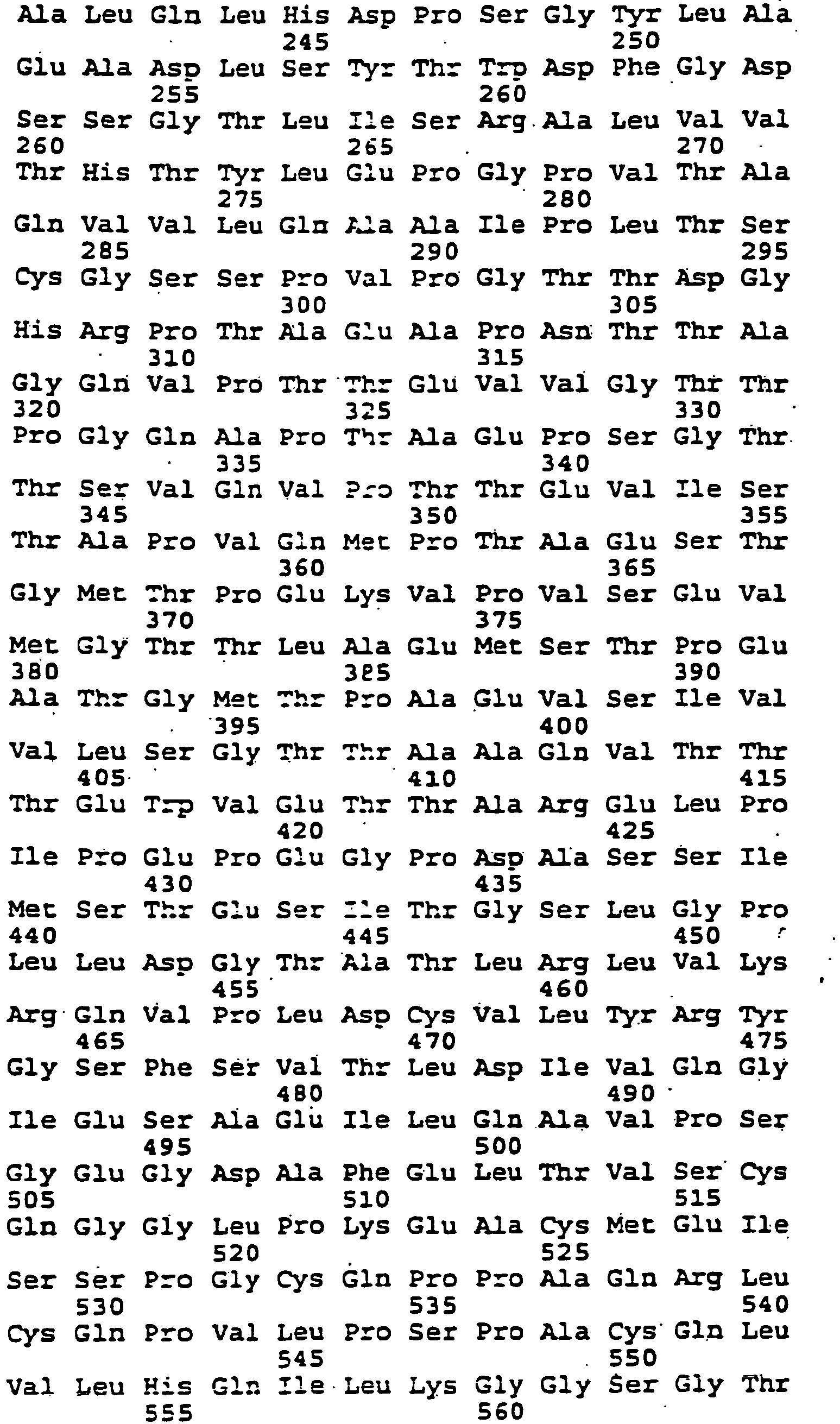

- (i) SEQUENCE CHARACTERISTICS:
- (2) INFORMATION FOR SEQ ID NO: 28:
- (i) SEQUENCE CHARACTERISTICS:
- (A) LENGTH: 7
- (B) TYPE: amino acid
- (C) STRANDEDNESS: Unknown
- (D) TOPOLOGY: Unknown
- (ii) MOLECULE TYPE: Protein
- (xi) SEQUENCE DESCRIPTION: SEQ ID NO: 28:

- (i) SEQUENCE CHARACTERISTICS:
- (2) INFORMATION FOR SEQ ID NO: 29:
- (i) SEQUENCE CHARACTERISTICS:
- (A)
LENGTH 7 - (B) TYPE: amino acid
- (C) STRANDEDNESS: Unknown
- (D) TOPOLOGY: Unknown
- (A)
- (ii) MOLECULE TYPE: Protein
- (xi) SEQUENCE DESCRIPTION: SEQ ID NO: 29:

- (i) SEQUENCE CHARACTERISTICS:
- (2) INFORMATION FOR SEQ ID NO: 30:
- (i) SEQUENCE CHARACTERISTICS:
- (A) LENGTH: 12
- (B) TYPE: amino acid
- (C) STRANDEDNESS: Unknown
- (D) TOPOLOGY: Unknown
- (ii) MOLECULE TYPE: Protein
- (xi) SEQUENCE DESCRIPTION: SEQ ID NO: 30:

- (i) SEQUENCE CHARACTERISTICS:
- (2) INFORMATION FOR SEQ ID NO : 31:
- (i) SEQUENCE CHARACTERISTICS:
- (A) LENGTH: 9
- (B) TYPE: amino acid
- (C) STRANDEDNESS: Unknown
- (D) TOPOLOGY: Unknown
- (ii) MOLECULE TYPE: Peptide
- (xi) SEQUENCE DESCRIPTION: SEQ ID NO: 31:

- (i) SEQUENCE CHARACTERISTICS:
- (2) INFORMATION FOR SEQ ID NO: 32:
- (i) SEQUENCE CHARACTERISTICS:
- (A) LENGTH: 9
- (B) TYPE: amino acid
- (C) STRANDEDNESS: Unknown
- (D) TOPOLOGY: Unknown
- (ii) MOLECULE TYPE: Peptide
- (xi) SEQUENCE DESCRIPTION: SEQ ID NO: 32:

- (i) SEQUENCE CHARACTERISTICS:
- (2) INFORMATION FOR SEQ ID NO: 33:
- (i) SEQUENCE CHARACTERISTICS:
- (A) LENGTH: 10
- (B) TYPE: amino acid
- (C) STRANDEDNESS : Unknown
- (D) TOPOLOGY: Unknown
- (ii) MOLECULE TYPE: Peptide
- (xi) SEQUENCE DESCRIPTION: SEQ ID NO: 33:

- (i) SEQUENCE CHARACTERISTICS:
- (2) INFORMATION FOR SEQ ID NO: 34:
- (i) SEQUENCE CHARACTERISTICS:
- (A) LENGTH: 10
- (B) TYPE: amino acid
- (C) STRANDEDNESS: Unknown
- (D) TOPOLOGY: Unknown
- (ii) MOLECULE TYPE: Peptide
- (xi) SEQUENCE DESCRIPTION: SEQ ID NO: 34:

- (i) SEQUENCE CHARACTERISTICS:
- (2) INFORMATION FOR SEQ ID NO : 35:
- (i) SEQUENCE CHARACTERISTICS:
- (A) LENGTH: 10
- (B) TYPE: amino acid
- (C) STRANDEDNESS: Unknown
- (D) TOPOLOGY: Unknown
- (ii) MOLECULE TYPE: Peptide
- (xi) SEQUENCE DESCRIPTION: SEQ ID NO: 35:

- (i) SEQUENCE CHARACTERISTICS:
- (2) INFORMATION FOR SEQ ID NO: 36:
- (i) SEQUENCE CHARACTERISTICS:
- (A) LENGTH: 9
- (B) TYPE: amino acid
- (C) STRANDEDNESS: Unknown
- (D) TOPOLOGY: Unknown
- (ii) MOLECULE TYPE: Peptide
- (xi) SEQUENCE DESCRIPTION: SEQ ID NO: 36:

- (i) SEQUENCE CHARACTERISTICS:
- (2) INFORMATION FOR SEQ ID NO: 37:
- (i) SEQUENCE CHARACTERISTICS:
- (A) LENGTH: 10
- (B) TYPE: amino acid
- (C) STRANDEDNESS: Unknown
- (D) TOPOLOGY: Unknown
- (ii) MOLECULE TYPE: Peptide
- (xi) SEQUENCE DESCRIPTION: SEQ ID NO: 37:

- (i) SEQUENCE CHARACTERISTICS:
- (2) INFORMATION FOR SEQ ID NO: 38:
- (i) SEQUENCE CHARACTERISTICS:
- (A) LENGTH: 10
- (B) TYPE: amino acid
- (C) STRANDEDNESS: Unknown
- (D) TOPOLOGY: Unknown
- (ii) MOLECULE TYPE: Peptide
- (xi) SEQUENCE DESCRIPTION: SEQ ID NO: 38:

- (i) SEQUENCE CHARACTERISTICS:
- (2) INFORMATION FOR SEQ ID NO: 39:
- (i) SEQUENCE CHARACTERISTICS:
- (A) LENGTH: 9
- (B) TYPE: amino acid
- (C) STRANDEDNESS: Unknown
- (D) TOPOLOGY: Unknown
- (ii) MOLECULE TYPE: Peptide
- (xi) SEQUENCE DESCRIPTION: SEQ ID NO: 39:

- (i) SEQUENCE CHARACTERISTICS:
- (2) INFORMATION FOR SEQ ID NO: 40:
- (i) SEQUENCE CHARACTERISTICS:
- (A) LENGTH: 9
- (B) TYPE: amino acid
- (C) STRANDEDNESS: Unknown
- (D) TOPOLOGY: Unknown
- (ii) MOLECULE TYPE: Peptide
- (xi) SEQUENCE DESCRIPTION: SEQ ID NO: 40:

- (i) SEQUENCE CHARACTERISTICS:
- (2) INFORMATION FOR SEQ ID NO: 41:
- (i) SEQUENCE CHARACTERISTICS:
- (A) LENGTH: 9 .
- (B) TYPE: amino acid
- (C) STRANDEDNESS: Unknown
- (D) TOPOLOGY: Unknown
- (ii) MOLECULE TYPE: Peptide
- (xi) SEQUENCE DESCRIPTION: SEQ ID NO: 41:

- (i) SEQUENCE CHARACTERISTICS:
- (2) INFORMATION FOR SEQ ID NO: 42:
- (i) SEQUENCE CHARACTERISTICS:
- (A) LENGTH: 58
- (B) TYPE: NUCLEOTIDE
- (C) STRANDEDNESS: DOUBLE
- (D) TOPOLOGY: UNKNOWN
- (ii) MOLECULE TYPE: cDNA
- (xi) SEQUENCE DESCRIPTION: SEQ ID NO: 42:

- (i) SEQUENCE CHARACTERISTICS:
- (2) INFORMATION FOR SEQ ID NO: 43:
- (i) SEQUENCE CHARACTERISTICS:
- (A) LENGTH: 12
- (B) TYPE: NUCLEOTIDE
- (C) STRANDEDNESS: DOUBLE
- (D) TOPOLOGY: UNKNOWN
- (ii) MOLECULE TYPE: cDNA
- (xi) SEQUENCE DESCRIPTION: SEQ ID NO: 43: CCAATCGCGA CC
- (i) SEQUENCE CHARACTERISTICS:
- (2) INFORMATION FOR SEQ ID NO: 44:
- (i) SEQUENCE CHARACTERISTICS:
- (A) LENGTH: 14
- (B) TYPE: NUCLEOTIDE
- (C) STRANDEDNESS: DOUBLE
- (D) TOPOLOGY: UNKNOWN
- (ii) MOLECULE TYPE: cDNA
- (xi) SEQUENCE DESCRIPTION: SEQ ID NO: 44: GGTCGCGATTG GTAA
- (i) SEQUENCE CHARACTERISTICS:
- (2) INFORMATION FOR SEQ ID NO: 45:
- (i) SEQUENCE CHARACTERISTICS:
- (A) LENGTH: 9
- (B) TYPE: amino acid
- (C) STRANDEDNESS: Unknown
- (D) TOPOLOGY: Unknown
- (ii) MOLECULE TYPE: Peptide
- (xi) SEQUENCE DESCRIPTION: SEQ ID NO: 45:

- (i) SEQUENCE CHARACTERISTICS:
- (2) INFORMATION FOR SEQ ID NO: 46:
- (i) SEQUENCE CHARACTERISTICS:
- (A) LENGTH: 9
- (B) TYPE: amino acid
- (C) STRANDEDNESS: Unknown
- (D) TOPOLOGY: Unknown
- (ii) MOLECULE TYPE: Peptide
- (xi) SEQUENCE DESCRIPTION: SEQ ID NO: 46:

- (i) SEQUENCE CHARACTERISTICS:
- (2) INFORMATION FOR SEQ ID NO: 47:
- (i) SEQUENCE CHARACTERISTICS:
- (A) LENGTH: 10
- (B) TYPE: amino acid
- (C) STRANDEDNESS: Unknown
- (D) TOPOLOGY: Unknown
- (ii) MOLECULE TYPE: Peptide
- (Xi) SEQUENCE DESCRIPTION: SEQ ID NO: 47:

- (i) SEQUENCE CHARACTERISTICS:
- (2) INFORMATION FOR SEQ ID NO: 48:
- (i) SEQUENCE CHARACTERISTICS:
- (A) LENGTH: 9
- (B) TYPE: amino acid
- (C) STRANDEDNESS: Unknown
- (D) TOPOLOGY: Unknown
- (ii) MOLECULE TYPE: Peptide
- (xi) SEQUENCE DESCRIPTION: SEQ ID NO: 48:

- (i) SEQUENCE CHARACTERISTICS:
- (2) INFORMATION FOR SEQ ID NO: 49:
- (i) SEQUENCE CHARACTERISTICS:
- (A) LENGTH: 10
- (B) TYPE: amino acid
- (C) STRANDEDNESS: Unknown
- (D) TOPOLOGY: Unknown
- (ii) MOLECULE TYPE: Peptide
- (xi) SEQUENCE DESCRIPTION: SEQ ID NO: 49:

- (i) SEQUENCE CHARACTERISTICS:
- (2) INFORMATION FOR SEQ ID NO: 50:
- (i) SEQUENCE CHARACTERISTICS:
- (A) LENGTH: 9
- (B) TYPE: amino acid
- (C) STRANDEDNESS: Unknown
- (D) TOPOLOGY: Unknown
- (ii) MOLECULE TYPE: Peptide
- (xi) SEQUENCE DESCRIPTION: SEQ ID NO: 50:

- (i) SEQUENCE CHARACTERISTICS:
- (2) INFORMATION FOR SEQ ID NO: 51:
- (i) SEQUENCE CHARACTERISTICS:
- (A) LENGTH: 9
- (B) TYPE: amino acid
- (C) STRANDEDNESS: Unknown
- (D) TOPOLOGY: Unknown
- (ii) MOLECULE TYPE: Peptide
- (xi) SEQUENCE DESCRIPTION: SEQ ID NO: 51:

- (i) SEQUENCE CHARACTERISTICS:
- (2) INFORMATION FOR SEQ ID NO: 52:
- (i) SEQUENCE CHARACTERISTICS:
- (A) LENGTH: 9
- (B) TYPE: amino acid
- (C) STRANDEDNESS: Unknown
- (D) TOPOLOGY: Unknown
- (ii) MOLECULE TYPE: Peptide
- (xi) SEQUENCE DESCRIPTION: SEQ ID NO: 52:

- (i) SEQUENCE CHARACTERISTICS:
- (2) INFORMATION FOR SEQ ID NO: 53:
- (i) SEQUENCE CHARACTERISTICS:
- (A) LENGTH: 9
- (B) TYPE: amino acid
- (C) STRANDEDNESS: Unknown
- (D) TOPOLOGY: Unknown
- (ii) MOLECULE TYPE: Peptide
- (xi) SEQUENCE DESCRIPTION: SEQ ID NO: 53:

- (i) SEQUENCE CHARACTERISTICS:
- (2) INFORMATION FOR SEQ ID NO: 54:
- (i) SEQUENCE CHARACTERISTICS:
- (A) LENGTH: 9
- (B) TYPE: amino acid
- (C) STRANDEDNESS: Unknown
- (D) TOPOLOGY: Unknown
- (ii) MOLECULE TYPE: Peptide
- (xi) SEQUENCE DESCRIPTION: SEQ ID NO: 54:

- (i) SEQUENCE CHARACTERISTICS:
- (2) INFORMATION FOR SEQ ID NO: 55:
- (i) SEQUENCE CHARACTERISTICS:
- (A) LENGTH: 9
- (B) TYPE: amino acid
- (C) STRANDEDNESS: Unknown
- (D) TOPOLOGY: Unknown .
- (ii) MOLECULE TYPE: Peptide
- (xi) SEQUENCE DESCRIPTION: SEQ ID NO: 55:

- (i) SEQUENCE CHARACTERISTICS:
- (2) INFORMATION FOR SEQ ID NO: 56:
- (i) SEQUENCE CHARACTERISTICS:
- (A) LENGTH: 9
- (B) TYPE: amino acid
- (C) STRANDEDNESS: Unknown
- (D) TOPOLOGY: Unknown
- (ii) MOLECULE TYPE: peptide
- (xi) SEQUENCE DESCRIPTION: SEQ ID No: 56 :

- (i) SEQUENCE CHARACTERISTICS:
- (2) INFORMATION FOR SEQ ID NO: 57:
- (i) SEQUENCE CHARACTERISTICS:
- (A) LENGTH: 9
- (B) TYPE: amino acid
- (C) STRANDEDNESS: Unknown
- (D) TOPOLOGY: Unknown
- (ii) MOLECULE TYPE: Peptide
- (xi) SEQUENCE DESCRIPTION: SEQ ID NO: 57:

- (i) SEQUENCE CHARACTERISTICS:
- (2) INFORMATION FOR SEQ ID NO: 58:
- (i) SEQUENCE CHARACTERISTICS:
- (A) LENGTH: 9
- (B) TYPE: amino acid
- (C) STRANDEDNESS: Unknown
- (D) TOPOLOGY: Unknown
- (ii) MOLECULE TYPE: Peptide
- (xi) SEQUENCE DESCRIPTION: SEQ ID NO: 58:

- (i) SEQUENCE CHARACTERISTICS:
- (2) INFORMATION FOR SEQ ID NO: 59:
- (i) SEQUENCE CHARACTERISTICS:
- (A) LENGTH: 9
- (B) TYPE: amino acid
- (C) STRANDEDNESS: Unknown
- (D) TOPOLOGY: Unknown
- (ii) MOLECULE TYPE: Peptide
- (xi) SEQUENCE DESCRIPTION: SEQ ID NO: 59:

- (i) SEQUENCE CHARACTERISTICS:
- (2) INFORMATION FOR SEQ ID NO: 60:
- (i) SEQUENCE CHARACTERISTICS:
- (A) LENGTH: 9
- (B) TYPE: amino acid
- (C) STRANDEDNESS: Unknown
- (D) TOPOLOGY: Unknown
- (ii) MOLECULE TYPE: Peptide
- (xi) SEQUENCE DESCRIPTION: SEQ ID NO: 60:

- (i) SEQUENCE CHARACTERISTICS:
- (2) INFORMATION FOR SEQ ID NO: 61:
- (i) SEQUENCE CHARACTERISTICS:
- (A) LENGTH: 9
- (B) TYPE: amino acid
- (C) STRANDEDNESS: Unknown
- (D) TOPOLOGY: Unknown
- (ii) MOLECULE TYPE: Peptide
- (xi) SEQUENCE DESCRIPTION: SEQ ID NO: 61:

- (i) SEQUENCE CHARACTERISTICS:
- (2) INFORMATION FOR SEQ ID NO: 62:
- (i) SEQUENCE CHARACTERISTICS:
- (A) LENGTH: 9
- (B) TYPE: amino acid
- (C) STRANDEDNESS: Unknown
- (D) TOPOLOGY: Unknown
- (ii) MOLECULE TYPE: Peptide
- (xi) SEQUENCE DESCRIPTION: SEQ ID NO: 62:

- (i) SEQUENCE CHARACTERISTICS:
- (2) INFORMATION FOR SEQ ID NO: 63:
- (i) SEQUENCE CHARACTERISTICS:
- (A) LENGTH: 9
- (B) TYPE: amino acid
- (C) STRANDEDNESS: Unknown
- (D) TOPOLOGY: Unknown
- (ii) MOLECULE TYPE: Peptide
- (xi) SEQUENCE DESCRIPTION: SEQ ID NO: 63:

- (i) SEQUENCE CHARACTERISTICS:
- (2) INFORMATION FOR SEQ ID NO: 64:
- (i) SEQUENCE CHARACTERISTICS:
- (A) LENGTH: 9
- (B) TYPE: amino acid
- (C) STRANDEDNESS: Unknown
- (D) TOPOLOGY: Unknown
- (ii) MOLECULE TYPE: Peptide
- (xi) SEQUENCE DESCRIPTION: SEQ ID NO: 64:

- (i) SEQUENCE CHARACTERISTICS:
- (2) INFORMATION FOR SEQ ID NO: 65:
- (i) SEQUENCE CHARACTERISTICS:
- (A) LENGTH: 9
- (B) TYPE: amino acid
- (C) STRANDEDNESS : Unknown
- (D) TOPOLOGY: Unknown
- (ii) MOLECULE TYPE: Peptide
- (xi) SEQUENCE DESCRIPTION: SEQ ID NO: 65:

- (i) SEQUENCE CHARACTERISTICS:
- (2) INFORMATION FOR SEQ ID NO: 66:
- (i) SEQUENCE CHARACTERISTICS:
- (A) LENGTH: 9
- (B) TYPE: amino acid
- (C) STRANDEDNESS: Unknown
- (D) TOPOLOGY: Unknown
- (ii) MOLECULE TYPE: Peptide
- (xi) SEQUENCE DESCRIPTION: SEQ ID NO: 66:

- (i) SEQUENCE CHARACTERISTICS:
- (2) INFORMATION FOR SEQ ID NO: 67:
- (i) SEQUENCE CHARACTERISTICS:
- (A) LENGTH: 9
- (B) TYPE: amino acid
- (C) STRANDEDNESS : Unknown
- (D) TOPOLOGY: Unknown
- (ii) MOLECULE TYPE: Peptide
- (xi) SEQUENCE DESCRIPTION: SEQ ID NO: 67:

- (i) SEQUENCE CHARACTERISTICS:
- (2) INFORMATION FOR SEQ ID NO: 68:
- (i) SEQUENCE CHARACTERISTICS:
- (A) LENGTH: 9
- (B) TYPE: amino acid
- (C) STRANDEDNESS: Unknown
- (D) TOPOLOGY: Unknown
- (ii) MOLECULE TYPE: Peptide
- (xi) SEQUENCE DESCRIPTION: SEQ ID NO: 68:

- (i) SEQUENCE CHARACTERISTICS:
- (2) INFORMATION FOR SEQ ID NO: 69:
- (i) SEQUENCE CHARACTERISTICS:
- (A) LENGTH: 9
- (B) TYPE: amino acid
- (C) STRANDEDNESS: Unknown
- (D) TOPOLOGY: Unknown
- (ii) MOLECULE TYPE: Peptide
- (xi) SEQUENCE DESCRIPTION : SEQ ID NO: 69 :

- (i) SEQUENCE CHARACTERISTICS:
- (2) INFORMATION FOR SEQ ID NO: 70:
- (i) SEQUENCE CHARACTERISTICS:
- (A) LENGTH: 9
- (B) TYPE: amino acid
- (C) STRANDEDNESS : Unknown
- (D) TOPOLOGY: Unknown
- (ii) MOLECULE TYPE: Peptide
- (xi) SEQUENCE DESCRIPTION: SEQ ID NO: 70:

- (i) SEQUENCE CHARACTERISTICS:
- (2) INFORMATION FOR SEQ ID NO: 71:
- (i) SEQUENCE CHARACTERISTICS:
- (A) LENGTH: 9
- (B) TYPE: amino acid
- (C) STRANDEDNESS: Unknown
- (D) TYPOLOGY: Unknown
- (ii) MOLECULE TYPE: Peptide
- (xi) SEQUENCE DESCRIPTION: SEQ ID NO: 71:

- (i) SEQUENCE CHARACTERISTICS:
- (2) INFORMATION FOR SEQ ID NO : 72 :
- (i) SEQUENCE CHARACTERISTICS:
- (A) LENGTH: 9
- (B) TYPE: amino acid
- (C) STRANDEDNESS: Unknown
- (D) TOPOLOGY: Unknown
- (ii) MOLECULE TYPE: Peptide
- (xi) SEQUENCE DESCRIPTION: SEQ ID NO: 7.2 :

- (i) SEQUENCE CHARACTERISTICS:
- (2) INFORMATION FOR SEQ ID NO : 73:
- (i) SEQUENCE CHARACTERISTICS:
- (A) LENGTH: 9
- (B) TYPE: amino acid
- (C) STRANDEDNESS: Unknown
- (D) TOPOLOGY: Unknown
- (ii) MOLECULE TYPE: Peptide
- (xi) SEQUENCE DESCRIPTION: SEQ ID NO: 73:

- (i) SEQUENCE CHARACTERISTICS:
- (2) INFORMATION FOR SEQ ID NO: 74:
- (i) SEQUENCE CHARACTERISTICS:
- (A) LENGTH: 9
- (B) TYPE: amino acid
- (C) STRANDEDNESS: Unknown
- (D) TOPOLOGY: Unknown
- (ii) MOLECULE TYPE: Peptide
- (xi) SEQUENCE DESCRIPTION: SEQ ID NO: 74:

- (i) SEQUENCE CHARACTERISTICS:
- (2) INFORMATION FOR SEQ ID NO: 75:
- (i) SEQUENCE CHARACTERISTICS:
- (A) LENGTH: 9
- (B) TYPE: amino acid
- (C) STRANDEDNESS: Unknown
- (D) TOPOLOGY: Unknown
- (ii) MOLECULE TYPE: Peptide
- (xi) SEQUENCE DESCRIPTION: SEQ ID NO: 75:

- (i) SEQUENCE CHARACTERISTICS:
- (2) INFORMATION FOR SEQ ID NO: 76:
- (i) SEQUENCE CHARACTERISTICS:
- (A) LENGTH: 9
- (B) TYPE: amino acid
- (C) STRANDEDNESS: Unknown
- (D) TOPOLOGY: Unknown
- (ii) MOLECULE TYPE: Peptide
- (xi) SEQUENCE DESCRIPTION: SEQ ID NO: 76:

- (i) SEQUENCE CHARACTERISTICS:
- (2) INFORMATION FOR SEQ ID NO: 77:
- (i) SEQUENCE CHARACTERISTICS:
- (A) LENGTH: 9
- (B) TYPE: amino acid
- (C) STRANDEDNESS: Unknown
- (D) TOPOLOGY: Unknown
- (ii) MOLECULE TYPE: Peptide
- (xi) SEQUENCE DESCRIPTION: SEQ ID NO: 77:

- (i) SEQUENCE CHARACTERISTICS:
- (2) INFORMATION FOR SEQ ID NO: 78:
- (i) SEQUENCE CHARACTERISTICS:
- (A) LENGTH: 9
- (B) TYPE: amino acid
- (C) STRANDEDNESS : Unknown
- (D) TOPOLOGY: Unknown
- (ii) MOLECULE TYPE : Peptide
- (xi) SEQUENCE DESCRIPTION: SEQ ID NO: 78:

- (i) SEQUENCE CHARACTERISTICS:
- (2) INFORMATION FOR SEQ ID NO: 79:
- (i) SEQUENCE CHARACTERISTICS:
- (A) LENGTH: 9
- (B) TYPE: amino acid
- (C) STRANDEDNESS: Unknown
- (D) TOPOLOGY: Unknown
- (ii) MOLECULE TYPE: Peptide
- (xi) SEQUENCE DESCRIPTION: SEQ ID NO: 79:

- (i) SEQUENCE CHARACTERISTICS:
- (2) INFORMATION FOR SEQ ID NO: 80:
- (i) SEQUENCE CHARACTERISTICS:
- (A) LENGTH: 9
- (B) TYPE: amino acid
- (C) STRANDEDNESS: Unknown
- (D) TOPOLOGY: Unknown
- (ii) MOLECULE TYPE : Peptide
- (xi) SEQUENCE DESCRIPTION: SEO ID NO: 80:

- (i) SEQUENCE CHARACTERISTICS:
- (2) INFORMATION FOR SEQ ID NO: B1:
- (i) SEQUENCE CHARACTERISTICS:
- (A) LENGTH: 9
- (B) TYPE: amino acid
- (C) STRANDEDNESS: Unknown
- (D) TYPOLOGY: Unknown
- (ii) MOLECULE HYPE: Peptide
- (xi) SEQUENCE DESCRIPTION: SEQ ID NO: 81:

- (i) SEQUENCE CHARACTERISTICS:
- (2) INFORMATION FOR SEQ ID NO : 82:
- (i) SEQUENCE CHARACTERISTICS:
- (A) LENGTH: 9
- (B) 7YPE: amino acid
- (C) STRANDEDNESS: Unknown
- (D) TOPOLOGY: Unknown
- (ii) MOLECULE TYPE : Peptide
- (xi) SEQUENCE DESCRIPTION: SEQ ID NO: 82:

- (i) SEQUENCE CHARACTERISTICS:
- (2) INFORMATION FOR SEQ ID NO: ε3:
- (i) SEQUENCE CHARACTERISTICS:
- (A) LENGTH: 9 '
- (B) TYPE: amino acid
- (C) STRANDEDNESS: Unknown
- (D) TOPOLOGY: Unknown
- (ii) MOLECULE TYPE: Peptide
- (xi) SEQUENCE DESCRIPTION: SEQ ID NO: 83:

- (i) SEQUENCE CHARACTERISTICS:
- (2) INFORMATION FOR SEQ ID NO: 84:
- (i) SEQUENCE CHARACTERISTICS:
- (A) LENGTH: 9
- (B) TYPE: amino acid
- (C) STRANDEDNESS: Unknown
- (D) TOPOLOGY: Unknown
- (ii) MOLECULE TYPE: Peptide
- (xi) SEQUENCE DESCRIPTION: SEQ ID NO: 84:

- (i) SEQUENCE CHARACTERISTICS:
- (2) INFORMATION FOR SEQ ID NO: 85:
- (i) SEQUENCE CHARACTERISTICS:
- (A) LENGTH: 9
- (B) TYPE: amino acid
- (C) STRANDEDNESS: Unknown
- (D) TOPOLOGY : Unknown
- (ii) MOLECULE TYPE: Peptide
- (xi SEQUENCE DESCRIPTION: SEQ ID NO: 85:

- (i) SEQUENCE CHARACTERISTICS:
- (2) INFORMATION FOR SEQ ID NO: 86 :
- (i) SEQUENCE CHARACTERISTICS:
- (A) LENGTH:
- (B) TYPE: amino acid
- (C) STRANDEDNESS: Unknown
- (D) TOPOLOGY: Unknown
- (ii) MOLECULE TYPE: Peptide
- (xi) SEQUENCE DESCRIPTION: SEQ ID NO: 86:

- (i) SEQUENCE CHARACTERISTICS:
- (2) INFORMATION FOR SEQ ID NO: 87:
- (i) SEQUENCE CHARACTERISTICS:
- (A) LENGTH: 9
- (B) TYPE: amino acid
- (C) STRANDEDNESS : Unknown
- (D) TOPOLOGY: Unknown
- (ii) MOLECULE TYPE: Peptide
- (xi) SEQUENCE DESCRIPTION: SEQ ID NO: 87:

- (i) SEQUENCE CHARACTERISTICS:
- (2) INFORMATION FOR SEQ ID NO: 88:
- (i) SEQUENCE CHARACTERISTICS:
- (A) LENGTH: 9
- (B) TYPE: amino acid
- (C) STRANDEDNESS: Unknown
- (D) TOPOLOGY: Unknown
- (ii) MOLECULE TYPE: Peptide
- (xi) SEQUENCE DESCRIPTION: SEQ ID NO: 88:

- (i) SEQUENCE CHARACTERISTICS:
- (2) INFORMATION FOR SEQ ID NO: 89 :
- (i) SEQUENCE CHARACTERISTICS:
- (A) LENGTH: 9
- (B) TYPE: amino acid
- (C) STRAKDEDNESS: Unknown
- (D) TOPOLOGY: Unknown
- (ii) MOLECULE TYPE: Peptide
- (xi) SEQUENCE DESCRIPTION: SEQ ID NO: 89:

- (i) SEQUENCE CHARACTERISTICS:
- (2) INFORMATION FOR SEQ ID NO: 90:
- (i) SEQUENCE CHARACTERISTICS:
- (A) LENGTH: 9
- (B) TYPE: amino acid
- (C) STRANDEDNESS: Unknown
- (D) TOPOLOGY: Unknown
- (ii) MOLECULE TYPE: Peptide
- (xi) SEQUENCE DESCRIPTION: SEQ ID NO: 90:

- (i) SEQUENCE CHARACTERISTICS:
- (2) INFORMATION FOR SEQ ID NO: 91:
- (i) SEQUENCE CHARACTERISTICS:
- (A) LENGTH: 9
- (B) TYPE: amino acid
- (C) STRANDEDNESS : Unknown
- (D) TOPOLOGY : Unknown
- (ii) MOLECULE TYPE: Peptide
- (xi) SEQUENCE DESCRIPTION: SEQ ID NO: 91:

- (i) SEQUENCE CHARACTERISTICS:
- (2) INFORMATION FOR SEQ ID NO: 92:
- (i) SEQUENCE CHARACTERISTICS:
- (A) LENGTH: 9
- (B) TYPE: amino acid
- (C) STRANDEDNESS: Unknown
- (D) TOPOLOGY: Unknown
- (ii) MOLECULE TYPE: Peptide
- (xi) SEQUENCE DESCRIPTION: SEQ ID NO: 92:

- (i) SEQUENCE CHARACTERISTICS:
- (2) INFORMATION FOR SEQ ID NO: 93:
- (i) SEQUENCE CHARACTERISTICS:
- (A) LENGTH: 9
- (B) TYPE: amino acid
- (C) STRANDEDNESS: Unknown
- (D) TOPOLOGY: Unknown
- (ii) MOLECULE TYPE: Peptide
- (xi) SEQUENCE DESCPIPTION : SEQ ID NO: 93:

- (i) SEQUENCE CHARACTERISTICS:
- (2) INFORMATION FOR SEQ ID NO: 94:
- (i) SEQUENCE CHARACTERISTICS:
- (A) LENGTH: 9
- (B) TYPE: amino acid
- (C) STRANDEDNESS: Unknown
- (D) TOPOLOGY: Unknown
- (ii) MOLECULE TYPE: Peptide
- (xi) SEQUENCE DESCRIPTION: SEQ ID NO: 94:

- (i) SEQUENCE CHARACTERISTICS:
- (2) INFORMATION FOR SEQ ID NO: 95:
- (i) SEQUENCE CHARACTERISTICS:
- (A) LENGTH: 9
- (B) TYPE: amino acid
- (C) STRANDEDNESS : Unknown
- (D) TOPOLOGY: Unknown
- (ii) MOLECULE TYPE: Peptide
- (xi) SEQUENCE DESCRIPTION: SEQ ID NO: 95:

- (i) SEQUENCE CHARACTERISTICS:
- (2) INFORMATION FOR SEQ ID NO: 96 :
- (i) SEQUENCE CHARACTERISTICS:
- (A) LENGTH: 9
- (B) TYPE: amino acid
- (C) STRANDEDNESS: Unknown
- (D) TOPOLOGY: Unknown
- (ii) MOLECULE TYPE: Peptide
- (xi) SEQUENCE DESCRIPTION: SEQ ID NO: 96:

- (i) SEQUENCE CHARACTERISTICS:
- (2) INFORMATION FOR SEQ ID NO: 97:
- (i) SEQUENCE CHARACTERISTICS:
- (A) LENGTH: 9
- (B) TYPE: amino acid
- (C) STRANDEDNESS: Unknown (D) TOPOLOGY: Unknown
- (ii) MOLECULE TYPE: Peptide
- (xi) SEQUENCE DESCRIPTION: SEQ ID NO: 97:

- (i) SEQUENCE CHARACTERISTICS:
- (2) INFORMATION FOR SEQ ID NO: 98:
- (i) SEQUENCE CHARACTERISTICS:
- (A) LENGTH: 9
- (B) TYPE: amino acid
- (C) STRANDEDNESS: Unknown
- (D) TOPOLOGY: Unknown
- (ii) MOLECULE TYPE: Peptide
- (xi) SEQUENCE DESCRIPTION: SEQ ID NO: 98:

- (i) SEQUENCE CHARACTERISTICS:
- (2) INFORMATION FOR SEQ ID NO: 99:
- (i) SEQUENCE CHARACTERISTICS:
- (A) LENGTH: 9
- (B) TYPE: amino acid
- (C) STRANDEDNESS : Unknown
- (D) TOPOLOGY: Unknown
- (ii) MOLECULE TYPE: Peptide
- (xi) SEQUENCE DESCRIPTION: SEQ ID NO: 99:

- (i) SEQUENCE CHARACTERISTICS:
- (2) INFORMATION FOR SEQ ID NO: 100 :
- (1) SEQUENCE CHARACTERISTICS:
- (A) LENGTH: 9
- (B) TYPE : amino acid
- (C) STRANDEDNESS: Unknown
- (D) TOPOLOGY: Unknown
- (ii) MOLECULE TYPE: Peptide
- (xi) SEQUENCE DESCRIPTION: SEQ ID NO: 100:

- (1) SEQUENCE CHARACTERISTICS:
- (2) INFORMATION FOR SEQ ID NO: 101:
- (i) SEQUENCE CHARACTERISTICS:
- (A) LENGTH: 9
- (B) TYPE: amino acid
- (C) STRANDEDNESS: Unknown
- (D) TOPOLOGY: Unknown
- (ii) MOLECULE TYPE: Peptide
- (xi) SEQUENCE DESCRIPTION: SEQ ID NO: 101:

- (i) SEQUENCE CHARACTERISTICS:
- (2) INFORMATION FOR SEQ ID NO: 102:
- (i) SEQUENCE CHARACTERISTICS:
- (A) LENGTH: 9
- (B) TYPE: amino acid
- (C) STRANDEDNESS : Unknown
- (D) TOPOLOGY: Unknown
- (ii) MOLECULE TYPE: Peptide
- (xi) SEQUENCE DESCRIPTION: SEQ ID NO: 102:

- (i) SEQUENCE CHARACTERISTICS:
- (2) INFORMATION FOR SEQ ID NO: 103:
- (i) SEQUENCE CHARACTERISTICS:
- (A) LENGTH: 9
- (B) TYPE: amino acid
- (C) STRANDEDNESS : Unknown
- (D) TOPOLOGY: Unknown
- (ii) MOLECULE TYPE: Peptide
- (xi) SEQUENCE DESCRIPTION: SEQ ID NO: 103:

- (i) SEQUENCE CHARACTERISTICS:
- (2) INFORMATION FOR SEQ ID NO: 104:
- (i) SEQUENCE CHARACTERISTICS:
- (A) LENGTH: 9
- (B) TYPE: amino acid
- (C) STRANDEDNESS: Unknown
- (D) TOPOLOGY: Unknown
- (ii) MOLECULE TYPE: Peptide
- (xi) SEQUENCE DESCRIPTION: SEQ ID NO: 104:

- (i) SEQUENCE CHARACTERISTICS:
- (2) INFORMATION FOR SEQ ID NO: 105:
- (i) SEQUENCE CHARACTERISTICS:
- (A) LENGTH: 9
- (B) TYPE: amino acid
- (C) STRANDEDNESS: Unknown
- (D) TOPOLOGY: Unknown
- (ii) MOLECULE TYPE: Peptide
- (xi) SEQUENCE DESCRIPTION: SEQ ID NO: 105:

- (i) SEQUENCE CHARACTERISTICS:
- (2) INFORMATION FOR SEQ ID NO: 106:
- (i) SEQUENCE CHARACTERISTICS:
- (A) LENGTH: 9
- (B) TYPE: amino acid
- (C) STRANDEDNESS: Unknown
- (D) TOPOLOGY: Unknown
- (ii) MOLECULE TYPE: Peptide
- (xi) SEQUENCE DESCRIPTION: SEQ ID NO: 106:

- (i) SEQUENCE CHARACTERISTICS:
- (2) INFORMATION FOR SEQ ID NO: 107:
- (i) SEQUENCE CHARACTERISTICS:
- (A) LENGTH: 9
- (B) TYPE: amino acid
- (C) STRANDEDNESS : Unknown
- (D) TOPOLOGY: Unknown
- (ii) MOLECULE TYPE: Peptide ,
- (xi) SEQUENCE DESCRIPTION: SEQ ID NO: 107:

- (i) SEQUENCE CHARACTERISTICS:
- (2) INFORMATION FOR SEQ ID NO: 10B :
- (i) SEQUENCE CHARACTERISTICS:
- (A) LENGTH: 9
- (B) TYPE: amino acid
- (C) STRANDEDNESS: Unknown
- (D) TOPOLOGY: Unknown
- (ii) MOLECULE TYPE: Peptide
- (xi) SEQUENCE DESCRIPTION: SEQ ID NO: 108:

- (i) SEQUENCE CHARACTERISTICS:
- (2) INFORMATION FOR SEQ ID NO: 109:
- (i) SEQUENCE CHARACTERISTICS:
- (A) LENGTH: 9
- (B) TYPE: amino acid
- (C) STRANDEDNESS : Unknown
- (D) TOPOLOGY: Unknown
- (ii) MOLECULE TYPE: Peptide
- (xi) SEQUENCE DESCRIPTION: SEQ ID NO: 109:

- (i) SEQUENCE CHARACTERISTICS:
- (2) INFORMATION FOR SEQ ID NO: 110:
- (i) SEQUENCE CHARACTERISTICS:
- (A) LENGTH: 9
- (B) TYPE: amino acid
- (C) STRANDEDNESS: Unknown
- (D) TOPOLOGY: Unknown
- (ii) MOLECULE TYPE: Peptide
- (xi) SEQUENCE DESCRIPTION: SEQ ID NO: 110:

- (i) SEQUENCE CHARACTERISTICS:
- (2) INFORMATION FOR SEQ ID NO: 111:
- (i) SEQUENCE CHARACTERISTICS:
- (A) LENGTH: 9
- (B) TYPE: amino acid
- (C) STRANDEDNESS : Unknown
- (D) TOPOLOGY: Unknown
- (ii) MOLECULE TYPE: Peptide
- (xi) SEQUENCE DESCRIPTION: SEQ ID NO: 111:

- (i) SEQUENCE CHARACTERISTICS:
- (2) INFORMATION FOR SEQ ID NO: 112 :
- (i) SEQUENCE CHARACTERISTICS:
- (A) LENGTH: 9
- (B) TYPE: amino acid
- (C) STRANDEDNESS: Unknown
- (D) TOPOLOGY: Unknown
- (ii) MOLECULE TYPE: Peptide
- (xi) SEQUENCE DESCRIPTION: SEQ ID NO: 112:

- (i) SEQUENCE CHARACTERISTICS:
- (2) INFORMATION FOR SEQ ID NO: 113 :
- (i) SEQUENCE CHARACTERISTICS:
- (A) LENGTH: 9
- (B) TYPE: amino acid
- (C) STRANDEDNESS: Unknown
- (D) TOPOLOGY: Unknown
- (ii) MOLECULE TYPE: Peptide
- (xi) SEQUENCE DESCRIPTION: SEQ ID NO: 113:

- (i) SEQUENCE CHARACTERISTICS:
- (2) INFORMATION FOR SEQ ID NO: 114 :
- (1) SEQUENCE CHARACTERISTICS:
- (A) LENGTH: 9
- (B) TYPE: amino acid
- (C) STRANDEDNESS : Unknown
- (D) TOPOLOGY: Unknown
- (ii) MOLECULE TYPE: Peptide
- (xi) SEQUENCE DESCRIPTION: SEQ ID NO: 114:

- (1) SEQUENCE CHARACTERISTICS:
- (2) INFORMATION FOR SEQ ID NO: 115:
- (i) SEQUENCE CHARACTERISTICS:
- (A) LENGTH : 9
- (B) TYPE : amino acid
- (C) STRANDEDNESS: Unknown
- (D) TOPOLOGY: Unknown
- (ii) MOLECULE TYPE: Peptide
- (xi) SEQUENCE DESCRIPTION: SEQ ID NO: 115:

- (i) SEQUENCE CHARACTERISTICS:
- (2) INFORMATION FOR SEQ ID NO: 116:
- (i) SEQUENCE CHARACTERISTICS:
- (A) LENGTH: 9
- (B) TYPE: amino acid
- (C) STRANDEDNESS: Unknown
- (D) TOPOLOGY: Unknown
- (ii) MOLECULE TYPE : Peptide
- (xi) SEQUENCE DESCRIPTION: SEQ ID NO: 116:

- (i) SEQUENCE CHARACTERISTICS:
- (2) INFORMATION FOR SEQ ID NO: 117:
- (i) SEQUENCE CHARACTERISTICS:
- (A) LENGTH: 9
- (B) TYPE: amino acid
- (C) STRANDEDNESS: Unknown
- (D) TOPOLOGY: Unknown
- (ii) MOLECULE TYPE: Peptide
- (xi) SEQUENCE DESCRIPTION: SEQ ID NO: 117:

- (i) SEQUENCE CHARACTERISTICS:
- (2) INFORMATION FOR SEQ ID NO: 118 :
- (i) SEQUENCE CHARACTERISTICS:
- (A) LENGTH: 9
- (B) TYPE: amino acid
- (C) STRANDEDNESS: Unknown
- (D) TOPOLOGY: Unknown
- (ii) MOLECULE TYPE: Peptide
- (xi) SEQUENCE DESCRIPTION: SEQ ID NO: 118:

- (i) SEQUENCE CHARACTERISTICS:
- (2) INFORMATION FOR SEQ ID NO: 119:
- (i) SEQUENCE CHARACTERISTICS:
- (A) LENGTH: 9
- (B) TYPE: amino acid
- (C) STRANDEDNESS: Unknown
- (D) TOPOLOGY: Unknown
- (ii) MOLECULE TYPE : Peptide
- (xi) SEQUENCE DESCRIPTION: SEQ ID NO: 119:

- (i) SEQUENCE CHARACTERISTICS:
- (2) INFORMATION FOR SEQ ID NO: 120: (i) SEQUENCE CHARACTERISTICS: (A) LENGTH: 9 (B) TYPE: amino acid (C) STRANDEDNESS: Unknown (D) TOPOLOGY: Unknown (ii) MOLECULE TYPE: Peptide (xi) SEQUENCE DESCRIPTION: SEQ ID NO: 120:

- (2) INFORMATION FOR SEQ ID NO: 121:
- (i) SEQUENCE CHARACTERISTICS:
- (A) LENGTH: 661
- (B) TYPE: amino acid
- (C) STRANDEDNESS: Unknown
- (D) TOPOLOGY: Unknown
- (ii) MOLECULE TYPE: Protein
- (xi) SEQUENCE DESCRIPTION: SEQ ID NO: 121:


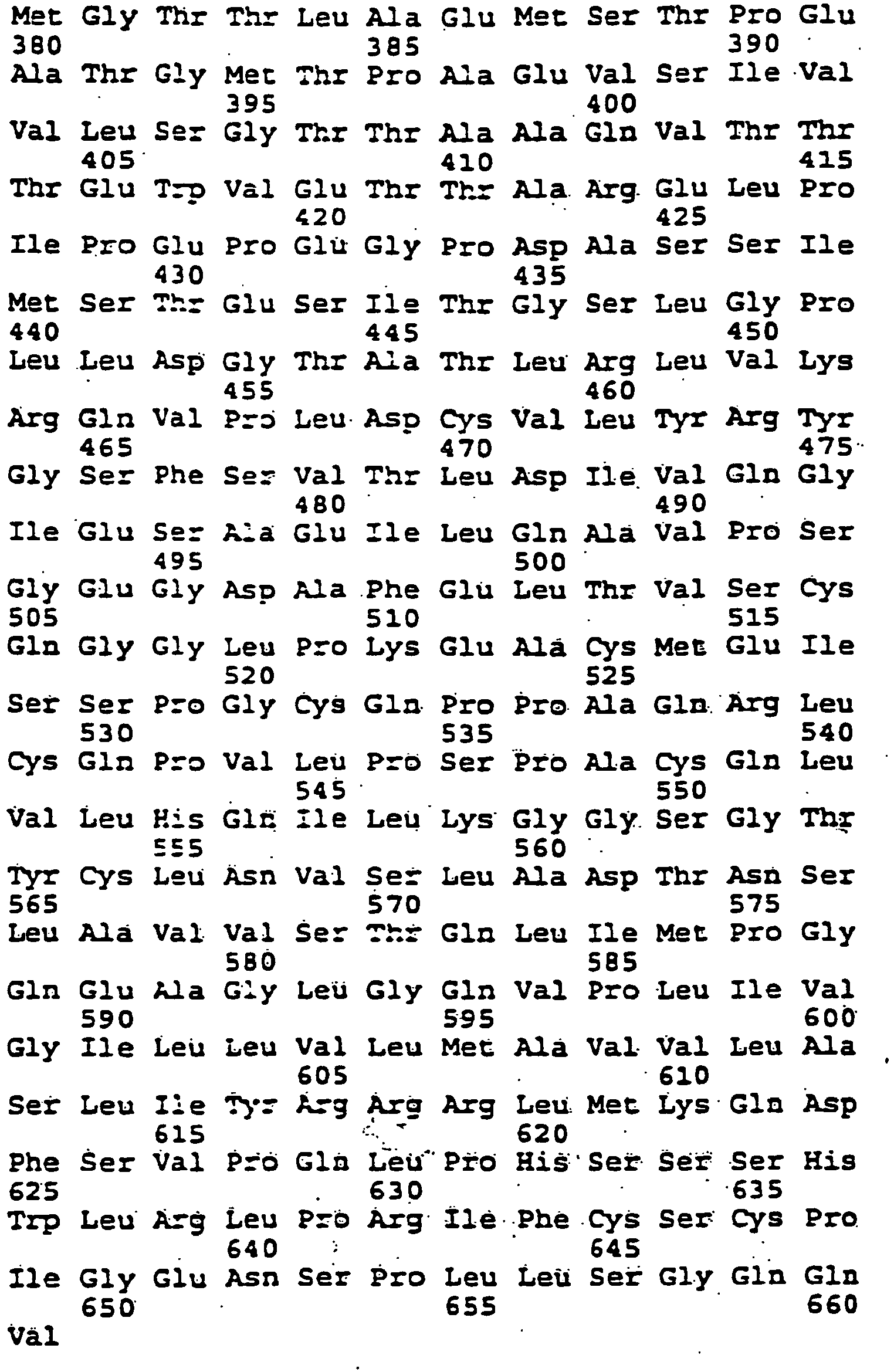
- (i) SEQUENCE CHARACTERISTICS:
- (2) INFORMATION FOR SEQ ID NO : 122 :
- (i) SEQUENCE CHARACTERISTICS:
- (A) LENGTH: 9
- (B) TYPE: amino acid
- (C) STRANDEDNESS : Unknown
- (D) TOPOLOGY: Unknown
- (ii) MOLECULE TYPE: Peptide
- (xi) SEQUENCE DESCRIPTION: SEQ ID NO: 122:

- (i) SEQUENCE CHARACTERISTICS:
- (2) INFORMATION FOR SEQ ID NO: 123:
- (i) SEQUENCE CHARACTERISTICS:
- (A) LENGTH: 9
- (B) TYPE: amino acid
- (C) STRANDEDNESS: Unknown
- (D) TOPOLOGY: Unknown
- (ii) MOLECULE TYPE: Peptide
- (xi) SEQUENCE DESCRIPTION: SEQ ID NO: 123:

- (i) SEQUENCE CHARACTERISTICS:
- (2) INFORMATION FOR SEQ ID NO: 124:
- (i) SEQUENCE CHARACTERISTICS:
- (A) LENGTH: 9
- (B) TYPE: amino acid
- (C) STRANDEDNESS: Unknown
- (D) TOPOLOGY: Unknown
- (ii) MOLECULE TYPE: Peptide
- (xi) SEQUENCE DESCRIPTION: SEQ ID NO: 124:

- (i) SEQUENCE CHARACTERISTICS:
- (2) INFORMATION FOR SEQ ID NO: 125:
- (i) SEQUENCE CHARACTERISTICS:
- (A) LENGTH: 9
- (B) TYPE: amino acid
- (C) STRANDEDNESS: Unknown
- (D) TOPOLOGY: Unknown
- (ii) MOLECULE TYPE: Peptide
- (xi) SEQUENCE DESCRIPTION: SEQ ID NO: 125:

- (i) SEQUENCE CHARACTERISTICS:
- (2) INFORMATION FOR SEQ ID NO: 126:
- (i) SEQUENCE CHARACTERISTICS:
- (A) LENGTH: 10
- (B) TYPE: amino acid
- (C) STRANDEDNESS: Unknown
- (D) TOPOLOGY: Unknown
- (ii) MOLECULE TYPE: Peptide
- (xi) SEQUENCE DESCRIPTION: SEQ ID NO: 126:

- (i) SEQUENCE CHARACTERISTICS:
Claims (24)
- An isolated immunogenic peptide of SEQ ID NO: 121, which peptide stimulates TIL, selected from the group consisting of:D3: amino acids 1-154 of SEQ ID NO: 121;D5: amino acids 1-163 of SEQ ID NO: 121;D4: amino acids 1-195 of SEQ ID NO: 121;C4: amino acids 1-270 of SEQ ID NO: 121;25TR: amino acids 236-661 of SEQ ID NO: 121;SEQ ID NO: 35;SEQ ID NO: 36;SEQ ID NO: 39;SEQ ID NO: 41;SEQ ID NO: 48; andSEQ ID NO: 49.
- An analog of the isolated immunogenic peptide of claim 1, wherein the analog consists of the amino acid sequence of one of the peptides of claim 1, and one amino acid substitution, wherein the analog stimulates TIL.
- The analog of claim 2, wherein the analog has a length of 9-10 amino acids and the amino acid substitution occurs at the first, second, third, or ninth amino acid position of the peptide.
- An isolated immunogenic peptide selected from the group consisting of: SEQ ID NOs: 68-70, 74-79, 83-88, and 101-108.
- An isolated immunogenic peptide selected from the group consisting of: SEQ ID NOs: 123-124; wherein X1 is any amino acid, X2 is any hydrophobic amino acid; X3 is any amino acid; and X4 is any hydrophobic amino acid, provided that the peptide does not consist of the amino acid sequence of SEQ ID NO: 46 or 47.
- An isolated immunogenic peptide consisting of SEQ ID NO: 125, wherein X1 is any amino acid, X2 is any hydrophobic amino acid; X3 is selected from the group consisting of: Leu, Met, Ala, Ile, Val, Thr, Gly, Lys, Phe, Trp, Tyr, Asp, and Ser; and X4 is any hydrophobic amino acid.
- The isolated immunogenic peptide of claim 5 or 6, wherein X1 is a hydrophobic, aliphatic amino acid or aromatic amino acid.
- The isolated immunogenic peptide of claim 7, wherein X1 is selected from the group consisting of Ala, Leu, Lys, Ile, Gly, Met, Val, Thr, Trp, Phe, Lys, Ser, and Tyr.
- The isolated immunogenic peptide of any of claims 5 to 8, wherein X2 is a hydrophobic aliphatic amino acid.
- The isolated immunogenic peptide of claim 9, wherein X2 is selected from the group consisting of Leu, Met, Ile, Ala, Thr, Gly, and Val.
- The isolated immunogenic peptide of any of claims 5, and 7 to 10, wherein X3 is a hydrophobic, aliphatic amino acid.
- The isolated immunogenic peptide of claim 11, wherein X3 is selected from the group consisting of Ala, Leu, Lys, Ile, Gly, Met, Val, Thr, Trp, Phe, Ser, and Tyr.
- The isolated immunogenic peptide of any of claims 5 to 12, wherein X4 is a hydrophobic aliphatic amino acid.
- The isolated immunogenic peptide of any claim 13, wherein X4 is selected from the group consisting of Val, Leu, Ile, Met, Ala, Thr, and Gly.
- An isolated nucleic acid encoding the peptide of any of claims 1-14.
- An expression vector comprising the nucleic acid of claim 15.
- A host cell comprising the expression vector of claim 16.
- A nonhuman host organism comprising the expression vector of claim 16.
- An antibody that specifically binds to the peptide of any of claims 1-14.
- A vaccine comprising the immunogenic peptide of any of claims 1 to 14, the nucleic acid of claim 15, the expression vector of claim 16, the host cell of claim 17, or the antibody of claim 19.
- A pharmaceutical composition comprising the immunogenic peptide of any of claims 1 to 14, the nucleic acid of claim 15, the expression vector of claim 16, the host cell of claim 17 or the antibody of claim 19.
- Use of the immunogenic peptide of any of claims 1 to 14, the nucleic acid of claim 15, the expression vector of claim 16, the host cell of claim 17, or the antibody of claim 19 for the manufacture of a medicament for treating or preventing cancer in a human.
- The use of claim 22, wherein the immunogenic peptide is SEQ ID NO: 48 or 49 and the medicament further comprises IL-2.
- The use of claim 22, wherein the cancer is melanoma.
Applications Claiming Priority (3)
| Application Number | Priority Date | Filing Date | Title |
|---|---|---|---|
| US08/231,565 US5874560A (en) | 1994-04-22 | 1994-04-22 | Melanoma antigens and their use in diagnostic and therapeutic methods |
| US08/417,174 US5844075A (en) | 1994-04-22 | 1995-04-05 | Melanoma antigens and their use in diagnostic and therapeutic methods |
| EP95917151A EP0756604A1 (en) | 1994-04-22 | 1995-04-21 | Melanoma antigens |
Related Parent Applications (2)
| Application Number | Title | Priority Date | Filing Date |
|---|---|---|---|
| EP95917151A Division EP0756604A1 (en) | 1994-04-22 | 1995-04-21 | Melanoma antigens |
| EP95917151.3 Division | 1995-11-02 |
Publications (3)
| Publication Number | Publication Date |
|---|---|
| EP1630229A2 EP1630229A2 (en) | 2006-03-01 |
| EP1630229A3 EP1630229A3 (en) | 2006-06-14 |
| EP1630229B1 true EP1630229B1 (en) | 2013-04-03 |
Family
ID=26925218
Family Applications (2)
| Application Number | Title | Priority Date | Filing Date |
|---|---|---|---|
| EP05018103A Expired - Lifetime EP1630229B1 (en) | 1994-04-22 | 1995-04-21 | Melanoma antigens |
| EP95917151A Withdrawn EP0756604A1 (en) | 1994-04-22 | 1995-04-21 | Melanoma antigens |
Family Applications After (1)
| Application Number | Title | Priority Date | Filing Date |
|---|---|---|---|
| EP95917151A Withdrawn EP0756604A1 (en) | 1994-04-22 | 1995-04-21 | Melanoma antigens |
Country Status (7)
| Country | Link |
|---|---|
| US (9) | US6537560B1 (en) |
| EP (2) | EP1630229B1 (en) |
| JP (2) | JPH10505481A (en) |
| AU (1) | AU706443B2 (en) |
| CA (1) | CA2188432C (en) |
| FI (1) | FI121710B (en) |
| WO (1) | WO1995029193A2 (en) |
Families Citing this family (91)
| Publication number | Priority date | Publication date | Assignee | Title |
|---|---|---|---|---|
| DE69533295T3 (en) * | 1994-02-16 | 2009-07-16 | The Government Of The United States Of America, As Represented By The Secretary, The Department Of Health And Human Services | Melanoma-associated antigens, epitopes thereof and melanoma-containing vaccines |
| WO1995029193A2 (en) * | 1994-04-22 | 1995-11-02 | The Government Of The United States Of America Represented By The Secretary, Department Of Health And Human Services | Melanoma antigens |
| US5874560A (en) * | 1994-04-22 | 1999-02-23 | The United States Of America As Represented By The Department Of Health And Human Services | Melanoma antigens and their use in diagnostic and therapeutic methods |
| AU5507098A (en) * | 1995-12-19 | 1998-03-19 | Government Of The United States Of America, As Represented By The Secretary Of The Department Of Health And Human Services, The | Enhancement of dna immunization through the use of cytokines |
| US7019112B1 (en) * | 1996-03-19 | 2006-03-28 | University Of Virginia Patent Foundation | Peptides recognized by melanoma-specific A1-, A2- and A3-restricted cytoxic lymphocytes, and uses therefor |
| US5674749A (en) * | 1996-03-26 | 1997-10-07 | Ludwig Institute For Cancer Research | Monoclonal antibodies which bind to tumor rejection antigen precursor melan-A, and uses thereof |
| ZA975858B (en) * | 1996-07-11 | 1998-09-01 | Akzo Nobel Nv | Melanoma associated peptide analogues and vaccines against melanoma |
| US20040156861A1 (en) * | 1996-07-11 | 2004-08-12 | Figdor Carl Gustav | Melanoma associated peptide analogues and vaccines against melanoma |
| WO1998006746A2 (en) * | 1996-08-16 | 1998-02-19 | The Johns Hopkins University School Of Medicine | Melanoma cell lines expressing shared immunodominant melanoma antigens and methods of using same |
| US6218363B1 (en) * | 1996-08-28 | 2001-04-17 | Thomas Jefferson University | MHC peptides and methods of use |
| FR2763071B1 (en) * | 1997-05-07 | 2003-05-16 | Centre Nat Rech Scient | PEPTIDE ANALOGS AND THEIR USES IN PARTICULAR IN PHARMACEUTICAL COMPOSITIONS AND FOR DIAGNOSIS |
| NZ501752A (en) * | 1997-06-23 | 2000-12-22 | Ludwig Inst Cancer Res | Isolated nona- and decapeptides which bind to HLA molecules, and the use thereof |
| US6025470A (en) | 1997-06-23 | 2000-02-15 | Ludwig Institute For Cancer Research | Isolated nona- and decapeptides which bind to HLA molecules, and the use thereof |
| CA2323232A1 (en) * | 1998-03-20 | 1999-09-23 | Genzyme Corporation | Compositions and methods for antigen-specific vaccination |
| US6080399A (en) * | 1998-04-23 | 2000-06-27 | Arch Development Corporation | Vaccine adjuvants for immunotherapy of melanoma |
| EP1085892A4 (en) * | 1998-06-12 | 2002-07-17 | Sloan Kettering Inst Cancer | VACCINE STRATEGY FOR THE PREVENTION AND TREATMENT OF CANCER |
| AU1013601A (en) * | 1999-10-22 | 2001-05-08 | Aventis Pasteur Limited | Method of inducing and/or enhancing an immune response to tumor antigens |
| ATE298346T1 (en) | 1999-12-06 | 2005-07-15 | Chu Sainte Justine | COMPOUNDS FOR THE TREATMENT OF ABNORMAL GLOMERULAR FILTRATION, PATENT DUCTUS ARTERIOSUS AND OSTEOPOROSIS |
| US20040191260A1 (en) | 2003-03-26 | 2004-09-30 | Technion Research & Development Foundation Ltd. | Compositions capable of specifically binding particular human antigen presenting molecule/pathogen-derived antigen complexes and uses thereof |
| ATE346925T1 (en) * | 2000-05-10 | 2006-12-15 | Sanofi Pasteur Ltd | IMMUNOGENIC POLYPEPTIDES ENCODED BY MAGE MINIGENE AND USES THEREOF |
| WO2001092294A2 (en) * | 2000-05-31 | 2001-12-06 | Genzyme Corporation | Therapeutic anti-melanoma compounds |
| WO2002012272A2 (en) * | 2000-08-04 | 2002-02-14 | Genzyme Corporation | Therapeutic anti-melanoma compounds |
| US7122018B2 (en) * | 2000-12-26 | 2006-10-17 | Sensormedics Corporation | Device and method for treatment of wounds with nitric oxide |
| WO2003008537A2 (en) * | 2001-04-06 | 2003-01-30 | Mannkind Corporation | Epitope sequences |
| AU2002305138A1 (en) * | 2001-04-09 | 2002-10-21 | Mayo Foundation For Medical Education And Research | Methods and materials for cancer treatment |
| WO2003018610A2 (en) | 2001-08-22 | 2003-03-06 | The Government Of The United States Of America, Represented By The Secretary, Department Of Health And Human Services | Peptides of melanoma antigen and their use in diagnostic, prophylactic and therapeutic methods |
| US20040067882A1 (en) * | 2001-10-22 | 2004-04-08 | Alsobrook John P. | Therapeutic polypeptides, nucleic acids encoding same, and methods of use |
| US6992176B2 (en) * | 2002-02-13 | 2006-01-31 | Technion Research & Development Foundation Ltd. | Antibody having a T-cell receptor-like specificity, yet higher affinity, and the use of same in the detection and treatment of cancer, viral infection and autoimmune disease |
| EP1485075A4 (en) | 2002-02-20 | 2006-04-26 | Dyax Corp | Mhc-peptide complex binding ligands |
| US20030206916A1 (en) * | 2002-05-03 | 2003-11-06 | Rush-Presbyterian-St. Luke's Medical Center | Immunogenic peptides |
| AU2003278036B2 (en) * | 2002-10-22 | 2009-12-10 | Aventis Pasteur Limited | Anti-cancer vaccines and high-dose cytokines as adjuvants |
| RU2351362C2 (en) * | 2003-03-26 | 2009-04-10 | Цитос Байотекнолоджи Аг | CONJUGATES OF PEPTIDE Melan-A, VIRUS-LIKE PARTICLE ANALOGUE |
| US20060210588A1 (en) * | 2003-03-26 | 2006-09-21 | Cytos Biotechnology Ag | Hiv-peptide-carrier-conjugates |
| US7537767B2 (en) | 2003-03-26 | 2009-05-26 | Cytis Biotechnology Ag | Melan-A- carrier conjugates |
| EP1670513B1 (en) * | 2003-10-06 | 2013-01-23 | Cedars-Sinai Medical Center | Cox-2 inhibitors and dendritic cells for use in the treatment of cancer. |
| US7939090B2 (en) * | 2003-10-21 | 2011-05-10 | Cedars-Sinai Medical Center | System and method for the treatment of cancer, including cancers of the central nervous system |
| CN1894587A (en) | 2003-10-29 | 2007-01-10 | 财团法人熊本高新技术产业财团 | Diagnostic agent for malignant melanoma |
| JP2007511760A (en) * | 2003-11-03 | 2007-05-10 | ベックマン コールター インコーポレーティッド | Solution-based method for detecting MHC binding peptides |
| GB0716992D0 (en) | 2007-08-31 | 2007-10-10 | Immune Targeting Systems Its L | Influenza antigen delivery vectors and constructs |
| GB0408164D0 (en) | 2004-04-13 | 2004-05-19 | Immune Targeting Systems Ltd | Antigen delivery vectors and constructs |
| WO2006037059A2 (en) | 2004-09-27 | 2006-04-06 | The Johns Hopkins Universtiy | Point-of-care mass spectrometer system |
| US8129184B2 (en) | 2006-09-26 | 2012-03-06 | Cedars-Sinai Medical Center | Cancer stem cell antigen vaccines and methods |
| US8871211B2 (en) | 2006-09-28 | 2014-10-28 | Cedars-Sinai Medical Center | Cancer vaccines and vaccination methods |
| EP2144935A2 (en) | 2007-03-29 | 2010-01-20 | Technion Research & Development Foundation Ltd. | Antibodies, methods and kits for diagnosing and treating melanoma |
| JP2010539057A (en) * | 2007-09-11 | 2010-12-16 | モンドバイオテック ラボラトリーズ アクチエンゲゼルシャフト | Use of human pancreatic polypeptides as therapeutic agents |
| WO2009043478A2 (en) * | 2007-09-11 | 2009-04-09 | Mondobiotech Laboratories Ag | Use of a peptide as a therapeutic agent |
| US8747855B2 (en) | 2008-04-09 | 2014-06-10 | Technion Research & Development Foundation Limited | Anti human immunodeficiency antibodies and uses thereof |
| PL2328923T3 (en) * | 2008-09-02 | 2016-06-30 | Cedars Sinai Medical Center | CD133 epitopes |
| US20130224235A1 (en) * | 2008-10-17 | 2013-08-29 | University Of Tartu | Potato virus a coat protein-based vaccines for melanoma |
| US8524669B2 (en) * | 2008-10-17 | 2013-09-03 | Dana-Farber Cancer Institute, Inc. | MUC-1 cytoplasmic domain peptides as inhibitors of cancer |
| KR20120081972A (en) | 2009-07-02 | 2012-07-20 | 아이티에이치 임뮨 세라피 홀딩스 에이비 | Exosome based treatment of cancer |
| AU2013266066A1 (en) | 2012-05-25 | 2014-12-11 | Agenus Inc. | Identification of MHC class I phospho-peptide antigens from breast cancer utilizing sHLA technology and complementary enrichment strategies |
| HK1212237A1 (en) | 2012-08-31 | 2016-06-10 | University Of Virginia Patent Foundation | Target peptides for immunotherapy and diagnostics |
| EP4088737A3 (en) | 2012-09-05 | 2023-02-08 | University Of Virginia Patent Foundation | Target peptides for colorectal cancer therapy and diagnostics |
| US20160000893A1 (en) * | 2012-12-13 | 2016-01-07 | University Of Virginia Patent Foundation | Target peptides for ovarian cancer therapy and diagnostics |
| US20140234351A1 (en) | 2013-02-14 | 2014-08-21 | Immunocellular Therapeutics, Ltd. | Cancer vaccines and vaccination methods |
| EP3593812A3 (en) | 2014-03-15 | 2020-05-27 | Novartis AG | Treatment of cancer using chimeric antigen receptor |
| EP3137100B1 (en) | 2014-04-15 | 2023-12-20 | University Of Virginia Patent Foundation | Isolated t cell receptors and methods of use therefor |
| WO2016014530A1 (en) | 2014-07-21 | 2016-01-28 | Novartis Ag | Combinations of low, immune enhancing. doses of mtor inhibitors and cars |
| US11161907B2 (en) | 2015-02-02 | 2021-11-02 | Novartis Ag | Car-expressing cells against multiple tumor antigens and uses thereof |
| EP3286211A1 (en) | 2015-04-23 | 2018-02-28 | Novartis AG | Treatment of cancer using chimeric antigen receptor and protein kinase a blocker |
| US11667691B2 (en) | 2015-08-07 | 2023-06-06 | Novartis Ag | Treatment of cancer using chimeric CD3 receptor proteins |
| ES2987957T3 (en) | 2015-11-23 | 2024-11-18 | Novartis Ag | Optimized lentiviral transfer vectors and their uses |
| GB201521746D0 (en) | 2015-12-10 | 2016-01-27 | Immatics Biotechnologies Gmbh | Novel peptides and combination of peptides for use in immunotherapy against CLL and other cancers |
| BR112018013074A2 (en) | 2015-12-30 | 2018-12-11 | Novartis Ag | immune effector cell therapies with enhanced efficacy |
| US11549099B2 (en) | 2016-03-23 | 2023-01-10 | Novartis Ag | Cell secreted minibodies and uses thereof |
| WO2018111340A1 (en) | 2016-12-16 | 2018-06-21 | Novartis Ag | Methods for determining potency and proliferative function of chimeric antigen receptor (car)-t cells |
| EP3574005B1 (en) | 2017-01-26 | 2021-12-15 | Novartis AG | Cd28 compositions and methods for chimeric antigen receptor therapy |
| US20190381157A1 (en) * | 2017-01-29 | 2019-12-19 | Zequn Tang | Methods of immune modulation against foreign and/or auto antigens |
| JP2020506700A (en) | 2017-01-31 | 2020-03-05 | ノバルティス アーゲー | Cancer treatment using multispecific chimeric T cell receptor protein |
| CN110662762A (en) | 2017-05-24 | 2020-01-07 | 诺华股份有限公司 | Antibody cytokine graft proteins and methods for treating cancer |
| WO2018229715A1 (en) | 2017-06-16 | 2018-12-20 | Novartis Ag | Compositions comprising anti-cd32b antibodies and methods of use thereof |
| WO2019081983A1 (en) | 2017-10-25 | 2019-05-02 | Novartis Ag | Antibodies targeting cd32b and methods of use thereof |
| WO2019084288A1 (en) | 2017-10-25 | 2019-05-02 | Novartis Ag | Methods of making chimeric antigen receptor-expressing cells |
| EP3784351A1 (en) | 2018-04-27 | 2021-03-03 | Novartis AG | Car t cell therapies with enhanced efficacy |
| EP3788369A1 (en) | 2018-05-01 | 2021-03-10 | Novartis Ag | Biomarkers for evaluating car-t cells to predict clinical outcome |
| WO2019227003A1 (en) | 2018-05-25 | 2019-11-28 | Novartis Ag | Combination therapy with chimeric antigen receptor (car) therapies |
| US20210123075A1 (en) | 2018-06-08 | 2021-04-29 | Novartis Ag | Compositions and methods for immunooncology |
| AR116109A1 (en) | 2018-07-10 | 2021-03-31 | Novartis Ag | DERIVATIVES OF 3- (5-AMINO-1-OXOISOINDOLIN-2-IL) PIPERIDINE-2,6-DIONA AND USES OF THE SAME |
| WO2020128972A1 (en) | 2018-12-20 | 2020-06-25 | Novartis Ag | Dosing regimen and pharmaceutical combination comprising 3-(1-oxoisoindolin-2-yl)piperidine-2,6-dione derivatives |
| BR112021015672A2 (en) | 2019-02-15 | 2021-10-05 | Novartis Ag | 3-(1-OXOISOINDOLIN-2-YL)PIPERIDINE-2,6-DIONE DERIVATIVES AND USES THEREOF |
| ES3032659T3 (en) | 2019-02-15 | 2025-07-23 | Novartis Ag | 3-(1-oxo-5-(piperidin-4-yl)isoindolin-2-yl)piperidine-2,6-dione derivatives and uses thereof |
| KR20220116257A (en) | 2019-12-20 | 2022-08-22 | 노파르티스 아게 | Combination of anti-TIM-3 antibody MBG453 and anti-TGF-beta antibody NIS793 with or without decitabine or anti-PD-1 antibody spartalizumab for treating myelofibrosis and myelodysplastic syndrome |
| CN116096862A (en) | 2020-06-11 | 2023-05-09 | 诺华股份有限公司 | ZBTB32 inhibitors and uses thereof |
| CN115916199A (en) | 2020-06-23 | 2023-04-04 | 诺华股份有限公司 | Dosage regimens comprising 3-(1-oxoisoindolin-2-yl)piperidine-2,6-dione derivatives |
| US20230271940A1 (en) | 2020-08-03 | 2023-08-31 | Novartis Ag | Heteroaryl substituted 3-(1-oxoisoindolin-2-yl)piperidine-2,6-dione derivatives and uses thereof |
| TW202304979A (en) | 2021-04-07 | 2023-02-01 | 瑞士商諾華公司 | USES OF ANTI-TGFβ ANTIBODIES AND OTHER THERAPEUTIC AGENTS FOR THE TREATMENT OF PROLIFERATIVE DISEASES |
| TW202309294A (en) | 2021-04-27 | 2023-03-01 | 瑞士商諾華公司 | Viral vector production system |
| WO2023214325A1 (en) | 2022-05-05 | 2023-11-09 | Novartis Ag | Pyrazolopyrimidine derivatives and uses thereof as tet2 inhibitors |
| AU2023369684A1 (en) | 2022-10-26 | 2025-04-17 | Novartis Ag | Lentiviral formulations |
| EP4720059A1 (en) | 2023-05-24 | 2026-04-08 | Kumquat Biosciences Inc. | Heterocyclic compounds and uses thereof |
Family Cites Families (19)
| Publication number | Priority date | Publication date | Assignee | Title |
|---|---|---|---|---|
| GB213543A (en) | 1923-03-27 | 1924-11-13 | Separator Ab | Improvements in or relating to lubricating systems for bearings |
| JPS5990052A (en) | 1982-11-16 | 1984-05-24 | Katsu Taniguchi | Melanoma diagnosis medicine using monoclonal specific antibody |
| US5262177A (en) | 1986-02-07 | 1993-11-16 | Oncogen | Recombinant viruses encoding the human melanoma-associated antigen |
| US5679511A (en) * | 1986-10-06 | 1997-10-21 | Donald Guthrie Foundation For Medical Research, Inc. | CDNA clones for a regulatory protein in the melanin-production pathway |
| US5342774A (en) | 1991-05-23 | 1994-08-30 | Ludwig Institute For Cancer Research | Nucleotide sequence encoding the tumor rejection antigen precursor, MAGE-1 |
| WO1992021767A1 (en) * | 1991-06-05 | 1992-12-10 | Bristol-Myers Squibb Company | Me20: monoclonal antibodies and antigen for human melanoma |
| EP0624189A4 (en) | 1992-01-21 | 1995-05-17 | Ludwig Inst Cancer Res | METHOD FOR DETERMINING PRECURSORS OF CYTOLYTIC T-LYMPHOCYTES. |
| US5856091A (en) | 1993-03-18 | 1999-01-05 | Ludwig Institute For Cancer Research | Isolated nucleic acid sequence coding for a tumor rejection antigen precursor processed to at least one tumor rejection antigen presented by HLA-A2 |
| US6001975A (en) * | 1993-03-18 | 1999-12-14 | Ludwig Institute For Cancer Research | Precursor processed to at least one tumor rejection antigen presented by HLA-A2 |
| US5620886A (en) * | 1993-03-18 | 1997-04-15 | Ludwig Institute For Cancer Research | Isolated nucleic acid sequence coding for a tumor rejection antigen precursor processed to at least one tumor rejection antigen presented by HLA-A2 |
| AU6496094A (en) | 1993-03-31 | 1994-10-24 | Abbott Laboratories | Tumor-associated antigens recognized by t cells and the uses of these antigens |
| US6660276B1 (en) * | 1994-02-16 | 2003-12-09 | The University Of Virginia Patent Foundation | Peptides recognized by melanoma-specific cytotoxic lymphocytes, and uses therefor |
| DE69533295T3 (en) | 1994-02-16 | 2009-07-16 | The Government Of The United States Of America, As Represented By The Secretary, The Department Of Health And Human Services | Melanoma-associated antigens, epitopes thereof and melanoma-containing vaccines |
| US5864203A (en) * | 1994-03-25 | 1999-01-26 | Mitsubishi Denki Kabushiki Kaisha | Dynamic focusing electron gun |
| WO1995029193A2 (en) * | 1994-04-22 | 1995-11-02 | The Government Of The United States Of America Represented By The Secretary, Department Of Health And Human Services | Melanoma antigens |
| US5874560A (en) | 1994-04-22 | 1999-02-23 | The United States Of America As Represented By The Department Of Health And Human Services | Melanoma antigens and their use in diagnostic and therapeutic methods |
| JP3751329B2 (en) * | 1994-12-06 | 2006-03-01 | コマツ電子金属株式会社 | Epitaxial wafer manufacturing method |
| US5837476A (en) * | 1995-03-03 | 1998-11-17 | Ludwig Institute | Methods for determining disorders by assaying for a non-tyrosinase, tumor rejection antigen precursor |
| US5866091A (en) * | 1996-07-17 | 1999-02-02 | Texaco Inc | Method for minimizing hydrogen halide corrosion in a partial oxidation process |
-
1995
- 1995-04-21 WO PCT/US1995/005063 patent/WO1995029193A2/en not_active Ceased
- 1995-04-21 AU AU23958/95A patent/AU706443B2/en not_active Expired
- 1995-04-21 EP EP05018103A patent/EP1630229B1/en not_active Expired - Lifetime
- 1995-04-21 JP JP7527821A patent/JPH10505481A/en not_active Withdrawn
- 1995-04-21 EP EP95917151A patent/EP0756604A1/en not_active Withdrawn
- 1995-04-21 CA CA2188432A patent/CA2188432C/en not_active Expired - Lifetime
-
1996
- 1996-10-21 FI FI964235A patent/FI121710B/en not_active IP Right Cessation
-
1998
- 1998-05-05 US US09/073,138 patent/US6537560B1/en not_active Expired - Lifetime
-
1999
- 1999-03-12 US US09/267,439 patent/US6270778B1/en not_active Expired - Lifetime
-
2001
- 2001-07-03 US US09/898,860 patent/US6965017B2/en not_active Expired - Lifetime
-
2003
- 2003-10-15 US US10/685,977 patent/US7232887B2/en not_active Expired - Lifetime
-
2005
- 2005-09-01 JP JP2005253464A patent/JP4494315B2/en not_active Expired - Lifetime
-
2007
- 2007-05-21 US US11/751,233 patent/US7612044B2/en not_active Expired - Lifetime
- 2007-07-02 US US11/772,277 patent/US7807805B2/en not_active Expired - Fee Related
- 2007-10-31 US US11/932,338 patent/US7745212B2/en not_active Expired - Fee Related
- 2007-10-31 US US11/932,826 patent/US7763586B2/en not_active Expired - Lifetime
- 2007-10-31 US US11/932,406 patent/US7749719B2/en not_active Expired - Fee Related
Also Published As
| Publication number | Publication date |
|---|---|
| US7749719B2 (en) | 2010-07-06 |
| JP4494315B2 (en) | 2010-06-30 |
| US20040214995A1 (en) | 2004-10-28 |
| US7807805B2 (en) | 2010-10-05 |
| US20070259422A1 (en) | 2007-11-08 |
| US7612044B2 (en) | 2009-11-03 |
| US20070254844A1 (en) | 2007-11-01 |
| WO1995029193A2 (en) | 1995-11-02 |
| US6965017B2 (en) | 2005-11-15 |
| US20030144482A1 (en) | 2003-07-31 |
| JP2006075161A (en) | 2006-03-23 |
| US6270778B1 (en) | 2001-08-07 |
| AU706443B2 (en) | 1999-06-17 |
| US20080289055A1 (en) | 2008-11-20 |
| CA2188432C (en) | 2011-02-01 |
| CA2188432A1 (en) | 1995-11-02 |
| US7763586B2 (en) | 2010-07-27 |
| FI121710B (en) | 2011-03-15 |
| EP0756604A1 (en) | 1997-02-05 |
| US7745212B2 (en) | 2010-06-29 |
| EP1630229A2 (en) | 2006-03-01 |
| FI964235A0 (en) | 1996-10-21 |
| JPH10505481A (en) | 1998-06-02 |
| EP1630229A3 (en) | 2006-06-14 |
| US20080292647A1 (en) | 2008-11-27 |
| US6537560B1 (en) | 2003-03-25 |
| FI964235L (en) | 1996-12-20 |
| WO1995029193A3 (en) | 1996-01-04 |
| US7232887B2 (en) | 2007-06-19 |
| US20080286804A1 (en) | 2008-11-20 |
| AU2395895A (en) | 1995-11-16 |
Similar Documents
| Publication | Publication Date | Title |
|---|---|---|
| US5844075A (en) | Melanoma antigens and their use in diagnostic and therapeutic methods | |
| US6965017B2 (en) | Melanoma antigens and their use in diagnostic and therapeutic methods | |
| US7041297B1 (en) | Tumor antigen peptides originating in cyclophilin B | |
| EP0871725B1 (en) | P15 melanoma antigens and their use in diagnostic and therapeutic methods | |
| US8252545B2 (en) | Peptides of a melanoma antigen and their use in diagnostic, prophylactic, and therapeutic methods |
Legal Events
| Date | Code | Title | Description |
|---|---|---|---|
| PUAI | Public reference made under article 153(3) epc to a published international application that has entered the european phase |
Free format text: ORIGINAL CODE: 0009012 |
|
| AC | Divisional application: reference to earlier application |
Ref document number: 0756604 Country of ref document: EP Kind code of ref document: P |
|
| AK | Designated contracting states |
Kind code of ref document: A2 Designated state(s): AT BE CH DE DK ES FR GB GR IE IT LI LU MC NL PT SE |
|
| RIN1 | Information on inventor provided before grant (corrected) |
Inventor name: KAWAKAMI, YUTAKA Inventor name: ROSENBERG, STEVEN A. |
|
| PUAL | Search report despatched |
Free format text: ORIGINAL CODE: 0009013 |
|
| AK | Designated contracting states |
Kind code of ref document: A3 Designated state(s): AT BE CH DE DK ES FR GB GR IE IT LI LU MC NL PT SE |
|
| 17P | Request for examination filed |
Effective date: 20060731 |
|
| 17Q | First examination report despatched |
Effective date: 20060830 |
|
| AKX | Designation fees paid |
Designated state(s): AT BE CH DE DK ES FR GB GR IE IT LI LU MC NL PT SE |
|
| GRAP | Despatch of communication of intention to grant a patent |
Free format text: ORIGINAL CODE: EPIDOSNIGR1 |
|
| RIN1 | Information on inventor provided before grant (corrected) |
Inventor name: KAWAKAMI, YUTAKA Inventor name: ROSENBERG, STEVEN A. |
|
| GRAS | Grant fee paid |
Free format text: ORIGINAL CODE: EPIDOSNIGR3 |
|
| GRAP | Despatch of communication of intention to grant a patent |
Free format text: ORIGINAL CODE: EPIDOSNIGR1 |
|
| GRAJ | Information related to disapproval of communication of intention to grant by the applicant or resumption of examination proceedings by the epo deleted |
Free format text: ORIGINAL CODE: EPIDOSDIGR1 |
|
| GRAS | Grant fee paid |
Free format text: ORIGINAL CODE: EPIDOSNIGR3 |
|
| GRAP | Despatch of communication of intention to grant a patent |
Free format text: ORIGINAL CODE: EPIDOSNIGR1 |
|
| GRAA | (expected) grant |
Free format text: ORIGINAL CODE: 0009210 |
|
| AC | Divisional application: reference to earlier application |
Ref document number: 0756604 Country of ref document: EP Kind code of ref document: P |
|
| AK | Designated contracting states |
Kind code of ref document: B1 Designated state(s): AT BE CH DE DK ES FR GB GR IE IT LI LU MC NL PT SE |
|
| REG | Reference to a national code |
Ref country code: GB Ref legal event code: FG4D |
|
| REG | Reference to a national code |
Ref country code: AT Ref legal event code: REF Ref document number: 604777 Country of ref document: AT Kind code of ref document: T Effective date: 20130415 Ref country code: CH Ref legal event code: EP |
|
| REG | Reference to a national code |
Ref country code: IE Ref legal event code: FG4D |
|
| REG | Reference to a national code |
Ref country code: DE Ref legal event code: R096 Ref document number: 69536280 Country of ref document: DE Effective date: 20130529 |
|
| REG | Reference to a national code |
Ref country code: AT Ref legal event code: MK05 Ref document number: 604777 Country of ref document: AT Kind code of ref document: T Effective date: 20130403 |
|
| REG | Reference to a national code |
Ref country code: NL Ref legal event code: VDEP Effective date: 20130403 |
|
| PG25 | Lapsed in a contracting state [announced via postgrant information from national office to epo] |
Ref country code: BE Free format text: LAPSE BECAUSE OF FAILURE TO SUBMIT A TRANSLATION OF THE DESCRIPTION OR TO PAY THE FEE WITHIN THE PRESCRIBED TIME-LIMIT Effective date: 20130403 Ref country code: NL Free format text: LAPSE BECAUSE OF FAILURE TO SUBMIT A TRANSLATION OF THE DESCRIPTION OR TO PAY THE FEE WITHIN THE PRESCRIBED TIME-LIMIT Effective date: 20130403 Ref country code: GR Free format text: LAPSE BECAUSE OF FAILURE TO SUBMIT A TRANSLATION OF THE DESCRIPTION OR TO PAY THE FEE WITHIN THE PRESCRIBED TIME-LIMIT Effective date: 20130704 Ref country code: PT Free format text: LAPSE BECAUSE OF FAILURE TO SUBMIT A TRANSLATION OF THE DESCRIPTION OR TO PAY THE FEE WITHIN THE PRESCRIBED TIME-LIMIT Effective date: 20130805 Ref country code: AT Free format text: LAPSE BECAUSE OF FAILURE TO SUBMIT A TRANSLATION OF THE DESCRIPTION OR TO PAY THE FEE WITHIN THE PRESCRIBED TIME-LIMIT Effective date: 20130403 Ref country code: SE Free format text: LAPSE BECAUSE OF FAILURE TO SUBMIT A TRANSLATION OF THE DESCRIPTION OR TO PAY THE FEE WITHIN THE PRESCRIBED TIME-LIMIT Effective date: 20130403 Ref country code: ES Free format text: LAPSE BECAUSE OF FAILURE TO SUBMIT A TRANSLATION OF THE DESCRIPTION OR TO PAY THE FEE WITHIN THE PRESCRIBED TIME-LIMIT Effective date: 20130714 |
|
| REG | Reference to a national code |
Ref country code: CH Ref legal event code: PL |
|
| REG | Reference to a national code |
Ref country code: IE Ref legal event code: MM4A |
|
| PG25 | Lapsed in a contracting state [announced via postgrant information from national office to epo] |
Ref country code: MC Free format text: LAPSE BECAUSE OF FAILURE TO SUBMIT A TRANSLATION OF THE DESCRIPTION OR TO PAY THE FEE WITHIN THE PRESCRIBED TIME-LIMIT Effective date: 20130403 Ref country code: DK Free format text: LAPSE BECAUSE OF FAILURE TO SUBMIT A TRANSLATION OF THE DESCRIPTION OR TO PAY THE FEE WITHIN THE PRESCRIBED TIME-LIMIT Effective date: 20130403 Ref country code: LI Free format text: LAPSE BECAUSE OF NON-PAYMENT OF DUE FEES Effective date: 20130430 Ref country code: CH Free format text: LAPSE BECAUSE OF NON-PAYMENT OF DUE FEES Effective date: 20130430 |
|
| PLBE | No opposition filed within time limit |
Free format text: ORIGINAL CODE: 0009261 |
|
| STAA | Information on the status of an ep patent application or granted ep patent |
Free format text: STATUS: NO OPPOSITION FILED WITHIN TIME LIMIT |
|
| PG25 | Lapsed in a contracting state [announced via postgrant information from national office to epo] |
Ref country code: IT Free format text: LAPSE BECAUSE OF FAILURE TO SUBMIT A TRANSLATION OF THE DESCRIPTION OR TO PAY THE FEE WITHIN THE PRESCRIBED TIME-LIMIT Effective date: 20130403 |
|
| 26N | No opposition filed |
Effective date: 20140106 |
|
| REG | Reference to a national code |
Ref country code: DE Ref legal event code: R097 Ref document number: 69536280 Country of ref document: DE Effective date: 20140106 |
|
| PG25 | Lapsed in a contracting state [announced via postgrant information from national office to epo] |
Ref country code: IE Free format text: LAPSE BECAUSE OF NON-PAYMENT OF DUE FEES Effective date: 20130421 |
|
| PGFP | Annual fee paid to national office [announced via postgrant information from national office to epo] |
Ref country code: GB Payment date: 20140428 Year of fee payment: 20 |
|
| PGFP | Annual fee paid to national office [announced via postgrant information from national office to epo] |
Ref country code: FR Payment date: 20140417 Year of fee payment: 20 Ref country code: DE Payment date: 20140429 Year of fee payment: 20 |
|
| REG | Reference to a national code |
Ref country code: DE Ref legal event code: R071 Ref document number: 69536280 Country of ref document: DE |
|
| REG | Reference to a national code |
Ref country code: DE Ref legal event code: R071 Ref document number: 69536280 Country of ref document: DE |
|
| REG | Reference to a national code |
Ref country code: GB Ref legal event code: PE20 Expiry date: 20150420 |
|
| PG25 | Lapsed in a contracting state [announced via postgrant information from national office to epo] |
Ref country code: GB Free format text: LAPSE BECAUSE OF EXPIRATION OF PROTECTION Effective date: 20150420 Ref country code: LU Free format text: LAPSE BECAUSE OF NON-PAYMENT OF DUE FEES Effective date: 20130421 |