EP1629011B1 - Human anti-human cd3 binding molecules - Google Patents
Human anti-human cd3 binding molecules Download PDFInfo
- Publication number
- EP1629011B1 EP1629011B1 EP04739377A EP04739377A EP1629011B1 EP 1629011 B1 EP1629011 B1 EP 1629011B1 EP 04739377 A EP04739377 A EP 04739377A EP 04739377 A EP04739377 A EP 04739377A EP 1629011 B1 EP1629011 B1 EP 1629011B1
- Authority
- EP
- European Patent Office
- Prior art keywords
- human
- acid sequence
- nucleic acid
- antigen
- cells
- Prior art date
- Legal status (The legal status is an assumption and is not a legal conclusion. Google has not performed a legal analysis and makes no representation as to the accuracy of the status listed.)
- Expired - Lifetime
Links
Images


Classifications
-
- C—CHEMISTRY; METALLURGY
- C07—ORGANIC CHEMISTRY
- C07K—PEPTIDES
- C07K16/00—Immunoglobulins [IG], e.g. monoclonal or polyclonal antibodies
- C07K16/18—Immunoglobulins [IG], e.g. monoclonal or polyclonal antibodies against material from animals or humans
- C07K16/24—Immunoglobulins [IG], e.g. monoclonal or polyclonal antibodies against material from animals or humans against cytokines, lymphokines or interferons
-
- C—CHEMISTRY; METALLURGY
- C07—ORGANIC CHEMISTRY
- C07K—PEPTIDES
- C07K16/00—Immunoglobulins [IG], e.g. monoclonal or polyclonal antibodies
- C07K16/18—Immunoglobulins [IG], e.g. monoclonal or polyclonal antibodies against material from animals or humans
- C07K16/28—Immunoglobulins [IG], e.g. monoclonal or polyclonal antibodies against material from animals or humans against receptors, cell surface antigens or cell surface determinants
- C07K16/2803—Immunoglobulins [IG], e.g. monoclonal or polyclonal antibodies against material from animals or humans against receptors, cell surface antigens or cell surface determinants against the immunoglobulin superfamily
-
- C—CHEMISTRY; METALLURGY
- C07—ORGANIC CHEMISTRY
- C07K—PEPTIDES
- C07K16/00—Immunoglobulins [IG], e.g. monoclonal or polyclonal antibodies
- C07K16/18—Immunoglobulins [IG], e.g. monoclonal or polyclonal antibodies against material from animals or humans
- C07K16/28—Immunoglobulins [IG], e.g. monoclonal or polyclonal antibodies against material from animals or humans against receptors, cell surface antigens or cell surface determinants
- C07K16/2803—Immunoglobulins [IG], e.g. monoclonal or polyclonal antibodies against material from animals or humans against receptors, cell surface antigens or cell surface determinants against the immunoglobulin superfamily
- C07K16/2809—Immunoglobulins [IG], e.g. monoclonal or polyclonal antibodies against material from animals or humans against receptors, cell surface antigens or cell surface determinants against the immunoglobulin superfamily against the T-cell receptor (TcR)-CD3 complex
-
- C—CHEMISTRY; METALLURGY
- C07—ORGANIC CHEMISTRY
- C07K—PEPTIDES
- C07K16/00—Immunoglobulins [IG], e.g. monoclonal or polyclonal antibodies
- C07K16/18—Immunoglobulins [IG], e.g. monoclonal or polyclonal antibodies against material from animals or humans
- C07K16/28—Immunoglobulins [IG], e.g. monoclonal or polyclonal antibodies against material from animals or humans against receptors, cell surface antigens or cell surface determinants
- C07K16/2896—Immunoglobulins [IG], e.g. monoclonal or polyclonal antibodies against material from animals or humans against receptors, cell surface antigens or cell surface determinants against molecules with a "CD"-designation, not provided for elsewhere
-
- C—CHEMISTRY; METALLURGY
- C07—ORGANIC CHEMISTRY
- C07K—PEPTIDES
- C07K16/00—Immunoglobulins [IG], e.g. monoclonal or polyclonal antibodies
- C07K16/18—Immunoglobulins [IG], e.g. monoclonal or polyclonal antibodies against material from animals or humans
- C07K16/28—Immunoglobulins [IG], e.g. monoclonal or polyclonal antibodies against material from animals or humans against receptors, cell surface antigens or cell surface determinants
- C07K16/30—Immunoglobulins [IG], e.g. monoclonal or polyclonal antibodies against material from animals or humans against receptors, cell surface antigens or cell surface determinants from tumour cells
-
- A—HUMAN NECESSITIES
- A61—MEDICAL OR VETERINARY SCIENCE; HYGIENE
- A61K—PREPARATIONS FOR MEDICAL, DENTAL OR TOILETRY PURPOSES
- A61K39/00—Medicinal preparations containing antigens or antibodies
- A61K2039/505—Medicinal preparations containing antigens or antibodies comprising antibodies
-
- C—CHEMISTRY; METALLURGY
- C07—ORGANIC CHEMISTRY
- C07K—PEPTIDES
- C07K2317/00—Immunoglobulins specific features
- C07K2317/20—Immunoglobulins specific features characterized by taxonomic origin
- C07K2317/21—Immunoglobulins specific features characterized by taxonomic origin from primates, e.g. man
-
- C—CHEMISTRY; METALLURGY
- C07—ORGANIC CHEMISTRY
- C07K—PEPTIDES
- C07K2317/00—Immunoglobulins specific features
- C07K2317/30—Immunoglobulins specific features characterized by aspects of specificity or valency
- C07K2317/31—Immunoglobulins specific features characterized by aspects of specificity or valency multispecific
-
- C—CHEMISTRY; METALLURGY
- C07—ORGANIC CHEMISTRY
- C07K—PEPTIDES
- C07K2317/00—Immunoglobulins specific features
- C07K2317/30—Immunoglobulins specific features characterized by aspects of specificity or valency
- C07K2317/34—Identification of a linear epitope shorter than 20 amino acid residues or of a conformational epitope defined by amino acid residues
Definitions
- the present invention relates to human binding molecules specifically binding to the human CD3 complex and means comprising said human binding molecules.
- Human CD3 antigen denotes an antigen which is expressed on T cells as part of the multimolecular T cell receptor complex (TCR) and which consists of three different chains: CD3- ⁇ , CD3- ⁇ and CD3- ⁇ .
- TCR multimolecular T cell receptor complex
- CD3- ⁇ Clustering of CD3 on T cells, e.g, by immobilized antibodies specific for the CD3 molecule leads to T cell activation similar to the engagement of the T cell receptor but independent of its clone-typical specificity.
- the individual proteins of the CD3 complex are referred to as ⁇ , ⁇ (both glycosylated) and ⁇ (non-glycosylated).
- CD3- ⁇ exists as a dimer with either the CD3- ⁇ or the CD3- ⁇ signaling subunit.
- CD3- ⁇ Upon dimerization with either the CD3- ⁇ or the CD3- ⁇ subunit, solely expressed, i.e. monomeric, CD3- ⁇ undergoes a conformational change into its natural conformation ( Kastrup, Scand. J. Immunol. 56 (2002), 436-42 ).
- the TCR/CD3 complex Owing to its central role in the modulation of T cell activity, the TCR/CD3 complex has been the subject of much research aimed at developing molecules capable of binding TCR/CD3. Much of this work has focused on the development of anti-TCR/CD3 monoclonal antibodies.
- OKT3 murine monoclonal antibody
- OKT3 was the first monoclonal antibody drug approved by the FDA (approved in 1986).
- OKT3 has been described in the literature as a potent T cell mitogen ( Van Wauve, J. Immunol. 124 (1980), 2708-18 ) as well as a potent T cell killer ( Wong Transplantation 50 (1990), 683-9 ).
- OKT3 exhibits both of these activities in a time-dependent fashion; following early activation of T cells leading to cytokine release, upon further administration OKT3 later blocks all known T cell functions. It is due to this later blocking of T cell function that OKT3 has found such wide application as an immunosuppressant in therapy regimens for reduction or even abolition of allograft tissue rejection.
- OKT3 reverses allograft tissue rejection most probably by blocking the function of all T cells which play a major role in acute rejection.
- OKT3 reacts with and blocks the function of the CD3 complex in the membrane of human T cells which is associated with the antigen recognition structure of T cells (TCR) and is essential for signal transduction.
- TCR antigen recognition structure of T cells
- which subunit of the TCR/CD3 is bound by OKT3 has been the subject of multiple studies. Though some evidence has pointed to a specificity of OKT3 for the ⁇ -subunit of the TCR/CD3 complex ( Tunnacliffe, Int. Immunol. 1 (1989), 546-50 ). Further evidence has shown that OKT3 binding of the TCR/CD3 complex requires other subunits of this complex to be present ( Salmeron, J. Immunol. 147 (1991), 3047-52 ). The evidence relating to the exact epitope bound by OKT3 remains, then, ultimately inconclusive.
- CD3 specific antibodies are listed in Tunnacliffe, Int. Immunol. 1 (1989), 546-50 .
- such CD3 specific antibodies are able to induce various T cell responses such as lymphokine production (Von Wussow, J. Immunol. 127 (1981), 1197 ; Palacious, J. Immunol. 128 (1982), 337 ), proliferation ( Van Wauve, J. Immunol. 124 (1980), 2708-18 ) and suppressor-T cell induction ( Kunicka, in "Lymphocyte Typing II” 1 (1986), 223 ). That is, depending on the experimental conditions, CD3 specific monoclonal antibody can either inhibit or induce cytotoxicity ( Leewenberg, J. Immunol.
- HAMAs human anti-mouse antibodies
- the HAMA response can depend on a number of factors, including the nature of the murine constant (“Fc”) antibody regions, the nature of the murine variable (“V”) regions, human Ig allotypes, unusual glycosylation of the murine antibody, the patient's MHC haplotype, the specificity of the murine antibody, whether the murine antibody binds to a surface antigen or a soluble antigen, whether the murine antibody forms immune complexes with the antigen, complement activation by the murine antibody, the ability of the murine antibody to bind the Fc receptor as well as the state of inflammation and/or cytokine release.
- Fc murine constant
- V murine variable
- the prior art contains various approaches to reducing or preventing the production of HAMAs by modifying monoclonal antibodies.
- humanization entails substitutions of non-human antibody sequences for corresponding human sequences, as for example is the case with CDR-grafting.
- CDR-grafting murine CDRs are inserted into human framework regions.
- the grafted CDR regions can themselves lead to the production of HAMAs,
- CDR-grafting may affect the folding properties of the CDR regions, as the latter are at least partially dependent on amino acids in the original framework. Such alterations in the folding properties of the CDR regions can decrease the binding affinity and bioactivity of the CDR-grafted molecules.
- deimmunization Another approach to reducing the immunogenicity of such antibodies is by deimmunization, as for example described in WO 92/10755 , WO 00/34317 , WO 98/52976 , WO 02/079415 , WO 02/012899 and WO 02/069232 .
- deimmunization entails carrying out substitutions of amino acids within potential T cell epitopes. In this way, the likelihood that a given sequence will give rise to T cell epitopes upon intracellular protein processing is reduced.
- deimmunization procedures such as those indicated above are purely predictive in nature, based as they are on theoretical approaches.
- predictions of potential T cell epitopes are made based on lists of known MHC haplotypes which are representative but incomplete. Therefore, it is impossible to ensure that removal of all predicted T cell epitopes will truly result in a fully deimmunized molecule.
- the technical problem underlying the present invention was to provide means and methods for binding molecules with specificity for the human CD3 complex which avoid the disadvantages of immunogenicity of molecules known in the art.
- the present invention relates to human binding molecules as defined in the appended claims. Furthermore, a method for the preparation of a human binding molecule, fragment or derivative thereof which specifically binds to the human CD3 complex as described herein, said method comprising the steps of
- binding to/interacting with defines a binding/interaction of at least two "antigen-interaction-sites" with each other.
- the term "antigen-interaction-site” defines, in accordance with the present invention, a motif of a polypeptide which is able to specifically interact with a specific antigen or a specific group of antigens. Said binding/interaction is also understood to define a "specific recognition”.
- specifically recognizing means in accordance with this invention that the antibody molecule is capable of specifically interacting with and/or binding to at least two, preferably at least three, more preferably at least four amino acids of the human CD3 antigen as defined herein.
- Said term relates to the specificity of the antibody molecule, i.e. to its ability to discriminate between the specific regions of the human CD3 molecule as defined herein.
- the specific interaction of the antigen-interaction-site with its specific antigen may result in the initiation of a signal, e.g. due to the induction of a change of the conformation of the antigen, an oligomerization of the antigen, etc.
- said binding may be exemplified by the specificity of a "lock-and-key-principle".
- specific motifs in the amino acid sequence of the antigen-interaction-site and the antigen bind to each other as a result of their primary, secondary or tertiary structure as well as the result of secondary modifications of said structure.
- the specific interaction of the antigen-interaction-site with its specific antigen may result as well in a simple binding of said site to the antigen.
- the term "specific interaction" as used in accordance with the present invention means that the binding molecule does not or does not significantly cross-react with (poly)peptides which have similar structure as those bound by the binding molecule, and which are expressed by the same cells as the (poly)peptide of interest.
- Cross-reactivity of a panel of binding molecules under investigation may be tested, for example, by assessing binding of said panel of binding molecules under conventional conditions (see, e.g., Harlow and Lane, Antibodies: A Laboratory Manual, Cold Spring Harbor Laboratory Press, 1988 and Using Antibodies: A Laboratory Manual, Cold Spring Harbor Laboratory Press, 1999 ).
- Examples for the specific interaction of an antigen-interaction-site with a specific antigen comprise the specificity of a ligand for its receptor. Said definition particularly comprises the interaction of ligands which induce a signal upon binding to its specific receptor. Examples for corresponding ligands comprise cytokines which interact/bind with/to their specific cytokine-receptors. Also particularly comprised by said definition is the binding of an antigen-interaction-site to antigens such as antigens of the selectin family, integrins and of the family of growth factors like EGF. Another example for said interaction, which is also particularly comprised by said definition, is the interaction of an antigenic determinant (epitope) with the antigenic binding site of an antibody.
- binding to/interacting with may also relate to a conformational epitope, a structural epitope or a discontinuous epitope consisting of two regions of the human CD3 molecule or parts thereof.
- a conformational epitope is defined by two or more discrete amino acid sequences which are separated in the primary sequence, but which come together on the surface when the polypeptide folds to the native protein ( Sela, (1969) Science 166, 1365 and Laver, (1990) Cell 61, 553-6 ).
- discontinuous epitope means in context of the invention non-linear epitopes that are assembled from residues from distant portions of the polypeptide chain.
- binding molecules of the present invention are also envisaged to specifically bind to/interact with a conformational/structural epitope(s) composed of and/or comprising at least two regions of the human CD3 complex, or composed of/comprising individual components, like CD3- ⁇ , CD3- ⁇ and CD3- ⁇ and/or combinations of said components, such as CD3- ⁇ / CD3- ⁇ or CD3- ⁇ / CD3- ⁇ .
- said conformational/structural epitope(s) described herein comprises individual parts/regions/stretches of at least two regions of a single component of the human CD3 complex, preferably at least two parts/regions/stretches of CD3- ⁇ , even more preferably of the extracellular domain of CD3- ⁇ .
- human binding molecules of this invention can be determined experimentally by methods known in the art and methods as disclosed and described herein. Such methods comprise, but are not limited to Western blots, ELISA-, EIA-1, RIA-, ECL-, IRMA-tests and peptide scans.
- human binding molecules are (poly)peptides which specifically bind to the human CD3 complex and/or its individual components.
- the term "(poly)peptide” as used herein describes a group of molecules which comprise the group of peptides, as well as the group of polypeptides.
- the group of peptides is consisting of molecules with up to 30 amino acids
- the group of polypeptides is consisting of molecules with consisting of more than 30 amino acids.
- said "human binding molecules” are selected from the group of antibodies, antibody fragments, antibody derivatives, specific binding peptides and specific binding proteins.
- Said antibody fragments are known in the art and comprise, but are not limited to, Fab-fragments, F(ab 2 )' fragments, Fv fragments and the like. As will be detailed below, particular preferred fragments of antibodies in context of this invention are scFv fragments.
- Antibody derivatives comprise but are not limited to labeled antibodies/antibody fragments as well as chemically modified antibody molecules/antibody fragments. Therefore, fragments or derivatives of the recited human binding molecules also relate to (poly)peptides which are parts of the above defined (poly)peptides or which are modified posttranslationally or by chemical/biochemical or molecular biological methods.
- the recited extracellular domain of the recombinant ⁇ -chain of the CD3 complex is known in the art and relates, in accordance with this invention, to the term "extracellular domain of recombinant ⁇ -chain" and/or to the term extracellular domain of human CD3- ⁇ ".
- Said extracellular domain comprises the amino acid positions 23 to 126 of the known human CD3- ⁇ , as documented in NM_000733 /NP_000724 Homo sapiens CD3E [gi:4502670 / gi:450267].
- a particular preferred extracellular domain of human CD3- ⁇ is encoded by a nucleic acid molecule as shown in SEQ ID NO. 140.
- the method described herein may also be carried out with recombinantly expressed extracellular domains of CD3 ⁇ -chain which are homologous or mutated in relation to the extracellular domain shown in SEQ ID NO: 141.
- the method may be carried out with recombinantly expressed extracellular domains of CD3 ⁇ -chains which are genetic variants, for example allelic variants, of the extracellular domain of the human CD3 ⁇ chain.
- Such variants also comprise CD3 ⁇ -chains from closely related primates.
- said ⁇ -chain is expressed independent of the human CD3 complex in the absence of other members of said CD3 complex. In the absence of other members of the complex the ⁇ -chain is folded without any support in its folding by said other members. Consequently, the antigenicity of some domains of the ⁇ -chain is different from the antigenicity of said other domains when the ⁇ -chain is expressed in the context of the CD3 complex.
- the cloning and expression of the CD3 ⁇ -chain is exemplified in the appended Example 1.
- the selection recited in steps (a) and (b) of the method herein disclosed particularly comprises the selection of binding molecules by affinity selection.
- the term "population of candidate polypeptides" defines the population of polypeptides from which the binding molecules are selected from. Said polypeptides are encoded by a library. Thus, said population may be a peptide library as well as a population of peptides encoded by a library.
- Examples for said libraries comprise DNA-libraries such as cDNA-libraries and RNA-libraries, see, inter alia, Hoogenboom (2000) Immun Today 21, 371-378 .
- the preparation of a cDNA-library is exemplified in the appended examples.
- One population of candidate polypeptides is described in the appended examples as peptides encoded by a library derived from human cells, wherein the genes of said library encode human antibody molecules.
- Human antibody molecules encoded by said genes with specificity for the human CD3 complex are not expressed by mature B cells in the human body.
- said molecules are most preferably non-immunogenic (or at least minimally immunogenic) for the human immune system under physiological conditions.
- preparation defines method steps such as recovery of a peptide from a binding partner, i.e. from the ⁇ -chain of the human CD3 complex, the purification of said peptide as well as their corresponding isolation, their further determination (like sequencing), the isolation/generation of corresponding encoding nucleic acid molecules as well as their expansion (general reproduction, recombinant reproduction, chemical synthesis, in-vitro translation etc).
- a particularly surprising advantage of the present invention is the provision of human binders (in particular antibody binders) against the human CD3 antigen, which overcome several major limitations in the field of antibody technology.
- CD3-epsilon protein differs from that adopted in its natural form, that is, the form in which the CD3-epsilon subunit of the TCR/CD3 complex exists as part of a noncovalent complex with either the CD3-delta or the CD3-gamma subunit of the TCR/CD3 complex ( Kastrup, Scand. J. Immunol. 56 (2002) 436-42 ).
- CD3-epsilon in its complexed form to be used as an antigen for selection of antibodies from an antibody library, the probability of identifying an antibody specifically binding to this protein would be very low.
- a major advantage according to the invention is that binders, like antibodies identified from (antibody) libraries as specific binders of solely expressed recombinant CD3-epsilon are also able to specifically bind CD3-epsilon in its natural, complexed form. This is especially surprising, given the failure of natural, complexed CD3-epsilon to identify specific antibodies when this protein is used as the selection antigen for human antibody libraries.
- the recombinant form of CD3-epsilon thus acts as a surrogate antigen which can be used to identify human antibodies to the human CD3 antigen. Such antibodies would normally remain unidentifiable using naturally-expressed human CD3 antigen in its complexed form.
- the method described herein may further comprise a step (d), relating to the identification of the nucleic acid sequence encoding the human CD3 binding molecule in the library.
- said identification may comprise the sequence-analysis of the amino acid sequence of the selected polypeptide or the sequence-analysis of the nucleic acid molecule coding for the selected polypeptide. Methods for said analysis are known in the art. Furthermore, examples for said identification are described in the appended examples 2.8 and 3.2.
- the recited library which encodes the candidate polypeptides may be derived from a DNA-library or an RNA-library. Particularly comprised by said definition are cDNA-libraries.
- the library which encodes the candidate polypeptides is derived from primary or secondary lymphatic organs of humans. Said sources particularly comprise fetuses. Examples for said lymphatic organs are spleen, lymph nodes, MALT (mucosa associated lymphatic tissues), bone marrow, blood, proliferative disorder tissue, tonsil, umbilical cord blood and inflammatory tissue.
- said selection of the human binding molecules may be effected by exposing the human binding molecules to immobilized recombinant, solely expressed ⁇ - chain, expressed independent of the human CD3 complex in the absence of other members of said CD3 complex.
- the recombinant ⁇ - chain may be immobilized or adsorbed on a solid support/surface.
- solid support/surface are known in the art and comprise, inter alia, polymeric surfaces, such as polystyrene, polypropylene as well as specifically coated surfaces like PolySorpTM, MaxiSorpTM, MultiSorpTM, etc.
- the selection step of the method described herein comprises a phage display method.
- Phage display technology has been described in the literature, for example in Hoogenboom, Methods Mol Biol 178 (2002), 1-37 , Pini, Curr Protein Prep Sci 1 (2000), 155-69 .
- a description of the selection of phage-displayed antibodies is described e.g. in Mutuberria, J. Immunol. Methods 231 (1999), 65-81 .
- Phage display methology is also described in WO 99/25818 and is also exemplified in the appended examples.
- the human binding molecule, fragment or derivative thereof, which is prepared/isolated by the method described herein is preferably an antibody molecule, antibody fragment or derivative thereof or a construct comprising at least one antibody, antibody fragment or derivative thereof which specifically binds to the human CD3 complex.
- antibody fragment or derivative thereof relates to single chain antibodies, or fragment thereof, synthetic antibodies, antibody fragments, such as Fab, a F(ab 2 )', Fv or scFv fragments etc., or a chemically modified derivative of any of these.
- Antibodies to be employed in accordance with the invention or their corresponding immunoglobulin chain(s) can be further modified using conventional techniques known in the art, for example, by using amino acid deletion(s), insertion(s), substitution(s), addition(s), and/or recombination(s) and/or any other modification(s) known in the art either alone or in combination. Methods for introducing such modifications in the DNA sequence underlying the amino acid sequence of an immunoglobulin chain are well known to the person skilled in the art; see, e.g., Sambrook (1989), loc. cit.
- the method described herein for the production of human anti-human CD3 binding molecules may further comprise the steps of:
- antibody fragment or derivative thereof particularly relates to (poly)peptide constructs comprising at least one CDR
- the complementary determining region (CDR) dictates the antigen specificity of the response and plays a central role in initiating activation. However, this interaction, by itself, is not sufficient to fully activate na ⁇ ve T cells. After the initial T cell stimulation there have to be further, antigen-independent co-stimulatory signals.
- said derivative of an antibody is an scFv.
- the term "scFv fragment” single-chain Fv fragment
- ScFvs are preferred in context of this invention, due to their small size and the possibility of recombinantly producing these antibody fragments.
- said human binding molecule specifically binding to the human CD3 complex is an antibody molecule, antibody fragment, derivative thereof or antibody construct comprising at least one further antigen-interaction-site and/or at least one further effector domain.
- further effector domain comprise signal inducing domains of molecules, wherein the initiation of said signal results in the uptake of the molecule into the cell, cell death, cell proliferation or cell stimulation.
- effector domains comprise, but are not limited to, B7.1, Fas-ligand, cytokines (like IL2), NKG-2D, NKD-46, OX40L or 4.1BBL.
- Corresponding binding molecules comprise bispecific constructs in molecular format as described e.g. in WO 99/54440 as well as molecular formats as described in WO 00/06605 .
- bispecific constructs are bispecific single chain antibody construct.
- single-chain as used in accordance with the present invention means that said first and second domain of the bispecific single chain construct are covalently linked, preferably in the form of a co-linear amino acid sequence encodable by a single nucleic acid molecule.
- said antibody molecule, antibody fragment, derivative thereof or construct is humanized and/or deimmunized in an additional step.
- the domain specifically binding to/interacting with the human CD3 complex is a human (poly)peptide or human (poly)peptide fragment it is envisaged that only parts of said at least one further antigen-interaction-site and/or at least one further effector domain which are non-human derived are humanized.
- the non-human derived domains of said antibody molecule, antibody fragment, derivative thereof or construct may be also or alternatively be deimmunized.
- the fusion of human derived domains to non-human derived domains preferably by the linker referred above, may result in immunogenic epitopes.
- Corresponding epitopes at said junctions between human derived domains and non-human derived domains may also be deleted by deimmunization.
- said further antigen-interaction-site of the human binding molecule specific for human CD3 complex and as described herein is, in addition, specific for one or more cell surface molecule.
- the term "cell surface molecule” as used herein denotes molecules which are presented or/and attached on/to the surface of a cell. Examples for said cell surface molecules are membrane and transmembrane proteins (including modified variants, such as glycosylated variants), molecules attached to said proteins or the cell surface as well as glycosylated moieties such as for example lipids.
- Attachment is to be understood as being effected by way of an integral membrane protein, a GPI-linked (glycosyl phosphatidyl inositol-linked) protein, a proteinaceous or non-proteinaceous moiety bound covalently or non-covalently to another carrier molecule such as sugar moieties or ganglioside moieties.
- a cell surface molecule is a tumor-specific marker.
- a tumor-specific marker is a tumor-associated cell surface antigen which is either found exclusively on tumor cells or is overexpressed on tumor cells as compared to non-malignant cells.
- Tumor-associated cell surface antigens can be expressed not only on tumor cells but also on cells/tissue which are/is not essential for survival or which can be replenished by stem cells not expressing tumor-associated cell surface antigen. Furthermore, tumor-associated cell surface antigen can be expressed on malignant cells and non-malignant cells but is better accessible by a therapeutic agent of interest on malignant cells. Examples of over-expressed tumor-associated cell surface antigens are Her2/neu, EGF-Receptor, Her-3 and Her-4. An example of a tumor-associated cell surface antigen which is tumor specific is EGFRV-III. An example of a tumor-associated cell surface antigen which is presented on a cell which is non-essential for survival is PSMA.
- tumor-associated cell surface antigens which are presented on cells which are replenished are CD19, CD20 and CD33.
- said at least one further antigen-interaction-site is a domain which is derived from an antibody or antibody construct.
- said domain which is an antibody-derived region comprises a polypeptide sequence which corresponds to at least one variable region of an antibody. More preferably, said at least one further antigen-interaction-site is a further scFv.
- a particularly preferred molecular format of the invention provides a polypeptide construct wherein the antibody-derived region comprises one V H and one V L region.
- bispecific single chain antibody construct relates to a construct comprising one domain consisting of (at least one) variable region(s) (or parts thereof) as defined above capable of specifically interacting with/binding to human CD3/human CD3 complex and comprising a second domain consisting of (at least one) variable region(s) (or parts thereof) as defined above capable of specifically interacting with/binding to a further antigen.
- binding/interaction is also understood to define a "specific recognition”.
- the term “specifically recognizing” means in accordance with this invention that the antibody molecule is capable of specifically interacting with and/or binding to at least two amino acids of each of the human target molecule as defined herein. Said term relates to the specificity of the antibody molecule, i.e.
- the specific interaction of the antigen-interaction-site with its specific antigen may result in an initiation of a signal, e.g. due to the induction of a change of the conformation of the antigen, an oligomerization of the antigen, etc. Further, said binding may be exemplified by the specificity of a "key-lock-principle".
- specific motifs in the amino acid sequence of the antigen-interaction-site and the antigen bind to each other as a result of their primary, secondary or tertiary structure as well as the result of secondary modifications of said structure.
- the specific interaction of the antigen-interaction-site with its specific antigen may result as well in a simple binding of said site to the antigen.
- the term "specific interaction" as used in accordance with the present invention means that the bispecific single chain construct does not or essentially does not cross-react with (poly)peptides of similar structures.
- Cross-reactivity of a panel of bispecific single chain construct under investigation may be tested, for example, by assessing binding of said panel of bispecific single chain construct under conventional conditions (see, e.g., Harlow and Lane, Antibodies: A Laboratory Manual, Cold Spring Harbor Laboratory Press, 1988 and Using Antibodies: A Laboratory Manual, Cold Spring Harbor Laboratory Press, 1999 ) to the (poly)peptide of interest as well as to a number of more or less (structurally and/or functionally) closely related (poly)peptides. Only those antibodies that bind to the (poly)peptide/protein of interest but do not or do not essentially bind to any of the other (poly)peptides are considered specific for the (poly)peptide/protein of interest.
- Examples for the specific interaction of an antigen-interaction-site with a specific antigen comprise the specificity of a ligand for its receptor. Said definition particularly comprises the interaction of ligands which induce a signal upon binding to its specific receptor. Examples for corresponding ligands comprise cytokines which interact/bind with/to its specific cytokine-receptors. Also particularly comprised by said definition is the binding of an antigen-interaction-site to antigens like antigens of the selectin family, integrins and of the family of growth factors like EGF. An other example for said interaction, which is also particularly comprised by said definition, is the interaction of an antigenic determinant (epitope) with the antigenic binding site of an antibody.
- binding to/interacting with may also relate to a conformational epitope, a structural epitope or a discountinuous epitope consisting of two regions of the human target molecules or parts thereof.
- a conformational epitope is defined by two or more discrete amino acid sequences separated in the primary sequence which come together on the surface of the molecule when the polypeptide folds to the native protein ( Sela, (1969) Science 166, 1365 and Laver, (1990) Cell 61, 553-6 ).
- discontinuous epitope means in context of the invention non-linear epitopes that are assembled from residues from distant portions of the polypeptide chain.
- the "specific binding" of an antibody is characterized primarily by two parameters: a qualitative parameter (the binding epitope, or where the antibody binds) and a quantitative parameter (the binding affinity, or how strongly it binds where it does).
- Which epitope is bound by an antibody can advantageously be determined by e.g. known FACS methodology, peptide-spot epitope mapping, mass spectroscopy.
- the strength of antibody binding to a particular epitope may be advantageously be determined by e.g. known BlAcore and/or ELISA methodologies. A combination of such techniques allows the calculation of a signal:noise ratio as a representative measure of binding specificity.
- the signal represents the strength of antibody binding to the epitope of interest
- the noise represents the strength of antibody binding to other, non-related epitopes differing from the epitope of interest.
- any time an antibody binds more frequently and/or strongly to one epitope than another epitope such antibody may be said to bind the former epitope specifically.
- a signal:noise ratio for an epitope of interest which is about 50-fold higher than for other epitopes different from the epitope of interest may be taken as an indication that the antibody evaluated binds the epitope of interest in a specific manner, i.e. is a "specific binder".
- a part of a variable region may be at least one CDR ("Complementary Determining Region"), most preferably at least the CDR3 region.
- Said two domains/regions in the single chain antibody construct are preferably covalently connected to one another as a single chain. This connection can be effected either directly (domain1 interacting with CD3 - domain2 interacting with the further antigen or domain1 interacting with the further antigen - domain2 interacting with CD3) or through an additional polypeptide linker sequence (domain1 - linker sequence - domain2 or domain2 - linker sequence - domain1).
- this linker is preferably of a length and sequence sufficient to ensure that each of the first and second domains can, independently from one another, retain their differential binding specificities.
- the "bispecific single chain antibody construct" is a bispecific single chain Fv (bscFv). Bispecific single chain molecules are known in the art and are described in WO 99/54440 , Mack, J. Immunol. (1997), 158, 3965-3970 , Mack, PNAS, (1995), 92, 7021-7025 ; Kufer, Cancer Immunol.
- said at least one further antigen-interaction-site specifically binds to/interacts with an antigen selected from the group consisting of EpCAM, CCR5, CD19, EphA2, HER-2 neu, HER-3, HER-4, EGFR, PSMA, CEA, MUC-1 (mucin), MUC2, MUC3, MUC4, MUC5 AC .
- MUC5 B MUC7, ⁇ hCG, Lewis-Y, CD20, CD22, CD52, CD33, CD30, ganglioside GD3, 9-O-Acetyl-GD3, GM2, Globo H, fucosyl GM1, Poly SA, GD2, Carboanhydrase IX (MN/CA IX), CD44v6, Sonic Hedgehog (Shh), Wue-1, Plasma Cell Antigen, (membrane-bound) IgE, Melanoma Chondroitin Sulfate Proteoglycan (MCSP), CCR8, TNF-alpha precursor, STEAP, STEAP II, mesothelin, A33 Antigen, Prostate Stem Cell Antigen (PSCA), Ly-6; desmoglein 4, E-cadherin neoepitope, Fetal Acetylcholine Receptor, CD25, CA19-9 marker, CA-125 marker and Muellerian Inhibitory Substance (MIS) Receptor type II, MIS
- the herein described method may further comprise the step of recombinant expression of the binding molecule, fragment or derivative thereof encoded by a nucleic acid sequence selected from the group consisting of:
- said optional step of recombinant expression of the binding molecule, fragment or derivative thereof encoded by a nucleic acid sequence comprises the preparation of a vector comprising said nucleic acid sequence.
- Suitable vectors are known to those skilled in molecular biology, the choice of which would depend on the function desired and would include plasmids, cosmids, viruses, bacteriophages and other vectors used conventionally in genetic engineering. Methods which are well known to those skilled in the art can be used to construct various plasmids and vectors; see, for example, the techniques described in Sambrook et al. (loc cit.) and Ausubel, Current Protocols in Molecular Biology, Green Publishing Associates and Wiley Interscience, N.Y. (1989), (1994 ). Alternatively, the polynucleotides and vectors of the invention can be reconstituted into liposomes for delivery to target cells.
- a cloning vector was used to isolate individual sequences of DNA. Relevant sequences can be transferred into expression vectors where expression of a particular polypeptide is required.
- Typical cloning vectors include pBluescript SK, pGEM, pUC9, pBR322 and pGBT9.
- Typical expression vectors include pTRE, pCAL-n-EK, pESP-1, pOP13CAT.
- said vector further comprises a nucleic acid sequence which is a regulatory sequence operably linked to said identified nucleic acid sequence.
- regulatory sequence refers to DNA sequences which are necessary to effect the expression of coding sequences to which they are ligated. The nature of such control sequences differs depending upon the host organism. In prokaryotes, control sequences generally include promoter, ribosomal binding site, and terminators. In eukaryotes control sequences generally include promoters, terminators and, in some instances, enhancers, transactivators or transcription factors. The term “control sequence” is intended to include, at a minimum, all components the presence of which are necessary for expression, and may also include additional advantageous components.
- operably linked refers to a arrangement/configuration wherein the components so described are in a relationship permitting them to function in their intended manner.
- a control sequence "operably linked" to a coding sequence is ligated in such a way that expression of the coding sequence is achieved under conditions compatible with the control sequences.
- the control sequence is a promoter, it is obvious for a skilled person that double-stranded nucleic acid is preferably used.
- the recited vector is preferably an expression vector.
- An "expression vector” is a construct that can be used to transform a selected host and provides for expression of a coding sequence in the selected host. Expression vectors can for instance be cloning vectors, binary vectors or integrating vectors.
- Expression comprises transcription of the nucleic acid molecule preferably into a translatable mRNA.
- Regulatory elements ensuring expression in prokaryotes and/or eukaryotic cells are well known to those skilled in the art. In the case of eukaryotic cells they comprise normally promoters ensuring initiation of transcription and optionally poly-A signals ensuring termination of transcription and stabilization of the transcript.
- Possible regulatory elements permitting expression in prokaryotic hosts comprise, e.g., the P L , lac , trp or tac promoter in E.
- regulatory elements permitting expression in eukaryotic host cells are the AOX1 or GAL1 promoter in yeast or the CMV-, SV40-, RSV-promoter (Rous sarcoma virus), CMV-enhancer; SV40-enhancer or a globin intron in mammalian and other animal cells.
- regulatory elements may also comprise transcription termination signals, such as the SV40-poly-A site or the tk-poly-A site, downstream of the polynucleotide.
- leader sequences capable of directing the polypeptide to a cellular compartment or secreting it into the medium may be added to the coding sequence of the recited nucleic acid sequence and are well known in the art; see also, e.g., the appended examples.
- the leader sequence(s) is (are) assembled in appropriate phase with translation, initiation and termination sequences, and preferably, a leader sequence capable of directing secretion of translated protein, or a portion thereof, into the periplasmic space or extracellular medium.
- the heterologous sequence can encode a fusion protein including an N-terminal identification peptide imparting desired characteristics, e.g., stabilization or simplified purification of expressed recombinant product; see supra.
- suitable expression vectors are known in the art such as Okayama-Berg cDNA expression vector pcDV1 (Pharmacia), pCDM8, pRc/CMV, pcDNA1, pcDNA3 (In-vitrogene), pEF-DHFR and pEF-ADA ( Kunststoff et al. Cancer Immunol Immunother (2001) 50(3), 141-150 ) or pSPORT1 (GIBCO BRL).
- the expression control sequences will be eukaryotic promoter systems in vectors capable of transforming or transfecting eukaryotic host cells, but control sequences for prokaryotic hosts may also be used.
- the host is maintained under conditions suitable for high level expression of the nucleotide sequences, and as desired, the collection and purification of the polypeptide of the invention may follow; see, e.g., the appended examples.
- An alternative expression system which could be used to express a cell, cycle interacting protein is an insect system.
- Autographa californica nuclear polyhedrosis virus (AcNPV) is used as a vector to express foreign genes in Spodoptera frugiperda cells or in Trichoplusia larvae.
- the coding sequence of a recited nucleic acid molecule may be cloned into a nonessential region of the virus, such as the polyhedrin gene, and placed under control of the polyhedrin promoter. Successful insertion of said coding sequence will render the polyhedrin gene inactive and produce recombinant virus lacking coat protein coat.
- the recombinant viruses are then used to infect S. frugiperda cells or Trichoplusia larvae in which the protein of the invention is expressed ( Smith, J. Virol. 46 (1983), 584 ; Engelhard, Proc. Nat. Acad. Sci. USA 91 (1994), 3224-3227 ). Additional regulatory elements may include transcriptional as well as translational enhancers.
- the above-described vectors of the invention comprises a selectable and/or scorable marker.
- Selectable marker genes useful for the selection of transformed cells and, e.g., plant tissue and plants are well known to those skilled in the art and comprise, for example, antimetabolite resistance as the basis of selection for dhfr, which confers resistance to methotrexate ( Reiss, Plant Physiol. (Life Sci. Adv.) 13 (1994), 143-149 ); npt, which confers resistance to the aminoglycosides neomycin, kanamycin and paromycin ( Herrera-Estrella, EMBO J.
- mannose-6-phosphate isomerase which allows cells to utilize mannose ( WO 94/20627 ) and ODC (ornithine decarboxylase) which confers resistance to the ornithine decarboxylase inhibitor, 2-(difluoromethyl)-DL-omithine, DFMO ( McConlogue, 1987, In: Current Communications in Molecular Biology, Cold Spring Harbor Laboratory ed .) or deaminase from Aspergillus terreus which confers resistance to Blasticidin S ( Tamura, Biosci. Biotechnol. Biochem. 59 (1995), 2336-2338 ).
- Useful scorable marker are also known to those skilled in the art and are commercially available.
- said marker is a gene encoding luciferase ( Giacomin, PI. Sci. 116 (1996), 59-72 ; Scikantha, J. Bact. 178 (1996), 121 ), green fluorescent protein ( Gerdes, FEBS Lett. 389 (1996), 44-47 ) or ⁇ -glucuronidase ( Jefferson, EMBO J. 6 (1987), 3901-3907 ).
- This embodiment is particularly useful for simple and rapid screening of cells, tissues and organisms containing a recited vector.
- the recited nucleic acid molecule can be used alone or as part of a vector to express the encoded polypeptide in cells, for, e.g., gene therapy.
- nucleic acid molecules or vectors containing the DNA sequence(s) encoding any one of the above described human binding molecules is introduced into the cells which in turn produce the polypeptide of interest.
- Gene therapy which is based on introducing therapeutic genes into cells by ex-vivo or in-vivo techniques is one of the most important applications of gene transfer.
- Suitable vectors, methods or gene-delivery systems for in-vitro or in-vivo gene therapy are described in the literature and are known to the person skilled in the art; see, e.g., Giordano, Nature Medicine 2 (1996), 534-539 ; Schaper, Circ. Res.
- nucleic acid molecules and vectors may be designed for direct introduction or for introduction via liposomes, or viral vectors (e.g., adenoviral, retroviral) into the cell.
- said cell is a germ line cell, embryonic cell, or egg cell or derived therefrom, most preferably said cell is a stem cell.
- An example for an embryonic stem cell can be; inter alia, a stem cell as described in, Nagy, Proc. Natl. Acad. Sci. USA 90 (1993), 8424-8428 .
- the present invention relates to vectors, particularly plasmids, cosmids, viruses and bacteriophages used conventionally in genetic engineering that comprise a nucleic acid molecule encoding the polypeptide sequence of a human binding molecule.
- said vector is an expression vector and/or a gene transfer or targeting vector.
- Expression vectors derived from viruses such as retroviruses, vaccinia virus, adeno-associated virus, herpes viruses, or bovine papilloma virus, may be used for delivery of the recited polynucleotides or vector into targeted cell populations.
- nucleic acid molecules and vectors can be reconstituted into liposomes for delivery to target cells.
- the vectors containing the nucleic acid molecules of the invention can be transferred into the host cell by well-known methods, which vary depending on the type of cellular host. For example, calcium chloride transfection is commonly utilized for prokaryotes, whereas calcium phosphate treatment or electroporation may be used for other cellular hosts; see Sambrook, supra.
- the recited vector may be the pEF-Neo, pEF-DHFR or pEF-ADA.
- the vectors pEF-DHFR and pEF-ADA have been described in the art, e.g. in Mack, PNAS (1995) 92, 7021-7025 and Griffin, Cancer Immunol Immunother (2001) 50(3), 141-150 . It is further envisaged that the method described herein further comprises the step of transforming a host with the above described vector.
- Said host may be produced by introducing said at least one of the above described vector or at least one of the above described nucleic acid molecules into the host.
- the presence of said at least one vector or at least one nucleic acid molecule in the host may mediate the expression of a gene encoding the above described human binding molecule.
- the described nucleic acid molecule or vector which is introduced in the host may either integrate into the genome of the host or it may be maintained extrachromosomally.
- the host can be any prokaryotes or eukaryotic cell.
- prokaryote is meant to include all bacteria which can be transformed or transfected with a DNA or RNA molecules for the expression of a protein of the invention.
- Prokaryotic hosts may include gram negative as well as gram positive bacteria such as, for example, E. coli, S. typhimurium, Serratia marcescens and Bacillus subtilis.
- eukaryotic is meant to include yeast, higher plant, insect and preferably mammalian cells.
- the protein encoded by the polynucleotide of the present invention may be glycosylated or may be non-glycosylated.
- the length of said FLAG-tag is about 4 to 8 amino acids, most preferably 8 amino acids.
- said host is a bacteria, an insect, fungal, plant or animal cell. It is particularly envisaged that the recited host may be a mammalian cell, more preferably a human cell or human cell line. Particularly preferred host cells comprise CHO cells, COS cells, myeloma cell lines like SP2/0 or NS/0.
- the method described herein may comprise the step of isolation and/or purification of the recombinantly expressed human binding molecule, fragment or derivative thereof.
- Said method consequently comprises the step of culturing a recited host under conditions allowing the expression of the recited human binding molecule and recovering said binding molecule from the culture.
- the transformed hosts can be grown in fermentors and cultured according to techniques known in the art to achieve optimal cell growth.
- the human binding molecule can then be isolated from the culture medium, cellular lysates, or cellular membrane fractions.
- isolation and purification of the, say, microbially expressed human binding molecules may be effected by any conventional means such as, for example, preparative chromatographic separations and immunological separations such as those involving the use of monoclonal or polyclonal antibodies directed, for example against a tag of the human binding molecule or as described in the appended examples.
- the conditions for the culturing of a host which allow the expression are known in the art to depend on the host system and the expression system/vector used in such process.
- the parameters to be modified in order to achieve conditions allowing the expression of a recombinant polypeptide are known in the art.
- suitable conditions can be determined by the person skilled in the art in the absence of further inventive input.
- the human binding molecule can be purified according to standard procedures of the art, including ammonium sulfate precipitation, affinity columns, column chromatography, gel electrophoresis and the like; see, Scopes, "Protein Purification", Springer-Verlag, N.Y. (1982 ).
- Substantially pure polypeptides of at least about 90 to 95% homogeneity are preferred, and 98 to 99% or more homogeneity are most preferred, for pharmaceutical uses.
- the human binding molecules may then be used therapeutically (including extracorporeally) or in developing and performing assay procedures. Furthermore, examples for methods for the recovery human binding molecules from a culture are described in detail in the appended examples.
- the method described herein may further comprise the step of formulating the human binding molecule as a pharmaceutical composition, which optionally further comprises a pharmaceutically acceptable carrier or diluent.
- composition as well as the terms “pharmaceutically acceptable carrier” and “pharmaceutically acceptable diluent” are defined in more detail herein below.
- the present invention relates to a human binding molecule specifically binding to/interacting with the human CD3 complex, whereby said binding molecule is an antibody molecule, antibody fragment or derivative thereof or an antibody construct.
- the method disclosed herein provides means for the preparation/isolation of a human binding molecule of the invention.
- Said particular human binding molecule of the invention is an antibody molecule, antibody fragment or derivative thereof or an antibody construct.
- the terms "antibody molecule”, “antibody fragment or derivative thereof” and “antibody construct” have been defined in the context of the above described method of the invention.
- the antibody fragment or derivative thereof or an antibody construct of the invention comprises at least one CDR of a human antibody with specificity for the human CD3 complex, preferably at least one CDR3, which provides the capacity of the binding molecule to bind to/interact with the human CD3 complex.
- the human binding molecule of the invention may comprise an additional domain which may be used for the isolation/preparation of the molecule.
- additional domain comprise label and tags.
- Said tag may be selected from the group consisting of: His-tag, Flag-tag, Myc-tag, HA-tag, GST-tag, T100TM, VSV-G, V5, S-tagTM, HSV, CFP, RFP, YFP, GFP, BFP, Cellulose binding domain (CBD), Maltose binding protein (MBP), NusA-tag, thioredoxin (Trx), DsbA, DabC and a biotinylation sequence.
- a human binding molecule wherein said derivative of an antibody is an scFv is particularly envisaged by the present invention.
- the terms "antibody fragment or derivative thereof” and “antibody construct” relate, inter alia, to scFv constructs.
- the intramolecular orientation of the V H -domain and the V L -domain, which are linked to each other by a linker-domain, in the scFv format is not decisive for binding molecules of the invention.
- scFvs with both possible arrangements (V H -domain - linker domain - V L -domain; V L -domain - linker domain - V H -domain) are particular embodiments of the human binding molecule of the invention.
- a corresponding format of the human binding molecule of the invention is described in the appended example 3.2.
- a human binding molecule disclosed herein is characterized as an antibody molecule, antibody fragment or derivative thereof or an antibody construct, which comprises a variable V H -region as encoded by a nucleic acid molecule as shown in a SEQ ID NO selected from the group consisting of SEQ ID NOs: 1,17, 33, 49, 65, 81, 97 and 113 or a variable V H -region having an amino acid sequence as shown in a SEQ ID NO selected from the group consisting of SEQ ID NOs: 2, 18, 34, 50, 66, 82, 98 and 114.
- the human binding molecule disclosed herein is characterized as an antibody molecule, antibody fragment or derivative thereof or an antibody construct, which comprises a variable V L -region as encoded by a nucleic acid molecule as shown in a SEQ ID NO selected from the group consisting of SEQ ID NOs: 9, 25, 41, 57, 73, 89, 105 and 121 or a variable V L -region having an amino acid sequence as shown in a SEQ ID NO selected from the group consisting of SEQ ID NOs: 10, 26, 42, 58, 74, 90, 106 and 122.
- the human binding molecule disclosed herein may also comprise at least one CDR3 of a V H -region as encoded by a nucleic acid molecule as shown in SEQ ID NOs: 7, 23, 39, 55, 71, 87, 103 or 119 or at least one CDR3 amino acid sequence of an V H -region as shown in SEQ ID NOs: 8, 24, 40, 56, 72, 88, 104 or 120 and/or wherein said antibody molecule comprises at least one CDR3 of an V L -region as encoded by a nucleic acid molecule as shown in SEQ ID NOs: 15, 31, 47, 63, 79, 95, 111 or 127 or at least one CDR3 amino acid sequence of a V L -region as shown in SEQ ID NOs: 16, 32, 48, 64, 80, 96, 112 or 128.
- the human binding molecule disclosed herein may also comprise at least one CDR2 of an V H -region as encoded by a nucleic acid molecule as shown in SEQ ID NOs: 5, 21, 37, 53, 69, 85, 101 or 117 or at least one CDR2 amino acid sequence of an V H -region as shown in SEQ ID NOs: 6, 22, 38, 54, 70, 86, 102 or 118 and/or wherein said antibody molecule comprises at least one CDR2 of an V L -region as encoded by a nucleic acid molecule as shown in SEQ ID NOs: 13, 29, 45, 61, 77, 93, 109 or 125 or at least one CDR2 amino acid sequence of an V L -region as shown in SEQ ID NOs: 14, 30, 46, 62, 78, 94, 110 or 126.
- a further human binding molecule disclosed herein is characterized as an antibody molecule, antibody fragment or derivative thereof or an antibody construct, which comprises at least one CDR1 of an V H -region as encoded by a nucleic acid molecule as shown in SEQ ID NOs: 3, 19, 35, 51, 67, 83, 99 or 115 or at least one CDR1 amino acid sequence of an V H -region as shown in SEQ ID NOs: 4, 20, 36, 52, 68, 84, 100 or 116 and/or wherein said antibody molecule comprises at least one CDR1 of an V L -region as encoded by a nucleic acid molecule as shown in SEQ ID NOs: 11, 27, 43, 59, 75, 91, 107 or 123 or at least one CDR1 amino acid sequence of an V L -region as shown in SEQ ID NOs: 12, 28, 44, 60, 76, 92, 108 or 124.
- the present invention provides for a human binding molecule specifically binding to/interacting with the human CD3 complex, whereby said binding molecule is an antibody molecule, antibody fragment or derivative thereof or an antibody construct comprising
- the human binding molecule additionally comprises at least one further antigen-interaction-site and/or at least one further effector domain.
- the invention also relates to multispecific binding molecules.
- Bi-, tri-, tetra- as well as oligo-specific binding molecule are particularly comprised.
- the term "further effector domain" has been defined and exemplified herein above.
- Human binding molecules of the invention which comprise at least one further antigen-interaction-site and/or at least one further effector domain, may be humanized and/or deimmunized.
- non-human derived domains of the binding molecule may be humanized.
- immunogenic epitopes, at the junctions between human derived domains and non-human derived domains and in non-human derived domains may be deleted by deimmunization. Methods for the humanization and or deimmunization of corresponding domains are known by the person skilled in the art and some possible methods have been discussed herein above.
- said at least one further antigen-interaction-site is specific for one or more cell surface molecule.
- cell surface molecule and “tumor marker/tumor specific marker” have been defined herein above.
- tumor specific markers are TAG72, PSMA, CD44v6, CEA, Her2-neu, Her-3, Her-4, Lewis Y. Further markers have been described above.
- said at least one further antigen-interaction-site is a domain which is derived from an antibody or antibody construct.
- said domain which is an antibody-derived region comprises a polypeptide sequence which corresponds to at least one variable region of an antibody. More preferably, said at least one further antigen-interaction-site is a further scFv.
- a particularly preferred molecular format of the invention provides a polypeptide construct wherein the antibody-derived region comprises one V H and one V L region. The intramolecular orientation (N-terminal or C-terminal) of the different antigen interaction-sites (domains) in the human binding molecule is not decisive for the invention as long as said different antigen interaction-sites specifically bind to/interact with its specific antigens.
- the domain specific for the human CD3 complex may be N-terminal (and the further antigen-interaction-site C-terminal) as well as C-terminal (and the further antigen-interaction-site N-terminal).
- said at least one further antigen-interaction-site is a further scFv.
- the orientation of the V H -domain and the V L -domain in the domain of the further antigen-interaction-site is not decisive for the present invention.
- scFvs with both possible arrangements in the portion of the further antigen-interaction-site (V H -domain - linker domain - V L -domain; V L -domain - linker domain - V H -domain) are particular embodiments of the human binding molecule of the invention. Accordingly, for a corresponding bispecific human binding molecule said permutation of domains results in eight possible constructs for one specific human binding molecule.
- said at least one further antigen-interaction-site specifically binds to/interacts with an antigen selected from the group consisting of EpCAM, CCR5, CD19, EphA2, HER-2 neu, HER-3, HER-4, EGFR, PSMA, CEA, TAG-72, MUC-1 (mucin), MUC2, MUC3, MUC4, MUC5 AC , MUC5 B , MUC7, ⁇ hCG, Lewis-Y, CD20, CD33, CD30, ganglioside GD3, 9-O-Acetyl-GD3, GM2, Globo H, fucosyl GM1, Poly SA, GD2, Carboanhydrase IX (MN/CA IX), CD44v6, Sonic Hedgehog (Shh), Wue-1, Plasma Cell Antigen, (membrane-bound) IgE, Melanoma Chondroitin Sulfate Proteoglycan (MCSP), CCR8, TNF-alpha precursor, STE
- Table 1 Construct SEQ ID NO in construct portion ... h anti-hCD3 construct / Specificity (N -> C) / Domain Arrangement A B C D E F G 1nuc 9 174 1 176 129 174 131 3-106 / CD3xCD19 / LHHL 2nuc 1 174 9 176 129 174 131 3-106 / CD3xCD19 / HLHL 3nuc 9 174 1 176 131 174 129 3-106 / CD3xCD19 / LHLH 4nuc 1 174 9 176 131 174 129 3-106 / CD3xCD19 HLLH 5nuc 131 174 129 176 1 174 9 3-106 / CD19xCD3 / LHHL 6nuc 129 174 131 176 1 174 9 3-106 / CD19xCD3 / HLHL 7nuc 131
- h anti-hCD3 construct / Specificity (N -> C) / Domain Arrangement A B C D E F G 65nuc 9 174 1 176 133 174 135 3-106 / CD3xEpCAM / LHHL 66nuc 1 174 9 176 133 174 135 3-106 / CD3xEpCAM / HLHL 67nuc 9 174 1 176 135 174 133 3-106 / CD3xEpCAM / LHLH 68nuc 1 174 9 176 135 174 133 3-106 / CD3xEpCAM / HLLH 69nuc 135 174 133 176 1 174 9 3-106 / EpCAMxCD3 / LHHL 70nuc 133 174 135 176 1 174 9 3-106 / EpCAMxCD3 / HLHL 70nuc 133 174 135 176 1 174 9 3-106 / EpCAMxCD3 / HLHL 71nuc 1
- h anti-hCD3 construct / Specificity (N -> C) / Domain Arrangement A B C D E F G 129nuc 9 174 1 176 196 174 198 3-106 / CD3xCCR5 / LHHL 130nuc 1 174 9 176 196 174 198 3-106 / CD3xCCR5 / HLHL 131nuc 9 174 1 176 198 174 196 3-106 / CD3xCCR5 / LHLH 132nuc 1 174 9 176 198 174 196 3-106 / CD3xCCR5 / HLLH 133nuc 198 174 196 176 1 174 9 3-106 / CCR5xCD3 / LHHL 134nuc 196 174 198 176 1 174 9 3-106 / CCR5xCD3 / LHHL 134nuc 196 174 198 176 1 174 9 3-106 / CCR5xCD3 / LHHL
- h anti-hCD3 construct / Specificity (N -> C) / Domain Arrangement A B C D E F G 193nuc 9 174 1 176 200 174 202 3-106 / CD3xCD20 / LHHL 194nuc 1 174 9 176 200 174 202 3-106 / CD3xCD20 / HLHL 195nuc 9 174 1 176 202 174 200 3-106 / CD3xCD20 / LHLH 196nuc 1 174 9 176 202 174 200 3-106 / CD3xCD20 / HLLH 197nuc 202 174 200 176 1 174 9 3-106 / CD20xCD3 / LHHL 198nuc 200 174 202 176 1 174 9 3-106 / CD20xCD3 / HLHL 199nuc 202 174 200 176 9 174 1 3-106 / CD20xCD3 / LHLH 200nuc 200 174 202 176 9 174
- Nucleic acid constructs are as defined in Tables 1, 3, 5 and 7. Each nucleic acid construct in each of Tables 1, 3, 5 and 7 comprises 7 distinct nucleic acid modules, denoted A-G. Nucleic acid modules A-G are directly and covalently linked to one another in a single contiguous nucleotide chain by phosphate glycoside bonds in the order A-B-C-D-E-F-G, with nucleic acid module A at the 5'-end and nucleic acid module G at the 3'-end of a respective nucleic acid construct. Nucleic acid modules A, C, E and G denote encoding regions for antibody variable domains which can be either VH or VL domains of antibodies having specificity for different antigens.
- Each construct comprises nucleic acid modules encoding a VH and VL region from a human antibody or fragment thereof having specificity for human CD3 as well as a VH and VL region from an antibody or fragment thereof having specificity for one of the human antigens CD19, EpCAM, CCR5 or CD20.
- nucleic acid module A encodes a VH antibody domain
- nucleic acid module C encodes a VL protein domain, and vice versa.
- nucleic acid module E encodes a VH antibody domain
- nucleic acid module G encodes a VL protein domain, and vice versa.
- Nucleic acid molecules encoding VH domains of human antibodies or fragments thereof having specificity for the human CD3 antigen can be selected from the sequences as set out in SEQ ID NOs: 1, 17, 33, 49, 65, 81, 97 or 113.
- Nucleic acid molecules encoding VL domains of human antibodies or fragments thereof having specificity for the human CD3 antigen can be selected from the sequences as set out in SEQ ID NOs: 9, 25, 41, 57, 73, 89, 105, 121.
- Nucleic acid molecules encoding VH and VL domains of an antibody or fragment thereof having specificity for the CD19 antigen have the nucleic acid sequences as set out in SEQ ID NOs: 129 and 131, respectively.
- Nucleic acid molecules encoding VH and VL domains of an antibody or fragment thereof having specificity for the EpCAM antigen have the nucleic acid sequences as set out in SEQ ID NOs: 133 and 135, respectively.
- Nucleic acid molecules encoding VH and VL domains of an antibody or fragment thereof having specificity for the CCR5 antigen have the nucleic acid sequences as set out in SEQ ID NOs: 196 and 198, respectively.
- Nucleic acid molecules encoding VH and VL domains of an antibody or fragment thereof having specificity for the CD20 antigen have the nucleic acid sequences as set out in SEQ ID NOs: 200 and 202, respectively.
- nuclei acid module pairs A/C and E/G Pairs of nucleic acids encoding antibody variable domains denoted by the nuclei acid module pairs A/C and E/G are joined by additional linking nucleic acid modules, wherein nucleic acid module B serves to directly link the module pair A/C and nucleic acid module F serves to directly link the module pair E/G. Nucleic acid modules B and F each have the nucleotide sequence as set out in SEQ ID NO: 174.
- nucleic acid modules A-B-C and the combination of nucleic acid modules E-F-G each respectively constitute one scFv fragment of an antibody having specificity for either the human CD3 antigen or for any of the human antigens CD19, EpCAM, CCR5 or CD20.
- the respective groups of nucleic acid modules A-B-C and E-F-G are connected to one another through nucleic acid module D, having the nucleotide sequence as set out in SEQ ID NO: 176.
- Nucleic acid module D serves to connect the 3'-end of nucleic acid module C with the 5'-end of nucleic acid module E.
- nucleic acid constructs encoding bispecific single chain binding molecules comprising human anti-CD3 portions are (with corresponding SEQ ID NOs in parentheses) 20nuc (SEQ ID NO: 205), 21 nuc (SEQ ID NO: 215), 28nuc (SEQ ID NO: 207), 29nuc (SEQ ID NO: 217), 36nuc (SEQ ID NO: 209), 37nuc (SEQ ID NO: 219), 52nuc (SEQ ID NO: 211), 53nuc (SEQ ID NO: 221), 60nuc (SEQ ID NO: 213), 61nuc (SEQ ID NO: 223), 82nuc (SEQ ID NO: 225), 85nuc (SEQ ID NO: 235), 90nuc (SEQ ID NO: 227), 93nuc (SEQ ID NO: 237), 98nuc (SEQ ID NO: 229), 101nuc (SEQ ID NO: 239), 114n
- Proteinaceous constructs as encoded, inter alia, by the nucleic acid molecules shown in Tables 1, 3, 5 and 7 (as detailed above) are shown and illustrated in the corresponding tables 2, 4, 6 and 8.
- Table 2 Construct SEQ ID NO in construct portion ... h anti-hCD3 construct / Specificity (N -> C) / Domain Arrangement A B C D E F G 1 prot 10 175 2 177 130 175 132 3-106 / CD3xCD19 / LHHL 2prot 2 175 10 177 130 175 132 3-106 / CD3xCD19 / HLHL 3prot 10 175 2 177 132 175 130 3-106 / CD3xCD19 / LHLH 4prot 2 175 10 177 132 175 130 3-106 / CD3xCD19 / HLLH 5prot 132 175 130 177 2 175 10 3-106 / CD19xCD3 / LHHL 6prot 130 175 132 177 2 175 10 3-106 / CD19
- h anti-hCD3 construct / Specificity (N -> C) / Domain Arrangement A B C D E F G 193prot 10 175 2 177 201 175 203 3-106 / CD3xCD20 / LHHL 194prot 2 175 10 177 201 175 203 3-106 / CD3xCD20 / HLHL 195prot 10 175 2 177 203 175 201 3-106 / CD3xCD20 / LHLH 196prot 2 175 10 177 203 175 201 3-106 / CD3xCD20 / HLLH 197prot 203 175 201 177 2 175 10 3-106 / CD20xCD3 / LHHL 198prot 201 175 203 177 2 175 10 3-106 / CD20xCD3 / LHHL 198prot 201 175 203 177 2 175 10 3-106 / CD20xCD3 / HLHL 199prot 203 175 201 177 10 1
- Protein constructs are as defined in Tables 2, 4, 6 and 8. Each protein construct in each of the Tables 2, 4, 6 and 8 comprises 7 distinct protein modules, denoted A-G. Protein modules A-G are directly and covalently linked to one another in a single contiguous polypeptide chain by peptide bonds in the order A-B-C-D-E-F-G, with protein module A at the N-terminus and protein module G at the C-terminus. Protein modules A, C, E and G denote antibody variable domains which can be either VH or VL domains of antibodies having specificity for the different antigens.
- protein module A is a VH antibody domain
- protein module C is a VL protein domain, and vice versa.
- protein module E is a VH antibody domain
- protein module G is a VL protein domain, and vice versa.
- VH domains of human antibodies or fragments thereof having specificity for the human CD3 antigen can be selected from the amino acid sequences as set out in SEQ ID NOs: 2, 18, 34, 50, 66, 82, 98 or 114.
- VL domains of human antibodies or fragments thereof having specificity for the human CD3 antigen can be selected from the amino acid sequences as set out in SEQ ID NOs: 10, 26, 42, 58, 74, 90, 106 or 122.
- Protein molecules encoding VH and VL domains of an antibody or fragment thereof having specificity for the CD19 antigen have the amino acid sequences as set out in SEQ ID NOs: 130 and 132, respectively.
- Protein molecules encoding VH and VL domains of an antibody or fragment thereof having specificity for the EpCAM antigen have the amino acid sequences as set out in SEQ ID NOs: 134 and 136, respectively.
- Protein molecules encoding VH and VL domains of an antibody or fragment thereof having specificity for the CCR5 antigen have the amino acid sequences as set out in SEQ ID NOs: 197 and 199, respectively.
- Protein molecules encoding VH and VL domains of an antibody or fragment thereof having specificity for the CD20 antigen have the amino acid sequences as set out in SEQ ID NOs: 201 and 203, respectively.
- Pairs of antibody variable domains denoted by the protein module pairs A/C and E/G are joined by additional linking protein modules, wherein protein module B serves to directly link the module pair A/C and protein module F serves to directly link the module pair E/G.
- Protein modules B and F each have the amino acid sequence as set out in SEQ ID NO: 175.
- the combination of protein modules A-B-C and the combination of protein modules E-F-G each respectively constitute one scFv fragment of an antibody having specificity for either the human CD3 antigen or for any of the human antigens CD19, EpCAM, CCR5 or CD20.
- Protein module D serves to connect the C-terminal end of protein module C with the N-terminal end of protein module E.
- Especially preferred bispecific single chain binding molecules comprising human anti-CD3 portions are (with corresponding SEQ ID NOs in parentheses): 20prot (SEQ ID NO: 206), 21prot (SEQ ID NO: 216), 28prot (SEQ ID NO: 208), 29prot (SEQ ID NO: 218), 36prot (SEQ ID NO: 210), 37prot (SEQ ID NO: 220), 52prot (SEQ ID NO: 212), 53prot (SEQ ID NO: 222), 60prot (SEQ ID NO: 214), 61prot (SEQ ID NO: 224), 82prot (SEQ ID NO: 226), 85prot (SEQ ID NO: 236), 90prot (SEQ ID NO: 228), 93prot (SEQ ID NO: 238), 98prot (SEQ -1 D NO: 230), 101prot (SEQ ID NO: 240), 114prot (SEQ ID NO: 232), 117prot (SEQ ID NO:
- the inventive human binding molecule specifically binding to human CD3/human CD3 complex comprises a further antigen binding site, said further antigen binding site specifically binds to/interacts with the CD19 molecule.
- the corresponding human binding molecule of the invention comprises an amino acid sequence selected from the group of:
- nucleic acid sequences referred to herein above specifically code for human anti-human CD3 bispecific constructs with a second specificity for CD19, preferably human CD19.
- Particular preferred human anti-human CD3xCD19 constructs are depicted in SEQ ID NOs: 205, 206, 207, 208, 209, 210, 211, 212, 213 and 214.
- Illustrative and preferred examples of said constructs (nucleic acid and protein) are shown in SEQ ID NOs.: 215, 216, 217, 218, 219, 220, 221, 222, 223 and 224.
- said further antigen binding site specifically binds to/interacts with the EpCAM molecule.
- EpCAM defines a cell surface molecule also known as 17-1, GA733, CO17-1a, EGP, KS1-4, KSA,ESA, Epithelial cell adhesion molecule, etc. (see, e.g. Helrich, Int. J. Cancer 76 (1998), 232-9 ; Staib, Int. J. Cancer 92 (2001), 79-87 ).
- the inventive human binding molecule specifically binding to human CD3/human CD3 complex comprises a further antigen binding site, said further antigen binding site specifically binds to/interacts with the EpCAM molecule.
- the corresponding human binding molecule comprises an amino acid sequence selected from the group of:
- FIG. 225 Particular preferred human anti-human CD3xEpCAM constructs (nucleic acid and protein) are depicted in SEQ ID NOs: 225, 226, 227, 228, 229, 230, 231, 232, 233 and 234. Also envisaged are constructs in the format EpCAM x human anti-human CD3. Illustrative and preferred examples of said constructs (nucleic acid and protein) are shown in SEQ ID NOs.: 235, 236, 237, 238, 239, 240, 241, 242, 243 or 244.
- a human binding molecule of the invention comprises a further antigen binding site, said further antigen binding site being capable of specifically binding to/interacting with the CCR5 molecule. Therefore, it is envisaged that a human binding molecule of the invention comprises an amino acid sequence selected from the group of:
- a human binding molecule of the invention having at least two binding specificities, comprises a binding specificity for human CD3/CD3 complex and a further antigen binding site, further antigen binding site specifically binding to/interacting with the CD20 molecule, preferably the human CD20 molecule.
- a human binding molecule of the invention comprises an amino acid sequence selected from the group of:
- nucleic acid and protein in the format CD20x human anti-human CD3 are shown in SEQ ID NOs.: 245, 246, 247 and 248. Also described herein are human binding molecules, which are obtainable by a method described herein.
- nucleic acid sequence encoding a human binding molecule described herein.
- Said nucleic acid molecule may be a naturally nucleic acid molecule as well as a recombinant nucleic acid molecule.
- the nucleic acid molecule of the invention may, therefore, be of natural origin, synthetic or semisynthetic. It may comprise DNA, RNA as well as PNA and it may be a hybrid thereof.
- the present invention relates, in a preferred embodiment, to a nucleic acid molecule comprising a nucleotide sequence selected from the group consisting of:
- nucleic acid molecule may contain, for example, thioester bonds and/or nucleotide analogues. Said modifications may be useful for the stabilization of the nucleic acid molecule against endo- and/or exonucleases in the cell. Said nucleic acid molecules may be transcribed by an appropriate vector containing a chimeric gene which allows for the transcription of said nucleic acid molecule in the cell.
- polynucleotide of the invention can be used for "gene targeting" or "gene therapeutic” approaches.
- said nucleic acid molecules are labeled.
- nucleic acid molecule(s) may be a recombinantly produced chimeric nucleic acid molecule comprising any of the aforementioned nucleic acid molecules either alone or in combination.
- the nucleic acid molecule is part of a vector. Examples for suitable vectors have been described herein above.
- said vector of the invention comprises a nucleic acid sequence which is a regulatory sequence operably linked to said nucleic acid sequence encoding a human binding molecule of the invention. It is envisaged that the vector of the invention is an expression vector wherein the nucleic acid molecule encodes a human binding molecule of the invention.
- control sequence and "operably linked", as well as examples for suitable vector systems have been described herein above.
- the present invention furthermore relates to host containing at least one vector or at least one nucleic acid molecule of the invention.
- Said host may be produced by introducing said at least one vector or at least one nucleic acid molecule into the host.
- the presence of said at least one vector or at leas one nucleic acid molecule in the host may mediate the expression of a gene encoding a human binding molecule of the invention.
- the nucleic acid molecule or vector of the invention which is present in the host may either be integrated into the genome of the host or it may be maintained extrachromosomally. Suitable hosts are discussed herein above. Yet, the host can be any prokaryote or eukaryotic cell. In a preferred embodiment the host is a bacteria, an insect, fungal, plant or animal cell. Preferably it is envisaged that the host of the invention may be a mammalian cell, more preferably a human cell or human cell line. Particularly preferred host cells comprise CHO cells, COS cells, myeloma cell lines like SP2/0 or NS/0. A method for the production of such host is exemplified in the appended examples.
- An alternative embodiment of the invention relates to a process for the production of a human binding molecule of the invention, said process comprising culturing a host of the invention under conditions allowing the expression of the polypeptide construct and recovering the produced polypeptide construct from the culture.
- the transformed hosts can be grown in fermentors and cultured according to techniques known in the art to achieve optimal cell growth.
- the polypeptide of the invention can then be isolated from the growth medium, cellular lysates, or cellular membrane fractions.
- the isolation and purification of the, e.g., microbially expressed polypeptides of the invention may be by any conventional means such as, for example, preparative chromatographic separations and immunological separations such as those involving the use of monoclonal or polyclonal antibodies directed, e.g., against a tag of the polypeptide of the invention or as described in the appended examples.
- the conditions for the culturing of a host which allow the expression are known in the art and discussed herein above. The same holds true for procedures for the purification/recovery of said polypeptide constructs.
- compositions of the invention which are pharmaceutical compositions may be administered simultaneously or in a non-simultaneous way with an above defined proteinaceous compound capable of mediating an activation signal for immune effector cells.
- said "proteinaceous compounds” providing an activation signal for immune effector cells” may be, e.g. an activation signal for T cells.
- Preferred formats of proteinaceous compounds comprise bispecifc antibodies and fragments or derivatives thereof, e.g. bispecific scFv.
- said activation signal for T cells may be provided via the T cell receptor (TCR), more preferably via CD3 molecule of the TCR.
- TCR T cell receptor
- Proteinaceous compounds can comprise, but are not limited to, scFv fragments specific for CD3, scFv fragments specific for the T cell receptor or superantigens.
- the proteinaceous compound may also provide an activation signal for an immune effector cell which is a non-T cell.
- immune effector cells which are non-T cells comprise, inter alia, B cells and NK cells.
- compositions which are pharmaceutical compositions comprising these aforementioned human binding molecule(s), nucleic acid molecule(s), vector(s) or host(s) of the invention and, optionally, the described proteinaceous compound capable of an activation signal for immune effector cells.
- compositions of the invention which are pharmaceutical compositions may be administered simultaneously or in a non-simultaneous way with an above defined proteinaceous compound capable of mediating an activation signal for immune effector cells.
- composition which is a pharmaceutical composition, further comprises suitable formulations of carrier, stabilizers and/or excipients.
- the term "pharmaceutical composition” relates to a composition for administration to a patient, preferably a human patient.
- the pharmaceutical composition comprises a composition for parenteral, intraluminal, intra-arterial, intrathecal or transdermal administration.
- parenteral, intraluminal, intra-arterial, intrathecal or transdermal administration is also envisaged.
- direct injection into a tissue for example into a tumor.
- said pharmaceutical composition is administered to a patient via infusion or injection.
- Administration of the suitable compositions may be effected by different ways, e.g., by intravenous, intraperitoneal, subcutaneous, intramuscular, topical or intradermal administration.
- the pharmaceutical composition of the present invention may further comprise a pharmaceutically acceptable carrier.
- Suitable pharmaceutical carriers include phosphate buffered saline solutions, water, emulsions, such as oil/water emulsions, various types of wetting agents, sterile solutions, etc.
- Compositions comprising such carriers can be formulated by well known conventional methods. These pharmaceutical compositions can be administered to the subject at a suitable dose. The dosage regimen will be determined by the attending physician and clinical factors. As is well known in the medical arts, dosages for any one patient depends upon many factors, including the patient's size, body surface area, age, the particular compound to be administered, sex, time and route of administration, general health, and other drugs being administered concurrently.
- a preferred dosage for administration might be in the range of 0.24 ⁇ g to 48 mg units per kilogram of body weight per day, preferably 0.24 ⁇ g to 24 mg, more preferably 0.24 ⁇ g to 2.4 mg even more preferably 0.24 ⁇ g to 0.24 ⁇ g and most preferably 0.24 ⁇ g to 240 ⁇ g units per kg body weight per day. Particularly preferred dosages are recited herein below. Progress can be monitored by periodic assessment. Dosages will vary but a preferred dosage for intravenous administration of DNA is from approximately 10 6 to 10 12 copies of the DNA molecule.
- the compositions of the invention may be administered locally or systematically.
- Administration will generally be parenterally, e.g., intravenously; DNA may also be administered directed to the target site, e.g., by biolistic delivery to an internal or external target site or by catheter to a site in an artery.
- Preparations for parenteral administration include sterile aqueous or non-aqueous solutions, suspensions, and emulsions.
- non-aqueous solvents are propylene glycol, polyethylene glycol, vegetable oils such as olive oil, and injectable organic esters such as ethyl oleate.
- Aqueous carriers include water, alcoholic/aqueous solutions, emulsions or suspensions, including saline and buffered media.
- Parenteral vehicles include sodium chloride solution, Ringer's dextrose, dextrose and sodium chloride, lactated Ringer's, or fixed oils.
- Intravenous vehicles include fluid and nutrient replenishes, electrolyte replenishers (such as those based on Ringer's dextrose), and the like. Preservatives and other additives may also be present such as, for example, antimicrobials, anti-oxidants, chelating agents, and inert gases and the like.
- the pharmaceutical composition of the present invention might comprise proteinaceous carriers, like, e.g., serum albumine or immunoglobuline, preferably of human origin.
- the pharmaceutical composition of the invention might comprise, in addition to the proteinaceous bispecific single chain antibody constructs or nucleic acid molecules or vectors encoding the same (as described in this invention), further biologically active agents, depending on the intended use of the pharmaceutical composition.
- agents might be drugs acting on the gastro-intestinal system, drugs acting as cytostatica, drugs preventing hyperurikemia, drugs inhibiting immunoreactions (e.g. corticosteroids), drugs acting on the circulatory system and/or agents such as T-cell co-stimulatory molecules or cytokines known in the art.
- tumorous diseases especially epithelial cancers/carcinomas such as breast cancer, colon cancer, prostate cancer, ovarian cancer or lung cancer or other tumorous diseases like haematological tumors, gliomas, sarcomas, osteosarcomas, head and neck cancer, skin cancer, cancers of the genitor-urinary tract, endometrial cancer, cervix cancer, kidney cancer, gastric cancer, cancer of the small intestine, liver cancer, pancreas cancer, gall bladder cancer, cancers of the bile duct, esophageal cancer, cancer of the salivatory glands or cancer of the thyroid gland.
- epithelial cancers/carcinomas such as breast cancer, colon cancer, prostate cancer, ovarian cancer or lung cancer or other tumorous diseases like haematological tumors, gliomas, sarcomas, osteosarcomas, head and neck cancer, skin cancer, cancers of the genitor-urinary tract, endometrial cancer, cervix cancer,
- the administration of the human binding molecule(s) of the invention of the invention is especially indicated for minimal residual disease, which is characterize by the local and non-local reoccurrance of the tumor caused by the survival of single cells.
- Further possible indications for administration of the constructs of the invention may comprise autoimmune diseases, especially T cell mediated autoimmune diseases, inflammatory diseases, infectious diseases, especially bacterial and fungal infections, viral diseases, allergic reactions, parasitic reactions, graft versus host disease, host versus graft disease, transplant rejection.
- the invention further envisages co-administration protocols with other compounds, e.g. bispecific antibody constructs, targeted toxins or other compounds, which act via T cells.
- the clinical regimen for co-administration of the inventive compound(s) may encompass co-administration at the same time, before or after the administration of the other component.
- the human binding molecule of the invention may also be modified or derivatized. Corresponding modifications may comprise the use of recombinant DNA technologies to improve the binding specificity, avidity, half life etc of the inventive human binding molecule.
- a possible approach to demonstrate the efficacy/activity of the inventive constructs is an in vivo model.
- a suitable model may be a mouse model.
- such models may be chosen from mouse models expressing human CD3 and human EpCAM, chimeric mouse models expressing human CD3 and mouse models into which human tumors have been transplanted or tumor cells injected or chimeric mouse models into which human PBMCs have been injected.
- the pharmaceutical composition of the invention may be administered to a patient in need of medical intervention (preferably a human patient).
- the pharmaceutical composition may administered alone or in combination with other medicaments/pharmaceutical compositions.
- These further medicaments/pharmaceutical compositions may be administered simultaneously or non-simultaneously with the pharmaceutical composition of the invention.
- the present invention relates in a preferred embodiment to a composition, which is a diagnostic composition further comprising, optionally, means and methods for detection.
- a further alternative embodiment of the invention relates to the use of a human binding molecule of the invention or as produced by a process of the invention, a nucleic acid molecule of the invention, a vector of the invention or a host of the invention for the preparation of a pharmaceutical composition for the prevention, treatment or amelioration of a proliferative disease, a tumorous disease, an inflammatory disease, an immunological disorder, an autoimmune disease, an infectious disease, viral disease, allergic reactions, parasitic reactions, graft-versus-host diseases or host-versus-graft diseases.
- the pharmaceutical composition of the present invention may be particularly useful in preventing, ameliorating and/or treating cancer, B-cell malignancies, acute myeloid leukaemia, chronic myeloid leukemia, leukemias, arthritis, asthma, allergies or other autoimmune disorders/diseases.
- immunological disorders relates to autoimmune diseases, skin diseases, allergic diseases, inflammatory diseases, diabetes, transplant rejections and graft versus host disease, wherein said autoimmune disease is selected from the group consisting of type I diabetes and rheumatoid arthritis.
- Said skin diseases may comprise psoriatic lesions, psoriasis, atrophic dermatitis, atopic dermatitis, skin inflammation and the like.
- Inflammatory disease mentioned hereinabove is selected from the group consisting of inflammatory joint diseases, inflammatory renal diseases, inflammatory bowel diseases.
- said inflammatory bowel disease may comprise Morbus Crohn, sarcoidosis, systemic sclerosis, collagenosis, myositis, neuritis.
- Inflammatory renal diseases may comprise nephritis, glomerulonephritis, lupus nephritis, or IgA nephropathy and inflammatory joint disease may comprise (chronic) arthritis.
- Said autoimmune disease is selected from the group consisting of Pemphigus vulgaris, Bullous pemphigoid, Goodpasture's syndrome, autoimmune haemolytic anemia (AIHA), rheumatoid arthritis, Systemic Lupus erythematosus, Grave's disease (autoimmune hyperthyroidism), contact dermatitis, Myasthenia gravis, juvenile diabetes, Sjogren's syndrome, autoimmune thyroiditis, primary hypoadrenalism (Addison's disease), thrombocytopenic purpura, Morbus Wegener (granulomatosis), pemphigus foliaceous and celiac disease. Autoimmune diseases are generally divided into three types: B-cell dominant, T-cell dominant or combinational types.
- B-cell dominant autoimmune diseases are caused by autoantibodies produced by autoreactive B-cells, while those of the T-cell dominant type are caused by tissue damage mediated by activated T-cells. These T-cells are activated by other cells presenting autoreactive peptide-MHC complexes on their surface. Yet, these distinctions are not perspicuous, since B-cells and T-cells cooperate with and depend on each other in each type of autoimmune disease. Autoimmune diseases are classified as combinatorial when both autoreactive B- and T-cells contribute directly to the pathogenesis observed (" Immunobiology", 4th edt.
- Antibody-mediated autoimmune diseases can be differentiated into two major groups based on their immunopathogenic mechanism. The first group comprises autoimmune responses against cell-surface or extracellular matrix antigens, while the second group consists of immune-complex diseases.
- Examples of the first group of antibody-mediated autoimmune diseases are autoimmune hemolytic anemia, autoimmune thrombocytopenic purpura, myasthenia gravis, Goodpasture's syndrome, immunologically mediated blistering diseases like Pemphigus vulgaris and Pernphigus foliaceus, and acute rheumatic fever.
- Examples of the second group comprise mixed essential cryoglobulinemia, subacute bacterial endocarditis, and several rheumatic autoimmune diseases
- tumorous diseases especially epithelial cancers/carcinomas such as mamma carcinoma, colon carcinoma, prostate carcinoma, ovarial carcinoma or lung carcinoma or other tumorous diseases like haematological tumors, glioma, sarcoma or osteosarcoma.
- the constructs of the invention may be administered in non-small cell lung cancer, prostate cancer, adenocarcinoma, squamous and adenocarcinomas of the cervix, breast cancer, squamous cell carcinoma, of the head and neck, small cell lung cancer, colorectal and pancreatic carcinoma or colon carcinoma.
- tumorous disease is epithelial cancer or a minimal residual cancer.
- Indications for administration of pharmaceutical compositions of the invention providing to a subject a human binding molecules with specificity for the human CD3 complex and at least one further second target domain are exemplified in table 9.
- Table 9 Indications for preferred second target domains Preferred second target domains Exemplified disorders/malignancies to be treated CD19 ( Kvalheim, Bone Marrow Transplant 1988, 3(1):31-41 ; Dörken, Lancet 1974 Dec 14;2(7894):1463 )
- Various B cell malignancies Various B cell malignancies: Various B cell malignancies: CD30 ( Wahl, Cancer Res 2002, 62(13):3736-42 ) Hodgkin lymphoma CD33 ( Maslak, Expert Opin Investing Drugs 2000, 9(6):1197-205 ; Stadtmauer, Curr Oncol Rep 2002, 4(5):375-80 )
- AML acute myeloic leukemia
- CD33 Lopez-Karpovitch, Rev Invest Clin 1997, 49(1)
- the human binding molecules, nucleic acid molecules and vectors of the invention are administered either alone or in any combination using standard vectors and/or gene delivery systems, and optionally together with a pharmaceutically acceptable carrier or excipient.
- said nucleic acid molecules or vectors may be stably integrated into the genome of the subject.
- viral vectors may be used which are specific for certain cells or tissues and persist in said cells. Suitable pharmaceutical carriers and excipients are well known in the art.
- the pharmaceutical compositions prepared according to the invention can be used for the prevention or treatment or delaying the above identified diseases. Furthermore, it is possible to use a pharmaceutical composition of the invention which comprises nucleic acid molecules or vectors of the invention in gene therapy.
- Suitable gene delivery systems may include liposomes, receptor-mediated delivery systems, naked DNA, and viral vectors such as herpes viruses, retroviruses, adenoviruses, and adeno-associated viruses, among others. Delivery of nucleic acids to a specific site in the body for gene therapy may also be accomplished using a biolistic delivery system, such as that described by Williams (Proc. Natl. Acad. Sci. USA 88 (1991), 2726-2729 ). Further methods for the delivery of nucleic acids comprise particle-mediated gene transfer as, e.g., described in Verma, Gene Ther.15 (1998), 692-699 .
- Also disclosed herein is a method for the prevention, treatment or amelioration of a proliferative disease, a tumorous disease, an inflammatory disease, an immunological disorder, an autoimmune disease, an infectious disease, viral disease, allergic reactions, parasitic reactions, graft-versus-host diseases or host-versus-graft diseases in a subject in the need thereof, said method comprising the step of administration of an effective amount of a human binding molecule of the invention or as produced by a process of the invention, a nucleic acid molecule of the invention, a vector of the invention or a host of the invention.
- said subject is a human.
- the method for the prevention, treatment or amelioration of the invention may comprise the co-administration of an above defined proteinaceous compound capable of an activation signal for immune effector cells to the subject.
- the co-administration may be a simultaneous co-administration or a non-simultaneous co-administration.
- kits comprising a human binding molecule of the invention or as produced by a process of the invention, a nucleic acid molecule of the invention, a vector of the invention or a host of the invention. It is also envisaged that the kit of this invention comprises a pharmaceutical composition as described herein above, either alone or in combination with further medicaments to be administered to a patient in need of medical treatment or intervention.
- Fig. 1 DNA and protein sequences of the mouse C kappa domain with a C-terminally attached His6-tag. Numbers indicate the nucleotide (nt) positions, amino acids (aa) are presented in single letter code. The relevant restriction sites for cloning this DNA-fragment into a translational fusion protein with scFvs are shown (BsiWl, Notl).
- Fig. 2 CD3-specific ELISA-analysis of periplasmic preparations containing scFv protein fragments from the selection of huLIB-ST (phage pool 1). Periplasmic preparations of soluble scFv protein fragments were added to wells of an ELISA-plate, which had been coated with a soluble recombinant CD3-epsilon antigen.
- Detection of scFv-fragments bound to immobilized recombinant human CD3 epsilon antigen was carried out using a biotinylated anti-mouse kappa antibody (Southern Biotech, 1 ⁇ g/ml PBS) detected with horseradish peroxidase-conjugated avidine (Dako, 1 ⁇ g/ml PBS).
- the ELISA was developed by an ABTS (2,2'-Azinodi[3-ethylbenzthiazoline sulfonate (6)] diammonium salt )-substrate solution as described in Example 3.
- the OD-values (y axis) were measured at 405 nm by an ELISA reader. Clone numbers are presented on the x axis, where the first number of the clone number indicates the round of panning in which the respective clone was obtained, while the second number indicates the respective clone of this round.
- Fig. 3 CD3-specific ELISA-analysis of periplasmic preparations containing scFv-protein fragments from the selection of huLIB-ST (phage pool 2). Periplasmic preparations of soluble scFv protein fragments were added to wells of an ELISA-plate that had been coated with soluble recombinant CD3-epsilon antigen.
- Detection of scFv-fragments bound to immobilized recombinant human CD3 epsilon antigen was carried out using a biotinylated anti-mouse, kappa antibody (Southern Biotech, 1 ⁇ g/ml PBS) detected with horseradish peroxidase-conjugated avidine (Dako, 1 ⁇ g/ml PBS).
- the ELISA was developed by an ABTS-substrate solution as described in example 3.
- the OD-values (Y-axis) were measured at 405 nm by an ELISA-reader. Clone numbers are presented on the X-axis, where the first number of the clone number indicates the round of panning in which the respective clone was obtained, while the second number indicates the respective clone of this round.
- Fig. 4 CD3-specific ELISA-analysis of periplasmic preparations containing scFv protein fragments from the selection of huLIB-STE (phage pool 3). Periplasmic preparations of soluble scFv protein fragments were added to wells of an ELISA-plate that had been coated with soluble recombinant CD3-epsilon antigen.
- Detection of scFv-fragments bound to immobilized recombinant human CD3 epsilon antigen was carried out using a biotinylated anti-mouse kappa antibody (Southern Biotech, 1 ⁇ g/ml PBS) detected with horseradish peroxidase-conjugated avidine (Dako, 1 ⁇ g/ml PBS).
- the ELISA was developed by an ABTS-substrate solution as described in Example 3.
- the OD-values (Y-axis) were measured at 405 nm by an ELISA-reader. Clone numbers are presented on the X-axis, where the first number of the clone number indicates the round of panning in which the respective clone was obtained, while the second number indicates the respective clone of this round.
- Fig. 5 CD3-specific ELISA-analysis of periplasmic preparations containing scFv protein fragments from the selection of huLIB-STE (phage pool 4). Periplasmic preparations of soluble scFv protein fragments were added to wells of an ELISA-plate that had been coated with soluble recombinant CD3-epsilon antigen.
- Detection of scFv-fragments bound to immobilized recombinant human CD3 epsilon antigen was carried out using an anti-Flag-tag antibody (M2 Sigma 1 ⁇ g/ml PBS) detected with horseradish peroxidase-conjugated polyclonal anti-mouse antibody (Dako 1 ⁇ g/ml PBS).
- the ELISA was developed by an ABTS-substrate solution as described in Example 3.
- the OD-values (Y-axis) were measured at 405 nm by an ELISA-reader. Clone numbers are presented on the X-axis, where the first number of the clone number indicates the round of panning in which the respective clone was obtained, while the second number indicates the respective clone of this round.
- Fig. 6 DNA and protein sequences of the human scFv fragment 3-106. Numbers indicate the nucleotide (nt) positions, amino acids (aa) are presented in single letter code.
- the coding region of the DNA sequence for the V-region of the heavy chain starts at nt 1 and ends at nt 375 followed by a (G 4 S 1 ) 3 -linker.
- the coding region of the DNA sequence for the V-region of the kappa (light) chain starts at nt 421 and ends at nt 741.
- Complementarity determining regions are indicated by boxes surrounding the relevant nt and aa positions (according to the Kabat definition for CDRs).
- Fig. 7 DNA and protein sequences of the human scFv fragment 3-114. Numbers indicate the nucleotide (nt) positions, amino acids (aa) are presented in single letter code.
- the coding region of the DNA sequence for the V-region of the heavy chain starts at nt 1 and ends at nt 366 followed by a (G 4 S 1 ) 3 -linker.
- the coding region of the DNA sequence for the V-region of the kappa (light) chain starts at nt 412 and ends at nt 732.
- Complementarity determining regions are indicated by boxes surrounding the relevant nt and aa positions (according to the Kabat definition for CDRs).
- Fig. 8 DNA and protein sequences of the human scFv fragment 3-148. Numbers indicate the nucleotide (nt) positions, amino acids (aa) are presented in single letter code.
- the coding region of the DNA sequence for the V-region of the heavy chain starts at nt 1 and ends at nt 360 followed by a (G 4 S 1 ) 3 -linker.
- the coding region of the DNA sequence for the V-region of the kappa (light) chain starts at nt 406 and ends at nt 726.
- Complementarity determining regions are indicated by boxes surrounding the relevant nt and aa positions (according to the Kabat definition for CDRs).
- Fig. 9 DNA and protein sequences of the human scFv fragment 3-190. Numbers indicate the nucleotide (nt) positions, amino acids (aa) are presented in single letter code.
- the coding region of the DNA sequence for the V-region of the heavy chain starts at nt 1 and ends at nt 378 followed by a (G 4 S 1 ) 3 -linker.
- the coding region of the DNA sequence for the V-region of the kappa (light) chain starts at nt 424 and ends at nt 744.
- Complementarity determining regions are indicated by boxes surrounding the relevant nt and aa positions (according to the Kabat definition for CDRs).
- Fig. 10 DNA and protein sequences of the human scFv fragment 3-271. Numbers indicate the nucleotide (nt) positions, amino acids (aa) are presented in single letter code.
- the coding region of the DNA sequence for the V-region of the heavy chain starts at nt 1 and ends at nt 357 followed by a (G 4 S 1 ) 3 -linker.
- Complementarity determining regions are indicated by boxes surrounding the relevant nt and aa positions (according to the Kabat definition for CDRs).
- Fig. 11 DNA and protein sequences of the human scFv fragment 3-550. Numbers indicate the nucleotide (nt) positions, amino acids (aa) are presented in single letter code.
- the coding region of the DNA sequence for the V-region of the heavy chain starts at nt 1 and ends at nt 381 followed by a (G 4 S 1 ) 3 -linker.
- the coding region of the DNA sequence for the V-region of the kappa (light) chain starts at nt 427 and ends at nt 750.
- Complementarity determining regions are indicated by boxes surrounding the relevant nt and aa positions (according to the Kabat definition for CDRs).
- Fig. 12 DNA and protein sequence of the human scFv fragment 4-10. Numbers indicate the nucleotide (nt) positions, amino acids (aa) are presented in single letter code.
- the coding region of the DNA sequence for the V-region of the heavy chain starts at nt 1 and ends at nt 363 followed by a (G 4 S 1 ) 3 -linker.
- the coding region of the DNA sequence for the V-region of the kappa (light) chain starts at nt 409 and ends at nt 729.
- Complementarity determining regions are indicated by boxes surrounding the relevant nt and aa positions (according to the Kabat definition for CDRs).
- Fig. 13 DNA and protein sequence of the human scFv fragment 4-48. Numbers indicate the nucleotide (nt) positions, amino acids (aa) are presented in single letter code.
- the coding region of the DNA sequence for the V-region of the heavy chain starts at nt 1 and ends at nt 369 followed by a (G 4 S 1 ) 3 -linker.
- the coding region of the DNA sequence for the V-region of the kappa (light) chain starts at nt 415 and ends at nt 753.
- Complementarity determining regions are indicated by boxes surrounding the relevant nt and aa positions (according to the Kabat definition for CDRs).
- Fig. 14 CD3-specific ELISA-analysis of periplasmic preparations containing Flag-tagged scFv protein fragments from selected clones.
- Periplasmic preparations of soluble scFv protein fragments were added to wells of an ELISA-plate, which had been coated with soluble recombinant CD3-epsilon antigen and had been additionally blocked with PBS 3 % BSA.
- Detection was performed by a monoclonal anti-Flag antibody followed by a peroxidase-conjugated polyclonal goat anti-mousse antibody.
- the ELISA was developed by an ABTS-substrate solution as described in Example 3.
- OD-values were measured at 405 nm by an ELISA-reader. Clone numbers are presented on the X-axis, where the first number of the clone number indicates the round of panning in which the respective clone was obtained, while the second number indicates the respective clone of this round.
- Periplasmic preparation from cells producing the anti-CD3 scFv L2K was included, as well as the purified anti-CD3 antibodies UCHT1, SP34 and OKT3 (concentration of each 1 ⁇ g/ml).
- Fig. 15 ELISA-analysis of periplasmic preparations containing Flag-tagged scFv protein fragments from selected clones.
- the same periplasmic preparations of soluble scFv protein fragments as in Fig. 14 were added to wells of an ELISA-plate that had not been coated with soluble recombinant CD3-epsilon antigen but rather only blocked with PBS 3 % BSA.
- Detection was performed by a monoclonal anti-Flag antibody followed by a peroxidase-conjugated polyclonal goat anti-mouse antibody.
- the ELISA was developed by an ABTS-substrate solution as described in example 3.
- OD-values were measured at 405 nm by an ELISA-reader. Clone numbers are presented on the X-axis, where the first number of the clone number indicates the round of panning in which the respective clone was obtained, while the second number indicates the respective clone of this round.
- Periplasmic preparation from cells producing the anti-CD3 scFv L2K was included, as well as the purified anti-CD3 antibodies UCHT1, SP34 and OKT3 (concentration of each 1 ⁇ g/ml).
- Fig. 16 Flow cytometric analysis of periplasmic preparations containing Flag-tagged scFv protein fragments from selected clones (4-10, 4-48, 3-550, 3-106, 3-114, 3-148, 3-190, 3-271). Periplasmic preparations of soluble scFv protein fragments were added to CD3-positive Jurkat cells. Detection was performed by a monoclonal anti-Flag antibody followed by a PE-labeled polyclonal anti-mouse antibody. ScFvs binding to cells was measured by an increase in fluorescence intensity as compared to cells that were incubated with PBS alone.
- Fluorescence intensity is blotted on the X-axis, the number of events is blotted on the Y-axis.
- the negative control is shown in black, the respective scFvs are shown in gray. Shifting to the right indicates positive binding to the cells.
- Periplasmic preparation from cells producing the anti-CD3 scFv L2K was included, as well as the purified anti-CD3 antibodies UCHT1, SP34 and OKT3 (concentration of each 5 ⁇ g/ml).
- Fig. 17 Flow cytometric analysis of periplasmic preparations containing Flag-tagged scFv protein fragments from selected clones (3-106, 3-114, 3-148, 3-190, 3-271, 3-550, 4-10, 4-48). Periplasmic preparations of soluble scFv protein fragments were added to CD3-negative CHO cells. Detection was performed by a monoclonal anti-Flag antibody followed by a PE-labeled polyclonal anti-mouse antibody. Binding of scFv constructs to cells was measured by an increase in fluorescence intensity as compared to cells that were incubated with PBS alone.
- Fluorescence intensity is blotted on the X-axis, the number of events is blotted on the Y-axis.
- the negative control is shown in black, the respective scFvs are shown in gray. Shifting to the right indicates positive binding to the cells.
- Periplasmic preparation from cells producing the anti-CD3 scFv L2K was included, as well as the purified anti-CD3 antibodies UCHT1, SP34 and OKT3 (concentration of each 5 ⁇ g/ml).
- Fig. 18 Flow cytometric analysis of periplasmic preparations containing Flag-tagged scFv protein fragments from selected clones (3-106, 3-114, 3-148, 3-190, 3-271, 3-550, 4-10, 4-48). Periplasmic preparations of soluble scFv protein fragments were added to CD3-negative Katolll cells. Detection was performed by a monoclonal anti-Flag antibody followed by a PE-labeled polyclonal anti-mouse antibody. Binding of scFv constructs to cells was measured by an increase in fluorescence intensity as compared to cells that were incubated with PBS alone.
- Fluorescence intensity is blotted on the X-axis, the number of events is blotted on the Y-axis.
- the negative control is shown in black, the respective scFvs are shown in gray. Shifting to the right indicates positive binding to the cells.
- Periplasmic preparation from cells producing the anti-CD3 scFv L2K was included, as well as the purified anti-CD3 antibodies UCHT1, SP34 and OKT3 (concentration of each 5 ⁇ g/ml).
- Fig. 19 Flow cytometric analysis of periplasmic preparations containing Flag-tagged 3-271 scFv protein fragments from selected clones.
- Periplasmic preparation of soluble 3-271 scFv protein fragments was added to CD3-positive Jurkat cells. Detection was performed by a monoclonal anti-Flag antibody followed by a PE-labeled polyclonal anti-mouse antibody. Binding of scFv constructs to cells was measured by an increase in fluorescence intensity as compared to cells that were incubated with PBS alone. Fluorescence intensity is blotted on the X-axis, the number of events is blotted on the Y-axis.
- the negative control is shown in black, the respective scFvs are shown in gray. Shifting to the right indicates positive binding to the cells.
- Periplasmic preparation from cells producing the anti-CD3 scFv L2K was included, as well as the purified anti-CD3 antibodies UCHT1, SP34 and OKT3 (concentrations of each 5 ⁇ g/ml).
- Fig. 20 Flow cytometric analysis of periplasmic preparations containing Flag-tagged 3-271 scFv protein fragments from selected clones.
- Periplasmic preparations of soluble 3-271 scFv protein fragments was added to CD3-negative CHO cells. Detection was performed by a monoclonal anti-Flag antibody followed by a PE-labeled polyclonal anti-mouse antibody. ScFvs binding to cells was measured by an increase in fluorescence intensity as compared to cells that were incubated with PBS alone. Fluorescence intensity is blotted on the X-axis, the number of events is blotted on the Y-axis.
- the negative control is shown in black, the respective scFvs are shown in grey. Shifting to the right indicated positive binding to the cells.
- Periplasmic preparation from cells producing the anti-CD3 scFv L2K was included, as well as the purified anti-CD3 antibodies UCHT1, SP34 and OKT3 (concentration of each 5 ⁇ g/ml).
- Fig. 21 Flow cytometric analysis of periplasmic preparations containing Flag-tagged 3-271 scFv protein fragments from selected clones.
- Periplasmic preparation of soluble 3-271 scFv protein fragments was added to CD3-epsilon and -gamma cotransfected CHO cells. Detection was performed by a monoclonal anti-Flag antibody followed by a PE-labeled polyclonal anti-mouse antibody. ScFvs binding to cells were measured by an increase in fluorescence intensity as compared to cells that were incubated with PBS alone. Fluorescence intensity is blotted on the X-axis, the number of events is blotted on the Y-axis.
- the negative control is shown in black, the respective scFvs are shown in grey. Shifting to the right indicates positive binding to the cells.
- Periplasmic preparation from cells producing the anti-CD3 scFv L2K was included, as well as the purified anti-CD3 antibodies UCHT1, SP34 and OKT3 (concentration of each 5 ⁇ g/ml).
- Fig. 22 DNA and protein sequences of the anti-EpCAM scFv fragment 5-10. Numbers indicate the nucleotide (nt) positions, amino acids (aa) are presented in single letter code.
- the coding region of the DNA sequence for the V-region of the heavy chain starts at nt 1 and ends at nt 360 followed by a (G 4 S 1 ) 3 -linker.
- the coding region of the DNA sequence for the V-region of the kappa (light) chain starts at nt 406 and ends at nt 744.
- Fig. 23 DNA and protein sequences of the anti-CD19 scFv fragment. Numbers indicate the nucleotide (nt) positions, amino acids (aa) are presented in single letter code.
- the coding region of the DNA sequence for the V-region of the heavy chain starts at nt 1 and ends at nt 372 followed by a (G 4 S 1 ) 3 -linker.
- the coding region of the DNA sequence for the V-region of the kappa (light) chain starts at nt 418 and ends at nt 750.
- Fig. 24 DNA and protein sequences of the anti-CD20 scFv fragment. Numbers indicate the nucleotide (nt) positions, amino acids (aa) are presented in single letter code.
- the coding region of the DNA sequence for the V-region of the heavy chain starts at nt 1 and ends at nt 366 followed by a (G 4 S 1 ) 3 -linker.
- the coding region of the DNA sequence for the V-region of the kappa (light) chain starts at nt 412 and ends at nt 729.
- Fig. 25 DNA and protein sequences of the anti-CCR5 scFv fragment. Numbers indicate the nucleotide (nt) positions, amino acids (aa) are presented in single letter code.
- the coding region of the DNA sequence for the V-region of the heavy chain starts at nt 1 and ends at nt 354 followed by a (G 4 S 1 ) 3 -linker.
- the coding region of the DNA sequence for the V-region of the kappa (light) chain starts at nt 399 and ends at nt 732.
- Fig. 26 Cytotoxic activity of the bispecific antibody biscFv anti-CD3 3-190 x anti-CD19 as measured by fluorochrome release.
- Purified bispecific scFv (“biscFv”) 3-190 x anti-CD19 P2 was evaluated using CD19 positive fluorochrome-labeled NALM6 cells as target cells and CD3-positive CB15 T cells as effector cells (E:T ratio of 1:10).
- biscFv 3-190 x anti-CD19 P2 was diluted in RPMI/10% FCS in a dilution series and 50 ⁇ l of the respective solution were added to the cell suspension.
- Fig. 27 Cytotoxic activity of the bispecific antibodies anti-CD3 3-271 x 5-10 anti-EpCAM, 5-10 anti-EpCAM x anti-CD3 3-148 and 5-10 anti-EpCAM x anti-CD3 3-190 measured by fluorochrome release.
- anti-CD3 3-271 x 5-10 anti-EpCAM P2 (protein fraction 2 from gel filtration)
- anti-CD3 3-271 x 5-10 anti-EpCAM P3 protein fraction 3 from gelfiltration
- 5-10 anti-EpCAM x anti-CD3 3-148 main protein fraction from gelfiltration
- 5-10 anti-EpCAM x anti-CD3 3-190 P3 protein fraction 3 from gelfiltration
- EpCAM-transfected CHO and non-transfected cells as target cells and CD3-positive CB15 T cells as effector cells (E:T ratio of 1:5)
- Anti-CD3 3-190 x anti-CD19 P2 (protein fraction 2 from gelfiltration) was included as an appropriate negative control for CD19-negative CHO-EpCAM cells.
- an anti-EpCAM x anti-CD3 biscFv construct was used as a positive control.
- Untransfected CHO as well as CHO-EpCAM were labeled with PKH26.
- Antibody constructs were diluted in RPMI/10% FCS and added to the cell suspension. After 16 h propidium iodide (PI) was added and the cells were analyzed by flow cytometry. Cytotoxicity was measured as the ratio of PI positive celts over all target cells.
- the biscFv constructs are plotted on the X-axis according to the respective concentrations used for each construct, and the % specific cytotoxicity is plotted on the Y-axis.
- Example 1 Procurement of the extracellular domain of the human CD3-epsilon chain for use as selection antigen in phage display
- cDNA was isolated from human peripheral blood mononuclear cells. Preparation of the cells was performed according to standard protocols ( Current Protocols in Immunology (Coligan, Kruisbeek, Margulies, Shevach and Strober, John Wiley & Sons, Inc., USA, 2002 )). The isolation of total RNA and cDNA synthesis by random-primed reverse transcription was performed according to standard protocols ( Sambrock, Molecular Cloning; Laboratory Manual, 2nd edition, Cold Spring Harbor laboratory Press, Cold Spring Harbor, New York (1989 )). PCR was used to amplify the coding sequence of the extracellular domain of the human CD3 epsilon chain.
- the primers used in the PCR were designed so as to introduce restriction sites at the beginning and the end of the cDNA coding for the extracellular portion of the human CD3 epsilon chain (SEQ ID NO: 137 and SEQ ID NO:138).
- the introduced restriction sites, BsrGI and BspEI were utilised in the following cloning procedures.
- the PCR product was then cloned via BsrGI and BspEI into a plasmid designated BS-Fss-Lsp derived from the Bluescript KS + cloning vector (Stratagene Europe, Amsterdam-Zuiddoost, the Netherlands) following standard protocols.
- the vector was generated by cloning a DNA fragment (SEQ ID NO: 139) via EcoRI and SalI into Bluescript KS + .) The sequence of different clones was determined by sequencing according to standard protocols. By cloning into BS-Fss-Lsp the coding sequence of a murine immunoglobulin heavy chain leader peptide was fused in-frame to the 5' end of the coding sequence for the extracellular portion of the human CD3 epsilon chain.
- the cDNA was then cloned via EcoRI and BspEI into another plasmid designated as BSCTI to attach a sequence to the C-terminus, coding for a polyhistidine tag of six consecutive histidine residues followed by a stop codon (BSCTI is described in Kufer, Cancer Immunity 1 (2001), 10 ).
- BSCTI Bacillus subtilis
- the BspEI site of the cDNA was fused into an Xmal site of the plasmid thereby destroying both sites. All cloning steps were designed so as to generate an intact reading frame for the construct.
- the plasmid now contained a sequence coding for a protein comprising a murine immunoglobulin heavy chain leader peptide, to allow for secreted expression, followed by the extracellular domain of the human CD3 epsilon chain followed by a polyhistidine tag of six consecutive histidine residues, to allow for purification and detection via the polyhistidine tag (SEQ ID NO: 140 and SEQ ID NO: 141).
- This sequence was then cloned into the plasmid pFastBac1 TM (Invitrogen GmbH, Düsseldorf, FRG) via EcoRI and Sall.
- an anti-human CD3 epsilon antibody (UCHT1 BD biosciences, Heidelberg, FRG) was coated (50 ⁇ l at 5 ⁇ g/ml in PBS) on a Maxisorp ELISA plate (Nunc GmbH, Wiesbaden, FRG) overnight. Unspecific binding was blocked with 1.5 % BSA in PBS for 1 hour. All prior and subsequent washing steps were performed three times with 200 ⁇ l PBS. Afterwards eluate fractions were incubated for 1 hour in the prepared cavities of the plate.
- Detection of the recombinant protein was performed with a horseradish peroxidase conjugated anti-His antibody (Roche Diagnostics GmbH, Mannheim, FRG; 50 ⁇ l of antibody diluted 1:500 in 1.5 % BSA in PBS). Development of the ELISA was performed with ABTS (2,2'-Azino-bis(3-Ethylbenz-Thiazolin)-6-Sulfonic acid) Roche Diagnostics GmbH, Mannheim, FRG) according to the specifications of the manufacturer. Positive fractions were further purified over a cobalt-chelate column which preferentially binds histidine-tagged proteins. Eluate fractions were tested using the described ELISA assay. Positive fractions were pooled and concentrated.
- Example 2 Construction of the combinatorial antibody library and phage display
- PBMCs Peripheral blood mononuclear cells
- IgD-positive cells 1 ml anti-mouse IgG-beads (CELLection TM Pan Mouse IgG Kit; DYNAL) were coated with 20 ⁇ g mouse anti-human IgD-antibody (PharMingen). Approximately 2.5 x 10 7 PBMCs were added to the beads and incubated at 4°C for 15 minutes.
- IgD-positive cells After washing four times with 1 ml RPMI-medium (BioChrom) IgD-positive cells were released from the beads by adding 8 ⁇ l release buffer (DNase) and transferred to a fresh tube. By this method 0.9 x 10 5 to 3.7 x 10 6 IgD-positive cells could be obtained.
- Total RNA was isolated from IgD-positive cells using the RNeasy® Midi Kit (QIAGEN) following the manufacturers instructions. cDNA was synthesized according to standard methods (Sambrook, Cold Spring Harbor Laboratory Press 1989, Second Edition).
- cDNA was synthesized according to standard methods (Sambrook, Cold Spring Harbor Laboratory Press 1989, Second Edition).
- V-regions variable regions
- RT-PCR was carried out using Vkappa- (5'-huVK1-Sac-2001 (SEQ ID NO: 150), 5'-huVK2/4-Sac-2001 (SEQ ID NO: 152), 5'-huVK3-Sac-2001 (SEQ ID NO: 151), 5'-huVK5-Sac-2001 (SEQ ID NO: 153), 5'-huVK6-Sac-2001 (SEQ ID NO: 154), 3'-hu-Vk-J1-SpeI-BsiWI (SEQ ID NO: 155), 3'-hu-Vk-J2/4-Spel-BsiWI (SEQ ID NO: 156), 3'-hu-Vk-J3-Spel-BsiWI (SEQ ID NO: 157), 3'-hu-Vk-J5-SpeI-BsiWI (SEQ ID NO: 158) and Vkappa- (5'-huV
- RNA from IgD positive B-cells was transcribed into cDNA (as described before) and used as template DNA in PCR reactions.
- one 5'-primer was combined with one 3'-primer, wherein the 5'-kappa primer was combined with the 3'-kappa primer and the 5'-lambda primer was combined with the 3'-lambda primer.
- the number of different PCR reactions was determined by the number of possible combinations of 5'- and 3'-primers.
- PCR-program was used for amplification: Denaturation at 94°C for 15 seconds, primer annealing at 52°C for 50 seconds and primer extension at 72°C for 90 seconds were performed over 40 cycles, followed by final extension at 72°C for 10 minutes. Light chain DNA V-fragments were then isolated according to standard protocols. For the isolation of VH-regions, RT-PCR was carried out in two steps.
- the heavy chain Fd-fragments of IgD were PCR-amplified using a 5'-VH-specific primer set (5'-huVH1,3,5-Xhol-2001 (SEQ ID NO: 142), 5'-huVH2-XhoI-2001 (SEQ ID NO: 143), 5'-huVH4-Xhol-2001 (SEQ ID NO: 144), 5'-huVH4B-Xhol-2001 (SEQ ID NO: 145), 5'-huVH6-Xhol-2001 (SEQ ID NO: 146)) and a 3'-specific IgD-primer (3'-CD1 (SEQ ID NO: 147)).
- a 5'-VH-specific primer set 5'-huVH1,3,5-Xhol-2001 (SEQ ID NO: 142), 5'-huVH2-XhoI-2001 (SEQ ID NO: 143), 5'-huVH4-Xhol-2001 (SEQ ID NO: 144),
- cDNA transcribed from RNA isolated from unselected PBMCs was used as template.
- one 5'-primer was combined with the 3'-primer; the number of different PCR reactions was determined by the number of possible combinations of 5'- and the 3'-primer.
- the following PCR-programm was used for amplification: Denaturation at 94°C for 15 seconds, primer annealing at 52°C for 50 seconds and primer extension at 72°C for 90 seconds was performed over 40 cycles, followed by final extension at 72°C for 10 minutes.
- IgD-Fd DNA-fragments were then used as templates in a second PCR reaction using again the 5'VH-specific primer set and a set of two 3'-VH-specific primers (3'-hu-VH-BstEII-2001 (SEQ ID NO: 148), 3'-hu-VH-J3-BstEII-2001 (SEQ ID NO: 149)).
- 3'-hu-VH-BstEII-2001 SEQ ID NO: 148
- 3'-hu-VH-J3-BstEII-2001 SEQ ID NO: 149)
- Per PCR reaction one 5'-primer was combined with one 3'-primer; the number of different PCR reactions was determined by the number of possible combinations of 5'- and 3'-primers.
- PCR-program was used for the second step: Denaturation at 94°C for 15 seconds, primer annealing at 52°C for 50 seconds and primer extension at 72°C for 90 seconds was performed over 40 cycles, followed by final extension at 72°C for 10 minutes. Heavy chain DNA-fragments were then isolated according to standard protocols.
- a phage display library was generally constructed based on standard procedures, as for example disclosed in " Phage Display: A Laboratory Manual”; Ed. Barbas, Burton, Scott & Silverman; Cold Spring Harbor Laboratory Press, 2001 .
- the primers chosen for PCR amplification gave rise to a 5'- Xhol and a 3'-BstEII recognition site for the heavy chain V-fragments and to a 5'-Sacl and a 3'- Spel recognition site for light chain V-fragments.
- Kappa (light chain) DNA-fragments from the different PCR amplification were weighted for each ligation as follows: Each 5'-primer defines a specific group. Within these groups the 3'-primers define the subgroups. The subgroups were weighted 1:2:1:1 corresponding to the primers 3'-hu-Vk-J1-SpeI-BsiWI : 3'-hu-Vk-J2/4-Spel-BsiWI : 3'-hu-Vk-J3-SpeI-BsiWI : 3'-hu-Vk-J5-SpeI-BsiWI .
- the groups were weighted according to their germline distribution 1:1:1:0.2:0.2 corresponding to the primers 5'-huVK1-Sac-2001 : 5'-huVK3-Sac-2001 : 5'-huVK2/4-Sac-2001 : 5'-huVK5-Sac-2001 : 5'-huVK6-Sac-2001.
- Lambda DNA-fragments from the different PCR amplifications were weighted for each ligation as follows: Each 5'-primer defines a specific group. Within these groups the 3'-primers define the subgroups. The subgroups were weighted 3:1 corresponding to the primers 3'-hu-Vlam-Blnl-Spel-2001 : 3'-hu-Vlam2-BlnI-SpeI-2002.
- the groups were weighted according to their germline distribution 1:1:2:3:1:1:1 corresponding to the primers 5'-huVL1a-SacI-2001 : 5'-huVL1b-Sacl-2001 : 5'-huVL2-Sacl-2001 : 5'-huVL3/9-Sacl-2001 : 5'-huVL4-Sacl-2001 : 5'-huVL5-Sacl-2001 : 5'-huVL6-Sacl-2001 : 5'-huVL7/8-Sacl-2001.
- Heavy chain DNA fragments were first subcloned into pBluescript KS vector (Stratagene). To this end, eight times 400 ng of VH fragments (Xhol-BstEII digested) were each ligated with 1200 ng pBluescript KS (Xhol-BstEII digested), and transformed into electrocompetent E. coli XL1 Blue by electroporation as described for the light chains resulting in a library of 1.4 x 10 9 independent clones in total. Heavy chain DNA-fragments from the different PCR amplifications were weighted for each ligation as follows: Each 5'-primer defines a specific group. Within these groups the 3'-primers define the subgroups.
- the subgroups were weighted 3:1 corresponding to the primers 3'-hu-VH-BstEII-2001 : 3'-hu-VH-J3-BstEII.
- the groups were weighted according to their germline distribution 7:1:1:1:0.3 corresponding to the primers 5'-huVH1,3,5-XhoI-2001 : 5'-huVH2-Xhol-2001 : 5'-huVH4-Xhol-2001 : 5'-huVH4B-Xhol-2001 : 5'-huVH6-Xhol-2001.
- each assay was incubated in SOC for phenotype expression.
- Vkappa/VH-library and the Vlambda/VH-library were then infected with an infectious dose of 1 x 10 12 particles of helper phage VCSM13 resulting in the production and secretion of filamentous M13 phage, wherein each phage particle contained single stranded pComb3H5BHis-DNA encoding a human scFv-fragment and displayed the corresponding scFv-protein as a translational fusion to phage coat protein III.
- Vkappa/VH-library and the Vlambda/VH-library were then infected with an infectious dose of 1 x 10 12 particles of helper phage VCSM13 resulting in the production and secretion of filamentous M13 phage, wherein each phage particle contained single stranded pComb3H5BHis-DNA encoding a human scFv-fragment and displayed the corresponding scFv-protein with the N2 domain on the phage surface as a translational fusion to phage coat protein III.
- Phage libraries huLIB-ST (kappa and lambda) and huLIB-STE (kappa and lambda) carrying the cloned scFv-repertoire were each harvested from the culture supernatant by PEG8000/NaCl precipitation and centrifugation.
- phage pool 2 approximately 1 x 10 10 to 1 x 10 11 kappa phages and roughly the same amount of lambda phages were each resuspended in 150 ⁇ l of PBS/10% FCS and then pooled.
- huLIB-ST phage pool 1 was incubated with recombinant soluble human CD3 epsilon immobilized on 96 well ELISA plates.
- Recombinant soluble human CD3 epsilon was prepared as described in Example 1.
- Human CD3-epsilon normally exists in the TCR/CD3 complex as a dimer together with either the gamma or the delta subunit of the same complex.
- CD3-epsilon Upon dimerization with either CD3-gamma or CD3-delta, CD3-epsilon undergoes a conformational change.
- the conformation of recombinant, soluble CD3-epsilon differs significantly from its native conformation in the CD3 complex ( Kastrup, Scand. J.
- huLIB-STE phage pool 3 was incubated with recombinant soluble human CD3 epsilon immobilized on 96 well ELISA plates.
- Recombinant soluble human CD3 epsilon was prepared as described in example 1.
- scFv phage which did not specifically bind to the target antigen were eliminated by up to ten washing steps with TBS/Tween.
- Binding entities were eluted by using HCI-Glycine pH 2.2 and after neutralization with 2 M Tris pH 12, the eluate was used for infection of a fresh uninfected E . coli XL1 Blue culture.
- periplasmic preparations of single clones were then tested by ELISA for CD3epsilon-binding scFv-fusion-proteins.
- Detection of scFv-fragments bound to immobilized recombinant human CD3 epsilon antigen was carried out using a biotinylated anti-mouse kappa antibody (Southern Biotech, 1 ⁇ g/ml PBS) detected with horseradish peroxidase-conjugated avidine (Dako, 1 ⁇ g/ml PBS).
- the signal was developed by adding ABTS substrate solution and detected at a wavelength of 405 nm.
- huLIB-STE phage pool 4 was incubated with 2 x 10 5 Jurkat cells (a CD3-positive human T-cell line) for 1 hour on ice under slow agitation. These Jurkat cells had been grown in RPMI medium enriched with fetal calf serum (10 %), glutamine and penicillin/streptomycin, harvested by centrifugation, washed in PBS and resuspended in PBS 10 % FCS. scFv phage which did not specifically bind to the Jurkat cells were eliminated by up to five washing steps with PBS/10 % FCS.
- Jurkat cells with bound scFv phages were then incubated with 10 ⁇ g (100 ⁇ l) soluble recombinant CD3-epsilon antigen for 2 hours on ice under gentle agitation. Cells were pelleted by centrifugation and the supernatant was used for infection of fresh uninfected E. coli XL1 blue cells. Cells successfully transduced with a phagemid copy encoding a human scFv-fragment were again selected for carbenicillin resistance and subsequently infected with VCMS13 helper phage to start the second round of antibody display and in vitro selection.
- plasmid DNA from E. coli cultures was isolated corresponding to 3, 4 and 5 rounds of panning as well as to the unselected repertoire prior to the first round of panning.
- the cloning and expression of soluble scFvs in E. coli and the processing of the periplasmic preparations were carried out as described earlier for selection of huLIB-ST on recombinant antigen.
- These periplasmic preparations of single clones were then tested by ELISA for CD3epsilon-binding scFv-fusion-proteins.
- Detection of scFv-fragments bound to immobilized recombinant human CD3 epsilon antigen was carried out using an anti-Flag-tag antibody (M2 Sigma 1 ⁇ g/ml PBS) detected with horseradish peroxidase-conjugated polyclonal anti-mouse antibody (Dako) 1 ⁇ g/ml PBS).
- the signal was developed by adding ABTS substrate solution and detected at a wavelength of 405 nm.
- some of the clones after panning round 3 and 4 show strong ELISA signals, as seen in Figure 5 .
- Figure 5 is illustrative of typical ELISA results. Of particular interest in Figure 5 is the identification of clone 4-48. This clone later showed positive binding to Jurkat CD3+ cells.
- scFv-producing E. coli clones were isolated and used for periplasmic preparations as described in the next example. These scFv-containing periplasmic preparations were then screened for positive binding signals on Jurkat cells in flow cytometric assays as described in the next example. Binding signals of positive clones were reproduced at least twice and respective scFv DNA-sequences were determined by plasmid preparation and sequencing.
- the ratio of CD3-positive clones versus high sequence diversity likely reached its highest level after round 3 of panning on immobilized, solely expressed recombinant CD3 using huLIB-ST and huLIB-STE. For this reason, most of the clones that were screened originated from round 3. From huLIB-ST and huLIB-STE pannings on Jurkat cells with selection by elution using recombinant CD3 antigen, periplasmic preparations ("PPPs”) from scFv-producing E. coli clones obtained after round 3, 4 and 5 were used for screening.
- PPPs periplasmic preparations
- Example 3 Prokaryotic expression as scFv: Binding to recombinant antigen and to human CD3-positive and negative cells
- E. coli XL1 Blue transformed with pComb3H5BHis containing a VL- and VH-segment produce soluble scFv in sufficient amounts after excision of the gene III fragment and induction with 1 mM IPTG.
- the scFv-chain is exported into the periplasm where it folds into a functional conformation.
- the cells were grown in SB-medium supplemented with 20 mM MgCl 2 and carbenicillin 50 ⁇ g/ml and redissolved in PBS after harvesting.
- L2K scFv was again included in the experiments as well as the commercially available murine anti-human CD3 antibodies OKT3, UCHT1 and SP34.
- PBS was used as an appropriate negative control.
- the human T-cell line Jurkat and CHO cells cotransfected with both human CD3-epsilon and human CD3-gamma (CHOe/g) were used as CD3-positive cell lines.
- the human gastric cancer cell line KatoIII and untransfected CHO cells were included as CD3-negative cell lines.
- Cell binding assays were carried out by initially incubating between 100,000 and 200,000 cells with periplasmic preparation (PPP) containing human scFv or relevant controls.
- PPPP periplasmic preparation
- FIGS 16-21 depict binding of human scFv constructs to various cell lines as measured by flow cytometric analysis.
- Each of figures 16-21 contains multiple diagrams, one for each construct tested. In any given diagram, the black distribution shows fluorescence intensity for cells incubated only with PBS alone in the absence of any construct but with all appropriate detection agents as used for detection of scFvs.
- any fluorescence shift observed can be definitely attributed to scFv construct rather than detection agents or buffer. Shifts in fluorescence which are indicative of construct binding to the respective cell line are depicted by a gray line in each diagram. Generally, a shift of higher magnitude away from, i.e. further to the (black) control indicates stronger binding, whereas a shift of lower magnitude away from, i.e. closer to the (black) control indicates weaker binding.
- Figure 16 depicts the results of experiments measuring the binding of human scFv constructs to CD3(+) human Jurkat cells as measured by flow cytometric analysis.
- constructs 3-148, 3-190, 3-271, 4-10 and 4-48 show clearly discemable shifts in fluorescence intensity as compared to the respective control.
- Constructs 3-106, 3-114 and 3-550 show only minimal, although reproducible shifts in fluorescence.
- the positive controls L2K scFv (anti-CD3), UCHT1, SP34 and OKT3 all show, as expected, clearly discemable shifts in fluorescence intensity as compared to the respective control. All constructs tested therefore show binding to CD3+ cells, with 3-148, 3-190, 3-271, 4-10 and 4-48 being stronger than 3-106, 3-114 and 3-550.
- Figures 17 and 18 each show the results of the test constructs' binding to CD3(-) cells.
- the CD3(-) cells are of non-human origin.
- the CD3(-) cells are of human origin.
- Figure 17 depicts the results of experiments measuring the binding of human scFv constructs to CD3(-) CHO cells as measured by flow cytometric analysis. It can be seen that none of the constructs show clearly discemable shifts in fluorescence intensity as compared to the respective control, regardless of whether these constructs are scFv test constructs or the positive control constructs L2K scFv (anti-CD3), UCHT1, SP34 and OKT3.
- Figure 18 depicts the results of experiments measuring the binding of human scFv constructs to CD3(-) Katoill cells as measured by flow cytometric analysis. It can be seen that none of the constructs show clearly discemable shifts in fluorescence intensity as compared to the respective control, regardless of whether these constructs are scFv constructs to be tested or the positive control constructs L2K scFv (anti-CD3 scFv), UCHT1, SP34 and OKT3. Comparing the results from Figure 16 with those from either Figure 17 or Figure 18 vindicates that the magnitudes of fluorescence shift for the constructs 3-148, 3-190, 3-271, 4-10 and 4-48 are significantly less when binding to CD3(-) cells than when binding to CD3(+) cells.
- Figures 19-21 depict the results of such binding studies as measured by flow cytometric analysis.
- Figure 19 depicts the results of binding by the scFv construct 3-271 to CD3(+) Jurkat cells.
- Figure 20 depicts the results of binding by the scFv construct 3-271 to CHO cells lacking CD3 antigen.
- Figure 21 depicts the results of binding by the scFv construct 3-271 to CHO cells transfected with both CD3-epsilon and CD3-gamma. Therefore, the only difference between non-transfected CHO cells and CHO cells doubly transfected with CD3-epsilon and CD3-gamma is the presence of CD3-e and CD3-g on the doubly transfected cells.
- the human scFv construct 3-271 showed a weak signal in the ELISA experiment using recombinant CD3-epsilon as antigen ( Figure 14 ).
- CD3 epsilon likely makes up only part of the epitope recognized by 3-271.
- the epitope specifically recognized by human scFv construct 3-271 comprises CD3-epsilon as well as at least another subunit of the CD3 complex.
- Example 4 Eukaryotic expression of human anti-human CD3 binding specificity as part of bispecific single chain constructs: Testing the biological activity as T-cell engagers against various target cells
- bispecific single chain antibody molecules comprising anti-CD3 specificity were prepared.
- Each bispecific single chain antibody comprised a human scFv specifically binding to the human CD3 antigen as well as another molecular domain which specifically binds to an antigen other than the human CD3 antigen.
- Expression in bacteria might lead to functionally less active bispecific single chain antibodies.
- Mammalian cells were used for the production of functional bispecific antibodies.
- the chosen human anti-human CD3 scFvs were cloned into the eukaryotic expression vector pEFDHFR ( Mack, Proc. Natl. Acad. Sci. U.S.A.
- anti-CD19 e.g. VH-VL as in Figure 23
- anti-EpCAM e.g. 5-10 from WO 99/25818 , e.g. VH-VL as in Figure 22
- anti-CD20 e.g. as in Figure 24
- anti-CCR5 e.g. as in Figure 25
- nucleic acid sequences encoding these human anti-human CD3 scFvs were cloned into a eukaryotic expression vector in translational fusion to an scFv antibody fragment directed against human CD19.
- the pEFDHFR vector containing the DNA sequence coding for mouse anti-human CD3 x anti-human CD19 was used to generate corresponding human anti-CD3 constructs wherein the human anti-CD3 portion was expressed co-linearly with and N-terminal to the anti-CD19 portion in the resulting bispecific single chain antibody construct (biscFv).
- biscFv bispecific single chain antibody construct
- the primers 5'-5BVH-BsrGl (SEQ ID NO: 172) and 3'-5BVL-BspEI (SEQ ID NO: 173) added a BsrGI-site to the 5-prime VH-domain and a BspEI-site to the 3' VL-domain, respectively.
- the PCR amplified DNA-fragments were then purified.
- These fragments and the vector anti-human CD19 pEFDHFR were restricted with BrsGI and BspEI (NEB enzymes) and ligated after purification in an appropriate ratio. The ligation was transformed into E.
- the pEFDHFR vector containing the DNA sequence coding for anti-human CD19 x mouse - anti-human CD3 was used as a vector backbone for subcloning.
- PCR was carried out on the anti-human CD3-pComb3H5BFlag-constructs using the specific 5'-VH and 3'-VK-J-primers corresponding to their individual sequence.
- the amplified DNA-fragments were cloned by restriction with Xhol and BsiWI enzymes (both from Roche) into pComb3H5BFlag.
- Anti-human CD3-pComb3H5BFlag constructs were then digested with BspEI (NEB) and BsiWI and the approximately 750 - 800 bp scFv DNA-fragments were isolated.
- the scFv fragment 4-10 was then ligated into an intermediate cloning vector 3B10 pEFDHFRHis which had been restricted with the same enzymes. This step introduced a His-Tag followed by a Sall restriction site, the latter being necessary for the next cloning step.
- Both 3B10 (anti-EpCAM) x anti-human CD3 4-10 pEFDHFR-His and the anti-CD19-bearing vector mentioned above were restricted with Sall (Roche) and BspEl(NEB).
- the scFv DNA fragment 4-10 His and the vector fragment of anti-CD19 pEFDHFR were isolated and then ligated in an appropriate ratio.
- the ligation was transformed into E. coli XLI blue by heatshock transformation, and clones were screened with the vector-specific primer leader seq and the insert-specific primer 3'-5BVL-BspEl. Positive clones were inoculated in 50 ml of SB-medium containing carbenicillin as a selection marker and grown overnight at 37°C in a shaker.
- Plasmid preparation was carried out using a commercial kit (QIAGEN plasmid midi kit) before the constructs were finally checked by sequencing.
- the isolated BspEl-BsiWI fragments of the other human CD3-specific clones (3-148, 3-190, 3-271, 4-48) were then cloned into the anti-CD19 x anti-CD3 4-10 pEFDHFR via the same cloning sites in place of 4-10.
- the following biscFv-constructs were generated:
- the pEFDHFR vector containing the sequence coding for mouse anti-human CD3 x 5-10 was used as a vector backbone for subcloning.
- anti human-EpCAM anti human-EpCAM
- the appropriate restriction sites had to be introduced into the human anti-human CD3-constructs by standard PCR and DNA from the human CD3-binders as template.
- the primers 5'-5BVH-BsrGI (SEQ ID NO: 172) and 3'-5BVL-BspEI (SEQ ID NO: 173) added a BsrGI-site to the 5' VH-domain and a BspEI-site to the 3'-VL-domain, respectively.
- the PCR-amplified DNA fragments were then purified.
- These fragments and the pEFDHFR vector containing the DNA-fragment coding for mouse anti-CD3 x 5-10 were restricted with BrsGI and BspEI (NEB enzymes) and were ligated after purification in an appropriate ratio.
- the ligation was transformed into E. coli XLI blue by heatshock transformation and clones were screened with the vector-specific primer leader sequence and the insert-specific primer 3'-5BVL-BspEI (SEQ ID NO: 173).
- the pEFDHFR vector containing the sequence coding for mouse anti-human CD3 x 5-10 was used as a vector backbone for subcloning.
- an intermediate cloning was necessary to insert the required restriction site Sall.
- the 5' mouse anti-human CD3 scFv-domain was exchanged with another 5-10 domain to create a suitable intermediate cloning vector 5-10 x 5-10 pEFDHFR.
- the vector 5-10 x mouse (anti-human CD3) pEFDHFR was restricted with the restriction enzymes BsrGl and BspEI. The excised and purified fragment was then ligated into the vector mentioned above, which had been restricted with the same enzymes.
- the resulting construct 5-10 x 5-10 pEFDHFR could than be used for further cloning of human anti-human CD3-epitopes at the C-terminus of 5-10 by restricting the latter with BspEI and Sall.
- the chosen human anti-human CD3-specificities were taken from the pEFDHFR constructs containing the sequence mouse anti-human CD19 x human anti-human CD3 which were created as described in Example 4.1.2.
- the 3' anti-CD3-domains were restricted from the vector with BspEI and Sall and were ligated into 5'- 5-10 pEFDHFR (x BspEI-Sall) in an appropriate ratio. The ligation was transformed into E.
- Human CD3-specificities were combined with a single chain directed against the human CD20 antigen in an analogous manner as described herein above for the cloning of anti-CD19 (or anti-EpCAM) x human anti-human CD3 and human anti-human CD3 x anti-CD19 (or anti-EpCAM).
- anti-CD19 or anti-EpCAM
- human anti-human CD3 or anti-EpCAM
- Human CD3-specificities were combined with a single chain directed against the human CCR5 antigen in an analogous manner as described herein above for the cloning of anti-CD19 (or anti-EpCAM) x human anti-human CD3 and human anti-human CD3 x anti-CD19 (or anti-EpCAM).
- anti-CD19 or anti-EpCAM
- human anti-human CD3 or anti-EpCAM
- DHFR-negative CHO cells were grown in alpha MEM-medium ((Bio Whittaker) +10 % FCS +glutamine + Penicillin/Streptomycin) containing a mix of nucleosides. The day before transfection, 4x10 5 cells/3 ml were seeded in a sterile 6-well cell culture plastic plate and grown overnight in supplemented medium. Transient transfection was carried out using the PolyFect® Transfection Reagent (QIAGEN) following the manufacturer's instructions. The cells were incubated for four days, followed by harvesting of the supernatants.
- CHO cells were transferred into a small cell-culture bottle (25 cm 2 ) and grown in selection medium (alpha-MEM without nucleosides). Cells were expanded (75 cm 2 culture flask) in this medium and finally grown in a 150 cm 2 cell culture flask to confluence.
- the single chain bispecific antibodies were isolated in a two step purification process of immobilized metal affinity chromatography (IMAC) and gel filtration. All constructs were purified according to this method. ⁇ kta® FPLC System (Pharmacia) and Unicorn® Software were used for chromatography. All chemicals were of research grade and purchased from Sigma (Deisenhofen) or Merck (Darmstadt). IMAC was performed using a Fractogel® column (Merck) which was loaded with ZnCl 2 according to the protocol provided by the manufacturer.
- IMAC immobilized metal affinity chromatography
- the column was equilibrated with buffer A2 (20 mM sodium phosphate pH 7.5, 0.4 M NaCl) and the cell culture supernatant (500 ml) was applied to the column (10 ml) at a flow rate of 3 ml/min. The column was washed with buffer A2 to remove unbound sample. Bound protein was eluted using a 2 step gradient of buffer B2 (20 mM sodium phosphate pH 7.5, 0.4 M NaCl, 0.5 M Imidazol) according to the following:
- Cytotoxic activity of the bispecific antibodies was determined in a fluorochrome release based cytotoxicity assay.
- purified biscFv 3-190 x anti-CD19 P2 protein fraction 2 from gel filtration
- CD19 positive NALM6 cells were used as target cells (1.5x10 7 ) labeled with 10 ⁇ M calcein AM (Molecular Probes, Leiden, Netherland, no. C-1430) for 30 min at 37°C in cell culture medium. After two washes in cell culture medium, cells were counted and mixed with cells from the CD4-positive T cell clone CB15 (kindly provided by Dr. Fickenscher, University of Er Weg/Nuemberg, Germany). 2 x 10 6 CB15 cells and 2 x 10 5 Nalm6 cells were mixed per ml (E:T ratio of 1:10) and 50 ⁇ l of this suspension were used per well in a 96 well round bottom plate.
- biscFv 3-190 x anti-CD19 P2 was diluted in RPMI/10% FCS in a dilution series and 50 ⁇ l of the respective solution were added to the cell suspension.
- the same dilution series was performed using PBS as negative control, starting with the same volume as for biscFv 3-190 x anti-CD19 P2.
- a standard reaction was incubated at 37C/5% CO2 for 2 hours. After the cytotoxic reaction, the released dye in the incubation medium was quantitated in a fluorescence reader (Tecan, Crailsheim, Germany) and compared with the fluorescence signal from a control reaction (without bispecific antibody), and the fluorescence signal obtained for totally lysed cells (for 10 min in 1% saponin).
- anti-CD3 3-271 x 5-10 anti-EpCAM P2 protein fraction 2 from gel filtration
- anti-CD3 3-271 x 5-10 anti-EpCAM P3 protein fraction 3 from gelfiltration
- 5-10 anti-EpCAM x anti-CD3 3-148 main protein fraction from gelfiltration
- 5-10 anti-EpCAM x anti-CD3 3-190 P3 protein fraction 3 from gelfiltration
- CHO cells from the American Type Cell Culture Collection (ATCC, Manassas, USA) were transfected with epithelial cell adhesion molecule (EpCAM). A cell clone derived from this transfection, referred to as CHO-EpCAM cells, was used for the experiments. Untransfected CHO cells were also included in this experiment to demonstrate the EpCAM specificity of the cytotoxicity to the EpCAM-transfected cells.
- the CD4-positive T cell clone CB15 was kindly provided by Dr. Fickenscher, University of Er Weg/Nuemberg, Germany. Cells were cultured as recommended by the suppliers.
- Untransfected CHO as well as CHO-EpCAM (1.5x10 7 ) cells were respectively washed free of serum two times with PBS and incubated with PKH26 dye (Sigma-Aldrich Co.) according to the manufacturer's instructions. After staining, cells were washed two times with RPMI/10% FCS. Cells were counted and mixed with CB15 effector cells. The resulting cell suspension contained 400,000 target and 2 x 10 6 effector cells per ml. 50 ⁇ l of the mixture was used per well in a 96 well round bottom plate. Antibody constructs were diluted in RPMI/10% FCS to the required concentration and 50 ⁇ l of this solution were added to the cell suspension.
- This figure shows the mediation of a significant specific cytotoxicity by the anti-human CD3 x anti-human EpCAM biscFvs as well as by the positive control M79 x CD3 ( Mack Proc Natl Acad sci USA 92 (1995), 7021-5 ) on EpCAM-transfected CHO-cells. No relevant specific lysis could be obtained on non-transfected CHO cells as well as in case of the CHO-EpCAM cells irrelevant biscFv 3-190 x anti CD19.
Landscapes
- Health & Medical Sciences (AREA)
- Chemical & Material Sciences (AREA)
- Immunology (AREA)
- Organic Chemistry (AREA)
- Life Sciences & Earth Sciences (AREA)
- Biochemistry (AREA)
- Biophysics (AREA)
- General Health & Medical Sciences (AREA)
- Genetics & Genomics (AREA)
- Medicinal Chemistry (AREA)
- Molecular Biology (AREA)
- Proteomics, Peptides & Aminoacids (AREA)
- Cell Biology (AREA)
- Peptides Or Proteins (AREA)
- Micro-Organisms Or Cultivation Processes Thereof (AREA)
- Medicines That Contain Protein Lipid Enzymes And Other Medicines (AREA)
- Medicines Containing Antibodies Or Antigens For Use As Internal Diagnostic Agents (AREA)
- Acyclic And Carbocyclic Compounds In Medicinal Compositions (AREA)
- Steroid Compounds (AREA)
- Organic Low-Molecular-Weight Compounds And Preparation Thereof (AREA)
- Lubricants (AREA)
- Liquid Crystal Substances (AREA)
- Separation Of Suspended Particles By Flocculating Agents (AREA)
- Pharmaceuticals Containing Other Organic And Inorganic Compounds (AREA)
Abstract
Description
- The present invention relates to human binding molecules specifically binding to the human CD3 complex and means comprising said human binding molecules.
- A variety of documents is cited throughout this specification. The disclosure content of said documents is herewith incorporated by reference.
- Human CD3 antigen denotes an antigen which is expressed on T cells as part of the multimolecular T cell receptor complex (TCR) and which consists of three different chains: CD3-ε, CD3-δ and CD3-γ. Clustering of CD3 on T cells, e.g, by immobilized antibodies specific for the CD3 molecule leads to T cell activation similar to the engagement of the T cell receptor but independent of its clone-typical specificity. The individual proteins of the CD3 complex are referred to as δ, γ (both glycosylated) and ε (non-glycosylated). In the TCR/CD3 complex, CD3-ε exists as a dimer with either the CD3-δ or the CD3-γ signaling subunit. Upon dimerization with either the CD3-δ or the CD3-γ subunit, solely expressed, i.e. monomeric, CD3-ε undergoes a conformational change into its natural conformation (Kastrup, Scand. J. Immunol. 56 (2002), 436-42).
- Owing to its central role in the modulation of T cell activity, the TCR/CD3 complex has been the subject of much research aimed at developing molecules capable of binding TCR/CD3. Much of this work has focused on the development of anti-TCR/CD3 monoclonal antibodies.
- The prior art exemplifies antibodies with specificity for the human CD3 antigen. One such CD3 specific antibody is the murine monoclonal antibody OKT3 (proprietary name ORTHOCLONE®). OKT3 was the first monoclonal antibody drug approved by the FDA (approved in 1986). OKT3 has been described in the literature as a potent T cell mitogen (Van Wauve, J. Immunol. 124 (1980), 2708-18) as well as a potent T cell killer (Wong Transplantation 50 (1990), 683-9). OKT3 exhibits both of these activities in a time-dependent fashion; following early activation of T cells leading to cytokine release, upon further administration OKT3 later blocks all known T cell functions. It is due to this later blocking of T cell function that OKT3 has found such wide application as an immunosuppressant in therapy regimens for reduction or even abolition of allograft tissue rejection.
- OKT3 reverses allograft tissue rejection most probably by blocking the function of all T cells which play a major role in acute rejection. OKT3 reacts with and blocks the function of the CD3 complex in the membrane of human T cells which is associated with the antigen recognition structure of T cells (TCR) and is essential for signal transduction. Which subunit of the TCR/CD3 is bound by OKT3 has been the subject of multiple studies. Though some evidence has pointed to a specificity of OKT3 for the ε-subunit of the TCR/CD3 complex (Tunnacliffe, Int. Immunol. 1 (1989), 546-50). Further evidence has shown that OKT3 binding of the TCR/CD3 complex requires other subunits of this complex to be present (Salmeron, J. Immunol. 147 (1991), 3047-52). The evidence relating to the exact epitope bound by OKT3 remains, then, ultimately inconclusive.
- Patients treated with OKT3 experience a rapid and concomitant decrease in the number of circulating CD2+, CD3+, CD4+ and CD8+ cells within minutes following administration. This decrease in the number of CD3+ T cells results from the specific interaction between OKT3 and the CD3 surface antigen, ubiquitous on all T cells.
- Other well known antibodies specific for the CD3 molecule are listed in Tunnacliffe, Int. Immunol. 1 (1989), 546-50. As indicated above, such CD3 specific antibodies are able to induce various T cell responses such as lymphokine production (Von Wussow, J. Immunol. 127 (1981), 1197; Palacious, J. Immunol. 128 (1982), 337), proliferation (Van Wauve, J. Immunol. 124 (1980), 2708-18) and suppressor-T cell induction (Kunicka, in "Lymphocyte Typing II" 1 (1986), 223). That is, depending on the experimental conditions, CD3 specific monoclonal antibody can either inhibit or induce cytotoxicity (Leewenberg, J. Immunol. 134 (1985), 3770; Phillips, J. Immunol. 136 (1986) 1579; Platsoucas, Proc. Natl. Acad. Sci. USA 78 (1981), 4500; Itoh, Cell. Immunol. 108 (1987), 283-96; Mentzer, J. Immunol. 135 (1985), 34; Landegren, J. Exp. Med. 155 (1982), 1579; Choi (2001), Eur. J. Immunol. 31, 94-106; Xu (2000), Cell Immunol. 200, 16-26; Kimball (1995), Transpl. Immunol. 3, 212-221).
- Another serious problem hampering the therapeutic use of, especially, murine monoclonal antibodies in humans is the mounting of a humoral immune response against such antibodies, resulting in the production of human anti-mouse antibodies ("HAMAs"). HAMAs are typically generated during the second week of treatment with the murine antibody and neutralize the murine antibodies, thereby blocking their ability to bind to their intended target. The HAMA response can depend on a number of factors, including the nature of the murine constant ("Fc") antibody regions, the nature of the murine variable ("V") regions, human Ig allotypes, unusual glycosylation of the murine antibody, the patient's MHC haplotype, the specificity of the murine antibody, whether the murine antibody binds to a surface antigen or a soluble antigen, whether the murine antibody forms immune complexes with the antigen, complement activation by the murine antibody, the ability of the murine antibody to bind the Fc receptor as well as the state of inflammation and/or cytokine release.
- The prior art contains various approaches to reducing or preventing the production of HAMAs by modifying monoclonal antibodies.
- One approach to reducing the immunogenicity of antibodies is by humanization, as for example described in
WO 91/09968 US 6,407,213 . In general, humanization entails substitutions of non-human antibody sequences for corresponding human sequences, as for example is the case with CDR-grafting. In CDR-grafting, murine CDRs are inserted into human framework regions. However, being of murine origin, the grafted CDR regions can themselves lead to the production of HAMAs, Furthermore, CDR-grafting may affect the folding properties of the CDR regions, as the latter are at least partially dependent on amino acids in the original framework. Such alterations in the folding properties of the CDR regions can decrease the binding affinity and bioactivity of the CDR-grafted molecules. To ensure that the grafted CDR regions can fold into a conformation sufficient for the desired binding, certain murine amino acid residues considered important for the proper conformation of the amino acid residues within the CDR regions are reintroduced into the human framework region. However, such reintroduction can itself contribute to a production of HAMAs. - Another approach to reducing the immunogenicity of such antibodies is by deimmunization, as for example described in
WO 92/10755 WO 00/34317WO 98/52976 WO 02/079415WO 02/012899WO 02/069232 - Furthermore, both humanization and deimmunization approaches often lead to a significantly decreased binding affinity and therefore bioactivity with respect to the non-modified species.
- Despite the numerous disadvantages indicated above, recent literature indicates - in the specific context of CD3 specific antibodies - that humanization still represents the most feasible and promising mode of decreasing immunogenicity (Chatenoud, Nature Reviews 3 (2003), 123-32).
- Thus, the technical problem underlying the present invention was to provide means and methods for binding molecules with specificity for the human CD3 complex which avoid the disadvantages of immunogenicity of molecules known in the art.
- The solution to said technical problem is achieved by providing the embodiments characterized in the claims.
- The present invention relates to human binding molecules as defined in the appended claims. Furthermore, a method for the preparation of a human binding molecule, fragment or derivative thereof which specifically binds to the human CD3 complex as described herein, said method comprising the steps of
- (a) selecting molecules specifically binding to/interacting with the extracellular domain of the recombinant ε-chain, expressed independent of the human CD3 complex in the absence of other members of said CD3 complex, from a population of candidate polypeptides encoded by a library;
- (b) selecting molecules from a population identified in step (a) for binding to the human CD3 complex; and
- (c) preparation of said selected binding molecule.
The term "human binding molecule" defines a molecule of human source, i.e. of human origin. Said "human binding molecule" relates, for example, to (an) antibody molecule(s) or (a) fragment(s) thereof or to (a) specifically binding peptide(s) or protein(s) which is/are characterized by its/their ability to bind to the human CD3 complex, in particular to CD3 ε chain. As defined herein above, the CD3 complex is a part of the T-cell receptor (TCR). - The term "binding to/interacting with" as used in the context of the present invention defines a binding/interaction of at least two "antigen-interaction-sites" with each other. The term "antigen-interaction-site" defines, in accordance with the present invention, a motif of a polypeptide which is able to specifically interact with a specific antigen or a specific group of antigens. Said binding/interaction is also understood to define a "specific recognition". The term "specifically recognizing" means in accordance with this invention that the antibody molecule is capable of specifically interacting with and/or binding to at least two, preferably at least three, more preferably at least four amino acids of the human CD3 antigen as defined herein. Said term relates to the specificity of the antibody molecule, i.e. to its ability to discriminate between the specific regions of the human CD3 molecule as defined herein. The specific interaction of the antigen-interaction-site with its specific antigen may result in the initiation of a signal, e.g. due to the induction of a change of the conformation of the antigen, an oligomerization of the antigen, etc. Further, said binding may be exemplified by the specificity of a "lock-and-key-principle". Thus, specific motifs in the amino acid sequence of the antigen-interaction-site and the antigen bind to each other as a result of their primary, secondary or tertiary structure as well as the result of secondary modifications of said structure. The specific interaction of the antigen-interaction-site with its specific antigen may result as well in a simple binding of said site to the antigen. The term "specific interaction" as used in accordance with the present invention means that the binding molecule does not or does not significantly cross-react with (poly)peptides which have similar structure as those bound by the binding molecule, and which are expressed by the same cells as the (poly)peptide of interest. Cross-reactivity of a panel of binding molecules under investigation may be tested, for example, by assessing binding of said panel of binding molecules under conventional conditions (see, e.g., Harlow and Lane, Antibodies: A Laboratory Manual, Cold Spring Harbor Laboratory Press, 1988 and Using Antibodies: A Laboratory Manual, Cold Spring Harbor Laboratory Press, 1999).
- Examples for the specific interaction of an antigen-interaction-site with a specific antigen comprise the specificity of a ligand for its receptor. Said definition particularly comprises the interaction of ligands which induce a signal upon binding to its specific receptor. Examples for corresponding ligands comprise cytokines which interact/bind with/to their specific cytokine-receptors. Also particularly comprised by said definition is the binding of an antigen-interaction-site to antigens such as antigens of the selectin family, integrins and of the family of growth factors like EGF. Another example for said interaction, which is also particularly comprised by said definition, is the interaction of an antigenic determinant (epitope) with the antigenic binding site of an antibody. The term "binding to/interacting with" may also relate to a conformational epitope, a structural epitope or a discontinuous epitope consisting of two regions of the human CD3 molecule or parts thereof. In the context of this invention, a conformational epitope is defined by two or more discrete amino acid sequences which are separated in the primary sequence, but which come together on the surface when the polypeptide folds to the native protein (Sela, (1969) Science 166, 1365 and Laver, (1990) ).
The term "discontinuous epitope" means in context of the invention non-linear epitopes that are assembled from residues from distant portions of the polypeptide chain. These residues come together on the surface when the polypeptide chain folds into a three-dimensional structure to constitute a conformational/structural epitope.
The binding molecules of the present invention are also envisaged to specifically bind to/interact with a conformational/structural epitope(s) composed of and/or comprising at least two regions of the human CD3 complex, or composed of/comprising individual components, like CD3-ε, CD3-δ and CD3-γ and/or combinations of said components, such as CD3-ε/ CD3-δ or CD3-ε/ CD3-γ. Furthermore, it is envisaged that said conformational/structural epitope(s) described herein comprises individual parts/regions/stretches of at least two regions of a single component of the human CD3 complex, preferably at least two parts/regions/stretches of CD3-ε, even more preferably of the extracellular domain of CD3-ε. - Specificity of the human binding molecules of this invention can be determined experimentally by methods known in the art and methods as disclosed and described herein. Such methods comprise, but are not limited to Western blots, ELISA-, EIA-1, RIA-, ECL-, IRMA-tests and peptide scans.
- According to the present invention "human binding molecules" are (poly)peptides which specifically bind to the human CD3 complex and/or its individual components. The term "(poly)peptide" as used herein describes a group of molecules which comprise the group of peptides, as well as the group of polypeptides. The group of peptides is consisting of molecules with up to 30 amino acids, the group of polypeptides is consisting of molecules with consisting of more than 30 amino acids. Most preferably, said "human binding molecules" are selected from the group of antibodies, antibody fragments, antibody derivatives, specific binding peptides and specific binding proteins. Said antibody fragments are known in the art and comprise, but are not limited to, Fab-fragments, F(ab2)' fragments, Fv fragments and the like. As will be detailed below, particular preferred fragments of antibodies in context of this invention are scFv fragments. Antibody derivatives comprise but are not limited to labeled antibodies/antibody fragments as well as chemically modified antibody molecules/antibody fragments. Therefore, fragments or derivatives of the recited human binding molecules also relate to (poly)peptides which are parts of the above defined (poly)peptides or which are modified posttranslationally or by chemical/biochemical or molecular biological methods. Corresponding methods are known in the art and are described, inter alia, in laboratory manuals (see Sambrook et al.; Molecular Cloning: A Laboratory Manual; Cold Spring Harbor Laboratory Press, 2nd edition 1989 and 3rd edition 2001; Gerhardt et al.; Methods for General and Molecular Bacteriology; ASM Press, 1994; Lefkovits; Immunology Methods Manual: The Comprehensive Sourcebook of Techniques; Academic Press, 1997; Golemis; Protein-Protein Interactions: A Molecular Cloning Manual; Cold Spring Harbor Laboratory Press, 2002).
- The recited extracellular domain of the recombinant ε-chain of the CD3 complex is known in the art and relates, in accordance with this invention, to the term "extracellular domain of recombinant ε-chain" and/or to the term extracellular domain of human CD3-ε". Said extracellular domain comprises the amino acid positions 23 to 126 of the known human CD3-ε, as documented in NM_000733 /NP_000724 Homo sapiens CD3E [gi:4502670 / gi:450267]. In accordance with the present invention, a particular preferred extracellular domain of human CD3-ε is encoded by a nucleic acid molecule as shown in SEQ ID NO. 140. It is also envisaged that only parts of the extracellular domain of human CD3 ε-chain are employed in the method described herein. Yet, it is to be understood that the method described herein may also be carried out with recombinantly expressed extracellular domains of CD3 ε-chain which are homologous or mutated in relation to the extracellular domain shown in SEQ ID NO: 141. For example, the method may be carried out with recombinantly expressed extracellular domains of CD3 ε-chains which are genetic variants, for example allelic variants, of the extracellular domain of the human CD3 ε chain. Such variants also comprise CD3 ε-chains from closely related primates. Preferably, said primate CD3 ε-chains, respectively their extracellular domains shown an homology to the human CD3 ε-chain of at least 80%, more preferably of at least 90%, most preferably of at least 95%.
- For the step of selection of binding molecules of the method described herein, said ε-chain is expressed independent of the human CD3 complex in the absence of other members of said CD3 complex. In the absence of other members of the complex the ε-chain is folded without any support in its folding by said other members. Consequently, the antigenicity of some domains of the ε-chain is different from the antigenicity of said other domains when the ε-chain is expressed in the context of the CD3 complex. The cloning and expression of the CD3 ε-chain is exemplified in the appended Example 1.
The selection recited in steps (a) and (b) of the method herein disclosed particularly comprises the selection of binding molecules by affinity selection. Methods for affinity selection are known in the art (see Golemis, loc cit.; Rehm, Der Experimentator: Proteinbiochemie/Proteomics, Spektrum Akademischer Verlag, 2002; Lottspeich and Zorbas, Bioanalytik, Spektrum Akademischer Verlag, 1998). A corresponding method is described in appended Example 2.
The term "population of candidate polypeptides" defines the population of polypeptides from which the binding molecules are selected from. Said polypeptides are encoded by a library. Thus, said population may be a peptide library as well as a population of peptides encoded by a library. Examples for said libraries comprise DNA-libraries such as cDNA-libraries and RNA-libraries, see, inter alia, Hoogenboom (2000) Immun Today 21, 371-378. The preparation of a cDNA-library is exemplified in the appended examples.
One population of candidate polypeptides is described in the appended examples as peptides encoded by a library derived from human cells, wherein the genes of said library encode human antibody molecules. Human antibody molecules encoded by said genes with specificity for the human CD3 complex are not expressed by mature B cells in the human body. This is because of the above discussed lack of T helper cell signals for self-reactive B cells (specific for autoantigens) since the necessary self-reactive T helper cells have been eliminated during the intrathymic clonal deletion of self-reactive pre-T cells. Identification of autoantigen-specific human antibodies from a library derived from such human B cells is therefore highly unlikely. When considering the human CD3 antigen, ubiquitously expressed throughout the entire T cell compartment, this likelihood is decreased even further. Human B cells which, by chance or accident; produce and surface-display an lg with specificity for human CD3, and which by binding of the human CD3 antigen activate CD8+ T cells, will be killed by the very CD8+ cells they themselves activate (Itoh, Cellular Immunol. 108 (1987) 313-22; Hoffman, J. Immunol. 135 (1985), 5-8). The subpopulation of B cells bearing anti-human CD3 specific antibodies on their surface therefore becomes depleted below its already naturally low level within the entire B cell compartment. A random cross-section of B cells from the B cell compartment, such as is used in constructing a diverse antibody library, is thus all the less likely to comprise B cells producing antibody specific for human CD3. This explains the difficulty in identifying antibodies of human origin against the autoantigen human CD3 using antibody library techniques.
Said obstacle has been surprisingly overcome by the present invention which resulted in the preparation of blinding molecules with specificity for the human CD3 complex, wherein said binding molecules themselves are human derived/of human origin. Due to the human source of the binding molecule, said molecules are most preferably non-immunogenic (or at least minimally immunogenic) for the human immune system under physiological conditions.
The term "preparation" as used in the present specification and in particular in the above described inventive method, defines method steps such as recovery of a peptide from a binding partner, i.e. from the ε-chain of the human CD3 complex, the purification of said peptide as well as their corresponding isolation, their further determination (like sequencing), the isolation/generation of corresponding encoding nucleic acid molecules as well as their expansion (general reproduction, recombinant reproduction, chemical synthesis, in-vitro translation etc). Corresponding techniques are detailed below and also shown in the appended examples. - A particularly surprising advantage of the present invention is the provision of human binders (in particular antibody binders) against the human CD3 antigen, which overcome several major limitations in the field of antibody technology.
- In general, it is known that developing B cells which exhibit a binding specificity for autoantigens are naturally selected against before the mature B cells are released into the bloodstream. As such, human B cells in the blood which produce monoclonal antibody against human antigen make up an exceedingly small proportion of the peripheral B cell compartment. Identification of autoantigen-specific human antibodies from an antibody library derived from such human B cells is therefore highly unlikely. When considering the human CD3 antigen, ubiquitously expressed throughout the entire T cell compartment, this likelihood is decreased even further. Human B cells which, by chance or accident, produce and surface-display an lg with specificity for human CD3, and which by binding of the human CD3 antigen activate CD8+ T cells, will be killed by the very CD8+ cells they themselves activate (i.e. redirected lysis) (Itoh, Cellular Immunol. 108 (1987) 313-22; Hoffman, J. Immunol. 135 (1985), 5-8). The subpopulation of B cells bearing anti-human CD3 antibodies on their surface therefore becomes depleted below its already naturally low level within the entire B cell compartment. A random cross-section of B cells from the B cell compartment, such as is used in constructing a diverse antibody library, is thus all the less likely to comprise B cells producing antibody specific for human CD3. This explains the difficulty in identifying antibodies of human origin against the autoantigen human CD3 using antibody library techniques.
- The above mentioned hurdles are surprisingly overcome by the method of the present invention in particular by using an appropriate protein for selection of antibodies from a library, in particular from an antibody library. In accordance with this invention, solely expressed recombinant CD3-epsilon protein is used for selection of a CD3 binder, in particular an antibody binder from a diverse library, e.g. an antibody library derived from human B cells. Without being bound by theory, it is believed that the conformation adopted by solely expressed recombinant CD3-epsilon protein differs from that adopted in its natural form, that is, the form in which the CD3-epsilon subunit of the TCR/CD3 complex exists as part of a noncovalent complex with either the CD3-delta or the CD3-gamma subunit of the TCR/CD3 complex (Kastrup, Scand. J. Immunol. 56 (2002) 436-42). Were natural CD3-epsilon in its complexed form to be used as an antigen for selection of antibodies from an antibody library, the probability of identifying an antibody specifically binding to this protein would be very low. This is because the cells generating antibodies with binding specificity for natural CD3-epsilon are preferentially depleted in vivo from the compartment used to construct the antibody library. However, when solely expressed recombinant CD3-epsilon protein is used as an antigen for selection of antibodies from an antibody library, antibodies specific for this antigen are identified from the library. This is due to the fact that solely expressed recombinant CD3-epsilon protein does not exist in vivo; it is not an autoantigen. Consequently, subpopulations of B cells expressing antibodies specific for this protein have not been depleted in vivo; an antibody library constructed from such B cells would contain genetic material for antibodies specific for solely expressed recombinant CD3-epsilon protein.
- A major advantage according to the invention is that binders, like antibodies identified from (antibody) libraries as specific binders of solely expressed recombinant CD3-epsilon are also able to specifically bind CD3-epsilon in its natural, complexed form. This is especially surprising, given the failure of natural, complexed CD3-epsilon to identify specific antibodies when this protein is used as the selection antigen for human antibody libraries. According to the invention, the recombinant form of CD3-epsilon thus acts as a surrogate antigen which can be used to identify human antibodies to the human CD3 antigen. Such antibodies would normally remain unidentifiable using naturally-expressed human CD3 antigen in its complexed form.
- The method described herein may further comprise a step (d), relating to the identification of the nucleic acid sequence encoding the human CD3 binding molecule in the library.
As mentioned above, said identification may comprise the sequence-analysis of the amino acid sequence of the selected polypeptide or the sequence-analysis of the nucleic acid molecule coding for the selected polypeptide. Methods for said analysis are known in the art. Furthermore, examples for said identification are described in the appended examples 2.8 and 3.2.
The recited library which encodes the candidate polypeptides may be derived from a DNA-library or an RNA-library. Particularly comprised by said definition are cDNA-libraries. - The library which encodes the candidate polypeptides is derived from primary or secondary lymphatic organs of humans. Said sources particularly comprise fetuses. Examples for said lymphatic organs are spleen, lymph nodes, MALT (mucosa associated lymphatic tissues), bone marrow, blood, proliferative disorder tissue, tonsil, umbilical cord blood and inflammatory tissue.
- As pointed out above, in the method described herein said selection of the human binding molecules may be effected by exposing the human binding molecules to immobilized recombinant, solely expressed ε- chain, expressed independent of the human CD3 complex in the absence of other members of said CD3 complex. The recombinant ε- chain may be immobilized or adsorbed on a solid support/surface. Examples for said solid support/surface are known in the art and comprise, inter alia, polymeric surfaces, such as polystyrene, polypropylene as well as specifically coated surfaces like PolySorp™, MaxiSorp™, MultiSorp™, etc.
- According to a preferred embodiment of the method of the invention, the selection step of the method described herein comprises a phage display method. Phage display technology has been described in the literature, for example in Hoogenboom, Methods Mol Biol 178 (2002), 1-37, Pini, Curr Protein Prep Sci 1 (2000), 155-69. A description of the selection of phage-displayed antibodies is described e.g. in Mutuberria, J. Immunol. Methods 231 (1999), 65-81. Phage display methology is also described in
WO 99/25818 - As mentioned above and illustrated in the appended examples, the human binding molecule, fragment or derivative thereof, which is prepared/isolated by the method described herein is preferably an antibody molecule, antibody fragment or derivative thereof or a construct comprising at least one antibody, antibody fragment or derivative thereof which specifically binds to the human CD3 complex.
- Said antibody is preferably a monoclonal antibody. The term "antibody fragment or derivative thereof" relates to single chain antibodies, or fragment thereof, synthetic antibodies, antibody fragments, such as Fab, a F(ab2)', Fv or scFv fragments etc., or a chemically modified derivative of any of these. Antibodies to be employed in accordance with the invention or their corresponding immunoglobulin chain(s) can be further modified using conventional techniques known in the art, for example, by using amino acid deletion(s), insertion(s), substitution(s), addition(s), and/or recombination(s) and/or any other modification(s) known in the art either alone or in combination. Methods for introducing such modifications in the DNA sequence underlying the amino acid sequence of an immunoglobulin chain are well known to the person skilled in the art; see, e.g., Sambrook (1989), loc. cit.
- The method described herein for the production of human anti-human CD3 binding molecules may further comprise the steps of:
- I:
- (a) taking the VH and/or VL region/s of at least one of the isolated human CD3 inders and (i) inserting mutations resulting in amino acid substitutions (e.g. as described in Barbas III, TIBTECH 14 (1996) 230, Schier, J. Mol. Biol. 263 (1996) 551, Hawkins, J. Mol. Biol. 226 (1992) 889) and/or (ii) shuffling a repertoire of human V-regions to the VH or VL of said human CD3 binders (e.g. as described in Raum, Cancer Immunol Immunother 50 (2001) 141-150) and/or
- (b) grafting at least one CDR of at least one of the isolated human CD3 binders into the corresponding position/s of the variable regions of a human antibody library (e.g. principle of inserting predetermined CDR/s into the context of human antibody libraries e.g. as described in Rader, PNAS 95 (1998) 8910-5, Steinberger, J Biol Chem 17 (2000) 36073-8)
- II: subjecting the resulting library/ies to further selection on human CD3 antigen or parts thereof (e.g. for the purpose of affinity maturation). The CD3 antigen or parts thereof used for selection may be expressed (naturally or via transfection) on cells (e.g. T cells, see Tunnacliffe, Intemational Immunology 1 (1989) 546) or isolated from a natural source (e.g. immunoprecipitated from human T cells following the method as described e.g. by Leo, PNAS 84 (1997) 1374-1378 adapted to human CD3) or produced as recombinant protein (as described e.g. in the present invention or by Tunnacliffe, Immunol. Lett. 21 (1989) 243, Kastrup, Scand. J. Immunol. 56 (2002) 436-442), Kim, J. Mol. Biol. 302 (2000) 899-916 adapted to human CD3) or synthesized as peptide.
- The term "antibody fragment or derivative thereof" particularly relates to (poly)peptide constructs comprising at least one CDR
The complementary determining region (CDR) dictates the antigen specificity of the response and plays a central role in initiating activation. However, this interaction, by itself, is not sufficient to fully activate naïve T cells. After the initial T cell stimulation there have to be further, antigen-independent co-stimulatory signals.
It is particularly envisaged by the invention that said derivative of an antibody is an scFv. The term "scFv fragment" (single-chain Fv fragment) is well understood in the art. ScFvs are preferred in context of this invention, due to their small size and the possibility of recombinantly producing these antibody fragments. - According to a preferred embodiment of the invention said human binding molecule specifically binding to the human CD3 complex is an antibody molecule, antibody fragment, derivative thereof or antibody construct comprising at least one further antigen-interaction-site and/or at least one further effector domain.
Examples for the recited "further effector domain" comprise signal inducing domains of molecules, wherein the initiation of said signal results in the uptake of the molecule into the cell, cell death, cell proliferation or cell stimulation. Examples of such effector domains comprise, but are not limited to, B7.1, Fas-ligand, cytokines (like IL2), NKG-2D, NKD-46, OX40L or 4.1BBL. Corresponding binding molecules comprise bispecific constructs in molecular format as described e.g. inWO 99/54440 WO 00/06605 - Preferably said bispecific constructs are bispecific single chain antibody construct. The term "single-chain" as used in accordance with the present invention means that said first and second domain of the bispecific single chain construct are covalently linked, preferably in the form of a co-linear amino acid sequence encodable by a single nucleic acid molecule.
- Preferably it is envisaged that said antibody molecule, antibody fragment, derivative thereof or construct is humanized and/or deimmunized in an additional step.
- Since the domain specifically binding to/interacting with the human CD3 complex is a human (poly)peptide or human (poly)peptide fragment it is envisaged that only parts of said at least one further antigen-interaction-site and/or at least one further effector domain which are non-human derived are humanized. The non-human derived domains of said antibody molecule, antibody fragment, derivative thereof or construct may be also or alternatively be deimmunized. Furthermore, the fusion of human derived domains to non-human derived domains, preferably by the linker referred above, may result in immunogenic epitopes. Corresponding epitopes at said junctions between human derived domains and non-human derived domains may also be deleted by deimmunization. Methods for the humanization of antibodies and antibody constructs are know in the art and described e.g. in
EP- ,A1 0 239 400WO 90/07861 WO 02/079415 WO02/069232 WO00/34317 - Further in accordance with the invention said further antigen-interaction-site of the human binding molecule specific for human CD3 complex and as described herein is, in addition, specific for one or more cell surface molecule.
The term "cell surface molecule" as used herein denotes molecules which are presented or/and attached on/to the surface of a cell. Examples for said cell surface molecules are membrane and transmembrane proteins (including modified variants, such as glycosylated variants), molecules attached to said proteins or the cell surface as well as glycosylated moieties such as for example lipids. Attachment is to be understood as being effected by way of an integral membrane protein, a GPI-linked (glycosyl phosphatidyl inositol-linked) protein, a proteinaceous or non-proteinaceous moiety bound covalently or non-covalently to another carrier molecule such as sugar moieties or ganglioside moieties. Preferably said cell surface molecule is a tumor-specific marker. A tumor-specific marker is a tumor-associated cell surface antigen which is either found exclusively on tumor cells or is overexpressed on tumor cells as compared to non-malignant cells. Tumor-associated cell surface antigens can be expressed not only on tumor cells but also on cells/tissue which are/is not essential for survival or which can be replenished by stem cells not expressing tumor-associated cell surface antigen. Furthermore, tumor-associated cell surface antigen can be expressed on malignant cells and non-malignant cells but is better accessible by a therapeutic agent of interest on malignant cells. Examples of over-expressed tumor-associated cell surface antigens are Her2/neu, EGF-Receptor, Her-3 and Her-4. An example of a tumor-associated cell surface antigen which is tumor specific is EGFRV-III. An example of a tumor-associated cell surface antigen which is presented on a cell which is non-essential for survival is PSMA. Examples of tumor-associated cell surface antigens which are presented on cells which are replenished are CD19, CD20 and CD33. An example of a tumor-associated cell surface antigen which is better accessible in a malignant state than in a non-malignant state is EpCAM. - It is envisaged and documented herein that said at least one further antigen-interaction-site is a domain which is derived from an antibody or antibody construct.
- Preferably, said domain which is an antibody-derived region comprises a polypeptide sequence which corresponds to at least one variable region of an antibody. More preferably, said at least one further antigen-interaction-site is a further scFv. A particularly preferred molecular format of the invention provides a polypeptide construct wherein the antibody-derived region comprises one VH and one VL region.
The term "bispecific single chain antibody construct" relates to a construct comprising one domain consisting of (at least one) variable region(s) (or parts thereof) as defined above capable of specifically interacting with/binding to human CD3/human CD3 complex and comprising a second domain consisting of (at least one) variable region(s) (or parts thereof) as defined above capable of specifically interacting with/binding to a further antigen.
Said binding/interaction is also understood to define a "specific recognition". The term "specifically recognizing" means in accordance with this invention that the antibody molecule is capable of specifically interacting with and/or binding to at least two amino acids of each of the human target molecule as defined herein. Said term relates to the specificity of the antibody molecule, i.e. to its ability to discriminate between the specific regions of the human target molecule as defined herein. The specific interaction of the antigen-interaction-site with its specific antigen may result in an initiation of a signal, e.g. due to the induction of a change of the conformation of the antigen, an oligomerization of the antigen, etc. Further, said binding may be exemplified by the specificity of a "key-lock-principle". Thus, specific motifs in the amino acid sequence of the antigen-interaction-site and the antigen bind to each other as a result of their primary, secondary or tertiary structure as well as the result of secondary modifications of said structure. The specific interaction of the antigen-interaction-site with its specific antigen may result as well in a simple binding of said site to the antigen.
The term "specific interaction" as used in accordance with the present invention means that the bispecific single chain construct does not or essentially does not cross-react with (poly)peptides of similar structures. Cross-reactivity of a panel of bispecific single chain construct under investigation may be tested, for example, by assessing binding of said panel of bispecific single chain construct under conventional conditions (see, e.g., Harlow and Lane, Antibodies: A Laboratory Manual, Cold Spring Harbor Laboratory Press, 1988 and Using Antibodies: A Laboratory Manual, Cold Spring Harbor Laboratory Press, 1999) to the (poly)peptide of interest as well as to a number of more or less (structurally and/or functionally) closely related (poly)peptides. Only those antibodies that bind to the (poly)peptide/protein of interest but do not or do not essentially bind to any of the other (poly)peptides are considered specific for the (poly)peptide/protein of interest. Examples for the specific interaction of an antigen-interaction-site with a specific antigen comprise the specificity of a ligand for its receptor. Said definition particularly comprises the interaction of ligands which induce a signal upon binding to its specific receptor. Examples for corresponding ligands comprise cytokines which interact/bind with/to its specific cytokine-receptors. Also particularly comprised by said definition is the binding of an antigen-interaction-site to antigens like antigens of the selectin family, integrins and of the family of growth factors like EGF. An other example for said interaction, which is also particularly comprised by said definition, is the interaction of an antigenic determinant (epitope) with the antigenic binding site of an antibody.
The term "binding to/interacting with" may also relate to a conformational epitope, a structural epitope or a discountinuous epitope consisting of two regions of the human target molecules or parts thereof. In context of this invention, a conformational epitope is defined by two or more discrete amino acid sequences separated in the primary sequence which come together on the surface of the molecule when the polypeptide folds to the native protein (Sela, (1969) Science 166, 1365 and Laver, (1990) ).
The term "discontinuous epitope" means in context of the invention non-linear epitopes that are assembled from residues from distant portions of the polypeptide chain. These residues come together on the surface of the molecule when the polypeptide chain folds into a three-dimensional structure to constitute a conformational/structural epitope. According to the present invention the term "variable region" used in the context with Ig-derived antigen-interaction comprises fragments and derivatives of (poly)peptides which at least comprise one CDR derived from an antibody, antibody fragment or derivative thereof. It is envisaged by the invention, that said at least one CDR is preferably a CDR3, more preferably the CDR3 of the heavy chain of an antibody (CDR-H3). However, other antibody derived CDRs are also particularly comprised by the term "variable region"
The "specific binding" of an antibody is characterized primarily by two parameters: a qualitative parameter (the binding epitope, or where the antibody binds) and a quantitative parameter (the binding affinity, or how strongly it binds where it does). Which epitope is bound by an antibody can advantageously be determined by e.g. known FACS methodology, peptide-spot epitope mapping, mass spectroscopy. The strength of antibody binding to a particular epitope may be advantageously be determined by e.g. known BlAcore and/or ELISA methodologies. A combination of such techniques allows the calculation of a signal:noise ratio as a representative measure of binding specificity. In such a signal:noise ratio, the signal represents the strength of antibody binding to the epitope of interest, whereas the noise represents the strength of antibody binding to other, non-related epitopes differing from the epitope of interest. In general, any time an antibody binds more frequently and/or strongly to one epitope than another epitope, such antibody may be said to bind the former epitope specifically. Preferably, a signal:noise ratio for an epitope of interest which is about 50-fold higher than for other epitopes different from the epitope of interest may be taken as an indication that the antibody evaluated binds the epitope of interest in a specific manner, i.e. is a "specific binder".
A part of a variable region may be at least one CDR ("Complementary Determining Region"), most preferably at least the CDR3 region. Said two domains/regions in the single chain antibody construct are preferably covalently connected to one another as a single chain. This connection can be effected either directly (domain1 interacting with CD3 - domain2 interacting with the further antigen or domain1 interacting with the further antigen - domain2 interacting with CD3) or through an additional polypeptide linker sequence (domain1 - linker sequence - domain2 or domain2 - linker sequence - domain1). In the event that a linker is used, this linker is preferably of a length and sequence sufficient to ensure that each of the first and second domains can, independently from one another, retain their differential binding specificities. Most preferably and as documented in the appended examples, the "bispecific single chain antibody construct" is a bispecific single chain Fv (bscFv). Bispecific single chain molecules are known in the art and are described inWO 99/54440
In accordance with the invention, said at least one further antigen-interaction-site specifically binds to/interacts with an antigen selected from the group consisting of EpCAM, CCR5, CD19, EphA2, HER-2 neu, HER-3, HER-4, EGFR, PSMA, CEA, MUC-1 (mucin), MUC2, MUC3, MUC4, MUC5AC. MUC5B, MUC7, βhCG, Lewis-Y, CD20, CD22, CD52, CD33, CD30, ganglioside GD3, 9-O-Acetyl-GD3, GM2, Globo H, fucosyl GM1, Poly SA, GD2, Carboanhydrase IX (MN/CA IX), CD44v6, Sonic Hedgehog (Shh), Wue-1, Plasma Cell Antigen, (membrane-bound) IgE, Melanoma Chondroitin Sulfate Proteoglycan (MCSP), CCR8, TNF-alpha precursor, STEAP, STEAP II, mesothelin, A33 Antigen, Prostate Stem Cell Antigen (PSCA), Ly-6;desmoglein 4, E-cadherin neoepitope, Fetal Acetylcholine Receptor, CD25, CA19-9 marker, CA-125 marker and Muellerian Inhibitory Substance (MIS) Receptor type II, sTn (sialylated Tn antigen; TAG-72), FAP (fibroblast activation antigen), endosialin, EGFRvIII, L6, SAS , CD63, Tu, TF, Lea, Sialyl Lea, Leb, Lex, Sialyl Lex and polyfucosyl-Lex. - Optionally the herein described method may further comprise the step of recombinant expression of the binding molecule, fragment or derivative thereof encoded by a nucleic acid sequence selected from the group consisting of:
- (a) the identified nucleic acid sequence;
- (b) a nucleic acid sequence hybridizing with the complementary strand of the identified nucleic acid sequence under stringent hybridization conditions;
- (c) a nucleic acid sequence which is degenerate as a result of the genetic code to a nucleotide sequence of any one of (a) and (b).
- Also described herein is a method, wherein said optional step of recombinant expression of the binding molecule, fragment or derivative thereof encoded by a nucleic acid sequence comprises the preparation of a vector comprising said nucleic acid sequence.
- Many suitable vectors are known to those skilled in molecular biology, the choice of which would depend on the function desired and would include plasmids, cosmids, viruses, bacteriophages and other vectors used conventionally in genetic engineering. Methods which are well known to those skilled in the art can be used to construct various plasmids and vectors; see, for example, the techniques described in Sambrook et al. (loc cit.) and Ausubel, Current Protocols in Molecular Biology, Green Publishing Associates and Wiley Interscience, N.Y. (1989), (1994). Alternatively, the polynucleotides and vectors of the invention can be reconstituted into liposomes for delivery to target cells. As discussed in further details below, a cloning vector was used to isolate individual sequences of DNA. Relevant sequences can be transferred into expression vectors where expression of a particular polypeptide is required. Typical cloning vectors include pBluescript SK, pGEM, pUC9, pBR322 and pGBT9. Typical expression vectors include pTRE, pCAL-n-EK, pESP-1, pOP13CAT.
- Also in accordance with the invention said vector further comprises a nucleic acid sequence which is a regulatory sequence operably linked to said identified nucleic acid sequence.
The term " regulatory sequence" refers to DNA sequences which are necessary to effect the expression of coding sequences to which they are ligated. The nature of such control sequences differs depending upon the host organism. In prokaryotes, control sequences generally include promoter, ribosomal binding site, and terminators. In eukaryotes control sequences generally include promoters, terminators and, in some instances, enhancers, transactivators or transcription factors. The term "control sequence" is intended to include, at a minimum, all components the presence of which are necessary for expression, and may also include additional advantageous components.
The term "operably linked" refers to a arrangement/configuration wherein the components so described are in a relationship permitting them to function in their intended manner. A control sequence "operably linked" to a coding sequence is ligated in such a way that expression of the coding sequence is achieved under conditions compatible with the control sequences. In case the control sequence is a promoter, it is obvious for a skilled person that double-stranded nucleic acid is preferably used.
Thus, the recited vector is preferably an expression vector. An "expression vector" is a construct that can be used to transform a selected host and provides for expression of a coding sequence in the selected host. Expression vectors can for instance be cloning vectors, binary vectors or integrating vectors. Expression comprises transcription of the nucleic acid molecule preferably into a translatable mRNA. Regulatory elements ensuring expression in prokaryotes and/or eukaryotic cells are well known to those skilled in the art. In the case of eukaryotic cells they comprise normally promoters ensuring initiation of transcription and optionally poly-A signals ensuring termination of transcription and stabilization of the transcript. Possible regulatory elements permitting expression in prokaryotic hosts comprise, e.g., the PL, lac, trp or tac promoter in E. coli, and examples of regulatory elements permitting expression in eukaryotic host cells are the AOX1 or GAL1 promoter in yeast or the CMV-, SV40-, RSV-promoter (Rous sarcoma virus), CMV-enhancer; SV40-enhancer or a globin intron in mammalian and other animal cells.
Besides elements which are responsible for the initiation of transcription such regulatory elements may also comprise transcription termination signals, such as the SV40-poly-A site or the tk-poly-A site, downstream of the polynucleotide. Furthermore, depending on the expression system used, leader sequences capable of directing the polypeptide to a cellular compartment or secreting it into the medium may be added to the coding sequence of the recited nucleic acid sequence and are well known in the art; see also, e.g., the appended examples. The leader sequence(s) is (are) assembled in appropriate phase with translation, initiation and termination sequences, and preferably, a leader sequence capable of directing secretion of translated protein, or a portion thereof, into the periplasmic space or extracellular medium. Optionally, the heterologous sequence can encode a fusion protein including an N-terminal identification peptide imparting desired characteristics, e.g., stabilization or simplified purification of expressed recombinant product; see supra. In this context, suitable expression vectors are known in the art such as Okayama-Berg cDNA expression vector pcDV1 (Pharmacia), pCDM8, pRc/CMV, pcDNA1, pcDNA3 (In-vitrogene), pEF-DHFR and pEF-ADA (Raum et al. Cancer Immunol Immunother (2001) 50(3), 141-150) or pSPORT1 (GIBCO BRL).
Preferably, the expression control sequences will be eukaryotic promoter systems in vectors capable of transforming or transfecting eukaryotic host cells, but control sequences for prokaryotic hosts may also be used. Once the vector has been incorporated into the appropriate host, the host is maintained under conditions suitable for high level expression of the nucleotide sequences, and as desired, the collection and purification of the polypeptide of the invention may follow; see, e.g., the appended examples.
An alternative expression system which could be used to express a cell, cycle interacting protein is an insect system. In one such system, Autographa californica nuclear polyhedrosis virus (AcNPV) is used as a vector to express foreign genes in Spodoptera frugiperda cells or in Trichoplusia larvae. The coding sequence of a recited nucleic acid molecule may be cloned into a nonessential region of the virus, such as the polyhedrin gene, and placed under control of the polyhedrin promoter. Successful insertion of said coding sequence will render the polyhedrin gene inactive and produce recombinant virus lacking coat protein coat. The recombinant viruses are then used to infect S. frugiperda cells or Trichoplusia larvae in which the protein of the invention is expressed (Smith, J. Virol. 46 (1983), 584; Engelhard, Proc. Nat. Acad. Sci. USA 91 (1994), 3224-3227).
Additional regulatory elements may include transcriptional as well as translational enhancers. Advantageously, the above-described vectors of the invention comprises a selectable and/or scorable marker.
Selectable marker genes useful for the selection of transformed cells and, e.g., plant tissue and plants are well known to those skilled in the art and comprise, for example, antimetabolite resistance as the basis of selection for dhfr, which confers resistance to methotrexate (Reiss, Plant Physiol. (Life Sci. Adv.) 13 (1994), 143-149); npt, which confers resistance to the aminoglycosides neomycin, kanamycin and paromycin (Herrera-Estrella, EMBO J. 2 (1983), 987-995) and hygro, which confers resistance to hygromycin (Marsh, Gene 32 (1984), 481-485). Additional selectable genes have been described, namely trpB, which allows cells to utilize indole in place of tryptophan; hisD, which allows cells to utilize histinol in place of histidine (Hartman, Proc. Natl. Acad. Sci. USA 85 (1988), 8047); mannose-6-phosphate isomerase which allows cells to utilize mannose (WO 94/20627
Useful scorable marker are also known to those skilled in the art and are commercially available. Advantageously, said marker is a gene encoding luciferase (Giacomin, PI. Sci. 116 (1996), 59-72; Scikantha, J. Bact. 178 (1996), 121), green fluorescent protein (Gerdes, FEBS Lett. 389 (1996), 44-47) or β-glucuronidase (Jefferson, EMBO J. 6 (1987), 3901-3907). This embodiment is particularly useful for simple and rapid screening of cells, tissues and organisms containing a recited vector.
As described above, the recited nucleic acid molecule can be used alone or as part of a vector to express the encoded polypeptide in cells, for, e.g., gene therapy. The nucleic acid molecules or vectors containing the DNA sequence(s) encoding any one of the above described human binding molecules is introduced into the cells which in turn produce the polypeptide of interest. Gene therapy, which is based on introducing therapeutic genes into cells by ex-vivo or in-vivo techniques is one of the most important applications of gene transfer. Suitable vectors, methods or gene-delivery systems for in-vitro or in-vivo gene therapy are described in the literature and are known to the person skilled in the art; see, e.g., Giordano, Nature Medicine 2 (1996), 534-539; Schaper, Circ. Res. 79 (1996), 911-919; Anderson, Science 256 (1992), 808-813; Verma, Nature 389 (1994), 239; Isner, Lancet 348 (1996), 370-374; Muhihauser, Circ. Res. 77 (1995). 1077-1086; Onodera, Blood 91 (1998), 30-36; Verma, Gene Ther. 5 (1998), 692-699; Nabel, Ann. N.Y. Acad. Sci. 811 (1997), 289-292; Verzeletti, Hum. Gene Ther. 9 (1998), 2243-51; Wang, Nature Medicine 2 (1996), 714-716;WO 94/29469 WO 97/00957 US 5,580,859 ;US 5,589,466 ; or Schaper, Current Opinion in Biotechnology 7 (1996), 635-640. The recited nucleic acid molecules and vectors may be designed for direct introduction or for introduction via liposomes, or viral vectors (e.g., adenoviral, retroviral) into the cell. Preferably, said cell is a germ line cell, embryonic cell, or egg cell or derived therefrom, most preferably said cell is a stem cell. An example for an embryonic stem cell can be; inter alia, a stem cell as described in, Nagy, Proc. Natl. Acad. Sci. USA 90 (1993), 8424-8428. - In accordance with the above, the present invention relates to vectors, particularly plasmids, cosmids, viruses and bacteriophages used conventionally in genetic engineering that comprise a nucleic acid molecule encoding the polypeptide sequence of a human binding molecule. Preferably, said vector is an expression vector and/or a gene transfer or targeting vector. Expression vectors derived from viruses such as retroviruses, vaccinia virus, adeno-associated virus, herpes viruses, or bovine papilloma virus, may be used for delivery of the recited polynucleotides or vector into targeted cell populations. Methods which are well known to those skilled in the art can be used to construct recombinant vectors; see, for example, the techniques described in Sambrook et al. (loc cit.), Ausubel (1989, loc cit.) or other standard text books. Alternatively, the recited nucleic acid molecules and vectors can be reconstituted into liposomes for delivery to target cells. The vectors containing the nucleic acid molecules of the invention can be transferred into the host cell by well-known methods, which vary depending on the type of cellular host. For example, calcium chloride transfection is commonly utilized for prokaryotes, whereas calcium phosphate treatment or electroporation may be used for other cellular hosts; see Sambrook, supra. The recited vector may be the pEF-Neo, pEF-DHFR or pEF-ADA. The vectors pEF-DHFR and pEF-ADA have been described in the art, e.g. in Mack, PNAS (1995) 92, 7021-7025 and Raum, Cancer Immunol Immunother (2001) 50(3), 141-150.
It is further envisaged that the method described herein further comprises the step of transforming a host with the above described vector. - Said host may be produced by introducing said at least one of the above described vector or at least one of the above described nucleic acid molecules into the host. The presence of said at least one vector or at least one nucleic acid molecule in the host may mediate the expression of a gene encoding the above described human binding molecule. The described nucleic acid molecule or vector which is introduced in the host may either integrate into the genome of the host or it may be maintained extrachromosomally.
- The host can be any prokaryotes or eukaryotic cell.
- The term "prokaryote" is meant to include all bacteria which can be transformed or transfected with a DNA or RNA molecules for the expression of a protein of the invention. Prokaryotic hosts may include gram negative as well as gram positive bacteria such as, for example, E. coli, S. typhimurium, Serratia marcescens and Bacillus subtilis. The term "eukaryotic" is meant to include yeast, higher plant, insect and preferably mammalian cells. Depending upon the host employed in a recombinant production procedure, the protein encoded by the polynucleotide of the present invention may be glycosylated or may be non-glycosylated. Especially preferred is the use of a plasmid or a virus containing the coding sequence of the polypeptide of the invention and genetically fused thereto an N-terminal FLAG-tag and/or C-terminal His-tag. Preferably, the length of said FLAG-tag is about 4 to 8 amino acids, most preferably 8 amino acids. An above described polynucleotide can be used to transform or transfect the host using any of the techniques commonly known to those of ordinary skill in the art. Furthermore, methods for preparing fused, operably linked genes and expressing them in, e.g., mammalian cells and bacteria are well-known in the art (Sambrook, loc cit.).
- Preferably, said host is a bacteria, an insect, fungal, plant or animal cell. It is particularly envisaged that the recited host may be a mammalian cell, more preferably a human cell or human cell line. Particularly preferred host cells comprise CHO cells, COS cells, myeloma cell lines like SP2/0 or NS/0.
- It is further envisaged that the method described herein may comprise the step of isolation and/or purification of the recombinantly expressed human binding molecule, fragment or derivative thereof. Said method consequently comprises the step of culturing a recited host under conditions allowing the expression of the recited human binding molecule and recovering said binding molecule from the culture. The transformed hosts can be grown in fermentors and cultured according to techniques known in the art to achieve optimal cell growth. The human binding molecule can then be isolated from the culture medium, cellular lysates, or cellular membrane fractions. The isolation and purification of the, say, microbially expressed human binding molecules may be effected by any conventional means such as, for example, preparative chromatographic separations and immunological separations such as those involving the use of monoclonal or polyclonal antibodies directed, for example against a tag of the human binding molecule or as described in the appended examples.
- The conditions for the culturing of a host which allow the expression are known in the art to depend on the host system and the expression system/vector used in such process. The parameters to be modified in order to achieve conditions allowing the expression of a recombinant polypeptide are known in the art. Thus, suitable conditions can be determined by the person skilled in the art in the absence of further inventive input.
Once expressed, the human binding molecule can be purified according to standard procedures of the art, including ammonium sulfate precipitation, affinity columns, column chromatography, gel electrophoresis and the like; see, Scopes, "Protein Purification", Springer-Verlag, N.Y. (1982). Substantially pure polypeptides of at least about 90 to 95% homogeneity are preferred, and 98 to 99% or more homogeneity are most preferred, for pharmaceutical uses. Once purified, partially or to homogeneity as desired, the human binding molecules may then be used therapeutically (including extracorporeally) or in developing and performing assay procedures. Furthermore, examples for methods for the recovery human binding molecules from a culture are described in detail in the appended examples. - The method described herein may further comprise the step of formulating the human binding molecule as a pharmaceutical composition, which optionally further comprises a pharmaceutically acceptable carrier or diluent.
- The term "pharmaceutical composition" as well as the terms "pharmaceutically acceptable carrier" and "pharmaceutically acceptable diluent" are defined in more detail herein below.
- In an alternative embodiment the present invention relates to a human binding molecule specifically binding to/interacting with the human CD3 complex, whereby said binding molecule is an antibody molecule, antibody fragment or derivative thereof or an antibody construct.
- As described herein above, the method disclosed herein provides means for the preparation/isolation of a human binding molecule of the invention. Said particular human binding molecule of the invention is an antibody molecule, antibody fragment or derivative thereof or an antibody construct.
The terms "antibody molecule", "antibody fragment or derivative thereof" and "antibody construct" have been defined in the context of the above described method of the invention. In line with said definitions the antibody fragment or derivative thereof or an antibody construct of the invention comprises at least one CDR of a human antibody with specificity for the human CD3 complex, preferably at least one CDR3, which provides the capacity of the binding molecule to bind to/interact with the human CD3 complex.
The human binding molecule of the invention may comprise an additional domain which may be used for the isolation/preparation of the molecule. Examples for said additional domain comprise label and tags. Said tag may be selected from the group consisting of: His-tag, Flag-tag, Myc-tag, HA-tag, GST-tag, T100™, VSV-G, V5, S-tag™, HSV, CFP, RFP, YFP, GFP, BFP, Cellulose binding domain (CBD), Maltose binding protein (MBP), NusA-tag, thioredoxin (Trx), DsbA, DabC and a biotinylation sequence. - A human binding molecule wherein said derivative of an antibody is an scFv is particularly envisaged by the present invention.
As also discussed herein above, the terms "antibody fragment or derivative thereof" and "antibody construct" relate, inter alia, to scFv constructs. The intramolecular orientation of the VH-domain and the VL-domain, which are linked to each other by a linker-domain, in the scFv format is not decisive for binding molecules of the invention. Thus, scFvs with both possible arrangements (VH-domain - linker domain - VL-domain; VL-domain - linker domain - VH-domain) are particular embodiments of the human binding molecule of the invention.
A corresponding format of the human binding molecule of the invention is described in the appended example 3.2. - A human binding molecule disclosed herein is characterized as an antibody molecule, antibody fragment or derivative thereof or an antibody construct, which comprises a variable VH-region as encoded by a nucleic acid molecule as shown in a SEQ ID NO selected from the group consisting of SEQ ID NOs: 1,17, 33, 49, 65, 81, 97 and 113 or a variable VH-region having an amino acid sequence as shown in a SEQ ID NO selected from the group consisting of SEQ ID NOs: 2, 18, 34, 50, 66, 82, 98 and 114.
- Alternatively, the human binding molecule disclosed herein is characterized as an antibody molecule, antibody fragment or derivative thereof or an antibody construct, which comprises a variable VL-region as encoded by a nucleic acid molecule as shown in a SEQ ID NO selected from the group consisting of SEQ ID NOs: 9, 25, 41, 57, 73, 89, 105 and 121 or a variable VL-region having an amino acid sequence as shown in a SEQ ID NO selected from the group consisting of SEQ ID NOs: 10, 26, 42, 58, 74, 90, 106 and 122.
- The human binding molecule disclosed herein may also comprise at least one CDR3 of a VH-region as encoded by a nucleic acid molecule as shown in SEQ ID NOs: 7, 23, 39, 55, 71, 87, 103 or 119 or at least one CDR3 amino acid sequence of an VH-region as shown in SEQ ID NOs: 8, 24, 40, 56, 72, 88, 104 or 120 and/or wherein said antibody molecule comprises at least one CDR3 of an VL-region as encoded by a nucleic acid molecule as shown in SEQ ID NOs: 15, 31, 47, 63, 79, 95, 111 or 127 or at least one CDR3 amino acid sequence of a VL-region as shown in SEQ ID NOs: 16, 32, 48, 64, 80, 96, 112 or 128.
- Alternatively the human binding molecule disclosed herein may also comprise at least one CDR2 of an VH-region as encoded by a nucleic acid molecule as shown in SEQ ID NOs: 5, 21, 37, 53, 69, 85, 101 or 117 or at least one CDR2 amino acid sequence of an VH-region as shown in SEQ ID NOs: 6, 22, 38, 54, 70, 86, 102 or 118 and/or wherein said antibody molecule comprises at least one CDR2 of an VL-region as encoded by a nucleic acid molecule as shown in SEQ ID NOs: 13, 29, 45, 61, 77, 93, 109 or 125 or at least one CDR2 amino acid sequence of an VL-region as shown in SEQ ID NOs: 14, 30, 46, 62, 78, 94, 110 or 126.
- A further human binding molecule disclosed herein is characterized as an antibody molecule, antibody fragment or derivative thereof or an antibody construct, which comprises at least one CDR1 of an VH-region as encoded by a nucleic acid molecule as shown in SEQ ID NOs: 3, 19, 35, 51, 67, 83, 99 or 115 or at least one CDR1 amino acid sequence of an VH-region as shown in SEQ ID NOs: 4, 20, 36, 52, 68, 84, 100 or 116 and/or wherein said antibody molecule comprises at least one CDR1 of an VL-region as encoded by a nucleic acid molecule as shown in SEQ ID NOs: 11, 27, 43, 59, 75, 91, 107 or 123 or at least one CDR1 amino acid sequence of an VL-region as shown in SEQ ID NOs: 12, 28, 44, 60, 76, 92, 108 or 124.
- The present invention provides for a human binding molecule specifically binding to/interacting with the human CD3 complex, whereby said binding molecule is an antibody molecule, antibody fragment or derivative thereof or an antibody construct comprising
- (a) the mature form of a protein comprising the amino acid sequence of human binding molecule of the invention, as given in SEQ ID NOS: 189, 210, 220, 230 or 240;
- (b) an amino acid sequence encoded by a nucleotide sequence comprising or consisting of the DNA sequence as given in SEQ ID NOS: 188, 209, 219, 229 or 239; and
- (c) an amino acid sequence encoded by a nucleotide sequence which is degenerate as a result of the genetic code to a nucleotide sequence of any one of (a) to (b).
- It is further envisaged by the present invention that the human binding molecule additionally comprises at least one further antigen-interaction-site and/or at least one further effector domain.
- The invention also relates to multispecific binding molecules. Bi-, tri-, tetra- as well as oligo-specific binding molecule are particularly comprised. The term "further effector domain" has been defined and exemplified herein above.
- Human binding molecules of the invention, which comprise at least one further antigen-interaction-site and/or at least one further effector domain, may be humanized and/or deimmunized.
- As noted herein above only non-human derived domains of the binding molecule may be humanized. Furthermore, immunogenic epitopes, at the junctions between human derived domains and non-human derived domains and in non-human derived domains may be deleted by deimmunization. Methods for the humanization and or deimmunization of corresponding domains are known by the person skilled in the art and some possible methods have been discussed herein above.
- In one embodiment of the human binding molecule of the invention, said at least one further antigen-interaction-site is specific for one or more cell surface molecule.
The terms "cell surface molecule" and "tumor marker/tumor specific marker" have been defined herein above. Preferably said cell surface molecule is a tumor specific marker. Examples for said tumor specific markers are TAG72, PSMA, CD44v6, CEA, Her2-neu, Her-3, Her-4, Lewis Y. Further markers have been described above. - It is also envisaged that said at least one further antigen-interaction-site is a domain which is derived from an antibody or antibody construct.
- Preferably, said domain which is an antibody-derived region comprises a polypeptide sequence which corresponds to at least one variable region of an antibody. More preferably, said at least one further antigen-interaction-site is a further scFv. A particularly preferred molecular format of the invention provides a polypeptide construct wherein the antibody-derived region comprises one VH and one VL region.
The intramolecular orientation (N-terminal or C-terminal) of the different antigen interaction-sites (domains) in the human binding molecule is not decisive for the invention as long as said different antigen interaction-sites specifically bind to/interact with its specific antigens. Consequently, in the case of a bispecific binding molecule the domain specific for the human CD3 complex may be N-terminal (and the further antigen-interaction-site C-terminal) as well as C-terminal (and the further antigen-interaction-site N-terminal). - Preferably said at least one further antigen-interaction-site is a further scFv. In the particular format of an scFv the orientation of the VH-domain and the VL-domain in the domain of the further antigen-interaction-site is not decisive for the present invention. Thus, scFvs with both possible arrangements in the portion of the further antigen-interaction-site (VH-domain - linker domain - VL-domain; VL-domain - linker domain - VH-domain) are particular embodiments of the human binding molecule of the invention. Accordingly, for a corresponding bispecific human binding molecule said permutation of domains results in eight possible constructs for one specific human binding molecule.
- In accordance with the invention said at least one further antigen-interaction-site specifically binds to/interacts with an antigen selected from the group consisting of EpCAM, CCR5, CD19, EphA2, HER-2 neu, HER-3, HER-4, EGFR, PSMA, CEA, TAG-72, MUC-1 (mucin), MUC2, MUC3, MUC4, MUC5AC, MUC5B, MUC7, βhCG, Lewis-Y, CD20, CD33, CD30, ganglioside GD3, 9-O-Acetyl-GD3, GM2, Globo H, fucosyl GM1, Poly SA, GD2, Carboanhydrase IX (MN/CA IX), CD44v6, Sonic Hedgehog (Shh), Wue-1, Plasma Cell Antigen, (membrane-bound) IgE, Melanoma Chondroitin Sulfate Proteoglycan (MCSP), CCR8, TNF-alpha precursor, STEAP, STEAP II, mesothelin, A33 Antigen, Prostate Stem Cell Antigen (PSCA), Ly-6, desmoglein 4, E-cadherin neoepitope, Fetal Acetylcholine Receptor, CD25, CA19-9 marker, CA-125 marker and Muellerian Inhibitory Substance (MIS) Receptor type II, sTn (sialylated Tn antigen; TAG-72), FAP (fibroblast activation antigen), endosialin, EGFRvIII, L6, SAS, CD63, Tu, TF, Lea, Sialyl Lea, Leb, Lex, Sialyl Lex and polyfucosyl-Lex.
- In the following tables 1 to 8, specific bispecific constructs comprising as one specificity the human anti-human CD3 binder as described herein are provided. The following tables 1 and 2 relate to the above described embodiment of the present invention and describe specific human anti-human CD3xCD19 constructs. The following tables 3 and 4 relate to the above described embodiment of the present invention and describe specific human anti-human CD3xEpCAM constructs. The following tables 5 and 6 relate to the above described embodiment of the present invention and describe specific human anti-human CD3xCCR5 constructs. The following tables 7 and 8 relate to the above-described embodiment of the present invention and describe specific human anti-human CD3xCD20 constructs. In this context, it is of note that tables 1, 3, 5 and 7 relate to nucleic acid sequences encoding the corresponding bispecific constructs, whereas tables 2, 4, 6 and 8, respectively, show the corresponding proteinaceous sequences.
- The following tables 1,3, 5 and 7 depict selected nucleic acid molecules encoding specific constructs of the inventions.
Table 1: Construct SEQ ID NO in construct portion ... h anti-hCD3 construct / Specificity (N -> C) / Domain Arrangement A B C D E F G 1nuc 9 174 1 176 129 174 131 3-106 / CD3xCD19 / LHHL 2nuc 1 174 9 176 129 174 131 3-106 / CD3xCD19 / HLHL 3nuc 9 174 1 176 131 174 129 3-106 / CD3xCD19 / LHLH 4nuc 1 174 9 176 131 174 129 3-106 / CD3xCD19 HLLH 5nuc 131 174 129 176 1 174 9 3-106 / CD19xCD3 / LHHL 6nuc 129 174 131 176 1 174 9 3-106 / CD19xCD3 / HLHL 7nuc 131 174 129 176 9 174 1 3-106 / CD19xCD3 / LHLH 8nuc 129 174 131 176 9 174 1 3-106 / CD19xCD3 HLLH 9nuc 25 174 17 176 129 174 131 3-114 / CD3xCD19 / LHHL 10nuc 17 174 25 176 129 174 131 3-114 / CD3xCD19 / HLHL 11nuc 25 174 17 176 131 174 129 3-114 CD3xCD19 / LHLH 12nuc 17 174 25 176 131 174 129 3-114 / CD3xCD19 / HLLH 13nuc 131 174 129 176 17 174 25 3-114 / CD19xCD3 / LHHL 14nuc 129 174 131 176 17 174 25 3-114 / CD19xCD3 / HLHL 15nuc 131 174 129 176 25 174 17 3-114 / CD19xCD3 / LHLH 16nuc 129 174 131 176 25 174 17 3-114 / CD19xCD3 / HLLH 17nuc 41 174 33 176 129 174 131 3-148 / CD3xCD19 / LHHL 18nuc 33 174 41 176 129 174 131 3-148 / CD3xCD19 / HLHL 19nuc 41 174 33 176 131 174 129 3-148 / CD3xCD19 / LHLH 20nuc 33 174 41 176 131 174 129 3-148 / CD3xCD19 / HLLH 21nuc 131 174 129 176 33 174 41 3-148 / CD19xCD3 / LHHL 22nuc 129 174 131 176 33 174 41 3-148 / CD19xCD3 / HLHL 23nuc 131 174 129 176 41 174 33 3-148 / CD19xCD3 / LHLH 24nuc 129 174 131 176 41 174 33 3-148 / CD19xCD3 / HLLH 25nuc 57 174 49 176 129 174 131 3-190 / CD3xCD19 / LHHL 26nuc 49 174 57 176 129 174 131 3-190 / CD3xCD19 / HLHL 27nuc 57 174 49 176 131 174 129 3-190 / CD3xCD19 / LHLH 28nuc 49 174 57 176 131 174 129 3-190 / CD3xCD19 / HLLH 29nuc 131 174 129 176 49 174 57 3-190 / CD19xCD3 / LHHL 30nuc 129 174 131 176 49 174 57 3-190 / CD19xCD3 / HLHL 31nuc 131 174 129 176 57 174 49 3-190 / CD19xCD3 / LHLH 32nuc 129 174 131 176 57 174 49 3-190 / CD19xCD3 / HLLH 33nuc 73 174 65 176 129 174 131 3-271 / CD3xCD19 / LHHL 34nuc 65 174 73 176 129 174 131 3-271 / CD3xCD19 / HLHL 35nuc 73 174 65 176 131 174 129 3-271 / CD3xCD19 / LHLH 36nuc 65 174 73 176 131 174 129 3-271 / CD3xCD19 / HLLH 37nuc 131 174 129 176 65 174 73 3-271 / CD19xCD3 / LHHL 38nuc 129 174 131 176 65 174 73 3-271 / CD19xCD3 / HLHL 39nuc 131 174 129 176 73 174 65 3-271 / CD19xCD3 / LHLH 40nuc 129 174 131 176 73 174 65 3-271 / CD19xCD3 / HLLH 41nuc 89 174 81 176 129 174 131 3-550 / CD3xCD19 / LHHL 42nuc 81 174 89 176 129 174 131 3-550 / CD3xCD19 / HLHL 43nuc 89 174 81 176 131 174 129 3-550 / CD3xCD19 / LHLH 44nuc 81 174 89 176 131 174 129 3-550 / CD3xCD19 / HLLH 45nuc 131 174 129 176 81 174 89 3-550 / CD19xCD3 / LHHL 46nuc 129 174 131 176 81 174 89 3-550 / CD19xCD3 / HLHL 47nuc 131 174 129 176 89 174 81 3-550 / CD19xCD3 / LHLH 48nuc 129 174 131 176 89 174 81 3-550 / CD19xCD3 / HLLH 49nuc 105 174 97 176 129 174 131 4-10 / CD3xCD19 / LHHL 50nuc 97 174 105 176 129 174 131 4-10 / CD3xCD19 / HLHL 51nuc 105 174 97 176 131 174 129 4-10 / CD3xCD19 / LHLH 52nuc 97 174 105 176 131 174 129 4-10 / CD3xCD19 / HLLH 53nuc 131 174 129 176 97 174 105 4-10 / CD19xCD3 / LHHL 54nuc 129 174 131 176 97 174 105 4-10 / CD19xCD3 / HLHL 55nuc 131 174 129 176 105 174 97 4-10 / CD19xCD3 / LHLH 56nuc 129 174 131 176 105 174 97 4-10 / CD19xCD3 / HLLH 57nuc 121 174 113 176 129 174 131 4-48 / CD3xCD19 / LHHL 58nuc 113 174 121 176 129 174 131 4-48 / CD3xCD19 / HLHL 59nuc 121 174 113 176 131 174 129 4-48 / CD3xCD19 / LHLH 60nuc 113 174 121 176 131 174 129 4-48 / CD3xCD19 / HLLH 61nuc 131 174 129 176 113 174 121 4-48 / CD19xCD3 / LHHL 62nuc 129 174 131 176 113 174 121 4-48 / CD19xCD3 / HLHL 63nuc 131 174 129 176 121 174 113 4-48 / CD19xCD3 / LHLH 64nuc 129 174 131 176 121 174 113 4-48 / CD19xCD3 / HLLH Table 3: Construct SEQ ID NO in construct portion ... h anti-hCD3 construct / Specificity (N -> C) / Domain Arrangement A B C D E F G 65nuc 9 174 1 176 133 174 135 3-106 / CD3xEpCAM / LHHL 66nuc 1 174 9 176 133 174 135 3-106 / CD3xEpCAM / HLHL 67nuc 9 174 1 176 135 174 133 3-106 / CD3xEpCAM / LHLH 68nuc 1 174 9 176 135 174 133 3-106 / CD3xEpCAM / HLLH 69nuc 135 174 133 176 1 174 9 3-106 / EpCAMxCD3 / LHHL 70nuc 133 174 135 176 1 174 9 3-106 / EpCAMxCD3 / HLHL 71nuc 135 174 133 176 9 174 1 3-106 / EpCAMxCD3 / LHLH 72nuc 133 174 135 176 9 174 1 3-106 / EpCAMxCD3 / HLLH 73nuc 25 174 17 176 133 174 135 3-114 / CD3xEpCAM / LHHL 74nuc 17 174 25 176 133 174 135 3-114 / CD3xEpCAM / HLHL 75nuc 25 174 17 176 135 174 133 3-114 / CD3xEpCAM / LHLH 76nuc 17 174 25 176 135 174 133 3-114 / CD3xEpCAM / HLLH 77nuc 135 174 133 176 17 174 25 3-114 / EpCAMxCD3 / LHHL 78nuc 133 174 135 176 17 174 25 3-114 / EpCAMxCD3 / HLHL 79nuc 135 174 133 176 25 174 17 3-114 / EpCAMxCD3 / LHLH 80nuc 133 174 135 176 25 174 17 3-114 / EpCAMxCD3 / HLLH 81nuc 41 174 33 176 133 174 135 3-148 / CD3xEpCAM / LHHL 82nuc 33 174 41 176 133 174 135 3-148 / CD3xEpCAM / HLHL 83nuc 41 174 33 176 135 174 133 3-148 / CD3xEpCAM / LHLH 84nuc 33 174 41 176 135 174 133 3-148 / CD3xEpCAM / HLLH 85nuc 135 174 133 176 33 174 41 3-148 / EpCAMxCD3 / LHHL 86nuc 133 174 135 176 33 174 41 3-148 / EpCAMxCD3 / HLHL 87nuc 135 174 133 176 41 174 33 3-148 / EpCAMxCD3 / LHLH 88nuc 133 174 135 176 41 174 33 3-148 / EpCAMxCD3 / HLLH 89nuc 57 174 49 176 133 174 135 3-190 / CD3xEpCAM / LHHL 90nuc 49 174 57 176 133 174 135 3-190 / CD3xEpCAM / HLHL 91nuc 57 174 49 176 135 174 133 3-190 / CD3xEpCAM / LHLH 92nuc 49 174 57 176 135 174 133 3-190 / CD3xEpCAM / HLLH 93nuc 135 174 133 176 49 174 57 3-190 / EpCAMxCD3 / LHHL 94nuc 133 174 135 176 49 174 57 3-190 / EpCAMxCD3 / HLHL 95nuc 135 174 133 176 57 174 49 3-190 / EpCAMxCD3 / LHLH 96nuc 133 174 135 176 57 174 49 3-190 / EpCAMxCD3 / HLLH 97nuc 73 174 65 176 133 174 135 3-271 / CD3xEpCAM / LHHL 98nuc 65 174 73 176 133 174 135 3-271 / CD3xEpCAM / HLHL 99nuc 73 174 65 176 135 174 133 3-271 / CD3xEpCAM / LHLH 100nuc 65 174 73 176 135 174 133 3-271 / CD3xEpCAM / HLLH 101nuc 135 174 133 176 65 174 73 3-271 / EpCAMxCD3 / LHHL 102nuc 133 174 135 176 65 174 73 3-271 / EpCAMxCD3 / HLHL 103nuc 135 174 133 176 73 174 65 3-271 / EpCAMxCD3 / LHLH 104nuc 133 174 135 176 73 174 65 3-271 / EpCAMxCD3 / HLLH 105nuc 89 174 81 176 133 174 135 3-550 / CD3xEpCAM / LHHL 106nuc 81 174 89 176 133 174 135 3-550 / CD3xEpCAM / HLHL 107nuc 89 174 81 176 135 174 133 3-550 / CD3xEpCAM / LHLH 108nuc 81 174 89 176 135 174 133 3-550 / CD3xEpCAM / HLLH 109nuc 135 174 133 176 81 174 89 3-550 / EpCAMxCD3 / LHHL 110nuc 133 174 135 176 81 174 89 3-550 / EpCAMxCD3 / HLHL 111nuc 135 174 133 176 89 174 81 3-550 / EpCAMxCD3 / LHLH 112nuc 133 174 135 176 89 174 81 3-550 / EpCAMxCD3 / HLLH 113nuc 105 174 97 176 133 174 135 4-10 / CD3xEpCAM / LHHL 114nuc 97 174 105 176 133 174 135 4-10 / CD3xEpCAM / HLHL 115nuc 105 174 97 176 135 174 133 4-10 / CD3xEpCAM / LHLH 116nuc 97 174 105 176 135 174 133 4-10 / CD3xEpCAM / HLLH 117nuc 135 174 133 176 97 174 105 4-10 / EpCAMxCD3 / LHHL 118nuc 133 174 135 176 97 174 105 4-10 / EpCAMxCD3 / HLHL 119nuc 135 174 133 176 105 174 97 4-10 / EpCAMxCD3 / LHLH 120nuc 133 174 135 176 105 174 97 4-10 / EpCAMxCD3 / HLLH 121nuc 121 174 113 176 133 174 135 4-48 / CD3xEpCAM / LHHL 122nuc 113 174 121 176 133 174 135 4-48 / CD3xEpCAM / HLHL 123nuc 121 174 113 176 135 174 133 4-48 / CD3xEpCAM / LHLH 124nuc 113 174 121 176 135 174 133 4-48 / CD3xEpCAM / HLLH 125nuc 135 174 133 176 113 174 121 4-48 / EpCAMxCD3 / LHHL 126nuc 133 174 135 176 113 174 121 4-48 / EpCAMxCD3 / HLHL 127nuc 135 174 133 176 121 174 113 4-48 / EpCAMxCD3 / LHLH 128nuc 133 174 135 176 121 174 113 4-48 / EpCAMxCD3 / HLLH Table 5: Construct SEQ ID NO in construct portion ... h anti-hCD3 construct / Specificity (N -> C) / Domain Arrangement A B C D E F G 129nuc 9 174 1 176 196 174 198 3-106 / CD3xCCR5 / LHHL 130nuc 1 174 9 176 196 174 198 3-106 / CD3xCCR5 / HLHL 131nuc 9 174 1 176 198 174 196 3-106 / CD3xCCR5 / LHLH 132nuc 1 174 9 176 198 174 196 3-106 / CD3xCCR5 / HLLH 133nuc 198 174 196 176 1 174 9 3-106 / CCR5xCD3 / LHHL 134nuc 196 174 198 176 1 174 9 3-106 / CCR5xCD3 / HLHL 135nuc 198 174 196 176 9 174 1 3-106 / CCR5xCD3 / LHLH 136nuc 196 174 198 176 9 174 1 3-106 / CCR5xCD3 / HLLH 137nuc 25 174 17 176 196 174 198 3-114 / CD3xCCR5 / LHHL 138nuc 17 174 25 176 196 174 198 3-114 / CD3xCCR5 / HLHL 139nuc 25 174 17 176 198 174 196 3-114 / CD3xCCR5 / LHLH 140nuc 17 174 25 176 198 174 196 3-114 / CD3xCCR5 / HLLH 141nuc 198 174 196 176 17 174 25 3-114 / CCR5xCD3 / LHHL 142nuc 196 174 198 176 17 174 25 3-114 / CCR5xCD3 / HLHL 143nuc 198 174 196 176 25 174 17 3-114 / CCR5xCD3 / LHLH 144nuc 196 174 198 176 25 174 17 3-114 / CCR5xCD3 / HLLH 145nuc 41 174 33 176 196 174 198 3-148 / CD3xCCR5 / LHHL 146nuc 33 174 41 176 196 174 198 3-148 / CD3xCCR5 / HLHL 147nuc 41 174 33 176 198 174 196 3-148 / CD3xCCR5 / LHLH 148nuc 33 174 41 176 198 174 196 3-148 / CD3xCCR5 / HLLH 149nuc 198 174 196 176 33 174 41 3-148 / CCR5xCD3 / LHHL 150nuc 196 174 198 176 33 174 41 3-148 / CCR5xCD3 / HLHL 151nuc 198 174 196 176 41 174 33 3-148 / CCR5xCD3 / LHLH 152nuc 196 174 198 176 41 174 33 3-148 / CCR5xCD3 / HLLH 153nuc 57 174 49 176 196 174 198 3-190 / CD3xCCR5 / LHHL 154nuc 49 174 57 176 196 174 198 3-190 / CD3xCCR5 / HLHL 155nuc 57 174 49 176 198 174 196 3-190 / CD3xCCR5 / LHLH 156nuc 49 174 57 176 198 174 196 3-190 / CD3xCCR5 / HLLH 157nuc 198 174 196 176 49 174 57 3-190 / CCR5xCD3 / LHHL 158nuc 196 174 198 176 49 174 57 3-190 / CCR5xCD3 / HLHL 159nuc 198 174 196 176 57 174 49 3-190 / CCR5xCD3 / LHLH 160nuc 196 174 198 176 57 174 49 3-190 / CCR5xCD3 / HLLH 161nuc 73 174 65 176 196 174 198 3-271 / CD3xCCR5 / LHHL 162nuc 65 174 73 176 196 174 198 3-271 / CD3xCCR5 / HLHL 163nuc 73 174 65 176 198 174 196 3-271 / CD3xCCR5 / LHLH 164nuc 65 174 73 176 198 174 196 3-271 / CD3xCCR5 / HLLH 165nuc 198 174 196 176 65 174 73 3-271 / CCR5xCD3 / LHHL 166nuc 196 174 198 176 65 174 73 3-271 / CCR5xCD3 / HLHL 167nuc 198 174 196 176 73 174 65 3-271 / CCR5xCD3 / LHLH 168nuc 196 174 198 176 73 174 65 3-271 / CCR5xCD3 / HLLH 169nuc 89 174 81 176 196 174 198 3-550 / CD3xCCR5 / LHHL 170nuc 81 174 89 176 196 174 198 3-550 / CD3xCCR5 / HLHL 171nuc 89 174 81 176 198 174 196 3-550 / CD3xCCR5 / LHLH 172nuc 81 174 89 176 198 174 196 3-550 / CD3xCCR5 / HLLH 173nuc 198 174 196 176 81 174 89 3-550 / CCR5xCD3 / LHHL 174nuc 196 174 198 176 81 174 89 3-550 / CCR5xCD3 / HLHL 175nuc 198 174 196 176 89 174 81 3-550 / CCR5xCD3 / LHLH 176nuc 196 174 198 176 89 174 81 3-550 / CCR5xCD3 / HLLH 177nuc 105 174 97 176 196 174 198 4-10 / CD3xCCR5 / LHHL 178nuc 97 174 105 176 196 174 198 4-10 / CD3xCCR5 L HLHL 179nuc 105 174 97 176 198 174 196 4-10 / CD3xCCR5 / LHLH 180nuc 97 174 105 176 198 174 196 4-10 / CD3xCCR5 / HLLH 181nuc 198 174 196 176 97 174 105 4-10 / CCR5xCD3 / LHHL 182nuc 196 174 198 176 97 174 105 4-10 / CCR5xCD31 / HLHL 183nuc 198 174 196 176 105 174 97 4-10 / CCR5xCD3 LHLH 184nuc 196 174 198 176 105 174 97 4-10 / CCR5xCD3 / HLLH 185nuc 121 174 113 176 196 174 198 4-48 / CD3xCCR5 / LHHL 186nuc 113 174 121 176 196 174 198 4-48 / CD3xCCR5 / HLHL 187nuc 121 174 113 176 198 174 196 4-48 / CD3xCCR5 LHLH 188nuc 113 174 121 176 198 174 196 4-48 / CD3xCCR5 / HLLH 189nuc 198 174 196 176 113 174 121 4-48 / CCR5xCD3 / LHHL 190nuc 196 174 198 176 113 174 121 4-48 / CCR5xCD3 / HLHL 191nuc 198 174 196 176 121 174 113 4-48 / CCR5xCD3 / LHLH 192nuc 196 174 198 176 121 174 113 4-48 / CCR5xCD3 / HLLH Table 7: Construct SEQ ID NO in construct portion ... h anti-hCD3 construct / Specificity (N -> C) / Domain Arrangement A B C D E F G 193nuc 9 174 1 176 200 174 202 3-106 / CD3xCD20 / LHHL 194nuc 1 174 9 176 200 174 202 3-106 / CD3xCD20 / HLHL 195nuc 9 174 1 176 202 174 200 3-106 / CD3xCD20 / LHLH 196nuc 1 174 9 176 202 174 200 3-106 / CD3xCD20 / HLLH 197nuc 202 174 200 176 1 174 9 3-106 / CD20xCD3 / LHHL 198nuc 200 174 202 176 1 174 9 3-106 / CD20xCD3 / HLHL 199nuc 202 174 200 176 9 174 1 3-106 / CD20xCD3 / LHLH 200nuc 200 174 202 176 9 174 1 3-106 / CD20xCD3 / HLLH 201nuc 25 174 17 176 200 174 202 3-114 / CD3xCD20 / LHHL 202nuc 17 174 25 176 200 174 202 3-114 / CD3xCD20 / HLHL 203nuc 25 174 17 176 202 174 200 3-114 / CD3xCD20 / LHLH 204nuc 17 174 25 176 202 174 200 3-114 / CD3xCD20 / HLLH 205nuc 202 174 200 176 17 174 25 3-114 / CD20xCD3 / LHHL 206nuc 200 174 202 176 17 174 25 3-114 / CD20xCD3 / HLHL 207nuc 202 174 200 176 25 174 17 3-114 / CD20xCD3 / LHLH 208nuc 200 174 202 176 25 174 17 3-114 / CD20xCD3 / HLLH 209nuc 41 174 33 176 200 174 202 3-148 / CD3xCD20 / LHHL 210nuc 33 174 41 176 200 174 202 3-148 / CD3xCD20 / HLHL 211nuc 41 174 33 176 202 174 200 3-148 / CD3xCD20 / LHLH 212nuc 33 174 41 176 202 174 200 3-148 / CD3xCD20 / HLLH 213nuc 202 174 200 176 33 174 41 3-148 / CD20xCD3 / LHHL 214nuc 200 174 202 176 33 174 41 3-148 / CD20xCD3 / HLHL 215nuc 202 174 200 176 41 174 33 3-148 / CD20xCD3 / LHLH 216nuc 200 174 202 176 41 174 33 3-148 / CD20xCD3 / HLLH 217nuc 57 174 49 176 200 174 202 3-190 / CD3xCD20 / LHHL 218nuc 49 174 57 176 200 174 202 3-190 / CD3xCD20 / HLHL 219nuc 57 174 49 176 202 174 200 3-190 / CD3xCD20 / LHLH 220nuc 49 174 57 176 202 174 200 3-190 / CD3xCD20 / HLLH 221nuc 202 174 200 176 49 174 57 3-190 / CD20xCD3 / LHHL 222nuc 200 174 202 176 49 174 57 3-190 / CD20xCD3 / HLHL 223nuc 202 174 200 176 57 174 49 3-190 / CD20xCD3 / LHLH 224nuc 200 174 202 176 57 174 49 3-190 / CD20xCD3 HLLH 225nuc 73 174 65 176 200 174 202 3-271 / CD3xCD20 / LHHL 226nuc 65 174 73 176 200 174 202 3-271 / CD3xCD20 / HLHL 227nuc 73 174 65 176 202 174 200 3-271 / CD3xCD20 / LHLH 228nuc 65 174 73 176 202 174 200 3-271 / CD3xCD20 / HLLH 229nuc 202 174 200 176 65 174 73 3-271 / CD20xCD3 / LHHL 230nuc 200 174 202 176 65 174 73 3-271 / CD20xCD3 / HLHL 231nuc 202 174 200 176 73 174 65 3-271 / CD20xCD3 / LHLH 232nuc 200 174 202 176 73 174 65 3-271 / CD20xCD3 / HLLH 233nuc 89 174 81 176 200 174 202 3-550 / CD3xCD20 / LHHL 234nuc 81 174 89 176 200 174 202 3-550 / CD3xCD20 / HLHL 235nuc 89 174 81 176 202 174 200 3-550 / CD3xCD20 / LHLH 236nuc 81 174 89 176 202 174 200 3-550 / CD3xCD20 / HLLH 237nuc 202 174 200 176 81 174 89 3-550 / CD20xCD3 / LHHL 238nuc 200 174 202 176 81 174 89 3-550 / CD20xCD3 / HLHL 239nuc 202 174 200 176 89 174 81 3-550 / CD20xCD3 / LHLH 240nuc 200 174 202 176 89 174 81 3-550 / CD20xCD3 / HLLH 241nuc 105 174 97 176 200 174 202 4-10 / CD3xCD20 / LHHL 242nuc 97 174 105 176 200 174 202 4-10 / CD3xCD20 / HLHL 243nuc 105 174 97 176 202 174 200 4-10 / CD3xCD20 / LHLH 244nuc 97 174 105 176 202 174 200 4-10 / CD3xCD20 / HLLH 245nuc 202 174 200 176 97 174 105 4-10 / CD20xCD3 / LHHL 246nuc 200 174 202 176 97 174 105 4-10 / CD20xCD3 / HLHL 247nuc 202 174 200 176 105 174 97 4-10 / CD20xCD3 / LHLH 248nuc 200 174 202 176 105 174 97 4-10 / CD20xCD3 / HLLH 249nuc 121 174 113 176 200 174 202 4-48 / CD3xCD20 / LHHL 250nuc 113 174 121 176 200 174 202 4-48 / CD3xCD20 / HLHL 251nuc 121 174 113 176 202 174 200 4-48 / CD3xCD20 / LHLH 252nuc 113 174 121 176 202. 174 200 4-48 / CD3xCD20 / HLLH 253nuc 202 174 200 176 113 174 121 4-48 / CD20xCD3 / LHHL 254nuc 200 174 202 176 113 174 121 4-48 / CD20xCD3 / HLHL 255nuc 202 174 200 176 121 174 113 4-48 / CD20xCD3 / LHLH 256nuc 200 174 202 176 121 174 113 4-48 / CD20xCD3 / HLLH - Nucleic acid constructs are as defined in Tables 1, 3, 5 and 7. Each nucleic acid construct in each of Tables 1, 3, 5 and 7 comprises 7 distinct nucleic acid modules, denoted A-G. Nucleic acid modules A-G are directly and covalently linked to one another in a single contiguous nucleotide chain by phosphate glycoside bonds in the order A-B-C-D-E-F-G, with nucleic acid module A at the 5'-end and nucleic acid module G at the 3'-end of a respective nucleic acid construct. Nucleic acid modules A, C, E and G denote encoding regions for antibody variable domains which can be either VH or VL domains of antibodies having specificity for different antigens. Each construct comprises nucleic acid modules encoding a VH and VL region from a human antibody or fragment thereof having specificity for human CD3 as well as a VH and VL region from an antibody or fragment thereof having specificity for one of the human antigens CD19, EpCAM, CCR5 or CD20.
- If nucleic acid module A encodes a VH antibody domain, then nucleic acid module C encodes a VL protein domain, and vice versa. If nucleic acid module E encodes a VH antibody domain, then nucleic acid module G encodes a VL protein domain, and vice versa.
- Nucleic acid molecules encoding VH domains of human antibodies or fragments thereof having specificity for the human CD3 antigen can be selected from the sequences as set out in SEQ ID NOs: 1, 17, 33, 49, 65, 81, 97 or 113. Nucleic acid molecules encoding VL domains of human antibodies or fragments thereof having specificity for the human CD3 antigen can be selected from the sequences as set out in SEQ ID NOs: 9, 25, 41, 57, 73, 89, 105, 121. Nucleic acid molecules encoding VH and VL domains of an antibody or fragment thereof having specificity for the CD19 antigen have the nucleic acid sequences as set out in SEQ ID NOs: 129 and 131, respectively. Nucleic acid molecules encoding VH and VL domains of an antibody or fragment thereof having specificity for the EpCAM antigen have the nucleic acid sequences as set out in SEQ ID NOs: 133 and 135, respectively. Nucleic acid molecules encoding VH and VL domains of an antibody or fragment thereof having specificity for the CCR5 antigen have the nucleic acid sequences as set out in SEQ ID NOs: 196 and 198, respectively. Nucleic acid molecules encoding VH and VL domains of an antibody or fragment thereof having specificity for the CD20 antigen have the nucleic acid sequences as set out in SEQ ID NOs: 200 and 202, respectively.
- Pairs of nucleic acids encoding antibody variable domains denoted by the nuclei acid module pairs A/C and E/G are joined by additional linking nucleic acid modules, wherein nucleic acid module B serves to directly link the module pair A/C and nucleic acid module F serves to directly link the module pair E/G. Nucleic acid modules B and F each have the nucleotide sequence as set out in SEQ ID NO: 174.
- The combination of nucleic acid modules A-B-C and the combination of nucleic acid modules E-F-G each respectively constitute one scFv fragment of an antibody having specificity for either the human CD3 antigen or for any of the human antigens CD19, EpCAM, CCR5 or CD20. The respective groups of nucleic acid modules A-B-C and E-F-G are connected to one another through nucleic acid module D, having the nucleotide sequence as set out in SEQ ID NO: 176. Nucleic acid module D serves to connect the 3'-end of nucleic acid module C with the 5'-end of nucleic acid module E.
- Especially preferred nucleic acid constructs encoding bispecific single chain binding molecules comprising human anti-CD3 portions are (with corresponding SEQ ID NOs in parentheses) 20nuc (SEQ ID NO: 205), 21 nuc (SEQ ID NO: 215), 28nuc (SEQ ID NO: 207), 29nuc (SEQ ID NO: 217), 36nuc (SEQ ID NO: 209), 37nuc (SEQ ID NO: 219), 52nuc (SEQ ID NO: 211), 53nuc (SEQ ID NO: 221), 60nuc (SEQ ID NO: 213), 61nuc (SEQ ID NO: 223), 82nuc (SEQ ID NO: 225), 85nuc (SEQ ID NO: 235), 90nuc (SEQ ID NO: 227), 93nuc (SEQ ID NO: 237), 98nuc (SEQ ID NO: 229), 101nuc (SEQ ID NO: 239), 114nuc (SEQ ID NO: 231), 117nuc (SEQ ID NO: 241), 122nuc (SEQ ID NO: 233), 125nuc (SEQ ID NO: 243). 149nuc (SEQ ID NO: 251), 157nuc (SEQ ID NO: 249), 213nuc (SEQ ID NO: 247) and 221nuc (SEQ ID NO: 245).
- Proteinaceous constructs as encoded, inter alia, by the nucleic acid molecules shown in Tables 1, 3, 5 and 7 (as detailed above) are shown and illustrated in the corresponding tables 2, 4, 6 and 8.
Table 2: Construct SEQ ID NO in construct portion ... h anti-hCD3 construct / Specificity (N -> C) / Domain Arrangement A B C D E F G 1 prot 10 175 2 177 130 175 132 3-106 / CD3xCD19 / LHHL 2prot 2 175 10 177 130 175 132 3-106 / CD3xCD19 / HLHL 3prot 10 175 2 177 132 175 130 3-106 / CD3xCD19 / LHLH 4prot 2 175 10 177 132 175 130 3-106 / CD3xCD19 / HLLH 5prot 132 175 130 177 2 175 10 3-106 / CD19xCD3 / LHHL 6prot 130 175 132 177 2 175 10 3-106 / CD19xCD3 / HLHL 7prot 132 175 130 177 10 175 2 3-106 / CD19xCD3 / LHLH 8prot 130 175 132 177 10 175 2 3-106 / CD19xCD3 / HLLH 9prot 26 175 18 177 130 175 132 3-114 / CD3xCD19 / LHHL 10prot 18 175 26 177 130 175 132 3-114 / CD3xCD19 / HLHL 11 prot 26 175 18 177 132 175 130 3-114 / CD3xCD19 / LHLH 12prot 18 175 26 177 132 175 130 3-114 / CD3xCD19 / HLLH 13prot 132 175 130 177 18 175 26 3-114 / CD19xCD3 / LHHL 14prot 130 175 132 177 18 175 26 3-114 / CD19xCD3 / HLHL 15prot 132 175 130 177 26 175 18 3-1.14 / CD19xCD3 / LHLH 16prot 130 175 132 177 26 175 18 3-114 / CD19xCD3 / HLLH 17prot 42 175 34 177 130 175 132 3-148 / CD3xCD19 / LHHL 18prot 34 175 42 177 130 175 132 3-148 / CD3xCD19 / HLHL 19prot 42 175 34 177 132 175 130 3-148 / CD3xCD19 / LHLH 20prot 34 175 42 177 132 175 130 3-148 / CD3xCD19 / HLLH 21prot 132 175 130 177 34 175 42 3-148 / CD19xCD3 / LHHL 22prot 130 175 132 177 34 175 42 3-148 / CD19xCD3 / HLHL 23prot 132 175 130 177 42 175 34 3-148 / CD19xCD3/ LHLH 24prot 130 175 132 177 42 175 34 3-148 / CD19xCD3 / HLLH 25prot 58 175 50 177 130 175 132 3-190 / CD3xCD19 / LHHL 26prot 50 175 58 177 130 175 132 3-190 / CD3xCD19 / HLHL 27prot 58 175 50 177 132 175 130 3-190 / CD3xCD19 / LHLH 28prot 50 175 58 177 132 175 130 3-190 / CD3xCD19 / HLLH 29prot 132 175 130 177 50 175 58 3-190 / CD19xCD3 / LHHL 30prot 130 175 132 177 50 175 58 3-190 / CD19xCD3 / HLHL 31 prot 132 175 130 177 58 175 50 3-190 / CD19xCD3 / LHLH 32prot 130 175 132 177 58 175 50 3-190 / CD19xCD3 / HLLH 33prot 74 175 66 177 130 175 132 3-271 / CD3xCD19 / LHHL 34prot 66 175 74 177 130 175 132 3-271 / CD3xCD19 / HLHL 35prot 74 175 66 177 132 175 130 3-271 / CD3xCD19 / LHLH 36prot 66 175 74 177 132 175 130 3-271 / CD3xCD19/ HLLH 37prot 132 175 130 177 66 175 74 3-271 / CD19xCD3 / LHHL 38prot 130 175 132 177 66 175 74 3-271 / CD19xCD3 / HLHL 39prot 132 175 130 177 74 175 66 3-271 / CD19xCD3 / LHLH 40prot 130 175 132 177 74 175 66 3-271 / CD19xCD3 / HLLH 41 prot 90 175 82 177 130 175 132 3-550 / CD3xCD19 / LHHL 42prot 82 175 90 177 130 175 132 3-550 / CD3xCD19 / HLHL. 43prot 90 175 82 177 132 175 130 3-550 / CD3xCD19 / LHLH 44prot 82 175 90 177 132 175 130 3-550 / CD3xCD19 / HLLH 45prot 132 175 130 177 82 175 90 3-550 / CD19xCD3 / LHHL 46prot 130 175 132 177 82 175 90 3-550 / CD19xCD3 / HLHL 47prot 132 175 130 177 90 175 82 3-550 / CD19xCD3 / LHLH 48prot 130 175 132 177 90 175 82 3-550 / CD19xCD3 / HLLH 49prot 106 175 98 177 130 175 132 4-10 / CD3xCD19 / LHHL 50prot 98 175 106 177 130 175 132 4-10 / CD3xCD19 / HLHL 51 prot 106 175 98 177 132 175 130 4-10 / CD3xCD19 / LHLH 52prot 98 175 106 177 132 175 130 4-10 / CD3xCD19 / HLLH 53prot 132 175 130 177 98 175 106 4-10 / CD19xCD3 / LHHL 54prot 130 175 132 177 98 175 106 4-10 / CD19xCD3 / HLHL 55prot 132 175 130 177 106 175 98 4-10 / CD19xCD3 / LHLH 56prot 130 175 132 177 106 175 98 4-10 / CD19xCD3 / HLLH 57prot 122 175 114 177 130 175 132 4-48 / CD3xCD19 / LHHL 58prot 114 175 122 177 130 175 132 4-48 / CD3xCD19 / HLHL 59prot 122 175 114 177 132 175 130 4-48 / CD3xCD19 / LHLH 60prot 114 175 122 177 132 175 130 4-48 / CD3xCD19 / HLLH 61 prot 132 175 130 177 114 175 122 4-48 / CD19xCD3 / LHHL 62prot 130 175 132 177 114 175 122 4-48 / CD19xCD3 / HLHL 63prot 132 175 130 177 122 175 114 4-48 / CD19xCD3 / LHLH 64prot 130 175 132 177 122 175 114 4-48 / CD19xCD3 / HLLH Table 4: Construct SEQ ID NO in construct portion ... h anti-hCD3 construct / Specificity (N -> C) / Domain Arrangement A B C D E F G 65prot 10 175 2 177 134 175 136 3-106 / CD3xEpCAM / LHHL 66prot 2 175 10 177 134 175 136 3-106 / CD3xEpCAM / HLHL 67prot 10 175 2 177 136 175 134 3-106 / CD3xEpCAM / LHLH 68prot 2 175 10 177 136 175 134 3-106 / CD3xEpCAM / HLLH 69prot 136 175 134 177 2 175 10 3-106 / EpCAMxCD3 / LHHL 70prot 134 175 136 177 2 175 10 3-106 / EpCAMxCD3 / HLHL 71 prot 136 175 134 177 10 175 2 3-106 / EpCAMxCD3 / LHLH 72prot 134 175 136 177 10 175 2 3-106 / EpCAMxCD3 / HLLH 73prot 26 175 18 177 134 175 136 3-114 / CD3xEpCAM / LHHL 74prot 18 175 26 177 134 175 136 3-114 / CD3xEpCAM / HLHL 75prot 26 175 18 177 136 175 134 3-114 / CD3xEpCAM / LHLH 76prot 18 175 26 177 136 175 134 3-114 / CD3xEpCAM / HLLH 77prot 136 175 134 177 18 175 26 3-114 / EpCAMxCD3 / LHHL 78prot 134 175 136 177 18 175 26 3-114 / EpCAMxCD3 / HLHL 79prot 136 175 134 177 26 175 18 3-114 / EpCAMxCD3 / LHLH 80prot 134 175 136 177 26 175 18 3-114 / EpCAMxCD3 / HLLH 81 prot 42 175 34 177 134 175 136 3-148 / CD3xEpCAM / LHHL 82prot 34 175 42 177 134 175 136 3-148 / CD3xEpCAM / HLHL 83prot 42 175 34 177 136 175 134 3-148 / CD3xEpCAM / LHLH 84prot 34 175 42 177 136 175 134 3-148 / CD3xEpCAM / HLLH 85prot 136 175 134 177 34 175 42 3-148 / EpCAMxCD3 / LHHL 86prot 134 175 136 177 34 175 42 3-148 / EpCAMxCD3 / HLHL 87prot 136 175 134 177 42 175 34 3-148 / EpCAMxCD3 / LHLH 88prot 134 175 136 177 42 175 34 3-148 / EpCAMxCD3 / HLLH 89prot 58 175 50 177 134 175 136 3-190 / CD3xEpCAM / LHHL 90prot 50 175 58 177 134 175 136 3-190 / CD3xEpCAM / HLHL 91 prot 58 175 50 177 136 175 134 3-190 / CD3xEpCAM / LHLH 92prot 50 175 58 177 136 175 134 3-190 / CD3xEpCAM / HLLH 93prot 136 175 134 177 50 175 58 3-190 / EpCAMxCD3 / LHHL 94prot 134 175 136 177 50 175 58 3-190 / EpCAMxCD3 / HLHL 95prot 136 175 134 177 58 175 50 3-190 / EpCAMxCD3 / LHLH 96prot 134 175 136 177 58 175 50 3-190 / EpCAMxCD3 / HLLH 97prot 74 175 66 177 134 175 136 3-271 / CD3xEpCAM / LHHL 98prot 66 175 74 177 134 175 136 3-271 / CD3xEpCAM / HLHL 99prot 74 175 66 177 136 175 134 3-271 / CD3xEpCAM / LHLH 100prot 66 175 74 177 136 175 134 3-271 / CD3xEpCAM / HLLH 101prot 136 175 134 177 66 175 74 3-271 / EpCAMxCD3 / LHHL 102prot 134 175 136 177 66 175 74 3-271 / EpCAMxCD3 / HLHL 103prot 136 175 134 177 74 175 66 3-271 / EpCAMxCD3 / LHLH 104prot 134 175 136 177 74 175 66 3-271 / EpCAMxCD3 / HLLH 105prot 90 175 82 177 134 175 136 3-550 / CD3xEpCAM / LHHL 106prot 82 175 90 177 134 175 136 3-550 / CD3xEpCAM / HLHL 107prot 90 175 82 177 136 175 134 3-550 / CD3xEpCAM / LHLH 108prot 82 175 90 177 136 175 134 3-550 / CD3xEpCAM / HLLH 109prot 136 175 134 177 82 175 90 3-550 / EpCAMxCD3 / LHHL 110prot 134 175 136 177 82 175 90 3-550 / EpCAMxCD3 / HLHL 111prot 136 175 134 177 90 175 82 3-550 / EpCAMxCD3 / LHLH 112prot 134 175 136 177 90 175 82 3-550 / EpCAMxCD3 / HLLH 113prot 106 175 98 177 134 175 136 4-10 / CD3xEpCAM / LHHL 114prot 98 175 106 177 134 175 136 4-10 / CD3xEpCAM / HLHL 115prot 106 175 98 177 136 175 134 4-10 / CD3xEpCAM / LHLH 116prot 98 175 106 177 136 175 134 4-10 / CD3xEpCAM / HLLH 117prot 136 175 134 177 98 175 106 4-10 / EpCAMxCD3 / LHHL 118prot 134 175 136 177 98 175 106 4-10 / EpCAMxCD3 / HLHL 119prot 136 175 134 177 106 175 98 4-10 / EpCAMxCD3 / LHLH 120prot 134 175 136 177 106 175 98 4-10 / EpCAMxCD3 / HLLH 121prot 122 175 114 177 134 175 136 4-48 / CD3xEpCAM / LHHL 122prot 114 175 122 177 134 175 136 4-48 / CD3xEpCAM / HLHL 123prot 122 175 114 177 136 175 134 4-48 / CD3xEpCAM / LHLH 124prot 114 175 122 177 136 175 134 4-48 / CD3xEpCAM / HLLH 125prot 136 175 134 177 114 175 122 4-48 / EpCAMxCD3 / LHHL 126prot 134 175 136 177 114 175 122 4-48 / EpCAMxCD3 / HLHL 127prot 136 175 134 177 122 175 114 4-48 / EpCAMxCD3 / LHLH 128prot 134 175 136 177 122 175 114 4-48 / EpCAMxCD3 / HLLH Table 6: Construct SEQ ID NO in construct portion ... h anti-hCD3 construct / Specificity (N -> C) / Domain Arrangement A B C D E F G 129prot 10 175 2 177 197 175 199 3-106 / CD3xCCR5 / LHHL 130prot 2 175 10 177 197 175 199 3-106 / CD3xCCR5 / HLHL 131 prot 10 175 2 177 199 175 197 3-106 / CD3xCCR5 / LHLH 132prot 2 175 10 177 199 175 197 3-106 / CD3xCCR5 / HLLH 133prot 199 175 197 177 2 175 10 3-106 / CCR5xCD3 / LHHL 134prot 197 175 199 177 2 175 10 3-106 / CCR5xCD3 / HLHL 135prot 199 175 197 177 10 175 2 3-106 / CCR5xCD3 / LHLH 136prot 197 175 199 177 10 175 2 3-106 / CCR5xCD3 / HLLH 137prot 26 175 18 177 197 175 199 3-114 / CD3xCCR5 / LHHL 138prot 18 175 26 177 197 175 199 3-114 / CD3xCCR5 / HLHL 139prot 26 175 18 177 199 175 197 3-114 / CD3xCCR5 / LHLH 140prot 18 175 26 177 199 175 197 3-114 / CD3xCCR5 / HLLH 141 prot 199 175 197 177 18 175 26 3-114 / CCR5xCD3 / LHHL 142prot 197 175 199 177 18 175 26 3-114 / CCR5xCD3 / HLHL 143prot 199 175 197 177 26 175 18 3-114 / CCR5xCD3 / LHLH 144prot 197 175 199 177 26 175 18 3-114 / CCR5xCD3 / HLLH 145prot 42 175 34 177 197 175 199 3-148 / CD3xCCR5 / LHHL 146prot 34 175 42 177 197 175 199 3-148 / CD3xCCR5 / HLHL 147prot 42 175 34 177 199 175 197 3-148 / CD3xCCR5 / LHLH 148prot 34 175 42 177 199 175 197 3-148 / CD3xCCR5 / HLLH 149prot 199 175 197 177 34 175 42 3-148 / CCR5xCD3 / LHHL 150prot 197 175 199 177 34 175 42 3-148 / CCR5xCD3 / HLHL 151prot 199 175 197 177 42 175 34 3-148 / CCR5xCD3 / LHLH 152prot 197 175 199 177 42 175 34 3-148 / CCR5xCD3 / HLLH 153prot 58 175 50 177 197 175 199 3-190 / CD3xCCR5 / LHHL 154prot 50 175 58 177 197 175 199 3-190 / CD3xCCR5 / HLHL 155prot 58 175 50 177 199 175 197 3-190 / CD3xCCR5 / LHLH 156prot 50 175 58 177 199 175 197 3-190 / CD3xCCR5 / HLLH 157prot 199 175 197 177 50 175 58 3-190 / CCR5xCD3 / LHHL 158prot 197 175 199 177 50 175 58 3-190 / CCR5xCD3 / HLHL 159prot 199 175 197 177 58 175 50 3-190 / CCR5xCD3 / LHLH 160prot 197 175 199 177 58 175 50 3-190 / CCR5xCD3 / HLLH 161prot 74 175 66 177 197 175 199 3-271 / CD3xCCR5 / LHHL 162prot 66 175 74 177 197 175 199 3-271 / CD3xCCR5 / HLHL 163prot 74 175 66 177 199 175 197 3-271 / CD3xCCR5 / LHLH 164prot 66 175 74 177 199 175 197 3-271 / CD3xCCR5 / HLLH 165prot 199 175 197 177 66 175 74 3-271 / CCR5xCD3 / LHHL 166prot 197 175 199 177 66 175 74 3-271 / CCR5xCD3 / HLHL 167prot 199 175 197 177 74 175 66 3-271 / CCR5xCD3 / LHLH 168prot 197 175 199 177 74 175 66 3-271 / CCR5xCD3 / HLLH 169prot 90 175 82 177 197 175 199 3-550 / CD3xCCR5 / LHHL 170prot 82 175 90 177 197 175 199 3-550 / CD3xCCR5 / HLHL 171prot 90 175 82 177 199 175 197 3-550 / CD3xCCR5 / LHLH 172prot 82 175 90 177 199 175 197 3-550 / CD3xCCR5 / HLLH 173prot 199 175 197 177 82 175 90 3-550 / CCR5xCD3 / LHHL 174prot 197 175 199 177 82 175 90 3-550 / CCR5xCD3 / HLHL 175prot 199 175 197 177 90 175 82 3-550 / CCR5xCD3 / LHLH 176prot 197 175 199 177 90 175 82 3-550 / CCR5xCD3 / HLLH 177prot 106 175 98 177 197 175 199 4-10 / CD3xCCR5 / LHHL 178prot 98 175 106 177 197 175 199 4-10 / CD3xCCR5 / HLHL 179prot 106 175 98 177 199 175 197 4-10 / CD3xCCR5 / LHLH 180prot 98 175 106 177 199 175 197 4-10 / CD3xCCR5 / HLLH 181prot 199 175 197 177 98 175 106 4-10 / CCR5xCD3 / LHHL 182prot 197 175 199 177 98 175 106 4-10 / CCR5xCD3 / HLHL 183prot 199 175 197 177 106 175 98 4-10 / CCR5xCD3 / LHLH 184prot 197 175 199 177 106 175 98 4-10 / CCR5xCD3 / HLLH 185prot 122 175 114 177 197 175 199 4-48 / CD3xCCR5 / LHHL 186prot 114 175 122 177 197 175 199 4-48 / CD3xCCR5 / HLHL 187prot 122 175 114 177 199 175 197 4-48 / CD3xCCR5 / LHLH 188prot 114 175 122 177 199 175 197 4-48 / CD3xCCR5 / HLLH 189prot 199 175 197 177 114 175 122 4-48 / CCR5xCD3 / LHHL 190prot 197 175 199 177 114 175 122 4-48 / CCR5xCD3 / HLHL 191prot 199 175 197 177 122 175 114 4-48 / CCR5xCD3 / LHLH 192prot 197 175 199 177 122 175 114 4-48 / CCR5xCD3 / HLLH Table 8: Construct SEQ ID NO in construct portion ... h anti-hCD3 construct / Specificity (N -> C) / Domain Arrangement A B C D E F G 193prot 10 175 2 177 201 175 203 3-106 / CD3xCD20 / LHHL 194prot 2 175 10 177 201 175 203 3-106 / CD3xCD20 / HLHL 195prot 10 175 2 177 203 175 201 3-106 / CD3xCD20 / LHLH 196prot 2 175 10 177 203 175 201 3-106 / CD3xCD20 / HLLH 197prot 203 175 201 177 2 175 10 3-106 / CD20xCD3 / LHHL 198prot 201 175 203 177 2 175 10 3-106 / CD20xCD3 / HLHL 199prot 203 175 201 177 10 175 2 3-106 / CD20xCD3 / LHLH 200prot 201 175 203 177 10 175 2 3-106 / CD20xCD3 / HLLH 201 prot 26 175 18 177 201 175 203 3-114 / CD3xCD20 / LHHL 202prot 18 175 26 177 201 175 203 3-114 / CD3xCD20 / HLHL 203prot 26 175 18 177 203 175 201 3-114 / CD3xCD20 / LHLH 204prot 18 175 26 177 203 175 201 3-114 / CD3xCD20 / HLLH 205prot 203 175 201 177 18 175 26 3-114 / CD20xCD3 / LHHL 206prot 201 175 203 177 18 175 26 3-114 / CD20xCD3 / HLHL 207prot 203 175 201 177 26 175 18 3-114 / CD20xCD3 / LHLH 208prot 201 175 203 177 26 175 18 3-114 / CD20xCD3 / HLLH 209prot 42 175 34 177 201 175 203 3-148 / CD3xCD20 / LHHL 210prot 34 175 42 177 201 175 203 3-148 / CD3xCD20 / HLHL 211 prot 42 175 34 177 203 175 201 3-148 / CD3xCD20 / LHLH 212prot 34 175 42 177 203 175 201 3-148 / CD3xCD20 / HLLH 213prot 203 175 201 177 34 175 42 3-148 / CD20xCD3 / LHHL 214prot 201 175 203 177 34 175 42 3-148 / CD20xCD3 / HLHL 215prot 203 175 201 177 42 175 34 3-148 / CD20xCD3 / LHLH 216prot 201 175 203 177 42 175 34 3-148 / CD20xCD3 / HLLH 217prot 58 175 50 177 201 175 203 3-190 / CD3xCD20 / LHHL 218prot 50 175 58 177 201 175 203 3-190 / CD3xCD20 / HLHL 219prot 58 175 50 177 203 175 201 3-190 / CD3xCD20 / LHLH 220prot 50 175 58 177 203 175 201 3-190 / CD3xCD20 / HLLH 221 prot 203 175 201 177 50 175 58 3-190 / CD20xCD3 / LHHL 222prot 201 175 203 177 50 175 58 3-190 / CD20xCD3 / HLHL 223prot 203 175 201 177 58 175 50 3-190 / CD20xCD3 / LHLH 224prot 201 175 203 177 58 175 50 3-190 / CD20xCD3 / HLLH 225prot 74 175 66 177 201 175 203 3-271 / CD3xCD20 / LHHL 226prot 66 175 74 177 201 175 203 3-271 / CD3xCD20 / HLHL 227prot 74 175 66 177 203 175 201 3-271 / CD3xCD20 / LHLH 228prot 66 175 74 177 203 175 201 3-271 / CD3xCD20 / HLLH 229prot 203 175 201 177 66 175 74 3-271 / CD20xCD3 / LHHL 230prot 201 175 203 177 66 175 74 3-271 / CD20xCD3 / HLHL 231 prot 203 175 201 177 74 175 66 3-271 / CD20xCD3 / LHLH 232prot 201 175 203 177 74 175 66 3-271 / CD20xCD3 / HLLH 233prot 90 175 82 177 201 175 203 3-550 / CD3xCD20 / LHHL 234prot 82 175 90 177 201 175 203 3-550 / CD3xCD20 / HLHL 235prot 90 175 82 177 203 175 201 3-550 / CD3xCD20 / LHLH 236prot 82 175 90 177 203 175 201 3-550 / CD3xCD20 / HLLH 237prot 203 175 201 177 82 175 90 3-550 / CD20xCD3 / LHHL 238prot 201 175 203 177 82 175 90 3-550 / CD20xCD3 / HLHL 239prot 203 175 201 177 90 175 82 3-550 / CD20xCD3 / LHLH 240prot 201 175 203 177 90 175 82 3-550 / CD20xCD3 / HLLH 241 prot 106 175 98 177 201 175 203 4-10 / CD3xCD20 / LHHL 242prot 98 175 106 177 201 175 203 4-101 CD3xCD20 / HLHL 243prot 106 175 98 177 203 175 201 4-10 / CD3xCD20 / LHLH 244prot 98 175 106 177 203 175 201 4-10 / CD3xCD20 / HLLH 245prot 203 175 201 177 98 175 106 4-10 / CD20xCD3 / LHHL 246prot 201 175 203 177 98 175 106 4-10 / CD20xCD3 / HLHL 247prot 203 175 201 177 106 175 98 4-10 / CD20xCD3 / LHLH 248prot 201 175 203 177 106 175 98 4-10 / CD20xCD3 / HLLH 249prot 122 175 114 177 201 175 203 4-48 / CD3xCD20 / LHHL 250prot 114 175 122 177 201 175 203 4-48 / CD3xCD20 / HLHL 251 prot 122 175 114 177 203 175 201 4-48 / CD3xCD20 / LHLH 252prot 114 175 122 177 203 175 201 4-48 / CD3xCD20 / HLLH 253prot 203 175 201 177 114 175 122 4-48 / CD20xCD3 / LHHL 254prot 201 175 203 177 114 175 122 4-48 CD20xCD3 / HLHL 255prot 203 175 201 177 122 175 114 4-48 / CD20xCD3 / LHLH 256prot 201 175 203 177 122 175 114 4-48 / CD20xCD3 / HLLH - Protein constructs are as defined in Tables 2, 4, 6 and 8. Each protein construct in each of the Tables 2, 4, 6 and 8 comprises 7 distinct protein modules, denoted A-G. Protein modules A-G are directly and covalently linked to one another in a single contiguous polypeptide chain by peptide bonds in the order A-B-C-D-E-F-G, with protein module A at the N-terminus and protein module G at the C-terminus. Protein modules A, C, E and G denote antibody variable domains which can be either VH or VL domains of antibodies having specificity for the different antigens. Each construct protein modules encoding a VH and VL region from a human antibody or fragment thereof having specificity for human CD3 as well as a VH and VL region from an antibody or fragment thereof having specificity for one of the human antigens CD19, EpCAM, CCR5 or CD20.
- If protein module A is a VH antibody domain, then protein module C is a VL protein domain, and vice versa. If protein module E is a VH antibody domain, then protein module G is a VL protein domain, and vice versa.
- VH domains of human antibodies or fragments thereof having specificity for the human CD3 antigen can be selected from the amino acid sequences as set out in SEQ ID NOs: 2, 18, 34, 50, 66, 82, 98 or 114. VL domains of human antibodies or fragments thereof having specificity for the human CD3 antigen can be selected from the amino acid sequences as set out in SEQ ID NOs: 10, 26, 42, 58, 74, 90, 106 or 122. Protein molecules encoding VH and VL domains of an antibody or fragment thereof having specificity for the CD19 antigen have the amino acid sequences as set out in SEQ ID NOs: 130 and 132, respectively. Protein molecules encoding VH and VL domains of an antibody or fragment thereof having specificity for the EpCAM antigen have the amino acid sequences as set out in SEQ ID NOs: 134 and 136, respectively. Protein molecules encoding VH and VL domains of an antibody or fragment thereof having specificity for the CCR5 antigen have the amino acid sequences as set out in SEQ ID NOs: 197 and 199, respectively. Protein molecules encoding VH and VL domains of an antibody or fragment thereof having specificity for the CD20 antigen have the amino acid sequences as set out in SEQ ID NOs: 201 and 203, respectively.
- Pairs of antibody variable domains denoted by the protein module pairs A/C and E/G are joined by additional linking protein modules, wherein protein module B serves to directly link the module pair A/C and protein module F serves to directly link the module pair E/G. Protein modules B and F each have the amino acid sequence as set out in SEQ ID NO: 175.
The combination of protein modules A-B-C and the combination of protein modules E-F-G each respectively constitute one scFv fragment of an antibody having specificity for either the human CD3 antigen or for any of the human antigens CD19, EpCAM, CCR5 or CD20. The respective groups of protein modules A-B-C and E-F-G are connected to each other through protein module D, having the amino acid sequence as set out in SEQ ID NO: 177. Protein module D serves to connect the C-terminal end of protein module C with the N-terminal end of protein module E. - Especially preferred bispecific single chain binding molecules comprising human anti-CD3 portions are (with corresponding SEQ ID NOs in parentheses): 20prot (SEQ ID NO: 206), 21prot (SEQ ID NO: 216), 28prot (SEQ ID NO: 208), 29prot (SEQ ID NO: 218), 36prot (SEQ ID NO: 210), 37prot (SEQ ID NO: 220), 52prot (SEQ ID NO: 212), 53prot (SEQ ID NO: 222), 60prot (SEQ ID NO: 214), 61prot (SEQ ID NO: 224), 82prot (SEQ ID NO: 226), 85prot (SEQ ID NO: 236), 90prot (SEQ ID NO: 228), 93prot (SEQ ID NO: 238), 98prot (SEQ -1 D NO: 230), 101prot (SEQ ID NO: 240), 114prot (SEQ ID NO: 232), 117prot (SEQ ID NO: 242), 122prot (SEQ ID NO: 234), 125prot (SEQ ID NO: 244), 149prot (SEQ ID NO: 252), 157prot (SEQ ID NO: 250), 213prot (SEQ ID NO: 248) and 221prot (SEQ ID NO: 246).
- Accordingly, in one embodiment of the inventive human binding molecule specifically binding to human CD3/human CD3 complex comprises a further antigen binding site, said further antigen binding site specifically binds to/interacts with the CD19 molecule.
Preferably, the corresponding human binding molecule of the invention comprises an amino acid sequence selected from the group of: - (a) an amino acid sequence corresponding to construct 33prot to 40prot of table 2 herein above;
- (b) (an amino acid sequence encoded by a nucleic acid sequence corresponding to construct 33nuc to 40nuc of table 1 herein above; and
- (c) an amino acid sequence encoded by a nucleic acid sequence which is degenerate as a result of the genetic code to a nucleotide sequence of (b).
- It is of note that the nucleic acid sequences referred to herein above specifically code for human anti-human CD3 bispecific constructs with a second specificity for CD19, preferably human CD19.
Particular preferred human anti-human CD3xCD19 constructs (nucleic acid and protein) are depicted in SEQ ID NOs: 205, 206, 207, 208, 209, 210, 211, 212, 213 and 214. Also envisaged are constructs in the format CD19x human anti-human CD3. Illustrative and preferred examples of said constructs (nucleic acid and protein) are shown in SEQ ID NOs.: 215, 216, 217, 218, 219, 220, 221, 222, 223 and 224.
In an alternative embodiment of the human binding molecule of the invention said further antigen binding site specifically binds to/interacts with the EpCAM molecule. The term "EpCAM" defines a cell surface molecule also known as 17-1, GA733, CO17-1a, EGP, KS1-4, KSA,ESA, Epithelial cell adhesion molecule, etc. (see, e.g. Helrich, Int. J. Cancer 76 (1998), 232-9; Staib, Int. J. Cancer 92 (2001), 79-87). - Accordingly, in one embodiment of the inventive human binding molecule specifically binding to human CD3/human CD3 complex comprises a further antigen binding site, said further antigen binding site specifically binds to/interacts with the EpCAM molecule. Preferably, the corresponding human binding molecule comprises an amino acid sequence selected from the group of:
- (a) an amino acid sequence corresponding to construct 97prot to 104prot of table 4 as shown above;
- (b) an amino acid sequence encoded by a nucleic acid sequence corresponding to construct 97nuc to 104nuc of table 3 as shown above; and
- (c) an amino acid sequence encoded by a nucleic acid sequence which is degenerate as a result of the genetic code to a nucleotide sequence of (b).
- Particular preferred human anti-human CD3xEpCAM constructs (nucleic acid and protein) are depicted in SEQ ID NOs: 225, 226, 227, 228, 229, 230, 231, 232, 233 and 234. Also envisaged are constructs in the format EpCAM x human anti-human CD3. Illustrative and preferred examples of said constructs (nucleic acid and protein) are shown in SEQ ID NOs.: 235, 236, 237, 238, 239, 240, 241, 242, 243 or 244.
- In a further alternative embodiment of the human binding molecule of the invention, it comprises a further antigen binding site, said further antigen binding site being capable of specifically binding to/interacting with the CCR5 molecule. Therefore, it is envisaged that a human binding molecule of the invention comprises an amino acid sequence selected from the group of:
- (a) an amino acid sequence corresponding to construct 161prot to 168prot of table 6 herein above;
- (b) an amino acid sequence encoded by a nucleic acid sequence corresponding to construct 161nuc to 168nuc of table 5 herein above; and
- (c) an amino acid sequence encoded by a nucleic acid sequence which is degenerate as a result of the genetic code to a nucleotide sequence of (b).
- Particular preferred human anti-human CD3xCCR5 constructs (nucleic acid and protein) are depicted in SEQ ID NOs: 249, 250, 251 and 252.
- Moreover, in an alternative embodiment of the human binding molecule of the invention, having at least two binding specificities, comprises a binding specificity for human CD3/CD3 complex and a further antigen binding site, further antigen binding site specifically binding to/interacting with the CD20 molecule, preferably the human CD20 molecule. Accordingly, it is also envisaged that a human binding molecule of the invention comprises an amino acid sequence selected from the group of:
- (a) an amino acid sequence corresponding to construct 225prot to 232prot of table 8 shown herein above;
- (b) an amino acid sequence encoded by a nucleic acid sequence corresponding to construct 225nuc to 232nuc of table 7 shown herein above; and
- (c) an amino acid sequence encoded by a nucleic acid sequence which is degenerate as a result of the genetic code to a nucleotide sequence of (b).
- Particular preferred constructs (nucleic acid and protein) in the format CD20x human anti-human CD3 are shown in SEQ ID NOs.: 245, 246, 247 and 248.
Also described herein are human binding molecules, which are obtainable by a method described herein. - Further, the invention relates to a nucleic acid sequence encoding a human binding molecule described herein. Said nucleic acid molecule may be a naturally nucleic acid molecule as well as a recombinant nucleic acid molecule. The nucleic acid molecule of the invention may, therefore, be of natural origin, synthetic or semisynthetic. It may comprise DNA, RNA as well as PNA and it may be a hybrid thereof.
- Thus, the present invention relates, in a preferred embodiment, to a nucleic acid molecule comprising a nucleotide sequence selected from the group consisting of:
- (a) a nucleotide sequence encoding the mature form of a protein comprising the amino acid sequence of human binding molecule of the invention, as given in SEQ ID Nos: 189, 210, 220, 230 or 240;
- (b) a nucleotide sequence comprising or consisting of the DNA sequence as given in SEQ ID NOs: 188, 209, 219, 229 or 239;
- (c) a nucleotide sequence which is degenerate as a result of the genetic code to a nucleotide sequence of any one of (a) to (b);
- Furthermore, it is envisaged for further purposes that nucleic acid molecule may contain, for example, thioester bonds and/or nucleotide analogues. Said modifications may be useful for the stabilization of the nucleic acid molecule against endo- and/or exonucleases in the cell. Said nucleic acid molecules may be transcribed by an appropriate vector containing a chimeric gene which allows for the transcription of said nucleic acid molecule in the cell. In this respect, it is also to be understood that the polynucleotide of the invention can be used for "gene targeting" or "gene therapeutic" approaches. In another embodiment said nucleic acid molecules are labeled. Methods for the detection of nucleic acids are well known in the art, e.g., Southern and Northern blotting, PCR or primer extension. This embodiment may be useful for screening methods for verifying successful introduction of the nucleic acid molecules described above during gene therapy approaches.
Said nucleic acid molecule(s) may be a recombinantly produced chimeric nucleic acid molecule comprising any of the aforementioned nucleic acid molecules either alone or in combination. Preferably, the nucleic acid molecule is part of a vector. Examples for suitable vectors have been described herein above.
Preferably said vector of the invention comprises a nucleic acid sequence which is a regulatory sequence operably linked to said nucleic acid sequence encoding a human binding molecule of the invention.
It is envisaged that the vector of the invention is an expression vector wherein the nucleic acid molecule encodes a human binding molecule of the invention.
The terms "control sequence" and "operably linked", as well as examples for suitable vector systems have been described herein above. - The present invention furthermore relates to host containing at least one vector or at least one nucleic acid molecule of the invention.
Said host may be produced by introducing said at least one vector or at least one nucleic acid molecule into the host. The presence of said at least one vector or at leas one nucleic acid molecule in the host may mediate the expression of a gene encoding a human binding molecule of the invention. - The nucleic acid molecule or vector of the invention which is present in the host may either be integrated into the genome of the host or it may be maintained extrachromosomally.
Suitable hosts are discussed herein above. Yet, the host can be any prokaryote or eukaryotic cell. In a preferred embodiment the host is a bacteria, an insect, fungal, plant or animal cell. Preferably it is envisaged that the host of the invention may be a mammalian cell, more preferably a human cell or human cell line. Particularly preferred host cells comprise CHO cells, COS cells, myeloma cell lines like SP2/0 or NS/0.
A method for the production of such host is exemplified in the appended examples. An alternative embodiment of the invention relates to a process for the production of a human binding molecule of the invention, said process comprising culturing a host of the invention under conditions allowing the expression of the polypeptide construct and recovering the produced polypeptide construct from the culture.
The transformed hosts can be grown in fermentors and cultured according to techniques known in the art to achieve optimal cell growth. The polypeptide of the invention can then be isolated from the growth medium, cellular lysates, or cellular membrane fractions. The isolation and purification of the, e.g., microbially expressed polypeptides of the invention may be by any conventional means such as, for example, preparative chromatographic separations and immunological separations such as those involving the use of monoclonal or polyclonal antibodies directed, e.g., against a tag of the polypeptide of the invention or as described in the appended examples.
The conditions for the culturing of a host which allow the expression are known in the art and discussed herein above. The same holds true for procedures for the purification/recovery of said polypeptide constructs. - The invention also provides for a composition comprising a human binding molecule(s) of the invention. The compositions of the invention, which are pharmaceutical compositions may be administered simultaneously or in a non-simultaneous way with an above defined proteinaceous compound capable of mediating an activation signal for immune effector cells.
- In the light of the present invention, said "proteinaceous compounds" providing an activation signal for immune effector cells" may be, e.g. an activation signal for T cells. Preferred formats of proteinaceous compounds comprise bispecifc antibodies and fragments or derivatives thereof, e.g. bispecific scFv. Preferably, said activation signal for T cells may be provided via the T cell receptor (TCR), more preferably via CD3 molecule of the TCR. Proteinaceous compounds can comprise, but are not limited to, scFv fragments specific for CD3, scFv fragments specific for the T cell receptor or superantigens. Superantigens directly bind to certain subfamilies of T cell receptor variable regions in an MHC-independent manner thus mediating the primary T cell activation signal. The proteinaceous compound may also provide an activation signal for an immune effector cell which is a non-T cell. Examples of immune effector cells which are non-T cells comprise, inter alia, B cells and NK cells.
- The present invention also relates to compositions which are pharmaceutical compositions comprising these aforementioned human binding molecule(s), nucleic acid molecule(s), vector(s) or host(s) of the invention and, optionally, the described proteinaceous compound capable of an activation signal for immune effector cells.
- The compositions of the invention, which are pharmaceutical compositions may be administered simultaneously or in a non-simultaneous way with an above defined proteinaceous compound capable of mediating an activation signal for immune effector cells.
- In a further preferred embodiment of the invention the composition, which is a pharmaceutical composition, further comprises suitable formulations of carrier, stabilizers and/or excipients.
- In accordance with this invention, the term "pharmaceutical composition" relates to a composition for administration to a patient, preferably a human patient. In preferred embodiment, the pharmaceutical composition comprises a composition for parenteral, intraluminal, intra-arterial, intrathecal or transdermal administration. Also envisaged is direct injection into a tissue, for example into a tumor. It is in particular envisaged that said pharmaceutical composition is administered to a patient via infusion or injection. Administration of the suitable compositions may be effected by different ways, e.g., by intravenous, intraperitoneal, subcutaneous, intramuscular, topical or intradermal administration. The pharmaceutical composition of the present invention may further comprise a pharmaceutically acceptable carrier. Examples of suitable pharmaceutical carriers are well known in the art and include phosphate buffered saline solutions, water, emulsions, such as oil/water emulsions, various types of wetting agents, sterile solutions, etc. Compositions comprising such carriers can be formulated by well known conventional methods. These pharmaceutical compositions can be administered to the subject at a suitable dose. The dosage regimen will be determined by the attending physician and clinical factors. As is well known in the medical arts, dosages for any one patient depends upon many factors, including the patient's size, body surface area, age, the particular compound to be administered, sex, time and route of administration, general health, and other drugs being administered concurrently. A preferred dosage for administration might be in the range of 0.24 µg to 48 mg units per kilogram of body weight per day, preferably 0.24 µg to 24 mg, more preferably 0.24 µg to 2.4 mg even more preferably 0.24 µg to 0.24 µg and most preferably 0.24 µg to 240 µg units per kg body weight per day. Particularly preferred dosages are recited herein below. Progress can be monitored by periodic assessment. Dosages will vary but a preferred dosage for intravenous administration of DNA is from approximately 106 to 1012 copies of the DNA molecule. The compositions of the invention may be administered locally or systematically. Administration will generally be parenterally, e.g., intravenously; DNA may also be administered directed to the target site, e.g., by biolistic delivery to an internal or external target site or by catheter to a site in an artery. Preparations for parenteral administration include sterile aqueous or non-aqueous solutions, suspensions, and emulsions. Examples of non-aqueous solvents are propylene glycol, polyethylene glycol, vegetable oils such as olive oil, and injectable organic esters such as ethyl oleate. Aqueous carriers include water, alcoholic/aqueous solutions, emulsions or suspensions, including saline and buffered media. Parenteral vehicles include sodium chloride solution, Ringer's dextrose, dextrose and sodium chloride, lactated Ringer's, or fixed oils. Intravenous vehicles include fluid and nutrient replenishes, electrolyte replenishers (such as those based on Ringer's dextrose), and the like. Preservatives and other additives may also be present such as, for example, antimicrobials, anti-oxidants, chelating agents, and inert gases and the like. In addition, the pharmaceutical composition of the present invention might comprise proteinaceous carriers, like, e.g., serum albumine or immunoglobuline, preferably of human origin. It is envisaged that the pharmaceutical composition of the invention might comprise, in addition to the proteinaceous bispecific single chain antibody constructs or nucleic acid molecules or vectors encoding the same (as described in this invention), further biologically active agents, depending on the intended use of the pharmaceutical composition. Such agents might be drugs acting on the gastro-intestinal system, drugs acting as cytostatica, drugs preventing hyperurikemia, drugs inhibiting immunoreactions (e.g. corticosteroids), drugs acting on the circulatory system and/or agents such as T-cell co-stimulatory molecules or cytokines known in the art.
Possible indications for administration of the human binding molecule(s) of the invention are tumorous diseases, especially epithelial cancers/carcinomas such as breast cancer, colon cancer, prostate cancer, ovarian cancer or lung cancer or other tumorous diseases like haematological tumors, gliomas, sarcomas, osteosarcomas, head and neck cancer, skin cancer, cancers of the genitor-urinary tract, endometrial cancer, cervix cancer, kidney cancer, gastric cancer, cancer of the small intestine, liver cancer, pancreas cancer, gall bladder cancer, cancers of the bile duct, esophageal cancer, cancer of the salivatory glands or cancer of the thyroid gland. The administration of the human binding molecule(s) of the invention of the invention is especially indicated for minimal residual disease, which is characterize by the local and non-local reoccurrance of the tumor caused by the survival of single cells. Further possible indications for administration of the constructs of the invention may comprise autoimmune diseases, especially T cell mediated autoimmune diseases, inflammatory diseases, infectious diseases, especially bacterial and fungal infections, viral diseases, allergic reactions, parasitic reactions, graft versus host disease, host versus graft disease, transplant rejection.
The invention further envisages co-administration protocols with other compounds, e.g. bispecific antibody constructs, targeted toxins or other compounds, which act via T cells. The clinical regimen for co-administration of the inventive compound(s) may encompass co-administration at the same time, before or after the administration of the other component.
The human binding molecule of the invention may also be modified or derivatized. Corresponding modifications may comprise the use of recombinant DNA technologies to improve the binding specificity, avidity, half life etc of the inventive human binding molecule.
A possible approach to demonstrate the efficacy/activity of the inventive constructs is an in vivo model. A suitable model may be a mouse model. Illustratively, such models may be chosen from mouse models expressing human CD3 and human EpCAM, chimeric mouse models expressing human CD3 and mouse models into which human tumors have been transplanted or tumor cells injected or chimeric mouse models into which human PBMCs have been injected. Preferred models for such assays are nude mice.
As detailed herein, the pharmaceutical composition of the invention may be administered to a patient in need of medical intervention (preferably a human patient). The pharmaceutical composition may administered alone or in combination with other medicaments/pharmaceutical compositions. These further medicaments/pharmaceutical compositions may be administered simultaneously or non-simultaneously with the pharmaceutical composition of the invention. - Alternatively the present invention relates in a preferred embodiment to a composition, which is a diagnostic composition further comprising, optionally, means and methods for detection.
- A further alternative embodiment of the invention relates to the use of a human binding molecule of the invention or as produced by a process of the invention, a nucleic acid molecule of the invention, a vector of the invention or a host of the invention for the preparation of a pharmaceutical composition for the prevention, treatment or amelioration of a proliferative disease, a tumorous disease, an inflammatory disease, an immunological disorder, an autoimmune disease, an infectious disease, viral disease, allergic reactions, parasitic reactions, graft-versus-host diseases or host-versus-graft diseases.
- In particular, the pharmaceutical composition of the present invention may be particularly useful in preventing, ameliorating and/or treating cancer, B-cell malignancies, acute myeloid leukaemia, chronic myeloid leukemia, leukemias, arthritis, asthma, allergies or other autoimmune disorders/diseases.
The term "immunological disorders" relates to autoimmune diseases, skin diseases, allergic diseases, inflammatory diseases, diabetes, transplant rejections and graft versus host disease, wherein said autoimmune disease is selected from the group consisting of type I diabetes and rheumatoid arthritis.
Said skin diseases, may comprise psoriatic lesions, psoriasis, atrophic dermatitis, atopic dermatitis, skin inflammation and the like. Inflammatory disease mentioned hereinabove is selected from the group consisting of inflammatory joint diseases, inflammatory renal diseases, inflammatory bowel diseases. In particular, said inflammatory bowel disease may comprise Morbus Crohn, sarcoidosis, systemic sclerosis, collagenosis, myositis, neuritis. Inflammatory renal diseases may comprise nephritis, glomerulonephritis, lupus nephritis, or IgA nephropathy and inflammatory joint disease may comprise (chronic) arthritis.
Said autoimmune disease is selected from the group consisting of Pemphigus vulgaris, Bullous pemphigoid, Goodpasture's syndrome, autoimmune haemolytic anemia (AIHA), rheumatoid arthritis, Systemic Lupus erythematosus, Grave's disease (autoimmune hyperthyroidism), contact dermatitis, Myasthenia gravis, juvenile diabetes, Sjogren's syndrome, autoimmune thyroiditis, primary hypoadrenalism (Addison's disease), thrombocytopenic purpura, Morbus Wegener (granulomatosis), pemphigus foliaceous and celiac disease.
Autoimmune diseases are generally divided into three types: B-cell dominant, T-cell dominant or combinational types. Pathogenic phenotypes of B-cell dominant autoimmune diseases are caused by autoantibodies produced by autoreactive B-cells, while those of the T-cell dominant type are caused by tissue damage mediated by activated T-cells. These T-cells are activated by other cells presenting autoreactive peptide-MHC complexes on their surface. Yet, these distinctions are not perspicuous, since B-cells and T-cells cooperate with and depend on each other in each type of autoimmune disease. Autoimmune diseases are classified as combinatorial when both autoreactive B- and T-cells contribute directly to the pathogenesis observed ("Immunobiology", 4th edt. (1999), Chapter 13 pp 489-536, Janeway, C.A., Travers, P., Walport, M., Capra, J.D. eds and "Harrison's Principles in Internal Medicine" ,14th edt, Fauci, Braunwald, Isselbacher, Wilson, Martin, Kasper, Hauser, Longo, eds).
The pathogenic effects of autoreactive B cells are caused by the secreted autoreactive antibodies. Antibody-mediated autoimmune diseases can be differentiated into two major groups based on their immunopathogenic mechanism. The first group comprises autoimmune responses against cell-surface or extracellular matrix antigens, while the second group consists of immune-complex diseases.
Examples of the first group of antibody-mediated autoimmune diseases are autoimmune hemolytic anemia, autoimmune thrombocytopenic purpura, myasthenia gravis, Goodpasture's syndrome, immunologically mediated blistering diseases like Pemphigus vulgaris and Pernphigus foliaceus, and acute rheumatic fever. Examples of the second group comprise mixed essential cryoglobulinemia, subacute bacterial endocarditis, and several rheumatic autoimmune diseases Possible indications for administration of the human binding molecule of the invention are tumorous diseases especially epithelial cancers/carcinomas such as mamma carcinoma, colon carcinoma, prostate carcinoma, ovarial carcinoma or lung carcinoma or other tumorous diseases like haematological tumors, glioma, sarcoma or osteosarcoma. Preferably, the constructs of the invention may be administered in non-small cell lung cancer, prostate cancer, adenocarcinoma, squamous and adenocarcinomas of the cervix, breast cancer, squamous cell carcinoma, of the head and neck, small cell lung cancer, colorectal and pancreatic carcinoma or colon carcinoma. - Preferably said tumorous disease is epithelial cancer or a minimal residual cancer.
- Indications for administration of pharmaceutical compositions of the invention providing to a subject a human binding molecules with specificity for the human CD3 complex and at least one further second target domain are exemplified in table 9.
Table 9: Indications for preferred second target domains Preferred second target domains Exemplified disorders/malignancies to be treated CD19 (Kvalheim, Bone Marrow Transplant 1988, 3(1):31-41; Dörken, Lancet 1974 Dec 14;2(7894):1463) Various B cell malignancies: Various B cell malignancies: CD30 (Wahl, Cancer Res 2002, 62(13):3736-42) Hodgkin lymphoma CD33 (Maslak, Expert Opin Investing Drugs 2000, 9(6):1197-205; Stadtmauer, Curr Oncol Rep 2002, 4(5):375-80) AML (acute myeloic leukemia) CD33 (Lopez-Karpovitch, Rev Invest Clin 1997, 49(1):31-6) CML (chronic myeloic leukemia) CD25 (Dahmoush, Cancer 2002, 96(2):110-6; Savoie, Curr Treat Options Oncol 2001, 2(3):217-24) Certain T-cell and B-cell leukemias CD19 (Issacs, Arthritis Rheum 2001, 44(9):1998-2008), CCR5 (Bruhl, J Immunol 2001, 166(4):2420-6), TNF-alpha precursor (Wollheim, Expert Opin Investig Drugs 2002, 11(7):947-53) Rheumatoid arthritis CD19 (Sato, J Immunol 2000, 165(11):6635-43, CCR5 (Zapico, Genes Immun 2000;1(4):288-9), CD30 (Okumura, J Clin Endocrinol Metab 1997, 82(6):1757-60) Autoimmune diseases CCR8 (Owen, Pulm Pharmacol Ther 2001;14(3):193-202) Asthma IgE (membrane bound) (Lustgarten, Eur J Immunol 1995, 25(10):2985-91) Allergy EpCAM (Naundorf, Int J Cancer 2002, 100(1):101-10), EGFR (Liu, Br J Cancer 2000, 82(12):1991-9), CEA (Stewart, Cancer Immunol Immunother 1999, 47(6):299-306; Durbin, Proc Natl Acad Sci U S A 1994,91(10):4313-7), TAG-72 (tumor associated glycoprotein =>sTn antigen) (Kashmiri, Crit Rev Oncol Hematol 2001, 38(1):3-16), Sonic Hedgehog (Shh) (Lacour, Br J Dermatol 2002, 146 Suppl 61:17-9; Tojo, Br J Dermatol 2002, 146(1):69-73) Pan-carcinoma Her-2 (Arteaga, Semin Oncol 2002, 29(3 Suppl 11):4-10; Wester, Acta Oncol 2002;41 (3):282-8) Mamma carcinoma and other carcinoma EGFR (Bonner, Semin Radiat Oncol 2002, 12: 11-20 ; Kiyota, Oncology 2002; 63 (1) : 92-8), CD44v6 related to stage of carcinoma (Rodrigo, Am J Clin Pathol 2002, 118(1):67-72; Fonseca, J Surg Oncol 2001, 76(2):115-20) Squamous cell carcinoma PSMA (Fracasso, Prostate 2002, 53(1):9-23), STEAP (Hubert, Proc. Natl Acad Sci U S A 1999, 96(25):14523-8), PSCA (prostate stem cell antigen) (Jalkut, Curr Opin Urol 2002, 12(5):401-6) Prostate cancer CEA (Stewart, Cancer Immunol Immunother 1999, 47(6):299-306; Durbin, Proc Natl Acad Sci U S A 1994, 91(10):4313-7), TAG-72 (tumor associated glycoprotein =>sTn antigen) (Kashmiri, Crit Rev Oncol Hematol 2001, 38(1):3-16), Adenocarcinoma MUC-1 (mucin) (Couto, Adv Exp Med Biol 1994;353:55-9) Breast cancer ganglioside GD3 (Brezicka, Lung Cancer 2000, 28(1):29-36; Sheperd, Semin Oncol 2001, 28(2 Suppl 4):30-7) SCLC (small cell lung cancer) mesothelin (Scholler, Proc Natl Acad Sci U S A 1999, 96(20):11531-6; Brinkmann, Int J Cancer 1997, 71(4):638-44), CA-125 (Hogdall, Anticancer Res 2002, 22(3):1765-8), Muellerian Inhibitory Substance (MIS) Receptor Type II (Stephen, Clin Cancer Res 2002, 8(8):2640-6) Ovarian cancer Ly-6 (Eshel, Int J Cancer 2002, 98(6):803-10), desmoglein 4 (Tomson, Clin Exp Metastasis 1996, 14(6):501-11) Head and Neck cancer Lewis-Y (Power, Cancer Immunol Imniunother 2001, 50(5):241-50) Epithelial cancers E-cadherin neoepitope (Becker, Surg Oncol 2000, 9(1):5-11) Gastric cancers MUC-1 (mucin) (Hanski, Cancer Res 1993, 53(17):4082-8), Lewis-Y (Flieger, Clin Exp Immunol 2001, 123(1):9-14; Power, Cancer Immunol Immunother 2001, 50(5):241-50), A33 antigen (Heath, Proc Natl Acad Sci U S A 1997, 94(2):469-74) Colon carcinoma Carbonic anhydrase IX (MN/CA IX) (Uemura, Br J Cancer 1999, 81(4):741-6 Renal cell carcinoma Carbonic anhydrase IX (MN/CA IX) (Longcaster, Cancer Res 2001, 61(17):6394-9) Cervix carcinoma Carbonic anhydrase IX (MN/CA IX) (Chia, J Clin Oncol 2001, 19(16):3660-8) Breast carcinoma Carbonic anhydrase IX (MN/CA IX) (Beasley, Cancer Res 2001, 61(13):5262-7) Head and neck squamous cell carcinoma CA19-9 marker (Brockmann, Anticancer Res 2000, 20(6D):4941-7) Pancreas carcinoma Fetal AchR (acetylcholine receptor) (Gattenloehner, Am J Pathol 1998, 152(2):437-44) Rhabdomyosarcoma EGFR (Kuan, Brain Tumor Pathol 2000;17(2):71-8) Glioma Wue-1 Plasma cell antigen (Greiner, Virchows Arch 2000, 437(4):372-9) Multiple Myeloma ganglioside GD3 (Dippold, Cancer Res 1984, 44(2):806-10; Scott, J Clin Oncol 2001,19(19):3976-87), MCSP (melanoma chondroitin sulfate proteoglycan) (Pluschke, Proc Natl Acad Sci U S A 1996, 93(18):9710-5: Geyser, Cancer Res 1999, 59(4):905-10) Melanoma CD44v6 (Rodrigo, Am J Clin Pathol 2002, 1118(1):67-72; Fonseca, J Surg Oncol 2001, 76(2):115-20) Metastatic disease CD20 (Chu, 2002, Leuk. Lymphoma 43, 23335-23337) Non-Hodgkin's lymphoma - It is envisaged by the present invention that the human binding molecules, nucleic acid molecules and vectors of the invention are administered either alone or in any combination using standard vectors and/or gene delivery systems, and optionally together with a pharmaceutically acceptable carrier or excipient. Subsequent to administration, said nucleic acid molecules or vectors may be stably integrated into the genome of the subject.
On the other hand, viral vectors may be used which are specific for certain cells or tissues and persist in said cells. Suitable pharmaceutical carriers and excipients are well known in the art. The pharmaceutical compositions prepared according to the invention can be used for the prevention or treatment or delaying the above identified diseases.
Furthermore, it is possible to use a pharmaceutical composition of the invention which comprises nucleic acid molecules or vectors of the invention in gene therapy. Suitable gene delivery systems may include liposomes, receptor-mediated delivery systems, naked DNA, and viral vectors such as herpes viruses, retroviruses, adenoviruses, and adeno-associated viruses, among others. Delivery of nucleic acids to a specific site in the body for gene therapy may also be accomplished using a biolistic delivery system, such as that described by Williams (Proc. Natl. Acad. Sci. USA 88 (1991), 2726-2729). Further methods for the delivery of nucleic acids comprise particle-mediated gene transfer as, e.g., described in Verma, Gene Ther.15 (1998), 692-699. - Also disclosed herein is a method for the prevention, treatment or amelioration of a proliferative disease, a tumorous disease, an inflammatory disease, an immunological disorder, an autoimmune disease, an infectious disease, viral disease, allergic reactions, parasitic reactions, graft-versus-host diseases or host-versus-graft diseases in a subject in the need thereof, said method comprising the step of administration of an effective amount of a human binding molecule of the invention or as produced by a process of the invention, a nucleic acid molecule of the invention, a vector of the invention or a host of the invention.
- The method for the prevention, treatment or amelioration of the invention may comprise the co-administration of an above defined proteinaceous compound capable of an activation signal for immune effector cells to the subject. The co-administration may be a simultaneous co-administration or a non-simultaneous co-administration.
- Finally, the present invention relates to a kit comprising a human binding molecule of the invention or as produced by a process of the invention, a nucleic acid molecule of the invention, a vector of the invention or a host of the invention. It is also envisaged that the kit of this invention comprises a pharmaceutical composition as described herein above, either alone or in combination with further medicaments to be administered to a patient in need of medical treatment or intervention.
- Throughout the following figure legends as well as the appended examples, the abbreviation "Hu" shall denote "Human".
-
Fig. 1 : DNA and protein sequences of the mouse C kappa domain with a C-terminally attached His6-tag. Numbers indicate the nucleotide (nt) positions, amino acids (aa) are presented in single letter code. The relevant restriction sites for cloning this DNA-fragment into a translational fusion protein with scFvs are shown (BsiWl, Notl). -
Fig. 2 : CD3-specific ELISA-analysis of periplasmic preparations containing scFv protein fragments from the selection of huLIB-ST (phage pool 1). Periplasmic preparations of soluble scFv protein fragments were added to wells of an ELISA-plate, which had been coated with a soluble recombinant CD3-epsilon antigen. Detection of scFv-fragments bound to immobilized recombinant human CD3 epsilon antigen was carried out using a biotinylated anti-mouse kappa antibody (Southern Biotech, 1µg/ml PBS) detected with horseradish peroxidase-conjugated avidine (Dako, 1µg/ml PBS). The ELISA was developed by an ABTS (2,2'-Azinodi[3-ethylbenzthiazoline sulfonate (6)] diammonium salt )-substrate solution as described in Example 3. The OD-values (y axis) were measured at 405 nm by an ELISA reader. Clone numbers are presented on the x axis, where the first number of the clone number indicates the round of panning in which the respective clone was obtained, while the second number indicates the respective clone of this round. -
Fig. 3 : CD3-specific ELISA-analysis of periplasmic preparations containing scFv-protein fragments from the selection of huLIB-ST (phage pool 2). Periplasmic preparations of soluble scFv protein fragments were added to wells of an ELISA-plate that had been coated with soluble recombinant CD3-epsilon antigen. Detection of scFv-fragments bound to immobilized recombinant human CD3 epsilon antigen was carried out using a biotinylated anti-mouse, kappa antibody (Southern Biotech, 1µg/ml PBS) detected with horseradish peroxidase-conjugated avidine (Dako, 1µg/ml PBS). The ELISA was developed by an ABTS-substrate solution as described in example 3. The OD-values (Y-axis) were measured at 405 nm by an ELISA-reader. Clone numbers are presented on the X-axis, where the first number of the clone number indicates the round of panning in which the respective clone was obtained, while the second number indicates the respective clone of this round. -
Fig. 4 : CD3-specific ELISA-analysis of periplasmic preparations containing scFv protein fragments from the selection of huLIB-STE (phage pool 3). Periplasmic preparations of soluble scFv protein fragments were added to wells of an ELISA-plate that had been coated with soluble recombinant CD3-epsilon antigen. Detection of scFv-fragments bound to immobilized recombinant human CD3 epsilon antigen was carried out using a biotinylated anti-mouse kappa antibody (Southern Biotech, 1µg/ml PBS) detected with horseradish peroxidase-conjugated avidine (Dako, 1µg/ml PBS). The ELISA was developed by an ABTS-substrate solution as described in Example 3. The OD-values (Y-axis) were measured at 405 nm by an ELISA-reader. Clone numbers are presented on the X-axis, where the first number of the clone number indicates the round of panning in which the respective clone was obtained, while the second number indicates the respective clone of this round. -
Fig. 5 : CD3-specific ELISA-analysis of periplasmic preparations containing scFv protein fragments from the selection of huLIB-STE (phage pool 4). Periplasmic preparations of soluble scFv protein fragments were added to wells of an ELISA-plate that had been coated with soluble recombinant CD3-epsilon antigen. Detection of scFv-fragments bound to immobilized recombinant human CD3 epsilon antigen was carried out using an anti-Flag-tag antibody (M2 Sigma 1µg/ml PBS) detected with horseradish peroxidase-conjugated polyclonal anti-mouse antibody (Dako 1µg/ml PBS). The ELISA was developed by an ABTS-substrate solution as described in Example 3. The OD-values (Y-axis) were measured at 405 nm by an ELISA-reader. Clone numbers are presented on the X-axis, where the first number of the clone number indicates the round of panning in which the respective clone was obtained, while the second number indicates the respective clone of this round. -
Fig. 6 : DNA and protein sequences of the human scFv fragment 3-106. Numbers indicate the nucleotide (nt) positions, amino acids (aa) are presented in single letter code. The coding region of the DNA sequence for the V-region of the heavy chain starts at nt 1 and ends at nt 375 followed by a (G4S1)3-linker. The coding region of the DNA sequence for the V-region of the kappa (light) chain starts at nt 421 and ends at nt 741. Complementarity determining regions (CDRs) are indicated by boxes surrounding the relevant nt and aa positions (according to the Kabat definition for CDRs). -
Fig. 7 : DNA and protein sequences of the human scFv fragment 3-114. Numbers indicate the nucleotide (nt) positions, amino acids (aa) are presented in single letter code. The coding region of the DNA sequence for the V-region of the heavy chain starts at nt 1 and ends at nt 366 followed by a (G4S1)3-linker. The coding region of the DNA sequence for the V-region of the kappa (light) chain starts at nt 412 and ends at nt 732. Complementarity determining regions (CDRs) are indicated by boxes surrounding the relevant nt and aa positions (according to the Kabat definition for CDRs). -
Fig. 8 : DNA and protein sequences of the human scFv fragment 3-148. Numbers indicate the nucleotide (nt) positions, amino acids (aa) are presented in single letter code. The coding region of the DNA sequence for the V-region of the heavy chain starts at nt 1 and ends at nt 360 followed by a (G4S1)3-linker. The coding region of the DNA sequence for the V-region of the kappa (light) chain starts at nt 406 and ends at nt 726. Complementarity determining regions (CDRs) are indicated by boxes surrounding the relevant nt and aa positions (according to the Kabat definition for CDRs). -
Fig. 9 : DNA and protein sequences of the human scFv fragment 3-190. Numbers indicate the nucleotide (nt) positions, amino acids (aa) are presented in single letter code. The coding region of the DNA sequence for the V-region of the heavy chain starts at nt 1 and ends at nt 378 followed by a (G4S1)3-linker. The coding region of the DNA sequence for the V-region of the kappa (light) chain starts at nt 424 and ends at nt 744. Complementarity determining regions (CDRs) are indicated by boxes surrounding the relevant nt and aa positions (according to the Kabat definition for CDRs). -
Fig. 10 : DNA and protein sequences of the human scFv fragment 3-271. Numbers indicate the nucleotide (nt) positions, amino acids (aa) are presented in single letter code. The coding region of the DNA sequence for the V-region of the heavy chain starts at nt 1 and ends at nt 357 followed by a (G4S1)3-linker. The coding region of the DNA sequence for the V-region of the kappa (light) chain.starts at nt 403 and ends at nt 723. Complementarity determining regions (CDRs) are indicated by boxes surrounding the relevant nt and aa positions (according to the Kabat definition for CDRs). -
Fig. 11 : DNA and protein sequences of the human scFv fragment 3-550. Numbers indicate the nucleotide (nt) positions, amino acids (aa) are presented in single letter code. The coding region of the DNA sequence for the V-region of the heavy chain starts at nt 1 and ends at nt 381 followed by a (G4S1)3-linker. The coding region of the DNA sequence for the V-region of the kappa (light) chain starts at nt 427 and ends at nt 750. Complementarity determining regions (CDRs) are indicated by boxes surrounding the relevant nt and aa positions (according to the Kabat definition for CDRs). -
Fig. 12 : DNA and protein sequence of the human scFv fragment 4-10. Numbers indicate the nucleotide (nt) positions, amino acids (aa) are presented in single letter code. The coding region of the DNA sequence for the V-region of the heavy chain starts at nt 1 and ends at nt 363 followed by a (G4S1)3-linker. The coding region of the DNA sequence for the V-region of the kappa (light) chain starts at nt 409 and ends at nt 729. Complementarity determining regions (CDRs) are indicated by boxes surrounding the relevant nt and aa positions (according to the Kabat definition for CDRs). -
Fig. 13 : DNA and protein sequence of the human scFv fragment 4-48. Numbers indicate the nucleotide (nt) positions, amino acids (aa) are presented in single letter code. The coding region of the DNA sequence for the V-region of the heavy chain starts at nt 1 and ends at nt 369 followed by a (G4S1)3-linker. The coding region of the DNA sequence for the V-region of the kappa (light) chain starts at nt 415 and ends at nt 753. Complementarity determining regions (CDRs) are indicated by boxes surrounding the relevant nt and aa positions (according to the Kabat definition for CDRs). -
Fig. 14 : CD3-specific ELISA-analysis of periplasmic preparations containing Flag-tagged scFv protein fragments from selected clones. Periplasmic preparations of soluble scFv protein fragments were added to wells of an ELISA-plate, which had been coated with soluble recombinant CD3-epsilon antigen and had been additionally blocked withPBS 3 % BSA. Detection was performed by a monoclonal anti-Flag antibody followed by a peroxidase-conjugated polyclonal goat anti-mousse antibody. The ELISA was developed by an ABTS-substrate solution as described in Example 3. The OD-values (Y-axis) were measured at 405 nm by an ELISA-reader. Clone numbers are presented on the X-axis, where the first number of the clone number indicates the round of panning in which the respective clone was obtained, while the second number indicates the respective clone of this round. Periplasmic preparation from cells producing the anti-CD3 scFv L2K was included, as well as the purified anti-CD3 antibodies UCHT1, SP34 and OKT3 (concentration of each 1 µg/ml). -
Fig. 15 : ELISA-analysis of periplasmic preparations containing Flag-tagged scFv protein fragments from selected clones. The same periplasmic preparations of soluble scFv protein fragments as inFig. 14 were added to wells of an ELISA-plate that had not been coated with soluble recombinant CD3-epsilon antigen but rather only blocked withPBS 3 % BSA. Detection was performed by a monoclonal anti-Flag antibody followed by a peroxidase-conjugated polyclonal goat anti-mouse antibody. The ELISA was developed by an ABTS-substrate solution as described in example 3. The OD-values (Y-axis) were measured at 405 nm by an ELISA-reader. Clone numbers are presented on the X-axis, where the first number of the clone number indicates the round of panning in which the respective clone was obtained, while the second number indicates the respective clone of this round. Periplasmic preparation from cells producing the anti-CD3 scFv L2K was included, as well as the purified anti-CD3 antibodies UCHT1, SP34 and OKT3 (concentration of each 1 µg/ml). -
Fig. 16 : Flow cytometric analysis of periplasmic preparations containing Flag-tagged scFv protein fragments from selected clones (4-10, 4-48, 3-550, 3-106, 3-114, 3-148, 3-190, 3-271). Periplasmic preparations of soluble scFv protein fragments were added to CD3-positive Jurkat cells. Detection was performed by a monoclonal anti-Flag antibody followed by a PE-labeled polyclonal anti-mouse antibody. ScFvs binding to cells was measured by an increase in fluorescence intensity as compared to cells that were incubated with PBS alone. Fluorescence intensity is blotted on the X-axis, the number of events is blotted on the Y-axis. The negative control is shown in black, the respective scFvs are shown in gray. Shifting to the right indicates positive binding to the cells. Periplasmic preparation from cells producing the anti-CD3 scFv L2K was included, as well as the purified anti-CD3 antibodies UCHT1, SP34 and OKT3 (concentration of each 5 µg/ml). -
Fig. 17 : Flow cytometric analysis of periplasmic preparations containing Flag-tagged scFv protein fragments from selected clones (3-106, 3-114, 3-148, 3-190, 3-271, 3-550, 4-10, 4-48). Periplasmic preparations of soluble scFv protein fragments were added to CD3-negative CHO cells. Detection was performed by a monoclonal anti-Flag antibody followed by a PE-labeled polyclonal anti-mouse antibody. Binding of scFv constructs to cells was measured by an increase in fluorescence intensity as compared to cells that were incubated with PBS alone. Fluorescence intensity is blotted on the X-axis, the number of events is blotted on the Y-axis. The negative control is shown in black, the respective scFvs are shown in gray. Shifting to the right indicates positive binding to the cells. Periplasmic preparation from cells producing the anti-CD3 scFv L2K was included, as well as the purified anti-CD3 antibodies UCHT1, SP34 and OKT3 (concentration of each 5 µg/ml). -
Fig. 18 : Flow cytometric analysis of periplasmic preparations containing Flag-tagged scFv protein fragments from selected clones (3-106, 3-114, 3-148, 3-190, 3-271, 3-550, 4-10, 4-48). Periplasmic preparations of soluble scFv protein fragments were added to CD3-negative Katolll cells. Detection was performed by a monoclonal anti-Flag antibody followed by a PE-labeled polyclonal anti-mouse antibody. Binding of scFv constructs to cells was measured by an increase in fluorescence intensity as compared to cells that were incubated with PBS alone. Fluorescence intensity is blotted on the X-axis, the number of events is blotted on the Y-axis. The negative control is shown in black, the respective scFvs are shown in gray. Shifting to the right indicates positive binding to the cells. Periplasmic preparation from cells producing the anti-CD3 scFv L2K was included, as well as the purified anti-CD3 antibodies UCHT1, SP34 and OKT3 (concentration of each 5 µg/ml). -
Fig. 19 : Flow cytometric analysis of periplasmic preparations containing Flag-tagged 3-271 scFv protein fragments from selected clones. Periplasmic preparation of soluble 3-271 scFv protein fragments was added to CD3-positive Jurkat cells. Detection was performed by a monoclonal anti-Flag antibody followed by a PE-labeled polyclonal anti-mouse antibody. Binding of scFv constructs to cells was measured by an increase in fluorescence intensity as compared to cells that were incubated with PBS alone. Fluorescence intensity is blotted on the X-axis, the number of events is blotted on the Y-axis. The negative control is shown in black, the respective scFvs are shown in gray. Shifting to the right indicates positive binding to the cells. Periplasmic preparation from cells producing the anti-CD3 scFv L2K was included, as well as the purified anti-CD3 antibodies UCHT1, SP34 and OKT3 (concentrations of each 5 µg/ml). -
Fig. 20 : Flow cytometric analysis of periplasmic preparations containing Flag-tagged 3-271 scFv protein fragments from selected clones. Periplasmic preparations of soluble 3-271 scFv protein fragments was added to CD3-negative CHO cells. Detection was performed by a monoclonal anti-Flag antibody followed by a PE-labeled polyclonal anti-mouse antibody. ScFvs binding to cells was measured by an increase in fluorescence intensity as compared to cells that were incubated with PBS alone. Fluorescence intensity is blotted on the X-axis, the number of events is blotted on the Y-axis. The negative control is shown in black, the respective scFvs are shown in grey. Shifting to the right indicated positive binding to the cells. Periplasmic preparation from cells producing the anti-CD3 scFv L2K was included, as well as the purified anti-CD3 antibodies UCHT1, SP34 and OKT3 (concentration of each 5 µg/ml). -
Fig. 21 : Flow cytometric analysis of periplasmic preparations containing Flag-tagged 3-271 scFv protein fragments from selected clones. Periplasmic preparation of soluble 3-271 scFv protein fragments was added to CD3-epsilon and -gamma cotransfected CHO cells. Detection was performed by a monoclonal anti-Flag antibody followed by a PE-labeled polyclonal anti-mouse antibody. ScFvs binding to cells were measured by an increase in fluorescence intensity as compared to cells that were incubated with PBS alone. Fluorescence intensity is blotted on the X-axis, the number of events is blotted on the Y-axis. The negative control is shown in black, the respective scFvs are shown in grey. Shifting to the right indicates positive binding to the cells. Periplasmic preparation from cells producing the anti-CD3 scFv L2K was included, as well as the purified anti-CD3 antibodies UCHT1, SP34 and OKT3 (concentration of each 5 µg/ml). -
Fig. 22 : DNA and protein sequences of the anti-EpCAM scFv fragment 5-10. Numbers indicate the nucleotide (nt) positions, amino acids (aa) are presented in single letter code. The coding region of the DNA sequence for the V-region of the heavy chain starts at nt 1 and ends at nt 360 followed by a (G4S1)3-linker. The coding region of the DNA sequence for the V-region of the kappa (light) chain starts at nt 406 and ends at nt 744. -
Fig. 23 : DNA and protein sequences of the anti-CD19 scFv fragment. Numbers indicate the nucleotide (nt) positions, amino acids (aa) are presented in single letter code. The coding region of the DNA sequence for the V-region of the heavy chain starts at nt 1 and ends at nt 372 followed by a (G4S1)3-linker. The coding region of the DNA sequence for the V-region of the kappa (light) chain starts at nt 418 and ends at nt 750. -
Fig. 24 : DNA and protein sequences of the anti-CD20 scFv fragment. Numbers indicate the nucleotide (nt) positions, amino acids (aa) are presented in single letter code. The coding region of the DNA sequence for the V-region of the heavy chain starts at nt 1 and ends at nt 366 followed by a (G4S1)3-linker. The coding region of the DNA sequence for the V-region of the kappa (light) chain starts at nt 412 and ends at nt 729. -
Fig. 25 : DNA and protein sequences of the anti-CCR5 scFv fragment. Numbers indicate the nucleotide (nt) positions, amino acids (aa) are presented in single letter code. The coding region of the DNA sequence for the V-region of the heavy chain starts at nt 1 and ends at nt 354 followed by a (G4S1)3-linker. The coding region of the DNA sequence for the V-region of the kappa (light) chain starts at nt 399 and ends at nt 732. -
Fig. 26 : Cytotoxic activity of the bispecific antibody biscFv anti-CD3 3-190 x anti-CD19 as measured by fluorochrome release. Purified bispecific scFv ("biscFv") 3-190 x anti-CD19 P2 (protein fraction 2 from gel filtration) was evaluated using CD19 positive fluorochrome-labeled NALM6 cells as target cells and CD3-positive CB15 T cells as effector cells (E:T ratio of 1:10). biscFv 3-190 x anti-CD19 P2 was diluted in RPMI/10% FCS in a dilution series and 50 µl of the respective solution were added to the cell suspension. The same dilution series was performed using PBS as negative control, starting with the same volume as for biscFv 3-190 x anti-CD19 P2. After lysis of target cells, the dye released into the incubation medium was quantitated in a fluorescence reader and compared with the fluorescence signal from a control reaction (without bispecific antibody), and the fluorescence signal was obtained for totally lysed cells. Specific cytotoxicity was calculated according to the following formula: [Fluorescence (Sample) Fluorescence (Control)] : [Fluorescence (Total Lysis)- Fluorescence (Control)] x 100, wherein ":" denotes mathematical division. Concentrations are plotted on the X-axis, % specific toxicity on the Y-axis. -
Fig. 27 : Cytotoxic activity of the bispecific antibodies anti-CD3 3-271 x 5-10 anti-EpCAM, 5-10 anti-EpCAM x anti-CD3 3-148 and 5-10 anti-EpCAM x anti-CD3 3-190 measured by fluorochrome release. For this experiment anti-CD3 3-271 x 5-10 anti-EpCAM P2 (protein fraction 2 from gel filtration), anti-CD3 3-271 x 5-10 anti-EpCAM P3 (protein fraction 3 from gelfiltration), 5-10 anti-EpCAM x anti-CD3 3-148 (main protein fraction from gelfiltration) and 5-10 anti-EpCAM x anti-CD3 3-190 P3 (protein fraction 3 from gelfiltration) were used with EpCAM-transfected CHO and non-transfected cells as target cells and CD3-positive CB15 T cells as effector cells (E:T ratio of 1:5). Anti-CD3 3-190 x anti-CD19 P2 (protein fraction 2 from gelfiltration) was included as an appropriate negative control for CD19-negative CHO-EpCAM cells. M79, an anti-EpCAM x anti-CD3 biscFv construct was used as a positive control. Untransfected CHO as well as CHO-EpCAM were labeled with PKH26. Antibody constructs were diluted in RPMI/10% FCS and added to the cell suspension. After 16 h propidium iodide (PI) was added and the cells were analyzed by flow cytometry. Cytotoxicity was measured as the ratio of PI positive celts over all target cells. The biscFv constructs are plotted on the X-axis according to the respective concentrations used for each construct, and the % specific cytotoxicity is plotted on the Y-axis. - The invention will now be described by reference to the following biological examples which are merely illustrative and are not to be construed as a limitation of scope of the present invention.
- cDNA was isolated from human peripheral blood mononuclear cells. Preparation of the cells was performed according to standard protocols (Current Protocols in Immunology (Coligan, Kruisbeek, Margulies, Shevach and Strober, John Wiley & Sons, Inc., USA, 2002)). The isolation of total RNA and cDNA synthesis by random-primed reverse transcription was performed according to standard protocols (Sambrock, Molecular Cloning; Laboratory Manual, 2nd edition, Cold Spring Harbor laboratory Press, Cold Spring Harbor, New York (1989)). PCR was used to amplify the coding sequence of the extracellular domain of the human CD3 epsilon chain. The primers used in the PCR were designed so as to introduce restriction sites at the beginning and the end of the cDNA coding for the extracellular portion of the human CD3 epsilon chain (SEQ ID NO: 137 and SEQ ID NO:138). The introduced restriction sites, BsrGI and BspEI, were utilised in the following cloning procedures. The PCR product was then cloned via BsrGI and BspEI into a plasmid designated BS-Fss-Lsp derived from the Bluescript KS+ cloning vector (Stratagene Europe, Amsterdam-Zuiddoost, the Netherlands) following standard protocols. (The vector was generated by cloning a DNA fragment (SEQ ID NO: 139) via EcoRI and SalI into Bluescript KS+.) The sequence of different clones was determined by sequencing according to standard protocols.
By cloning into BS-Fss-Lsp the coding sequence of a murine immunoglobulin heavy chain leader peptide was fused in-frame to the 5' end of the coding sequence for the extracellular portion of the human CD3 epsilon chain. The cDNA was then cloned via EcoRI and BspEI into another plasmid designated as BSCTI to attach a sequence to the C-terminus, coding for a polyhistidine tag of six consecutive histidine residues followed by a stop codon (BSCTI is described in Kufer, Cancer Immunity 1 (2001), 10). In this step the BspEI site of the cDNA was fused into an Xmal site of the plasmid thereby destroying both sites. All cloning steps were designed so as to generate an intact reading frame for the construct. The plasmid now contained a sequence coding for a protein comprising a murine immunoglobulin heavy chain leader peptide, to allow for secreted expression, followed by the extracellular domain of the human CD3 epsilon chain followed by a polyhistidine tag of six consecutive histidine residues, to allow for purification and detection via the polyhistidine tag (SEQ ID NO: 140 and SEQ ID NO: 141). This sequence was then cloned into the plasmid pFastBac1™ (Invitrogen GmbH, Karlsruhe, FRG) via EcoRI and Sall. - Expression of the extracellular domain of the human CD3 epsilon chain in High Five™ cells was performed using the Bac-to-Bac ® Baculovirus Expression System (Invitrogen GmbH, Karlsruhe, FRG) according to the specifications of the manufacturer. 10 litres of supernatant in batches of 500 ml were produced. The construct was then purified out of the culture supernatant. Purification was performed as a two-step purification. First the diluted supernatants were loaded on ion exchange columns. The fractionated eluate was tested in an ELISA assay. To this end, an anti-human CD3 epsilon antibody (UCHT1 BD biosciences, Heidelberg, FRG) was coated (50 µl at 5 µg/ml in PBS) on a Maxisorp ELISA plate (Nunc GmbH, Wiesbaden, FRG) overnight. Unspecific binding was blocked with 1.5 % BSA in PBS for 1 hour. All prior and subsequent washing steps were performed three times with 200 µl PBS. Afterwards eluate fractions were incubated for 1 hour in the prepared cavities of the plate. Detection of the recombinant protein was performed with a horseradish peroxidase conjugated anti-His antibody (Roche Diagnostics GmbH, Mannheim, FRG; 50 µl of antibody diluted 1:500 in 1.5 % BSA in PBS). Development of the ELISA was performed with ABTS (2,2'-Azino-bis(3-Ethylbenz-Thiazolin)-6-Sulfonic acid) Roche Diagnostics GmbH, Mannheim, FRG) according to the specifications of the manufacturer. Positive fractions were further purified over a cobalt-chelate column which preferentially binds histidine-tagged proteins. Eluate fractions were tested using the described ELISA assay. Positive fractions were pooled and concentrated.
- 100 ml blood were taken from five healthy human donors. Peripheral blood mononuclear cells (PBMCs) were isolated by a ficoll-gradient according to standard methods. To select IgD-positive cells, 1 ml anti-mouse IgG-beads (CELLection™ Pan Mouse IgG Kit; DYNAL) were coated with 20 µg mouse anti-human IgD-antibody (PharMingen). Approximately 2.5 x 10 7 PBMCs were added to the beads and incubated at 4°C for 15 minutes. After washing four times with 1 ml RPMI-medium (BioChrom) IgD-positive cells were released from the beads by adding 8 µl release buffer (DNase) and transferred to a fresh tube. By this method 0.9 x 10⁵ to 3.7 x 10⁶ IgD-positive cells could be obtained. Total RNA was isolated from IgD-positive cells using the RNeasy® Midi Kit (QIAGEN) following the manufacturers instructions. cDNA was synthesized according to standard methods (Sambrook, Cold Spring Harbor Laboratory Press 1989, Second Edition).
- 100 ml blood from five healthy donors were taken, PBMCs were separated using a ficoll-gradient and total RNA was isolated from approximately 2.5 x 10⁷ cells with the RNeasy® Midi Kit (QIAGEN) following the manufactures instructions. cDNA was synthesized according to standard methods (Sambrook, Cold Spring Harbor Laboratory Press 1989, Second Edition).
- For the isolation of light chain V-region DNA, RT-PCR was carried out using Vkappa- (5'-huVK1-Sac-2001 (SEQ ID NO: 150), 5'-huVK2/4-Sac-2001 (SEQ ID NO: 152), 5'-huVK3-Sac-2001 (SEQ ID NO: 151), 5'-huVK5-Sac-2001 (SEQ ID NO: 153), 5'-huVK6-Sac-2001 (SEQ ID NO: 154), 3'-hu-Vk-J1-SpeI-BsiWI (SEQ ID NO: 155), 3'-hu-Vk-J2/4-Spel-BsiWI (SEQ ID NO: 156), 3'-hu-Vk-J3-Spel-BsiWI (SEQ ID NO: 157), 3'-hu-Vk-J5-SpeI-BsiWI (SEQ ID NO: 158) and V-lambda-specific (5'-huVL1a-SacI-2001 (SEQ ID NO: 159), 5'-huVL1b-SacI-2001 (SEQ ID NO: 160), 5'-huVL2-SacI-2001 (SEQ ID NO: 161), 5'-huVL4-SacI-2001 (SEQ ID NO: 162), 5'-huVL5-SacI-2001 (SEQ ID NO: 163), 5'-huVL6-SacI-2001 (SEQ ID NO: 164), 5'-huVL3/9-SacI-2001 (SEQ ID NO: 165), 5'-huVL7/8-Sacl-2001 (SEQ ID NO: 166), 3'-hu-Vlam-Blnl-Spel-2001 (SEQ ID NO: 167), 3'-hu-Vlam2-Blnl-Spel-2002 (SEQ ID NO: 168) primer sets. RNA from IgD positive B-cells was transcribed into cDNA (as described before) and used as template DNA in PCR reactions. Per PCR reaction, one 5'-primer was combined with one 3'-primer, wherein the 5'-kappa primer was combined with the 3'-kappa primer and the 5'-lambda primer was combined with the 3'-lambda primer. The number of different PCR reactions was determined by the number of possible combinations of 5'- and 3'-primers. The following PCR-program was used for amplification:
Denaturation at 94°C for 15 seconds, primer annealing at 52°C for 50 seconds and primer extension at 72°C for 90 seconds were performed over 40 cycles, followed by final extension at 72°C for 10 minutes. Light chain DNA V-fragments were then isolated according to standard protocols.
For the isolation of VH-regions, RT-PCR was carried out in two steps. First, the heavy chain Fd-fragments of IgD were PCR-amplified using a 5'-VH-specific primer set (5'-huVH1,3,5-Xhol-2001 (SEQ ID NO: 142), 5'-huVH2-XhoI-2001 (SEQ ID NO: 143), 5'-huVH4-Xhol-2001 (SEQ ID NO: 144), 5'-huVH4B-Xhol-2001 (SEQ ID NO: 145), 5'-huVH6-Xhol-2001 (SEQ ID NO: 146)) and a 3'-specific IgD-primer (3'-CD1 (SEQ ID NO: 147)). cDNA transcribed from RNA isolated from unselected PBMCs (as described before) was used as template. Per PCR reaction, one 5'-primer was combined with the 3'-primer; the number of different PCR reactions was determined by the number of possible combinations of 5'- and the 3'-primer. The following PCR-programm was used for amplification: Denaturation at 94°C for 15 seconds, primer annealing at 52°C for 50 seconds and primer extension at 72°C for 90 seconds was performed over 40 cycles, followed by final extension at 72°C for 10 minutes.
These IgD-Fd DNA-fragments were then used as templates in a second PCR reaction using again the 5'VH-specific primer set and a set of two 3'-VH-specific primers (3'-hu-VH-BstEII-2001 (SEQ ID NO: 148), 3'-hu-VH-J3-BstEII-2001 (SEQ ID NO: 149)). Per PCR reaction, one 5'-primer was combined with one 3'-primer; the number of different PCR reactions was determined by the number of possible combinations of 5'- and 3'-primers.
The following PCR-program was used for the second step: Denaturation at 94°C for 15 seconds, primer annealing at 52°C for 50 seconds and primer extension at 72°C for 90 seconds was performed over 40 cycles, followed by final extension at 72°C for 10 minutes. Heavy chain DNA-fragments were then isolated according to standard protocols. - A phage display library was generally constructed based on standard procedures, as for example disclosed in "Phage Display: A Laboratory Manual"; Ed. Barbas, Burton, Scott & Silverman; Cold Spring Harbor Laboratory Press, 2001.
The primers chosen for PCR amplification gave rise to a 5'- Xhol and a 3'-BstEII recognition site for the heavy chain V-fragments and to a 5'-Sacl and a 3'- Spel recognition site for light chain V-fragments.
In two parallel reactions each 400 ng of the kappa light chain fragments (Sacl-Spel digested) were ligated with 1400 ng of the phagemid pComb3H5BHis (Sacl-Spel digested; large fragment, vector described inWO 99/25818
Kappa (light chain) DNA-fragments from the different PCR amplification were weighted for each ligation as follows: Each 5'-primer defines a specific group. Within these groups the 3'-primers define the subgroups. The subgroups were weighted 1:2:1:1 corresponding to the primers 3'-hu-Vk-J1-SpeI-BsiWI : 3'-hu-Vk-J2/4-Spel-BsiWI : 3'-hu-Vk-J3-SpeI-BsiWI : 3'-hu-Vk-J5-SpeI-BsiWI . The groups were weighted according to their germline distribution 1:1:1:0.2:0.2 corresponding to the primers 5'-huVK1-Sac-2001 : 5'-huVK3-Sac-2001 : 5'-huVK2/4-Sac-2001 : 5'-huVK5-Sac-2001 : 5'-huVK6-Sac-2001. - As described for the kappa DNA fragments, two parallel ligation reactions were carried out, each containing 400 ng of the lambda light chain fragments (Sacl-spel digested) and 1400 ng of the phagemid pComb3H5BHis (Sacl-Spel digested). The two resulting antibody V-light chain libraries were then each transformed into 300 µl of electrocompetent Escherichia coli XL1 Blue by electroporation (2.5 kV, 0.2 cm gap cuvette, 25 FD, 200 Ohm, Biorad gene-pulser) resulting in a library size of 2.3 x 10⁸ independent clones in total.
Lambda DNA-fragments from the different PCR amplifications were weighted for each ligation as follows: Each 5'-primer defines a specific group. Within these groups the 3'-primers define the subgroups. The subgroups were weighted 3:1 corresponding to the primers 3'-hu-Vlam-Blnl-Spel-2001 : 3'-hu-Vlam2-BlnI-SpeI-2002. The groups were weighted according to their germline distribution 1:1:2:3:1:1:1:1 corresponding to the primers 5'-huVL1a-SacI-2001 : 5'-huVL1b-Sacl-2001 : 5'-huVL2-Sacl-2001 : 5'-huVL3/9-Sacl-2001 : 5'-huVL4-Sacl-2001 : 5'-huVL5-Sacl-2001 : 5'-huVL6-Sacl-2001 : 5'-huVL7/8-Sacl-2001.
Heavy chain DNA fragments were first subcloned into pBluescript KS vector (Stratagene). To this end, eight times 400 ng of VH fragments (Xhol-BstEII digested) were each ligated with 1200 ng pBluescript KS (Xhol-BstEII digested), and transformed into electrocompetent E. coli XL1 Blue by electroporation as described for the light chains resulting in a library of 1.4 x 10⁹ independent clones in total.
Heavy chain DNA-fragments from the different PCR amplifications were weighted for each ligation as follows: Each 5'-primer defines a specific group. Within these groups the 3'-primers define the subgroups. The subgroups were weighted 3:1 corresponding to the primers 3'-hu-VH-BstEII-2001 : 3'-hu-VH-J3-BstEII. The groups were weighted according to their germline distribution 7:1:1:1:0.3 corresponding to the primers 5'-huVH1,3,5-XhoI-2001 : 5'-huVH2-Xhol-2001 : 5'-huVH4-Xhol-2001 : 5'-huVH4B-Xhol-2001 : 5'-huVH6-Xhol-2001.
After electroporation each assay was incubated in SOC for phenotype expression. The different cultures were then separately incubated in 500 ml of SB selection medium containing 50 µg/ml Carbenicillin and 2 % v/v Glucose overnight. The next day, cells of each culture were harvested by centrifugation and plasmid preparation of each culture was carried out using a commercially available plasmid preparation kit (Qiagen). - 3600 ng of this plasmid-DNA containing the VK-library (Sall-BstEll digested; large fragment) were ligated with 1200 ng of the heavy chain V-fragments (XhoI-BstEII digested, pooled in equal amounts from restriction of the eight different pBluescript/VH-plasmid preparations) and again transformed into three 300 µl aliquots of electrocompetent E. coli XL1 Blue by electroporation (2.5 kV, 0.2 cm gap cuvette, 25 FD, 200 Ohm) resulting in a total VH-VK scFv (single chain variable fragment) library size of 5.4 x 10⁸ independent clones.
3600 ng of this plasmid-DNA containing the Vlambda-library (Sall-BstEll digested; large fragment) were ligated with 1200 ng of the heavy chain V-fragments (Xhol-BstEll digested, pooled in equal amounts from restriction of the eight different pBluescript/VH-plasmid preparations) and again transformed into three 300 µl aliquots of electrocompetent E. coli XL1 Blue by electroporation (2.5 kV, 0.2 cm gap cuvette, 25 FD, 200 Ohm) resulting in a total VH-Vlambda scFv library size of 4.1 x 10⁸ independent clones.
After phenotype expression and slow adaption to Carbenicillin one third of the overall kappa library and one third of the overall lambda library were each transferred into SB-Carbenicillin (50 µg/ml) selection medium; the rest was used for expansion and long term storage of the library. The Vkappa/VH-library and the Vlambda/VH-library were then infected with an infectious dose of 1 x 10¹² particles of helper phage VCSM13 resulting in the production and secretion of filamentous M13 phage, wherein each phage particle contained single stranded pComb3H5BHis-DNA encoding a human scFv-fragment and displayed the corresponding scFv-protein as a translational fusion to phage coat protein III. - 6 µg of this plasmid-DNA containing the VK-library (XhoI-BstEII digested; large fragment) were ligated with 2000 ng of the heavy chain V-fragments (XhoI-BstEII digested, pooled in equal amounts from restriction of the eight different pBluescript/VH-plasmid preparations) and again transformed into five 300 µl aliquots of electrocompetent E. coli XL1 Blue by electroporation (2.5 kV, 0.2 cm gap cuvette, 25 FD, 200 Ohm) resulting in a total VH-VK scFv (single chain variable fragment) library size of 4.3 x 10⁹ independent clones.
6 µg of the plasmid-DNA containing the Vlambda-library (XhoI-BstEII digested; large fragment) were ligated with 2 µg of the heavy chain V-fragments (XhoI-BstEII digested, pooled in equal amounts from restriction of the eight different pBluescript/VH-plasmid preparations) and again transformed into five 300 µl aliquots of electrocompetent E. coli XL1 Blue by electroporation (2.5 kV, 0.2 cm gap cuvette, 25 FD, 200 Ohm) resulting in a total VH-Vlambda scFv (single chain variable fragment) library size of 4.6 x 10⁹ independent clones.
After phenotype expression and slow adaption to Carbenicillin one third of the overall kappa library and one third of the overall lambda library were each transferred into SB-Carbenicillin (50 µg/ml) selection medium; the rest was used for expansion and long term storage of the library. The Vkappa/VH-library and the Vlambda/VH-library were then infected with an infectious dose of 1 x 10¹² particles of helper phage VCSM13 resulting in the production and secretion of filamentous M13 phage, wherein each phage particle contained single stranded pComb3H5BHis-DNA encoding a human scFv-fragment and displayed the corresponding scFv-protein with the N2 domain on the phage surface as a translational fusion to phage coat protein III. - Phage libraries huLIB-ST (kappa and lambda) and huLIB-STE (kappa and lambda) carrying the cloned scFv-repertoire were each harvested from the culture supernatant by PEG8000/NaCl precipitation and centrifugation.
- For huLIB-ST approximately 1 x 10¹¹ to 1 x 10¹² kappa phages and roughly the same amount of lambda phages were each resuspended in 150 µl of TBS/1% BSA and then pooled (= phage pool 1). For
phage pool 2, approximately 1 x 10¹⁰ to 1 x 10¹¹ kappa phages and roughly the same amount of lambda phages were each resuspended in 150 µl of PBS/10% FCS and then pooled. - huLIB-
ST phage pool 1 was incubated with recombinant soluble human CD3 epsilon immobilized on 96 well ELISA plates. Recombinant soluble human CD3 epsilon was prepared as described in Example 1. Human CD3-epsilon normally exists in the TCR/CD3 complex as a dimer together with either the gamma or the delta subunit of the same complex. Upon dimerization with either CD3-gamma or CD3-delta, CD3-epsilon undergoes a conformational change.
The conformation of recombinant, soluble CD3-epsilon differs significantly from its native conformation in the CD3 complex (Kastrup, Scand. J. Immunol. 56 (2002), 436-42) and therefore is not significantly bound by most existing anti-CD3 antibodies (note the low ELISA signals of OKT3, SP34 and UCHT1 in binding to soluble, recombinant CD3-epsilon inFigure 14 ).
scFv phage that did not specifically bind to the target antigen were eliminated by up to ten washing steps with TBS/Tween. Binding entities were eluted by using HCl-Glycine pH 2.2 and after neutralization with 2 M Tris pH 12, the eluate was used for infection of a fresh uninfected E. coli XL1 Blue culture. Cells successfully transduced with a phagemid copy, encoding a human scFv-fragment, were again selected for Carbenicillin resistance and subsequently infected with VCMS13 helper phage to start the second round of antibody display and in vitro selection. After five rounds of production and selection for antigen-binding scFv-displaying phage, plasmid DNA from E. coli cultures was isolated that corresponds to 3, 4 and 5 rounds of panning as well as to the unselected repertoire prior to the first round of panning.
For the production of soluble scFv-proteins two strategies were pursued: - I) ScFv-DNA fragments were excised from the plasmids (EcoRI-BsiWI), and cloned via the same restriction sites in a similar plasmid pComb3H5BFlag differing in that it adds to the expression construct (e.g. scFv) a Flag-tag (TGDYKDDDDK) instead of a His6-tag and having the phage gene III deleted.
Instead of excising the fragments from the plasmids, standard PCR amplifications of the scFv-DNA were carried out by using the previously mentioned 3'-Vkappa primers (3'-hu-Vk-J1-Spel-BsiWI, 3'-hu-Vk-J2/4-SpeI-BsiWI, 3'-hu-Vk-J3-Spel-BsiWI, 3'-hu-Vk-J5-SpeI-BsiWI) and the 5'-VH-primers (5'-huVH1,3,5-XhoI-2001, 5'-huVH2-XhoI-2001, 5'-huVH4-Xhol-2001, 5'-huVH4B-XhoI-2001, 5'-huVH6-XhoI-2001). The scFv-fragments were isolated and cloned via XhoI-BsiWI- (fragments) and Sall-BsiWI-restriction (expression plasmid) into the above mentioned expression vector including the Flag-tag. - II) The isolated plasmids from the phage selection rounds were cut with BsiWI and Not I and ligated with a DNA fragment (BsiWI /Not I restricted) which added, in-frame, a murine ckappa domain, a myc tag (EEQKLISEEDL) and a His6-tag to the scFv. This ckappa/myc/His6 domain was derived by a first standard PCR using 5'-primer (5'-Mouse ckappa BsiWI (SEQ ID NO: 169)), 3'-primer (3'-Mouse-ck-myc (SEQ ID NO: 170) and murine PBMC cDNA as template. In a second standard PCR reaction the isolated product (approximately 350 bp) from the first PCR was used as template using 5'-primer (5'-Mouse ckappa BsiWI), 3'-primer (3'-myc-His6-Not (SEQ ID NO: 171). The resulting PCR-fragment was isolated (approximately 370 bp), cut with BsiWI/NotI and subcloned into a pComb3H5BHis-derivative. From this vector the ckappa/myc/His6 fragment was excised via BsiWI/NotI and used for cloning (the respective sequence is shown in
Figure 1 (SEQ ID NO: 178 and SEQ ID NO: 179)). - For huLIB-STE approximately 5 x 10¹¹ to 5 x 10¹² kappa phages and roughly the same amount of lambda phage were each resuspended in 150 µl of TBS/1 % BSA and then pooled (= phage pool 3). For
phage pool 4, approximately 2 x 10¹¹ to 2 x 10¹² kappa phages and roughly the same amount of lambda phage were each resuspended in 150 µl of PBS/10% FCS and then poled. - huLIB-
STE phage pool 3 was incubated with recombinant soluble human CD3 epsilon immobilized on 96 well ELISA plates. Recombinant soluble human CD3 epsilon was prepared as described in example 1. scFv phage which did not specifically bind to the target antigen were eliminated by up to ten washing steps with TBS/Tween. Binding entities were eluted by using HCI-Glycine pH 2.2 and after neutralization with 2 M Tris pH 12, the eluate was used for infection of a fresh uninfected E. coli XL1 Blue culture. Cells successfully transduced with a phagemid copy encoding a human scFv-fragment were again selected for Carbenicillin resistance and subsequently infected with VCMS13 helper phage to start the second round of antibody display and in vitro selection. After five rounds of production and selection for antigen-binding scFv-displaying phage, plasmid DNA from E. coli cultures were isolated corresponding to 3, 4 and 5 rounds of panning as well as to the unselected repertoire prior to the first round of panning. The cloning and expression of soluble scFvs in E. coli and the processing of the periplasmic preparations were carried out as described earlier for selection of huLIB-ST on recombinant antigen.
These periplasmic preparations of single clones were then tested by ELISA for CD3epsilon-binding scFv-fusion-proteins. Detection of scFv-fragments bound to immobilized recombinant human CD3 epsilon antigen was carried out using a biotinylated anti-mouse kappa antibody (Southern Biotech, 1µg/ml PBS) detected with horseradish peroxidase-conjugated avidine (Dako, 1µg/ml PBS). The signal was developed by adding ABTS substrate solution and detected at a wavelength of 405 nm. PBS as negative control and the clones after three and four rounds of panning did not show significant ELISA signals (Figure 4 ). Only one clone (5-13), obtained after the fifth panning round, showed a strong signal. In further experiments positive clones could be obtained after panninground
Flow cytometric analysis showed that clone 5-13 fromFig. 4 was not able to significantly bind to CD3-positive Jurkat cells. From restriction analysis of the plasmid preparations after three, four and five rounds of panning, it is probable that clones identical or at least similar to 5-13 were overgrowing other clones inrounds 4 and 5. Further investigations for different CD3-positive clones were therefore focused on clones of round three, which seems to have a higher diversity thanrounds 4 and 5 (see Example 2.8). - huLIB-
STE phage pool 4 was incubated with 2 x 10⁵ Jurkat cells (a CD3-positive human T-cell line) for 1 hour on ice under slow agitation. These Jurkat cells had been grown in RPMI medium enriched with fetal calf serum (10 %), glutamine and penicillin/streptomycin, harvested by centrifugation, washed in PBS and resuspended inPBS 10 % FCS. scFv phage which did not specifically bind to the Jurkat cells were eliminated by up to five washing steps with PBS/10 % FCS. Jurkat cells with bound scFv phages were then incubated with 10 µg (100 µl) soluble recombinant CD3-epsilon antigen for 2 hours on ice under gentle agitation. Cells were pelleted by centrifugation and the supernatant was used for infection of fresh uninfected E. coli XL1 blue cells.
Cells successfully transduced with a phagemid copy encoding a human scFv-fragment were again selected for carbenicillin resistance and subsequently infected with VCMS13 helper phage to start the second round of antibody display and in vitro selection. After five rounds of production and selection for antigen-binding scFv-displaying phages, plasmid DNA from E. coli cultures was isolated corresponding to 3, 4 and 5 rounds of panning as well as to the unselected repertoire prior to the first round of panning.
The cloning and expression of soluble scFvs in E. coli and the processing of the periplasmic preparations were carried out as described earlier for selection of huLIB-ST on recombinant antigen. These periplasmic preparations of single clones were then tested by ELISA for CD3epsilon-binding scFv-fusion-proteins. Detection of scFv-fragments bound to immobilized recombinant human CD3 epsilon antigen was carried out using an anti-Flag-tag antibody (M2 Sigma 1µg/ml PBS) detected with horseradish peroxidase-conjugated polyclonal anti-mouse antibody (Dako) 1µg/ml PBS). The signal was developed by adding ABTS substrate solution and detected at a wavelength of 405 nm. Compared to the negative control, some of the clones after panninground Figure 5 .
Figure 5 is illustrative of typical ELISA results. Of particular interest inFigure 5 is the identification of clone 4-48. This clone later showed positive binding to Jurkat CD3+ cells. - From huLIB-ST and huLIB-STE (selections on immobilized antigen or specific elution from Jurkat cells), more than 1000 scFv-producing E. coli clones were isolated and used for periplasmic preparations as described in the next example. These scFv-containing periplasmic preparations were then screened for positive binding signals on Jurkat cells in flow cytometric assays as described in the next example. Binding signals of positive clones were reproduced at least twice and respective scFv DNA-sequences were determined by plasmid preparation and sequencing.
The ratio of CD3-positive clones versus high sequence diversity likely reached its highest level afterround 3 of panning on immobilized, solely expressed recombinant CD3 using huLIB-ST and huLIB-STE. For this reason, most of the clones that were screened originated fromround 3.
From huLIB-ST and huLIB-STE pannings on Jurkat cells with selection by elution using recombinant CD3 antigen, periplasmic preparations ("PPPs") from scFv-producing E. coli clones obtained afterround - As previously mentioned, E. coli XL1 Blue transformed with pComb3H5BHis containing a VL- and VH-segment produce soluble scFv in sufficient amounts after excision of the gene III fragment and induction with 1 mM IPTG. The scFv-chain is exported into the periplasm where it folds into a functional conformation. For periplasmic preparations the cells were grown in SB-medium supplemented with 20 mM MgCl2 and carbenicillin 50 µg/ml and redissolved in PBS after harvesting. By four rounds of freezing at -70°C and thawing at 37°C, the outer membrane of the bacteria was destroyed by temperature shock and the soluble periplasmic proteins including the scFvs were released into the supernatant. After elimination of intact cells and cell-debris by centrifugation, the supernatant containing the human anti-human CD3-scFvs was collected and used for further examination. These crude supernatants containing scFv will be further termed PPP.
- After screening experiments (see Example 2.8) the following human scFv clones (all FLAG-tagged) were chosen for further investigation (sequences are shown in
Figures 6-13 , corresponding to SEQ ID NOs: 180-195): - From huLIB-ST:
- 4-10 (SEQ ID NO: 192, 193); 3-106 (SEQ ID NO: 180, 181); 3-114 (SEQ ID NO: 182,183); 3-148 (SEQ ID NO: 184, 185).
- From huLIB-STE:
- 4-48 (SEQ ID NO: 194, 195); 3-190 (SEQ ID NO: 186, 187); 3-271 (SEQ ID NO: 188, 189), 5-550 (SEQ ID NO: 190, 191).
- The test for binding to CD3-positive and -negative cell lines was also carried out with the periplasmic preparations:
- From huLIB-ST:
- 4-10 (SEQ ID NO: 192, 193);
3-106 (SEQ ID NO: 180, 181);
3-114 (SEQ ID NO: 182,183);
3-148 (SEQ ID NO: 184, 185). - From huLIB-STE:
- 4-48 (SEQ ID NO: 194,195);
3-190 (SEQ ID NO: 186,187);
3-271 (SEQ ID NO: 188, 189),
3-550 (SEQ ID NO: 190, 191). - Positive controls: L2K scFv was again included in the experiments as well as the commercially available murine anti-human CD3 antibodies OKT3, UCHT1 and SP34. PBS was used as an appropriate negative control. The human T-cell line Jurkat and CHO cells cotransfected with both human CD3-epsilon and human CD3-gamma (CHOe/g) were used as CD3-positive cell lines. The human gastric cancer cell line KatoIII and untransfected CHO cells were included as CD3-negative cell lines.
Cell binding assays were carried out by initially incubating between 100,000 and 200,000 cells with periplasmic preparation (PPP) containing human scFv or relevant controls. After incubation the cells were washed in PBS/1 % FCS (fetal calf serum) and further incubated with 5-10 µg/ml of anti-FLAG M2 antibody. After the cells had again been washed, they were incubated with polyclonal, PE-labeled anti-mouse antibodies (Dianova) and subsequently analyzed by flow cytometry.
Figures 16-21 depict binding of human scFv constructs to various cell lines as measured by flow cytometric analysis. Each offigures 16-21 contains multiple diagrams, one for each construct tested. In any given diagram, the black distribution shows fluorescence intensity for cells incubated only with PBS alone in the absence of any construct but with all appropriate detection agents as used for detection of scFvs. In this way, any fluorescence shift observed can be definitely attributed to scFv construct rather than detection agents or buffer. Shifts in fluorescence which are indicative of construct binding to the respective cell line are depicted by a gray line in each diagram. Generally, a shift of higher magnitude away from, i.e. further to the (black) control indicates stronger binding, whereas a shift of lower magnitude away from, i.e. closer to the (black) control indicates weaker binding.
Figure 16 depicts the results of experiments measuring the binding of human scFv constructs to CD3(+) human Jurkat cells as measured by flow cytometric analysis. It can be seen that the constructs 3-148, 3-190, 3-271, 4-10 and 4-48 show clearly discemable shifts in fluorescence intensity as compared to the respective control. Constructs 3-106, 3-114 and 3-550 show only minimal, although reproducible shifts
in fluorescence. The positive controls L2K scFv (anti-CD3), UCHT1, SP34 and OKT3 all show, as expected, clearly discemable shifts in fluorescence intensity as compared to the respective control. All constructs tested therefore show binding to CD3+ cells, with 3-148, 3-190, 3-271, 4-10 and 4-48 being stronger than 3-106, 3-114 and 3-550.
Figures 17 and18 each show the results of the test constructs' binding to CD3(-) cells. In one case (Figure 17 ), the CD3(-) cells are of non-human origin. In the other case (Figure 18 ), the CD3(-) cells are of human origin.Figure 17 depicts the results of experiments measuring the binding of human scFv constructs to CD3(-) CHO cells as measured by flow cytometric analysis. It can be seen that none of the constructs show clearly discemable shifts in fluorescence intensity as compared to the respective control, regardless of whether these constructs are scFv test constructs or the positive control constructs L2K scFv (anti-CD3), UCHT1, SP34 and OKT3.Figure 18 depicts the results of experiments measuring the binding of human scFv constructs to CD3(-) Katoill cells as measured by flow cytometric analysis. It can be seen that none of the constructs show clearly discemable shifts in fluorescence intensity as compared to the respective control, regardless of whether these constructs are scFv constructs to be tested or the positive control constructs L2K scFv (anti-CD3 scFv), UCHT1, SP34 and OKT3.
Comparing the results fromFigure 16 with those from eitherFigure 17 orFigure 18 vindicates that the magnitudes of fluorescence shift for the constructs 3-148, 3-190, 3-271, 4-10 and 4-48 are significantly less when binding to CD3(-) cells than when binding to CD3(+) cells. Taken together with the ELISA results described above, it can therefore be concluded that at least the human scFv constructs 3-148, 3-190, 3-271, 4-10 and 4-48 bind to the native human CD3 on human CD3+ cells.
To further confirm the specificity of the scFv constructs, binding studies were also performed using CHO cells which had been cotransfected with both CD3-epsilon ("CD3-e") and CD3-gamma ("CD3-g"). Non-transfected CHO cells express neither CD3-e nor CD3-g. CHO cells transfected with both CD3-e and CD3-g express these two proteins much more weakly than do Jurkat cells. For this reason, using CHO cells tranfected with CD3-e and CD3-g confers a higher degree of stringency on the binding experiments than would be possible using Jurkat cells. -
Figures 19-21 depict the results of such binding studies as measured by flow cytometric analysis.Figure 19 depicts the results of binding by the scFv construct 3-271 to CD3(+) Jurkat cells.Figure 20 depicts the results of binding by the scFv construct 3-271 to CHO cells lacking CD3 antigen.Figure 21 depicts the results of binding by the scFv construct 3-271 to CHO cells transfected with both CD3-epsilon and CD3-gamma. Therefore, the only difference between non-transfected CHO cells and CHO cells doubly transfected with CD3-epsilon and CD3-gamma is the presence of CD3-e and CD3-g on the doubly transfected cells. ConsideringFigures 19 and21 , it is clear that interaction of scFv construct 3-271 with Jurkat cells (Figure 19 ) and with CHO cells doubly transfected with CD3-epsilon and CD3-gamma (Figure 21 ) in each case gives rise to a strong fluorescence shift away from the control. In contrast, the interaction of scFv 3-271 with non-transfected CHO cells (Figure 20 ) gives rise to no significant fluorescence shift as compared to those observed inFigures 19 and21 . This emphasizes the specificity for CD3 previously shown for the scFv construct 3-271.
It should not be forgotten that the human scFv construct 3-271 showed a weak signal in the ELISA experiment using recombinant CD3-epsilon as antigen (Figure 14 ). Taken together with the strong binding manifested by 3-271 on CD3(+) Jurkat cells, it becomes clear that CD3 epsilon likely makes up only part of the epitope recognized by 3-271. Without being bound by theory, it is therefore believed that the epitope specifically recognized by human scFv construct 3-271 comprises CD3-epsilon as well as at least another subunit of the CD3 complex.
Similar experiments were performed for the remaining human scFv constructs, but these constructs were unable to significantly bind to CHO cells transfected with CD3-epsilon and CD3-gamma. This finding is most likely due to the lower activity of these constructs as compared to the construct 3-271 as well as to the fact that the level of expression of CD3 on the transfected CHO cells is much lower than that on Jurkat CD3+ cells. This assumption is supported by the weaker binding, inFigure 21 , of positive controls UCHT1, OKT3, SP34 and scFv L2K (anti-CD3) to the transfected CHO cells. - In order to test whether the human anti-CD3 molecules as disclosed herein above retain their biological activity in a more complicated molecular context, various bispecific single chain antibody molecules comprising anti-CD3 specificity were prepared. Each bispecific single chain antibody comprised a human scFv specifically binding to the human CD3 antigen as well as another molecular domain which specifically binds to an antigen other than the human CD3 antigen. Expression in bacteria might lead to functionally less active bispecific single chain antibodies.
Mammalian cells were used for the production of functional bispecific antibodies. To this end, the chosen human anti-human CD3 scFvs were cloned into the eukaryotic expression vector pEFDHFR (Mack, Proc. Natl. Acad. Sci. U.S.A. 92 (1995) 7021-5) already containing a target specificity, for example anti-CD19 (e.g. VH-VL as inFigure 23 ), anti-EpCAM (e.g. 5-10 fromWO 99/25818 Figure 22 ), anti-CD20 (e.g. as inFigure 24 ) or anti-CCR5 (e.g. as inFigure 25 ). - In order to create a model to test the chosen human anti-human CD3 scFvs as part of a bispecific antibody, nucleic acid sequences encoding these human anti-human CD3 scFvs were cloned into a eukaryotic expression vector in translational fusion to an scFv antibody fragment directed against human CD19.
- The pEFDHFR vector containing the DNA sequence coding for mouse anti-human CD3 x anti-human CD19 was used to generate corresponding human anti-CD3 constructs wherein the human anti-CD3 portion was expressed co-linearly with and N-terminal to the anti-CD19 portion in the resulting bispecific single chain antibody construct (biscFv).
To replace the mouse anti-human CD3 specificity in VH-VL configuration with the selected human anti-human CD3 specificity, the appropriate restriction sites had to be introduced into the human anti-human CD3-constructs by PCR. The primers 5'-5BVH-BsrGl (SEQ ID NO: 172) and 3'-5BVL-BspEI (SEQ ID NO: 173) added a BsrGI-site to the 5-prime VH-domain and a BspEI-site to the 3' VL-domain, respectively.
The PCR amplified DNA-fragments were then purified. These fragments and the vector anti-human CD19 pEFDHFR were restricted with BrsGI and BspEI (NEB enzymes) and ligated after purification in an appropriate ratio. The ligation was transformed into E. coli XLI blue by heatshock transformation and clones were screened with the vector-specific primer leader seq and the insert-specific primer 3'-5BVL-BspEI. Positive clones were inoculated in 50 ml of SB-medium containing carbenicillin as a selection marker and were grown overnight at 37°C in a shaker. Plasmid preparation was carried out using a commercial kit (QIAGEN plasmid midi kit) before the constructs were finally checked by sequencing.
In this way, the following biscFv-constructs were prepared: - Human anti-CD3 3-148 VH-VL x anti-CD19 VL-VH (nucleic acid: Table 1, Construct 20nuc; amino acid: Table 2, Construct 20prot; SEQ ID NOS: 205, 206)
- Human anti-CD3 3-190 VH-VL x anti-CD19 VL-VH (nucleic acid: Table 1, Construct 28nuc; amino acid: Table 2, Construct 28prot; SEQ ID NOS: 207, 208)
- Human anti-CD3 3-271 VH-VL x anti-CD19 VL-VH (nucleic acid: Table 1, Construct 36nuc; amino acid: Table 2, Construct 36prot; SEQ ID NOS: 209, 210)
- Human anti-CD3 4-10 VH-VL x anti-CD19 VL-VH (nucleic acid: Table 1, Construct 52nuc; amino acid: Table 2, Construct 52prot; SEQ ID NOS: 211, 212)
- Human anti-CD3 4-48 VH-VL x anti-CD19 VL-VH (nucleic acid: Table 1, Construct 60nuc; amino acid: Table 2, Construct 60prot; SEQ ID NOS: 213, 214)
- The pEFDHFR vector containing the DNA sequence coding for anti-human CD19 x mouse - anti-human CD3 was used as a vector backbone for subcloning.
To replace the VHVL mouse anti-human CD3-epsilon specificities with the selected human anti-human CD3-specificities the appropriate restriction sites had to be introduced into the human anti-human CD3-constructs by PCR and cloning into intermediate vectors. First, to introduce an XhoI-site to clones derived from selection within the library huLIB-ST, PCR was carried out on the anti-human CD3-pComb3H5BFlag-constructs using the specific 5'-VH and 3'-VK-J-primers corresponding to their individual sequence. The amplified DNA-fragments were cloned by restriction with Xhol and BsiWI enzymes (both from Roche) into pComb3H5BFlag. Anti-human CD3-pComb3H5BFlag constructs were then digested with BspEI (NEB) and BsiWI and the approximately 750 - 800 bp scFv DNA-fragments were isolated. The scFv fragment 4-10 was then ligated into an intermediate cloning vector 3B10 pEFDHFRHis which had been restricted with the same enzymes. This step introduced a His-Tag followed by a Sall restriction site, the latter being necessary for the next cloning step.
Both 3B10 (anti-EpCAM) x anti-human CD3 4-10 pEFDHFR-His and the anti-CD19-bearing vector mentioned above were restricted with Sall (Roche) and BspEl(NEB). The scFv DNA fragment 4-10 His and the vector fragment of anti-CD19 pEFDHFR were isolated and then ligated in an appropriate ratio. The ligation was transformed into E. coli XLI blue by heatshock transformation, and clones were screened with the vector-specific primer leader seq and the insert-specific primer 3'-5BVL-BspEl. Positive clones were inoculated in 50 ml of SB-medium containing carbenicillin as a selection marker and grown overnight at 37°C in a shaker.
Plasmid preparation was carried out using a commercial kit (QIAGEN plasmid midi kit) before the constructs were finally checked by sequencing. The isolated BspEl-BsiWI fragments of the other human CD3-specific clones (3-148, 3-190, 3-271, 4-48) were then cloned into the anti-CD19 x anti-CD3 4-10 pEFDHFR via the same cloning sites in place of 4-10. Finally, the following biscFv-constructs were generated: - anti-CD19 VL-VH x human anti-CD3 3-148 VH-VL (nucleic acid: Table 1, Construct 21nuc; amino acid: Table 2, Construct 21prot; SEQ ID NOS: 215, 216)
- anti-CD19 VL-VH x human anti-CD3 3-190 VH-VL (nucleic acid: Table 1, Construct 29nuc; amino acid: Table 2, Construct 29prot; SEQ ID NOS: 217, 218)
- anti-CD19 VL-VH x human anti-CD3 3-271 VH-VL (nucleic acid: Table 1, Construct 37nuc; amino acid: Table 2, Construct 37prot; SEQ ID NOS: 219, 220)
- anti-CD19 VL-VH x human anti-CD3 4-10 VH-VL (nucleic acid: Table 1, Construct 53nuc; amino acid: Table 2, Construct 53prot; SEQ ID NOS: 221, 222)
- anti-CD19 VL-VH x human anti-CD3 4-48 VH-VL (nucleic acid: Table 1, Construct 61nuc; amino acid: Table 2, Construct 61 prot; SEQ ID NOS: 223, 224)
- The pEFDHFR vector containing the sequence coding for mouse anti-human CD3 x 5-10 (anti human-EpCAM) was used as a vector backbone for subcloning.
To replace the mouse anti-human CD3-epitope in VH/VL configuration with the selected human anti-human CD3 specificities, the appropriate restriction sites had to be introduced into the human anti-human CD3-constructs by standard PCR and DNA from the human CD3-binders as template. The primers 5'-5BVH-BsrGI (SEQ ID NO: 172) and 3'-5BVL-BspEI (SEQ ID NO: 173) added a BsrGI-site to the 5' VH-domain and a BspEI-site to the 3'-VL-domain, respectively.
The PCR-amplified DNA fragments were then purified. These fragments and the pEFDHFR vector containing the DNA-fragment coding for mouse anti-CD3 x 5-10 were restricted with BrsGI and BspEI (NEB enzymes) and were ligated after purification in an appropriate ratio. The ligation was transformed into E. coli XLI blue by heatshock transformation and clones were screened with the vector-specific primer leader sequence and the insert-specific primer 3'-5BVL-BspEI (SEQ ID NO: 173). - Positive clones were inoculated into 50 ml of SB-medium containing carbenicillin as a selection-marker and grown overnight at 37°C in a shaker. Plasmid preparation was carried out using a commercial kit (QIAGEN plasmid midi kit) before the constructs were finally checked by sequencing. In this way, the following biscFv-constructs were generated:
- Human anti-CD3 3-148 VH-VL x anti-EpCAM 5-10 VH-VL (nucleic acid: Table 3, Construct 82nuc; amino acid: Table 4, Construct 82prot; SEQ ID NOS: 225, 226)
- Human anti-CD3 3-190 VH-VL x anti- EpCAM 5-10 VH-VL (nucleic acid: Table 3, Construct 90nuc; amino acid: Table 4, Construct 90prot; SEQ ID NOS: 227, 228)
- Human anti-CD3 3-271 VH-VL x anti- EpCAM 5-10 VH-VL (nucleic acid: Table 3, Construct 98nuc; amino acid: Table 4, Construct 98prot; SEQ ID NOS: 229, 230)
- Human anti-CD3 4-10 VH-VL x anti- EpCAM 5-10 VH-VL (nucleic acid: Table 3, Construct 114nuc; amino acid: Table 4, Construct 114prot; SEQ ID NO: 231, 232)
- Human anti-CD3 4-48 VH-VL x anti- EpCAM 5-10 VH-VL (nucleic acid: Table 3, Construct 122nuc; amino acid: Table 4, Construct 122prot; SEQ ID NOS: 233, 234)
- The pEFDHFR vector containing the sequence coding for mouse anti-human CD3 x 5-10 (anti human-EpCAM) was used as a vector backbone for subcloning.
To create the desired constructs, an intermediate cloning was necessary to insert the required restriction site Sall. First, the 5' mouse anti-human CD3 scFv-domain was exchanged with another 5-10 domain to create a suitable intermediate cloning vector 5-10 x 5-10 pEFDHFR.
For the insert, the vector 5-10 x mouse (anti-human CD3) pEFDHFR was restricted with the restriction enzymes BsrGl and BspEI. The excised and purified fragment was then ligated into the vector mentioned above, which had been restricted with the same enzymes. The resulting construct 5-10 x 5-10 pEFDHFR could than be used for further cloning of human anti-human CD3-epitopes at the C-terminus of 5-10 by restricting the latter with BspEI and Sall.
The chosen human anti-human CD3-specificities were taken from the pEFDHFR constructs containing the sequence mouse anti-human CD19 x human anti-human CD3 which were created as described in Example 4.1.2. The 3' anti-CD3-domains were restricted from the vector with BspEI and Sall and were ligated into 5'- 5-10 pEFDHFR (x BspEI-Sall) in an appropriate ratio.
The ligation was transformed into E. coli XLI blue by heatshock transformation, clones were screened with the vector-specific primers leader seq and EF pol. Positive clones were inoculated in 50 ml of SB-medium containing carbenicillin as a selection marker and grown overnight at 37°C in a shaker. Plasmid preparation was carried out using a commercial kit (QIAGEN plasmid midi kit) before the constructs were finally checked by sequencing.
In this way, the following biscFv-constructs were generated: - anti-EpCAM 5-10 VL-VH x human anti-CD3 3-148 VH-VL (nucleic acid: Table 3, Construct 85nuc; amino acid: Table 4, Construct 85prot; SEQ ID NOS: 235, 236)
- anti- EpCAM 5-10 VL-VH x human anti-CD3 3-190 VH-VL (nucleic acid: Table 3, Construct 93nuc; amino acid: Table 4, Construct 93prot; SEQ ID NOS: 237, 238)
- anti- EpCAM 5-10 VL-VH x human anti-CD3 3-271 VH-VL (nucleic acid: Table 3, Construct 101nuc; amino acid: Table 4, Construct 101prot; SEQ ID NOS: 239, 240)
- anti- EpCAM 5-10 VL-VH x human anti-CD3 4-10 VH-VL (nucleic acid: Table 3, Construct 117nuc; amino acid: Table 4, Construct 117prot; SEQ ID NOS: 241, 242)
- anti- EpCAM 5-10 VL-VH x human anti-CD3 4-48 VH-VL (nucleic acid: Table 3, Construct 125nuc; amino acid: Table 4, Construct 125prot; SEQ ID NOS: 243, 244)
- Human CD3-specificities were combined with a single chain directed against the human CD20 antigen in an analogous manner as described herein above for the cloning of anti-CD19 (or anti-EpCAM) x human anti-human CD3 and human anti-human CD3 x anti-CD19 (or anti-EpCAM). The following examples of such constructs were generated:
- anti-CD20 VL-VH x human anti-CD3 3-190 VH-VL (nucleic acid: Table 7, Construct 221nuc; amino acid: Table 8, Construct 221prot; SEQ ID NOs: 245, 246)
- anti-CD20 VL-VH x human anti-CD3 3-148 VH-VL (nucleic acid: Table 7, Construct 213nuc; amino acid: Table 8, Construct 213prot; SEQ ID NOs: 247, 248)
- Human CD3-specificities were combined with a single chain directed against the human CCR5 antigen in an analogous manner as described herein above for the cloning of anti-CD19 (or anti-EpCAM) x human anti-human CD3 and human anti-human CD3 x anti-CD19 (or anti-EpCAM). The following examples are generated:
- anti-CCR5 VL-VH x human anti-CD3 3-190 VH-VL (nucleic acid: Table 5, Construct 157nuc; amino acid: Table 7, Construct 157prot; SEQ ID NOs: 249, 250)
- anti-CCR5 VL-VH x human anti-CD3 3-148 VH-VL (nucleic acid: Table 5, Construct 149nuc; amino acid: Table 7, Construct 149prot; SEQ ID NOs: 251,252)
- Before transfection, DHFR-negative CHO cells were grown in alpha MEM-medium ((Bio Whittaker) +10 % FCS +glutamine + Penicillin/Streptomycin) containing a mix of nucleosides. The day before transfection, 4x10⁵ cells/3 ml were seeded in a sterile 6-well cell culture plastic plate and grown overnight in supplemented medium. Transient transfection was carried out using the PolyFect® Transfection Reagent (QIAGEN) following the manufacturer's instructions. The cells were incubated for four days, followed by harvesting of the supernatants. After sedimentation of cells by centrifugation and transfer into fresh tubes, the supernatants could be tested for binding in an ELISA- and/or FACS-assay.
For stable transfection after collecting the transiently produced supernatants, CHO cells were transferred into a small cell-culture bottle (25 cm2) and grown in selection medium (alpha-MEM without nucleosides). Cells were expanded (75 cm2 culture flask) in this medium and finally grown in a 150 cm2 cell culture flask to confluence. Cells from one 150 cm2 culture flask were then transferred to plastic roller bottles with 500 ml HyQ®-medium (HyClone; serumfree, containing Penicilin/Streptomycin) and grown for 7 days before harvest of the cell culture supernatant. The cells were removed by centrifugation and the supernatant containing the expressed protein was stored at -20°C. - The single chain bispecific antibodies were isolated in a two step purification process of immobilized metal affinity chromatography (IMAC) and gel filtration. All constructs were purified according to this method. Äkta® FPLC System (Pharmacia) and Unicorn® Software were used for chromatography. All chemicals were of research grade and purchased from Sigma (Deisenhofen) or Merck (Darmstadt).
IMAC was performed using a Fractogel® column (Merck) which was loaded with ZnCl2 according to the protocol provided by the manufacturer. The column was equilibrated with buffer A2 (20 mM sodium phosphate pH 7.5, 0.4 M NaCl) and the cell culture supernatant (500 ml) was applied to the column (10 ml) at a flow rate of 3 ml/min. The column was washed with buffer A2 to remove unbound sample. Bound protein was eluted using a 2 step gradient of buffer B2 (20 mM sodium phosphate pH 7.5, 0.4 M NaCl, 0.5 M Imidazol) according to the following: - Step 1:20% buffer B2 in 6 column volumes;
- Step 2: 100% buffer B2 in 6 column volumes.
- Cytotoxic activity of the bispecific antibodies was determined in a fluorochrome release based cytotoxicity assay. For this experiment purified biscFv 3-190 x anti-CD19 P2 (
protein fraction 2 from gel filtration) was used. - CD19 positive NALM6 cells were used as target cells (1.5x10⁷) labeled with 10 µM calcein AM (Molecular Probes, Leiden, Netherland, no. C-1430) for 30 min at 37°C in cell culture medium. After two washes in cell culture medium, cells were counted and mixed with cells from the CD4-positive T cell clone CB15 (kindly provided by Dr. Fickenscher, University of Erlangen/Nuemberg, Germany). 2 x 10⁶ CB15 cells and 2 x 10⁵ Nalm6 cells were mixed per ml (E:T ratio of 1:10) and 50 µl of this suspension were used per well in a 96 well round bottom plate. biscFv 3-190 x anti-CD19 P2 was diluted in RPMI/10% FCS in a dilution series and 50 µl of the respective solution were added to the cell suspension. The same dilution series was performed using PBS as negative control, starting with the same volume as for biscFv 3-190 x anti-CD19 P2. A standard reaction was incubated at 37C/5% CO2 for 2 hours. After the cytotoxic reaction, the released dye in the incubation medium was quantitated in a fluorescence reader (Tecan, Crailsheim, Germany) and compared with the fluorescence signal from a control reaction (without bispecific antibody), and the fluorescence signal obtained for totally lysed cells (for 10 min in 1% saponin). On the basis of these readings, the specific cytotoxicity was calculated according to the following formula: [Fluorescence (Sample) - Fluorescence (Control)] : [Fluorescence (Total Lysis)- Fluorescence (Control)] x 100.
Sigmoidal dose response curves typically had R2 values >0.97 as determined by Prism Software (GraphPad Software Inc., San Diego, USA; EC50 values calculated by the analysis program were used for comparison of bioactivity).
As shown inFigure 26 the anti-human CD3 x anti-CD19 biscFv mediated cytotoxic activity against the CD19-positive NALM6-cells using CB15 cells as effector cells.
In this experiment half maximal toxicity could be obtained in the low ng/ml concentrations of the biscFv 3-190 x anti-CD19. Control dilutions with PBS instead of biscFv did not show significant cytotoxicity. Based on the results shown inFigure 26 , it can therefore be concluded that the anti-human CD3 x anti-CD19 biscFv binds to the CD19 antigen on target cells and to the CD3 antigen on the effector inducing lysis of target. - For this experiment anti-CD3 3-271 x 5-10 anti-EpCAM P2 (
protein fraction 2 from gel filtration), anti-CD3 3-271 x 5-10 anti-EpCAM P3 (protein fraction 3 from gelfiltration), 5-10 anti-EpCAM x anti-CD3 3-148 (main protein fraction from gelfiltration) and 5-10 anti-EpCAM x anti-CD3 3-190 P3 (protein fraction 3 from gelfiltration) were evaluated with respect to their ability to mediate specific toxicity. Anti-CD3 3-190 x anti-CD19 P2 (protein fraction 2 from gelfiltration) which proved to mediate specific cytotoxicity to CD19-positive NALM6-cells (seeFigure 26 ) was included as an appropriate negative control for CD19-negative CHO-EpCAM cells. M79, an anti-EpCAM x anti-CD3 biscFv construct (Mack, PNAS 92 (1995), 7021-5), was used as a positive control.
CHO cells from the American Type Cell Culture Collection (ATCC, Manassas, USA) were transfected with epithelial cell adhesion molecule (EpCAM). A cell clone derived from this transfection, referred to as CHO-EpCAM cells, was used for the experiments. Untransfected CHO cells were also included in this experiment to demonstrate the EpCAM specificity of the cytotoxicity to the EpCAM-transfected cells. The CD4-positive T cell clone CB15 was kindly provided by Dr. Fickenscher, University of Erlangen/Nuemberg, Germany. Cells were cultured as recommended by the suppliers. - Untransfected CHO as well as CHO-EpCAM (1.5x10⁷) cells were respectively washed free of serum two times with PBS and incubated with PKH26 dye (Sigma-Aldrich Co.) according to the manufacturer's instructions. After staining, cells were washed two times with RPMI/10% FCS. Cells were counted and mixed with CB15 effector cells. The resulting cell suspension contained 400,000 target and 2 x 10⁶ effector cells per ml. 50 µl of the mixture was used per well in a 96 well round bottom plate. Antibody constructs were diluted in RPMI/10% FCS to the required concentration and 50 µl of this solution were added to the cell suspension. A standard reaction was incubated for 16 h at 37°C / 5% CO2. Propidium iodide was added to a final concentration of 1 µg/ml. After 10 min of incubation at room temperature, cells were analyzed by flow cytometry. PKH26 fluorescence was used for positive identification of target cells. Cytotoxicity was measured as ratio of PI positive over all target cells.
Specific cytotoxicity mediated by the purified biscFv-constructs was plotted inFigure 27 . This figure shows the mediation of a significant specific cytotoxicity by the anti-human CD3 x anti-human EpCAM biscFvs as well as by the positive control M79 x CD3 (Mack Proc Natl Acad sci USA 92 (1995), 7021-5) on EpCAM-transfected CHO-cells. No relevant specific lysis could be obtained on non-transfected CHO cells as well as in case of the CHO-EpCAM cells irrelevant biscFv 3-190 x anti CD19.
In the cytotoxicity experiments shown inFigure 26 and27 it is clearly demonstrated that the respective human anti-CD3 binders, combined with a target specificity in the context of a bispecific single chain construct, are able to mediate specific toxicity to target cells by recruiting CD3-positive effector cells. This not only corroborates the CD3 specificity of the respective human anti-CD3 constructs themselves, but also clearly demonstrates the retention of this specificity as well as T cell triggering potential within a bispecific single chain antibody format. -
- <110> Micromet AG
- <120> Human anti-human CD3 Binders
- <130> H1524 PCT
- <150>
EP 03012132.1
<151> 2003-05-31 - <160> 252
- <170> PatentIn version 3.1
- <210> 1
<211> 375
<212> DNA
<213> artificial sequence - <220>
<223> 3-106 VH: nuc
<400> 1
- <210> 2
<211> 125
<212> PRT
<213> artificial sequence - <220>
<223> 3-106 VH: aa
<400> 2
- <210> 3
<211> 15
<212> DNA
<213> artificial sequence - <220>
<223> 3-106 VH: CDR1 nuc
<400> 3
agctatggca tgcac 15 - <210> 4
<211> 5
<212> PRT
<213> artificial sequence - <220>
<223> 3-106 VH: CDR1 aa
<400> 4
- <210> 5
<211> 51
<212> DNA
<213> artificial sequence - <220>
<223> 3-106 VH: CDR2 nuc
<400> 5
gttatatcat atgatggaag taataaatac tacgcagact ccgtgaaggg c 51 - <210> 6
<211> 17
<212> PRT
<213> artificial sequence - <220>
<223> 3-106 VH: CDR2 aa
<400> 6
- <210> 7
<211> 48
<212> DNA
<213> artificial sequence - <220>
<223> 3-106 VH: CDR3 nuc
<400> 7
ctcagcccgt attgtactaa tggtgtatgc tgggatgctt ttgatatc 48 - <210> 8
<211> 16
<212> PRT
<213> artificial sequence - <220>
<223> 3-106 VH: CDR3 aa
<400> 8
- <210> 9
<211> 321
<212> DNA
<213> artificial sequence - <220>
<223> 3-106 VL: nuc
<400> 9
- <210> 10
<211> 107
<212> PRT
<213> artificial sequence - <220>
<223> 3-106 VL: aa
<400> 10

- <210> 11
<211> 33
<212> DNA
<213> artificial sequence - <220>
<223> 3-106 VL: CDR1 nuc
<400> 11 33
cgggcaagtc agaccattag caattattta aat 33 - <210> 12
<211> 11
<212> PRT
<213> artificial sequence - <220>
<223> 3-106 VL: CDR1 aa
<400> 12
- <210> 13
<211> 21
<212> DNA
<213> artificial sequence - <220>
<223> 3-106 VL: CDR2 nuc
<400> 13
gctgcatcca ctttgcaaag t 21 - <210> 14
<211> 7
<212> PRT
<213> artificial sequence - <220>
<223> 3-106 VL: CDR2 aa
<400> 14
- <210> 15
<211> 27
<212> DNA
<213> artificial sequence - <220>
<223> 3-106 VL: CDR3 nuc
<400> 15
caacagttta atagttatcc tcgaacg 27 - <210> 16
<211> 9
<212> PRT
<213> artificial sequence - <220>
<223> 3-106 VL: CDR3 aa
<400> 16
- <210> 17
<211> 366
<212> DNA
<213> artificial sequence - <220>
<223> 3-114 VH: nuc
<400> 17
- <210> 18
<211> 122
<212> PRT
<213> artificial sequence - <220>
<223> 3-114 VH: aa
<400> 18

- <210> 19
<211> 15
<212> DNA
<213> artificial sequence - <220>
<223> 3-114 VH: CDR1 nuc
<400> 19
gattatgcta tgagc 15 - <210> 20
<211> 5
<212> PRT
<213> artificial sequence - <220>
<223> 3-114 VH: CDR1 aa
<400> 20
- <210> 21
<211> 57
<212> DNA
<213> artificial sequence - <220>
<223> 3-114 VH: CDR2 nuc
<400> 21
ttcattagaa gcaaagctta tggtgggaca acagaatacg ccgcgtctgt gaaaggc 57 - <210> 22
<211> 19
<212> PRT
<213> artificial sequence - <220>
<223> 3-114 VH: CDR2 aa
<400> 22
- <210> 23
<211> 33
<212> DNA
<213> artificial sequence - <220>
<223> 3-114 VH: CDR3 nuc
<400> 23
cagctatggt tactacaaga tgcttttgat atc 33 - <210> 24
<211> 11
<212> PRT
<213> artificial sequence - <220>
<223> 3-114 VH: CDR3 aa
<400> 24
- <210> 25
<211> 321
<212> DNA
<213> artificial sequence - <220>
<223> 3-114 VL: nuc
<400> 25
- <210> 26
<211> 107
<212> PRT
<213> artificial sequence - <220>
<223> 3-114 VL: aa
<400> 26
- <210> 27
<211> 33
<212> DNA
<213> artificial sequence - <220>
<223> 3-114 VL: CDR1 nuc
<400> 27
cgggcgagtc agggcattag caattattta gcc 33 - <210> 28
<211> 11
<212> PRT
<213> artificial sequence - <220>
<223> 3-114 VL: CDR1 aa
<400> 28
- <210> 29
<211> 21
<212> DNA
<213> artificial sequence - <220>
<223> 3-114 VL: CDR2 nuc
<400> 29
gctgcatcca ctttgcaatc a 21 - <210> 30
<211> 7
<212> PRT
<213> artificial sequence - <220>
<223> 3-114 VL: CDR2 aa
<400> 30
- <210> 31
<211> 27
<212> DNA
<213> artificial sequence - <220>
<223> 3-114 VL: CDR3 nuc
<400> 31
caacagagtt acagtacccc tccgacg 27 - <210> 32
<211> 9
<212> PRT
<213> artificial sequence - <220>
<223> 3-114 VL: CDR3 aa
<400> 32
- <210> 33
<211> 360
<212> DNA
<213> artificial sequence - <220>
<223> 3-148 VH: nuc
<400> 33
- <210> 34
<211> 120
<212> PRT
<213> artificial sequence - <220>
<223> 3-148 VH: aa
<400> 34
- <210> 35
<211> 18
<212> DNA
<213> artificial sequence - <220>
<223> 3-148 VH: CDR1 nuc
<400> 35
agtagaaact ggtggagt 18 - <210> 36
<211> 6
<212> PRT
<213> artificial sequence - <220>
<223> 3-148 VH: CDR1 aa
<400> 36
- <210> 37
<211> 48
<212> DNA
<213> artificial sequence - <220>
<223> 3-148 VH: CDR2 nuc
<400> 37
gatatctatc atagtgggag caccaactac aacccgtccc tcaagagt 48 - <210> 38
<211> 16
<212> PRT
<213> artificial sequence - <220>
<223> 3-148 VH: CDR2 aa
<400> 38
- <210> 39
<211> 33
<212> DNA
<213> artificial sequence - <220>
<223> 3-148 VH: CDR3 nuc
<400> 39
gggtatacca gctgtcgtga tgcttttgat atc 33 - <210> 40
<211> 11
<212> PRT
<213> artificial sequence - <220>
<223> 3-148 VH: CDR3 aa
<400> 40
- <210> 41
<211> 321
<212> DNA
<213> artificial sequence - <220>
<223> 3-148 VL: nuc
<400> 41
- <210> 42
<211> 107
<212> PRT
<213> artificial sequence - <220>
<223> 3-148 VL: aa
<400> 42 <210> 43
<211> 33
<212> DNA
<213> artificial sequence - <220>
<223> 3-148 VL: CDR1 nuc
<400> 43
cgggcgagtc agggcattgg caattattta gcc 33 - <210> 44
<211> 11
<212> PRT
<213> artificial sequence - <220>
<223> 3-148 VL: CDR1 aa
<400> 44
- <210> 45
<211> 21
<212> DNA
<213> artificial sequence
<220>
<223> 3-148 VL: CDR2 nuc
<400> 45
tgggcatcaa tccgggaatc c 21 - <210> 46
<211> 7
<212> PRT
<213> artificial sequence - <220>
<223> 3-148 VL: CDR2 aa
<400> 46
- <210> 47
<211> 27
<212> DNA
<213> artificial sequence - <220>
<223> 3-148 VL: CDR3 nuc
<400> 47
cagcaatatt atagtaatcc tcagacg 27 - <210> 48
<211> 9
<212> PRT
<213> artificial sequence - <220>
<223> 3-148 VL: CDR3 aa
<400> 48
- <210> 49
<211> 378
<212> DNA
<213> artificial sequence - <220>
<223> 3-190 VH: nuc
<400> 49
- <210> 50
<211> 126
<212> PRT
<213> artificial sequence - <220>
<223> 3-190 VH: aa
<400> 50

- <210> 51
<211> 15
<212> DNA
<213> artificial sequence - <220>
<223> 3-190 VH: CDR1 nuc
<400> 51
ggttactact ggagc 15 - <210> 52
<211> 5
<212> PRT
<213> artificial sequence - <220>
<223> 3-190 VH: CDR1 aa
<400> 52
- <210> 53
<211> 48
<212> DNA
<213> artificial sequence - <220>
<223> 3-190 VH: CDR2 nuc
<400> 53
gaaatcaatc atagtggaag caccaactac aacccgtccc tcaagagt 48 - <210> 54
<211> 16
<212> PRT
<213> artificial sequence - <220>
<223> 3-190 VH: CDR2 aa
<400> 54
- <210> 55
<211> 54
<212> DNA
<213> artificial sequence - <220>
<223> 3-190 VH: CDR3 nuc
<400> 55
ggccgaggcc gatttttggg gtggttatta gggggctcca actggttcga cccc 54 - <210> 56
<211> 18
<212> PRT
<213> artificial sequence - <220>
<223> 3-190 VH: CDR3 aa
<400> 56
- <210> 57
<211> 321
<212> DNA
<213> artificial sequence - <220>
<223> 3-190 VL: nuc - <400> 57

- <210> 58
<211> 107
<212> PRT
<213> artificial sequence - <220>
<223> 3-190 VL: aa
<400> 58
- <210> 59
<211> 33
<212> DNA
<213> artificial sequence - <220>
- <223> 3-190 VL: CDR1 nuc
<400> 59
cgggcgagtc agggcattag caattattta aat 33 - <210> 60
<211> 11
<212> PRT
<213> artificial sequence - <220>
<223> 3-190 VL: CDR1 aa
<400> 60
- <210> 61
<211> 21
<212> DNA
<213> artificial sequence - <220>
<223> 3-190 VL: CDR2 nuc
<400> 61
gatgcatcca atttggaaac a 21 - <210> 62
<211> 7
<212> PRT
<213> artificial sequence - <220>
<223> 3-190 VL: CDR2 aa
<400> 62
- <210> 63
<211> 27
<212> DNA
<213> artificial sequence - <220>
<223> 3-190 VL: CDR3 nuc
<400> 63
caacagagtt acagtacccc gtacact 27 - <210> 64
<211> 9
<212> PRT
<213> artificial sequence - <220>
<223> 3-190 VL: CDR3 aa
<400> 64
- <210> 65
<211> 357
<212> DNA
<213> artificial sequence - <220>
<223> 3-271 VH: nuc
<400> 65
- <210> 66
<211> 119
<212> PRT
<213> artificial sequence - <220>
<223> 3-271 VH: aa
<400> 66
- <210> 67
<211> 15
<212> DNA
<213> artificial sequence - <220>
<223> 3-271 VH: CDR1 nuc
<400> 67
ggttactact ggagc 15 - <210> 68
<211> 5
<212> PRT
<213> artificial sequence - <220>
<223> 3-271 VH: CDR1 aa
<400> 68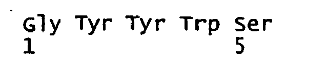
- <210> 69
<211> 48
<212> DNA
<213> artificial sequence - <220>
<223> 3-271 VH: CDR2 nuc
<400> 69
gaaatcaatc atagtggaag caccaactac aacccgtccc tcaagagt 48 - <210> 70
<211> 16
<212> PRT
<213> artificial sequence - <220>
<223> 3-271 VH: CDR2 aa
<400> 70
- <210> 71
<211> 33
<212> DNA
<213> artificial sequence - <220>
<223> 3-271 VH: CDR3 nuc
<400> 71
ggccctgacc gaatggggca tggttttgat atc 33 - <210> 72
<211> 11
<212> PRT
<213> artificial sequence - <220>
<223> 3-271 VH: CDR3 aa
<400> 72
- <210> 73
<211> 321
<212> DNA
<213> artificial sequence - <220>
<223> 3-271 VL: nuc
<400> 73
- <210> 74
<211> 107
<212> PRT
<213> artificial sequence - <220>
<223> 3-271 VL: aa
<400> 74

- <210> 75
<211> 33
<212> DNA
<213> artificial sequence - <220>
<223> 3-271 VL: CDR1 nuc
<400> 75
cgggcaagtc agagcattag cagctattta aat 33 - <210> 76
<211> 11
<212> PRT
<213> artificial sequence - <220>
<223> 3-271 VL: CDR1 aa
<400> 76
- <210> 77
<211> 21
<212> DNA
<213> artificial sequence - <220>
<223> 3-271 VL: CDR2 nuc
<400> 77 21
gctgcatcca gtttgcaaag t 21 - <210> 78
<211> 7
<212> PRT
<213> artificial sequence - <220>
<223> 3-271 VL: CDR2 aa
<400> 78
- <210> 79
<211> 21
<212> DNA
<213> artificial sequence - <220>
<223> 3-271 VL: CDR3 nuc
<400> 79 21
caacagagtt acagtagccc g 21 - <210> 80
<211> 7
<212> PRT
<213> artificial sequence - <220>
<223> 3-271 VL: CDR3 aa
<400> 80
- <210> 81
<211> 381
<212> DNA
<213> artificial sequence - <220>
<223> 3-550 VH: nuc
<400> 81
- <210> 82
<211> 127
<212> PRT
<213> artificial sequence - <220>
<223> 3-550 VH: aa
<400> 82

- <210> 83
<211> 21
<212> DNA
<213> artificial sequence - <220>
<223> 3-550 VH: CDR1 nuc
<400> 83
agcaacagtg ctgcttggaa c 21 - <210> 84
<211> 7
<212> PRT
<213> artificial sequence - <220>
<223> 3-550 VH: CDR1 aa
<400> 84
- <210> 85
<211> 54
<212> DNA
<213> artificial sequence - <220>
<223> 3-550 VH: CDR2 nuc
<400> 85
aggacatact acaggtccaa gtggtataat gattatgcag tatctgtgaa aagt 54 - <210> 86
<211> 18
<212> PRT
<213> artificial sequence - <220>
<223> 3-550 VH: CDR2 aa
<400> 86
- Lys Ser
- <210> 87
<211> 45
<212> DNA
<213> artificial sequence - <220>
<223> 3-550 VH: CDR3 nuc
<400> 87
gatcgtcgac gtatagcagc tcgtcaatac tacggtatgg acgtc 45 - <210> 88
<211> 15
<212> PRT
<213> artificial sequence - <220>
<223> 3-550 VH: CDR3 aa
<400> 88
- <210> 89
<211> 324
<212> DNA
<213> artificial sequence - <220>
<223> 3-550 VL: nuc
<400> 89
- <210> 90
<211> 108
<212> PRT
<213> artificial sequence - <220>
<223> 3-550 VL: aa
<400> 90
- <210> 91
<211> 36
<212> DNA
<213> artificial sequence - <220>
<223> 3-550 VL: CDR1 nuc
<400> 91
agggcaagtc agagtgttag cagcaactac ttagcc 36 - <210> 92
<211> 12
<212> PRT
<213> artificial sequence - <220>
<223> 3-550 VL: CDR1 aa
<400> 92
- <210> 93
<211> 21
<212> DNA
<213> artificial sequence - <220>
<223> 3-550 VL: CDR2 nuc
<400> 93
ggtgcatcca gcagggccac t 21 - <210> 94
<211> 7
<212> PRT
<213> artificial sequence - <220>
<223> 3-550 VL: CDR2 aa
<400> 94
- <210> 95
<211> 21
<212> DNA
<213> artificial sequence - <220>
<223> 3-550 VL: CDR3 nuc
<400> 95
caaaagtata acagtgcccc t 21 - <210> 96
<211> 7
<212> PRT
<213> artificial sequence - <220>
<223> 3-550 VL: CDR3 aa
<400> 96
- <210> 97
<211> 363
<212> DNA
<213> artificial sequence - <220>
<223> 4-10 VH: nuc
<400> 97
- <210> 98
<211> 121
<212> PRT
<213> artificial sequence - <220>
<223> 4-10 VH: aa
<400> 98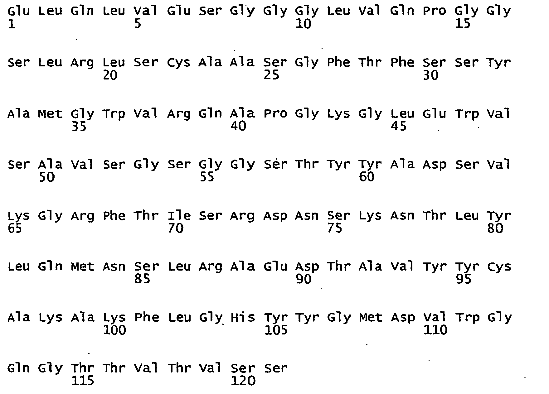
- <210> 99
<211> 15
<212> DNA
<213> artificial sequence - <220>
<223> 4-10 VH: CDR1 nuc
<400> 99
agctatgcca tgggc 15 - <210> 100
<211> 5
<212> PRT
<213> artificial sequence - <220>
<223> 4-10 VH: CDR1 aa
<400> 100
- <210> 101
<211> 51
<212> DNA
<213> artificial sequence - <220>
<223> 4-10 VH: CDR2 nuc
<400> 101
gctgttagtg gtagtggtgg tagcacatac tacgcagact ccgtgaaggg c 51 - <210> 102
<211> 17
<212> PRT
<213> artificial sequence - <220>
<223> 4-10 VH: CDR2 aa
<400> 102
- <210> 103
<211> 36
<212> DNA
<213> artificial sequence - <220>
<223> 4-10 VH: CDR3 nuc
<400> 103
gccaaattcc tgggccacta ctacggtatg gacgtc 36 - <210> 104
<211> 12
<212> PRT
<213> artificial sequence - <220>
<223> 4-10 VH: CDR3 aa
<400> 104
- <210> 105
<211> 321
<212> DNA
<213> artificial sequence - <220>
<223> 4-10 VL: nuc
<400> 105
- <210> 106
<211> 107
<212> PRT
<213> artificial sequence - <220>
<223> 4-10 VL: aa
<400> 106

- <210> 107
<211> 33
<212> DNA
<213> artificial sequence - <220>
<223> 4-10 VL: CDR1 nuc
<400> 107
cgggcaagtc agagcattag cagctattta aat 33 - <210> 108
<211> 11
<212> PRT
<213> artificial sequence - <220>
<223> 4-10 VL: CDR1 aa
<400> 108
- <210> 109
<211> 21
<212> DNA
<213> artificial sequence - <220>
<223> 4-10 VL: CDR2 nuc
<400> 109
gctgcatcca gtttgcaaag t 21 - <210> 110
<211> 7
<212> PRY
<213> artificial sequence - <220>
<223> 4-10 VL: CDR2 aa
<400> 110
- <210> 111
<211> 27
<212> DNA
<213> artificial sequence - <220>
<223> 4-10 VL: CDR3 nuc
<400> 111
cttcagcata atgcttaccc gtacact 27 - <210> 112
<211> 9
<212> PRT
<213> artificial sequence - <220>
<223> 4-10 VL: CDR3 aa
<400> 112
- <210> 113
<211> 369
<212> DNA
<213> artificial sequence - <220>
<223> 4-48 VH: nuc - <113>

- <210> 114
<211> 123
<212> PRT
<213> artificial sequence - <220>
<223> 4-48 VH: aa
<400> 114

- <210> 115
<211> 21
<212> DNA
<213> artificial sequence - <220>
<223> 4-48 VH: CDR1 nuc
<400> 115
aatcctagaa tgggtgtgag c 21 - <210> 116
<211> 7
<212> PRT
<213> artificial sequence - <220>
<223> 4-48 VH: CDR1 aa
<400> 116
- <210> 117
<211> 48
<212> DNA
<213> artificial sequence - <220>
<223> 4-48 VH: COR2 nuc
<400> 117
cacatttttc cgagtgacgc aaaagcccac agtgcatctc tgaagagc 48 - <210> 118
<211> 16
<212> PRT
<213> artificial sequence - <220>
<223> 4-48 VH: CDR2 aa
<400> 118
- <210> 119
<211> 39
<212> DNA
<213> artificial sequence - <220>
<223> 4-48 VH: CDR3 nuc
<400> 119
atattggggg aatactatcc cccagcctgg ttcgacccc 39 - <210> 120
<211> 13
<212> PRT
<213> artificial sequence - <220>
<223> 4-48 VH: CDR3 aa
<400> 120
- <210> 121
<211> 339
<212> DNA
<213> artificial sequence - <220>
<223> 4-48 VL: nuc
<400> 121
- <210> 122
<211> 113
<212> PRT
<213> artificial sequence - <220>
<223> 4-48 VL: aa
<400> 122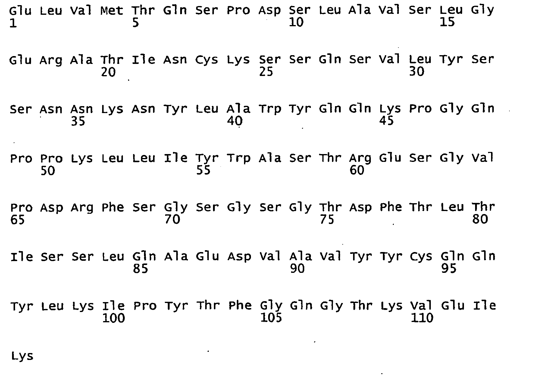
- <210> 123
<211> 51
<212> DNA
<213> artificial sequence - <220>
<223> 4-48 VL: CDR1 nuc
<400> 123
aagtccagcc agagtgtttt atacagctcc aacaataaga actacttagc t 51 - <210> 124
<211> 17
<212> PRT
<213> artificial sequence - <220>
<223> 4-48 VL: CDR1 aa
<400> 124<210> 125
<211> 21
<212> DNA
<213> artificial sequence - <220>
<223> 4-48 VL: CDR2 nuc
<400> 125
tgggcatcta cccgggaatc c 21 - <210> 126
<211> 7
<212> PRT
<213> artificial sequence - <220>
<223> 4-48 VL: CDR2 aa
<400> 126
- <210> 127
<211> 27
<212> DNA
<213> artificial sequence - <220>
<223> 4-48 VL: CDR3 nuc
<400> 127
cagcaatatt tgaaaatccc ttatact 27 - <210> 128
<211> 9
<212> PRT
<213> artificial sequence - <220>
<223> 4-48 VL: CDR3 aa
<400> 128
- <210> 129
<211> 372
<212> DNA
<213> artificial sequence - <220>
<223> anti-CD19 VH: nuc
<400> 129

- <210> 130
<211> 124
<212> PRT
<213> artificial sequence - <220>
<223> anti-CD19 VH: aa
<400> 130
- <210> 131
<211> 333
<212> DNA
<213> artificial sequence - <220>
<223> anti-CD19 VL: nuc
<400> 131

- <210> 132
<211> 111
<212> PRT
<213> artificial sequence - <220>
<223> anti-CD19 VL: aa
<400> 132
- <210> 133
<211> 360
<212> DNA
<213> artificial sequence - <220>
<223> anti-EpCAM (5-10) VH: nuc
<400> 133
- <210> 134
<211> 120
<212> PRT
<213> artificial sequence - <220>
<223> anti-EpCAM (5-10) VH: aa
<400> 134
- <210> 135
<211> 339
<212> DNA
<213> artificial sequence - <220>
<223> anti-EpCAM (5-10) VL: nuc
<400> 135
- <210> 136
<211> 113
<212> PRT
<213> artificial sequence - <220>
<223> anti-EpCAM (5-10) VL: aa
<400> 136
- <210> 137
- <211> 36
<212> DNA
<213> artificial sequence - <220>
<223> 5' BsrGI primer
<400> 137
aggtgtacac tccgatggta atgaagaaat gggtgg 36 - <210> 138
<211> 42
<212> DNA
<213> artificial sequence - <220>
<223> 3' BspEI primer
<400> 138
cgatccggaa tccatctcca tgcagttctc acacactctt gc 42 - <210> 139
<211> 146
<212> DNA
<213> artificial sequence - <220>
<223> mouse Ig leader and cloning site
<400> 139
- <210> 140
<211> 393
<212> DNA
<213> artificial sequence - <220>
<223> CD3-epsilon extracellular domain with leader
<400> 140
- <210> 141
<211> 131
<212> PRT
<213> artificial sequence - <220>
<223> translation of CD3-epsilon extracellular domain with leader
<400> 141
- <210> 142
<211> 22
<212> DNA
<213> artificial sequence - <220>
<223> 5'-huVH1,3,5-xhoI-2001
<400> 142
aggtgcagct gctcgagtct gg 22 - <210> 143
<211> 23
<212> DNA
<213> artificial sequence - <220>
<223> 5'-huVH2-xhoI-2001
<400> 143
cagrtcacct tgctcgagtc tgg 23 - <210> 144
<211> 23
<212> DNA
<213> artificial sequence - <220>
<223> 5'-huVH4-xhoI-2001
<400> 144
caggtgcagc tgctcgagtc ggg 23 - <210> 145
<211> 23
<212> DNA
<213> artificial sequence - <220>
<223> 5'-huVH4B-xhoI-2001
<400> 145 23
caggtgcagc tactcgagtg ggg 23 - <210> 146
<211> 23
<212> DNA
<213> artificial sequence - <220>
<223> 5'-huVH6-xhoI-2001
<400> 146
caggtacagc tgctcgagtc agg 23 - <210> 147
<211> 30
<212> DNA
<213> artificial sequence - <220>
<223> 3'-CD1
<400> 147
tgccttacta gtctctggcc agcggaagat 30 - <210> 148
<211> 17
<212> DNA
<213> artificial sequence - <220>
<223> 3'-hu-VH-BstEII-2001
<400> 148
ctgaggagac ggtgacc 17 - <210> 149
<211> 17
<212> DNA
<213> artificial sequence - <220>
<223> 3'-hu-VH-J3-BstEII
<400> 149
ctgaagagac ggtgacc 17 - <210> 150
<211> 38
<212> DNA
<213> artificial sequence - <220>
<223> 5'-huVK1-Sac-2001
<400> 150
gagccgcacg agcccgagct ccagatgacc cagtctcc 38 - <210> 151
<211> 38
<212> DNA
<213> artificial sequence - <220>
<223> 5'-huVK3-Sac-2001
<400> 151
gagccgcacg agcccgagct cgtgwtgacr cagtctcc 38 - <210> 152
<211> 38
<212> DNA
<213> artificial sequence - <220>
<223> 5'-huVK2/4-Sac-2001
<400> 152
gagccgcacg agcccgagct cgtgatgacy cagtctcc 38 - <210> 153
<211> 38
<212> DNA
<213> artificial sequence - <220>
<223> 5'-huVK5-Sac-2001
<400> 153
gagccgcacg agcccgagct cacactcacg cagtctcc 38 - <210> 154
<211> 38
<212> DNA
<213> artificial sequence - <220>
<223> 5'-huVK6-Sac-2001
<400> 154
gagccgcacg agcccgagct cgtgctgact cagtctcc 38 - <210> 155
<211> 47
<212> DNA
<213> artificial sequence - <220>
<223> 3'-hu-Vk-J1-SpeI-BsiWI
<400> 155
gacgacacta gttgcagcca ccgtacgttt gatttccacc ttggtcc 47 - <210> 156
<211> 47
<212> DNA
<213> artificial sequence - <220>
<223> 3'-hu-Vk-J2/4-SpeI-BsiWI
<400> 156
gacgacacta gttgcagcca ccgtacgttt gatctccasc ttggtcc 47 - <210> 157
<211> 47
<212> DNA
<213> artificial sequence - <220>
<223> 3'-hu-VK-J3-SpeI-BsiWI
<400> 157
gacgacacta gttgcagcca ccgtacgttt gatatccacg ttggtcc 47 - <210> 158
<211> 47
<212> DNA
<213> artificial sequence - <220>
<223> 3'-hu-Vk-J5-SpeI-BsiWI
<400> 158
gacgacacta gttgcagcca ccgtacgttt aatctccagt cgtgtcc 47 - <210> 159
<211> 41
<212> DNA
<213> artificial sequence - <220>
<223> 5'-huVL1a-SacI-2001
<400> 159
gagccgcacg agcccgagct cgtgttgacg cagccgccct c 41 - <210> 160
<211> 41
<212> DNA
<213> artificial sequence - <220>
<223> 5'-huVL1b-SacI-2001
<400> 160
gagccgcacg agcccgagct cgtgctgact cagccaccct c 41 - <210> 161
<211> 44
<212> DNA
<213> artificial sequence - <220>
<223> 5'-huVL2-SacI-2001
<400> 161
gagccgcagg agcccgagct cgccctgact cagcctscct ccgt 44 - <210> 162
<211> 35
<212> DNA
<213> artificial sequence - <220>
<223> 5'-huVL4-SacI-2001
<400> 162
acctgcgagc tcgtgctgac tcarycmycc tctgc 35 - <210> 163
<211> 33
<212> DNA
<213> artificial sequence - <220>
<223> 5'-huVL5-SacI-2001
<400> 163
acctgcgagc tcgtgctgac tcagccrsct tcc 33 - <210> 164
<211> 32
<212> DNA
<213> artificial sequence - <220>
<223> 5'-huVL6-sacI-2001
<400> 164
acctgcgagc tcatgctgac tcagccccac tc 32 - <210> 165
<211> 41
<212> DNA
<213> artificial sequence - <220>
<223> 5'-huVL3/9-SacI-2001
<400> 165
gagccgcacg agcccgagct cgwgctgact cagccaccyt c 41 - <210> 166
<211> 41
<212> DNA
<213> artificial sequence - <220>
<223> 5'-huVL7/8-SacI-2001
<400> 166
gagccgcacg agcccgagct cgtggtgacy caggagccmt c 41 - <210> 167
<211> 33
<212> DNA
<213> artificial sequence - <220>
<223> 3'-hu-Vlam-BlnI-SpeI-2001
<400> 167
cgtgggacta gtcttgggct gacctaggac ggt 33 - <210> 168
<211> 33
<212> DNA
<213> artificial sequence - <220>
<223> 3'-hu-Vlam2-BlnI-SpeI-2002
<400> 168
cgtgggacta gtcttgggct gaccgaggac ggt 33 - <210> 169
<211> 36
<212> DNA
<213> artificial sequence - <220>
<223> 5'-Mouse ckappa BsiwI
<400> 169
actcgacgta cggatgctgc accaactgta tccatc 36 - <210> 170
<211> 58
<212> DNA
<213> artificial sequence - <220>
<223> 3'-Mouse-ck-myc
<400> 170
cagatcttcc tctgagatca gcttctgttc ctcacactca ttcctgttga agctcttg 58 - <210> 171
<211> 68
<212> DNA
<213> artificial sequence - <220>
<223> 3'-myc-His6-Not
<400> 171
- <210> 172
<211> 34
<212> DNA
<213> artificial sequence
<220>
<223> 5'-5BVH-BsrGI
<400> 172
acgtcatgta cactccgagg tgcagctgct cgag 34 - <210> 173
<211> 35
<212> DNA
<213> artificial sequence - <220>
<223> 3'-5BVL-BspEI
<400> 173
tgcatgtccg gacgtacgtt tgatctcaag cttgg 35 - <210> 174
<211> 45
<212> DNA
<213> artificial sequence - <220>
<223> (G4S)3 linker nuc
<400> 174
ggtggtggtg gttctggcgg cggcggctcc ggtggtggtg gttct 45 - <210> 175
<211> 15
<212> PRT
<213> artificial sequence - <220>
<223> (G4S)3 linker aa
<400> 175
- <210> 176
<211> 15
<212> DNA
<213> artificial sequence - <220>
<223> (G4S)1 linker nuc
<400> 176
ggaggtggtg gatcc 15 - <210> 177
<211> 5
<212> PRT
<213> artificial sequence - <220>
<223> (G4S)1 linker aa
<400> 177
- <210> 178
<211> 383
<212> DNA
<213> artificial sequence - <220>
<223> mouse ckappa-myc-his6
<400> 178
- <210> 179
<211> 124
<212> PRT
<213> artificial sequence - <220>
<223> translation of mouse ckappa-myc-his6
<400> 179
- <210> 180
<211> 741
<212> DNA
<213> artificial sequence - <220>
<223> 3-106 nuc
<400> 180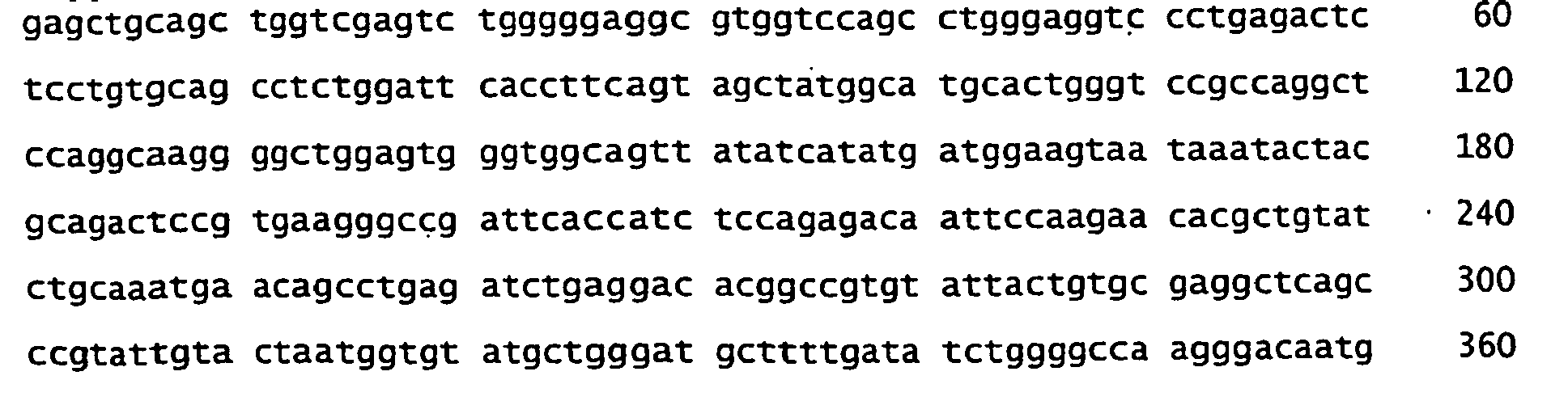
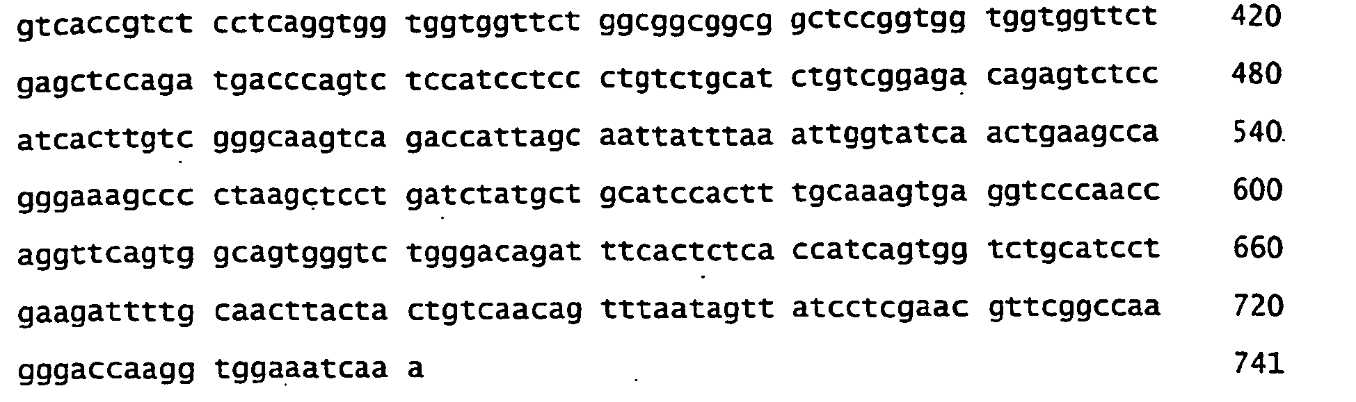
- <210> 181
<211> 247
<212> PRT
<213> artificial sequence - <220>
<223> 3-106 aa
<400> 181
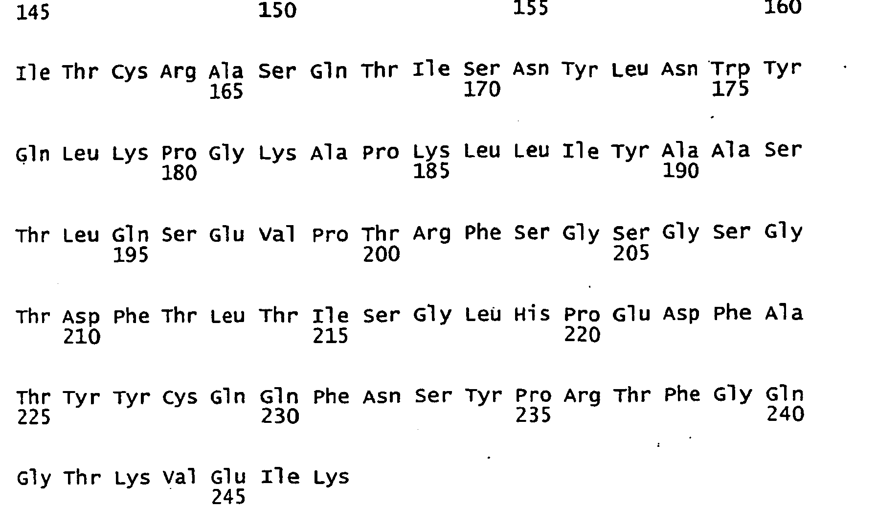
- <210> 182
<211> 732
<212> DNA
<213> artificial sequence - <220>
<223> 3-114 nuc
<400> 182
- <210> 183
<211> 244
<212> PRT
<213> artificial sequence - <220>
<223> 3-114 aa
<400> 183

- <210> 184
<211> 726
<212> DNA
<213> artificial sequence - <220>
<223> 3-148 nuc
<400> 184
- <210> 185
<211> 242
<212> PRT
<213> artificial sequence - <220>
<223> 3-148 aa
<400> 185
- <210> 186
<211> 744
<212> DNA
<213> artificial sequence - <220>
<223> 3-190 nuc
<400> 186
- <210> 187
<211> 248
<212> PRT
<213> artificial sequence - <220>
<223> 3-190 aa
<400> 187

- <210> 188
<211> 723
<212> DNA
<213> artificial sequence - <220>
<223> 3-271 nuc
<400> 188

- <210> 189
<211> 241
<212> PRT
<213> artificial sequence - <220>
<223> 3-271 aa
<400> 189

- <210> 190
<211> 750
<212> DNA
<213> artificial sequence - <220>
<223> 3-550 nuc
<400> 190

- <210> 191
<211> 250
<212> PRT
<213> artificial sequence - <220>
<223> 3-550 aa
<400> 191

- <210> 192
<211> 729
<212> DNA
<213> artificial sequence - <220>
<223> 4-10 nuc
<400> 192
- <210> 193
<211> 243
<212> PRT
<213> artificial sequence - <220>
<223> 4-10 aa
<400> 193
- <210> 194
<211> 753
<212> DNA
<213> artificial sequence - <220>
<223> 4-48 nuc
<400> 194
- <210> 195
<211> 251
<212> PRT
<213> artificial sequence - <220>
<223> 4-48 aa
<400> 195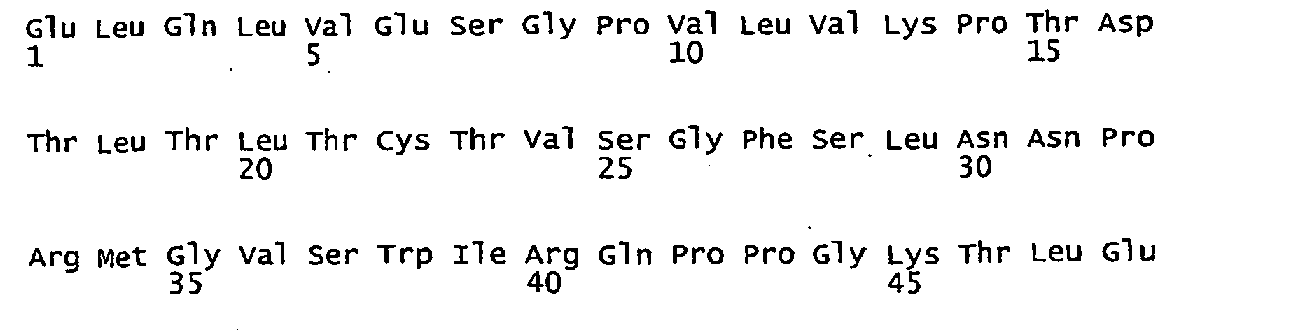

- <210> 196
<211> 354
<212> DNA
<213> artificial sequence - <220>
<223> anti-CCR5 VH nuc
<400> 196

- <210> 197
<211> 118
<212> PRT
<213> artificial sequence - <220>
<223> anti-CCR5 VH aa
<400> 197
- <210> 198
<211> 333
<212> DNA
<213> artificial sequence - <220>
<223> anti-CCR5 VL nuc
<400> 198
- <210> 199
<211> 111
<212> PRT
<213> artificial sequence - <220>
<223> anti-CCR5 VL aa
<400> 199
- <210> 200
<211> 366
<212> DNA
<213> artificial sequence - <220>
<223> anti-CD20 VH: nuc
<400> 200
- <210> 201
<211> 122
<212> PRT
<213> artificial sequence - <220>
<223> anti-CD20 VH: aa
<400> 201

- <210> 202
<211> 318
<212> DNA
<213> artificial sequence - <220>
<223> anti-CD20 VL: nuc
<400> 202
- <210> 203
<211> 106
<212> PRT
<213> artificial sequence - <220>
<223> anti-CD20 VL:aa
<400> 203

- <210> 204
<211> 21
<212> DNA
<213> artificial sequence - <220>
<223> leader seq
<400> 204 21
cctcttcttg gtagcaacag c 21 - <210> 205
<211> 1491
<212> DNA
<213> artificial sequence - <220>
<223> Human anti-CD3 3-148 VH-VL x anti-CD19 VL-VH
<400> 205
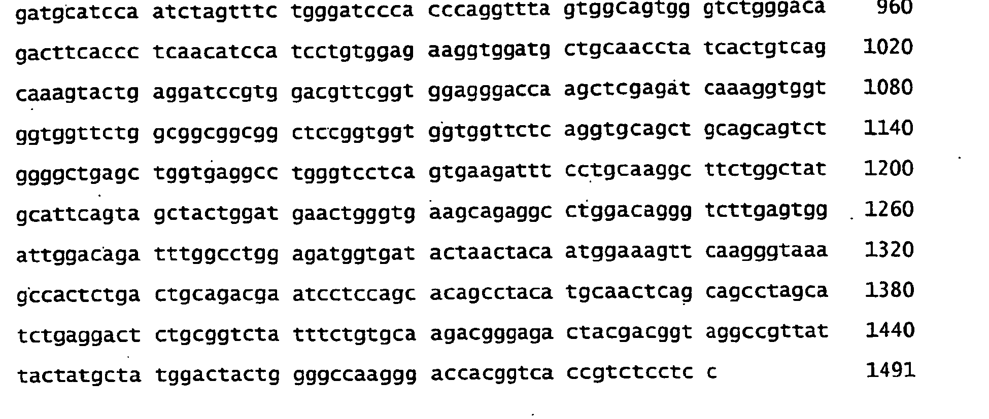
- <210> 206
<211> 497
<212> PRT
<213> artificial sequence - <220>
<223> Human anti-CD3 3-148 VH-VL x anti-CD19 VL-VH
<400> 206


- <210> 207
<211> 1509
<212> DNA
<213> artificial sequence - <220>
<223> Human anti-CD3 3-190 VH-VL x anti-CD19 VL-VH
<400> 207

- <210> 208
<211> 503
<212> PRT
<213> artificial sequence - <220>
<223> Human anti-CD3 3-190 VH-VL x anti-CD19 VL-VH
<400> 208


- <210> 209
<211> 1488
<212> DNA
<213> artificial sequence - <220>
<223> Human anti-CD3 3-271 VH-VL x anti-CD19 VL-VH
<400> 209

- <210> 210
<211> 496
<212> PRT
<213> artificial sequence - <220>
<223> Human anti-CD3 3-271 VH-VL x anti-CD19 VL-VH
<400> 210


- <210> 211
<211> 1494
<212> DNA
<213> artificial sequence - <220>
<223> Human anti-CD3 4-10 VH-VL x anti-CD19 VL-VH
<400> 211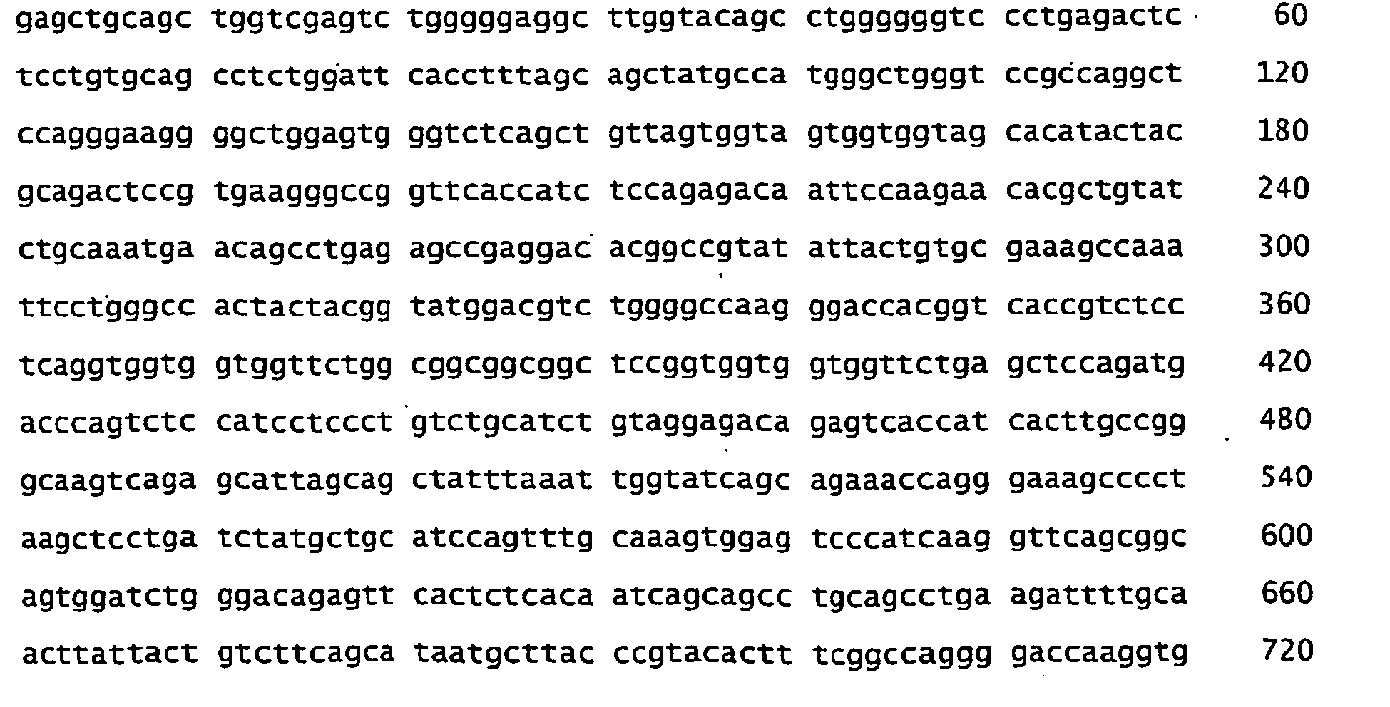

- <210> 212
<211> 498
<212> PRT
<213> artificial sequence - <220>
<223> Human anti-CD3 4-10 VH-VL x anti-CD19 VL-VH
<400> 212


- <210> 213
<211> 1518
<212> DNA
<213> artificial sequence - <220>
<223> Human anti-CD3 4-48 VH-VL x anti-CD19 VL-VH
<400> 213

- <210> 214
<211> 506
<212> PRT
<213> artificial sequence - <220>
<223> Human anti-CD3 4-48 VH-VL x anti-CD19 VL-VH
<400> 214

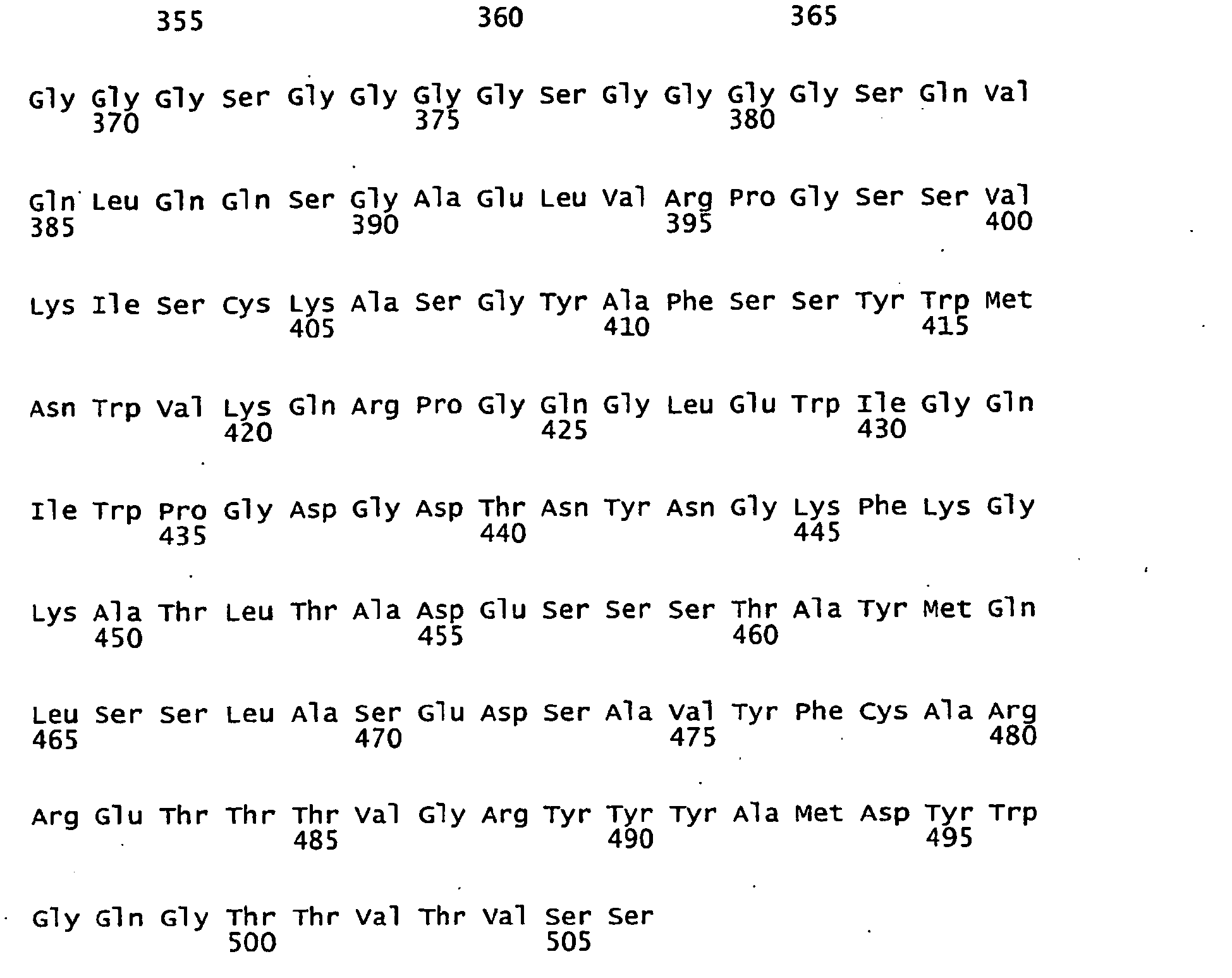
- <210> 215
<211> 1491
<212> DNA
<213> artificial sequence - <220>
<223> anti-CD19 VL-VH x human anti-CD3 3-148 VH-VL
<400> 215

- <210> 216
<211> 497
<212> PRT
<213> artificial sequence - <220>
<223> anti-CD19 VL-VH x human anti-CD3 3-148 VH-VL
<400> 216


- <210> 217
<211> 1509
<212> DNA
<213> artificial sequence - <220>
<223> anti-CD19 VL-VH x human anti-CD3 3-190 VH-VL
<400> 217

- <210> 218
<211> 503
<212> PRT
<213> artificial sequence - <220>
<223> anti-CD19 VL-VH x human anti-CD3 3-190 VH-VL
<400> 218


- <210> 219
<211> 1488
<212> DNA
<213> artificial sequence - <220>
<223> anti-CD19 VL-VH x human anti-CD3 3-271 VH-VL
<400> 219

- <210> 220
<211> 496
<212> PRT
<213> artificial sequence - <220>
<223> anti-CD19 VL-VH x human anti-CD3 3-271 VH-VL
<400> 220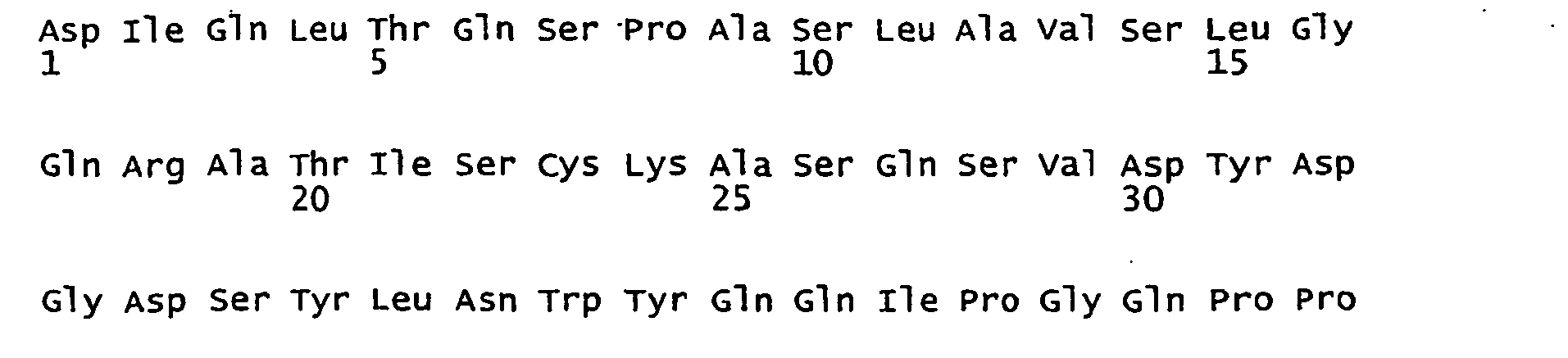


- <210> 221
<211> 1494
<212> DNA
<213> artificial sequence - <220>
<223> anti-CD19 VL-VH x human anti-CD3 4-10 VH-VL
<400> 221

- <210> 222
<211> 498
<212> PRT
<213> artificial sequence - <220>
<223> anti-CD19 VL-VH x human anti-CD3 4-10 VH-VL
<400> 222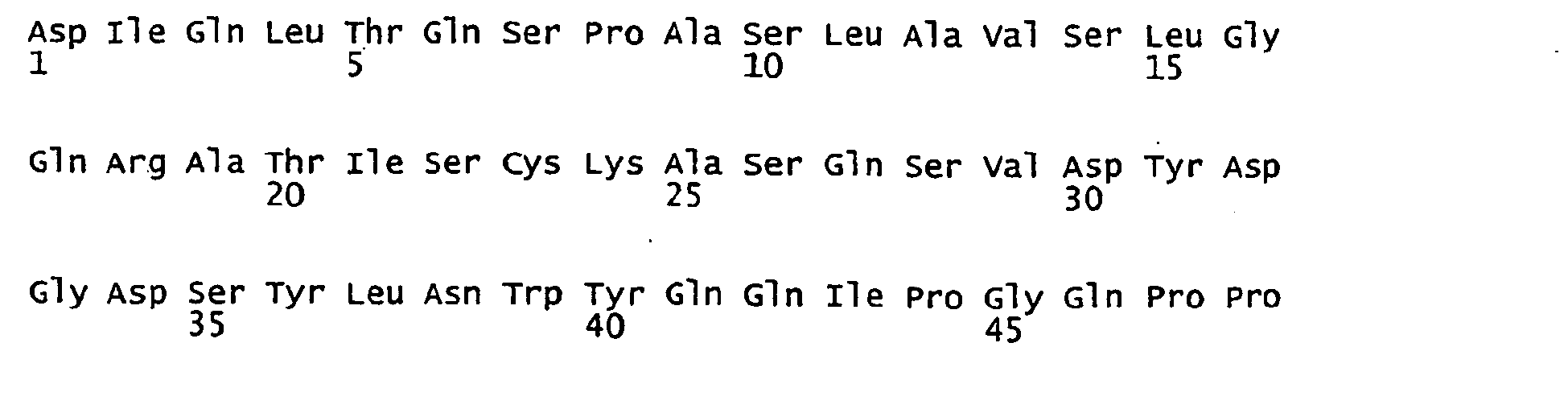


- <210> 223
<211> 1518
<212> DNA
<213> artificial sequence - <220>
<223> anti-CD19 VL-VH x human anti-CD3 4-48 VH-VL
<400> 223

- <210> 224
<211> 506
<212> PRT
<213> artificial sequence - <220>
<223> anti-CD19 VL-VH x human anti-CD3 4-48 VH-VL
<400> 224


- <210> 225
<211> 1485
<212> DNA
<213> artificial sequence - <220>
<223> Human anti-CD3 3-148 VH-VL x anti-EpCAM 5-10 VH-VL
<400> 225
- <210> 226
<211> 495
<212> PRT
<213> artificial sequence - <220>
<223> Human anti-CD3 3-148 VH-VL x anti-EpCAM 5-10 VH-VL
<400> 226


- <210> 227
<211> 1503
<212> DNA
<213> artificial sequence - <220>
<223> Human anti-CD3 3-190 VH-VL x anti- EpCAM 5-10 VH-VL
<400> 227

- <210> 228
<211> 501
<212> PRT
<213> artificial sequence - <220>
<223> Human anti-CD3 3-190 VH-VL x anti- EpCAM 5-10 VH-VL
<400> 228

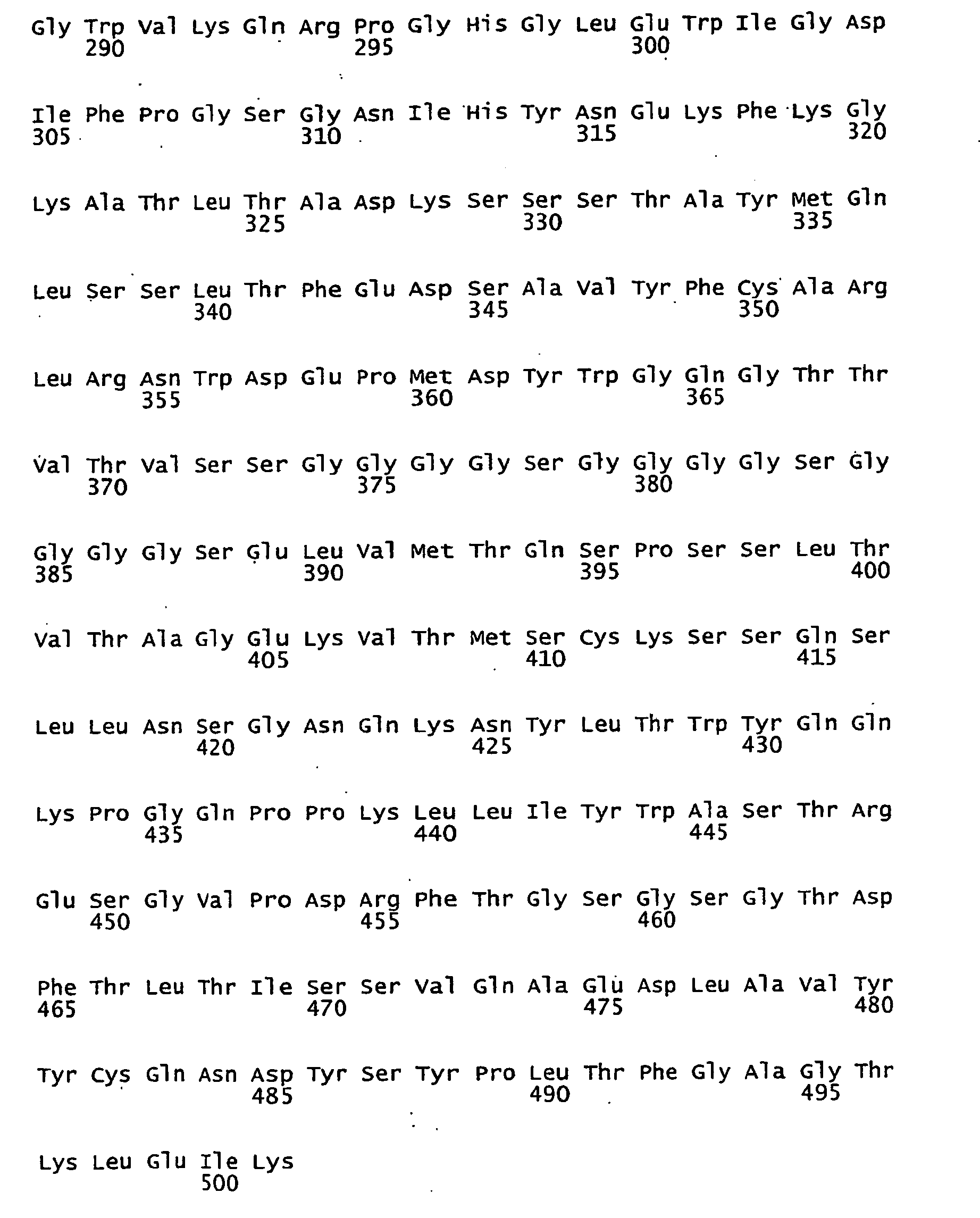
- <210> 229
<211> 1482
<212> DNA
<213> artificial sequence - <220>
<223> Human anti-CD3 3-271 VH-VL x anti- EpCAM 5-10 VH-VL
<400> 229
- <210> 230
<211> 494
<212> PRT
<213> artificial sequence - <220>
<223> Human anti-CD3 3-271 VH-VL x anti- EpCAM 5-10 VH-VL
<400> 230

- <210> 231
<211> 1488
<212> DNA
<213> artificial sequence - <220>
<223> Human anti-CD3 4-10 VH-VL x anti- EpCAM 5-10 VH-VL
<400> 231
- <210> 232
<211> 496
<212> PRT
<213> artificial sequence - <220>
<223> Human anti-CD3 4-10 VH-VL x anti- EpCAM 5-10 VH-VL
<400> 232

- <210> 233
<211> 1512
<212> DNA
<213> artificial sequence - <220>
<223> Human anti-CD3 4-48 VH-VL x anti- EpCAM 5-10 VH-VL
<400> 233
- <210> 234
<211> 504
<212> PRT
<213> artificial sequence - <220>
<223> Human anti-CD3 4-48 VH-VL x anti- EpCAM 5-10 VH-VL
<400> 234

- <210> 235
<211> 1485
<212> DNA
<213> artificial sequence - <220>
<223> anti-EpCAM 5-10 VL-VH x human anti-CD3 3-148 VH-VL
<400> 235
- <210> 236
<211> 495
<212> PRT
<213> artificial sequence - <220>
- <223> anti-EpCAM 5-10 VL-VH x human anti-CD3 3-148 VH-VL
<400> 236

- <210> 237
<211> 1503
<212> DNA
<213> artificial sequence - <220>
<223> anti-EpCAM 5-10 VL-VH x human anti-CD3 3-190 VH-VL
<400> 237
- <210> 238
<211> 501
<212> PRT
<213> artificial sequence - <220>
<223> anti-EpCAM 5-10 VL-VH x human anti-CD3 3-190 VH-VL
<400> 238

- <210> 239
<211> 1482
<212> DNA
<213> artificial sequence - <220>
<223> anti-EpCAM 5-10 VL-VH x human anti-CD3 3-271 VH-VL
<400> 239
- <210> 240
<211> 494
<212> PRT
<213> artificial sequence - <220>
<223> anti-EpCAM 5-10 VL-VH x human anti-CD3 3-271 VH-VL
<400> 240

- <210> 241
<211> 1488
<212> DNA
<213> artificial sequence - <220>
<223> anti-EpCAM 5-10 VL-VH x human anti-CD3 4-10 VH-VL
<400> 241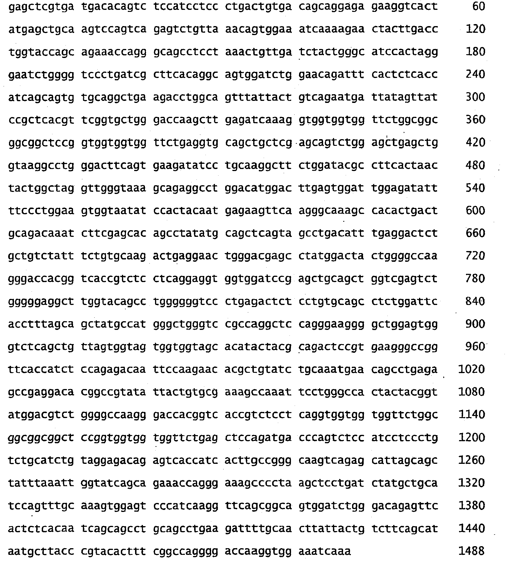
- <210> 242
<211> 496
<212> PRT
<213> artificial sequence - <220>
<223> anti-EpCAM 5-10 VL-VH x human anti-CD3 4-10 VH-VL
<400> 242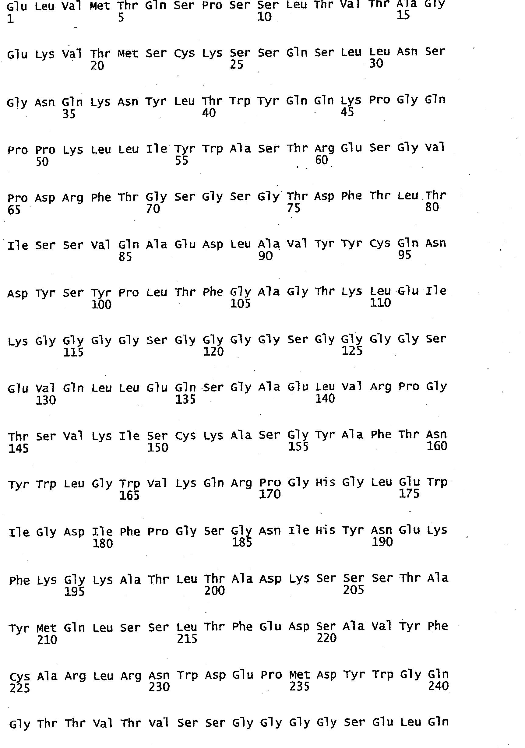

- <210> 243
<211> 1512
<212> DNA
<213> artificial sequence - <220>
<223> anti-EpCAM 5-10 VL-VH x human anti-CD3 4-48 VH-VL
<400> 243
- <210> 244
<211> 504
<212> PRT
<213> artificial sequence - <220>
<223> anti-EpCAM 5-10 VL-VH x human anti-CD3 4-48 VH-VL
<400> 244

- <210> 245
<211> 1488
<212> DNA
<213> artificial sequence - <220>
<223> anti-CD20 VL-VH x human anti-CD3 3-190 VH-VL
<400> 245
- <210> 246
<211> 496
<212> PRT
<213> artificial sequence - <220>
<223> anti-CD20 VL-VH x human anti-CD3 3-190 VH-VL
<400> 246

- <210> .247
<211> 1470
<212> DNA
<213> artificial sequence - <220>
<223> anti-CD20 VL-VH x human anti-CD3 3-148 VH-VL
<400> 247
- <210> 248
<211> 490
<212> PRT
<213> artificial sequence - <220>
<223> anti-CD20 VL-VH x human anti-CD3 3-148 VH-VL
<400> 248

- <210> 249
<211> 1491
<212> DNA
<213> artificial sequence - <220>
<223> anti-CCR5 VL-VH x human anti-CD3 3-190 VH-VL
<400> 249
- <210> 250
<211> 497
<212> PRT
<213> artificial sequence - <220>
<223> anti-CCR5 VL-VH x human anti-CD3 3-190 VH-VL
<400> 250

- <210> 251
<211> 1473
<212> DNA
<213> artificial sequence - <220>
<223> anti-CCR5 VL-VH x human anti-CD3 3-148 VH-VL
<400> 251
- <210> 252
<211> 491
<212> PRT
<213> artificial sequence - <220>
<223> anti-CCRS VL-VH x human anti-CD3 3-148 VH-VL
<400> 252

It is well known in the art how to perform hybridization experiments with nucleic acid molecules, i.e. the person skilled in the art knows what hybridization conditions s/he has to use in accordance with the present invention. Such hybridization conditions are referred to in standard text books such as Sambrook et al. (loc cit.) and other standard laboratory manuals known by the person skilled in the art or as recited above. Preferred in accordance with the present inventions are polynucleotides which are capable of hybridizing to the polynucleotides of the invention or parts thereof, under stringent hybridization conditions.
"Stringent hybridization conditions" refer, i.e. to an overnight incubation at 42°C in a solution comprising 50% formamide, 5x SSC (750 mM NaCl, 75 mM sodium citrate), 50 mM sodium phosphate (pH 7.6), 5x Denhardt's solution, 10% dextran sulfate, and 20 µg/ml denatured, sheared salmon sperm DNA, followed by washing the filters in 0.1 x SSC at about 65°C. Also contemplated are nucleic acid molecules that hybridize to the polynucleotides of the invention at lower stringency hybridization conditions. Changes in the stringency of hybridization and signal detection are primarily accomplished through the manipulation of formamide concentration (lower percentages of formamide result in lowered stringency); salt conditions, or temperature. For example, lower stringency conditions include an overnight incubation at 37°C in a solution comprising 6X SSPE (20X SSPE = 3M NaCl; 0.2M NaH2PO4; 0.02M EDTA, pH 7.4), 0.5% SDS, 30% formamide, 100 µg/ml salmon sperm blocking DNA; followed by washes at 50°C with 1 X SSPE, 0.1% SDS. In addition, to achieve even lower stringency, washes performed following stringent hybridization can be done at higher salt concentrations (e.g. 5X SSC). It is of note that variations in the above conditions may be accomplished through the inclusion and/or substitution of alternate blocking reagents used to suppress background in hybridization experiments. Typical blocking reagents include Denhardt's reagent, BLOTTO, heparin, denatured salmon sperm DNA, and commercially available proprietary formulations. The inclusion of specific blocking reagents may require modification of the hybridization conditions described above, due to problems with compatibility.
The recited nucleic acid molecules may be, e.g., DNA, cDNA, RNA or synthetically produced DNA or RNA or a recombinantly produced chimeric nucleic acid molecule comprising any of those polynucleotides either alone or in combination.
The term "mature form of the protein" defines in the context of the present invention a protein translated from its corresponding mRNA and optionally subsequently modified.
The term "hybridizing" has been defined in the context of the present invention herein above.
It is evident to the person skilled in the art that regulatory sequences may be added to the nucleic acid molecule of the invention. For example, promoters, transcriptional enhancers and/or sequences which allow for induced expression of the polynucleotide of the invention may be employed. A suitable inducible system is for example tetracycline-regulated gene expression as described, e.g., by Gossen and Bujard (Proc. Natl. Acad. Sci. USA 89 (1992), 5547-5551) and Gossen et al. (Trends Biotech. 12 (1994), 58-62), or a dexamethasone-inducible gene expression system as described, e.g. by Crook (1989) EMBO J. 8, 513-519.
These cells were harvested after 20 hours by centrifugation and typically resuspended in 500 µl PBS. Through four rounds of freezing at -70°C and thawing at 37°C the outer membrane of the bacteria was destroyed by temperature shock so that the soluble periplasmic proteins including the scFv fusion-proteins were released into the liquid. After elimination of intact cells and cell-debris by centrifugation, the supernatant was tested by ELISA for CD3 epsilon-binding scFv-fusion-proteins.
Detection of scFv-fragments bound to immobilized recombinant human CD3 epsilon antigen was carried out using a biotinylated anti-mouse kappa antibody (Southern Biotech, 1µg/ml PBS) detected with horseradish peroxidase-conjugated avidine (Dako, 1 µg/ml PBS). The signal was developed by adding ABTS substrate solution and detected at a wavelength of 405 nm. In contrast to PBS as negative control and most of the clones after three rounds of panning, many clones after four rounds of panning already showed significant ELISA signals.
Sequence analysis showed that most of the positive clones from
huLIB-
Cells successfully transduced with a phagemid copy encoding a human scFv-fragment were again selected for Carbenicillin resistance and subsequently infected with VCMS13 helper phage to start the second round of antibody display and in vitro selection. After five rounds of production and selection for antigen-binding scFv-displaying phage, plasmid DNA from E. coli cultures was isolated corresponding to 3, 4 and 5 rounds of panning as well as to the unselected repertoire prior to the first round of panning.
The cloning and expression of soluble scFvs in E. coli and the processing of the periplasmic preparations were carried out as described earlier for selection of huLIB-ST on recombinant antigen. These periplasmic preparations of single clones were then tested by ELISA for CD3epsilon-binding scFv-fusion-proteins. Detection of scFv-fragments bound to immobilized recombinant human CD3 epsilon antigen was carried out using a biotinylated anti-mouse kappa antibody (Southern Biotech, 1 µg/ml PBS) detected with horseradish peroxidase-conjugated avidine (Dako, 1 µg/ml PBS). The signal was developed by adding ABTS substrate solution and detected at a wavelength of 405 nm. Compared to PBS as negative control,
Human CD3-specific antibodies OKT3 (Janssen Cilag GmbH), SP34 (Pharmingen) and UCHT1 (Pharmingen) were also tested for binding to the recombinant ("r") CD3-epsilon antigen (
As shown in
To exclude the possibility that the positive binding shown in
Gel filtration chromatography was performed on a Sephadex S200 HiPrep column (Pharmacia) equilibrated with PBS (Gibco). Eluted protein samples (flow
Purified bispecific protein was analyzed by SDS PAGE under reducing conditions performed with pre-cast 4-12% Bis Tris gels (Invitrogen). Sample preparation and application were performed according to the protocol provided by the manufacturer. The molecular weight was determined with MultiMark protein standard (Invitrogen). The gel was stained with colloidal Coomassie (invitrogen protocol). Western Blot was performed using an Optitran® BA-S83 membrane and the Invitrogen Blot Module according to the protocol provided by the manufacturer. The antibodies used were directed against the His Tag (Penta His, Qiagen) and goat-anti-mouse lg labeled with alkaline phosphatase (AP) (Sigma), and BCIP/NBT (Sigma) as substrate. The single chain bispecific antibody could be specifically detected by Western Blot.
Claims (30)
- A human binding molecule specifically binding to/interacting with the human CD3 complex, whereby said binding molecule is an antibody molecule, antibody fragment or derivative thereof or an antibody construct comprising(a) the mature form of a protein comprising the amino acid sequence of human binding molecule of the invention, as given in SEQ ID NOs: 189, 210, 220, 230 or 240;(b) an amino acid sequence encoded by a nucleotide sequence comprising or consisting of the DNA sequence as given in SEQ ID NOs: 188, 209, 219, 229 or 239; and(c) an amino acid sequence encoded by a nucleotide sequence which is degenerate as a result of the genetic code to a nucleotide sequence of (b).
- The human binding molecule of claim 1, wherein said derivative of an antibody is an scFv.
- The human binding molecule according to claim 1 or 2, additionally comprising at least one further antigen-interaction-site and/or at least one further effector domain.
- The human binding molecule according to claim 3, which is humanized and/or deimmunized.
- The human binding molecule according to claim 3 or 4, wherein said at least one further antigen-interaction-site is specific for one or more cell surface molecule.
- The human binding molecule according to claim 5, wherein said cell surface molecule is a tumor specific marker.
- The human binding molecule according to any of claims 3 to 6, wherein said at least one further antigen-interaction-site is a further scFv.
- The human binding molecule according to any of claims 3 to 7, wherein said at least one further antigen-interaction-site specifically binds to/interacts with an antigen selected from the group consisting of EpCAM, CCR5, CD19, EphA2, HER-2 neu, HER-3, HER-4, EGFR, PSMA, CEA, MUC-1 (mucin), MUC2, MUC3, MUC4, MUC5AC. MUC5B, MUC7, bhCG, Lewis-Y, CD20, CD33, CD30, ganglioside GD3, 9-O-Acetyl-GD3, GM2, Globo H, fucosyl GM1, Poly SA, GD2, Carboanhydrase IX (MN/CA IX), CD44v6, Sonic Hedgehog (Shh), Wue-1, Plasma Cell Antigen, (membrane-bound) IgE, Melanoma Chondroitin Sulfate Proteoglycan (MCSP), CCR8, TNF-alpha precursor, STEAP, mesothelin, A33 Antigen, Prostate Stem Cell Antigen (PSCA), Ly-6, desmoglein 4, E-cadherin neoepitope, Fetal Acetylcholine Receptor, CD25, CA19-9 marker, CA-125 marker, Muellerian Inhibitory Substance (MIS) Receptor type II, sTn (sialylated Tn antigen; TAG-72), FAP (fibroblast activation antigen), endosialin, EGFRvIII, LG, SAS and CD63.
- The human binding molecule according to claim 8, wherein said further antigen binding site specifically binds to/interacts with the CD19 molecule.
- The human binding molecule according to claim 9, comprising an amino acid sequence selected from the group of(a) an amino acid sequence corresponding to construct 33prot to 40 prot of table 2;(b) an amino acid sequence encoded by a nucleic acid sequence corresponding to construct 33nuc to 40nuc of table 1; and(c) an amino acid sequence encoded by a nucleic acid sequence which is degenerate as a result of the genetic code to a nucleotide sequence of (b).
- The human binding molecule according to claim 8, wherein said further antigen binding site specifically binds to/interacts with the EpCAM molecule.
- The human binding molecule according to claim 11, comprising an amino acid sequence selected from the group of(a) an amino acid sequence corresponding to construct 97prot to 104prot of table 4;(b) an amino acid sequence encoded by a nucleic acid sequence corresponding to construct 97nuc to 104nuc of table 3; and(c) an amino acid sequence encoded by a nucleic acid sequence which is degenerate as a result of the genetic code to a nucleotide sequence of (b).
- The human binding molecule according to claim 8, wherein said further antigen binding site specifically binds to/interacts with the CCR5 molecule.
- The human binding molecule according to claim 13, comprising an amino acid sequence selected from the group of(a) an amino acid sequence corresponding to construct 161prot to 168prot of table 6;(b) an amino acid sequence encoded by a nucleic acid sequence corresponding to construct 161 nuc to 168nuc of table 5; and(c) an amino acid sequence encoded by a nucleic acid sequence which is degenerate as a result of the genetic code to a nucleotide sequence of (b).
- The human binding molecule according to claim 8, wherein said further antigen binding site specifically binds to/interacts with the CD20 molecule.
- The human binding molecule according to claim 15, comprising an amino acid sequence selected from the group of(a) an amino acid sequence corresponding to construct 225prot to 232prot of table 8;(b) an amino acid sequence encoded by a nucleic acid sequence corresponding to construct 225nuc to 232nuc of table 7; and(c) an amino acid sequence encoded by a nucleic acid sequence which is degenerate as a result of the genetic code to a nucleotide sequence of (b).
- A nucleic acid sequence encoding a human binding molecule according to any of claims 1 to 16.
- The nucleic acid molecule of claim 17 comprising a nucleotide sequence selected from the group consisting of:(a) a nucleotide sequence encoding the mature form of a protein comprising the amino acid sequence of human binding molecule of the invention, as given in SEQ ID NOs: 189, 210, 220, 230 or 240;(b) a nucleotide sequence comprising or consisting of the DNA sequence as given in SEQ ID NOs: 188, 209, 219, 229 or 239; and(c) a nucleotide sequence which is degenerate as a result of the genetic code to a nucleotide sequence of (a) or (b).
- A vector comprising a nucleic acid sequence according to claim 17 or 18.
- The vector of claim 19, which further comprises a nucleic acid sequence which is a regulatory sequence operably linked to said nucleic acid sequence according to claim 17 or 18.
- The vector of claim 19 or 20, wherein the vector is an expression vector.
- A non-human host transformed or transfected with a vector according to any of claims 19 to 21.
- A process for the production of a human binding molecule according to any of clams 1 to 16 said process comprising culturing a host of claim 22 under conditions allowing the expression of the polypeptide construct and recovering the produced polypeptide construct from the culture.
- A composition comprising a human binding molecule according to any of claims 1 to 16 or as produced by the process of claim 23, a nucleic acid molecule of claim 17 or 18, a vector of any one of claims 19 to 21 or a host of claim 22 and, optionally, a proteinaceous compound capable of providing an activation signal for immune effector cells.
- The composition of claim 24 which is a pharmaceutical composition further comprising, optionally, suitable formulations of carrier, stabilizers and/or excipients.
- The composition of claim 24 which is a diagnostic composition further comprising, optionally, means and methods for detection.
- Use of a human binding molecule according to any of claims 1 to 16 or as produced by the process of claim 23, a nucleic acid molecule of claim 17 or 18, a vector of any one of claims 19 to 21 or a host of claim 22 for the preparation of a pharmaceutical composition for the prevention, treatment or amelioration of a proliferative disease, a tumorous disease, an inflammatory disease, an immunological disorder, an autoimmune disease, an infectious disease, viral disease, allergic reactions, parasitic reactions, graft-versus-host diseases or host-versus-graft diseases.
- The use of claim 27, wherein the pharmaceutical composition is to be administered in combination with a proteinaceous compound capable of providing an activation signal for immune effector cells.
- The use of claim 28, wherein said proteinaceous compound is to be administered simultaneously or non-simultaneously with the pharmaceutical composition.
- A kit comprising a human binding molecule according to any of claims 1 to 16 or as produced by the process of claim 23, a nucleic acid molecule of claim 17 or 18, a vector of any one of claims 19 to 21 or a host of claim 22.
Priority Applications (2)
| Application Number | Priority Date | Filing Date | Title |
|---|---|---|---|
| EP04739377A EP1629011B1 (en) | 2003-05-31 | 2004-05-26 | Human anti-human cd3 binding molecules |
| SI200431372T SI1629011T1 (en) | 2003-05-31 | 2004-05-26 | Human anti-human cd3 binding molecules |
Applications Claiming Priority (3)
| Application Number | Priority Date | Filing Date | Title |
|---|---|---|---|
| EP03012132 | 2003-05-31 | ||
| EP04739377A EP1629011B1 (en) | 2003-05-31 | 2004-05-26 | Human anti-human cd3 binding molecules |
| PCT/EP2004/005684 WO2004106380A2 (en) | 2003-05-31 | 2004-05-26 | Human-anti-human cd3 binding molecules |
Publications (2)
| Publication Number | Publication Date |
|---|---|
| EP1629011A2 EP1629011A2 (en) | 2006-03-01 |
| EP1629011B1 true EP1629011B1 (en) | 2010-01-13 |
Family
ID=33483897
Family Applications (1)
| Application Number | Title | Priority Date | Filing Date |
|---|---|---|---|
| EP04739377A Expired - Lifetime EP1629011B1 (en) | 2003-05-31 | 2004-05-26 | Human anti-human cd3 binding molecules |
Country Status (9)
| Country | Link |
|---|---|
| US (1) | US8784821B1 (en) |
| EP (1) | EP1629011B1 (en) |
| AT (1) | ATE455127T1 (en) |
| AU (1) | AU2004242845B2 (en) |
| CA (1) | CA2523716C (en) |
| DE (1) | DE602004025101D1 (en) |
| DK (1) | DK1629011T3 (en) |
| SI (1) | SI1629011T1 (en) |
| WO (1) | WO2004106380A2 (en) |
Cited By (7)
| Publication number | Priority date | Publication date | Assignee | Title |
|---|---|---|---|---|
| US8772459B2 (en) | 2009-12-02 | 2014-07-08 | Imaginab, Inc. | J591 minibodies and Cys-diabodies for targeting human prostate specific membrane antigen (PSMA) and methods for their use |
| US8940298B2 (en) | 2007-09-04 | 2015-01-27 | The Regents Of The University Of California | High affinity anti-prostate stem cell antigen (PSCA) antibodies for cancer targeting and detection |
| US8940871B2 (en) | 2006-03-20 | 2015-01-27 | The Regents Of The University Of California | Engineered anti-prostate stem cell antigen (PSCA) antibodies for cancer targeting |
| US8951737B2 (en) | 1996-05-06 | 2015-02-10 | Cornell Research Foundation, Inc. | Treatment and diagnosis of cancer |
| US9782478B1 (en) | 2011-04-22 | 2017-10-10 | Aptevo Research And Development Llc | Prostate-specific membrane antigen binding proteins and related compositions and methods |
| US11352426B2 (en) | 2015-09-21 | 2022-06-07 | Aptevo Research And Development Llc | CD3 binding polypeptides |
| US11713358B2 (en) | 2015-08-28 | 2023-08-01 | Amunix Pharmaceuticals, Inc. | Chimeric polypeptide assembly and methods of making and using the same |
Families Citing this family (149)
| Publication number | Priority date | Publication date | Assignee | Title |
|---|---|---|---|---|
| AU2003256299A1 (en) | 2002-07-01 | 2004-01-19 | Human Genome Sciences, Inc. | Antibodies that specifically bind to reg iv |
| MXPA06014075A (en) | 2004-06-03 | 2007-03-15 | Novimmune Sa | Anti-cd3 antibodies and methods of use thereof. |
| US8029783B2 (en) | 2005-02-02 | 2011-10-04 | Genentech, Inc. | DR5 antibodies and articles of manufacture containing same |
| CA2604032C (en) * | 2005-04-06 | 2017-08-22 | Ibc Pharmaceuticals, Inc. | Methods for generating stably linked complexes composed of homodimers, homotetramers or dimers of dimers and uses |
| ES2417065T3 (en) | 2005-04-26 | 2013-08-05 | Trion Pharma Gmbh | Combination of antibodies with glucocorticoids for cancer treatment |
| EP2004695A2 (en) | 2005-07-08 | 2008-12-24 | Xencor, Inc. | Optimized anti-ep-cam antibodies |
| AU2011265342B2 (en) * | 2005-07-18 | 2014-12-04 | Amgen Inc. | Human anti-B7RP1 neutralizing antibodies |
| DK1915398T3 (en) | 2005-07-18 | 2016-07-04 | Amgen Inc | Humane anti-B7RP1 neutraliserende antistoffer |
| PE20071101A1 (en) * | 2005-08-31 | 2007-12-21 | Amgen Inc | POLYPEPTIDES AND ANTIBODIES |
| JP5686953B2 (en) * | 2005-10-11 | 2015-03-18 | アムゲン リサーチ (ミュンヘン) ゲーエムベーハー | Compositions comprising cross-species-specific antibodies and uses of the compositions |
| JP2009521474A (en) * | 2005-12-21 | 2009-06-04 | メディミューン,エルエルシー | EphA2BiTE molecules and uses thereof |
| US8663635B2 (en) | 2006-03-21 | 2014-03-04 | The Regents Of The University Of California | N-cadherin: target for cancer diagnosis and therapy |
| CA2673725C (en) * | 2006-12-20 | 2016-01-12 | Mmr Information Systems, Inc. | Antibodies and methods for making and using them |
| AU2013202392B2 (en) * | 2006-12-20 | 2016-02-25 | Mmrglobal, Inc. | Antibodies and methods for making and using them |
| SG10201808730VA (en) | 2007-04-03 | 2018-11-29 | Amgen Res Munich Gmbh | Cross-species-specific binding domain |
| CN109456410B (en) | 2007-04-03 | 2022-01-28 | 安进研发(慕尼黑)股份有限公司 | Cross-species specific CD 3-epsilon binding domains |
| SI2199390T1 (en) | 2007-08-30 | 2017-02-28 | Daiichi Sankyo Company, Limited | Anti-epha2 antibody |
| KR20100132998A (en) | 2008-04-04 | 2010-12-20 | 더 리젠츠 오브 더 유니버시티 오브 캘리포니아 | Antibodies to cancers aimed at blocking tumor growth, angiogenesis and metastasis |
| CA2738568C (en) | 2008-10-01 | 2024-02-20 | Micromet Ag | Cross-species-specific single domain bispecific single chain antibody |
| SG194399A1 (en) | 2008-10-01 | 2013-11-29 | Amgen Res Munich Gmbh | Cross-species-specific pscaxcd3, cd19xcd3, c-metxcd3, endosialinxcd3, epcamxcd3, igf-1rxcd3 or fapalpha xcd3 bispecific single chain antibody |
| EP4180458A1 (en) | 2008-10-01 | 2023-05-17 | Amgen Research (Munich) GmbH | Cross-species-specific psma x cd3 bispecific single chain antibody |
| DK2352763T4 (en) | 2008-10-01 | 2022-10-17 | Amgen Res Munich Gmbh | BISPECIFIC SINGLE CHAIN ANTIBODIES WITH SPECIFICITY FOR HIGH MOLECULAR TARGET ANTIGENS |
| US8703920B2 (en) * | 2008-11-10 | 2014-04-22 | The Regents Of The University Of California | Fully human antibodies against N-cadherin |
| EP3495000A1 (en) | 2009-02-17 | 2019-06-12 | Cornell Research Foundation, Inc. | Methods and kits for diagnosis of cancer and prediction of therapeutic value |
| TWI653333B (en) | 2010-04-01 | 2019-03-11 | 安進研究(慕尼黑)有限責任公司 | Cross-species specific PSMAxCD3 bispecific single chain antibody |
| US20130273055A1 (en) | 2010-11-16 | 2013-10-17 | Eric Borges | Agents and methods for treating diseases that correlate with bcma expression |
| CA2827170A1 (en) | 2011-02-11 | 2012-08-16 | David M. Hilbert | Monovalent and multivalent multispecific complexes and uses thereof |
| WO2012158818A2 (en) | 2011-05-16 | 2012-11-22 | Fabion Pharmaceuticals, Inc. | Multi-specific fab fusion proteins and methods of use |
| EP2714738B1 (en) | 2011-05-24 | 2018-10-10 | Zyngenia, Inc. | Multivalent and monovalent multispecific complexes and their uses |
| CN102898527B (en) * | 2011-07-25 | 2016-12-21 | 三星电子株式会社 | Fusion protein, pharmaceutical composition and prevention or the method for the treatment of cancer |
| JP6216317B2 (en) * | 2011-09-09 | 2017-10-18 | メディミューン リミテッド | Anti-Siglec-15 antibody and use thereof |
| CA2850291A1 (en) * | 2011-09-29 | 2013-04-04 | Apo-T B.V. | Multi-specific binding molecules targeting aberrant cells |
| TWI679212B (en) | 2011-11-15 | 2019-12-11 | 美商安進股份有限公司 | Binding molecules for e3 of bcma and cd3 |
| US10946104B2 (en) | 2012-01-13 | 2021-03-16 | Apo-Tb.V. | Aberrant cell-restricted immunoglobulins provided with a toxic moiety |
| HK1212237A1 (en) | 2012-08-31 | 2016-06-10 | University Of Virginia Patent Foundation | Target peptides for immunotherapy and diagnostics |
| EP4088737A3 (en) | 2012-09-05 | 2023-02-08 | University Of Virginia Patent Foundation | Target peptides for colorectal cancer therapy and diagnostics |
| JOP20200236A1 (en) * | 2012-09-21 | 2017-06-16 | Regeneron Pharma | Anti-cd3 antibodies, bispecific antigen-binding molecules that bind cd3 and cd20, and uses thereof |
| US20160000893A1 (en) | 2012-12-13 | 2016-01-07 | University Of Virginia Patent Foundation | Target peptides for ovarian cancer therapy and diagnostics |
| TWI682941B (en) | 2013-02-01 | 2020-01-21 | 美商再生元醫藥公司 | Antibodies comprising chimeric constant domains |
| GB201302447D0 (en) | 2013-02-12 | 2013-03-27 | Oxford Biotherapeutics Ltd | Therapeutic and diagnostic target |
| PH12022550138A1 (en) | 2013-03-13 | 2023-03-06 | Amgen Inc | Proteins specific for baff and b7rp1 and uses thereof |
| US9458246B2 (en) | 2013-03-13 | 2016-10-04 | Amgen Inc. | Proteins specific for BAFF and B7RP1 |
| JP6501270B2 (en) | 2013-03-14 | 2019-04-17 | ザ カリフォルニア インスティテュート フォー バイオメディカル リサーチ | Targeting agent-antibody conjugates and uses thereof |
| MX2015013163A (en) | 2013-03-15 | 2016-04-04 | Zyngenia Inc | Multivalent and monovalent multispecific complexes and their uses. |
| JP6693872B2 (en) | 2013-07-25 | 2020-05-13 | シトムクス セラピューティクス,インコーポレイティド | Multispecific antibodies, multispecific activatable antibodies, and methods of use thereof |
| CN105829349B (en) | 2013-10-15 | 2023-02-03 | 斯克利普斯研究所 | Peptide chimeric antigen receptor T cell switches and uses thereof |
| CN106459990B (en) | 2014-02-07 | 2021-06-01 | 麦克马斯特大学 | Trifunctional T cell antigen conjugate and preparation method and use thereof |
| TWI754319B (en) | 2014-03-19 | 2022-02-01 | 美商再生元醫藥公司 | Methods and antibody compositions for tumor treatment |
| EP3137100B1 (en) | 2014-04-15 | 2023-12-20 | University Of Virginia Patent Foundation | Isolated t cell receptors and methods of use therefor |
| KR20170010863A (en) | 2014-07-01 | 2017-02-01 | 화이자 인코포레이티드 | Bispecific heterodimeric diabodies and uses thereof |
| WO2016014974A2 (en) | 2014-07-25 | 2016-01-28 | Cytomx Therapeutics, Inc. | Anti-cd3 antibodies, activatable anti-cd3 antibodies, multispecific anti-cd3 antibodies, multispecific activatable anti-cd3 antibodies, and methods of using the same |
| EP3201227A4 (en) | 2014-09-29 | 2018-04-18 | Duke University | Bispecific molecules comprising an hiv-1 envelope targeting arm |
| US11773166B2 (en) | 2014-11-04 | 2023-10-03 | Ichnos Sciences SA | CD3/CD38 T cell retargeting hetero-dimeric immunoglobulins and methods of their production |
| ES2809455T3 (en) | 2014-11-17 | 2021-03-04 | Regeneron Pharma | Methods for tumor treatment using CD3xCD20 bispecific antibody |
| AU2016232693B2 (en) | 2015-03-19 | 2021-08-12 | Duke University | HIV-1 neutralizing antibodies and uses thereof |
| WO2016149698A2 (en) | 2015-03-19 | 2016-09-22 | Duke University | Hiv-1 neutralizing antibodies and uses thereof (v3 antibodies) |
| WO2016149710A2 (en) | 2015-03-19 | 2016-09-22 | Duke University | Hiv-1 neutralizing antibodies and uses thereof |
| US10450368B2 (en) | 2015-03-19 | 2019-10-22 | Duke University | HIV-1 neutralizing antibodies and uses thereof (CD4bs antibodies) |
| WO2016154621A1 (en) | 2015-03-26 | 2016-09-29 | The California Institute For Biomedical Research | SWITCHABLE NON-scFv CHIMERIC RECEPTORS, SWITCHES, AND USES THEREOF |
| WO2016161010A2 (en) | 2015-03-30 | 2016-10-06 | Regeneron Pharmaceuticals, Inc. | Heavy chain constant regions with reduced binding to fc gamma receptors |
| EP3283113A4 (en) | 2015-04-15 | 2018-12-05 | The California Institute for Biomedical Research | Optimized pne-based chimeric receptor t cell switches and uses thereof |
| MX383614B (en) | 2015-05-21 | 2025-03-14 | Harpoon Therapeutics Inc | TRISPECIFIC BINDING PROTEINS AND METHODS OF USE. |
| WO2016204966A1 (en) * | 2015-06-16 | 2016-12-22 | Genentech, Inc. | Anti-cd3 antibodies and methods of use |
| JOP20160154B1 (en) | 2015-07-31 | 2021-08-17 | Regeneron Pharma | Anti-psma antibodies, bispecific antigen-binding molecules that bind psma and cd3, and uses thereof |
| AU2016306304A1 (en) | 2015-08-07 | 2018-03-22 | The University Of Birmingham | Identification of class I MHC associated glycopertides as targets for cancer immunotherapy |
| KR20250008141A (en) | 2015-08-07 | 2025-01-14 | 이미지냅 인코포레이티드 | Antigen binding constructs to target molecules |
| KR102777500B1 (en) | 2015-09-23 | 2025-03-11 | 리제너론 파아마슈티컬스, 인크. | Optimized anti-CD3 bispecific antibodies and uses thereof |
| CN116059316A (en) | 2015-10-23 | 2023-05-05 | 优瑞科生物技术公司 | Antibody/T cell receptor chimeric constructs and uses thereof |
| WO2017077382A1 (en) | 2015-11-06 | 2017-05-11 | Orionis Biosciences Nv | Bi-functional chimeric proteins and uses thereof |
| GB201520191D0 (en) | 2015-11-16 | 2015-12-30 | Cancer Rec Tech Ltd | T-cell receptor and uses thereof |
| CN109069635B (en) | 2016-02-04 | 2023-09-26 | 斯克利普斯研究所 | Humanized anti-CD3 antibodies, conjugates and uses thereof |
| CN116769054A (en) | 2016-02-05 | 2023-09-19 | 奥里尼斯生物科学私人有限公司 | Bispecific signaling agents and uses thereof |
| JP7034489B2 (en) | 2016-03-15 | 2022-03-14 | アイタブメッド (エイチケイ) リミテッド | Multispecific Fab fusion protein and its use |
| US11623958B2 (en) | 2016-05-20 | 2023-04-11 | Harpoon Therapeutics, Inc. | Single chain variable fragment CD3 binding proteins |
| AU2017267793B2 (en) | 2016-05-20 | 2024-01-25 | Harpoon Therapeutics, Inc. | Single chain variable fragment CD3 binding proteins |
| US10100106B2 (en) | 2016-05-20 | 2018-10-16 | Harpoon Therapeutics, Inc. | Single domain serum albumin binding protein |
| US10738130B2 (en) | 2016-09-23 | 2020-08-11 | Regeneron Pharmaceuticals, Inc. | Bispecific antigen-binding molecules that bind MUC16 and CD3, and compositions thereof |
| MA46417A (en) | 2016-09-23 | 2019-07-31 | Regeneron Pharma | ANTI-STEAP2 ANTIBODIES, ANTIBODY-DRUG CONJUGATES, AND BISPECIFIC ANTIGEN-BINDING MOLECULES THAT BIND TO STEAP2 AND CD3, AND THEIR USES |
| KR102613430B1 (en) | 2016-10-19 | 2023-12-18 | 더 스크립스 리서치 인스티튜트 | Chimeric antigen receptor effector cell switches with humanized targeting moieties and/or optimized chimeric antigen receptor-interacting domains and uses thereof |
| KR102275008B1 (en) | 2016-11-23 | 2021-07-13 | 하푼 테라퓨틱스, 인크. | prostate specific membrane antigen binding protein |
| US10844134B2 (en) | 2016-11-23 | 2020-11-24 | Harpoon Therapeutics, Inc. | PSMA targeting trispecific proteins and methods of use |
| CN110573172A (en) | 2017-02-06 | 2019-12-13 | 奥里尼斯生物科学有限公司 | Targeted engineered interferon and uses thereof |
| DK3579848T3 (en) | 2017-02-08 | 2025-01-20 | Dragonfly Therapeutics Inc | MULTISPECIFIC BINDING PROTEINS FOR ACTIVATING NATURAL KILLER CELLS AND THERAPEUTIC APPLICATIONS THEREOF FOR THE TREATMENT OF CANCER |
| WO2018147960A1 (en) | 2017-02-08 | 2018-08-16 | Imaginab, Inc. | Extension sequences for diabodies |
| KR20240060739A (en) | 2017-02-20 | 2024-05-08 | 드래곤플라이 쎄라퓨틱스, 인크. | Proteins binding her2, nkg2d and cd16 |
| WO2018160754A2 (en) | 2017-02-28 | 2018-09-07 | Harpoon Therapeutics, Inc. | Inducible monovalent antigen binding protein |
| AU2018258045B2 (en) | 2017-04-26 | 2024-02-29 | Eureka Therapeutics, Inc. | Chimeric antibody/T-cell receptor constructs and uses thereof |
| AU2018266711B2 (en) * | 2017-05-08 | 2025-02-06 | Adimab, Llc | Anti-CD3-binding domains and antibodies comprising them, and methods for their generation and use |
| CA3063362A1 (en) | 2017-05-12 | 2018-11-15 | Harpoon Therapeutics, Inc. | Msln targeting trispecific proteins and methods of use |
| AU2018265856B2 (en) | 2017-05-12 | 2023-04-27 | Harpoon Therapeutics, Inc. | Mesothelin binding proteins |
| WO2019000223A1 (en) | 2017-06-27 | 2019-01-03 | Nanjing Legend Biotech Co., Ltd. | Chimeric antibody immune effctor cell engagers and methods of use thereof |
| CA3067772A1 (en) | 2017-06-30 | 2019-01-03 | Marieke GRIFFIOEN | Treatment of haematological malignancies |
| US11390686B2 (en) | 2017-08-09 | 2022-07-19 | University Of Saskatchewan | HER3 binding agents and uses thereof |
| JP7227630B2 (en) | 2017-10-12 | 2023-02-22 | マックマスター ユニバーシティー | T cell-antigen coupler with Y182T mutation and methods and uses thereof |
| IL315737A (en) | 2017-10-13 | 2024-11-01 | Harpoon Therapeutics Inc | B cell maturation antigen binding proteins |
| SI3694529T1 (en) | 2017-10-13 | 2024-10-30 | Harpoon Therapeutics, Inc. | Trispecific proteins and methods of use |
| SG11202007579TA (en) | 2018-02-08 | 2020-09-29 | Dragonfly Therapeutics Inc | Combination therapy of cancer involving multi-specific binding proteins that activate natural killer cells |
| SG11202007482WA (en) | 2018-02-08 | 2020-09-29 | Dragonfly Therapeutics Inc | Antibody variable domains targeting the nkg2d receptor |
| AR115360A1 (en) | 2018-02-08 | 2021-01-13 | Genentech Inc | ANTIGEN BINDING MOLECULES AND METHODS OF USE |
| JP7353576B2 (en) | 2018-02-20 | 2023-10-02 | ドラゴンフライ セラピューティクス, インコーポレイテッド | Multispecific binding proteins that bind to CD33, NKG2D, and CD16, and methods of use thereof |
| BR112020023330A2 (en) | 2018-05-14 | 2021-04-20 | Harpoon Therapeutics, Inc. | binding portion for conditional activation of immunoglobulin molecules |
| MA52777A (en) | 2018-05-24 | 2021-04-14 | Janssen Biotech Inc | PSMA LIAISON OFFICERS AND CORRESPONDING USES |
| WO2019231326A1 (en) | 2018-05-31 | 2019-12-05 | ACADEMISCH ZIEKENHUIS LEIDEN (h.o.d.n.LUMC) | Teipp neoantigens and uses thereof |
| MY208638A (en) | 2018-07-02 | 2025-05-21 | Amgen Inc | Anti-steap1 antigen-binding protein |
| US10640562B2 (en) | 2018-07-17 | 2020-05-05 | Mcmaster University | T cell-antigen coupler with various construct optimizations |
| US11110123B2 (en) | 2018-07-17 | 2021-09-07 | Triumvira Immunologics Usa, Inc. | T cell-antigen coupler with various construct optimizations |
| EA202091888A1 (en) | 2018-08-08 | 2020-10-23 | Драгонфлай Терапьютикс, Инк. | VARIABLE ANTIBODY DOMAINS TARGETED ON THE NKG2D RECEPTOR |
| EP3833385A4 (en) | 2018-08-08 | 2021-12-22 | Dragonfly Therapeutics, Inc. | NKG2D, CD16 AND TUMOR-ASSOCIATED ANTIGEN BINDING PROTEINS |
| EP3833392A4 (en) | 2018-08-08 | 2022-05-18 | Dragonfly Therapeutics, Inc. | MULTI-SPECIFIC BINDING PROTEINS BINDING TO BCMA, NKG2D AND CD16, AND METHODS OF USE |
| PT3844189T (en) | 2018-08-31 | 2025-01-29 | Regeneron Pharma | Dosing strategy that mitigates cytokine release syndrome for cd3/c20 bispecific antibodies |
| WO2020052542A1 (en) | 2018-09-10 | 2020-03-19 | Nanjing Legend Biotech Co., Ltd. | Single-domain antibodies against cll1 and constructs thereof |
| US12195544B2 (en) | 2018-09-21 | 2025-01-14 | Harpoon Therapeutics, Inc. | EGFR binding proteins and methods of use |
| EP3856771A4 (en) | 2018-09-25 | 2022-06-29 | Harpoon Therapeutics, Inc. | Dll3 binding proteins and methods of use |
| NL2021789B1 (en) | 2018-10-10 | 2020-05-14 | Academisch Ziekenhuis Leiden | Binding proteins specific for HA-1H and uses thereof |
| WO2020132810A1 (en) | 2018-12-24 | 2020-07-02 | Generon (Shanghai) Corporation Ltd. | Multispecific antigen binding proteins capable of binding cd19 and cd3, and use thereof |
| AR118720A1 (en) | 2019-04-19 | 2021-10-27 | Janssen Biotech Inc | METHODS FOR TREATING PROSTATE CANCER WITH AN ANTI-PSMA / CD3 ANTIBODY |
| KR20220008866A (en) | 2019-05-14 | 2022-01-21 | 하푼 테라퓨틱스, 인크. | EpCAM binding proteins and methods of use |
| WO2020247929A1 (en) | 2019-06-07 | 2020-12-10 | Adimab, Llc | High affinity anti-cd3 antibodies, and methods for their generation and use |
| BR112022011357A2 (en) | 2019-12-13 | 2022-08-23 | Genentech Inc | ISOLATED ANTIBODIES, ONE OR MORE ISOLATED NUCLEIC ACIDS, ONE OR MORE VECTORS, ONE OR MORE HOST CELLS, COMPOSITION, KIT, ANTIBODY USE, METHODS TO PRODUCE THE ANTIBODY AND METHOD TO TREAT OR DELAY PROGRESSION OF A LY6G6D POSITIVE CANCER |
| WO2021146328A1 (en) | 2020-01-13 | 2021-07-22 | Aptevo Research And Development Llc | Formulations for protein therapeutics |
| KR20220140500A (en) | 2020-01-13 | 2022-10-18 | 압테보 리서치 앤드 디벨롭먼트 엘엘씨 | Methods and compositions for preventing adsorption of therapeutic proteins to components of drug delivery systems |
| CA3170833A1 (en) | 2020-02-21 | 2021-08-26 | Harpoon Therapeutics, Inc. | Flt3 binding proteins and methods of use |
| IL297412A (en) | 2020-05-04 | 2022-12-01 | Immunorizon Ltd | Advance trispecific antibodies and methods for their use |
| CN115836088A (en) | 2020-05-06 | 2023-03-21 | 蜻蜓疗法股份有限公司 | Proteins that bind NKG2D, CD16, and CLEC12A |
| NL2026614B1 (en) | 2020-10-02 | 2022-06-03 | Academisch Ziekenhuis Leiden | T cell receptors directed against bob1 and uses thereof |
| EP4259193A4 (en) | 2020-12-09 | 2025-01-15 | Janux Therapeutics, Inc. | COMPOSITIONS AND METHODS RELATED TO TUMOR-ACTIVATED ANTIBODIES AGAINST PSMA AND EFFECTOR CELL ANTIGENS |
| KR20230137393A (en) | 2021-01-28 | 2023-10-04 | 얀센 바이오테크 인코포레이티드 | PSMA binding protein and its uses |
| WO2022187539A1 (en) | 2021-03-03 | 2022-09-09 | Dragonfly Therapeutics, Inc. | Methods of treating cancer using multi-specific binding proteins that bind nkg2d, cd16 and a tumor-associated antigen |
| US12258406B2 (en) | 2021-03-24 | 2025-03-25 | Twist Bioscience Corporation | Antibodies that bind CD3 Epsilon |
| KR20240007171A (en) | 2021-05-14 | 2024-01-16 | 제넨테크, 인크. | Methods for treating CD20 positive proliferative disorders using mosunetuzumab and polatuzumab vedotin |
| JP2024519964A (en) | 2021-05-21 | 2024-05-21 | アプティーボ リサーチ アンド デベロップメント エルエルシー | Dosing regimens for protein therapeutics |
| EP4347660A4 (en) | 2021-06-01 | 2025-06-04 | Triumvira Immunologics USA, Inc. | Claudin 18.2 t cell-antigen couplers and uses thereof |
| US11453723B1 (en) | 2021-06-25 | 2022-09-27 | Mcmaster University | BCMA T cell-antigen couplers and uses thereof |
| JP2024540461A (en) * | 2021-11-19 | 2024-10-31 | 武▲漢▼友芝友生物制▲薬▼股▲ふん▼有限公司 | Bispecific antibodies and uses thereof |
| NL2030990B1 (en) | 2022-02-17 | 2023-09-01 | Academisch Ziekenhuis Leiden | T cell receptors directed against jchain and uses thereof |
| NL2031118B1 (en) | 2022-03-01 | 2023-09-07 | Academisch Ziekenhuis Leiden | T cell receptors directed against transcription factor wt1 and uses thereof |
| EP4493592A1 (en) | 2022-03-14 | 2025-01-22 | LamKap Bio gamma AG | Bispecific gpc3xcd28 and gpc3xcd3 antibodies and their combination for targeted killing of gpc3 positive malignant cells |
| US20250304651A1 (en) | 2022-05-13 | 2025-10-02 | Academisch Ziekenhuis Leiden (H.O.D.N. Leids Universitair Medisch Centrum) | Treatment of haematological malignancies |
| NL2032130B1 (en) | 2022-06-10 | 2023-12-18 | Academisch Ziekenhuis Leiden | T cell receptors directed against melanoma-associated antigen and uses thereof |
| NL2033510B1 (en) | 2022-11-11 | 2024-05-28 | Academisch Ziekenhuis Leiden | T cell receptors directed against cancer-associated antigens and uses thereof |
| CN120917051A (en) | 2023-03-16 | 2025-11-07 | 天劢源和有限公司(开曼) | Multispecific antigen-binding proteins and uses thereof |
| NL2034658B1 (en) | 2023-04-21 | 2024-10-28 | Academisch Ziekenhuis Leiden | TIMP3-derived TEIPP neoantigens and uses thereof |
| NL2034657B1 (en) | 2023-04-21 | 2024-10-28 | Academisch Ziekenhuis Leiden | RCN1-derived TEIPP neoantigens and uses thereof |
| NL2036854B1 (en) | 2024-01-22 | 2025-08-01 | Academisch Ziekenhuis Leiden | Haematopoietic-restricted Minor Histocompatibility Antigens and uses thereof |
| NL2036853B1 (en) | 2024-01-22 | 2025-08-01 | Academisch Ziekenhuis Leiden | Haematopoietic-restricted minor histocompatibility antigens and uses thereof |
| WO2025188909A1 (en) | 2024-03-06 | 2025-09-12 | Mdx Management Llc | Shp-1 inhibitor conjugates with pro-inflammatory compounds |
| WO2025245399A1 (en) | 2024-05-23 | 2025-11-27 | Indapta Therapeutics, Inc. | Method of treating cancer with natural killer cells |
| NL2037811B1 (en) | 2024-05-29 | 2025-12-12 | Univ Oslo | Treatment for Cancer |
| WO2026086936A1 (en) | 2024-10-24 | 2026-04-30 | Itabmed Co., Ltd. | Uses of multispecific antigen binding proteins capable of binding cd19 and cd3 |
Family Cites Families (3)
| Publication number | Priority date | Publication date | Assignee | Title |
|---|---|---|---|---|
| PT1696031E (en) * | 1991-12-02 | 2010-06-25 | Medical Res Council | Production of anti-self antibodies from antibody segment repertoires and displayed on phage |
| KR100508289B1 (en) | 1998-04-21 | 2005-08-17 | 마이크로메트 에이지 | Cd19×cd3 specific polypeptides and uses thereof |
| WO2001071005A2 (en) * | 2000-03-24 | 2001-09-27 | Micromet Ag | Multifunctional polypeptides comprising a binding site to an epitope of the nkg2d receptor complex |
-
2004
- 2004-05-26 DK DK04739377.2T patent/DK1629011T3/en active
- 2004-05-26 AT AT04739377T patent/ATE455127T1/en not_active IP Right Cessation
- 2004-05-26 WO PCT/EP2004/005684 patent/WO2004106380A2/en not_active Ceased
- 2004-05-26 AU AU2004242845A patent/AU2004242845B2/en not_active Expired
- 2004-05-26 US US10/554,731 patent/US8784821B1/en active Active
- 2004-05-26 EP EP04739377A patent/EP1629011B1/en not_active Expired - Lifetime
- 2004-05-26 SI SI200431372T patent/SI1629011T1/en unknown
- 2004-05-26 DE DE602004025101T patent/DE602004025101D1/en not_active Expired - Lifetime
- 2004-05-26 CA CA2523716A patent/CA2523716C/en not_active Expired - Lifetime
Cited By (9)
| Publication number | Priority date | Publication date | Assignee | Title |
|---|---|---|---|---|
| US8951737B2 (en) | 1996-05-06 | 2015-02-10 | Cornell Research Foundation, Inc. | Treatment and diagnosis of cancer |
| US8940871B2 (en) | 2006-03-20 | 2015-01-27 | The Regents Of The University Of California | Engineered anti-prostate stem cell antigen (PSCA) antibodies for cancer targeting |
| US8940298B2 (en) | 2007-09-04 | 2015-01-27 | The Regents Of The University Of California | High affinity anti-prostate stem cell antigen (PSCA) antibodies for cancer targeting and detection |
| US9527919B2 (en) | 2007-09-04 | 2016-12-27 | The Regents Of The University Of California | High affinity anti-prostate stem cell antigen (PSCA) antibodies for cancer targeting and detection |
| US8772459B2 (en) | 2009-12-02 | 2014-07-08 | Imaginab, Inc. | J591 minibodies and Cys-diabodies for targeting human prostate specific membrane antigen (PSMA) and methods for their use |
| US9782478B1 (en) | 2011-04-22 | 2017-10-10 | Aptevo Research And Development Llc | Prostate-specific membrane antigen binding proteins and related compositions and methods |
| US11713358B2 (en) | 2015-08-28 | 2023-08-01 | Amunix Pharmaceuticals, Inc. | Chimeric polypeptide assembly and methods of making and using the same |
| US11981744B2 (en) | 2015-08-28 | 2024-05-14 | Amunix Pharmaceuticals, Inc. | Chimeric polypeptide assembly and methods of making and using the same |
| US11352426B2 (en) | 2015-09-21 | 2022-06-07 | Aptevo Research And Development Llc | CD3 binding polypeptides |
Also Published As
| Publication number | Publication date |
|---|---|
| AU2004242845B2 (en) | 2011-06-02 |
| EP1629011A2 (en) | 2006-03-01 |
| US8784821B1 (en) | 2014-07-22 |
| WO2004106380A2 (en) | 2004-12-09 |
| AU2004242845A1 (en) | 2004-12-09 |
| CA2523716C (en) | 2014-11-25 |
| CA2523716A1 (en) | 2004-12-09 |
| SI1629011T1 (en) | 2010-05-31 |
| DE602004025101D1 (en) | 2010-03-04 |
| ATE455127T1 (en) | 2010-01-15 |
| WO2004106380A3 (en) | 2005-06-23 |
| DK1629011T3 (en) | 2010-05-03 |
Similar Documents
| Publication | Publication Date | Title |
|---|---|---|
| US8784821B1 (en) | Human-anti-human cd3 binding molecules | |
| AU2004283850C1 (en) | Multispecific deimmunized CD3-binders | |
| EP1716178B1 (en) | Less immunogenic binding molecules | |
| EP1629013B1 (en) | Pharmaceutical composition comprising a bispecific antibody specific for epcam | |
| EP1629012B1 (en) | Pharmaceutical compositions comprising bispecific anti-cd3, anti-cd19 antibody constructs for the treatment of b-cell related disorders | |
| US20200093861A1 (en) | Antigen binding receptor formats | |
| KR20050108349A (en) | Enduring t cell response | |
| JP5723279B2 (en) | Anti-CXCR4 antibodies and their use for cancer treatment | |
| HK1149281A (en) | Multispecific deimmunized cd3-binders | |
| HK1086846B (en) | Multispecific deimmunized cd3-binders |
Legal Events
| Date | Code | Title | Description |
|---|---|---|---|
| PUAI | Public reference made under article 153(3) epc to a published international application that has entered the european phase |
Free format text: ORIGINAL CODE: 0009012 |
|
| 17P | Request for examination filed |
Effective date: 20051115 |
|
| AK | Designated contracting states |
Kind code of ref document: A2 Designated state(s): AT BE BG CH CY CZ DE DK EE ES FI FR GB GR HU IE IT LI LU MC NL PL PT RO SE SI SK TR |
|
| DAX | Request for extension of the european patent (deleted) | ||
| RIN1 | Information on inventor provided before grant (corrected) |
Inventor name: ZEMAN, STEVEN Inventor name: KISCHEL, ROMAN Inventor name: KRINNER, EVA Inventor name: BERRY, MEERA Inventor name: KOHLEISEN, BIRGIT Inventor name: MANGOLD, SUSANNE Inventor name: ITIN, CHRISTIAN Inventor name: RAUM, TOBIAS Inventor name: KUFER, PETER Inventor name: BAEUERLE, PATRICK |
|
| RIN1 | Information on inventor provided before grant (corrected) |
Inventor name: KOHLEISEN, BIRGIT Inventor name: BERRY, MEERA Inventor name: RAUM, TOBIAS Inventor name: KISCHEL, ROMAN Inventor name: BAEUERLE, PATRICK Inventor name: KRINNER, EVA Inventor name: ITIN, CHRISTIAN Inventor name: MANGOLD, SUSANNE Inventor name: ZEMAN, STEVEN Inventor name: KUFER, PETER |
|
| RIN1 | Information on inventor provided before grant (corrected) |
Inventor name: BERRY, MEERA Inventor name: KUFER, PETER Inventor name: MANGOLD, SUSANNE Inventor name: ZEMAN, STEVEN Inventor name: BAEUERLE, PATRICK Inventor name: KISCHEL, ROMAN Inventor name: ITIN, CHRISTIAN Inventor name: KRINNER, EVA Inventor name: RAUM, TOBIAS Inventor name: KOHLEISEN, BIRGIT |
|
| 17Q | First examination report despatched |
Effective date: 20070423 |
|
| GRAP | Despatch of communication of intention to grant a patent |
Free format text: ORIGINAL CODE: EPIDOSNIGR1 |
|
| GRAS | Grant fee paid |
Free format text: ORIGINAL CODE: EPIDOSNIGR3 |
|
| GRAA | (expected) grant |
Free format text: ORIGINAL CODE: 0009210 |
|
| RTI1 | Title (correction) |
Free format text: HUMAN ANTI-HUMAN CD3 BINDING MOLECULES |
|
| AK | Designated contracting states |
Kind code of ref document: B1 Designated state(s): AT BE BG CH CY CZ DE DK EE ES FI FR GB GR HU IE IT LI LU MC NL PL PT RO SE SI SK TR |
|
| REG | Reference to a national code |
Ref country code: GB Ref legal event code: FG4D |
|
| REG | Reference to a national code |
Ref country code: CH Ref legal event code: EP |
|
| REG | Reference to a national code |
Ref country code: IE Ref legal event code: FG4D |
|
| REF | Corresponds to: |
Ref document number: 602004025101 Country of ref document: DE Date of ref document: 20100304 Kind code of ref document: P |
|
| REG | Reference to a national code |
Ref country code: SE Ref legal event code: TRGR |
|
| REG | Reference to a national code |
Ref country code: NL Ref legal event code: T3 |
|
| REG | Reference to a national code |
Ref country code: CH Ref legal event code: NV Representative=s name: VOSSIUS & PARTNER |
|
| REG | Reference to a national code |
Ref country code: DK Ref legal event code: T3 |
|
| PG25 | Lapsed in a contracting state [announced via postgrant information from national office to epo] |
Ref country code: AT Free format text: LAPSE BECAUSE OF FAILURE TO SUBMIT A TRANSLATION OF THE DESCRIPTION OR TO PAY THE FEE WITHIN THE PRESCRIBED TIME-LIMIT Effective date: 20100113 |
|
| PG25 | Lapsed in a contracting state [announced via postgrant information from national office to epo] |
Ref country code: ES Free format text: LAPSE BECAUSE OF FAILURE TO SUBMIT A TRANSLATION OF THE DESCRIPTION OR TO PAY THE FEE WITHIN THE PRESCRIBED TIME-LIMIT Effective date: 20100424 Ref country code: PT Free format text: LAPSE BECAUSE OF FAILURE TO SUBMIT A TRANSLATION OF THE DESCRIPTION OR TO PAY THE FEE WITHIN THE PRESCRIBED TIME-LIMIT Effective date: 20100513 |
|
| PG25 | Lapsed in a contracting state [announced via postgrant information from national office to epo] |
Ref country code: PL Free format text: LAPSE BECAUSE OF FAILURE TO SUBMIT A TRANSLATION OF THE DESCRIPTION OR TO PAY THE FEE WITHIN THE PRESCRIBED TIME-LIMIT Effective date: 20100113 Ref country code: FI Free format text: LAPSE BECAUSE OF FAILURE TO SUBMIT A TRANSLATION OF THE DESCRIPTION OR TO PAY THE FEE WITHIN THE PRESCRIBED TIME-LIMIT Effective date: 20100113 |
|
| PG25 | Lapsed in a contracting state [announced via postgrant information from national office to epo] |
Ref country code: GR Free format text: LAPSE BECAUSE OF FAILURE TO SUBMIT A TRANSLATION OF THE DESCRIPTION OR TO PAY THE FEE WITHIN THE PRESCRIBED TIME-LIMIT Effective date: 20100414 Ref country code: RO Free format text: LAPSE BECAUSE OF FAILURE TO SUBMIT A TRANSLATION OF THE DESCRIPTION OR TO PAY THE FEE WITHIN THE PRESCRIBED TIME-LIMIT Effective date: 20100113 Ref country code: BE Free format text: LAPSE BECAUSE OF FAILURE TO SUBMIT A TRANSLATION OF THE DESCRIPTION OR TO PAY THE FEE WITHIN THE PRESCRIBED TIME-LIMIT Effective date: 20100113 Ref country code: CY Free format text: LAPSE BECAUSE OF FAILURE TO SUBMIT A TRANSLATION OF THE DESCRIPTION OR TO PAY THE FEE WITHIN THE PRESCRIBED TIME-LIMIT Effective date: 20100113 Ref country code: EE Free format text: LAPSE BECAUSE OF FAILURE TO SUBMIT A TRANSLATION OF THE DESCRIPTION OR TO PAY THE FEE WITHIN THE PRESCRIBED TIME-LIMIT Effective date: 20100113 |
|
| PLBE | No opposition filed within time limit |
Free format text: ORIGINAL CODE: 0009261 |
|
| STAA | Information on the status of an ep patent application or granted ep patent |
Free format text: STATUS: NO OPPOSITION FILED WITHIN TIME LIMIT |
|
| PG25 | Lapsed in a contracting state [announced via postgrant information from national office to epo] |
Ref country code: CZ Free format text: LAPSE BECAUSE OF FAILURE TO SUBMIT A TRANSLATION OF THE DESCRIPTION OR TO PAY THE FEE WITHIN THE PRESCRIBED TIME-LIMIT Effective date: 20100113 Ref country code: BG Free format text: LAPSE BECAUSE OF FAILURE TO SUBMIT A TRANSLATION OF THE DESCRIPTION OR TO PAY THE FEE WITHIN THE PRESCRIBED TIME-LIMIT Effective date: 20100413 Ref country code: SK Free format text: LAPSE BECAUSE OF FAILURE TO SUBMIT A TRANSLATION OF THE DESCRIPTION OR TO PAY THE FEE WITHIN THE PRESCRIBED TIME-LIMIT Effective date: 20100113 |
|
| 26N | No opposition filed |
Effective date: 20101014 |
|
| PG25 | Lapsed in a contracting state [announced via postgrant information from national office to epo] |
Ref country code: IT Free format text: LAPSE BECAUSE OF FAILURE TO SUBMIT A TRANSLATION OF THE DESCRIPTION OR TO PAY THE FEE WITHIN THE PRESCRIBED TIME-LIMIT Effective date: 20100113 |
|
| REG | Reference to a national code |
Ref country code: DE Ref legal event code: R082 Ref document number: 602004025101 Country of ref document: DE Representative=s name: KOCH & SCHIWECK GBR PATENTANWAELTE, DE Ref country code: DE Ref legal event code: R082 Ref document number: 602004025101 Country of ref document: DE Representative=s name: KOCH & SCHIWECK PATENTANWAELTE, DE |
|
| REG | Reference to a national code |
Ref country code: DE Ref legal event code: R082 Ref document number: 602004025101 Country of ref document: DE Representative=s name: KOCH & SCHIWECK GBR PATENTANWAELTE, DE |
|
| REG | Reference to a national code |
Ref country code: DE Ref legal event code: R081 Ref document number: 602004025101 Country of ref document: DE Owner name: AMGEN RESEARCH (MUNICH) GMBH, DE Free format text: FORMER OWNER: MICROMET AG, 81477 MUENCHEN, DE Effective date: 20120727 Ref country code: DE Ref legal event code: R082 Ref document number: 602004025101 Country of ref document: DE Representative=s name: KOCH & SCHIWECK GBR PATENTANWAELTE, DE Effective date: 20120727 Ref country code: DE Ref legal event code: R082 Ref document number: 602004025101 Country of ref document: DE Representative=s name: KOCH & SCHIWECK GBR PATENTANWAELTE, DE Effective date: 20111014 |
|
| PG25 | Lapsed in a contracting state [announced via postgrant information from national office to epo] |
Ref country code: HU Free format text: LAPSE BECAUSE OF FAILURE TO SUBMIT A TRANSLATION OF THE DESCRIPTION OR TO PAY THE FEE WITHIN THE PRESCRIBED TIME-LIMIT Effective date: 20100714 |
|
| PG25 | Lapsed in a contracting state [announced via postgrant information from national office to epo] |
Ref country code: TR Free format text: LAPSE BECAUSE OF FAILURE TO SUBMIT A TRANSLATION OF THE DESCRIPTION OR TO PAY THE FEE WITHIN THE PRESCRIBED TIME-LIMIT Effective date: 20100113 |
|
| REG | Reference to a national code |
Ref country code: FR Ref legal event code: PLFP Year of fee payment: 13 |
|
| REG | Reference to a national code |
Ref country code: CH Ref legal event code: PFA Owner name: MICROMET AG, DE Free format text: FORMER OWNER: MICROMET AG, DE |
|
| REG | Reference to a national code |
Ref country code: FR Ref legal event code: PLFP Year of fee payment: 14 |
|
| REG | Reference to a national code |
Ref country code: FR Ref legal event code: PLFP Year of fee payment: 15 |
|
| REG | Reference to a national code |
Ref country code: DE Ref legal event code: R082 Ref document number: 602004025101 Country of ref document: DE Representative=s name: SCHIWECK WEINZIERL KOCH PATENTANWAELTE PARTNER, DE |
|
| PGFP | Annual fee paid to national office [announced via postgrant information from national office to epo] |
Ref country code: NL Payment date: 20230419 Year of fee payment: 20 Ref country code: LU Payment date: 20230419 Year of fee payment: 20 |
|
| PGFP | Annual fee paid to national office [announced via postgrant information from national office to epo] |
Ref country code: MC Payment date: 20230421 Year of fee payment: 20 Ref country code: IE Payment date: 20230420 Year of fee payment: 20 Ref country code: FR Payment date: 20230420 Year of fee payment: 20 Ref country code: DK Payment date: 20230419 Year of fee payment: 20 Ref country code: DE Payment date: 20230419 Year of fee payment: 20 Ref country code: CH Payment date: 20230602 Year of fee payment: 20 |
|
| PGFP | Annual fee paid to national office [announced via postgrant information from national office to epo] |
Ref country code: SI Payment date: 20230515 Year of fee payment: 20 Ref country code: SE Payment date: 20230419 Year of fee payment: 20 |
|
| PGFP | Annual fee paid to national office [announced via postgrant information from national office to epo] |
Ref country code: GB Payment date: 20230420 Year of fee payment: 20 |
|
| REG | Reference to a national code |
Ref country code: DE Ref legal event code: R071 Ref document number: 602004025101 Country of ref document: DE |
|
| REG | Reference to a national code |
Ref country code: DK Ref legal event code: EUP Expiry date: 20240526 |
|
| REG | Reference to a national code |
Ref country code: NL Ref legal event code: MK Effective date: 20240525 |
|
| REG | Reference to a national code |
Ref country code: CH Ref legal event code: PL |
|
| REG | Reference to a national code |
Ref country code: GB Ref legal event code: PE20 Expiry date: 20240525 |
|
| REG | Reference to a national code |
Ref country code: SE Ref legal event code: EUG |
|
| REG | Reference to a national code |
Ref country code: IE Ref legal event code: MK9A |
|
| PG25 | Lapsed in a contracting state [announced via postgrant information from national office to epo] |
Ref country code: IE Free format text: LAPSE BECAUSE OF EXPIRATION OF PROTECTION Effective date: 20240526 |
|
| PG25 | Lapsed in a contracting state [announced via postgrant information from national office to epo] |
Ref country code: GB Free format text: LAPSE BECAUSE OF EXPIRATION OF PROTECTION Effective date: 20240525 |
|
| PG25 | Lapsed in a contracting state [announced via postgrant information from national office to epo] |
Ref country code: IE Free format text: LAPSE BECAUSE OF EXPIRATION OF PROTECTION Effective date: 20240526 Ref country code: GB Free format text: LAPSE BECAUSE OF EXPIRATION OF PROTECTION Effective date: 20240525 Ref country code: SI Free format text: LAPSE BECAUSE OF EXPIRATION OF PROTECTION Effective date: 20240527 |