EP1623028B1 - Crystal structure of human coronavirus 229e main proteinase, and uses thereof for developing sars inhibitors - Google Patents
Crystal structure of human coronavirus 229e main proteinase, and uses thereof for developing sars inhibitors Download PDFInfo
- Publication number
- EP1623028B1 EP1623028B1 EP04739187A EP04739187A EP1623028B1 EP 1623028 B1 EP1623028 B1 EP 1623028B1 EP 04739187 A EP04739187 A EP 04739187A EP 04739187 A EP04739187 A EP 04739187A EP 1623028 B1 EP1623028 B1 EP 1623028B1
- Authority
- EP
- European Patent Office
- Prior art keywords
- pro
- sars
- hcov
- coronavirus
- tgev
- Prior art date
- Legal status (The legal status is an assumption and is not a legal conclusion. Google has not performed a legal analysis and makes no representation as to the accuracy of the status listed.)
- Expired - Lifetime
Links
Images


Classifications
-
- C—CHEMISTRY; METALLURGY
- C07—ORGANIC CHEMISTRY
- C07K—PEPTIDES
- C07K14/00—Peptides having more than 20 amino acids; Gastrins; Somatostatins; Melanotropins; Derivatives thereof
- C07K14/005—Peptides having more than 20 amino acids; Gastrins; Somatostatins; Melanotropins; Derivatives thereof from viruses
-
- A—HUMAN NECESSITIES
- A61—MEDICAL OR VETERINARY SCIENCE; HYGIENE
- A61P—SPECIFIC THERAPEUTIC ACTIVITY OF CHEMICAL COMPOUNDS OR MEDICINAL PREPARATIONS
- A61P31/00—Antiinfectives, i.e. antibiotics, antiseptics, chemotherapeutics
- A61P31/12—Antivirals
- A61P31/14—Antivirals for RNA viruses
-
- C—CHEMISTRY; METALLURGY
- C12—BIOCHEMISTRY; BEER; SPIRITS; WINE; VINEGAR; MICROBIOLOGY; ENZYMOLOGY; MUTATION OR GENETIC ENGINEERING
- C12N—MICROORGANISMS OR ENZYMES; COMPOSITIONS THEREOF; PROPAGATING, PRESERVING, OR MAINTAINING MICROORGANISMS; MUTATION OR GENETIC ENGINEERING; CULTURE MEDIA
- C12N9/00—Enzymes; Proenzymes; Compositions thereof; Processes for preparing, activating, inhibiting, separating or purifying enzymes
- C12N9/14—Hydrolases (3)
- C12N9/48—Hydrolases (3) acting on peptide bonds (3.4)
- C12N9/50—Proteinases, e.g. Endopeptidases (3.4.21-3.4.25)
- C12N9/503—Proteinases, e.g. Endopeptidases (3.4.21-3.4.25) derived from viruses
- C12N9/506—Proteinases, e.g. Endopeptidases (3.4.21-3.4.25) derived from viruses derived from RNA viruses
-
- C—CHEMISTRY; METALLURGY
- C07—ORGANIC CHEMISTRY
- C07K—PEPTIDES
- C07K2299/00—Coordinates from 3D structures of peptides, e.g. proteins or enzymes
-
- C—CHEMISTRY; METALLURGY
- C12—BIOCHEMISTRY; BEER; SPIRITS; WINE; VINEGAR; MICROBIOLOGY; ENZYMOLOGY; MUTATION OR GENETIC ENGINEERING
- C12N—MICROORGANISMS OR ENZYMES; COMPOSITIONS THEREOF; PROPAGATING, PRESERVING, OR MAINTAINING MICROORGANISMS; MUTATION OR GENETIC ENGINEERING; CULTURE MEDIA
- C12N2770/00—MICROORGANISMS OR ENZYMES; COMPOSITIONS THEREOF; PROPAGATING, PRESERVING, OR MAINTAINING MICROORGANISMS; MUTATION OR GENETIC ENGINEERING; CULTURE MEDIA ssRNA viruses positive-sense
- C12N2770/00011—Details
- C12N2770/20011—Coronaviridae
- C12N2770/20022—New viral proteins or individual genes, new structural or functional aspects of known viral proteins or genes
Definitions
- HCV Human coronaviruses
- SARS Severe Acute Respiratory Syndrome
- Coronaviruses are positive-stranded RNA viruses featuring the largest viral RNA genomes known to date (27-31 kb).
- the human coronavirus 229E replicase gene encompassing more than 20,000 nucleotides, encodes two overlapping polyproteins, pp1a ( ⁇ 450 kD) and pp1ab ( ⁇ 750 kD) (5) that mediate all the functions required for viral replication and transcription ( 6 ).
- Expression of the COOH-proximal portion of pp1ab requires (-1) ribosomal frameshifting ( 5 ).
- the functional polypeptides are released from the polyproteins by extensive proteolytic processing.
- M pro 33.1-kDa HCoV main proteinase (M pro ) ( 7 ), also called 3C-like proteinase or 3CL pro , which cleaves the polyprotein at 11 conserved sites involving mostly Leu-Gln- ⁇ (Ser,Ala,Gly) sequences, a process initiated by the enzyme's own autolytic cleavage from pp1a and pp1ab ( 8,9 ).
- M pro 33.1-kDa HCoV main proteinase
- 3CL pro 3C-like proteinase
- coronavirus M pro of transmissible gastroenteritis virus (TGEV), a coronavirus infecting pigs ( 10 ).
- TGEV transmissible gastroenteritis virus
- coronavirus M pro consists of three domains, the first two of which together distantly resemble chymotrypsin.
- the catalytic site comprises a Cys-His dyad rather than the Ser-His-Asp triad found in typical chymotrypsin-like serine proteinases.
- Claim A1 The crystal structure of HCoV M pro shows that the molecule comprises three domains (Fig. 1A). Domains I and II (residues 8-99 and 100-183, respectively) are six-stranded antiparallel ⁇ -barrels and together resemble the architecture of chymotrypsin and of picornavirus 3C proteinases. The substrate-binding site is located in a cleft between these two domains. A long loop (residues 184 to 199) connects domain II to the COOH-terminal domain (domain III, residues 200-300). This latter domain, a globular cluster of five helices, has been implicated in the proteolytic activity of M Pro ( 13 ).
- the HCoV M pro structure is very similar to that of TGEV M pro (10) .
- the r.m.s. deviation between the two structures is ⁇ 1.5 ⁇ for all 300 C ⁇ positions of the molecule* but the isolated domains exhibit r.m.s. deviations of only ⁇ 0.8 ⁇ .
- HCoV 229E and TGEV both being group I coronaviruses (14) , their main proteinases share 61 % sequence identity.
- HCoV M pro ⁇ (301-302) has the same enzymatic properties as full-length HCoV M pro but yields much superior crystals. In the structure of full-length M pro , residues 301 and 302 are disordered and not seen in the electron density.
- Claim A2 For comparison of its enzymatic properties with those of the HCoV and TGEV M pro s, we have expressed SARS-CoV (strain TOR2) M pro in E. coli** and preliminarily characterized the proteinase.
- SARS-CoV M pro displays 40 and 44% sequence identity to HCoV 229E M pro and TGEV M pro , respectively- (see Fig. 1B for a structure-based alignment).
- Identity levels are 50% and 49%, respectively, between SARS-CoV M pro and the corresponding proteinases from the group II coronaviruses, mouse hepatitis virus (MHV) and bovine coronavirus (BCoV).
- HCoV 229E M pro forms a tight dimer (contact interface, predominantly between domain II of molecule A and the NH 2 -terminal residues of molecule B: ⁇ 1300 ⁇ 2 ) in the crystal, with the two molecules oriented perpendicular to one another (Fig. 2).
- Our previous crystal structure of the TGEV M pro (10) revealed the same type of dimer.
- HCoV M pro In the active site of HCoV M pro , Cys 144 and His 41 form a catalytic dyad. In contrast to serine proteinases and other cysteine proteinases, which have a catalytic triad, there is no third catalytic residue present.
- HCoV M pro has Val 84 in the corresponding position (Cys in SARS-CoV M pro ), with its side chain pointing away from the active site.
- a buried water molecule is found in the place that would normally be occupied by the third member of the triad; this water is hydrogen-bonded to His 41 N ⁇ 1, Gln 163 N ⁇ 2, and Asp 186 O ⁇ 1 (His, His, and Asp in both SARS-CoV and TGEV M pro ).
- Claim A3 To allow structure-based design of drugs directed at coronavirus M pro s, we sought to determine the exact binding mode of M pro substrates. To this end, we synthesized the substrate-analog chloromethyl ketone inhibitor Cbz-Val-Asn-Ser-Thr-Leu-Gln-CMK ('CMK' in what follows) and soaked it into crystals of TGEV M pro because these were of better quality and diffracted to higher resolution than those of HCoV M pro .
- the sequence of the inhibitor was derived from the P6 - P1 residues of the NH 2 -terminal autoprocessing site of TGEV M pro (SARS-CoV M pro and HCoV M pro have Thr-Ser-Ala-Val-Leu-Gln and Tyr-Gly-Ser-Thr-Leu-Gln, respectively, at the corresponding positions; see Fig. 1B).
- X-ray crystallographic analysis at 2.37 ⁇ resolution revealed difference density for all residues (except the benzyloxycarbonyl (Cbz) protective group) of the inhibitor, in two (B and F) out of the six TGEV M pro monomers in the asymmetric unit (Fig. 3A). In these monomers, there is a covalent bond between the S ⁇ atom of Cys 144 and the methylene group of the chloromethyl ketone.
- the substrate-analog inhibitor binds in the shallow substrate-binding site at the surface of the proteinase, between domains I and II (Fig. 3A).
- the residues Val-Asn-Ser-Thr-Leu-Gln occupy, and thereby define, the subsites S6 to S1 of the proteinase.
- Residues P5 to P3 form an antiparallel ⁇ -sheet with segment 164-167 of the long strand ell on one side, and they also interact with segment 189-191 of the loop linking domains II and III on the other (Fig. 3A).
- the functional significance of this latter interaction is supported by the complete loss of proteolytic activity upon deletion of the loop region in TGEV M pro (10).
- the P1 position is invariably occupied by Gln.
- the imidazole of His 162 is suitably positioned to interact with the P1 glutamine side chain (Figs. 3A,B).
- the required neutral state of His 162 over a broad pH range appears to be maintained by two important interactions: i) , stacking onto the phenyl ring of Phe 139 , and ii), accepting a hydrogen bond from the hydroxyl group of the buried Tyr 160 .
- coronavirus M pro cleavage sites have a Leu residue in the P2 position (8).
- the hydrophobic S2 subsite of the proteinase is formed by the side chains of Leu 164 , Ile 51 , Thr 47 , His 41 and Tyr 53 .
- the corresponding residues in SARS-CoV M pro are Met, Pro, Asp, His and Tyr.
- residues 186-188 line the S2 subsite with some of their main-chain atoms.
- the Leu side chain of the inhibitor is well accommodated in this pocket.
- M pro has an alanine residue (Ala 46 ) inserted in the loop between His 41 and Ile 51 , but this is easily accommodated in the structural model and does not change the size or chemical properties of the S2 specificity site (see Fig. 3B).
- the P5 Asn side chain interacts with the main chain at Gly 167 , Ser 189 , and Gln 191 (Pro, Thr, Gln in the enzyme), thus involving the loop linking domains II and III, whereas the P6 Val residue is not in contact with the protein.
- the inhibitor used in the present study does not include a P1' residue, it is easily seen that the common small P1' residues (Ser, Ala, or Gly) can be easily accommodated in the S1' subsite of TGEV M pro formed by Leu 27 , His 41 , and Thr 47 , with the latter two residues also being involved in the S2 subsite (Leu, His, and Asp in M pro ).
- Claim B We developed a method to express SARS-CoV M pro in E. coli, as a fusion protein with maltose-binding protein (MBP). The free SARS-CoV M pro was released from this fusion protein by cleavage with factor Xa. We demonstrated that the purified, recombinant SARS-CoV M pro processes the peptide H 2 N-VSVNSTLQ ⁇ SGLRKMA-COOH. This peptide, which represents the NH 2 -terminal autoprocessing site of TGEV M pro (cleavage site indicated by ⁇ ; see Fig. 1B) and contains the sequence of our CMK inhibitor; is efficiently cleaved by SARS-CoV M pro but not by its inactive catalytic-site mutant C145A (see Fig. 4).
- Claim C While peptidyl chloromethyl ketone inhibitors themselves are not .useful as drugs because of their high reactivity and their sensitivity to cleavage by gastric and enteric proteinases, they are excellent substrate mimetics.
- CMK template structure we compared the binding mechanism to that seen in the distantly related picornavirus 3C proteinases (3C pro ).
- the latter enzymes have a chymotrypsin-related structure, similar to domains I and II of HCoV M pro , although some of the secondary-structure elements are arranged differently, making structural alignment difficult (sequence identity ⁇ 10%). Also, they completely lack a counterpart to domain III of coronavirus M pro s.
- Both inhibitors the hexapeptidyl chloromethyl ketone and AG7088, bind to their respective target proteinases through formation of an antiparallel ⁇ -sheet with strand ell (Fig. 4).
- completely different segments of the polypeptide chain interact with the substrate analogs on the opposite site: residues 188 - 191 of the loop connecting domains II and III in M pro , as opposed to the short ⁇ -strand 126 - 128 in HRV 3C pro .
- the architectures of the S2 subsites are entirely different between the two enzymes; hence, the different specificities for the P2 residues of the substrates (Leu vs. Phe).
- the inhibitor AG7088 has a p -fluorophenylalanine side chain ( p -fluorobenzyl) in this position. Based on molecular modeling, we believe that this side chain might be too long to fit into the S2 pocket of coronavirus M pro , but an unmodified benzyl group would probably fit, as evidenced by Phe occuring in the P2 position of the COOH-terminal autocleavage site of the SARS coronavirus enzyme (deduced from the genomic sequence (11) ). Apart from this difference, the superimposition of the two complexes (Fig.
- RDD rational drug design
- Software applications typically utilized for such puposes include RIBBONS ( Carson, M. (1997) Methods in Enzymology 277: 25 ), O ( Jones, TA. et al. (1991) Acta Crystallogr A47:110 ), DINO (DINO: Visualizing Structural Biology (2001) http://www.dino3d.org); and QUANTA, CHARMM, INSIGHT, SYBYL, MACROMODE, ICM, MOLMOL, RASMOL and GRASP (reviewed in Kraulis, J. (1991) Appl Crystallogr. 24:946 ). Additional information regarding RDD can be found in " Rational Drug Design” by Truhlar et al. (1999; Springer-Verlag New York, Incorporated ).
- peptide encompasses native peptides (either degradation products, synthetically synthesized peptides or recombinant peptides) and peptidomimetics (typically, synthetically synthesized peptides), as well as as peptoids and semipeptoids which are peptide analogs, which may have, for example, modifications rendering the peptides more stable while in a body or more capable of penetrating into cells.
- Methods for preparing peptidomimetic compounds are well known in the art and are specified, for example, in Quantitative Drug Design, C.A. Ramsden Gd., Chapter 17.2, F. Choplin Pergamon Press (1992 ), which is incorporated by reference as if fully set forth herein. Further details in this respect are provided hereinunder.
- R is any alkyl, e.g.,
- Natural aromatic amino acids, Trp, Tyr and Phe may be substituted for synthetic non-natural acid such as TIC, naphthylelanine (Nol), ring-methylated derivatives of Phe, halogenated derivatives of Phe or o-methyl-Tyr.
- synthetic non-natural acid such as TIC, naphthylelanine (Nol), ring-methylated derivatives of Phe, halogenated derivatives of Phe or o-methyl-Tyr.
- the peptides of the present invention may also include one or more modified amino acids or one or more non-amino acid monomers (e.g. fatty acids, complex carbohydrates etc).
- amino acid or “amino acids” is understood to include the 20 naturally occurring amino acids; those amino acids often modified post-translationally in vivo, including, for example, hydroxyproline, phosphoserine and phosphothreonine; and other unusual amino acids including, but not limited to, 2-aminoadipic acid, hydroxylysine, isodesmosine, nor-valine, nor-leucine and ornithine.
- amino acid includes both D- and L-amino acids.
- the peptides of the present invention are preferably utilized in a linear form, although it will be appreciated that in cases where cyclicization does not severely interfere with peptide characteristics, cyclic forms of the peptide can also be utilized.
- the peptides of the present invention may be synthesized by any techniques that are known to those skilled in the art of peptide synthesis.
- solid phase peptide synthesis a summary of the many techniques may be found in J. M. Stewart and J. D. Young, Solid Phase Peptide Synthesis, W. H. Freeman Co. (San Francisco), 1963 and J. Meienhofer, Hormonal Proteins and Peptides, vol. 2, p. 46, Academic Press (New York), 1973 .
- For classical solution synthesis see G. Schroder and K. Lupke, The Peptides, vol. 1, Academic Press (New York), 1965 .
- these methods comprise the sequential addition of one or more amino acids or suitably protected amino acids to a growing peptide chain.
- amino acids or suitably protected amino acids Normally, either the amino or carboxyl group of the first amino acid is protected by a suitable protecting group.
- the protected or derivatized amino acid can then either be attached to an inert solid support or utilized in solution by adding the next amino acid in the sequence having the complimentary (amino or carboxyl) group suitably protected, under conditions suitable for forming the amide linkage.
- the protecting group is then removed from this newly added amino acid residue and the next amino acid (suitably protected) is then added, and so forth. After all the desired amino acids have been linked in the proper sequence, any remaining protecting groups (and any solid support) are removed sequentially or concurrently, to afford the final peptide compound.
- a preferred method of preparing the peptide compounds of the present invention involves solid phase peptide synthesis.
- the peptides of the present invention can be provided to the subject per se, or as part of a pharmaceutical composition where it is mixed with a pharmaceutically acceptable carrier.
- a "pharmaceutical composition” refers to a preparation of one or more of the active ingredients described herein with other chemical components such as physiologically suitable carriers and excipients.
- the purpose of a pharmaceutical composition is to facilitate administration of a compound to an organism.
- active ingredient refers to the preparation accountable for the biological effect.
- physiologically acceptable carrier and “pharmaceutically acceptable carrier” which may be interchangeably used refer to a carrier or a diluent that does not cause significant irritation to an organism and does not abrogate the biological activity and properties of the administered compound.
- An adjuvant is included under these phrases.
- Such high molecular weight carriers include, but are not limited to, polyalkylene glycol and polyethylene glycol (PEG), which are biocompatible polymers with a wide range of solubility in both organic and aqueous media (Mutter et al. (1979).
- microparticles such as microcapsules or cationic lipids can serve as the pharmaceutically acceptable carriers of this aspect of the present invention.
- microparticles include liposomes, virosomes, microspheres and microcapsules formed of synthetic and/or natural polymers.
- Methods for making microcapsules and microspheres are known to the skilled in the art and include solvent evaporation, solvent casting, spray drying and solvent extension.
- useful polymers which can be incorporated into various microparticles include polysaccharides, polyanhydrides, polyorthoesters, polyhydroxides and proteins and peptides.
- Liposomes can be generated by methods well known in the art such as those reported by Kim et al., Biochim. Biophys. Acta, 728:339-348 (1983) ; Liu et al., Biochim. Biophys. Acta, 1104:95-101 (1992) ; and Lee et al., Biochim. Biophys. Acta, 1103:185-197 (1992) ; Wang et al., Biochem., 28:9508-9514 (1989) .
- the peptide molecules of this aspect of the present invention can be incorporated within microparticles, or bound to the outside of the microparticles, either ionically or covalently.
- compositions of this aspect of the present invention may further include excipients.
- excipient refers to an inert substance added to a pharmaceutical composition to further facilitate administration of an active ingredient.
- excipients include calcium carbonate, calcium phosphate, various sugars and types of starch, cellulose derivatives, gelatin, vegetable oils and polyethylene glycols.
- Suitable routes of administration may, for example, include oral, rectal, transmucosal, especially transnasal, intestinal or parenteral delivery, including intramuscular, subcutaneous and intramedullary injections as well as intrathecal, direct intraventricular, intravenous, inrtaperitoneal, intranasal, or intraocular injections.
- compositions of the present invention may be manufactured by processes well known in the art, e.g., by means of conventional mixing, dissolving, granulating, dragee-making, levigating, emulsifying, encapsulating, entrapping or lyophilizing processes.
- the peptide or peptides can be formulated into a composition in a neutral or salt form.
- Pharmaceutically acceptable salts include the acid addition salts (formed with the free amino groups of the peptide) and which are formed with inorganic acids such as, for example, hydrochloric or phosphoric acids, or such organic acids as acetic, oxalic, tartaric, mandelic, and the like. Salts formed with the free carboxyl groups can also be derived from inorganic bases such as, for example, sodium, potassium, ammonium, calcium, or ferric hydroxides, and such organic bases as isopropylamine, trimethylamine, histidine, procaine, and the like
- compositions for use in accordance with the present invention may be formulated in conventional manner using one or more physiologically acceptable carriers comprising excipients and auxiliaries, which facilitate processing of the active ingredients into preparations which, can be used pharmaceutically. Proper formulation is dependent upon the route of administration chosen.
- the active ingredients of the invention may be formulated in aqueous solutions, preferably in physiologically compatible buffers such as Hank's solution, Ringer's solution, or physiological salt buffer.
- physiologically compatible buffers such as Hank's solution, Ringer's solution, or physiological salt buffer.
- penetrants appropriate to the barrier to be permeated are used in the formulation. Such penetrants are generally known in the art.
- the compounds can be formulated readily by combining the active compounds with pharmaceutically acceptable carriers well known in the art.
- Such carriers enable the compounds of the invention to be formulated as tablets, pills, dragees, capsules, liquids, gels, syrups, slurries, suspensions, and the like, for oral ingestion by a patient.
- Pharmacological preparations for oral use can be made using a solid excipient, optionally grinding the resulting mixture, and processing the mixture of granules, after adding suitable auxiliaries if desired, to obtain tablets or dragee cores.
- Suitable excipients are, in particular, fillers such as sugars, including lactose, sucrose, mannitol, or sorbitol; cellulose preparations such as, for example, maize starch, wheat starch, rice starch, potato starch, gelatin, gum tragacanth, methyl cellulose, hydroxypropylmethyl-cellulose, sodium carbomethylcellulose; and/or physiologically acceptable polymers such as polyvinylpyrrolidone (PVP).
- disintegrating agents may be added, such as cross-linked polyvinyl pyrrolidone, agar, or alginic acid or a salt thereof such as sodium alginate.
- Dragee cores are provided with suitable coatings.
- suitable coatings For this purpose, concentrated sugar solutions may be used which may optionally contain gum arabic, talc, polyvinyl pyrrolidone, carbopol gel, polyethylene glycol, titanium dioxide, lacquer solutions and suitable organic solvents or solvent mixtures.
- Dyestuffs or pigments may be added to the tablets or dragee coatings for identification or to characterize different combinations of active compound doses.
- compositions which can be used orally, include push-fit capsules made of gelatin as well as soft, sealed capsules made of gelatin and a plasticizer, such as glycerol or sorbitol.
- the push-fit capsules may contain the active ingredients in admixture with filler such as lactose, binders such as starches, lubricants such as talc or magnesium stearate and, optionally, stabilizers.
- the active ingredients may be dissolved or suspended in suitable liquids, such as fatty oils, liquid paraffin, or liquid polyethylene glycols.
- stabilizers may be added. All formulations for oral administration should be in dosages suitable for the chosen route of administration.
- compositions may take the form of tablets or lozenges formulated in conventional manner.
- the active ingredients for use according to the present invention are conveniently delivered in the form of an aerosol spray presentation from a pressurized pack or a nebulizer with the use of a suitable propellant, e.g., dichlorodifluoromethane, trichlorofluoromethane, dichloro-tetrafluoroethane or carbon dioxide.
- a suitable propellant e.g., dichlorodifluoromethane, trichlorofluoromethane, dichloro-tetrafluoroethane or carbon dioxide.
- the dosage unit may be determined by providing a valve to deliver a metered amount.
- Capsules and cartridges of, e.g., gelatin for use in a dispenser may be formulated containing a powder mix of the compound and a suitable powder base such as lactose or starch.
- compositions described herein may be formulated for parenteral administration, e.g., by bolus injection or continuous infusion.
- Formulations for injection may be presented in unit dosage form, e.g., in ampoules or in multidose containers with optionally, an added preservative.
- the compositions may be suspensions, solutions or emulsions in oily or aqueous vehicles, and may contain formulatory agents such as suspending, stabilizing and/or dispersing agents.
- compositions for parenteral administration include aqueous solutions of the active preparation in water-soluble form. Additionally, suspensions of the active ingredients may be prepared as appropriate oily or water based injection suspensions. Suitable lipophilic solvents or vehicles include fatty oils such as sesame oil, or synthetic fatty acids esters such as ethyl oleate, triglycerides or liposomes. Aqueous injection suspensions may contain substances, which increase the viscosity of the suspension, such as sodium carboxymethyl cellulose, sorbitol or dextran. Optionally, the suspension may also contain suitable stabilizers or agents which increase the solubility of the active ingredients to allow for the preparation of highly concentrated solutions.
- the active ingredient may be in powder form for constitution with a suitable vehicle, e.g., sterile, pyrogen-free water based solution, before use.
- a suitable vehicle e.g., sterile, pyrogen-free water based solution
- the preparation of the present invention may also be formulated in rectal compositions such as suppositories or retention enemas, using, e.g., conventional suppository bases such as cocoa butter or other glycerides.
- compositions suitable for use in context of the present invention include compositions wherein the active ingredients are contained in an amount effective to achieve the intended purpose. More specifically, a therapeutically effective amount means an amount of active ingredients effective to prevent, alleviate or ameliorate symptoms of disease or prolong the survival of the subject being treated.
- the therapeutically effective amount or dose can be estimated initially from in vitro assays.
- a dose can be formulated in animal models and such information can be used to more accurately determine useful doses in humans.
- Toxicity and therapeutic efficacy of the active ingredients described herein can be determined by standard pharmaceutical procedures in vitro, in cell cultures or experimental animals.
- the data obtained from these in vitro and cell culture assays and animal studies can be used in formulating a range of dosage for use in human.
- the dosage may vary depending upon the dosage form employed and the route of administration utilized.
- the exact formulation, route of administration and dosage can be chosen by the individual physician in view of the patient's condition. (See e.g., Fingl, et al., 1975, in "The Pharmacological Basis of Therapeutics", Ch. 1 p.1).
- dosing can be of a single or a plurality of administrations, with course of treatment lasting from several days to several weeks or until cure is effected or diminution of the disease state is achieved.
- compositions to be administered will, of course, be dependent on the subject being treated, the severity of the affliction, the manner of administration, the judgment of the prescribing physician, etc.
- compositions including the preparation of the present invention formulated in a compatible pharmaceutical carrier may also be prepared, placed in an appropriate container, and labeled for treatment of an indicated condition.
- compositions of the present invention may, if desired, be presented in a pack or dispenser device, such as an FDA approved kit, which may contain one or more unit dosage forms containing the active ingredient.
- the pack may, for example, comprise metal or plastic foil, such as a blister pack.
- the pack or dispenser device may be accompanied by instructions for administration.
- the pack or dispenser may also be accommodated by a notice associated with the container in a form prescribed by a governmental agency regulating the manufacture, use or sale of pharmaceuticals, which notice is reflective of approval by the agency of the form of the compositions or human or veterinary administration.
- Such notice for example, may be of labeling approved by the U.S. Food and Drug Administration for prescription drugs or of an approved product insert.
- HCoV 229E M pro ⁇ (301-302) (residues 1 to 300; COOH-terminal residues 301 and 302 deleted) was expressed and purified essentially as described previously for the FIPV and full-length HCoV main proteinases (13,15) . Briefly, fusion proteins in which the HCoV pp1a/pp1ab amino acids 2966 to 3265 (5) had been fused to the E. coli maltose-binding protein (MBP), were expressed in E.coli TB1 cells (New England Biolabs).
- MBP E. coli maltose-binding protein
- the fusion protein MBP-HCoV-M pro ⁇ (301-302) was purified by amylose-affinity chromatography and cleaved with factor Xa to release NCoV M pro ⁇ (301-302). Subsequently, the recombinant proteinase was purified to homogeneity using phenyl Sepharose HP (Amersham Biosciences), Uno-Q (Bio-Rad Laboratories), and Superdex 75 (Amersham Biosciences) columns and concentrated to ⁇ 15 mg/ml (Centricon-YM3, Millipore).
- SARS-CoV M pro ⁇ (305-306) which also had its two COOH-terminal residues deleted, was produced in an analogous way.
- a SARS-CoV M pro mutant SARS-CoV M pro ⁇ (305-306)-C145A
- Cys 145 corresponding to Cys 3385 of the pp1 a/pp1 ab polyprotein
- Ala Ala
- TGEV M pro was expressed and purified as described (10, 15).
- selenomethionine-derivatized HCoV M pro Preparation of selenomethionine-derivatized HCoV M pro .
- SeMet selenomethionine
- the coding sequence of the MBP-HCoV-M pro ⁇ (301-302) fusion protein was amplified by PCR and inserted into the unique Ncol site of pET-11d plasmid DNA (Novagen).
- the resulting plasmid, pET-HCoV-M pro ⁇ (301-302) was used to transform the methionine auxotrophic 834(DE3) E. coli strain (Novagen), which was propagated in minimal medium containing 40 ⁇ g/ml seleno-L-methionine.
- the SeMet-substituted HCoV M pro ⁇ (301-302) was purified as described above and concentrated to ⁇ 7.1 mg/ml (Centricon-YM3, Millipore).
- TGEV M pro Cleavage of a TGEV M pro cleavage site by recombinant SARS-CoV M pro .
- the peptide used in this assay was H 2 N-VSVNSTLQSGLRKMA-COOH, which represents the NH 2 -terminal autocleavage site of TGEV M pro (9) and corresponds to TGEV pp1a/pp1ab residues 2871 - 2885.
- SARS-CoV M pro ⁇ (305-306) and M pro (305-306)-C145A proteins were incubated with 0.25 mM of the peptide for 45 min at 25°C in buffer consisting of 20 mM Tris-HCl, pH 7.5, 200 mM NaCl, 1 mM EDTA, and 1 mM dithiothreitol.
- HPLC analysis of the cleavage reactions was done on a Delta Pak C 18 column as described previously (13) .
- TGEV M pro in complex with the hexapeptidyl chloromethyl ketone inhibitor was determined by difference Fourier methods.
- the maps showed density for all residues (except the benyzyloxycarbonyl (Cbz) protective group) of the inhibitor in the substrate-binding sites of monomers B and F.
- Density was weak at the C ⁇ atom of the P1 Gin residue, but the orientation of this side chain was still well defined due to the strong density for the carboxamide group. Density was also relatively weak for the side chains of the P5 and P6 residues of the inhibitor, indicating high mobility (particularly in the complex with monomer F).
- Insightll (Molecular Simulations). was used to construct the three-dimensional model for SARS-CoV M pro on the basis of the sequence alignment with HCoV M pro and TGEV M pro , and the crystal structures of these two enzymes.
- the model was energy-minimized in Insightll and inspected for steric consistency.
- This quality indicator corresponds to an R sym that is indenpendent of the redundancy of the measurements ( 33 ).
- g R pim 100 x ⁇ i (1/N-1) 1/2 ⁇ hkl
- This factor provides information about the average precision of the data (33) .
Landscapes
- Life Sciences & Earth Sciences (AREA)
- Health & Medical Sciences (AREA)
- Chemical & Material Sciences (AREA)
- Organic Chemistry (AREA)
- Virology (AREA)
- Molecular Biology (AREA)
- Genetics & Genomics (AREA)
- Medicinal Chemistry (AREA)
- General Health & Medical Sciences (AREA)
- Bioinformatics & Cheminformatics (AREA)
- Wood Science & Technology (AREA)
- Zoology (AREA)
- Engineering & Computer Science (AREA)
- Biochemistry (AREA)
- Microbiology (AREA)
- Biotechnology (AREA)
- Biomedical Technology (AREA)
- Proteomics, Peptides & Aminoacids (AREA)
- Gastroenterology & Hepatology (AREA)
- Biophysics (AREA)
- General Engineering & Computer Science (AREA)
- General Chemical & Material Sciences (AREA)
- Veterinary Medicine (AREA)
- Public Health (AREA)
- Animal Behavior & Ethology (AREA)
- Pharmacology & Pharmacy (AREA)
- Nuclear Medicine, Radiotherapy & Molecular Imaging (AREA)
- Chemical Kinetics & Catalysis (AREA)
- Oncology (AREA)
- Communicable Diseases (AREA)
- Medicines That Contain Protein Lipid Enzymes And Other Medicines (AREA)
- Peptides Or Proteins (AREA)
- Enzymes And Modification Thereof (AREA)
- Medicines Containing Material From Animals Or Micro-Organisms (AREA)
Abstract
Description
- Human coronaviruses (HCoV) are major causes of upper respiratory tract illness in humans, in particular, the common cold (1). To date, only the 229E strain of HCoV has been characterized in detail because it used to be the only isolate that grows efficiently in cell culture. It has recently been shown that a novel coronavirus causes the Severe Acute Respiratory Syndrome (SARS), a disease that is rapidly spreading from its likely origin in Southern China to several countries in other parts of the world (2,3). SARS is characterized by high fever, malaise, rigor, headache, non-productive cough or dyspnea and may progress to generalized, interstitial inflitrates in the lung, requiring incubation and mechanical ventilation (4). The fatility rate among persons with illness meeting the current definition of SARS is around 15% (calculated on outcome, i.e. deaths/(deaths + recovered patients)). Epidemiological evidence suggests that the transmission of this newly emerging pathogen occurs mainly by face-to-face contact, although airborne transmission cannot be fully excluded. By May 05, 2003, more than 6400 cases of SARS had been diagnosed world-wide, with the numbers still rapidly increasing. At present, no efficacious therapy is available.
- Coronaviruses are positive-stranded RNA viruses featuring the largest viral RNA genomes known to date (27-31 kb). The human coronavirus 229E replicase gene, encompassing more than 20,000 nucleotides, encodes two overlapping polyproteins, pp1a (≈450 kD) and pp1ab (≈750 kD) (5) that mediate all the functions required for viral replication and transcription (6). Expression of the COOH-proximal portion of pp1ab requires (-1) ribosomal frameshifting (5). The functional polypeptides are released from the polyproteins by extensive proteolytic processing. This is primarily achieved by the 33.1-kDa HCoV main proteinase (Mpro) (7), also called 3C-like proteinase or 3CLpro, which cleaves the polyprotein at 11 conserved sites involving mostly Leu-Gln-↓(Ser,Ala,Gly) sequences, a process initiated by the enzyme's own autolytic cleavage from pp1a and pp1ab (8,9). The functional importance of Mpro in the viral life cycle makes this proteinase an attractive target for the development of drugs directed against SARS and other coronavirus infections.
- The design of anticoronaviral drugs directed against the viral main proteinases requires the availability of data on the three-dimensional structures of the target enzymes. In 2002, we determined the crystal structure of the Mpro of transmissible gastroenteritis virus (TGEV), a coronavirus infecting pigs (10). The structure revealed that coronavirus Mpro consists of three domains, the first two of which together distantly resemble chymotrypsin. However, the catalytic site comprises a Cys-His dyad rather than the Ser-His-Asp triad found in typical chymotrypsin-like serine proteinases.
- We determined the crystal structure, at 2.6 Å resolution, of the free enzyme of human coronavirus (strain 229E) Mpro ( claim A1, PDB file no. 1). Further, we constructed a three-dimensional model for the Mpro of SARS coronavirus (SARS-CoV) ( claim A2, PDB file no. 2), based on our crystal structures for HCoV and TGEV Mpros (claim A1 and (10)) and on the genomic sequence of SARS-CoV (11). SARS-CoV Mpro shares 40 and 44% amino-acid sequence identity with its TGEV and HCoV counterparts, respectively. We also analyzed the putative cleavage sites of Mpro in the viral polyprotein as derived from the genomic sequence (11) and found them to be highly similar to those of Mpros of HCoV, TGEV and other coronaviruses. Further, we developed a method to produce recombinant SARS-CoV Mpro and modifications (mutants) thereof (claim B). We show that the recombinant wild-type enzyme exhibits proteolytic activity while an active-site mutant (C145A) does not. We demonstrate that recombinant SARS-CoV Mpro cleaves a pentadecapeptide representing the NH2-terminal autocleavage site of TGEV main proteinase. Comparison of the crystal structures for HCoV and TGEV Mpro and the model for SARS-CoV Mpro shows that the substrate-binding sites are well conserved among coronavirus main proteinases.
- In order to determine the exact binding mode of the substrate and to enable the structure-based design of drugs directed at coronavirus Mpro, we have synthesized the substrate-analog chloromethyl ketone inhibitor Cbz-Val-Asn-Ser-Thr-Leu-Gln-CMK, the sequence of which was derived from the P4 - P1 residues of the NH2-terminal autoprocessing site of HCoV Mpro. We have determined the 2.37 Å crystal structure of a complex between this inhibitor and porcine transmissible gastroenteritis (corona)virus (TGEV) main proteinase ( claim A3, PDB file no. 3). Analysis of the binding mode of this inhibitor shows that it is similar to that seen for an inhibitor of the distantly related human rhinovirus 3C proteinase (12). On the basis of the combined structural information, a group of prototype inhibitors, 1, is proposed that should block all these enzymes and thus be suitable drugs targeting a majority of viral infections of the respiratory tract, including SARS ( claim C).
-
- Figure 1: Three-dimensional structure of coronavirus Mpro. A. ( illustrating claim A1; PDB file no. 1): Monomer of HCoV Mpro. Domains I (top), II, and III (bottom) are indicated. Helices are red and strands green. α-helices are labeled A to F according to occurence along the primary structure, with the additional one-turn A' α-helix in the N-terminal segment (residues 11 - 14). β-strands are labeled a to f, followed by an indication of the domain to which they belong (I or II). NH2- and COOH-terminus are labeled N and C, respectively. Residues of the catalytic dyad, Cys144 and His41, are indicated. B. (illustrating claims A1, A2): Structure-based sequence alignment of the main proteinases of coronaviruses from all three groups. HCoV, human coronavirus 229E (group I); TGEV, porcine transmissible gastroenteritis virus (group I); MHV, mouse hepatitis virus (group II); BCoV, bovine coronavirus (group II); SCoV, SARS coronavirus (between groups II and III); IBV, avian infectious bronchitis virus (group III). The autocleavage sites of the proteinases are marked by vertical arrows above the sequences. In addition to the sequences of the mature enzymes, four residues each of the viral polyprotein NH2-terminal to the first and COOH-terminal to the second autocleavage site are shown. Note the conservation of the cleavage pattern, (small)-Xaa-Leu-Gln↓(Ala,Ser,Gly). Thick bars above the sequences indicate α-helices (numbered A', A to F); horizontal arrows indicate β-strands (numbered a-f, followed by the domain to which they belong). Residue numbers for HCoV Mpro are given below the sequence; 3-digit numbers are centered about the residue labeled. Symbols in the second row below the alignment mark residues involved in dimerization of HCoV and TGEV Mpro: open circle (o), only main chain involved; asterisk (*), only side chain involved; plus (+), both main chain and side chain involved. From the almost absolute conservation of side chains involved in dimerization, it can be concluded that SARS-CoV Mpro also has the capacity to form dimers. In addition, side chains involved in inhibitor binding in the TGEV Mpro complex are indicated by triangles (Δ), and catalytic-site residues Cys 144 and His41 as well the conserved "Y160MH162" motif are shaded. C. (illustrating claim A2; PDB file no. 2): Cα plot of a monomer of Mpro as model-built on the basis of the crystal structures of HCoV 229E Mpro and TGEV Mpro. Residues identical in HCoV Mpro and Mpro are indicated in red.
- Figure 2 (illustrating claims A1, A2; PDB. file no. 1): Dimer of HCoV Mpro. The NH2-terminal residues of each chain squeeze between domains II and III of the parent monomer and domain II of the other monomer. NH2- and COOH-termini are labeled by cyan and magenta spheres, and letters N and C, respectively.
- Figure 3. A (illustrating claim A3; PDB file. no. 3): Refined model of the TGEV Mpro-bound hexapeptidyl chloromethyl ketone inhibitor built into electron density (2∥Fo|-|Fc∥, contoured at 1 σ above the mean). There was no density for the Cbz group and for the Cβ atom of the P1 Gln. Inhibitor shown in red, protein in gray Cys 144 is yellow. B: Inhibitors will bind to different coronavirus Mpros in an identical manner. Superimposition (stereo image) of the substrate-binding regions of the free enzymes of HCoV 229E Mpro (blue; PDB file no. 1) and SARS-CoV Mpro (magenta; PDB file no. 2), and of TGEV Mpro (green; PDB file no. 3) in complex with the hexapeptidyl chloromethyl ketone inhibitor (red; PDB file no. 3). The covalent bond between the inhibitor and Cys 144 of TGEV Mpro is in orange.
- Figure 4 (illustrating claim B): A TGEV Mpro cleavage site is recognized and cleaved by recombinant SARS-CoV Mpro. The peptide H2N-VSVNSTLQ↓SGLRKMA-COOH (vertical arrow indicates the cleavage site), representing the NH2-terminal autoprocessing site of TGEV Mpro, was efficiently cleaved by MPro but not by an inactive catalytic-site mutant of this enzyme. HPLC elution profiles of A, uncleaved peptide (incubated with buffer) in the absence of proteinase; B, peptide incubated with Mpro; C, peptide incubated with Mpro-C145A.
- Figure 5 (illustrating claim C): Derivatives of the antirhinoviral drug AG7088 should inhibit coronavirus Mpros. Superimposition (stereo image) of the substrate-binding regions of TGEV Mpro (green) in complex with the hexapeptidyl chloromethyl ketone inhibitor (red) and HRV2 3Cpro (marine) in complex with the inhibitor AG7088 (yellow).
- Figure 6 (illustrating claim C): Derivatives of AG7088, compounds 1, proposed for inhibition of coronavirus main proteinases, including SARS coronavirus (SARS-CoV) Mpro. P2 = p-fluoro-benzyl: AG7088. We claim all derivatives of this compound, with any P2 group. We would also like to claim more distantly related compounds, such as AA1-AA2-AA3-AA4-P2-Gln-vinylogous ester (also the methyl and isopropylester, and other alkyl), with AA1, AA2, AA4: any amino acid or absent; AA3: small (such as Thr, Val, Ser, Ala); P2: Leu, Phe, Met, and derivatives thereof.
- Claim A1: The crystal structure of HCoV Mpro shows that the molecule comprises three domains (Fig. 1A). Domains I and II (residues 8-99 and 100-183, respectively) are six-stranded antiparallel β-barrels and together resemble the architecture of chymotrypsin and of picornavirus 3C proteinases. The substrate-binding site is located in a cleft between these two domains. A long loop (residues 184 to 199) connects domain II to the COOH-terminal domain (domain III, residues 200-300). This latter domain, a globular cluster of five helices, has been implicated in the proteolytic activity of MPro (13). The HCoV Mpro structure is very similar to that of TGEV Mpro (10). The r.m.s. deviation between the two structures is ~1.5 Å for all 300 Cα positions of the molecule* but the isolated domains exhibit r.m.s. deviations of only ~0.8 Å. With HCoV 229E and TGEV both being group I coronaviruses (14), their main proteinases share 61 % sequence identity.
*Footnote: The construct of HCoV Mpro used in this work lacks two amino acid residues from the COOH-terminus. HCoV Mpro Δ(301-302) has the same enzymatic properties as full-length HCoV Mpro but yields much superior crystals. In the structure of full-length Mpro, residues 301 and 302 are disordered and not seen in the electron density. - Claim A2: For comparison of its enzymatic properties with those of the HCoV and TGEV Mpros, we have expressed SARS-CoV (strain TOR2) Mpro in E. coli** and preliminarily characterized the proteinase. The amino-acid sequence of SARS-CoV Mpro displays 40 and 44% sequence identity to HCoV 229E Mpro and TGEV Mpro, respectively- (see Fig. 1B for a structure-based alignment). Identity levels are 50% and 49%, respectively, between SARS-CoV Mpro and the corresponding proteinases from the group II coronaviruses, mouse hepatitis virus (MHV) and bovine coronavirus (BCoV). Finally, Mpro shares 39% sequence identity with avian infectious bronchitis virus (IBV) Mpro, the only group III coronavirus for which a main proteinase sequence is available. These data are in agreement with the conclusion deducible from the sequence of the whole genome (11) that the new virus is most similar to group II coronaviruses, although some common features with IBV (group III) can also be detected.
**Footnote: SARS-CoV Mpro from strain TOR2; acc: AY274119, SARS-CoV pp1a/pp1ab residues 3241 to 3544 - The level of similarity between SARS-CoV Mpro and HCoV as well as TGEV Mpros allowed us to construct a reliable three-dimensional model for SARS-CoV Mpro (Fig. 1C). There are three 1- or 2-residue insertions in Mpro, relative to the structural templates; as to be expected, these are all located in loops and do not present a problem in model building. Interestingly, domains I and II show a higher degree of sequence conservation (42-48% identity) than domain III (36%-40%) between SARS-CoV Mpro and the coronavirus group I enzymes.
- Claims A1 and A2: HCoV 229E Mpro forms a tight dimer (contact interface, predominantly between domain II of molecule A and the NH2-terminal residues of molecule B: ~1300 Å2) in the crystal, with the two molecules oriented perpendicular to one another (Fig. 2). Our previous crystal structure of the TGEV Mpro (10) revealed the same type of dimer. We could show by dynamic light scattering that both HCoV - and TGEV Mpro exist as a mixture of monomers (~65%) and dimers (~35%) in diluted solutions (1-2 mg proteinase/ml). However, since the architecture of the dimers including most details of intermolecular interaction are the same in both TGEV Mpro (three independent dimers per asymmetric unit) and HCoV Mpro (one dimer per asymmetric unit), i.e., in completely different crystalline environments, we believe that dimer formation is of biological relevance in these enzymes. In the Mpro dimer, the NH2-terminal amino-acid residues are squeezed in between domains II and III of the parent monomer and domain II of the other monomer, where they make a number of very specific interactions that appear tailor-made to bind this segment with high affinity after autocleavage. This mechanism would immediately enable the catalytic site to act on other cleavage sites in the polyprotein. However, the exact placement of the amino terminus also seems to have a structural role for the mature Mpro, since deletion of residues 1 to 5 lead to a decrease in activity to 0.3% in the standard peptide-substrate assay (10). Nearly all side chains of TGEV Mpro and HCoV Mpro involved in formation of this dimer (marked in Fig. 1B) are conserved in the SARS-CoV enzyme so that it is safe to assume a dimerization capacity for the latter as well.
- Claims A1, A2: In the active site of HCoV Mpro, Cys144 and His41 form a catalytic dyad. In contrast to serine proteinases and other cysteine proteinases, which have a catalytic triad, there is no third catalytic residue present. HCoV Mpro has Val84 in the corresponding position (Cys in SARS-CoV Mpro), with its side chain pointing away from the active site. A buried water molecule is found in the place that would normally be occupied by the third member of the triad; this water is hydrogen-bonded to His41 Nδ1, Gln163 Nε2, and Asp186 Oδ1 (His, His, and Asp in both SARS-CoV and TGEV Mpro).
- Claim A3: To allow structure-based design of drugs directed at coronavirus Mpros, we sought to determine the exact binding mode of Mpro substrates. To this end, we synthesized the substrate-analog chloromethyl ketone inhibitor Cbz-Val-Asn-Ser-Thr-Leu-Gln-CMK ('CMK' in what follows) and soaked it into crystals of TGEV Mpro because these were of better quality and diffracted to higher resolution than those of HCoV Mpro. The sequence of the inhibitor was derived from the P6 - P1 residues of the NH2-terminal autoprocessing site of TGEV Mpro (SARS-CoV Mpro and HCoV Mpro have Thr-Ser-Ala-Val-Leu-Gln and Tyr-Gly-Ser-Thr-Leu-Gln, respectively, at the corresponding positions; see Fig. 1B). X-ray crystallographic analysis at 2.37 Å resolution revealed difference density for all residues (except the benzyloxycarbonyl (Cbz) protective group) of the inhibitor, in two (B and F) out of the six TGEV Mpro monomers in the asymmetric unit (Fig. 3A). In these monomers, there is a covalent bond between the Sγatom of Cys144 and the methylene group of the chloromethyl ketone.
- There are no significant differences between the structures of the enzyme in the free and in the complexed state. The substrate-analog inhibitor binds in the shallow substrate-binding site at the surface of the proteinase, between domains I and II (Fig. 3A). The residues Val-Asn-Ser-Thr-Leu-Gln occupy, and thereby define, the subsites S6 to S1 of the proteinase. Residues P5 to P3 form an antiparallel β-sheet with segment 164-167 of the long strand ell on one side, and they also interact with segment 189-191 of the loop linking domains II and III on the other (Fig. 3A). The functional significance of this latter interaction is supported by the complete loss of proteolytic activity upon deletion of the loop region in TGEV Mpro (10).
- In coronavirus Mpro polyprotein cleavage sites, the P1 position is invariably occupied by Gln. At the very bottom of the Mpro S1 subsite, the imidazole of His162 is suitably positioned to interact with the P1 glutamine side chain (Figs. 3A,B). The required neutral state of His162 over a broad pH range appears to be maintained by two important interactions: i), stacking onto the phenyl ring of Phe139, and ii), accepting a hydrogen bond from the hydroxyl group of the buried Tyr160. In agreement with this structural interpretation, any replacement of His162 completely abolishes the proteolytic activity of HCoV and feline coronavirus (FIPV) Mpro (13,15). Furthermore, FIPV Mpro Tyr160 mutants have their proteolytic activity reduced by a factor of >30 (15). All of these residues are conserved in Mpro and, in fact, in all coronavirus main proteinases. Other elements involved in the S1 pocket of the Mpro are the main-chain atoms of Ile51, Leu164, Glu165, and His171. in Mpro, Ile51 becomes Pro and Leu164 is Met, although this is less relevant since these residues contribute to the subsite with their main-chain atoms only (Fig. 3B; side chains involved in specificity pockets are marked by "Δ" in Fig. 1B).
- Apart from a few exceptions, coronavirus Mpro cleavage sites have a Leu residue in the P2 position (8). The hydrophobic S2 subsite of the proteinase is formed by the side chains of Leu164, Ile51, Thr47, His41 and Tyr53. The corresponding residues in SARS-CoV Mpro are Met, Pro, Asp, His and Tyr. In addition, residues 186-188 line the S2 subsite with some of their main-chain atoms. The Leu side chain of the inhibitor is well accommodated in this pocket. It is noteworthy that Mpro has an alanine residue (Ala46) inserted in the loop between His41 and Ile51, but this is easily accommodated in the structural model and does not change the size or chemical properties of the S2 specificity site (see Fig. 3B).
- There is no specificity for any particular side chain at the P3 position of coronavirus Mpro cleavage sites. This agrees with the P3 side chain of our substrate analog being oriented towards bulk solvent. At the P4 position, there has to be a small amino-acid residue such as Ser, Thr, Val, or Pro because of the congested cavity formed by the side chains of Leu164, Leu166, and Gln191 as well as the main-chain atoms of Ser189. These are conserved or conservatively substituted (L164M, S189T) in SARS-CoV Mpro. The P5 Asn side chain interacts with the main chain at Gly167, Ser189, and Gln191 (Pro, Thr, Gln in the enzyme), thus involving the loop linking domains II and III, whereas the P6 Val residue is not in contact with the protein. Although the inhibitor used in the present study does not include a P1' residue, it is easily seen that the common small P1' residues (Ser, Ala, or Gly) can be easily accommodated in the S1' subsite of TGEV Mpro formed by Leu27, His41, and Thr47, with the latter two residues also being involved in the S2 subsite (Leu, His, and Asp in Mpro). Superimposition of the structures of the TGEV Mpro-CMK complex and the free enzyme of HCoV Mpro shows that the two substrate-binding sites are basically the same (Fig. 3B). All residues along the P site of the cleft are identical, with the exception of the conservative M190L replacement (Ala in SARS-CoV Mpro). In other coronavirus species including the SARS pathogen, Mpro residues 167 and 187 - 189 show some substitutions but since these residues contribute to substrate binding with their main-chain atoms only, the identity of the side chains is less important. Indeed, the substrate-binding site of the SARS-CoV Mpro model matches those of its TGEV and HCoV counterparts perfectly (Fig. 3B). Thus, there is no doubt that the CMK inhibitor will bind to the HCoV Mpro and SARS-CoV Mpro as well as all other coronavirus homologs with similar affinity and in the same way as it does to TGEV Mpro.
- Claim B: We developed a method to express SARS-CoV Mpro in E. coli, as a fusion protein with maltose-binding protein (MBP). The free SARS-CoV Mpro was released from this fusion protein by cleavage with factor Xa. We demonstrated that the purified, recombinant SARS-CoV Mpro processes the peptide H2N-VSVNSTLQ↓SGLRKMA-COOH. This peptide, which represents the NH2-terminal autoprocessing site of TGEV Mpro (cleavage site indicated by ↓; see Fig. 1B) and contains the sequence of our CMK inhibitor; is efficiently cleaved by SARS-CoV Mpro but not by its inactive catalytic-site mutant C145A (see Fig. 4).
- Claim C : While peptidyl chloromethyl ketone inhibitors themselves are not .useful as drugs because of their high reactivity and their sensitivity to cleavage by gastric and enteric proteinases, they are excellent substrate mimetics. With the CMK template structure at hand, we compared the binding mechanism to that seen in the distantly related picornavirus 3C proteinases (3Cpro). The latter enzymes have a chymotrypsin-related structure, similar to domains I and II of HCoV Mpro, although some of the secondary-structure elements are arranged differently, making structural alignment difficult (sequence identity <10%). Also, they completely lack a counterpart to domain III of coronavirus Mpros. Nevertheless, the substrate specificity of picornavirus 3Cpros (16, 17) for the P1', P1 and P4 sites is very similar to that of the coronavirus Mpros (8). As shown in Fig. 4, we found similar interactions between inhibitor and enzyme in case of the human rhinovirus (HRV) serotype 2 3Cpro in complex with AG7088, an inhibitor carrying a vinylogous ethyl ester instead of a CMK group (12). Only parts of the two structures can be spatially superimposed (r.m.s. deviation of 2.10 Å for 134 pairs of Cα positions out of the ~180 residues in domains and II). Both inhibitors, the hexapeptidyl chloromethyl ketone and AG7088, bind to their respective target proteinases through formation of an antiparallel β-sheet with strand ell (Fig. 4). However, completely different segments of the polypeptide chain interact with the substrate analogs on the opposite site: residues 188 - 191 of the loop connecting domains II and III in Mpro, as opposed to the short β-strand 126 - 128 in HRV 3Cpro. As a result, the architectures of the S2 subsites are entirely different between the two enzymes; hence, the different specificities for the P2 residues of the substrates (Leu vs. Phe). The inhibitor AG7088 has a p-fluorophenylalanine side chain (p-fluorobenzyl) in this position. Based on molecular modeling, we believe that this side chain might be too long to fit into the S2 pocket of coronavirus Mpro, but an unmodified benzyl group would probably fit, as evidenced by Phe occuring in the P2 position of the COOH-terminal autocleavage site of the SARS coronavirus enzyme (deduced from the genomic sequence (11)). Apart from this difference, the superimposition of the two complexes (Fig. 4) suggests that the side chains of AG7088 binding to subsites S1 (lactone derivative of glutamine) and S4 (5-methyl-isoxazole-3-carbonyl) can be easily accommodated by the coronavirus Mpro. Thus, AG7088 could well serve as a starting point for modifications which should quickly lead to an efficient and bioavailable inhibitor for coronavirus main proteinases.
- Since AG7088 is already clinically tested for treatment of the "common cold" (targeted at rhinovirus 3Cpro), and since there are no cellular proteinases with which the inhibitors could interfere, prospects for developing broad-spectrum antiviral drugs on the basis of the structures presented here are good. Such drugs can be expected to be active against several viral proteinases exhibiting Gln↓(Ser,Ala,Gly) specificity, including the SARS coronavirus enzyme.
- The structural information provided herein can be utilized to design or identify novel peptide drugs using, for example, a rational drug design (RDD) approach. Software applications typically utilized for such puposes include RIBBONS (Carson, M. (1997) Methods in Enzymology 277: 25), O (Jones, TA. et al. (1991) Acta Crystallogr A47:110), DINO (DINO: Visualizing Structural Biology (2001) http://www.dino3d.org); and QUANTA, CHARMM, INSIGHT, SYBYL, MACROMODE, ICM, MOLMOL, RASMOL and GRASP (reviewed in Kraulis, J. (1991) Appl Crystallogr. 24:946). Additional information regarding RDD can be found in "Rational Drug Design" by Truhlar et al. (1999; Springer-Verlag New York, Incorporated).
- The term "peptide" as used herein encompasses native peptides (either degradation products, synthetically synthesized peptides or recombinant peptides) and peptidomimetics (typically, synthetically synthesized peptides), as well as as peptoids and semipeptoids which are peptide analogs, which may have, for example, modifications rendering the peptides more stable while in a body or more capable of penetrating into cells. Such modifications include, but are not limited to N terminus modification, C terminus modification, peptide bond modification, including, but not limited to, CH2-NH, CH2-S, CH2-S=O, O=C-NH, CH2-O, CH2-CH2, S=C-NH, CH=CH or CF=CH, backbone modifications, and residue modification. Methods for preparing peptidomimetic compounds are well known in the art and are specified, for example, in Quantitative Drug Design, C.A. Ramsden Gd., Chapter 17.2, F. Choplin Pergamon Press (1992), which is incorporated by reference as if fully set forth herein. Further details in this respect are provided hereinunder.
- Peptide bonds (-CO-NH-) within the peptide may be substituted, for example, by N-methylated bonds (-N(CH3)-CO-), ester bonds (-C(R)H-C-O-O-C(R)-N-), ketomethylen bonds (-CO-CH2-), α-aza bonds (-NH-N(R)-CO-), wherein R is any alkyl, e.g., methyl, carba bonds (-CH2-NH-), hydroxyethylene bonds (-CH(OH)-CH2-), thioamide bonds (-CS-NH-), olefinic double bonds (-CH=CH-), retro amide bonds. (-NH-CO-), peptide derivatives (-N(R)-CH2-CO-), wherein R is the "normal" side chain, naturally presented on the carbon atom.
- These modifications can occur at any of the bonds along the peptide chain and even at several (2-3) at the same time.
- Natural aromatic amino acids, Trp, Tyr and Phe, may be substituted for synthetic non-natural acid such as TIC, naphthylelanine (Nol), ring-methylated derivatives of Phe, halogenated derivatives of Phe or o-methyl-Tyr.
- In addition to the above, the peptides of the present invention may also include one or more modified amino acids or one or more non-amino acid monomers (e.g. fatty acids, complex carbohydrates etc).
- The term "amino acid" or "amino acids" is understood to include the 20 naturally occurring amino acids; those amino acids often modified post-translationally in vivo, including, for example, hydroxyproline, phosphoserine and phosphothreonine; and other unusual amino acids including, but not limited to, 2-aminoadipic acid, hydroxylysine, isodesmosine, nor-valine, nor-leucine and ornithine. Furthermore, the term "amino acid" includes both D- and L-amino acids.
- The peptides of the present invention are preferably utilized in a linear form, although it will be appreciated that in cases where cyclicization does not severely interfere with peptide characteristics, cyclic forms of the peptide can also be utilized.
- The peptides of the present invention may be synthesized by any techniques that are known to those skilled in the art of peptide synthesis. For solid phase peptide synthesis, a summary of the many techniques may be found in J. M. Stewart and J. D. Young, Solid Phase Peptide Synthesis, W. H. Freeman Co. (San Francisco), 1963 and J. Meienhofer, Hormonal Proteins and Peptides, vol. 2, p. 46, Academic Press (New York), 1973. For classical solution synthesis see G. Schroder and K. Lupke, The Peptides, vol. 1, Academic Press (New York), 1965.
- In general, these methods comprise the sequential addition of one or more amino acids or suitably protected amino acids to a growing peptide chain. Normally, either the amino or carboxyl group of the first amino acid is protected by a suitable protecting group. The protected or derivatized amino acid can then either be attached to an inert solid support or utilized in solution by adding the next amino acid in the sequence having the complimentary (amino or carboxyl) group suitably protected, under conditions suitable for forming the amide linkage. The protecting group is then removed from this newly added amino acid residue and the next amino acid (suitably protected) is then added, and so forth. After all the desired amino acids have been linked in the proper sequence, any remaining protecting groups (and any solid support) are removed sequentially or concurrently, to afford the final peptide compound. By simple modification of this general procedure, it is possible to add more than one amino acid at a time to a growing chain, for example, by coupling (under conditions which do not racemize chiral centers) a protected tripeptide with a properly protected dipeptide to form, after deprotection, a pentapeptide and so forth. Further description of peptide synthesis is disclosed in
U.S. Pat. No. 6,472,505 . - A preferred method of preparing the peptide compounds of the present invention involves solid phase peptide synthesis.
- Large scale peptide synthesis is described by Andersson Biopolymers 2000;55(3):227-50.
- The peptides of the present invention can be provided to the subject per se, or as part of a pharmaceutical composition where it is mixed with a pharmaceutically acceptable carrier.
- As used herein a "pharmaceutical composition" refers to a preparation of one or more of the active ingredients described herein with other chemical components such as physiologically suitable carriers and excipients. The purpose of a pharmaceutical composition is to facilitate administration of a compound to an organism.
- Herein the term "active ingredient" refers to the preparation accountable for the biological effect.
- Hereinafter, the phrases "physiologically acceptable carrier" and "pharmaceutically acceptable carrier" which may be interchangeably used refer to a carrier or a diluent that does not cause significant irritation to an organism and does not abrogate the biological activity and properties of the administered compound. An adjuvant is included under these phrases.
- Since activity of peptides is directly correlated with a molecular weight thereof, measures are taken to conjugate the peptides of the present invention to high molecular weight carriers. Such high molecular weight carriers include, but are not limited to, polyalkylene glycol and polyethylene glycol (PEG), which are biocompatible polymers with a wide range of solubility in both organic and aqueous media (Mutter et al. (1979).
- Alternatively, microparticles such as microcapsules or cationic lipids can serve as the pharmaceutically acceptable carriers of this aspect of the present invention.
- As used herein, microparticles include liposomes, virosomes, microspheres and microcapsules formed of synthetic and/or natural polymers. Methods for making microcapsules and microspheres are known to the skilled in the art and include solvent evaporation, solvent casting, spray drying and solvent extension. Examples of useful polymers which can be incorporated into various microparticles include polysaccharides, polyanhydrides, polyorthoesters, polyhydroxides and proteins and peptides.
- Liposomes can be generated by methods well known in the art such as those reported by Kim et al., Biochim. Biophys. Acta, 728:339-348 (1983); Liu et al., Biochim. Biophys. Acta, 1104:95-101 (1992); and Lee et al., Biochim. Biophys. Acta, 1103:185-197 (1992); Wang et al., Biochem., 28:9508-9514 (1989). Alternatively, the peptide molecules of this aspect of the present invention can be incorporated within microparticles, or bound to the outside of the microparticles, either ionically or covalently.
- As mentioned hereinabove the pharmaceutical compositions of this aspect of the present invention may further include excipients. The term "excipient", refers to an inert substance added to a pharmaceutical composition to further facilitate administration of an active ingredient. Examples, without limitation, of excipients include calcium carbonate, calcium phosphate, various sugars and types of starch, cellulose derivatives, gelatin, vegetable oils and polyethylene glycols.
- Techniques for formulation and administration of drugs may be found in "Remington's Pharmaceutical Sciences," Mack Publishing Co., Easton, PA, latest edition, which is incorporated herein by reference.
- Suitable routes of administration may, for example, include oral, rectal, transmucosal, especially transnasal, intestinal or parenteral delivery, including intramuscular, subcutaneous and intramedullary injections as well as intrathecal, direct intraventricular, intravenous, inrtaperitoneal, intranasal, or intraocular injections.
- Alternately, one may administer a preparation in a local rather than systemic manner, for example, via injection of the preparation directly into a specific region of a patient's body.
- Pharmaceutical compositions of the present invention may be manufactured by processes well known in the art, e.g., by means of conventional mixing, dissolving, granulating, dragee-making, levigating, emulsifying, encapsulating, entrapping or lyophilizing processes.
- The peptide or peptides can be formulated into a composition in a neutral or salt form. Pharmaceutically acceptable salts, include the acid addition salts (formed with the free amino groups of the peptide) and which are formed with inorganic acids such as, for example, hydrochloric or phosphoric acids, or such organic acids as acetic, oxalic, tartaric, mandelic, and the like. Salts formed with the free carboxyl groups can also be derived from inorganic bases such as, for example, sodium, potassium, ammonium, calcium, or ferric hydroxides, and such organic bases as isopropylamine, trimethylamine, histidine, procaine, and the like
- Pharmaceutical compositions for use in accordance with the present invention may be formulated in conventional manner using one or more physiologically acceptable carriers comprising excipients and auxiliaries, which facilitate processing of the active ingredients into preparations which, can be used pharmaceutically. Proper formulation is dependent upon the route of administration chosen.
- For injection, the active ingredients of the invention may be formulated in aqueous solutions, preferably in physiologically compatible buffers such as Hank's solution, Ringer's solution, or physiological salt buffer. For transmucosal administration, penetrants appropriate to the barrier to be permeated are used in the formulation. Such penetrants are generally known in the art.
- For oral administration, the compounds can be formulated readily by combining the active compounds with pharmaceutically acceptable carriers well known in the art. Such carriers enable the compounds of the invention to be formulated as tablets, pills, dragees, capsules, liquids, gels, syrups, slurries, suspensions, and the like, for oral ingestion by a patient. Pharmacological preparations for oral use can be made using a solid excipient, optionally grinding the resulting mixture, and processing the mixture of granules, after adding suitable auxiliaries if desired, to obtain tablets or dragee cores. Suitable excipients are, in particular, fillers such as sugars, including lactose, sucrose, mannitol, or sorbitol; cellulose preparations such as, for example, maize starch, wheat starch, rice starch, potato starch, gelatin, gum tragacanth, methyl cellulose, hydroxypropylmethyl-cellulose, sodium carbomethylcellulose; and/or physiologically acceptable polymers such as polyvinylpyrrolidone (PVP). If desired, disintegrating agents may be added, such as cross-linked polyvinyl pyrrolidone, agar, or alginic acid or a salt thereof such as sodium alginate.
- Dragee cores are provided with suitable coatings. For this purpose, concentrated sugar solutions may be used which may optionally contain gum arabic, talc, polyvinyl pyrrolidone, carbopol gel, polyethylene glycol, titanium dioxide, lacquer solutions and suitable organic solvents or solvent mixtures. Dyestuffs or pigments may be added to the tablets or dragee coatings for identification or to characterize different combinations of active compound doses.
- Pharmaceutical compositions, which can be used orally, include push-fit capsules made of gelatin as well as soft, sealed capsules made of gelatin and a plasticizer, such as glycerol or sorbitol. The push-fit capsules may contain the active ingredients in admixture with filler such as lactose, binders such as starches, lubricants such as talc or magnesium stearate and, optionally, stabilizers. In soft capsules, the active ingredients may be dissolved or suspended in suitable liquids, such as fatty oils, liquid paraffin, or liquid polyethylene glycols. In addition, stabilizers may be added. All formulations for oral administration should be in dosages suitable for the chosen route of administration.
- For buccal administration, the compositions may take the form of tablets or lozenges formulated in conventional manner.
- For administration by nasal inhalation, the active ingredients for use according to the present invention are conveniently delivered in the form of an aerosol spray presentation from a pressurized pack or a nebulizer with the use of a suitable propellant, e.g., dichlorodifluoromethane, trichlorofluoromethane, dichloro-tetrafluoroethane or carbon dioxide. In the case of a pressurized aerosol, the dosage unit may be determined by providing a valve to deliver a metered amount. Capsules and cartridges of, e.g., gelatin for use in a dispenser may be formulated containing a powder mix of the compound and a suitable powder base such as lactose or starch.
- The preparations described herein may be formulated for parenteral administration, e.g., by bolus injection or continuous infusion. Formulations for injection may be presented in unit dosage form, e.g., in ampoules or in multidose containers with optionally, an added preservative. The compositions may be suspensions, solutions or emulsions in oily or aqueous vehicles, and may contain formulatory agents such as suspending, stabilizing and/or dispersing agents.
- Pharmaceutical compositions for parenteral administration include aqueous solutions of the active preparation in water-soluble form. Additionally, suspensions of the active ingredients may be prepared as appropriate oily or water based injection suspensions. Suitable lipophilic solvents or vehicles include fatty oils such as sesame oil, or synthetic fatty acids esters such as ethyl oleate, triglycerides or liposomes. Aqueous injection suspensions may contain substances, which increase the viscosity of the suspension, such as sodium carboxymethyl cellulose, sorbitol or dextran. Optionally, the suspension may also contain suitable stabilizers or agents which increase the solubility of the active ingredients to allow for the preparation of highly concentrated solutions.
- Alternatively, the active ingredient may be in powder form for constitution with a suitable vehicle, e.g., sterile, pyrogen-free water based solution, before use.
- The preparation of the present invention may also be formulated in rectal compositions such as suppositories or retention enemas, using, e.g., conventional suppository bases such as cocoa butter or other glycerides.
- Pharmaceutical compositions suitable for use in context of the present invention include compositions wherein the active ingredients are contained in an amount effective to achieve the intended purpose. More specifically, a therapeutically effective amount means an amount of active ingredients effective to prevent, alleviate or ameliorate symptoms of disease or prolong the survival of the subject being treated.
- Determination of a therapeutically effective amount is well within the capability of those skilled in the art.
- For any preparation used in the methods of the invention, the therapeutically effective amount or dose can be estimated initially from in vitro assays. For example, a dose can be formulated in animal models and such information can be used to more accurately determine useful doses in humans.
- Toxicity and therapeutic efficacy of the active ingredients described herein can be determined by standard pharmaceutical procedures in vitro, in cell cultures or experimental animals. The data obtained from these in vitro and cell culture assays and animal studies can be used in formulating a range of dosage for use in human. The dosage may vary depending upon the dosage form employed and the route of administration utilized. The exact formulation, route of administration and dosage can be chosen by the individual physician in view of the patient's condition. (See e.g., Fingl, et al., 1975, in "The Pharmacological Basis of Therapeutics", Ch. 1 p.1).
- Depending on the severity and responsiveness of the condition to be treated, dosing can be of a single or a plurality of administrations, with course of treatment lasting from several days to several weeks or until cure is effected or diminution of the disease state is achieved.
- The amount of a composition to be administered will, of course, be dependent on the subject being treated, the severity of the affliction, the manner of administration, the judgment of the prescribing physician, etc.
- Compositions including the preparation of the present invention formulated in a compatible pharmaceutical carrier may also be prepared, placed in an appropriate container, and labeled for treatment of an indicated condition.
- Pharmaceutical compositions of the present invention may, if desired, be presented in a pack or dispenser device, such as an FDA approved kit, which may contain one or more unit dosage forms containing the active ingredient. The pack may, for example, comprise metal or plastic foil, such as a blister pack. The pack or dispenser device may be accompanied by instructions for administration. The pack or dispenser may also be accommodated by a notice associated with the container in a form prescribed by a governmental agency regulating the manufacture, use or sale of pharmaceuticals, which notice is reflective of approval by the agency of the form of the compositions or human or veterinary administration. Such notice, for example, may be of labeling approved by the U.S. Food and Drug Administration for prescription drugs or of an approved product insert.
- Protein expression and purification. Recombinant HCoV 229E MproΔ(301-302) (residues 1 to 300; COOH-terminal residues 301 and 302 deleted) was expressed and purified essentially as described previously for the FIPV and full-length HCoV main proteinases (13,15). Briefly, fusion proteins in which the HCoV pp1a/pp1ab amino acids 2966 to 3265 (5) had been fused to the E. coli maltose-binding protein (MBP), were expressed in E.coli TB1 cells (New England Biolabs). The fusion protein MBP-HCoV-MproΔ(301-302) was purified by amylose-affinity chromatography and cleaved with factor Xa to release NCoV MproΔ(301-302). Subsequently, the recombinant proteinase was purified to homogeneity using phenyl Sepharose HP (Amersham Biosciences), Uno-Q (Bio-Rad Laboratories), and Superdex 75 (Amersham Biosciences) columns and concentrated to ≥15 mg/ml (Centricon-YM3, Millipore).
- SARS-CoV MproΔ(305-306), which also had its two COOH-terminal residues deleted, was produced in an analogous way. As a control, a SARS-CoV Mpro mutant (SARS-CoV MproΔ(305-306)-C145A) was expressed and purified in an identical manner. In the latter, the active-site nucleophile, Cys145 (corresponding to Cys3385 of the pp1 a/pp1 ab polyprotein), was replaced by Ala. TGEV Mpro was expressed and purified as described (10, 15).
- Preparation of selenomethionine-derivatized HCoV Mpro . To produce selenomethionine (SeMet)-substituted protein, the coding sequence of the MBP-HCoV-MproΔ(301-302) fusion protein was amplified by PCR and inserted into the unique Ncol site of pET-11d plasmid DNA (Novagen). The resulting plasmid, pET-HCoV-MproΔ(301-302), was used to transform the methionine auxotrophic 834(DE3) E. coli strain (Novagen), which was propagated in minimal medium containing 40 µg/ml seleno-L-methionine. The SeMet-substituted HCoV MproΔ(301-302) was purified as described above and concentrated to ≥7.1 mg/ml (Centricon-YM3, Millipore).
- Dynamic light scattering. DLS experiments were performed using a DynaPro 801 device (Protein Solutions) with sample volumes of 15 µl.
- Cleavage of a TGEV Mpro cleavage site by recombinant SARS-CoV Mpro . The peptide used in this assay was H2N-VSVNSTLQSGLRKMA-COOH, which represents the NH2-terminal autocleavage site of TGEV Mpro (9) and corresponds to TGEV pp1a/pp1ab residues 2871 - 2885. The SARS-CoV MproΔ(305-306) and Mpro(305-306)-C145A proteins (each at 0.5 µM) were incubated with 0.25 mM of the peptide for 45 min at 25°C in buffer consisting of 20 mM Tris-HCl, pH 7.5, 200 mM NaCl, 1 mM EDTA, and 1 mM dithiothreitol. HPLC analysis of the cleavage reactions was done on a Delta Pak C18 column as described previously (13).
- Synthesis and purification of the hexapeptidyl chloromethyl ketone (Cbz-Val-Asn-Ser-Thr-Leu-Gln-CMK). Peptide synthesis was performed on an Applied Biosystems 433A peptide synthesizer using standard Fmoc-solid phase peptide synthesis protocols (18). The reverse-phase HPLC chromatogram showed well-resolved peaks corresponding to the free NH2-terminal peptide and the desired peptide carrying the Cbz group at the NH2-terminal valine. The identity of the product was confirmed by mass spectrometry. Conversion of the free COOH-terminal of the purified, NH2-protected peptide to the chloromethyl ketone functionality was performed as previously reported (19). The product was then again purified by RP-HPLC and its identity confirmed by mass spectrometry.
- Crystallization. Selenomethionine-HCoV MproΔ(301-302) crystals were grown at 10 °C in hanging drops by equilibration of 7.1 mg/ml protein in 11 mM Tris-HCl (pH 8.0), 200 mM NaCl, 0.1 mM EDTA, 1 mM DTT, 1% 1,6-hexanediol, and 10% polyethylene glycol 10,000 against 20% polyethylene glycol 10,000, 2% 1,6-hexanediol, 5 mM DTT, 12% dioxane and 100 mM HEPES, pH 8.5. Within about a week, fragile, plate-like crystals (~0.2x0.2x0.05 mm3) were obtained. Crystals displayed space group P21 with unit cell dimensions a = 53.3 Å, b = 76.1 Å, c = 73.4 Å, β = 103.7°, and two proteinase monomers per asymmetric unit.
- TGEV Mpro crystals were grown as described previously (10) and soaked for 16 h in a fivefold molar access of Cbz-Val-Asn-Ser-Thr-Leu-Gln-CMK, dissolved in a 1:1 mixture of dimethyl sulfoxide and acetonitrile. These crystals displayed space group P21 with unit cell dimensions a = 72.4 Å, b = 158.5 Å, c = 88.2 Å, β = 94.4°, and six proteinase molecules per asymmetric unit.
- Collection of diffraction data. Using a Mar345 detector (X-ray Research), diffraction data from crystals of SeMet-HCoV MproΔ(301-302) were collected at 100 K using synchrotron radiation at the XRD beamline of ELETTRA (Sincrotrone Trieste, Italy) at four different wavelengths around the selenium absorption edge (see Table 1). Due to the high concentration of polyethylene glycol in the mother liquor, these crystals did not require any cryoprotectant.
- Crystals of TGEV Mpro that had been soaked with hexapeptidyl chloromethyl ketone inhibitor, were rinsed with mustard oil (10) before cryo-cooling in liquid nitrogen. A full diffraction data set was collected at 100 K, using the Joint IMB Jena/University of Hamburg/EMBL synchrotron beamline X13 at DESY (Hamburg, Germany) at a wavelength of 0.802 Å and equipped with a MarCCD detector (X-ray Research).
- For both proteins, diffraction data were processed using the DENZO and SCALEPACK programs (20). Diffraction data statistics are given in Table 1.
- Structure solution. The anomalous signal from selenium in crystals of HCoV MproΔ(301-302) was weak and did not provide sufficient phase information for solving the structure. Therefore, data collected at all four wavelengths were merged and used for structure elucidation by molecular replacement using AMoRe (21), with a monomer of TGEV Mpro (10) as the search model (Table 2).
- The structure of TGEV Mpro in complex with the hexapeptidyl chloromethyl ketone inhibitor was determined by difference Fourier methods. The maps showed density for all residues (except the benyzyloxycarbonyl (Cbz) protective group) of the inhibitor in the substrate-binding sites of monomers B and F. Density was weak at the Cβ atom of the P1 Gin residue, but the orientation of this side chain was still well defined due to the strong density for the carboxamide group. Density was also relatively weak for the side chains of the P5 and P6 residues of the inhibitor, indicating high mobility (particularly in the complex with monomer F). There was only little difference density near the S2 subsite in the substrate-binding clefts of the remaining four monomers, A, C, D, and E, indicating that these sites were occupied by 2-methyl-2,4-pentanediol (MPD) molecules from the crystallization medium, as in the free TGEV Mpro (10).
- Model building and refinement. Both the HCoV Mpro and TGEV Mpro CMK complex models were refined using CNS (22). A random set of reflections containing 4% of the total data was excluded from the refinement for calculation of Rfree (23). Model building was carried out using the program 'O' (24). σA-weighted maps (25) were used to avoid model bias. All residues of the HCoV MproΔ(301-302) dimer were in unambiguous electron density. The final model comprises 600 amino-acid residues, 2 dioxane molecules and 221 water molecules. For the TGEV Mpro complex structure, all amino-acid residues in all six copies of the protein had well-defined electron density, with the exception of residues 301 and 302. The final model comprises 1799 amino-acid residues, 2 hexapeptidyl chloromethyl ketones, 4 MPD molecules, 27 sulfate ions, and 925 water molecules. Refinement statistics are summarized in Table 3.
- Homology model building. Insightll (Molecular Simulations).was used to construct the three-dimensional model for SARS-CoV Mpro on the basis of the sequence alignment with HCoV Mpro and TGEV Mpro, and the crystal structures of these two enzymes. The model was energy-minimized in Insightll and inspected for steric consistency.
- Analysis of the structural models. Overall geometric quality of the models was assessed using PROCHECK (26). For HCoV Mpro and TGEV Mpro, respectively, 85.1% and 89.0% of the amino-acid residues were found in the most favored regions of the Ramachandran plot, and 15.5% and 10.5% were in additionally allowed regions. The corresponding numbers for the homology model of SARS-CoV Mpro were 87.1 % and 11.3%. The agreement between structure-factor data and the atomic model was analyzed using SFCHECK (27). Solvent accessibilities were calculated using the algorithm of Lee and Richards (28) as implemented in the program NACCESS (probe radius 1.4 Å). Molecular diagrams were drawn using the programs MOLSCRIPT (29), PyMol (30), and RASTER 3D (31).
Table 1. Crystal Parameters and Statistics of Diffraction Data Diffraction data statistics HCoV MPro TGEV Mpro-CMK complex Crystal Information Space group P21 P21 Unit cell parameters (A, °) a = 53.3, b = 76.1, a = 72.4, b = 158.5, c = c = 73.4, β = 103.7 88.2, β = 94.4 Estimated solvent contenta 44 51 (%) Differaction data statistics X-ray source Synchrotron radiationb Synchrotron radiationc Detector Mar 345 MarCCD detector No. of frames 600 720 Crystal oscillation (°) 1.0 0.5 Wavelength (Å) 0.980 (average) 0.802 Temperature (K) 100 100 Resolution (Å)d 25-2.60 (2.69-2.60) 50-2.37 Å (2.41-2.37) Completeness (%) 98.9 99.8 Rmerge(%)d,e 14.2 (41.2)d 8.0 (28.0)d Rrim (%)d,f 14.2 (43.9)d 2.2 (8.5)d Rpim (%)d,g 3.0 (13.0)d 0.058 (22.0)d Redundancy 12.3 7.1 I/σ(I) 9.1 9.9 Mosaicity (°) 1.80 0.49 No. of reflections measured 216,984 569,126 Unique reflections 17,533 79,667 a Solvent content estimated according to (32).
b X-ray diffraction beamline at ELETTRA, Trieste, equipped with a Mar345 detector
c Joint IMB Jena/University of Hamburg/EMBL synchrotron beamline X13 at Deutsches Elektronen-Synchrotron (DESY), Hamburg, equipped with a MarCCD detector
d Highest resolution bin in parentheses
e Rmerge=100 x ΣiΣhkl|li - <l>| ΣiΣhklli, where li is the observed intensity and <l> is the average intensity from multiple measurements
fRrim = 100 x Σi (N/N-1)1/2Σhkl|li-<l>|/ΣiΣhklli where N is the number of times a given reflection has been measured. This quality indicator corresponds to an Rsym that is indenpendent of the redundancy of the measurements (33).
gRpim = 100 x Σi (1/N-1)1/2Σhkl|li-<l>|/ΣiΣhklli. This factor provides information about the average precision of the data (33).Table 2: Structure solution by molecular replacement: HCoV Mpro Resolution range 110.0 - 4.0 Å Rotation and translation function (1st monomer) Best solution □ =21.64°, β = 59.58°, γ = 256.95° tx = 0.483, ty = 0.000, tz = 0.250 Å Correlation coefficient 0.217 R-factor 51.9% Rotation and translation function (2nd monomer) Best solution □ = 319.92°, β = 79.38°, γ = 5.39° tx = 0.054, ty = 0.481, tz = 0.785 Å Correlation coefficient 0.213 R-factor 52.1 % Refinement of combined solution Monomer 1 □ = 21.80°, β = 60.40°, γ = 257.02° tx =0.478, ty = -0.002, tz = 0.250 Å Monomer 2 □ = 320.45°, β = 79.89°, γ = 5.89° tx = 0.057, ty = 0.482, tz = 0.784 Å Correlation coefficient 0.30 R-factor 48.8% Table 3: Phasing and refinement statistics, and model quality Phasing HCoV Mpro TGEV Mpro-C Mk complex Refinement Resolution range (Å) 25 - 2.6 50 - 2.37 R factora 0.219 19.1 Rfree 0.283 23.5 No. of non-hydrogen atoms (average B value (Å2)) Protein 4594 (28.12) 13,819 (43.0) Water 221 (24.9) 925 (51.3) MPD - 32 (78.6) Sulfate - 135 (59.8) Dioxane 12 (58.39) - Substrate-analog inhibitor - 92 (71.0) Bonds (Å) 0.012 0.006 Angles (°) 1.5 1.3 aR-factor = Σ (lFol - k|Fc|) / Σ |Fo| -
- 1. S. H. Myint, in The Coronavirdae, S. G. Siddell, Ed. (Plenum Press, New York, 1995), pp. 389.
- 2. C. Drosten et al., Identification of a Novel Coronavirus in Patients with Severe Acute Respiratory Syndrome (http://content.nejm.orglcgilcontent/abstract/NEJMoa030747v2) N. Engl. J. Med., in the press (2003).
- 3. T. G. Ksiazek et al., A Novel Coronavirus Associated with Severe Acute Respiratory Syndrome (http:llcontent.nejm.org/cgi/content/abstract! NEJMoa030781v2) N. Engl. J. Med., in the press (2003).
- 4. N. Lee et al., A Major Outbreak of Severe Acute Respiratory Syndrome in Hong Kong (http://content.nejm.org/cgi/content/abstract/NEJMoa030685v1) N. Engl. J. Med., in the press (2003).
- 5. J. Herold, T. Raabe, B. Schelle-Prinz, S. G. Siddell, Virology 195, 680 (1993).
- 6. V. Thiel, J. Herold, B. Schelle, S. G. Siddell, J. Gen. Virol. 75, 6676 (2001).
- 7. J. Ziebuhr, J. Herold, S. G. Siddell, J. Virol. 69, 4331 (1995).
- 8. J. Ziebuhr, E. J. Snijder, A. E. Gorbalenya, J. Virol. 81, 853 (2000).
- 9. A. Hegyi, J. Ziebuhr, J. Gen. Virol. 83, 595 (2002).
- 10. K. Anand et al., EMBO J. 21, 3213 (2002).
- 11. M. Marra et al., http://www.bcgsc.ca/bioinfo/SARS/
- 12. D. A. Matthews et al., Proc. Natl. Acad Sci. USA 96, 11000 (1999).
- 13. J. Ziebuhr, G. Heusipp, S. G. Siddell, J. Virol. 71, 3992 (1997).
- 14. S. G. Siddell, in The Coronaviridae, S. G. Siddell, Ed. (Plenum Press, New York, 1995), p. 1
- 15. A. Hegyi, A. Friebe, A. E. Gorbalenya, J. Ziebuhr, J. Gen. Virol. 83, 581 (2002).
- 16. H. G. Kräusslich, E. Wimmer, Annu. Rev. Biochem. 57, 701 (1988).
- 17. M. D. Ryan, M. Flint, J. Gen. Virol. 78, 699 (1997).
- 18. G. B. Fields, R. L. Noble, Int. J. Pept. Prot. Res. 35, 161 (1990).
- 19. A. Krantz, L. J. Copp, P. J. Coles, R. A. Smith, S. B. Heard, ).
- 20. Z. Otwinowski, W. Minor Methods Enzymol. 276, 307 (1997).
- 21. J. Navaza, Acta Crystallogr. A50, 157 (1994).
- 22. A. T. Bringer et al., Acta Crystallogr. D54, 905 (1998).
- 23. A. T. Brünger, Nature 355, 472 (1992).
- 24. T. A Jones, S. Cowan, J-Y. Zou, M. Kjeldgaard, Acta Crystallogr. A47, 110 (1991).
- 25. R. J. Read, Acta Crystallogr. A42, 140 (1986).
- 26. R. A. Laskowski, M. W. MacArthur, D. S. Moss, J. M. Thornton, J. Appl. Crystallogr. 26, 283 (1993).
- 27. A. A. Vaguine, J. Richelle, S. J. Wodak, Acta Crystallogr. D55, 191 (1999).
- 28. B. Lee, F. M. Richards, J. Mol. Biol. 55, 379 (1971).
- 29. P. J. Kraulis, J. Appl. Crystallogr. 24, 946 (1991).
- 30. W. L. DeLano, The PyMOL Molecular Graphics System. DeLano Scientific, San Carlos, CA, USA. http://www.pymol.org/ (2002).
- 31. E. A. Merritt, D. J. Bacon, Meth. Enzymol. 277, 505 (1997).
- 32. B. W. Matthews, J. Mol. Biol. 33, 491 (1968).
- 33. M. S. Weiss, R. Hilgenfeld, J. Appl. Crystallogr. 30, 203 (1997).
- The Annex enclosed herewith contains the print of the following 3 text files:
- PDB file1
- PDB file2
- PDB file3


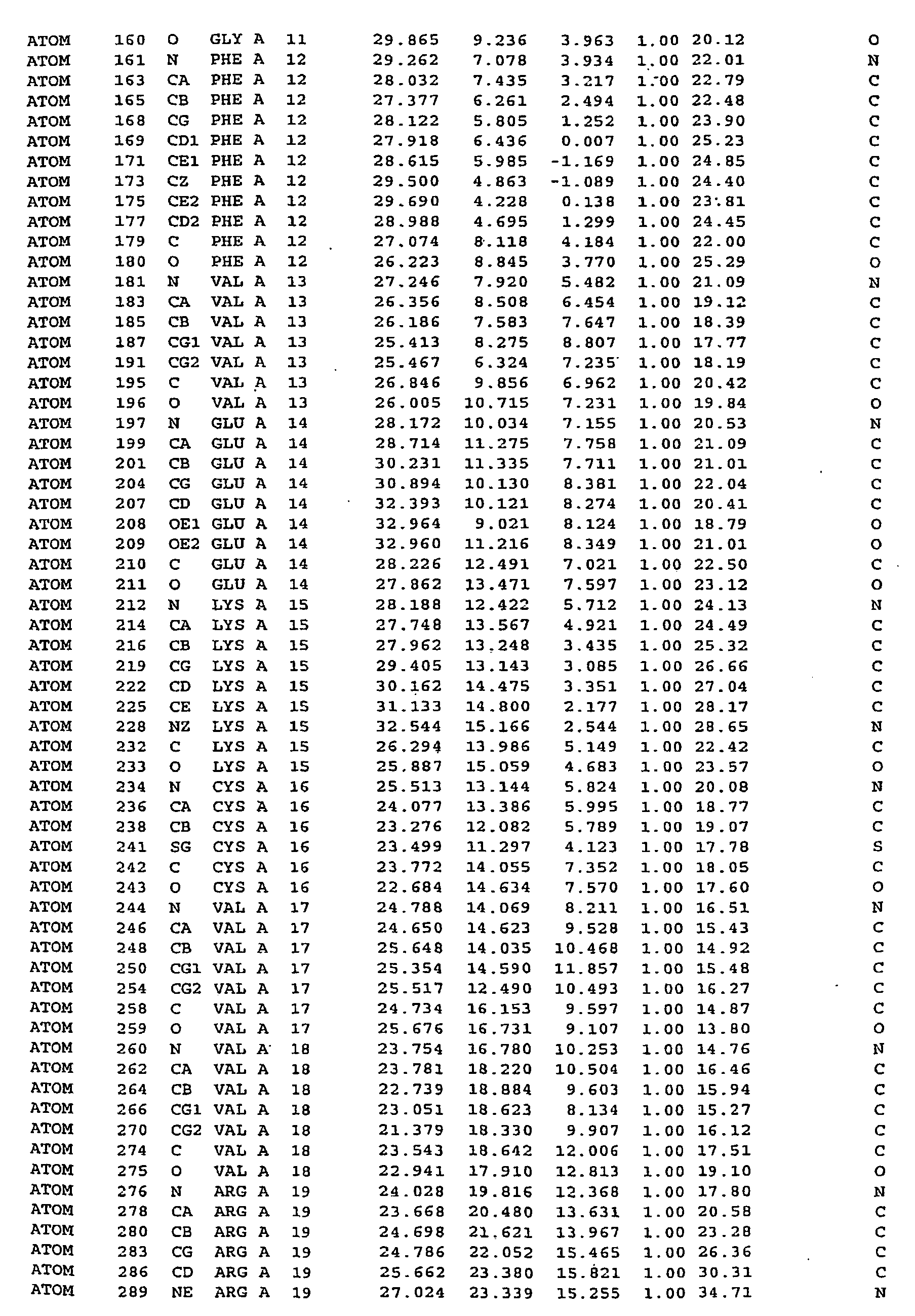


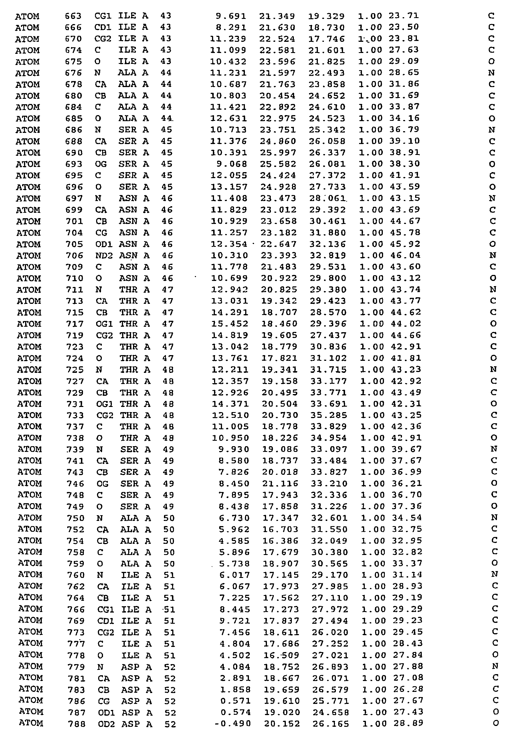


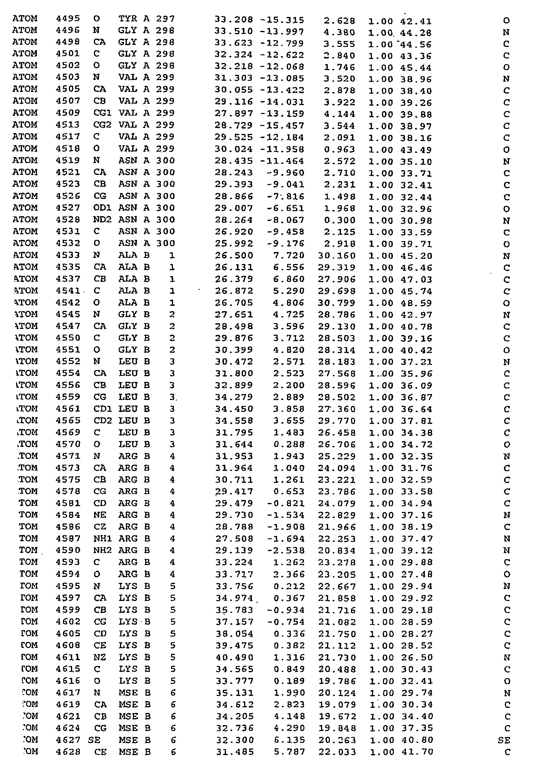


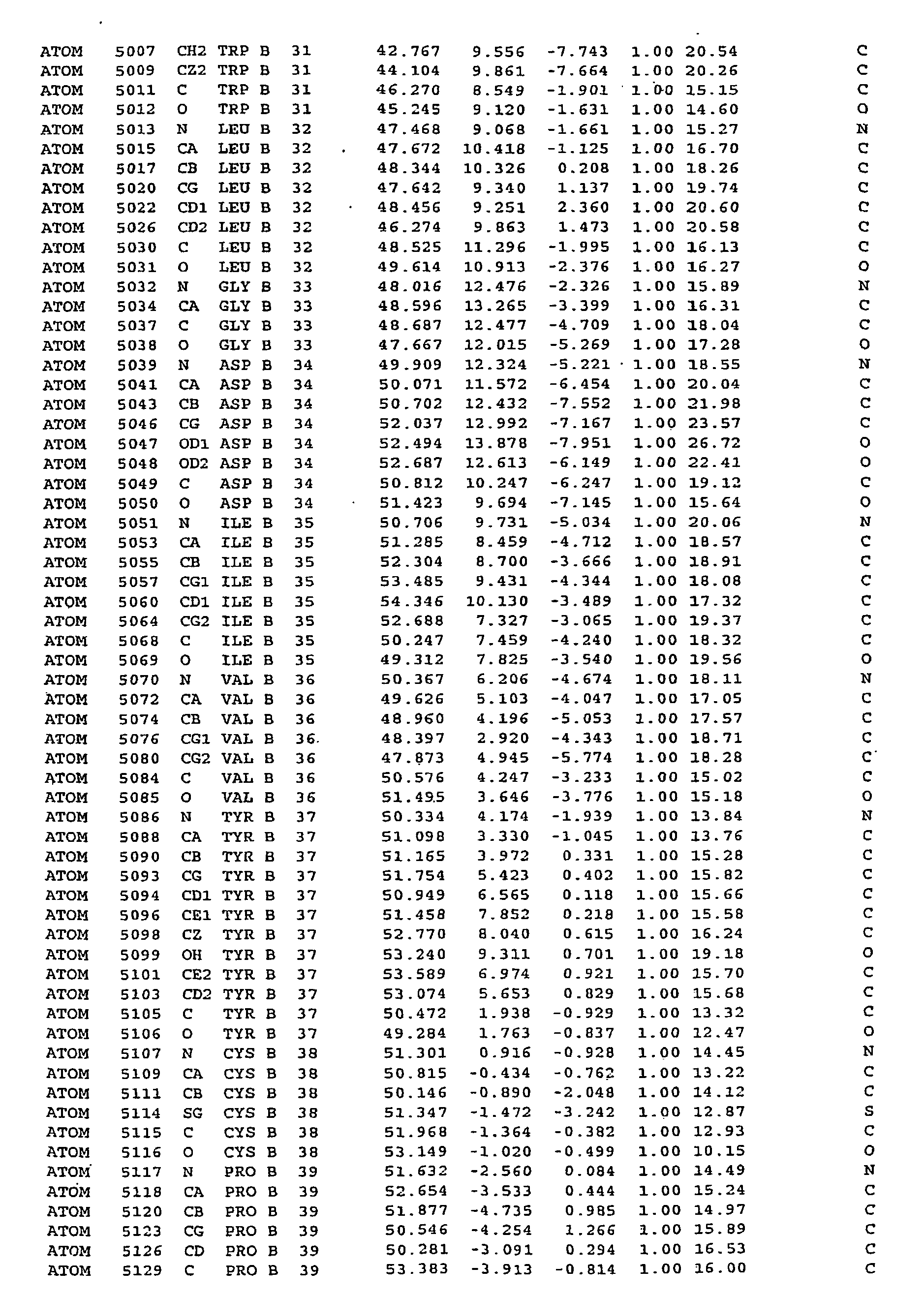


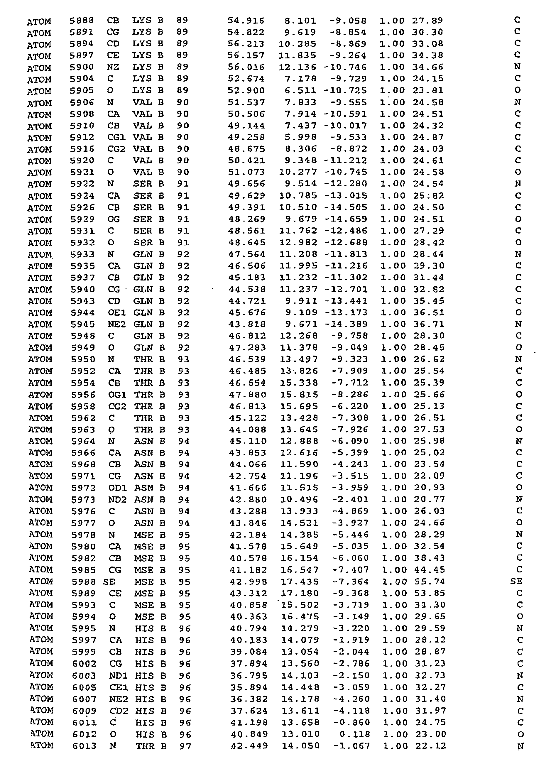


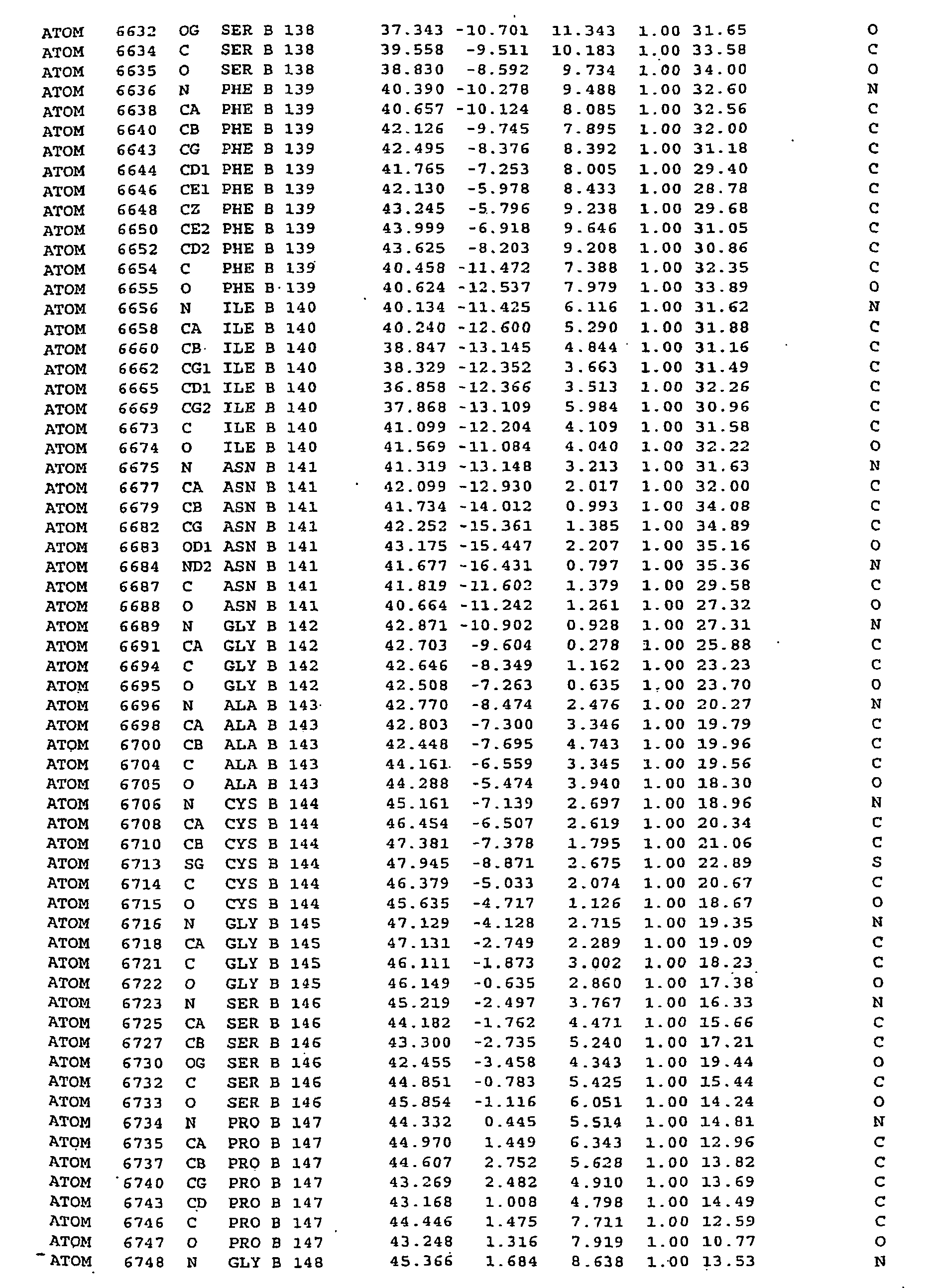


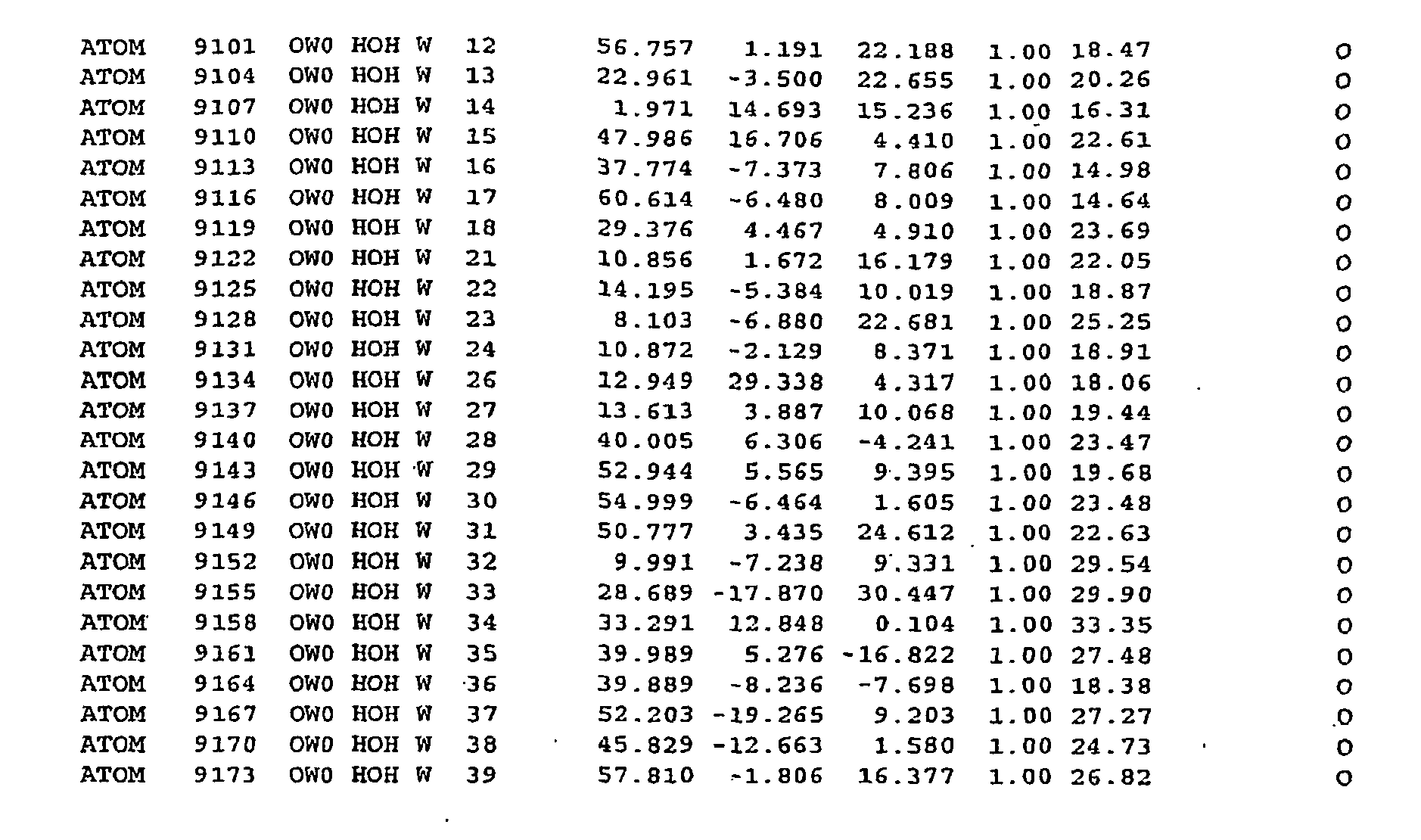


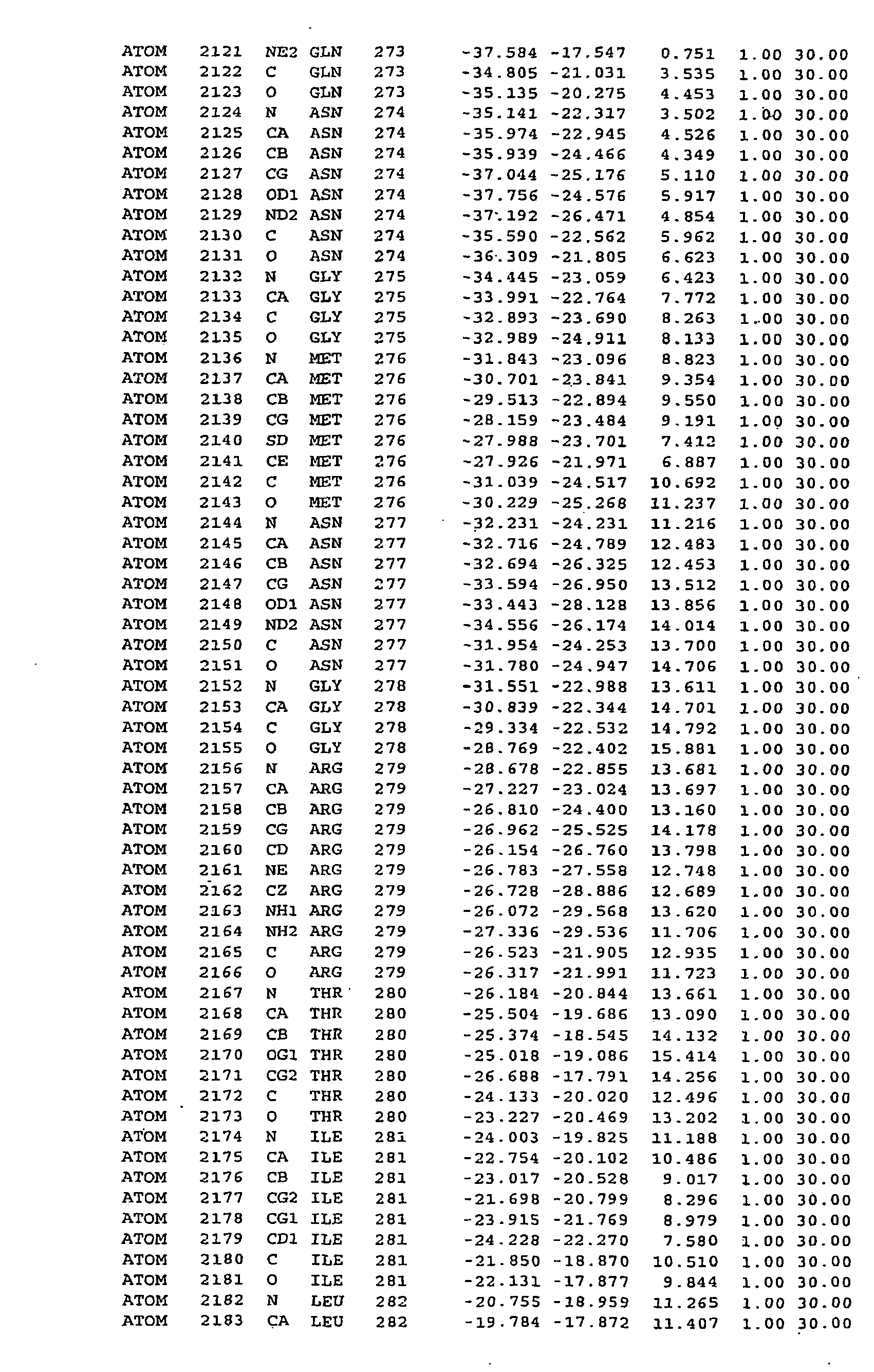


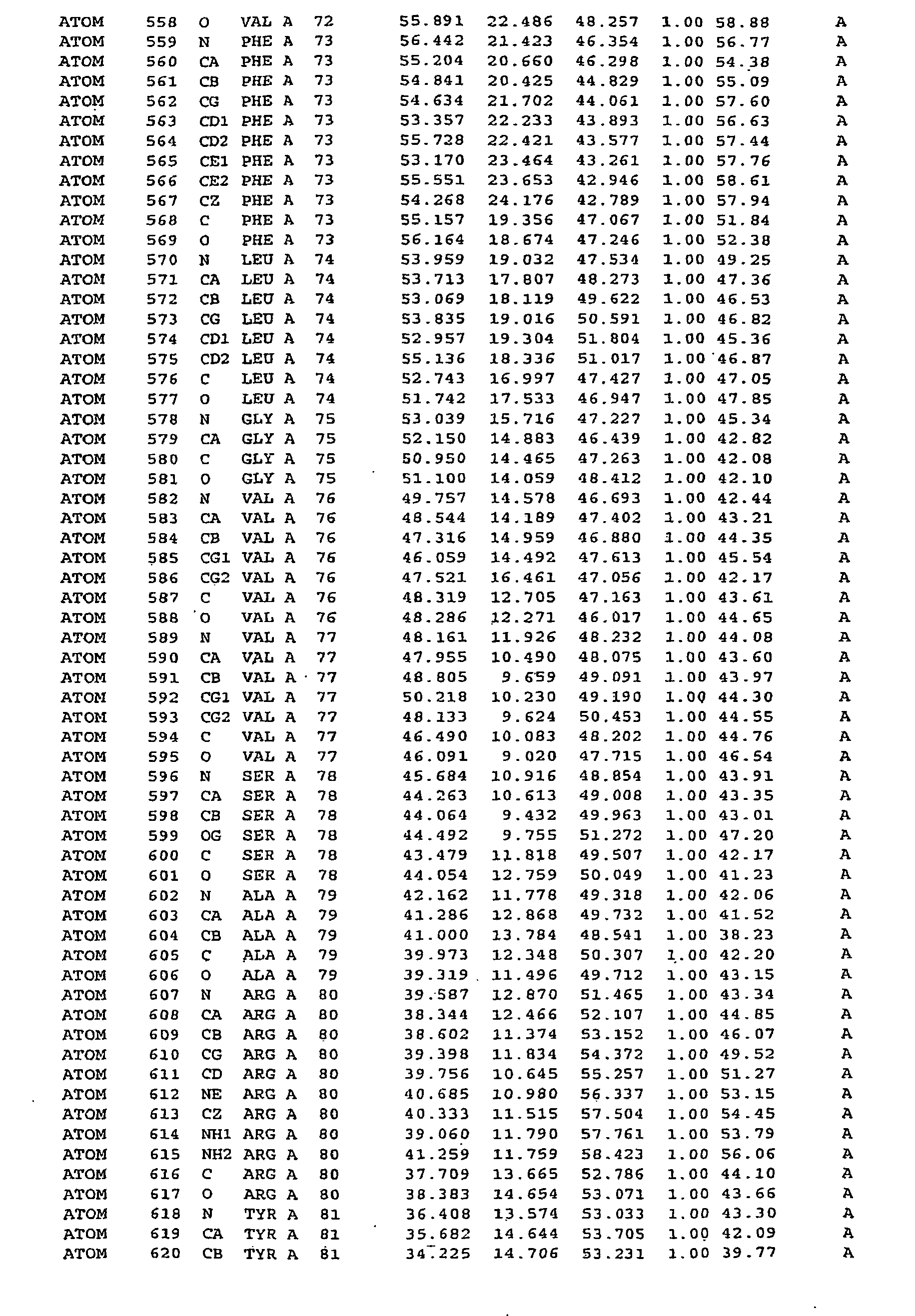


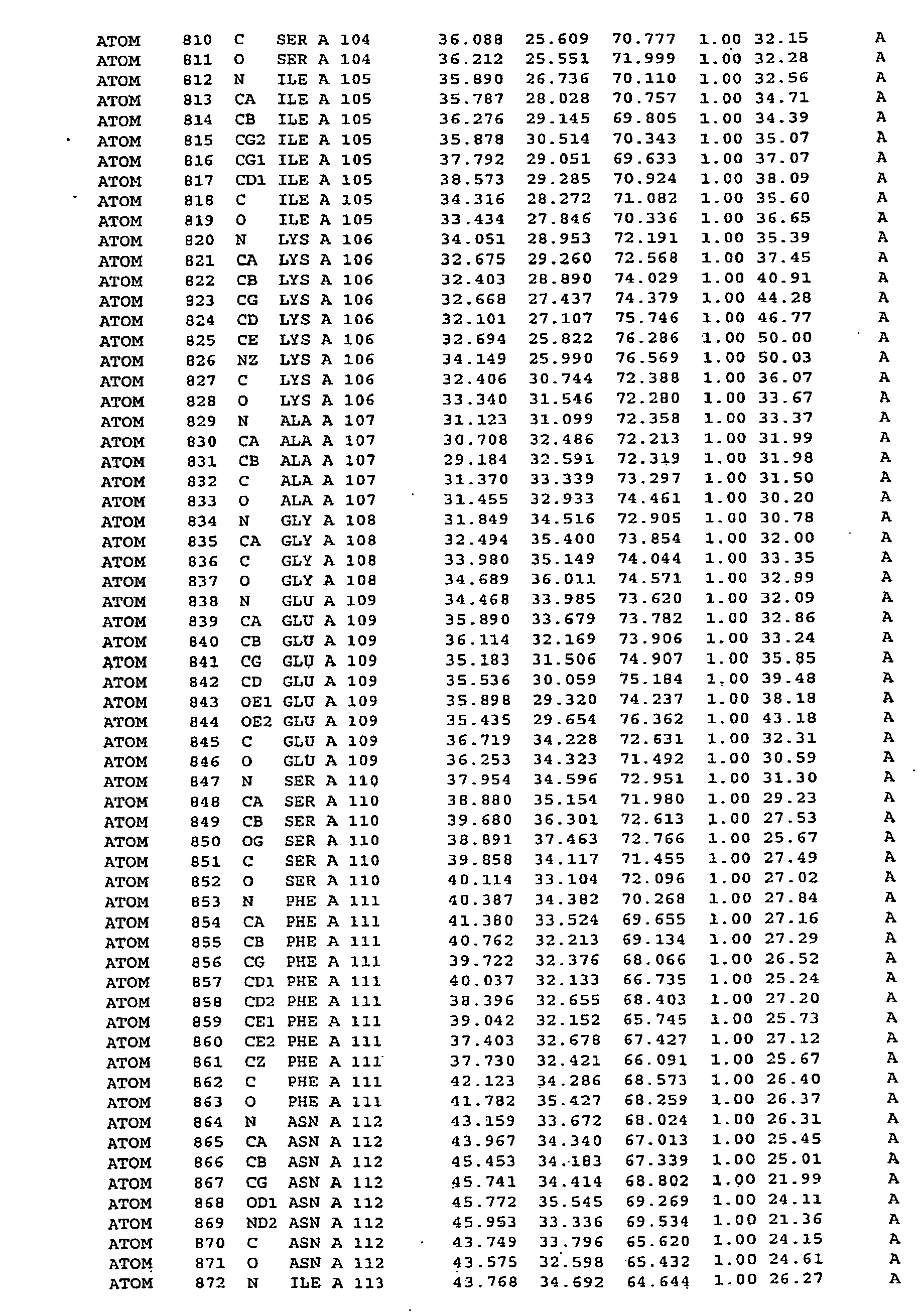


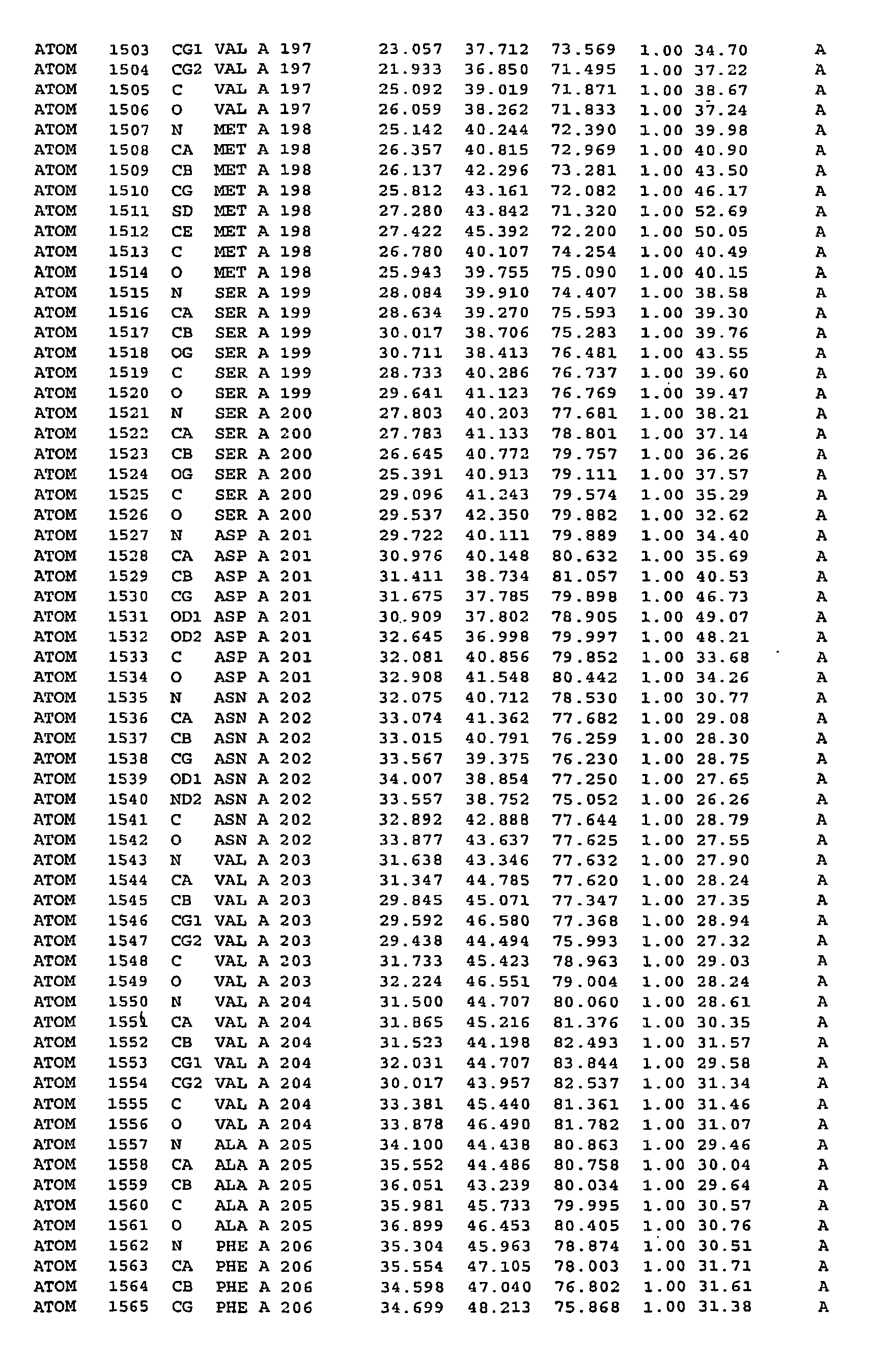

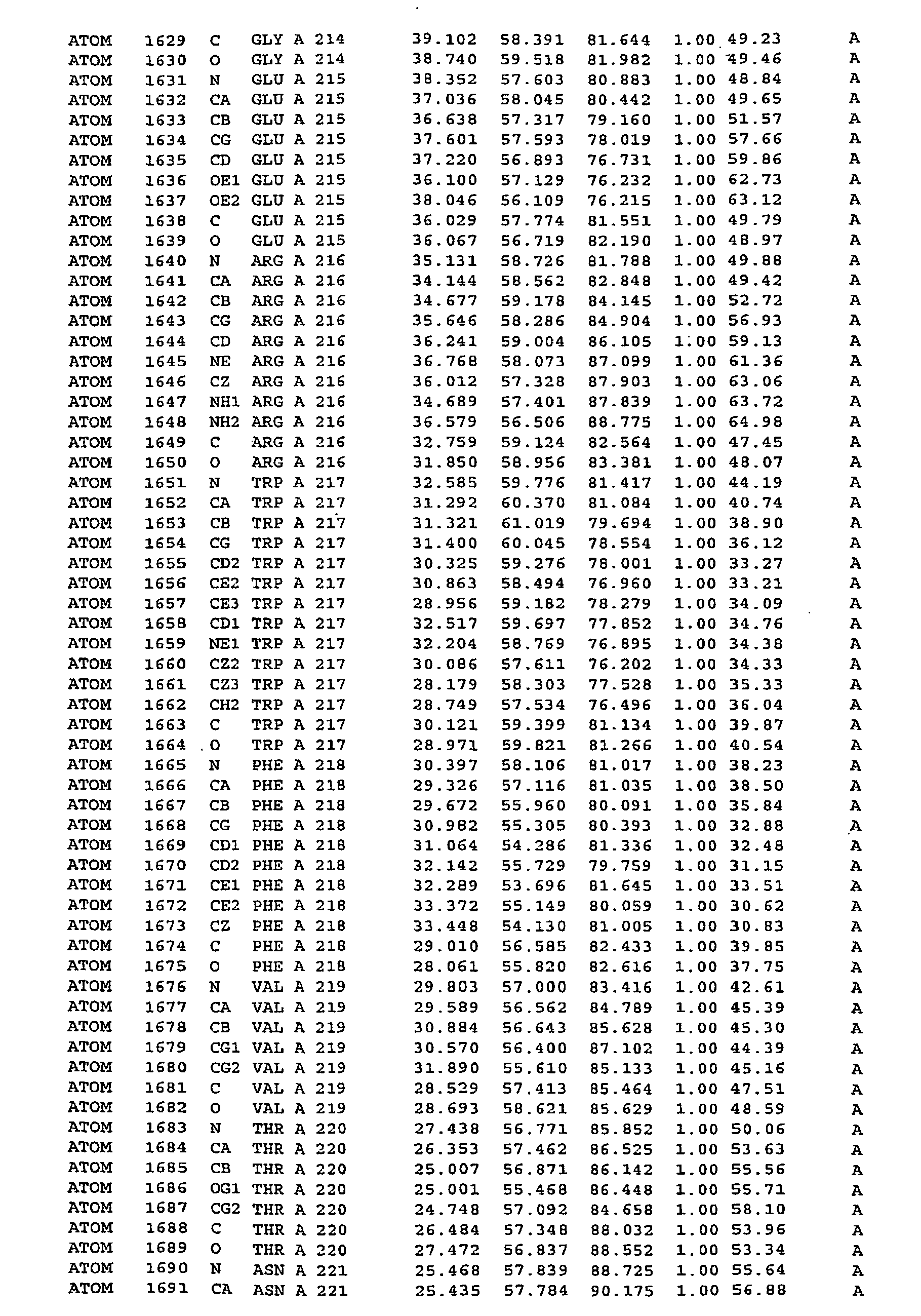


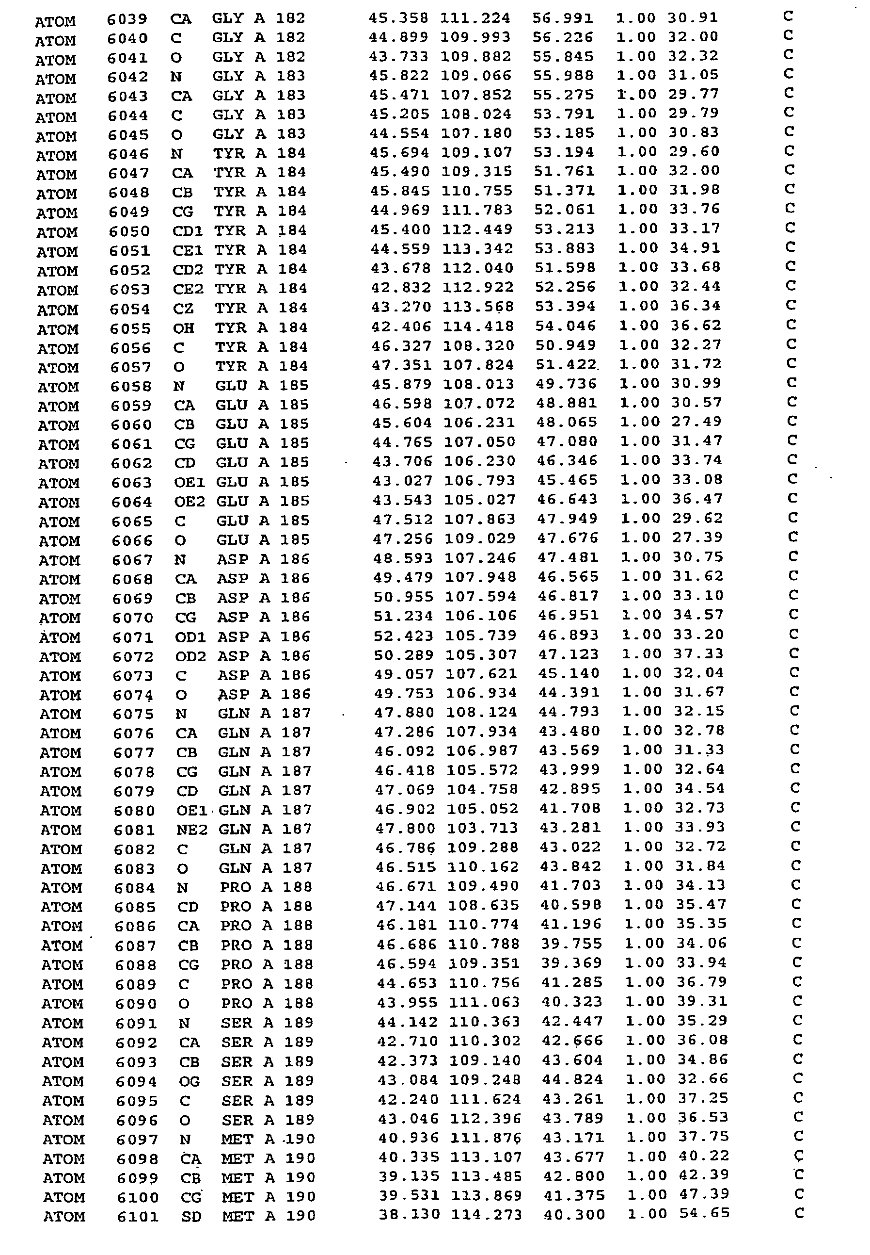


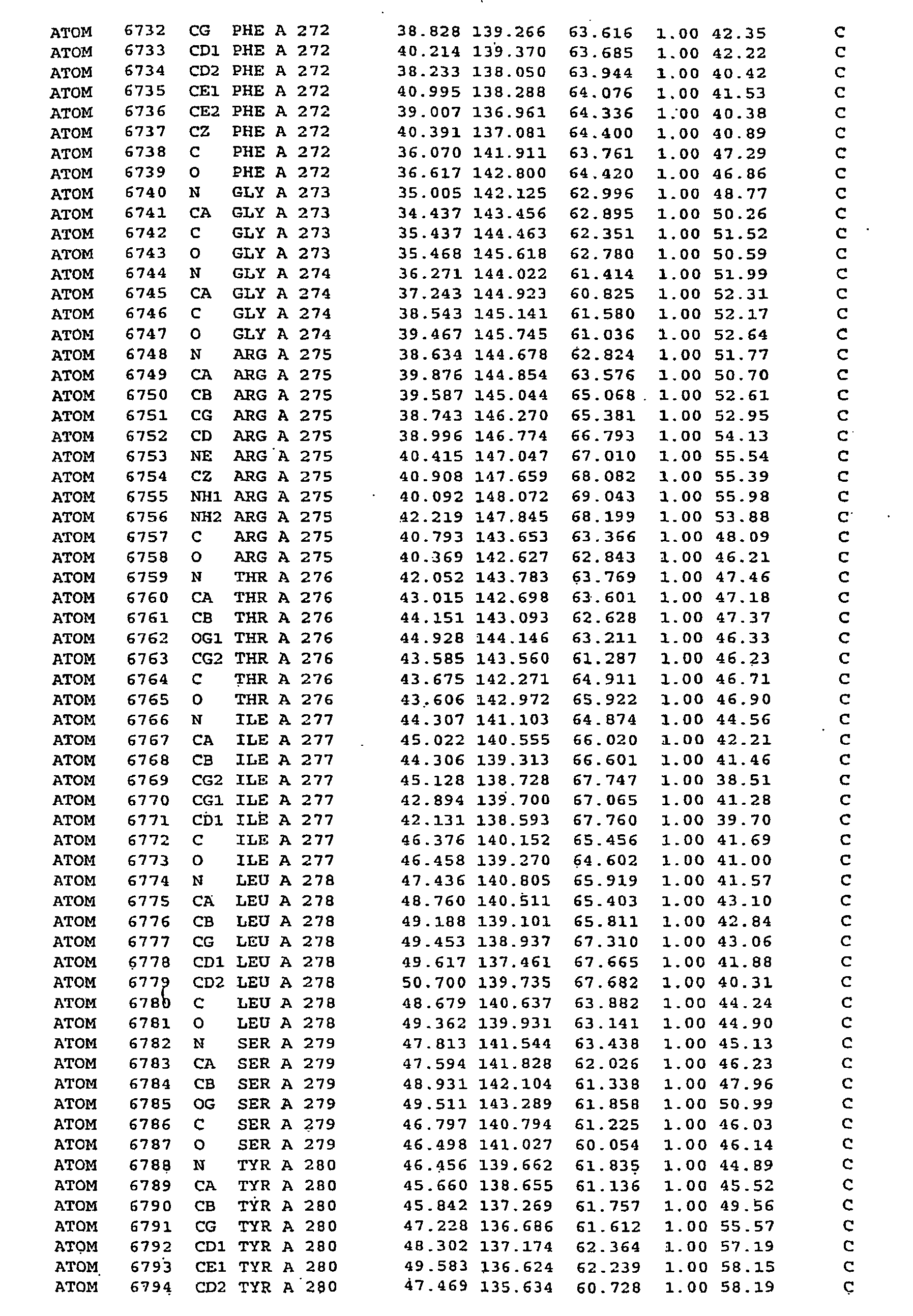


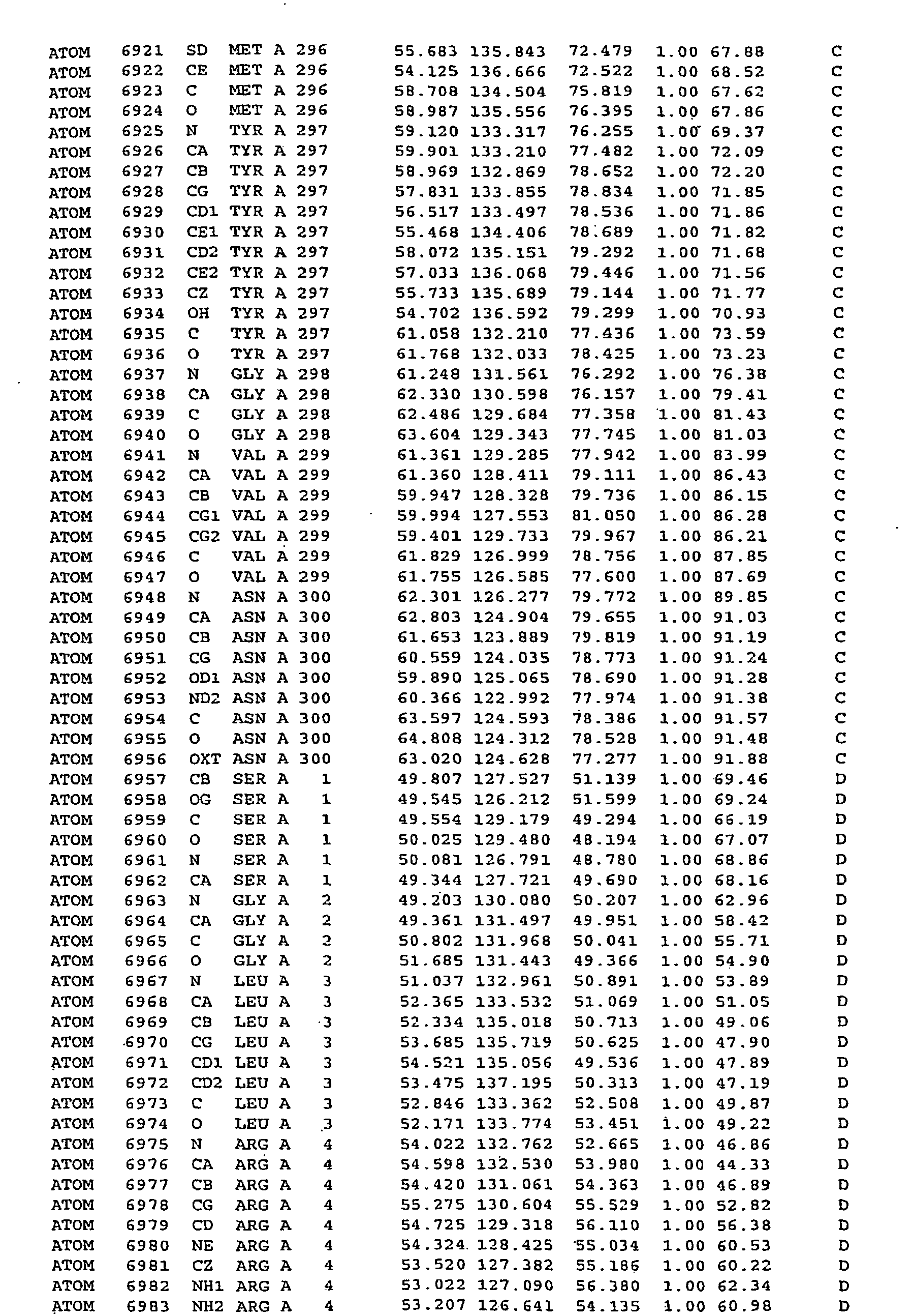


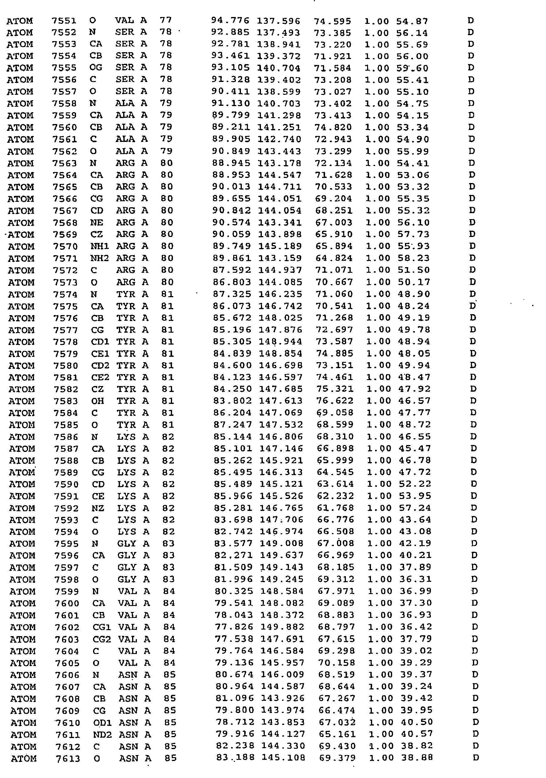


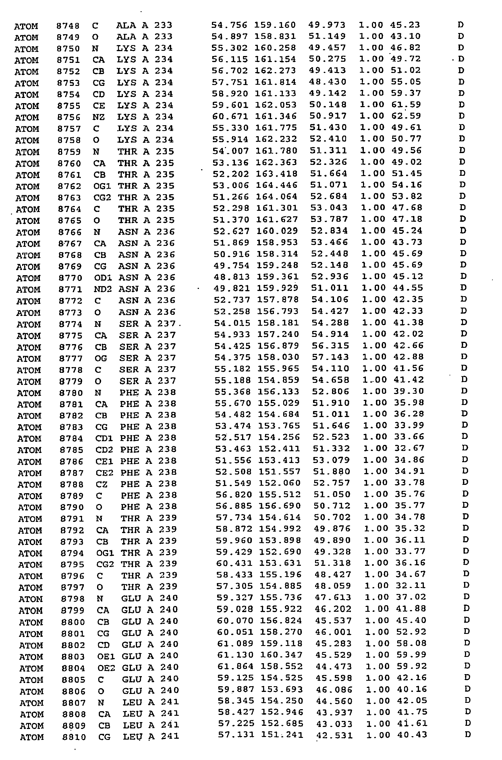


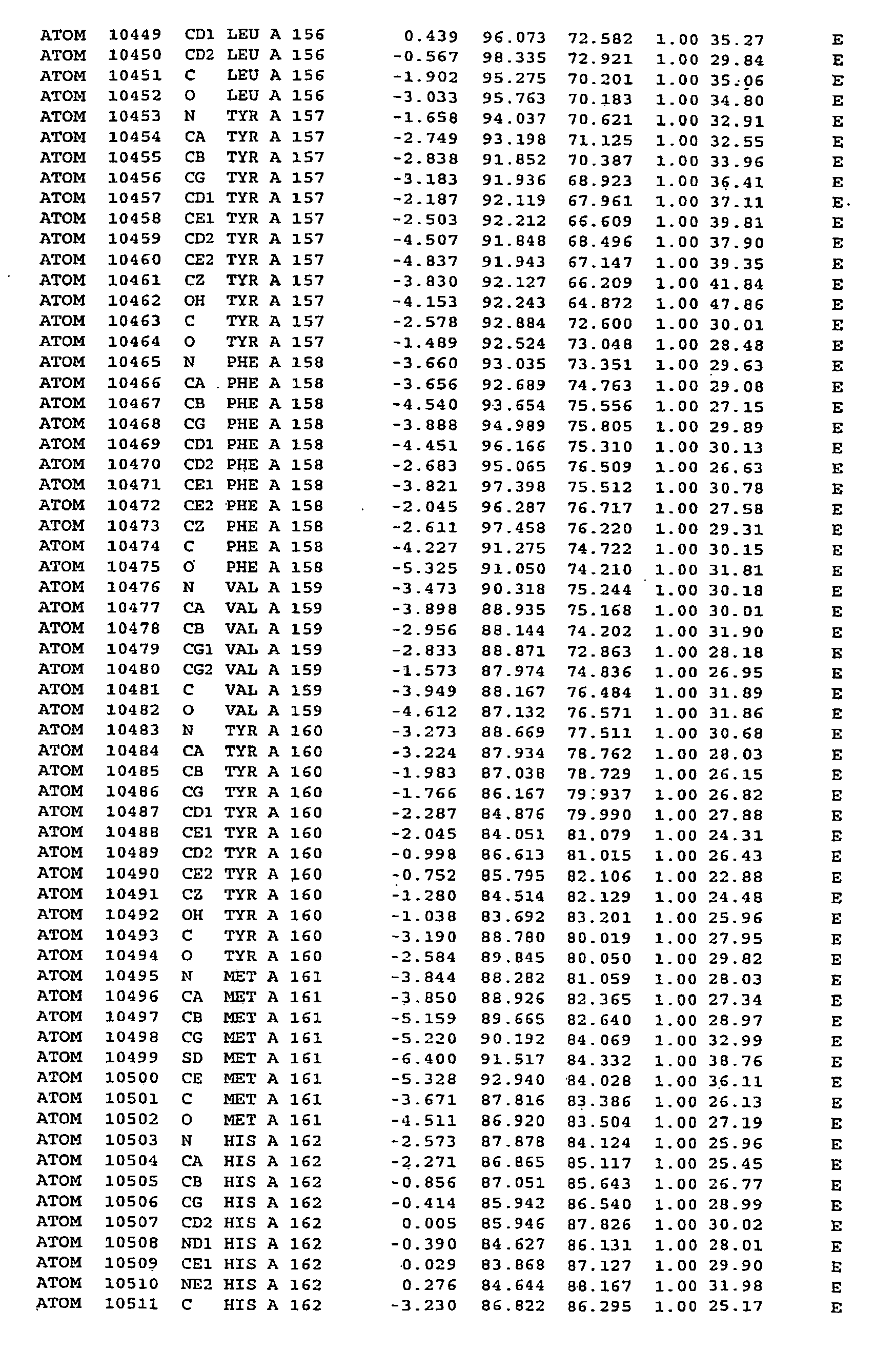


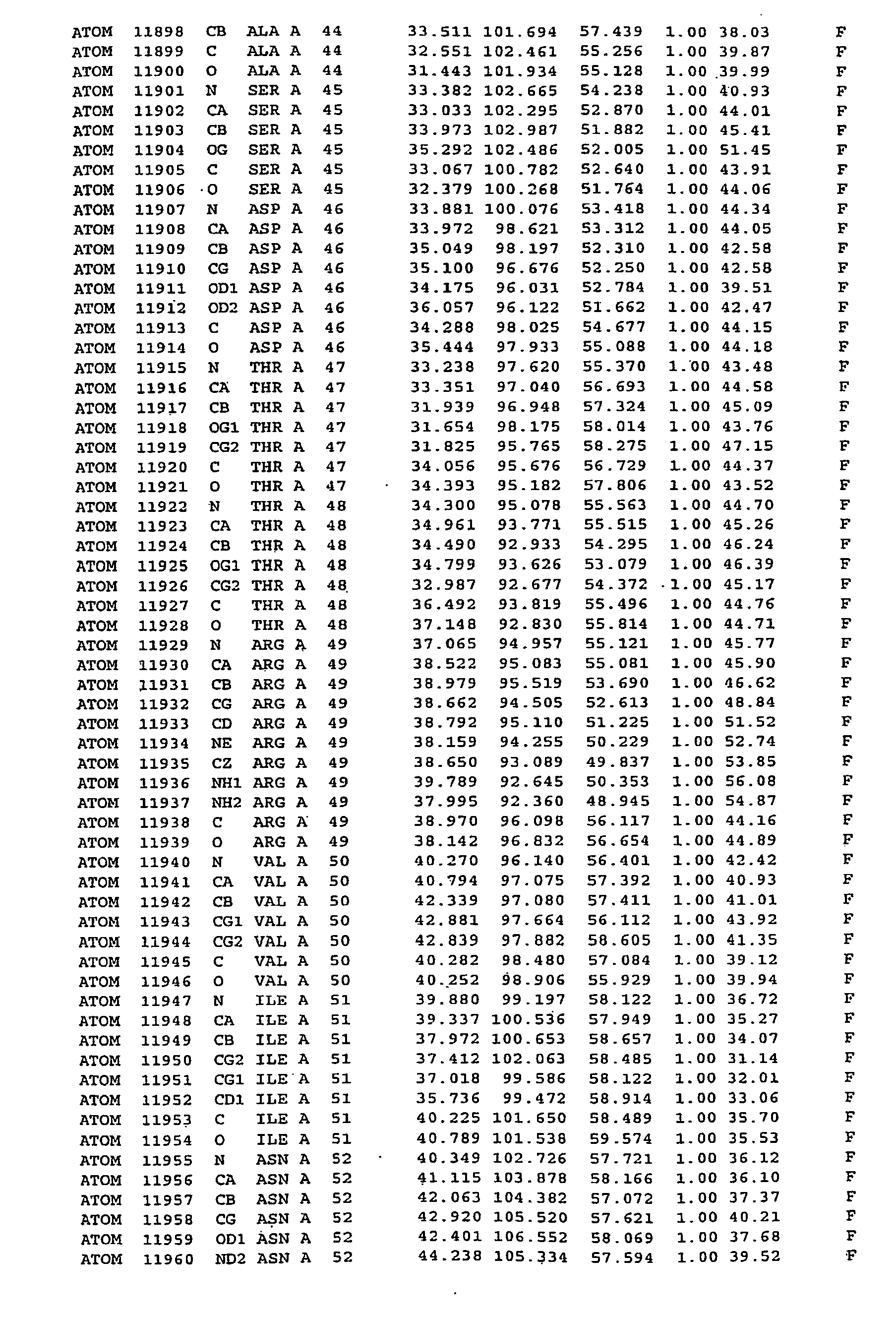




-
- <110> Universität zu Lübeck
- <120> Crystal Structure of Human Coronavirus 229E Main Proteinase, and Uses thereof for Developing SARS inhibitors
- <130> P5460
- <140>
EP 04 739 187.5
<141> 2003-05-13 - <160> 1
- <170> Patent In version 3.3
- <210> 1
<211> 5
<212> PRT
<213> Artificial Sequence - <220>
<223> Xaa is an amino acid selected from the group of amino acids as set forth inClaim 5 - <220>
<221> DOMAIN
<222> (1)..(5)
<223> Structural Motif of an Inhibitor of Rhinoviral 3C Proteinase - <220>
<221> NON_TER
<222> (1). .(1) - <400> 1

Claims (6)
- Use of a substance with the structural formula

- Use of a substance according to claim 1, wherein the disease is SARS.
- Use according to one of the preceding claims, characterized in that the substance is AG 7088 or a derivative thereof.
- Use according to claim 1, characterized in that the residue P2 is an unmodified benzyl group.
- Use of a substance including the structural motif
AA1-AA2-AA3-AA4-P2-Gln-X
for the preparation of a pharmaceutical composition for the treatment of a disease caused by a coronavirus, wherein- AA1, AA2, AA3 and AA4 are selected from the group of amino acids consisting of threonine, valine, serine and alanine,- P2 being leucine, phenylalanine, methionine or derivatives thereof fitting in the S2 pocket of coronavirus main proteinases, and- X being a vinylogous ester, methylester, isopropylester or any other alkyl. - Use according to claim 5, characterized in that AA1, AA2 and AA4 being any amino acid or absent.
Applications Claiming Priority (2)
| Application Number | Priority Date | Filing Date | Title |
|---|---|---|---|
| US46981803P | 2003-05-13 | 2003-05-13 | |
| PCT/EP2004/005109 WO2004101781A1 (en) | 2003-05-13 | 2004-05-13 | Crystal structure of human coronavirus 229e main proteinase, and uses thereof for developing sars inhibitors |
Publications (2)
| Publication Number | Publication Date |
|---|---|
| EP1623028A1 EP1623028A1 (en) | 2006-02-08 |
| EP1623028B1 true EP1623028B1 (en) | 2008-01-02 |
Family
ID=33452324
Family Applications (1)
| Application Number | Title | Priority Date | Filing Date |
|---|---|---|---|
| EP04739187A Expired - Lifetime EP1623028B1 (en) | 2003-05-13 | 2004-05-13 | Crystal structure of human coronavirus 229e main proteinase, and uses thereof for developing sars inhibitors |
Country Status (6)
| Country | Link |
|---|---|
| US (1) | US7956078B2 (en) |
| EP (1) | EP1623028B1 (en) |
| CN (1) | CN1788084A (en) |
| AT (1) | ATE382686T1 (en) |
| DE (1) | DE602004011047T2 (en) |
| WO (1) | WO2004101781A1 (en) |
Cited By (1)
| Publication number | Priority date | Publication date | Assignee | Title |
|---|---|---|---|---|
| WO2021160220A1 (en) | 2020-02-11 | 2021-08-19 | Universität Zu Lübeck Körperschaft Des Öffentlichen Rechts | Antiviral substances with a wide spectrum of activity |
Families Citing this family (21)
| Publication number | Priority date | Publication date | Assignee | Title |
|---|---|---|---|---|
| US7129042B2 (en) | 2003-11-03 | 2006-10-31 | Diagnostic Hybrids, Inc. | Compositions and methods for detecting severe acute respiratory syndrome coronavirus |
| CN100480440C (en) * | 2007-04-04 | 2009-04-22 | 南开大学 | Method for screening SARS coronaviruses main Protease inhibitors from natural product such as Chinese medicinal materials mixture prepared storeroom |
| SMT202500351T1 (en) | 2020-07-20 | 2025-11-10 | Enanta Pharm Inc | Functionalized peptides containing two pyrrolidine rings as anticoronavirus agents |
| US11384090B2 (en) | 2020-11-23 | 2022-07-12 | Enanta Pharmaceuticals, Inc. | Spiropyrrolidine derived antiviral agents |
| US12540141B2 (en) | 2020-11-23 | 2026-02-03 | Enanta Pharmaceuticals, Inc. | Spiropyrrolidine derived antiviral agents |
| WO2022109363A1 (en) | 2020-11-23 | 2022-05-27 | Enanta Pharmaceuticals, Inc. | Novel spiropyrrolidine derived antiviral agents |
| KR20230124583A (en) | 2020-11-23 | 2023-08-25 | 이난타 파마슈티칼스, 인코포레이티드 | Novel spiropyrrolidine-derived antiviral agent |
| US11352363B1 (en) | 2020-11-23 | 2022-06-07 | Enanta Pharmaceuticals, Inc. | Spiropyrrolidine derived antiviral agents |
| WO2022235605A1 (en) | 2021-05-04 | 2022-11-10 | Enanta Pharmaceuticals, Inc. | Novel macrocyclic antiviral agents |
| US11319325B1 (en) | 2021-05-11 | 2022-05-03 | Enanta Pharmaceuticals, Inc. | Macrocyclic spiropyrrolidine derived antiviral agents |
| US12398147B2 (en) | 2021-05-11 | 2025-08-26 | Enanta Pharmaceuticals, Inc. | Macrocyclic spiropyrrolidine derived antiviral agents |
| US11325916B1 (en) | 2021-07-29 | 2022-05-10 | Enanta Pharmaceuticals, Inc. | Spiropyrrolidine derived antiviral agents |
| US11339170B1 (en) | 2021-07-23 | 2022-05-24 | Enanta Pharmaceuticals, Inc. | Spiropyrrolidine derived antiviral agents |
| US12479854B2 (en) | 2021-07-29 | 2025-11-25 | Enanta Pharmaceuticals, Inc. | Spiropyrrolidine derived antiviral agents |
| US11912714B2 (en) | 2021-11-12 | 2024-02-27 | Enanta Pharmaceuticals, Inc. | Spiropyrrolidine derived antiviral agents |
| US11858945B2 (en) | 2021-11-12 | 2024-01-02 | Enanta Pharmaceuticals, Inc. | Alkyne-containing antiviral agents |
| US11919910B2 (en) | 2021-11-12 | 2024-03-05 | Enanta Pharmaceuticals, Inc. | Spiropyrrolidine derived antiviral agents |
| US11993600B2 (en) | 2021-12-08 | 2024-05-28 | Enanta Pharmaceuticals, Inc. | Saturated spirocyclics as antiviral agents |
| WO2023107417A1 (en) | 2021-12-08 | 2023-06-15 | Enanta Pharmaceuticals, Inc. | Heterocyclic antiviral agents |
| US12145942B2 (en) | 2022-04-05 | 2024-11-19 | Enanta Pharmaceuticals, Inc. | Spiropyrrolidine derived antiviral agents |
| US11707504B1 (en) | 2022-06-27 | 2023-07-25 | King Faisal University | Fusion peptide inhibitors of human coronavirus 229E |
Family Cites Families (1)
| Publication number | Priority date | Publication date | Assignee | Title |
|---|---|---|---|---|
| US6906198B1 (en) * | 1998-04-30 | 2005-06-14 | Agouron Pharmaceuticals, Inc. | Antipicornaviral compounds, their preparation and use |
-
2004
- 2004-05-13 WO PCT/EP2004/005109 patent/WO2004101781A1/en not_active Ceased
- 2004-05-13 AT AT04739187T patent/ATE382686T1/en not_active IP Right Cessation
- 2004-05-13 EP EP04739187A patent/EP1623028B1/en not_active Expired - Lifetime
- 2004-05-13 CN CNA2004800127168A patent/CN1788084A/en active Pending
- 2004-05-13 DE DE602004011047T patent/DE602004011047T2/en not_active Expired - Lifetime
-
2005
- 2005-11-10 US US11/273,270 patent/US7956078B2/en not_active Expired - Fee Related
Cited By (1)
| Publication number | Priority date | Publication date | Assignee | Title |
|---|---|---|---|---|
| WO2021160220A1 (en) | 2020-02-11 | 2021-08-19 | Universität Zu Lübeck Körperschaft Des Öffentlichen Rechts | Antiviral substances with a wide spectrum of activity |
Also Published As
| Publication number | Publication date |
|---|---|
| CN1788084A (en) | 2006-06-14 |
| US7956078B2 (en) | 2011-06-07 |
| DE602004011047T2 (en) | 2008-12-24 |
| EP1623028A1 (en) | 2006-02-08 |
| WO2004101781A1 (en) | 2004-11-25 |
| DE602004011047D1 (en) | 2008-02-14 |
| ATE382686T1 (en) | 2008-01-15 |
| US20090137818A1 (en) | 2009-05-28 |
Similar Documents
| Publication | Publication Date | Title |
|---|---|---|
| US7956078B2 (en) | Crystal structure of human coronavirus 229E main proteinase and uses for developing SARS inhibitors | |
| Lee et al. | Structural basis of inhibition specificities of 3C and 3C-like proteases by zinc-coordinating and peptidomimetic compounds | |
| Petersen et al. | The structure of the 2A proteinase from a common cold virus: a proteinase responsible for the shut‐off of host‐cell protein synthesis | |
| Hsu et al. | Mechanism of the maturation process of SARS-CoV 3CL protease | |
| Sutton et al. | The nsp9 replicase protein of SARS-coronavirus, structure and functional insights | |
| US7894996B2 (en) | Structure of the hepatitis C NS5A protein | |
| Robin et al. | Structure of West Nile virus NS3 protease: ligand stabilization of the catalytic conformation | |
| Yu et al. | Crystal structure of the severe acute respiratory syndrome (SARS) coronavirus nucleocapsid protein dimerization domain reveals evolutionary linkage between corona-and arteriviridae | |
| Zunszain et al. | Insights into cleavage specificity from the crystal structure of foot-and-mouth disease virus 3C protease complexed with a peptide substrate | |
| Lei et al. | Crystal structure of the papain-like protease of MERS coronavirus reveals unusual, potentially druggable active-site features | |
| Ye et al. | Structural basis for the dimerization and substrate recognition specificity of porcine epidemic diarrhea virus 3C-like protease | |
| Tian et al. | Structure and cleavage specificity of the chymotrypsin-like serine protease (3CLSP/nsp4) of porcine reproductive and respiratory syndrome virus (PRRSV) | |
| Kong et al. | Structural view and substrate specificity of papain-like protease from avian infectious bronchitis virus | |
| Kuo et al. | Characterization and inhibition of the main protease of severe acute respiratory syndrome coronavirus | |
| Anand et al. | Coronavirus main proteinase: target for antiviral drug therapy | |
| Steinberger et al. | Foot-and-mouth disease virus leader proteinase: structural insights into the mechanism of intermolecular cleavage | |
| US6737234B1 (en) | Structure of adenovirus bound to cellular receptor car | |
| Woo et al. | In silico analysis of ORF1ab in coronavirus HKU1 genome reveals a unique putative cleavage site of coronavirus HKU1 3C‐like protease | |
| Sun et al. | An open conformation determined by a structural switch for 2A protease from coxsackievirus A16 | |
| Xu et al. | Structure of the SARS coronavirus main proteinase as an active C2 crystallographic dimer | |
| US6008033A (en) | Proteases, compositions capable of binding to said site, and methods of use thereof | |
| Heaslet et al. | Structural insights into the mechanisms of drug resistance in HIV-1 protease NL4-3 | |
| CA2378032A1 (en) | Crystallization and structure determination of staphylococcus aureus udp-n-acetylenolpyruvylglucosamine reductase (s. aureus murb) | |
| US20030077813A1 (en) | Structure of adenovirus bound to cellular receptor car | |
| Krishna et al. | Structural studies on the empty capsids of Physalis mottle virus |
Legal Events
| Date | Code | Title | Description |
|---|---|---|---|
| PUAI | Public reference made under article 153(3) epc to a published international application that has entered the european phase |
Free format text: ORIGINAL CODE: 0009012 |
|
| 17P | Request for examination filed |
Effective date: 20051111 |
|
| AK | Designated contracting states |
Kind code of ref document: A1 Designated state(s): AT BE BG CH CY CZ DE DK EE ES FI FR GB GR HU IE IT LI LU MC NL PL PT RO SE SI SK TR |
|
| DAX | Request for extension of the european patent (deleted) | ||
| 17Q | First examination report despatched |
Effective date: 20070205 |
|
| GRAP | Despatch of communication of intention to grant a patent |
Free format text: ORIGINAL CODE: EPIDOSNIGR1 |
|
| GRAS | Grant fee paid |
Free format text: ORIGINAL CODE: EPIDOSNIGR3 |
|
| GRAA | (expected) grant |
Free format text: ORIGINAL CODE: 0009210 |
|
| AK | Designated contracting states |
Kind code of ref document: B1 Designated state(s): AT BE BG CH CY CZ DE DK EE ES FI FR GB GR HU IE IT LI LU MC NL PL PT RO SE SI SK TR |
|
| REG | Reference to a national code |
Ref country code: GB Ref legal event code: FG4D |
|
| REG | Reference to a national code |
Ref country code: IE Ref legal event code: FG4D |
|
| REG | Reference to a national code |
Ref country code: CH Ref legal event code: EP |
|
| REF | Corresponds to: |
Ref document number: 602004011047 Country of ref document: DE Date of ref document: 20080214 Kind code of ref document: P |
|
| REG | Reference to a national code |
Ref country code: CH Ref legal event code: AEN Free format text: DAS PATENT IST AUF GRUND DES WEITERBEHANDLUNGSANTRAGS VOM 03.04.2008 REAKTIVIERT WORDEN. Ref country code: CH Ref legal event code: NV Representative=s name: ISLER & PEDRAZZINI AG Ref country code: CH Ref legal event code: PL |
|
| PG25 | Lapsed in a contracting state [announced via postgrant information from national office to epo] |
Ref country code: NL Free format text: LAPSE BECAUSE OF FAILURE TO SUBMIT A TRANSLATION OF THE DESCRIPTION OR TO PAY THE FEE WITHIN THE PRESCRIBED TIME-LIMIT Effective date: 20080102 Ref country code: SI Free format text: LAPSE BECAUSE OF FAILURE TO SUBMIT A TRANSLATION OF THE DESCRIPTION OR TO PAY THE FEE WITHIN THE PRESCRIBED TIME-LIMIT Effective date: 20080102 |
|
| NLV1 | Nl: lapsed or annulled due to failure to fulfill the requirements of art. 29p and 29m of the patents act | ||
| PG25 | Lapsed in a contracting state [announced via postgrant information from national office to epo] |
Ref country code: LI Free format text: LAPSE BECAUSE OF FAILURE TO SUBMIT A TRANSLATION OF THE DESCRIPTION OR TO PAY THE FEE WITHIN THE PRESCRIBED TIME-LIMIT Effective date: 20080102 Ref country code: FI Free format text: LAPSE BECAUSE OF FAILURE TO SUBMIT A TRANSLATION OF THE DESCRIPTION OR TO PAY THE FEE WITHIN THE PRESCRIBED TIME-LIMIT Effective date: 20080102 Ref country code: ES Free format text: LAPSE BECAUSE OF FAILURE TO SUBMIT A TRANSLATION OF THE DESCRIPTION OR TO PAY THE FEE WITHIN THE PRESCRIBED TIME-LIMIT Effective date: 20080413 |
|
| PGRI | Patent reinstated in contracting state [announced from national office to epo] |
Ref country code: CH Effective date: 20080403 |
|
| PG25 | Lapsed in a contracting state [announced via postgrant information from national office to epo] |
Ref country code: BG Free format text: LAPSE BECAUSE OF FAILURE TO SUBMIT A TRANSLATION OF THE DESCRIPTION OR TO PAY THE FEE WITHIN THE PRESCRIBED TIME-LIMIT Effective date: 20080402 Ref country code: AT Free format text: LAPSE BECAUSE OF FAILURE TO SUBMIT A TRANSLATION OF THE DESCRIPTION OR TO PAY THE FEE WITHIN THE PRESCRIBED TIME-LIMIT Effective date: 20080102 |
|
| PGRI | Patent reinstated in contracting state [announced from national office to epo] |
Ref country code: CH Effective date: 20080403 |
|
| ET | Fr: translation filed | ||
| PG25 | Lapsed in a contracting state [announced via postgrant information from national office to epo] |
Ref country code: PL Free format text: LAPSE BECAUSE OF FAILURE TO SUBMIT A TRANSLATION OF THE DESCRIPTION OR TO PAY THE FEE WITHIN THE PRESCRIBED TIME-LIMIT Effective date: 20080102 Ref country code: PT Free format text: LAPSE BECAUSE OF FAILURE TO SUBMIT A TRANSLATION OF THE DESCRIPTION OR TO PAY THE FEE WITHIN THE PRESCRIBED TIME-LIMIT Effective date: 20080602 |
|
| PG25 | Lapsed in a contracting state [announced via postgrant information from national office to epo] |
Ref country code: SK Free format text: LAPSE BECAUSE OF FAILURE TO SUBMIT A TRANSLATION OF THE DESCRIPTION OR TO PAY THE FEE WITHIN THE PRESCRIBED TIME-LIMIT Effective date: 20080102 Ref country code: SE Free format text: LAPSE BECAUSE OF FAILURE TO SUBMIT A TRANSLATION OF THE DESCRIPTION OR TO PAY THE FEE WITHIN THE PRESCRIBED TIME-LIMIT Effective date: 20080402 Ref country code: DK Free format text: LAPSE BECAUSE OF FAILURE TO SUBMIT A TRANSLATION OF THE DESCRIPTION OR TO PAY THE FEE WITHIN THE PRESCRIBED TIME-LIMIT Effective date: 20080102 Ref country code: CZ Free format text: LAPSE BECAUSE OF FAILURE TO SUBMIT A TRANSLATION OF THE DESCRIPTION OR TO PAY THE FEE WITHIN THE PRESCRIBED TIME-LIMIT Effective date: 20080102 |
|
| PLBE | No opposition filed within time limit |
Free format text: ORIGINAL CODE: 0009261 |
|
| STAA | Information on the status of an ep patent application or granted ep patent |
Free format text: STATUS: NO OPPOSITION FILED WITHIN TIME LIMIT |
|
| PG25 | Lapsed in a contracting state [announced via postgrant information from national office to epo] |
Ref country code: RO Free format text: LAPSE BECAUSE OF FAILURE TO SUBMIT A TRANSLATION OF THE DESCRIPTION OR TO PAY THE FEE WITHIN THE PRESCRIBED TIME-LIMIT Effective date: 20080102 |
|
| 26N | No opposition filed |
Effective date: 20081003 |
|
| PG25 | Lapsed in a contracting state [announced via postgrant information from national office to epo] |
Ref country code: MC Free format text: LAPSE BECAUSE OF NON-PAYMENT OF DUE FEES Effective date: 20080531 |
|
| PG25 | Lapsed in a contracting state [announced via postgrant information from national office to epo] |
Ref country code: EE Free format text: LAPSE BECAUSE OF FAILURE TO SUBMIT A TRANSLATION OF THE DESCRIPTION OR TO PAY THE FEE WITHIN THE PRESCRIBED TIME-LIMIT Effective date: 20080102 |
|
| PG25 | Lapsed in a contracting state [announced via postgrant information from national office to epo] |
Ref country code: CY Free format text: LAPSE BECAUSE OF FAILURE TO SUBMIT A TRANSLATION OF THE DESCRIPTION OR TO PAY THE FEE WITHIN THE PRESCRIBED TIME-LIMIT Effective date: 20080102 |
|
| PG25 | Lapsed in a contracting state [announced via postgrant information from national office to epo] |
Ref country code: IT Free format text: LAPSE BECAUSE OF FAILURE TO SUBMIT A TRANSLATION OF THE DESCRIPTION OR TO PAY THE FEE WITHIN THE PRESCRIBED TIME-LIMIT Effective date: 20080102 |
|
| PG25 | Lapsed in a contracting state [announced via postgrant information from national office to epo] |
Ref country code: HU Free format text: LAPSE BECAUSE OF FAILURE TO SUBMIT A TRANSLATION OF THE DESCRIPTION OR TO PAY THE FEE WITHIN THE PRESCRIBED TIME-LIMIT Effective date: 20080703 Ref country code: LU Free format text: LAPSE BECAUSE OF NON-PAYMENT OF DUE FEES Effective date: 20080513 |
|
| PG25 | Lapsed in a contracting state [announced via postgrant information from national office to epo] |
Ref country code: TR Free format text: LAPSE BECAUSE OF FAILURE TO SUBMIT A TRANSLATION OF THE DESCRIPTION OR TO PAY THE FEE WITHIN THE PRESCRIBED TIME-LIMIT Effective date: 20080102 |
|
| PG25 | Lapsed in a contracting state [announced via postgrant information from national office to epo] |
Ref country code: GR Free format text: LAPSE BECAUSE OF FAILURE TO SUBMIT A TRANSLATION OF THE DESCRIPTION OR TO PAY THE FEE WITHIN THE PRESCRIBED TIME-LIMIT Effective date: 20080403 |
|
| REG | Reference to a national code |
Ref country code: DE Ref legal event code: R082 Ref document number: 602004011047 Country of ref document: DE Representative=s name: BOEHMERT & BOEHMERT, DE Ref country code: DE Ref legal event code: R082 Ref document number: 602004011047 Country of ref document: DE Representative=s name: BOEHMERT & BOEHMERT ANWALTSPARTNERSCHAFT MBB -, DE |
|
| REG | Reference to a national code |
Ref country code: FR Ref legal event code: PLFP Year of fee payment: 13 |
|
| PGFP | Annual fee paid to national office [announced via postgrant information from national office to epo] |
Ref country code: DE Payment date: 20160520 Year of fee payment: 13 Ref country code: GB Payment date: 20160520 Year of fee payment: 13 Ref country code: IE Payment date: 20160523 Year of fee payment: 13 Ref country code: CH Payment date: 20160519 Year of fee payment: 13 |
|
| PGFP | Annual fee paid to national office [announced via postgrant information from national office to epo] |
Ref country code: BE Payment date: 20160519 Year of fee payment: 13 Ref country code: FR Payment date: 20160520 Year of fee payment: 13 |
|
| REG | Reference to a national code |
Ref country code: DE Ref legal event code: R119 Ref document number: 602004011047 Country of ref document: DE |
|
| REG | Reference to a national code |
Ref country code: CH Ref legal event code: PL |
|
| GBPC | Gb: european patent ceased through non-payment of renewal fee |
Effective date: 20170513 |
|
| REG | Reference to a national code |
Ref country code: IE Ref legal event code: MM4A |
|
| PG25 | Lapsed in a contracting state [announced via postgrant information from national office to epo] |
Ref country code: CH Free format text: LAPSE BECAUSE OF FAILURE TO SUBMIT A TRANSLATION OF THE DESCRIPTION OR TO PAY THE FEE WITHIN THE PRESCRIBED TIME-LIMIT Effective date: 20170531 Ref country code: LI Free format text: LAPSE BECAUSE OF FAILURE TO SUBMIT A TRANSLATION OF THE DESCRIPTION OR TO PAY THE FEE WITHIN THE PRESCRIBED TIME-LIMIT Effective date: 20170531 |
|
| REG | Reference to a national code |
Ref country code: FR Ref legal event code: ST Effective date: 20180131 |
|
| REG | Reference to a national code |
Ref country code: BE Ref legal event code: MM Effective date: 20170531 |
|
| PG25 | Lapsed in a contracting state [announced via postgrant information from national office to epo] |
Ref country code: DE Free format text: LAPSE BECAUSE OF NON-PAYMENT OF DUE FEES Effective date: 20171201 Ref country code: GB Free format text: LAPSE BECAUSE OF NON-PAYMENT OF DUE FEES Effective date: 20170513 Ref country code: IE Free format text: LAPSE BECAUSE OF NON-PAYMENT OF DUE FEES Effective date: 20170513 |
|
| PG25 | Lapsed in a contracting state [announced via postgrant information from national office to epo] |
Ref country code: FR Free format text: LAPSE BECAUSE OF NON-PAYMENT OF DUE FEES Effective date: 20170531 |
|
| PG25 | Lapsed in a contracting state [announced via postgrant information from national office to epo] |
Ref country code: BE Free format text: LAPSE BECAUSE OF NON-PAYMENT OF DUE FEES Effective date: 20170531 |