BACKGROUND OF THE INVENTION
1. TECHNICAL FIELD
-
The present invention relates to the field of animal health and in particular to Lawsonia
intracellularis. In particular, the invention relates to a method of diagnosing Lawsonia
intracellularis infection and a diagnostic test kit using Lawsonia intracellularis-specific
antibodies. The invention also relates to the use of the method or test kit for diagnosing
Lawsonia intracellularis infections.
2. BACKGROUND INFORMATION
-
L. intracellularis, the causative agent of porcine proliferative enteropathy ("PPE"), affects
virtually all animals, including humans, rabbits, ferrets, hamsters, fox, horses, and other
animals as diverse as ostriches and emus. L. intracellularis is a particularly great cause of
losses in swine herds in Europe as well as in the United States.
-
A consistent feature of PPE is the occurrence of intracytoplasmic, non-membrane bound
curved bacilli within enterocytes in affected portions of intestine. The bacteria associated
with PPE have been referred to as "Campylobacter-like organisms." S. McOrist et al., Vet.
Pathol., Vol. 26, 260-264 (1989). Subsequently, the causative bacteria have been
identified as a novel taxonomic genus and species, vernacularly referred to as Ileal
symbiont (IS) intracellularis. C. Gebhart et al., Int'l. J. of Systemic Bacteriology, Vol. 43,
No. 3, 533-538 (1993). More recently, these novel bacteria have been given the taxonomic
name Lawsonia (L.) intracellularis. S. McOrist et al., Int'l. J. of Systemic Bacteriology,
Vol. 45, No. 4, 820-825 (1995). These three names have been used interchangeably to
refer to the same organism as further identified and described herein.
-
L. intracellularis is an obligate, intracellular bacterium which cannot be cultured by normal
bacteriological methods on conventional cell-free media and has been thought to require
attached epithelial cells for growth. S. McOrist et al., Infection and Immunity, Vol. 61,
No. 19, 4286-4292 (1993) and G. Lawson et al., J. of Clinical Microbiology, Vol. 31, No.
5, 1136-1142 (1993) discuss cultivation of L. intracellularis using IEC-18 rat intestinal
epithelial cell monolayers in conventional tissue culture flasks. In addition, H. Stills,
Infection and Immunity, Vol. 59, No. 9, 3227-3236 (1991) discusses using Intestine 407
human embryonic intestinal cell monolayers and GPC-16 guinea pig colonic
adenocarcinoma cell monolayers in conventional tissue culture flasks.
-
In particular, L. intracellularis can be cultivated be methods known in the art, preferably,
according to United States Patent Nos. 5,714,375 and 5,885,823. For example, culture
cells may first be inoculated with an inoculum comprising L. intracellularis bacteria so as
to infect the cells with the bacteria. Numerous cell lines can be used in practicing the
invention, including, but not limited to, IEC-18 (ATCC 1589)-rat intestinal epithelial cells,
HEp-2 (ATCC 23)-human epidermoid carcinoma cells, McCoys (ATCC 1696)-mouse
(non-specified) cells, BGMK (Biowhittaker #71-176)-buffalo green monkey kidney cells,
and swine intestinal epithelium cells. The preferred culture cells are HEp-2, McCoys or
IEC-18 cells.
-
If culture cells are used, prior to being inoculated, the cells may be in the form of a
monolayer. To form a monolayer, the cells may be seed into conventional flasks. Each
flask is generally seeded with between about 1x10⁵ cells to about 10x105 cells per 25, 75,
150, 850 cm2 flask or roller bottle mixed with growth media. The growth media may be
any media for cell cultivation which includes a nitrogen source, necessary growth factors
for the chosen culture cells, and a carbon source, such as glucose or lactose. The preferred
media is DMEM fortified with Ham's F 12 with 1-5% fetal bovine serum, although other
commercially available media may be used with good results.
-
Successful cultivation of L. intracellularis is enhanced by maintaining the culture cells in a
constant state of growth. Therefore, the culture cell monolayer should be at about 20
percent to about 50 percent confluency at the time of inoculation. Preferably, the cells
should be at about 30 percent to about 40 percent confluency at the time of inoculation,
most preferably at about 30 percent confluency.
-
Alternatively, the cells, prior to being inoculated, may be grown in suspension, as
described infra. Preferably, the cells are first grown to 100% confluency in the form of a
monolayer in an adherent type system, e.g., a roller bottle system, and then transferred to
3-3000 liters and grown in suspension.
-
The inoculum may be a culture of L. intracellularis obtained from infected swine or other
animals.
-
The inoculum can be an intestinal homogenate prepared by scraping the mucosa off of the
ileum of a swine or other animal infected with PPE. When preparing an intestinal
homogenate, ileal sections selected for culture should show severe lesions with gross
thickening of the gut. Due to the fragile nature of the bacteria, samples should preferably
be stored at -70°C as quickly as possible after necropsy. An antibiotic to which L.
intracellularis is resistant such as Vancomycin, Amphotericin B or members of the
aminoglycoside group of antibiotics, including Gentamicin and Neomycin, to name a few,
is preferably added to the inoculum to suppress contaminating bacteria while permitting L.
intracellularis growth. Whether the inoculum is a pure culture or an intestinal
homogenate, inoculation of the culture cells can be performed by various techniques
known in the art, given the teachings herein.
-
The bacteria and/or inoculated culture cells are then incubated under a reduced dissolved
O2 concentration. At dissolved oxygen concentrations greater than 10% L. intracellularis
growth is less than optimal with cessation of growth eventually occurring at oxygen
concentrations outside this range. Preferably, the bacteria and/or inoculated culture cells
are incubated in a dissolved oxygen concentration in the range of about 0% to about 10%.
More preferably, the bacteria and/or cells are incubated in an oxygen concentration in the
range of about 0% to about 8%, with an oxygen concentration of about 0% to about 3.0%
being most preferred.
-
The proper concentration of carbon dioxide is also important to the proper growth of L.
intracellularis. At carbon dioxide concentrations greater than 0% and less than 4%, non-optimum
growth occurs with cessation of growth eventually occurring at carbon dioxide
concentrations outside this range. Preferably, the carbon dioxide concentration is in the
range from about 6% to about 10%, with a carbon dioxide concentration of about 8.8%
being most preferred.
-
In addition, the cells are preferably incubated at a hydrogen concentration in the range
from about 73% to about 96%. Nitrogen may be used in place of some or all of the
hydrogen present. Most preferably, the cells are incubated in about 0 to about 8.0% O2,
about 8.8% CO2, and about 83.2% H2.
-
Inoculated cells may be incubated in a dual gas incubator or other gas chambers which
contains the proper hydrogen, oxygen and carbon dioxide concentrations and which allows
the cells to be suspended during incubation. The chamber should comprise a means for
maintaining the inoculated cells in suspension, and a gas monitor and supply source to
supply and maintain the proper gas concentrations. The incubation temperature should be
in the range of from 30°C to about 45°C and is more preferably in the range of from about
36°C to about 38°C. Most preferably, the temperature is about 37°C. The necessary
equipment for cultivation and attenuation is readily available to those or ordinary skill in
the art given the teachings herein. One example of equipment suitable for carrying out the
resent invention is a dual gas incubator, e.g., model 480 (Lab-Line, Melrose Park, IL) in
conjunction with spinner flasks to maintain the cells in suspension. The presently
preferred equipment comprises a fermentor, bioreactor, stir plate or rotary shaker
containing media and capable of maintaining the culture cells in suspension via sparging
gas of the appropriate concentration, or other means of mechanical agitation, and
continuously monitoring dissolved O2 levels in the media. New Brunswick, Braun and
other companies make suitable fermentors and bioreactors for this purpose.
-
By maintaining the inoculated cells in a suspended state during incubation, maximum
growth of the cells, and hence L. intracellularis, is achieved by increasing each individual
cell's exposure to growth media and the proper mixture of hydrogen, oxygen and carbon
dioxide. The culture cells can be agitated and maintained in suspension by a variety of
methods known in the art including, for example, culture flasks, roller bottles, membrane
cultures, biobags, WAVE™ bioreactor systems, and spinner flasks. The cells may be kept
in suspension during incubation by incubating the cells in a spinner flask inside a dual gas
incubator or similar apparatus. The term "spinner flask", as used herein, means a flask or
other container which employs a paddles, propeller or other means to agitate the culture
and keep the cells contained therein in suspension.
-
Alternatively, the inoculated cells are incubated until the cells reach confluency and then
the cells are placed in a spinner flask containing growth media and incubated in a dual gas
incubator while spinning the flask. Preferably, the inoculated cells are scraped or
trypsinized and passaged into the spinner flask. This can be achieved by a variety of
methods known in the art such as using a cell scraper to detach the cells. Once the cells are
introduced into the spinner flask, the paddle of the spinner flask is typically rotated in the
range of from about 30 to about 60 rpm on a magnetic stir plate in order to maintain the
infected cells in suspension.
-
A portion of the cultivated L. intrcacellularis is then passaged to fresh culture to increase
the production of L. intracellularis bacteria. The term "passaging" or variations thereof
herein means the process of transferring a portion of the cultivated L. intracellularis to
fresh culture cells in order to infect the fresh cells with the bacterium. The term "fresh", as
used herein, means cells which have not yet been infected by L. intracellularis. Preferably
such cells are on the average no more than approximately one day old.
-
The passage of L. intracellularis in suspension cultures may be accomplished by removing
a portion of the original culture and adding it to a new flask containing fresh culture cells.
If the original culture has a high number of bacteria/ml, for example, greater than about 104
bacteria/ml, it is preferable to add between about 1 to 10% (volume to volume) of culture
from the infected flask to a new flask containing fresh cells. This is preferably done when
50-100% of the cells are infected. If fewer than 50% of the cells are infected, passaging is
preferably accomplished by splitting the culture 1:2 into a new flask and scaling-up the
volume by adding fresh media. In either case, cell lysis and other steps are not required, in
direct contrast to the passage of monolayer cultures, as in the prior art.
-
After sufficient growth of the culture cells and subsequent infection by L. intracellularis,
as determined by indirect fluorescent antibody (IFA) staining, TCID50 or another
comparable method, at least a portion of the cultivated L. intracellularis bacteria is then
harvested. The harvesting step may be performed by separating the bacteria from the
suspension by various techniques known to those of ordinary skill in the art, given the
teachings herein. Preferably, the L. intracellularis bacteria is harvested by centrifuging the
contents of all or a portion of the suspension to pellet the culture cells, resuspending the
resulting cell pellets, and lysing the infected cells. Typically, at least a portion of the
contents is centrifuged at about 3000xg for about 20 minutes in order to pellet the cells and
bacteria. The pellet may then be resuspended in, for example, a sucrose-phosphate-glutamate
(SPG) solution and passed approximately 20 times through a 25 gauge needle in
order to lyse the cells. If further purification is desired, the samples can be centrifuged at
about 145xg for about five minutes to remove cellular nuclei and debris. The supernatant
may then be centrifuged at about 3000xg for about twenty minutes and the resulting pellet
resuspended in an appropriate diluent, such as SPG with fetal bovine serum (to prepare
harvested bacteria suitable for lyophilization, freezing, or use as an inoculant) or growth
media (to prepare harvested bacteria more suitable for passaging to fresh cells).
-
As previously mentioned, effective growth of L. intracellularis for large-scale production
is enhanced by keeping the tissue cells actively growing. Using suspension cultures
greatly facilitates keeping the cells actively growing and permits continuous culture
expansion and scale-up. Using a fermentor and between about 0 to 3% dissolved O2 as
explained above, enables growth of up to 108 bacteria/ml.
-
When using McCoys or IEC-18 cells, it is preferable to add gelatin, agarose, collagen,
acrylamide or silica beads, such as Cultisphere-G porous microcarriers (HyClone
Laboratories, Logan Utah), along with the growth media. However, HEp-2 cells and
others do not require microcarriers according to the methods used herein.
-
For culture maintenance purposes, with HEp-2 cultures, preferably 25% to 50% of the
culture is removed and replaced with fresh media at weekly intervals. For cell cultures
with microcarriers or beads, preferably 25% to 50% of the culture is removed and replaced
with fresh media 1-2 times weekly. For scale-up purposes, an additional 25% to 50% of
media, or media with microcarriers, may be added to the culture.
-
Depending upon the rate at which the culture cells become infected, passage to fresh cells
generally occurs between about every 2 to about 7 days. Assuming that the culture cells
become at least 70% infected within 2 to 7 days, preferably passage occurs between about
every 5 to 7 days.
-
Diagnosis of L. intracellularis antigen is carried out today by using direct immuno-fluorescence
and PCR. Diagnosis of antibodies specific to L. intracellularis is carried out
today by using immuno-fluorescence. These methods are laborious and time consuming
and are not suitable for large scale screenings.
-
Effective diagnosis of PPE has also been hindered by the time required to culture the
causative bacteria. As a result of the present invention, development of diagnostic tools
promoting rapid and accurate assays for the presence of L. intracellularis in biological
samples taken from swine and other animals susceptible to PPE is now possible.
-
Therefore, the technical problem underlying the present invention is to provide improved
methods for diagnosis of L. intracellularis disease.
BRIEF SUMMARY OF THE INVENTION
-
The present invention relates to the field of animal health and in particular to Lawsonia
intracellularis. In particular, the invention relates to a method of diagnosing Lawsonia
intracellularis infection and a diagnostic test kit using Lawsonia intracellularis-specific
antibodies. The invention also relates to the use of the method or test kit for diagnosing
Lawsonia intracellularis infections.
BRIEF DESCRIPTION OF THE DRAWINGS
-
- Fig. 1: Results of Blocking ELISA according to the invention
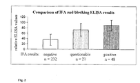
- Fig. 2: Results of 301 field sera compared in IFA and ELISA
- Fig. 3: Results of Ileitis blocking ELISA - cross-sectional screenings in 3 farms
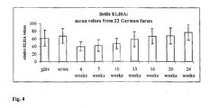
- Fig. 4: Results of Ileitis ELISA- 22 German farms
- Fig. 5: Correlation of ELISA results with clinical symptoms.
-
DETAILED DESCRIPTION OF THE INVENTION
-
Definitions of terms used in the description:
-
Before the embodiments of the present invention it must be noted that as used herein and in
the appended claims, the singular forms "a", "an", and "the" include plural reference unless
the context clearly dictates otherwise. Thus, for example, reference to "a bacterium"
includes a plurality of such bacteria, reference to the "cell" is a reference to one or more
cells and equivalents thereof known to those skilled in the art, and so forth. Unless defined
otherwise, all technical and scientific terms used herein have the same meanings as
commonly understood by one of ordinary skill in the art to which this invention belongs.
Although any methods and materials similar or equivalent to those described herein can be
used in the practice or testing of the present invention, the preferred methods, devices, and
materials are now described. All publications mentioned herein are incorporated herein by
reference for the purpose of describing and disclosing the cell lines, vectors, and
methodologies as reported in the publications which might be used in connection with the
invention. Nothing herein is to be construed as an admission that the invention is not
entitled to antedate such disclosure by virtue of prior invention.
-
As used herein, the term "L. intracellularis" means the intracellular, curved gram-negative
bacteria described in detail by C. Gebhart et al., Int'l. J. of Systemic Bacteriology, Vol. 43,
No. 3, 533-538 (1993) and S. McOrist et al., Int'l. J. of Systemic Bacteriology, Vol. 45,
No. 4, 820-825 (1995), each of which is incorporated herein by reference in their entireties;
the causative bacteria which can be obtained from PPE infected swine or other animals
throughout the world given the knowledge in the art and the teachings herein; and variants
or mutants of any of the above bacteria, whether spontaneously or artificially obtained and
DNA, RNA and bacterial proteins specific for L. intracellularis, including proteins
expressed in vectors or after in vivo application, and also fragments or antigenic derivatives
of L. intracellularis. This term also includes adapted isolates and attenuated isolates as
defined below.
-
As used herein, the term 'adapted isolate' means any L. intracellularis isolate that is
prepared according to the cultivation and passaging techniques in cell culture or any other
technique to replicate L. intracellularis for purposes of antigen preparation.
-
As used herein, the term "attenuated isolate" means any L. intracellularis isolate that is
prepared according to the cultivation and passaging techniques taught herein to achieve
avirulence while maintaining immunogenic properties when administered to a host animal.
-
As used herein, the term "large-scale cultivation" means a level of cultivation of L.
intracellularis greater than approximately 2.0 to 3.0 liters and includes production on a
scale of 100 liters or more. "Cultivation" as used herein, means the process of promoting
the growth, reproduction and/or proliferation of L. intracellularis.
Disclosure of the invention
-
The solution to the above technical problem is achieved by the description and the
embodiments characterized in the claims.
-
The aim of the present invention is to provide a method of diagnosing pre-clinical or
clinical L. intracellularis infection. Furthermore, an immune response to L. intracellularis
shall be detected and a tool for L. intracellularis epidemiology is to be provided.
-
This objective has been achieved according to the present invention within the scope of the
specification and claims by means of a method of diagnosing pre-clinical or clinical L.
intracellularis infection.
-
The invention relates to the detection of antibodies against L. intracellularis and to the
detection of L. intracellularis itself. Such antibodies include any antibody type known to
the skilled person, in particular immunoglobulins of the subtypes IgA, souble IgA, IgM,
IgG (IgG1, IgG2a, IgG2b, IgG3), and also IgD, IgE.
-
To detect antibodies specific for
L. intracellularis, according to the invention, the method
is based on the fact that
- a) a liquid sample is taken from a mammal
- b) the specific binding of such liquid sample to L. intracellularis is detected
- c) the result obtained is compared with a control.
-
-
As defined above, L. intracellularis also includes fragments or antigenic derivatives of L.
intracellularis, which may be used in a method as above.
-
Similarily, according to the invention, to detect
L. intracellularis, the method according to
the invention is as follows:
- the method is based on the fact that
- a) a liquid sample is taken from a mammal
- b) the specific binding of such liquid sample to L. intracellularis antibodies is detected
- c) the result obtained is compared with a control.
-
-
The liquid sample may include any bodily fluid including saliva, sweat, urine, blood,
secretions, excretions or other bodily fluid samples known to the expert in the field.
In a preferred embodiment, the liquid sample is collected using either a collection
container (for urine or other liquid), a syringe/collection tube (for blood) or some type of
collection swab (for saliva, sweat etc.)
Preferably, if the sample in step a) is blood, serum or plasma is isolated from this blood
sample and step b) is carried out with serum or plasma instead of the liquid sample.
Subsequently, step c) is carried out as above.
-
In a preferred embodiment, the method is an immune test. An immune test uses
monoclonal antibodies or polyclonal antisera specific to L. intracellularis.
Immune tests according to the invention include the methods of detection known in the art
such as the ELISA test (enzyme-linked immuno-sorbent assay) or the so-called sandwich-ELISA
test, dot blots, immunoblots, radioimmuno tests (radioimmunoassay RIA),
diffusion-based Ouchterlony test or rocket immunofluorescent assays. Other immune tests
are the indirect or direct immunofluorescent antibody test ("IFA"). Another immune test is
the so-called Western blot (also known as Western transfer procedure or Western blotting).
The purpose of Western blot is to transfer proteins or polypeptides separated by
polyacrylamide gel electrophoresis onto a nitrocellulose filter or other suitable carrier and
at the same time retain the relative positions of the proteins or polypeptides obtained from
the gel electrophoresis. The Western blot is then incubated with an antibody which
specifically binds to the protein or polypeptide under consideration. These methods of
detection can be used by the average skilled person to perform the invention described
herein. Literary references in which the skilled person can find the above-mentioned
methods and other detection methods are listed as follows: An Introduction to
Radioimmunoassay and Related Techniques, Elsevier Science Publishers, Amsterdam, The
Netherlands (1986); Bullock et al., Techniques in Immunocytochemistry, Academic Press,
Orlando, FL Vol. 1 (1982), Vol. 2 (1983), Vol. 3 (1985); Tijssen, Practice and Theory of
Enzyme Immunoassays: Laboratory Techniques in Biochemistry and Molecular Biology,
Elsevier Science Publishers, Amsterdam, The Netherlands (1985). Solid and fluid protein
chip technologies or microarray technologies using labeled or unlabeled reagents are
further immune tests according to the invention. Such test are widely described, for
example by Kozak KR, et al., Identification of biomarkers for ovarian cancer using strong
anion-exchange Protein Chips: potential use in diagnosis and prognosis..Proc Natl Acad
Sci U S A. 2003 Oct 14;100(21): 12343-8 (2003), or by Fulton, R.J., et al., Advanced
multiplexed analysis with the FlowMetrix™ system. Clinical Chemistry 43 (9), 1749-1756
(1997).
-
The invention further relates to a diagnostic test kit for detecting L. intracellularis infection
which contains all the elements required to carry out a method of diagnosing pre-clinical or
clinical L. intracellularis infection as described herein.
-
The invention further relates, in particular, to a diagnostic test kit which contains
antibodies specific to L. intracellularis.
-
The invention further relates, in particular, to a diagnostic test kit, characterised in that the
antibodies according to the invention are polyclonal.
-
The invention further relates, in particular to a diagnostic test kit, characterised in that the
antibodies according to the invention are monoclonal.
-
A diagnostic test kit is a collection of all the components for a method of diagnosis
according to the invention. Some examples (not an exhaustive list) of other elements for
performing a process according to the invention include containers such as 96-well plates
or microtitre plates, test tubes, other suitable containers, surfaces and substrates,
membranes such as nitrocellulose filter, washing reagents and buffers. A diagnostic test
kit may also contain reagents which may detect bound antibodies, such as for example
labelled secondary antibodies, chromophores, enzymes (e.g. conjugated with antibodies)
and the substrates thereof or other substances which are capable of binding antibodies.
-
The
L. intracellularis bacteria as disclosed herein or known in the art, or components
derived from such bacteria, can be used as an antigen in an ELISA or other immunoassay,
such as an immunofluorescent antibody test ("IFA"), to detect antibodies to
L.
intracellularis in the serum and other body fluids of animals suspected of being infected
with the bacteria. The presently preferred immunoassay is an IFA as described in the
example below. Alternatively, the bacteria of the instant invention can be used in a
Western Blot assay.
The preferred Western Blot protocol is as follows:
- 1. Run antigen on 12% SDS-PAGE and transfer to nitrocellulose membrane.
- 2. Place membrane in blocking buffer for 2 hours.
- 3. Remove blocking buffer and rinse with PBS for 1 minute.
- 4. Dilute serum in blocking buffer and add to membrane. Incubate for 2 hours at
room temperature.
- 5. Wash 3 times with wash buffer (5 minutes for each wash).
- 6. Dilute conjugate in blocking buffer and add to membrane. Incubate for 1 hour at
room temperature.
- 7. Wash 3 times with wash buffer.
- 8. Add substrate for 10 minutes or until strong banding occurs.
- 9. Rinse with PBS.
- 10. Air dry and store in the dark.
- The most preferred immune test according to the invention is an ELISA.
Most preferably, such ELISA is a sandwich ELISA (= capture ELISA). Such an ELISA is
more specific, as it detects with two antibodies specific to two different epitopes on the
same antigen. In a sandwich ELISA, unlabelled antibodies are coated to the microtiter
plates. The L. intracellularis as antigen is subsequently added. After a washing step, the
bound L. intracellularis antigen is detected with a second, labeled, L. intracellularis-specific
antibody reactive to a different epitope on L. intracellularis.
An exemplary coating procedure is as follows:
- The plates are coated with unlabelled antibody, e.g. in 10% Sucrose/10% Normal Horse
Serum in ddH2O and incubated with Lawsonia antigen. The plates are then dried and
sealed and stored at 37°C.
-
-
A preferred ELISA protocol according to the invention is as follows:
- 1. Add 0.1 ml/well antigen diluted in coating buffer. Incubate for 18 hours at 4°C.
- 2. Wash 3 times with PBS.
- 3. Add 0.25 ml of blocking buffer to each well of plate. Incubate 1 to 2 hours at
37°C.
- 4. Wash 3 times with wash buffer.
- 5. Dilute serum in blocking buffer and add 0.1 ml to the first wells of plate. Make
serial 1:2 dilutions across the plate. Incubate for 1 hour at 37.
- 6. Wash 3 to 5 times with wash buffer.
- 7. Dilute conjugate in blocking buffer and add 0.1 ml to wells of plate and incubate
for 1 hour at 37°C.
- 8. Wash 3 to 5 times with wash buffer.
- 9. Add substrate.
- 10. Measure absorbance of light with a spectrophotometer.
- 11. Wells in which antigen was not added are used as blanks.
- 12. Positive and negative control swine serum should also be used with each test.
-
A preferred ELISA is carried out as follows:
-
The plates are coated with unlabelled antibody, e.g. in 10% Sucrose/10% Normal Horse
Serum in ddH
2O and incubated with Lawsonia antigen. The plates are then dried and
sealed and stored at 37°C.
- 1. Add 90 µl buffer into all wells.
- 2. Add 10 µl serum into selected wells.
- 3. Incubate plate for 1 hour at 37C.
- 4. Wash plate 3 times with PBST.
- 5. Add HRP conjugate diluted in CDS-C+0.5M NaCl, 100µl /well.
- 6. Incubate plate for 1 hour at 37C.
- 7. Wash plate 3 times with PBST.
- 8. Add substrate, 100µl/well and incubate at room temperature for 10 minutes.
- 9. Add 50 µl stop/well.
- 10. Read the plate at 450 nm in a spectrophotometer:
- The most preferred sandwich ELISA protocol according to the invention is as follows:
- 1. Add 0.1 ml/well mAb diluted in coating buffer. Incubate for 18 hours at 4°C.
- 2. Wash 3 times with PBS.
- 3. Add 0.1 ml/well antigen diluted in buffer to each well of plate. Incubate 1 to 2
hours at 37°C.
- 4. Add 0.25 ml of blocking buffer to each well of plate. Incubate 1 to 2 hours at
37°C.
- 5. Wash 3 times with wash buffer.
- 6. Dilute serum in blocking buffer and add 0.1 ml to the first wells of plate. Make
serial 1:2 dilutions across the plate. Incubate for 1 hour at 37.
- 7. Wash 3 to 5 times with wash buffer.
- 8. Dilute conjugate in blocking buffer and add 0.1 ml to wells of plate and incubate
for 1 hour at 37°C.
- 9. Wash 3 to 5 times with wash buffer.
- 10. Add substrate.
- 11. Measure absorbance of light with a spectrophotometer.
- 12. Wells in which antigen was not added are used as blanks.
- 13. Positive and negative control swine serum should also be used with each test.
- Antigen as used herein is L. intracellularis as defined above. mAb relates to a monoclonal
antibody specific for L. intracellularis. Preferably, such antibody is an antibody as
disclosed below.
Surprisingly, outstanding antibodies were generated to be used in the ELISA according to
the invention. The antibodies have the following reference numbers: 301:39, 287:6,
268:29, 110:9, 113:2 and 268:18. All antibodies are specific for antigens of L.
intracellularis bacteria. Preferably as capture antibodies in a sandwich ELISA are 110:9,
113:2 or 287:6, and as conjugated antibodies 268:18, 268:29 or 287:6. Most preferred as
capture antibody is antibody 110:9 and as conjugated antibody is antibody 268:29.
The antibodies according to the invention are produced by hybridoma cells.
Said hybridoma cells according to the invention were deposited at the "Centre for Applied
Microbiology and Research (CAMR) and European Collection of Cell Cultures
(ECACC)", Salisbury, Wiltshire SP4 0JG, UK, as patent deposit according to the Budapest
Treaty. The date of deposit was May 11, 2004. The preliminary identification references
are "Hybridoma cell line 301:39", "Hybridoma cell line 287:6", "Hybridoma cell line
268:29", "Hybridoma cell line 110:9", "Hybridoma cell line 113:2" "Hybridoma cell line
268:18", and the accession number given by the international depositary authority
ECACC/CAMR are .
-
-
The present invention is further described in the following examples which are provided
for illustrative purposes only and are not to be construed as limiting. Indeed, other variants
of the invention will be readily apparent to one of ordinary skill in the art.
-
All publications and patents cited herein are incorporated by reference in their entireties.
EXAMPLES
EXAMPLE 1
1.0 Material
-
anti-Lawsonia hybridomal celline : 372:13, 113:2, 268:18, 287:6, 110:9, 268:29, 301:39.
Equipment and media for hybridomal cell cultivation.
Equipment and buffers for purification and HRP conjugation of monoclonal antibodies
(mAb).
Equipment and solutions for ELISA
Lawsonia antigen SF 1289 N343 12/20/00
Lawsonia positive sera: lot#052902pig#26 (experimentally infected),
lot#052902pig#69 (vaccinate)
Lawsonia negative sera: p 25.8#273 Day 52 6-30-95,
116705
1.1 Methods
-
The preparation of the monoclonal antibodies was performed according to standard
operating procedures at Svanova Biotech.
1.1.1 Culturing of hybridomal cells and preparation of HRP conjugated monoclonal
antibodies
-
- To secure the hybridomal cell-lines 15 vials of each cell line was prepared and
stored in -135°C.
- One vial of each hybridomal cell-line was thawed and the cells were grown in cell-culture
flasks.
- 500ml to 1000 ml of supernatant from each cell line was purified and conjugated to
horse-radish peroxidase.
1.1.2 Blocking ELISA
-
The purified and HRP conjugated mAb's were tested for their ability to be blocked by
positive Lawsonia pig sera in ELISA.
- The ELISA plate was coated with Lawsonia antigen in a dilution of 1/200,
100µl/well
- The pig sera was diluted 1/10, 100µl/well
- The HRP conjugated mAb's were diluted and added, 100µl/well.
-
The results are expressed as Percent inhibition (PI) according to the following formulae:
PI = 1 - (OD sample/ OD mAb) x 100.
1.2 Results
1.2.1 Culturing of hybridoma cells and preparation of HRP conjugated monoclonal
antibodies
-
- All but one of the mAb's were successfully purified and HRP conjugated. They had
good production capacity.
- mAb 372:13 had poor production capacity. This mAb was not successfully HRP
conjugated and was therefore not tested in the blocking ELISA.
1.2.2 Blocking ELISA
-
- Two positive (#26, #69) and two negative (25.8#273, 116705) sera were used in the
experiments.
- The experimentally infected sera (#26) inhibited all the six HRP conjugated mAb's,
but in different levels. The sera from a vaccinated pig inhibited the mAb's to a much lesser
extent.
- The negative sera showed no or very low inhibition.
-
The results are presented in the table below and in Fig. 1.
| Serum | mAb 301:39 | 110:9 | 268:29 | 287:6 | 113:2 | 268:18 |
| #26 exp + | 57 | 45 | 78 | 74 | 42 | 72 |
| #69vacc + | 18 | 23 | 52 | 43 | 45 | 25 |
| 25.8#273 - | 7 | 3 | 27 | 20 | 14 | 21 |
| 116705 - | -7 | -9 | -24 | -13 | 18 | -19 |
1.3 Discussion
-
The results of this study clearly show that one ore more of the monoclonal antibodies
tested are suited for a blocking ELISA test against Lawsonia intracellularis. MAb 268:29,
287:6 and 268:18 show the clearest distinction between the positive and negative sera i.e.
are strong candidates in the development of the blocking ELISA.
EXAMPLE 2
2.0 Summary
-
Development and establishment of a sandwich b-ELISA prototype for the detection of
antibodies to Lawsonia intracellularis:
- After calculation of sensitivity and specificity by using immunoflourescence test IFA as
gold standard the following can be concluded:
- Capture mAb 110:9 gives a clear distinction between positive and negative
samples.
- Detection mAb 268:29 HRP gives a clear distinction between positive and negative
sera showing a sensitivity and specificity of 100%.
-
-
It is recommended that the prototype will be as follows:
- mAb 110:9 as capture mAb, coated in Nunc maxi-sorp strips C-8
- Antigen(Ag): Lawsonia intracellularis culture (preferably the concentrated material
produced in R&D at BI)
- Serum samples diluted 1/10 in 0.5M NaCl/PBST. Incubation for 1 hour at 37C.
- HRP mAb 268:29 , lyophlized in CDS-C buffer containing 0.5M NaCl. Incubation
for 1 hour at 37C.
- Substrate K-Blue Max, incubation for 10 minutes at room temperature.
- Svanova's stop solution
2.1 Material and methods
-
The ELISA was performed according to the following procedure:
2.1.0 ELISA procedure
-
Add 90 µl buffer into all wells.
Add 10 µl serum into selected wells.
Incubate plate for 1 hour at 37C.
Wash plate 3 times with PBST
Add HRP conjugate diluted in CDS-C+0.5M NaCl, 100 µl /well.
Incubate plate for 1 hour at 37C.
Wash plate 3 times with PBST.
Add substrate, 100µl/well and incubate at room temperature for 10 minutes.
Add 50 µl stop/well
Read the plate at 450 nm in a spectrophotometer.
-
The results were presented as 1- OD values or 2- PI (percent inhibition) values i.e. 100 -
sample OD/ mAb OD x 100.
2.1.1 Test 1 (ELISA 030922, 030923 x2)
-
C-8 maxi strips coated with mAb 110:9, 113:2, 301:39,287:6, 268:29, 300ng/well
Blocking of strips with 10% 2251HÄS(normal horse serum), 10% sucrose/UHP H2O.
150µl/well, incubated for 30 seconds at room temperature.
Ag # 030619 diluted 1/10 in PBST, 100µl/well, 3hours incubation in 4C.
Strips dried in 37C for 3 hours. Kept in 4C.
Sera: IFA pos: #26, #69. IFA neg: #6, C1, D1, E1. Diluted 1/10 in 0.5M NaCl/PBST
Conjugate: 268:18, 113:2, 301:39110:9, 287:6, 268:29 HRP diluted in 0.5M NaCl/PBST.
All capture mAb's were tested against all HRP mAb except against its own.
2.1.2 Test 2 (ELISA 030924)
-
Strips coated with 110:9, 113:2 and 287:6 according to test 1 above.
Sera: IFA pos: #26, #69. IFA neg: #6, C1, D1, E1. Diluted 1/10 in 0.5M NaCl/PBST
Conjugate 268:18, 268:29, 287:6 HRP diluted in 0.5M NaCl/CDS-C (standard buffer for
lyophilizing of conjugates as Svanova in production).
2.1.3 Test 3 (ELISA 030925)
-
Strips coated with 110:9, 113:2 and 287:6 according to test 1 above.
Sera: 24 sera (10 IFA pos, 10 IFA neg and 4 doubtful) were tested. ID can be found in
appendix 4. Dilution: 1/10 in 0.5M NaCl/PBST.
Conjugate: 268:18 HRP diluted in 0.5M NaCl/CDS-C.
2.1.4 Test 4 (ELISA 030930, 031002, 031008)
-
Strips coated with 110:9 according to test 1 above.
Sera: 21 IFA positive and 69 IFA negative (for details see appendix 5). Dilution: 1/10 in
0.5M NaCl/PBST
Conjugate: 268:18 HRP and 268:29 HRP diluted in 0.5M NaCl/CDS-C.
2.2 Results
2.2.1 Test 1 (ELISA 030922, 030923 x2)
-
The OD and PI values obtained can be found in appendix 2. Capture mAb 110:9, 113:2 and
287:6 with conjugates 268:18 and 268:29 and 287:6 seems to be the best candidates for the
sandwich b-ELISA as these combinations showed the best blocking capacity on the
positive sera. Sera no #6 scored positive in most of the tests. This was suspected as this
sera also scored positive in the previously developed b-ELISA prototype.
2.2.2 Test 2 (ELISA 030924)
-
This test was mainly performed to assure that the standard buffer used to lyophilizing of
the conjugate could be used. The results show that blocking was achieved with the positive
sera. The best blocking was found with detection mAb 268:18 or 268:29.
2.2.3 Test 3 (ELISA 030925)
-
The capture mAb 110:9 was found to be the best choice as it gave the clearest
differentiation between positive and negative samples.
2.2.4 Test 4 (ELISA 030930, 031002. 031008)
-
Obtained OD and PI values can be found in appendix 5. When plotting the OD values
against the IFA results HRP mAb 268:29 gives the clearest distinction between positive -
and negative samples with a 100 % sensitivity and specificity. mAb 268:18 also gives a
sensitivity and specificity of 100% but the area between positive and negative samples is
slight. When plotting the PI values mAb 268:18 gives a sensitivity of 95.7 and specificity
of 100% at cut-off 20.9 while mAb 268:29 gives both a sensitivity and specificity of 100%
at cut-off28.6
2.3. Discussion
-
After calculation of sensitivity and specificity against the immunoflourescence test IFA the
following can be concluded:
- Capture mAb 110:9 is the best choice as it gives a clear distinction between
positive and negative samples.
- Detection mAb 268:29 HRP is the best choice as it gives a clear distinction
between positive and negative sera with a sensitivity and specificity of 100%.
-
It is recommended that the prototype will be as follows:
- mAb 110:9 as capture mAb, coated in Nunc maxi-sorp strips C-8
- Ag: Lawsonia intracellularis culture (preferably the concentrated material produced
in R&D at BI)
- Serum samples diluted 1/10 in 0.5M NaCl/PBST. Incubation for 1 hour at 37C.
- HRP mAb 268:29 , lyophlized in CDS-C buffer containing 0.5M NaCl. Incubation
for 1 hour at 37C.
- Substrate K-Blue Max, incubation for 10 minutes at room temperature.
- Svanova's stop solution
EXAMPLE 3
3.1 Summary
-
The performed accelerated stability studies indicate that the Lawsonia intracellularis
antigen captured ELISA plate has a stability of at least one year and that the a- Lawsonia
HRP conjugate 268:29 has a stability of 6 to 12 months in a lyophilized form.
3.2 Introduction
-
In order to estimate the stability of the components of a future Lawsonia intracellularis
ELISA kit accelerated stability tests has to be performed. The component to be tested is
stored at 37C for 4 weeks and is thereafter tested in the ELISA in comparison with a
component kept at 4C. 4 weeks stability in 37C indicates a shelf-life of about 12 months.
This report describes accelerated stability studies performed on the antigen captured
ELISA plate and on the HRP conjugate.
3.3 Materials and methods
-
Accelerated stability on plates (ELISA 031024)
Plates were coated with Lawsonia mAb 110:9 blocked with 10% Sucrose /10% Normal
Horse serum in ddH2O and incubated with Lawsonia antigen. Plates were dried and sealed
and put in +37°C. Plates were taken from +37C weeks 1, 2, 3 and 4 and stored in a
refrigerator. Plates were also stored in +4C for the whole test-period as a reference.
Positive sera #26, #69 and #06 and negative C2, D1 and E2 were used to check the
performance of the sandwich ELISA.
HRP conjugate mAb 268:29#030120 was used.
Accelerated stability on lyophilized conjugate (ELISA 031027)
HRP mAb 268:29 #030120 was diluted 1/1000 in CDS-C buffer dispensed into glass
bottles and lyophilized according to standard procedures. Four lyophilized bottles were
stored at +37C for 1, 2, 3 and 4 weeks. Bottles were also stored at 4C. The different bottles
were tested in the sandwich ELISA.
As positive serum #26 was used and negative serum 030926 were used.
It was found that a conjugate dilution of 1/1000 gave too high OD values in the test. The
conjugate was therefore titrated in the plate to obtain a correct signal for the test.
3.4. Results
-
Accelerated stability on plates
A big drop in OD values were seen after week 1 compared to the OD value from the strips
kept in +4oC. No big differences were seen when comparing week 1 to 4.
When looking at the PI values no significant was found between week 1 through 4 and
+4°C.
-
Accelerated stability on lyophilized conjugate
At a conjugate dilution of 1/8000 serum # 26 showed a decrease from 0.547 to 0.398 after
4 weeks and serum 030926 dropped from 1,654 to 0.99 after the 4 week period.
3.5. Discussion
-
The performed accelerated stability studies indicate that the Lawsonia intracellularis
ELISA plate has a stability of at least one year.The a- Lawsonia HRP conjugate 268:29
showed a 40% decrease in OD value against the negative sera and after 4 weeks. This
might indicate some instability of the conjugate. When calculating on the OD values a 40%
decrease of a signal at 1.5 means and OD value of 0.9 after 1 year which is still within the
acceptance criteria.
EXAMPLE 4
4.1. Summary
-
The verification of the prototype (plate batch 040107) was performed by testing 50 sera
previously used when developing the prototype. The verification study showed a 100%
agreement with the positive sera while 2 of the 25 negative sera scored positive in the
verified batch.
-
It was found that normal horse serum can be used in the CDS-C buffer instead of swine
sera which is the case today.
-
It was found that PBST containing 0.042% PVP showed similar results as using the PBST
tablets (that contains PVP).
4.2. Introduction
-
In order to verify the sandwich ELISA prototype a larger number of sera have to be tested
on a large scale produced plate batch. The study was performed on plate batch 040107 that
was tested by examining 50 serum samples that had previously been used to develop the
prototype.
-
Buffer CDS-C is used as preservative when lyophilizing the HRP conjugate. This buffer
contains swine sera. As it can be problematic in the future to find Lawsonia negative sera
to be used in the CDS-C buffer an alternative has to be tested. In this study normal horse
serum and foetal bovine serum were tested.
-
The majority of the laboratory work on this ELISA has been performed with PBST tablets.
It has been seen that the 20x conc. PBST that is included in Svanovir kits can not be used
since the test will then not correctly differentiate between positive and negative samples.
When comparing the recipes of the tablets and the 20x conc. PBST it was found that all
components were the same except for 0.042% PVP (Polyvinylpyrrolidone K25) that was
included in the tablets as a stabilizing factor. Experiments showed that when adding the
PVP to the 20x PBST the same results as the tablets will be achieved.
4.3. Materials and methods
Verification of prototype (ELISA 040312)
-
50 sera (received from Bioscreen) previously tested in the prototype ELISA was tested on
the large scale plate batch # 040107. PBST 20x conc buffer with 0.042% PVP was used.
Lyophilized conjugate batch 040308 was used.
CDS-C buffer with normal horse serum
Test ELISA 040203
-
CDS-C buffer was prepared with; 1- swine serum (as in TJ4/05F), 2- with normal horse
serum and 3- with foetal bovine serum. The buffers were tested as conjugate buffer in the
ELISA. Serum tested were gris 2, 4, 13 and 17, 1-29664, 3-29664, A-27239, B-27239, #26
and C2. HRP conjugate 268:29#030318 was used. 20 x conc PBST with 0.042% PVP was
used
Test ELISA 040311
-
CDS-C buffer containing normal horse serum has been used to prepare lyophilized
conjugate at two occasions (040211, 040308). The conjugate was prepared to obtain a final
dilution of 1/20k, 1/25k and 1/30k. Freshly prepared conjugate was tested in parallel.
Serum #26, 1-29664, 3-29664, A-27239, B-27239, pos pool 030926 and neg pool 030926
were used. HRP conjugate 268:29#030318 was used. 20 x conc PBST with 0.042% PVP
was used.
PBST with PVP (ELISA 040301)
-
PBST buffer 20x concentrated was diluted 1/20 and a final concentration of 0.042% PVP
was added. This buffer was tested in parallel with PBST tablets (already containing PVP)
and 20x conc PBST buffer diluted 1/20. The buffers were used in all steps, i.e. in the
sample and conjugate dilution buffer as well as in the washing buffer.
Serum #26, E1, 1-29664, 3-29664, A-27239, B-27239 and gris 13 were used. HRP
conjugate 268:29#030318 was used. As conjugate buffer CDS-C with swine serum was
used.
4.4. Results
Verification of prototype
-
The results show that all 25 previously positive sera also scored positive in the verification
batch. Two out of the 25 previously negative sera scored positive.
CDS-C buffer with normal horse serum
-
The buffer containing normal horse serum showed similar results as the one with swine
sera.
-
The reading of the lyophilized HRP mAb decreased at the most 0.4 OD when compared to
the freshly prepared conjugate. This drop was noticed in both batches of lyophilized
conjugate. It is not affecting the test performance as the calculated PI values corresponded
well regardless of obtained OD values.
PBST with PVP
-
The results show that 20x concentrated PBST solution diluted 1/20 with addition of
0.042% PVP gave similar results as the PBST tablets while 20x conc PBST diluted 1/20
without the PVP showed false negative results.
4.5. Discussion
-
The verification of the prototype (plate batch 040107) was performed by testing 50 sera
previously used when developing the prototype. The verification study showed a 100%
agreement with the positive sera while 2 of the 25 negative sera scored positive in the
verified batch. The "false" negative results are acceptable as the major importance of this
test is to detect positive samples. It has also not been proven that the results from the
previous testing are indeed true.
-
It was found that PBST containing 0.042% PVP showed similar results as using the PBST
tablets (that contains PVP). The future kits will therefore contain this buffer. A stability
study of the PBST 20x conc with PVP has to be performed to ensure the validity of the
kits.
EXAMPLE 5
5.1 Introduction and Objectives
-
Detection of serum antibodies against Lawsonia intracellularis so far is done by the use of
IFA tests (1). These tests rely on an individual and highly subjective judgment in
interpretation of results. Recently a sandwich blocking ELISA was developed using highly
specific monoclonal antibodies against L. intracellularis. The data from this test can be
measured and evaluated on an objective basis, which improves reproducibility,
repeatability and comparative analyses. In this study first comparative data to the IFA test
are presented, demonstrating the suitability and performance of the new test.
5.2 Material and Methods
-
L. intracellularis antigen grown on a McCoy cell culture and captured by a specific
monoclonal antibody on microtiter plates was used to detect antibodies against L.
intracellularis in porcine serum samples. Sera were tested in a 1 to 10 dilution. After 1 h of
incubation at 37 °C plates were washed with PBS Tween solution, horseradish-peroxidase
labeled monoclonal antibody (different to the capture antibody) was added. The plates
were incubated for an additional hour at 37 °C. Unbound antibodies were removed by
washing. After incubation with TMB substrate for 10 min at room temperature the
reaction was stopped by adding a stop solution. The OD450 was read in a photometer for
each well. Results were calculated as percent inhibition of controls without serum.
301 field sera, tested in a validated IFA test (2) for antibodies against L. intracellularis,
were used to evaluate the new ELISA. In addition cross-sectional samples (10 samples
each from 9 age groups) from two farms with known Ileitis and one farm with suspected
Ileitis were used to demonstrate the diagnostic value of the ELISA in comparison to the
IFA.
5.3 Results
-
Many negative sera resulted in negative inhibition values. Therefore the lowest measured
value was set to 0 and all other values were related to this. These data are declared as
"relative ELISA values".
Results from the 301 field sera are summarized in Figure 2. The ELISA clearly
discriminates between negative (mean relative ELISA value 36) and positive sera (mean
relative ELISA value 89). Majority of the samples that scored doubtful in the IFA were
positive in the ELISA.
Results from the cross-sectional screenings in the 3 farms are shown in Figure 3. Both tests
clearly detect antibodies against L. intracellularis in the 3 farms.
5.4 Discussion
-
In comparison to the IFA test results the ELISA provides a higher sensitivity. In some
groups of the 3 tested farms the result from the ELISA is much more unambiguous than in
the IFA test, that gave a lot of questionable results, due to unspecific fluorescence. The
serological profiles in the 3 herds corresponded well to previously published data from
European herds (3,4). The increase of ELISA values between the group of 10 and 16
weeks old pigs in comparison to the IFA test results indicate, that sera with relative ELISA
values greater 55 can be judged as positive.
This new test could help to improve diagnosis of Ileitis in routine samples. The new
blocking Ileitis ELISA provides a tool for fast and sensitive screening of large amounts of
samples on an objective basis.
-
- 1. Guedes RMC et al., 2002. Can J Vet Res 66:99-107.
- 2. Knittel JP et al.,1998. AJVR 59:722-726.
- 3. Biksi I et al., 2002. Proc. 17th IPVS, Ames, Iowa, USA
- 4. van Aken N et al., 2002. Proc. 17th IPVS, Ames, Iowa, USA.
-
EXAMPLE 6
6.1 Introduction and Objectives
-
Specific diagnosis of Ileitis so far is done by the use of IFA tests for antibody detection
(1,2) or PCR (3,4), histological and immuno-staining methods (2,5,6) for antigen
detection in feces or intestinal tissue samples. All these methods have specific
disadvantages concerning specificity, contamination risk or workload and are not suitable
for routine herd screenings on a large scale. So most diagnostic tests are done only for few
pigs from a herd. By the use of a recently developed blocking ELISA serological testing
becomes easier, more reliable and suitable for herd profiling. Cross-sectional screenings of
22 farms in Germany were performed to demonstrate the diagnostic performance of the
new test.
6.2 Material and Methods
-
Cross-sectional samples from twenty-two German farms were tested in the ELISA. From
gilts, sows and pigs 4, 7, 10, 13, 16, 20 and 24 weeks of age samples were taken on each
farm (10 samples in each age group). The ELISA was done as described in example 5.
6.3 Results
-
In Figure 4 the mean relative ELISA values from all 22 farms are summarized. Sera from
gilts and sows are highly reactive in the ELISA, whereas results from flatdeck piglets of 4
to 10 weeks of age are low. In the fatteners ELISA values increase from 13 to 24 weeks.
-
In Figure 5 mean relative ELISA values are correlated with the clinical symptoms in the
farms. The farms No. 5, 6 and 15 had no clinical enteric problems at the time of sampling.
In farm No. 3 piglets from 4 to 13 weeks had enteric problems, that were diagnosed as E.
coli diarrhea. In farm No. 18 prior serological tests indicated the presence of Lawsonia
intracellularis in the farm without clinical symptoms. In the farms No. 1, 2, 7, 8, 9, 10, 11,
12, 19, 21 and 22 Ileitis was diagnosed before sampling for this study. The farms No. 4,
13, 14, 16 and 20 report enteric problems in growers and fatteners, farms No. 4 and 13
with clinical suspected Ileitis. In farm No. 17 no clinical signs of diarrhea are observed, but
antibiotics are used in growers and at the beginning of fattening.
6.4 Discussion
-
The summarized seroprofile from the 22 farms shows high reactivity in sows and gilts and
rising antibody reaction starting at 13 weeks up to 24 weeks. These findings are in
agreement with serological data published so far for Ileitis (7,8,9).
Pigs from the farms without diarrhea (No. 5, 6, 15 and 18) and the farm with E. coli
diarrhea (No. 3) showed no significant seroconversion in the ELISA. In the farms with
known Ileitis (No. 1, 2, 7, 8, 9, 10, 11, 12, 19, 21 and 22) a clear seroconversion can be
detected between week 7 and 13.
Interestingly the samples from most of the sow and gilt groups are highly reactive in the
ELISA, although most of these pigs have no apparent enteric problems. This might be due
to a subclinical infection with Lawsonia intracellularis. Results from farm No. 17 seem to
indicate a subclinical Ileitis.
The new ELISA according to the invention provides a powerful tool for serological
diagnosis of Ileitis suitable for routine testing of large sample sizes and cross-sectional
herd screenings.
References
-
- 1. Knittel JP et al., 1998. AJVR 59:722-726.
- 2. Guedes RMC et al., 2002. Can J Vet Res 66:99-107.
- 3. Jones GF et al., 1993. J Clin Microbiol 31: 2611-2615.
- 4. Suh-DK et al., 2000. J Vet Sci 1:33-37.
- 5. McOrist S et al., 1989. Vet Pathol 26: 260-264.
- 6. Huerta B et al., 2003. J Comp Path 129: 179-185.
- 7. Biksi I et al., 2002. Proc. 17th IPVS, Ames, Iowa, USA
- 8. van Aken N et al., 2002. Proc. 17th IPVS, Ames, Iowa,
USA
- 9. Holyoake PK et al., 1994. J Clin Microbiol 32: 1980-1985.
-