TECHNICAL FIELD
The present invention relates to a material for repairing
biological tissues used for repairing a defect in biological
tissues, and a process for producing the same.
BACKGROUND ART
Recently, it has become possible to repair a defect in
biological tissue such as bone caused by osteoncus extraction,
trauma, or the like by regenerating the bone by filling a
material for repairing biological tissues such as a bone
substitute or the like. For such a bone substitute,
hydroxyapatite (HAP) and tricalcium phosphate TCP) are known.
However, from the viewpoint of leaving no foreign matter
inside the body, a scaffold made of a porous calcium phosphate
material, for example, such as β-TCP is used. If the β-TCP is
left in contact with bone cells of a defect part of bone, so-called
remodeling is performed in which osteoclasts eat the β-TCP
and osteoblasts form a new bone. That is, the bone
substitute filled in the defect part of the bone is replaced
by autologous bone as time goes by (for example, refer to:
Uemura and two others, "Tissue engineering in bone using
biodegradable β-TCP porous material - a new material
strengthened in vivo Osferion", Medical Asahi, The Asahi
Shimbun Company, October 1, 2001, Vol. 30, No. 10, p. 38-41).
When the bone substitute is to be filled into a defect
part of the bone, in order to increase the adhesiveness with
the bone tissue, various inventions have been proposed in
which: the surface to be in contact with the defect part of
the bone is made uneven by plasma spraying (for example, refer
to Japanese Patent No. 3166352); or a binding packing made
from a nonwoven fabric is arranged on the surface in contact
with the defect part of the bone (for example, refer to
Japanese Unexamined Patent Application, First Publication No.
2003-19149).
On the other hand, according to the condition of the
defect part of the bone, there is a case where the bone
forming function is promoted by adding growth factors to the
bone substitute in order to increase the activity of the cells
surrounding the defect part of the bone.
As a method of adding the growth factors in this case,
there is known a method wherein the bone substitute is
provided with various sizes of holes which are filled with
biodegradable viscous material such as collagens containing
the growth factors, and the bone substitute is applied as a
filling.
However, the binding force between the abovementioned
conventional bone substitute, which is provided only with the
uneven part or the binding packing, and the surrounding bone
tissue is weak, so that the bone substitute and the bone
tissues may become separated before the bone has grown.
Therefore, although such a structure has been employed,
bone regeneration still takes a long time and it has been
difficult to sufficiently demonstrate the bone regenerating
function.
Moreover, in the abovementioned conventional bone
substitute, although micropores are reliably in communication
with each other inside of the porous body, there has been a
problem in that straight penetrating holes are not provided
and blood vessels can not be sufficiently formed. Moreover,
there has been a problem in that growth factors of sufficient
types and amounts can not be added, so that supply with a
sustained-release property where they are leaked out into the
biological tissue surrounding the defect as time goes by
cannot be achieved, and therefore, the effect can not be
maintained for a long time.
DISCLOSURE OF INVENTION
In view of the above problems, an object of the present
invention is to provide a material for repairing biological
tissues which sustains a sufficient binding force after
application to a biological tissue and promotes biological
tissue formation.
Moreover, the present invention has an object of
providing a material for repairing biological tissues which
can provide agonistic factors of sufficient types and amounts
to a biological tissue, and can control the sustained-release
dose thereof.
The present invention employs the following solution in
order to solve the above problems.
In the material for repairing biological tissues of the
present invention, a plurality of grooves are formed in the
surface of a plate-type base member.
Since a plurality of grooves are formed in the surface of
the base member, the material for repairing biological
tissues, can be more in contact with the biological tissues.
As a result, places where the biological tissue formation is
induced, are increased. Moreover, for example, if it is used
as a bone substitute, it is filled so that its groove side is
pressed against and inserted into the cancellous bone inside
the bone, and its base plate side is in contact with the
cortical bone being the outer layer, so that the adhesiveness
with the cancellous bone is increased. As a result, even if a
load is applied in a separation direction, the connection of
the bone substitute with the biological tissues can be
maintained, and the effect of forming a biological tissue can
be maintained, enabling an increase in the healing speed.
Moreover, in the material for repairing biological
tissues according to the present invention, it is preferable
that the grooves be formed along almost the same direction.
According to the material for repairing biological
tissues, since a plate-type base member can be readily formed
into a curved shape along the direction where the grooves are
extending, it can be provided in an optimum shape for defects
in biological tissue.
A process for producing a material for repairing
biological tissues of the present invention comprises: a step
for supplying a raw material formed in a slurry form into a
mold, and forming a compact in which a plurality of
penetrating holes extending in a single direction are
separated from each other by partition wall members having an
almost uniform thickness; and a step for cutting and dividing
the compact by a plane in parallel with the direction in which
the penetrating holes are extending.
According to the process for producing the material for
repairing biological tissues, by dividing the penetrating
holes formed in the compact, grooves are formed by the
partition wall members that divided the penetrating holes in
the compact, and by pressing these grooves against the
biological tissue, the adhesiveness thereof with the
biological tissue can be improved. Moreover, since the
compact is formed by supplying the raw material formed in a
slurry form into the mold, the size and the shape of the
penetrating holes which become the grooves, and the thickness
of the partition wall member can be freely controlled, and the
material for repairing biological tissues can be formed in an
optimum shape and size considering the position to be repaired
and the adhesiveness with the biological tissue.
The compact may have a honeycomb shape.
Moreover, another material for repairing biological
tissues of the present invention comprises: a main body of a
shape in which a plurality of penetrating holes extending in a
single direction are formed separated from each other by
partition wall members having an almost uniform thickness; and
a biodegradable viscous member which is filled into at least
one of the penetrating holes in the main body, and contains an
agonistic factor capable of increasing cell activity.
In the main body of the material for repairing biological
tissues, the size and shape of the penetrating holes can be
freely controlled for molding. Moreover, since the
biodegradable viscous member containing the agonistic factor
is filled into the penetrating holes, then by controlling the
position of the penetrating holes and the number thereof to be
filled, the sequence of providing the agonistic factor into a
defect, and the sustained-release dose can be controlled, so
that the biological tissue formation can be effectively
promoted.
The main body may be in a honeycomb shape.
BRIEF DESCRIPTION OF DRAWINGS
FIG. 1 is a perspective view showing a material for
repairing biological tissues according to a first embodiment
of the present invention.
FIG. 2 shows a production flow of the material for
repairing biological tissues according to the first embodiment
of the present invention.
FIG. 3 is a perspective view showing a honeycomb compact
according to the first embodiment of the present invention.
FIG. 4 is a perspective view showing a honeycomb mold
used for production of the material for repairing biological
tissues according to the first embodiment of the present
invention.
FIG. 5 shows an example of use of the material for
repairing biological tissues according to the first embodiment
of the present invention.
FIG. 6 is a perspective view showing a material for
repairing biological tissues according to a second embodiment
of the present invention.
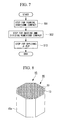
FIG. 7 shows a production flow of the material for
repairing biological tissues according to the second
embodiment of the present invention.
FIG. 8 is a perspective view showing a honeycomb compact
according to the second embodiment of the present invention.
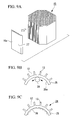
FIG. 9A to FIG. 9C show an intermediate product part way
through production of the material for repairing biological
tissues according to the second embodiment of the present
invention.
FIG. 10 shows an example of use of the material for
repairing biological tissues according to the second
embodiment of the present invention.
FIG. 11 is a perspective view showing a general outline
of a bone substitute main body according to a third embodiment
of the present invention.
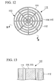
FIG. 12 is a front view showing the bone substitute
according to the third embodiment of the present invention.
FIG. 13 shows a cross-section taken along the line II-II
of FIG. 12.
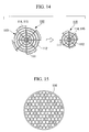
FIG. 14 is a schematic diagram showing a bone forming
function by the bone substitute according to the third
embodiment of the present invention.
FIG. 15 is a front view showing another example of the
bone substitute according to the third embodiment of the
present invention.
BEST MODE FOR CARRYING OUT THE INVENTION
First Embodiment
Hereunder is a description of a material for repairing
biological tissues according to a first embodiment of the
present invention, with reference to FIG. 1 to FIG. 4.
As shown in FIG. 1, a bone substitute (material for
repairing biological tissues) 10 according to the present
embodiment is made from β-TCP as the raw material, and
comprises a plate-type base member 11 on the surface of which
a plurality of grooves 12 extending in a single direction are
formed and separated from each other by partition wall members
13 having an almost uniform thickness.
Next is a description of a process for producing the bone
substitute 10.
As shown in FIG. 2, the production process for the bone
substitute 10 according to the present embodiment comprises: a
step (S01) for supplying a raw material formed in a slurry
form into a honeycomb mold 14, and forming a honeycomb compact
15; and a step (S02) for cutting and dividing the honeycomb
compact 15. Hereunder is a description of the respective
steps.
In step (S01) for forming the honeycomb compact 15,
firstly, for example by a method disclosed in Japanese
Unexamined Patent Application, First Publication No. Hei 5-237178,
an aqueous foamed slurry which has been mixed and
foamed, is adjusted and mixed with granular compacts made from
β-TCP so as to make a raw material which is formed in a slurry
form.
The raw material is supplied into the honeycomb mold 14
to make a honeycomb prototype 16 shown in FIG. 3.
As shown in FIG.4, in the top of the honeycomb mold 14 is
formed raw material inlets 17 that have been formed in a
funnel shape. Meanwhile, the inside at the bottom is formed
with slits 18 extending respectively in parallel with the
respective sides of the honeycomb mold 14. The bottom ends of
the raw material inlets 17 and one end of the slits 19 are
communicated through passages 19.
The width of the slits 18 and the intervals therebetween
are previously determined considering the adhesiveness
according to the shape and the size of the bone tissue to be
repaired.
The obtained honeycomb prototype 16 is sintered after
drying, for example in a method disclosed in Japanese
Unexamined Patent Application, First Publication No. Hei 5-237178.
In this manner, the honeycomb compact 15 in which a
plurality of penetrating holes 20 extending in a single
direction are separated from each other by partition wall
members 13, is obtained.
In the step (S02) for cutting and dividing the honeycomb
compact 15, it is cut and divided by a plane (for example, the
IV-IV position shown in FIG. 3) in parallel with the direction
in which the penetrating holes 20 in the honeycomb compact 15
are extending.
In this case, the bone substitute 10 wherein the wall 15a
constituting the external surface of the honeycomb compact 15
is serving as the base member 11, and the respective
penetrating holes 20 are serving as grooves 12, is obtained.
Next is a description of the effect of the bone
substitute 10 of the present embodiment having the above
structure.
As shown in FIG. 5, the bone substitute 10 is filled into
a defect in an ilium 21. At this time, the surface 11a formed
with the grooves 12 is pressed against and inserted into the
cancellous bone 22 in the ilium 21, to repair it so that the
surface of the base member 11 without the grooves 12 is on the
same surface with the cortical bone 23. In this case, since
the partition wall members 13 is driven into the inside of the
cancellous bone 22 like a wedge, the adhesiveness is mutually
increased, and they are kept fixed even if the bone substitute
10 is loaded from the outside. At the same time, a new bone
is formed using the bone substitute 10 as a scaffold.
Moreover, since the bone tissue and the bone substitute
10 are in contact over a large area including the surface of
the partition wall members 13 as well as the surface of the
base member 11, the bone is formed over a large area.
According to the bone substitute 10, since it is formed
by honeycomb molding, the size and the shape of the
penetrating holes 20 serving as the grooves 12 can be freely
controlled, and it can be formed in an optimum shape and size
considering the adhesiveness of the bone substitute to the
bone to be repaired and its vicinity. Moreover, since the
grooves 12 are formed, the bone substitute can be in contact
with the cancellous bone 22 over a large area, and the bone
formation can be promoted so as to increase the healing speed
after the repair, more than in the conventional case.
Second Embodiment
Next is a description of a material for repairing
biological tissues according to a second embodiment of the
present invention, with reference to FIG. 6 to FIG. 9. In the
following description, the same reference symbols are used for
components the same as those in the abovementioned embodiment,
and description thereof is omitted.
The point where the second embodiment is different from
the first embodiment is that in the first embodiment the
material of the base member 11 of the bone substitute 10 is β-TCP,
whereas in the second embodiment the material is a
bioabsorbent material such as polylactic acid (PLA).
As shown in FIG. 6, the bone substitute 24 according to
the present embodiment is made from PLA as the raw material,
and comprises a curved base member 25 with a uniform
thickness, on one surface 25a of which a plurality of grooves
12 extending in a single direction are formed and separated
from each other by partition wall members 13 having an almost
uniform thickness. On the other surface 25a of the base
member 25 is applied β-TCP 26 together with surfactants.
Next is a description of a process for producing the bone
substitute 24.
As shown in FIG. 7, similarly to the first embodiment,
the production process for the bone substitute 24 according to
the present embodiment comprises: a step (S01) for supplying a
raw material formed in a slurry form into a honeycomb mold 14,
and forming a honeycomb compact 15; and a step (S02) for
cutting and dividing the honeycomb compact 15. The present
embodiment further comprises a step (S13) for forming the
divided body 27 obtained from the division in a curved shape,
and then applying the β-TCP 26 onto the surface together with
surfactants. Hereunder is a description of the respective
steps.
In step (S01) for forming the honeycomb compact 15,
firstly, the raw material of PLA that has been formed into a
slurry form is made.
Similarly to the first embodiment, the raw material is
supplied into the honeycomb mold 14 to make a honeycomb
prototype 16.
The obtained honeycomb prototype 16 is then dried, and as
shown in FIG. 8, the honeycomb compact 15 in which a plurality
of penetrating holes 20 extending in a single direction are
separated from each other by partition wall members 13, is
obtained.
Next, the process proceeds to step (S02) for cutting and
dividing the honeycomb compact 15. In this step, similarly to
the first embodiment, as shown in FIG. 9A, it is cut so that
the wall 15a of the external surface of the honeycomb compact
15 is serving as the base member 25, and the respective
penetrating holes 20 are serving as grooves 12, so as to
obtain the divided body 27.
In the step (S13) for applying the β-TCP 26, the base
member 11 is heated, and then as shown in FIG. 9B, the divided
body 27 is curved by a roller 28 extending in the direction of
the grooves 12, so that the surface 25a of the base member 25
without the formed grooves 12 becomes the inner surface.
Then, as shown in FIG. 9C, on the surface 25a is applied
surfactants and the β-TCP 26, so as to obtain the bone
substitute 24.
Next is a description of the effect of the bone
substitute 24 of the present embodiment having the above
structure.
As shown in FIG. 10, the bone substitute 24 is inserted
into a hip joint 29 and connected to a reamed pelvic
acetabular bone 30. When fixing in this manner, the bone
substitute 24 is squeezed, and the partition wall members 13
is driven into the inside of the cancellous bone in the
acetabular bone 30 like a wedge, to be fixed. Therefore, even
if the hip joint is loaded from various directions, it is not
loosened.
Meanwhile, since the β-TCP 26 applied onto the surface
25a, and the bone cells being the defect part of the bone are
in contact, so-called remodeling is performed in which
osteoclasts eat the β-TCP 26 and osteoblasts form a new bone.
Then, the bone substitute 24 is replaced by the autologous
bone as time goes by.
According to the bone substitute 24, since it is formed
by honeycomb molding, the size and the shape of the
penetrating holes 20 serving as the grooves 12 can be freely
controlled. Moreover, since after molding the base member 11
is curved and inserted, the adhesiveness of the bone
substitute to the bone to be repaired and its vicinity can be
further improved. Furthermore, since the β-TCP 26 is applied,
then even if the bone substitute 24 is absorbed into the body,
the bone regeneration is accelerated due to the bone forming
function, and the healing speed after the repair can be
increased more than for the conventional case.
The technical scope of the present invention is not
limited by the abovementioned first and second embodiments,
and various modifications can be made thereto without
departing from the gist of the present invention.
For example, in the present embodiments, the base plate
24 is curved by heating. However, a honeycomb mold 14 which
has been previously curved into a predetermined shape may be
used to form the honeycomb compact 15 comprising a curved wall
15a.
Moreover, the shape of the penetrating holes 20 is not
limited to the quadrangular shape, and may be a hexagonal
shape, a triangular shape, or a circular shape, according to
the part to be repaired. Moreover, the size of the respective
penetrating holes are not necessarily uniform, and may be
nonuniform.
The material of the honeycomb compact it is not
necessarily β-TCP nor PLA, and provided it has an affinity
with the biological tissue, it may be other bioabsorbent
materials such as hydroxyapatite (HAP), or combinations
thereof.
Third Embodiment
Hereunder is a description of a material for repairing
biological tissues according to a third embodiment of the
present invention, with reference to FIG. 11 to FIG. 14.
A bone substitute (material for repairing biological
tissues) 110 according to the present embodiment is made from
β-TCP as the raw material and comprises: a honeycomb shaped
main body 113 in which a plurality of penetrating holes 111
extending in a single direction are formed and separated from
each other by partition wall members 112 having an almost
uniform thickness; and collagens (biodegradable viscous
member) 115 which are impregnated with growth factors
(agonistic factor) 114 for increasing the cell activity, and
filled into a plurality of penetrating holes 111.
As shown in FIG. 11, the partition wall members 112
separate the inside of the main body 113 in a concentric
manner, forming a plurality of penetrating holes 111 that are
formed in a sector shape.
The size of the penetrating holes 111 and the positions
of the penetrating holes 111 that are filled with the
collagens 115 are determined according to the sustained-release
dose of the growth factors 114 which has impregnated
into the collagens 115, and the amount of the collagens 115.
Next is a description of a process for producing the main
body 113.
Firstly, for example, by a method disclosed in Japanese
Unexamined Patent Application, First Publication No. Hei 5-237178,
an aqueous foamed slurry which has been mixed and
foamed, is adjusted and mixed with granular compacts made from
β-TCP so as to make a raw material which is formed in a slurry
form. By inserting the raw material into a honeycomb mold of
a predetermined shape and size provided with slits, the
honeycomb compact formed into a honeycomb shape is obtained,
which is further sintered so as to obtain the main body 113.
As shown in FIG. 12 and FIG. 13, the collagen 115 is
filled in the penetrating holes 111 at predetermined
positions. The collagen 115 is a kind of protein which
functions as a structural material that supports the shape of
the body and organs, and as an adhesive that adheres between
cells, and performs biological activities such as cell
proliferation, organ formation, promotion of healing a wound,
and the like.
The collagen 115 is made into sol form at a temperature
from 40°C to 60°C, then added with growth factors 114. This
is then poured into the penetrating holes 111, and cooled to
make into gel form, to thereby fill the penetrating holes 111.
The growth factor 114 is a cytokine such as PRP
(platelet-rich plasma), and has a function of promoting bone
regeneration, blood vessel formation, and the like.
Next is a description of the effect of the bone
substitute 110 of the third embodiment having the above
structure.
When the bone substitute 110 is grafted into a defect
part of the bone, the bone regeneration is started from the
periphery of the main body 113 which is in contact with the
bone tissue. In this case, as shown in FIG. 14, the growth
factors 114 are gradually released to the outside from the
collagens 115 that have been filled in the periphery of the
main body 113. Then, the surrounding osteoblasts are
activated and the β-TCP constituting the main body 113 is
absorbed from the periphery of the main body 113 so as to form
bone.
Even if such absorption and bone formation progresses,
since the penetrating holes 111 arranged in the center of the
main body 113 are also impregnated with growth factor 114,
then the function of the growth factors continues, and bone
regeneration is performed, until the whole main body 113 is
absorbed.
According to the bone substitute 110, since it is also
impregnated with growth factors 114, then compared to the case
where only β-TCP carriers are grafted for the repair, the
activity of the surrounding tissue cells can be increased, and
bone formation can be promoted.
Moreover, since it is formed into a honeycomb shape, the
positions and the number of the penetrating holes 111 to be
filled with the collagen 115 including the growth factor 114
can be determined, considering optimum sustained-release.
Furthermore, since the size and the shape of the
penetrating holes can be freely controlled when making the
honeycomb shape, an optimum shape can be provided for the
condition of the affected part to be filled with the bone
substitute 110.
The technical scope of the present invention is not
limited by the abovementioned embodiments, and various
modifications can be made without departing from the gist of
the present invention.
For example, the shape of the penetrating holes is not
limited to a sector shape, and may be a polygonal shape such
as with the hexagonal penetrating holes 116 shown in FIG. 15,
or a combination thereof. Moreover, the biodegradable viscous
member to be filled into the penetrating holes is not limited
to collagens, and may be proteins such as gelatin that is
produced by extraction from collagens or other biodegradable
viscous members. Furthermore, the arrangement and the size of
the penetrating holes may be uniform or nonuniform.
For the material of the main body 113, it is not
necessarily β-TCP, and provided it has an affinity with the
biological tissue, it may be any material such as calcium
phosphate ceramics, polylactic acid, or combinations thereof.
Moreover, the agonistic factor is not limited to PRP, and may
be any as long as it is required for the regeneration of
biological tissues. Bone forming factors such as BMP (Bone
Morphogenetic Protein), FGF (Fibroblast Growth Factor), TGF-β
(Transforming Growth Factor-β), IGF (Insulin-like Growth
Factor), PDGF (Platelet-Derived Growth Factor), VEGF (Vascular
Endothelial cell Growth Factor), and the like can be filled
solely or in combinations thereof.
INDUSTRIAL APPLICABILITY
The following effects are demonstrated in the above
described present invention.
According to the material for repairing biological
tissues of the present invention wherein a plurality of
grooves are formed in the surface of the plate-type base
member, there is a sufficient binding force after application
to a biological tissue, and the effect of forming a biological
tissue can be promoted, so as to increase the healing speed of
a defect in the biological tissue.
Using the material for repairing biological tissues
according to the present invention, which comprises a
honeycomb shaped main body in which a plurality of penetrating
holes extending in a single direction are formed and separated
from each other by partition wall members having an almost
uniform thickness and a biodegradable viscous member which is
filled into at least one of the penetrating holes in the main
body and contains an agonistic factor capable of increasing
the cell activity, agonistic factors of sufficient types and
amounts can be supplied to a biological tissue, and the
sustained-release dose thereof can be controlled, so that
regeneration of the biological tissue can be performed under
optimum conditions.