EP1608281B1 - Apparatus for accurately delimited cryoablation - Google Patents
Apparatus for accurately delimited cryoablation Download PDFInfo
- Publication number
- EP1608281B1 EP1608281B1 EP04725137.6A EP04725137A EP1608281B1 EP 1608281 B1 EP1608281 B1 EP 1608281B1 EP 04725137 A EP04725137 A EP 04725137A EP 1608281 B1 EP1608281 B1 EP 1608281B1
- Authority
- EP
- European Patent Office
- Prior art keywords
- cryoablation
- target
- heating
- gas
- cooling
- Prior art date
- Legal status (The legal status is an assumption and is not a legal conclusion. Google has not performed a legal analysis and makes no representation as to the accuracy of the status listed.)
- Expired - Lifetime
Links
- 239000000523 sample Substances 0.000 claims description 215
- 238000010438 heat treatment Methods 0.000 claims description 162
- 238000011282 treatment Methods 0.000 claims description 144
- 238000001816 cooling Methods 0.000 claims description 128
- 239000007789 gas Substances 0.000 claims description 115
- 239000000112 cooling gas Substances 0.000 claims description 31
- 239000000203 mixture Substances 0.000 claims description 16
- 210000001519 tissue Anatomy 0.000 description 130
- 238000000034 method Methods 0.000 description 117
- 230000006378 damage Effects 0.000 description 61
- 238000002679 ablation Methods 0.000 description 56
- 210000002307 prostate Anatomy 0.000 description 27
- 239000001307 helium Substances 0.000 description 22
- 229910052734 helium Inorganic materials 0.000 description 22
- SWQJXJOGLNCZEY-UHFFFAOYSA-N helium atom Chemical compound [He] SWQJXJOGLNCZEY-UHFFFAOYSA-N 0.000 description 22
- 230000000694 effects Effects 0.000 description 19
- 238000003384 imaging method Methods 0.000 description 18
- 238000002604 ultrasonography Methods 0.000 description 17
- 210000000056 organ Anatomy 0.000 description 14
- 230000035515 penetration Effects 0.000 description 10
- 238000002681 cryosurgery Methods 0.000 description 8
- 230000006870 function Effects 0.000 description 8
- 230000001575 pathological effect Effects 0.000 description 8
- 238000003780 insertion Methods 0.000 description 7
- 230000037431 insertion Effects 0.000 description 7
- 238000013507 mapping Methods 0.000 description 7
- 206010028980 Neoplasm Diseases 0.000 description 6
- 230000008901 benefit Effects 0.000 description 6
- 230000008569 process Effects 0.000 description 6
- 244000208734 Pisonia aculeata Species 0.000 description 5
- 238000010276 construction Methods 0.000 description 5
- 238000007710 freezing Methods 0.000 description 5
- 230000008014 freezing Effects 0.000 description 5
- 239000000463 material Substances 0.000 description 5
- 210000003708 urethra Anatomy 0.000 description 5
- XKRFYHLGVUSROY-UHFFFAOYSA-N Argon Chemical compound [Ar] XKRFYHLGVUSROY-UHFFFAOYSA-N 0.000 description 4
- IJGRMHOSHXDMSA-UHFFFAOYSA-N Atomic nitrogen Chemical compound N#N IJGRMHOSHXDMSA-UHFFFAOYSA-N 0.000 description 4
- 210000004027 cell Anatomy 0.000 description 4
- 238000007689 inspection Methods 0.000 description 4
- 238000002955 isolation Methods 0.000 description 4
- 238000012986 modification Methods 0.000 description 4
- 230000004048 modification Effects 0.000 description 4
- 210000002640 perineum Anatomy 0.000 description 4
- 230000001681 protective effect Effects 0.000 description 4
- 239000004020 conductor Substances 0.000 description 3
- 230000000875 corresponding effect Effects 0.000 description 3
- 230000000906 cryoablative effect Effects 0.000 description 3
- 239000012530 fluid Substances 0.000 description 3
- 239000011159 matrix material Substances 0.000 description 3
- 210000000664 rectum Anatomy 0.000 description 3
- 238000001356 surgical procedure Methods 0.000 description 3
- 238000012800 visualization Methods 0.000 description 3
- 238000010792 warming Methods 0.000 description 3
- 229910052786 argon Inorganic materials 0.000 description 2
- 201000011510 cancer Diseases 0.000 description 2
- 238000004891 communication Methods 0.000 description 2
- 230000001276 controlling effect Effects 0.000 description 2
- 230000002596 correlated effect Effects 0.000 description 2
- 238000001514 detection method Methods 0.000 description 2
- 238000005516 engineering process Methods 0.000 description 2
- 230000012010 growth Effects 0.000 description 2
- 230000036210 malignancy Effects 0.000 description 2
- 230000007246 mechanism Effects 0.000 description 2
- 229910052757 nitrogen Inorganic materials 0.000 description 2
- 238000013021 overheating Methods 0.000 description 2
- 230000000149 penetrating effect Effects 0.000 description 2
- 238000010257 thawing Methods 0.000 description 2
- 206010004446 Benign prostatic hyperplasia Diseases 0.000 description 1
- 241000237519 Bivalvia Species 0.000 description 1
- 0 CC*N1*C1 Chemical compound CC*N1*C1 0.000 description 1
- CURLTUGMZLYLDI-UHFFFAOYSA-N Carbon dioxide Chemical compound O=C=O CURLTUGMZLYLDI-UHFFFAOYSA-N 0.000 description 1
- 208000000094 Chronic Pain Diseases 0.000 description 1
- GQPLMRYTRLFLPF-UHFFFAOYSA-N Nitrous Oxide Chemical compound [O-][N+]#N GQPLMRYTRLFLPF-UHFFFAOYSA-N 0.000 description 1
- 208000002193 Pain Diseases 0.000 description 1
- 239000003570 air Substances 0.000 description 1
- 238000013459 approach Methods 0.000 description 1
- 230000015572 biosynthetic process Effects 0.000 description 1
- 210000000481 breast Anatomy 0.000 description 1
- 201000007295 breast benign neoplasm Diseases 0.000 description 1
- 238000004364 calculation method Methods 0.000 description 1
- 230000001413 cellular effect Effects 0.000 description 1
- 210000003850 cellular structure Anatomy 0.000 description 1
- 230000008859 change Effects 0.000 description 1
- 235000020639 clam Nutrition 0.000 description 1
- 239000012141 concentrate Substances 0.000 description 1
- 239000002872 contrast media Substances 0.000 description 1
- 230000006735 deficit Effects 0.000 description 1
- 230000002939 deleterious effect Effects 0.000 description 1
- 230000001419 dependent effect Effects 0.000 description 1
- 238000013461 design Methods 0.000 description 1
- 230000001066 destructive effect Effects 0.000 description 1
- 230000002708 enhancing effect Effects 0.000 description 1
- 238000000605 extraction Methods 0.000 description 1
- 230000036541 health Effects 0.000 description 1
- 238000009413 insulation Methods 0.000 description 1
- 210000003734 kidney Anatomy 0.000 description 1
- 229910052743 krypton Inorganic materials 0.000 description 1
- DNNSSWSSYDEUBZ-UHFFFAOYSA-N krypton atom Chemical compound [Kr] DNNSSWSSYDEUBZ-UHFFFAOYSA-N 0.000 description 1
- 239000007788 liquid Substances 0.000 description 1
- 210000004185 liver Anatomy 0.000 description 1
- 230000004807 localization Effects 0.000 description 1
- 230000007774 longterm Effects 0.000 description 1
- 230000003211 malignant effect Effects 0.000 description 1
- 239000003550 marker Substances 0.000 description 1
- 238000013178 mathematical model Methods 0.000 description 1
- 239000002184 metal Substances 0.000 description 1
- 230000017074 necrotic cell death Effects 0.000 description 1
- 210000004977 neurovascular bundle Anatomy 0.000 description 1
- 230000002093 peripheral effect Effects 0.000 description 1
- 230000008092 positive effect Effects 0.000 description 1
- 238000011084 recovery Methods 0.000 description 1
- 230000009467 reduction Effects 0.000 description 1
- 230000002441 reversible effect Effects 0.000 description 1
- 238000012360 testing method Methods 0.000 description 1
- TXEYQDLBPFQVAA-UHFFFAOYSA-N tetrafluoromethane Chemical compound FC(F)(F)F TXEYQDLBPFQVAA-UHFFFAOYSA-N 0.000 description 1
- 230000000451 tissue damage Effects 0.000 description 1
- 231100000827 tissue damage Toxicity 0.000 description 1
- 238000011269 treatment regimen Methods 0.000 description 1
- 230000000007 visual effect Effects 0.000 description 1
- 229910052724 xenon Inorganic materials 0.000 description 1
- FHNFHKCVQCLJFQ-UHFFFAOYSA-N xenon atom Chemical compound [Xe] FHNFHKCVQCLJFQ-UHFFFAOYSA-N 0.000 description 1
Images



Classifications
-
- A—HUMAN NECESSITIES
- A61—MEDICAL OR VETERINARY SCIENCE; HYGIENE
- A61B—DIAGNOSIS; SURGERY; IDENTIFICATION
- A61B18/00—Surgical instruments, devices or methods for transferring non-mechanical forms of energy to or from the body
- A61B18/02—Surgical instruments, devices or methods for transferring non-mechanical forms of energy to or from the body by cooling, e.g. cryogenic techniques
-
- A—HUMAN NECESSITIES
- A61—MEDICAL OR VETERINARY SCIENCE; HYGIENE
- A61B—DIAGNOSIS; SURGERY; IDENTIFICATION
- A61B18/00—Surgical instruments, devices or methods for transferring non-mechanical forms of energy to or from the body
- A61B2018/00005—Cooling or heating of the probe or tissue immediately surrounding the probe
- A61B2018/00041—Heating, e.g. defrosting
-
- A—HUMAN NECESSITIES
- A61—MEDICAL OR VETERINARY SCIENCE; HYGIENE
- A61B—DIAGNOSIS; SURGERY; IDENTIFICATION
- A61B18/00—Surgical instruments, devices or methods for transferring non-mechanical forms of energy to or from the body
- A61B18/02—Surgical instruments, devices or methods for transferring non-mechanical forms of energy to or from the body by cooling, e.g. cryogenic techniques
- A61B2018/0231—Characteristics of handpieces or probes
- A61B2018/0262—Characteristics of handpieces or probes using a circulating cryogenic fluid
-
- A—HUMAN NECESSITIES
- A61—MEDICAL OR VETERINARY SCIENCE; HYGIENE
- A61B—DIAGNOSIS; SURGERY; IDENTIFICATION
- A61B18/00—Surgical instruments, devices or methods for transferring non-mechanical forms of energy to or from the body
- A61B18/02—Surgical instruments, devices or methods for transferring non-mechanical forms of energy to or from the body by cooling, e.g. cryogenic techniques
- A61B2018/0231—Characteristics of handpieces or probes
- A61B2018/0262—Characteristics of handpieces or probes using a circulating cryogenic fluid
- A61B2018/0268—Characteristics of handpieces or probes using a circulating cryogenic fluid with restriction of flow
- A61B2018/0281—Characteristics of handpieces or probes using a circulating cryogenic fluid with restriction of flow using a tortuous path, e.g. formed by fins or ribs
- A61B2018/0287—Characteristics of handpieces or probes using a circulating cryogenic fluid with restriction of flow using a tortuous path, e.g. formed by fins or ribs the fluid flowing through a long thin tube with spiral shape
-
- A—HUMAN NECESSITIES
- A61—MEDICAL OR VETERINARY SCIENCE; HYGIENE
- A61B—DIAGNOSIS; SURGERY; IDENTIFICATION
- A61B18/00—Surgical instruments, devices or methods for transferring non-mechanical forms of energy to or from the body
- A61B18/02—Surgical instruments, devices or methods for transferring non-mechanical forms of energy to or from the body by cooling, e.g. cryogenic techniques
- A61B2018/0293—Surgical instruments, devices or methods for transferring non-mechanical forms of energy to or from the body by cooling, e.g. cryogenic techniques using an instrument interstitially inserted into the body, e.g. needle
-
- A—HUMAN NECESSITIES
- A61—MEDICAL OR VETERINARY SCIENCE; HYGIENE
- A61B—DIAGNOSIS; SURGERY; IDENTIFICATION
- A61B90/00—Instruments, implements or accessories specially adapted for surgery or diagnosis and not covered by any of the groups A61B1/00 - A61B50/00, e.g. for luxation treatment or for protecting wound edges
- A61B90/10—Instruments, implements or accessories specially adapted for surgery or diagnosis and not covered by any of the groups A61B1/00 - A61B50/00, e.g. for luxation treatment or for protecting wound edges for stereotaxic surgery, e.g. frame-based stereotaxis
- A61B90/11—Instruments, implements or accessories specially adapted for surgery or diagnosis and not covered by any of the groups A61B1/00 - A61B50/00, e.g. for luxation treatment or for protecting wound edges for stereotaxic surgery, e.g. frame-based stereotaxis with guides for needles or instruments, e.g. arcuate slides or ball joints
Definitions
- the present invention relates to an apparatus and method for accurately delimited cryoablation of unwanted body tissues. More particularly, the present invention relates to method and apparatus for cryoablating a selected target volume of body tissue while surrounding or partially surrounding said target volume with a protective envelope of mildly heated tissue, so as to enhance accuracy of delimitation of the volume of cryogenic destruction, and so as to minimize the volume of tissue, exterior to the selected target volume, damaged by cryogenic cooling.
- cryoablation of tissues has become an increasingly popular method of treatment for a variety of pathological conditions.
- Malignancies in body organs such as the breast, prostate, kidney, liver, and other organs are successfully treated by cryoablation, and a variety of non-malignant pathological conditions, such as benign prostate hyperplasia, benign breast tumors, and similar growths are also well treated by cryoablation of unwanted tissues.
- Certain cases of intractable chronic pain are also treatable through cryosurgery, by cryoablation of selected nervous tissue.
- Cryoablation of pathological tissues or other unwanted tissues is typically accomplished by utilizing imaging modalities, such as x-ray, ultrasound, CT, and MRI, to identify a locus for ablative treatment, then inserting one or more cryoprobes into that selected treatment locus, then cooling the treatment heads of the inserted cryoprobes sufficiently to cause the tissues surrounding the treatment heads to reach cryoablation temperatures, typically below about - 40 ° C.
- imaging modalities such as x-ray, ultrasound, CT, and MRI
- Tissues thus cooled are thereby caused to loose their functional and structural integrity.
- Cancerous cells cease growing and multiplying, and cryoablated tumor tissue materials, whether from malignant tumors or from benign growths, lose their structural integrity and are subsequently sloughed off or absorbed by the body.
- cryosurgical ablative treatment is the danger of partially or completely destroying the functional and structural integrity of healthy tissues near the treatment locus, thereby impeding the patient's recovery from the surgical procedure and potentially causing serious and long-term deleterious effects on the patient's health and on his quality of life.
- cryoablation technique two well-known limitations inherent in currently known cryoablation technique are primarily responsible for damage caused to healthy tissue while cryoablating pathological tissue.
- the first problem is that in all cryoablation the "ablation volume”, a first volume within which tissue structure and functionality are destroyed, is inevitably surrounded by a “damage envelope", a second volume within which tissue structure and function are damaged. Tissues in the damage envelope are exposed to temperatures which, although not sufficiently cold to thoroughly cryoablate those tissues and wholly destroy their physiological functionality, yet are cold enough to do significant damage to those tissues, impair their functionality, and significantly alter cellular and other structures therein. To reliably ablate a first selected target volume of tissue, one is inevitably obliged to damage second volume of tissue surrounding that first selected volume.
- the second problem is that cryosurgery is difficult to control, because the border between the ablation volume and the damage envelope is not directly visible under any known imaging modalities.
- the borders of the ice-ball which forms around the cold operating tip of a functioning cryoprobe is visible under ultrasound or MRI imaging modalities
- the border of the ablation volume, the volume within which cell functionality is reliably destroyed is itself not directly visible under known imaging modalities, and it's position, somewhere within the visible ice-ball, must be estimated or indirectly detected or guessed.
- An example of the former category is the well-known technique of introducing a heating device or a heated fluid into the urethra of a patient, thereby heating the urethra and tissues adjacent to it during cryoablation of portions of the prostate, thereby helping to protect the urethra from damage while prostate tissues nearby are being cooled to cryoablation temperatures.
- U. S. Patent 6,505,629 to Mikus et. al. teaches a similar method, using a heating probe to protect an object, the neuro-vascular bundle, during cryoablation of the prostate, by interposing a heating probe between that object and a cooling cryoprobe.
- Schatzberger's apparatus includes (a) a plurality of cryosurgical probes of small diameter, the probes being for insertion into the patient's organ, the probes being for producing ice-balls for locally freezing selected portions of the organ; (b) a guiding element including a net of apertures for inserting the cryosurgical probes therethrough; and (c) an imaging device for providing a set of images, the images being for providing information on specific planes located at specific depths within the organ, each of the images including a net of marks being correlated to the net of apertures of the guiding element, wherein the marks represent the locations of ice-balls which may be formed by the cryosurgical probes when introduced through the apertures of the guiding element to the distinct depths within the organ.
- Schatzberger's method and apparatus enable a surgeon to place a set of cryoablation probes within a prostate with relatively high accuracy, and to operate those probes to ablate selected tissues while avoiding, to a large extent, inadvertent and undesirable ablation of healthy tissues near the ablation site.
- Schatzberger also demonstrates that by utilizing multiple small cryoprobes in a dense array, the volume of the damage envelope may to some extent be reduced.
- cryosurgery technology relates to the difficulty experienced by surgeons in knowing the exact extent of the tissue which will be ablated by a given cryoablation procedure.
- the ice-ball produced by a functioning cryoprobe is visible under ultrasound and other imaging modalities, but the delimitation of the cryoablation volume (the area of total cell destruction) within that iceball is not directly visible under known imaging technologies.
- Mikus op. cit. teaches use of low-pressure helium supplied to a cryoprobe having a Joule-Thomson orifice, to supply heating to a probe. According to Mikus, low-pressure helium is used in place of high-pressure helium, to assure that a tissues will not be heated beyond a temperature which would be destructive to those heated tissues.
- small-diameter cryosurgical devices are desirable in many cryosurgery contexts, and small diameter cryoprobes comprise even smaller diameter gas input supply conduits.
- use of low-pressure helium to heat today's miniaturized cryoprobes substantially limits the heating ability of such a probe.
- Zvuloni et. al. in U.S. Patent Application No. 10/255,834 teach use, in a cooling cryoprobe, of a gas mixture comprising both a cryogenic cooling gas and a heating gas such as helium.
- Zvuloni contemplates use of such a gas so as to enable fine control of cooling, and to enable leak detection in a balloon catheter based on detection of trace amounts of helium.
- the apparatus for accurately delimitating a cryoablation volume of the invention is characterized by the features claimed in the characterizing part of claim 11, and the invention provides a cryoprobe according to the characterizing part of clams 1 and 3.
- the present invention is of a system for accurate cryoablation allowing enhancing a surgeon's ability to accurately cryoablate a selected cryoablation target and to limit cryablation to that selected target.
- the present invention encompases (a) an apparatus for accurately delimiting a cryoablation volume; (b) an apparatus for minimizing damage to tissues surrounding a cryoablation volume; (c) an apparatus for real-time visualization of a border of a cryoablation volume during cryoablation; (d) an apparatus for mildly heating tissues during cryoablation; (e) a cryoprobe operable to simultaneously cool first tissues while heating second tissues.
- Described is a method for sharply delimiting a cryoablation volume when cryoablating a selected cryoablation target in the body of a patient comprising defining a three-dimensional shape as a border of a cryoablation target; inserting into the patient a plurality of probes each comprising at least one treatment module; positioning the probes so that a first set of the treatment modules is adjacent to the defined shaped border and anterior to the selected cryoablation target, and a second set of the treatment modules is adjacent to the defined shaped border and exterior to the selected cryoablation target; cooling the first set of treatment modules to cryoablation temperatures, thereby cryoablating tissues within the cryoablation target and adjacent to the border; and heating the second set of treatment modules during the cooling of the first set of treatment modules, thereby creating a sharp temperature gradient at a vicinity of the shaped border of the cryoablation target, thereby sharply delimiting the cryoablation volume.
- At least one of the plurality of probes comprises at least two independently controllable treatment modules.
- the method further comprises heating a first of the independently controllable treatment modules while cooling a second of the independently controllable treatment modules, preferably heating the first independently controllable treatment module by expansion, through a Joule-Thomson orifice, of a mixture of cooling gas and heating gas.
- the described method further comprises orienting the probes with respect to the cryoablation target by positioning, exterior to a patient and in a position having a known spatial relationship to the cryoablation target, a template having an array of apertures each operable to orient a probe passing therethrough to a predetermined angle with respect to the template; and passing a plurality of the probes through ones of the array of apertures, and thence into the patient, thereby orienting the inserted probes with respect to the cryoablation target.
- the template may be positioned at a perineum of a patient.
- the template is designed and constructed to ensure parallel orientations of a plurality of cryoprobes inserted therethrough.
- at least one of the probes comprises an external marking on the probe, designed and constructed to render visible to an operator a depth of penetration of the probe through the template.
- the second set of treatment modules surrounds the cryoablation target.
- Also described is a method for minimizing damage to tissues surrounding a cryoablation target when cryoablating the target comprising defining a three-dimensional shape as a border of a cryoablation target; inserting into a patient a plurality of probes each comprising at least one treatment module; positioning the probes so that a first set of the treatment modules is inside the cryoablation target, and a second set of the treatment modules is exterior to the target and surrounds at least a portion of the target; cooling the first set of treatment modules to cryoablation temperatures, thereby ablating tissues within the target; and heating the second set of treatment modules during cooling of the first set of treatment modules, thereby preventing cooling of tissues surrounding the cryoablation target, thereby minimizing damage to tissues surrounding the cryoablation target while cryoablating the target.
- Cooling of the first set of treatment modules may entirely ablate an organ, such as a prostate, or may entirely ablate a tumor.
- each treatment module of the first set of treatment modules is positioned adjacent to at least one treatment module of the second set of treatment modules.
- an apparatus for accurate delimitating a cryoablation volume comprising: a positioning device for positioning a plurality of cryoprobes within and around a cryoablation target, at least one probe operable to heat tissues adjacent to a border of the cryoablation target and external to the target, while cooling tissues adjacent to the border of the cryoablation target and internal to the target.
- the at least one probe comprises a plurality of independently controllable and simultaneously operable treatment modules, each of the modules being operable to cool adjacent tissues and also being operable to heat.adjacent tissues.
- the independently controllable treatment modules may be coolable by Joule-Thomson cooling, and/or heatable by Joule-Thomson heating.
- the probe may comprise two treatment modules laterally positioned, such that the probe is operable to cool along a first face of a longitudinally extended section thereof, while heating along a second face of the longitudinally extended section thereof.
- the probe may comprise two treatment modules longitudinally positioned, such that the probe is operable to cool a distal treatment module while heating a proximal treatment module, and is further operable to cool the proximal treatment module while heating the distal treatment module.
- the positioning device may comprise a template presenting an array of apertures for inserting the probes therethrough, the apertures being operable to guide placement of the probes within and around a cryoablation target.
- the apparatus may further comprise a plurality of probes, each operable to pass through one of the apertures prior to insertion into a body of a patient, and may further comprise a probe having external markings designed and constructed to render visible to an operator a degree of penetration of the probe through one of the apertures.
- the apparatus comprises a gas supply system operable to individually control a supply of gas to each of the probes.
- At least one of the probes comprises a plurality of treatment modules.
- the gas supply system may be operable to supply gas to a cryoprobe which comprises a plurality of treatment modules, and further operable to individually control supply of gas to each module of the plurality of treatment modules.
- the apparatus may be operable to supply a mixture of cooling gas and heating gas to one of the probes, or to supply a mixture of cooling gas and heating gas to one of the treatment modules.
- the apparatus is further operable to supply a selected mixture of cooling gas and heating gas, under control of a control module.
- Also disclosed is a method for real-time visualization of a border of a cryoablation volume during cryoablation of a cryoablation target comprising creating a cryoablation volume having a well-defined delimitation surface, by inserting into a cryoablation target a plurality of probes each having a treatment module operable to cool tissues to cryoablation temperatures; inserting into a patient around the cryoablation target a plurality of probes each having a treatment module operable to heat tissues; and heating those of the treatment modules positioned outside the target while cooling to cryoablation temperatures those of the treatment modules positioned inside the target, thereby creating a cryoablation volume having a delimited surface extending between the plurality of heated modules and the plurality of cooled modules, and thereby having a known positional relationship to the treatment modules of the probes; and utilizing visualization modalities to display to an operator positions of at least some of the cooling and heating treatment modules, thereby enabling an operator, seeing a display of the positions of the cooling and heating modules
- the method may further comprise displaying a border of a cryoablation target.
- the target border may be visualized utilizing equipment selected from a group including ultrasound equipment, MRI equipment, x-ray equipment, and fluoroscope equipment.
- the target border may be rendered visible by digital display of a mathematical model of the target.
- at least some probes of the plurality of probes comprise a marker, visible under an imaging modality, marking a border between first treatment modules of the probes and second treatment modules of the probes.
- an apparatus for adjustable heating of body tissues comprising: a probe comprising a treatment module operable to be heated by Joule-Thomson heating; and a gas supply operable to supply a mixture of cooling gas and heating gas, in selected proportions, to the treatment module.
- the gas supply preferably comprises a processor operable to select proportions of heating and of cooling gas supplied to the treatment module according to an algorithm responsive to temperature data garnered by thermal sensors.
- the sensors may be positioned within the probe or among tissues of a patient.
- a cryoprobe operable to cool first tissues to cryoablation temperatures while heating second tissues.
- the cryoprobe may further comprise a first treatment module operable to cool the first tissues and a second treatment module operable to heat the second tissues.
- the first treatment module is also operable to heat tissues
- the second treatment module is also operable to cool tissues.
- both the first treatment module and the second treatment module are operable both to heat tissues and to cool tissues, and each of the first treatment module and the second treatment module is operable to be independently controlled in cooling and heating.
- the first treatment module may be positioned laterally to the second treatment module, or positioned longitudinally to the second treatment module.
- the probe may further comprise a third treatment module operable to heat and to cool.
- the first, second, and third treatment nodes may be positioned longitudinally one to another.
- the heating may be Joule-Thomson heating and the cooling may be Joule-Thomson cooling.
- a method for accurately delimited cryoablation of a target comprising inserting into a patient a plurality of cryoprobes, each of the cryoprobes comprising at least one treatment module and at least some of the cryoprobes comprising a plurality of treatment modules; positioning the cryoprobes so that a first plurality of the treatment modules are positioned within the target and a second plurality of the treatment modules are positioned exterior to, but adjacent to, the target; and warming the second plurality of treatment modules while cooling the first plurality of treatment modules to cryoablation temperatures, thereby creative a warming envelope around the target while cryoablating the target, thereby effecting accurately delimited cryoablation of the target.
- the method may further comprise utilizing imaging modalities to visual the target and the cryoprobes.
- the treatment modules of the first plurality of treatment modules may be cooled by Joule-Thomson cooling and treatment modules of the second plurality of treatment modules may be heated by Joule-Thomson heating, which may be provided by expansion of a mixture of cooling gas and heating gas.
- a method for cryoablating a target while minimizing damage to tissues surrounding the target comprising: introducing into the target a plurality of first treatment modules operable to perform cryogenic cooling; surrounding the target with a plurality of second treatment modules operable to heat tissues; utilizing the first treatment modules to cool tissues of the target to cryoablation temperatures; and utilizing the second treatment modules to heat tissues surrounding the target during cooling of the first treatment modules, thereby surrounding the target with an envelope of heated tissues during cryoablation of the target, thereby cryoablating the target while minimizing damage to tissues surrounding the target.
- Also disclosed is a method for accurately localizing a border of a cryoablation volume at a desired locus comprising positioning a first treatment module within a cryoablation target; positioning a second treatment module in a vicinity of the first treatment module and outside the cryoablation target; determining or estimating distances of the first and the second treatment modules from the desired locus of a border of the cryoablation volume; calculating temperatures and durations for cooling of the first treatment module and for heating of the second treatment module, such as will create a cryoablation volume surrounding the first treatment module, which cryoablation volume will extend up to, and not beyond, the desired locus; and cooling the first treatment module and heating the second treatment module according to the calculated temperatures and durations, thereby creating a cryoablation volume having an accurately localized border positioned at the desired locus.
- the present invention successfully addresses the shortcomings of the presently known configurations by providing an apparatus for cryoablation which results in a reduced volume of damaged tissue surrounding a selected cryoablation target, yet enables full and reliable cryoablation of that selected target.
- the described method comprises establishing a protective envelope of gently heated tissue, formed to conform to the shape of at least a portion of a cryoablation target and positioned so as to at least partially surround that cryoablation target, while cryoablating that target, thereby substantially limiting tissue damage to the intended cryoablation target during cryoablation of that target.
- the present invention further successfully addresses the shortcomings of the presently known configurations by providing an apparatus for cryosurgery enabling accurate delimitation of an ablation volume.
- the described method comprises establishing a steep temperature gradient at the border of a cryoablation target, utilizing equipment visible to imaging modalities, thereby enabling a surgeon to directly observe the position of a sharply delimited border of a cryoablation operation in real time.
- the present invention further successfully addresses the shortcomings of the presently known configurations by providing an apparatus for Joule-Thomson heating of a probe, which device and method provide heating to an upper limit of temperature, thereby protecting heated tissues from overheating, yet have a higher heating capacity than that provided by a Joule-Thomson probe heated by expansion of low-pressure helium gas.
- Implementation of the system of the present invention involves performing or completing selected tasks or steps manually, automatically, or a combination thereof.
- several selected steps could be implemented by hardware or by software on any operating system of any firmware or a combination thereof.
- selected steps of the invention could be implemented as a chip or a circuit.
- selected steps of the invention could be implemented as a plurality of software instructions being executed by a computer using any suitable operating system.
- selected steps of the system of the invention could be described as being performed by a data processor, such as a computing platform for executing a plurality of instructions.
- the present invention relates to an apparatus and method for accurately delimited cryoablation of unwanted body tissues. More particularly, the present invention relates to method and apparatus for cryoablating a selected target volume of body tissue while surrounding or partially surrounding the target volume with a protective envelope of mildly heated tissue, so as to sharply delimit the volume of cryogenic destruction, minimize the volume of healthy tissue exterior to the selected cryoablation target which is damaged by the cryoablation process, and facilitate alignment, by a surgeon, of the borders of the sharply delimited actual volume of cryogenic ablation with the intended cryoablation target volume.
- Joule-Thomson heat exchanger refers, in general, to any device used for cryogenic cooling or for heating, in which a gas is passed from a first region of the device, wherein it is held under higher pressure, to a second region of the device, wherein it is enabled to expand to lower pressure.
- a Joule-Thomson heat exchanger may be a simple conduit, or it may include an orifice through which gas passes from the first, higher pressure, region of the device to the second, lower pressure, region of the device.
- a Joule-Thomson heat exchanger may further include a heat-exchanging configuration, for example a heat-exchanging configuration used to cool gasses within a first region of the device, prior to their expansion into a second region of the device.
- cooling gasses is used herein to refer to gasses which have the property of becoming colder when passed through a Joule-Thomson heat exchanger.
- gasses such as argon, nitrogen, air, krypton, CO 2 , CF 4 , xenon, and N 2 O
- these gasses cool and may to some extent liquefy, creating a cryogenic pool of liquefied gas. This process cools the Joule-Thomson heat exchanger itself, and also cools any thermally conductive materials in contact therewith.
- a gas having the property of becoming colder when passing through a Joule-Thomson heat exchanger is referred to as a "cooling gas" in the following.
- heating gasses is used herein to refer to gasses which have the property of becoming hotter when passed through a Joule-Thomson heat exchanger.
- Helium is an example of a gas having this property.
- passing helium through a Joule-Thomson heat exchanger has the effect of causing the helium to heat, thereby heating the Joule-Thomson heat exchanger itself and also heating any thermally conductive materials in contact therewith.
- Helium and other gasses having this property are referred to as "heating gasses” in the following.
- a "Joule Thomson cooler” is a Joule Thomson heat exchanger used for cooling.
- a “Joule Thomson heater” is a Joule Thomson heat exchanger used for heating.
- ablation temperatures is the temperature at which cell functionality and structure are destroyed by cooling. Temperatures below approximately -40° C. are generally considered to be ablation temperatures.
- ablation target or “cryoablation target” refers to the volume of tissue desired to be ablated.
- the "ablation volume” or "actual ablation volume” is the volume of tissue actually ablated during a cryoablation procedure. This is the volume cooled by a functioning cryoprobe to cryoablation temperatures. Cellular structures within the ablation volume are functionally and structurally destroyed. It is a general goal of cryoablative surgery that the actual ablation volume correspond, as closely as possible, to the intended cryoablation target.
- the "damage envelope”, as that term is used herein, is a volume of tissue, surrounding an ablation volume, within which structure and functionality of healthy tissues are damaged by a cryoablative procedure.
- a cryosurgical apparatus includes a plurality of cryosurgical probes.
- FIG. 1 presents a simplified schematic of an exemplary cryoprobe, according to the methods of prior art.
- FIG. 1 presents a cryoprobe 50 having an operating tip 52 including a Joule-Thomson cooler for freezing a patient's tissue and a holding member 72 for holding by a surgeon.
- operating tip 52 includes at least one passageway 78 extending therethrough for providing gas of high pressure to orifice 80 located at the end of operating tip 52, orifice 80 being for passage of high pressure cooling gas therethrough, so as to cool operating tip 52 and produce an ice-ball at its end 90.
- a high pressure cooling gas such as argon expands through orifice 80 it may liquefy, so as to form a cryogenic pool within chamber 82 of operating tip 52, which cryogenic pool effectively cools surface 84 of operating tip 52.
- Surface 84 of operating tip 52 is preferably made of a heat conducting material such as metal so as to enable the formation of an ice-ball at end 90 thereof.
- a high pressure heating gas such as helium may be used for heating operating tip 52 via a reverse Joule-Thomson process, so as to enable treatment by cycles of cooling-heating, and further for preventing sticking of the probe to the tissue when extracted from the patient's body, and to enable fast extraction when so desired.
- a high pressure heating gas such as helium expands through orifice 80 it heats chamber 82, thereby heating surface 84 of operating tip 52.
- Operating tip 52 includes at least one evacuating passageway 96 extending therethrough for evacuating gas from operating tip 52 to the atmosphere.
- holding member 72 may include a heat exchanger for pre-cooling the gas flowing through passageway 78.
- the upper portion of passageway 78 may be in the form of a spiral tube 76 wrapped around evacuating passageway 96, the spiral tube being accommodated within a chamber 98.
- gas evacuated through passageway 96 may pre-cool the incoming gas flowing through spiral tube 76.
- holding member 72 may include an insulating body 92 for thermally insulating the heat exchanger from the external environment.
- operating tip 52 may include at least one thermal sensor 87 for sensing the temperature within chamber 82, the wire 89 of which extending through evacuating passageway 96 or a dedicated passageway (not shown).
- Probe 50 may further comprise one or more external thermal sensors 86, preferably placed at some distance from operating tip 52, operable to report on temperatures induced in surrounding tissues by cooling of operating tip 52.
- holding member 72 may include a plurality of switches 99 for manually controlling the operation of probe 50 by a surgeon.
- switches may provide functions such as on/off, heating, cooling, and predetermined cycles of heating and cooling by selectively and controllably communicating incoming passageway 70 with an appropriate external gas container including a cooling or a heating gas.
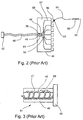
- Figure 2 presents a simplified schematic of a gas distribution module connecting a plurality of cryosurgical probes 50 to a common gas source, according to the methods of prior art.
- FIG. 2 presents a gas distribution module 40, wherein each of cryosurgical probes 50 is connected via a flexible connecting line 54 to a connecting site 56 on a housing element 58, preferably by means of a linking element 51.
- Cryosurgical probes 50 may be detachably connected to connecting sites 56.
- evacuating passageway 96 extends through connecting line 54, such that the outgoing gas is evacuated through an opening located at linking element 51 or at any other suitable location, e.g., manifold 55, see below.
- line 54 further includes electrical wires for providing electrical signals to the thermal sensor and switches (not shown).
- cryosurgical probes 50 is in fluid communication with a manifold 55 received within a housing 58, manifold 55 being for distributing the incoming high pressure gas via lines 57 to cryosurgical probes 50.
- housing 58 is connected to a connector 62 via a flexible cable 60 including a gas tube (not shown), connector 62 being for connecting the apparatus to a high pressure gas source and an electrical source.
- the apparatus further includes electrical wires (not shown) extending through cable 60 and housing 58 for providing electrical communication between the electrical source and cryosurgical probes 50.
- housing 58 includes a pre-cooling element, generally designated as 61, for pre-cooing the high pressure gas flowing to cryosurgical probes 50.
- pre-cooling element 61 is a Joule-Thomson cooler, including a tubular member 48 received within a chamber 49, tubular member 48 including an orifice 59 for passage of high pressure gas therethrough, so as to cool chamber 49, thereby cooling the gas flowing through tubular member 48 into manifold 55.
- Figure 3 presents an alternative configuration of a pre-cooling element 61 according to the methods of prior art, wherein tubular member 48 is in the form of a spiral tube wrapped around a cylindrical element 47, so as to increase the area of contact between tubular member 48 and the cooling gas in chamber 49.
- housing 58 includes a first tubular member for supplying a first high pressure gas to manifold 55, and a second tubular member for supplying a second high pressure gas to pre-cooling element 61. Any combination of gases may be used for cooling and/or heating the gases flowing through such tubular members.
- a cryogenic fluid such as liquid nitrogen may be used for pre-cooling the gas flowing through housing 58.
- an electrical pre-cooling element may be used for pre-cooling the gas.
- thermal sensors may be located within cable 60 and manifold 55 for measuring the temperature of gas flowing therethrough.
- Figures 4-6 present a prior art method and apparatus utilizing an imaging device to form a three-dimensional grid of the patient's treated organ, e.g., prostate, the three dimensional grid serves for providing information on the three dimensional shape of the organ.
- Each of a set of cryosurgical probes is then inserted to a specific depth within the organ according to the information provided by the grid.
- Figure 4 is a simplified schematic of an apparatus comprising an ultrasound probe and a guiding element for guiding insertion of a plurality of cryoprobes into a patient's body, according to the methods of prior art.
- the example given is of an apparatus adapted for cryoablation of a prostate.
- an ultrasound probe 530 is provided for insertion into the patient's rectum, ultrasound probe 530 being received within a housing element 128.
- a guiding element 115 is connected to housing element 128 by means of a connecting arm 126.
- guiding element 115 is in the form of a plate 110 (also called a "guide 110" or a "template 110") having an array or net of apertures 121, each aperture serves for insertion of a cryosurgical probe therethrough.
- the distance between each pair of adjacent apertures 121 is between about 2 millimeters and about 5 millimeters.
- Figure 5 is a simplified schematic showing a method of use of the apparatus presented in Figure 4 .
- ultrasound probe 530 is introduced to a specific depth 113 within the patient's rectum 3.
- a net of marks 112 is provided on the obtained ultrasound image 114, the net of marks 112 on image 114 being accurately correlated to the net of apertures 121 on guiding element 115.
- marks 112 on image 114 sign the exact locations of the centers of ice-balls which may be formed at the end of the cryosurgical probes inserted through apertures 121 to the patient's prostate 2, wherein image 114 relates to a specific depth of penetration 113 of the cryosurgical probes into the prostate 2.
- ultrasound probe 530 is gradually introduced to various depths 113 of rectum 3, thereby producing a set of images 114, wherein each image relates to a respective depth of penetration into the prostate 2.
- each of images 114 relates to a specific plane perpendicular to the axis of penetration of the cryosurgical probes.
- the set of images 114 provides a three dimensional grid of the prostate. Such three-dimensional grid is then used for planning the cryosurgical procedure.
- the introduction of a cryosurgical probe along a given axis of penetration to a first depth may effectively destroy a prostatic tissue segment, while introduction of the probe to a second depth may severely damage the prostatic urethra.
- each probe may be introduced to a specific depth so as to locally provide an effective treatment to a limited portion of the prostate while avoiding the damaging of non-prostatic or prostatic tissues located at other depths of penetration.
- Figure 6 is a simplified schematic presenting a further step in the use of the apparatus presented in Figure 4 , according to the methods of prior art.
- Figure 6 shows the insertion of an operating tip 52 of a cryosurgical probe 50 through an aperture of guiding element 115 into the prostate 2 of a patient.
- cryosurgical probes are sequentially inserted through apertures 121 of guiding element 115 into the patient's prostate, wherein each probe is introduced to a specific depth, thereby providing substantially local effective treatment to distinct segments of the prostatic tissue while avoiding the damaging of other prostatic or non-prostatic tissue segments.
- each of the cryosurgical probes includes a scale for indicating the depth of penetration into the prostate.
- the prior art apparatus and methods presented by Figures 1-6 enable diagnostic mapping of areas to be treated within a prostate, and further enable guiding a plurality of cryogenic probes into a prostate in such a manner that the cryogenic probes are placed according to the planned treatment areas so mapped. It will be clear to one skilled in the art that the prior art methods presented by figures 1-6 may be adapted, with appropriate modifications, to cryoablation of various other organs of the body.
- FIG. 7A is an illustration of the profile of temperature distribution across an ice-ball formed at the tip of a cryosurgical probe.
- the temperature at a surface 104 of the ice-ball is 0° C.
- the temperature declines exponentially towards a cooled center 100 of the ball where it preferably reaches the value of -170° C, such that an isothermal surface 102 of about -40° C is typically located within the ice-ball approximately half way between the center of the ball and its outer surface 104.
- the ice-ball features a radius R
- the radius of the -40° C isothermal surface 102 is about R/2.
- the tissue volume contained within isothermal surface 102 generally corresponds to the "ablation volume" defined hereinabove.
- Figure 7B is a graph showing the effectiveness of a cryosurgical treatment (given in percentage of tissue destruction) as a function of temperature.
- the temperature required for effectively destroying a tissue is at least about -40° C.
- the isothermal surface of -40° C (marked as surface 102 in FIG. 1A ) should be placed at the periphery of the treated tissue so that the entire volume of the treated tissue is exposed to cryoablation temperatures, temperatures at or below -40° C.
- FIGS 7A and 7B together illustrate the fact that when a volume of tissue is treated by being exposed to cryoablation temperatures, that volume is enveloped by a second volume of tissues, termed the "damage envelope" herein, wherein healthy tissues and organs exposed to the external portion of the ice-ball are subject to temperatures of between about -40° C. and 0° C.
- the "ablation volume” 120 shown in figure 7A approximately corresponds to the volume of tissues contained within surface 102
- the "damage envelope” 130 shown in Figure 7A approximately corresponds to the volume of tissue between the isotherm at -40° C, surface 102, and the isotherm at 0° C., surface 104, the surface of the ice ball. (In practice, exact dimensions and positioning of the actual ablation volume is dependent not only on temperature but also on duration of freezing, on freeze/thaw cycles, etc.)
- FIG. 7B makes it clear that tissues cooled to between 0° C. and -40° C. are subject to damage, which damage may result in necrosis of healthy tissue and in temporary or permanent impairment of the function of otherwise healthy organs.
- Apparatus and methods other than those depicted in figures 3-6 may be utilized to accurately deliver one or more cryoprobes to a selected locus for cryoablation of tissues thereat, and to accurately deliver one or more heating probes to selected locations, as will be explained hereinbelow.
- FIG 8A is a simplified graph showing effects at a cryoablation site when cryosurgical cooling is combined with mild heating of tissues at an adjacent site.
- Cold source 100 which may be a cryoprobe 111 functional in cooling, creates an ablation volume 120 surrounded by a damage envelope 130.
- the isothermal surface 102A marks the limit of ablation volume 120A, which is the ablation volume produced when cold source 100 is cooled to cryoablation temperatures, and no heating is used.
- ablation volume 120A presents a shape similar to the shape presented as ablation volume 120 in Figure 7A .
- Isothermal line 102B shows, in approximate form, a shape of an ablation volume under the influence of heat source 140 as well as of cold source 100. Ablation volume 120B may be seen to be flattened on the side exposed to heat source 140.
- isothermal surface 104A shows the outer border of damage envelope 130A when cold source 100 is activated and heat source 140 is inactive, and consequently resembles the shape of damage envelope 130 portrayed in Figure 7A .
- Isothermal surface 104B shows, in approximate form, a shape of an outer border of a damage envelope 130B under the influence of heat source 140 as well as of cold source 100. Damage envelope 130B may be seen to be flattened on the side exposed to heat source 140.
- distance between cold source 100 and the border of ablation volume 120A is smaller than the distance between cold source 100 and the border of ablation volume 120B, on the side facing heat source 140.
- ablation volume 120B, with heating is considerably thinner than ablation volume 120A, without heating.
- isothermal surface 102B and isothermal surface 104B are also considerably smaller than the distance between isothermal surface 102A and isothermal surface 104A.
- Isothermal surface 102B is the internal border of damage envelope 130B, and isothermal surface 104B is its external border, while isothermal surface 102A is the internal border of damage envelope 130A, and isothermal surface 104A is its external border.
- damage envelope 130B, under heating is considerably thinner than damage envelope 130A, absent heating, on the side of heater 140.
- Figure 8B is a simplified graph showing a steep temperature gradient produced when cryogenic cooling at a first site is associated with mild heating of a second, adjacent, site, according to an embodiment of the present invention.
- Gradient 132 of Figure 8B reproduces gradient 132 of Figure 7A .
- the scale of Figure 8B has been expanded somewhat, in the horizontal direction, for clarity of the image.
- gradient 132 depicts a rise in temperature as an exponential function of distance from a center of cooling 100.
- Point X1A is the intersection of that gradient with the -40° C isotherm
- point X1B is the intersection of that gradient with the 0° C isotherm.
- cooling center 100 is taken to be the origin of the graph
- X1A is the distance from the center of cooling to the border of an ablation volume
- (X2A - X1A) is the thickness of the damage envelope, corresponding to the distance between points 102 and 104 of Figure 7A .
- Gradient 134 in Figure 8B , represents a temperature gradient produced when a heating element such as heater 140 of Figure 8A is operated in conjunction with cryogenic cooling. If a heater 140 is operated at a distance X3B from cooling center 100, a gradient similar to gradient 134 will be produced. As may be observed from inspection of Figure 8B , gradient 134 is such that the indicated thickness of the damage envelope (X2B - X1B) is much reduced in comparison to that produced by gradient 132 (without associated heating), and the distance from center of cooling 100 to the border of the ablation volume is also much reduced when compared to that produced by gradient 132. That is, ((X2A - X1A) > (X2B - X1B)), and (X1A > X1B).
- gradient 134 can be made to approximate a step-wise temperature change, and distances X1B and (X2B - X1B) can be substantially reduced, when compared to distances X1A and (X2A - X1A) of the prior art gradient 132 shown in Figures 7A and 8B .
- FIGS 9 - 11 demonstrate that combinations of cooling probes and heating probes, placed in selected positions and cooled to selected temperatures at selected times, are operable to create a combined ablation volume whose shape may be crafted to substantially conform to a three-dimensional shape of a desired cryoablation target such as a particularly shaped organ or tumor.
- Figure 9 is a simplified graph showing effects of cryosurgical cooling at three selected sites, together with mild heating at a single adjacent site, according to an embodiment of the present invention.
- isotherms 102A and 104A shown as solid lines, represent isothermal surfaces at -40° C and 0° C, respectively, expected to obtain under cryogenic cooling at the three depicted cooling sites 100, in the absence of heating at heating site 140.
- Isotherm 102A constitutes an outer border of ablation volume 120A, and an inner border of damage envelope 130A, whose outer border isotherm 104A.
- FIG 9 As in Figure 8A , broken lines are used to show isotherms 102B and 104B, representing isothermal surfaces at -40° C and 0° C, respectively, obtained when cooling at the depicted cooling sites 100 is accompanied by heating at heating site 140.
- Isotherm 102B constitutes an outer border of ablation volume 120B, obtained when cooling at sites 100 is accompanied by heating at site 140.
- Damage envelope 130B similarly obtained during cooling at sites 100 while heating at site 140, has an inner border formed by -40° C isotherm 102B, and an outer border formed by 0° C isotherm 104B.
- heating at heater 140 during cooling at sites 100 has an effect of indenting the ablation volume in a vicinity of site 140, and of reducing the thickness of damage envelope 130B in that vicinity.
- Figures 10 and 11 are simplified graphs showing effects of cryosurgical cooling at three selected sites, together with mild heating at three adjacent sites, according to an embodiment of the present invention.
- Figures 10 and 11 are similar to Figure 9 , except that whereas only one heating site 140 was presented in Figure 9 , three heating sites 140 are presented in Figures 10 and 11 , with consequent modification of the size and shape of ablation volumes 120B, and damage envelopes 130B, as shown.
- Figures 9-11 demonstrate that use of a plurality of cooling cryoprobes in a first selected configuration, together with use of a plurality of heating probes proximate to those cooling probes and in a second selected configuration, enables to craft an ablation volume 120B having a border which conforms to a desired three-dimensional shape, which border may be crafted to substantially conform to size and three-dimensional shape of a desired cryoablation target.
- Two benefits are thereby obtained. Two benefits, already discussed hereinabove, are: (i) substantially limiting cryoablation to a desired cryoablation target; and (ii) significantly reducing the thickness of a damage envelope surrounding a cryoablation volume
- Figures 10 and 11 demonstrate, in a general manner, the fact that use of a plurality of heating sites in conjunction to a plurality of cooling sites can have the effect of moving isotherm 102B, the outer border of cryoablation volume 120B, relatively close to each of a plurality of cooling probes 111 or other cooling sites 100.
- border 102 of a cryoablation volume 120 is not directly visible under known imaging modalities.
- Ultrasound for example, can easily render visible border 104, the 0° C isothermal surface, corresponding to the edge of an ice ball of frozen tissue, yet the significant isotherm 102, border of the area within which tissues are reliably cryoablated, is not directly visible, and its presence and position must be calculated or guessed or inferred from such known facts as the position of border 104.
- Both probes 111 at sites 100, and border 104B, the edge of an iceball formed during cryoablation, may be rendered visible by use of appropriate imaging modalities or combinations of imaging modalities such as ultrasound, fluoroscope, or MRI.
- imaging modalities such as ultrasound, fluoroscope, or MRI.
- the position of border 102, somewhere between probes 111 and border 104 can only be approximately inferred.
- the distance between probes 111 and border 104 is substantially reduced, as shown in Figures 10 and 11 , then an operator's uncertainty as to the exact position of border 102, the cryoablation volume border, is reduced proportionally.
- Figures 12 is a simplified graph comparing a border of a damage envelope to a border of a cryoablation target, according to methods of prior art. It is to be contrasted with Figure 13 , which presents a simplified graph comparing the border of a damage envelope to a border of a cryoablation target, according to an embodiment of the present invention.
- line 145 represents a border of a desired cryoablation target 146.
- a plurality of cryoprobes or other cooling sources 100 are placed within cryoablation target 145, and cooled to create an ablation volume 120A having a border 102A, and a damage envelope 130A having an external border 104A.
- Figure 12 represents what might generally be considered a reasonably good fit of ablation volume 120A to target border 145, according to methods of prior art.
- Figure 12 may be contrasted to Figure 13 , which presents a simplified graph comparing a border of a damage envelope to a border of a cryoablation target, according to an embodiment of the present invention.
- a plurality of heating probes 140 are placed in proximity to a plurality of cooling probes 100, and heating probes 140 are heated during cooling of cooling probes 100.
- Border 102B is an approximate rendition of a border of an ablation volume 120B
- border 104B is an approximate rendition of an exterior border of a damage envelope 130B.
- heating probes 140 Close proximity of heating probes 140 to cooling probes 100 effects a steep temperature gradient between each cooling probe 100 and a nearby heating probe 140.
- the resultant steep gradient enables placement of probes 100 relatively near to target border 145, and has an effect of compressing damage envelope 130B into a relatively thin envelope, when compared to damage envelope 130A presented in Figure 12 .
- accuracy of ablation of target 146 is enhanced, and damage to tissues exterior to target 146 is minimized.
- the disclosed method requires locating (typically, by use of imaging modalities) a cryoablation target in three-dimensional space, then utilizing a probe placement mechanism to place a plurality of cooling cryoprobes within that cryoablation target, and also to place a plurality of heating probes partially surrounding, and preferably completely surrounding, that cryoablation target, then heating those heating probes while cooling those cooling probes to accurately cryoablate the target.
- the apparatus and method described hereinabove with particular reference to Figures 4 , 5, and 6 may be used for this purpose.
- Guiding template 110 may then be used to guide cooling and heating probes to appropriate positions in three-dimensional space, in accordance with the treatment strategies described herein.
- template 110 is operable to guide placement in three dimensions of both heating and cooling probes.
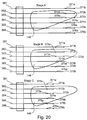
- Figure 14 is a simplified schematic of three stages in a procedure for cryoablation of a target, with emphasis on treatment of lateral borders of that target.
- Figure 14 presents what is termed in the art a "pull-back" procedure: a plurality of cryoprobes, labeled 100A-100F, operable both to heat and to cool, are passed through template 110, into the body of a patient, both into and around a cryoablation target 146 previously identified and localized using imaging modalities such as ultrasound probe 130 or other well-known diagnostic tools.
- a pull-back procedure involves ablating target 146 in several stages.
- a plurality of cryoprobes is introduced into a target, such as a prostate, at a selected depth, those cryoprobes are cooled to cryoablation temperatures, then preferably heated to free the probes from adhesion to frozen tissue.
- the probes are then typically withdrawn to a lesser depth of penetration, whereupon they are again cooled to ablate an additional layer of target 146, and so on, for as many iterations as necessary to treat the entire target.
- cryoprobes 100A-100F are operable to cool tissues to cryoablation temperatures, and are also operable to heat tissues.
- cryoprobe presented in Figure 1 is operable both to heat and to cool, depending on whether cooling gas or heating gas is supplied through Joule-Thomson orifice 80 therein.
- a plurality of cryoprobes are inserted to a first depth into the body of a patient, in such manner that operating tips of selected ones of that plurality of cryoprobes are positioned within target 146, and operating tips of others of the inserted cryoprobes are positioned near, but outside, cryoablation target 146.
- an operating tip of probe 100B is positioned within target 146, and probes 100A, 100C, and 100D are positioned near target 146, and outside it.
- probe 100B is cooled, probes 100A and 100C are heated, thereby surrounding the distal portion of target 146 with a protective heated envelope during cryoablation of a portion of target 146 by probe 100B.
- stage A After one or more freezing and thawing iterations are performed in the position shown as stage A, a surgeon partially withdraws probes 100, bringing them to the position shown as stage B. At this point, probes 100B and 100C are within target 146, and probes 100A and 100D are flanking the target. In this position, probes 100B and 100C are cooled to cryoablation temperatures, while probes 100A and 100D are gently heated.
- probes 100 After one or more freezing and thawing iterations are performed in the position shown as stage B, a surgeon further partially withdraws probes 100, bringing them to the position shown as stage C. At this point, probes 100B, 100C and 100D are within target 146, and probes 100A and 100E are flanking the target. In this position, probes 100B, 100C, and 100D are cooled to cryoablation temperatures, while probes 100A and 100E are gently heated.
- probes within target 146 are cooled to cryoablation temperatures, while probes surrounding (or partially surrounding) target 146 are gently heated.
- Probe 100C in stage A presents a somewhat special case.
- the operating tip of probe C is shown as being exterior to target 146, but very close to target 146.
- imaging modalities may be used in real time to determine the actual localization of probes 100, and that information may be used to calculate an appropriate combination of selected temperatures for each probe, and/or a schedule of timing for heating and cooling of those probes, so as to cause a border of a cryoablation volume 120 to be formed closely to a border 145 of cryoablation target 146.
- probe 100C in stage A of Figure 14 , probe 100C might be heated only slightly, and probe 100A might be heated more strongly, so as to cause cryoablation volume border 102B to form near probe 100C and somewhat far from probe 100A, as shown in Figure 14 .
- control of timing of cooling and heating including possible intermittent cooling and heating, may be used in place of, or in addition to, control of the intensity of cooling of individual probes (e.g., by variable control of gas pressure in a Joule-Thomson probe) to finely tune the influence of individual probes as desired.
- Figure 14 demonstrates a strategy for applying a method to lateral borders of a cryoablation target 146.
- Strategies for handling proximal and distal borders of a cryoablation target will be presented in Figures 15-21 below.
- Figure 15 is a simplified schematic of a stage in a procedure for cryoablation of a target, showing a method for treatment of a proximal or distal border of a target.
- Figure 15 presents a cryoablation target 146, a first probe 100A penetrating a body of a patient from a first angle, and having an operating tip positioned within target 146, and a second probe 100B penetrating a body of a patient from a second angle, and having an operating tip near the operating tip of probe 100A, but positioned outside target 146.
- probe 100A may be cooled and probe 100B heated, thereby causing a border of a cryoablation volume to substantially coincide with a border 145 of target 146.
- Figure 16 is a simplified schematic of a stage in a procedure for cryoablation of a target, showing an additional method for treatment of a proximal or distal border of a target
- Figure 16 is similar to Figure 14 , in that it displays a plurality of probes 100 inserted through a guiding template 110, and into a body of a patient.
- a plurality of probes 100 is positioned in a configuration appropriate for ablation at a three-dimensionally shaped distal border of a cryoablation target 146.
- a first selected set of probes 100 may be positioned within target 146, and a second selected set of probes 100 may be positioned adjacent to a border 145 of target 146, but exterior to target 146.
- the first selected set of probes may be cooled to cryoablation temperatures while the second selected set of probes is mildly heated.
- probes are selected into sets such that probes of the first set alternate with probes of the second set, as shown in Figure 16 .
- the method shown in Figure 16 may be implemented utilizing the apparatus presented in Figures 4-6 , which is operable to introduce into a body of a patient a plurality of probes oriented in parallel orientations. The method is thus appropriate, for example, for ablating a prostate through the perineum.
- Template 110 comprising an array of discrete apertures, does not offer total freedom of position in what we might call the "x" and "y” directions, across the face of template 110, yet does not restrict movement in the "z” direction, the direction of penetration into a body of a patient.
- a probe inserted through an aperture in template 110 may be inserted to any desired depth.
- the configuration provided in Figure 16 could alternatively have been described in such a way that distances of all operating tips from the distal portion of border 145 might have been made equal across all probes 100.
- Figure 16 was drawn having a certain variability in distances of various operating tips from the desired target border, with the understanding that temperature and timing of heating and cooling may be modified to compensate for minor differences of position.
- This method of heating, using low-pressure rather than high-pressure helium has a disadvantage previously discussed, that use of low pressure gas, particularly in a highly miniaturized system, will result in limited heating capacity, due to the limited gas throughput that can be expected from such a system.
- a first alternative method would be to use Joule-Thomson heating with a heating gas, but without utilizing a heat exchanger to pre-heat that gas. This would have the advantage of allowing use of high-pressure helium, and consequently allowing increased gas throughput while avoiding high temperatures that would be obtained if pre-heating were used.
- This method however, has the disadvantage of requiring separate chambers for heating (without a heat exchanger for pre-heating) and for cooling (with a heat exchanger for pre-cooling).
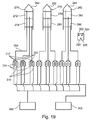
- a preferred solution is to heat a probe by expansion through a Joule-Thomson orifice of a mixture of gases, a mixture including both cooling gas and heating gas in a selected proportion so as to achieve a required degree of heating (or of cooling).
- Use of a mixture of heating gas and cooling gas enables fine control of heating and cooling, yet does not require lowered gas pressure and consequent limited throughput of gas.
- a system including a gas supply for this purpose is presented in Figure 19 hereinbelow.
- the method for treatment of a proximal or distal border of a cryoablation target presented by Figure 16 has the advantage of being susceptible to implementation using standard coolable and heatable cryoprobes such as the prior art cryoprobe presented in Figure 1 .
- a disadvantage of the method presented by Figure 16 is that alternating probes used for heating with probes used for cooling produces what one might call a "low resolution" effect both in heating and in cooling, thereby posing an upper limit to the accuracy of the method.
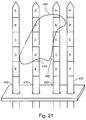
- Figures 17-21 present yet another method for treating distal and proximal borders of a cryoablation target, which method provides a "high resolution" result in comparison to the method of Figure 16 .
- Figure 17 is a simplified schematic of the operating portion of a cryoprobe, comprising a plurality of independently controllable operating modules each operable to cool and to heat.
- FIG 17 presents a multi-module cryoprobe 200 having a first operating module 180 and a second operating module 190.
- Operating modules 180 and 190 are also sometimes referred to as “treatment modules” in the following.
- Operating module 180 comprises a gas input conduit 182, a chamber 184, a Joule-Thomson orifice 186, a gas exhaust conduit 188, and a heat-exchanging configuration 189.
- pressurized cooling gas is supplied through gas input conduit 182, that pressurized cooling gas expands through Joule-Thomson orifice 186 into chamber 184, cooling chamber 184. Expanded cooling gas is then exhausted through gas exhaust conduit 188, which preferably contains a heat-exchanging configuration 189 for pre-cooling incoming cooling gas in gas input conduit 182.
- pressurized heating gas is supplied through gas input conduit 182, that pressurized heating gas expands through Joule-Thomson orifice 186 into chamber 184, heating chamber 184. Expanded heating gas is then exhausted through gas exhaust conduit 188, which preferably contains a heat-exchanging configuration 189 for pre-heating incoming heating gas in gas input conduit 182.
- Operating module 190 is similar in function, and can be similar in construction, to operating module 180.
- Operating module 190 comprises a gas input conduit 192, a chamber 194, a Joule-Thomson orifice 196, a gas exhaust conduit 198, and a heat-exchanging configuration 199.
- pressurized cooling gas is supplied through gas input conduit 192, that pressurized cooling gas expands through Joule-Thomson orifice 196 into chamber 194, cooling chamber 194. Expanded cooling gas is then exhausted through gas exhaust conduit 198, which preferably contains a heat-exchanging configuration 199 for pre-cooling incoming cooling gas in gas input conduit 192.
- pressurized heating gas is supplied through gas input conduit 192, that pressurized heating gas expands through Joule-Thomson orifice 196 into chamber 194, heating chamber 194. Expanded heating gas is then exhausted through gas exhaust conduit 198, which preferably contains a heat-exchanging configuration 199 for pre-heating incoming heating gas in gas input conduit 192.
- Multimodule probe 200 is shown in Figure 17 as having two treatment modules, yet alternatively probe 200 may comprise three or more treatment modules.
- Each treatment module of probe 200 is designed and constructed to be independently controlled in heating and cooling. Control is preferably effected by controlling a supply of gas delivered to each module, as will be shown in Figure 19 hereinbelow.
- module 180 and module 190, and additional modules if present may be operated to cool, or to heat, at selected times and in selected degrees, each independently of the others.
- Thermal sensors 187 and 197 preferably provide real-time feedback to an external control system, which feedback is useful in using probe 200 to best effect.
- module 180 and module 190 may be operated both to cool, or both to heat, or one to heat and another to cool, in any order, at the same time or at different times.
- Multi-module probe 200 preferably comprises thermal insulation serving to thermally isolate modules 180 and 190 (and other modules optionally present) each from the others, to further enhance independence of operation of each module.
- Figures 18A, 18B, and 18C are simplified schematics of alternate configurations of multi-module cryoprobes, according to embodiments of the present invention.
- Figure 18A presents a multi-module cryoprobe 202 in which modules 180 and 190 are laterally disposed (i.e., side-by-side), in contrast to multi-module cryoprobe 200 of Figure 17 , in which modules 180 and 190 are longitudinally disposed (i.e., one distal, one proximal).
- Figure 18B presents a multi-module cryoprobe 204 comprising more than two independently controllable treatment modules.
- Figure 18C presents a multi-module cryoprobe 206 comprising independently controllable treatment modules 180 and 190, and further comprising shaft isolation element 220, designed and constructed to protect tissues in a vicinity of a proximal portion of probe 206 from being damaged by cold induced by contact with shaft portion 226 of probe 206, which shaft portion is liable to be inadvertently cooled by passage therethrough of cold exhaust gasses exhausting from module 180, or from module 190, or from both.
- shaft isolation element 220 is thermal isolation material 222.
- shaft isolation element 220 is an electrical resistance heater 224.
- System 250 comprises a plurality of cryoprobes, represented in the figure by cryoprobes 260, 262 and 264, each having a plurality of independently controllable operating modules, represented in the figure by modules 270, 272, 274, 276, 278, and 280, 282, 284, and 288. It is to be understood that these chosen representatives are arbitrarily chosen; cryoprobes of system 250 may be of any of the configurations presented in Figures 1 , 17 , or 18 , or other configurations.
- System 250 is operable to supply an independently selected mixture of gasses to each treatment module of each probe, at selected times.
- System 250 comprises a source of heating gas 300 and a source of cooling gas 302.
- System 250 is operable to supply heating gas from heating gas source 300, through heating gas control valves 310 and one-way valves 312, to gas input conduits of a plurality of treatment modules in probes 260, 262, 264, and optionally other probes.
- System 250 is further operable to supply heating gas from cooling gas source 302, through cooling gas control valves 314 and one-way valves 316, to gas input conduits of a plurality of treatment modules in probes 260, 262, 264, and optionally other probes.
- Valves 310 and 314 may be manual valves, but preferably they are remotely contolled valves under control of a control module 320.
- Control module 320 is preferably designed and constructed to respond to data from sensors, such as thermal sensors 187 and 197, and preferably to additional thermal and pressure sensors operable to report temperatures in various parts of system 250 and in tissues of a patient, and to report pressures in various parts of system 250.
- Control module 320 preferably comprises a memory 322 and a processor 326, and is operable to respond to input data from the above-mentioned sensors, and to respond to operator commands, and to control valves 310 and 314, under control of algorithms 324 stored in memory 322.
- system 250 is operable to supply cooling gas, heating gas, or a mixture of cooling gas to each treatment module of each cryoprobe of the system, thus enabling system 250 to effect various combinations of hot modules and cool modules, in a variety of configurations.
- Figure 20 is a simplified schematic showing three stages in a procedure for cryoablation of a target, utilizing a plurality of cryoprobes each having a plurality of independently controllable operating modules.
- Figure 20 is similar to Figure 14 , in that it shows three stages of "pull-back" during treatment of a cryoablation target 146.
- a plurality of probes (shown as probes 361-366) are passed through a guiding template 110 and positioned in a patient's body, in and around cryoablation target 146.
- Probes 361-366 are shown as each having two independently controllable treatment modules, numbered 371a, 371b, 372a, 372b, 373a, 373b, 374a, 374b, 375a, 375b, 376a, and 376b.
- the particular configuration depicted is exemplary only, and that the invention can be practiced under a variety of alternative configurations.
- the treatment method comprises positioning treatment modules 371a - 376b in and around cryoablation target 146, cooling those modules positioned within target 146 to cryoablation temperatures to cryoablated target 146, while heating those modules positioned external to and adjacent to target 146, thereby effecting accurately delimited cryoablation of target 146, as has been described in detail hereinabove.
- modules 372a and 372b would be cooled to cryoablation temperatures, and modules 371a, 371b, 372b, 373b, 374a and possible 374b would be activated to heat tissues in their vicinity.
- modules 372a, 372b, 373a, 373b, and 374a would be cooled, while modules 371a, 371b, 374b, 375a, and possibly 375b would be heated.
- modules 372a, 372b, 373a, 373b, 374a, and 374b would be cooled, modules 371a, 371b, 375a, 375b, and possibly 376a would be heated.
- the effect in each case, is to cool inside of, and to warm outside of, a selected three-dimensional shape formed to conform to a three-dimensional shape of a portion of a border of a cryoablation target, thereby causing a border of a resultant cryoablation volume to closely conform to a form of that intended cryoablation target.
- Figure 21 is a simplified schematic of a treatment of a cryoablation target by a plurality of cryoprobes each having multiple treatment modules, the treatment not requiring pullback.
- each probe is shown as having five treatment modules.
- each module is independently controllable to heat or to cool, yet this is not a requirement of the invention.
- it might be convenient or economical to utilize probes whose most distal and most proximal modules (modules 400A, 410A, 420A, 430A, 400E, 410E, 420E, and 430E in our example), are designed and constructed to heat but not to cool.
- Such modules might then utilize a heating methodology, such as electrical resistive heating, which is less appropriate for modules B, C, and D, which might be designed and constructed to be operable both to cool and to heat, for example utilizing Joule-Thomson heating and cooling.
- cryoablation target 146 is large in comparison to the available cooling surfaces presented by the depicted plurality of cryoprobes. Consequently, cryoablation of target 146 is executed in stages, several of which stages are presented in Figures 14 and 20 .
- cryoablation target 146 presented by Figure 21 is relatively small in relation to depicted cryoprobes 400, 410, 420, and 430.
- cryoprobes 400, 410, 420, and 430 each comprise multiple dual-purpose treatment modules. Consequently, cryoprobes according to a preferred embodiment presented by Figure 21 are operable to accurately cryoablated target 146 without necessitation a multi-stage pull-back ablation process.
- modules 410C, 410D, and 420 B are wholly or substantially positioned within target 146, and would, according to the present embodiment, be cooled to cryoablation temperatures to ablate target 146.
- Modules 400C, 400D, 400E, 410A, 410E, 420A, 420C, 420D, 420E, and 430B are each external to, but near, target 146, and would, according to this embodiment, be heated during the cryoablation process, thereby producing the various positive effects generally described hereinabove.
- Module 410B presents a special case, partly within and partly outside target 146.
- module 410 may be left inactive, neither heated nor cooled.
- calculation means such as that provided by command module 320 may be used to calculate an optimal temperature for module 410, such as to guarantee full ablation of target 146 while also minimizing ablation of tissues outside target 146.
- the contemplated method preferably comprises calculating optimal temperatures for each module, and/or scheduling of changes in temperatures of each module over time, in order to guarantee full ablation of target 146, while also minimizing ablation of, and damage to, tissues outside target 146.
- module 420D would preferably be heated to a lesser degree, or for a shorter time, than module 410E.
- a portion 440 of a border of target 146 falls close to the interface between module 410D and module 410E, and that border traverses probe 410 within a vicinity of module 410D. Consequently, module 410D can be used to strongly cool surrounding tissues, and module 410E may be used to heat surrounding tissues, thereby successfully producing a step-wise drop in temperature corresponding well to the shape of target 146.
- a portion 442 of a border of target 146 falls near module 420D, and is relatively distant from module 410D which must provide sufficient cooling to ablate all tissues within that border.
- computing means such as provided by control module 320 would preferably be used to calculate optimal temperatures, over time, for each treatment module, based on whatever is known about positions of target 146 and modules 400A-430E, and further based on real-time information gleaned from thermal sensors within probes 400, 410, 420 and 430, from thermal sensors in and around tissues of target 146, and, optionally, from real-time information received from an operator or gleaned from computerize interpretation of real-time images, concerning actual positions of probes, target, and iceballs created by the cryoablation process.
Landscapes
- Health & Medical Sciences (AREA)
- Surgery (AREA)
- Life Sciences & Earth Sciences (AREA)
- Nuclear Medicine, Radiotherapy & Molecular Imaging (AREA)
- Medical Informatics (AREA)
- Engineering & Computer Science (AREA)
- Biomedical Technology (AREA)
- Heart & Thoracic Surgery (AREA)
- Otolaryngology (AREA)
- Molecular Biology (AREA)
- Animal Behavior & Ethology (AREA)
- General Health & Medical Sciences (AREA)
- Public Health (AREA)
- Veterinary Medicine (AREA)
- Thermotherapy And Cooling Therapy Devices (AREA)
- Surgical Instruments (AREA)
Description
- The present invention relates to an apparatus and method for accurately delimited cryoablation of unwanted body tissues. More particularly, the present invention relates to method and apparatus for cryoablating a selected target volume of body tissue while surrounding or partially surrounding said target volume with a protective envelope of mildly heated tissue, so as to enhance accuracy of delimitation of the volume of cryogenic destruction, and so as to minimize the volume of tissue, exterior to the selected target volume, damaged by cryogenic cooling.
- In recent years, cryoablation of tissues has become an increasingly popular method of treatment for a variety of pathological conditions. Malignancies in body organs such as the breast, prostate, kidney, liver, and other organs are successfully treated by cryoablation, and a variety of non-malignant pathological conditions, such as benign prostate hyperplasia, benign breast tumors, and similar growths are also well treated by cryoablation of unwanted tissues. Certain cases of intractable chronic pain are also treatable through cryosurgery, by cryoablation of selected nervous tissue.
- Cryoablation of pathological tissues or other unwanted tissues is typically accomplished by utilizing imaging modalities, such as x-ray, ultrasound, CT, and MRI, to identify a locus for ablative treatment, then inserting one or more cryoprobes into that selected treatment locus, then cooling the treatment heads of the inserted cryoprobes sufficiently to cause the tissues surrounding the treatment heads to reach cryoablation temperatures, typically below about - 40 ° C.
- Tissues thus cooled are thereby caused to loose their functional and structural integrity. Cancerous cells cease growing and multiplying, and cryoablated tumor tissue materials, whether from malignant tumors or from benign growths, lose their structural integrity and are subsequently sloughed off or absorbed by the body.
- The principle danger and disadvantage of cryosurgical ablative treatment, however, is the danger of partially or completely destroying the functional and structural integrity of healthy tissues near the treatment locus, thereby impeding the patient's recovery from the surgical procedure and potentially causing serious and long-term deleterious effects on the patient's health and on his quality of life.
- In particular, two well-known limitations inherent in currently known cryoablation technique are primarily responsible for damage caused to healthy tissue while cryoablating pathological tissue.
- Using terms defined hereinbelow, we would say that the first problem is that in all cryoablation the "ablation volume", a first volume within which tissue structure and functionality are destroyed, is inevitably surrounded by a "damage envelope", a second volume within which tissue structure and function are damaged. Tissues in the damage envelope are exposed to temperatures which, although not sufficiently cold to thoroughly cryoablate those tissues and wholly destroy their physiological functionality, yet are cold enough to do significant damage to those tissues, impair their functionality, and significantly alter cellular and other structures therein. To reliably ablate a first selected target volume of tissue, one is inevitably obliged to damage second volume of tissue surrounding that first selected volume.
- The second problem is that cryosurgery is difficult to control, because the border between the ablation volume and the damage envelope is not directly visible under any known imaging modalities. Although the borders of the ice-ball which forms around the cold operating tip of a functioning cryoprobe is visible under ultrasound or MRI imaging modalities, the border of the ablation volume, the volume within which cell functionality is reliably destroyed, is itself not directly visible under known imaging modalities, and it's position, somewhere within the visible ice-ball, must be estimated or indirectly detected or guessed.
- Various devices and methods have been proposed to enable cryoablation of pathological tissue while limiting damage to non-pathological tissue. These fall roughly into two categories: devices and methods which protect tissues by preventing excessive cooling of those tissues during a cryoablation procedure in their vicinity, and devices and methods which enable accurate placement of cryoprobes used in cryoablation, so as to successfully concentrate the cooling effect of such cryoprobes at or near pathological tissue, thereby minimizing unwanted cooling of non-pathological tissue.
- An example of the former category is the well-known technique of introducing a heating device or a heated fluid into the urethra of a patient, thereby heating the urethra and tissues adjacent to it during cryoablation of portions of the prostate, thereby helping to protect the urethra from damage while prostate tissues nearby are being cooled to cryoablation temperatures.
U. S. Patent 6,505,629 to Mikus et. al. teaches a similar method, using a heating probe to protect an object, the neuro-vascular bundle, during cryoablation of the prostate, by interposing a heating probe between that object and a cooling cryoprobe. - An example of the latter category is provided by
U. S. Patent No. 6,142,991 to Schatzberger. Schatzberger describes a high resolution cryosurgical method and device for treating a patient's prostate, including the steps of (a) introducing a plurality of cryosurgical probes to the prostate, the probes having a substantially small diameter, the probes being distributed across the prostate, so as to form an outer arrangement of probes adjacent the periphery of the prostate and an inner arrangement of probes adjacent the prostatic urethra; and (b) producing an ice-ball at the end of each of the cryosurgical probes, so as to locally freeze a tissue segment of the prostate. - Schatzberger's apparatus includes (a) a plurality of cryosurgical probes of small diameter, the probes being for insertion into the patient's organ, the probes being for producing ice-balls for locally freezing selected portions of the organ; (b) a guiding element including a net of apertures for inserting the cryosurgical probes therethrough; and (c) an imaging device for providing a set of images, the images being for providing information on specific planes located at specific depths within the organ, each of the images including a net of marks being correlated to the net of apertures of the guiding element, wherein the marks represent the locations of ice-balls which may be formed by the cryosurgical probes when introduced through the apertures of the guiding element to the distinct depths within the organ.
- Thus, Schatzberger's method and apparatus enable a surgeon to place a set of cryoablation probes within a prostate with relatively high accuracy, and to operate those probes to ablate selected tissues while avoiding, to a large extent, inadvertent and undesirable ablation of healthy tissues near the ablation site. Schatzberger also demonstrates that by utilizing multiple small cryoprobes in a dense array, the volume of the damage envelope may to some extent be reduced.
- However, neither Schatzberger's technique nor any other known technique has proven sufficiently accurate to prevent damage to peripheral tissues in general. An ablation target ablated according to the methods of Schatzberger is still surrounded by broad envelope of damaged tissue. Further, Mikus' invention, while solving the specific problem of unwanted damage to a specific object, does not address the general problem of the overall "sloppiness" of the cryoablation procedure. Cryoablation, as practiced under all known prior art methods, results in cryoablation of a first volume, only approximately conforming to an intended cryoablation target, which first volume is surrounded by a second volume of healthy tissue, unavoidably damaged.
- Thus there is a widely recognized need for, and it would be highly advantageous to have, apparatus and method for cryoablation which results in reduced volume of damaged tissue surrounding the selected cryoablation target, yet enables full and reliable cryoablation of the selected target.
- As mentioned above, a second basic problem in cryosurgery technology relates to the difficulty experienced by surgeons in knowing the exact extent of the tissue which will be ablated by a given cryoablation procedure. The ice-ball produced by a functioning cryoprobe is visible under ultrasound and other imaging modalities, but the delimitation of the cryoablation volume (the area of total cell destruction) within that iceball is not directly visible under known imaging technologies. The surgeon, who in the case of treatment of a malignancy must err on the side of caution, often ablates more tissue than was really necessary, and damages more additional tissue than was really necessary, because he is unable to accurately command the exact delimitation of the destruction volume he creates, and is further unable to accurately observe, in real time, the actual border of the destruction volume created by his cryoablative intervention.
- Thus there is a widely recognized need for, and it would be highly advantageous to have, a cryosurgery apparatus and method enabling accurate delimitation of an ablation volume.
- With respect to prior art relevant to another aspect of the invention, Mikus op. cit. teaches use of low-pressure helium supplied to a cryoprobe having a Joule-Thomson orifice, to supply heating to a probe. According to Mikus, low-pressure helium is used in place of high-pressure helium, to assure that a tissues will not be heated beyond a temperature which would be destructive to those heated tissues.
- Use of low-pressure helium for heating a Joule-Thomson probe does indeed ensure that a desired maxiumum temperature of the probe will not be exceeded. There is, however, a disadvantage to use of low-pressure helium for heating such a probe, namely that the heating capacity of a probe so heated is somewhat limited. Use of low pressure of the supplied helium, supplied through a small-diameter gas-supply conduit, insures that only a relatively small quantity of helium gas will be passed through the Joule-Thomson orifice per unit of time. This limitation is particularly noticeable when the method is applied to probes of small dimensions. Yet, as taught by Schatzberger op. cit., small-diameter cryosurgical devices are desirable in many cryosurgery contexts, and small diameter cryoprobes comprise even smaller diameter gas input supply conduits. Thus, use of low-pressure helium to heat today's miniaturized cryoprobes substantially limits the heating ability of such a probe.
- Thus, there is a widely recognized need for, and it would be highly desirable to have, a device and method for Joule-Thomson heating of a probe, which device and method provide heating to an upper limit of temperature, thereby protecting heated tissues from overheating, provide for a high throughput of gas, and therefore provide a higher heating capacity than that provided by a Joule-Thomson probe heated by expansion of low-pressure helium gas.
- Note is here taken of three additional prior art documents presenting devices or methods having elements in common with devices and methods presented herein, or presenting devices for which new uses are presented hereinbelow.
- First,
Zvuloni et. al. in U.S. Patent Application No. 10/255,834 (Publication No.2003-0060762-A1 ) teach use, in a cooling cryoprobe, of a gas mixture comprising both a cryogenic cooling gas and a heating gas such as helium. Zvuloni contemplates use of such a gas so as to enable fine control of cooling, and to enable leak detection in a balloon catheter based on detection of trace amounts of helium. - Second, in
PCT application IL02/01062 Zvuloni WO03059247 U. S. Pat. No. 6,074 ,412 , Mikus et. al. teach a probe having both heating elements and cooling elements, yet the heating and cooling elements of Mikus'probe are designed for, and can only be used, sequentially and not simultaneously. - An example of known apparatus for accurately delimitating a cryoablation volume is disclosed in the document
WO 83/03961 A1 - It is against the background, and the limitations and problems associated therewith, that the present invention has been developed.
- Therefore, it is a primary objective of the present invention to provide a cryosurgery apparatus and method enabling accurate delimitation of an ablation volume.
- To achieve this, the apparatus for accurately delimitating a cryoablation volume of the invention is characterized by the features claimed in the characterizing part of claim 11, and the invention provides a cryoprobe according to the characterizing part of
clams - The present invention is of a system for accurate cryoablation allowing enhancing a surgeon's ability to accurately cryoablate a selected cryoablation target and to limit cryablation to that selected target. The present invention encompases (a) an apparatus for accurately delimiting a cryoablation volume; (b) an apparatus for minimizing damage to tissues surrounding a cryoablation volume; (c) an apparatus for real-time visualization of a border of a cryoablation volume during cryoablation; (d) an apparatus for mildly heating tissues during cryoablation; (e) a cryoprobe operable to simultaneously cool first tissues while heating second tissues.
- Described is a method for sharply delimiting a cryoablation volume when cryoablating a selected cryoablation target in the body of a patient, comprising defining a three-dimensional shape as a border of a cryoablation target; inserting into the patient a plurality of probes each comprising at least one treatment module; positioning the probes so that a first set of the treatment modules is adjacent to the defined shaped border and anterior to the selected cryoablation target, and a second set of the treatment modules is adjacent to the defined shaped border and exterior to the selected cryoablation target; cooling the first set of treatment modules to cryoablation temperatures, thereby cryoablating tissues within the cryoablation target and adjacent to the border; and heating the second set of treatment modules during the cooling of the first set of treatment modules, thereby creating a sharp temperature gradient at a vicinity of the shaped border of the cryoablation target, thereby sharply delimiting the cryoablation volume.
- According to further features described below, at least one of the plurality of probes comprises at least two independently controllable treatment modules.
- According to still further features the method further comprises heating a first of the independently controllable treatment modules while cooling a second of the independently controllable treatment modules, preferably heating the first independently controllable treatment module by expansion, through a Joule-Thomson orifice, of a mixture of cooling gas and heating gas.
- According to still further features the described method further comprises orienting the probes with respect to the cryoablation target by positioning, exterior to a patient and in a position having a known spatial relationship to the cryoablation target, a template having an array of apertures each operable to orient a probe passing therethrough to a predetermined angle with respect to the template; and passing a plurality of the probes through ones of the array of apertures, and thence into the patient, thereby orienting the inserted probes with respect to the cryoablation target. The template may be positioned at a perineum of a patient. Preferably the template is designed and constructed to ensure parallel orientations of a plurality of cryoprobes inserted therethrough. Preferably at least one of the probes comprises an external marking on the probe, designed and constructed to render visible to an operator a depth of penetration of the probe through the template. Preferably, the second set of treatment modules surrounds the cryoablation target.
- Also described is a method for minimizing damage to tissues surrounding a cryoablation target when cryoablating the target, comprising defining a three-dimensional shape as a border of a cryoablation target; inserting into a patient a plurality of probes each comprising at least one treatment module; positioning the probes so that a first set of the treatment modules is inside the cryoablation target, and a second set of the treatment modules is exterior to the target and surrounds at least a portion of the target; cooling the first set of treatment modules to cryoablation temperatures, thereby ablating tissues within the target; and heating the second set of treatment modules during cooling of the first set of treatment modules, thereby preventing cooling of tissues surrounding the cryoablation target, thereby minimizing damage to tissues surrounding the cryoablation target while cryoablating the target.
- According to further features in the second set of treatment modules entirely surrounds the cryoablation target. Cooling of the first set of treatment modules may entirely ablate an organ, such as a prostate, or may entirely ablate a tumor.
- Preferably, each treatment module of the first set of treatment modules is positioned adjacent to at least one treatment module of the second set of treatment modules.
- According to an aspect of the present invention there is provided an apparatus for accurate delimitating a cryoablation volume, comprising: a positioning device for positioning a plurality of cryoprobes within and around a cryoablation target, at least one probe operable to heat tissues adjacent to a border of the cryoablation target and external to the target, while cooling tissues adjacent to the border of the cryoablation target and internal to the target.
- Preferably, the at least one probe comprises a plurality of independently controllable and simultaneously operable treatment modules, each of the modules being operable to cool adjacent tissues and also being operable to heat.adjacent tissues. The independently controllable treatment modules may be coolable by Joule-Thomson cooling, and/or heatable by Joule-Thomson heating. The probe may comprise two treatment modules laterally positioned, such that the probe is operable to cool along a first face of a longitudinally extended section thereof, while heating along a second face of the longitudinally extended section thereof. Alternatively, the probe may comprise two treatment modules longitudinally positioned, such that the probe is operable to cool a distal treatment module while heating a proximal treatment module, and is further operable to cool the proximal treatment module while heating the distal treatment module.
- The positioning device may comprise a template presenting an array of apertures for inserting the probes therethrough, the apertures being operable to guide placement of the probes within and around a cryoablation target. The apparatus may further comprise a plurality of probes, each operable to pass through one of the apertures prior to insertion into a body of a patient, and may further comprise a probe having external markings designed and constructed to render visible to an operator a degree of penetration of the probe through one of the apertures.
- Preferably, the apparatus comprises a gas supply system operable to individually control a supply of gas to each of the probes.
- Preferably, at least one of the probes comprises a plurality of treatment modules.
- The gas supply system may be operable to supply gas to a cryoprobe which comprises a plurality of treatment modules, and further operable to individually control supply of gas to each module of the plurality of treatment modules.
- The apparatus may be operable to supply a mixture of cooling gas and heating gas to one of the probes, or to supply a mixture of cooling gas and heating gas to one of the treatment modules. Preferably the apparatus is further operable to supply a selected mixture of cooling gas and heating gas, under control of a control module.
- Also disclosed is a method for real-time visualization of a border of a cryoablation volume during cryoablation of a cryoablation target, comprising creating a cryoablation volume having a well-defined delimitation surface, by inserting into a cryoablation target a plurality of probes each having a treatment module operable to cool tissues to cryoablation temperatures; inserting into a patient around the cryoablation target a plurality of probes each having a treatment module operable to heat tissues; and heating those of the treatment modules positioned outside the target while cooling to cryoablation temperatures those of the treatment modules positioned inside the target, thereby creating a cryoablation volume having a delimited surface extending between the plurality of heated modules and the plurality of cooled modules, and thereby having a known positional relationship to the treatment modules of the probes; and utilizing visualization modalities to display to an operator positions of at least some of the cooling and heating treatment modules, thereby enabling an operator, seeing a display of the positions of the cooling and heating modules, to accurately infer a position of the delimited cryoablation border.
The method may further comprise displaying a border of a cryoablation target. The target border may be visualized utilizing equipment selected from a group including ultrasound equipment, MRI equipment, x-ray equipment, and fluoroscope equipment. The target border may be rendered visible by digital display of a mathematical model of the target. Preferably, at least some probes of the plurality of probes comprise a marker, visible under an imaging modality, marking a border between first treatment modules of the probes and second treatment modules of the probes. - According to an additional aspect of the present invention there is provided an apparatus for adjustable heating of body tissues, comprising: a probe comprising a treatment module operable to be heated by Joule-Thomson heating; and a gas supply operable to supply a mixture of cooling gas and heating gas, in selected proportions, to the treatment module.
The gas supply preferably comprises a processor operable to select proportions of heating and of cooling gas supplied to the treatment module according to an algorithm responsive to temperature data garnered by thermal sensors. The sensors may be positioned within the probe or among tissues of a patient. - According to yet an additional aspect of the present invention there is provided a cryoprobe operable to cool first tissues to cryoablation temperatures while heating second tissues. The cryoprobe may further comprise a first treatment module operable to cool the first tissues and a second treatment module operable to heat the second tissues. Preferably the first treatment module is also operable to heat tissues, and the second treatment module is also operable to cool tissues. Most preferably, both the first treatment module and the second treatment module are operable both to heat tissues and to cool tissues, and each of the first treatment module and the second treatment module is operable to be independently controlled in cooling and heating.
- The first treatment module may be positioned laterally to the second treatment module, or positioned longitudinally to the second treatment module. The probe may further comprise a third treatment module operable to heat and to cool. The first, second, and third treatment nodes may be positioned longitudinally one to another. The heating may be Joule-Thomson heating and the cooling may be Joule-Thomson cooling.
- Also disclosed is a method for accurately delimited cryoablation of a target, comprising inserting into a patient a plurality of cryoprobes, each of the cryoprobes comprising at least one treatment module and at least some of the cryoprobes comprising a plurality of treatment modules; positioning the cryoprobes so that a first plurality of the treatment modules are positioned within the target and a second plurality of the treatment modules are positioned exterior to, but adjacent to, the target; and warming the second plurality of treatment modules while cooling the first plurality of treatment modules to cryoablation temperatures, thereby creative a warming envelope around the target while cryoablating the target, thereby effecting accurately delimited cryoablation of the target. The method may further comprise utilizing imaging modalities to visual the target and the cryoprobes. The treatment modules of the first plurality of treatment modules may be cooled by Joule-Thomson cooling and treatment modules of the second plurality of treatment modules may be heated by Joule-Thomson heating, which may be provided by expansion of a mixture of cooling gas and heating gas.
- Also disclosed is a method for cryoablating a target while minimizing damage to tissues surrounding the target, comprising: introducing into the target a plurality of first treatment modules operable to perform cryogenic cooling; surrounding the target with a plurality of second treatment modules operable to heat tissues; utilizing the first treatment modules to cool tissues of the target to cryoablation temperatures; and utilizing the second treatment modules to heat tissues surrounding the target during cooling of the first treatment modules, thereby surrounding the target with an envelope of heated tissues during cryoablation of the target, thereby cryoablating the target while minimizing damage to tissues surrounding the target.
- Also disclosed is a method for accurately localizing a border of a cryoablation volume at a desired locus, comprising positioning a first treatment module within a cryoablation target; positioning a second treatment module in a vicinity of the first treatment module and outside the cryoablation target; determining or estimating distances of the first and the second treatment modules from the desired locus of a border of the cryoablation volume; calculating temperatures and durations for cooling of the first treatment module and for heating of the second treatment module, such as will create a cryoablation volume surrounding the first treatment module, which cryoablation volume will extend up to, and not beyond, the desired locus; and cooling the first treatment module and heating the second treatment module according to the calculated temperatures and durations, thereby creating a cryoablation volume having an accurately localized border positioned at the desired locus.
- The present invention successfully addresses the shortcomings of the presently known configurations by providing an apparatus for cryoablation which results in a reduced volume of damaged tissue surrounding a selected cryoablation target, yet enables full and reliable cryoablation of that selected target. The described method comprises establishing a protective envelope of gently heated tissue, formed to conform to the shape of at least a portion of a cryoablation target and positioned so as to at least partially surround that cryoablation target, while cryoablating that target, thereby substantially limiting tissue damage to the intended cryoablation target during cryoablation of that target.
- The present invention further successfully addresses the shortcomings of the presently known configurations by providing an apparatus for cryosurgery enabling accurate delimitation of an ablation volume. The described method comprises establishing a steep temperature gradient at the border of a cryoablation target, utilizing equipment visible to imaging modalities, thereby enabling a surgeon to directly observe the position of a sharply delimited border of a cryoablation operation in real time.
- The present invention further successfully addresses the shortcomings of the presently known configurations by providing an apparatus for Joule-Thomson heating of a probe, which device and method provide heating to an upper limit of temperature, thereby protecting heated tissues from overheating, yet have a higher heating capacity than that provided by a Joule-Thomson probe heated by expansion of low-pressure helium gas.
- Unless otherwise defined, all technical and scientific terms used herein have the same meaning as commonly understood by one of ordinary skill in the art to which this invention belongs. Although methods and materials similar or equivalent to those described herein can be used in the practice or testing of the present invention, suitable methods and materials are described below. In case of conflict, the patent specification, including definitions, will control. In addition, the materials, methods, and examples are illustrative only and not intended to be limiting.
- Implementation of the system of the present invention involves performing or completing selected tasks or steps manually, automatically, or a combination thereof. Moreover, according to actual instrumentation and equipment of preferred embodiments of the system of the present invention, several selected steps could be implemented by hardware or by software on any operating system of any firmware or a combination thereof. For example, as hardware, selected steps of the invention could be implemented as a chip or a circuit. As software, selected steps of the invention could be implemented as a plurality of software instructions being executed by a computer using any suitable operating system. In any case, selected steps of the system of the invention could be described as being performed by a data processor, such as a computing platform for executing a plurality of instructions.
- The invention is herein described, by way of example only, with reference to the accompanying drawings. With specific reference now to the drawings in detail, it is stressed that the particulars shown are by way of example and for purposes of illustrative discussion of the preferred embodiments of the present invention only, and are presented in the cause of providing what is believed to be the most useful and readily understood description of the principles and conceptual aspects of the invention. In this regard, no attempt is made to show structural details of the invention in more detail than is necessary for a fundamental understanding of the invention, the description taken with the drawings making apparent to those skilled in the art how the several forms of the invention may be embodied in practice.
- In the drawings:
-
FIG. 1 is a simplified schematic of an exemplary cryoprobe, according to the methods of prior art; -
FIG. 2 is a simplified schematic of a manifold structure connecting a plurality of cryosurgical probes to a common gas source, according to the methods of prior art; -
FIG. 3 is a simplified schematic of an alternative configuration of a pre-cooling element, according to the methods of prior art; -
FIG. 4 is a simplified schematic of an apparatus comprising an ultrasound probe and a guiding element for guiding insertion of a plurality of cryoprobes into a patient's body, according to the methods of prior art; -
FIG. 5 is a simplified schematic showing a method of use of the apparatus presented inFigure 4 , according to the methods of prior art; -
FIG. 6 is a simplified schematic showing a further step in the use of the apparatus presented inFigure 4 , according to the methods of prior art; -
FIG. 7A is a graph showing the profile of temperature distribution within an ice-ball formed at the tip of a cryosurgical probe; -
FIG. 7B is a graph showing the effectiveness of a cryosurgical treatment, given in percentage of tissue destruction, as a function of temperature; -
FIG. 8A is a simplified graph showing effects of cryosurgical cooling at a selected site together with mild heating of an adjacent site, according to an embodiment of the present invention; -
FIG. 8B is a simplified graph showing a steep temperature gradient produced when cryogenic cooling is associated with mild heating of an adjacent site, according to an embodiment of the present invention; -
FIG. 9 is a simplified graph showing effects of cryosurgical cooling at three selected sites, together with mild heating at an adjacent site, according to an embodiment of the present invention; -
FIG. 10 is a simplified graph showing effects of cryosurgical cooling at three selected sites, together with mild heating at three adjacent sites, according to an embodiment of the present invention; -
FIG. 11 is another simplified graph showing effects of cryosurgical cooling at three selected sites, together with mild heating at three adjacent sites, according to an embodiment of the present invention; -
FIG. 12 is a simplified graph comparing the border of a damage envelope to a border of a cryoablation target, according to methods of prior art; -
FIG. 13 is a simplified graph comparing the border of a damage envelope to a border of a cryoablation target, according to an embodiment of the present invention; -
FIG. 14 is a simplified schematic of three stages in a procedure for cryoablation of a target, with emphasis on treatment of lateral borders of a target; -
FIG. 15 is a simplified schematic of a stage in a procedure for cryoablation of a target showing a method for treatment of a proximal or distal border of a target; -
FIG. 16 is a simplified schematic of a stage in a procedure for cryoablation of a target showing an additional method for treatment of a proximal or distal border of a target; -
FIG. 17 is a simplified schematic of the operating section of a cryoprobe having a plurality of independently controllable treatment modules, according to an embodiment of the present invention; -
FIGS. 18A, 18B, and 18C are simplified schematics of alternate configurations of multi-module cryoprobes, according to embodiments of the present invention. -
FIG. 19 is a simplified schematic of a system for cryoablation comprising a plurality of cryoprobes each having a plurality of independently controllable treatment modules, the system being operable to supply an independently selected mixture of gasses to each module of each probe, at selected times. -
FIG. 20 is a simplified schematic showing three stages in a procedure for cryoablation of a target, utilizing a plurality of cryoprobes each having a plurality of independently controllable operating modules; and -
FIG. 21 is a simplified schematic of a stage in a procedure for cryoablation of a target, utilizing a plurality of cryoprobes each having a plurality of independently controllable operating moduless, presenting an additional method for treatment of a proximal or distal border of a cryoablation target. - The present invention relates to an apparatus and method for accurately delimited cryoablation of unwanted body tissues. More particularly, the present invention relates to method and apparatus for cryoablating a selected target volume of body tissue while surrounding or partially surrounding the target volume with a protective envelope of mildly heated tissue, so as to sharply delimit the volume of cryogenic destruction, minimize the volume of healthy tissue exterior to the selected cryoablation target which is damaged by the cryoablation process, and facilitate alignment, by a surgeon, of the borders of the sharply delimited actual volume of cryogenic ablation with the intended cryoablation target volume.
- Before explaining at least one embodiment of the invention in detail, it is to be understood that the invention is not limited in its application to the details of construction and the arrangement of the components set forth in the following description or illustrated in the drawings. The invention is capable of other embodiments or of being practiced or carried out in various ways. Also, it is to be understood that the phraseology and terminology employed herein is for the purpose of description and should not be regarded as limiting.
- To enhance clarity of the following descriptions, the following terms and phrases will first be defined:
- The phrase "heat-exchanging configuration" is used herein to refer to component configurations traditionally known as "heat exchangers", namely configurations of components situated in such a manner as to facilitate the passage of heat from one component to another. Examples of "heat-exchanging configurations" of components include a porous matrix used to facilitate heat exchange between components, a structure integrating a tunnel within a porous matrix, a structure including a coiled conduit within a porous matrix, a structure including a first conduit coiled around a second conduit, a structure including one conduit within another conduit, or any similar structure.
- The phrase "Joule-Thomson heat exchanger" as used herein refers, in general, to any device used for cryogenic cooling or for heating, in which a gas is passed from a first region of the device, wherein it is held under higher pressure, to a second region of the device, wherein it is enabled to expand to lower pressure. A Joule-Thomson heat exchanger may be a simple conduit, or it may include an orifice through which gas passes from the first, higher pressure, region of the device to the second, lower pressure, region of the device. A Joule-Thomson heat exchanger may further include a heat-exchanging configuration, for example a heat-exchanging configuration used to cool gasses within a first region of the device, prior to their expansion into a second region of the device.
- The phrase "cooling gasses" is used herein to refer to gasses which have the property of becoming colder when passed through a Joule-Thomson heat exchanger. As is well known in the art, when gasses such as argon, nitrogen, air, krypton, CO2, CF4, xenon, and N2O, and various other gasses pass from a region of higher pressure to a region of lower pressure in a Joule-Thomson heat exchanger, these gasses cool and may to some extent liquefy, creating a cryogenic pool of liquefied gas. This process cools the Joule-Thomson heat exchanger itself, and also cools any thermally conductive materials in contact therewith. A gas having the property of becoming colder when passing through a Joule-Thomson heat exchanger is referred to as a "cooling gas" in the following.
- The phrase "heating gasses" is used herein to refer to gasses which have the property of becoming hotter when passed through a Joule-Thomson heat exchanger. Helium is an example of a gas having this property. When helium passes from a region of higher pressure to a region of lower pressure, it is heated as a result. Thus, passing helium through a Joule-Thomson heat exchanger has the effect of causing the helium to heat, thereby heating the Joule-Thomson heat exchanger itself and also heating any thermally conductive materials in contact therewith. Helium and other gasses having this property are referred to as "heating gasses" in the following.
- As used herein, a "Joule Thomson cooler" is a Joule Thomson heat exchanger used for cooling. As used herein, a "Joule Thomson heater" is a Joule Thomson heat exchanger used for heating.
- The term "ablation temperatures", as used herein, is the temperature at which cell functionality and structure are destroyed by cooling. Temperatures below approximately -40° C. are generally considered to be ablation temperatures.
- The term "ablation target" or "cryoablation target" refers to the volume of tissue desired to be ablated.
- The "ablation volume" or "actual ablation volume" is the volume of tissue actually ablated during a cryoablation procedure. This is the volume cooled by a functioning cryoprobe to cryoablation temperatures. Cellular structures within the ablation volume are functionally and structurally destroyed. It is a general goal of cryoablative surgery that the actual ablation volume correspond, as closely as possible, to the intended cryoablation target.
- The "damage envelope", as that term is used herein, is a volume of tissue, surrounding an ablation volume, within which structure and functionality of healthy tissues are damaged by a cryoablative procedure.
- In discussion of the various figures described hereinbelow, like numbers refer to like parts.
- For purposes of better understanding the present invention, as illustrated in
Figures 8-21 of the drawings, reference is first made to the construction and operation of conventional (i.e., prior art) cryosurgery apparatus and treatment method as illustrated inFigures 1 - 7 . - Referring to
Figures 1-3 , a cryosurgical apparatus according to methods of prior art includes a plurality of cryosurgical probes. -
Figure 1 presents a simplified schematic of an exemplary cryoprobe, according to the methods of prior art. -
Figure 1 presents acryoprobe 50 having an operatingtip 52 including a Joule-Thomson cooler for freezing a patient's tissue and a holdingmember 72 for holding by a surgeon. As shown inFigure 1 , operatingtip 52 includes at least onepassageway 78 extending therethrough for providing gas of high pressure to orifice 80 located at the end of operatingtip 52,orifice 80 being for passage of high pressure cooling gas therethrough, so as to cool operatingtip 52 and produce an ice-ball at itsend 90. - When a high pressure cooling gas such as argon expands through
orifice 80 it may liquefy, so as to form a cryogenic pool withinchamber 82 of operatingtip 52, which cryogenic pool effectively coolssurface 84 of operatingtip 52.Surface 84 of operatingtip 52 is preferably made of a heat conducting material such as metal so as to enable the formation of an ice-ball atend 90 thereof. - Alternatively, a high pressure heating gas such as helium may be used for
heating operating tip 52 via a reverse Joule-Thomson process, so as to enable treatment by cycles of cooling-heating, and further for preventing sticking of the probe to the tissue when extracted from the patient's body, and to enable fast extraction when so desired. - When a high pressure heating gas such as helium expands through
orifice 80 it heatschamber 82, therebyheating surface 84 of operatingtip 52. - Operating
tip 52 includes at least one evacuatingpassageway 96 extending therethrough for evacuating gas from operatingtip 52 to the atmosphere. - As shown in
Figure 1 , holdingmember 72 may include a heat exchanger for pre-cooling the gas flowing throughpassageway 78. Specifically, the upper portion ofpassageway 78 may be in the form of aspiral tube 76 wrapped around evacuatingpassageway 96, the spiral tube being accommodated within achamber 98. Thus, gas evacuated throughpassageway 96 may pre-cool the incoming gas flowing throughspiral tube 76. - As further shown in
Figure 1 , holdingmember 72 may include an insulatingbody 92 for thermally insulating the heat exchanger from the external environment. - Furthermore, operating
tip 52 may include at least onethermal sensor 87 for sensing the temperature withinchamber 82, thewire 89 of which extending through evacuatingpassageway 96 or a dedicated passageway (not shown).Probe 50 may further comprise one or more externalthermal sensors 86, preferably placed at some distance from operatingtip 52, operable to report on temperatures induced in surrounding tissues by cooling ofoperating tip 52. - In addition, holding
member 72 may include a plurality ofswitches 99 for manually controlling the operation ofprobe 50 by a surgeon. Such switches may provide functions such as on/off, heating, cooling, and predetermined cycles of heating and cooling by selectively and controllably communicatingincoming passageway 70 with an appropriate external gas container including a cooling or a heating gas. - Attention is now drawn to
Figure 2 , which presents a simplified schematic of a gas distribution module connecting a plurality ofcryosurgical probes 50 to a common gas source, according to the methods of prior art. -
Figure 2 presents agas distribution module 40, wherein each ofcryosurgical probes 50 is connected via a flexible connectingline 54 to a connectingsite 56 on ahousing element 58, preferably by means of a linkingelement 51. Cryosurgical probes 50 may be detachably connected to connectingsites 56. - Preferably, evacuating
passageway 96 extends through connectingline 54, such that the outgoing gas is evacuated through an opening located at linkingelement 51 or at any other suitable location, e.g., manifold 55, see below. Preferably,line 54 further includes electrical wires for providing electrical signals to the thermal sensor and switches (not shown). - Each of
cryosurgical probes 50 is in fluid communication with a manifold 55 received within ahousing 58,manifold 55 being for distributing the incoming high pressure gas vialines 57 to cryosurgical probes 50. - As shown,
housing 58 is connected to aconnector 62 via aflexible cable 60 including a gas tube (not shown),connector 62 being for connecting the apparatus to a high pressure gas source and an electrical source. - The apparatus further includes electrical wires (not shown) extending through
cable 60 andhousing 58 for providing electrical communication between the electrical source and cryosurgical probes 50. - Preferably,
housing 58 includes a pre-cooling element, generally designated as 61, for pre-cooing the high pressure gas flowing to cryosurgical probes 50. Preferably,pre-cooling element 61 is a Joule-Thomson cooler, including atubular member 48 received within achamber 49,tubular member 48 including anorifice 59 for passage of high pressure gas therethrough, so as to coolchamber 49, thereby cooling the gas flowing throughtubular member 48 intomanifold 55. - Attention is now drawn to
Figure 3 , which presents an alternative configuration of apre-cooling element 61 according to the methods of prior art, whereintubular member 48 is in the form of a spiral tube wrapped around acylindrical element 47, so as to increase the area of contact betweentubular member 48 and the cooling gas inchamber 49. - According to yet another configuration (not shown),
housing 58 includes a first tubular member for supplying a first high pressure gas tomanifold 55, and a second tubular member for supplying a second high pressure gas topre-cooling element 61. Any combination of gases may be used for cooling and/or heating the gases flowing through such tubular members. - Alternatively, a cryogenic fluid such as liquid nitrogen may be used for pre-cooling the gas flowing through
housing 58. Alternatively, an electrical pre-cooling element may used for pre-cooling the gas. - Preferably, thermal sensors (not shown) may be located within
cable 60 andmanifold 55 for measuring the temperature of gas flowing therethrough. - Attention is now drawn to
Figures 4-6 , which present a prior art method and apparatus utilizing an imaging device to form a three-dimensional grid of the patient's treated organ, e.g., prostate, the three dimensional grid serves for providing information on the three dimensional shape of the organ. Each of a set of cryosurgical probes is then inserted to a specific depth within the organ according to the information provided by the grid. -
Figure 4 is a simplified schematic of an apparatus comprising an ultrasound probe and a guiding element for guiding insertion of a plurality of cryoprobes into a patient's body, according to the methods of prior art. The example given is of an apparatus adapted for cryoablation of a prostate. - As shown in
Figure 4 , anultrasound probe 530 is provided for insertion into the patient's rectum,ultrasound probe 530 being received within ahousing element 128. A guidingelement 115 is connected tohousing element 128 by means of a connectingarm 126. As shown, guidingelement 115 is in the form of a plate 110 (also called a "guide 110" or a "template 110") having an array or net ofapertures 121, each aperture serves for insertion of a cryosurgical probe therethrough. Preferably, the distance between each pair ofadjacent apertures 121 is between about 2 millimeters and about 5 millimeters. - Attention is now drawn to
Figure 5 , which is a simplified schematic showing a method of use of the apparatus presented inFigure 4 . - As shown in
Figure 5 ,ultrasound probe 530 is introduced to aspecific depth 113 within the patient'srectum 3. A net ofmarks 112 is provided on the obtainedultrasound image 114, the net ofmarks 112 onimage 114 being accurately correlated to the net ofapertures 121 on guidingelement 115. - Thus, marks 112 on
image 114 sign the exact locations of the centers of ice-balls which may be formed at the end of the cryosurgical probes inserted throughapertures 121 to the patient'sprostate 2, whereinimage 114 relates to a specific depth ofpenetration 113 of the cryosurgical probes into theprostate 2. - As shown in
Figure 5 ,ultrasound probe 530 is gradually introduced tovarious depths 113 ofrectum 3, thereby producing a set ofimages 114, wherein each image relates to a respective depth of penetration into theprostate 2. Thus, each ofimages 114 relates to a specific plane perpendicular to the axis of penetration of the cryosurgical probes. - The set of
images 114 provides a three dimensional grid of the prostate. Such three-dimensional grid is then used for planning the cryosurgical procedure. - For example, the introduction of a cryosurgical probe along a given axis of penetration to a first depth may effectively destroy a prostatic tissue segment, while introduction of the probe to a second depth may severely damage the prostatic urethra.
- Since the ice-ball is locally formed at the end of the cryosurgical probe, each probe may be introduced to a specific depth so as to locally provide an effective treatment to a limited portion of the prostate while avoiding the damaging of non-prostatic or prostatic tissues located at other depths of penetration.
- Attention is now drawn to
Figure 6 , which is a simplified schematic presenting a further step in the use of the apparatus presented inFigure 4 , according to the methods of prior art. -
Figure 6 shows the insertion of anoperating tip 52 of acryosurgical probe 50 through an aperture of guidingelement 115 into theprostate 2 of a patient. - Preferably, a plurality of cryosurgical probes are sequentially inserted through
apertures 121 of guidingelement 115 into the patient's prostate, wherein each probe is introduced to a specific depth, thereby providing substantially local effective treatment to distinct segments of the prostatic tissue while avoiding the damaging of other prostatic or non-prostatic tissue segments. - Preferably, each of the cryosurgical probes includes a scale for indicating the depth of penetration into the prostate.
- Thus, it may be seen that the prior art apparatus and methods presented by
Figures 1-6 enable diagnostic mapping of areas to be treated within a prostate, and further enable guiding a plurality of cryogenic probes into a prostate in such a manner that the cryogenic probes are placed according to the planned treatment areas so mapped. It will be clear to one skilled in the art that the prior art methods presented byfigures 1-6 may be adapted, with appropriate modifications, to cryoablation of various other organs of the body. - Attention is now drawn to
Figure 7A , which is an illustration of the profile of temperature distribution across an ice-ball formed at the tip of a cryosurgical probe. As shown, the temperature at asurface 104 of the ice-ball is 0° C. The temperature declines exponentially towards a cooledcenter 100 of the ball where it preferably reaches the value of -170° C, such that anisothermal surface 102 of about -40° C is typically located within the ice-ball approximately half way between the center of the ball and itsouter surface 104. Thus, if the ice-ball features a radius R, then the radius of the -40° Cisothermal surface 102 is about R/2. The tissue volume contained withinisothermal surface 102 generally corresponds to the "ablation volume" defined hereinabove. - Attention is now drawn to
Figure 7B , which is a graph showing the effectiveness of a cryosurgical treatment (given in percentage of tissue destruction) as a function of temperature. As shown, the temperature required for effectively destroying a tissue is at least about -40° C. Accordingly, in order to effectively destroy a tissue, the isothermal surface of -40° C (marked assurface 102 inFIG. 1A ) should be placed at the periphery of the treated tissue so that the entire volume of the treated tissue is exposed to cryoablation temperatures, temperatures at or below -40° C. -
Figures 7A and 7B together illustrate the fact that that when a volume of tissue is treated by being exposed to cryoablation temperatures, that volume is enveloped by a second volume of tissues, termed the "damage envelope" herein, wherein healthy tissues and organs exposed to the external portion of the ice-ball are subject to temperatures of between about -40° C. and 0° C. - Thus, the "ablation volume" 120 shown in
figure 7A approximately corresponds to the volume of tissues contained withinsurface 102, and the "damage envelope" 130 shown inFigure 7A approximately corresponds to the volume of tissue between the isotherm at -40° C,surface 102, and the isotherm at 0° C.,surface 104, the surface of the ice ball. (In practice, exact dimensions and positioning of the actual ablation volume is dependent not only on temperature but also on duration of freezing, on freeze/thaw cycles, etc.) -
Figure 7B makes it clear that tissues cooled to between 0° C. and -40° C. are subject to damage, which damage may result in necrosis of healthy tissue and in temporary or permanent impairment of the function of otherwise healthy organs. - Preferred embodiments of the present invention may now be described, utilizing the exemplary context of the prior art apparatus and methods described hereinabove and presented in
Figures 1-7 . It is noted, however, that the aforementioned prior art context is here described for exemplary purposes only. The invention disclosed herein is not limited to the exemplary context. In particular, alternative methods of diagnostic mapping may be utilized, such as x-ray mapping, CT mapping with or without use of a contrast medium, MRI mapping, ultrasound mapping not utilizing the anal probe described above, and others. Cryoprobes dissimilar to cryoprobe 50 presented inFigure 1 may be utilized in embodiments of the present invention, on condition that they are capable of cooling tissues to cryoablation temperatures. Apparatus and methods other than those depicted infigures 3-6 may be utilized to accurately deliver one or more cryoprobes to a selected locus for cryoablation of tissues thereat, and to accurately deliver one or more heating probes to selected locations, as will be explained hereinbelow. - Attention is now drawn to
Figure 8A , which is a simplified graph showing effects at a cryoablation site when cryosurgical cooling is combined with mild heating of tissues at an adjacent site.Cold source 100, which may be acryoprobe 111 functional in cooling, creates anablation volume 120 surrounded by adamage envelope 130. The isothermal surface 102A marks the limit of ablation volume 120A, which is the ablation volume produced whencold source 100 is cooled to cryoablation temperatures, and no heating is used. Thus, ablation volume 120A presents a shape similar to the shape presented asablation volume 120 inFigure 7A . - When a
heat source 140 is utilized in conjunction withcooling source 100, tissues in their vicinity will be cooled by coolingsource 100 and heated byheating source 140. Consequently, the temperature of such tissues will be a function of their distance from both coolingsource 100 andheating source 140, and of the temperature of those two sources over time. Isothermal line 102B shows, in approximate form, a shape of an ablation volume under the influence ofheat source 140 as well as ofcold source 100. Ablation volume 120B may be seen to be flattened on the side exposed toheat source 140. - Similarly, isothermal surface 104A shows the outer border of damage envelope 130A when
cold source 100 is activated andheat source 140 is inactive, and consequently resembles the shape ofdamage envelope 130 portrayed inFigure 7A . Isothermal surface 104B shows, in approximate form, a shape of an outer border of a damage envelope 130B under the influence ofheat source 140 as well as ofcold source 100. Damage envelope 130B may be seen to be flattened on the side exposed toheat source 140. - It is further noted that distance between
cold source 100 and the border of ablation volume 120A is smaller than the distance betweencold source 100 and the border of ablation volume 120B, on the side facingheat source 140. In other words, ablation volume 120B, with heating, is considerably thinner than ablation volume 120A, without heating. - Similarly, it is further noted that the distance between isothermal surface 102B and isothermal surface 104B is also considerably smaller than the distance between isothermal surface 102A and isothermal surface 104A. Isothermal surface 102B is the internal border of damage envelope 130B, and isothermal surface 104B is its external border, while isothermal surface 102A is the internal border of damage envelope 130A, and isothermal surface 104A is its external border. Thus, damage envelope 130B, under heating, is considerably thinner than damage envelope 130A, absent heating, on the side of
heater 140. - Attention is now drawn to
Figure 8B , which is a simplified graph showing a steep temperature gradient produced when cryogenic cooling at a first site is associated with mild heating of a second, adjacent, site, according to an embodiment of the present invention. -
Gradient 132 ofFigure 8B reproducesgradient 132 ofFigure 7A . (The scale ofFigure 8B has been expanded somewhat, in the horizontal direction, for clarity of the image.) Thus,gradient 132 depicts a rise in temperature as an exponential function of distance from a center of cooling 100. Point X1A is the intersection of that gradient with the -40° C isotherm, and point X1B is the intersection of that gradient with the 0° C isotherm. Thus,cooling center 100 is taken to be the origin of the graph, X1A is the distance from the center of cooling to the border of an ablation volume, and (X2A - X1A) is the thickness of the damage envelope, corresponding to the distance betweenpoints Figure 7A . -
Gradient 134, inFigure 8B , represents a temperature gradient produced when a heating element such asheater 140 ofFigure 8A is operated in conjunction with cryogenic cooling. If aheater 140 is operated at a distance X3B from coolingcenter 100, a gradient similar togradient 134 will be produced. As may be observed from inspection ofFigure 8B ,gradient 134 is such that the indicated thickness of the damage envelope (X2B - X1B) is much reduced in comparison to that produced by gradient 132 (without associated heating), and the distance from center of cooling 100 to the border of the ablation volume is also much reduced when compared to that produced bygradient 132. That is, ((X2A - X1A) > (X2B - X1B)), and (X1A > X1B). - The distance of
heater 140 from cooler 100 is arbitrarily placed at position X3B, yet it will be appreciated that as X3B is moved closer to the X = 0 position,gradient 134. becomes correspondingly steeper, and the distances X1B (radius of the ablation volume at that point) and (X2B - X1B), thickness of the damage envelope at that point, are correspondingly reduced. In other words, within certain practical limits, by proper placement of awarming source 140 in a vicinity of acooling source 100,gradient 134 can be made to approximate a step-wise temperature change, and distances X1B and (X2B - X1B) can be substantially reduced, when compared to distances X1A and (X2A - X1A) of theprior art gradient 132 shown inFigures 7A and8B . - Attention is now drawn to
Figures 9 - 11 , which together demonstrate that combinations of cooling probes and heating probes, placed in selected positions and cooled to selected temperatures at selected times, are operable to create a combined ablation volume whose shape may be crafted to substantially conform to a three-dimensional shape of a desired cryoablation target such as a particularly shaped organ or tumor. -
Figure 9 is a simplified graph showing effects of cryosurgical cooling at three selected sites, together with mild heating at a single adjacent site, according to an embodiment of the present invention. InFigure 9 , isotherms 102A and 104A, shown as solid lines, represent isothermal surfaces at -40° C and 0° C, respectively, expected to obtain under cryogenic cooling at the three depicted coolingsites 100, in the absence of heating atheating site 140. Isotherm 102A constitutes an outer border of ablation volume 120A, and an inner border of damage envelope 130A, whose outer border is isotherm 104A. - In
Figure 9 , as inFigure 8A , broken lines are used to show isotherms 102B and 104B, representing isothermal surfaces at -40° C and 0° C, respectively, obtained when cooling at the depictedcooling sites 100 is accompanied by heating atheating site 140. Isotherm 102B constitutes an outer border of ablation volume 120B, obtained when cooling atsites 100 is accompanied by heating atsite 140. Damage envelope 130B, similarly obtained during cooling atsites 100 while heating atsite 140, has an inner border formed by -40° C isotherm 102B, and an outer border formed by 0° C isotherm 104B. As may be seen from inspection ofFigure 9 , heating atheater 140 during cooling atsites 100 has an effect of indenting the ablation volume in a vicinity ofsite 140, and of reducing the thickness of damage envelope 130B in that vicinity. - Attention is now drawn to
Figures 10 and 11 , which are simplified graphs showing effects of cryosurgical cooling at three selected sites, together with mild heating at three adjacent sites, according to an embodiment of the present invention. -
Figures 10 and 11 are similar toFigure 9 , except that whereas only oneheating site 140 was presented inFigure 9 , threeheating sites 140 are presented inFigures 10 and 11 , with consequent modification of the size and shape of ablation volumes 120B, and damage envelopes 130B, as shown. - It may be appreciated from inspection of
Figure 10 that appropriate placement ofsites 100 for cooling and ofsites 140 for heating enables to obtain an ablation volume border 102B which is substantially straight along a substantial segment, and which is relatively close to coolingsites 100. It may be similarly appreciated from inspection ofFigure 11 that an alternative placement of coolingsites 100 and ofheating sites 140 produces an ablation volume border indented almost 90°, and having a damage envelope which is extremely thin in that region. - Collectively,
Figures 9-11 demonstrate that use of a plurality of cooling cryoprobes in a first selected configuration, together with use of a plurality of heating probes proximate to those cooling probes and in a second selected configuration, enables to craft an ablation volume 120B having a border which conforms to a desired three-dimensional shape, which border may be crafted to substantially conform to size and three-dimensional shape of a desired cryoablation target. - Various benefits are thereby obtained. Two benefits, already discussed hereinabove, are: (i) substantially limiting cryoablation to a desired cryoablation target; and (ii) significantly reducing the thickness of a damage envelope surrounding a cryoablation volume
- A third benefit is now noted, and its significance explained.
Figures 10 and 11 demonstrate, in a general manner, the fact that use of a plurality of heating sites in conjunction to a plurality of cooling sites can have the effect of moving isotherm 102B, the outer border of cryoablation volume 120B, relatively close to each of a plurality of coolingprobes 111 orother cooling sites 100. - An important advantage is thereby obtained. As discussed in the background section hereinabove, one significant problem associated with cryoablation procedures known to prior art is that
border 102 of acryoablation volume 120 is not directly visible under known imaging modalities. Ultrasound, for example, can easily rendervisible border 104, the 0° C isothermal surface, corresponding to the edge of an ice ball of frozen tissue, yet thesignificant isotherm 102, border of the area within which tissues are reliably cryoablated, is not directly visible, and its presence and position must be calculated or guessed or inferred from such known facts as the position ofborder 104. - This prior art limitation is significantly alleviated under the conditions shown, by way of example, in
Figures 10 and 11 . Bothprobes 111 atsites 100, and border 104B, the edge of an iceball formed during cryoablation, may be rendered visible by use of appropriate imaging modalities or combinations of imaging modalities such as ultrasound, fluoroscope, or MRI. When the distance betweenprobes 111 andborder 104 is great, the position ofborder 102, somewhere betweenprobes 111 andborder 104, can only be approximately inferred. When, however, the distance betweenprobes 111 andborder 104 is substantially reduced, as shown inFigures 10 and 11 , then an operator's uncertainty as to the exact position ofborder 102, the cryoablation volume border, is reduced proportionally. - As shown schematically by
Figures 8B ,9 ,10, and 11 , appropriate selection and placement of a plurality of heating sites in proximity to a plurality of cooling sites can have the effect of producing a steep temperature gradient, approximating a step-wise reduction in temperature, at a selected shaped three-dimensional locus. Thus, not only can such a locus be designed and caused to conform to a three-dimensional shape of a cryoablation target, but the effective border of the resultant cryoablation volume can be caused to be close both to the heating and to the cooling probes employed, and be close to the edge of an iceball created during cryoablation. Yet, heating probes, cooling probes, and the iceball edge may all be visible under ultrasound and other imaging modalities. Thus, using the technique here described, an operator can not only design a refined three-dimensionally shaped cryoablation volume, but he can also "see" where his ablation intervention is actually taking place, in real time. - Attention is now drawn to
Figures 12 , which is a simplified graph comparing a border of a damage envelope to a border of a cryoablation target, according to methods of prior art. It is to be contrasted withFigure 13 , which presents a simplified graph comparing the border of a damage envelope to a border of a cryoablation target, according to an embodiment of the present invention. - In
Figure 12 ,line 145 represents a border of a desiredcryoablation target 146. A plurality of cryoprobes orother cooling sources 100 are placed withincryoablation target 145, and cooled to create an ablation volume 120A having a border 102A, and a damage envelope 130A having an external border 104A.Figure 12 represents what might generally be considered a reasonably good fit of ablation volume 120A to targetborder 145, according to methods of prior art. -
Figure 12 may be contrasted toFigure 13 , which presents a simplified graph comparing a border of a damage envelope to a border of a cryoablation target, according to an embodiment of the present invention. InFigure 13 , a plurality ofheating probes 140 are placed in proximity to a plurality of cooling probes 100, andheating probes 140 are heated during cooling of cooling probes 100. Border 102B is an approximate rendition of a border of an ablation volume 120B, and border 104B is an approximate rendition of an exterior border of a damage envelope 130B. - Close proximity of
heating probes 140 to coolingprobes 100 effects a steep temperature gradient between each coolingprobe 100 and anearby heating probe 140. The resultant steep gradient enables placement ofprobes 100 relatively near to targetborder 145, and has an effect of compressing damage envelope 130B into a relatively thin envelope, when compared to damage envelope 130A presented inFigure 12 . Thus, accuracy of ablation oftarget 146 is enhanced, and damage to tissues exterior to target 146 is minimized. - In a preferred embodiment of the present invention, the principles discussed with reference to
Figures 8-13 above are implemented utilizing the prior art apparatus described hereinabove with particular reference toFigures 4 ,5, and 6 . Thus, that prior art apparatus may be adapted to a new utilization in the context of embodiments of the present invention. - Generally speaking, the disclosed method requires locating (typically, by use of imaging modalities) a cryoablation target in three-dimensional space, then utilizing a probe placement mechanism to place a plurality of cooling cryoprobes within that cryoablation target, and also to place a plurality of heating probes partially surrounding, and preferably completely surrounding, that cryoablation target, then heating those heating probes while cooling those cooling probes to accurately cryoablate the target.
- The apparatus and method described hereinabove with particular reference to
Figures 4 ,5, and 6 , may be used for this purpose. One might, for example, utilize ultrasound probe 130 (Figure 4 ) to gather information enabling three-dimensional mapping of a cryoablation target such as a tumor. Guidingtemplate 110 may then be used to guide cooling and heating probes to appropriate positions in three-dimensional space, in accordance with the treatment strategies described herein. In particular,template 110 is operable to guide placement in three dimensions of both heating and cooling probes. - Attention is now drawn to
Figure 14 , which is a simplified schematic of three stages in a procedure for cryoablation of a target, with emphasis on treatment of lateral borders of that target. -
Figure 14 presents what is termed in the art a "pull-back" procedure: a plurality of cryoprobes, labeled 100A-100F, operable both to heat and to cool, are passed throughtemplate 110, into the body of a patient, both into and around acryoablation target 146 previously identified and localized using imaging modalities such asultrasound probe 130 or other well-known diagnostic tools. As is well known in the art, a pull-back procedure involves ablatingtarget 146 in several stages. Typically, a plurality of cryoprobes is introduced into a target, such as a prostate, at a selected depth, those cryoprobes are cooled to cryoablation temperatures, then preferably heated to free the probes from adhesion to frozen tissue. The probes are then typically withdrawn to a lesser depth of penetration, whereupon they are again cooled to ablate an additional layer oftarget 146, and so on, for as many iterations as necessary to treat the entire target. - According to a preferred embodiment of the present invention, cryoprobes 100A-100F are operable to cool tissues to cryoablation temperatures, and are also operable to heat tissues. For example, the cryoprobe presented in
Figure 1 is operable both to heat and to cool, depending on whether cooling gas or heating gas is supplied through Joule-Thomson orifice 80 therein. - According to a preferred method, at stage "A" a plurality of cryoprobes are inserted to a first depth into the body of a patient, in such manner that operating tips of selected ones of that plurality of cryoprobes are positioned within
target 146, and operating tips of others of the inserted cryoprobes are positioned near, but outside,cryoablation target 146. Thus, in the simplified schematic presented inFigure 14 , at stage A an operating tip of probe 100B is positioned withintarget 146, and probes 100A, 100C, and 100D are positioned neartarget 146, and outside it. At this stage, probe 100B is cooled, probes 100A and 100C are heated, thereby surrounding the distal portion oftarget 146 with a protective heated envelope during cryoablation of a portion oftarget 146 by probe 100B. - After one or more freezing and thawing iterations are performed in the position shown as stage A, a surgeon partially withdraws
probes 100, bringing them to the position shown as stage B. At this point, probes 100B and 100C are withintarget 146, and probes 100A and 100D are flanking the target. In this position, probes 100B and 100C are cooled to cryoablation temperatures, while probes 100A and 100D are gently heated. - After one or more freezing and thawing iterations are performed in the position shown as stage B, a surgeon further partially withdraws
probes 100, bringing them to the position shown as stage C. At this point, probes 100B, 100C and 100D are withintarget 146, and probes 100A and 100E are flanking the target. In this position, probes 100B, 100C, and 100D are cooled to cryoablation temperatures, while probes 100A and 100E are gently heated. - Thus, at each stage, probes within
target 146 are cooled to cryoablation temperatures, while probes surrounding (or partially surrounding)target 146 are gently heated. - Probe 100C in stage A presents a somewhat special case. The operating tip of probe C is shown as being exterior to target 146, but very close to
target 146. In general, given practical limitations both on the flexibility of cryoprobe placement mechanisms and inherent inaccuracies of surgical procedure, it may be difficult or impractical to place heating and cooling probes exactly in optimal positions. In a preferred method, imaging modalities may be used in real time to determine the actual localization ofprobes 100, and that information may be used to calculate an appropriate combination of selected temperatures for each probe, and/or a schedule of timing for heating and cooling of those probes, so as to cause a border of acryoablation volume 120 to be formed closely to aborder 145 ofcryoablation target 146. Thus, in the case, say, of probe 100C in stage A ofFigure 14 , probe 100C might be heated only slightly, and probe 100A might be heated more strongly, so as to cause cryoablation volume border 102B to form near probe 100C and somewhat far from probe 100A, as shown inFigure 14 . Alternatively, control of timing of cooling and heating, including possible intermittent cooling and heating, may be used in place of, or in addition to, control of the intensity of cooling of individual probes (e.g., by variable control of gas pressure in a Joule-Thomson probe) to finely tune the influence of individual probes as desired. -
Figure 14 demonstrates a strategy for applying a method to lateral borders of acryoablation target 146. Strategies for handling proximal and distal borders of a cryoablation target will be presented inFigures 15-21 below. - Attention is now draw to
Figure 15 , which is a simplified schematic of a stage in a procedure for cryoablation of a target, showing a method for treatment of a proximal or distal border of a target. -
Figure 15 presents acryoablation target 146, a first probe 100A penetrating a body of a patient from a first angle, and having an operating tip positioned withintarget 146, and a second probe 100B penetrating a body of a patient from a second angle, and having an operating tip near the operating tip of probe 100A, but positioned outsidetarget 146. In this configuration, probe 100A may be cooled and probe 100B heated, thereby causing a border of a cryoablation volume to substantially coincide with aborder 145 oftarget 146. - Unfortunately, a configuration such as that presented by
Figure 15 is not always practical. In the case of cryoablation of a prostate, for example, the most practicable approach to the prostate is through the perineum. Introducing all probes through the perineum, however, does not permit widely differing orientations of cryoprobes as shown inFigure 15 . Similarly, use of a guidingtemplate 110 such as that shown inFigures 4 and14 does not allow for such a configuration. - Attention is now drawn to
Figure 16 , which is a simplified schematic of a stage in a procedure for cryoablation of a target, showing an additional method for treatment of a proximal or distal border of a target -
Figure 16 is similar toFigure 14 , in that it displays a plurality ofprobes 100 inserted through a guidingtemplate 110, and into a body of a patient. In the embodiment presented inFigure 16 , a plurality ofprobes 100 is positioned in a configuration appropriate for ablation at a three-dimensionally shaped distal border of acryoablation target 146. As shown inFigure 16 , a first selected set ofprobes 100 may be positioned withintarget 146, and a second selected set ofprobes 100 may be positioned adjacent to aborder 145 oftarget 146, but exterior to target 146. In this configuration, the first selected set of probes may be cooled to cryoablation temperatures while the second selected set of probes is mildly heated. Preferably, probes are selected into sets such that probes of the first set alternate with probes of the second set, as shown inFigure 16 . - The method shown in
Figure 16 may be implemented utilizing the apparatus presented inFigures 4-6 , which is operable to introduce into a body of a patient a plurality of probes oriented in parallel orientations. The method is thus appropriate, for example, for ablating a prostate through the perineum. - With respect to fine adjustment of the position of
border 102 ofablation volume 120 under the method presented inFigure 16 , we note that two options are available.Template 110, comprising an array of discrete apertures, does not offer total freedom of position in what we might call the "x" and "y" directions, across the face oftemplate 110, yet does not restrict movement in the "z" direction, the direction of penetration into a body of a patient. A probe inserted through an aperture intemplate 110 may be inserted to any desired depth. Thus, the configuration provided inFigure 16 could alternatively have been described in such a way that distances of all operating tips from the distal portion ofborder 145 might have been made equal across all probes 100. Instead,Figure 16 was drawn having a certain variability in distances of various operating tips from the desired target border, with the understanding that temperature and timing of heating and cooling may be modified to compensate for minor differences of position. - We do note, however, one limitation which must be taken into account: whereas a great variability of cooling temperatures are available, and may be monitored by thermal sensors within and without
probes 100, heating temperatures have an upper limit: excessive heating of tissues, while preventing unwanted damage from cold, risks causing equivalent or worse damage due to heat. InU. S. Patent 6,505,629 to Mikus et. al., discussed in the background section hereinabove, it was mentioned that Mikus teaches Joule-Thomson heating wherein compressed helium of limited pressure is used, to avoid excessive heating of tissues. This method of heating, using low-pressure rather than high-pressure helium, has a disadvantage previously discussed, that use of low pressure gas, particularly in a highly miniaturized system, will result in limited heating capacity, due to the limited gas throughput that can be expected from such a system. A first alternative method would be to use Joule-Thomson heating with a heating gas, but without utilizing a heat exchanger to pre-heat that gas. This would have the advantage of allowing use of high-pressure helium, and consequently allowing increased gas throughput while avoiding high temperatures that would be obtained if pre-heating were used. This method, however, has the disadvantage of requiring separate chambers for heating (without a heat exchanger for pre-heating) and for cooling (with a heat exchanger for pre-cooling). - A preferred solution, according to a preferred embodiment of the present invention, is to heat a probe by expansion through a Joule-Thomson orifice of a mixture of gases, a mixture including both cooling gas and heating gas in a selected proportion so as to achieve a required degree of heating (or of cooling). Use of a mixture of heating gas and cooling gas enables fine control of heating and cooling, yet does not require lowered gas pressure and consequent limited throughput of gas. A system including a gas supply for this purpose is presented in
Figure 19 hereinbelow. - The method for treatment of a proximal or distal border of a cryoablation target presented by
Figure 16 has the advantage of being susceptible to implementation using standard coolable and heatable cryoprobes such as the prior art cryoprobe presented inFigure 1 . A disadvantage of the method presented byFigure 16 is that alternating probes used for heating with probes used for cooling produces what one might call a "low resolution" effect both in heating and in cooling, thereby posing an upper limit to the accuracy of the method.Figures 17-21 present yet another method for treating distal and proximal borders of a cryoablation target, which method provides a "high resolution" result in comparison to the method ofFigure 16 . - Attention is now drawn to
Figure 17 , which is a simplified schematic of the operating portion of a cryoprobe, comprising a plurality of independently controllable operating modules each operable to cool and to heat. -
Figure 17 presents amulti-module cryoprobe 200 having afirst operating module 180 and asecond operating module 190.Operating modules -
Operating module 180 comprises agas input conduit 182, achamber 184, a Joule-Thomson orifice 186, agas exhaust conduit 188, and a heat-exchangingconfiguration 189. When pressurized cooling gas is supplied throughgas input conduit 182, that pressurized cooling gas expands through Joule-Thomson orifice 186 intochamber 184, coolingchamber 184. Expanded cooling gas is then exhausted throughgas exhaust conduit 188, which preferably contains a heat-exchangingconfiguration 189 for pre-cooling incoming cooling gas ingas input conduit 182. When pressurized heating gas is supplied throughgas input conduit 182, that pressurized heating gas expands through Joule-Thomson orifice 186 intochamber 184,heating chamber 184. Expanded heating gas is then exhausted throughgas exhaust conduit 188, which preferably contains a heat-exchangingconfiguration 189 for pre-heating incoming heating gas ingas input conduit 182. -
Operating module 190 is similar in function, and can be similar in construction, to operatingmodule 180. -
Operating module 190 comprises agas input conduit 192, achamber 194, a Joule-Thomson orifice 196, agas exhaust conduit 198, and a heat-exchangingconfiguration 199. When pressurized cooling gas is supplied throughgas input conduit 192, that pressurized cooling gas expands through Joule-Thomson orifice 196 intochamber 194, coolingchamber 194. Expanded cooling gas is then exhausted throughgas exhaust conduit 198, which preferably contains a heat-exchangingconfiguration 199 for pre-cooling incoming cooling gas ingas input conduit 192. When pressurized heating gas is supplied throughgas input conduit 192, that pressurized heating gas expands through Joule-Thomson orifice 196 intochamber 194,heating chamber 194. Expanded heating gas is then exhausted throughgas exhaust conduit 198, which preferably contains a heat-exchangingconfiguration 199 for pre-heating incoming heating gas ingas input conduit 192. -
Multimodule probe 200 is shown inFigure 17 as having two treatment modules, yet alternatively probe 200 may comprise three or more treatment modules. - Each treatment module of
probe 200 is designed and constructed to be independently controlled in heating and cooling. Control is preferably effected by controlling a supply of gas delivered to each module, as will be shown inFigure 19 hereinbelow. Thus,module 180 andmodule 190, and additional modules if present, may be operated to cool, or to heat, at selected times and in selected degrees, each independently of the others.Thermal sensors probe 200 to best effect. Thus,module 180 andmodule 190 may be operated both to cool, or both to heat, or one to heat and another to cool, in any order, at the same time or at different times.Multi-module probe 200 preferably comprises thermal insulation serving to thermally isolatemodules 180 and 190 (and other modules optionally present) each from the others, to further enhance independence of operation of each module. - Attention is now drawn to
Figures 18A, 18B, and 18C , which are simplified schematics of alternate configurations of multi-module cryoprobes, according to embodiments of the present invention.Figure 18A presents amulti-module cryoprobe 202 in whichmodules multi-module cryoprobe 200 ofFigure 17 , in whichmodules -
Figure 18B presents amulti-module cryoprobe 204 comprising more than two independently controllable treatment modules. -
Figure 18C presents amulti-module cryoprobe 206 comprising independentlycontrollable treatment modules shaft isolation element 220, designed and constructed to protect tissues in a vicinity of a proximal portion ofprobe 206 from being damaged by cold induced by contact withshaft portion 226 ofprobe 206, which shaft portion is liable to be inadvertently cooled by passage therethrough of cold exhaust gasses exhausting frommodule 180, or frommodule 190, or from both. In a preferred construction,shaft isolation element 220 isthermal isolation material 222. In an alternate preferred construction,shaft isolation element 220 is anelectrical resistance heater 224. - Attention is now drawn to
Figure 19 , which is a simplified schematic of a system for cryoablation.System 250 comprises a plurality of cryoprobes, represented in the figure bycryoprobes modules system 250 may be of any of the configurations presented inFigures 1 ,17 , or18 , or other configurations. -
System 250 is operable to supply an independently selected mixture of gasses to each treatment module of each probe, at selected times. -
System 250 comprises a source ofheating gas 300 and a source of coolinggas 302.System 250 is operable to supply heating gas fromheating gas source 300, through heatinggas control valves 310 and one-way valves 312, to gas input conduits of a plurality of treatment modules inprobes System 250 is further operable to supply heating gas from coolinggas source 302, through coolinggas control valves 314 and one-way valves 316, to gas input conduits of a plurality of treatment modules inprobes -
Valves control module 320. -
Control module 320 is preferably designed and constructed to respond to data from sensors, such asthermal sensors system 250 and in tissues of a patient, and to report pressures in various parts ofsystem 250.Control module 320 preferably comprises amemory 322 and aprocessor 326, and is operable to respond to input data from the above-mentioned sensors, and to respond to operator commands, and to controlvalves algorithms 324 stored inmemory 322. - In a preferred embodiment,
system 250 is operable to supply cooling gas, heating gas, or a mixture of cooling gas to each treatment module of each cryoprobe of the system, thus enablingsystem 250 to effect various combinations of hot modules and cool modules, in a variety of configurations. - Attention is now drawn to
Figure 20 , which is a simplified schematic showing three stages in a procedure for cryoablation of a target, utilizing a plurality of cryoprobes each having a plurality of independently controllable operating modules. -
Figure 20 is similar toFigure 14 , in that it shows three stages of "pull-back" during treatment of acryoablation target 146. In a preferred embodiment portrayed inFigure 20 , a plurality of probes (shown as probes 361-366) are passed through a guidingtemplate 110 and positioned in a patient's body, in and aroundcryoablation target 146. - Probes 361-366 are shown as each having two independently controllable treatment modules, numbered 371a, 371b, 372a, 372b, 373a, 373b, 374a, 374b, 375a, 375b, 376a, and 376b. Of course, it is to be understood that the particular configuration depicted is exemplary only, and that the invention can be practiced under a variety of alternative configurations.
- In general, the treatment method comprises
positioning treatment modules 371a - 376b in and aroundcryoablation target 146, cooling those modules positioned withintarget 146 to cryoablation temperatures tocryoablated target 146, while heating those modules positioned external to and adjacent to target 146, thereby effecting accurately delimited cryoablation oftarget 146, as has been described in detail hereinabove. - Thus, in stage A,
modules modules - In stage B,
modules modules - In stage C,
modules modules - The effect, in each case, is to cool inside of, and to warm outside of, a selected three-dimensional shape formed to conform to a three-dimensional shape of a portion of a border of a cryoablation target, thereby causing a border of a resultant cryoablation volume to closely conform to a form of that intended cryoablation target.
- Attention is now drawn to
Figure 21 , which is a simplified schematic of a treatment of a cryoablation target by a plurality of cryoprobes each having multiple treatment modules, the treatment not requiring pullback. - In
Figure 21 a plurality of probes, represented in the figure asprobes template 110 into the body of a patient, where they pass through and around acryoablation target 146. Each probe is shown as having five treatment modules. Preferably, each module is independently controllable to heat or to cool, yet this is not a requirement of the invention. For example, it might be convenient or economical to utilize probes whose most distal and most proximal modules (modules 400A, 410A, 420A, 430A, 400E, 410E, 420E, and 430E in our example), are designed and constructed to heat but not to cool. Such modules might then utilize a heating methodology, such as electrical resistive heating, which is less appropriate for modules B, C, and D, which might be designed and constructed to be operable both to cool and to heat, for example utilizing Joule-Thomson heating and cooling. - In the examples presented in
Figure 14 and inFigure 20 ,cryoablation target 146 is large in comparison to the available cooling surfaces presented by the depicted plurality of cryoprobes. Consequently, cryoablation oftarget 146 is executed in stages, several of which stages are presented inFigures 14 and20 . - In contrast,
cryoablation target 146 presented byFigure 21 is relatively small in relation to depictedcryoprobes cryoprobes Figure 21 are operable to accuratelycryoablated target 146 without necessitation a multi-stage pull-back ablation process. Referring to the figure, it may be observed that modules 410C, 410D, and 420 B are wholly or substantially positioned withintarget 146, and would, according to the present embodiment, be cooled to cryoablation temperatures to ablatetarget 146. Modules 400C, 400D, 400E, 410A, 410E, 420A, 420C, 420D, 420E, and 430B, are each external to, but near,target 146, and would, according to this embodiment, be heated during the cryoablation process, thereby producing the various positive effects generally described hereinabove. - Module 410B presents a special case, partly within and partly outside
target 146. In one alternative method,module 410 may be left inactive, neither heated nor cooled. In a second alternative, calculation means such as that provided bycommand module 320 may be used to calculate an optimal temperature formodule 410, such as to guarantee full ablation oftarget 146 while also minimizing ablation of tissues outsidetarget 146. In general, the contemplated method preferably comprises calculating optimal temperatures for each module, and/or scheduling of changes in temperatures of each module over time, in order to guarantee full ablation oftarget 146, while also minimizing ablation of, and damage to, tissues outsidetarget 146. Thus it may be noted, for example, that module 420D would preferably be heated to a lesser degree, or for a shorter time, than module 410E. Aportion 440 of a border oftarget 146 falls close to the interface between module 410D and module 410E, and that border traversesprobe 410 within a vicinity of module 410D. Consequently, module 410D can be used to strongly cool surrounding tissues, and module 410E may be used to heat surrounding tissues, thereby successfully producing a step-wise drop in temperature corresponding well to the shape oftarget 146. In contrast, aportion 442 of a border oftarget 146 falls near module 420D, and is relatively distant from module 410D which must provide sufficient cooling to ablate all tissues within that border. Even though module 420D is exterior to target 146, strong heating of module 420D would prevent ablation of tissues close toportion 442 and withintarget 146. Thus, in a preferred method, computing means such as provided bycontrol module 320 would preferably be used to calculate optimal temperatures, over time, for each treatment module, based on whatever is known about positions oftarget 146 and modules 400A-430E, and further based on real-time information gleaned from thermal sensors withinprobes target 146, and, optionally, from real-time information received from an operator or gleaned from computerize interpretation of real-time images, concerning actual positions of probes, target, and iceballs created by the cryoablation process. - It is appreciated that certain features of the invention, which are, for clarity, described in the context of separate embodiments, may also be provided in combination in a single embodiment. Conversely, various features of the invention, which are, for brevity, described in the context of a single embodiment, may also be provided separately or in any suitable subcombination.
- Although the invention has been described in conjunction with specific embodiments thereof, it is evident that many alternatives, modifications and variations will be apparent to those skilled in the art. Accordingly, it is intended to embrace all such alternatives, modifications and variations that fall within the scope of the appended claims.
Claims (15)
- A cryoprobe (202) operable to cool first tissues while simultaneously heating second tissues, characterized in that
said cryoprobe (202) comprises a plurality of laterally positioned treatment modules (180, 190) wherein at least one of said plurality of treatment modules (180, 190) is operable to cool said first tissues to cryoablation temperatures and at least one of said plurality of treatment modules (180, 190) is operable to heat said second tissues. - The cryoprobe (202) of claim 1, wherein each of said at least two treatment modules (180, 190) is operable to be independently controlled for cooling and heating.
- A cryoprobe (260) operable to simultaneously cool and heat tissues adjacent thereto, said cryoprobe (260) characterized in
comprising at least three longitudinally disposed treatment modules (270, 272, 274), wherein at least one of said at least three treatment modules (270, 272, 274) is operable to cool tissues adjacent thereto and at least one of said at least three treatment modules (270, 272, 274) is operable to heat tissues adjacent thereto. - The cryoprobe (202) of claim 2, further comprising a gas supply system (300, 302, 310, 312, 314, 316) operable to individually control supply of gas (300, 302) to each of said treatment modules (180, 190).
- The cryoprobe (202) of claim 4, wherein said gas supply system (300, 302, 310, 312, 314, 316) is under control of a control module (320).
- The cryoprobe (202) of claim 4, wherein said gas supply system (300, 302,310,312, 314, 316) is further operable to supply a mixture of cooling gas (302) and heating gas (300) to each of said treatment modules (180, 190).
- The cryoprobe (260) of claim 3, wherein each of said treatment modules (270, 272, 274) is independently operable to both heat and cool tissues adjacent thereto.
- The cryoprobe (260) of claim 7, further comprising a gas supply system (300, 302, 310, 312, 314, 316) operable to individually control supply of gas (300, 302) to each of said treatment modules (270, 272, 274),
- The cryoprobe (260) of claim 8, wherein said gas supply system (300, 302,310,312, 314, 316) is further operable to supply a mixture of cooling gas (302) and heating gas (300) to each of said treatment modules (270, 272, 274).
- The cryoprobe (260) of claim 9, wherein said gas supply system (300, 302, 310, 312, 314, 316) is under control of a control module (320).
- An apparatus for accurately delimitating a cryoablation volume, comprising a plurality of cryoprobes (202) as set forth in claim 1, each cryoprobes (202) surrounding a cryoablation target (146), at least one probe (202) operable to heat tissues adjacent to a border of said cryoablation target (146) and external to said target (146), while simultaneously cooling tissues adjacent to said border of said cryoablation target (146) and internal to said target (146), characterized in
comprising a positioning device (115) for positioning said plurality of cryoprobes (202) within and around said cryoablation target (146), wherein each of said treatment module (180, 190) is independently controllable and simultaneously operable to cool tissues adjacent a first face of a longitudinally extended section of said probe (202), while heating tissues adjacent a second face of said longitudinally extended section of said probe (202). - The apparatus of claim 11, wherein said positioning device (115) comprises a template (110) presenting an array of apertures (121) for inserting said probes (202) therethrough, said apertures (121) being operable to guide placement of said probes (202) within and around said cryoablation target (146).
- The apparatus of claim 11, further comprising a gas supply system (300, 302,310,312, 314, 316) operable to individually control supply of gas (300, 302) to each module of said plurality of treatment modules (180, 190).
- The apparatus of claim 13, wherein said gas supply system (300, 302, 310, 312, 314, 316) is further operable to supply a mixture of cooling gas (302) and heating gas (300) to one of said treatment modules (180, 190).
- The apparatus of claim 13, wherein said gas supply system (300, 302, 310, 312, 314, 316) is under control of a control module (320).
Applications Claiming Priority (3)
| Application Number | Priority Date | Filing Date | Title |
|---|---|---|---|
| US45960803P | 2003-04-03 | 2003-04-03 | |
| US459608P | 2003-04-03 | ||
| PCT/IL2004/000303 WO2004086936A2 (en) | 2003-04-03 | 2004-04-01 | Apparatus and method for accurately delimited cryoablation |
Publications (3)
| Publication Number | Publication Date |
|---|---|
| EP1608281A2 EP1608281A2 (en) | 2005-12-28 |
| EP1608281A4 EP1608281A4 (en) | 2010-10-20 |
| EP1608281B1 true EP1608281B1 (en) | 2016-07-13 |
Family
ID=33131893
Family Applications (1)
| Application Number | Title | Priority Date | Filing Date |
|---|---|---|---|
| EP04725137.6A Expired - Lifetime EP1608281B1 (en) | 2003-04-03 | 2004-04-01 | Apparatus for accurately delimited cryoablation |
Country Status (7)
| Country | Link |
|---|---|
| US (1) | US7942870B2 (en) |
| EP (1) | EP1608281B1 (en) |
| JP (1) | JP2007527728A (en) |
| CA (1) | CA2521019A1 (en) |
| DK (1) | DK1608281T3 (en) |
| ES (1) | ES2597377T3 (en) |
| WO (1) | WO2004086936A2 (en) |
Cited By (2)
| Publication number | Priority date | Publication date | Assignee | Title |
|---|---|---|---|---|
| CN110292437A (en) * | 2019-06-18 | 2019-10-01 | 海南省妇幼保健院 | A kind of targeting ablation needle control system |
| EP4611670A4 (en) * | 2022-11-03 | 2026-02-25 | Focused Cryo Inc | DIRECTIONAL CRYOABLATION SYSTEM |
Families Citing this family (62)
| Publication number | Priority date | Publication date | Assignee | Title |
|---|---|---|---|---|
| US7479139B2 (en) * | 2002-01-04 | 2009-01-20 | Galil Medical Ltd. | Apparatus and method for protecting tissues during cryoablation |
| US20060167445A1 (en) * | 2002-08-27 | 2006-07-27 | Gal Shafirstein | Selective conductive interstitial thermal therapy device |
| DE602007010807D1 (en) * | 2006-09-08 | 2011-01-05 | Arbel Medical Ltd | DEVICE FOR COMBINED TREATMENT |
| US20100162730A1 (en) * | 2007-06-14 | 2010-07-01 | Arbel Medical Ltd. | Siphon for delivery of liquid cryogen from dewar flask |
| US20100324546A1 (en) * | 2007-07-09 | 2010-12-23 | Alexander Levin | Cryosheath |
| US9403029B2 (en) * | 2007-07-18 | 2016-08-02 | Visualase, Inc. | Systems and methods for thermal therapy |
| WO2009066292A1 (en) * | 2007-11-21 | 2009-05-28 | Arbel Medical Ltd. | Pumping unit for delivery of liquid medium from a vessel |
| WO2009090647A2 (en) * | 2008-01-15 | 2009-07-23 | Arbel Medical Ltd. | Cryosurgical instrument insulating system |
| US8083733B2 (en) * | 2008-04-16 | 2011-12-27 | Icecure Medical Ltd. | Cryosurgical instrument with enhanced heat exchange |
| US20110178514A1 (en) * | 2008-06-18 | 2011-07-21 | Alexander Levin | Cryosurgical Instrument Insulating System |
| US20110191082A1 (en) * | 2008-06-20 | 2011-08-04 | Mayo Foundation For Medical Education And Research | Interactive planning tool and simulation system for cryoablation surgery |
| US8945106B2 (en) * | 2008-07-03 | 2015-02-03 | Steve Arless | Tip design for cryogenic probe with inner coil injection tube |
| US9089700B2 (en) | 2008-08-11 | 2015-07-28 | Cibiem, Inc. | Systems and methods for treating dyspnea, including via electrical afferent signal blocking |
| US10182859B2 (en) * | 2008-09-03 | 2019-01-22 | Endocare, Inc. | Medical device for the transport of subcooled cryogenic fluid through a linear heat exchanger |
| US9089316B2 (en) | 2009-11-02 | 2015-07-28 | Endocare, Inc. | Cryogenic medical system |
| US9408654B2 (en) * | 2008-09-03 | 2016-08-09 | Endocare, Inc. | Modular pulsed pressure device for the transport of liquid cryogen to a cryoprobe |
| US8784409B2 (en) * | 2008-09-03 | 2014-07-22 | Endocare, Inc. | Cryogenic system and method of use |
| US8439905B2 (en) * | 2008-09-19 | 2013-05-14 | Endocare, Inc. | Nucleation enhanced surface modification to support physical vapor deposition to create a vacuum |
| US20100281917A1 (en) * | 2008-11-05 | 2010-11-11 | Alexander Levin | Apparatus and Method for Condensing Contaminants for a Cryogenic System |
| ES2652463T3 (en) | 2008-12-23 | 2018-02-02 | Cryomedix Llc | Isothermal tissue ablation control system |
| US7967814B2 (en) | 2009-02-05 | 2011-06-28 | Icecure Medical Ltd. | Cryoprobe with vibrating mechanism |
| US8162812B2 (en) * | 2009-03-12 | 2012-04-24 | Icecure Medical Ltd. | Combined cryotherapy and brachytherapy device and method |
| US8888768B2 (en) | 2009-04-30 | 2014-11-18 | Cryomedix, Llc | Cryoablation system having docking station for charging cryogen containers and related method |
| US20100305439A1 (en) * | 2009-05-27 | 2010-12-02 | Eyal Shai | Device and Method for Three-Dimensional Guidance and Three-Dimensional Monitoring of Cryoablation |
| WO2011037235A1 (en) * | 2009-09-28 | 2011-03-31 | 株式会社アドメテック | Living body heating device and temperature control method |
| US7967815B1 (en) | 2010-03-25 | 2011-06-28 | Icecure Medical Ltd. | Cryosurgical instrument with enhanced heat transfer |
| US7938822B1 (en) | 2010-05-12 | 2011-05-10 | Icecure Medical Ltd. | Heating and cooling of cryosurgical instrument using a single cryogen |
| US8080005B1 (en) | 2010-06-10 | 2011-12-20 | Icecure Medical Ltd. | Closed loop cryosurgical pressure and flow regulated system |
| US8647336B2 (en) * | 2010-06-16 | 2014-02-11 | Medtronic Ablation Frontiers Llc | Cryogenic medical device with thermal guard and method |
| US8598881B2 (en) | 2011-01-11 | 2013-12-03 | General Electric Company | Magnetic resonance imaging system with thermal reservoir and method for cooling |
| US9937002B2 (en) | 2011-03-08 | 2018-04-10 | Nexus Control Systems, Llc | Ablation catheter system with safety features |
| WO2012122389A1 (en) * | 2011-03-08 | 2012-09-13 | Cohen Todd J | Ablation catheter system with safety features |
| CN103442657B (en) | 2011-05-11 | 2016-05-25 | 艾斯酷瑞医药有限公司 | For the coil exchanger of Cryobiopsy probe |
| US9039689B2 (en) * | 2011-05-11 | 2015-05-26 | Icecure Medical Ltd. | Phase separation of cryogen in cryosurgical instrument |
| CN107693114A (en) | 2012-04-24 | 2018-02-16 | 西比姆公司 | The catheter in blood vessel and method extractd for carotid body |
| EP2854681A4 (en) * | 2012-06-01 | 2016-02-17 | Cibiem Inc | Percutaneous methods and devices for carotid body ablation |
| WO2013181660A1 (en) | 2012-06-01 | 2013-12-05 | Cibiem, Inc. | Methods and devices for cryogenic carotid body ablation |
| US10729490B2 (en) | 2012-10-25 | 2020-08-04 | Medtronic Holding Company Sàrl | Electrosurgical mapping tools and methods |
| CA2906286C (en) * | 2013-03-15 | 2022-06-21 | 9234438 Canada Inc. | Electrosurgical mapping tools and methods |
| WO2015138795A1 (en) | 2014-03-12 | 2015-09-17 | Cibiem, Inc. | Carotid body ablation with a transvenous ultrasound imaging and ablation catheter |
| US10265137B2 (en) | 2014-07-09 | 2019-04-23 | Neil Glossop | Systems, methods, and devices for assisting or performing guided interventional procedures using custom templates |
| EP3166516A4 (en) * | 2014-07-09 | 2018-03-14 | Neil Glossop | Systems, methods, and devices for assisting or performing guided interventional procedures using custom templates |
| US10398488B2 (en) * | 2014-09-04 | 2019-09-03 | Medtronic Cryocath Lp | Cryoadhesive device for left atrial appendage occlusion |
| US11172821B2 (en) | 2016-04-28 | 2021-11-16 | Medtronic Navigation, Inc. | Navigation and local thermometry |
| US11413085B2 (en) * | 2017-04-27 | 2022-08-16 | Medtronic Holding Company Sàrl | Cryoprobe |
| CN107485443A (en) * | 2017-09-21 | 2017-12-19 | 北京阳光易帮医疗科技有限公司 | One kind freezing compound ablation system of microwave |
| CN112969423B (en) * | 2018-11-01 | 2025-02-14 | 生物相容英国有限公司 | Cryoprobe with reinforcing element |
| WO2020180686A1 (en) | 2019-03-01 | 2020-09-10 | Rampart Health, L.L.C. | Pharmaceutical composition combining immunologic and chemotherapeutic method for the treatment of cancer |
| AU2020245381A1 (en) * | 2019-03-25 | 2021-11-04 | Biocompatibles Uk Limited | Cryoprobe |
| CN110507405B (en) * | 2019-08-13 | 2025-01-03 | 上海导向医疗系统有限公司 | Cryoablation needle with adjustable targeting area |
| US11633224B2 (en) | 2020-02-10 | 2023-04-25 | Icecure Medical Ltd. | Cryogen pump |
| IL297170A (en) * | 2020-04-09 | 2022-12-01 | Bard Peripheral Vascular Inc | Systems and methods for using an integrated multi-probe electrothermal modules (etms) device for tumor removal |
| JP2024506914A (en) | 2021-02-12 | 2024-02-15 | ランパート ヘルス リミテッド ライアビリティー カンパニー | Therapeutic compositions and methods combining multiplex immunotherapy and cancer vaccines for the treatment of cancer |
| US20240148424A1 (en) * | 2021-02-26 | 2024-05-09 | SenoGuard, Inc. | Apparatus and method for marginal ablation in tissue cavity |
| CN113967067B (en) * | 2021-09-10 | 2024-04-09 | 中国医学科学院肿瘤医院 | Conformal ablation simulation method, device, storage medium and equipment for ablation needle |
| CN113729917B (en) * | 2021-11-08 | 2022-06-07 | 海杰亚(北京)医疗器械有限公司 | A cryoablation temperature control method, device and cryosurgery system |
| CN113842204B (en) * | 2021-11-11 | 2023-03-31 | 上海导向医疗系统有限公司 | double-J-T groove cryoablation needle |
| US12426934B2 (en) | 2022-02-28 | 2025-09-30 | Icecure Medical Ltd. | Cryogen flow control |
| US20230371997A1 (en) * | 2022-05-20 | 2023-11-23 | Varian Medical Systems, Inc. | Apparatuses and methods for asymmetric ice formation during cryoablation treatments |
| US12215811B2 (en) | 2022-07-18 | 2025-02-04 | Icecure Medical Ltd. | Cryogenic system connector |
| US12527613B2 (en) | 2023-09-11 | 2026-01-20 | Icecure Medical Ltd. | Cryoprobe |
| US20260020890A1 (en) * | 2024-07-19 | 2026-01-22 | Varian Medical Systems, Inc. | Cryoablation probes for directional ice formation |
Family Cites Families (21)
| Publication number | Priority date | Publication date | Assignee | Title |
|---|---|---|---|---|
| US6074A (en) | 1849-01-30 | Machine fob making suspender-buckles | ||
| US412A (en) | 1837-09-28 | Water-wheel aktd mode of letting the water ow the same | ||
| PH14917A (en) | 1978-10-23 | 1982-01-29 | Abbott Lab | Anti-bacterial peptide |
| EP0108112B1 (en) | 1982-05-17 | 1988-09-21 | Andrzej Kamil Drukier | Cryosurgical apparatus, especially for the cryosurgery of deeply lying lesions |
| US4946460A (en) | 1989-04-26 | 1990-08-07 | Cryo Instruments, Inc. | Apparatus for cryosurgery |
| US5647868A (en) * | 1994-02-02 | 1997-07-15 | Chinn; Douglas Owen | Cryosurgical integrated control and monitoring system and method |
| US6505629B1 (en) | 1996-07-23 | 2003-01-14 | Endocare, Inc. | Cryosurgical system with protective warming feature |
| US5800488A (en) | 1996-07-23 | 1998-09-01 | Endocare, Inc. | Cryoprobe with warming feature |
| US5899897A (en) * | 1996-09-26 | 1999-05-04 | Allegheny-Singer Research Institute | Method and apparatus for heating during cryosurgery |
| US6142991A (en) * | 1998-03-31 | 2000-11-07 | Galil Medical, Ltd. | High resolution cryosurgical method and apparatus |
| EP2180050B1 (en) | 1998-05-07 | 2016-04-20 | Universite Libre De Bruxelles | Cytotoxin-based biological containment |
| US20020198518A1 (en) * | 1999-05-26 | 2002-12-26 | Mikus Paul W. | Entry position grid for computer guided cryosurgery |
| US6139544A (en) * | 1999-05-26 | 2000-10-31 | Endocare, Inc. | Computer guided cryosurgery |
| HUP0202512A2 (en) | 1999-08-09 | 2002-11-28 | Tripep Ab | Protein polymerization inhibitors and methods of use |
| US6706037B2 (en) * | 2000-10-24 | 2004-03-16 | Galil Medical Ltd. | Multiple cryoprobe apparatus and method |
| GB0111986D0 (en) * | 2001-05-16 | 2001-07-04 | Optomed As | Cryosurgical apparatus and methods |
| JP2005503241A (en) * | 2001-09-27 | 2005-02-03 | ガリル メディカル リミテッド | Cryoplasty apparatus and method |
| CA2471904A1 (en) * | 2002-01-04 | 2003-07-24 | Galil Medical Ltd. | Apparatus and method for protecting the neurovascular bundle during cryosurgical treatment of the prostate |
| ATE509886T1 (en) | 2002-12-09 | 2011-06-15 | Univ Ramot | PEPTIDE DNA NOSTRUCTURES, METHOD FOR THE PRODUCTION AND USE THEREOF |
| EP1583713B1 (en) | 2003-01-07 | 2009-03-25 | Ramot at Tel Aviv University Ltd. | Peptide nanostructures encapsulating a foreign material and method of manufacturing same |
| WO2005031362A2 (en) | 2003-10-02 | 2005-04-07 | Ramot At Tel Aviv University Ltd. | Novel antibacterial agents and methods of identifying and utilizing same |
-
2004
- 2004-04-01 JP JP2006507599A patent/JP2007527728A/en active Pending
- 2004-04-01 WO PCT/IL2004/000303 patent/WO2004086936A2/en not_active Ceased
- 2004-04-01 CA CA002521019A patent/CA2521019A1/en not_active Abandoned
- 2004-04-01 EP EP04725137.6A patent/EP1608281B1/en not_active Expired - Lifetime
- 2004-04-01 DK DK04725137.6T patent/DK1608281T3/en active
- 2004-04-01 ES ES04725137.6T patent/ES2597377T3/en not_active Expired - Lifetime
-
2005
- 2005-10-03 US US11/240,556 patent/US7942870B2/en active Active
Cited By (3)
| Publication number | Priority date | Publication date | Assignee | Title |
|---|---|---|---|---|
| CN110292437A (en) * | 2019-06-18 | 2019-10-01 | 海南省妇幼保健院 | A kind of targeting ablation needle control system |
| CN110292437B (en) * | 2019-06-18 | 2020-05-12 | 海南省妇幼保健院 | Targeted ablation needle control system |
| EP4611670A4 (en) * | 2022-11-03 | 2026-02-25 | Focused Cryo Inc | DIRECTIONAL CRYOABLATION SYSTEM |
Also Published As
| Publication number | Publication date |
|---|---|
| DK1608281T3 (en) | 2016-10-31 |
| JP2007527728A (en) | 2007-10-04 |
| EP1608281A2 (en) | 2005-12-28 |
| US20060079867A1 (en) | 2006-04-13 |
| WO2004086936A2 (en) | 2004-10-14 |
| ES2597377T3 (en) | 2017-01-18 |
| EP1608281A4 (en) | 2010-10-20 |
| CA2521019A1 (en) | 2004-10-14 |
| US7942870B2 (en) | 2011-05-17 |
| WO2004086936A3 (en) | 2004-12-16 |
Similar Documents
| Publication | Publication Date | Title |
|---|---|---|
| DK1608281T3 (en) | DEVICE FOR EXACTLY LIMITED CRYOABLATION | |
| US7479139B2 (en) | Apparatus and method for protecting tissues during cryoablation | |
| EP0947172B1 (en) | High resolution cryosurgical apparatus | |
| US6706037B2 (en) | Multiple cryoprobe apparatus and method | |
| US20050251124A1 (en) | Apparatus and method for cryosurgery within a body cavity | |
| US20030181896A1 (en) | Apparatus and method for cryosurgical treatment of tumors of the breast | |
| US20060224149A1 (en) | Apparatus and method positioning a therapeutic probe with respect to a therapeutic target | |
| US20080051776A1 (en) | Thin uninsulated cryoprobe and insulating probe introducer | |
| US20060155268A1 (en) | Systems for MRI-guided cryosurgery | |
| JP4309281B2 (en) | Apparatus and method for protecting neurovascular bundles during cryosurgical treatment of the prostate | |
| AU2005218066A1 (en) | Apparatus and Method for Accurately Delimited Cryoablation |
Legal Events
| Date | Code | Title | Description |
|---|---|---|---|
| PUAI | Public reference made under article 153(3) epc to a published international application that has entered the european phase |
Free format text: ORIGINAL CODE: 0009012 |
|
| 17P | Request for examination filed |
Effective date: 20051006 |
|
| AK | Designated contracting states |
Kind code of ref document: A2 Designated state(s): AT BE BG CH CY CZ DE DK EE ES FI FR GB GR HU IE IT LI LU MC NL PL PT RO SE SI SK TR |
|
| AX | Request for extension of the european patent |
Extension state: AL HR LT LV MK |
|
| DAX | Request for extension of the european patent (deleted) | ||
| A4 | Supplementary search report drawn up and despatched |
Effective date: 20100922 |
|
| RIC1 | Information provided on ipc code assigned before grant |
Ipc: A61B 18/02 20060101AFI20100916BHEP |
|
| 17Q | First examination report despatched |
Effective date: 20110112 |
|
| GRAP | Despatch of communication of intention to grant a patent |
Free format text: ORIGINAL CODE: EPIDOSNIGR1 |
|
| INTG | Intention to grant announced |
Effective date: 20160404 |
|
| GRAS | Grant fee paid |
Free format text: ORIGINAL CODE: EPIDOSNIGR3 |
|
| GRAA | (expected) grant |
Free format text: ORIGINAL CODE: 0009210 |
|
| AK | Designated contracting states |
Kind code of ref document: B1 Designated state(s): AT BE BG CH CY CZ DE DK EE ES FI FR GB GR HU IE IT LI LU MC NL PL PT RO SE SI SK TR |
|
| REG | Reference to a national code |
Ref country code: GB Ref legal event code: FG4D |
|
| REG | Reference to a national code |
Ref country code: AT Ref legal event code: REF Ref document number: 811644 Country of ref document: AT Kind code of ref document: T Effective date: 20160715 Ref country code: CH Ref legal event code: EP |
|
| REG | Reference to a national code |
Ref country code: IE Ref legal event code: FG4D |
|
| REG | Reference to a national code |
Ref country code: DE Ref legal event code: R096 Ref document number: 602004049581 Country of ref document: DE |
|
| REG | Reference to a national code |
Ref country code: CH Ref legal event code: NV Representative=s name: STOLMAR AND PARTNER INTELLECTUAL PROPERTY S.A., CH |
|
| REG | Reference to a national code |
Ref country code: NL Ref legal event code: FP |
|
| REG | Reference to a national code |
Ref country code: DK Ref legal event code: T3 Effective date: 20161025 |
|
| REG | Reference to a national code |
Ref country code: SE Ref legal event code: TRGR |
|
| REG | Reference to a national code |
Ref country code: AT Ref legal event code: MK05 Ref document number: 811644 Country of ref document: AT Kind code of ref document: T Effective date: 20160713 |
|
| REG | Reference to a national code |
Ref country code: ES Ref legal event code: FG2A Ref document number: 2597377 Country of ref document: ES Kind code of ref document: T3 Effective date: 20170118 |
|
| PG25 | Lapsed in a contracting state [announced via postgrant information from national office to epo] |
Ref country code: FI Free format text: LAPSE BECAUSE OF FAILURE TO SUBMIT A TRANSLATION OF THE DESCRIPTION OR TO PAY THE FEE WITHIN THE PRESCRIBED TIME-LIMIT Effective date: 20160713 |
|
| PG25 | Lapsed in a contracting state [announced via postgrant information from national office to epo] |
Ref country code: PL Free format text: LAPSE BECAUSE OF FAILURE TO SUBMIT A TRANSLATION OF THE DESCRIPTION OR TO PAY THE FEE WITHIN THE PRESCRIBED TIME-LIMIT Effective date: 20160713 Ref country code: AT Free format text: LAPSE BECAUSE OF FAILURE TO SUBMIT A TRANSLATION OF THE DESCRIPTION OR TO PAY THE FEE WITHIN THE PRESCRIBED TIME-LIMIT Effective date: 20160713 Ref country code: PT Free format text: LAPSE BECAUSE OF FAILURE TO SUBMIT A TRANSLATION OF THE DESCRIPTION OR TO PAY THE FEE WITHIN THE PRESCRIBED TIME-LIMIT Effective date: 20161114 Ref country code: GR Free format text: LAPSE BECAUSE OF FAILURE TO SUBMIT A TRANSLATION OF THE DESCRIPTION OR TO PAY THE FEE WITHIN THE PRESCRIBED TIME-LIMIT Effective date: 20161014 |
|
| REG | Reference to a national code |
Ref country code: FR Ref legal event code: PLFP Year of fee payment: 14 |
|
| REG | Reference to a national code |
Ref country code: DE Ref legal event code: R097 Ref document number: 602004049581 Country of ref document: DE |
|
| PG25 | Lapsed in a contracting state [announced via postgrant information from national office to epo] |
Ref country code: RO Free format text: LAPSE BECAUSE OF FAILURE TO SUBMIT A TRANSLATION OF THE DESCRIPTION OR TO PAY THE FEE WITHIN THE PRESCRIBED TIME-LIMIT Effective date: 20160713 Ref country code: EE Free format text: LAPSE BECAUSE OF FAILURE TO SUBMIT A TRANSLATION OF THE DESCRIPTION OR TO PAY THE FEE WITHIN THE PRESCRIBED TIME-LIMIT Effective date: 20160713 |
|
| PLBE | No opposition filed within time limit |
Free format text: ORIGINAL CODE: 0009261 |
|
| STAA | Information on the status of an ep patent application or granted ep patent |
Free format text: STATUS: NO OPPOSITION FILED WITHIN TIME LIMIT |
|
| PG25 | Lapsed in a contracting state [announced via postgrant information from national office to epo] |
Ref country code: CZ Free format text: LAPSE BECAUSE OF FAILURE TO SUBMIT A TRANSLATION OF THE DESCRIPTION OR TO PAY THE FEE WITHIN THE PRESCRIBED TIME-LIMIT Effective date: 20160713 Ref country code: BG Free format text: LAPSE BECAUSE OF FAILURE TO SUBMIT A TRANSLATION OF THE DESCRIPTION OR TO PAY THE FEE WITHIN THE PRESCRIBED TIME-LIMIT Effective date: 20161013 Ref country code: SK Free format text: LAPSE BECAUSE OF FAILURE TO SUBMIT A TRANSLATION OF THE DESCRIPTION OR TO PAY THE FEE WITHIN THE PRESCRIBED TIME-LIMIT Effective date: 20160713 |
|
| 26N | No opposition filed |
Effective date: 20170418 |
|
| PG25 | Lapsed in a contracting state [announced via postgrant information from national office to epo] |
Ref country code: SI Free format text: LAPSE BECAUSE OF FAILURE TO SUBMIT A TRANSLATION OF THE DESCRIPTION OR TO PAY THE FEE WITHIN THE PRESCRIBED TIME-LIMIT Effective date: 20160713 |
|
| REG | Reference to a national code |
Ref country code: CH Ref legal event code: PCAR Free format text: NEW ADDRESS: RUE DU CENDRIER 15 CP 1489, 1201 GENEVE (CH) |
|
| PG25 | Lapsed in a contracting state [announced via postgrant information from national office to epo] |
Ref country code: MC Free format text: LAPSE BECAUSE OF FAILURE TO SUBMIT A TRANSLATION OF THE DESCRIPTION OR TO PAY THE FEE WITHIN THE PRESCRIBED TIME-LIMIT Effective date: 20160713 |
|
| PG25 | Lapsed in a contracting state [announced via postgrant information from national office to epo] |
Ref country code: LU Free format text: LAPSE BECAUSE OF NON-PAYMENT OF DUE FEES Effective date: 20170401 |
|
| REG | Reference to a national code |
Ref country code: FR Ref legal event code: PLFP Year of fee payment: 15 |
|
| PG25 | Lapsed in a contracting state [announced via postgrant information from national office to epo] |
Ref country code: HU Free format text: LAPSE BECAUSE OF FAILURE TO SUBMIT A TRANSLATION OF THE DESCRIPTION OR TO PAY THE FEE WITHIN THE PRESCRIBED TIME-LIMIT; INVALID AB INITIO Effective date: 20040401 |
|
| PGFP | Annual fee paid to national office [announced via postgrant information from national office to epo] |
Ref country code: SE Payment date: 20190410 Year of fee payment: 16 |
|
| PGFP | Annual fee paid to national office [announced via postgrant information from national office to epo] |
Ref country code: CH Payment date: 20190416 Year of fee payment: 16 |
|
| PG25 | Lapsed in a contracting state [announced via postgrant information from national office to epo] |
Ref country code: CY Free format text: LAPSE BECAUSE OF NON-PAYMENT OF DUE FEES Effective date: 20160713 |
|
| PGFP | Annual fee paid to national office [announced via postgrant information from national office to epo] |
Ref country code: BE Payment date: 20200316 Year of fee payment: 17 |
|
| PGFP | Annual fee paid to national office [announced via postgrant information from national office to epo] |
Ref country code: TR Payment date: 20200401 Year of fee payment: 17 |
|
| REG | Reference to a national code |
Ref country code: DK Ref legal event code: EBP Effective date: 20200430 |
|
| REG | Reference to a national code |
Ref country code: CH Ref legal event code: PL |
|
| PG25 | Lapsed in a contracting state [announced via postgrant information from national office to epo] |
Ref country code: CH Free format text: LAPSE BECAUSE OF NON-PAYMENT OF DUE FEES Effective date: 20200430 Ref country code: LI Free format text: LAPSE BECAUSE OF NON-PAYMENT OF DUE FEES Effective date: 20200430 Ref country code: SE Free format text: LAPSE BECAUSE OF NON-PAYMENT OF DUE FEES Effective date: 20200402 |
|
| PG25 | Lapsed in a contracting state [announced via postgrant information from national office to epo] |
Ref country code: DK Free format text: LAPSE BECAUSE OF NON-PAYMENT OF DUE FEES Effective date: 20200430 |
|
| PGFP | Annual fee paid to national office [announced via postgrant information from national office to epo] |
Ref country code: FR Payment date: 20210309 Year of fee payment: 18 Ref country code: IT Payment date: 20210310 Year of fee payment: 18 |
|
| PGFP | Annual fee paid to national office [announced via postgrant information from national office to epo] |
Ref country code: GB Payment date: 20210318 Year of fee payment: 18 |
|
| PGFP | Annual fee paid to national office [announced via postgrant information from national office to epo] |
Ref country code: ES Payment date: 20210506 Year of fee payment: 18 |
|
| REG | Reference to a national code |
Ref country code: BE Ref legal event code: MM Effective date: 20210430 |
|
| PG25 | Lapsed in a contracting state [announced via postgrant information from national office to epo] |
Ref country code: BE Free format text: LAPSE BECAUSE OF NON-PAYMENT OF DUE FEES Effective date: 20210430 |
|
| GBPC | Gb: european patent ceased through non-payment of renewal fee |
Effective date: 20220401 |
|
| PG25 | Lapsed in a contracting state [announced via postgrant information from national office to epo] |
Ref country code: GB Free format text: LAPSE BECAUSE OF NON-PAYMENT OF DUE FEES Effective date: 20220401 Ref country code: FR Free format text: LAPSE BECAUSE OF NON-PAYMENT OF DUE FEES Effective date: 20220430 |
|
| PGFP | Annual fee paid to national office [announced via postgrant information from national office to epo] |
Ref country code: IE Payment date: 20230322 Year of fee payment: 20 |
|
| REG | Reference to a national code |
Ref country code: ES Ref legal event code: FD2A Effective date: 20230526 |
|
| PG25 | Lapsed in a contracting state [announced via postgrant information from national office to epo] |
Ref country code: IT Free format text: LAPSE BECAUSE OF NON-PAYMENT OF DUE FEES Effective date: 20220401 |
|
| PGFP | Annual fee paid to national office [announced via postgrant information from national office to epo] |
Ref country code: NL Payment date: 20230321 Year of fee payment: 20 |
|
| PG25 | Lapsed in a contracting state [announced via postgrant information from national office to epo] |
Ref country code: ES Free format text: LAPSE BECAUSE OF NON-PAYMENT OF DUE FEES Effective date: 20220402 |
|
| PGFP | Annual fee paid to national office [announced via postgrant information from national office to epo] |
Ref country code: DE Payment date: 20230321 Year of fee payment: 20 |
|
| REG | Reference to a national code |
Ref country code: DE Ref legal event code: R071 Ref document number: 602004049581 Country of ref document: DE |
|
| REG | Reference to a national code |
Ref country code: NL Ref legal event code: MK Effective date: 20240331 |
|
| REG | Reference to a national code |
Ref country code: IE Ref legal event code: MK9A |
|
| PG25 | Lapsed in a contracting state [announced via postgrant information from national office to epo] |
Ref country code: IE Free format text: LAPSE BECAUSE OF EXPIRATION OF PROTECTION Effective date: 20240401 |
|
| PG25 | Lapsed in a contracting state [announced via postgrant information from national office to epo] |
Ref country code: IE Free format text: LAPSE BECAUSE OF EXPIRATION OF PROTECTION Effective date: 20240401 |