EP1602927A1 - Method of examining specimen and specimen container to be used in the examination method - Google Patents
Method of examining specimen and specimen container to be used in the examination method Download PDFInfo
- Publication number
- EP1602927A1 EP1602927A1 EP04718761A EP04718761A EP1602927A1 EP 1602927 A1 EP1602927 A1 EP 1602927A1 EP 04718761 A EP04718761 A EP 04718761A EP 04718761 A EP04718761 A EP 04718761A EP 1602927 A1 EP1602927 A1 EP 1602927A1
- Authority
- EP
- European Patent Office
- Prior art keywords
- specimen
- labeled antibody
- container
- filter
- cap
- Prior art date
- Legal status (The legal status is an assumption and is not a legal conclusion. Google has not performed a legal analysis and makes no representation as to the accuracy of the status listed.)
- Granted
Links
Images
Classifications
-
- B—PERFORMING OPERATIONS; TRANSPORTING
- B01—PHYSICAL OR CHEMICAL PROCESSES OR APPARATUS IN GENERAL
- B01L—CHEMICAL OR PHYSICAL LABORATORY APPARATUS FOR GENERAL USE
- B01L3/00—Containers or dishes for laboratory use, e.g. laboratory glassware; Droppers
- B01L3/50—Containers for the purpose of retaining a material to be analysed, e.g. test tubes
- B01L3/508—Rigid containers without fluid transport within
- B01L3/5082—Test tubes per se
- B01L3/50825—Closing or opening means, corks, bungs
-
- G—PHYSICS
- G01—MEASURING; TESTING
- G01N—INVESTIGATING OR ANALYSING MATERIALS BY DETERMINING THEIR CHEMICAL OR PHYSICAL PROPERTIES
- G01N33/00—Investigating or analysing materials by specific methods not covered by groups G01N1/00 - G01N31/00
- G01N33/48—Biological material, e.g. blood, urine; Haemocytometers
- G01N33/50—Chemical analysis of biological material, e.g. blood, urine; Testing involving biospecific ligand binding methods; Immunological testing
- G01N33/53—Immunoassay; Biospecific binding assay; Materials therefor
- G01N33/5302—Apparatus specially adapted for immunological test procedures
- G01N33/5304—Reaction vessels, e.g. agglutination plates
-
- B—PERFORMING OPERATIONS; TRANSPORTING
- B01—PHYSICAL OR CHEMICAL PROCESSES OR APPARATUS IN GENERAL
- B01L—CHEMICAL OR PHYSICAL LABORATORY APPARATUS FOR GENERAL USE
- B01L2300/00—Additional constructional details
- B01L2300/04—Closures and closing means
- B01L2300/046—Function or devices integrated in the closure
-
- B—PERFORMING OPERATIONS; TRANSPORTING
- B01—PHYSICAL OR CHEMICAL PROCESSES OR APPARATUS IN GENERAL
- B01L—CHEMICAL OR PHYSICAL LABORATORY APPARATUS FOR GENERAL USE
- B01L2300/00—Additional constructional details
- B01L2300/06—Auxiliary integrated devices, integrated components
- B01L2300/0681—Filter
-
- G—PHYSICS
- G01—MEASURING; TESTING
- G01N—INVESTIGATING OR ANALYSING MATERIALS BY DETERMINING THEIR CHEMICAL OR PHYSICAL PROPERTIES
- G01N2333/00—Assays involving biological materials from specific organisms or of a specific nature
- G01N2333/005—Assays involving biological materials from specific organisms or of a specific nature from viruses
- G01N2333/08—RNA viruses
- G01N2333/11—Orthomyxoviridae, e.g. influenza virus
Definitions
- the present invention relates to a method of determining the presence or absence of an analyte in a specimen in an immunological assay using an antigen-antibody reaction and to a specimen container to be used in such an examination method.
- the present invention relates to a novel determination method by which the presence or absence of an analyte in a specimen in an examination method adapting an immune reaction by an antigen-antibody can be examined simply and quickly and to a novel specimen container to be used in such an examination method.
- An immunological examination method using an antigen-antibody reaction has been widely applied in various clinical examinations.
- a typical procedure thereof is to bring a specimen such as blood, urine, sputum, saliva, and nasal discharge or a specimen-diluted solution thereof into contact with a labeled-antibody solution in which antibody is labeled with a coloring identification material such as an enzyme, noble metal colloid, stained latex, and pigment to allow the antigen in the specimen to specifically react with the labeled antibody to make an antigen-antibody immune complex, followed by determining the amount of the immune complex through visual observation or as an optical change to conduct a qualitative or quantitative determination of antigen in the specimen.
- a coloring identification material such as an enzyme, noble metal colloid, stained latex, and pigment
- the conventional simplified diagnostic kit for inspecting influenza infection is a simple examination tool, but with several disadvantages which should be improved.
- an operator should drop the colloidal gold labeled antibody solution into the diluted liquid specimen as described above, so that complicated operations such as transfer from the first container to second container can be required.
- the amounts of the droplets of the colloidal gold labeled antibody solution tend to vary due to differences among individuals of operators, so that there is a difficulty in reproducibility of test results.
- the colloidal gold labeled antibody solution tends to aggregate alone depending on the temperature thereof, so that it may not pass through the filter cap.
- the colloidal gold labeled antibody is not washed and filtered, and may remain on the test device during the examination.
- the colloidal gold labeled antibody may color the whole carrier or be non-specifically bound to the antibody immobilized on the carrier, causing false coloration which tends to be a cause of being judged false positive.
- the long-term storage more than one year will be speculated difficult.
- the present invention tends to solve all of the problems of the conventional examination methods and tools.
- a first object of the present invention is to provide, in an immunological examination method using an antigen-antibody reaction, an examination method having a small effect of individual differences among operators, preventing the generation of a nonspecific reaction, and having high reproducibility of diagnostic results and preservation stability of a test reagent; and a specimen container to be used in such an examination method.
- a second object of the present invention is to provide, in an examination method for inspecting influenza virus infection, an examination method having a small effect of individual differences among operators, preventing the generation of a nonspecific reaction, and having high reproducibility of diagnostic results and preservation stability of a test reagent; and a specimen container to be used in such an examination method.
- a third object of the present invention is to provide, in a simplified diagnostic kit on which an immunological examination method is applied, an examination method, which allow the usage of the kit more simply, having a small effect of individual differences among operators, preventing the generation of a nonspecific reaction, and having high reproducibility of diagnostic results and preservation stability of a test reagent; and a specimen container to be used in such an examination method.
- the invention according to claim 1 relates to a method of immunologically examining a specimen, comprising: attaching a cap having a built-in filter impregnated with a labeled antibody to a container body that houses a diluted liquid specimen; introducing the diluted liquid specimen from the container to a test device and observing a reaction; and determining the presence or absence of an analyte in the specimen.
- the invention according to claim 2 of the present invention relates to the method according to claim 1, wherein the labeled antibody is a colloidal gold labeled antibody.
- the invention according to claim 3 of the present invention relates to the method according to claim 1 or 2, wherein the analyte in the specimen is influenza virus.
- the invention according to claim 4 of the present invention relates to the method according to any one of claims 1 to 3, further comprising using the method for reacting the specimen with the labeled antibody in a simple diagnostic kit.
- the invention according to claim 5 of the present invention relates to a specimen container to be used in the examination method according to any one of claims 1 to 4, comprising a cap having a built-in filter impregnated with a labeled antibody which is attached to a container body that is to house a diluted liquid specimen.
- the invention according to claim 6 of the present invention relates to the specimen container according to claim 5, wherein the labeled antibody is a colloidal gold labeled antibody.
- the invention according to claim 7 of the present invention relates to the specimen container according to claim 5 or 6, wherein the analyte in the specimen is influenza virus.
- the invention according to claim 8 of the present invention relates to the specimen container according to any one of claims 5 to 7, wherein the container is used as a component of a simplified diagnostic kit.
- the present invention as a method of bringing a specimen into contact with a labeled antibody, instead of mixing a diluted liquid specimen with a labeled antibody solution by using a method of dropping the labeled antibody solution in the conventional way, there is adopted a method of preparing a filter impregnated with a labeled antibody, installing the filter into a cap, and attaching the cap to a container body that houses a diluted liquid specimen to bring the specimen in the diluted liquid specimen into contact with the labeled antibody made to have impregnated in the filter to form an immunocomplex when the diluted liquid specimen is injected into a test device through the filter of the cap.
- the filter may be a member generally used as filter cloth or filter paper such as high-density polyethylene polymer filter cloth, glass fiber-based filter paper, and cellulose-based filter paper.
- cap means at least one which can be attached on a container body filled with or housing a diluted liquid specimen or the like and one having a structure capable of passing liquid through and having a filter installed therein.
- the shape of cap may be selected a desired form such as a conical, a disk, a combination of tubular and conical or the like.
- any method such as a method of dipping a filter member (filter cloth or filter paper) in a labeled antibody solution or alternatively a method of coating with or dropping the labeled antibody solution on the member may be adopted.
- a certain amount of the solution may be applied using a coater.
- a labeled antibody solution is sufficiently dropped on a proper filter cloth or proper filter paper, which has been cut into a same shape and size as those of the inside of a cap to be attached to the main body of a container that houses a diluted liquid specimen, to impregnate the filter with the solution, followed by drying at 35 to 38°C for 30 to 40 minutes. Alternatively, it may be naturally dried at normal temperature.
- the filter impregnated with the labeled antibody may be installed in the cap in any manner.
- a multi-layer structure which is formed by making a combination of the filter impregnated with the labeled antibody and other filters, for instance, which from the nozzle side (discharge opening side) of the cap, a filter comprising a glass fiber-based filter paper impregnated with labeled antibody, a filter comprising another glass fiber-based filter paper, and a filter comprising high density polyethylene polymer filter cloth are laminated in that order, may be installed in the cap.
- any material such as enzyme, noble metal colloid, pigment, and stained latex can be used.
- particles of noble metal colloid may be used preferably.
- colloidal gold particles prepared by a metal colloid preparation method which has been well known in the art.
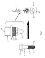
- Fig.1 is a schematic diagram for explaining a method of examining influenza virus and a specimen container to be used in the examination method as an example of the present invention.
- reference numeral 1 denotes a plastic container body containing a diluted liquid specimen 11, which has a test tube form.
- the diluted liquid specimen 11 is prepared such that a specimen collected from the nasal discharge of a patient, which contains influenza A virus antigen 1A (represented by a diamond shape) and influenza B virus antigen 1B (represented by a square shape), is prepared in a buffer solution with the addition of a surfactant and the like.
- reference numeral 2 denotes a plastic cap having a fitting portion 24, which fits in an open end portion 12 of a container body 1, and a nozzle portion 25.
- a filter 21 prepared by impregnating glass-fiber filter paper with colloidal gold labeled antibody, a filter 22 comprising a glass fiber filter paper, and a filter 23 comprising a high density polyethylene polymer filter cloth are loaded in that order to build in a three-layered structure (in the figure, black circles in the filter 21 represent gold colloids, and the process of preparing the colloidal gold labeled antibody solution or the like is explained in Example 1 described latter).
- Reference numeral 3 represents the status of making a specimen container by fitting the cap 2 to the container body 1 which houses the diluted liquid specimen 11.
- Reference numeral 4 denotes a plastic test device having an opening portion 41 formed in the surface thereof. In the test device, from the bottom side, absorbent cotton, adsorbent paper, and a membrane on which influenza A and B viruses are immobilized, are housed in this order. The diluted liquid specimen 11 is placed in the specimen container 3 and left to stand for approximately 5 minutes and the total amount thereof is then dropped from a nozzle portion 25 of the cap 2 to the opening portion 41 of the test device 4. Subsequently, the membrane is washed with a washing solution and then subjected to a visual observation to investigate the status of spots generated in the opening portion 41 and to conduct diagnosis on the influenza virus infection, and the like.
- the alphabetical letters A, R, and B represented on the test device [4] designate the regions on which the mouse-anti-influenza A virus antibody solution, the anti-mouse IgG antibody solution, and the mouse-anti-influenza B virus antibody solution were immobilized in spots, respectively.
- a hydrogen tetrachloroaurate (III) aqueous solution and a trisodium citrate aqueous solution were added to boiled distilled water and then heated while stirring. Subsequently, the mixture was cooled in ice water, producing gold colloid.
- the gold colloid has a particle size of approximately 60 nm.
- the resulting gold colloid was mixed with the mouse-anti-influenza A virus antibody and mouse-anti-influenza B virus antibody (which recognize other epitopes different from the antibodies immobilized on the nitrocellulose membrane, respectively) in a boric acid buffer (pH 9.0), followed by stirring at room temperature to provided labeled antibody.
- the mixture was further subjected to a blocking treatment by the addition of a 10% BSA aqueous solution and then precipitated by centrifugation, resulting in two kinds of colloidal gold labeled antibodies.
- colloidal gold labeled antibodies Two different colloidal gold labeled antibodies were suspended together in PBS containing 0.2% Tween 20, 1.0% sucrose, and 0.2% BSA, thereby preparing a colloidal gold labeled antibody solution containing both a colloidal gold labeled antibody for influenza A virus and a colloidal gold labeled antibody for influenza B virus.
- glass fiber-based filter paper FO75-14 was selected and then cut into disk-shaped sections (7.5 mm in diameter) so as to fit to the shape and size of the inside of the cap.
- disk-shaped sections 60 ⁇ L of the colloidal gold labeled antibody solution was dropped and then dried naturally, thereby preparing a filter [21] impregnated with the colloidal gold labeled antibody.
- a filter constructed of high-density polyethylene polymer filter cloth [23], a filter constructed of glass fiber -based filter paper [22], and a filter impregnated with the colloidal gold labeled antibody [21] were stacked in this order and housed in the cap [2], thereby preparing a cap having a built-in filter impregnated with the colloidal gold labeled antibody (hereinafter, referred to as "cap of the present invention").
- the resulting cap of the present invention was attached to the container body [1] which housed the diluted liquid specimen [11] and used.
- PBS (pH 7.4) containing 12% urea was used.
- the cap of the present invention was prepared by the method of Example 1 using members listed in Table 1.
- the cap of the present invention attached to the container body in which 1.2 mL of a diluted liquid specimen containing neither the influenza purification antigen nor nasal discharge, followed by dropping the whole amount thereof in a test tube. From the absorbance of the resulting filtrate at a wavelength of 537 nm, the amount of the colloidal gold labeled antibody in the filtrate was calculated and then compared with the amount of the colloidal gold labeled antibody with which the filter had been impregnated to obtain the recovery rate. The results of the test are shown in Table 1.
- a cap of the present invention was prepared using LG20 as a filter member and then attached to the container body in which 1.2 mL of diluted liquid specimens containing purified influenza antigens (Types A and B) having concentrations shown in Table 2 were contained, and the test according to the method of Example 1 was carried out.
- the test according to the method of Example 1 was carried out.
- the container body in which a diluted liquid specimen containing the purified influenza antigen was further added with 60 ⁇ L of a colloidal gold labeled antibody solution.
- a filter impregnated with the labeled antibody which was housed in the cap of the present invention was replaced with a filter not impregnated with the labeled antibody, followed by conducting the test.
- the test results are shown in Table 2.
- LG20 was used as a filtration filter member and the amount of the colloidal gold labeled antibody solution with which the filter was impregnated was varied as shown in Table 3 to prepare the cap of the present invention.
- the cap of the present invention was attached to a container body that housed 1.2 mL of a diluted liquid specimen containing a nasal discharge specimen (negative specimen) which the nonexistence of influenza A virus or influenza B virus was confirmed by the PCR method. Subsequently, the examination was carried out according to the method of Example 1.
- Test results are listed in Table 3. Number of specimen determined as "+" among 20 specimen Negative specimen Type A Type B Control method 1 drop (corresponding to 30 ⁇ L) 0/20 0/20 2 drops (corresponding to 60 ⁇ L) 0/20 0/20 3 drops (corresponding to 90 ⁇ L) 1/20 1/20 Method of the invention impregnated with 54 ⁇ L 0/20 0/20 impregnated with 60 ⁇ L 0/20 0/20 impregnated with 66 ⁇ L 0/20 0/20
- LG20 was used as a filter member and caps of the present invention were then prepared according to Example 1, and stored at 30°C (30% in humidity) or 37°C (30% in humidity).
- caps of the present invention were then prepared according to Example 1, and stored at 30°C (30% in humidity) or 37°C (30% in humidity).
- Each of the caps at time periods of zero month, 6 months, and 9 months from the start of storage were attached to the container body that housed 1.2 mL of a diluted sample solution (diluted sample solution containing 2 x 10 6 on the basis of virus TCID 50 /test unit for each virus) containing the purified influenza antigen (A-type virus: originated from the strain Kitakyusyu/159/93, B-type virus: from the strain Lee/40), followed by carrying out the test according to the method of Example 1.
- A-type virus originated from the strain Kitakyusyu/159/93
- B-type virus from the strain Lee/40
- LG20 was used as a filter member and caps of the present invention were then prepared according to Example 1.
- a dropping bottle containing the cap of the invention and the colloidal gold labeled antibody solution was placed together with a desiccant in an air-tight container and then stored for whole day and night at -30°C.
- the cap was thawed to room temperature and then attached to a container body that housed 1.2 mL of a diluted liquid specimen containing a nasal discharge specimen (negative specimen), in which the absence of influenza A virus and influenza B virus was confirmed by the PCR method, or a diluted liquid specimen of influenza-purified antigen (A-type virus: originated from the strain Kitakyusyu/159/93, B-type virus: originated from the strain Lee/40, each virus was contained 2 x 10 6 with TCID 50 /test unit), followed by carrying out the test according to the method of Example 1.
- A-type virus originated from the strain Kitakyusyu/159/93
- B-type virus originated from the strain Lee/40
- each virus was contained 2 x 10 6 with TCID 50 /test unit
- the cap which the filter impregnated with the labeled antibody housed in the cap of the invention was replaced with the filter not impregnated with labeled antibody, was colored with red when dropping a mixture of the colloidal gold labeled antibody solution and the diluted liquid specimen to the opening portion of the test device.
- the colloidal gold labeled antibody was captured by the filter.
- the R region showed coloration of "+” instead of its normal coloration of "++". Besides, even the negative specimen, which no coloration should not be observed essentially, showed a coloration of "+”.
- the cap of the present invention shows an excellent stability in storage of the labeled antibody, so that there is no need to strictly control the temperature during transportation and the like.
- the cap having a built-in filter impregnated with labeled antibody is attached to the container body that houses a diluted liquid specimen, and the diluted liquid specimen is injected from the container to a test device, followed by observing a reaction occurred therein. Therefore, in contrast to the conventional examination, there is no need to drop the labeled antibody solution into the diluted liquid specimen. Besides, there is no need to transfer the diluted liquid specimen from the first container to the second container.
- the method of the invention is a very simple examination method.
- the specimen container of the present invention is constructed such that a cap having a built-in filter impregnated with labeled antibody is attached to a container body for housing a diluted liquid specimen. Therefore, the container can be easily operated even though it has an extremely simple structure. Besides, the examination accuracy does not vary depending on operator individuals. Therefore, it will be most applicable to components for a simplified diagnostic kit of a specimen applying an immune reaction.
- the method of examining a specimen and a specimen container to be used in the examination method can be applied to the examination and diagnosis of various symptoms such as the infection by virus, e.g., influenza virus, and a pregnancy status.
- virus e.g., influenza virus
Landscapes
- Health & Medical Sciences (AREA)
- Chemical & Material Sciences (AREA)
- Immunology (AREA)
- Life Sciences & Earth Sciences (AREA)
- Engineering & Computer Science (AREA)
- Hematology (AREA)
- Molecular Biology (AREA)
- Analytical Chemistry (AREA)
- Chemical Kinetics & Catalysis (AREA)
- General Health & Medical Sciences (AREA)
- Biomedical Technology (AREA)
- Urology & Nephrology (AREA)
- Medicinal Chemistry (AREA)
- Biochemistry (AREA)
- Cell Biology (AREA)
- Biotechnology (AREA)
- Food Science & Technology (AREA)
- Clinical Laboratory Science (AREA)
- Physics & Mathematics (AREA)
- Microbiology (AREA)
- General Physics & Mathematics (AREA)
- Pathology (AREA)
- Investigating Or Analysing Biological Materials (AREA)
- Sampling And Sample Adjustment (AREA)
- Measuring Or Testing Involving Enzymes Or Micro-Organisms (AREA)
- Automatic Analysis And Handling Materials Therefor (AREA)
- Analysing Materials By The Use Of Radiation (AREA)
- Other Investigation Or Analysis Of Materials By Electrical Means (AREA)
Abstract
Description
| Filtration filter | Brand name (Company name) | Thickness of filter (mm) | Pore size (µm) | Recovery rate (%) |
| High density polyethylene polymer | LG20 (Flon Industry Co., Ltd.) | 1.9 | 20 | 96.7 |
| Glass fiber-based filter paper | F075-14 (Advantec MFS Inc.) | 0.355 | 23 | 95.4 |
| Glass fiber-based filter paper | GB-140 (Advantec MFS Inc.) | 0.56 | 0.4 | 71.2 |
| Cellulose-based filter paper | 17chr (Whatman Japan KK) | 0.92 | Not represented | 57.7 |
| a. Detection of influenza A virus | ||||||
| Amount of | 2×10⁶ | 8×10⁵ | 4×10⁵ | 2×10⁵ | 1×10⁵ | Blank |
| Method of the invention | ++ | ++ | ++ | + | + | - |
| Control method | ++ | ++ | ++ | + | + | - |
| (The amount of virus was represented by unit of "TCID50/test") | ||||||
| b. Detection of influenza B virus | ||||||
| Amount of | 2×10⁶ | 8×10⁵ | 4×10⁵ | 2×10⁵ | 1×10⁵ | Blank |
| Method of the invention | ++ | ++ | ++ | + | + | - |
| Control method | ++ | ++ | ++ | + | + | - |
| (The amount of virus was represented by unit of "TCID50/test") |
| Number of specimen determined as "+" among 20 specimen | |||
| Negative specimen | |||
| Type A | Type | ||
| Control method | |||
| 1 drop (corresponding to 30 µL) | 0/20 | 0/20 | |
| 2 drops (corresponding to 60 µL) | 0/20 | 0/20 | |
| 3 drops (corresponding to 90 µL) | 1/20 | 1/20 | |
| Method of the invention | impregnated with 54 µL | 0/20 | 0/20 |
| impregnated with 60 µL | 0/20 | 0/20 | |
| impregnated with 66 µL | 0/20 | 0/20 |
| Storage temperature | Storage period | O month | 6 months | 9 months | |||
| Purified influenza antigen | Type A | Type B | Type A | Type B | Type A | Type B | |
| 37°C | Control method | + | + | ± | ± | - | - |
| Method of the invention | + | + | + | + | + | + | |
| 30°C | Control method | + | + | + | + | + | + |
| Method of the invention | + | + | + | + | + | + |
| Storage temperature | Negative specimen | Purified-influenza antigen | |||||
| Type A | Type B | R part | Type A | Type B | R part | ||
| - 30° C | Control method | + | + | + | + | + | + |
| Method of the invention | - | - | ++ | ++ | ++ | ++ | |
| Room temperature | Standard method | - | - | ++ | ++ | ++ | ++ |
Claims (8)
- A method for immunologically examining a specimen, comprising: attaching a cap having a built-in filter impregnated with a labeled antibody to a container body that houses a diluted liquid specimen; introducing the diluted liquid specimen from the container to a test device and observing a reaction; and determining a presence or absence of an analyte in the specimen.
- The method according to claim 1, wherein the labeled antibody is a colloidal gold labeled antibody.
- The method according to claim 1 or 2, wherein the analyte in the specimen is influenza virus.
- The method according to any one of claims 1 to 3, further comprising using the method for reacting the specimen with the labeled antibody in a simplified diagnostic kit.
- A specimen container to be used in the examination method according to any one of claims 1 to 4, comprising a cap having a built-in filter impregnated with a labeled antibody which is attached to a container body that is to house a diluted liquid specimen.
- The specimen container according to claim 5, wherein the labeled antibody is a colloidal gold labeled antibody.
- The specimen container according to claim 5 or 6, wherein the analyte in the specimen is influenza virus.
- The specimen container according to any one of claims 5 to 7, wherein the container is used as a component of a simplified diagnostic kit.
Applications Claiming Priority (3)
| Application Number | Priority Date | Filing Date | Title |
|---|---|---|---|
| JP2003063832 | 2003-03-10 | ||
| JP2003063832 | 2003-03-10 | ||
| PCT/JP2004/003025 WO2004081568A1 (en) | 2003-03-10 | 2004-03-09 | Method of examining specimen and specimen container to be used in the examinaton method |
Publications (3)
| Publication Number | Publication Date |
|---|---|
| EP1602927A1 true EP1602927A1 (en) | 2005-12-07 |
| EP1602927A4 EP1602927A4 (en) | 2007-08-15 |
| EP1602927B1 EP1602927B1 (en) | 2010-11-10 |
Family
ID=32984446
Family Applications (1)
| Application Number | Title | Priority Date | Filing Date |
|---|---|---|---|
| EP04718761A Expired - Lifetime EP1602927B1 (en) | 2003-03-10 | 2004-03-09 | Method of examining specimen and specimen container to be used in the examination method |
Country Status (9)
| Country | Link |
|---|---|
| US (1) | US7442498B2 (en) |
| EP (1) | EP1602927B1 (en) |
| JP (1) | JP4229943B2 (en) |
| CN (1) | CN1759318B (en) |
| AT (1) | ATE487941T1 (en) |
| AU (1) | AU2004219769A1 (en) |
| DE (1) | DE602004029990D1 (en) |
| NO (1) | NO20054629D0 (en) |
| WO (1) | WO2004081568A1 (en) |
Cited By (2)
| Publication number | Priority date | Publication date | Assignee | Title |
|---|---|---|---|---|
| WO2010122392A1 (en) * | 2009-04-21 | 2010-10-28 | Span Diagnostics Ltd. | Single-step sample processing device |
| WO2018220557A1 (en) * | 2017-05-31 | 2018-12-06 | Waters Technologies Corporation | Systems and methods for measuring a concentration of an analyte |
Families Citing this family (7)
| Publication number | Priority date | Publication date | Assignee | Title |
|---|---|---|---|---|
| CA2721064C (en) * | 2008-04-09 | 2016-11-01 | Kristin Weidemaier | Sensitive immunoassays using coated nanoparticles |
| JP5065503B2 (en) * | 2010-02-05 | 2012-11-07 | 株式会社シン・コーポレイション | Inspection kit |
| CN102906567B (en) * | 2010-04-14 | 2015-02-04 | 日东纺绩株式会社 | Test instrument for measuring analyte in sample, and method for measuring analyte using same |
| JP6820865B2 (en) | 2015-04-29 | 2021-01-27 | ペルキネルマー ヘルス サイエンシーズ, インコーポレイテッド | Sample collection and delivery device |
| CN211235888U (en) * | 2018-10-11 | 2020-08-11 | 东莞东阳光医疗智能器件研发有限公司 | Immunoassay kit |
| JPWO2022107876A1 (en) * | 2020-11-20 | 2022-05-27 | ||
| CN115902214B (en) * | 2022-11-05 | 2026-01-20 | 深圳市疾病预防控制中心(深圳市卫生检验中心、深圳市预防医学研究所) | Air blowing type sampling virus antigen detection kit and use method thereof |
Family Cites Families (14)
| Publication number | Priority date | Publication date | Assignee | Title |
|---|---|---|---|---|
| IL78854A (en) * | 1985-05-31 | 1991-11-21 | Murex Corp | Biological fluid diagnostic device |
| US4874691A (en) | 1987-10-16 | 1989-10-17 | Quadra Logic Technologies Inc. | Membrane-supported immunoassays |
| US5429803A (en) | 1991-04-18 | 1995-07-04 | Lamina, Inc. | Liquid specimen container and attachable testing modules |
| US5042502A (en) * | 1989-09-18 | 1991-08-27 | La Mina Ltd. | Urine testing module with cytology cup |
| CN2113490U (en) * | 1992-03-24 | 1992-08-19 | 祝加贝 | Multipurpose testing drug kit by diaphragm type percolation filtration |
| CA2101472A1 (en) | 1992-08-03 | 1994-02-04 | Robert W. Rosenstein | Solid phase assay |
| JP3299330B2 (en) | 1993-03-18 | 2002-07-08 | 持田製薬株式会社 | Simple measuring device and method |
| JP3203631B2 (en) | 1994-03-18 | 2001-08-27 | ニプロ株式会社 | Sample collection equipment |
| US5589344A (en) * | 1994-06-15 | 1996-12-31 | Johnson & Johnson Clinical Diagnostics, Inc. | Test kit and method for competitive specific binding assay |
| JP3357196B2 (en) | 1994-09-13 | 2002-12-16 | 積水化学工業株式会社 | Fecal occult blood determination device |
| CN2230037Y (en) * | 1995-04-06 | 1996-06-26 | 王继华 | Solid state colloidal gold immunoassay agent box |
| JP2000230931A (en) | 1998-12-07 | 2000-08-22 | Katakura Industries Co Ltd | Method for detecting antibody against livestock influenza virus and kit used therefor |
| CA2373341A1 (en) | 1999-05-24 | 2000-11-30 | Abbott Laboratories | Apparatus for pretreating a sample containing an analyte |
| EP1081496A1 (en) | 1999-08-31 | 2001-03-07 | Becton, Dickinson and Company | Flow-through assay for visually detecting the presence of influenza A and B |
-
2004
- 2004-03-09 AT AT04718761T patent/ATE487941T1/en not_active IP Right Cessation
- 2004-03-09 DE DE602004029990T patent/DE602004029990D1/en not_active Expired - Lifetime
- 2004-03-09 CN CN200480006372.XA patent/CN1759318B/en not_active Expired - Fee Related
- 2004-03-09 EP EP04718761A patent/EP1602927B1/en not_active Expired - Lifetime
- 2004-03-09 US US10/546,691 patent/US7442498B2/en not_active Expired - Lifetime
- 2004-03-09 JP JP2005503524A patent/JP4229943B2/en not_active Expired - Fee Related
- 2004-03-09 AU AU2004219769A patent/AU2004219769A1/en not_active Abandoned
- 2004-03-09 WO PCT/JP2004/003025 patent/WO2004081568A1/en not_active Ceased
-
2005
- 2005-10-07 NO NO20054629A patent/NO20054629D0/en not_active Application Discontinuation
Cited By (2)
| Publication number | Priority date | Publication date | Assignee | Title |
|---|---|---|---|---|
| WO2010122392A1 (en) * | 2009-04-21 | 2010-10-28 | Span Diagnostics Ltd. | Single-step sample processing device |
| WO2018220557A1 (en) * | 2017-05-31 | 2018-12-06 | Waters Technologies Corporation | Systems and methods for measuring a concentration of an analyte |
Also Published As
| Publication number | Publication date |
|---|---|
| JP4229943B2 (en) | 2009-02-25 |
| WO2004081568A1 (en) | 2004-09-23 |
| NO20054629L (en) | 2005-10-07 |
| CN1759318A (en) | 2006-04-12 |
| EP1602927B1 (en) | 2010-11-10 |
| US7442498B2 (en) | 2008-10-28 |
| JPWO2004081568A1 (en) | 2006-06-15 |
| ATE487941T1 (en) | 2010-11-15 |
| AU2004219769A1 (en) | 2004-09-23 |
| DE602004029990D1 (en) | 2010-12-23 |
| CN1759318B (en) | 2013-12-18 |
| US20070015140A1 (en) | 2007-01-18 |
| NO20054629D0 (en) | 2005-10-07 |
| EP1602927A4 (en) | 2007-08-15 |
Similar Documents
| Publication | Publication Date | Title |
|---|---|---|
| US10415104B2 (en) | Devices for the detection of multiple analytes in a sample | |
| JP2818191B2 (en) | Solid phase analyzer | |
| EP0568664B1 (en) | Capillary blood antigen testing apparatus | |
| CA2597177C (en) | Immunoassay test device and method of use | |
| RU2025732C1 (en) | Method of analyte immunoassay in aqueous sample | |
| CA2416886C (en) | Reagent delivery device and method of use | |
| WO1997035189A1 (en) | Fluorescent reporter beads for fluid analysis | |
| JPH01140066A (en) | Indicator flow diagnosing apparatus and method | |
| JPH01503174A (en) | Test method | |
| US7442498B2 (en) | Method of examining specimen and specimen container to be used in the examination method | |
| JP2007526993A (en) | Particle aggregation detection method and apparatus | |
| US20040002063A1 (en) | Rapid vaccinia antibody detection device, method and test kit | |
| JP2021514060A (en) | In vitro diagnostic device containing beads and its use | |
| CN100367034C (en) | Measurement method of fluorescence quenching of immune colloidal gold particles | |
| CN101120249A (en) | Blood type method system and device | |
| JP2001502052A (en) | Diagnostic test device with improved resistance to liquid movement and interference | |
| JP4980944B2 (en) | Immunological measurement method | |
| WO1997023781A1 (en) | Fecal test method and device | |
| CN107980096B (en) | Use of a low density immiscible compound in a device for detecting an analyte in a sample | |
| JP4426122B2 (en) | Blood antigen detection method and apparatus | |
| EP1718968A2 (en) | Chromatographic exclusion agglutination assay and methods of use thereof | |
| EP0439917B1 (en) | Apparatus for detection and semi-quantitative measurement of analytes | |
| JP3713178B2 (en) | Immunoanalyzer for syphilis antibody detection | |
| US10969388B2 (en) | Assay and kit for detection of endotoxin | |
| JP4067208B2 (en) | Biological specific reaction measuring method and apparatus |
Legal Events
| Date | Code | Title | Description |
|---|---|---|---|
| PUAI | Public reference made under article 153(3) epc to a published international application that has entered the european phase |
Free format text: ORIGINAL CODE: 0009012 |
|
| 17P | Request for examination filed |
Effective date: 20050922 |
|
| AK | Designated contracting states |
Kind code of ref document: A1 Designated state(s): AT BE BG CH CY CZ DE DK EE ES FI FR GB GR HU IE IT LI LU MC NL PL PT RO SE SI SK TR |
|
| AX | Request for extension of the european patent |
Extension state: AL LT LV MK |
|
| DAX | Request for extension of the european patent (deleted) | ||
| A4 | Supplementary search report drawn up and despatched |
Effective date: 20070717 |
|
| 17Q | First examination report despatched |
Effective date: 20071227 |
|
| GRAP | Despatch of communication of intention to grant a patent |
Free format text: ORIGINAL CODE: EPIDOSNIGR1 |
|
| GRAS | Grant fee paid |
Free format text: ORIGINAL CODE: EPIDOSNIGR3 |
|
| GRAA | (expected) grant |
Free format text: ORIGINAL CODE: 0009210 |
|
| RAP1 | Party data changed (applicant data changed or rights of an application transferred) |
Owner name: SEKISUI MEDICAL CO., LTD. |
|
| AK | Designated contracting states |
Kind code of ref document: B1 Designated state(s): AT BE BG CH CY CZ DE DK EE ES FI FR GB GR HU IE IT LI LU MC NL PL PT RO SE SI SK TR |
|
| REG | Reference to a national code |
Ref country code: GB Ref legal event code: FG4D |
|
| REG | Reference to a national code |
Ref country code: CH Ref legal event code: EP |
|
| REG | Reference to a national code |
Ref country code: IE Ref legal event code: FG4D |
|
| REF | Corresponds to: |
Ref document number: 602004029990 Country of ref document: DE Date of ref document: 20101223 Kind code of ref document: P |
|
| REG | Reference to a national code |
Ref country code: NL Ref legal event code: VDEP Effective date: 20101110 |
|
| PG25 | Lapsed in a contracting state [announced via postgrant information from national office to epo] |
Ref country code: PT Free format text: LAPSE BECAUSE OF FAILURE TO SUBMIT A TRANSLATION OF THE DESCRIPTION OR TO PAY THE FEE WITHIN THE PRESCRIBED TIME-LIMIT Effective date: 20110310 Ref country code: AT Free format text: LAPSE BECAUSE OF FAILURE TO SUBMIT A TRANSLATION OF THE DESCRIPTION OR TO PAY THE FEE WITHIN THE PRESCRIBED TIME-LIMIT Effective date: 20101110 Ref country code: SE Free format text: LAPSE BECAUSE OF FAILURE TO SUBMIT A TRANSLATION OF THE DESCRIPTION OR TO PAY THE FEE WITHIN THE PRESCRIBED TIME-LIMIT Effective date: 20101110 Ref country code: BG Free format text: LAPSE BECAUSE OF FAILURE TO SUBMIT A TRANSLATION OF THE DESCRIPTION OR TO PAY THE FEE WITHIN THE PRESCRIBED TIME-LIMIT Effective date: 20110210 Ref country code: CY Free format text: LAPSE BECAUSE OF FAILURE TO SUBMIT A TRANSLATION OF THE DESCRIPTION OR TO PAY THE FEE WITHIN THE PRESCRIBED TIME-LIMIT Effective date: 20101110 Ref country code: FI Free format text: LAPSE BECAUSE OF FAILURE TO SUBMIT A TRANSLATION OF THE DESCRIPTION OR TO PAY THE FEE WITHIN THE PRESCRIBED TIME-LIMIT Effective date: 20101110 Ref country code: SI Free format text: LAPSE BECAUSE OF FAILURE TO SUBMIT A TRANSLATION OF THE DESCRIPTION OR TO PAY THE FEE WITHIN THE PRESCRIBED TIME-LIMIT Effective date: 20101110 Ref country code: NL Free format text: LAPSE BECAUSE OF FAILURE TO SUBMIT A TRANSLATION OF THE DESCRIPTION OR TO PAY THE FEE WITHIN THE PRESCRIBED TIME-LIMIT Effective date: 20101110 |
|
| PG25 | Lapsed in a contracting state [announced via postgrant information from national office to epo] |
Ref country code: GR Free format text: LAPSE BECAUSE OF FAILURE TO SUBMIT A TRANSLATION OF THE DESCRIPTION OR TO PAY THE FEE WITHIN THE PRESCRIBED TIME-LIMIT Effective date: 20110211 |
|
| PG25 | Lapsed in a contracting state [announced via postgrant information from national office to epo] |
Ref country code: ES Free format text: LAPSE BECAUSE OF FAILURE TO SUBMIT A TRANSLATION OF THE DESCRIPTION OR TO PAY THE FEE WITHIN THE PRESCRIBED TIME-LIMIT Effective date: 20110221 Ref country code: BE Free format text: LAPSE BECAUSE OF FAILURE TO SUBMIT A TRANSLATION OF THE DESCRIPTION OR TO PAY THE FEE WITHIN THE PRESCRIBED TIME-LIMIT Effective date: 20101110 Ref country code: CZ Free format text: LAPSE BECAUSE OF FAILURE TO SUBMIT A TRANSLATION OF THE DESCRIPTION OR TO PAY THE FEE WITHIN THE PRESCRIBED TIME-LIMIT Effective date: 20101110 Ref country code: EE Free format text: LAPSE BECAUSE OF FAILURE TO SUBMIT A TRANSLATION OF THE DESCRIPTION OR TO PAY THE FEE WITHIN THE PRESCRIBED TIME-LIMIT Effective date: 20101110 |
|
| PG25 | Lapsed in a contracting state [announced via postgrant information from national office to epo] |
Ref country code: PL Free format text: LAPSE BECAUSE OF FAILURE TO SUBMIT A TRANSLATION OF THE DESCRIPTION OR TO PAY THE FEE WITHIN THE PRESCRIBED TIME-LIMIT Effective date: 20101110 Ref country code: SK Free format text: LAPSE BECAUSE OF FAILURE TO SUBMIT A TRANSLATION OF THE DESCRIPTION OR TO PAY THE FEE WITHIN THE PRESCRIBED TIME-LIMIT Effective date: 20101110 Ref country code: RO Free format text: LAPSE BECAUSE OF FAILURE TO SUBMIT A TRANSLATION OF THE DESCRIPTION OR TO PAY THE FEE WITHIN THE PRESCRIBED TIME-LIMIT Effective date: 20101110 Ref country code: DK Free format text: LAPSE BECAUSE OF FAILURE TO SUBMIT A TRANSLATION OF THE DESCRIPTION OR TO PAY THE FEE WITHIN THE PRESCRIBED TIME-LIMIT Effective date: 20101110 |
|
| PLBE | No opposition filed within time limit |
Free format text: ORIGINAL CODE: 0009261 |
|
| STAA | Information on the status of an ep patent application or granted ep patent |
Free format text: STATUS: NO OPPOSITION FILED WITHIN TIME LIMIT |
|
| 26N | No opposition filed |
Effective date: 20110811 |
|
| PG25 | Lapsed in a contracting state [announced via postgrant information from national office to epo] |
Ref country code: MC Free format text: LAPSE BECAUSE OF NON-PAYMENT OF DUE FEES Effective date: 20110331 |
|
| REG | Reference to a national code |
Ref country code: CH Ref legal event code: PL |
|
| REG | Reference to a national code |
Ref country code: DE Ref legal event code: R097 Ref document number: 602004029990 Country of ref document: DE Effective date: 20110811 |
|
| PG25 | Lapsed in a contracting state [announced via postgrant information from national office to epo] |
Ref country code: IT Free format text: LAPSE BECAUSE OF FAILURE TO SUBMIT A TRANSLATION OF THE DESCRIPTION OR TO PAY THE FEE WITHIN THE PRESCRIBED TIME-LIMIT Effective date: 20101110 |
|
| REG | Reference to a national code |
Ref country code: IE Ref legal event code: MM4A |
|
| PG25 | Lapsed in a contracting state [announced via postgrant information from national office to epo] |
Ref country code: LI Free format text: LAPSE BECAUSE OF NON-PAYMENT OF DUE FEES Effective date: 20110331 Ref country code: IE Free format text: LAPSE BECAUSE OF NON-PAYMENT OF DUE FEES Effective date: 20110309 Ref country code: CH Free format text: LAPSE BECAUSE OF NON-PAYMENT OF DUE FEES Effective date: 20110331 |
|
| PG25 | Lapsed in a contracting state [announced via postgrant information from national office to epo] |
Ref country code: LU Free format text: LAPSE BECAUSE OF NON-PAYMENT OF DUE FEES Effective date: 20110309 |
|
| PG25 | Lapsed in a contracting state [announced via postgrant information from national office to epo] |
Ref country code: TR Free format text: LAPSE BECAUSE OF FAILURE TO SUBMIT A TRANSLATION OF THE DESCRIPTION OR TO PAY THE FEE WITHIN THE PRESCRIBED TIME-LIMIT Effective date: 20101110 |
|
| PG25 | Lapsed in a contracting state [announced via postgrant information from national office to epo] |
Ref country code: HU Free format text: LAPSE BECAUSE OF FAILURE TO SUBMIT A TRANSLATION OF THE DESCRIPTION OR TO PAY THE FEE WITHIN THE PRESCRIBED TIME-LIMIT Effective date: 20101110 |
|
| REG | Reference to a national code |
Ref country code: FR Ref legal event code: PLFP Year of fee payment: 13 |
|
| REG | Reference to a national code |
Ref country code: FR Ref legal event code: PLFP Year of fee payment: 14 |
|
| REG | Reference to a national code |
Ref country code: FR Ref legal event code: PLFP Year of fee payment: 15 |
|
| PGFP | Annual fee paid to national office [announced via postgrant information from national office to epo] |
Ref country code: GB Payment date: 20220127 Year of fee payment: 19 Ref country code: DE Payment date: 20220203 Year of fee payment: 19 |
|
| PGFP | Annual fee paid to national office [announced via postgrant information from national office to epo] |
Ref country code: FR Payment date: 20220209 Year of fee payment: 19 |
|
| REG | Reference to a national code |
Ref country code: DE Ref legal event code: R119 Ref document number: 602004029990 Country of ref document: DE |
|
| GBPC | Gb: european patent ceased through non-payment of renewal fee |
Effective date: 20230309 |
|
| PG25 | Lapsed in a contracting state [announced via postgrant information from national office to epo] |
Ref country code: GB Free format text: LAPSE BECAUSE OF NON-PAYMENT OF DUE FEES Effective date: 20230309 |
|
| PG25 | Lapsed in a contracting state [announced via postgrant information from national office to epo] |
Ref country code: GB Free format text: LAPSE BECAUSE OF NON-PAYMENT OF DUE FEES Effective date: 20230309 Ref country code: FR Free format text: LAPSE BECAUSE OF NON-PAYMENT OF DUE FEES Effective date: 20230331 Ref country code: DE Free format text: LAPSE BECAUSE OF NON-PAYMENT OF DUE FEES Effective date: 20231003 |