EP1598363A1 - Protein regulating the concentration of calcium in endoplasmic reticulum - Google Patents
Protein regulating the concentration of calcium in endoplasmic reticulum Download PDFInfo
- Publication number
- EP1598363A1 EP1598363A1 EP05008653A EP05008653A EP1598363A1 EP 1598363 A1 EP1598363 A1 EP 1598363A1 EP 05008653 A EP05008653 A EP 05008653A EP 05008653 A EP05008653 A EP 05008653A EP 1598363 A1 EP1598363 A1 EP 1598363A1
- Authority
- EP
- European Patent Office
- Prior art keywords
- erp44
- cells
- amino acid
- sequence
- rfp
- Prior art date
- Legal status (The legal status is an assumption and is not a legal conclusion. Google has not performed a legal analysis and makes no representation as to the accuracy of the status listed.)
- Withdrawn
Links
Images



Classifications
-
- C—CHEMISTRY; METALLURGY
- C07—ORGANIC CHEMISTRY
- C07K—PEPTIDES
- C07K14/00—Peptides having more than 20 amino acids; Gastrins; Somatostatins; Melanotropins; Derivatives thereof
- C07K14/435—Peptides having more than 20 amino acids; Gastrins; Somatostatins; Melanotropins; Derivatives thereof from animals; from humans
- C07K14/46—Peptides having more than 20 amino acids; Gastrins; Somatostatins; Melanotropins; Derivatives thereof from animals; from humans from vertebrates
- C07K14/47—Peptides having more than 20 amino acids; Gastrins; Somatostatins; Melanotropins; Derivatives thereof from animals; from humans from vertebrates from mammals
- C07K14/4701—Peptides having more than 20 amino acids; Gastrins; Somatostatins; Melanotropins; Derivatives thereof from animals; from humans from vertebrates from mammals not used
- C07K14/4702—Regulators; Modulating activity
Definitions
- This invention relates to a controlling molecular including a specific polypeptide, a recombinant vector including an nucleic acid encoding polypeptide, and an antibody of the polypeptide, and especially relates to a controlling molecular including a specific polypeptide, a recombinant vector including an nucleic acid encoding said polypeptide, and an antibody of the polypeptide, concerning the endoplasmic reticulum.
- Ca 2+ plays a role in regulating a lot of physiology as a second messenger in cells.
- Ca 2+ release from endoplasmic reticulum (ER) induced by a stimulation of a cells is mainly induced by actiration of inositol 1, 4, 5 trisphosphate acid receptor (IP 3 Rs).
- IP 3 Rs have three types of subtype, and it is thought that there activity is strongly affected on a micro environment of ER, but details of this are unclear.
- Cytosolic Ca 2+ concentration ([Ca 2+ ]c) is the focal point of many signal transduction pathways and regulates versatile cellular activities ranging from fertilization to cell death (Berridge et al).
- [Ca 2+ ]c is tightly regulated in time, in space and in amplitude because cells extract specific information from these parameters.
- [Ca 2+ ] ions can be supplied to the cytosol from the extracellular space or from intracellular Ca 2+ stores such as endoplasmic reticulum (ER).
- IP 3 Rs are Ca 2+ release channels on the ER that play critical roles in generation of complex patterning of [Ca 2+ ]c, such as Ca 2+ waves and oscillations.
- IP 3 R1, IP 3 R2 and IP 3 R3 There are three subtypes in IP 3 Rs (IP 3 R1, IP 3 R2 and IP 3 R3) in birds and mammals (Sorrentino et al. 2000).
- IP 3 R1 (Furuichi et al., 1989) is the dominant subtype in brain (Taylor et al., 2002) and has been implicated in neuronal development (tTakei et al., 1998; Xiang et al., 2002), in higher functions of central nervous system (Matumoto et al., 1996; Nishiyama et al., 2002), and in the human neuropathology (Tang et al., 2003).
- a striking feature of IP 3 Rs is the presence of very large cytosolic regions including IP 3 binding region, which contain approximately 80% of the whole structure (Patel et al., 1999).
- the channel domain contains six transmembrane domains and thus there are three loops that reside in the ER lumen.
- the third lumenal loop (L3) is the largest one and it can be divided into two subdomains.
- the first half (L3V domain) has quite divergent primary sequence among subtypes ( Figure 1b), and second half (L3C domain), including pore-forming region is almost completely conserved among types and species.
- the primary sequence of L3V domain of each subtype is well conserved among species, suggesting that this domain is involved in subtype-specific regulation.
- the function of the L3V domain has been largely unexplored, except that it contains glycosylation sites and can bind Ca 2+ (Sienaert et al)
- IP 3 Rs is directly regulated by Ca 2+ concentration of the ER lumen ([Ca 2+ ]) is still a matter of debate (Caroppo et al., 2003).
- Calreticulin CRT
- IICR IP 3 -induced Ca 2+ -release
- SERCA sarcoendoplasmic reticulum Ca 2+ ATPase
- the inventors have made various studies with respect to a mechanism of a regulation of ER calcium ER, and found regulatory molecular including a polypeptide of the present invention, an antibody of said polypeptide and a recombinant vector including a nucleic acid encoding said polypeptide.
- a molecular of controlling calcium according to a first aspect of the invention is (a) an amino acid sequence shown in an amino acid sequence number 1-406 of a sequence No.1 of a sequence table, or (b) an amino acid sequence wherein a part of the amino acid sequence of the amino acid sequence number 1-406 is deleted, substituted or added.
- a expression vector according to a second aspect of the invention comprises (a) a nucleic acid comprising a base sequence shown in a base sequence number 1-1221 of a sequence No.2 of a sequence table, or (b) a nucleic acid wherein a part of the base sequence of the base sequence number 1-1221 is deleted, substituted or added, and the nucleic acid has a homology of 80% for the base sequence.
- a molecular of controlling a calcium according to the invention is (a) an amino acid sequence shown in an amino acid sequence number 1-406 of a sequence No.3 of a sequence table, or (b) an amino acid sequence wherein a part of the amino acid sequence of the amino acid sequence number 1-406 is deleted, substituted or added.
- a recombinant vector according to the invention comprises (a) a nucleic acid comprising a base sequence shown in a base sequence number 1-1221 of a sequence No.4 of a sequence table, or (b) a nucleic acid wherein a part of the base sequence of the base sequence number 1-1221 is deleted, substituted or added, and the nucleic acid has a homology of 80% for the base sequence.
- An antibody of the present invention is against the polypeptide described in claims 1 or 2.
- a cultured cell according to the present invention is transformed by said recombinant vector described in claims 2-4.
- the cultured cell according to the invention is characterized that an ability of Ca 2+ release is reduced by over-expressing the polypeptide described in claims 1 or 3.
- the cultured cell according to the invention is characterized that an ability of Ca 2+ release is increased by restraining the express of the polypeptide described in claims 1 or 3.
- the cultured cell according to the invention is characterized that an ability of Ca 2+ release is increased by introducing the antibody described in claim 5 into said cultured cell and inducing an antigen-antibody reaction between the antibody and the polypeptide of claims 1 or 3.
- a molecular of controlling a calcium according to a first aspect of the invention is (a) an amino acid sequence shown in an amino acid sequence number 1-406 of a sequence No.1 of a sequence table, or (b) an amino acid sequence wherein a part of the amino acid sequence of the amino acid sequence number 1-406 is deleted, substituted or added.
- a molecular of controlling a calcium according to the invention is (a) an amino acid sequence shown in an amino acid sequence number 1-406 of a sequence No.3 of a sequence table, or (b) an amino acid sequence wherein a part of the amino acid sequence of the amino acid sequence number 1-406 is deleted, substituted or added.
- the molecule of controlling a calcium according to the present invention has an ability of specifically binding to IP 3 R1 among three types (IP 3 R1, IP 3 R2, IP 3 R3) of an inositol 1,4,5 -triphosphate receptor, allowing IP 3 R1 to be inactivate, to reduce the release of a calcium. That is, the present inventions depends on the fact that the inventors found out that a channel activity of IP 3 R1 is controlled by pH, redox state, and Lurmenal Ca 2+ concentration by an effect of ERp44, in a functional interaction between ERp44 and IP 3 R1.
- a expression vector according to a second aspect of the invention comprises (a) a nucleic acid comprising a base sequence shown in a base sequence number 1-1221 of a sequence No.2 of a sequence table, or (b) a nucleic acid wherein a part of the base sequence of the base sequence number 1-1221 is deleted, substituted or added, and the nucleic acid has a homology of 80% for the base sequence.
- a expression vector according to the invention comprises (a) a nucleic acid comprising a base sequence shown in a base sequence number 1-1221 of a sequence No.4 of a sequence table, or (b) a nucleic acid wherein a part of the base sequence of the base sequence number 1-1221 is deleted, substituted or added, and the nucleic acid has a homology of 80% for the base sequence.
- the above nucleic acid is a nucleic acid encoding a polypeptide which is included in a molecular of controlling calcium according to the presence invention. For example, it is revealed that the use of nucleic acid wherein T of the base sequence number 172 is substituted by A make it possible to control a calcium. And as a result of the above substitution of the nucleic acid, it is also possible to control calcium in case that the use of polypeptide wherein a cysteine of sequence number 58 is substituted by a serine.
- a vector including the nucleic acid is introduced into a cultured cell to be transformed, a polypeptide may be expressed which is include in a molecular of controlling calcium according to the present invention.
- the cultured cell has an advantage that it is an essential material for experiment in chemical biology, medical science, and pharmaceutical sciences field, and renders substantial contribution.
- nucleic acid for example. can be purified and isolated by the following procedure.
- a cDNA is synthesized by using a reverse transcriptase after all RNA is extracted by using a kit such as Rneasy Mini Kit (Qiagen) from a cerebella of mouse at the age of 2 weeks.
- a nucleic acid encoding full length of ERp44 can be obtained by performing a PCR using an artificial composition DNA including a proper base sequence such as a base sequence number 1-21 and 1200-1221 as a primer and a cDNA as a genetic template.
- the above nucleic acid is incorporated into a vector including a base sequence encoding a glutachione S-transferase (GST) or maltose binding protein (MBP).
- GST glutachione S-transferase
- MBP maltose binding protein
- GST-ERp44, MBP-ERp44 are specifically bound by the use of Glutathione Sepharose or Amylose Resin, and then it can be specifically eluted by a glutathione solution or maltose solution.
- a glutathione or a maltose included in the solution is removed by an ultrafiltration, and GST-ERp44 and MBP-ERp44 is purified, and then a polypeptide can be obtained.
- the nucleic acid obtained in the way is needed to bind a promoter DNA to express, since it does not have a promoter region which is normally needed for the expression of the nucleic acid.
- a promoter existed in a vector-plasmid can be used as said promoter.
- a pcDNA3.1 having a cytomegalovirus (CMV) promoter can be mentioned as a vector having said promoter.
- a cultured cell can be transformed by using a recombinant vector obtained in the way.
- the cultured cell as transformed above has an ability of reducing Ca 2+ release by means of over-expression of the polypeptide, or an ability of increasing Ca 2+ release by means of suppressing the expression of the polypeptide.
- an antibody of the present invention is against the polypeptide described in claims 1 or 2.
- An antibody has an ability of being introducing into a cell, and binding a ERp44 to lose its function and controlling Ca 2+ concentration in a cell.
- the cultured cell according to the present invention can increase an ability of Ca 2+ release by means of introducing the antibody claimed in claim 5 into a cell, and inducing an antigen-antibody reaction between the antibody and the polypeptide of claims 1 or 3.
- a conjunction between ERp44 and IP 3 R can be accelerated under existing a reducing agent such as DDT. Furthermore, a binding between ERp44 and IP 3 R can be also accelerated under acidic condition (about pH 5.2), or neutral condition such as about pH 7.5 and existing a reducing agent such as DDT.
- a conjunction between ERp44 and IP 3 R depends on a cysteine residue of IP 3 R1. That is, a cysteine residue of IP 3 R1 play a key role in a suppression of IP 3 R1 activity by ERp44, as a result of this, a controlling Ca 2+ release or suppression can be carried out, depending on whether the cysteine residue exists or not.
- ERp44 has an ability of making IP 3 R1 inactivate at a low Ca 2+ concentration so that such Ca 2+ concentration of ER lumen may be a factor for controlling Ca 2+ release or suppression.
- a conjunction between ERp44 and IP 3 R is controlled by various sorts of factors such as, reducing agent, pH, a cysteine residue, Ca 2+ concentration.
- a conjunction between ERp44 and IP 3 R controls a Ca 2+ release or suppression.
- a cultured cell having an ability of controlling Ca 2+ dynamic state can be produced, it can contribute for such as a medical science or pharmaceutical sciences field.
- L3V protein of each subtype IP 3 Rs fused to an immune globulin Fc was purified and an affinity column was made.
- a protein with about 40kDa was found which is able to specifically bind to IP 3 R1 L3V (1L3V) from a microsome fractionation of a mouse cerebella at age of 2 weeks. It was analyzed by a mass spectrometry, and an amino acid sequence was determined, it was revealed to be homology with ERp44.
- ERp44 was a protein of ER lumen having a homology to thioredoxin (TRX) which is a redox protein. It is confirmed that ERp44 can bind to IP 3 R1 by using immunoprecipitation. To further examine a bind between ERp44 and IP 3 R1 in detail, GST pulldown was carried out by using a glutathione S-transferase (GST) fused L3V (GST-L3V) and maltose binding protein (MBP) fused ERp44 (MBP-ERp44) expressed in E. coli and purified. As a result of this, it was found that MBP-ERp44 and 1L3V can directly bind each other in acid buffer including Ca 2+ .
- GST glutathione S-transferase
- MBP maltose binding protein
- ERp44 and 1L3V can bind under a neutral condition including a reducing reagent dithiothreitol (DTT). Furthermore, the bind depended on a cysteine residue of 1L3V and Ca 2+ concentration. Furthermore, it was confirmed that only ERp44 can bind to IP 3 R1 among three subtypes of IP 3 Rs. These experiment suggests that ERp44 can specifically bind to IP 3 R1 by detecting a redox state of ER and Ca 2+ concentration.
- DTT reducing reagent dithiothreitol
- IP 3 R1 was hardly expressed.
- an expression of RFP-ERp44 has no affect to IICR.
- a result of Ca 2+ imaging experiment using these HeLa and COS-7 cells and a bind experiment in vitro strongly suggest that ERp44 can bind only IP 3 R1 among all subtypes and suppress their activity.
- B lymphocyte cell type DT40 of a chicken was used. Taking advantage of a characterization of this cell, cells which all subtypes of IP 3 R are lost (DT40-TKO cell) and, a cell which only IP 3 R1 is lost (DT40-1KO cell) were made.
- IP 3 R1 is expressed in DT40-TKO cell (DT40-KMN60)
- IICR was examined after BCR was cross-linked with IgM antibody.
- a IICR significantly reduced in cells which express RFP-ERp44, but it was not observed in DT40-1KO cell about an affect of IICR. This result suggests that ERp44 can suppress IICR through not IP 3 R2 or IP 3 R3, but IP 3 R1.
- RNA interference RNA interference
- ERp44 was re-expressed in a knockdown cell of ERp44.
- An expression of ERp44 significantly reduced a IICR.
- IPBD IP 3 R1 binding domain
- IP 3 R1 activity on single-channel level was carried out by using an artificial lipid bilayer system.
- This in vitro reconfiguration system allows us to assess how affect on IP 3 R1 activity induced by ERp44 since Ca 2+ concentration of ER lumen and redox state can be strictly controlled.
- ERp44 made IP 3 R1 inactivation under conditions of an existence of DTT and a low Ca 2+ concentration. This result clearly suggests that ERp44 can detect a redox state and Ca 2+ concentration of ER to inhibit IP 3 R1 specifically.
- IP 3 Rs inositol 1, 4, 5 triphosphoric acid receptor
- ER endoplasmic reticula
- IP 3 Rs is regulated by distinguishing various cytosol proteins, it is still unsolved about a regulation of ER lumen proteins.
- ERp44 that is, ER lumen proteins of thioredoxin family, directly interacts with the third lumen loop of IP 3 R (IP 3 R1), and this interaction depends on pH, redox state, and Ca 2+ concentration of ER lumen.
- IP 3 R1 A Ca 2+ imaging experiment of IP 3 R1 and single channel record using lipid bilayer system clearly demonstrate an direct inhibition of IP 3 R1 by ERp44. These results strongly suggested that ERp44 is a micro environment of ER lumen and it was translated into a cytosol Ca 2+ concentration by means of controlling IP 3 R1.
- ERp44 a protein of thioredoxin family preciously implicated in oxidative protein folding, as a specific binding protein with the L3V domain of IP 3 R1.
- the unique characteristics of the functional interaction between ERp44 and IP 3 R1 suggest that the channel activity of IP 3 R1 is modulated by pH, redox state, and [Ca 2+ ]ER by virtue of ERp44.
- the 85kDa protein was identified as acoonitase, a mitochondrial protein, and was not further characterized in this study. Using the same procedures, we also searched for bindings protein for the L3 domain of IP 3 R2 or IP 3 R3 (2L3V or 3L3V, respectively) from mouse brain, but we have been unable to identify any so far.
- ERp44 an ER lumenal protein of chioredoxin (TRX) family, contains a signal peptide (SP), TRX-homology domain (THD), and an ER retention signal, RDEL ( Figure 1D) (Anelli et al, 2002).
- TRX signal peptide
- TDD TRX-homology domain
- RDEL Figure 1D
- ERp44 is widely expressed in mouse tissues ( Figure 1).
- Haemagglutinin A epitope (HA)-tagged ERp44 was expressed in COS-7 cells, either alone or with IP 3 R1 fused N-terminally with green fluorescent protein.
- HA-ERp44 was observed as a diffusive network, as GFP-IP 3 R1 and endogenous CRT, indicating that ERp44 colocalizes with IP 3 R1 in the ER.
- HA-ERp44 and IP 3 R1 were expressed in COS-7 cells. We first found that both HA-ERp44 and IP 3 R1 were severely degraded when whole cell lysate was prepared with acidic buffer (in which association was observed in experiments shown in Figure 1C), presumably by lysosomal proteases.
- transfected cells were treated with a membrane-permeable crosslinker dithiobissuccinimidylpropionate (DSP), and solubilization and immunopresipitation was performed in neutral buffer.
- DSP membrane-permeable crosslinker dithiobissuccinimidylpropionate
- Figure 1F anti-IP 3 R1 antibody coprecipitated HA-ERp44 and, reciprocally, anti-HA antibody coprecipitated IP 3 R1
- Figure 1F To examine endogenous association, immunoprecipitations were performed from whole cell lysate of untransfected HeLa cells, which expressed IP 3 R1 at relatibely high level. Coprecipitation between ERp44 and IP 3 R1 was clearly observed, indicating that they are in a same complex in vivo ( Figure 1G).
- ERp44 specifically interact with IP 3 R1, but not with IP 3 R2 or IP 3 R3, in condition-dependent manner.
- Glutachione (GSH) S-trandferase (GST) and maltose binding protein (MBP) were fused to 1L3V and ERp44, respectively ( Figure 2A), and these proteins were expressed in E. coli and purified.
- GSH Glutachione
- GST maltose binding protein
- ERp44 inhibits IICR via IP 3 R1.
- IP 3 R1 constitutes about half of total IP 3 Rs, and association between IP 3 R1 and ERp44 is observed ( Figure 1G).
- Red fluorescent protein (RFP)-tagged ERp44 (RFP-ERp44) or RFP with an ER retention signal (REP-RDEL, negative control) was expressed in HeLa cells, and they were stimulated with ATP in nominally Ca 2+ -free medium.
- DT40-1KO cells express in production of IP 3 R2 and IP 3 R3 , but not IP 3 R1, since the gene for IP 3 R1 was disrupted (Sugawara et al., 1997).
- Crosslinking of B-cells antigen receptor (BCR) activates PLC- ⁇ , resulting in production of IP 3 in DT40 cells.
- BCR B-cells antigen receptor
- RFP-RDEL did not affect BCR-induced Ca 2+ release (96 ⁇ 2.9%) compared with non-transfected cells.
- expression of RFP-ERp44 decreased the peak amplitude of BCR-induced Ca 2+ release in DT40-KMN60 cells (60.6 ⁇ 9.6%, Figure 3I and 3K)).
- RNA interference technique To investigate roles of endogenous ERp44, we next employed the RNA interference technique.
- sRNA small interfering RNA sequences that were rargeted to either the open reading frame (siERp44-ORF) or the 3'-untranslated region (siERp44-3U), respectively, of human ERp44.
- siRNAs As negative controls, we introduced three-point mutations in these siRNAs (siControl-ORF or siControl-3U, respectively).
- SiRNA-rtansfected HeLa cells were stimulated with 1, 3, 10 ⁇ M ATP.
- Subsequent stimulation of 3 ⁇ M ATP elicited IICR in 46.9 ⁇ 3.1%, of control cells and 90.3 ⁇ 2.4% of cells transfected with siERp44-3U ( Figure 4B and 4C).
- IPBD IP 3 R1 binding domain
- ERp44 inactivates the channel activity of IP 3 R1 in the lipid bilayer system.
- IP 3 R1 we carried out single-channel currents recoding in planar lipid bilayer fused with mouse cerebellar microsome, in which more than 99% of IP 3 R is IP 3 R1.
- MBP-ERp44 with GST-1L3V in the lumenal-side solution containing 3 mMDTT ( Figure 7A).
- IP 3 R1 activity was recorded with Ba 2+ . as a charge carrier in the presence of 3 mM DTT in the trans (lumenal) compartment.
- IP 3 R1 was maximally activated by addition of 6.4 ⁇ MIP 3 to the cis (cytosolic) compartment (data not shown).
- addition of MBP-ERp44 resulted in inhibition of the channel activity in a dose-dependent manner ( Figure 7B).
- Single-channel open probability was reduced with IC50 of 1.0 ⁇ M MBP-ERp44 and with a Hill coefficient of 2.3 ( Figure 7C), suggesting that binding of at least two ERp44 molecules to a heterotetramer of IP 3 R1 is required for inactivation.
- IP 3 R1 is sensitized by thimerosal treatment (Missiaen et al.,1998). These phenomena may be the reflection of the redox-dependent inhibitory effect of ERp44 on IP 3 R1.
- IP 3 R1 by ERp44 What is the physiological significance of inhibition of IP 3 R1 by ERp44?
- One possibility is to translate the ER lumenal environment into [Ca 2+ ]c.
- the ER is the crosspoint of many signaling pathways (Berridge, 2002; Orrenius et al., 2003). Therefore, inhibition of IP 3 R1 by ERp44 may be a part of the feedback system by which information converged at the ER was conveyed to the cytosol/nucleus.
- Another possibility is to prevent [Ca 2+ ]ER from becoming too low for other ER lumenal proteins (such as Ca 2+ -dependent enzymes and chaperones) to exert their functions.
- ERp44 was first identified as a protein that forms mixed disulfide bonds with Erol ⁇ , an ER oxidoreductase (Anelli et al., 2002), and it was shown to be involved in its ER retention (Anelli et al., 2003).
- the detailed function of Erol family proteins are not full understood, but it plays a pivotal role in oxidative protein folding in the ER lumen (Tu and Weissman, 2002).
- ERp44 overexpression shifts the redox status of Erol ⁇ to oxidized form and thus is thought to promote oxidative folding (Anelli et al., 2002). Therefore, ERp44 may play dual roles in protein folding: inhibiting Ca 2+ release (which reinforces Ca 2+ -dependent chaperones) by inactivating IP3R1 and supporting disulfide bond formation by boosting Ero1 ⁇ /oxidoreductase system. This idea is further supported by the fact that ERp44 is induced during the unfolded protein responses (Anelli et al., 2002).
- IP 3 R1 is retained in the ER by virtue of ERp44 since the L3V domain of IP 3 R1 is dispensable for the ER retention (Galvan et al., 1999) and IP 3 R1 is still localized to the ER in cells in which ERp44 was knocked down (data not shown).
- yeast does not have an ERp44 orthologue (Anelli et al., 2003).
- Yeast does not have an IP 3 R, either, and C. elegans has both ERp44 and IP 3 R, suggesting that these proteins may have emerged at similar time points in the course of evolution.
- IP 3 R1 is regulated by ERp44
- IP 3 R2 and IP 3 R3 are insensitive to the ER lumenal environment.
- ERp44 cDNA was obtained by reverse transcriptase-polymerase chain reaction from P14 mouse cerebellum.
- the expression vectors for epitope-tagged or fluorescent protein-tagged ERp44, IP 3 Rs and their mutants were constructed by utilizing polymerase chain reaction. Detailed methods, including the sequence of primers used, will be supplied on request. All constructions were verified by sequencing.
- COS-7, HeLa, and 293T cells were cultured in Dulbecco's modified Eagle medium supplemented 10% heat-inactivated fetal bovine serum.
- DT40 cells were cultured in RPMI1640 medium supplemented 10% heat-inactivated fetal bovine serum, 1% chicken serum, 100 U/mL penicillin and streptomycin, 2 mM glutamine and 50 ⁇ M 2-mercaptoethanol.
- HeLa and COS-7 cells were transfected with expression vectores or siRNAs using TransIT (Mirus) or Lipofectamine2000 (Invitrogen), respectively, as described previously (Hattori et al., 2004).
- DT40 cells were transfected with expression vectors using Nucleofector (Amaxa).
- 1L3V-Fc or control Fc was expressed in 293T cells.
- the medium was collected and incubated with Protein-G Sepharose (Amersham) for 2 hr at 4°C and then the beads were washed with 10 mM Tris-HCl (pH 8.0), 150 mM NaCl. Approximately 0.2 mg of 1L3V-Fc or control Fc was coupled to the beads, which was laid onto a column.
- P14 mice cerebella were washed with ice - cold PBS and homogenized in a buffer containing 0.32 M sucrose, 10 mM Tris-HCl (pH 7.5), 2 mM EDTA, and 1 mM DTT.
- the homogenates were centrifuged for 1hr at 100,000 x g at 2°C.
- the pellet was solubilized in buffer containing 30 mM sodium acetate, 150 mM NaCl, 4 mM CaCl2, 1% TritonX-100 for 1 hr at 4°C and centrifuged at 10,000 x g for 15 min. The supernatant was applied to the affinity columns.
- Proteins were resolved by SDS-PAGE and transferred to a polyvinylidene difluoride membrane.
- Membranes were blocked with 5% skim milk in PBS including 0.2% Tween-20 (PBST) for 30 min and probed with the first antibody for 3 hr at room temperature (RT). After washing with PBST, membranes were incubated with a suitable secondary antibody for 1hr, and signals were detected using ECL Plus kit (Amersham).
- Cells were fixed with 49% formaldehyde for 10 min at RT and permeabilized with 0.2 %Triton X-100 in PBS for 10 min at RT. After blocking in PBS including 5% goat serum for 1hr at RT, the cells were incubated with first antibodies for 3 hr at RT. Followinged by incubation with suitable Alexa Fluor 488 or 594 secondary antibodies (Molecular probes) for 1 hr, cover glasses were mounted using Vectashield mounting medium (Vector) and analyzed using a confocal fluorescence microscope (FV-300, Olympus).
- Vector Vectashield mounting medium
- FV-300 confocal fluorescence microscope
- TNE buffer 150 mM Tris-Hcl (pH 7.5), 500 mM NaCl, 1 mM EDTA, 1% TritonX-100, 0.1% SDS.
- the lysates were incubated with the indicated antibodies and Protein-G Sepharose for 2hr at 4C. The beads were washed five times with TNE buffer and proteins were eluted by boiling in SDS-PAGE sampling buffer.
- Recombinant proteins were expressed in BL21 bacterial strains and purified using GSH-Sepharose (Amersham) or Amylose Resin (New England Biolabs). ERp44 was removed from GST-ERp44 by digestion with Thrombin Protease (Amersham Biosciences).
- GST or GST fusion proteins were incubated with MBP, or MBP fusion proteins for 1 hr at 4C in acidic solution (30 mM NaOAc (pH 5.2), 150 mM NaCl, 4mM CaCl2, 0. 1 % Triton X-100).
- Proteins were incubated in neutral solution (20 mM Tris-HCl (pH 7.5), 150 mM NaCl, 5 mM EGTA, 0.1% Triton X-100)with or without 3 mM DTT
- GST-1L3V and MBP-ERp44 were incubated in pH 7.5 buffer or pH 7.5 buffer, in which EGTA was replaced to indicated concentration of CaCl2.
- Experiments in Figure 2H and 5E were carried out in neutral solution containing 3 mM DTT. Each mixture was then incubated for additional 2 hr with GSH-Sepharose. The beads were washed five times and subjected to western blot analysis.
- SiRNA duplexes were purchased from Dharmacon. Target sequences were:
- HeLa, COS-7, DT40 - KMN60, DT40-1KO,and DT40 - TKO cells were incubated for 30 min with 5 ⁇ M fura-2 acetoxymethyl ester (Dojindo).
- the fura-2-loaded cells were placed on the stage of an inverted fluorescence microscope (IX-70; Olympus) and perfused with balanced salt solution (BSS). Image capture and processing were perfomed with Argus 50/CA system (Hamamatsu photonics) at RT.
- pairs of fluorescence images were obtained through an objectivelens (UApo 20/340 or UplanApo 100 Oil Iris; Olympus) and an emission filter (510-550 nm), captured with a silicon-intensified-target video camera (C2400-8;Hamamatsu Photonics). Fluorescence images of GFP and RFP were separately acquired and saved in the computer in the same optical fields as those for Ca 2+ imaging.
- the trans chamber contained 250 mM HEPES (pH7.33), 53 mM Ba (OH)2, 3 mM DTT IP 3 R1 were activated by addition of 6.4 mM IP 3 and 0.5 mM ATP to the cis chamber.
- Purified MBP or MBP-ERp44 was added directly to transside.
- the IP 3 R1 single channel currents were amplified (Axon Instruments),filtered at 1 kHz by a low-pass Bessel filter (model, NF Instruments), sampled at 10kHz, and analyzed (Michikawa et al., 1999). Single-channel amplitudes were determined by fitting Gaussian curves to the all-point amplitude histograms of the datafiltered at 1 kHz. For presentation of the current traces, data were filtered at 0.5 kHz.
Landscapes
- Chemical & Material Sciences (AREA)
- Health & Medical Sciences (AREA)
- Life Sciences & Earth Sciences (AREA)
- Organic Chemistry (AREA)
- General Health & Medical Sciences (AREA)
- Gastroenterology & Hepatology (AREA)
- Biochemistry (AREA)
- Biophysics (AREA)
- Zoology (AREA)
- Genetics & Genomics (AREA)
- Medicinal Chemistry (AREA)
- Molecular Biology (AREA)
- Proteomics, Peptides & Aminoacids (AREA)
- Toxicology (AREA)
- Micro-Organisms Or Cultivation Processes Thereof (AREA)
- Peptides Or Proteins (AREA)
Abstract
A molecular of controlling a calcium according to a first aspect of the invention is (a) an amino acid sequence shown in an amino acid sequence number 1-406 of a sequence No.1 of a sequence table, or (b) an amino acid sequence wherein a part of the amino acid sequence of the amino acid sequence number 1-406 is deleted, substituted or added.
Description
This invention relates to a controlling molecular including a specific
polypeptide, a recombinant vector including an nucleic acid encoding polypeptide,
and an antibody of the polypeptide, and especially relates to a controlling molecular
including a specific polypeptide, a recombinant vector including an nucleic acid
encoding said polypeptide, and an antibody of the polypeptide, concerning the
endoplasmic reticulum.
Ca2+ plays a role in regulating a lot of physiology as a second messenger
in cells. Ca2+ release from endoplasmic reticulum (ER) induced by a stimulation of
a cells is mainly induced by actiration of inositol 1, 4, 5 trisphosphate acid receptor
(IP3Rs). IP3Rs have three types of subtype, and it is thought that there activity is
strongly affected on a micro environment of ER, but details of this are unclear.
Cytosolic Ca2+ concentration ([Ca2+]c) is the focal point of many signal
transduction pathways and regulates versatile cellular activities ranging from
fertilization to cell death (Berridge et al). [Ca2+]c is tightly regulated in time, in
space and in amplitude because cells extract specific information from these
parameters. [Ca2+] ions can be supplied to the cytosol from the extracellular space
or from intracellular Ca2+ stores such as endoplasmic reticulum (ER). Dysfunciton
of molecules involved in [Ca2+]c regulation, especially those on the ER, is assumed
to play major roles in apoptosis (Orrenius et al., 2003) and neuropathological
conditions, including Huntington's and Alzheimer's diseases (Mattson and Chan,
2003; Paschen, 2003). IP3Rs are Ca2+ release channels on the ER that play critical
roles in generation of complex patterning of [Ca2+]c, such as Ca2+ waves and
oscillations. There are three subtypes in IP3Rs (IP3R1, IP3R2 and IP3R3) in birds
and mammals (Sorrentino et al. 2000). IP3R1 (Furuichi et al., 1989) is the dominant
subtype in brain (Taylor et al., 2002) and has been implicated in neuronal
development (tTakei et al., 1998; Xiang et al., 2002), in higher functions of central
nervous system (Matumoto et al., 1996; Nishiyama et al., 2002), and in the human
neuropathology (Tang et al., 2003). A striking feature of IP3Rs is the presence of
very large cytosolic regions including IP3 binding region, which contain
approximately 80% of the whole structure (Patel et al., 1999). The channel domain
contains six transmembrane domains and thus there are three loops that reside in the
ER lumen. Among these, the third lumenal loop (L3) is the largest one and it can
be divided into two subdomains. The first half (L3V domain) has quite divergent
primary sequence among subtypes (Figure 1b), and second half (L3C domain),
including pore-forming region is almost completely conserved among types and
species. Interestingly, the primary sequence of L3V domain of each subtype is well
conserved among species, suggesting that this domain is involved in subtype-specific
regulation. However, the function of the L3V domain has been largely unexplored,
except that it contains glycosylation sites and can bind Ca2+(Sienaert et al)
Recently, it is becoming evident that the intralumenal environment of the
ER regulates numerous protein's function (Berridge, 2002).
However, although much effort has been contributed, how it regulates the
activity of IP3Rs remains unclear. Whether or not IP3R is directly regulated by Ca2+
concentration of the ER lumen ([Ca2+]) is still a matter of debate (Caroppo et al.,
2003). Calreticulin (CRT), and ER lumenal lectin with high Ca2+-binding capacity,
regulates IP3-induced Ca2+-release (IICR)(Camacho and Lechleiter, 1995; Roderick
et al., 1998), but it appears that this is rather indirect effect because CRT does not
interact with IP3Rs (Joseph et al), and CRT critically regulates the activity of
sarcoendoplasmic reticulum Ca2+ ATPase (SERCA) 2b. In secretory granules,
chromogranins associate with the L3C domain of IP3Rs (Yoo, 2000). Although in
vitro studies indicate that chromogranins modulate the activity of IP3Rs, their role
has not been characterized at the cellulae level.
It is, therefore, an object of the invention to provide a molecular which
regulates calcium dynamic, and mechanism of a regulation of ER calcium
concentration.
In order to achieve the above object, the inventors have made various
studies with respect to a mechanism of a regulation of ER calcium ER, and found
regulatory molecular including a polypeptide of the present invention, an antibody of
said polypeptide and a recombinant vector including a nucleic acid encoding said
polypeptide.
A molecular of controlling calcium according to a first aspect of the
invention is (a) an amino acid sequence shown in an amino acid sequence number 1-406
of a sequence No.1 of a sequence table, or (b) an amino acid sequence wherein a
part of the amino acid sequence of the amino acid sequence number 1-406 is deleted,
substituted or added.
Furthermore, a expression vector according to a second aspect of the
invention comprises (a) a nucleic acid comprising a base sequence shown in a base
sequence number 1-1221 of a sequence No.2 of a sequence table, or (b) a nucleic
acid wherein a part of the base sequence of the base sequence number 1-1221 is
deleted, substituted or added, and the nucleic acid has a homology of 80% for the
base sequence.
A molecular of controlling a calcium according to the invention is (a) an
amino acid sequence shown in an amino acid sequence number 1-406 of a sequence
No.3 of a sequence table, or (b) an amino acid sequence wherein a part of the amino
acid sequence of the amino acid sequence number 1-406 is deleted, substituted or
added.
Furthermore, a recombinant vector according to the invention comprises
(a) a nucleic acid comprising a base sequence shown in a base sequence number 1-1221
of a sequence No.4 of a sequence table, or (b) a nucleic acid wherein a part of
the base sequence of the base sequence number 1-1221 is deleted, substituted or
added, and the nucleic acid has a homology of 80% for the base sequence.
An antibody of the present invention is against the polypeptide described
in claims 1 or 2.
A cultured cell according to the present invention is transformed by said
recombinant vector described in claims 2-4.
In a preferable embodiment, the cultured cell according to the invention
is characterized that an ability of Ca2+ release is reduced by over-expressing the
polypeptide described in claims 1 or 3.
In a preferable embodiment, the cultured cell according to the invention
is characterized that an ability of Ca2+ release is increased by restraining the express
of the polypeptide described in claims 1 or 3.
In a preferable embodiment, the cultured cell according to the invention
is characterized that an ability of Ca2+ release is increased by introducing the
antibody described in claim 5 into said cultured cell and inducing an antigen-antibody
reaction between the antibody and the polypeptide of claims 1 or 3.
The invention will be described with reference to the accompanying
drawings, wherein:
A molecular of controlling a calcium according to a first aspect of the
invention is (a) an amino acid sequence shown in an amino acid sequence number 1-406
of a sequence No.1 of a sequence table, or (b) an amino acid sequence wherein a
part of the amino acid sequence of the amino acid sequence number 1-406 is deleted,
substituted or added. Furthermore, a molecular of controlling a calcium according
to the invention is (a) an amino acid sequence shown in an amino acid sequence
number 1-406 of a sequence No.3 of a sequence table, or (b) an amino acid sequence
wherein a part of the amino acid sequence of the amino acid sequence number 1-406
is deleted, substituted or added.
The molecule of controlling a calcium according to the present invention
has an ability of specifically binding to IP3R1 among three types (IP3R1, IP3R2,
IP3R3) of an inositol 1,4,5 -triphosphate receptor, allowing IP3R1 to be inactivate, to
reduce the release of a calcium. That is, the present inventions depends on the fact
that the inventors found out that a channel activity of IP3R1 is controlled by pH,
redox state, and Lurmenal Ca2+ concentration by an effect of ERp44, in a functional
interaction between ERp44 and IP3R1.
Furthermore, a expression vector according to a second aspect of the
invention comprises (a) a nucleic acid comprising a base sequence shown in a base
sequence number 1-1221 of a sequence No.2 of a sequence table, or (b) a nucleic
acid wherein a part of the base sequence of the base sequence number 1-1221 is
deleted, substituted or added, and the nucleic acid has a homology of 80% for the
base sequence.
Furthermore, a expression vector according to the invention comprises
(a) a nucleic acid comprising a base sequence shown in a base sequence number 1-1221
of a sequence No.4 of a sequence table, or (b) a nucleic acid wherein a part of
the base sequence of the base sequence number 1-1221 is deleted, substituted or
added, and the nucleic acid has a homology of 80% for the base sequence.
The above nucleic acid is a nucleic acid encoding a polypeptide which is included in
a molecular of controlling calcium according to the presence invention. For
example, it is revealed that the use of nucleic acid wherein T of the base sequence
number 172 is substituted by A make it possible to control a calcium. And as a
result of the above substitution of the nucleic acid, it is also possible to control
calcium in case that the use of polypeptide wherein a cysteine of sequence number
58 is substituted by a serine.
If a vector including the nucleic acid is introduced into a cultured cell to
be transformed, a polypeptide may be expressed which is include in a molecular of
controlling calcium according to the present invention. The cultured cell has an
advantage that it is an essential material for experiment in chemical biology, medical
science, and pharmaceutical sciences field, and renders substantial contribution.
With respect to the nucleic acids previously described, a purification and
isolation method will be described below. The above nucleic acid, for example. can
be purified and isolated by the following procedure. A cDNA is synthesized by
using a reverse transcriptase after all RNA is extracted by using a kit such as Rneasy
Mini Kit (Qiagen) from a cerebella of mouse at the age of 2 weeks. A nucleic acid
encoding full length of ERp44 can be obtained by performing a PCR using an
artificial composition DNA including a proper base sequence such as a base
sequence number 1-21 and 1200-1221 as a primer and a cDNA as a genetic template.
Furthermore, an example of obtaining a polypeptide will be described
below. At first, the above nucleic acid is incorporated into a vector including a base
sequence encoding a glutachione S-transferase (GST) or maltose binding protein
(MBP). After these vector is transformed by using a host such as E. coli, a lot of
polypeptide of GST or MBP fused ERp44 (GST-ERp44, MBP-ERp44) can be
expressed in E. coli according to stimulation of isopropyl thiogalactoside. After E..
coli is broken by an ultrasonic treatment, GST-ERp44, MBP-ERp44 are specifically
bound by the use of Glutathione Sepharose or Amylose Resin, and then it can be
specifically eluted by a glutathione solution or maltose solution. At last, a
glutathione or a maltose included in the solution is removed by an ultrafiltration, and
GST-ERp44 and MBP-ERp44 is purified, and then a polypeptide can be obtained.
The nucleic acid obtained in the way is needed to bind a promoter DNA
to express, since it does not have a promoter region which is normally needed for the
expression of the nucleic acid. For example, a promoter existed in a vector-plasmid
can be used as said promoter. A pcDNA3.1 having a cytomegalovirus (CMV)
promoter can be mentioned as a vector having said promoter.
Furthermore, a cultured cell can be transformed by using a recombinant
vector obtained in the way. The cultured cell as transformed above has an ability of
reducing Ca2+ release by means of over-expression of the polypeptide, or an ability of
increasing Ca2+ release by means of suppressing the expression of the polypeptide.
Furthermore, an antibody of the present invention is against the
polypeptide described in claims 1 or 2. An antibody has an ability of being
introducing into a cell, and binding a ERp44 to lose its function and controlling Ca2+
concentration in a cell.
In a preferable embodiment, the cultured cell according to the present
invention can increase an ability of Ca2+ release by means of introducing the
antibody claimed in claim 5 into a cell, and inducing an antigen-antibody reaction
between the antibody and the polypeptide of claims 1 or 3.
A conjunction between ERp44 and IP3R can be accelerated under
existing a reducing agent such as DDT. Furthermore, a binding between ERp44
and IP3R can be also accelerated under acidic condition (about pH 5.2), or neutral
condition such as about pH 7.5 and existing a reducing agent such as DDT.
Moreover, a conjunction between ERp44 and IP3R depends on a cysteine
residue of IP3R1. That is, a cysteine residue of IP3R1 play a key role in a
suppression of IP3R1 activity by ERp44, as a result of this, a controlling Ca2+ release
or suppression can be carried out, depending on whether the cysteine residue exists
or not.
Furthermore, ERp44 has an ability of making IP3R1 inactivate at a low
Ca2+ concentration so that such Ca2+ concentration of ER lumen may be a factor for
controlling Ca2+ release or suppression.
As mentioned above, a conjunction between ERp44 and IP3R is
controlled by various sorts of factors such as, reducing agent, pH, a cysteine residue,
Ca2+ concentration. In other words, a conjunction between ERp44 and IP3R
controls a Ca2+ release or suppression. According to the mechanism of the above
control, a cultured cell having an ability of controlling Ca2+ dynamic state can be
produced, it can contribute for such as a medical science or pharmaceutical sciences
field.
The following examples are given in illustration of the invention and are
not intended as limitations thereof. The following examples are intended to
illustrate an embodiment of the invention, and it is naturally possible to properly
change the invention without departing from the spirit and the scope of the invention
claimed in claims.
At first, to reveal the molecular mechanism, we noted a domain having a
low homology (L3V) among all subtypes of IP3Rs existing in ER lumen domain, and
examined a protein having an ability of binding these domain. L3V protein of each
subtype IP3Rs fused to an immune globulin Fc was purified and an affinity column
was made. A protein with about 40kDa was found which is able to specifically bind
to IP3R1 L3V (1L3V) from a microsome fractionation of a mouse cerebella at age of
2 weeks. It was analyzed by a mass spectrometry, and an amino acid sequence was
determined, it was revealed to be homology with ERp44. ERp44 was a protein of
ER lumen having a homology to thioredoxin (TRX) which is a redox protein. It is
confirmed that ERp44 can bind to IP3R1 by using immunoprecipitation. To further
examine a bind between ERp44 and IP3R1 in detail, GST pulldown was carried out
by using a glutathione S-transferase (GST) fused L3V (GST-L3V) and maltose
binding protein (MBP) fused ERp44 (MBP-ERp44) expressed in E. coli and purified.
As a result of this, it was found that MBP-ERp44 and 1L3V can directly bind each
other in acid buffer including Ca2+. Next, it was also revealed that ERp44 and
1L3V can bind under a neutral condition including a reducing reagent dithiothreitol
(DTT). Furthermore, the bind depended on a cysteine residue of 1L3V and Ca2+
concentration. Furthermore, it was confirmed that only ERp44 can bind to IP3R1
among three subtypes of IP3Rs. These experiment suggests that ERp44 can
specifically bind to IP3R1 by detecting a redox state of ER and Ca2+ concentration.
Next, it was examined how effects a ERp44- IP3R1 bind gives a channel
activity in vivo according to a Ca2+ imaging experiment using various cells. A red
fluorescent protein (RFP) fused ERp44 (RFP-ERp44) was expressed in a cell, and an
agonist ATP stimulation of a IP3 production was carried out. IP3 -induced Ca2+
release (IICR) significantly reduced in HeLa cell which expresses RFP-ERp44
(IP3R1 among subtypes was predominantly expressed.).
Further, in a COS-7 cell (IP3R1 was hardly expressed.), an expression of
RFP-ERp44 has no affect to IICR. A result of Ca2+ imaging experiment using these
HeLa and COS-7 cells and a bind experiment in vitro strongly suggest that ERp44
can bind only IP3R1 among all subtypes and suppress their activity. To further
confirm this, B lymphocyte cell type DT40 of a chicken was used. Taking
advantage of a characterization of this cell, cells which all subtypes of IP3R are lost
(DT40-TKO cell) and, a cell which only IP3R1 is lost (DT40-1KO cell) were made.
Furthermore, a cell wherein IP3R1 is expressed in DT40-TKO cell (DT40-KMN60)
is made. IICR was examined after BCR was cross-linked with IgM antibody.
A IICR significantly reduced in cells which express RFP-ERp44, but it was not
observed in DT40-1KO cell about an affect of IICR. This result suggests that
ERp44 can suppress IICR through not IP3R2 or IP3R3, but IP3R1.
To further examine a function of endogenous ERp44, an experiment was
carried out by using RNA interference (RNAi). In a HeLa cell wherein knockdown
of ERp44 is carried out, a ratio of a cell in which IICR is induced by ATP
stimulation and an intensity of Ca2+ release are significantly increased. On the
other hand, in COS-7 cells, knockdown of ERp44 had no effect on IICR. These
loss-of-function experiment strongly suggests that ERp44 can suppress IP3-induced
Ca2+ release via IP3R1, but not IP3R2 or IP3R3. Further, this result also suggests a
result obtained by gain-of-function experiment.
To further confirm the result obtained from RNAi, ERp44 was re-expressed
in a knockdown cell of ERp44. An expression of ERp44 significantly
reduced a IICR. As a result of this, it was concluded that ERp44 can inhibit IP3R1.
Furthermore, we examined how domain of ERp44 is important for inhibition of
IP3R1 by using this re-expressed lines. At first, it was found that an amino acid
from 236 to 345 of ERp44 is IP3R1 binding domain (IPBD). Surprisingly, used
mutants, however, even if it was a IPBD, were not able to inhibit IP3R1 compared
with that of full lengths of ERp44. These results suggest that an inhibition of IP3R1
requests not only a binding domain but other domain except for said binding domain.
Next, an importance of a cysteine residue is examined. A binding between cysteine
mutant of IP3R1 which is expressed in COS-7 cells, and ERp44 significantly reduced.
To examine a physiological means, a cysteine mutant of IP3R1 and RFP-ERp44 were
expressed in DT40-TKO, and they were stimulated by IgM antibody, but an
inhibition of IICR was not observed. According to this result, it was found that a
cysteine residue is necessary to inhibit IP3R1 activity by ERp44.
Furthermore, to examine how affect on channel activity according to a
bind between ERp44 and IP3R1, a measurement of IP3R1 activity on single-channel
level was carried out by using an artificial lipid bilayer system. This in vitro
reconfiguration system allows us to assess how affect on IP3R1 activity induced by
ERp44 since Ca2+ concentration of ER lumen and redox state can be strictly controlled.
As a result, ERp44 made IP3R1 inactivation under conditions of an existence of DTT
and a low Ca2+ concentration. This result clearly suggests that ERp44 can detect a
redox state and Ca2+ concentration of ER to inhibit IP3R1 specifically.
According to the fact mentioned above, a new mechanism of controlling
Ca2+ signaling was found by using a manner performed in a chemical biology, a
biochemistry, a cell biological study, and an electrophysiology.
A detailed explanation will be below.
An inositol 1, 4, 5 triphosphoric acid receptor (IP3Rs) is a internal
channel protein and relates to a number of biologic processes and disorders wherein a
medium Ca2+ is extracted from endoplasmic reticula (ER). IP3Rs is regulated by
distinguishing various cytosol proteins, it is still unsolved about a regulation of ER
lumen proteins. We found that ERp44, that is, ER lumen proteins of thioredoxin
family, directly interacts with the third lumen loop of IP3R (IP3R1), and this
interaction depends on pH, redox state, and Ca2+ concentration of ER lumen.
A Ca2+ imaging experiment of IP3R1 and single channel record using lipid bilayer
system clearly demonstrate an direct inhibition of IP3R1 by ERp44. These results
strongly suggested that ERp44 is a micro environment of ER lumen and it was
translated into a cytosol Ca2+ concentration by means of controlling IP3R1.
An
Introduction
In this example, to elucidate the molecular mechanism of type-specific regulation of IP3Rs from the ER lumen, we searched for binding proteins of the L3V domain of IP3Rs. We then identified ERp44 a protein of thioredoxin family preciously implicated in oxidative protein folding, as a specific binding protein with the L3V domain of IP3R1. The unique characteristics of the functional interaction between ERp44 and IP3R1 suggest that the channel activity of IP3R1 is modulated by pH, redox state, and [Ca2+]ER by virtue of ERp44.
In this example, to elucidate the molecular mechanism of type-specific regulation of IP3Rs from the ER lumen, we searched for binding proteins of the L3V domain of IP3Rs. We then identified ERp44 a protein of thioredoxin family preciously implicated in oxidative protein folding, as a specific binding protein with the L3V domain of IP3R1. The unique characteristics of the functional interaction between ERp44 and IP3R1 suggest that the channel activity of IP3R1 is modulated by pH, redox state, and [Ca2+]ER by virtue of ERp44.
Results
Identification of ERp44 as a Binding Protein of the L3V Domain of IP3R1
We aimed to find proteins that bind to the L3V domain of IP3Rs. Yeast two-hybrid screening, which has been successful for identification of IP3Rs-binding proteins in cytosol, was considered not to be appropriate for this purpose, since chemical environment of nucleus is completely different from that of ER lumen. We therefore, made the 1L3V-Fe, in which secretion signal and human immunoglobulin Fe domain were fused to the N-and C-terminus, respectively, of the L3V domain of IP3R1 (1L3V). This protein was expressed in 293T cells, collected from the medium, and utilized to make affinity resin. Initially, we failed to detect any 1L3V-Fc-interacting protein from mouse cerebellar microsomal fraction solubilized in neutral buffer (pH 7.5, data not shown). Then, we performed the similar experiment under acidic conditions (pH 5.2), as chromogranin A binds to the L3C domain of IP3R only under this condition. Consequently, we identified two proteins that bound to 1L3V-Fc, but not to control-Fc (Figure 1C). We excised these bands and analyzed them by mass spectrometry. Two peptide sequences, HPLLHIQK and QFVFDLHSGK, were obtained from the 44kDa protein (Figure 1C), indicating that this protein is ERp44 (Anelli et al). The 85kDa protein was identified as acoonitase, a mitochondrial protein, and was not further characterized in this study. Using the same procedures, we also searched for bindings protein for the L3 domain of IP3R2 or IP3R3 (2L3V or 3L3V, respectively) from mouse brain, but we have been unable to identify any so far.
Identification of ERp44 as a Binding Protein of the L3V Domain of IP3R1
We aimed to find proteins that bind to the L3V domain of IP3Rs. Yeast two-hybrid screening, which has been successful for identification of IP3Rs-binding proteins in cytosol, was considered not to be appropriate for this purpose, since chemical environment of nucleus is completely different from that of ER lumen. We therefore, made the 1L3V-Fe, in which secretion signal and human immunoglobulin Fe domain were fused to the N-and C-terminus, respectively, of the L3V domain of IP3R1 (1L3V). This protein was expressed in 293T cells, collected from the medium, and utilized to make affinity resin. Initially, we failed to detect any 1L3V-Fc-interacting protein from mouse cerebellar microsomal fraction solubilized in neutral buffer (pH 7.5, data not shown). Then, we performed the similar experiment under acidic conditions (pH 5.2), as chromogranin A binds to the L3C domain of IP3R only under this condition. Consequently, we identified two proteins that bound to 1L3V-Fc, but not to control-Fc (Figure 1C). We excised these bands and analyzed them by mass spectrometry. Two peptide sequences, HPLLHIQK and QFVFDLHSGK, were obtained from the 44kDa protein (Figure 1C), indicating that this protein is ERp44 (Anelli et al). The 85kDa protein was identified as acoonitase, a mitochondrial protein, and was not further characterized in this study. Using the same procedures, we also searched for bindings protein for the L3 domain of IP3R2 or IP3R3 (2L3V or 3L3V, respectively) from mouse brain, but we have been unable to identify any so far.
ERp44, an ER lumenal protein of chioredoxin (TRX) family, contains a
signal peptide (SP), TRX-homology domain (THD), and an ER retention signal,
RDEL (Figure 1D) (Anelli et al, 2002). ERp44 is widely expressed in mouse
tissues (Figure 1). Haemagglutinin A epitope (HA)-tagged ERp44 (Anelli et al,
2002) was expressed in COS-7 cells, either alone or with IP3R1 fused N-terminally
with green fluorescent protein. HA-ERp44 was observed as a diffusive network, as
GFP-IP3R1 and endogenous CRT, indicating that ERp44 colocalizes with IP3R1 in
the ER. To verify interaction between IP3R1 and ERp44, HA-ERp44 and IP3R1
were expressed in COS-7 cells. We first found that both HA-ERp44 and IP3R1
were severely degraded when whole cell lysate was prepared with acidic buffer (in
which association was observed in experiments shown in Figure 1C), presumably by
lysosomal proteases. To circumvent this, the transfected cells were treated with a
membrane-permeable crosslinker dithiobissuccinimidylpropionate (DSP), and
solubilization and immunopresipitation was performed in neutral buffer. Under this
condition, anti-IP3R1 antibody coprecipitated HA-ERp44 and, reciprocally, anti-HA
antibody coprecipitated IP3R1 (Figure 1F), indicating that ERp44 and IP3R1 are in a
same complex in transfected COS-7 cells. To examine endogenous association,
immunoprecipitations were performed from whole cell lysate of untransfected HeLa
cells, which expressed IP3R1 at relatibely high level. Coprecipitation between
ERp44 and IP3R1 was clearly observed, indicating that they are in a same complex in
vivo (Figure 1G).
ERp44 specifically interact with IP3R1, but not with IP3R2 or IP3R3, in
condition-dependent manner.
To further characterize association between ERp44 and IP3Rs, we next performed series of pull-down experiments. Glutachione (GSH) S-trandferase (GST) and maltose binding protein (MBP) were fused to 1L3V and ERp44, respectively (Figure 2A), and these proteins were expressed in E. coli and purified. Under acidic condition (pH 5.2), MBP-ERp44, but not MBP alone, interacted with GST-1L3V, but not with GST (Figure 2B). This interaction, however was not observed under neutral (pH 7.2) condition (data not shown). The pH of the ER lumen was estimated to be near neutral in most of the literatures (e.g. Kim et al., 1998; Foyouzi-Youssefi et al., 2000). We reasoned that certain factor might have been missed in our pull-down assays, and next sought for conditions under which 1L3V and ERp44 interacts at neutral pH. The presence of THD in ERp44 suggests that the function of this protein is redox-dependent, and it was reported that ERp44 forms mized disulfide bonds with a number of proteins (Anelli et al., 2003; Anelli et al., 2002). Thus, we examined whether interaction between 1L3V and ERp44 was dependent on the redox state. Both GST-1L3V and ERp44 were mostly in their oxidized forms in the absence of a reducing reagent, dithiothreitol (DTT), as judged by their migration pattern on SDS-PAGE under non-reducing condition. Interaction between these oxidized forms was not observed at pH7.5 (Figure 2C,lane 1, middle panel).
Addition of DTT reduces GST-1L3V and ERp44 and, strikingly, it was found that
reduced forms of these proteins were able to interact with each other at pH7.5
(Figure 2C, lane 2). It was also revealed that this interaction is not caused by mixed
disulfide bonds since no high-molecular weight band (i.e. covalently linked GST-1L3V-ERp44)
was observed (data not shown). To investigate redox state of which
protein is important for this interaction, the cysteine residues of these proteins were
mutated. Regarding 1L3V, mutation of either Cys2496, Cys2504, or Cys2527, led
to decreased binding to recombinant ERp44 (Figure 2D). By contrast, none of
ERp44 cysteine mutations affected their binding to GST-1L3V, but none of ERp44,
are important for the interaction between ERp44 and 1L3V was required for
interaction between 1L3V and ERp44.
To further characterize association between ERp44 and IP3Rs, we next performed series of pull-down experiments. Glutachione (GSH) S-trandferase (GST) and maltose binding protein (MBP) were fused to 1L3V and ERp44, respectively (Figure 2A), and these proteins were expressed in E. coli and purified. Under acidic condition (pH 5.2), MBP-ERp44, but not MBP alone, interacted with GST-1L3V, but not with GST (Figure 2B). This interaction, however was not observed under neutral (pH 7.2) condition (data not shown). The pH of the ER lumen was estimated to be near neutral in most of the literatures (e.g. Kim et al., 1998; Foyouzi-Youssefi et al., 2000). We reasoned that certain factor might have been missed in our pull-down assays, and next sought for conditions under which 1L3V and ERp44 interacts at neutral pH. The presence of THD in ERp44 suggests that the function of this protein is redox-dependent, and it was reported that ERp44 forms mized disulfide bonds with a number of proteins (Anelli et al., 2003; Anelli et al., 2002). Thus, we examined whether interaction between 1L3V and ERp44 was dependent on the redox state. Both GST-1L3V and ERp44 were mostly in their oxidized forms in the absence of a reducing reagent, dithiothreitol (DTT), as judged by their migration pattern on SDS-PAGE under non-reducing condition. Interaction between these oxidized forms was not observed at pH7.5 (Figure 2C,
We also examined whether the interaction between ERp44 and 1L3V is
dependent on Ca2+ concentration. As shown in Figure 2E, the interaction
diminished when Ca2+ concentration was higher than 100µM. The [Ca2+]ER was
estimated to be roughly between 200 µM and 1mM under resting conditions
(Meldolesi and Pozzan, 1998), suggesting that interaction between ERp44 and 1L3V
is not very robust at resting state and is enhanced when [Ca2+]ER decreased (e.g.
after Ca2+ release).
The above findings suggested that interaction between ERp44 and IP3R1
favors reducing and/or low [Ca2+]ER conditions. To examine this in vivo, COS-7
cells expressing GFP- IP3R1 and HA-ERp44 were treated with 5mM DTT, and then
HA-ERp44 was immunoprecipitated (Figure 2F). Although HA-ERp44
precipitated with anti-HA antibody was consistently diminished under reducing
conditions for unknown reasons (Figure 2F, bottom panel), GFP-IP3R1 co-precipitated
with HA-ERp44 was significantly increased (Figure 2F, middle panel).
Next, CoS-7 cells epressing HA-ERp44 an acivator of purinergic receptors. This
treatment increased GFP-IP3R1 that was co-precipitated with HA-ERp44 by
approximately 2-folds, indicating that lowering [Ca2+]ER augments association
between IP3R1 and ERp44. These results indicated that the dynamic change of the
ER luminal environment controls interaction between IP3R1 and ERp44. Finally,
we found that MBP-ERp44 interact with GST-1L3V, but neither with GST-2L3V
nor GST-3L3V, which is consistent with the results of initial screening using L3V-Fc
(Figure 1). Take together, it was concluded that ERp44 directly interacts with the
L3V domain of IP3R1, in a subtype-specific and the ER lumenal environment manner.
Expression of ERp44 inhibits IICR via IP3R1.
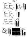
To explore the functional consequence of binding of ERp44 to IP3R1, we carried out single-cell Ca2+ imaging experiments using a fluorescent Ca2+ indicator fura-2. First, we employed HeLa cells, in which IP3R1 constitutes about half of total IP3Rs, and association between IP3R1 and ERp44 is observed (Figure 1G). Red fluorescent protein (RFP)-tagged ERp44 (RFP-ERp44) or RFP with an ER retention signal (REP-RDEL, negative control) was expressed in HeLa cells, and they were stimulated with ATP in nominally Ca2+-free medium. In the following experiments, Ca2+ signals between transfected (RFP signal-positive) and non-transfected (RFP signal-negative) cells in a same dish were compared. In non-transfected cells, ATP (3 µM) typically caused a single Ca2+ transient occasionally with small oscillations (Figure 3A black trace). HeLa cells expressing RFP-RDEL showed indistinguishable responses Figure 3A, red trace). On the other hand, HeLa cells expressing RFP-ERp44 typically shown significantly smaller Ca2+ transient (Figure 3B, red trace). On average, the peak amplitude in cells expressing RFP-ERp44 was significantly decreased (69.3±1.8% of that of non-transfected cells; p<0.05, Figure 3C). This was not due to the reduction of releasable Ca2+ content in the ER, as a passive Ca2+ leak elicited by thapsigargin (Tg, a SERCA inhibitor) was unchanged (Figure 3D, 3E). These results indicated that ERp44 inhibited IICR without affecting Ca2+storage capacity in HeLa cells.
To explore the functional consequence of binding of ERp44 to IP3R1, we carried out single-cell Ca2+ imaging experiments using a fluorescent Ca2+ indicator fura-2. First, we employed HeLa cells, in which IP3R1 constitutes about half of total IP3Rs, and association between IP3R1 and ERp44 is observed (Figure 1G). Red fluorescent protein (RFP)-tagged ERp44 (RFP-ERp44) or RFP with an ER retention signal (REP-RDEL, negative control) was expressed in HeLa cells, and they were stimulated with ATP in nominally Ca2+-free medium. In the following experiments, Ca2+ signals between transfected (RFP signal-positive) and non-transfected (RFP signal-negative) cells in a same dish were compared. In non-transfected cells, ATP (3 µM) typically caused a single Ca2+ transient occasionally with small oscillations (Figure 3A black trace). HeLa cells expressing RFP-RDEL showed indistinguishable responses Figure 3A, red trace). On the other hand, HeLa cells expressing RFP-ERp44 typically shown significantly smaller Ca2+ transient (Figure 3B, red trace). On average, the peak amplitude in cells expressing RFP-ERp44 was significantly decreased (69.3±1.8% of that of non-transfected cells; p<0.05, Figure 3C). This was not due to the reduction of releasable Ca2+ content in the ER, as a passive Ca2+ leak elicited by thapsigargin (Tg, a SERCA inhibitor) was unchanged (Figure 3D, 3E). These results indicated that ERp44 inhibited IICR without affecting Ca2+storage capacity in HeLa cells.
We next tested the effect of ERp44 overexpression in COS-7 cells that
virtually expressed no IP3R1 (Boehning and Joseph, 2000; Hattori et al., 2004).
In non-transfected cells and cells expressing RFP-RDEL, stimulation with 1 µM
ATP elicited a Ca2+transient. Strikingly, overexpression of RFP-ERp44 did not
affect the amplitude of the transient (Figure G). The averaged amplitudes in cells
expressing RFP-RDEL and RFP-ERp44 were 98.6±0.7 % and 104±2.5 % of those in
non-transfected cells, respectively (n=60 and 80; Figure 3H). The effect of RFP-ERp44
overexpression was not observed when cells were stimulated with weaker
stimulation (0.3 µM ATP), too (data not shown). These results indicated that
ERp44 did not affect IICR in COS-7 cells. The above results suggested that ERp44
inhibits IP3R1, but not IP3R2 or IP3R3. To further confirm this, we employed two
more cell lines, DT40-KMN60 was established by stably-transfecting mouse IP3R1
gene on the background of DT40 cells in which all the IP3R genes were disrupted
(DT-TKO, Sugawara et al., 1997). DT40-1KO cells express in production of IP3R2
and IP3R3 , but not IP3R1, since the gene for IP3R1 was disrupted (Sugawara et al.,
1997). Crosslinking of B-cells antigen receptor (BCR) activates PLC-γ, resulting in
production of IP3 in DT40 cells. In DT40-KMN60 cells, expression of RFP-RDEL
did not affect BCR-induced Ca2+release (96±2.9%) compared with non-transfected
cells. On the other hand, expression of RFP-ERp44 decreased the peak amplitude
of BCR-induced Ca2+release in DT40-KMN60 cells (60.6±9.6%, Figure 3I and 3K)).
There was no reduction in Tg-induced Ca2+leak in these cells (Figure 3L and 3M).
In DT40-1KO cells, on the other hand, expression of RFP-ERp44 had little effect on
BCR-induced Ca2+ release (Figure 3J and 3K). These results provide strong
evidence that ERp44 inhibits IP3R1, but neither IP3R2 or IP3R3.
Since function of IP3Rs is known to be critical for BCR-induced
apoptosis in DT40 cells (Sugawara et al., 1997), we next examined whether
expression of ERp44 affects it. Expressing RFP-ERp44, but not RFP-RDEL,
significantly inhibited apoptosis in DT40-KMN60 cells (Figure 3N and 30).
On the other hand, this effect was not observed in DT40-1KO cells (Figure 30).
These results implied that modulation by ERp44 on IP3R1 affects cellular function
such as apoptosis.
Specific knockdown of ERp44 results in augmentation of IICR via IP3R1
To investigate roles of endogenous ERp44, we next employed the RNA interference
technique. We tested two different small interfering RNA (sRNA) sequences that
were rargeted to either the open reading frame (siERp44-ORF) or the 3'-untranslated
region (siERp44-3U), respectively, of human ERp44. As negative controls, we
introduced three-point mutations in these siRNAs (siControl-ORF or siControl-3U,
respectively). Western analysis revealed that both siERp44-ORF and siERp44-3U
efficiently (>90%) decreased expression of ERp44 without affecting that of actin,
IR3R1, and IR3R3 in HeLa and COS-7 cells. Unfortunately, no siRNA targeted to
chicken ERp44 decreased expression of ERp44 in DT40 cells (data not shown).
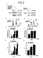
SiRNA-rtansfected HeLa cells were stimulated with 1, 3, 10 µM ATP.
In siControl-3U-transfected cells, 1 µM ATP rarely (4.0±0.6% of total cells, n=201)
evoked discernible Ca2+ signals (Figure 4B and 4C, black trace and bar). On the
other hand, the number of responding cells was higher (20.1±4.9%, n=213) in
siERp44-3U-transfected cells by approximately 5-folds (Figure 4B and 4C, red trace
and bar). Subsequent stimulation of 3 µM ATP elicited IICR in 46.9±3.1%, of
control cells and 90.3±2.4% of cells transfected with siERp44-3U (Figure 4B and
4C). When stimulated with 10 µM ATP, most of cells responded in either
preparation (96.9±1.9% and 99.6±0.4%, respectively; Figure 4C). The averaged
peak amplitude was higher in siERp44-3U-transfected cells than in siControl-3U-transfected
cells under all ATP concentrations (Figure 4D). Knockdown of ERp44
did not affect Ca2+ leak induced by Tg (data not shown). These results indicated
that the endogenous ERp44 negatively regulates IICR in HeLa cells. These results
indicated that the endogenous ERp44 negatively regulates IICR in HeLa cells.
Next, we performed the same experiments in COS-7 cells. As COS-7
cells do not express IP3R1, our prediction was that knockdown of ERp44 had no
effect on IICR in this cell line, and it was indeed the case (Figure 4E-4G). These
results support the idea that ERp44 inhibits only IP3R1.
As a further confirmation that the effect of ERp44 knockdown was
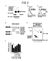
specific, we " restored "expression of ERp44 in siRNA-transfected cells. HeLa cells
were transfected with either sicontrol - 3U or siERp44-3U and 24 hours later the
cells were transfected with the expression vector for RFP-RDEL or RFP-ERp44.
As siERp44-3U targets the 3'-UTR sequence that is absent in the expression vector
for RFP-ERp44, expression of endogenous ERp44, but not that of RFP-ERp44, was
suppressed by siERp44-3U (Figure 5A, lane 4). Following these treatments, the cells
were stimulated with 3µM ATP. In siERp44-3U-pretreated cells (i.e. the amplitude
of IICR was increased),restoration of RFP-RDEL had little effect on IICR
(Figure 5B), while that of RFP-ERp44 significantly suppressed it (Figure 5C).
Altogether, it was concluded that ERp44 specifically inhibits IP3R1.
Furthermore, this system allowed us to assess which domain of ERp44 is
important for inhibition of IICR First, minimum 1L3V-interacting region of ERp44
was determined in vitro (Figure 5D and 5E). Full-length (without the N-terminal
signal sequence) or subfragments of ERp44 were fused to MBP and their interaction
with GST-1L3V was tested in the presence of DTT at neutral pH. Wild-type MBP-ERp44
and MBP-ERp44-a (C-terminal half) strongly interacted with GST-1L3V
(Figure 5E, lanes 2 and 3,respectively), indicating that THD is dispensable for the
interaction. MBP-ERp44-b,which contains highly conserved glutamate-rich region
but excludes histidine-rich region, also bound to GST-1L3V, albeit somewhat
weakly (Figure 5E, lane 4), whereas other mutants tested did not (Figure 5E, lanes 5-7,
respectively). These results indicated that amino acid residue 236-345 of ERp44
was necessary and sufficient for binding to 1L3V, and hereafter referred as IP3R1
binding domain (IPBD). The primary sequence of IPBD has no similarity to any
sequence in the database, but is well conserved among species (Figure 3).
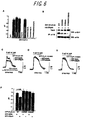
Then, HeLa cells, pretreated with siERp44-3U, were transfected with
various RFP-tagged ERp44 mutants and their ability to inhibit IICR was examined
(Fig. 5F). RFP-ERp44-f, which retains THD but not IPBD, had no effect.
Interestingly, neither RFP-ERp44-a nor RFP-ERp44-b-RDEL, both of which retain
IPBD, inhibited IICR at all (Figure 5F, lanes 5 and 6), indicating that IPBD alone is
not sufficient for inhibition of IP3R1. Moreover, RFP-ERp44-C58S, in which the
cysteine residue critical for formation of mixed disulfide bonds with other proteins is
mutated (Anelli et al., 2003),only slightly inhibited Ca2+ response (77.1±4.5%,
n=82, not statistically significant)compared with RFP-RDEL-transfected cells
(Figure 5F, lane 3). These results indicated that both THD (including Cys58) and
IPBD are required to inhibit IP3R1 activity.
Cysteine residues in 1L3V play crucial roles for inhibition of IP3R1
channel activity by ERp44
To examine the significance of cysteine residue(s) of 1L3V, we constructed
expression vectors for GFP-IP3R1 in which the cysteine residue in L3V region was
substituted for serine, either singly or in combinations. Each vector was transfected
to IP3R-deficient DT40-TKO cells and its IICR activity was checked. As a result, it
was revealed that substitution of C2527, which is adjacent to the channel pore region,
impairs the channel activity of IP3R1 (Figure 6A), and thus this mutant was not used
further. Mutations in C2496 and C2504 were without effect on the channel activity
(Figure 6A). Interactions between these cysteine mutants and ERp44 were
significantly decreased (Figure 6B),consistent with the results using recombinant
proteins (Figure 2D). These mutants were co-transfected to DT40-TKO cells either
with RFP-RDEL or with RFP-ERp44, and single-cell Ca2+-imaging experiment was
performed. IICR via GFP-IP3R1 (i.e. wild-type) was suppressed by co-expression
of RFP-ERp44, but not with RFP-RDEL (Figure 6C and 6F). Surprisingly, this
inhibition by ERp44 was almost completely abolished by substitution of C2496 or
C2504 of GFP-IP3R1 (Figure 6D and 6E, respectively, and Figure 6F). This could
be explained by reduced affinity between these mutants and ERp44, or alternatively,
it is possible that both cysteine residues are required for inhibition by ERp44, which
is supported by the fact that both IPBD and Cys58 of ERp44 were required for
inhibition of IP3R1 (Figure 5F). In any case, these results clearly demonstrated that
the presence of free thiol groups in 1L3V is important for inhibition by ERp44.
ERp44 inactivates the channel activity of IP3R1 in the lipid bilayer system.
To unambiguously show that ERp44 inhibits IP3R1, we carried out single-channel
currents recoding in planar lipid bilayer fused with mouse cerebellar microsome, in
which more than 99% of IP3R is IP3R1. We first confirmed interaction of MBP-ERp44
with GST-1L3V in the lumenal-side solution containing 3 mMDTT (Figure
7A). IP3R1 activity was recorded with Ba2+. as a charge carrier in the presence of
3 mM DTT in the trans (lumenal) compartment. Under this condition, IP3R1 was
maximally activated by addition of 6.4 µMIP3 to the cis (cytosolic) compartment
(data not shown). Addition of MBP (negative control) to the lumenal compartment
had no effect on IP3R1 activity (data not shown). In contrast, addition of MBP-ERp44
resulted in inhibition of the channel activity in a dose-dependent manner
(Figure 7B). Single-channel open probability was reduced with IC50 of 1.0 µM
MBP-ERp44 and with a Hill coefficient of 2.3 (Figure 7C), suggesting that binding
of at least two ERp44 molecules to a heterotetramer of IP3R1 is required for
inactivation. Very interestingly, this inactivation of was not observed in the
absence of DTT (data not shown), further supporting the idea that the free thiol
group(s) in 1L3V is involved by inhibition by ERp44. The mean open time, current
amplitude, and conductance of IP3R1 were unaffected by addition of MBP-ERp44
(data not shown), revealing that the function of ERp44 is to keep the closed state of
IP3R1 , rather than affecting the characters of its open state.
Discussion
In this study we found that ERp44, an ER lumenal protein, directly interacts with the L3V domain of IP3R1 and thereby inhibits its channel activity. This functional interaction is dependent on pH, the redox state, and [Ca2+]ER. This is the first example of a negative regulator of IP3Rs in the ER lumen, embodying the idea that IP3Rs are specifically and temporally controlled by the intralumenal environment.
In this study we found that ERp44, an ER lumenal protein, directly interacts with the L3V domain of IP3R1 and thereby inhibits its channel activity. This functional interaction is dependent on pH, the redox state, and [Ca2+]ER. This is the first example of a negative regulator of IP3Rs in the ER lumen, embodying the idea that IP3Rs are specifically and temporally controlled by the intralumenal environment.
Among pH, the redox state, [Ca2+]ER, which of the factors is most
dominant (or important) in regulation of ERp44/IP3R1 interaction? This question
relates to how intralumenal environment of the ER is regulated and/or subject to
change by cytosolic or extracellular conditions. A number of works have been
conducted to elucidate the intralumenal environment of the ER, but the conclusions
drawn from them are not necessarily consistent. Regarding pH, however, there
seems to be a consensus that it is around neutral and stable, even during or after Ca2+
release (Foyouzi-Youssefi et al., 2000; Kim et al., 1998). From these observations,
we do not favor the idea that pH plays major roles in regulation of ERp44/IP3R1
interaction in vivo. It is, however, still possible that pH changes drastically (perhaps
only for a very short period of time) in the neighbor of the ER membrane, or the
change of pH plays a role in the sarcoendoplasrmic reticulum or secretory granules
within which pH could be acidic (Kamp et al., 1998;Quesada et al., 2003).
It is widely accepted that the ER lumen is more oxidizing than cytosol.
In the ER lumen, the ratio of reduced GSH to oxidized one has been estimated to be 3:1 to 1:1 (Bass et al.,2003; Hwang et al., 1992). Cysteine residues of proteins that are destined for the extracellular milieu are mostly oxidized to form disulfide bonds by virtue of ER oxidoreductases. Molecular mechanisms by which the redox status of extracellular proteins and ER oxidoreductases are controlled are becoming clear (Sevier and Kaiser, 2002), but it remains unexplored how the redox status of other ER resident proteins (including IP3Rs) are regulated (if any). Our results indicated that interaction between ERp44 and 1L3V becomes weaker as the number of free thiol group on 1L3V decreases (Figures 2D, 2F, and 6B). Furthermore, the inhibitory effect of ERp44 on IP3R1 is rather partial in systems where contribution of other IP3R subtypes are excluded (Figures 3I,6C, and 7C), implying that not all of IP3R1 is subject to inhibition by ERp44. Altogether, we propose that some of the cysteine residues in 1L3V are in their free (reduced) form (i.e. subject to ERp44 inhibition), and others form disulfide bonds, either intramolecularly or intermolecularly (i.e. no ERp44 interaction or inhibition). It is likely that the balance between these statuses is tightly regulated (by virtue of the ER oxidoreductases, for example). It is well known that sulphydryl-oxidizing reagents (such as thimerosal) reversibly sensitizes IICR in many cell types including HeLa cells (Bootman et al., 1992; Karhapaa et al., 1996; Montero et al., 2001). Furthermore, IP3R1 ,but not IP3R3, is sensitized by thimerosal treatment (Missiaen et al.,1998). These phenomena may be the reflection of the redox-dependent inhibitory effect of ERp44 on IP3R1.
In the ER lumen, the ratio of reduced GSH to oxidized one has been estimated to be 3:1 to 1:1 (Bass et al.,2003; Hwang et al., 1992). Cysteine residues of proteins that are destined for the extracellular milieu are mostly oxidized to form disulfide bonds by virtue of ER oxidoreductases. Molecular mechanisms by which the redox status of extracellular proteins and ER oxidoreductases are controlled are becoming clear (Sevier and Kaiser, 2002), but it remains unexplored how the redox status of other ER resident proteins (including IP3Rs) are regulated (if any). Our results indicated that interaction between ERp44 and 1L3V becomes weaker as the number of free thiol group on 1L3V decreases (Figures 2D, 2F, and 6B). Furthermore, the inhibitory effect of ERp44 on IP3R1 is rather partial in systems where contribution of other IP3R subtypes are excluded (Figures 3I,6C, and 7C), implying that not all of IP3R1 is subject to inhibition by ERp44. Altogether, we propose that some of the cysteine residues in 1L3V are in their free (reduced) form (i.e. subject to ERp44 inhibition), and others form disulfide bonds, either intramolecularly or intermolecularly (i.e. no ERp44 interaction or inhibition). It is likely that the balance between these statuses is tightly regulated (by virtue of the ER oxidoreductases, for example). It is well known that sulphydryl-oxidizing reagents (such as thimerosal) reversibly sensitizes IICR in many cell types including HeLa cells (Bootman et al., 1992; Karhapaa et al., 1996; Montero et al., 2001). Furthermore, IP3R1 ,but not IP3R3, is sensitized by thimerosal treatment (Missiaen et al.,1998). These phenomena may be the reflection of the redox-dependent inhibitory effect of ERp44 on IP3R1.
Previous studies showed that IICR was diminished at low [Ca2+]ER,
which could not be fully explained by the decreased gradient of Ca2+ concentration
across the ER membrane (e.g. (Caroppo et al., 2003)). Our biochemical result (Figure
2E) indicated that the interaction between ERp44 and IP3R1 is weakened when
[Ca2+]ER is more than 100 µM (i.e. at resting level). It is therefore tempting to
speculate that the observed inhibition of IICR at under low [Ca2+]ER conditions is
due to enhanced binding by ERp44 to IP3R1. Such a mechanism may be effective
for preventing [Ca2+]ER from becoming too low for other intralumenal proteins
(such as Ca2+-dependent enzymes and chaperones) to exert their functions.
What is the physiological significance of inhibition of IP3R1 by ERp44?
One possibility is to translate the ER lumenal environment into [Ca2+]c. The ER is
the crosspoint of many signaling pathways (Berridge, 2002; Orrenius et al., 2003).
Therefore, inhibition of IP3R1 by ERp44 may be a part of the feedback system by
which information converged at the ER was conveyed to the cytosol/nucleus.
Another possibility is to prevent [Ca2+]ER from becoming too low for other ER
lumenal proteins (such as Ca2+-dependent enzymes and chaperones) to exert their
functions. ERp44 was first identified as a protein that forms mixed disulfide bonds
with Erolα, an ER oxidoreductase (Anelli et al., 2002), and it was shown to be
involved in its ER retention (Anelli et al., 2003). The detailed function of Erol family
proteins are not full understood, but it plays a pivotal role in oxidative protein folding
in the ER lumen (Tu and Weissman, 2002).
ERp44 overexpression shifts the redox status of Erolα to oxidized form
and thus is thought to promote oxidative folding (Anelli et al., 2002). Therefore,
ERp44 may play dual roles in protein folding: inhibiting Ca2+ release (which
reinforces Ca2+-dependent chaperones) by inactivating IP3R1 and supporting
disulfide bond formation by boosting Ero1α/oxidoreductase system. This idea is
further supported by the fact that ERp44 is induced during the unfolded protein
responses (Anelli et al., 2002).
It was also previously shown that ERp44 forms mixed disulfide bonds
with many proteins, including integral membrane proteins (Anelli et al., 2003;
Anelli et al., 2002). At present it is unknown whether ERp44 forms mixed
disulfide bonds with IP3R1. The pull-down assays using recombinant proteins
revealed that IPBD is sufficient for interaction between these proteins. On the other
hand, interaction between ERp44-C58S and IP3R1 in transfected cells was
diminished (Supp), and ERp44-C58S is less competent in inhibiting IP3R1
(Figure 6F). These results suggest that C58 plays some role in its interaction with
IP3R1 in vivo. However, another interpretation of these ,results is that because
ERp44 (but not ERp44-C58S, (Anelli et al., 2003)) can form a homodimer with
disulfide bonds in vivo, interaction of one ERp44 molecule may bring another
molecule that co-precipitates and inhibits IP3R1. In any case, further studies will be
required to understand the exact mechanism of ERp44's action. It is unlikely that
IP3R1 is retained in the ER by virtue of ERp44 since the L3V domain of IP3R1 is
dispensable for the ER retention (Galvan et al., 1999) and IP3R1 is still localized to
the ER in cells in which ERp44 was knocked down (data not shown).
It is also mentioning that while most of the biochemical characteristics
and functions of Erol are conserved between yeast and mammals, yeast does not
have an ERp44 orthologue (Anelli et al., 2003). Yeast does not have an IP3R, either,
and C. elegans has both ERp44 and IP3R, suggesting that these proteins may have
emerged at similar time points in the course of evolution.
Very recently, it was reported that the activity of SERCA 2b is modulated
by CRT and ERp57, an ER lumenal oxidoreductase, in [Ca2+]ER- and redox - state
dependent manner (Li and Camacho, 2004). The pump activity of SERCA 2b is
higher when thiol groups in its lumenal loop are reduced. Thus, when the ER
lumenal environment shifts to the reducing condition, Ca2+ uptake by SERCA 2b
would be enhanced (by ERp57) and Ca2+ - release via IP3R1 would be inhibited (by
ERp44). Moreover, reduced ER lumenal environment is not favorable for protein
folding. Thus, it is reasonable to increase [Ca 2+]ER by inhibiting IP3R1 and
activating SERCA 2b since function of many chaperones require relatively high
[Ca2+]ER.
Finally, although only IP3R1, not IP3R2 or IP3R3, is regulated by ERp44,
it does not necessarily mean that IP3R2 and IP3R3 are insensitive to the ER lumenal
environment. We were unable to find any binding protein to the lumenal domain to
IP3R2 or IP3R3 from the brain (where IP3R1 is exclusively expressed), but it is likely
that these subtypes have different binding proteins that are absent in the brain.
The fact that cysteine residues in the L3V domain are conserved (Figure1B) supports
this hypothesis.
Full-length ERp44 cDNA was obtained by reverse transcriptase-polymerase
chain reaction from P14 mouse cerebellum. The expression vectors for
epitope-tagged or fluorescent protein-tagged ERp44, IP3Rs and their mutants were
constructed by utilizing polymerase chain reaction. Detailed methods, including the
sequence of primers used, will be supplied on request. All constructions were
verified by sequencing.
Rabbit polyclonal antibody against ERp44 was raised against purified
His6-ERp44 and affinity-purified with MBP-ERp44 coupled to CNBr-activated
Sepharose 4B (Amersham). Other antibodies used were anti-IP3R1 (18A10, (Maeda
et al., 1988)),Anti-IP3R3 (KM1082, (Sugiyama et al., 1994)), anti-HA (12CA5)
gifted from Dr.Tadashi Yamamoto, anti-HA-Peroxidase (3F10, Roche), anti-CRT
(Affhity BioReagents), anti-MBP (New England Biolabs), anti-human IgG (Vector
Laboratories),anti-GST (Amersham), anti-GFP, and anti-actin (Santa Cruz).
The hybridoma producing anti-IgM (M-4 clone) was kindly provided by Dr.
Tomohiro Kurosaki (Kansai MedicalUniversity).
COS-7, HeLa, and 293T cells were cultured in Dulbecco's modified
Eagle medium supplemented 10% heat-inactivated fetal bovine serum. DT40 cells
were cultured in RPMI1640 medium supplemented 10% heat-inactivated fetal bovine
serum, 1% chicken serum, 100 U/mL penicillin and streptomycin, 2 mM glutamine
and 50 µM 2-mercaptoethanol. HeLa and COS-7 cells were transfected with
expression vectores or siRNAs using TransIT (Mirus) or Lipofectamine2000
(Invitrogen), respectively, as described previously (Hattori et al., 2004). DT40 cells
were transfected with expression vectors using Nucleofector (Amaxa).
1L3V-Fc or control Fc was expressed in 293T cells. The medium was
collected and incubated with Protein-G Sepharose (Amersham) for 2 hr at 4°C and
then the beads were washed with 10 mM Tris-HCl (pH 8.0), 150 mM NaCl.
Approximately 0.2 mg of 1L3V-Fc or control Fc was coupled to the beads, which
was laid onto a column. P14 mice cerebella were washed with ice - cold PBS and
homogenized in a buffer containing 0.32 M sucrose, 10 mM Tris-HCl (pH 7.5),
2 mM EDTA, and 1 mM DTT. The homogenates were centrifuged for 1hr at
100,000 x g at 2°C. The pellet was solubilized in buffer containing 30 mM sodium
acetate, 150 mM NaCl, 4 mM CaCl2, 1% TritonX-100 for 1 hr at 4°C and
centrifuged at 10,000 x g for 15 min. The supernatant was applied to the affinity
columns. The columns were washed with the same buffer and then neutral Ca2+
buffer (10 mM Tris-HCl (pH 7.5), 150 mM NaCl, 4 mM CaCl2, and 1 % TritonX-100),
and then neutral cherators buffer (10 mM Tris-HCl (pH 7.5), 150 mM NaCl, 4
mM EDTA, 4 mM EGTA, and 1%TritonX-100). Fractions were collected and
resolved in SDS-PAGE. After silver staining of the gels, the bands were excised
and analyzed as described previously (Ichimura et al., 2002).
Proteins were resolved by SDS-PAGE and transferred to a polyvinylidene
difluoride membrane. Membranes were blocked with 5% skim milk in
PBS including 0.2% Tween-20 (PBST) for 30 min and probed with the first antibody
for 3 hr at room temperature (RT). After washing with PBST, membranes were
incubated with a suitable secondary antibody for 1hr, and signals were detected using
ECL Plus kit (Amersham).
Cells were fixed with 49% formaldehyde for 10 min at RT and
permeabilized with 0.2 %Triton X-100 in PBS for 10 min at RT. After blocking in
PBS including 5% goat serum for 1hr at RT, the cells were incubated with first
antibodies for 3 hr at RT. Followed by incubation with suitable Alexa Fluor 488 or
594 secondary antibodies (Molecular probes) for 1 hr, cover glasses were mounted
using Vectashield mounting medium (Vector) and analyzed using a confocal
fluorescence microscope (FV-300, Olympus).
Cells were washed with PBS and then treated with 20 mM DSP (Pierce)
in PBS for 30 min at RT. After washing with PBS, cells were solubilized in TNE
buffer (150 mM Tris-Hcl (pH 7.5), 500 mM NaCl, 1 mM EDTA, 1% TritonX-100,
0.1% SDS). The lysates were incubated with the indicated antibodies and Protein-G
Sepharose for 2hr at 4C. The beads were washed five times with TNE buffer and
proteins were eluted by boiling in SDS-PAGE sampling buffer.
Recombinant proteins were expressed in BL21 bacterial strains and
purified using GSH-Sepharose (Amersham) or Amylose Resin (New England
Biolabs). ERp44 was removed from GST-ERp44 by digestion with Thrombin
Protease (Amersham Biosciences). For Figure 2B, GST or GST fusion proteins
were incubated with MBP, or MBP fusion proteins for 1 hr at 4C in acidic solution
(30 mM NaOAc (pH 5.2), 150 mM NaCl, 4mM CaCl2, 0. 1 % Triton X-100). For
Figure 2C and 2, proteins were incubated in neutral solution (20 mM Tris-HCl (pH
7.5), 150 mM NaCl, 5 mM EGTA, 0.1% Triton X-100)with or without 3 mM DTT
For Figure 2E, GST-1L3V and MBP-ERp44 were incubated in pH 7.5 buffer or pH
7.5 buffer, in which EGTA was replaced to indicated concentration of CaCl2.
Experiments in Figure 2H and 5E were carried out in neutral solution containing
3 mM DTT. Each mixture was then incubated for additional 2 hr with GSH-Sepharose.
The beads were washed five times and subjected to western blot
analysis.
SiRNA duplexes were purchased from Dharmacon. Target sequences
were:


After 24-48 hours following transfection, HeLa, COS-7, DT40 - KMN60,
DT40-1KO,and DT40 - TKO cells were incubated for 30 min with 5 µM fura-2
acetoxymethyl ester (Dojindo). The fura-2-loaded cells were placed on the stage of
an inverted fluorescence microscope (IX-70; Olympus) and perfused with balanced
salt solution (BSS). Image capture and processing were perfomed with Argus
50/CA system (Hamamatsu photonics) at RT. With alternate illumination at
340 nm and 380 nm excitation, pairs of fluorescence images (F340 and F380
respectively) were obtained through an objectivelens (UApo 20/340 or UplanApo
100 Oil Iris; Olympus) and an emission filter (510-550 nm), captured with a silicon-intensified-target
video camera (C2400-8;Hamamatsu Photonics). Fluorescence
images of GFP and RFP were separately acquired and saved in the computer in the
same optical fields as those for Ca2+ imaging.
Single-channel recordings of IP3R1 in mouse cerebellar microsomes
were performed as previously described (Michikawa et al., 1999) at 20 mV
transmembrane potential using 53 mM Ba2+. The cis chamber contained 108 mM
Tris dissolved in 250 mM HEPES (pH 7.33), 1.11 mM K2-H-HEDTA (N-hydroxymethylethylenediamine.N,N',N'-triaceticacid),
0.12 mM K-Ca2+-HEDTA.
The trans chamber contained 250 mM HEPES (pH7.33), 53 mM Ba (OH)2, 3 mM
DTT IP3R1 were activated by addition of 6.4 mM IP3 and 0.5 mM ATP to the cis
chamber. Purified MBP or MBP-ERp44 was added directly to transside.
The IP3R1 single channel currents were amplified (Axon Instruments),filtered at
1 kHz by a low-pass Bessel filter (model, NF Instruments), sampled at 10kHz, and
analyzed (Michikawa et al., 1999). Single-channel amplitudes were determined by
fitting Gaussian curves to the all-point amplitude histograms of the datafiltered at
1 kHz. For presentation of the current traces, data were filtered at 0.5 kHz.


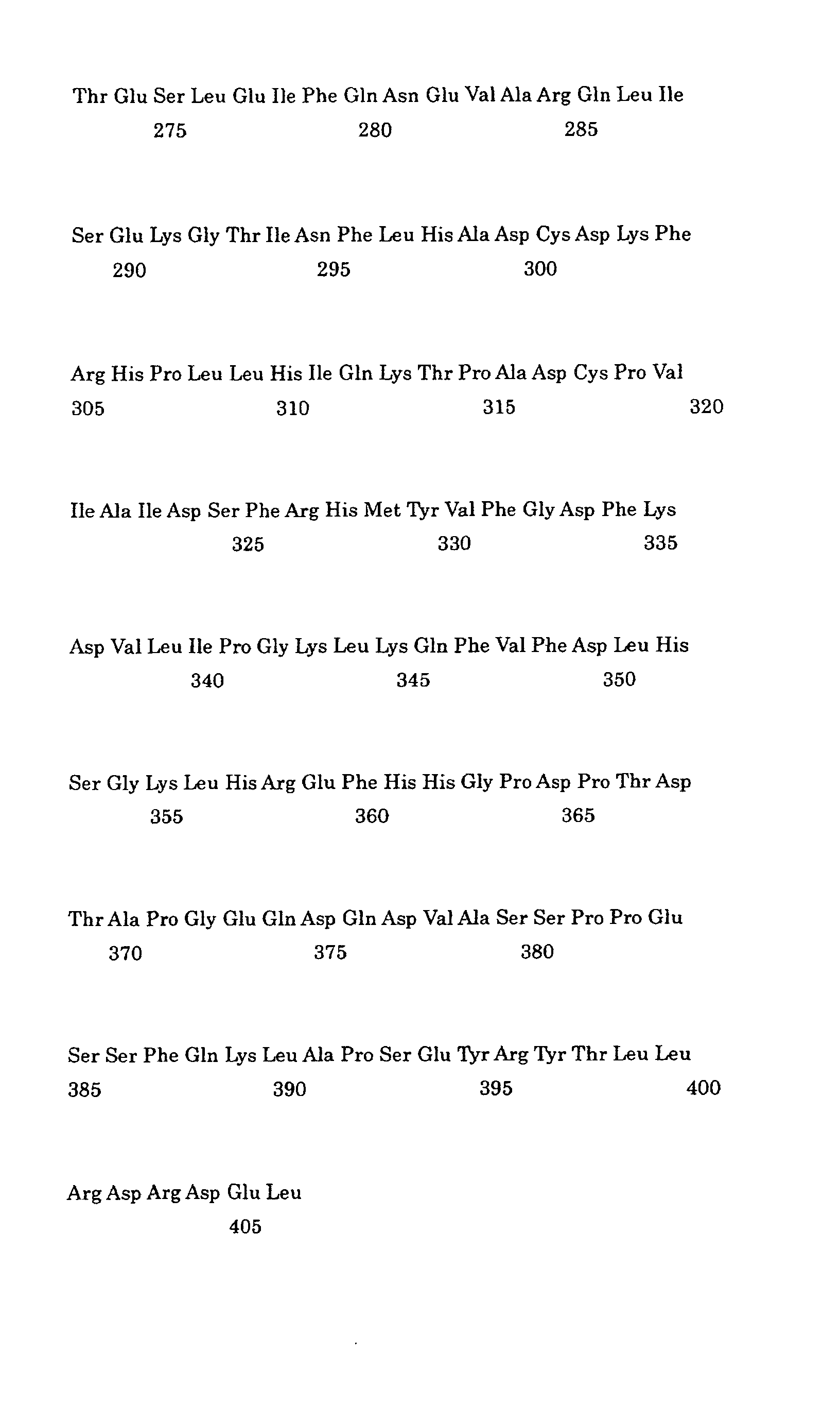
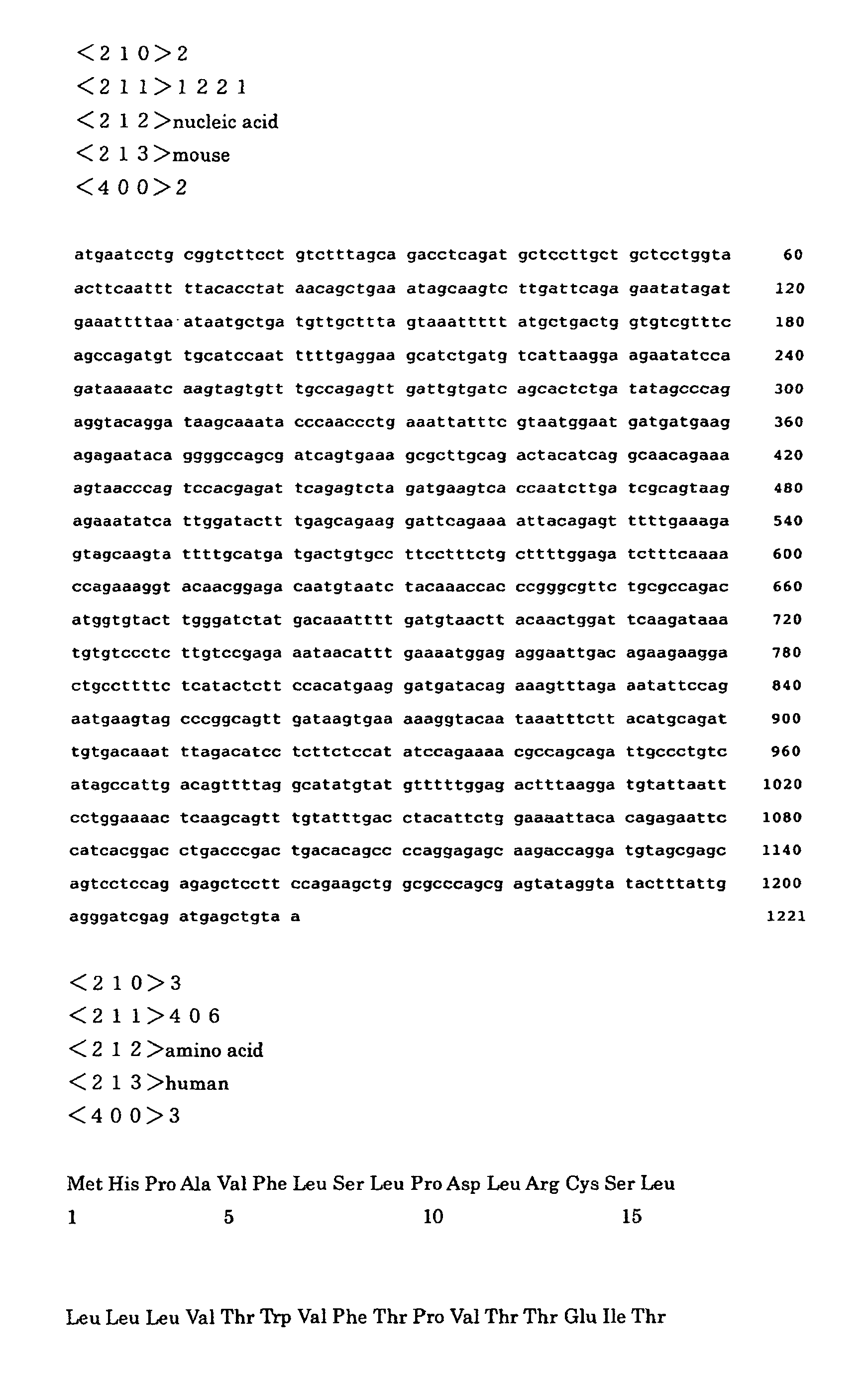
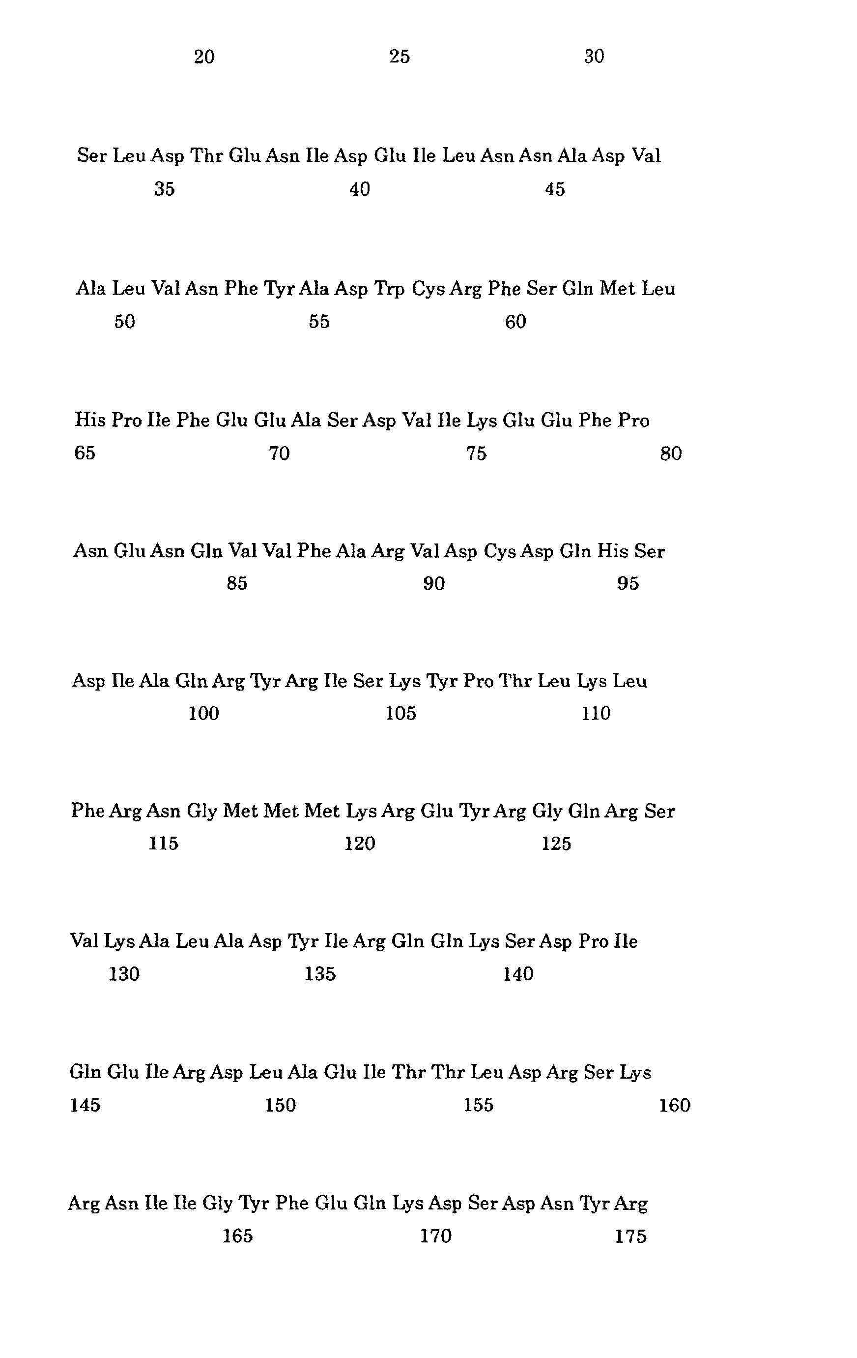
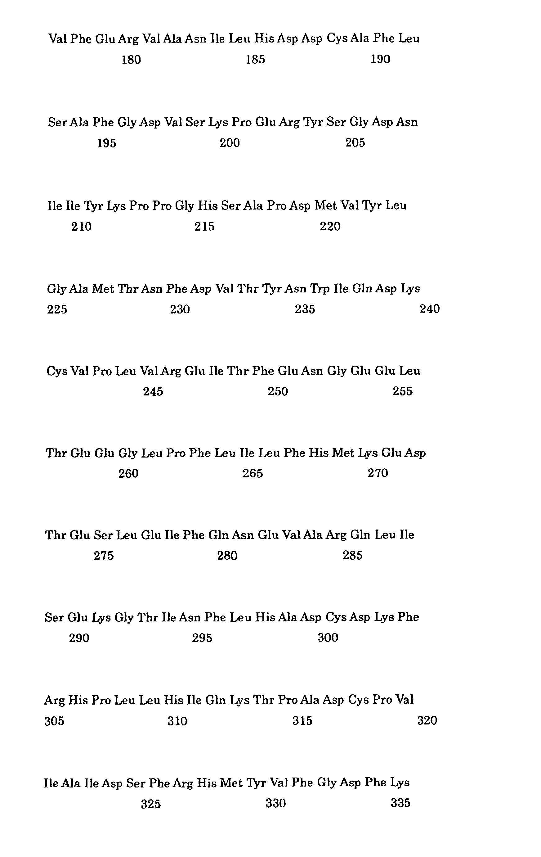


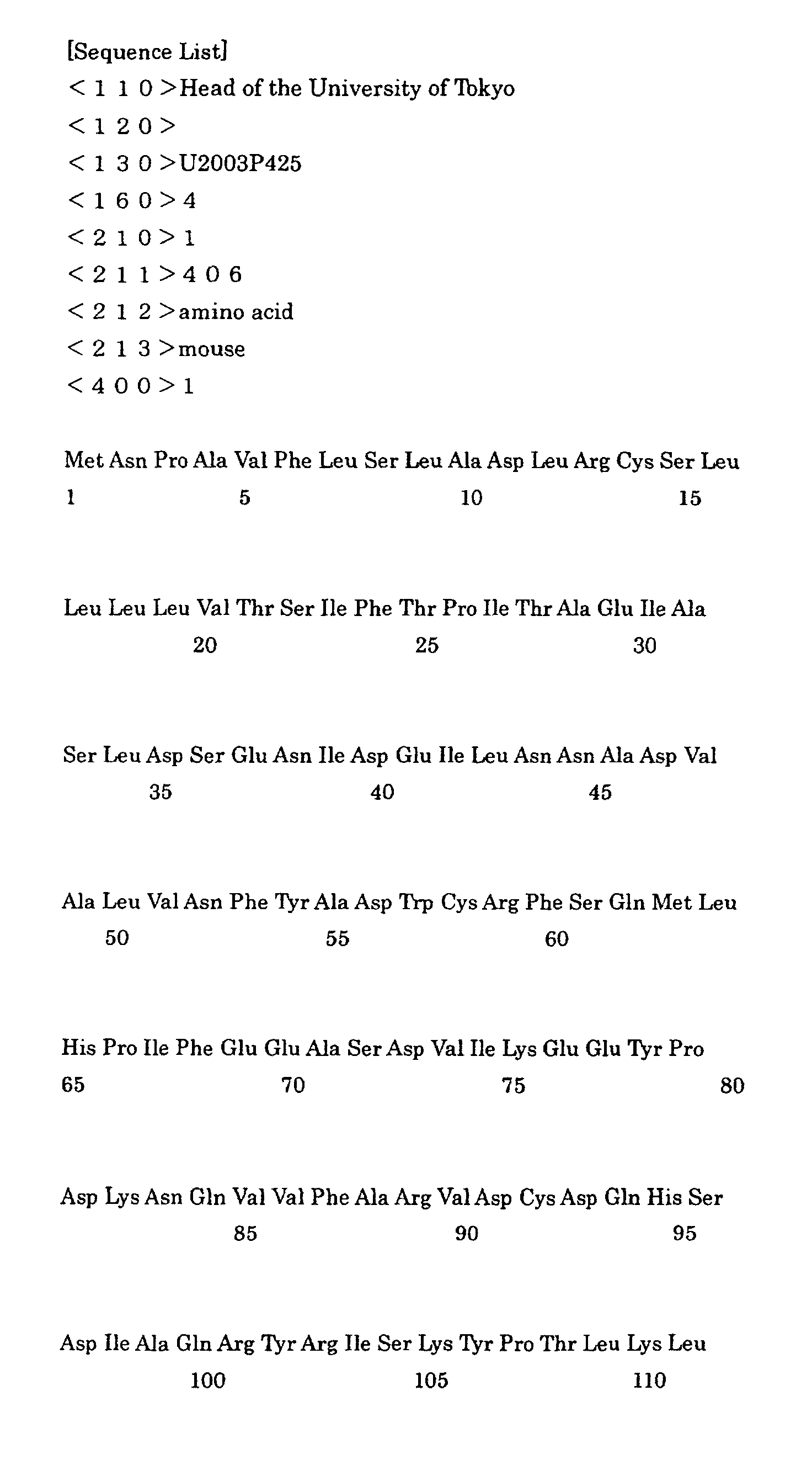



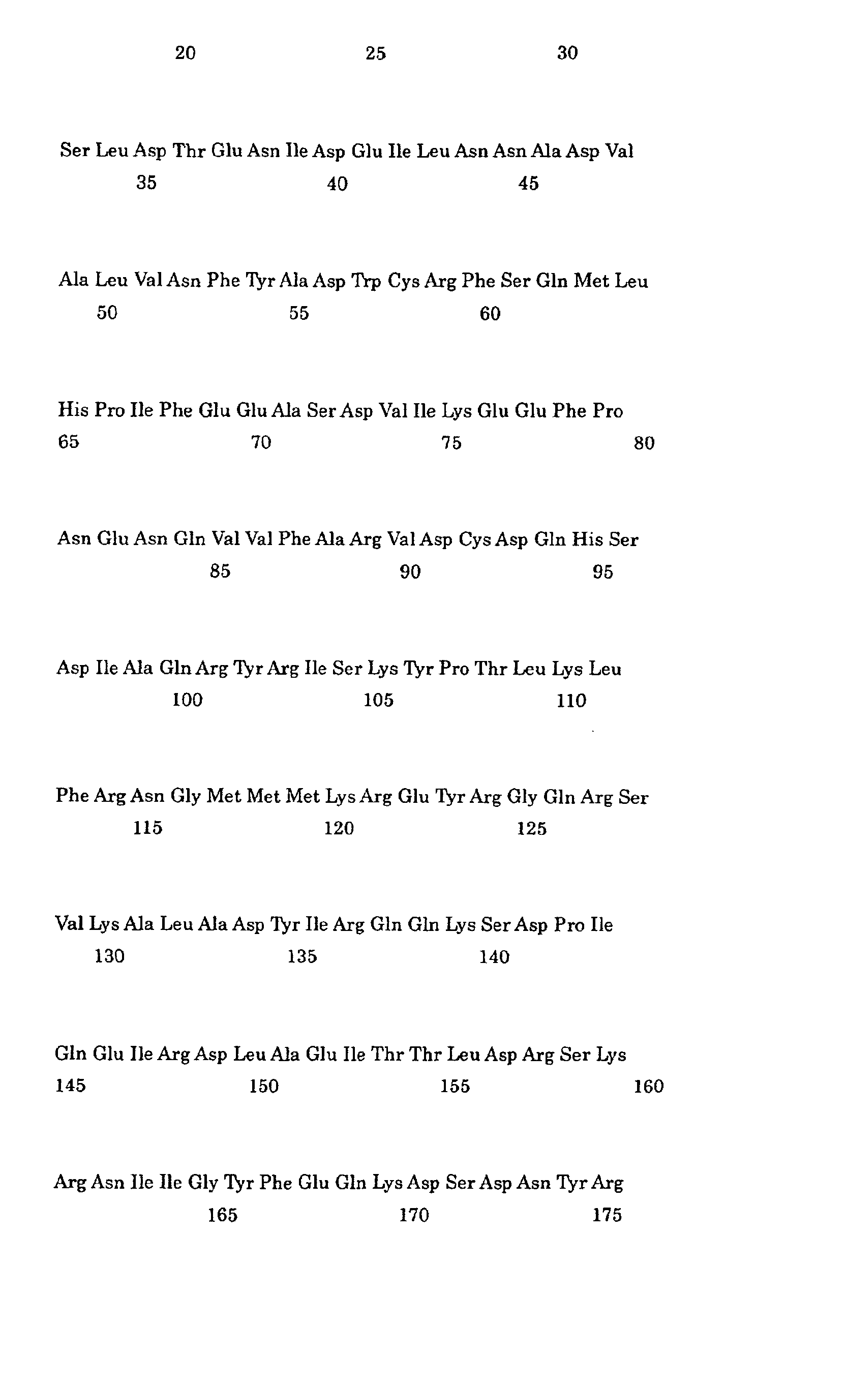

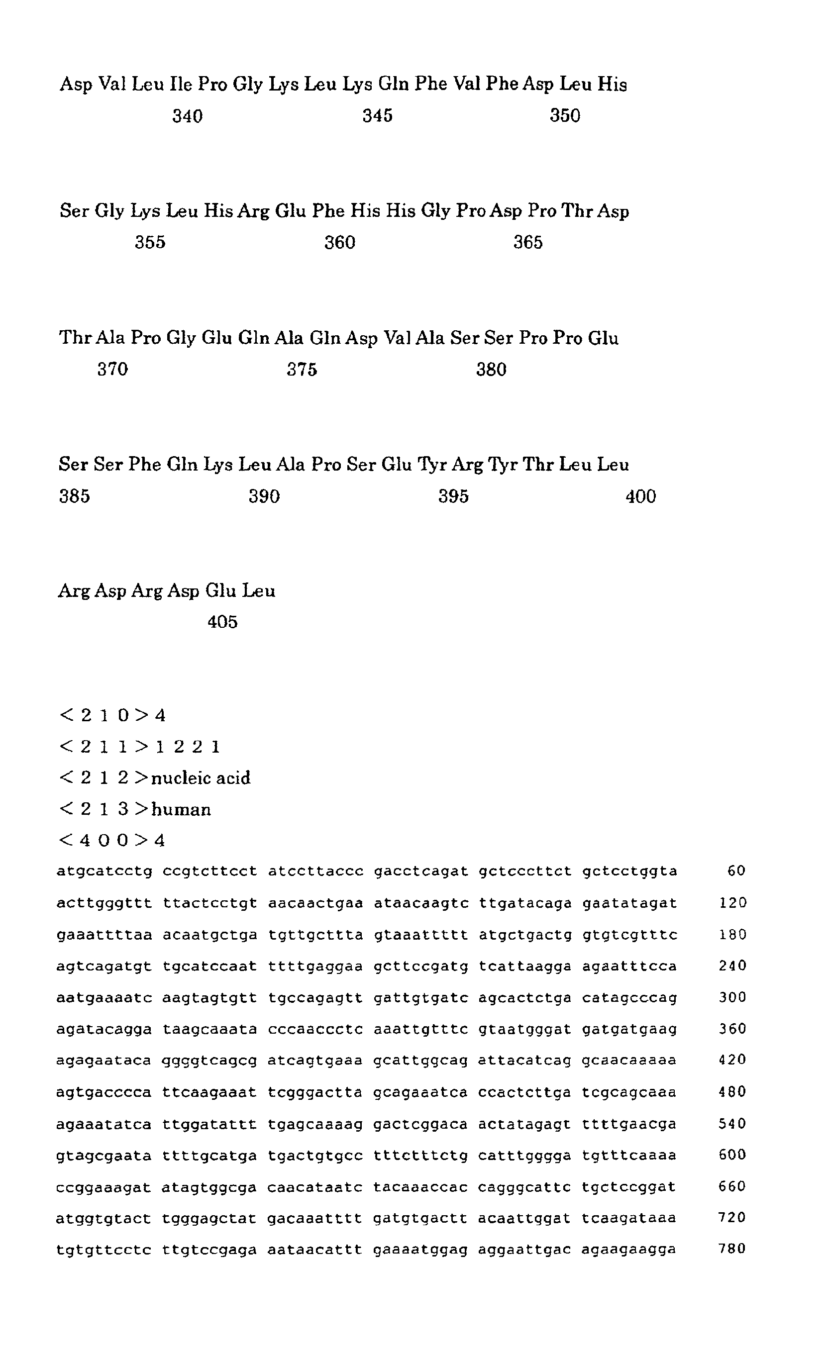

Claims (9)
- A molecular of controlling a calcium comprising the following (a) or (b):(a) an amino acid sequence shown in an amino acid sequence number 1-406 of a sequence No.1 of a sequence table, or(b) an amino acid sequence wherein a part of the amino acid sequence of the amino acid sequence number 1-406 is deleted, substituted or added.
- A recombinant vector comprising the following (a) or (b):(a) a nucleic acid comprising a base sequence shown in a base sequence number 1-1221 of a sequence No.2 of a sequence table, or(b) a nucleic acid wherein a part of the base sequence of the base sequence number 1-1221 is deleted, substituted or added, and the nucleic acid has a homology of 80% for the base sequence.
- A molecular of controlling a calcium comprising the following (a) or (b):(a) an amino acid sequence shown in an amino acid sequence number 1-406 of a sequence No.3 of a sequence table, or(b) an amino acid sequence wherein a part of the amino acid sequence of the amino acid sequence number 1-406 is deleted, substituted or added.
- A recombinant vector comprising the following (a) or (b):(a) a nucleic acid comprising a base sequence shown in a base sequence number 1-1221 of a sequence No.4 of a sequence table, or(b) a nucleic acid wherein a part of the base sequence of the base sequence number 1-1221 is deleted, substituted or added, and the nucleic acid has a homology of 80% for the base sequence.
- An antibody against the polypeptide described in claims 1 or 2.
- A cultured cell characterized in that the cell is transfected by said recombinant vector described in claims 2-4.
- The cultured cell is characterized in that an ability of Ca2+ release is reduced by over-expressing the polypeptide described in claims 1 or 3.
- The cultured cell is characterized in that an ability of Ca2+ release is increased by restraining the express of the polypeptide described in claims 1 or 3.
- The cultured cell is characterized that an ability of Ca2+ release is increased by introducing the antibody described in claim 5 into said cultured cell and inducing an antigen-antibody reaction between the antibody and the polypeptide of claims 1 or 3.
Applications Claiming Priority (2)
| Application Number | Priority Date | Filing Date | Title |
|---|---|---|---|
| JP2004124443 | 2004-04-20 | ||
| JP2004124443A JP2005304360A (en) | 2004-04-20 | 2004-04-20 | Calcium regulatory molecule |
Publications (1)
| Publication Number | Publication Date |
|---|---|
| EP1598363A1 true EP1598363A1 (en) | 2005-11-23 |
Family
ID=34935466
Family Applications (1)
| Application Number | Title | Priority Date | Filing Date |
|---|---|---|---|
| EP05008653A Withdrawn EP1598363A1 (en) | 2004-04-20 | 2005-04-20 | Protein regulating the concentration of calcium in endoplasmic reticulum |
Country Status (3)
| Country | Link |
|---|---|
| US (1) | US20050260657A1 (en) |
| EP (1) | EP1598363A1 (en) |
| JP (1) | JP2005304360A (en) |
Cited By (1)
| Publication number | Priority date | Publication date | Assignee | Title |
|---|---|---|---|---|
| WO2008104606A1 (en) * | 2007-03-01 | 2008-09-04 | Fondazione Centro San Raffaele Del Monte Tabor | Method for antibody production |
-
2004
- 2004-04-20 JP JP2004124443A patent/JP2005304360A/en active Pending
-
2005
- 2005-04-19 US US11/108,841 patent/US20050260657A1/en not_active Abandoned
- 2005-04-20 EP EP05008653A patent/EP1598363A1/en not_active Withdrawn
Non-Patent Citations (7)
| Title |
|---|
| ANELLI TIZIANA ET AL: "ERp44, a novel endoplasmic reticulum folding assistant of the thioredoxin family", EMBO (EUROPEAN MOLECULAR BIOLOGY ORGANIZATION) JOURNAL, vol. 21, no. 4, 15 February 2002 (2002-02-15), pages 835 - 844, XP002345741, ISSN: 0261-4189 * |
| ANELLI TIZIANA ET AL: "Thiol-mediated protein retention in the endoplasmic reticulum: The role of ERp44.", EMBO (EUROPEAN MOLECULAR BIOLOGY ORGANIZATION) JOURNAL, vol. 22, no. 19, 1 October 2003 (2003-10-01), pages 5015 - 5022, XP002345615, ISSN: 0261-4189 * |
| COLLINS F S: "Generation and initial analysis of more than 15,000 full-length human and mouse cDNA sequences", PROCEEDINGS OF THE NATIONAL ACADEMY OF SCIENCES OF USA, NATIONAL ACADEMY OF SCIENCE. WASHINGTON, US, vol. 99, no. 26, 24 December 2002 (2002-12-24), pages 16899 - 16903, XP002298284, ISSN: 0027-8424 * |
| DATABASE EMBL [online] 19 February 2002 (2002-02-19), "Homo sapiens mRNA for ERp44 protein", XP002345749, retrieved from EBI accession no. EM_HUM:HSA344330 Database accession no. AJ344330 * |
| DATABASE UniProt [online] 10 October 2003 (2003-10-10), "Thioredoxin domain containing protein 4 precursor (Endoplasmic reticulum resident protein ERp44).", XP002345747, retrieved from EBI accession no. UNIPROT:BC019558 Database accession no. BC019558 * |
| DATABASE UniProt [online] 10 October 2003 (2003-10-10), "Thioredoxin domain containing protein 4 precursor (Endoplasmic reticulum resident protein ERp44).", XP002345748, retrieved from EBI accession no. UNIPROT:TXND4_HUMAN Database accession no. Q9BS26 * |
| HIGO TAKAYASU ET AL: "Subtype-specific and ER lumenal environment-dependent regulation of inositol 1,4,5-trisphosphate receptor type 1 by ERp44", CELL, vol. 120, no. 1, 14 January 2005 (2005-01-14), pages 85 - 98, XP002345616, ISSN: 0092-8674 * |
Cited By (1)
| Publication number | Priority date | Publication date | Assignee | Title |
|---|---|---|---|---|
| WO2008104606A1 (en) * | 2007-03-01 | 2008-09-04 | Fondazione Centro San Raffaele Del Monte Tabor | Method for antibody production |
Also Published As
| Publication number | Publication date |
|---|---|
| JP2005304360A (en) | 2005-11-04 |
| US20050260657A1 (en) | 2005-11-24 |
Similar Documents
| Publication | Publication Date | Title |
|---|---|---|
| Hsieh-Wilson et al. | Characterization of the neuronal targeting protein spinophilin and its interactions with protein phosphatase-1 | |
| Higo et al. | Subtype-specific and ER lumenal environment-dependent regulation of inositol 1, 4, 5-trisphosphate receptor type 1 by ERp44 | |
| JP6025758B2 (en) | Regulatory factors of NFAT | |
| Pollmann et al. | Human EML4, a novel member of the EMAP family, is essential for microtubule formation | |
| Dobrosotskaya et al. | MAGI-1 interacts with β-catenin and is associated with cell–cell adhesion structures | |
| Krapivinsky et al. | SynGAP-MUPP1-CaMKII synaptic complexes regulate p38 MAP kinase activity and NMDA receptor-dependent synaptic AMPA receptor potentiation | |
| Kato et al. | WICH, a novel verprolin homology domain-containing protein that functions cooperatively with N-WASP in actin-microspike formation | |
| Helton et al. | Allosteric inhibition of Parkinson’s-linked LRRK2 by constrained peptides | |
| Tang et al. | A novel transforming growth factor-β receptor-interacting protein that is also a light chain of the motor protein dynein | |
| Rejon et al. | Activins bind and signal via bone morphogenetic protein receptor type II (BMPR2) in immortalized gonadotrope-like cells | |
| Tang et al. | Cytoplasmic PCNA is located in the actin belt and involved in osteoclast differentiation | |
| Bustos et al. | Synapsin I is expressed in epithelial cells: localization to a unique trans-Golgi compartment | |
| Wang et al. | SNX32 regulates sorting and trafficking of activated EGFR to the lysosomal degradation pathway | |
| Ledee et al. | A specific interaction between muskelin and the cyclin-dependent kinase 5 activator p39 promotes peripheral localization of muskelin | |
| US20140004532A1 (en) | Nucleic Acids Encoding a Mammalian Raptor Polypeptide and Uses Therefor | |
| US20050260657A1 (en) | Molecular entity for controlling a calcium | |
| US7838249B2 (en) | Assays for rab5 activity | |
| Chen et al. | The mental retardation associated protein, srGAP3 negatively regulates VPA-induced neuronal differentiation of Neuro2A cells | |
| Baron et al. | The α‐subunit of the trimeric GTP ase Go2 regulates axonal growth | |
| WO2026056131A1 (en) | Use of trpm7 activator in preparation of therapeutic product for treating alzheimer's disease | |
| Davis | Functional analysis of Miro GTPase domains in the mitochondrial motor adaptor complex | |
| Verpoort et al. | Exploring the role of cell surface-to-ER communication during synapse development | |
| Weissová | Multi-level regulation of microtubules in neurons by microtubule-associated proteins, phosphorylation, and conformational changes | |
| JP4472534B2 (en) | Functional control of intracellular Ca ions | |
| Lo | Functional characterization of the CNPY4 and PEAK3 proteins as regulators of cell signaling |
Legal Events
| Date | Code | Title | Description |
|---|---|---|---|
| PUAI | Public reference made under article 153(3) epc to a published international application that has entered the european phase |
Free format text: ORIGINAL CODE: 0009012 |
|
| 17P | Request for examination filed |
Effective date: 20050420 |
|
| AK | Designated contracting states |
Kind code of ref document: A1 Designated state(s): AT BE BG CH CY CZ DE DK EE ES FI FR GB GR HU IE IS IT LI LT LU MC NL PL PT RO SE SI SK TR |
|
| AX | Request for extension of the european patent |
Extension state: AL BA HR LV MK YU |
|
| AKX | Designation fees paid |
Designated state(s): DE FR GB |
|
| STAA | Information on the status of an ep patent application or granted ep patent |
Free format text: STATUS: THE APPLICATION IS DEEMED TO BE WITHDRAWN |
|
| 18D | Application deemed to be withdrawn |
Effective date: 20060913 |