EP1598082A1 - Contrast medium for mri - Google Patents
Contrast medium for mri Download PDFInfo
- Publication number
- EP1598082A1 EP1598082A1 EP04707636A EP04707636A EP1598082A1 EP 1598082 A1 EP1598082 A1 EP 1598082A1 EP 04707636 A EP04707636 A EP 04707636A EP 04707636 A EP04707636 A EP 04707636A EP 1598082 A1 EP1598082 A1 EP 1598082A1
- Authority
- EP
- European Patent Office
- Prior art keywords
- mri
- contrast agent
- group
- mri contrast
- imaging
- Prior art date
- Legal status (The legal status is an assumption and is not a legal conclusion. Google has not performed a legal analysis and makes no representation as to the accuracy of the status listed.)
- Withdrawn
Links
Images


Classifications
-
- A—HUMAN NECESSITIES
- A61—MEDICAL OR VETERINARY SCIENCE; HYGIENE
- A61K—PREPARATIONS FOR MEDICAL, DENTAL OR TOILETRY PURPOSES
- A61K49/00—Preparations for testing in vivo
- A61K49/06—Nuclear magnetic resonance [NMR] contrast preparations; Magnetic resonance imaging [MRI] contrast preparations
- A61K49/08—Nuclear magnetic resonance [NMR] contrast preparations; Magnetic resonance imaging [MRI] contrast preparations characterised by the carrier
- A61K49/085—Nuclear magnetic resonance [NMR] contrast preparations; Magnetic resonance imaging [MRI] contrast preparations characterised by the carrier conjugated systems
-
- A—HUMAN NECESSITIES
- A61—MEDICAL OR VETERINARY SCIENCE; HYGIENE
- A61K—PREPARATIONS FOR MEDICAL, DENTAL OR TOILETRY PURPOSES
- A61K49/00—Preparations for testing in vivo
- A61K49/06—Nuclear magnetic resonance [NMR] contrast preparations; Magnetic resonance imaging [MRI] contrast preparations
- A61K49/08—Nuclear magnetic resonance [NMR] contrast preparations; Magnetic resonance imaging [MRI] contrast preparations characterised by the carrier
- A61K49/10—Organic compounds
Definitions
- the present invention belongs to the field of in vivo reagent, and particularly relates to a novel contrast agent for use in MRI.
- an intravascular contrast agent In MRI (Magnetic Resonance Imaging), an intravascular contrast agent is used to ensure high-contrast images.
- Conventional intravascular MRI contrast agents are dedicated to observation of an overall vascular silhouette, i.e., the degree of stricture in the vascular lumen to provide information for the diagnosis and treatment of vascular diseases, and they cannot be applied for directly detecting lesion sites where the vascular endothelia have exfoliated.
- the object of the invention is to provide a novel MRI contrast agent which enables direct detection and imaging of exfoliated vascular endothelial sites and which is easy to prepare and inexpensive to use.
- a contrast agent for use in MRI comprising an imaging unit which contains an unpaired electron-carrying atom and/or molecule and is capable of amplifying or reducing MRI signals, and a detection unit which is bonded to said imaging unit and is capable of selectively recognizing exfoliated vascular endothelial sites and binding thereto.
- the present invention provides an MRI contrast agent which is novel in that, differently from the conventional MRI contrast agents which are dedicated only to imaging the overall vascular silhouette, it is capable of directly detecting exfoliated vascular endothelial sites as they are, as well as imaging the sites.
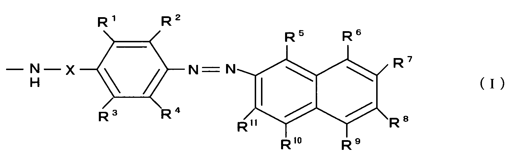
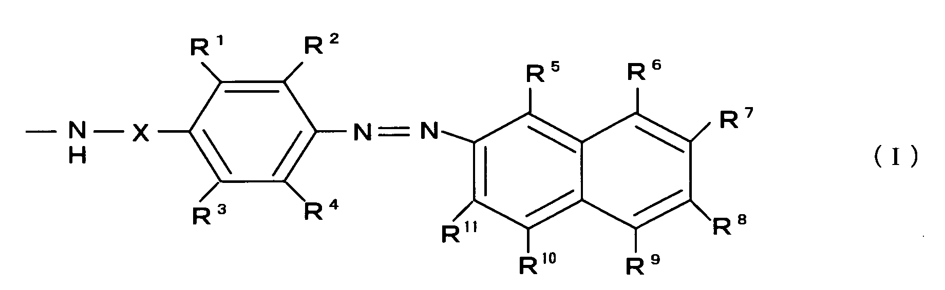
- a preferred but non-limiting example of the detection unit of the MRI contrast agent of the present invention is one having a chemical structure expressed by the following general formula (I):
- R 1 , R 2 , R 3 , R 4 , R 5 , R 6 , R 7 , R 8 , R 9 , R 10 and R 11 may be independently sulfonic acid group, hydroxyl group or amino group
- at least one of R 1 , R 2 , R 3 and R 4 may be independently an alkyl group or alkoxy group having 1 to 3 carbon atoms
- the remaining ones of R 1 , R 2 , R 3 , R 4 , R 5 , R 6 , R 7 , R 8 , R 9 , R 10 and R 11 which do not fall within any of the above-mentioned functional groups are all hydrogen atoms
- X if present, represents a phenyl group in which at least one site may be substituted with an alkyl group or alkoxy group having 1 to 3 carbon atoms.
- the MRI contrast agents according to the present invention are of outstanding utility in the early detection and early treatment of vascular diseases because they are capable of selectively detecting exfoliated vascular endothelia.
- the MRI contrast agents of the present invention can be easily prepared through known synthetic reactions and are low-cost because they are effective when used in a relatively small quantity.
- R 1 - R 11 may be independently sulfonic acid group (-SO 3 H), hydroxyl group (-OH) or amino group (-NH 2 ). More specifically, either all of R 1 - R 11 are hydrogen atoms (i.e. unsubstituted), or that at least one of R 1 - R 11 is sulfonic acid group, hydroxyl group, or amino group.
- R 1 - R 11 are sulfonic acid group, hydroxyl group or amino group, such two or more functional groups may be the same or different. It is generally preferable for realizing increased water-solubility of the MRI contrast agent of the present invention that at least one of R 4 - R 10 is sulfonic acid group (particularly in a case where the imaging unit is of low hydrophilicity).
- R 1 , R 2 , R 3 and R 4 may be independently an alkyl group or alkoxy group having 1 to 3 carbon atoms, between which methyl group is particularly preferable.
- the remaining ones of R 1 - R 11 which do not fall within any of the above-mentioned functional groups are all hydrogen atoms.
- X need not be present, in which case the benzene ring as shown in the formula (I) is directly bonded to -NH.
- X if present, represent a phenyl group in which at least one site may be substituted with an alkyl or alkoxy group having 1 to 3 carbon atoms (preferably methyl group).
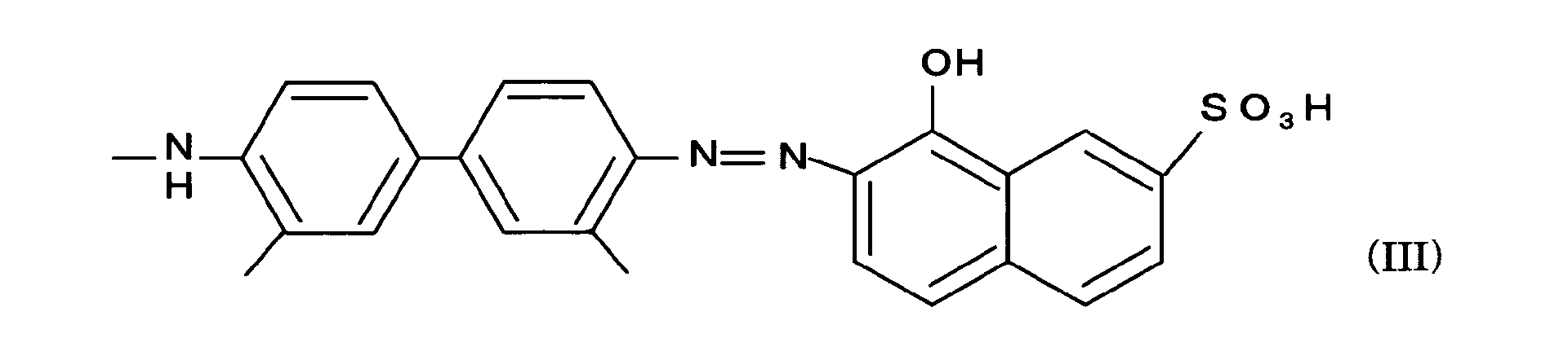
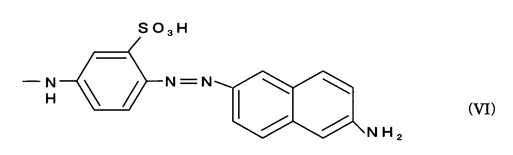
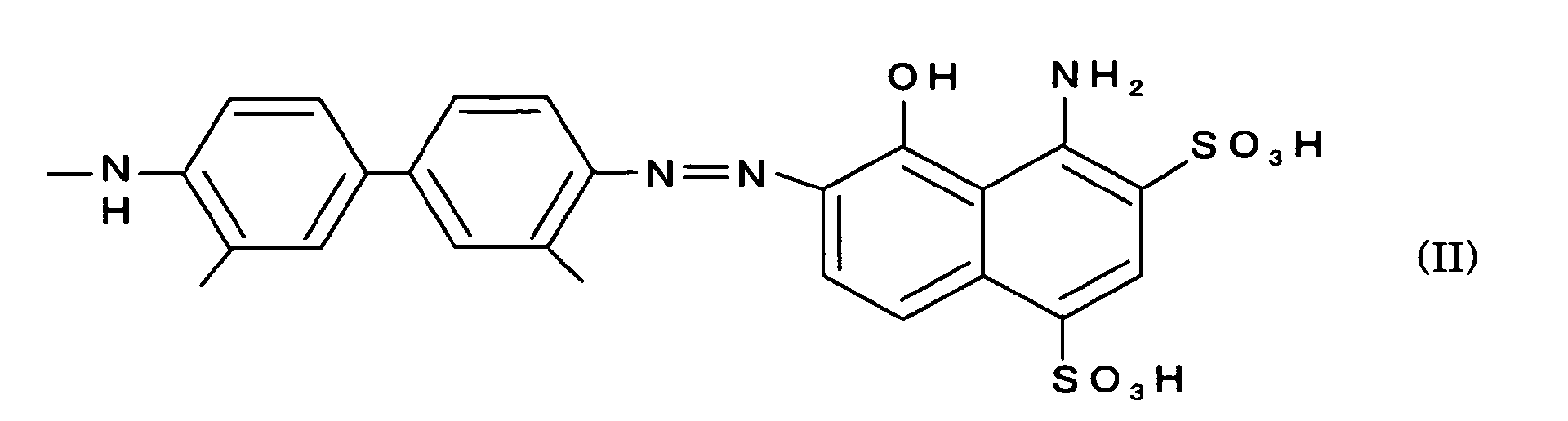
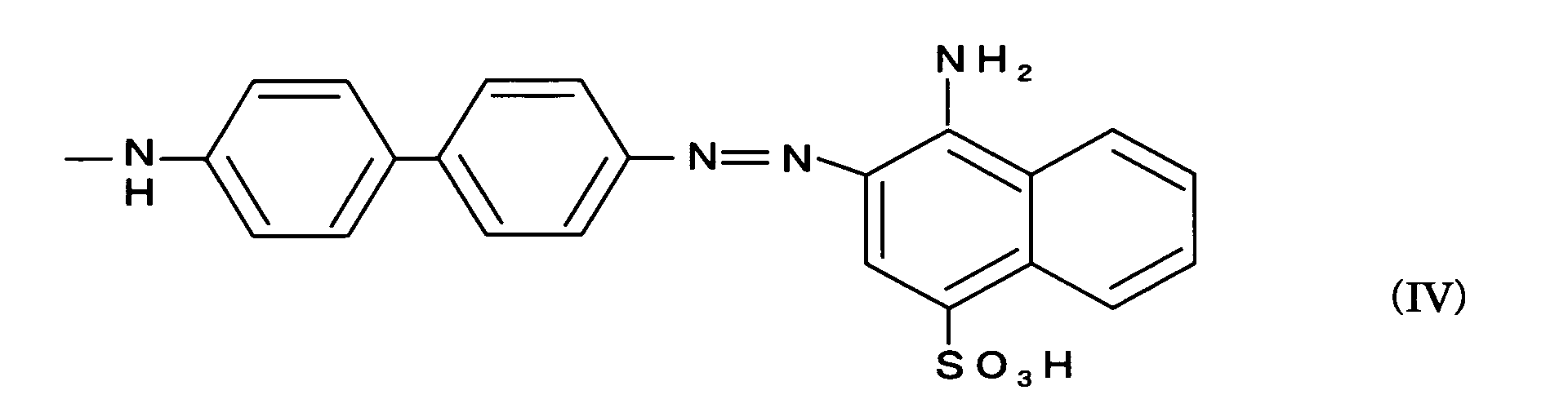
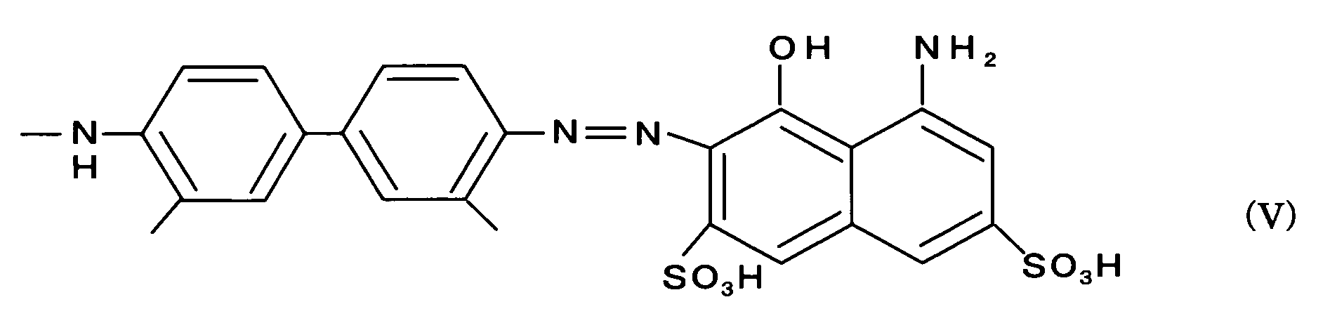
- preferable examples of the chemical formula of the detection unit of the MRI contrast agent of the present invention include, but are not limited to, the following chemical formulae (II), (III), (IV), (V) and (VI):
- the imaging unit of the MRI contrast agent of the present invention there are no particular restrictions with respect to the imaging unit of the MRI contrast agent of the present invention, except that it is required to contain an unpaired electron-carrying atom and/or molecule and be capable of amplifying or reducing MRI signals. More specifically, as the imaging unit of the MRI contrast agent, there can be utilized a variety of known substances capable of enabling imaging in MRI due to a function such as that of shortening the relaxation time of protons (T 1 , T 2 ).
- a chelated complex of a paramagnetic metal ion such as Cd 3+ , Dy 3+ Eu 3+ , Fe 3+ , Mn 2+ ; a nitroxide radical molecule such as a piperidine derivative and a pyrrolidine derivative; or a ferromagnetic material such as magnetite (Fe 3 O 4 ).
- a chelated complex of gadolinium [Cd(III)] in which is used such compound as DTPA, DOTA or EDTA as the ligand.
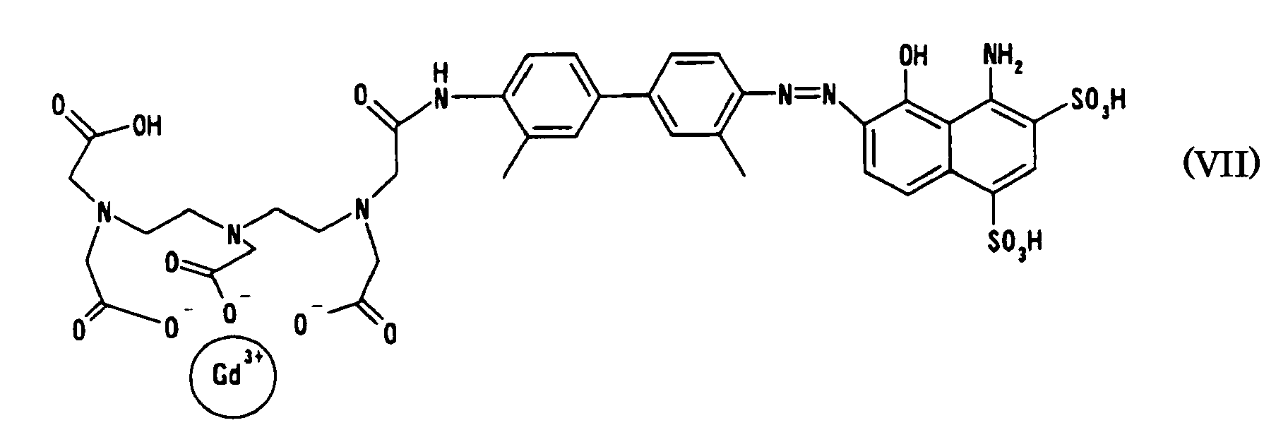
- MRI contrast agent As a preferable MRI contrast agent according to the present invention, there is exemplified, without limitation thereto, one expressed by the following formula (VII) in which the imaging unit is DTPA (diethylenetriaminepentaacetic acid) complex of Gd(III) and the detecting unit has the composition of the aforementioned chemical formula (II).
- DTPA diethylenetriaminepentaacetic acid
- the MRI contrast agent of the present invention can be easily synthesized by appropriately modifying known reactions.
- the desired MRI contrast agent can be obtained by introducing a Boc group into an amino compound (i.e. by t-butoxycarbonylating the amino compound) which corresponds to a portion of the detection unit expressed by the formula (I), allowing the resultant to react with the ligand portion of the metal complex, removing the Boc group, combining the resultant with the remaining portion of the detection unit through the diazo coupling, and subjecting the resultant to a complex formation with the metal (see the working example set out below).
- the MRI contrast agent of the present invention thus obtained is capable of selectively recognizing exfoliated vascular endothelial sites (lesion sites) and binding thereto through the detection unit while enabling said sites to be visualized as MRI signals through the imaging unit.
- the MRI contrast agent of the present invention can therefore provide very reliable information in the treatment and diagnosis of vascular diseases, as a contrast agent specific to exfoliated vascular endothelial sites.
- exfoliated vascular endothelial sites are unstable and arteriosclerosis will proceed more rapidly therein than in the sites without such exfoliation even if the degrees of the strictures are of the same level.
- the detection of exfoliated vascular endothelial sites using the MRI contrast agent of the present invention therefore contributes to early diagnosis and treatment of such circulatory diseases.
- Example 1 For the contrast agent as synthesized (prepared) in Example 1, evaluation of in vitro imaging capability by MRI was carried out with an aortic vascular section extirpated from a pig, as a specimen.
- the aortic vascular section extirpated from a pig was spread in a rectangular shape measuring about 2 cm x 3 cm to prepare a vascular section sample.
- the endothelial surface of the left half of the sample was exfoliated with a scalpel.
- the sample was then immersed in a 10 mM aqueous solution of the contrast agent for ten seconds. Following thorough washing with physiological saline solution, the sample was subjected to the evaluation by MRI as well as by visual observation.
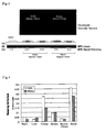
- Fig.2 (A) is a picture taken with a digital camera, which corresponds to the visual observation. It was visually observed that the exfoliated endothelial site (the left half) was colored blue, suggesting accumulation of the contrast agent on the site.
- Fig.2 (B) is an MRI image (by the T1 weighted spin echo method).
- Fig.2 (C) graphically shows MRI signal intensities at the exfoliated endothelial site and the normal endothelial site, in which the MRI signal intensities are digitized with a computer-aided image analysis system (NIHImage).
- NNIHImage computer-aided image analysis system
- the signal intensity at the exfoliated endothelial site is about ten times as high as that at the normal endothelial site, demonstrating that the contrast agent of the present invention is capable of specifically binding to the exfoliated endothelial site thereby producing a clear image through the MRI signal.
- the contrast agent of the present invention was evaluated for imaging capability by means of an ex vivo experiment using a living rat.
- a balloon-tip catheter was passed through the left femoral artery of the rat to injure the carotid artery with the balloon. Then 2 ml of aqueous saline solution containing 24 mM of the contrast agent (EBDTPA-Gd prepared in Example 1) was injected to the rat through the right jugular vein. After a predetermined period of time (10, 30 or 120 minutes), the right carotid artery and the left carotid artery were extirpated, developed, washed with a physiological saline, and then subjected to evaluation by MRI. The MRI evaluation was carried out by imaging the extirpated carotid artery sections, under the condition of being added with one or two droplets of physiological saline solution, by the T1 weighted spin echo method.
- FIG.3 The results are shown in Fig.3.
- in Fig.3 by “injured” is denoted the exfoliated endothelial site (the left carotid artery), which had been injured by the balloon, while “intact” denotes the normal endothelial site (the right carotid artery).
- the photographs of the developed vascular samples as presented in Fig.3 reveal purple coloring with intensities corresponding to the MRI signal intensities described later.
- the pictures (A) as presented in Fig.3 below the photographs of the developed vascular samples illustrate the states of the samples prepared for the study by MRI. As illustrated, each carotid artery section was developed on a glass plate and covered with one or two droplets of physiological saline solution, for the MRI study.
- FIG. 1 The leftmost picture of (A) shows a control composed of only physiological saline solution.
- FIG.3 Below (A) in Fig.3, there are shown MRI images (B) of the respective samples and digitized MRI signal intensities (C) (digitized with the computer-aided image analysis system, NIHImage, as in Example 2).
- the signal intensity of the injured site is 1.74 times as high as that of the normal site after a lapse of ten minutes from the injection, demonstrating that the contrast agent of the present invention is capable of selectively detecting and imaging vascular exfoliated endothelial sites even in a living body. It is also seen that after a lapse of thirty minutes the signal intensities of the exfoliated endothelial site and the normal sites weakened and the difference in the signal intensity therebetween shortened and further that after 120 minutes there was observed no substantial difference in the signal intensity between the exfoliated site and the normal site, indicating that the contrast agent of the present invention can be rapidly excreted out of the body.
- Gd gadolinium
- This example is to evaluate the imaging capability of the contrast agent of the present invention by means of an in vivo experiment using a living rat.
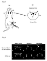
- a rat (weighing about 300 g) was anesthetized with pentobarbital, and inserted with a balloon-tip catheter through the left femoral artery [Fig.5 (a), 1].
- the left common carotid artery was scraped by the balloon to injure the endothelia thereof.
- the rat was injected, through the left femoral vein [Fig.5 (a), 2], with 2 ml (about 160 ⁇ M/kg) of physiological aqueous solution of the contrast agent as prepared in Example 1 (24 mM).
- Fig.6 shows the images of the rat taken in the vicinity of the common carotid artery at the cross section corresponding Fig.5 (b).
- Fig.6 shows the images of the rat taken in the vicinity of the common carotid artery at the cross section corresponding Fig.5 (b).
- in Fig.6 by "injured” is designated the left common carotid artery injured by the balloon, corresponding to 4 in Fig.5 (b), while by “intact” is designated the right common carotid artery, which is normal and corresponds to 5 in Fig.5 (b).
- the numeral 3 in Fig.5 (b) denotes the bronchium.
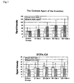
- Fig.7 showing the time-varying MRI signal intensities of the injured common carotid artery and the normal common carotid artery, in which the signal intensities are digitized (in the same manner as in Example 2 and Example 3).
- the MRI signal intensity for the injured left common carotid artery was higher than that for the normal right common carotid artery at ten minutes after the administration of the agent, and it increased with time, leveling off in about one hour after the administration.
- the conventional MRI agent, DTPA-Gd no substantial difference in the signal intensity was observed between the injured common carotid artery and the normal common carotid artery, making it impossible to detect any injured sites.
- the MRI contrast agent of the present invention is a new type of contrast agent which is capable of directly detecting and imaging exfoliated vascular endothelial sites, thereby contributing to the early detection and early diagnosis of vascular diseases.
Landscapes
- Health & Medical Sciences (AREA)
- Life Sciences & Earth Sciences (AREA)
- Nuclear Medicine, Radiotherapy & Molecular Imaging (AREA)
- Radiology & Medical Imaging (AREA)
- Epidemiology (AREA)
- Animal Behavior & Ethology (AREA)
- General Health & Medical Sciences (AREA)
- Public Health (AREA)
- Veterinary Medicine (AREA)
- Medicinal Chemistry (AREA)
- Chemical & Material Sciences (AREA)
- Medicines Containing Antibodies Or Antigens For Use As Internal Diagnostic Agents (AREA)
- Magnetic Resonance Imaging Apparatus (AREA)
Abstract
Description



Claims (5)
- A contrast agent for use in MRI, comprising an imaging unit which contains an unpaired electron-carrying atom and/or molecule and is capable of amplifying or reducing MRI signals, and a detection unit which is bonded to said imaging unit and is capable of selectively recognizing exfoliated vascular endothelial sites and binding thereto.
- An MRI contrast agent as claimed in claim 1, in which the detection unit is one having a chemical structure expressed by the following general formula (I):wherein at least one of R1, R2, R3, R4, R5, R6, R7, R8, R9, R10 and R11 may be independently sulfonic acid group, hydroxyl group or amino group, at least one of R1, R2, R3 and R4 may be independently an alkyl group or alkoxy group having 1 to 3 carbon atoms, the remaining ones of R1, R2, R3, R4, R5, R6, R7, R8, R9, R10 and R11 which do not fall within any of the above-mentioned functional groups are all hydrogen atom, and X, if present, represents a phenyl group in which at least one site may be substituted with an alkyl group or alkoxy group having 1 to 3 carbon atoms.
- An MRI contrast agent as claimed in claim 2, in which the detection unit is one having a chemical structure expressed by the following chemical formula (II), (III), (IV), (V) or (VI).


- An MRI contrast agent as claimed in any of claims 1 through 3, in which the imaging unit comprises a chelated complex of gadolinium.
- An MRI contrast agent as claimed in claim 4, which is expressed by the following chemical formula (VII):

Applications Claiming Priority (5)
| Application Number | Priority Date | Filing Date | Title |
|---|---|---|---|
| JP2003050574 | 2003-02-27 | ||
| JP2003050574 | 2003-02-27 | ||
| JP2003322515 | 2003-09-16 | ||
| JP2003322515 | 2003-09-16 | ||
| PCT/JP2004/001063 WO2004075925A1 (en) | 2003-02-27 | 2004-02-03 | Contrast medium for mri |
Publications (2)
| Publication Number | Publication Date |
|---|---|
| EP1598082A1 true EP1598082A1 (en) | 2005-11-23 |
| EP1598082A4 EP1598082A4 (en) | 2008-11-12 |
Family
ID=32929650
Family Applications (1)
| Application Number | Title | Priority Date | Filing Date |
|---|---|---|---|
| EP04707636A Withdrawn EP1598082A4 (en) | 2003-02-27 | 2004-02-03 | CONTRAST PRODUCT FOR MRI |
Country Status (4)
| Country | Link |
|---|---|
| US (1) | US7368099B2 (en) |
| EP (1) | EP1598082A4 (en) |
| JP (1) | JP4569875B2 (en) |
| WO (1) | WO2004075925A1 (en) |
Cited By (1)
| Publication number | Priority date | Publication date | Assignee | Title |
|---|---|---|---|---|
| CN103980297A (en) * | 2014-04-15 | 2014-08-13 | 大连大学 | Multi-functional imaging probe for mercury ion detection |
Families Citing this family (9)
| Publication number | Priority date | Publication date | Assignee | Title |
|---|---|---|---|---|
| JPWO2006025304A1 (en) * | 2004-08-30 | 2008-05-08 | 国立大学法人九州大学 | MRI contrast agent for arteriosclerosis detection |
| CN100358583C (en) * | 2005-04-01 | 2008-01-02 | 中山大学 | A kind of gadolinium-doped nanocomposite material and preparation method thereof |
| WO2012136813A2 (en) | 2011-04-07 | 2012-10-11 | Universitetet I Oslo | Agents for medical radar diagnosis |
| WO2015154007A1 (en) * | 2014-04-03 | 2015-10-08 | Lipella Pharmaceuticals, Inc. | Systems and methods of detecting interstitial cystitis |
| US20160045623A1 (en) | 2015-04-03 | 2016-02-18 | Lipella Pharmaceuticals, Inc. | Systems and Methods to Image Intercellular and Intercompartmental Defects with Magnetic Resonance Imaging (MRI) |
| US10709790B2 (en) | 2015-06-22 | 2020-07-14 | The United States Of America, As Represented By The Secretary, Department Of Health And Human Services | Chemical conjugates of Evans Blue derivatives and their use in the production of long-acting therapeutics |
| JP6946342B2 (en) | 2016-05-09 | 2021-10-06 | ザ ユナイテッド ステイツ オブ アメリカ, アズ リプレゼンテッド バイ ザ セクレタリー, デパートメント オブ ヘルス アンド ヒューマン サービシーズ | Chemical conjugates of Evans blue derivatives, and their use as radiotherapeutic and imaging agents |
| EP3692032B1 (en) | 2017-10-03 | 2024-06-19 | The U.S.A. as represented by the Secretary, Department of Health and Human Services | Chemical conjugates of evans blue derivatives and their use as radiotherapy and imaging agents |
| WO2019165200A1 (en) | 2018-02-22 | 2019-08-29 | The United States Of America, As Represented By The Secretary, Department Of Health And Human Services | Chemical conjugates of evans blue derivatives and their use as radiotherapy and imaging agents for targeting prostate cancer |
Family Cites Families (8)
| Publication number | Priority date | Publication date | Assignee | Title |
|---|---|---|---|---|
| US4877599A (en) * | 1986-11-10 | 1989-10-31 | New England Deaconess Hospital Corporation | Detection of vascular disease with labelled antibodies |
| JP2776953B2 (en) * | 1990-04-10 | 1998-07-16 | 日本メジフィジックス株式会社 | Multifunctional metal chelate compounds and their uses |
| CA2080493A1 (en) * | 1990-05-03 | 1991-11-04 | Robert S. Lees | Synthetic peptides for arterial imaging |
| JPH07145038A (en) * | 1993-11-26 | 1995-06-06 | Terumo Corp | Closed vesicle membrane components |
| JP3735921B2 (en) * | 1996-02-07 | 2006-01-18 | 三菱ウェルファーマ株式会社 | GPIb / lipid complex and uses thereof |
| CA2273609A1 (en) * | 1999-06-04 | 2000-12-04 | Resolution Pharmaceuticals Inc. | Radiopharmaceuticals and methods for imaging |
| JP2002348235A (en) * | 2001-03-23 | 2002-12-04 | Clinical Supply:Kk | Preventive against restenosis |
| AU2004206956A1 (en) * | 2003-01-22 | 2004-08-05 | The General Hospital Corporation | Amyloid-binding, metal-chelating agents |
-
2004
- 2004-02-03 EP EP04707636A patent/EP1598082A4/en not_active Withdrawn
- 2004-02-03 US US10/546,964 patent/US7368099B2/en not_active Expired - Fee Related
- 2004-02-03 WO PCT/JP2004/001063 patent/WO2004075925A1/en not_active Ceased
- 2004-02-03 JP JP2005502822A patent/JP4569875B2/en not_active Expired - Fee Related
Cited By (1)
| Publication number | Priority date | Publication date | Assignee | Title |
|---|---|---|---|---|
| CN103980297A (en) * | 2014-04-15 | 2014-08-13 | 大连大学 | Multi-functional imaging probe for mercury ion detection |
Also Published As
| Publication number | Publication date |
|---|---|
| US20060153773A1 (en) | 2006-07-13 |
| EP1598082A4 (en) | 2008-11-12 |
| JPWO2004075925A1 (en) | 2006-06-01 |
| US7368099B2 (en) | 2008-05-06 |
| WO2004075925A1 (en) | 2004-09-10 |
| JP4569875B2 (en) | 2010-10-27 |
Similar Documents
| Publication | Publication Date | Title |
|---|---|---|
| US6123921A (en) | Bifunctional detection agents having an optical dye linked to an MRI contrast agent | |
| Tsitovich et al. | Redox-activated MRI contrast agents based on lanthanide and transition metal ions | |
| Boros et al. | Gd (DOTAla)–A single amino acid Gd-complex as a modular tool for high relaxivity MR contrast agent development | |
| US6652835B1 (en) | Targeting multimeric imaging agents through multilocus binding | |
| Aime et al. | Lanthanide (III) chelates for NMR biomedical applications | |
| Asik et al. | Dinuclear Fe (III) hydroxypropyl-appended macrocyclic complexes as MRI probes | |
| US6770261B2 (en) | Magnetic resonance imaging agents for the detection of physiological agents | |
| US5466439A (en) | Polymeric contrast enhancing agents for magnetic resonance images | |
| Zhang et al. | Gadolinium meets medicinal chemistry: MRI contrast agent development | |
| Longo et al. | Gd-AAZTA-MADEC, an improved blood pool agent for DCE-MRI studies on mice on 1 T scanners | |
| JP4163381B2 (en) | Magnetic resonance contrast agents for the detection of physiological agents | |
| US6656450B2 (en) | Macrocyclic magnetic resonance imaging contrast agents | |
| US7368099B2 (en) | MRI contrast agents | |
| Koudrina et al. | Exploring the unique contrast properties of aptamer–gadolinium conjugates in magnetic resonance imaging for targeted imaging of thrombi | |
| US5138039A (en) | Chelating compounds and their use | |
| US20030198597A1 (en) | Novel macrocyclic activatible magnetic resonance imaging contrast agents | |
| KR100634246B1 (en) | Perfluoroalkylamides, Their Production and Their Use in Diagnosis | |
| US6019959A (en) | Oligomeric compounds that contain perfluoroalkyl, process for their production, and their use in NMR diagnosis | |
| US20060088475A1 (en) | Self-immolative magnetic resonance imaging contrast agents sensitive to beta-glucuronidase | |
| CN114632079B (en) | Preparation and application of iron pool targeting molecule image probe based on artemisinin | |
| Lalli et al. | Derivatives of GdAAZTA conjugated to amino acids: a multinuclear and multifrequency NMR study | |
| WO2006025304A1 (en) | Mri contrast agent for arteriosclerosis detection | |
| CA2744298A1 (en) | Smart contrast agents for mri imaging | |
| CN118359639A (en) | Near infrared two-region small molecule and synthesis and application thereof in biliary tract occlusion | |
| KR20120057324A (en) | Gd-complexs of macrocyclic type comprising ferrocene |
Legal Events
| Date | Code | Title | Description |
|---|---|---|---|
| PUAI | Public reference made under article 153(3) epc to a published international application that has entered the european phase |
Free format text: ORIGINAL CODE: 0009012 |
|
| 17P | Request for examination filed |
Effective date: 20050825 |
|
| AK | Designated contracting states |
Kind code of ref document: A1 Designated state(s): AT BE BG CH CY CZ DE DK EE ES FI FR GB GR HU IE IT LI LU MC NL PT RO SE SI SK TR |
|
| AX | Request for extension of the european patent |
Extension state: AL LT LV MK |
|
| DAX | Request for extension of the european patent (deleted) | ||
| RAP1 | Party data changed (applicant data changed or rights of an application transferred) |
Owner name: KYUSHU UNIVERSITY, NATIONAL UNIVERSITY CORPORATION |
|
| RBV | Designated contracting states (corrected) |
Designated state(s): DE FR GB IT NL |
|
| RIN1 | Information on inventor provided before grant (corrected) |
Inventor name: YAMAMOTO, TATSUHIRODAI-7-DAIWASOU-202 Inventor name: SHIMOKAWA, HIROAKI Inventor name: KATAYAMA, YOSHIKI |
|
| A4 | Supplementary search report drawn up and despatched |
Effective date: 20081010 |
|
| 17Q | First examination report despatched |
Effective date: 20090313 |
|
| STAA | Information on the status of an ep patent application or granted ep patent |
Free format text: STATUS: THE APPLICATION IS DEEMED TO BE WITHDRAWN |
|
| 18D | Application deemed to be withdrawn |
Effective date: 20090724 |