Technical Field
The present invention relates to a method of obtaining a
gene useful for predicting an efficacy, specifically an
antitumor effect, of a histone deacetylase inhibitor, and a
method of predicting the efficacy (antitumor effect) of the
histone deacetylase inhibitor, which comprises examining an
expression pattern of the gene.
Background Art
In recent years, a "tailor made medicine" is gaining
recognition, which takes into consideration individual
differences between patients, and a search for a marker to
distinguish a cancer against which a pharmaceutical agent is
effective from a cancer against which the pharmaceutical agent
is ineffective is considered to be necessary. It is an attempt
to ethically and medically improve cost performance of
medication treatment by administering a pharmaceutical agent
to patients after verification in advance of the probability
of effect thereof, thereby to enhance efficacy as well as
avoid toxicity of the pharmaceutical agent, and to reduce
insignificant use of the pharmaceutical agent. In cancer
treatment, the development of a method for predicting the
efficacy of anticancer agents has been desired, because it can
be an important means to bridge the gap between basic study
and clinical application.
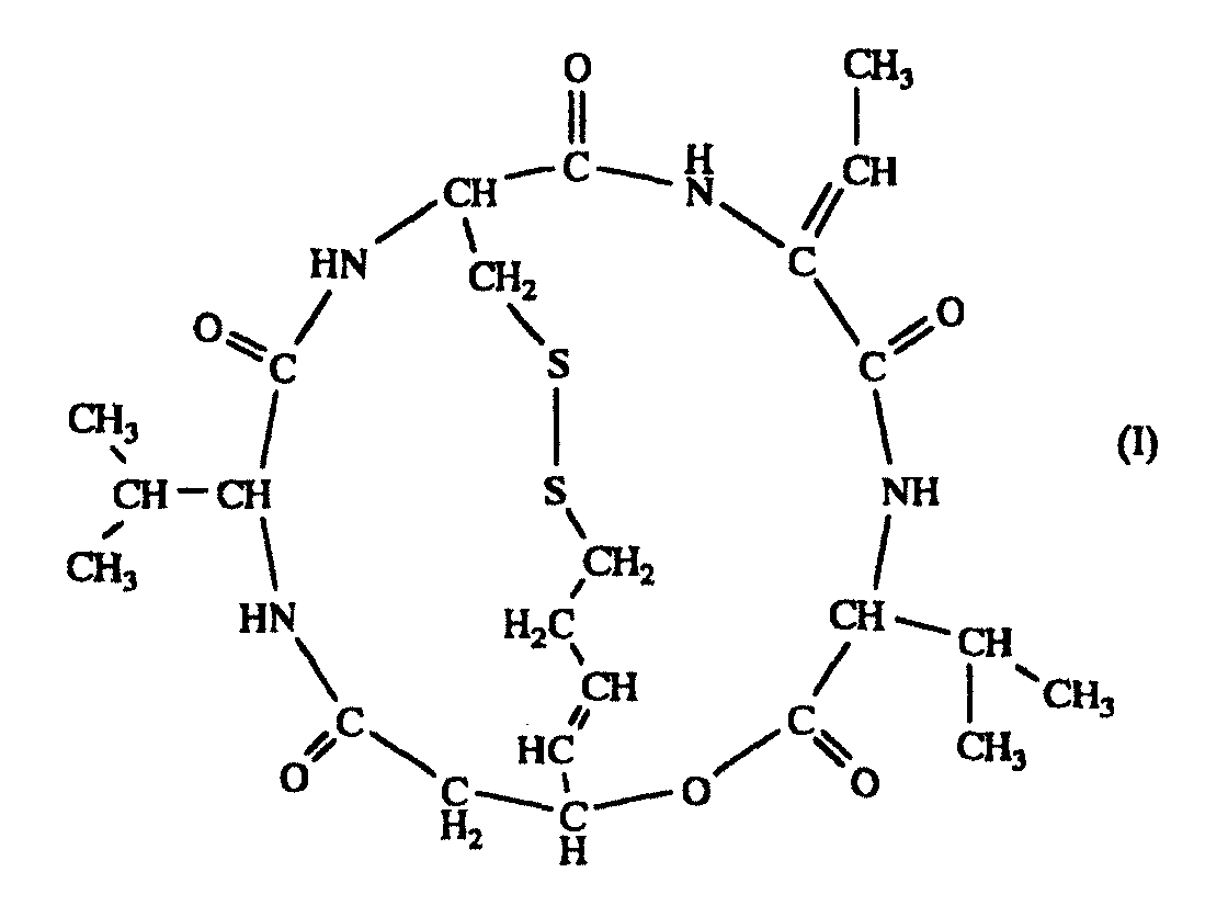
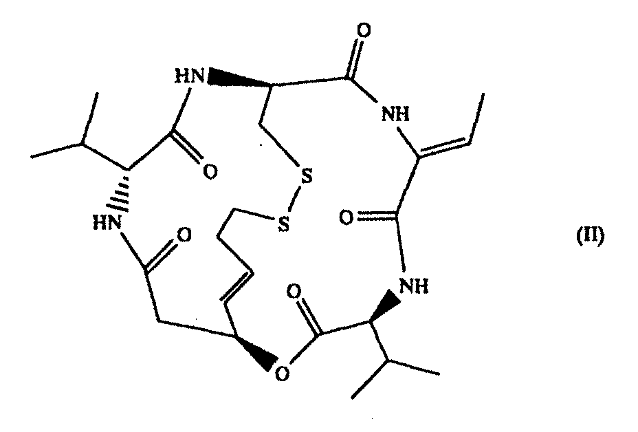
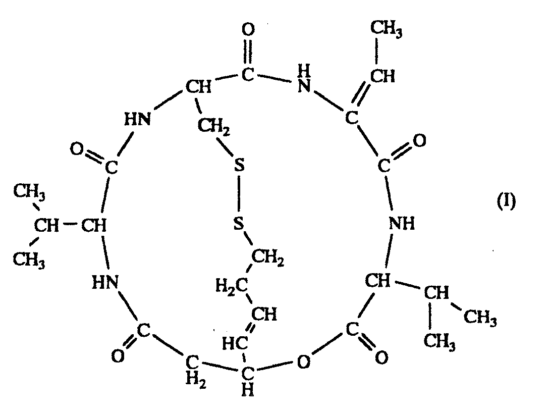
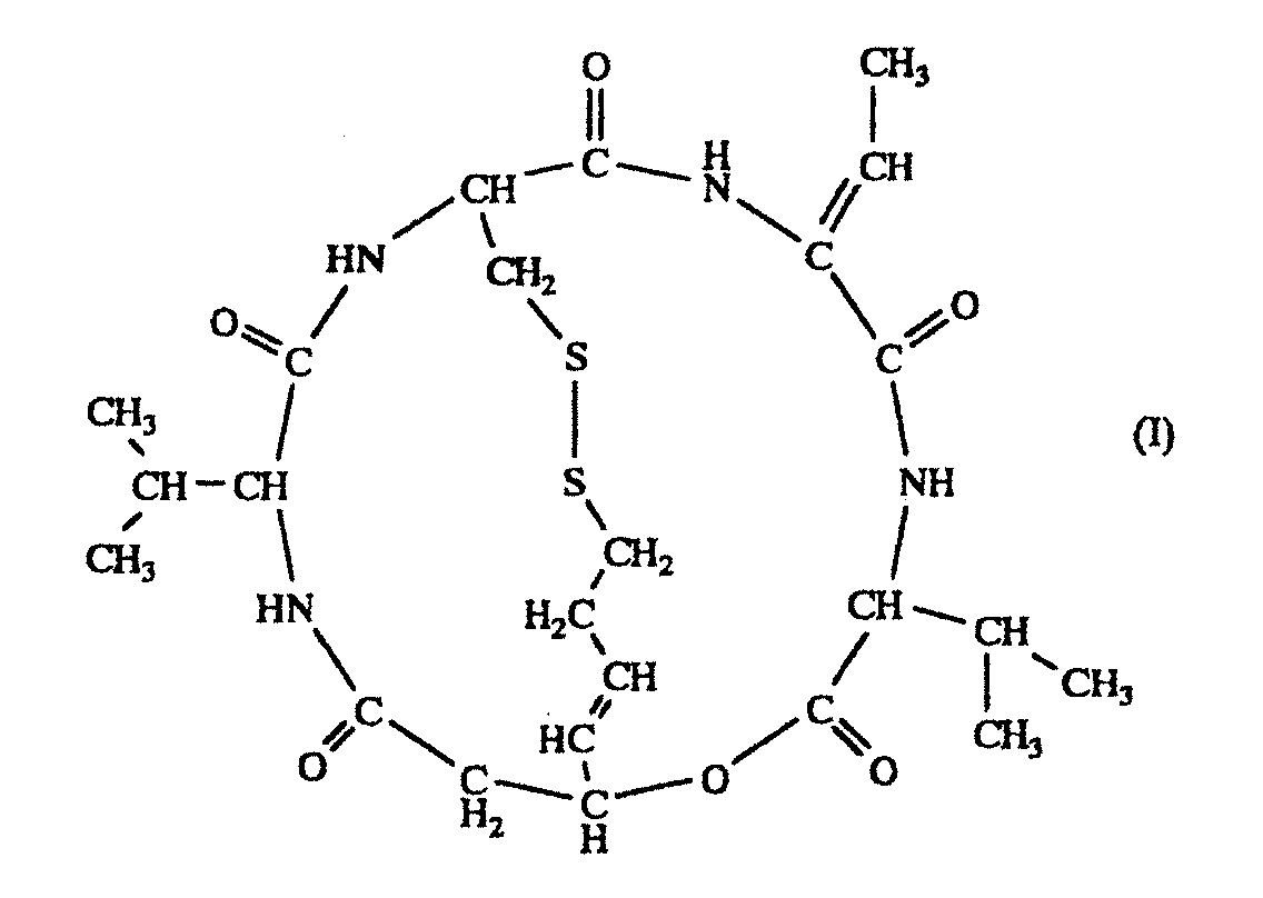
For example, it has been reported that a compound
represented by the formula (I)
(hereinafter to be also referred to as compound A; SEQ ID NO:
1), particularly a stereoisomer (FK228) represented by the
formula (II)
has a histone deacetylase inhibitory action and induces a
potent antitumor activity (e.g., JP-B-7-64872, Experimental
Cell Research, US (1998), vol. 241, pp. 126-133). However, no
report has established a factor capable of predicting an
antitumor effect of this compound, and as the situation stands,
many problems are yet to be solved, such as whether or not in
vitro results directly apply
in vivo, whether or not the
compound shows a practical effect
in vivo in any tumor and the
like.
An object of the present invention is to provide a
method of obtaining a gene capable of becoming an index of
efficacy prediction, which is useful for predicting the
efficacy of a histone deacetylase inhibitor, particularly
compound A known to have a potent histone deacetylase
inhibitory action, and a method of predicting the efficacy of
the histone deacetylase inhibitor, which comprises examining
the expression pattern of the gene.
The present inventors have conducted intensive studies
in an attempt to solve the above-mentioned problems and found
that the antitumor effect of the histone deacetylase inhibitor
varies depending on the kind of tumor, and the variation is
observed in association with changes in the expression pattern
of a particular gene or protein. They have identified these
particular genes (proteins), observed the expression pattern
of these genes in tumor, and based on such observation, found
a method of predicting or evaluating the efficacy of a histone
deacetylase inhibitor, which resulted in the completion of the
present invention. Accordingly, the present invention provides
the following.
Brief Description of the Drawings
Fig. 1 is a graph showing an antitumor effect of FK228
on human prostate cancer ((a); PC-3) and kidney cancer ((b);
ACHN), wherein the vertical axis shows a tumor growth rate,
the transverse axis shows the number of days lapsed from the
initial administration, and the tumor growth rate is expressed
in a relative ratio of tumor volume after day 0 relative to
that on day 0 as 1.
Detailed Description of the Invention
The present invention provides a method of obtaining a
gene capable of becoming an index for predicting an efficacy,
particularly an antitumor effect, of a histone deacetylase
inhibitor. By analyzing the expression pattern of the gene
obtained by the method in a tumor cell, the information such
as whether the histone deacetylase inhibitor is useful for the
treatment, or whether the histone deacetylase inhibitor is
effective for the target cancer and the like can be obtained,
which makes it possible to contribute to the "tailor made
medicine". In addition, a histone deacetylase inhibitor
capable of exhibiting an antitumor effect in a target tumor
cell can be found without actual administration to the human
body.
The "histone deacetylase inhibitor" means a compound
that binds to an active site of histone deacetylase
competitively with a substrate, or a compound having an action
to bind with a site different from the active site of histone
deacetylase to change the enzyme activity of the histone
deacetylase, and includes compounds whose use as a histone
deacetylase inhibitor is known, all compounds (synthetic and
natural) whose histone deacetylase inhibitory activity has
been reported and all the compounds that will be reported in
the future. Specifically, the aforementioned compound A, a
salt thereof, and a derivative thereof (e.g., acetylated
compound A, thiol form with reduced S-S bond etc.) can be
mentioned. Moreover, tricostatin A, sodium butyrate,
suberoylanilide hydroxamic acid (SAHA), MS-275, Cyclic
hydroxamic-acid-containing peptide, Apicidin, Trapoxin and the
like are the compounds whose histone deacetylase inhibitory
activity has been reported.
While compound A may have a steric isomer, such as
optical isomer, geometric isomer and the like, which is based
on the asymmetric carbon atom and a double bond (e.g., FK228),
all of these isomers and mixtures thereof are also encompassed
in the histone deacetylase inhibitor to be used in the present
invention.
Furthermore, solvate compounds of compound A, FK228 or a
salt thereof (e.g., inclusion compound (e.g., hydrate etc.))
are also encompassed in the scope of the invention.
In the present specification, unless otherwise specified,
a simple reference to compound A means a compound group
irrespective of stereoisomerism, including the compound
represented by the formula (II) (FK228).
The compound A and a salt thereof are known substances
and available. For example, FK228, which is one of the
stereoisomers of compound A, can be obtained by culturing a
strain belonging to the genus Chromobacterium, which is
capable of producing FK228 under aerobic conditions, and
harvesting the substance from the culture broth. As the strain
belonging to the genus Chromobacterium, which is capable of
producing FK228, for example, Chromobacterium violaceum WB968
(FERM BP-1968) can be mentioned. FK228 can be more
specifically obtained from the producing strain as described
in JP-B-7-64872 (US Patent No. 4977138). FK228 is preferably
harvested from the strain belonging to the genus
Chromobacterium, which is capable of producing FK228, because
FK228 can be more easily obtained, and synthetic and semisynthetic
FK228 are also advantageous because further
purification step is not necessary or can be simple. Similarly,
compound A other than FK228 can be semi-synthesized or totally
synthesized by a conventionally known method. More
specifically, it can be produced according to the method
reported by Khan W. Li, et al. (J. Am. Chem. Soc., vol. 118,
7237-7238 (1996)).
As a salt of compound A, salts with inorganic base (e.g.,
alkali metal salts such as sodium salt, potassium salt and the
like, alkaline earth metal salts such as calcium salt,
magnesium salt and the like, ammonium salt), salts with
organic base (e.g., organic amine salts such as triethylamine
salt, diisopropylethylamine salt, pyridine salt, picoline salt,
ethanolamine salt, triethanolamine salt, dicyclohexylamine
salt, N,N'-dibenzylethylenediamine salt and the like),
inorganic acid addition salt (e.g., hydrochloride,
hydrobromide, sulfate, phosphate etc.), organic carboxylic
acid or sulfonic acid addition salts (e.g., formate, acetate,
trifluoroacetate, maleate, tartrate, fumarate,
methanesulfonate, benzenesulfonate, toluenesulfonate etc.),
salts with basic or acidic amino acid (e.g., arginine,
aspartic acid, glutamic acid etc.) and the like, salts or acid
addition salts with base can be mentioned.
In the present invention, in vivo and in vitro generally
mean as used in the pertinent field. That is, "in vivo" refers
to a state where an object biological function or reaction is
expressed in the living organism, and "in vitro" refers to an
expression of such function or reaction in a test tube (tissue
culture system, cell culture system, cell free system etc.).
A method of obtaining a gene capable of becoming an
index for predicting an anti-tumor effect of a histone
deacetylase inhibitor of the present invention comprises at
least the following steps:
While the tumor cell as a test target to be used in the
present invention (hereinafter simply referred to as a test
cell) is not particularly limited as long as it has histone
deacetylase, since evaluation of an antitumor effect of the
histone deacetylase inhibitor, particularly tumor site
specificity of the inhibitor, is one of the problems of the
present invention, the test cell to be used is preferably
derived from a tumor on which the effect is desired to be
examined. For example, when the effect on prostate cancer is
to be evaluated, PC-3 cell, which is a cultured human prostate
cancer cell, and the like are used, and when the effect on
kidney cancer is to be evaluated, ACHN cell, which is a
cultured human kidney cancer cell and the like are used.
Various cultured human cancer cells to be used as test cells
including these cancer cells are commercially available, or
available from various cell banks and the like. For
examination of a long-term treatment effect, or effectiveness
for individual patients, namely, tailor made medicine, it is
possible to culture a cancer cell that can be obtained from a
tumor of patient and use the cancer cell as a test cell.
The step of examining an in vivo antitumor effect of a
histone deacetylase inhibitor in a tumor cell is conducted by,
for example, the following procedures.
First, a tumor cell to be the test target is implanted
into a test animal such as mouse, rat, rabbit, hamster, guinea
pig and the like to prepare a system to experimentally form a
tumor. In this system, a histone deacetylase inhibitor is
administered after tumor implantation and the tumor growth
rate is measured to evaluate an antitumor effect of the
histone deacetylase inhibitor. The growth rate is conveniently
determined by measuring the size of the tumor formed.
A tumor cell can be implanted to a test animal by a
conventional method in this field. For example, a method
comprising implanting, in a mouse, a part of a solid tumor
subcultured and grown in a nude mouse can be mentioned. In
addition, a histone deacetylase inhibitor can be administered
in a similar amount and in a similar manner as in the
administration generally expecting its antitumor effect. That
is, application by intraperitoneal, intravenous, intramuscular
or oral administration can be mentioned. While the dose varies
depending on the kind and body weight of the test animal to be
subjected to the administration, and the kind of the implanted
cancer, about 1 mg/kg - 5 mg/kg is administered for general
intravenous administration to a mouse.
The solvent to give a solution of a histone deacetylase
inhibitor for the administration to a test animal is not
particularly limited as long as it can dissolve the histone
deacetylase inhibitor and it does not show toxicity to the
tumor cell or the test animal as the test target. Generally, a
concentrated solution is prepared with ethanol, PEG400, 10%
HCO-60 solution, dimethyl sulfoxide and the like, a mixed
solvent thereof and the like, and diluted to a desired
concentration with physiological buffer such as physiological
saline and the like and used.
When a given histone deacetylase inhibitor is
administered in a certain dose, a tumor cell that exhibited a
strong growth suppressing effect as shown by the growth
suppressing rate of not less than 70% (preferably not less
than 80%) is divided as a "sensitive tumor cell" to the
histone deacetylase inhibitor, a tumor cell that exhibited a
weak growth suppressing effect as shown by the growth
suppressing rate of not more than 30% (preferably not more
than 20%) is divided as a "resistant tumor cell" to the
histone deacetylase inhibitor.
For example, when the histone deacetylase inhibitor is
compound A, the cells are divided into a compound A sensitive
tumor cell and a compound A resistant sensitive cell.
The compound A sensitive tumor cell is a tumor cell of
the type exhibiting a strong growth suppressing effect by
compound A on the tumor cell according to the aforementioned
criteria, and, for example, prostate cancer cell PC-3 can be
mentioned as shown in the below-mentioned Example. In addition,
SC-6, which is a stomach cancer cell, is one kind of compound
A sensitive tumor cells. On the other hand, compound A
resistant tumor cell is a tumor cell of the type exhibiting
only a weak growth suppressing effect by compound A according
to the aforementioned criteria, and, for example, kidney
cancer cell ACHN can be mentioned as shown in the below-mentioned
Examples. In addition, A498, which is a kidney
cancer cell, is one kind of compound A resistant tumor cells.
The step of examining the gene expression pattern in
each of the divided sensitive tumor cell and the resistant
tumor cell, and further selecting (i) a gene showing high
expression in a sensitive tumor cell and low expression in a
resistant tumor cell, or (ii) a gene showing low expression in
a sensitive tumor cell and high expression in a resistant
tumor cell (hereinafter the genes are to be also generally
referred to as an antitumor effect predicting gene) can be
conducted using the respective techniques described in the
present specification and methods generally employed in this
field. Preferably, a technique using a gene chip is employed
in view of the advantage that a large number of gene
expression patterns can be analyzed at once.
Analysis of the expression pattern of an antitumor
effect predicting gene in a tumor cell can be an effective
means for predicting the efficacy of a histone deacetylase
inhibitor by an in vitro test without administration of the
histone deacetylase inhibitor to patients.
The present invention provides a method of predicting an
antitumor effect of a histone deacetylase inhibitor, which
comprises examining the expression amount of an antitumor
effect predicting gene in at least one kind, preferably two or
more kinds of tumor cells.
The method of analyzing the gene expression pattern can
be also performed using methods generally employed in this
field.
For example, procedures described in the following can
be mentioned.
For a specific gene (or specific protein) to be measured
in the present invention, one kind of measurement is
sufficiently useful, but when a more detailed antitumor effect
needs to be known, two or more kinds of specific genes (or
specific proteins) are preferably measured simultaneously.
More preferably, the expression of at least one kind of gene
from each of a group of the genes showing high expression in a
sensitive tumor cell and low expression in a resistant tumor
cell and a group of the genes showing low expression in a
sensitive tumor cell and high expression in a resistant tumor
cell is preferably analyzed.
A substance having specific affinity for the specific
gene or specific protein is free of any particular limitation
as long as it has such a sensitivity as allows detection of
expression in the test cells. As used herein, by the "specific
affinity" is meant a property to hybridize or bind solely to
an object gene or protein. As the substance to detect the
specific gene, a substance absolutely complement to the whole
or a part of said gene, or a substance containing one to
several mismatches within the extent satisfying the above-mentioned
property can be mentioned. Specifically, oligo- or
poly-nucleotide containing a part or the entirety of the base
sequence of the gene and complementary sequences thereof, and
the like can be mentioned, and an appropriate substance is
selected depending on the form of the gene to be detected. The
derivation of the substance is not particularly limited as
long as it has specific affinity for the gene, and it may be
synthesized or formed by cleaving a necessary part from the
gene and purifying the part by a conventional method. The
substance may be labeled with a fluorescent substance, an
enzyme, a radioisotope and the like. As the substance to be
used for detecting a specific protein, for example, an
antibody having specific affinity for the protein or a
fragment thereof can be mentioned. The specific affinity
thereof means an ability to specifically recognize the protein
by an antigen-antibody reaction and bind thereto. The antibody
and the fragment thereof are not particularly limited as long
as they can specifically bind to the protein, and may be any
of a polyclonal antibody, a monoclonal antibody and functional
fragments thereof. These antibodies and functional fragments
thereof can be produced according to a method generally
employed in the pertinent field. These antibodies and
fragments thereof may be labeled with a fluorescent substance,
an enzyme, a radioisotope and the like.
Extraction of a gene, particularly mRNA, as well as
extraction of a protein from the test cell can be performed
according to a method generally employed in the pertinent
field, or by an appropriate combination of such methods. When
mRNA was extracted, its expression pattern is examined
according to a method generally employed in the pertinent
field, such as Northern blot, RT-PCR and the like, using a
substance having specific affinity for the above-mentioned
specific gene. On the other hand, when a protein was extracted,
its expression pattern is examined according to a method
generally employed in the pertinent field, such as immunoblot,
Western blot and the like, using a substance (antibody, a
fragment thereof etc.) having specific affinity for the above-mentioned
specific protein.
Moreover, for the analysis of the expression pattern of
the gene, the aforementioned methods as well as a technique
using a gene chip is preferably employed in view of the
advantage that a large number of gene expression patterns can
be analyzed at once.
By the analysis of the expression pattern of an
antitumor effect predicting gene, whether or not the tested
histone deacetylase inhibitor effectively shows an antitumor
activity against the tested tumor cell can be predicted. For
example, when the genes are divided into a gene group showing
twice or more higher expression in a sensitive tumor cell than
in a resistant tumor cell (antitumor effect predicting gene A)
and a gene group showing twice or more higher expression in a
resistant tumor cell than in a sensitive tumor cell (antitumor
effect predicting gene B), an antitumor effect of the histone
deacetylase inhibitor can be expected in a tumor cell where
the antitumor effect predicting gene A shows high expression,
and/or a tumor cell where the antitumor effect predicting gene
B shows low expression.
When a gene chip is used, the efficacy can be predicted
by the following operations.
When a clinically removed tumor cell from a patient is
used as a test cell, prediction of an antitumor effect
reflecting individual specificity of the individual patient
can be achieved.
The present invention can provide a screening method of
a histone deacetylase inhibitor having an antitumor activity
specific for a tumor site (kind), by utilizing the
aforementioned evaluation methods of the antitumor effect of a
histone deacetylase inhibitor. The tumor site specificity of
individual inhibitor can be evaluated using a test cell
derived from the target tumor, treating with a histone
deacetylase inhibitor to be examined for the effect, and
examining the presence of the antitumor effect according to
the aforementioned methods.
Examples
The present invention is explained specifically and in
detail in the following by referring to Examples, which are
not to be construed as limitative.
Example 1
Evaluation of compound A sensitive tumor and compound A
resistant tumor
(1) Preparation of pharmaceutical agent
A necessary amount of FK228 was weighed and a solvent
(10% HCO-60/saline) was added. The mixture was sonicated to
allow for dissolution. A positive control substance Paclitaxel
was dissolved in Cremophor EL/ethanol (1:1) solution to 24
mg/mL prior to the testing, and preserved in a refrigerator.
When in use, it was diluted with a 9-fold amount of
physiological saline to 2.4 mg/mL (solvent component: 5%
Cremophor EL-5% ethanol-90% saline).
(2) Test animal
For antitumor test of the pharmaceutical agent,
BALB/cANnNCrj-nu/nu mice (male, 6-week-old) were purchased
from Charles River Japan and, after acclimation for not less
than one week, used for the test. The mice were reared under
an SPF environment and allowed a free access to water and feed.
(3) Test tumor
Cultured human kidney cancer cell line 1 (ACHN:
available from ATCC) and human culture prostate cancer cell
line 1 (PC-3: available from ATCC) were subcutaneously
implanted at 2-3×10⁷ cells in a nude mouse. A grown solid
tumor was subcultured for not less than 3 generations and used
for the test.
(4) Experimental implantation and grouping
A solid tumor subcultured in a nude mouse was
subcutaneously implanted in the right back of a mouse as an
about 3 mm square tumor tissue fragment. After the tumor
implantation, when the tumor volume (1/2×longer
diameter×shorter diameter2) reached 100-300 mm3, the mice were
grouped into 6 mice per group to level the tumor size.
(5) Administration
The administration was started on the day of grouping
(Day 0). FK228 was intravenously administered to an FK228
administration group 3 times every 4 days (q4dx3) (3.2 and 1.8
mg/kg). Paclitaxel (24 mg/kg) was intravenously administered
for 5 consecutive days (qd×5) to a positive control substance
Paclitaxel administration group. Only a solvent (10% HCO-60/saline)
was administered (q4dx3) to a control group. The
amount of liquid for each administration was calculated (0.1
mL/10 g body weight) based on the body weight measured on the
administration day. Note that 3.2 mg/kg/day (q4dx3) of FK228
and 24 mg/kg/day (qdx5) of Paclitaxel were the maximum
tolerated doses (MTD) thereof.
(6) Measurement of tumor size and body weight
The tumor size (longer diameter, shorter diameter) and
body weight were measured twice a week from Day 0.
(7) Evaluation of antitumor effect
The level of tumor growth was evaluated based on the
tumor growth rate (Relative Tumor Volume). The growth
suppression rate was expressed in a relative proportion of
tumor volume after day 0 to the tumor volume at Day 0 as 1.
The results are shown in Fig. 1. FK228 showed a strong
antitumor action on PC-3 at a dose of 3.2 mg/kg (Fig. 1(a))
but did not show an antitumor action on ACHN (Fig. 1(b)).
Example 2
Evaluation of compound A sensitive tumor and compound A
resistant tumor
<material, procedure>
(1) test material
drug: compound A (FK228)
dose: 3.2 mg/kg administration volume: 10 mL/kg solvent: 10% HCO-60/saline solution dosage form: solution (prepared when in use)
tumor cell: human prostate cancer PC-3 (tumor fragment 3 mmx3
mmx3 mm/mouse implantation site s.c.)
human gastric cancer SC-6; obtained from Central Institute
for Experimental Animals (tumor fragment 3 mmx3 mmx3 mm/mouse
implantation site s.c.) human kidney cancer ACHN (tumor fragment 3 mmx3 mmx3
mm/mouse implantation site s.c.) human kidney cancer A498; obtained from ATCC (tumor
fragment 3 mm×3 mmx3 mm/mouse implantation site s.c.)
Subcultured animal: male BALB c/nu/nu
(2) procedure and results
By the method shown in Example 1, 3 mm square tumor
fragments (human prostate cancer PC-3, human stomach cancer
SC-6, human kidney cancer ACHN and human kidney cancer A498)
were subcutaneously implanted in nude mice, and when the tumor
volume (long diameter 9 mm, short diameter 8 mm) reached about
100-300 mm3, FK228 (3.2 mg/kg) was intravenously administered.
The tumor growth suppressing rates then were 98%, 84%, 20% and
29%, respectively. From these results, PC-3 and SC-6 were
evaluated as compound A sensitive tumors and ACHN and A498
were evaluated as compound A resistant tumors.
Example 3
Analysis of gene expression pattern of compound A sensitive
tumor and compound A resistant tumor using gene chip
The gene expression pattern of the tumor for which
sensitivity and resistivity were confirmed in Example 2 was
examined.
(1) test material
tumor cell human prostate cancer PC-3 (tumor fragment 3 mmx3
mmx3 mm/mouse implantation site s.c.)
human gastric cancer SC-6; obtained from Central Institute
for Experimental Animals (tumor fragment 3 mmx3 mmx3 mm/mouse
implantation site s.c.)
human kidney cancer ACHN (tumor fragment 3 mmx3 mmx3
mm/mouse implantation site s.c.)
human kidney cancer A498; obtained from ATCC (tumor
fragment 3 mmx3 mmx3 mm/mouse implantation site s.c.)
Subcultured animal: male BALB c/nu/nu
- RNA extraction:
- RNeasy Mini Kit (50) (Qiagen)
RNase, DNase free water (Life Technologies)
- DNA synthesis:
- Superscript Choice System (Life Technologies)
Ethachinmate (Nippon gene)
T7-(dT)24 Primer (Amersham Pharmacia)
- cRNA synthesis:
- BioArray RNA Transcript Labeling Kit (Amersham
Pharmacia)
- cRNA fragmentation:
- Trizma Base (SIGMA)
glacial acetic acid (SIGMA)
magnesium acetate (SIGMA)
potassium acetate (SIGMA)
- Hybridization:
- Eukaryotic Hybridization Control Kit (Amersham
Pharmacia)
0.5 M EDTA solution (SIGMA)
MES Sodium Salt (SIGMA)
MES Free Acid Monohydrate (SIGMA)
Herring Sperm DNA (Promega)
Acetylated Bovine Serum Albumin Soln. (Life
Technologies)
- Chip used:
- HuGeneFL array (Amersham Pharmacia)
(2) Cell preparation and RNA extraction
A 3 mm square tumor fragment (human prostate cancer PC-3,
human stomach cancer SC-6, human kidney cancer ACHN and human
kidney cancer A498) was subcutaneously implanted in a nude
mouse and, when the tumor reached about 100 to 300 mm3 (longer
diameter 9 mm, shorter diameter 8 mm), the tumor was removed
and total RNA was extracted according to the protocol of
RNeasy Mini Kit (50) (Qiagen). RNA was quantified and
confirmed by electrophoresis.
(3) Synthesis of cRNA
According to the GeneChip manual, Chapter 2 - Chapter 4,
and the manuals of RNeasy Mini Kit and RNA Transcript Labeling
Kit, RNA was purified, cDNA was synthesized, cRNA was
synthesized and cRNA was fragmented.
(4) Hybridization, Washing-staining, Scanning
Hybridization, washing-staining and scanning were
conducted according to the GeneChip manual Chapter 5 - Chapter
7.
(5) Analysis
Analyzed using GeneSpring (microarray data analysis
soft: manufactured by Silicon Genetics).
The analysis conditions were as follows.
Analysis conditions
- Raw data are not less than 100 in at least one of four tumor
cells.
- Difference in normalized data between compound A sensitive
tumors, or compound A resistant tumors is not more than 2-fold.
- Difference in normalized data between compound A sensitive
tumors, or compound A resistant tumors is not less than 2-fold.
As a result, 27 genes showing twice or more higher
expression in a compound A sensitive tumor than in a compound
A resistant tumor were obtained (Table 1), and 49 genes
showing twice or more higher expression in a compound A
resistant tumor than in a compound A sensitive tumor were
obtained (Tables 2 - 3).
| genes that showed high expression in a compound A sensitive tumor and low expression in a compound A resistant tumor |
| | Gene Accession | No. Description |
| 1 | L11005_at | aldehyde oxidase 1 |
| 2 | X12517_at | small nuclear ribonucleoprotein polypeptide C |
| 3 | U43944_at | malic enzyme 1, soluble |
| 4 | U52100_at | epithelial membrane protein 2 |
| 5 | U65579_at | NADH dehydrogenase (ubiquinone) Fe-S protein 8 (23kD) (NADH-coenzyme Q reductase) |
| 6 | X06272_at | signal recognition particle receptor ('docking protein') |
| 7 | D42047_at | The ha3662 gene product is related to mouse glycerophosphate dehydrogenase. |
| 8 | D90086_at | pyruvate dehydrogenase (lipoamide) beta |
| 9 | U61734_s_at |
| 10 | J03798_at | small nuclear riboprotein Sm-D |
| 11 | X05299_at | centromere protein B (80kD) |
| 12 | L34155_at | laminin, alpha 3(nicein (150kD), kalinin (165kD), BM600 (150kD), epilegrin) |
| 13 | U60521_at | caspase 9, apoptosis-related cysteine protease |
| 14 | Y10807_s_at | HMT1 (hnRNP methyltransferase, S. cerevisiae)-like 2 |
| 15 | U44754_at | small nuclear RNA activating complex, polypeptide 1, 43kD |
| 16 | U78107_at | N-ethylmaleimide-sensitive factor attachment protein, gamma |
| 17 | U60644_at | similar to Vaccinia virus HindIII K4L 0RF, and to Vaccinia virus p37 (HindIII F13L 0RF) |
| 18 | U32986_s_at | damage-specific DNA binding protein 1 (127kD) |
| 19 | D59253_at | RNA-binding protein that has two RNP consensus motifs |
| 20 | U41767_s_at | a disintegrin and metalloproteinase domain 15 (metargidin) |
| 21 | U10550_at | Source: Human Gem GTPase (gem) mRNA, complete cds. |
| 22 | D38521_at | The ha0919 gene product is novel. |
| 23 | J04823_rnal_at | cytochrome c oxidase subunit VIII |
| 24 | M21154_at | S-adenosylmethionine decarboxylase 1 |
| 25 | X78565_at | hexabrachion (tenascin C, cytotactin) |
| 26 | D50405_at | histone deacetylase 1 |
| 27 | X04366_at | calpain, large polypeptide L1 |
Table 2 - 3: genes that showed low expression in a compound A sensitive tumor and high expression in a compound A resistant tumor
| | Gene Accession No. | Description |
| 1 | U84487_at | small inducible cytokine subfamily D (Cys-X3-Cys), member 1 (fractalkine, neurotactin). |
| 2 | M33600_f_at | major histocompatibility complex, class II, DR beta 5 |
| 3 | J03474_at | amyloid A precursor |
| 4 | L24564_at | Ras-related associated with diabetes |
| 5 | J05428_at | UDP glycosyltransferase 2 family, polypeptide B7 |
| 6 | L03840_s_at | fibroblast growth factor receptor 4 |
| 7 | M35878_at | insulin-like growth factor binding protein 3 |
| 8 | HG4318-HT4588_s_at |
| 9 | X87241_at | FAT tumor suppressor (Drosophila) homolog |
| 10 | M59807_at | natural killer cell transcript 4 |
| 11 | HG2379-HT3996_s_at |
| 12 | D16532_at | very low density lipoprotein receptor |
| 13 | U37546_s_at | apoptosis inhibitor 1 |
| 14 | X72889_at | Source: H.sapiens hbrm mRNA. |
| 15 | U66838_at | cyclin Al |
| 16 | Z23090_at | heat shock 27kD protein 1 |
| 17 | U16031_at | signal transducer and activator of transcription 6, interleukin-4 induced |
| 18 | D84110_at | alternative splicing (see also D84107-D84111) |
| 19 | M55998_s_at |
| 20 | D28124_at | neuroblastoma candidate region, suppression of tumorigenicity 1 |
| 21 | HG3548-HT3749_at |
| 22 | X86809_at | phosphoprotein enriched in astrocytes 15 |
| 23 | M79462_at | promyelocytic leukemia |
| 24 | M59465_at | tumor necrosis factor, alpha-induced protein 1 (endothelial) |
| 25 | HG2379-HT3997_s_at |
| 26 | Z36715_at | ELK3, ETS-domain protein (SRF accessory protein 2) NOTE: Symbol and name provisional. |
| 27 | U41654_at | putative GTP-binding protein, homolog of small GTPase family members |
| 28 | U72882_s_at | Source: Human interferon-induced leucine zipper protein (IFP35) mRNA, partial cds. |
| 29 | U72649_at | rat PC3 and murine TIS21 genes homolog |
| 30 | U47621_at |
| 31 | L22214_at | adenosine A1 receptor |
| 32 | U68494_at | Source: Human hbc647 mRNA sequence. |
| 33 | X69699_at | paired box gene 8 |
| 34 | X63717_at | tumor necrosis factor receptor superfamily, member 6 |
| 35 | U04285_s_at |
| 36 | L38969_at | Source: Homo sapiens thrombospondin 3 (THBS3) mRNA, complete cds. |
| 37 | Y10032_at | Source: H.sapiens mRNA for putative serine/threonine protein kinase. |
| 38 | L48513_at | paraoxonase 2 |
| 39 | U89942_at | lysyl oxidase-like 2 |
| 40 | Z12173_at | glucosamine (N-acetyl)-6-sulfatase (Sanfilippo disease IIID) |
| 41 | U59423_at | Sma and Mad homolog |
| 42 | X68277_at | dual specificity phosphatase 1 |
| 43 | U45878_s_at | HIAP-1 |
| 44 | L08187_at |
| 45 | X71874_cds1_at | proteasome (prosome, macropain) subunit, beta type, 10 |
| 46 | D50863_at | testis-specific kinase 1 |
| 47 | X91911_s_at |
| 48 | U65011_at | encodes tumor antigen recognized by cytolytic T lymphocytes |
| 49 | U90313_at | glutathione-S-transferase like |
The gene obtained by the method of the present invention,
which can be an index of the efficacy, particularly an
antitumor effect, of a histone deacetylase inhibitor is
suggested to be correlated to the efficacy of histone
deacetylase inhibitor, as well as to the sensitivity or
resistivity to the histone deacetylase inhibitor, and the
possibility of use of the gene as an efficacy predicting
marker of a histone deacetylase inhibitor has been shown.
Sequence Listing Free Text
SEQ ID; No 1: Xaa is an amino acid represented by the formula
NH2C(CHCH3)COOH.
The carboxyl group of the formula COOHCH2CH(CHCHC2H4SH)OH
is bonded with an amino group of Val, which is the first amino
acid, a hydroxyl group is bonded with a carboxyl group of Val,
which is the fourth amino acid, and an SH group is disulfide-bonded
with an SH group of Cys, which is the second amino acid.
This application is based on a patent application No.
2003-041790 filed in Japan, the contents of which are hereby
incorporated by reference.