EP1595945A1 - Screening method for identifying compounds that have the ability to inhibit the activity of Myc - Google Patents
Screening method for identifying compounds that have the ability to inhibit the activity of Myc Download PDFInfo
- Publication number
- EP1595945A1 EP1595945A1 EP04011505A EP04011505A EP1595945A1 EP 1595945 A1 EP1595945 A1 EP 1595945A1 EP 04011505 A EP04011505 A EP 04011505A EP 04011505 A EP04011505 A EP 04011505A EP 1595945 A1 EP1595945 A1 EP 1595945A1
- Authority
- EP
- European Patent Office
- Prior art keywords
- myc
- hect
- ubiquitin
- hecth9
- ubiquitination
- Prior art date
- Legal status (The legal status is an assumption and is not a legal conclusion. Google has not performed a legal analysis and makes no representation as to the accuracy of the status listed.)
- Withdrawn
Links
- 101150039798 MYC gene Proteins 0.000 title claims abstract description 201
- 101100239628 Danio rerio myca gene Proteins 0.000 title claims abstract description 200
- 101100459258 Xenopus laevis myc-a gene Proteins 0.000 title claims abstract description 199
- 150000001875 compounds Chemical class 0.000 title claims abstract description 64
- 230000000694 effects Effects 0.000 title claims abstract description 22
- 238000000034 method Methods 0.000 title claims abstract description 22
- 238000012216 screening Methods 0.000 title claims abstract description 9
- 238000010798 ubiquitination Methods 0.000 claims abstract description 62
- 230000034512 ubiquitination Effects 0.000 claims abstract description 60
- 206010028980 Neoplasm Diseases 0.000 claims abstract description 19
- 238000002560 therapeutic procedure Methods 0.000 claims abstract description 9
- 102000044159 Ubiquitin Human genes 0.000 claims description 70
- 108090000848 Ubiquitin Proteins 0.000 claims description 70
- 238000012360 testing method Methods 0.000 claims description 41
- 238000012546 transfer Methods 0.000 claims description 23
- 230000004913 activation Effects 0.000 claims description 18
- 239000000758 substrate Substances 0.000 claims description 14
- 102000003431 Ubiquitin-Conjugating Enzyme Human genes 0.000 claims description 8
- 108060008747 Ubiquitin-Conjugating Enzyme Proteins 0.000 claims description 8
- 102100038895 Myc proto-oncogene protein Human genes 0.000 claims description 7
- 101710135898 Myc proto-oncogene protein Proteins 0.000 claims description 7
- 101710150448 Transcriptional regulator Myc Proteins 0.000 claims description 6
- 108010091546 Ubiquitin-Activating Enzymes Proteins 0.000 claims description 6
- 102000018478 Ubiquitin-Activating Enzymes Human genes 0.000 claims description 6
- 230000007423 decrease Effects 0.000 claims description 6
- 102000006275 Ubiquitin-Protein Ligases Human genes 0.000 abstract description 9
- 108010083111 Ubiquitin-Protein Ligases Proteins 0.000 abstract description 9
- 102100034893 E3 ubiquitin-protein ligase HUWE1 Human genes 0.000 description 91
- 101001019732 Homo sapiens E3 ubiquitin-protein ligase HUWE1 Proteins 0.000 description 91
- 108090000623 proteins and genes Proteins 0.000 description 61
- 102100036254 E3 SUMO-protein ligase PIAS2 Human genes 0.000 description 52
- 101001074629 Homo sapiens E3 SUMO-protein ligase PIAS2 Proteins 0.000 description 52
- 210000004027 cell Anatomy 0.000 description 42
- 102000004169 proteins and genes Human genes 0.000 description 38
- 235000018102 proteins Nutrition 0.000 description 35
- 238000003556 assay Methods 0.000 description 28
- 230000027455 binding Effects 0.000 description 26
- 238000002474 experimental method Methods 0.000 description 23
- 238000006243 chemical reaction Methods 0.000 description 22
- IAZDPXIOMUYVGZ-UHFFFAOYSA-N Dimethylsulphoxide Chemical compound CS(C)=O IAZDPXIOMUYVGZ-UHFFFAOYSA-N 0.000 description 21
- 102100038885 Histone acetyltransferase p300 Human genes 0.000 description 19
- 101000882390 Homo sapiens Histone acetyltransferase p300 Proteins 0.000 description 19
- 101000978776 Mus musculus Neurogenic locus notch homolog protein 1 Proteins 0.000 description 19
- 230000003993 interaction Effects 0.000 description 17
- 230000015572 biosynthetic process Effects 0.000 description 15
- 230000014509 gene expression Effects 0.000 description 13
- 238000001727 in vivo Methods 0.000 description 13
- 125000003588 lysine group Chemical group [H]N([H])C([H])([H])C([H])([H])C([H])([H])C([H])([H])C([H])(N([H])[H])C(*)=O 0.000 description 13
- 239000006166 lysate Substances 0.000 description 12
- 235000018977 lysine Nutrition 0.000 description 12
- 239000000243 solution Substances 0.000 description 12
- 150000007970 thio esters Chemical class 0.000 description 12
- 230000001419 dependent effect Effects 0.000 description 10
- 229910052693 Europium Inorganic materials 0.000 description 9
- OGPBJKLSAFTDLK-UHFFFAOYSA-N europium atom Chemical compound [Eu] OGPBJKLSAFTDLK-UHFFFAOYSA-N 0.000 description 9
- 239000013613 expression plasmid Substances 0.000 description 9
- 239000002244 precipitate Substances 0.000 description 9
- TWRXJAOTZQYOKJ-UHFFFAOYSA-L Magnesium chloride Chemical compound [Mg+2].[Cl-].[Cl-] TWRXJAOTZQYOKJ-UHFFFAOYSA-L 0.000 description 8
- 230000001404 mediated effect Effects 0.000 description 8
- 102000004190 Enzymes Human genes 0.000 description 7
- 108090000790 Enzymes Proteins 0.000 description 7
- 239000012190 activator Substances 0.000 description 7
- 238000000338 in vitro Methods 0.000 description 7
- 239000003112 inhibitor Substances 0.000 description 7
- 239000013642 negative control Substances 0.000 description 7
- 238000007423 screening assay Methods 0.000 description 7
- 235000009697 arginine Nutrition 0.000 description 6
- 201000011510 cancer Diseases 0.000 description 6
- 238000002487 chromatin immunoprecipitation Methods 0.000 description 6
- 238000011534 incubation Methods 0.000 description 6
- 230000035772 mutation Effects 0.000 description 6
- 230000002829 reductive effect Effects 0.000 description 6
- 230000002103 transcriptional effect Effects 0.000 description 6
- 229920000936 Agarose Polymers 0.000 description 5
- 239000004475 Arginine Substances 0.000 description 5
- 108020004414 DNA Proteins 0.000 description 5
- 150000001413 amino acids Chemical class 0.000 description 5
- 238000004458 analytical method Methods 0.000 description 5
- ODKSFYDXXFIFQN-UHFFFAOYSA-N arginine Natural products OC(=O)C(N)CCCNC(N)=N ODKSFYDXXFIFQN-UHFFFAOYSA-N 0.000 description 5
- 239000012133 immunoprecipitate Substances 0.000 description 5
- 230000005764 inhibitory process Effects 0.000 description 5
- 239000013612 plasmid Substances 0.000 description 5
- 210000004881 tumor cell Anatomy 0.000 description 5
- 238000010396 two-hybrid screening Methods 0.000 description 5
- 241001430294 unidentified retrovirus Species 0.000 description 5
- 101000807306 Homo sapiens Ubiquitin-like modifier-activating enzyme 1 Proteins 0.000 description 4
- 239000004472 Lysine Substances 0.000 description 4
- KDXKERNSBIXSRK-UHFFFAOYSA-N Lysine Natural products NCCCCC(N)C(O)=O KDXKERNSBIXSRK-UHFFFAOYSA-N 0.000 description 4
- 101710150912 Myc protein Proteins 0.000 description 4
- 108010068086 Polyubiquitin Proteins 0.000 description 4
- 102100037935 Polyubiquitin-C Human genes 0.000 description 4
- FAPWRFPIFSIZLT-UHFFFAOYSA-M Sodium chloride Chemical compound [Na+].[Cl-] FAPWRFPIFSIZLT-UHFFFAOYSA-M 0.000 description 4
- 102100037160 Ubiquitin-like modifier-activating enzyme 1 Human genes 0.000 description 4
- 235000001014 amino acid Nutrition 0.000 description 4
- 230000015556 catabolic process Effects 0.000 description 4
- 230000003197 catalytic effect Effects 0.000 description 4
- 239000002299 complementary DNA Substances 0.000 description 4
- 230000009918 complex formation Effects 0.000 description 4
- YPHMISFOHDHNIV-FSZOTQKASA-N cycloheximide Chemical compound C1[C@@H](C)C[C@H](C)C(=O)[C@@H]1[C@H](O)CC1CC(=O)NC(=O)C1 YPHMISFOHDHNIV-FSZOTQKASA-N 0.000 description 4
- 238000006731 degradation reaction Methods 0.000 description 4
- 230000005284 excitation Effects 0.000 description 4
- 238000002866 fluorescence resonance energy transfer Methods 0.000 description 4
- 238000003119 immunoblot Methods 0.000 description 4
- 229910001629 magnesium chloride Inorganic materials 0.000 description 4
- 230000007246 mechanism Effects 0.000 description 4
- 239000000203 mixture Substances 0.000 description 4
- 230000023603 positive regulation of transcription initiation, DNA-dependent Effects 0.000 description 4
- 230000029279 positive regulation of transcription, DNA-dependent Effects 0.000 description 4
- 239000011535 reaction buffer Substances 0.000 description 4
- 230000007115 recruitment Effects 0.000 description 4
- 238000013518 transcription Methods 0.000 description 4
- 230000035897 transcription Effects 0.000 description 4
- 108091035710 E-box Proteins 0.000 description 3
- 108010077850 Nuclear Localization Signals Proteins 0.000 description 3
- 102100021010 Nucleolin Human genes 0.000 description 3
- 108091023040 Transcription factor Proteins 0.000 description 3
- 102000040945 Transcription factor Human genes 0.000 description 3
- 239000011324 bead Substances 0.000 description 3
- 230000004663 cell proliferation Effects 0.000 description 3
- 238000012217 deletion Methods 0.000 description 3
- 230000037430 deletion Effects 0.000 description 3
- 238000010166 immunofluorescence Methods 0.000 description 3
- 108700024542 myc Genes Proteins 0.000 description 3
- 108010044762 nucleolin Proteins 0.000 description 3
- 201000008968 osteosarcoma Diseases 0.000 description 3
- 230000009467 reduction Effects 0.000 description 3
- 210000001995 reticulocyte Anatomy 0.000 description 3
- 238000003786 synthesis reaction Methods 0.000 description 3
- 238000003146 transient transfection Methods 0.000 description 3
- 239000013598 vector Substances 0.000 description 3
- YBJHBAHKTGYVGT-ZKWXMUAHSA-N (+)-Biotin Chemical compound N1C(=O)N[C@@H]2[C@H](CCCCC(=O)O)SC[C@@H]21 YBJHBAHKTGYVGT-ZKWXMUAHSA-N 0.000 description 2
- 108091032973 (ribonucleotides)n+m Proteins 0.000 description 2
- 241000622722 Acanthechiniscus victor Species 0.000 description 2
- 239000004382 Amylase Substances 0.000 description 2
- 102000013142 Amylases Human genes 0.000 description 2
- 108010065511 Amylases Proteins 0.000 description 2
- 241000212384 Bifora Species 0.000 description 2
- 206010009944 Colon cancer Diseases 0.000 description 2
- 108700011215 E-Box Elements Proteins 0.000 description 2
- 241000588724 Escherichia coli Species 0.000 description 2
- HVLSXIKZNLPZJJ-TXZCQADKSA-N HA peptide Chemical compound C([C@@H](C(=O)N[C@@H](CC(O)=O)C(=O)N[C@@H](C(C)C)C(=O)N1[C@@H](CCC1)C(=O)N[C@@H](CC(O)=O)C(=O)N[C@@H](CC=1C=CC(O)=CC=1)C(=O)N[C@@H](C)C(O)=O)NC(=O)[C@H]1N(CCC1)C(=O)[C@@H](N)CC=1C=CC(O)=CC=1)C1=CC=C(O)C=C1 HVLSXIKZNLPZJJ-TXZCQADKSA-N 0.000 description 2
- 102100022893 Histone acetyltransferase KAT5 Human genes 0.000 description 2
- 101710116149 Histone acetyltransferase KAT5 Proteins 0.000 description 2
- 101000796673 Homo sapiens Transformation/transcription domain-associated protein Proteins 0.000 description 2
- 101000772904 Homo sapiens Ubiquitin-conjugating enzyme E2 D1 Proteins 0.000 description 2
- 101150022024 MYCN gene Proteins 0.000 description 2
- DRBBFCLWYRJSJZ-UHFFFAOYSA-N N-phosphocreatine Chemical compound OC(=O)CN(C)C(=N)NP(O)(O)=O DRBBFCLWYRJSJZ-UHFFFAOYSA-N 0.000 description 2
- 208000009869 Neu-Laxova syndrome Diseases 0.000 description 2
- 108091028043 Nucleic acid sequence Proteins 0.000 description 2
- 229920001213 Polysorbate 20 Polymers 0.000 description 2
- 108010087705 Proto-Oncogene Proteins c-myc Proteins 0.000 description 2
- 102000009092 Proto-Oncogene Proteins c-myc Human genes 0.000 description 2
- 102000000341 S-Phase Kinase-Associated Proteins Human genes 0.000 description 2
- 108010055623 S-Phase Kinase-Associated Proteins Proteins 0.000 description 2
- 229920002684 Sepharose Polymers 0.000 description 2
- 102100032762 Transformation/transcription domain-associated protein Human genes 0.000 description 2
- 239000007983 Tris buffer Substances 0.000 description 2
- 102100030433 Ubiquitin-conjugating enzyme E2 D1 Human genes 0.000 description 2
- 108010004469 allophycocyanin Proteins 0.000 description 2
- 235000019418 amylase Nutrition 0.000 description 2
- 238000010171 animal model Methods 0.000 description 2
- 230000006907 apoptotic process Effects 0.000 description 2
- 210000000481 breast Anatomy 0.000 description 2
- 210000004899 c-terminal region Anatomy 0.000 description 2
- 238000004113 cell culture Methods 0.000 description 2
- 210000003855 cell nucleus Anatomy 0.000 description 2
- 208000029742 colonic neoplasm Diseases 0.000 description 2
- 238000007796 conventional method Methods 0.000 description 2
- 235000018417 cysteine Nutrition 0.000 description 2
- XUJNEKJLAYXESH-UHFFFAOYSA-N cysteine Natural products SCC(N)C(O)=O XUJNEKJLAYXESH-UHFFFAOYSA-N 0.000 description 2
- 125000000151 cysteine group Chemical group N[C@@H](CS)C(=O)* 0.000 description 2
- 230000002950 deficient Effects 0.000 description 2
- 230000002074 deregulated effect Effects 0.000 description 2
- 238000001514 detection method Methods 0.000 description 2
- UQLDLKMNUJERMK-UHFFFAOYSA-L di(octadecanoyloxy)lead Chemical compound [Pb+2].CCCCCCCCCCCCCCCCCC([O-])=O.CCCCCCCCCCCCCCCCCC([O-])=O UQLDLKMNUJERMK-UHFFFAOYSA-L 0.000 description 2
- 229940000406 drug candidate Drugs 0.000 description 2
- 238000006911 enzymatic reaction Methods 0.000 description 2
- 102000037865 fusion proteins Human genes 0.000 description 2
- 108020001507 fusion proteins Proteins 0.000 description 2
- 208000005017 glioblastoma Diseases 0.000 description 2
- RWSXRVCMGQZWBV-WDSKDSINSA-N glutathione Chemical compound OC(=O)[C@@H](N)CCC(=O)N[C@@H](CS)C(=O)NCC(O)=O RWSXRVCMGQZWBV-WDSKDSINSA-N 0.000 description 2
- 238000001114 immunoprecipitation Methods 0.000 description 2
- 230000006698 induction Effects 0.000 description 2
- 230000003211 malignant effect Effects 0.000 description 2
- 238000005259 measurement Methods 0.000 description 2
- 201000001441 melanoma Diseases 0.000 description 2
- 208000025113 myeloid leukemia Diseases 0.000 description 2
- 108091008800 n-Myc Proteins 0.000 description 2
- 238000010606 normalization Methods 0.000 description 2
- 230000002018 overexpression Effects 0.000 description 2
- -1 p400 Proteins 0.000 description 2
- 238000003359 percent control normalization Methods 0.000 description 2
- 235000010486 polyoxyethylene sorbitan monolaurate Nutrition 0.000 description 2
- 239000000256 polyoxyethylene sorbitan monolaurate Substances 0.000 description 2
- 239000000047 product Substances 0.000 description 2
- 230000000750 progressive effect Effects 0.000 description 2
- 230000035755 proliferation Effects 0.000 description 2
- 230000006916 protein interaction Effects 0.000 description 2
- 108010014750 prothymosin alpha Proteins 0.000 description 2
- 239000011541 reaction mixture Substances 0.000 description 2
- 238000003753 real-time PCR Methods 0.000 description 2
- 230000001105 regulatory effect Effects 0.000 description 2
- 238000007634 remodeling Methods 0.000 description 2
- 238000002805 secondary assay Methods 0.000 description 2
- 208000000587 small cell lung carcinoma Diseases 0.000 description 2
- 239000011780 sodium chloride Substances 0.000 description 2
- 239000007790 solid phase Substances 0.000 description 2
- 241000894007 species Species 0.000 description 2
- UCSJYZPVAKXKNQ-HZYVHMACSA-N streptomycin Chemical compound CN[C@H]1[C@H](O)[C@@H](O)[C@H](CO)O[C@H]1O[C@@H]1[C@](C=O)(O)[C@H](C)O[C@H]1O[C@@H]1[C@@H](NC(N)=N)[C@H](O)[C@@H](NC(N)=N)[C@H](O)[C@H]1O UCSJYZPVAKXKNQ-HZYVHMACSA-N 0.000 description 2
- 230000037426 transcriptional repression Effects 0.000 description 2
- 238000001890 transfection Methods 0.000 description 2
- 238000011830 transgenic mouse model Methods 0.000 description 2
- 230000001052 transient effect Effects 0.000 description 2
- 238000013519 translation Methods 0.000 description 2
- LENZDBCJOHFCAS-UHFFFAOYSA-N tris Chemical compound OCC(N)(CO)CO LENZDBCJOHFCAS-UHFFFAOYSA-N 0.000 description 2
- 230000004614 tumor growth Effects 0.000 description 2
- BICGOULMPJLRQV-MYINAIGISA-N 1-[(2s,4s,5r)-2-bromo-4-hydroxy-5-(hydroxymethyl)oxolan-2-yl]pyrimidine-2,4-dione Chemical compound C1[C@H](O)[C@@H](CO)O[C@@]1(Br)N1C(=O)NC(=O)C=C1 BICGOULMPJLRQV-MYINAIGISA-N 0.000 description 1
- JKMHFZQWWAIEOD-UHFFFAOYSA-N 2-[4-(2-hydroxyethyl)piperazin-1-yl]ethanesulfonic acid Chemical compound OCC[NH+]1CCN(CCS([O-])(=O)=O)CC1 JKMHFZQWWAIEOD-UHFFFAOYSA-N 0.000 description 1
- QKNYBSVHEMOAJP-UHFFFAOYSA-N 2-amino-2-(hydroxymethyl)propane-1,3-diol;hydron;chloride Chemical compound Cl.OCC(N)(CO)CO QKNYBSVHEMOAJP-UHFFFAOYSA-N 0.000 description 1
- 101710112984 20 kDa protein Proteins 0.000 description 1
- JYCQQPHGFMYQCF-UHFFFAOYSA-N 4-tert-Octylphenol monoethoxylate Chemical compound CC(C)(C)CC(C)(C)C1=CC=C(OCCO)C=C1 JYCQQPHGFMYQCF-UHFFFAOYSA-N 0.000 description 1
- 102100034064 Actin-like protein 6A Human genes 0.000 description 1
- 102100034540 Adenomatous polyposis coli protein Human genes 0.000 description 1
- 102000002260 Alkaline Phosphatase Human genes 0.000 description 1
- 108020004774 Alkaline Phosphatase Proteins 0.000 description 1
- 206010006187 Breast cancer Diseases 0.000 description 1
- 208000026310 Breast neoplasm Diseases 0.000 description 1
- 208000005623 Carcinogenesis Diseases 0.000 description 1
- 102000014914 Carrier Proteins Human genes 0.000 description 1
- 229940123587 Cell cycle inhibitor Drugs 0.000 description 1
- 229920002101 Chitin Polymers 0.000 description 1
- 108010077544 Chromatin Proteins 0.000 description 1
- 208000001333 Colorectal Neoplasms Diseases 0.000 description 1
- 102000004420 Creatine Kinase Human genes 0.000 description 1
- 108010042126 Creatine kinase Proteins 0.000 description 1
- 102000006312 Cyclin D2 Human genes 0.000 description 1
- 108010058544 Cyclin D2 Proteins 0.000 description 1
- 108010025464 Cyclin-Dependent Kinase 4 Proteins 0.000 description 1
- 102000013701 Cyclin-Dependent Kinase 4 Human genes 0.000 description 1
- 101710173353 Cytotoxicity-associated immunodominant antigen Proteins 0.000 description 1
- 102000052510 DNA-Binding Proteins Human genes 0.000 description 1
- 230000004568 DNA-binding Effects 0.000 description 1
- 101710096438 DNA-binding protein Proteins 0.000 description 1
- 239000006144 Dulbecco’s modified Eagle's medium Substances 0.000 description 1
- 102100031918 E3 ubiquitin-protein ligase NEDD4 Human genes 0.000 description 1
- 102100038631 E3 ubiquitin-protein ligase SMURF1 Human genes 0.000 description 1
- 238000002965 ELISA Methods 0.000 description 1
- 102100039556 Galectin-4 Human genes 0.000 description 1
- 241001241365 Gehyra xenopus Species 0.000 description 1
- 108010024636 Glutathione Proteins 0.000 description 1
- DHMQDGOQFOQNFH-UHFFFAOYSA-N Glycine Natural products NCC(O)=O DHMQDGOQFOQNFH-UHFFFAOYSA-N 0.000 description 1
- 239000004471 Glycine Substances 0.000 description 1
- 239000007995 HEPES buffer Substances 0.000 description 1
- 102100022846 Histone acetyltransferase KAT2B Human genes 0.000 description 1
- 108090000246 Histone acetyltransferases Proteins 0.000 description 1
- 102000003893 Histone acetyltransferases Human genes 0.000 description 1
- 101100490195 Homo sapiens ACTL6A gene Proteins 0.000 description 1
- 101000636713 Homo sapiens E3 ubiquitin-protein ligase NEDD4 Proteins 0.000 description 1
- 101000664993 Homo sapiens E3 ubiquitin-protein ligase SMURF1 Proteins 0.000 description 1
- 101000608765 Homo sapiens Galectin-4 Proteins 0.000 description 1
- 101001046967 Homo sapiens Histone acetyltransferase KAT2A Proteins 0.000 description 1
- 101001047006 Homo sapiens Histone acetyltransferase KAT2B Proteins 0.000 description 1
- 101000585703 Homo sapiens Protein L-Myc Proteins 0.000 description 1
- 101000825071 Homo sapiens Sclerostin domain-containing protein 1 Proteins 0.000 description 1
- 101100155061 Homo sapiens UBE3A gene Proteins 0.000 description 1
- 101000964478 Homo sapiens Zinc finger and BTB domain-containing protein 17 Proteins 0.000 description 1
- 108010001336 Horseradish Peroxidase Proteins 0.000 description 1
- ZDXPYRJPNDTMRX-VKHMYHEASA-N L-glutamine Chemical compound OC(=O)[C@@H](N)CCC(N)=O ZDXPYRJPNDTMRX-VKHMYHEASA-N 0.000 description 1
- 229930182816 L-glutamine Natural products 0.000 description 1
- ROHFNLRQFUQHCH-YFKPBYRVSA-N L-leucine Chemical compound CC(C)C[C@H](N)C(O)=O ROHFNLRQFUQHCH-YFKPBYRVSA-N 0.000 description 1
- ROHFNLRQFUQHCH-UHFFFAOYSA-N Leucine Natural products CC(C)CC(N)C(O)=O ROHFNLRQFUQHCH-UHFFFAOYSA-N 0.000 description 1
- 108700012912 MYCN Proteins 0.000 description 1
- 101710175625 Maltose/maltodextrin-binding periplasmic protein Proteins 0.000 description 1
- 102100025169 Max-binding protein MNT Human genes 0.000 description 1
- 241001465754 Metazoa Species 0.000 description 1
- 101710159910 Movement protein Proteins 0.000 description 1
- 241000699660 Mus musculus Species 0.000 description 1
- 101100155062 Mus musculus Ube3a gene Proteins 0.000 description 1
- 241000699670 Mus sp. Species 0.000 description 1
- 108091057508 Myc family Proteins 0.000 description 1
- 208000033776 Myeloid Acute Leukemia Diseases 0.000 description 1
- 108700026495 N-Myc Proto-Oncogene Proteins 0.000 description 1
- 102100030124 N-myc proto-oncogene protein Human genes 0.000 description 1
- 206010029260 Neuroblastoma Diseases 0.000 description 1
- 239000000020 Nitrocellulose Substances 0.000 description 1
- 102000007999 Nuclear Proteins Human genes 0.000 description 1
- 108010089610 Nuclear Proteins Proteins 0.000 description 1
- 102000052812 Ornithine decarboxylases Human genes 0.000 description 1
- 108700005126 Ornithine decarboxylases Proteins 0.000 description 1
- 239000002033 PVDF binder Substances 0.000 description 1
- 229930182555 Penicillin Natural products 0.000 description 1
- JGSARLDLIJGVTE-MBNYWOFBSA-N Penicillin G Chemical compound N([C@H]1[C@H]2SC([C@@H](N2C1=O)C(O)=O)(C)C)C(=O)CC1=CC=CC=C1 JGSARLDLIJGVTE-MBNYWOFBSA-N 0.000 description 1
- 102000007079 Peptide Fragments Human genes 0.000 description 1
- 108010033276 Peptide Fragments Proteins 0.000 description 1
- 102000005877 Peptide Initiation Factors Human genes 0.000 description 1
- 108010044843 Peptide Initiation Factors Proteins 0.000 description 1
- 102000004245 Proteasome Endopeptidase Complex Human genes 0.000 description 1
- 108090000708 Proteasome Endopeptidase Complex Proteins 0.000 description 1
- 102100030128 Protein L-Myc Human genes 0.000 description 1
- 102100037925 Prothymosin alpha Human genes 0.000 description 1
- 108700020978 Proto-Oncogene Proteins 0.000 description 1
- 102000052575 Proto-Oncogene Human genes 0.000 description 1
- 208000003251 Pruritus Diseases 0.000 description 1
- CZPWVGJYEJSRLH-UHFFFAOYSA-N Pyrimidine Chemical compound C1=CN=CN=C1 CZPWVGJYEJSRLH-UHFFFAOYSA-N 0.000 description 1
- 238000010240 RT-PCR analysis Methods 0.000 description 1
- 102000007056 Recombinant Fusion Proteins Human genes 0.000 description 1
- 108010008281 Recombinant Fusion Proteins Proteins 0.000 description 1
- PLXBWHJQWKZRKG-UHFFFAOYSA-N Resazurin Chemical compound C1=CC(=O)C=C2OC3=CC(O)=CC=C3[N+]([O-])=C21 PLXBWHJQWKZRKG-UHFFFAOYSA-N 0.000 description 1
- MTCFGRXMJLQNBG-UHFFFAOYSA-N Serine Natural products OCC(N)C(O)=O MTCFGRXMJLQNBG-UHFFFAOYSA-N 0.000 description 1
- 108020004459 Small interfering RNA Proteins 0.000 description 1
- 208000000389 T-cell leukemia Diseases 0.000 description 1
- 241000209140 Triticum Species 0.000 description 1
- 235000021307 Triticum Nutrition 0.000 description 1
- 102100030434 Ubiquitin-protein ligase E3A Human genes 0.000 description 1
- 108010005705 Ubiquitinated Proteins Proteins 0.000 description 1
- 101710090398 Viral interleukin-10 homolog Proteins 0.000 description 1
- 102000011254 WWE domains Human genes 0.000 description 1
- 108050001483 WWE domains Proteins 0.000 description 1
- 241000269368 Xenopus laevis Species 0.000 description 1
- 102100040761 Zinc finger and BTB domain-containing protein 17 Human genes 0.000 description 1
- 108091007916 Zinc finger transcription factors Proteins 0.000 description 1
- 102000038627 Zinc finger transcription factors Human genes 0.000 description 1
- 108020002494 acetyltransferase Proteins 0.000 description 1
- 102000005421 acetyltransferase Human genes 0.000 description 1
- 230000003213 activating effect Effects 0.000 description 1
- 239000004480 active ingredient Substances 0.000 description 1
- 239000000443 aerosol Substances 0.000 description 1
- 239000007801 affinity label Substances 0.000 description 1
- 238000001261 affinity purification Methods 0.000 description 1
- 230000004075 alteration Effects 0.000 description 1
- 239000003708 ampul Substances 0.000 description 1
- 230000002491 angiogenic effect Effects 0.000 description 1
- 230000001028 anti-proliverative effect Effects 0.000 description 1
- 238000013459 approach Methods 0.000 description 1
- 150000001484 arginines Chemical class 0.000 description 1
- 238000002820 assay format Methods 0.000 description 1
- 238000000211 autoradiogram Methods 0.000 description 1
- 210000003719 b-lymphocyte Anatomy 0.000 description 1
- 230000001580 bacterial effect Effects 0.000 description 1
- 108091008324 binding proteins Proteins 0.000 description 1
- 230000033228 biological regulation Effects 0.000 description 1
- 229960002685 biotin Drugs 0.000 description 1
- 235000020958 biotin Nutrition 0.000 description 1
- 239000011616 biotin Substances 0.000 description 1
- 210000000988 bone and bone Anatomy 0.000 description 1
- 210000004900 c-terminal fragment Anatomy 0.000 description 1
- 230000036952 cancer formation Effects 0.000 description 1
- 239000002775 capsule Substances 0.000 description 1
- FFQKYPRQEYGKAF-UHFFFAOYSA-N carbamoyl phosphate Chemical compound NC(=O)OP(O)(O)=O FFQKYPRQEYGKAF-UHFFFAOYSA-N 0.000 description 1
- 231100000504 carcinogenesis Toxicity 0.000 description 1
- 239000000969 carrier Substances 0.000 description 1
- 230000006369 cell cycle progression Effects 0.000 description 1
- 230000010261 cell growth Effects 0.000 description 1
- 239000013592 cell lysate Substances 0.000 description 1
- 239000003638 chemical reducing agent Substances 0.000 description 1
- 239000003795 chemical substances by application Substances 0.000 description 1
- 210000003483 chromatin Anatomy 0.000 description 1
- 230000004186 co-expression Effects 0.000 description 1
- 210000001072 colon Anatomy 0.000 description 1
- 238000010586 diagram Methods 0.000 description 1
- 230000004069 differentiation Effects 0.000 description 1
- 230000008034 disappearance Effects 0.000 description 1
- 238000006073 displacement reaction Methods 0.000 description 1
- VHJLVAABSRFDPM-QWWZWVQMSA-N dithiothreitol Chemical compound SC[C@@H](O)[C@H](O)CS VHJLVAABSRFDPM-QWWZWVQMSA-N 0.000 description 1
- 239000002552 dosage form Substances 0.000 description 1
- 239000003814 drug Substances 0.000 description 1
- 229940079593 drug Drugs 0.000 description 1
- 238000007876 drug discovery Methods 0.000 description 1
- 239000003623 enhancer Substances 0.000 description 1
- 230000002255 enzymatic effect Effects 0.000 description 1
- 210000002615 epidermis Anatomy 0.000 description 1
- 238000010195 expression analysis Methods 0.000 description 1
- 239000013604 expression vector Substances 0.000 description 1
- 238000009472 formulation Methods 0.000 description 1
- 239000012634 fragment Substances 0.000 description 1
- 230000005714 functional activity Effects 0.000 description 1
- 238000001502 gel electrophoresis Methods 0.000 description 1
- 229960003180 glutathione Drugs 0.000 description 1
- 125000003630 glycyl group Chemical group [H]N([H])C([H])([H])C(*)=O 0.000 description 1
- 210000003958 hematopoietic stem cell Anatomy 0.000 description 1
- 230000006195 histone acetylation Effects 0.000 description 1
- 102000057897 human SOSTDC1 Human genes 0.000 description 1
- 210000005260 human cell Anatomy 0.000 description 1
- 230000002779 inactivation Effects 0.000 description 1
- 238000010348 incorporation Methods 0.000 description 1
- 230000001939 inductive effect Effects 0.000 description 1
- 230000002401 inhibitory effect Effects 0.000 description 1
- 238000013383 initial experiment Methods 0.000 description 1
- 238000002372 labelling Methods 0.000 description 1
- 239000003446 ligand Substances 0.000 description 1
- 230000004807 localization Effects 0.000 description 1
- 239000000463 material Substances 0.000 description 1
- 239000011159 matrix material Substances 0.000 description 1
- 239000012528 membrane Substances 0.000 description 1
- 238000010208 microarray analysis Methods 0.000 description 1
- 238000002156 mixing Methods 0.000 description 1
- 229920001220 nitrocellulos Polymers 0.000 description 1
- 238000011275 oncology therapy Methods 0.000 description 1
- 102000027450 oncoproteins Human genes 0.000 description 1
- 108091008819 oncoproteins Proteins 0.000 description 1
- 210000004409 osteocyte Anatomy 0.000 description 1
- 230000037361 pathway Effects 0.000 description 1
- 229940049954 penicillin Drugs 0.000 description 1
- 239000008194 pharmaceutical composition Substances 0.000 description 1
- 239000000546 pharmaceutical excipient Substances 0.000 description 1
- 230000000144 pharmacologic effect Effects 0.000 description 1
- 229950007002 phosphocreatine Drugs 0.000 description 1
- 229920000768 polyamine Polymers 0.000 description 1
- 229920002981 polyvinylidene fluoride Polymers 0.000 description 1
- 238000010837 poor prognosis Methods 0.000 description 1
- 239000013641 positive control Substances 0.000 description 1
- 102000004196 processed proteins & peptides Human genes 0.000 description 1
- 108090000765 processed proteins & peptides Proteins 0.000 description 1
- 230000002062 proliferating effect Effects 0.000 description 1
- 235000004252 protein component Nutrition 0.000 description 1
- 238000002005 protein protein interaction detection Methods 0.000 description 1
- 238000002762 protein-protein interaction assay Methods 0.000 description 1
- 230000004850 protein–protein interaction Effects 0.000 description 1
- 230000004063 proteosomal degradation Effects 0.000 description 1
- 238000000746 purification Methods 0.000 description 1
- 230000002285 radioactive effect Effects 0.000 description 1
- 230000001172 regenerating effect Effects 0.000 description 1
- 230000001718 repressive effect Effects 0.000 description 1
- 230000002441 reversible effect Effects 0.000 description 1
- 210000002966 serum Anatomy 0.000 description 1
- 230000019491 signal transduction Effects 0.000 description 1
- 206010040882 skin lesion Diseases 0.000 description 1
- 231100000444 skin lesion Toxicity 0.000 description 1
- 238000002415 sodium dodecyl sulfate polyacrylamide gel electrophoresis Methods 0.000 description 1
- 239000007787 solid Substances 0.000 description 1
- 239000002904 solvent Substances 0.000 description 1
- 230000009870 specific binding Effects 0.000 description 1
- 239000007921 spray Substances 0.000 description 1
- 230000000638 stimulation Effects 0.000 description 1
- 229960005322 streptomycin Drugs 0.000 description 1
- 230000004960 subcellular localization Effects 0.000 description 1
- 230000004083 survival effect Effects 0.000 description 1
- 239000003826 tablet Substances 0.000 description 1
- 230000001225 therapeutic effect Effects 0.000 description 1
- 210000001519 tissue Anatomy 0.000 description 1
- 231100000419 toxicity Toxicity 0.000 description 1
- 230000001988 toxicity Effects 0.000 description 1
- 108091006107 transcriptional repressors Proteins 0.000 description 1
- 230000001131 transforming effect Effects 0.000 description 1
- 230000014621 translational initiation Effects 0.000 description 1
- 230000005748 tumor development Effects 0.000 description 1
- 102000019485 ubiquitin binding proteins Human genes 0.000 description 1
- 108091016210 ubiquitin binding proteins Proteins 0.000 description 1
- 108700026220 vif Genes Proteins 0.000 description 1
- 238000005406 washing Methods 0.000 description 1
- DGVVWUTYPXICAM-UHFFFAOYSA-N β‐Mercaptoethanol Chemical compound OCCS DGVVWUTYPXICAM-UHFFFAOYSA-N 0.000 description 1
Images






Classifications
-
- C—CHEMISTRY; METALLURGY
- C12—BIOCHEMISTRY; BEER; SPIRITS; WINE; VINEGAR; MICROBIOLOGY; ENZYMOLOGY; MUTATION OR GENETIC ENGINEERING
- C12N—MICROORGANISMS OR ENZYMES; COMPOSITIONS THEREOF; PROPAGATING, PRESERVING, OR MAINTAINING MICROORGANISMS; MUTATION OR GENETIC ENGINEERING; CULTURE MEDIA
- C12N9/00—Enzymes; Proenzymes; Compositions thereof; Processes for preparing, activating, inhibiting, separating or purifying enzymes
- C12N9/93—Ligases (6)
-
- C—CHEMISTRY; METALLURGY
- C12—BIOCHEMISTRY; BEER; SPIRITS; WINE; VINEGAR; MICROBIOLOGY; ENZYMOLOGY; MUTATION OR GENETIC ENGINEERING
- C12Q—MEASURING OR TESTING PROCESSES INVOLVING ENZYMES, NUCLEIC ACIDS OR MICROORGANISMS; COMPOSITIONS OR TEST PAPERS THEREFOR; PROCESSES OF PREPARING SUCH COMPOSITIONS; CONDITION-RESPONSIVE CONTROL IN MICROBIOLOGICAL OR ENZYMOLOGICAL PROCESSES
- C12Q1/00—Measuring or testing processes involving enzymes, nucleic acids or microorganisms; Compositions therefor; Processes of preparing such compositions
- C12Q1/25—Measuring or testing processes involving enzymes, nucleic acids or microorganisms; Compositions therefor; Processes of preparing such compositions involving enzymes not classifiable in groups C12Q1/26 - C12Q1/66
-
- G—PHYSICS
- G01—MEASURING; TESTING
- G01N—INVESTIGATING OR ANALYSING MATERIALS BY DETERMINING THEIR CHEMICAL OR PHYSICAL PROPERTIES
- G01N2333/00—Assays involving biological materials from specific organisms or of a specific nature
- G01N2333/90—Enzymes; Proenzymes
- G01N2333/9015—Ligases (6)
-
- G—PHYSICS
- G01—MEASURING; TESTING
- G01N—INVESTIGATING OR ANALYSING MATERIALS BY DETERMINING THEIR CHEMICAL OR PHYSICAL PROPERTIES
- G01N2500/00—Screening for compounds of potential therapeutic value
- G01N2500/02—Screening involving studying the effect of compounds C on the interaction between interacting molecules A and B (e.g. A = enzyme and B = substrate for A, or A = receptor and B = ligand for the receptor)
Definitions
- the present invention relates to the therapy of cancer.
- the present invention relates to screening methods for identifying compounds that inhibit the activity of the proto-oncogene Myc and are thus useful for the therapy of cancer.
- the proto-oncogene c-myc encodes a transcription factor (Myc) that can both activate and repress transcription.
- Myc activates transcription as part of a binary complex with its partner protein, Max.
- the complex binds to specific DNA sequences, termed E-boxes, and recruits a number of co-activators to DNA; indeed, four distinct classes of co-activators have been described that may mediate transcriptional activation by Myc.
- MycboxII a short conserved domain in the amino-terminus of Myc binds to the TRRAP protein and Myc recruits the histone acetyl-transferases, Tip60 and GCN5 via binding to TRAPP. Similarly, Myc recruits the p300 and CBP acetyl-transferases though a direct interaction of the terminus-terminus of Myc with each protein. Second, Myc interacts with mediator complexes in vitro and recruits mediator to E-boxes in vivo independently of histone acetylation, suggesting that interactions with mediator complexes contribute to transcriptional activation by Myc.
- Myc recruits the E3-ligase Skp2 to DNA and recruitment of Skp2 contributes to transcriptional activation by Myc.
- Myc binds to BAF53, a subunit of the chromatin-remodeling BAF complex, implying chromatin-remodeling as a mechanism that may contribute to Myc-dependent gene activation. The precise contribution of each co-factor to the activation of individual genes by Myc is largely unknown.
- Myc represses transcription at least in part through the formation of a ternary complex that contains Max and the transcription factor, Miz1.
- Miz1 binds to the start site of several Myc-repressed genes and activates these genes. Myc does not bind to the start sites of Myc-repressed genes directly but is recruited to these sites through interaction with Miz1.
- the Myc/Miz1 complex acts as a transcriptional repressor complex.
- Free Miz1 requires p300 as co-activator and binding of Myc to Miz1 displaces p300 from Miz1, explaining in part how Myc inhibits transactivation by Miz1.
- transcriptional repression by Myc requires domains in the amino-terminus that are not required for displacement of p300 from Miz1, suggesting that additional co-factors contribute to transcriptional repression by Myc.
- the (c-) MYC gene and two of its relatives, MYCN or MYCL contribute to the genesis of a wide variety of human tumors.
- the expression of MYC genes is enhanced relative to the surrounding or normal tissue, arguing that there is a selective pressure for high expression of Myc proteins during tumor development.
- the MYCN gene is amplified in a subset of childhood neuroblastoma, correlating with extremely poor prognosis of the affected children (Brön et al., 1984; Schwab et al., 1983).
- a MYC family gene is increased because mutations occur in the signaling pathway that control their expression: one example are the mutations in the APC pathway that affect cMYC expression in colorectal carcinomas (He et al., 1998; van de Wetering et al., 2002).
- c-Myc Besides colon cancer, elevated or deregulated expression of c-Myc has been detected in a wide range of human cancers and is often associated with aggressive, poorly differentiated tumors.
- cancers include breast, cervical, small cell lung carcinomas, osteosarcomas, glioblastomas, melanoma and myeloid leukemias (reviewed in Pelengaris et al., 2002a).
- Myc-activated target genes whose activation results in stimulation of tumor growth, proliferation and survival are cyclin D2 and CDK4 which are important for cell cycle progression, translation initiation factors eIF4 and eIF2 important for cell growth, as well as ornithine decarboxylase and carbamyl-phosphate synthase, enzymes required for polyamine and pyrimidine biosynthesis, respectively (reviewed in Grandori and Eisenman, 1997; Pelengaris et al., 2002a).
- Examples of Myc-repressed genes whose repression results in cell proliferation, are the cell cycle inhibitors p 15 ink4b and p21 Cip 1.
- Pelengaris et al. (1999) targeted expression of an inducible form of the c-Myc protein to the epidermis of mice and observed formation of angiogenic premalignant skin lesions, which regressed when the c-Myc protein was deactivated.
- Myc may exist in an activating and in a repressive state, potentially depending on the site its binds to DNA and/or its site-specific interaction partners.
- Miz1 is a zinc finger transcription factor that carries at its amino-terminus a POZ-domain.
- POZ domains are protein-protein interaction modules; in Miz1, the POZ-domain is required for transcriptional activation by Miz1.
- HectH9 HECT-domain E3-ligase alternatively named HectH9, LASU1 or KIAA0312 ( Figure 1a).
- a protein corresponding to the carboxyl-terminus of HectH9 has also been described as a DNA binding protein ("UREB1; Upstream-element-binding protein 1 "); however, the inventors have been unable to confirm a significant DNA binding activity of HectH9.
- HectH9 belongs to the HECT-domain family of ubiquitin ligases (" h omologous to E 6AP c arboxy- t erminus”), which are characterized by a conserved carboxy-terminal catalytic domain. Among the conserved amino acids is a cysteine residue, which is required for thioester formation with ubiquitin. The clones recovered in the two-hybrid screen define a small region sufficient for interaction with Miz1.
- HectH9 contains two UBA ( ub iquititn a ssociated motif) domains, that are known to bind ubiquitin in several ubiquitin-binding proteins, a WE protein-interaction domain and two putative nuclear localization signals (NLS; (a, b). Consistent with the presence of two NLS, two different antibodies directed against HectH9 detected the endogenous protein in the cell nucleus of Hela cells ( Figure 1b and data not shown).
- UBA ub iquititn a ssociated motif domains
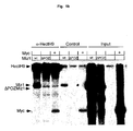
- Miz1 and HectH9 were synthesized by coupled transcription-translation in a reticulocyte lysate in the presence of 35 S-methionine ( Figure 1c). After synthesis, lysates were mixed and immuno-precipitated either with antibodies directed against HectH9 or a control serum. Miz1 was clearly detectable in anti-HectH9 immunoprecipitates, whereas control precipitates contained significantly lower levels of Miz1. To test whether the POZ domain of Miz1 is required for binding, the experiment was repeated with ⁇ POZ1.
- Miz1 binds to HectH9 in vivo
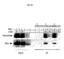
- the inventors transfected HeLa cells with expression vectors encoding Miz1, Miz1 ⁇ POZ and HectH9.
- Miz1 was recovered in anti-HectH9 immunoprecipitates, whereas control immunoprecipitates contained significantly less Miz1.
- No specific binding of Miz1 ⁇ POZ to HectH9 could be detected in vivo , consistent with the in vitro and two-hybrid data.
- ⁇ -HectH9 immunoprecipitates contained significant amounts of Myc, demonstrating that Myc binds to HectH9 in vivo . Most likely, therefore, binding of Myc to HectH9 is stabilized by additional proteins in vivo .
- Hect-domain proteins act as E3-ligases that ubiquitinate proteins to which they bind.
- the inventors transfected cells with expression plasmids encoding Myc, Miz1, HectH9 and a histidine-tagged ubiquitin.
- Transfected cells were lysed and ubiquitinated proteins were isolated by binding to Ni-agarose.
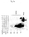
- Immunoblots of the eluates were probed with antibodies directed against Miz1 and Myc ( Figure 2a and data not shown). In these experiments, the inventors did not detect any activity of HectH9 against Miz1.
- HectH9 efficiently ubiquitinated Myc when both protein were co-expressed in the absence of Miz1.
- the inventors noted ubiquitinated forms of Myc up to a molecular weight of 250kd, strongly suggesting that HectH9 catalyzes the transfer of multiple ubiquitin moieties on Myc.
- Co-expression of Miz1 essentially abolished ubiquitination of Myc by Miz1.
- Progressive amino-terminal deletions in HectH9 revealed that the Miz1 binding domain on HectH9 is required for ubiquitination of Myc, suggesting that Myc and Miz1 compete for binding to HectH9 (data not shown).
- HectH9 might catalyze ubiquitination of Myc in two different manners: it might either catalyze the complex formation of Myc with an unknown E3-ligase or itself be the catalytically active E3-ligase.
- the inventors introduced a single point mutation into the carboxyl-terminus of HectH9 that replaces the catalytic cysteine with a serine.
- HectH9CS in contrast to wild type HectH9, did not ubiquitinate Myc in vivo , demonstrating that the catalytic activity of HectH9 is required for ubiquitination of Myc in vivo.
- poly-ubiquitin chains can be attached to target proteins and only those, in which ubiquitin is linked through lysine-48 target proteins for proteasomal degradation.
- poly-ubiquitin chains with a lysine-63 linkage do not target proteins to the proteasome.
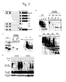
- the inventors performed ubiquitination assays using specific point mutants of his-tagged ubiquitin, in which either lysine 48 or lysine 63 is replaced by an arginine ( Figure 4c).
- Polyubiquitination can affect the function of the protein in different ways.
- the inventors In order to test how ubiquitination affects the function of Myc, the inventors initially performed transient reporter assays. No difference was seen in the ability of MycKR 6 to inhibit Miz1 dependent transactivation in these assays (data not shown). Also, no difference was seen in the ability of Myc and MycKR6 to activate an E-box dependent reporter derived from the prothymosin- ⁇ gene, in transient transfection assays.
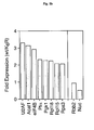
- RT-PCR analysis confirmed the differences in expression of these genes between cells expressing Myc and MycKR6 (Figure 5c). However, not all targets of Myc were differentially expressed between cells expressing Myc and MycKR 6 : for example, nucleolin and Rbb2 were induced equally well by both Myc and MycKR 6 . Taken together, the data show that MycKR6 is deficient in activation of a subset of Myc target genes in vivo.
- the six lysines that are replaced by arginines in KR 6 are located in a domain that has recently been shown to be involved in binding of the p300 and CBP co-activator. The inventors therefore speculated that ubiquitination of Myc might be required for efficient recruitment of p300.
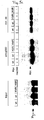
- the inventors co-transfected cells with HA-tagged p300 and either Myc or MyckKR 6 , either by itself or in the presence of HectH9. Lysates were then immunoprecipitated with antibodies against the HA-tag of p300 and the precipitates probed with antibodies directed against Myc (Figure 5c). In these experiments, efficient complex formation between Myc and p300 was observed in the presence of HectH9, but not in its absence.
- the experiments of the present invention have shown that the HECT-domain E3-Ligase, HECTH9, ubiquitinates Myc; Miz1 inhibits ubiquitination of Myc.
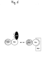
- Ubiquitination of Myc by HectH9 does not regulate Myc stability, since the polyubiquitin chain is linked via lysine-63 of ubiquitin. Rather, ubiquitination is required for transactivation of specific Myc target genes and for recruitment of the co-activator, p300. From this it is to be concluded that the transcriptional properties of Myc are regulated by site-specific ubiquitination.
- the present invention relates to a screening method for identifying compounds useful for the therapy of cancers in which the activity of Myc is causally involved, wherein a test compound's ability to interfere with the activity of HECT H9 is determined, said HECT H9 activity being selected from
- Myc encompasses the HECT H9 substrate c-Myc or any of its relatives that are activated by a ubiquitination reaction mediated by HECT H9 (n-Myc or 1-Myc).
- the present invention relates to a screening method for identifying compounds that inhibit transfer of ubiquitin to HECT H9, i.e. ubiquitination of HECT H9.
- HECT H9 is incubated, together with a ubiquitin activating enzyme (E1), a ubiquitin conjugating enzyme (E2), ubiquitin and ATP for a period of time sufficient to obtain a measurable level of ubiquitin associated with HECT H9, and the level of ubiquitination of HECT H9 in the presence or absence of a test compound is compared, and wherein a decrease in the level of HECT H9 ubiquitination in the presence of the test compound, as compared to the level of HECT H9 ubiquitination in the absence of the test compound, is indicative of the compound's ability to inhibit the activation of Myc.
- E1 ubiquitin activating enzyme
- E2 a ubiquitin conjugating enzyme
- ATP ubiquitin and ATP
- reaction that this embodiment of the invention is based on is a thioester formation between the active site cysteine of HECT H9 and the terminal glycine of ubiquitin.
- the proteins used in the screening assay are preferably recombinant proteins, which can be obtained according to conventional methods by transforming a suitable host with a plasmid carrying the sequence encoding the protein.
- the preferred host cells are bacterial cells, in particular E. coli cells, which are devoid of endogenous HECT H9.
- the cDNA sequences encoding the protein components HECT H9, E1, E2 and ubiquitin are available from the literature and from databases.
- the assay components HECT H9 preferably human HECT H9 (GenBank Accession No. BAC06833), E2 and ubiquitin are usually produced and purified as fusion proteins.
- the proteins may be fused to an affinity tag, which is a protein suitable for affinity purification, such as gluthathion S-transferase (GST, Amersham Pharmacia), maltose binding protein (MBP, New England Biolabs), chitin binding domain (New England Biolabs), the myc-epitope (EQKLISEEDL) or the His(6) tag (Qiagen, Novagene).
- GST gluthathion S-transferase
- MBP maltose binding protein
- chitin binding domain New England Biolabs
- EQKLISEEDL myc-epitope
- His(6) tag Qiagen, Novagene
- E1 may be purified due to its reversible interaction with ubiquitin according to known methods (e.g. Hatfield and Vierstra, 1990 and 1992).
- ubiquitin e.g. Hatfield and Vierstra, 1990 and 1992.
- a commercially available product e.g. from Sigma, Fluka
- Sigma, Fluka may be used as this assay component.
- the naturally occurring proteins are used; however, the proteins may contain deviations from the natural amino acid sequence as long as these deviations do not impair their functional activity.
- a suitable ubiquitin activating enzyme is the wheat UBA1 E1 (GenBank Accession number M55604), however, UBA1 E1 from other species, e.g. from Xenopus laevis, may also be used. E1 can be purified on a ubiquitin affinity matrix according to published procedures (e.g. Hatfield and Vierstra, 1990 and 1992).
- ubiquitin conjugating enzyme in a preferred embodiment, the human variant UBCH5b (GenBank accession number U39317) is used, although, also in this case, UBCH5b homologues from other species, e.g. Xenopus laevis, may be employed.
- UBCH5a GenBank accession number AAH05980
- UBCH5c GeneBank accession number AAH66917
- ubiquitin conjugating enzymes different from UBCH5a, b or c can be used, as long as these enzymes support the transfer of ubiquitin to the HECT H9 protein.
- the ubiquitin conjugating enzyme E2 is fused to an affinity tag which is selected from the ones listed above as suitable for HECT H9, but different from the tag chosen for HECT H9.
- an affinity tag which is selected from the ones listed above as suitable for HECT H9, but different from the tag chosen for HECT H9.
- His(6) or another tag different from GST is used for tagging E2.
- ATP regenerating system e.g. comprising 0.5 mM ATP, 60 ⁇ g/ml creatine phosphokinase, 6 .6 mM phosphocreatine, 10 mM Tris-HCl, 0.5 mM MgCl 2 , 1 mM KCl, 0.05 mM DTT
- ATP regenerating system e.g. comprising 0.5 mM ATP, 60 ⁇ g/ml creatine phosphokinase, 6 .6 mM phosphocreatine, 10 mM Tris-HCl, 0.5 mM MgCl 2 , 1 mM KCl, 0.05 mM DTT
- Ubiquitin is commercially available (Sigma), it may also be recombinantly produced; in this case it may be fused to various tags for purification, i.e. His(6), GST or for detection, i.e. myc-epitope, HA-epitope.
- ubiquitin comprises the N-terminal 76 amino acids required for its function.
- a tagged ubiquitin is employed in the assay.
- the ubiquitin used in the assay may also carry a non-proteinacious tag, e.g. biotin.
- the above-described assay essentially comprises the steps of the ubiquitination reaction itself and the step of measuring the extent of ubiquitin transfer to HECT H9.
- the first step comprises reacting the assay compounds listed above for a period of time sufficient to allow for the ubiquitination reaction, e.g. for 30 min.
- the reaction may either be conducted in solution by simply mixing the assay components, or alternatively, the reaction may be carried out by using immobilized HECT H9.
- HECT H9 carries an affinity tag (GST or one of the alternative tags mentioned above) that is used for its binding to a solid phase carrying the ligand for the respective affinity moiety, e.g. glutathione agarose or sepharose beads or microtiter plates coated with antibodies against the affinity moiety, e.g. commercially available anti-GST antibodies.
- the affinity-tagged, e.g. GST-tagged, HECT H9 is captured on microtiter plates that are coated with an antibody against GST (this step can be omitted in case the reaction has been carried out with HECT H9 bound to a solid phase).
- the unbound GST-HECT H9, the unincorporated ubiquitin and the other reaction partners are then washed off.
- the immobilized ubiquitin can be visualized by using an antibody that is directed against a tag epitope, e.g. the myc-epitope present in the recombinant tagged ubiquitin, which antibody carries a detectable label.
- Suitable labels are radioactive labels, e.g.
- enzymatic labels e.g. horseradish peroxidase or alkaline phosphatase, or fluorometric labels.
- quenched fluorophors e.g. Europium (Wallac, Turku, Finland) that will be dequenched upon incubation with an enhancer solution (Wallac) are used.
- the obtained values are compared to values obtained from reactions without HECT H9 (negative control, background) and to values obtained from a reaction mixture incubated in the presence of the solvent (usually DMSO) only (positive control).
- the physical proximity of ubiquitin molecules associated with HECT H9 (in the case of measuring the ubiquitin transfer to HECT H9) or to the HECT H9 substrate (in the case of determining ubiquitination of a HECT H9 substrate; see below) upon incubation at 37°C can be used to measure the extent of ubiquitin association with HECT H9 or its substrate by fluorescence resonance energy transfer (FRET, as described by Gershkovich et al., 1996, or by Matayoshi et al., 1990, ore recently reviewed by Selvin, 2000). FRET can only be achieved if certain conditions are fulfilled, i.e.
- fluorophor pairs with overlapping emission and excitation wavelengths, like europium/allophycocyanin, europium/Cy5, europium/PE (all commercially available from Wallac, Turku, Finland) and an minimal proximity of these fluorophors below 5-10nM.
- These fluorophors can be added either bound to antibodies directed against the affinity label, e.g. GST, or the epitope, e.g. the myc epitope, or can be directly coupled to HECT H9, HECT H9 substrate or ubiquitin (custom service of Wallac). When coupled to antibodies, the fluorophors are added to the reaction after its completion.
- the invention relates to a screening *method for identifying compounds that inhibit the transfer of ubiquitin from HECT H9 to Myc, i.e. ubiquitination mediated by HECT H9.
- HECT H9 is incubated, together with a ubiquitin activating enzyme (E1), a ubiquitin conjugating enzyme (E2), ubiquitin, ATP and a HECT H9 substrate protein (Myc) for a period of time sufficient to obtain a measurable level of ubiquitination of the substrate protein Myc and comparing the level of ubiquitination of Myc in the presence or absence of a test compound, wherein a decrease in the level of Myc ubiquitination in the presence of the test compound, as compared to the level of Myc ubiquitination in the absence of the test compound, is indicative of the compound's ability to inhibit the activation of Myc.
- E1 ubiquitin activating enzyme
- E2 a ubiquitin conjugating enzyme
- Myc HECT H9
- HECT H9 is able to ubiquitinate substrate proteins, but not substrate proteins that have mutations at positions necessary for the attachment of ubiquitin.
- Myc is the c-Myc onco-protein.
- embodiment a i.e. the assay based on transfer of ubiquitin to HECT H9
- embodiment b i.e. the assay based on substrate (Myc) ubiquitination.
- the substrate protein and DNA sequences are known from the literature and from data bases, e.g. c-Myc (GenBank accession No. P01106), 1-myc (GenBank accession No.AAO38672) or n-myc (GenBank accession No. P04198).
- c-Myc GenBank accession No. P01106
- 1-myc GenBank accession No.AAO38672
- n-myc GenBank accession No. P04198.
- a C-terminal fragment that contains the relevant site for ubiquitination by HECT H9 can be used.
- the size of the fragment may vary and can be easily determined in preliminary experiments by employing different sized peptide fragments in the intended assay format and determining suitable peptides.
- the screening methods of the invention are conducted in the high throughput format.
- such an assay is performed in 96 or 384 well plates in a suitable reaction volume, e.g. 50 ⁇ l, in the absence or presence of the test compounds, which are usually dissolved in DMSO.
- the substrate protein is captured by virtue of a tag, preferably MBP, and the amount of associated ubiquitin is quantified in the presence or absence of test compounds.
- a tag preferably MBP
- E1 ubiquitin activating enzyme
- E2 ubiquitin conjugating enzyme
- E1 is necessary to activate ubiquitin for the subsequent reaction, as it is the only enzyme that is able to recognize free ubiquitin in solution.
- E1 forms a thioester with ubiquitin in an ATP-dependent manner and transfers this now activated ubiquitin molecule to an E2 enzyme, where another thioester between E2 and ubiquitin is formed.
- the thioester formation of E2 with ubiquitin is strictly dependent on the presence of E1, which means that E2 alone is unable to form a thioester with free ubiquitin.
- thioesters are based on using the nature of a thioester with regard to reducibility by agents like dithiothreitol or ⁇ -mercapto ethanol.
- a mixture of E1, E2, ATP and ubiquitin is incubated at 37°C in the presence of the compounds identified to inhibit the transfer of ubiquitin to HECT H9 or DMSO as control.
- These samples are subsequently subjected to SDS-PAGE under nonreducing conditions; i.e. thioesters that have formed will not be broken up and transferred to PVDF or nitrocellulose membranes.
- Ubiquitin can be detected either by using monoclonal antibodies against ubiquitin (commercially available, e.g.
- a further secondary assay may be performed to confirm that the compounds identified in the assays of the invention are inhibitors specific for a HECT H9-mediated ubiquitination reaction and not inhibitors of reactions mediated by other HECT domain containing proteins.
- Such (one or more) assay(s) use another HECT domain containing protein, e.g. NEDD4 (GenBank Accession No. P46934.), SMURF1 (GenBank Accession No. Q9HCE7) or ITCHY (GenBank Accession No. Q96J02).
- the activity of Myc can be inhibited by preventing the interaction of the transcriptional co-activator p300 and the ubiquitinated Myc.
- a screening assay based on this principle is a protein-protein interaction assay, in which one of the interaction partners is usually bound to a solid support and binding of the other partner, which may carry a detectable epitope tag, is quantified.
- a compound that interferes with binding of the two proteins is a candidate for reduction of Myc activation.
- a candidate compound to inhibit tumor cell proliferation
- primary human tumor cells or various different human cell lines e.g. the colon cancer cell line HCT116 or the breast cancer cell line MCF7 are incubated with the candidate compound identified in the screen and the inhibition of tumor cell proliferation is tested by conventional methods, e.g. bromo-desoxyuridine or 3 H incorporation, Alamar BLUE.
- Compounds that exhibit an anti-proliferative effect in these assays may be further tested in tumor animal models and used for the therapy of tumors.
- Animal models useful for testing these compounds include those described above (Felsher and Bishop, 1999; Jain et al., 2002; Pelengaris et al., 1999; Pelengaris et al., 2002b) that exhibit Myc-dependent tumorigenesis.
- xenograft models of human cancer cell lines containing elevated and/or deregulated Myc such as breast, colon, cervical, small cell lung carcinomas, osteosarcomas, glioblastomas, melanoma and myeloid leukemias, can be used to test the efficacy of such compounds.
- Toxicity and therapeutic efficacy of the compounds identified as drug candidates can be determined by standard pharmaceutical procedures, which include conducting cell culture and animal experiments to determine the IC 50 , LD 50 , ED 50 .
- the data obtained are used for determining the human dose range, which will also depend on the dosage form (tablets, capsules, aerosol sprays, ampules, etc.) and the administration route (oral, buccal, nasal, paterental or rectal).
- a pharmaceutical composition containing the compound as the active ingredient can be formulated in conventional manner using one or more physiologically active carriers and excipients. Methods for making such formulations can be found in manuals, e.g. "Remington Pharmaceutical Sciences".
- the POZ domain of Miz-1 (amino acids 1-117) was amplified by PCR and inserted into pGBT9 (Clontech). A total of 1x10 6 independent transformants of a human HeLa cDNA library fused to the GAL4 activation domain (Matchmaker, Clontech) were screened. In vitro transcription/ translation was carried out in reticulocyte lysate (Promega) in the presence of [S 35 ]-methionine. The translated product were mixed and incubated for 90 min at 4°C. Immunoprecipitation was carried out using the indicated antibodies immobilized on protein G-sepharose beads.
- the beads were washed four times in 20 mM HEPES pH 7.8, 100 mM KCl, 5 mM MgCl 2 , 0.5 mM DTT, 0.5% Igepal CA-630. Bound proteins were analyzed by SDS- gel electrophoresis and fluorography.
- HeLa cells were grown in DMEM supplemented with 10% heat-inactivated FCS, 2mM L-glutamine, 100U/ml penicillin and 100 ⁇ g/ml streptomycin. Recombinant retroviruses were generated and used as described (Peukert et al., 1997). Infected cells were selected and analyzed within 2 passages after selection.
- pCDNA3 Invitrogen
- CMV-HectH9 kind gift of Kristian Helin
- pSUPER plasmids were a kind gift of Rene Bernards (Brummelkamp et al., 2002).
- Chromatin immunoprecipitation (ChIP)
- ChIP assays were performed as described previously (Bouchard et al., 2001) using the following antibodies: anti-Myc (N-262; sc-764), anti-p300 and control antibody.
- Immunoprecipitated DNA samples were amplified by real-time PCR (ABI-Prism) using a Eurogentech Cybr-Green Core kit and primers specific for the E-boxes of the indicated genes. Primer sequences are available upon request.
- HeLa cells were transfected with expression plasmids encoding the indicated proteins. Lysates were precipitated with Ni-agarose and the precipitates probed with antibodies against Myc.
- Figure 2 shows that it is Myc that is ubiquitinated by HectH9.
- the experiment was carried out as in panels b and c.
- the top panel shows Ni-agarose precipitates of lysates of transfected cells, which were probed with an antibody directed against Myc.
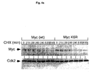
- the lower panels show immunoblots of aliquots of the transfected cells with ⁇ -Myc and ⁇ -Cdk2 antibodies showing equal expression of Myc and MycKR 6 after transfection.
- an enzyme mixture containing 1ng his-tagged UBA1 (E1), 30ng his-tagged UBCH5 (E2), 50ng MBP-HECT H9 and 100ng GST-ubiquitin are mixed in reaction buffer (20mM Tris/HCl pH 7.5, 150mM NaCl, 2mM MgCl 2 , 5%gylcerol, 0.5% DTT and 0.5% BSA) in a volume of 20 ⁇ l.
- reaction buffer 20mM Tris/HCl pH 7.5, 150mM NaCl, 2mM MgCl 2 , 5%gylcerol, 0.5% DTT and 0.5% BSA
- This solution is mixed with 5 ⁇ l of the test compound (100 ⁇ M in 20% DMSO), being dispensed by aspirating from a compound source plate.
- the enzymatic reaction is started by the addition of 5 ⁇ l 4mM ATP.
- Electrode-labeled antibodies recognizing the GST protein (Wallac/Perkin Elmer) are added (30 ⁇ l of a solution containing 3 ⁇ g/ml labeled antibody in PBS containing 0.05%Tween 20). After incubation for 60 min, the plates are washed with PBST and fluorescence is enhanced by addition of DELFIA enhancement solution (Wallac/PerkinElmer).
- Europium fluorescence is measured in a fluorescence plate reader (i.e. Victor 2 , Wallac/PerkinElmer) at an excitation wavelength of 340nm and an emission wavelength of 615nM.
- each plate contains a column of negative controls with 5 ⁇ l 20% DMSO solution instead of test compound.
- an enzyme mixture containing 1ng his-tagged UBA1 (E1), 30ng his-tagged UBCH5 (E2), 50ng his-tagged HECT H9, 50ng of MBP-tagged c-Myc (full-length protein) and 100ng GST-ubiquitin are mixed in reaction buffer (20mM Tris/HCl pH 7.5, 150mM NaCl, 2mM MgCl 2 , 5%gylcerol, 0.5% DTT and 0.5% BSA) in a volume of 20 ⁇ l.
- This solution is mixed with 5 ⁇ l of the test compound (100 ⁇ M in 20% DMSO), being dispensed by aspirating from a compound source plate.
- the enzymatic reaction is started by the addition of 5 ⁇ l 4mM ATP. Plates are incubated for 60min at room temperature and washed 3 times in reaction buffer. Subsequently, Europium-labeled antibodies recognizing the GST protein (Wallac/Perkin Elmer) are added (30 ⁇ l of a solution containing 3 ⁇ g/ml labeled antibody in PBS containing 0.05%Tween 20). After incubation for 60 min plates are washed with PBST and fluorescence is enhanced by addition of DELFIA enhancement solution (Wallac/PerkinElmer). Europium fluorescence is measured in a fluorescence plate reader (i.e.
Landscapes
- Chemical & Material Sciences (AREA)
- Life Sciences & Earth Sciences (AREA)
- Organic Chemistry (AREA)
- Health & Medical Sciences (AREA)
- Wood Science & Technology (AREA)
- Zoology (AREA)
- Engineering & Computer Science (AREA)
- Bioinformatics & Cheminformatics (AREA)
- Genetics & Genomics (AREA)
- Proteomics, Peptides & Aminoacids (AREA)
- General Health & Medical Sciences (AREA)
- Biotechnology (AREA)
- Molecular Biology (AREA)
- Biochemistry (AREA)
- General Engineering & Computer Science (AREA)
- Microbiology (AREA)
- Analytical Chemistry (AREA)
- Physics & Mathematics (AREA)
- Medicinal Chemistry (AREA)
- Biophysics (AREA)
- Biomedical Technology (AREA)
- Immunology (AREA)
- Medicines That Contain Protein Lipid Enzymes And Other Medicines (AREA)
- Investigating Or Analysing Biological Materials (AREA)
- Measuring Or Testing Involving Enzymes Or Micro-Organisms (AREA)
Abstract
Screening method for identifying compounds useful for the therapy of cancers in
which the activity of Myc is causally involved. The method is based on measuring the
activity of the E3 ligase HECT H9, which is required for ubiquitination of Myc.
Description
The present invention relates to the therapy of cancer. In particular, the present
invention relates to screening methods for identifying compounds that inhibit the
activity of the proto-oncogene Myc and are thus useful for the therapy of cancer.
The proto-oncogene c-myc encodes a transcription factor (Myc) that can both activate
and repress transcription. Myc activates transcription as part of a binary complex with
its partner protein, Max. The complex binds to specific DNA sequences, termed E-boxes,
and recruits a number of co-activators to DNA; indeed, four distinct classes of
co-activators have been described that may mediate transcriptional activation by Myc.
First, a short conserved domain in the amino-terminus of Myc ("MycboxII") binds to
the TRRAP protein and Myc recruits the histone acetyl-transferases, Tip60 and GCN5
via binding to TRAPP. Similarly, Myc recruits the p300 and CBP acetyl-transferases
though a direct interaction of the terminus-terminus of Myc with each protein.
Second, Myc interacts with mediator complexes in vitro and recruits mediator to E-boxes
in vivo independently of histone acetylation, suggesting that interactions with
mediator complexes contribute to transcriptional activation by Myc. Third, Myc
recruits the E3-ligase Skp2 to DNA and recruitment of Skp2 contributes to
transcriptional activation by Myc. Fourth, Myc binds to BAF53, a subunit of the
chromatin-remodeling BAF complex, implying chromatin-remodeling as a
mechanism that may contribute to Myc-dependent gene activation. The precise
contribution of each co-factor to the activation of individual genes by Myc is largely
unknown.
Myc represses transcription at least in part through the formation of a ternary complex
that contains Max and the transcription factor, Miz1. Miz1 binds to the start site of
several Myc-repressed genes and activates these genes. Myc does not bind to the start
sites of Myc-repressed genes directly but is recruited to these sites through interaction
with Miz1. In contrast to free Miz1, the Myc/Miz1 complex acts as a transcriptional
repressor complex. Free Miz1 requires p300 as co-activator and binding of Myc to
Miz1 displaces p300 from Miz1, explaining in part how Myc inhibits transactivation
by Miz1. However, transcriptional repression by Myc requires domains in the amino-terminus
that are not required for displacement of p300 from Miz1, suggesting that
additional co-factors contribute to transcriptional repression by Myc.
The (c-) MYC gene and two of its relatives, MYCN or MYCL, contribute to the genesis
of a wide variety of human tumors. In these tumors, the expression of MYC genes is
enhanced relative to the surrounding or normal tissue, arguing that there is a selective
pressure for high expression of Myc proteins during tumor development. For
example, the MYCN gene is amplified in a subset of childhood neuroblastoma,
correlating with extremely poor prognosis of the affected children (Brodeur et al.,
1984; Schwab et al., 1983). In other tumors, expression of a MYC family gene is
increased because mutations occur in the signaling pathway that control their
expression: one example are the mutations in the APC pathway that affect cMYC
expression in colorectal carcinomas (He et al., 1998; van de Wetering et al., 2002).
Besides colon cancer, elevated or deregulated expression of c-Myc has been detected
in a wide range of human cancers and is often associated with aggressive, poorly
differentiated tumors. Such cancers include breast, cervical, small cell lung
carcinomas, osteosarcomas, glioblastomas, melanoma and myeloid leukemias
(reviewed in Pelengaris et al., 2002a).
Examples of Myc-activated target genes, whose activation results in stimulation of
tumor growth, proliferation and survival are cyclin D2 and CDK4 which are important
for cell cycle progression, translation initiation factors eIF4 and eIF2 important for
cell growth, as well as ornithine decarboxylase and carbamyl-phosphate synthase,
enzymes required for polyamine and pyrimidine biosynthesis, respectively (reviewed
in Grandori and Eisenman, 1997; Pelengaris et al., 2002a). Examples of Myc-repressed
genes, whose repression results in cell proliferation, are the cell cycle
inhibitors p 15 ink4b and p21 Cip 1.
Inhibition of Myc activity is a highly attractive approach for drug discovery in
oncology, since recent experimental data suggested that even a brief inhibition of Myc
expression may be sufficient to permanently stop tumor growth and induce regression
of tumors. Jain et al. (2002) engineered a conditional transgenic mouse to overexpress
Myc, which induced formation of highly malignant osteogenic sarcoma. A brief loss
of Myc overexpression caused the tumor cells to differentiate into mature osteocytes
that formed histologically normal bone. Felsher and Bishop (1999) showed that
transgenic mice expressing the myc oncogene in hematopoietic cells developed
malignant T cell leukemias and acute myeloid leukemias. However, when this gene
was switched off the leukemic cells underwent proliferative arrest, differentiation, and
apoptosis. Pelengaris et al. (1999) targeted expression of an inducible form of the c-Myc
protein to the epidermis of mice and observed formation of angiogenic
premalignant skin lesions, which regressed when the c-Myc protein was deactivated.
In general, specific pharmacological interference with the function of transcription
factors has been difficult to achieve. This is particularly true for Myc: despite its
obvious value as a potential target for tumor therapy, no drugs have emerged that
specifically interfere with its function. For example, screens aimed at disrupting the
Myc/Max interface have only yielded compounds with extremely low potency (Berg
et al., 2002).
It was an object of the invention to elucidate the mechanisms that regulate the
activation and thus the transcriptional activity of Myc. The knowledge about such
mechanisms is the key for providing screening assay methods for identifying
compounds that interfere with Myc activation and are thus useful in the therapy of
cancer.
As mentioned above, it has been suggested that Myc may exist in an activating and in
a repressive state, potentially depending on the site its binds to DNA and/or its site-specific
interaction partners.
In the experiments of the invention, it has surprisingly been found that an E3-Ligase,
HectH9, ubiquitinates Myc on lysine residues close to the carboxyl-terminus of Myc.
It could be shown that ubiquitination does not target Myc for degradation, but is
required for the recruitment of the co-activator p300. It was shown that ubiquitination
of Myc by HectH9 is inhibited by Miz1. Since Miz1 binds to the start site of many
Myc-repressed genes but is absent from E-box elements in vivo, the findings of the
invention describe a mechanism how the transcriptional properties of Myc are
regulated by the local chromatin environment.
Miz1 is a zinc finger transcription factor that carries at its amino-terminus a POZ-domain.
POZ domains are protein-protein interaction modules; in Miz1, the POZ-domain
is required for transcriptional activation by Miz1.
In order to understand the function of this domain in more detail, the inventors
performed a two-hybrid screen looking for novel interaction partners. In this screen,
the inventors obtained multiple clones that are derived from a single gene, which
encodes a HECT-domain E3-ligase alternatively named HectH9, LASU1 or
KIAA0312 (Figure 1a). A protein corresponding to the carboxyl-terminus of HectH9
has also been described as a DNA binding protein ("UREB1; Upstream-element-binding
protein 1 "); however, the inventors have been unable to confirm a significant
DNA binding activity of HectH9. HectH9 belongs to the HECT-domain family of
ubiquitin ligases ("homologous to E6AP carboxy-terminus"), which are characterized
by a conserved carboxy-terminal catalytic domain. Among the conserved amino acids
is a cysteine residue, which is required for thioester formation with ubiquitin. The
clones recovered in the two-hybrid screen define a small region sufficient for
interaction with Miz1. The amino-terminus of HectH9 contains two UBA (ubiquititn
associated motif) domains, that are known to bind ubiquitin in several ubiquitin-binding
proteins, a WWE protein-interaction domain and two putative nuclear
localization signals (NLS; (a, b). Consistent with the presence of two NLS, two
different antibodies directed against HectH9 detected the endogenous protein in the
cell nucleus of Hela cells (Figure 1b and data not shown). Controls using cells that
were stably transfected with a pSUPER vector, which directs the synthesis of a siRNA
against HectH9, showed that the signal detected in the immune-fluorescence is
specific for HectH9 (Figure 1b, middle panel). Immunoblots confirmed that cells
transfected with pSUPER-HectH9 expressed lower levels of protein than cells
transfected with an empty pSUPER vector (Figure 1b, lower panel).
To test whether HectH9 binds to Miz1 in vitro, Miz1 and HectH9 were synthesized by
coupled transcription-translation in a reticulocyte lysate in the presence of 35S-methionine
(Figure 1c). After synthesis, lysates were mixed and immuno-precipitated
either with antibodies directed against HectH9 or a control serum. Miz1 was clearly
detectable in anti-HectH9 immunoprecipitates, whereas control precipitates contained
significantly lower levels of Miz1. To test whether the POZ domain of Miz1 is
required for binding, the experiment was repeated with ΔPOZ1. No ΔPOZ-Miz1 was
recovered in anti-HectH9 precipitates, demonstrating that the POZ-domain of Miz1
mediates binding to HectH9 in vitro. The inventors also tested whether HectH9 binds
to Myc in vitro and did not detect an interaction between HectH9 and Myc in these
experiments.
To test whether Miz1 binds to HectH9 in vivo, the inventors transfected HeLa cells
with expression vectors encoding Miz1, Miz1ΔPOZ and HectH9. Miz1 was recovered
in anti-HectH9 immunoprecipitates, whereas control immunoprecipitates contained
significantly less Miz1. No specific binding of Miz1ΔPOZ to HectH9 could be
detected in vivo, consistent with the in vitro and two-hybrid data. α-HectH9
immunoprecipitates contained significant amounts of Myc, demonstrating that Myc
binds to HectH9 in vivo. Most likely, therefore, binding of Myc to HectH9 is
stabilized by additional proteins in vivo.
Hect-domain proteins act as E3-ligases that ubiquitinate proteins to which they bind.
To test whether HectH9 ubiquitinates either Miz1 or Myc, the inventors transfected
cells with expression plasmids encoding Myc, Miz1, HectH9 and a histidine-tagged
ubiquitin. Transfected cells were lysed and ubiquitinated proteins were isolated by
binding to Ni-agarose. Immunoblots of the eluates were probed with antibodies
directed against Miz1 and Myc (Figure 2a and data not shown). In these experiments,
the inventors did not detect any activity of HectH9 against Miz1. In contrast, HectH9
efficiently ubiquitinated Myc when both protein were co-expressed in the absence of
Miz1. In these experiments, the inventors noted ubiquitinated forms of Myc up to a
molecular weight of 250kd, strongly suggesting that HectH9 catalyzes the transfer of
multiple ubiquitin moieties on Myc. Co-expression of Miz1 essentially abolished
ubiquitination of Myc by Miz1. Progressive amino-terminal deletions in HectH9
revealed that the Miz1 binding domain on HectH9 is required for ubiquitination of
Myc, suggesting that Myc and Miz1 compete for binding to HectH9 (data not shown).
HectH9 might catalyze ubiquitination of Myc in two different manners: it might either
catalyze the complex formation of Myc with an unknown E3-ligase or itself be the
catalytically active E3-ligase. To distinguish between the two possibilities, the
inventors introduced a single point mutation into the carboxyl-terminus of HectH9
that replaces the catalytic cysteine with a serine. HectH9CS, in contrast to wild type
HectH9, did not ubiquitinate Myc in vivo, demonstrating that the catalytic activity of
HectH9 is required for ubiquitination of Myc in vivo.
Surprisingly, neither overexpression nor depletion of HectH9 had any detectable
effect of Myc stability in several assays (data not shown). In order to be able to assess
the functional consequences of HectH9-mediated ubiquitination of Myc, the inventors
therefore decided to map the lysine residues in Myc that are targeted by HectH9
(Figure 3). To do so, the inventors measured in parallel both binding to HectH9 and
ubiquitination by HectH9 of a series of both N-terminal and C-terminal deletion
mutants described by Tansey and colleagues (Figure 3a). In these experiments, full-length
Myc bound efficiently to HectH9 and was efficiently ubiquitinated by HectH9,
as described above (Figure 3b). Deletion from the amino-terminus led to a progressive
loss of both binding and ubiquitination: for both ΔN1Myc and ΔN2Myc, residual
binding could be detected and there was some residual ubiquitination. In contrast,
ΔN3Myc neither bound HectH9 nor was ubiquitinated by HectH9. Most likely,
therefore, the amino-terminus of Myc is required for binding to HectH9 and the loss
of ubiquitination of amino-terminally deleted mutants of Myc by HectH9 is an
indirect consequence of the reduction in binding of such proteins.
Both ΔC1Myc and ΔC2Myc bound efficiently to Myc in vivo (Figure 3 c). ΔC1Myc
was ubiquitinated by HectH9 to the same extent as wild type Myc. In contrast,
ΔC2Myc was not detectably ubiquitinated by HectH9. Since ΔC2Myc bound to
HectH9 but was not ubiquitinated, the inventors speculated that the region between
ΔC1 and ΔC2 might contain the target lysines for HectH9. To test this idea, the
inventors replaced the six lysines in this region by arginine in full-length Myc,
generating MycKR6
Immunofluorescence experiments revealed that K6R was localized in the cell nucleus,
demonstrating that replacement of the lysines in the nuclear localisation signal by
arginine did not induce gross alterations in the subcellular localisation. The inventors
next repeated the ubiquitination assays using wild type Myc and MycKR6 in parallel.
Ubiquitination of MycKR6 by HectH9 was strongly reduced relative to wild type Myc
(Figure 3d), although it was not completely abolished. Strikingly, also the basal
ubiquitination that occurs in the absence of co-expressed HectH9 was strongly
reduced relative to wild-type Myc, demonstrating that a significant percentage of
ubiquitination of Myc occurs on one or more of the six lysine residues the inventors
have identified (Figure 3d, lower panel).
To test whether ubiquitination of Myc at these sites triggers degradation of Myc, the
inventors infected NIH3T3 cells with recombinant retroviruses that express Myc or
MycKR6. Pools of infected cells were treated with cycloheximide for different lengths
of time (Figure 4a). Cell lysates were prepared and immunoblots were probed with the
monoclonal antibody 9E10, which specifically recognizes human Myc. In these
experiments, the inventors observed that the rate of degradation of wild type Myc and
MycKR6 was identical. Quantitation of the results (Figure 4b) showed that the half-time
of MycKR6 was unaltered relative to wild type Myc. From these results and
those described above, the inventors concluded that ubiquitination by HectH9 does
not regulate the stability of the Myc protein.
Several types of poly-ubiquitin chains can be attached to target proteins and only
those, in which ubiquitin is linked through lysine-48 target proteins for proteasomal
degradation. In contrast, poly-ubiquitin chains with a lysine-63 linkage do not target
proteins to the proteasome. In order to determine the type of linkage, which HectH9
transfers to Myc, the inventors performed ubiquitination assays using specific point
mutants of his-tagged ubiquitin, in which either lysine 48 or lysine 63 is replaced by
an arginine (Figure 4c). Mutation of lysine 48 did not affect the ability of HectH9 to
transfer ubiquitin on Myc; in contrast, mutation of lysine 63 abolished the ability of
HectH9 to transfer ubiquitin on Myc. The inventors concluded that HectH9 catalyzes
the assembly of a lysine-63 linked poly-ubiquitin chain on Myc.
Polyubiquitination can affect the function of the protein in different ways. In order to
test how ubiquitination affects the function of Myc, the inventors initially performed
transient reporter assays. No difference was seen in the ability of MycKR6 to inhibit
Miz1 dependent transactivation in these assays (data not shown). Also, no difference
was seen in the ability of Myc and MycKR6 to activate an E-box dependent reporter
derived from the prothymosin-α gene, in transient transfection assays.
In order to test whether these effects extend to the regulation of endogenous target
genes by Myc, the inventors infected NIH3T3 cells with retroviruses expressing
either Myc, MycKR6 or an empty control vector (pBabepuro). Cells were then serum-starved
for 48 hours to deplete endogenous Myc and then RNA was extracted and
subjected to an array analysis using an 11.5k cDNA array (Figure 5b). This analysis
revealed a class of genes that was consistently downregulated in cells expressing KR6
relative to cells expressing wildtype Myc. Comparison with a database of Myc target
genes revealed that 80% of these genes were known target for activation of Myc. RT-PCR
analysis confirmed the differences in expression of these genes between cells
expressing Myc and MycKR6 (Figure 5c). However, not all targets of Myc were
differentially expressed between cells expressing Myc and MycKR6: for example,
nucleolin and Rbb2 were induced equally well by both Myc and MycKR6. Taken
together, the data show that MycKR6 is deficient in activation of a subset of Myc
target genes in vivo.
The six lysines that are replaced by arginines in KR6 are located in a domain that has
recently been shown to be involved in binding of the p300 and CBP co-activator. The
inventors therefore speculated that ubiquitination of Myc might be required for
efficient recruitment of p300. To test this idea, the inventors co-transfected cells with
HA-tagged p300 and either Myc or MyckKR6, either by itself or in the presence of
HectH9. Lysates were then immunoprecipitated with antibodies against the HA-tag of
p300 and the precipitates probed with antibodies directed against Myc (Figure 5c). In
these experiments, efficient complex formation between Myc and p300 was observed
in the presence of HectH9, but not in its absence. Importantly, the efficiency of
complex formation was reduced for MycKR6, suggesting that the lysine residues need
to be ubiquitinated for complex formation to occur. The data strongly suggest that
MycKR6 is deficient in transactivation of some target genes becuase binding of Myc
to p300 requires ubiquitination at one or more of the six lysines targeted by HectH9.
One prediction from this model is that Myc should recruit p300 sleectively to E-box
elements, but not to Miz1 sites, since Miz1 inhibits ubiquitination by HectH9 (see
Figure 2a). The inventors tested this model using a cell line that carries a tetracyclin-inducible
Myc. Chromatin-immunoprecipitation was performed both before and after
induction of Myc; as targets, the inventors chose nucleolin (E-box dependent
activation by Myc) and c/EBPα (a Miz1-dependent target of repression), since initial
experiments had shown that the amount of Myc bound to both loci is comparable to
each other and higher than for several other genes tested (Figure 5d and data not
shown). Induction of Myc led to a similar increase of Myc binding at both loci. At the
nucleolin locus, there was a clear increase in binding of p300; in contrast, the amount
of p300 at the c/EBPα locus remained unaltered. Similar experiments using antibodies
directed against TRRAP, p400, Tip60 and Tip48/49 did not reveal a similar difference
(data not shown), arguing that MycboxII-dependent interactions occur at both sites of
activation and repression.
In summary, the experiments of the present invention have shown that the HECT-domain
E3-Ligase, HECTH9, ubiquitinates Myc; Miz1 inhibits ubiquitination of Myc.
Ubiquitination of Myc by HectH9 does not regulate Myc stability, since the
polyubiquitin chain is linked via lysine-63 of ubiquitin. Rather, ubiquitination is
required for transactivation of specific Myc target genes and for recruitment of the co-activator,
p300. From this it is to be concluded that the transcriptional properties of
Myc are regulated by site-specific ubiquitination. These findings make, for the first
time, the cancer target Myc accessible for screening assays that allow for identifying
Myc inhibitors useful in cancer therapy.
The present invention relates to a screening method for identifying compounds useful
for the therapy of cancers in which the activity of Myc is causally involved, wherein a
test compound's ability to interfere with the activity of HECT H9 is determined, said
HECT H9 activity being selected from
For the present invention, the term "Myc" encompasses the HECT H9 substrate c-Myc
or any of its relatives that are activated by a ubiquitination reaction mediated by
HECT H9 (n-Myc or 1-Myc).
In the embodiment a), the present invention relates to a screening method for
identifying compounds that inhibit transfer of ubiquitin to HECT H9, i.e.
ubiquitination of HECT H9. In this method, HECT H9 is incubated, together with a
ubiquitin activating enzyme (E1), a ubiquitin conjugating enzyme (E2), ubiquitin and
ATP for a period of time sufficient to obtain a measurable level of ubiquitin
associated with HECT H9, and the level of ubiquitination of HECT H9 in the
presence or absence of a test compound is compared, and wherein a decrease in the
level of HECT H9 ubiquitination in the presence of the test compound, as compared
to the level of HECT H9 ubiquitination in the absence of the test compound, is
indicative of the compound's ability to inhibit the activation of Myc.
The reaction that this embodiment of the invention is based on is a thioester formation
between the active site cysteine of HECT H9 and the terminal glycine of ubiquitin.
The components for this type of assay can be obtained as follows:
The proteins used in the screening assay are preferably recombinant proteins, which
can be obtained according to conventional methods by transforming a suitable host
with a plasmid carrying the sequence encoding the protein. In the case of HECT H9,
the preferred host cells are bacterial cells, in particular E. coli cells, which are devoid
of endogenous HECT H9. The cDNA sequences encoding the protein components
HECT H9, E1, E2 and ubiquitin are available from the literature and from databases.
The assay components HECT H9, preferably human HECT H9 (GenBank Accession
No. BAC06833), E2 and ubiquitin are usually produced and purified as fusion
proteins. The proteins may be fused to an affinity tag, which is a protein suitable for
affinity purification, such as gluthathion S-transferase (GST, Amersham Pharmacia),
maltose binding protein (MBP, New England Biolabs), chitin binding domain (New
England Biolabs), the myc-epitope (EQKLISEEDL) or the His(6) tag (Qiagen,
Novagene). The fusion protein can be expressed, e.g. in E. coli, and purified
according to standard protocols.
E1 may be purified due to its reversible interaction with ubiquitin according to known
methods (e.g. Hatfield and Vierstra, 1990 and 1992). In the case of using untagged
ubiquitin, a commercially available product (e.g. from Sigma, Fluka) may be used as
this assay component.
Preferably, the naturally occurring proteins are used; however, the proteins may
contain deviations from the natural amino acid sequence as long as these deviations
do not impair their functional activity.
A suitable ubiquitin activating enzyme (E1) is the wheat UBA1 E1 (GenBank
Accession number M55604), however, UBA1 E1 from other species, e.g. from
Xenopus laevis, may also be used. E1 can be purified on a ubiquitin affinity matrix
according to published procedures (e.g. Hatfield and Vierstra, 1990 and 1992).
As ubiquitin conjugating enzyme (E2), in a preferred embodiment, the human variant
UBCH5b (GenBank accession number U39317) is used, although, also in this case,
UBCH5b homologues from other species, e.g. Xenopus laevis, may be employed.
Alternatively, UBCH5a (GenBank accession number AAH05980) or UBCH5c
(GenBank accession number AAH66917) can be used. Alternatively, ubiquitin
conjugating enzymes different from UBCH5a, b or c can be used, as long as these
enzymes support the transfer of ubiquitin to the HECT H9 protein. Preferably, the
ubiquitin conjugating enzyme E2 is fused to an affinity tag which is selected from the
ones listed above as suitable for HECT H9, but different from the tag chosen for
HECT H9. For example, in the case that GST-HECT H9 is used, His(6) or another tag
different from GST is used for tagging E2.
To sustain a sufficient ATP level during the entire ubiquitination reaction, a so-called
"ATP regenerating system" (e.g. comprising 0.5 mM ATP, 60 µg/ml creatine
phosphokinase, 6 .6 mM phosphocreatine, 10 mM Tris-HCl, 0.5 mM MgCl2, 1 mM
KCl, 0.05 mM DTT) may be advantageously employed (Murray, 1991).
Ubiquitin is commercially available (Sigma), it may also be recombinantly produced;
in this case it may be fused to various tags for purification, i.e. His(6), GST or for
detection, i.e. myc-epitope, HA-epitope. In both cases, ubiquitin comprises the N-terminal
76 amino acids required for its function. Preferably, a tagged ubiquitin is
employed in the assay. The ubiquitin used in the assay may also carry a non-proteinacious
tag, e.g. biotin.
The above-described assay essentially comprises the steps of the ubiquitination
reaction itself and the step of measuring the extent of ubiquitin transfer to HECT H9.
The first step comprises reacting the assay compounds listed above for a period of
time sufficient to allow for the ubiquitination reaction, e.g. for 30 min.
The reaction may either be conducted in solution by simply mixing the assay
components, or alternatively, the reaction may be carried out by using immobilized
HECT H9. In this case, HECT H9 carries an affinity tag (GST or one of the
alternative tags mentioned above) that is used for its binding to a solid phase carrying
the ligand for the respective affinity moiety, e.g. glutathione agarose or sepharose
beads or microtiter plates coated with antibodies against the affinity moiety, e.g.
commercially available anti-GST antibodies.
After the reaction has been completed, the extent of ubiquitin associated with HECT
H9 can be measured in different ways:
In case the ubiquitination reaction has been carried out in solution, the affinity-tagged,
e.g. GST-tagged, HECT H9 is captured on microtiter plates that are coated with an
antibody against GST (this step can be omitted in case the reaction has been carried
out with HECT H9 bound to a solid phase). The unbound GST-HECT H9, the
unincorporated ubiquitin and the other reaction partners are then washed off.
Subsequently, the immobilized ubiquitin can be visualized by using an antibody that
is directed against a tag epitope, e.g. the myc-epitope present in the recombinant
tagged ubiquitin, which antibody carries a detectable label. Suitable labels are
radioactive labels, e.g. 125I, enzymatic labels, e.g. horseradish peroxidase or alkaline
phosphatase, or fluorometric labels. In a preferred embodiment, quenched
fluorophors, e.g. Europium (Wallac, Turku, Finland) that will be dequenched upon
incubation with an enhancer solution (Wallac), are used. The obtained values are
compared to values obtained from reactions without HECT H9 (negative control,
background) and to values obtained from a reaction mixture incubated in the presence
of the solvent (usually DMSO) only (positive control).
Alternatively to using the ELISA type assay described above to detect the amount of
bound ubiquitin, the physical proximity of ubiquitin molecules associated with HECT
H9 (in the case of measuring the ubiquitin transfer to HECT H9) or to the HECT H9
substrate (in the case of determining ubiquitination of a HECT H9 substrate; see
below) upon incubation at 37°C can be used to measure the extent of ubiquitin
association with HECT H9 or its substrate by fluorescence resonance energy transfer
(FRET, as described by Gershkovich et al., 1996, or by Matayoshi et al., 1990, ore
recently reviewed by Selvin, 2000). FRET can only be achieved if certain conditions
are fulfilled, i.e. fluorophor pairs with overlapping emission and excitation
wavelengths, like europium/allophycocyanin, europium/Cy5, europium/PE (all
commercially available from Wallac, Turku, Finland) and an minimal proximity of
these fluorophors below 5-10nM. These fluorophors can be added either bound to
antibodies directed against the affinity label, e.g. GST, or the epitope, e.g. the myc
epitope, or can be directly coupled to HECT H9, HECT H9 substrate or ubiquitin
(custom service of Wallac). When coupled to antibodies, the fluorophors are added to
the reaction after its completion. No further washing steps are necessary and signals
(excitation at 340 nm and emission measurement at 665 nm in the case of the FRET
pair allophycocyanin and europium) are measured after incubation at 4°C for 30 min,
allowing the binding of the antibodies and the subsequent energy transfer between the
fluorophors. In case of direct labeling of reaction components, i.e. ubiquitin or HECT
H9, real time measurements can be performed allowing the detection of kinetic
differences in the reaction.
In the alternative embodiment b), the invention relates to a screening *method for
identifying compounds that inhibit the transfer of ubiquitin from HECT H9 to Myc,
i.e. ubiquitination mediated by HECT H9. In this embodiment HECT H9 is incubated,
together with a ubiquitin activating enzyme (E1), a ubiquitin conjugating enzyme
(E2), ubiquitin, ATP and a HECT H9 substrate protein (Myc) for a period of time
sufficient to obtain a measurable level of ubiquitination of the substrate protein Myc
and comparing the level of ubiquitination of Myc in the presence or absence of a test
compound, wherein a decrease in the level of Myc ubiquitination in the presence of
the test compound, as compared to the level of Myc ubiquitination in the absence of
the test compound, is indicative of the compound's ability to inhibit the activation of
Myc.
This embodiment is based on the observation obtained in the experiments of the
invention that HECT H9 is able to ubiquitinate substrate proteins, but not substrate
proteins that have mutations at positions necessary for the attachment of ubiquitin.
In a preferred embodiment, Myc is the c-Myc onco-protein.
With regard to the assay components and the reaction conditions, the features
described for embodiment a), i.e. the assay based on transfer of ubiquitin to
HECT H9, also apply to embodiment b), i.e. the assay based on substrate (Myc)
ubiquitination. These features refer both to to assay components and reaction
conditions.
The substrate protein and DNA sequences the are known from the literature and from
data bases, e.g. c-Myc (GenBank accession No. P01106), 1-myc (GenBank accession
No.AAO38672) or n-myc (GenBank accession No. P04198). Instead of using the full-length
protein, a C-terminal fragment that contains the relevant site for ubiquitination
by HECT H9 can be used. The size of the fragment may vary and can be easily
determined in preliminary experiments by employing different sized peptide
fragments in the intended assay format and determining suitable peptides.
In a further aspect, the screening methods of the invention are conducted in the high
throughput format. By way of example, such an assay is performed in 96 or 384 well
plates in a suitable reaction volume, e.g. 50 µl, in the absence or presence of the test
compounds, which are usually dissolved in DMSO.
In the embodiment b) that uses a HECT H9 substrate protein (Myc), the substrate
protein is captured by virtue of a tag, preferably MBP, and the amount of associated
ubiquitin is quantified in the presence or absence of test compounds.
Compounds identified as positive hits in the above-described primary screening
assays according to embodiment a) and/or b) are next confirmed to be specific
inhibitors of HECT H9 activity and not to be inhibitors of the other enzymes present
in the reaction mixture.
Compounds possibly obtained as unspecific ("false") hits are inhibitors of ubiquitin
activating enzyme (E1) and ubiquitin conjugating enzyme (E2). E1 is necessary to
activate ubiquitin for the subsequent reaction, as it is the only enzyme that is able to
recognize free ubiquitin in solution. E1 forms a thioester with ubiquitin in an ATP-dependent
manner and transfers this now activated ubiquitin molecule to an E2
enzyme, where another thioester between E2 and ubiquitin is formed. The thioester
formation of E2 with ubiquitin is strictly dependent on the presence of E1, which
means that E2 alone is unable to form a thioester with free ubiquitin. Compounds that
inhibit any step in this cascade, i.e. compounds that interfere with the ATP binding to
E1 or that prevent the formation of the thioesters between ubiquitin and E1 or
ubiquitin and E2 like any reducing agent will subsequently inhibit the formation of
multiubiquitin chains and appear as positive hits. The ATP-dependent thioester
formation can be used to identify such false hits, i.e. compounds that inhibit this first
step in the formation of multiubiquitin chains. Assays that are able to measure
thioester formation and can thus be used in a secondary assay to exclude false hits, are
known in the literature (Yu, et al., 1996). In brief, they are based on using the nature
of a thioester with regard to reducibility by agents like dithiothreitol or β-mercapto
ethanol. A mixture of E1, E2, ATP and ubiquitin is incubated at 37°C in the presence
of the compounds identified to inhibit the transfer of ubiquitin to HECT H9 or DMSO
as control. These samples are subsequently subjected to SDS-PAGE under nonreducing
conditions; i.e. thioesters that have formed will not be broken up and
transferred to PVDF or nitrocellulose membranes. Ubiquitin can be detected either by
using monoclonal antibodies against ubiquitin (commercially available, e.g. from
Santa Cruz) or by using and detecting tagged versions of ubiquitin (GST-ubiquitin,
anti-GST antibodies; 3-9 x myc-ubiquitin, 9E10 antibodies). In control reactions, free
ubiquitin can be detected as well as ubiquitin associated with an approx. 20 kDa
protein (E2) and an approximately 120 kDa protein (E1). Reduction or disappearance
of these thioesters upon incubation with inhibitory compounds can be taken as
indication for inactivation of either E1 or E2 or both and considered as unspecific.
Advantageously, a further secondary assay may be performed to confirm that the
compounds identified in the assays of the invention are inhibitors specific for a HECT
H9-mediated ubiquitination reaction and not inhibitors of reactions mediated by other
HECT domain containing proteins. Such (one or more) assay(s) use another HECT
domain containing protein, e.g. NEDD4 (GenBank Accession No. P46934.),
SMURF1 (GenBank Accession No. Q9HCE7) or ITCHY (GenBank Accession No.
Q96J02).
Alternatively, the activity of Myc can be inhibited by preventing the interaction of the
transcriptional co-activator p300 and the ubiquitinated Myc. A screening assay based
on this principle is a protein-protein interaction assay, in which one of the interaction
partners is usually bound to a solid support and binding of the other partner, which
may carry a detectable epitope tag, is quantified. A compound that interferes with
binding of the two proteins is a candidate for reduction of Myc activation.
Compounds identified in the screening methods of the invention, which function as
inhibitors of HECT H9activity , are expected to completely or partially inhibit
proliferation of tumor cells; this effect is expected to subsequently induce apoptotic
cell death. Due to this ability, such compounds are drug candidates for the therapy of
cancer.
To test the ability of a candidate compound to inhibit tumor cell proliferation, primary
human tumor cells or various different human cell lines (e.g. the colon cancer cell line
HCT116 or the breast cancer cell line MCF7) are incubated with the candidate
compound identified in the screen and the inhibition of tumor cell proliferation is
tested by conventional methods, e.g. bromo-desoxyuridine or 3H incorporation,
Alamar BLUE. Compounds that exhibit an anti-proliferative effect in these assays
may be further tested in tumor animal models and used for the therapy of tumors.
Animal models useful for testing these compounds include those described above
(Felsher and Bishop, 1999; Jain et al., 2002; Pelengaris et al., 1999; Pelengaris et al.,
2002b) that exhibit Myc-dependent tumorigenesis. Moreover, xenograft models of
human cancer cell lines containing elevated and/or deregulated Myc, such as breast,
colon, cervical, small cell lung carcinomas, osteosarcomas, glioblastomas, melanoma
and myeloid leukemias, can be used to test the efficacy of such compounds.
Toxicity and therapeutic efficacy of the compounds identified as drug candidates can
be determined by standard pharmaceutical procedures, which include conducting cell
culture and animal experiments to determine the IC50, LD50, ED50. The data obtained
are used for determining the human dose range, which will also depend on the dosage
form (tablets, capsules, aerosol sprays, ampules, etc.) and the administration route
(oral, buccal, nasal, paterental or rectal). A pharmaceutical composition containing the
compound as the active ingredient can be formulated in conventional manner using
one or more physiologically active carriers and excipients. Methods for making such
formulations can be found in manuals, e.g. "Remington Pharmaceutical Sciences".
In the following Examples, if not otherwise stated, the following materials and
methods were used:
To generate the bait plasmid, the POZ domain of Miz-1 (amino acids 1-117) was
amplified by PCR and inserted into pGBT9 (Clontech). A total of 1x10⁶ independent
transformants of a human HeLa cDNA library fused to the GAL4 activation domain
(Matchmaker, Clontech) were screened. In vitro transcription/ translation was carried
out in reticulocyte lysate (Promega) in the presence of [S35]-methionine. The
translated product were mixed and incubated for 90 min at 4°C. Immunoprecipitation
was carried out using the indicated antibodies immobilized on protein G-sepharose
beads. The beads were washed four times in 20 mM HEPES pH 7.8, 100 mM KCl, 5
mM MgCl2, 0.5 mM DTT, 0.5% Igepal CA-630. Bound proteins were analyzed by
SDS- gel electrophoresis and fluorography.
HeLa cells were grown in DMEM supplemented with 10% heat-inactivated FCS,
2mM L-glutamine, 100U/ml penicillin and 100µg/ml streptomycin. Recombinant
retroviruses were generated and used as described (Peukert et al., 1997). Infected cells
were selected and analyzed within 2 passages after selection.
All transient transfection experiments were carried out using a standard CaPO4-protocol.
The prothymosin α-reporter plasmids have been described in (Desbarats et
al., 1996). CMV-based expression plasmids encoding Myc, Miz1 and ΔPOZMiz1
have been described (Herold et al., 2002). Point mutants were introduced into Myc
using the Quick-exchange kit from Stratagene. Expression plasmids encoding his-tagged
ubiquitin and ubiquitin-mutants were a kind gift of Martin Scheffner
(University of Cologne). A full-length cDNA encoding HectH9 was inserted into
pCDNA3 (Invitrogen) to generate CMV-HectH9 (kind gift of Kristian Helin).
pSUPER plasmids were a kind gift of Rene Bernards (Brummelkamp et al., 2002).
Immunofluorescence and immunoprecipitation was carried out as described
previously (Staller et al., 2001). The following antibodies were used: Miz1:
Monoclonal antibody 10E2; Myc: 9E10 (Evan et al., 1985) and N262 (Santa Cruz);
HectH9: affinity-purified polyclonal antiserum against the carboxyl-terminus and
monoclonal antibody Ax8D1 (Kind gift of Kristian Helin).
ChIP assays were performed as described previously (Bouchard et al., 2001) using the
following antibodies: anti-Myc (N-262; sc-764), anti-p300 and control antibody.
Immunoprecipitated DNA samples were amplified by real-time PCR (ABI-Prism)
using a Eurogentech Cybr-Green Core kit and primers specific for the E-boxes of the
indicated genes. Primer sequences are available upon request.
The results of the experiments are shown in Figure 1:
Analysis of whether HectH9 ubiquitinates Miz1 or Myc
HeLa cells were transfected with expression plasmids encoding the indicated proteins.
Lysates were precipitated with Ni-agarose and the precipitates probed with antibodies
against Myc.
Figure 2 shows that it is Myc that is ubiquitinated by HectH9.
To test the functional consequences of HectH9-mediated ubiquitination of Myc, the
lysine residues in Myc that are targeted by HectH9 were mapped. It was found that
HectH9 targets lysines in the carboxyl-terminus of Myc. The findings of these
experiments are shown in detail in Figure 3.
The experiment was carried out as in panels b and c. The top panel shows Ni-agarose
precipitates of lysates of transfected cells, which were probed with an antibody
directed against Myc. The lower panels show immunoblots of aliquots of the
transfected cells with α-Myc and α-Cdk2 antibodies showing equal expression of
Myc and MycKR6 after transfection.
The results of the experiments, which show that HectH9 mediates lysine-63 linked
poly-ubiquitination of Myc, is depicted in Figure 4:
Testing how ubiquitination affects the function of Myc.
The transcriptional properties of Myc and MycKR6, obtained as a result of the
transient reporter assays, are shown in Figure 5:
In a 384 well amylase-coated microtiter plate (Pierce), an enzyme mixture containing
1ng his-tagged UBA1 (E1), 30ng his-tagged UBCH5 (E2), 50ng MBP-HECT H9 and
100ng GST-ubiquitin are mixed in reaction buffer (20mM Tris/HCl pH 7.5, 150mM
NaCl, 2mM MgCl2, 5%gylcerol, 0.5% DTT and 0.5% BSA) in a volume of 20µl. This
solution is mixed with 5µl of the test compound (100µM in 20% DMSO), being
dispensed by aspirating from a compound source plate. The enzymatic reaction is
started by the addition of 5 µl 4mM ATP. Plates are incubated for 60min at room
temperature and washed 3 times in reaction buffer. Subsequently, Europium-labeled
antibodies recognizing the GST protein (Wallac/Perkin Elmer) are added (30µl of a
solution containing 3µg/ml labeled antibody in PBS containing 0.05%Tween 20).
After incubation for 60 min, the plates are washed with PBST and fluorescence is
enhanced by addition of DELFIA enhancement solution (Wallac/PerkinElmer).
Europium fluorescence is measured in a fluorescence plate reader (i.e. Victor2,
Wallac/PerkinElmer) at an excitation wavelength of 340nm and an emission
wavelength of 615nM. For normalization, each plate contains a column of negative
controls with 5µl 20% DMSO solution instead of test compound. The values of the
negative control show the assays maximum signal intensity. Inhibition of GST-ubiquitin
transfer to HECT H9 will lead to a reduced signal. Results are calculated
using the following procedure: value of test compound divided by the average of the
negative controls on the same plate, multiplied by 100 (= % control).
In a 384 well amylase-coated microtiter plate (Pierce), an enzyme mixture containing
1ng his-tagged UBA1 (E1), 30ng his-tagged UBCH5 (E2), 50ng his-tagged HECT
H9, 50ng of MBP-tagged c-Myc (full-length protein) and 100ng GST-ubiquitin are
mixed in reaction buffer (20mM Tris/HCl pH 7.5, 150mM NaCl, 2mM MgCl2,
5%gylcerol, 0.5% DTT and 0.5% BSA) in a volume of 20µl. This solution is mixed
with 5µl of the test compound (100µM in 20% DMSO), being dispensed by aspirating
from a compound source plate. The enzymatic reaction is started by the addition of 5
µl 4mM ATP. Plates are incubated for 60min at room temperature and washed 3 times
in reaction buffer. Subsequently, Europium-labeled antibodies recognizing the GST
protein (Wallac/Perkin Elmer) are added (30µl of a solution containing 3µg/ml
labeled antibody in PBS containing 0.05%Tween 20). After incubation for 60 min
plates are washed with PBST and fluorescence is enhanced by addition of DELFIA
enhancement solution (Wallac/PerkinElmer). Europium fluorescence is measured in a
fluorescence plate reader (i.e. Victor2, Wallac/PerkinElmer) at an excitation
wavelength of 340nm and an emission wavelength of 615nM. For normalization, each
plate contains a column of negative controls with 5µl 20% DMSO solution instead of
test compound. The values of the negative control show the assays maximum signal
intensity. Inhibition of GST-ubiquitin transfer to Myc will lead to a reduced signal.
Results are calculated using the following procedure: value of test compound divided
by the average of the negative controls on the same plate, multiplied by 100 (= %
control).
Claims (4)
- A screening method for identifying compounds useful for the therapy of cancers in which the activity of Myc is causally involved, wherein a test compound's ability to interfere with the activity of HECT H9 is determined, said HECT H9 activity being selected fromand wherein a decrease in the level of HECT H9 activity in the presence of a test compound, as compared to the level of HECT H9 activity in the absence of the test compound, is indicative of the compound's ability to inhibit the activation of Myc.a) transfer of ubiquitin to HECT H9 orb) transfer of ubiquitin from HECT H9 to Myc,
- The method of claim 1, wherein Myc is c-Myc.
- The method of claim 1 or 2 for identifying compounds that inhibit transfer of ubiquitin to HECT H9, wherein HECT H9 is incubated, together with a ubiquitin activating enzyme (E1), a ubiquitin conjugating enzyme (E2), ubiquitin and ATP for a period of time sufficient to obtain a measurable level of ubiquitin associated with HECT H9, and wherein the levels of ubiquitination of HECT H9 in the presence or absence of a test compound are compared, and wherein a decrease in the level of HECT H9 ubiquitination in the presence of the test compound, as compared to the level of HECT H9 ubiquitination in the absence of the test compound, is indicative of the compound's ability to inhibit the activation of Myc.
- The method of claim 1 or 2 for identifying compounds that inhibit the transfer of ubiquitin from HECT H9 to Myc, wherein HECT H9 is incubated, together with a ubiquitin activating enzyme (E1), a ubiquitin conjugating enzyme (E2), ubiquitin, ATP and the HECT H9 substrate protein Myc for a period of time sufficient to obtain a measurable level of ubiquitination of Myc, wherein the levels of ubiquitination of Myc in the presence or absence of a test compound are compared and wherein a decrease in the level of Myc ubiquitination in the presence of the test compound, as compared to the level of Myc ubiquitination in the absence of the test compound, is indicative of the compound's ability to inhibit the activation of Myc.
Priority Applications (4)
| Application Number | Priority Date | Filing Date | Title |
|---|---|---|---|
| EP04011505A EP1595945A1 (en) | 2004-05-14 | 2004-05-14 | Screening method for identifying compounds that have the ability to inhibit the activity of Myc |
| EP05745045A EP1751277A2 (en) | 2004-05-14 | 2005-05-12 | Method for identifying compounds that have the ability to inhibit the activity of myc |
| PCT/EP2005/052189 WO2005113789A2 (en) | 2004-05-14 | 2005-05-12 | Method for identifying compounds that have the ability to inhibit the activity of myc |
| US11/128,572 US7322810B2 (en) | 2004-05-14 | 2005-05-13 | Method for identifying compounds that inhibit the activity of Myc |
Applications Claiming Priority (1)
| Application Number | Priority Date | Filing Date | Title |
|---|---|---|---|
| EP04011505A EP1595945A1 (en) | 2004-05-14 | 2004-05-14 | Screening method for identifying compounds that have the ability to inhibit the activity of Myc |
Publications (1)
| Publication Number | Publication Date |
|---|---|
| EP1595945A1 true EP1595945A1 (en) | 2005-11-16 |
Family
ID=34925001
Family Applications (2)
| Application Number | Title | Priority Date | Filing Date |
|---|---|---|---|
| EP04011505A Withdrawn EP1595945A1 (en) | 2004-05-14 | 2004-05-14 | Screening method for identifying compounds that have the ability to inhibit the activity of Myc |
| EP05745045A Ceased EP1751277A2 (en) | 2004-05-14 | 2005-05-12 | Method for identifying compounds that have the ability to inhibit the activity of myc |
Family Applications After (1)
| Application Number | Title | Priority Date | Filing Date |
|---|---|---|---|
| EP05745045A Ceased EP1751277A2 (en) | 2004-05-14 | 2005-05-12 | Method for identifying compounds that have the ability to inhibit the activity of myc |
Country Status (3)
| Country | Link |
|---|---|
| US (1) | US7322810B2 (en) |
| EP (2) | EP1595945A1 (en) |
| WO (1) | WO2005113789A2 (en) |
Cited By (1)
| Publication number | Priority date | Publication date | Assignee | Title |
|---|---|---|---|---|
| KR101442722B1 (en) * | 2007-02-12 | 2014-09-19 | 엘마르코 에스.알.오. | Method and apparatus for producing a layer of nanoparticles or a layer of nanofibers from a solution or melt of polymer |
Families Citing this family (4)
| Publication number | Priority date | Publication date | Assignee | Title |
|---|---|---|---|---|
| US7358335B2 (en) * | 2004-09-15 | 2008-04-15 | The Trustees Of Columbia University In The City Of New York | ARF-BP1 as mediator of p53-dependent and independent tumor suppression and uses thereof |
| EP2042592A1 (en) | 2007-09-28 | 2009-04-01 | IMBA-Institut für Molekulare Biotechnologie GmbH | Methods for modulating the proliferation and differentiation potential of stem cells and progenitor cells |
| EP2105742A1 (en) | 2008-03-26 | 2009-09-30 | Sanofi-Aventis | Use of cathepsin C |
| EP4065160A4 (en) * | 2019-12-02 | 2024-04-03 | The Trustees Of Dartmouth College | Targeting mb2 of the myc oncogene and its interaction with trrap in cancer |
Citations (3)
| Publication number | Priority date | Publication date | Assignee | Title |
|---|---|---|---|---|
| US6087122A (en) * | 1998-02-05 | 2000-07-11 | Zeneca Limited | Human E3 ubiquitin protein ligase |
| US20020042083A1 (en) * | 2000-04-03 | 2002-04-11 | Rigel Pharmaceuticals, Inc. | Ubiquitin ligase assay |
| US20020192160A1 (en) * | 1997-04-21 | 2002-12-19 | Callaghan Michelle J. | Novel human tumor suppressor gene |
Family Cites Families (1)
| Publication number | Priority date | Publication date | Assignee | Title |
|---|---|---|---|---|
| FR2828209A1 (en) * | 2001-08-01 | 2003-02-07 | Cytomics Systems | New nucleic acid encoding HECT-domain polypeptides, useful e.g. for treatment of cancer or inflammation, also new proteins, antibodies and modulators |
-
2004
- 2004-05-14 EP EP04011505A patent/EP1595945A1/en not_active Withdrawn
-
2005
- 2005-05-12 WO PCT/EP2005/052189 patent/WO2005113789A2/en not_active Ceased
- 2005-05-12 EP EP05745045A patent/EP1751277A2/en not_active Ceased
- 2005-05-13 US US11/128,572 patent/US7322810B2/en not_active Expired - Lifetime
Patent Citations (3)
| Publication number | Priority date | Publication date | Assignee | Title |
|---|---|---|---|---|
| US20020192160A1 (en) * | 1997-04-21 | 2002-12-19 | Callaghan Michelle J. | Novel human tumor suppressor gene |
| US6087122A (en) * | 1998-02-05 | 2000-07-11 | Zeneca Limited | Human E3 ubiquitin protein ligase |
| US20020042083A1 (en) * | 2000-04-03 | 2002-04-11 | Rigel Pharmaceuticals, Inc. | Ubiquitin ligase assay |
Non-Patent Citations (5)
| Title |
|---|
| ALMOND J.B. ET AL.: "The proteasome : a novel target for cancer chemotherapy.", LEUKEMIA, vol. 16, 2002, pages 433 - 443, XP002298703 * |
| AMATI B.: "Myc degradation : dancing with ubiquitin ligases.", PROC. NATL. ACAD. SCI. USA, vol. 101, no. 24, 15 June 2004 (2004-06-15), pages 8843 - 8844, XP002298706 * |
| JIN J. ET AL.: "A license to kill : transcriptional activation and enhanced turnover of Myc by the SCFSkp2 ubiquitin ligase.", CANCER CELL, vol. 3, no. 6, June 2003 (2003-06-01), pages 517 - 518, XP002298705 * |
| KIM S.Y. ET AL.: "Skp2 regulates Myc protein stability and activity.", MOL. CELL, vol. 11, May 2003 (2003-05-01), pages 1177 - 1188, XP002298704 * |
| SUN Y.: "Targeting E3 ubiquitin ligases for cancer therapy.", CANCER BIOLOGY AND THERAPY, vol. 2, no. 6, November 2003 (2003-11-01), pages 623 - 629, XP009037246 * |
Cited By (1)
| Publication number | Priority date | Publication date | Assignee | Title |
|---|---|---|---|---|
| KR101442722B1 (en) * | 2007-02-12 | 2014-09-19 | 엘마르코 에스.알.오. | Method and apparatus for producing a layer of nanoparticles or a layer of nanofibers from a solution or melt of polymer |
Also Published As
| Publication number | Publication date |
|---|---|
| WO2005113789A3 (en) | 2006-04-20 |
| US20050266513A1 (en) | 2005-12-01 |
| US7322810B2 (en) | 2008-01-29 |
| EP1751277A2 (en) | 2007-02-14 |
| WO2005113789A2 (en) | 2005-12-01 |
Similar Documents
| Publication | Publication Date | Title |
|---|---|---|
| Lu et al. | The F-box protein FBXO44 mediates BRCA1 ubiquitination and degradation | |
| Takahashi et al. | Establishment of a wheat cell-free synthesized protein array containing 250 human and mouse E3 ubiquitin ligases to identify novel interaction between E3 ligases and substrate proteins | |
| Yan et al. | Specific ubiquitin-conjugating enzymes promote degradation of specific nuclear receptor coactivators | |
| Huang et al. | Altered sumoylation of p63α contributes to the split-hand/foot malformation phenotype | |
| Häkli et al. | Transcriptional coregulator SNURF (RNF4) possesses ubiquitin E3 ligase activity | |
| Shu et al. | Nedd8 targets ubiquitin ligase Smurf2 for neddylation and promote its degradation | |
| Elu et al. | Detailed dissection of UBE3A-mediated DDI1 ubiquitination | |
| US6905836B2 (en) | Methods for identifying compounds that inhibit ubiquitin-mediated proteolysis of iκb | |
| Deng et al. | SET7 methylates the deubiquitinase OTUB1 at Lys 122 to impair its binding to E2 enzyme UBC13 and relieve its suppressive role on ferroptosis | |
| EP1718761B1 (en) | Methods of detecting methyl transferase activity and methods of screening for methyl transferase activity modulators | |
| US7322810B2 (en) | Method for identifying compounds that inhibit the activity of Myc | |
| CN101273142A (en) | Identification of compounds that modulate SMYD3 transferase for cancer treatment | |
| EP2283150B1 (en) | Assay for monitoring activity of jmjd6 | |
| Del Rincón et al. | Development and validation of a method for profiling post-translational modification activities using protein microarrays | |
| Kim et al. | Modification of serine 392 is a critical event in the regulation of p53 nuclear export and stability | |
| Fraser et al. | Phosphomimetic mutation of the N-terminal lid of MDM2 enhances the polyubiquitination of p53 through stimulation of E2-ubiquitin thioester hydrolysis | |
| EP1299559B1 (en) | Methods for identifying inhibitors of the anaphase promoting complex | |
| US7901906B2 (en) | Targeting of MKRN1 for identifying cancer treatment agents | |
| Modak et al. | Periodic Ubiquitination of Ku70 subunit regulates the cell cycle related activity of Ku protein | |
| US20020028472A1 (en) | Methods for identifying inhibitors of the anaphase promoting complex | |
| Grice | Decoding Lysine-11 Signals in Ubiquitination | |
| JP4395135B2 (en) | Cancer suppression method using BRCA1-BARD1 pathway | |
| Bojagora | Identifying Novel Interactions Between Human Ubiquitin-Specific Protease 7 (USP7) and Histone Methylating/Demethylating Proteins | |
| Nevitt | Alpha-amino methylation and acetylation are novel regulators of MYL9 function. | |
| Chen | Ubiquitination mediated by RING finger E3 ligases |
Legal Events
| Date | Code | Title | Description |
|---|---|---|---|
| PUAI | Public reference made under article 153(3) epc to a published international application that has entered the european phase |
Free format text: ORIGINAL CODE: 0009012 |
|
| AK | Designated contracting states |
Kind code of ref document: A1 Designated state(s): AT BE BG CH CY CZ DE DK EE ES FI FR GB GR HU IE IT LI LU MC NL PL PT RO SE SI SK TR |
|
| AX | Request for extension of the european patent |
Extension state: AL HR LT LV MK |
|
| 17P | Request for examination filed |
Effective date: 20060516 |
|
| AKX | Designation fees paid |
Designated state(s): DE |
|
| 17Q | First examination report despatched |
Effective date: 20060831 |
|
| STAA | Information on the status of an ep patent application or granted ep patent |
Free format text: STATUS: THE APPLICATION IS DEEMED TO BE WITHDRAWN |
|
| 18D | Application deemed to be withdrawn |
Effective date: 20070111 |