EP1585823B1 - Organic, metallorganic and biological material obtained from rock - Google Patents
Organic, metallorganic and biological material obtained from rock Download PDFInfo
- Publication number
- EP1585823B1 EP1585823B1 EP04704702A EP04704702A EP1585823B1 EP 1585823 B1 EP1585823 B1 EP 1585823B1 EP 04704702 A EP04704702 A EP 04704702A EP 04704702 A EP04704702 A EP 04704702A EP 1585823 B1 EP1585823 B1 EP 1585823B1
- Authority
- EP
- European Patent Office
- Prior art keywords
- structures
- globular
- fragments
- sample
- organic
- Prior art date
- Legal status (The legal status is an assumption and is not a legal conclusion. Google has not performed a legal analysis and makes no representation as to the accuracy of the status listed.)
- Expired - Lifetime
Links
- 239000011435 rock Substances 0.000 title claims abstract description 33
- 239000012620 biological material Substances 0.000 title claims description 13
- 239000000463 material Substances 0.000 claims abstract description 46
- 238000000034 method Methods 0.000 claims abstract description 38
- 239000012634 fragment Substances 0.000 claims description 45
- 239000001963 growth medium Substances 0.000 claims description 23
- XEEYBQQBJWHFJM-UHFFFAOYSA-N Iron Chemical compound [Fe] XEEYBQQBJWHFJM-UHFFFAOYSA-N 0.000 claims description 22
- 230000001954 sterilising effect Effects 0.000 claims description 12
- LFQSCWFLJHTTHZ-UHFFFAOYSA-N Ethanol Chemical compound CCO LFQSCWFLJHTTHZ-UHFFFAOYSA-N 0.000 claims description 11
- 229920002148 Gellan gum Polymers 0.000 claims description 11
- 230000015572 biosynthetic process Effects 0.000 claims description 11
- 238000005755 formation reaction Methods 0.000 claims description 11
- 229910052742 iron Inorganic materials 0.000 claims description 11
- 239000007787 solid Substances 0.000 claims description 11
- 239000012530 fluid Substances 0.000 claims description 10
- 239000002609 medium Substances 0.000 claims description 10
- 229920001817 Agar Polymers 0.000 claims description 9
- 239000008272 agar Substances 0.000 claims description 9
- 239000007788 liquid Substances 0.000 claims description 8
- 235000015097 nutrients Nutrition 0.000 claims description 8
- 238000010521 absorption reaction Methods 0.000 claims description 7
- 238000004659 sterilization and disinfection Methods 0.000 claims description 7
- QVGXLLKOCUKJST-UHFFFAOYSA-N atomic oxygen Chemical compound [O] QVGXLLKOCUKJST-UHFFFAOYSA-N 0.000 claims description 6
- 230000007062 hydrolysis Effects 0.000 claims description 6
- 238000006460 hydrolysis reaction Methods 0.000 claims description 6
- 229910052500 inorganic mineral Inorganic materials 0.000 claims description 6
- 239000011707 mineral Substances 0.000 claims description 6
- 239000001301 oxygen Substances 0.000 claims description 6
- 229910052760 oxygen Inorganic materials 0.000 claims description 6
- 230000003252 repetitive effect Effects 0.000 claims description 6
- 239000000243 solution Substances 0.000 claims description 6
- OVRNDRQMDRJTHS-UHFFFAOYSA-N N-acelyl-D-glucosamine Natural products CC(=O)NC1C(O)OC(CO)C(O)C1O OVRNDRQMDRJTHS-UHFFFAOYSA-N 0.000 claims description 5
- OVRNDRQMDRJTHS-FMDGEEDCSA-N N-acetyl-beta-D-glucosamine Chemical compound CC(=O)N[C@H]1[C@H](O)O[C@H](CO)[C@@H](O)[C@@H]1O OVRNDRQMDRJTHS-FMDGEEDCSA-N 0.000 claims description 5
- MBLBDJOUHNCFQT-LXGUWJNJSA-N N-acetylglucosamine Natural products CC(=O)N[C@@H](C=O)[C@@H](O)[C@H](O)[C@H](O)CO MBLBDJOUHNCFQT-LXGUWJNJSA-N 0.000 claims description 5
- 238000011161 development Methods 0.000 claims description 5
- 230000005071 geotropism Effects 0.000 claims description 5
- 229950006780 n-acetylglucosamine Drugs 0.000 claims description 5
- 229930182830 galactose Natural products 0.000 claims description 4
- 238000007790 scraping Methods 0.000 claims description 4
- 239000012138 yeast extract Substances 0.000 claims description 4
- WQZGKKKJIJFFOK-GASJEMHNSA-N Glucose Natural products OC[C@H]1OC(O)[C@H](O)[C@@H](O)[C@@H]1O WQZGKKKJIJFFOK-GASJEMHNSA-N 0.000 claims description 3
- 229940041514 candida albicans extract Drugs 0.000 claims description 3
- 239000003054 catalyst Substances 0.000 claims description 3
- 230000003197 catalytic effect Effects 0.000 claims description 3
- 239000008103 glucose Substances 0.000 claims description 3
- 239000003550 marker Substances 0.000 claims description 3
- 239000012528 membrane Substances 0.000 claims description 3
- 108020004707 nucleic acids Proteins 0.000 claims description 3
- 102000039446 nucleic acids Human genes 0.000 claims description 3
- 150000007523 nucleic acids Chemical class 0.000 claims description 3
- 230000003287 optical effect Effects 0.000 claims description 3
- 238000005842 biochemical reaction Methods 0.000 claims description 2
- 238000007598 dipping method Methods 0.000 claims description 2
- 238000000227 grinding Methods 0.000 claims description 2
- 230000004899 motility Effects 0.000 claims description 2
- 239000011149 active material Substances 0.000 claims 1
- 229940023476 agar Drugs 0.000 claims 1
- 150000002148 esters Chemical class 0.000 claims 1
- 235000019441 ethanol Nutrition 0.000 claims 1
- 239000000644 isotonic solution Substances 0.000 claims 1
- 239000000696 magnetic material Substances 0.000 claims 1
- 238000012986 modification Methods 0.000 claims 1
- 230000004048 modification Effects 0.000 claims 1
- WTGQALLALWYDJH-AKTDCHNFSA-N scopolamine hydrobromide Chemical compound Br.C1([C@@H](CO)C(=O)OC2C[C@@H]3N([C@@H](C2)[C@H]2[C@@H]3O2)C)=CC=CC=C1 WTGQALLALWYDJH-AKTDCHNFSA-N 0.000 claims 1
- 229940055835 triptone Drugs 0.000 claims 1
- 239000000126 substance Substances 0.000 abstract description 5
- 238000004458 analytical method Methods 0.000 description 16
- 239000000523 sample Substances 0.000 description 16
- 244000005700 microbiome Species 0.000 description 10
- 238000001228 spectrum Methods 0.000 description 10
- 238000011109 contamination Methods 0.000 description 9
- 239000011368 organic material Substances 0.000 description 8
- 238000011534 incubation Methods 0.000 description 7
- 238000004626 scanning electron microscopy Methods 0.000 description 7
- 239000000203 mixture Substances 0.000 description 6
- 230000000694 effects Effects 0.000 description 5
- 230000007613 environmental effect Effects 0.000 description 5
- FAPWRFPIFSIZLT-UHFFFAOYSA-M Sodium chloride Chemical compound [Na+].[Cl-] FAPWRFPIFSIZLT-UHFFFAOYSA-M 0.000 description 4
- 150000001413 amino acids Chemical class 0.000 description 4
- 230000003321 amplification Effects 0.000 description 4
- 230000001580 bacterial effect Effects 0.000 description 4
- 210000004027 cell Anatomy 0.000 description 4
- 239000013078 crystal Substances 0.000 description 4
- 230000018109 developmental process Effects 0.000 description 4
- 239000000216 gellan gum Substances 0.000 description 4
- 235000010492 gellan gum Nutrition 0.000 description 4
- 239000011521 glass Substances 0.000 description 4
- 238000003199 nucleic acid amplification method Methods 0.000 description 4
- 230000008520 organization Effects 0.000 description 4
- BZTDTCNHAFUJOG-UHFFFAOYSA-N 6-carboxyfluorescein Chemical compound C12=CC=C(O)C=C2OC2=CC(O)=CC=C2C11OC(=O)C2=CC=C(C(=O)O)C=C21 BZTDTCNHAFUJOG-UHFFFAOYSA-N 0.000 description 3
- 241000894006 Bacteria Species 0.000 description 3
- 241001573881 Corolla Species 0.000 description 3
- 241000196324 Embryophyta Species 0.000 description 3
- 239000006185 dispersion Substances 0.000 description 3
- 238000002474 experimental method Methods 0.000 description 3
- 238000010438 heat treatment Methods 0.000 description 3
- UQSXHKLRYXJYBZ-UHFFFAOYSA-N iron oxide Inorganic materials [Fe]=O UQSXHKLRYXJYBZ-UHFFFAOYSA-N 0.000 description 3
- 239000002184 metal Substances 0.000 description 3
- 229910052751 metal Inorganic materials 0.000 description 3
- 239000003068 molecular probe Substances 0.000 description 3
- 238000002360 preparation method Methods 0.000 description 3
- 108090000623 proteins and genes Proteins 0.000 description 3
- 102000004169 proteins and genes Human genes 0.000 description 3
- 239000002689 soil Substances 0.000 description 3
- 108090000371 Esterases Proteins 0.000 description 2
- 241000589945 Magnetospirillum magnetotacticum Species 0.000 description 2
- 108091034117 Oligonucleotide Proteins 0.000 description 2
- 238000000862 absorption spectrum Methods 0.000 description 2
- 125000002777 acetyl group Chemical group [H]C([H])([H])C(*)=O 0.000 description 2
- WQZGKKKJIJFFOK-PHYPRBDBSA-N alpha-D-galactose Chemical compound OC[C@H]1O[C@H](O)[C@H](O)[C@@H](O)[C@H]1O WQZGKKKJIJFFOK-PHYPRBDBSA-N 0.000 description 2
- WQZGKKKJIJFFOK-VFUOTHLCSA-N beta-D-glucose Chemical compound OC[C@H]1O[C@@H](O)[C@H](O)[C@@H](O)[C@@H]1O WQZGKKKJIJFFOK-VFUOTHLCSA-N 0.000 description 2
- 238000012512 characterization method Methods 0.000 description 2
- 239000003795 chemical substances by application Substances 0.000 description 2
- 230000003247 decreasing effect Effects 0.000 description 2
- 238000002149 energy-dispersive X-ray emission spectroscopy Methods 0.000 description 2
- 238000000605 extraction Methods 0.000 description 2
- 230000002068 genetic effect Effects 0.000 description 2
- 235000013980 iron oxide Nutrition 0.000 description 2
- 238000004519 manufacturing process Methods 0.000 description 2
- 238000004949 mass spectrometry Methods 0.000 description 2
- 239000011159 matrix material Substances 0.000 description 2
- 238000011169 microbiological contamination Methods 0.000 description 2
- 238000000386 microscopy Methods 0.000 description 2
- 230000000877 morphologic effect Effects 0.000 description 2
- 229930014626 natural product Natural products 0.000 description 2
- 239000002904 solvent Substances 0.000 description 2
- 238000010561 standard procedure Methods 0.000 description 2
- 238000012414 sterilization procedure Methods 0.000 description 2
- 239000012137 tryptone Substances 0.000 description 2
- 108020004465 16S ribosomal RNA Proteins 0.000 description 1
- MCSXGCZMEPXKIW-UHFFFAOYSA-N 3-hydroxy-4-[(4-methyl-2-nitrophenyl)diazenyl]-N-(3-nitrophenyl)naphthalene-2-carboxamide Chemical compound Cc1ccc(N=Nc2c(O)c(cc3ccccc23)C(=O)Nc2cccc(c2)[N+]([O-])=O)c(c1)[N+]([O-])=O MCSXGCZMEPXKIW-UHFFFAOYSA-N 0.000 description 1
- 241000251468 Actinopterygii Species 0.000 description 1
- 235000014469 Bacillus subtilis Nutrition 0.000 description 1
- OYPRJOBELJOOCE-UHFFFAOYSA-N Calcium Chemical compound [Ca] OYPRJOBELJOOCE-UHFFFAOYSA-N 0.000 description 1
- AEMOLEFTQBMNLQ-AQKNRBDQSA-N D-glucopyranuronic acid Chemical compound OC1O[C@H](C(O)=O)[C@@H](O)[C@H](O)[C@H]1O AEMOLEFTQBMNLQ-AQKNRBDQSA-N 0.000 description 1
- SHZGCJCMOBCMKK-UHFFFAOYSA-N D-mannomethylose Natural products CC1OC(O)C(O)C(O)C1O SHZGCJCMOBCMKK-UHFFFAOYSA-N 0.000 description 1
- 241000588724 Escherichia coli Species 0.000 description 1
- CWYNVVGOOAEACU-UHFFFAOYSA-N Fe2+ Chemical class [Fe+2] CWYNVVGOOAEACU-UHFFFAOYSA-N 0.000 description 1
- 241000192125 Firmicutes Species 0.000 description 1
- IAJILQKETJEXLJ-UHFFFAOYSA-N Galacturonsaeure Natural products O=CC(O)C(O)C(O)C(O)C(O)=O IAJILQKETJEXLJ-UHFFFAOYSA-N 0.000 description 1
- SHZGCJCMOBCMKK-JFNONXLTSA-N L-rhamnopyranose Chemical compound C[C@@H]1OC(O)[C@H](O)[C@H](O)[C@H]1O SHZGCJCMOBCMKK-JFNONXLTSA-N 0.000 description 1
- PNNNRSAQSRJVSB-UHFFFAOYSA-N L-rhamnose Natural products CC(O)C(O)C(O)C(O)C=O PNNNRSAQSRJVSB-UHFFFAOYSA-N 0.000 description 1
- 239000006142 Luria-Bertani Agar Substances 0.000 description 1
- 241001465754 Metazoa Species 0.000 description 1
- 102000015636 Oligopeptides Human genes 0.000 description 1
- 108010038807 Oligopeptides Proteins 0.000 description 1
- 244000061661 Orchis Species 0.000 description 1
- 238000012408 PCR amplification Methods 0.000 description 1
- JLCPHMBAVCMARE-UHFFFAOYSA-N [3-[[3-[[3-[[3-[[3-[[3-[[3-[[3-[[3-[[3-[[3-[[5-(2-amino-6-oxo-1H-purin-9-yl)-3-[[3-[[3-[[3-[[3-[[3-[[5-(2-amino-6-oxo-1H-purin-9-yl)-3-[[5-(2-amino-6-oxo-1H-purin-9-yl)-3-hydroxyoxolan-2-yl]methoxy-hydroxyphosphoryl]oxyoxolan-2-yl]methoxy-hydroxyphosphoryl]oxy-5-(5-methyl-2,4-dioxopyrimidin-1-yl)oxolan-2-yl]methoxy-hydroxyphosphoryl]oxy-5-(6-aminopurin-9-yl)oxolan-2-yl]methoxy-hydroxyphosphoryl]oxy-5-(6-aminopurin-9-yl)oxolan-2-yl]methoxy-hydroxyphosphoryl]oxy-5-(6-aminopurin-9-yl)oxolan-2-yl]methoxy-hydroxyphosphoryl]oxy-5-(6-aminopurin-9-yl)oxolan-2-yl]methoxy-hydroxyphosphoryl]oxyoxolan-2-yl]methoxy-hydroxyphosphoryl]oxy-5-(5-methyl-2,4-dioxopyrimidin-1-yl)oxolan-2-yl]methoxy-hydroxyphosphoryl]oxy-5-(4-amino-2-oxopyrimidin-1-yl)oxolan-2-yl]methoxy-hydroxyphosphoryl]oxy-5-(5-methyl-2,4-dioxopyrimidin-1-yl)oxolan-2-yl]methoxy-hydroxyphosphoryl]oxy-5-(5-methyl-2,4-dioxopyrimidin-1-yl)oxolan-2-yl]methoxy-hydroxyphosphoryl]oxy-5-(6-aminopurin-9-yl)oxolan-2-yl]methoxy-hydroxyphosphoryl]oxy-5-(6-aminopurin-9-yl)oxolan-2-yl]methoxy-hydroxyphosphoryl]oxy-5-(4-amino-2-oxopyrimidin-1-yl)oxolan-2-yl]methoxy-hydroxyphosphoryl]oxy-5-(4-amino-2-oxopyrimidin-1-yl)oxolan-2-yl]methoxy-hydroxyphosphoryl]oxy-5-(4-amino-2-oxopyrimidin-1-yl)oxolan-2-yl]methoxy-hydroxyphosphoryl]oxy-5-(6-aminopurin-9-yl)oxolan-2-yl]methoxy-hydroxyphosphoryl]oxy-5-(4-amino-2-oxopyrimidin-1-yl)oxolan-2-yl]methyl [5-(6-aminopurin-9-yl)-2-(hydroxymethyl)oxolan-3-yl] hydrogen phosphate Polymers Cc1cn(C2CC(OP(O)(=O)OCC3OC(CC3OP(O)(=O)OCC3OC(CC3O)n3cnc4c3nc(N)[nH]c4=O)n3cnc4c3nc(N)[nH]c4=O)C(COP(O)(=O)OC3CC(OC3COP(O)(=O)OC3CC(OC3COP(O)(=O)OC3CC(OC3COP(O)(=O)OC3CC(OC3COP(O)(=O)OC3CC(OC3COP(O)(=O)OC3CC(OC3COP(O)(=O)OC3CC(OC3COP(O)(=O)OC3CC(OC3COP(O)(=O)OC3CC(OC3COP(O)(=O)OC3CC(OC3COP(O)(=O)OC3CC(OC3COP(O)(=O)OC3CC(OC3COP(O)(=O)OC3CC(OC3COP(O)(=O)OC3CC(OC3COP(O)(=O)OC3CC(OC3COP(O)(=O)OC3CC(OC3COP(O)(=O)OC3CC(OC3CO)n3cnc4c(N)ncnc34)n3ccc(N)nc3=O)n3cnc4c(N)ncnc34)n3ccc(N)nc3=O)n3ccc(N)nc3=O)n3ccc(N)nc3=O)n3cnc4c(N)ncnc34)n3cnc4c(N)ncnc34)n3cc(C)c(=O)[nH]c3=O)n3cc(C)c(=O)[nH]c3=O)n3ccc(N)nc3=O)n3cc(C)c(=O)[nH]c3=O)n3cnc4c3nc(N)[nH]c4=O)n3cnc4c(N)ncnc34)n3cnc4c(N)ncnc34)n3cnc4c(N)ncnc34)n3cnc4c(N)ncnc34)O2)c(=O)[nH]c1=O JLCPHMBAVCMARE-UHFFFAOYSA-N 0.000 description 1
- 238000002835 absorbance Methods 0.000 description 1
- 230000021736 acetylation Effects 0.000 description 1
- 238000006640 acetylation reaction Methods 0.000 description 1
- 239000002253 acid Substances 0.000 description 1
- 238000005903 acid hydrolysis reaction Methods 0.000 description 1
- 230000004913 activation Effects 0.000 description 1
- 230000032683 aging Effects 0.000 description 1
- 238000012443 analytical study Methods 0.000 description 1
- 238000013459 approach Methods 0.000 description 1
- 230000008033 biological extinction Effects 0.000 description 1
- 238000009835 boiling Methods 0.000 description 1
- 229910052791 calcium Inorganic materials 0.000 description 1
- 239000011575 calcium Substances 0.000 description 1
- 150000001720 carbohydrates Chemical class 0.000 description 1
- 230000008859 change Effects 0.000 description 1
- 230000009920 chelation Effects 0.000 description 1
- 238000006243 chemical reaction Methods 0.000 description 1
- 239000003153 chemical reaction reagent Substances 0.000 description 1
- 238000004587 chromatography analysis Methods 0.000 description 1
- 238000012790 confirmation Methods 0.000 description 1
- 239000000470 constituent Substances 0.000 description 1
- QTCANKDTWWSCMR-UHFFFAOYSA-N costic aldehyde Natural products C1CCC(=C)C2CC(C(=C)C=O)CCC21C QTCANKDTWWSCMR-UHFFFAOYSA-N 0.000 description 1
- 230000001066 destructive effect Effects 0.000 description 1
- 229910003460 diamond Inorganic materials 0.000 description 1
- 239000010432 diamond Substances 0.000 description 1
- 238000005553 drilling Methods 0.000 description 1
- 238000005516 engineering process Methods 0.000 description 1
- GNBHRKFJIUUOQI-UHFFFAOYSA-N fluorescein Chemical compound O1C(=O)C2=CC=CC=C2C21C1=CC=C(O)C=C1OC1=CC(O)=CC=C21 GNBHRKFJIUUOQI-UHFFFAOYSA-N 0.000 description 1
- 238000000799 fluorescence microscopy Methods 0.000 description 1
- 239000007850 fluorescent dye Substances 0.000 description 1
- 238000001215 fluorescent labelling Methods 0.000 description 1
- 239000006260 foam Substances 0.000 description 1
- 235000012041 food component Nutrition 0.000 description 1
- 230000005021 gait Effects 0.000 description 1
- 239000007789 gas Substances 0.000 description 1
- 239000000499 gel Substances 0.000 description 1
- 125000002791 glucosyl group Chemical group C1([C@H](O)[C@@H](O)[C@H](O)[C@H](O1)CO)* 0.000 description 1
- 229940097043 glucuronic acid Drugs 0.000 description 1
- 150000004676 glycans Chemical class 0.000 description 1
- PCHJSUWPFVWCPO-UHFFFAOYSA-N gold Chemical compound [Au] PCHJSUWPFVWCPO-UHFFFAOYSA-N 0.000 description 1
- 239000010931 gold Substances 0.000 description 1
- 229910052737 gold Inorganic materials 0.000 description 1
- 235000003642 hunger Nutrition 0.000 description 1
- 150000004678 hydrides Chemical class 0.000 description 1
- 238000005286 illumination Methods 0.000 description 1
- 238000007654 immersion Methods 0.000 description 1
- 230000005764 inhibitory process Effects 0.000 description 1
- 238000002347 injection Methods 0.000 description 1
- 239000007924 injection Substances 0.000 description 1
- 230000003993 interaction Effects 0.000 description 1
- VBMVTYDPPZVILR-UHFFFAOYSA-N iron(2+);oxygen(2-) Chemical class [O-2].[Fe+2] VBMVTYDPPZVILR-UHFFFAOYSA-N 0.000 description 1
- ISTFUJWTQAMRGA-UHFFFAOYSA-N iso-beta-costal Natural products C1C(C(=C)C=O)CCC2(C)CCCC(C)=C21 ISTFUJWTQAMRGA-UHFFFAOYSA-N 0.000 description 1
- 238000002372 labelling Methods 0.000 description 1
- 210000002244 magnetosome Anatomy 0.000 description 1
- 150000002739 metals Chemical class 0.000 description 1
- 238000004452 microanalysis Methods 0.000 description 1
- 230000000813 microbial effect Effects 0.000 description 1
- 238000001000 micrograph Methods 0.000 description 1
- 238000002156 mixing Methods 0.000 description 1
- 230000037230 mobility Effects 0.000 description 1
- 150000002772 monosaccharides Chemical class 0.000 description 1
- 238000000399 optical microscopy Methods 0.000 description 1
- 238000011474 orchiectomy Methods 0.000 description 1
- 238000012856 packing Methods 0.000 description 1
- 239000000049 pigment Substances 0.000 description 1
- 230000019612 pigmentation Effects 0.000 description 1
- 229920001184 polypeptide Polymers 0.000 description 1
- 229920001282 polysaccharide Polymers 0.000 description 1
- 239000005017 polysaccharide Substances 0.000 description 1
- 239000002243 precursor Substances 0.000 description 1
- 108090000765 processed proteins & peptides Proteins 0.000 description 1
- 102000004196 processed proteins & peptides Human genes 0.000 description 1
- 230000002250 progressing effect Effects 0.000 description 1
- 230000008929 regeneration Effects 0.000 description 1
- 238000011069 regeneration method Methods 0.000 description 1
- 238000011160 research Methods 0.000 description 1
- 239000010979 ruby Substances 0.000 description 1
- 229910001750 ruby Inorganic materials 0.000 description 1
- 230000035945 sensitivity Effects 0.000 description 1
- 230000001953 sensory effect Effects 0.000 description 1
- 239000000344 soap Substances 0.000 description 1
- 239000011780 sodium chloride Substances 0.000 description 1
- 239000007858 starting material Substances 0.000 description 1
- 230000037351 starvation Effects 0.000 description 1
- 238000003756 stirring Methods 0.000 description 1
- 238000007669 thermal treatment Methods 0.000 description 1
- XLYOFNOQVPJJNP-UHFFFAOYSA-N water Substances O XLYOFNOQVPJJNP-UHFFFAOYSA-N 0.000 description 1
Images



Classifications
-
- C—CHEMISTRY; METALLURGY
- C12—BIOCHEMISTRY; BEER; SPIRITS; WINE; VINEGAR; MICROBIOLOGY; ENZYMOLOGY; MUTATION OR GENETIC ENGINEERING
- C12N—MICROORGANISMS OR ENZYMES; COMPOSITIONS THEREOF; PROPAGATING, PRESERVING, OR MAINTAINING MICROORGANISMS; MUTATION OR GENETIC ENGINEERING; CULTURE MEDIA
- C12N9/00—Enzymes; Proenzymes; Compositions thereof; Processes for preparing, activating, inhibiting, separating or purifying enzymes
- C12N9/14—Hydrolases (3)
- C12N9/16—Hydrolases (3) acting on ester bonds (3.1)
-
- Y—GENERAL TAGGING OF NEW TECHNOLOGICAL DEVELOPMENTS; GENERAL TAGGING OF CROSS-SECTIONAL TECHNOLOGIES SPANNING OVER SEVERAL SECTIONS OF THE IPC; TECHNICAL SUBJECTS COVERED BY FORMER USPC CROSS-REFERENCE ART COLLECTIONS [XRACs] AND DIGESTS
- Y10—TECHNICAL SUBJECTS COVERED BY FORMER USPC
- Y10T—TECHNICAL SUBJECTS COVERED BY FORMER US CLASSIFICATION
- Y10T436/00—Chemistry: analytical and immunological testing
- Y10T436/25—Chemistry: analytical and immunological testing including sample preparation
Definitions
- the present invention concerns methods to obtain organic, metallorganic or biological materials from samples of rock of different nature and materials so obtained.
- the invention concerns also their use in instrumentations that employ their physical and physicochemical properties and fluorimetric methods for the identification of those materials.
- Scope of the present application is then that of providing a new procedure that enables the search, identification and study of organic materials of different origins, comprising materials of biological origin or viable micro-organisms entrapped inside the crystal structure of ancient rocks, eliminating the problem of false/positive results due to contamination of samples during the operational procedures or to any other type of recent contamination.
- Scope of the present invention is also that of demonstrating if, by applying the above mentioned procedure, organic materials, biological materials or even viable micro-organisms may be found in samples of rocks, or of ancient minerals of Ga of age or even in rocks of meteorite origin.
- the method according to the invention entails a procedure of profound sterilization in alcohol and in a free flame and/or in autoclave for periods of time longer than these described in the state of the art.
- the method then implies the production of rock fragments directly from the surface of the sample, that is from the part where the heating sterilization procedure is expected to have been most effective.
- the procedure permits to eliminate the presence of any organic or biological material deriving from possible environmental contamination of the sample under examination and to put in evidence, through suitable means, the presence of organic or biological materials pre-existing in the very structure of the rock and protected by this against the destructive effect of the heating treatment.
- the method has permitted to demonstrate surprising results.
- the object of the present application is a method for obtaining from samples of rocks of different origins, but in preference from chondrites, organic, metallorganic or biological materials, this method comprises the treatment of the rock sample at sterilizing temperature for at least 5 minutes. Then, fragments of the sterilized sample are produced by means of a mechanical treatment of the sample surface and put on a sterile solid nutrient culture medium based on hydrolyzed natural products or synthetic derivatives thereof, either in the presence or in the absence of oxygen at room temperature. The material generated on the surface of the fragments is then separated from the fragments, isolated from the solid culture medium and optionally further elaborated. The sterilization step is performed by dipping the sample in alcohol and subsequently treating it directly on a flame for at least five minutes.
- chondrite samples are characterized by an iron contents bound or complexed to organic molecules, and comprising mixtures of saccharides and N-acetylaminosaccharides in defined ratios.
- the generation of such materials on the surface of chondrite fragments occurs through an initial developmental stage in which the material is organized in pigmented globular vesicular structures containing or not a fluid, followed by a phase of quiescence in which the material is organized in globular structures, rigid and highly pigmented, open or closed, showing a positive geotropism.
- the wall of these structures that is formed by repetitive units rich in iron and by thin membranes without iron, exhibits unexpected and surprising physical and physico-chemical properties such as an exceptionally strong capacity to absorb light energy and magnetic properties.
- the globular vesicular structures containing fluids when disaggregated in solution and labelled with a fluorescent marker for nucleic acids, show the presence of fluorescently labelled entities of size smaller than a micron, with very peculiar structure and endowed of a pulsating mobility.
- Further object of the invention are methods for identifying by fluorescence nucleic materials comprising the organic, metallorganic and/or biological materials obtained according to the described method.
- sensors or control elements that include the above described material, and make use of their absorbing or energy-dissipating or magnetic properties.
- objects of the invention are the use of these structures or of their composing parts or of materials obtained from them, as catalysts in chemical reaction.
- Crystals, rocks and minerals of different origins and ages contain organic and biological materials, including viable micro-organisms.
- the method described in the present application permits to identify and to obtain such materials from rock samples of various origins minimizing the risk of environmental microbiological contamination.
- samples of rocks are metamorphic, igneous, sedimentary, mineral rocks or obtained from different depths, these latter representing the precursors of such rocks, or, also chondrites, octahedrites and rocks of similar origin.
- the method is carried on iperstenic chondrites or enstatitic olivines.
- the method implies that the samples be exposed to a sterilizing treatment at high temperature. Suitable methods are the heating in a kiln, or in autoclave or direct exposure to microwave oven or to the flame of a burner, as for example a Bunsen.
- a treatment in autoclave preceded or followed by direct exposure to the flame, is the preferred procedure. Nonetheless, a preliminary treatment with alcohol or other sterilizing solvent, offers even more assurances that any environmental biological contamination is eliminated. In this case the sample, still wetted with solvent, is directly put in the flame of the burner.
- the time of exposure to the direct flame is of not less than 5 minutes. Longer times, for example, 10, 15 or 30 minutes, may nonetheless be utilized.
- the treatment times in autoclave vary between 20 minutes and 2 hours, though longer times may be envisaged.
- the surface of the so treated sample is fragmented in pieces not larger than 1 mm, by grinding with a suitable drill, scraping with suitable file or other similar tool, or excavating with a syringe needle or any other sufficiently hard acuminated tools. These operations are performed under a strictly sterilized atmosphere, operating with previously perfectly sterilized instruments. Fragments so obtained are deposited on a solid nutrient medium based on hydrolysates of natural products or their synthetic derivatives, or mixtures of their components, also synthetic, such as tryptone, yeast extracts, agar.
- the preferred culture medium contains for 1 g. of tryptone (Oxoid), 1 g. NaCl, 0,5 g of yeast extract (Oxoid) and 1.4 g of Bacto Agar (Difco) in 100 ml of water.
- a second culture medium is the product of gelrite gellan gum® (Sigma-Aldrich), that is a natural agent substituting agar, composed of glucose units, glucuronic acid and rhamnose, used in microbiology and for plant cultures at the following conditions: 0,8% gelrite gellan gum ® in growth medium DSM-182, in Petri dishes, at pH 4.0 or in growth medium TYS at pH 3.2, respectively; incubation at room temperature for 2 or more days.
- gelrite gellan gum® Sigma-Aldrich
- the preparation conditions of the culture media as well as the culture conditions are critical.
- the above indicated culture media are heated to boiling point to solubilize agar or gelrite, are sterilized for 20 min in autoclave at 121°C, and stored for at least one month to be sure that there are no residual contaminations.
- the solid culture medium is heated to melt it and is dispensed in requested amounts, for example about 20 ml, in each standard Petri dish (90 mm diameter), that is then sealed and cooled producing the gelification of the culture medium.
- an essential culture condition is the presence of oxygen. Anaerobic conditions do not normally produce the described results.
- the method is performed on types of iperstenic chondrites classified L6.
- types of iperstenic chondrites classified L6 Two of these types, Mocs and Holbrook, conserved in different museum collections, have been chosen because they are parts of two seen meteorite swarms, fallen in distant areas of Earth, at different climatic conditions, and because they remained for short time on the soil before their collection and conservation in different museums collections.
- fragments obtained according to the method of the invention having diameters of less than 1 mm, are put on a solid nutrient medium and incubated for a period of 5-7 hours, at room temperature and in the presence of oxygen, on their surfaces it is observed the generation of materials formed through a developmental phase followed by a phase of quiescence.
- the material In the initial phase, the material is organized in small globular blobs, as liquid vesicles or containing liquid, with a pigmentation ranging from pale amber yellow to orange or red, from pale to deep ruby, anchored to the black fragments of iperstene ( Fig. 1a ).
- the globular structures observed at an initial phase, show a great transparency and an elastic fluidity of the wall comparable to that of a soap bubble.
- Pigmented material initiates to form on the external surface of the fluid globe and in time it expands to the entire surface.
- the transparent liquid initially present is no more observed in the structures at advanced stages or in a quiescent state that acquire a quasi-crystalline appearance formed by modular units ( Fig. 1c and related enlargement). This type of development has been observed in almost 500 structures in a period of almost 2 years.
- the structure initially with a vesicular shape, may assume with development a typical club-like form.
- Property of these club-like structures is that of responding like a magnet to an external magnetic field, if mechanically detached from the fragment from which they had been generated. After a period of a few months of incubation, the structures enter an apparently quiescent state, in which their form does not change in a period of at least one year.
- a club-like form Three are the most frequently observed forms of the structures at the end of their growth and these depend on the structure initially presented.
- all the structures belong that have a club-like form and that during development may divide along a meridian, forming two valves united at the base, where a mass of small globular bodies is present embedded in a transparent, highly refractive matrix ( Fig. 1d ).
- the upper spherical part of the club-like form opens up producing a sort of reversed bell-shaped corolla that shows at the bottom, a complex aggregate of small spherical bodies ( fig. 1e ).
- the structures maintaining during aging the hemispherical form lose their refractive aspects of the liquid layer and assume a quasi-crystalline highly breakable form ( Fig.1g ). If this type of structure is mechanically removed from its site of formation on the fragment, a new one is formed with properties similar to the pre-existing structure through an initial stage in which a transparent and highly refractive liquid layer is formed again, and this in time is gradually inflated becoming hemispherical (Fig. lg). On this layer, then, the highly pigmented parts are produced, which initially seem to freely float on the surface of the liquid layer. This regeneration may occur several times.
- a first property of the structures, in the phase characterized by the typical club-shaped form, is that to respond like a magnet to a magnetic external field, if mechanically detached from the fragment from which they were generated.
- Another surprising property is the enormous capacity of the wall of aged rigid structures to absorb light energy. This property is deduced by the practical impossibility to measure reflection spectra, using a Zeiss micro-spectrophotometer, in the wavelength region from 280nm to 800 nm, because the quantity of reflected energy is insufficient for any reliable recording.
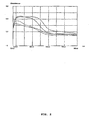
- microspectrophotometric analysis in the visible range of light performed directly on the fragments of the aged structures (like those of Fig. 1h ) show the presence of multiple absorption bands. That at the longest wavelength, 670 nm, has the lowest extinction coefficient. Other bands of higher intensity are observed at the wavelengths 530 nm, 488 nm, and 404 nm.
- the absorption spectrum in general, depends on the area of the fragment sampled by the micro beam ( Fig. 2 ). The pigments responsible of such absorption bands are not present in the culture medium and can only derive from a synthetic activity peculiar of the structure itself.
- a pigmented structure is treated with 2 M HCl, the structure and the red colour is destroyed in a few hours and the acid solution becomes yellow, as it is typical of ferrous salts in HCl. Since the spectra show multiple absorption bands, which do not correspond to those of hydrides or iron oxides, which monotonically increase in absorption at decreasing wavelength, the spectra indicate interactions between iron and organic molecules, such as in complex or chelation products.
- the structures' spectrum was determined directly by infrared analysis (IR), compressing the fragments in a diamond micro cell. During this analysis it is noted that the fragments do not fuse together, but instead they remain distinct, unlike rock materials.
- Spectra obtained by Fourier transform clearly show the presence of organic materials and, particularly, the resonating frequencies of the amide bond, but no clear presence of proteins.
- the chemical nature of the amide bond resulted to be that of N-acetyl-glucosamine by GC-mass spectroscopy performed on a single structure, by comparing the results to those obtained on a corresponding quantity of the culture medium.
- the structure has shown the presence of Galactose, glugose and N-acetyl-glucosamine.
- the relative ratios between these molecules is essentially about 3.2:6.3:0.5.
- This ratio between Glucose and Galactose is different from that of the culture medium that contains Galactose and Glucose in a ratio of about 8:2, and does not contain N-acetyl-glucosamine. Since the culture medium does not comprise N-acetyl-glucosamine, it can only derive from organic material generated on the surface of the fragments.
- Spectroscopic microanalysis by X ray energy dispersion (EDS) performed at the SEM on the fluid material initially present in the structures shows the presence of atoms normally present in organic molecules and absence of metals.
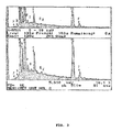
- the same type of analysis performed on highly pigmented structures shows the presence of iron as sole metal ( Fig. 3 ), and absence also of calcium.
- Fig. 4a The SEM pictures show that the corolla shaped structures have a complex organization in modules and layers ( Fig. 4a ). In the magnified area 1, spherical formations of about 0.4 micron in diameter are observed inside the wall.
- the area of enlargement 2 of Fig. 4a shows more details of the repetitive structures already apparent in Fig.
- FIG. 4c and enlargements An enlargement of a fractured part of a wall shows the presence of very small structures, tightly adjacent each other ( FIG. 4c and enlargements).
- the site reveals, by SEM analysis, the presence of thin appendages containing iron (EDS analysis) that are delicately curved and bound to the base of conical structures.
- EDS analysis thin appendages containing iron
- Observation of other parts of the walls interior shows the presence of other components of different morphology, such as clusters of small rods ( Fig. 4e and enlargement), that resemble bacterial cells and other sub-microscopic star-shaped bodies inside other hollow globular structures having 30-40 micron diameter ( Fig. 4 and enlargement).
- the present invention concerns also methods of fluorescence microscopy for identifying biological material or viable forms extracted from the samples of chondrites here described.
- the material is disaggregated in physiologic solution directly on a microscope glass slide and labelled with a solution used to fluorescently label nucleic materials.
- the labelling is specifically performed with a fluorophore that discriminates live from dead bacterial cells.
- the handling conditions of the fragments on the culture medium are critical: cultivation occurs on a solid medium, all nutritional components are necessary, it is necessary to work in oxygen atmosphere, in fact, in anaerobic atmosphere no formation of structures is observed. On the contrary, as soon as anaerobic conditions are discontinued, in the period of few hours the structures appear in the culture. Exposition to UV light of the cultures with the chondrite causes inhibition of the growth. Only the formation of small clusters of little pigmented bubbles is observed, like small aggregates of foam, with no further growth.
- mixobacteria are easy to cultivate and have a large DNA genome.
- the structures according to the invention show a positive geotropism and a complex structure with at least three distinct layers of components discriminated by SEM. This is probably the origin of multiple bands of amplification of rDNA by PCR that has not yet permitted the genetic characterization.
- a particularly interesting property is the capability of the structures here presented to grow from fragments that have been exposed to the sterilization with alcohol, to flaming and to the subsequent keeping for 5 minutes in the flame of a Bunsen burner.
- chondrite fragments that have been kept in a growth medium of E. coli (as an example of Gram negative bacteria) or of B. subtilis (as a an example of Gram positive bacteria) for 7 days no bacterial growth has been observed from them after this type of sterilization procedure.
- the growth has instead occurred of the pigmented structures and of the fluorescently labelled forms above described.
- the possibility that heat has not reached the analysed parts is not tenable considering that the fragments are less than 1 mm in size and the time in the flame is 5 minutes.
- the contamination should have occurred either before or during the fall on Earth, or while meteorites were lying on the soil.
- the contaminating form should be present in all environments and abundant to colonize in short time samples of meteorites fallen in so different environments (Agricultural soil of Transylvania and the desert of Arizona) in different years.
- the contaminating organism should have found a nutrient necessary to its growth in the meteorite considering that it does not grow from any terrestrial sample, and moreover, it should have established with the meteorite very peculiar relation that enabling it to resist both to the extreme sterilization conditions employed in the experiments here presented and to the time (at least one century) of conservation in the museums collections.
- the specific properties of absorbing light energy are utilized to prepare absorbing or dissipating materials or sensory elements, that can be integrated into instruments made to receive, absorb or utilize energy, specifically light energy.
- the magnetic properties also may be used for the preparation of control elements integrated as components into instrumentations for magnetic or optical orientation.
- the structure herein described o their parts or materials obtained therefrom produce a catalytic action on some organic chemistry and biochemical reactions such as hydrolysis and formation of ester bonds, of peptide bonds or glycosidic bonds.
- the glycosylasic activity of the materials of the invention is well evident, on culture dishes, from the formation of hollow tubular cavities in the width of the solid culture medium in correspondence to fragments of the materials.
- These hollow tubes are the result of the hydrolysis of the glycosidic bonds of the polysaccharides that compose the culture medium, such as gelrite gellan gum® (Sigma-Aldrich).
- the esterase activity may be put in evidence at the microscope in fluorescent light as shown in Fig. 6 .
- the material of the invention when in contact with carboxyfluoresceine diacetate (H2CFDA), produces the hydrolysis of the acetyl residues and the activation of fluoresceine with the production of the typical green fluorescence that is observed after incubation at 4°C for at least 15 minutes in the dark.
- H2CFDA carboxyfluoresceine diacetate
- Spectra in visible and infrared light have been taken respectively with a Zeiss micro spectrophotometer and with a Perkin Elmer instrument. Photographs under fluorescent light were taken with a Nikon microscope Eclipse E 1000 equipped with digital camera DMX 1200. Fluorescence labelling has been performed with the live/dead BacLight kit of the Molecular Probes.
Landscapes
- Life Sciences & Earth Sciences (AREA)
- Chemical & Material Sciences (AREA)
- Health & Medical Sciences (AREA)
- Genetics & Genomics (AREA)
- Organic Chemistry (AREA)
- Zoology (AREA)
- Engineering & Computer Science (AREA)
- Bioinformatics & Cheminformatics (AREA)
- Wood Science & Technology (AREA)
- Microbiology (AREA)
- Medicinal Chemistry (AREA)
- Biomedical Technology (AREA)
- Molecular Biology (AREA)
- Biochemistry (AREA)
- General Engineering & Computer Science (AREA)
- General Health & Medical Sciences (AREA)
- Biotechnology (AREA)
- Measuring Or Testing Involving Enzymes Or Micro-Organisms (AREA)
- Crystals, And After-Treatments Of Crystals (AREA)
- Laminated Bodies (AREA)
- Preparation Of Compounds By Using Micro-Organisms (AREA)
- Polymers With Sulfur, Phosphorus Or Metals In The Main Chain (AREA)
- Agricultural Chemicals And Associated Chemicals (AREA)
Abstract
Description
- The present invention concerns methods to obtain organic, metallorganic or biological materials from samples of rock of different nature and materials so obtained. The invention concerns also their use in instrumentations that employ their physical and physicochemical properties and fluorimetric methods for the identification of those materials.
- It is known that crystals, rocks and minerals of different origins and compositions contain organic materials, in some cases of biological origin, as also viable micro-organisms capable of exiting from a quiescent state once freed from the rock structure and suspended in a suitable nutrient medium (Geraci et al., Rend. Fis. Acc. Lincei, s. 9, v. 12:51:68, 2001). These forms of life, when entrapped within the rock, are not influenced by time, as viable micro-organisms, suitable for cultivation, have been found in samples of rocks of different ages, tracing back to about 2.8 Ga (billions of years), and by temperature and pressure, since they are present in metamorphic and igneous rocks (Geraci et al, supra).
- Research studies, with similar approaches, of microbial forms has demonstrated that also meteorite rocks may contain micro-organisms, indicating that these already existed during the initial stages of Earth formation. Some microbial species have been obtained in cultures, cloned and classified by the method based on the analysis of their 16S rDNA resulted not differing from the today micro-organisms. The method of extraction of biological materials described by Geraci et al. (supra) involves sterilization by flaming of relatively large rock samples for relatively brief flaming times (about 2 minutes), and then extraction from the most inner part of the sample, through series of drillings going deep about 10 mm, of small fragments of rock, which are then put in culture. Considering the sample size and the operative times, it cannot be excluded that the surface thermal treatment does not reach the necessary sterilization efficacy at a deep level. The doubt remains, however, that the viable life forms, apparently extracted from the rock structure, do not actually trace back to origin of the rock, but might represent environmental contamination occurred during the operative procedure or, in any case, in more recent times.
- Scope of the present application is then that of providing a new procedure that enables the search, identification and study of organic materials of different origins, comprising materials of biological origin or viable micro-organisms entrapped inside the crystal structure of ancient rocks, eliminating the problem of false/positive results due to contamination of samples during the operational procedures or to any other type of recent contamination. Scope of the present invention is also that of demonstrating if, by applying the above mentioned procedure, organic materials, biological materials or even viable micro-organisms may be found in samples of rocks, or of ancient minerals of Ga of age or even in rocks of meteorite origin.
- The method according to the invention entails a procedure of profound sterilization in alcohol and in a free flame and/or in autoclave for periods of time longer than these described in the state of the art. The method then implies the production of rock fragments directly from the surface of the sample, that is from the part where the heating sterilization procedure is expected to have been most effective. Hence, the procedure permits to eliminate the presence of any organic or biological material deriving from possible environmental contamination of the sample under examination and to put in evidence, through suitable means, the presence of organic or biological materials pre-existing in the very structure of the rock and protected by this against the destructive effect of the heating treatment. The method has permitted to demonstrate surprising results.
- Accordingly, the object of the present application is a method for obtaining from samples of rocks of different origins, but in preference from chondrites, organic, metallorganic or biological materials, this method comprises the treatment of the rock sample at sterilizing temperature for at least 5 minutes. Then, fragments of the sterilized sample are produced by means of a mechanical treatment of the sample surface and put on a sterile solid nutrient culture medium based on hydrolyzed natural products or synthetic derivatives thereof, either in the presence or in the absence of oxygen at room temperature. The material generated on the surface of the fragments is then separated from the fragments, isolated from the solid culture medium and optionally further elaborated. The sterilization step is performed by dipping the sample in alcohol and subsequently treating it directly on a flame for at least five minutes.
- Further object of the invention are the materials obtained by this method in the presence of oxygen; preferably obtained from chondrite samples. These latter materials are characterized by an iron contents bound or complexed to organic molecules, and comprising mixtures of saccharides and N-acetylaminosaccharides in defined ratios. The generation of such materials on the surface of chondrite fragments occurs through an initial developmental stage in which the material is organized in pigmented globular vesicular structures containing or not a fluid, followed by a phase of quiescence in which the material is organized in globular structures, rigid and highly pigmented, open or closed, showing a positive geotropism. The wall of these structures, that is formed by repetitive units rich in iron and by thin membranes without iron, exhibits unexpected and surprising physical and physico-chemical properties such as an exceptionally strong capacity to absorb light energy and magnetic properties. Moreover, the globular vesicular structures containing fluids, when disaggregated in solution and labelled with a fluorescent marker for nucleic acids, show the presence of fluorescently labelled entities of size smaller than a micron, with very peculiar structure and endowed of a pulsating mobility.
- Further object of the invention are methods for identifying by fluorescence nucleic materials comprising the organic, metallorganic and/or biological materials obtained according to the described method.
- Other objects of the invention are technical elements, such as sensors or control elements, that include the above described material, and make use of their absorbing or energy-dissipating or magnetic properties.
- These elements are parts integrated in instrumentations receiving, absorbing and utilizing energy, or instrumentation for magnetic or optical orientation, that also are objects of the invention.
- Finally, objects of the invention are the use of these structures or of their composing parts or of materials obtained from them, as catalysts in chemical reaction.
-
-
Figure 1 : The figure shows the types of structural shapes that are generated from fragments of iperstenic chondrite incubated in sterile Petri dishes. Panel (a): club-shaped structures at initial growth stages (about 12 hours incubation) that can be seen on thefragments 1 and 3. Panel (b): same area shown in Panel (a), after about 1 month; it is possible to recognize the initial structures increased in size and complexity and a new structure onfragment 2. Panel (c): wall of a structure at a growth time in which there are evident the modular elements that constitute it and, in the adjacent panel, an enlargement of the indicated area. Panel (d): aged structure that has divided into two valves showing in the interior, where the valves are joined, a mass of small globular objects embedded in a refracting matrix. Panel (e): aged structure with a form of a bell-shaped corolla. Panel (f); an aged structure with a hemispherical shape. Panel (g): the same fragment as shown in panel (f), showing a new structure in formation at the stage of hyper refractive transparent bubble, two weeks after the mechanical removal of the previous one for analytical studies. Panel (h): micro photograph of the wall of a structure in which modular, highly pigmented elements are evident. -
Figure 2 : The figure reports visible absorption spectra obtained with a Zeiss micro spectrophotometer. The different spectra correspond to different zones of the iper-pigmented fragments shown inFig. 1h . Magnified 1000x. The spectra show decreasing absorption at increasing wavelengths values, with a maximum of absorbance at 670 nm. The spectra show other bands of absorption at 530 nm, 488 nm and 404 nm. -
Figure 3 : The figure shows the results of the analysis of x-ray energy dispersion spectra (EDS) on samples of fragile, aged, pigmented, globular structures. The analysis shows iron as a most prominent metallic component. -
Figure 4 : The figure reports images obtained at the scanning electron microscope. Panel (a) shows details of the wall of opened structures with a cup-shaped form (bell). The wall is composed of a complex organization in layers and modules. Enlargement 1a, shows spherical bodies of about 0.4 microns comprised in the interior of the wall. Enlargement 2a shows details of the repetitive units that compose the same wall. Panel (b) shows other details of the repetitive elements and thin membranes that form the wall. Panel (c) shows thin structures in tight packing characterized by slightly curved filamentous protrusions departing from conical bases. Panel (d) shows wide, hollow, globular wall elements ranging in size 30-40 micron. On the inner surface of such globules, elements of different morphology can be observed (as reported in panel (e) and enlargement), such as clusters of rod-shaped objects resembling bacterial cells and minute star-shaped bodies. -
Figure 5 : The figure shows pictures in fluorescent light microscopy of globular vesicular structures at initial stages still containing liquid, disaggregated in physiologic saline solution directly on a microscope glass slide and then labelled. Scattered fluorescent spots of about one micron in diameter are observed, some of which, smaller and with star-shaped structures, show motility in a pulsed gait (enlargement). -
Figure 6 : The figure shows pictures in fluorescent light microscopy, of parts of the structures reported inFig. 1 , disaggregated in physiologic saline solution directly on a microscope glass slide and then treated with LB nutrient solution and carboxyfluoresceine diacetate (H2CFDA, Molecular Probes). The green fluorescence that is observed after incubation at 4°C for at least15 minutes in the dark, is due to the hydrolysis of the acetyl residues from the carboxy fluoresceine, caused by aspecific esterase activity present in the fragments of the structures shown inFig. 1 , that become, for this reason, fluorescent. -
Figure 7 : The figure shows at binocular microscope the glycosylase activity of the fragments of the materials of the invention; these fragments generate hollow tubular cavities produced by the hydrolysis of the gelrite gellan gum® (Sigma-Aldrich), that is a natural agent substitutive of agar used in microbiology and to cultivate plants in the following conditions: 0.8% gelrite in DSM-182 medium, in Petri dishes at pH 4.0 or 0.8% in TYS medium at pH 3.2, respectively; incubation at room temperature for 2 or more days.
The upper panel shows a general picture of a dish; the three lower panels show details of cavities where the presence of fragments of a structure of the invention can be noted at the extreme progressing part of a cavity. Observation made from the inferior side of the culture dish where the upper surface is on the opposite side. -
Figure 8 : the figure shows two enlargements of fragments that have produced the tubular cavities reported infig. 7 . - Crystals, rocks and minerals of different origins and ages contain organic and biological materials, including viable micro-organisms.
- The method described in the present application permits to identify and to obtain such materials from rock samples of various origins minimizing the risk of environmental microbiological contamination. Examples of samples of rocks are metamorphic, igneous, sedimentary, mineral rocks or obtained from different depths, these latter representing the precursors of such rocks, or, also chondrites, octahedrites and rocks of similar origin. Preferably the method is carried on iperstenic chondrites or enstatitic olivines.
- The method implies that the samples be exposed to a sterilizing treatment at high temperature. Suitable methods are the heating in a kiln, or in autoclave or direct exposure to microwave oven or to the flame of a burner, as for example a Bunsen. The combination of a treatment in autoclave, preceded or followed by direct exposure to the flame, is the preferred procedure. Nonetheless, a preliminary treatment with alcohol or other sterilizing solvent, offers even more assurances that any environmental biological contamination is eliminated. In this case the sample, still wetted with solvent, is directly put in the flame of the burner.
- The time of exposure to the direct flame is of not less than 5 minutes. Longer times, for example, 10, 15 or 30 minutes, may nonetheless be utilized. The treatment times in autoclave vary between 20 minutes and 2 hours, though longer times may be envisaged.
- The surface of the so treated sample is fragmented in pieces not larger than 1 mm, by grinding with a suitable drill, scraping with suitable file or other similar tool, or excavating with a syringe needle or any other sufficiently hard acuminated tools. These operations are performed under a strictly sterilized atmosphere, operating with previously perfectly sterilized instruments. Fragments so obtained are deposited on a solid nutrient medium based on hydrolysates of natural products or their synthetic derivatives, or mixtures of their components, also synthetic, such as tryptone, yeast extracts, agar. The preferred culture medium contains for 1 g. of tryptone (Oxoid), 1 g. NaCl, 0,5 g of yeast extract (Oxoid) and 1.4 g of Bacto Agar (Difco) in 100 ml of water.
- A second culture medium is the product of gelrite gellan gum® (Sigma-Aldrich), that is a natural agent substituting agar, composed of glucose units, glucuronic acid and rhamnose, used in microbiology and for plant cultures at the following conditions: 0,8% gelrite gellan gum® in growth medium DSM-182, in Petri dishes, at pH 4.0 or in growth medium TYS at pH 3.2, respectively; incubation at room temperature for 2 or more days.
- Mixtures of combinations of hydrolysates different from these, but equivalent, having various origins, also made by mixing pure components, are equally valid, although more expensive.
- The preparation conditions of the culture media as well as the culture conditions are critical. The above indicated culture media are heated to boiling point to solubilize agar or gelrite, are sterilized for 20 min in autoclave at 121°C, and stored for at least one month to be sure that there are no residual contaminations. Before use, the solid culture medium is heated to melt it and is dispensed in requested amounts, for example about 20 ml, in each standard Petri dish (90 mm diameter), that is then sealed and cooled producing the gelification of the culture medium.
- In order to obtain the structures described below, an essential culture condition is the presence of oxygen. Anaerobic conditions do not normally produce the described results.
- While the method before described by Geraci et al., (supra) permitted the identification of viable micro-organisms already known in samples of differing ages, starting from 2.8 Ga back, the method of the invention, applied to samples of chondrite material, has permitted to obtain completely unforeseeable and unexpected results.
- In a preferred embodiment of the invention, the method is performed on types of iperstenic chondrites classified L6. Two of these types, Mocs and Holbrook, conserved in different museum collections, have been chosen because they are parts of two seen meteorite swarms, fallen in distant areas of Earth, at different climatic conditions, and because they remained for short time on the soil before their collection and conservation in different museums collections.
- When fragments obtained according to the method of the invention, having diameters of less than 1 mm, are put on a solid nutrient medium and incubated for a period of 5-7 hours, at room temperature and in the presence of oxygen, on their surfaces it is observed the generation of materials formed through a developmental phase followed by a phase of quiescence. In the initial phase, the material is organized in small globular blobs, as liquid vesicles or containing liquid, with a pigmentation ranging from pale amber yellow to orange or red, from pale to deep ruby, anchored to the black fragments of iperstene (
Fig. 1a ). - After longer incubation times, other structures grow out that, in a period of one, two, three or more months, increase in size and structural complexity becoming dark orange or deep red, with a growth that shows a positive geotropism (
Fig. 1b ). The size of the structure is frequently larger than that of the fragment from which it is generated. - The globular structures, observed at an initial phase, show a great transparency and an elastic fluidity of the wall comparable to that of a soap bubble. Pigmented material initiates to form on the external surface of the fluid globe and in time it expands to the entire surface. The transparent liquid initially present is no more observed in the structures at advanced stages or in a quiescent state that acquire a quasi-crystalline appearance formed by modular units (
Fig. 1c and related enlargement). This type of development has been observed in almost 500 structures in a period of almost 2 years. - The structure, initially with a vesicular shape, may assume with development a typical club-like form. Property of these club-like structures is that of responding like a magnet to an external magnetic field, if mechanically detached from the fragment from which they had been generated. After a period of a few months of incubation, the structures enter an apparently quiescent state, in which their form does not change in a period of at least one year.
- Three are the most frequently observed forms of the structures at the end of their growth and these depend on the structure initially presented. To a first type, all the structures belong that have a club-like form and that during development may divide along a meridian, forming two valves united at the base, where a mass of small globular bodies is present embedded in a transparent, highly refractive matrix (
Fig. 1d ). In a second type of structures, the upper spherical part of the club-like form opens up producing a sort of reversed bell-shaped corolla that shows at the bottom, a complex aggregate of small spherical bodies (fig. 1e ). Finally, the structures maintaining during aging the hemispherical form lose their refractive aspects of the liquid layer and assume a quasi-crystalline highly breakable form (Fig.1g ). If this type of structure is mechanically removed from its site of formation on the fragment, a new one is formed with properties similar to the pre-existing structure through an initial stage in which a transparent and highly refractive liquid layer is formed again, and this in time is gradually inflated becoming hemispherical (Fig. lg). On this layer, then, the highly pigmented parts are produced, which initially seem to freely float on the surface of the liquid layer. This regeneration may occur several times. - The material that forms the above described structures in the various phases of their development or parts of them, when analysed by direct analytical methods that minimize the possibility of erroneous attributions, presents completely unexpected reproducible physicochemical characteristics, having interesting industrial applicability.
- A first property of the structures, in the phase characterized by the typical club-shaped form, is that to respond like a magnet to a magnetic external field, if mechanically detached from the fragment from which they were generated. Another surprising property is the enormous capacity of the wall of aged rigid structures to absorb light energy. This property is deduced by the practical impossibility to measure reflection spectra, using a Zeiss micro-spectrophotometer, in the wavelength region from 280nm to 800 nm, because the quantity of reflected energy is insufficient for any reliable recording.
- Moreover, microspectrophotometric analysis in the visible range of light, performed directly on the fragments of the aged structures (like those of
Fig. 1h ) show the presence of multiple absorption bands. That at the longest wavelength, 670 nm, has the lowest extinction coefficient. Other bands of higher intensity are observed at the wavelengths 530 nm, 488 nm, and 404 nm. The absorption spectrum, in general, depends on the area of the fragment sampled by the micro beam (Fig. 2 ). The pigments responsible of such absorption bands are not present in the culture medium and can only derive from a synthetic activity peculiar of the structure itself. If a pigmented structure is treated with 2 M HCl, the structure and the red colour is destroyed in a few hours and the acid solution becomes yellow, as it is typical of ferrous salts in HCl. Since the spectra show multiple absorption bands, which do not correspond to those of hydrides or iron oxides, which monotonically increase in absorption at decreasing wavelength, the spectra indicate interactions between iron and organic molecules, such as in complex or chelation products. The structures' spectrum was determined directly by infrared analysis (IR), compressing the fragments in a diamond micro cell. During this analysis it is noted that the fragments do not fuse together, but instead they remain distinct, unlike rock materials. Spectra obtained by Fourier transform clearly show the presence of organic materials and, particularly, the resonating frequencies of the amide bond, but no clear presence of proteins. The chemical nature of the amide bond resulted to be that of N-acetyl-glucosamine by GC-mass spectroscopy performed on a single structure, by comparing the results to those obtained on a corresponding quantity of the culture medium. - The structure has shown the presence of Galactose, glugose and N-acetyl-glucosamine. The relative ratios between these molecules, although somewhat variable, is essentially about 3.2:6.3:0.5. This ratio between Glucose and Galactose is different from that of the culture medium that contains Galactose and Glucose in a ratio of about 8:2, and does not contain N-acetyl-glucosamine. Since the culture medium does not comprise N-acetyl-glucosamine, it can only derive from organic material generated on the surface of the fragments.
- The amplification of genetic material, such as rDNA, by means of PCR on the aged structures failed to give significantly positive results, indicating either that the genetic material is no constituent of the structures or that it is shielded, or also that the oligonucleotides used as primers, not being the suitable ones, found no binding sites. On the contrary, on young vesicular structures, still in a fluid form, bands of DNA are obtained by PCR amplification using standard oligonucleotide mixtures aimed at amplifying rDNAs of eubacteria, archea and animal, as described in the literature (DeLong, E.F. Archea in costal marine environments. Proc. Natl. Acad. Scie. U.S.A. 89, 5685-5689 (1992)) and of plants (Aceto, S., Caputo, P., Cozzolino, S., Gaudio, L. and Moretti, A., Phylogeny and evolution of Orchis and allied genera based on ITS DNA variation: morphological gaps and molecular continuity. Mol. Phyl. Evol. 13, 67-76, (1999). However, two cycles of amplification have been necessary to obtain weak and still non conclusive signals. The results are reproducible for each analyzed structure, but always show more DNA bands. Due to the scarce quantity of available starting material that requires repeated amplification cycles, the obtained data need confirmation and it is not possible as yet to make a definitive genetic identification. Although not wishing to bound the invention to specific theories, the observed results seem to indicate that more than one biological form participates to the formation of the described structures. This seems to be in agreement with the results of scanning electron microscopy (SEM) reported below that show a great complexity of structural organization.
- Spectroscopic microanalysis by X ray energy dispersion (EDS) performed at the SEM on the fluid material initially present in the structures shows the presence of atoms normally present in organic molecules and absence of metals. The same type of analysis performed on highly pigmented structures shows the presence of iron as sole metal (
Fig. 3 ), and absence also of calcium. Hereinafter there are reported only the most relevant characteristics and details of the analysed structures. The SEM pictures show that the corolla shaped structures have a complex organization in modules and layers (Fig. 4a ). In the magnifiedarea 1, spherical formations of about 0.4 micron in diameter are observed inside the wall. The area ofenlargement 2 ofFig. 4a shows more details of the repetitive structures already apparent inFig. 1c and h , by optical microscopy. A peculiar characteristic of these repetitive units is their brush-like surface. It is interesting to note that a very thin layer (Fig. 4b ), which does not show metal components by EDS analysis, seems to be that organizing the pigmented, discrete, iron-rich units, that constitute the walls of the structures. This thin layer is probably what remains of the fluid hemisphere initially observed in the structures while just forming (Fig. 1g ), on which the pigmented parts later on appear. - An enlargement of a fractured part of a wall shows the presence of very small structures, tightly adjacent each other (
Fig. 4c and enlargements). The site reveals, by SEM analysis, the presence of thin appendages containing iron (EDS analysis) that are delicately curved and bound to the base of conical structures. Observation of other parts of the walls interior shows the presence of other components of different morphology, such as clusters of small rods (Fig. 4e and enlargement), that resemble bacterial cells and other sub-microscopic star-shaped bodies inside other hollow globular structures having 30-40 micron diameter (Fig. 4 and enlargement). - The present invention concerns also methods of fluorescence microscopy for identifying biological material or viable forms extracted from the samples of chondrites here described. The material is disaggregated in physiologic solution directly on a microscope glass slide and labelled with a solution used to fluorescently label nucleic materials. The labelling is specifically performed with a fluorophore that discriminates live from dead bacterial cells.
- The observation is finally performed at the fluorescence microscope with suitable filters. This technique has been applied to the globular vesicular structures still in a fluid state. Typically, a structure is suspended in 30 microliters of sterile culture medium without agar on a glass microscope slide. The sample is then disaggregated by crushing with the needle of a sterile syringe. A suitable quantity (usually 1 microliter) of fluorescent label SYTO (3.34 mM) (Molecular Probes Inc.), or other equivalent reagent, is added under accurate stirring and the sample is incubated in the dark for at least 15 minutes. The analysis at the fluorescence microscope reveals the presence of several fluorescent immobile spots, of about 1 micron in size, and slightly smaller other ones, with multiple appendages similar to small gel fish (
Fig. 5 ), showing halting movements. The shape of such objects (enlargementsFig. 5 ) has similarity with that shown in the enlargement of the units that compose the interior of the wall reported inFig. 4c . It has not been possible yet to put in culture these entities for their further characterization. - As seen above, the handling conditions of the fragments on the culture medium are critical: cultivation occurs on a solid medium, all nutritional components are necessary, it is necessary to work in oxygen atmosphere, in fact, in anaerobic atmosphere no formation of structures is observed. On the contrary, as soon as anaerobic conditions are discontinued, in the period of few hours the structures appear in the culture. Exposition to UV light of the cultures with the chondrite causes inhibition of the growth. Only the formation of small clusters of little pigmented bubbles is observed, like small aggregates of foam, with no further growth.
- Although without wishing to bind the invention to a specific theory, the presented data seem to indicate that the peculiar structures, which grow out of fragments of iperstenic chondrite meteorites, show physico-chemical characteristics peculiar of vital forms. There are some similarities with some properties of Magnetospirillum magnetotacticum for the sensitivity to magnetic fields. Yet, M. magnetotacticum produces chains of magnetosomes, magnetic crystals of iron oxide, with no resemblance with the morphology of the structures here presented for size, form and complexity of organization. There is also a certain similarity with the form shown by Mixobacteria in conditions of starvation, that is a spheroid, that however has no particular structure and does nor develop. Moreover, mixobacteria are easy to cultivate and have a large DNA genome. Furthermore, as already mentioned, the structures according to the invention show a positive geotropism and a complex structure with at least three distinct layers of components discriminated by SEM. This is probably the origin of multiple bands of amplification of rDNA by PCR that has not yet permitted the genetic characterization.
- A particularly interesting property is the capability of the structures here presented to grow from fragments that have been exposed to the sterilization with alcohol, to flaming and to the subsequent keeping for 5 minutes in the flame of a Bunsen burner. When chondrite fragments that have been kept in a growth medium of E. coli (as an example of Gram negative bacteria) or of B. subtilis (as a an example of Gram positive bacteria) for 7 days, no bacterial growth has been observed from them after this type of sterilization procedure. The growth has instead occurred of the pigmented structures and of the fluorescently labelled forms above described. The possibility that heat has not reached the analysed parts is not tenable considering that the fragments are less than 1 mm in size and the time in the flame is 5 minutes. If the structures derived from some form of environmental microbiological contamination, this could not derive from the museums where samples were stored or from the laboratory environment. In fact, the structures develop from all samples of two different meteorite swarms fallen in different years in different environments such as Transylvania and the desert of Arizona stored for more than a century in three different museums in different parts of the world. The hypothesis of laboratory contamination is not realistic because the search of similar structures from rock samples and minerals of terrestrial origin, from sedimentary rocks to purified iperstene, has produced no similar result, as also the intentional massive contamination produced with Gram positive and negative bacteria has not resisted to the sterilizing treatment. Hence, if the structures are produced by contaminating terrestrial micro-organisms, the contamination should have occurred either before or during the fall on Earth, or while meteorites were lying on the soil. In this case, the contaminating form should be present in all environments and abundant to colonize in short time samples of meteorites fallen in so different environments (Agricultural soil of Transylvania and the desert of Arizona) in different years. Even in this case, the contaminating organism should have found a nutrient necessary to its growth in the meteorite considering that it does not grow from any terrestrial sample, and moreover, it should have established with the meteorite very peculiar relation that enabling it to resist both to the extreme sterilization conditions employed in the experiments here presented and to the time (at least one century) of conservation in the museums collections.
- Due to the physico-chemical properties proper of the materials described here, these find applications in technological areas of importance.
- In particular, the specific properties of absorbing light energy are utilized to prepare absorbing or dissipating materials or sensory elements, that can be integrated into instruments made to receive, absorb or utilize energy, specifically light energy. Moreover, the magnetic properties also may be used for the preparation of control elements integrated as components into instrumentations for magnetic or optical orientation.
- It has been finally observed that the structure herein described o their parts or materials obtained therefrom produce a catalytic action on some organic chemistry and biochemical reactions such as hydrolysis and formation of ester bonds, of peptide bonds or glycosidic bonds. As shown in
Fig. 7 , the glycosylasic activity of the materials of the invention is well evident, on culture dishes, from the formation of hollow tubular cavities in the width of the solid culture medium in correspondence to fragments of the materials. These hollow tubes are the result of the hydrolysis of the glycosidic bonds of the polysaccharides that compose the culture medium, such as gelrite gellan gum® (Sigma-Aldrich). - The esterase activity may be put in evidence at the microscope in fluorescent light as shown in
Fig. 6 . The material of the invention, when in contact with carboxyfluoresceine diacetate (H2CFDA), produces the hydrolysis of the acetyl residues and the activation of fluoresceine with the production of the typical green fluorescence that is observed after incubation at 4°C for at least 15 minutes in the dark. - Finally, the catalytic activity for the formation of peptide bonds is evidenced by the capability of forming oligopeptides or polypeptides in the presence of mixtures containing the 20 amino acids present in the culture medium. In fact, although in the structures of
Fig. 1 no free amino acids are revealed by analytical procedures, analyses performed after acid hydrolysis reveal the presence of amino acids, but with ratios completely different with respect to those present and determined in the growth medium. This indicates that molecules with peculiar selections of amino acids linked to each other by peptide bonds are formed and present in the structures. - The invention is described below by means of a practical example.
- The Mocs and Holbrook iperstenic chondrites here, analysed are parts of samples fallen, respectively, on 3 February 1882 in Transylvania near Cluj (Mocs) and on 19 July 1912 in the Arizona Desert in Navajo County, near Holbrook, both classified as ordinary chondrites L6. A fragment from Mocs and one from Holbrook have been obtained from the Real Museo Mineralogico of the University of Naples (Italy) and from the collection of the Vatican Observatory of Castelgandolfo (Italy). Another Mocs fragment has been a gift from the collection of the Eötvös Museum of the University of Budapest. Each sample has a precise identification in the catalogs of the museums and is certified.
- Preparation of fragments and culture conditions.
- After immersion in ethanol for 30 minutes the meteorite samples have been removed from alcohol and further manipulated with sterile forceps under a laminar flow sterile hood. Ethanol has been removed from the samples by flaming under the hood, where thereafter have been kept for 5 more minutes in the flame of a Bunsen burner holding them with sterile Klemmer forceps. From these samples there have been obtained fragments by scraping their surface with the needle of a sterile syringe for injections, further sterilized in the flame at red heat, changing syringes for each sample and experiment. The scraping of the sample surface has been performed also with nail file, using a new one, flame sterilized, for each experiments, Fragments of 1 mm diameter or smaller, have been collected directly on a layer of solid nutrient in a Petri dish 9.1 cm in diameter that has been immediately covered and sealed with parafilm under sterile hood and then kept at laboratory temperature. The culture medium was LB-agar. Stereo-microscope observations in the sealed Petri dishes were performed at 30 enlargements under tangent light illumination. The photographs have been taken with a digital camera.
- Spectra in visible and infrared light have been taken respectively with a Zeiss micro spectrophotometer and with a Perkin Elmer instrument. Photographs under fluorescent light were taken with a Nikon microscope Eclipse E 1000 equipped with digital camera DMX 1200. Fluorescence labelling has been performed with the live/dead BacLight kit of the Molecular Probes.
- Analyses at the SEM have been performed with standard procedures using a microscope Cambridge 250 Mark 3. The EDS analyses (electronic energy dispersion of X rays) with a microscope Jeol 5310 linked to EDS link AN 10.000 Pentafet. Samples for only morphological observations were covered with gold while those for EDS analyses were not covered.
- Analyses have been made with standard methods for monosaccharides and their derivatives after acetylation and trimethylsilinization using a GC-Mass 5390sd quadruple mass spectrometer (Agilent Technologies) equipped with a Hewlett-Packard gas chromatograph.
Claims (20)
- Method for obtaining organic, metallorganic or biological materials from samples of metamorphic, igneous, sedimentary, mineral rock, from chondrites, octahedrites, or rocks of similar origin, comprising the following steps:a sample of rock is treated at sterilizing temperatures by treating it on the direct flame for at least 5 minutes;fragments of the sterilized sample are produced in sterile atmosphere by scraping, grinding or crushing with a suitable tool from the surface of the sample;the fragments are then put on a previously sterilized solid growth medium made of hydrolysates of natural produts or synthetic derivatives thereof such as tripton, yeast extract and agar in the presence of oxygen at room temperature;the material generated on the surface of the fragments is removed from the fragment itself, isolated from the solid nutrient medium and optionally further elaborated.
- Method according to claim 1 wherein the sterilization step is performed by dipping the sample in ethyl alcohol for at least 20 minutes and further treatment on the direct flame of a burner for at least 5 minutes and/or in autoclave for a period of 30 minutes to two hours in whatever order.
- Method according to anyone of the claims 1 or 2 wherein the solid medium comprises triptone, yeast extract and agar or gelrite.
- Method according to anyone of the claims 1 to 3, in which the generation of the material on the fragments surface proceeds through a phase of development in which the material is organized in globular, vesicular structures containing a fluid and a subsequent phase of quiescence in which the material is organized in opened or closed globular rigid structures.
- Method according to anyone of the claims 1 to 4, in which the rock sample is iperstenic or enstatitic choridrite.
- Organic, metallorganic or biological material obtainable through the method according to anyone of claims 1 to 5, characterized by:a) a content in iron bound or completed to organic molecules, andb) comprising galactose glucose and N-acetylglucosammine in a ratio about 3.2:6.3:0.5,c) and having at least one property selected from positive geotropism, magnetic property, light adsorbing property to the extend that the quantity of reflected energy in the wavelength 280-800 nm is insufficient for any reliable recording or catalytic property.
- Material according to claim 7, characterized by a content in nucleic acid.
- Material according to claim 6, characterized in that it is organized in globular vesicular pigmented structures containing a fluid and/or in opened or closed highly pigmented rigid globular structures having a positive geotropism.
- Material according to claim 8, characterized in that the rigid globular structures have walls formed by iron rich repetitive units having a brush-like surface and by thin membranes without iron.
- Material according to claim 9, wherein the wall of the globular structures has characteristics of maximum absorption and minimal reflection of light energy.
- Material according to claim 8, characterized in that the rigid globular structures in disaggregated form, exhibit magnetic properties.
- Material according to claim 8, wherein the globular vesicular pigmented structures containing a fluid, when disaggregated in physiologic solution and labelled with a fluorescent marker for nucleic acids, reveal spots of diffused fluorescence endowed of halting motility.
- Method according to any one of claims 1 to 5 further comprising identifying the obtained organic, metallorganic and/or biological material by fluorescence, wherein the globular structure is removed from the fragment, suspended in a liquid medium, broken up, treated with suitable fluorescence marker, and the signal suitably revealed.
- Method of identification according to claim 13, in which the material comprises molecules of DNA and/or RNA, the dispersing medium is an isotonic solution without agar, and the signal is revealed at the fluorescence microscope with a suitable filter.
- Sensor element, absorbing/dissipating light-, conductive- or magnetic-energy comprising as active material the material according to claim 10.
- Instrumentations to receive, absorb or utilize energy comprising the elements according to claim 15.
- Control elements comprising magnetic material according to claim 11.
- Instrumentation for magnetic or optical orientation comprising a component element according to claim 17.
- Catalyst for chemicorganic or biochemical reactions selected from hydrolysis, formation or modification of ester, peptidic, glycosidic or amidic bonds on N-acetyl-glucosamine comprising materials according to claim 6.
- Catalyst according to claim 19, which is magnetic.
Applications Claiming Priority (3)
| Application Number | Priority Date | Filing Date | Title |
|---|---|---|---|
| ITRM20030026 | 2003-01-23 | ||
| IT000026A ITRM20030026A1 (en) | 2003-01-23 | 2003-01-23 | ORGANIC MATERIAL OBTAINED FROM ROCK. |
| PCT/IT2004/000013 WO2004065333A2 (en) | 2003-01-23 | 2004-01-23 | Organic, metallorganic and biological material obtained from rock |
Related Child Applications (1)
| Application Number | Title | Priority Date | Filing Date |
|---|---|---|---|
| EP09164566.3 Division-Into | 2009-07-03 |
Publications (2)
| Publication Number | Publication Date |
|---|---|
| EP1585823A2 EP1585823A2 (en) | 2005-10-19 |
| EP1585823B1 true EP1585823B1 (en) | 2010-03-31 |
Family
ID=29765625
Family Applications (1)
| Application Number | Title | Priority Date | Filing Date |
|---|---|---|---|
| EP04704702A Expired - Lifetime EP1585823B1 (en) | 2003-01-23 | 2004-01-23 | Organic, metallorganic and biological material obtained from rock |
Country Status (6)
| Country | Link |
|---|---|
| US (1) | US9328337B2 (en) |
| EP (1) | EP1585823B1 (en) |
| AT (1) | ATE462799T1 (en) |
| DE (1) | DE602004026268D1 (en) |
| IT (1) | ITRM20030026A1 (en) |
| WO (1) | WO2004065333A2 (en) |
Families Citing this family (3)
| Publication number | Priority date | Publication date | Assignee | Title |
|---|---|---|---|---|
| US8324159B2 (en) * | 2005-06-03 | 2012-12-04 | Affymax, Inc. | Erythropoietin receptor peptide formulations and uses |
| FR2936155B1 (en) * | 2008-09-22 | 2011-02-11 | Gattefosse Sas | COSMETIC COMPOSITION COMPRISING A METEORITE EXTRACT, AND USE OF THE EXTRACT AS A COSMETIC AGENT CAPABLE OF STIMULATING THE DIFFERENTIATION OF HUMAN KERATINOCYTES |
| EP3837035B1 (en) | 2018-08-16 | 2026-05-20 | Emd Millipore Corp | CLOSED BIOPROTECTION DEVICE |
Family Cites Families (4)
| Publication number | Priority date | Publication date | Assignee | Title |
|---|---|---|---|---|
| US4280002A (en) * | 1979-12-03 | 1981-07-21 | Minnesota Mining And Manufacturing Company | Microbiological growth container |
| US5545535A (en) * | 1993-04-13 | 1996-08-13 | Molecular Probes, Inc. | Fluorescent assay for bacterial gram reaction |
| US5902742A (en) * | 1996-05-17 | 1999-05-11 | The United States Of America As Represented By The Secretary Of Agriculture | Complex growth supplement for maintenance of bacterial cell viability and induction of bacterial cell differentiation |
| JP4066215B2 (en) * | 1998-08-28 | 2008-03-26 | 独立行政法人海洋研究開発機構 | Microorganism detection method by flow cytometry |
-
2003
- 2003-01-23 IT IT000026A patent/ITRM20030026A1/en unknown
-
2004
- 2004-01-23 DE DE602004026268T patent/DE602004026268D1/en not_active Expired - Lifetime
- 2004-01-23 EP EP04704702A patent/EP1585823B1/en not_active Expired - Lifetime
- 2004-01-23 AT AT04704702T patent/ATE462799T1/en not_active IP Right Cessation
- 2004-01-23 WO PCT/IT2004/000013 patent/WO2004065333A2/en not_active Ceased
- 2004-01-23 US US10/543,292 patent/US9328337B2/en active Active
Also Published As
| Publication number | Publication date |
|---|---|
| WO2004065333A2 (en) | 2004-08-05 |
| ITRM20030026A0 (en) | 2003-01-23 |
| HK1088299A1 (en) | 2006-11-03 |
| DE602004026268D1 (en) | 2010-05-12 |
| US20060134798A1 (en) | 2006-06-22 |
| EP1585823A2 (en) | 2005-10-19 |
| WO2004065333A3 (en) | 2004-09-16 |
| US9328337B2 (en) | 2016-05-03 |
| ATE462799T1 (en) | 2010-04-15 |
| ITRM20030026A1 (en) | 2004-07-24 |
Similar Documents
| Publication | Publication Date | Title |
|---|---|---|
| Bergman et al. | Phycomyces | |
| Lukas et al. | Toward understanding bacterial ice nucleation | |
| Yin et al. | Directing effect of bacterial extracellular polymeric substances (EPS) on calcite organization and EPS–carbonate composite aggregate formation | |
| De Philippis et al. | Exocellular polysaccharides from cyanobacteria and their possible applications | |
| Wildermuth | Development and organization of the aerial mycelium in Streptomyces coelicolor | |
| Arnosti et al. | Contrasting extracellular enzyme activities of particle-associated bacteria from distinct provinces of the North Atlantic Ocean | |
| CN1196712C (en) | Human like collagen and production method thereof | |
| AU2014247991B2 (en) | Kits and methods for isolating protein from biological and environmental samples | |
| Meena et al. | Optimization and characterization of Alginic acid synthesized from a novel strain of Pseudomonas stutzeri | |
| Zinder et al. | Microbial ecology—new directions, new importance | |
| US9328337B2 (en) | Organic, metallorganic and biological material obtained from rock | |
| Yin et al. | Bacterial EPS in agarose hydrogels directs mineral organization in calcite precipitates: species-specific biosignatures of Bacillus subtilis, Mycobacterium Phley, Mycobacterium Smagmatis, and Pseudomonas putida EPS | |
| Purgina et al. | Morphological and ultrastructural features of selected Epidendroideae Pollen Dispersal Units and New insights into their Chemical Nature | |
| HK1088299B (en) | Organic, metallorganic and biological material obtained from rock | |
| Ezenwali | Optimization, isolation, and characterization of a keratinolytic protease from human hair-degrading bacterium Alcaligenes faecalis strain TPB18 | |
| Galitskaya et al. | Bacterial cultural media containing lipopeptides for heavy oil recovery enhancement: The results of sand-packed column experiment | |
| Ahmad et al. | Effect of substrate concentration and fecl2 addition on L-asparaginase activities of bacteria symbionts brown algae sargassum sp. and its potential as a new anti-dengue agents | |
| CN110343626A (en) | One plant of heat-resisting thermophilic Halomonas and its application | |
| CN107779442A (en) | The large scale production method of high-purity deuterated protein entirely | |
| Waghmare et al. | A textbook of botany: microbiology, phycology, mycology, and fungal technology | |
| Riley et al. | Culture of Escherichia coli and related bacteria | |
| Meena et al. | Detection and Diagnosis of Plant Diseases | |
| RU2160718C1 (en) | Method of purification of soil and water from oil and products | |
| CN104531618A (en) | Method for obtaining lots of three-dimensional growing tumour cells at low cost by utilizing graphene nano material | |
| Huld et al. | Experimental Mineralisation of a Filamentous Hydrogenotrophic Methanogen in Carbonate, Phosphate, and Silicate |
Legal Events
| Date | Code | Title | Description |
|---|---|---|---|
| PUAI | Public reference made under article 153(3) epc to a published international application that has entered the european phase |
Free format text: ORIGINAL CODE: 0009012 |
|
| 17P | Request for examination filed |
Effective date: 20050809 |
|
| AK | Designated contracting states |
Kind code of ref document: A2 Designated state(s): AT BE BG CH CY CZ DE DK EE ES FI FR GB GR HU IE IT LI LU MC NL PT RO SE SI SK TR |
|
| AX | Request for extension of the european patent |
Extension state: AL LT LV MK |
|
| DAX | Request for extension of the european patent (deleted) | ||
| REG | Reference to a national code |
Ref country code: HK Ref legal event code: DE Ref document number: 1088299 Country of ref document: HK |
|
| 17Q | First examination report despatched |
Effective date: 20070130 |
|
| GRAP | Despatch of communication of intention to grant a patent |
Free format text: ORIGINAL CODE: EPIDOSNIGR1 |
|
| GRAS | Grant fee paid |
Free format text: ORIGINAL CODE: EPIDOSNIGR3 |
|
| GRAA | (expected) grant |
Free format text: ORIGINAL CODE: 0009210 |
|
| AK | Designated contracting states |
Kind code of ref document: B1 Designated state(s): AT BE BG CH CY CZ DE DK EE ES FI FR GB GR HU IE IT LI LU MC NL PT RO SE SI SK TR |
|
| REG | Reference to a national code |
Ref country code: GB Ref legal event code: FG4D Ref country code: CH Ref legal event code: EP |
|
| REG | Reference to a national code |
Ref country code: IE Ref legal event code: FG4D |
|
| REF | Corresponds to: |
Ref document number: 602004026268 Country of ref document: DE Date of ref document: 20100512 Kind code of ref document: P |
|
| REG | Reference to a national code |
Ref country code: NL Ref legal event code: VDEP Effective date: 20100331 |
|
| PG25 | Lapsed in a contracting state [announced via postgrant information from national office to epo] |
Ref country code: AT Free format text: LAPSE BECAUSE OF FAILURE TO SUBMIT A TRANSLATION OF THE DESCRIPTION OR TO PAY THE FEE WITHIN THE PRESCRIBED TIME-LIMIT Effective date: 20100331 Ref country code: FI Free format text: LAPSE BECAUSE OF FAILURE TO SUBMIT A TRANSLATION OF THE DESCRIPTION OR TO PAY THE FEE WITHIN THE PRESCRIBED TIME-LIMIT Effective date: 20100331 Ref country code: SI Free format text: LAPSE BECAUSE OF FAILURE TO SUBMIT A TRANSLATION OF THE DESCRIPTION OR TO PAY THE FEE WITHIN THE PRESCRIBED TIME-LIMIT Effective date: 20100331 |
|
| PG25 | Lapsed in a contracting state [announced via postgrant information from national office to epo] |
Ref country code: NL Free format text: LAPSE BECAUSE OF FAILURE TO SUBMIT A TRANSLATION OF THE DESCRIPTION OR TO PAY THE FEE WITHIN THE PRESCRIBED TIME-LIMIT Effective date: 20100331 Ref country code: ES Free format text: LAPSE BECAUSE OF FAILURE TO SUBMIT A TRANSLATION OF THE DESCRIPTION OR TO PAY THE FEE WITHIN THE PRESCRIBED TIME-LIMIT Effective date: 20100712 Ref country code: SE Free format text: LAPSE BECAUSE OF FAILURE TO SUBMIT A TRANSLATION OF THE DESCRIPTION OR TO PAY THE FEE WITHIN THE PRESCRIBED TIME-LIMIT Effective date: 20100331 Ref country code: EE Free format text: LAPSE BECAUSE OF FAILURE TO SUBMIT A TRANSLATION OF THE DESCRIPTION OR TO PAY THE FEE WITHIN THE PRESCRIBED TIME-LIMIT Effective date: 20100331 Ref country code: RO Free format text: LAPSE BECAUSE OF FAILURE TO SUBMIT A TRANSLATION OF THE DESCRIPTION OR TO PAY THE FEE WITHIN THE PRESCRIBED TIME-LIMIT Effective date: 20100331 Ref country code: BE Free format text: LAPSE BECAUSE OF FAILURE TO SUBMIT A TRANSLATION OF THE DESCRIPTION OR TO PAY THE FEE WITHIN THE PRESCRIBED TIME-LIMIT Effective date: 20100331 Ref country code: CY Free format text: LAPSE BECAUSE OF FAILURE TO SUBMIT A TRANSLATION OF THE DESCRIPTION OR TO PAY THE FEE WITHIN THE PRESCRIBED TIME-LIMIT Effective date: 20100331 |
|
| PG25 | Lapsed in a contracting state [announced via postgrant information from national office to epo] |
Ref country code: SK Free format text: LAPSE BECAUSE OF FAILURE TO SUBMIT A TRANSLATION OF THE DESCRIPTION OR TO PAY THE FEE WITHIN THE PRESCRIBED TIME-LIMIT Effective date: 20100331 Ref country code: CZ Free format text: LAPSE BECAUSE OF FAILURE TO SUBMIT A TRANSLATION OF THE DESCRIPTION OR TO PAY THE FEE WITHIN THE PRESCRIBED TIME-LIMIT Effective date: 20100331 |
|
| PG25 | Lapsed in a contracting state [announced via postgrant information from national office to epo] |
Ref country code: DK Free format text: LAPSE BECAUSE OF FAILURE TO SUBMIT A TRANSLATION OF THE DESCRIPTION OR TO PAY THE FEE WITHIN THE PRESCRIBED TIME-LIMIT Effective date: 20100331 Ref country code: PT Free format text: LAPSE BECAUSE OF FAILURE TO SUBMIT A TRANSLATION OF THE DESCRIPTION OR TO PAY THE FEE WITHIN THE PRESCRIBED TIME-LIMIT Effective date: 20100802 |
|
| PLBE | No opposition filed within time limit |
Free format text: ORIGINAL CODE: 0009261 |
|
| STAA | Information on the status of an ep patent application or granted ep patent |
Free format text: STATUS: NO OPPOSITION FILED WITHIN TIME LIMIT |
|
| 26N | No opposition filed |
Effective date: 20110104 |
|
| PG25 | Lapsed in a contracting state [announced via postgrant information from national office to epo] |
Ref country code: IT Free format text: LAPSE BECAUSE OF FAILURE TO SUBMIT A TRANSLATION OF THE DESCRIPTION OR TO PAY THE FEE WITHIN THE PRESCRIBED TIME-LIMIT Effective date: 20100331 |
|
| REG | Reference to a national code |
Ref country code: HK Ref legal event code: GR Ref document number: 1088299 Country of ref document: HK |
|
| PGFP | Annual fee paid to national office [announced via postgrant information from national office to epo] |
Ref country code: FR Payment date: 20110627 Year of fee payment: 8 |
|
| PG25 | Lapsed in a contracting state [announced via postgrant information from national office to epo] |
Ref country code: MC Free format text: LAPSE BECAUSE OF NON-PAYMENT OF DUE FEES Effective date: 20110131 |
|
| GBPC | Gb: european patent ceased through non-payment of renewal fee |
Effective date: 20110123 |
|
| PG25 | Lapsed in a contracting state [announced via postgrant information from national office to epo] |
Ref country code: GB Free format text: LAPSE BECAUSE OF NON-PAYMENT OF DUE FEES Effective date: 20110123 |
|
| PGFP | Annual fee paid to national office [announced via postgrant information from national office to epo] |
Ref country code: CH Payment date: 20120625 Year of fee payment: 9 |
|
| REG | Reference to a national code |
Ref country code: FR Ref legal event code: ST Effective date: 20120928 |
|
| PG25 | Lapsed in a contracting state [announced via postgrant information from national office to epo] |
Ref country code: FR Free format text: LAPSE BECAUSE OF NON-PAYMENT OF DUE FEES Effective date: 20120131 |
|
| PGFP | Annual fee paid to national office [announced via postgrant information from national office to epo] |
Ref country code: DE Payment date: 20120709 Year of fee payment: 9 |
|
| PGFP | Annual fee paid to national office [announced via postgrant information from national office to epo] |
Ref country code: IE Payment date: 20130312 Year of fee payment: 10 |
|
| PG25 | Lapsed in a contracting state [announced via postgrant information from national office to epo] |
Ref country code: LU Free format text: LAPSE BECAUSE OF NON-PAYMENT OF DUE FEES Effective date: 20110123 |
|
| REG | Reference to a national code |
Ref country code: CH Ref legal event code: PL |
|
| PG25 | Lapsed in a contracting state [announced via postgrant information from national office to epo] |
Ref country code: TR Free format text: LAPSE BECAUSE OF FAILURE TO SUBMIT A TRANSLATION OF THE DESCRIPTION OR TO PAY THE FEE WITHIN THE PRESCRIBED TIME-LIMIT Effective date: 20100331 Ref country code: BG Free format text: LAPSE BECAUSE OF FAILURE TO SUBMIT A TRANSLATION OF THE DESCRIPTION OR TO PAY THE FEE WITHIN THE PRESCRIBED TIME-LIMIT Effective date: 20100630 |
|
| PG25 | Lapsed in a contracting state [announced via postgrant information from national office to epo] |
Ref country code: DE Free format text: LAPSE BECAUSE OF NON-PAYMENT OF DUE FEES Effective date: 20130801 Ref country code: CH Free format text: LAPSE BECAUSE OF NON-PAYMENT OF DUE FEES Effective date: 20130131 Ref country code: HU Free format text: LAPSE BECAUSE OF FAILURE TO SUBMIT A TRANSLATION OF THE DESCRIPTION OR TO PAY THE FEE WITHIN THE PRESCRIBED TIME-LIMIT Effective date: 20100331 Ref country code: LI Free format text: LAPSE BECAUSE OF NON-PAYMENT OF DUE FEES Effective date: 20130131 |
|
| REG | Reference to a national code |
Ref country code: DE Ref legal event code: R119 Ref document number: 602004026268 Country of ref document: DE Effective date: 20130801 |
|
| PG25 | Lapsed in a contracting state [announced via postgrant information from national office to epo] |
Ref country code: GR Free format text: LAPSE BECAUSE OF FAILURE TO SUBMIT A TRANSLATION OF THE DESCRIPTION OR TO PAY THE FEE WITHIN THE PRESCRIBED TIME-LIMIT Effective date: 20100331 |
|
| REG | Reference to a national code |
Ref country code: IE Ref legal event code: MM4A |
|
| PG25 | Lapsed in a contracting state [announced via postgrant information from national office to epo] |
Ref country code: IE Free format text: LAPSE BECAUSE OF NON-PAYMENT OF DUE FEES Effective date: 20140123 |