EP1582585A1 - Method of selecting novel immunosuppressant - Google Patents
Method of selecting novel immunosuppressant Download PDFInfo
- Publication number
- EP1582585A1 EP1582585A1 EP03768314A EP03768314A EP1582585A1 EP 1582585 A1 EP1582585 A1 EP 1582585A1 EP 03768314 A EP03768314 A EP 03768314A EP 03768314 A EP03768314 A EP 03768314A EP 1582585 A1 EP1582585 A1 EP 1582585A1
- Authority
- EP
- European Patent Office
- Prior art keywords
- hdac4
- activity
- hdac8
- enzyme
- hdac
- Prior art date
- Legal status (The legal status is an assumption and is not a legal conclusion. Google has not performed a legal analysis and makes no representation as to the accuracy of the status listed.)
- Withdrawn
Links
Images





Classifications
-
- G—PHYSICS
- G01—MEASURING; TESTING
- G01N—INVESTIGATING OR ANALYSING MATERIALS BY DETERMINING THEIR CHEMICAL OR PHYSICAL PROPERTIES
- G01N33/00—Investigating or analysing materials by specific methods not covered by groups G01N1/00 - G01N31/00
- G01N33/48—Biological material, e.g. blood, urine; Haemocytometers
- G01N33/50—Chemical analysis of biological material, e.g. blood, urine; Testing involving biospecific ligand binding methods; Immunological testing
- G01N33/53—Immunoassay; Biospecific binding assay; Materials therefor
- G01N33/573—Immunoassay; Biospecific binding assay; Materials therefor for enzymes or isoenzymes
-
- A—HUMAN NECESSITIES
- A61—MEDICAL OR VETERINARY SCIENCE; HYGIENE
- A61P—SPECIFIC THERAPEUTIC ACTIVITY OF CHEMICAL COMPOUNDS OR MEDICINAL PREPARATIONS
- A61P37/00—Drugs for immunological or allergic disorders
- A61P37/02—Immunomodulators
-
- A—HUMAN NECESSITIES
- A61—MEDICAL OR VETERINARY SCIENCE; HYGIENE
- A61P—SPECIFIC THERAPEUTIC ACTIVITY OF CHEMICAL COMPOUNDS OR MEDICINAL PREPARATIONS
- A61P37/00—Drugs for immunological or allergic disorders
- A61P37/02—Immunomodulators
- A61P37/06—Immunosuppressants, e.g. drugs for graft rejection
-
- A—HUMAN NECESSITIES
- A61—MEDICAL OR VETERINARY SCIENCE; HYGIENE
- A61P—SPECIFIC THERAPEUTIC ACTIVITY OF CHEMICAL COMPOUNDS OR MEDICINAL PREPARATIONS
- A61P43/00—Drugs for specific purposes, not provided for in groups A61P1/00-A61P41/00
-
- A—HUMAN NECESSITIES
- A61—MEDICAL OR VETERINARY SCIENCE; HYGIENE
- A61P—SPECIFIC THERAPEUTIC ACTIVITY OF CHEMICAL COMPOUNDS OR MEDICINAL PREPARATIONS
- A61P7/00—Drugs for disorders of the blood or the extracellular fluid
-
- C—CHEMISTRY; METALLURGY
- C12—BIOCHEMISTRY; BEER; SPIRITS; WINE; VINEGAR; MICROBIOLOGY; ENZYMOLOGY; MUTATION OR GENETIC ENGINEERING
- C12Q—MEASURING OR TESTING PROCESSES INVOLVING ENZYMES, NUCLEIC ACIDS OR MICROORGANISMS; COMPOSITIONS OR TEST PAPERS THEREFOR; PROCESSES OF PREPARING SUCH COMPOSITIONS; CONDITION-RESPONSIVE CONTROL IN MICROBIOLOGICAL OR ENZYMOLOGICAL PROCESSES
- C12Q1/00—Measuring or testing processes involving enzymes, nucleic acids or microorganisms; Compositions therefor; Processes of preparing such compositions
- C12Q1/34—Measuring or testing processes involving enzymes, nucleic acids or microorganisms; Compositions therefor; Processes of preparing such compositions involving hydrolase
-
- G—PHYSICS
- G01—MEASURING; TESTING
- G01N—INVESTIGATING OR ANALYSING MATERIALS BY DETERMINING THEIR CHEMICAL OR PHYSICAL PROPERTIES
- G01N2500/00—Screening for compounds of potential therapeutic value
- G01N2500/02—Screening involving studying the effect of compounds C on the interaction between interacting molecules A and B (e.g. A = enzyme and B = substrate for A, or A = receptor and B = ligand for the receptor)
Definitions
- the present invention relates to a method of selecting an immunosuppressant having low thrombocytopenic activity, and an IL-2 production inhibitor and/or immunocyte proliferation inhibitor having low GATA-1 production inhibitory activity, which selectively suppress HDAC4 and/or HDAC8 only, as well as an agent for suppressing immunity having low thrombocytopenic activity, and an agent for inhibiting IL-2 production and/or an agent for inhibiting immunocyte proliferation having low GATA-1 production inhibitory activity, which are obtained by the method, a kit used for the method, and the like.
- Cyclosporin A and tacrolimus which are major immunosuppressants that are today widely used in clinical settings to suppress acute graft rejection reactions following organ transplantation, inhibit the activity of calcineurin, which is a Ca 2+ /calmodulin-dependent protein phosphatase, by binding to respective specific immunophilins (for example, cyclophilin for CsA, and FKBP12 for FK506).
- calcineurin which is a Ca 2+ /calmodulin-dependent protein phosphatase
- immunophilins for example, cyclophilin for CsA, and FKBP12 for FK506
- NF-AT Nuclear Factor of Activated T-cell
- Research into this action mechanism has shown that the specific suppression of the expression of the IL-2 gene at the transcription level in activated T-cells is of paramount importance to the suppression of organ graft rejection and the therapeutic efficacy for various autoimmune diseases and the like.
- the cell nuclear DNA of eukaryotic organisms has a chromatin structure with nucleosome as the structural unit, and is folded in strata.
- Each nucleosome comprises two molecules of each of the H2A, H2B, H3 and H4 histones, and one molecule of the H1 histone.
- a particular lysine residue present on the N-terminal side of these histone proteins is known to often undergo modification by acetylation and to be associated with the expression regulation of various genes.
- histone acetyltransferase histone acetylation enzyme, HAT
- histone deacetylase histone deacetylation enzyme, hereinafter abbreviated HDAC as required.
- histone deacetylase a metal enzyme having zinc coordinated at the active center thereof, is responsible for the function of removing the acetyl group from acetylated histone; this acetyl group removal is considered to cause aggregation of the chromatin structure and lead to suppressed transcription.
- HDAC1, HDAC2, HDAC3 and HDAC8 are structurally highly homologous to the yeast Rpd3 protein, and are classified under class I.
- HDAC1 and HDAC2 are components of a complex comprising a large number of subunit structures, and are found in, for example, the SIN3 complex and the NURD/Mi2 complex.
- HDAC3 is known to form a complex with N-CoR (nuclear receptor corepressor) or SMRT (silencing mediator of retinoid and thyroid) and mediate the transcription suppression by, for example, thyroid hormone receptor (TR) or v-ErbA.
- N-CoR nuclear receptor corepressor
- SMRT stress mediator of retinoid and thyroid
- HDAC3 Although the recombinant HDAC3 itself is inactive as a single substance, data suggestive of its capability of functioning as an active enzyme with the addition of SMRT or N-CoR have been reported (see, for example, Matthew G. Guenther et al., Molecular and Cellular Biology, 21(18): 6091-6101 (2001)), and HDAC4, which is a class II enzyme, has been reported to interact with HDAC3, which is a class I enzyme, via SMRT or N-CoR in vivo (see, for example, Wolfgang Fischie et al., Molecular Cell, 9: 45-57 (2002)). However, much remains unknown concerning the accurate mechanism by which these HDAC isoforms are activated.
- WO00/080408 has been suggested to be useful as a therapeutic agent or prophylactic agent for many other diseases considered to develop due to an abnormality in gene expression.
- diseases include inflammatory disorders, diabetes, diabetic complications, homozygous thalassemia, fibrosis, liver cirrhosis, acute promyelocytic leukemia (APL), protozoal infection, cancers (tumors) and the like.
- APL acute promyelocytic leukemia
- protozoal infection cancers (tumors) and the like.
- many of these compounds that have HDAC inhibitory activity pose the problem of being likely to cause serious thrombocytopenia as a side effect when administered to the body, and this aspect has made it difficult to use such compounds as actual therapeutic drugs.
- GATA-1 is a DNA-binding protein that recognizes the (A/T)GATA(A/G) consensus sequence, which is characteristically present in the transcription control regions of hematopoietic genes.
- This GATA motif sequence is found in a variety of control regions such as enhancer regions of various globin genes, locus control regions (LCRs) of the ⁇ -globin gene, enhancer regions of the T-cell receptor ⁇ -chain and ⁇ -chain genes, and in promoters.
- GATA-1 mRNA is expressed at high levels in mature erythrocytes, mast cells, megakaryocyte and the like, with slight expression observed in multifunctional precursor cells and juvenile mouse testis.
- mice having the GATA-1 gene knocked out by an ordinary method are lethal due to primary hematopoietic cell hypoplasia in the stage of embryogenesis (see, for example, Y. Fujiwara et al., Proceedings of the National Academy of Sciences of the USA, 93: 12355-12358 (1996)).
- mice having only the expression of the GATA-1 gene in megakaryocytic lineage knocked out selectively and these mice have been shown to suffer dramatically decreased platelet counts and to lack normal maturation of megakaryocytic lineage (see, for example, R.A. Shivdasani et al. The EMBO Journal, 16: 3965-3973 (1997)).
- HDAC a large number of isozymes exist, some of which have a side effect serious to the body when inhibited.
- administering a conventional HDAC inhibitor, which has no isozyme selectivity causes thrombocytopenia as a side effect
- the present inventors showed that this action is possibly attributable to the transcription suppressive action of the GATA-1 gene, which plays a decisive role in the differentiation and maturation of erythrocytic lineage cells and megakaryocytic lineage, and provided a method of screening for an immunosuppressant having low thrombocytopenic activity based on this action (see Japanese Patent Application No. 2002-203901).
- the present inventors diligently studied the association of the transcription suppressive activities for the IL-2 gene and the GATA-1 gene and various HDACs, and found that if either or both of HDAC4 or HDAC8 out of the various HDACs, are selectively suppressed, IL-2 production can be inhibited without noticeably suppressing GATA-1 production inhibitory activity, the proliferation of immunocytes can be inhibited, and immunity can be suppressed without significantly decreasing the platelet count.
- the present inventors confirmed that HDAC4 forms a complex with N-CoR, and the complex of HDAC4 and N-CoR further forms a complex with HDAC3.
- an IL-2 production inhibitor having low GATA-1 production inhibitory activity, an immunocyte proliferation inhibitor, or an immunosuppressant having low thrombocytopenic activity can be obtained by selecting a compound that selectively suppresses HDAC4 and/or HDAC8 out of various HDACs, and inhibiting the formation of a complex of HDAC4 and N-CoR and, as required, the formation of a higher complex of HDAC4 and the N-CoR-HDAC3 complex, and developed the present invention.
- the present invention relates to the inventions shown below.
- the present invention relates to an agent for inhibiting IL-2 production (IL-2 production inhibitor) and/or an agent for inhibiting immunocyte proliferation (immunocyte proliferation inhibitor), each of which having low GATA-1 production inhibitory activity, comprising a selective HDAC4 and/or HDAC8 inhibitor.
- IL-2 production inhibitor an agent for inhibiting IL-2 production
- immunocyte proliferation inhibitor an agent for inhibiting immunocyte proliferation
- an immunocyte proliferation inhibitor, each of which having low GATA-1 production inhibitory activity, comprising a selective HDAC4 and/or HDAC8 inhibitor can be obtained by, for example, the selection method described below.
- a “selective HDAC4 and/or HDAC8 inhibitor” refers to an inhibitor that suppresses the HDAC enzyme activity of HDAC4 and/or HDAC8 more than suppresses the HDAC enzyme activity of HDAC1-3 and HDAC5-7, and is preferably an inhibitor that does not or substantially does not suppress the HDAC enzyme activity of HDAC1-3 and HDAC5-7, and that suppresses the HDAC enzyme activity of HDAC4 and/or HDAC8.
- an antisense nucleic acid, ribozyme, decoy nucleic acid, siRNA and antibody (for example, single-chain antibody) that are specific for HDAC4 and/or HDAC8, a dominant negative variant of HDAC4 and/or HDAC8, an expression vector carrying a nucleic acid that encodes one of them, and a substance obtained by the selection method described below, can be mentioned.
- GATA-1 production inhibitory activity may be any activity to suppress GATA-1 production.
- GATA-1 production inhibitory activity can be evaluated by a method known in the art. Specifically, GATA-1 production inhibitory activity can be evaluated by a reporter assay using the GATA-1 reporter gene as described below, or by measuring the expression level of GATA-1 mRNA or GATA-1 protein in a cell line, primary culture cells and the like that are naturally expressing GATA-1.
- Low GATA-1 production inhibitory activity as evaluated with the addition of a compound means that the activity thereof to inhibit GATA-1 production is lower than that obtained when a compound for a reference control is added; for example, whether or not the GATA-1 production inhibitory activity is low can be evaluated by an assay using the GATA-1 reporter gene.
- tricostatin TSA
- TSA tricostatin
- the “immunocyte proliferation inhibitor” may have any kind of activity, as long as it inhibits the proliferation of immunocytes. As examples of the mechanism of cell proliferation inhibition, cell cycle termination, apoptosis induction and the like can be mentioned. Immunocyte proliferation inhibitory activity can be evaluated by a method known in the art. Specifically, immunocyte proliferation inhibitory activity can be evaluated by thymidine uptake activity, the MTT method and the like.
- immunoocytes refer to cells responsible for immune function in the body.
- immunommunocytes refer to lymphocytes, dendritic cells and the like that are capable of produce an antibody or of expressing a cellular immune reaction, more preferably T-cells.
- the "IL-2 production inhibitor” may have any kind of activity, as long as it has IL-2 production inhibitory activity, that is, an activity to suppress IL-2 production.
- IL-2 production inhibitory activity can be evaluated by a method known in the art. Specifically, IL-2 production inhibitory activity can be evaluated by a reporter assay using the IL-2 reporter gene as described in Examples below, or by measuring the expression level of IL-2 mRNA or IL-2 protein in a cell line, primary culture cells and the like that are naturally expressing the IL-2.
- the present invention also relates to an agent for suppressing immunity (immunosuppressant) having low thrombocytopenic activity, comprising a selective HDAC4 and/or HDAC8 inhibitor.
- An immunosuppressant having low thrombocytopenic activity, comprising a selective HDAC4 and/or HDAC8 inhibitor can be obtained by, for example, the selection method described below. It is also possible to obtain an immunosuppressant having low thrombocytopenic activity, comprising a selective HDAC4 and/or HDAC8 inhibitor, by using the selection method described below and a method known in the art in combination.
- Thrombocytopenic activity refers to an activity to decrease the platelet count, and this thrombocytopenic activity can be directly confirmed by, for example, a method known in the art, such as administering a compound to a laboratory animal. Also, because it is known that there is a correlation between thrombocytopenic activity and GATA-1 production inhibitory activity, the thrombocytopenic activity can also be indirectly confirmed with GATA-1 production inhibitory activity as an index.
- "Low" thrombocytopenic activity of a compound or a drug means that the action to decrease the platelet count is lower when the compound or the drug is administered to a human or a laboratory animal and the like, than when a compound used as a reference control is administered, and it is also possible to confirm whether or not the thrombocytopenic activity is low, by, for example, determining whether or not the GATA-1 production inhibitory activity is low by a method described above.
- Oxamflatin can be mentioned.
- an “immunosuppressant” refers to an agent that induces immunosuppression; as an example of the immunosuppressant, an immunosuppressant having an activity to suppress IL-2 production can be mentioned. Therefore, the immunosuppressant can be obtained by, for example, determining whether or not it has IL-2 transcription suppressive activity using a method described above.
- the present invention also relates to a method of selecting an IL-2 production inhibitor and/or an immunocyte proliferation inhibitor, each of which having low GATA-1 production inhibitory activity, or an immunosuppressant having low thrombocytopenic activity, which comprises measuring the HDAC4 and/or HDAC8 enzyme inhibitory activity of a test substance.
- the present invention relates to a method of selecting an IL-2 production inhibitor and/or an immunocyte proliferation inhibitor, each of which having low GATA-1 production inhibitory activity, or an immunosuppressant having low thrombocytopenic activity, which comprises (i) measuring the HDAC4 and/or HDAC8 enzyme inhibitory activity of a test substance, and (ii) measuring the HDAC enzyme inhibitory activity of the test substance (here, the HDAC enzyme inhibitory activity is one or more HDAC enzyme inhibitory activities selected from a group consisting of HDAC1, HDAC2, HDAC3, HDAC5, HDAC6 and HDAC7 enzyme inhibitory activities).
- one or more HDAC enzyme inhibitory activities selected from a group consisting of HDAC1, HDAC2, HDAC3, HDAC5, HDAC6 and HDAC7 enzyme inhibitory activities are measured in the presence of the test substance; preferably all of HDAC1, HDAC2, HDAC3, HDAC5, HDAC6 and HDAC7 enzyme inhibitory activities are measured.
- This method may further comprises (iii) determining whether or not the test substance selectively inhibits HDAC4 and/or HDAC8 enzyme activity, on the basis of the results of (i) and (ii).
- the present invention relates to a method of selecting an IL-2 production inhibitor and/or an immunocyte proliferation inhibitor, each of which having low GATA-1 production inhibitory activity, or an immunosuppressant having low thrombocytopenic activity, wherein the IL-2 production inhibitor and/or the immunocyte proliferation inhibitor selectively inhibits HDAC4 and/or HDAC8 enzyme activity.
- the above-described method comprises (i) expressing the HDAC1, HDAC2, HDAC3, HDAC4, HDAC5, HDAC6, HDAC7 and HDAC8 genes to obtain respective enzyme solutions, then (ii) measuring HDAC enzyme activity for each of the enzyme solutions in the presence of a test substance, and (iii) selecting a test substance that selectively suppresses HDAC enzyme activity only when using an enzyme solution obtained by expressing the HDAC4 gene or the HDAC8 gene.
- the above-described method comprises (i) measuring HDAC enzyme activity for an enzyme solution partially purified from human cells, in the presence of a test substance, (ii) measuring HDAC enzyme activity for an enzyme solution obtained by expressing the HDAC4 gene and/or the HDAC8 gene, in the presence of the test substance, and (iii) selecting a test substance that selectively suppresses HDAC enzyme activity only when using an enzyme solution obtained by expressing the HDAC4 gene and/or the HDAC8 gene, by comparing the enzyme activities measured in (i) and (ii).
- the human cells used in (i) in this method are not subject to limitation, as long as they are of a human-derived cell line; preferably, a cell lines derived from the above-described immunocytes are used, and most preferably, Jurkat cells (ATCC accession number TIB-152) are used.
- test substance may be a known compound or a newly discovered or synthesized compound.
- HDAC inhibitor refers to a compound that inhibits HDAC enzyme activity, that is, a compound having HDAC enzyme inhibitory activity.
- HDAC enzyme activity refers to an activity to remove the acetyl group from the lysine residue of an acetylated protein, for example, a histone (i.e., deacetylation activity), and may be the deacetylation activity of a particular HDAC isozyme, or the deacetylation activity of one or two or more unspecified HDAC isozymes.
- HDAC4 enzyme activity refers to the deacetylation activity of HDAC4
- HDAC8 enzyme activity refers to the deacetylation activity of HDAC8.
- HDAC1 enzyme activity HDAC2 enzyme activity, HDAC3 enzyme activity, HDAC4 enzyme activity, HDAC5 enzyme activity, HDAC6 enzyme activity, and HDAC7 enzyme activity are also defined similarly.
- HDAC enzyme activity can be measured by reacting an enzyme solution containing each HDAC enzyme (hereinafter abbreviated "enzyme solution” as required) and a substrate of the HDAC enzyme in the presence/absence of a test substance by a method known in the art.
- HDAC enzyme inhibitory activity refers to an activity to suppress the deacetylation of the lysine residue of a protein, and may be an activity to specifically or non-specifically suppress the deacetylation by a particular HDAC isozyme.
- HDAC4 enzyme inhibitory activity refers to an activity to suppress the deacetylation by HDAC4
- HDAC8 enzyme inhibitory activity refers to an activity to suppress the deacetylation by HDAC8.
- HDAC1 enzyme inhibitory activity, HDAC2 enzyme inhibitory activity, HDAC3 enzyme inhibitory activity, HDAC4 enzyme inhibitory activity, HDAC5 enzyme inhibitory activity, HDAC6 enzyme inhibitory activity, and HDAC7 enzyme inhibitory activity are also defined similarly.
- a substrate known in the art can be used, and representatively, a [ 3 H]acetyl-labeled histone, a fluorescently labeled acetylated peptide and the like can be used.
- a [ 3 H]acetyl-labeled histone when used as a substrate, the [ 3 H]acetyl-labeled histone can be obtained from Jurkat cells. Specifically, the following procedure is followed.
- Jurkat cells are incubated with 300 MBq [ 3 H] sodium acetate in an RPMI-1640 medium supplemented with 10% FBS, penicillin (50 units/ml) and streptomycin (50 ⁇ g/ml), in the presence of 5mM sodium butyrate, for 30 minutes (5% CO 2 , 37°C), the cells are recovered in a centrifugal tube (50 ml), collected via centrifugation (1000 rpm, 10 minutes), and once washed with phosphate-buffered saline.
- the washed cells are suspended in 15 ml of an ice-cooled lytic buffer (10mM Tris-HCl, 50mM sodium hydrogen sulfite, 1% Triton X-100, 10mM MgCl 2 , 8.6% sucrose, pH 6.5). After Dounce homogenization (30 strokes), nuclei are collected via centrifugation (1000 rpm, 10 minutes), washed with 15 ml of the lytic buffer three times, and then once washed with 15 ml of an ice-cooled washing buffer (10mM Tris-HCl, 13mM EDTA, pH 7.4).
- an ice-cooled washing buffer 10mM Tris-HCl, 13mM EDTA, pH 7.4
- the pellet is suspended in 6 ml of ice-cooled water using a mixer, and 68 ml of H 2 SO 4 is added to this suspension to obtain a concentration of 0.4N. After incubation at 4°C for 1 hour, the suspension is centrifuged (15,000 rpm, 5 minutes), and the supernatant is mixed with 60 ml of acetone. Finally, overnight incubation at -20°C is followed by recovery of the aggregate to yield a [ 3 H]acetyl-labeled histone.
- a [ 3 H]acetyl-labeled histone see, for example, International Patent Publication No. WO00/08048, Yoshida, M. et al., J. Biol.
- the [ 3 H]acetyl-labeled histone thus obtained may be used in solution in an appropriate buffer and immediately used as is, or if it is not used immediately, it may be stored under freezing or refrigeration in a solution state, or may be stored under freezing in a dry solid state.
- To “express each gene” can be performed by incorporating the gene to an expression vector having a promoter sequence, transferring the expression vector incorporating the gene to cells, and cultivating the cells, and the like.
- the promoter, expression vector and the like used differ variously depending on the host cells used for expression, and those skilled in the art having ordinary expertise can easily find the best combination in commercially available products.
- to “express each gene” can also be performed by transferring the gene to an expression vector having a promoter sequence, such as the SP6 phage or the T7 phage, and allowing the reaction in vitro by the in vitro transcription/translation method. Those skilled in the art having ordinary experimental knowledge can easily synthesize the transcription product and protein of each of the genes by this method.
- a method of transformation known in the art such as the calcium phosphate method, the liposome method, the lipofection method, the electroporation method and the like, can be used, which are not to be construed as limiting.
- the cells to which a gene used in the present invention is transferred are not subject to limitation unless otherwise specified, as long as they are cells wherein a gene inserted in an expression vector is expressed, and mammalian cells including the above-described human cells, insect cells, yeast, Escherichia coli and the like can be used.
- an enzyme solution can be obtained from cells allowed to overexpress each HDAC gene by, for example, inserting each HDAC gene in an expression vector, incorporating the expression vector to cells, cultivating the cells obtained, and extracting a fraction having HDAC enzyme activity from a disruption solution of the cultured cells.
- a reaction solution having HDAC enzyme activity can be prepared by, for example, synthesizing the transcription product and protein of each HDAC from each HDAC gene joined to an appropriate vector using the in vitro transcription/in vitro translation method.
- Partial purification of an HDAC enzyme refers to purifying the HDAC enzyme to the extent that enables a measurement of HDAC enzyme activity, and can be conducted by a method known in the art, for example, a method described in International Patent Publication No. WO00/08048, Yoshida, M. et al., J. Biol. Chem. 265: p.17174-17179 (1990), and the like. Also, the partially purified HDAC enzyme can be purified to higher purity by another method of purification known in the art. For this partial purification, the following method of purification, for example, can be used.
- the test substance When the HDAC enzyme activity of each enzyme solution is measured in the presence of a test substance, the test substance may be added around the time of preparation of a mixture of 10 ⁇ l of the [ 3 H]acetyl-labeled histone and 90 ⁇ l of the enzyme solution, and the reaction may be initiated.
- the measurement of HDAC enzyme activity see, for example, International Patent Publication No. WO00/08048; Yoshida, M. et al., J. Biol. Chem., 265: 17174-17179 (1990) and the like, when a [ 3 H] acetyl-labeled histone is used as a substrate, and see, for example, International Patent Publication No. WO01/040506 and the like, when a fluorescently labeled acetylated peptide is used as a substrate.
- the present invention also relates to a method of selecting an IL-2 production inhibitor and/or an immunocyte proliferation inhibitor, each of which having low GATA-1 production inhibitory activity, or an immunosuppressant having low thrombocytopenic activity, which comprises selecting an inhibitor that specifically inhibits the formation of a complex of HDAC4 and N-CoR from among test HDAC inhibitors.
- this method comprises (i) expressing the HDAC4 gene and the N-CoR gene to obtain an enzyme solution, then (ii) allowing the enzyme solution to coexist with a test HDAC inhibitor, and (iii) determining whether or not a complex of HDAC4 and N-CoR is formed in the mixture of enzyme solution and test HDAC inhibitor obtained in (ii).
- the labeled substance used to label the secondary antibody in an analytical method using an antibody for example, Western blotting, an enzyme such as horseradish peroxidase and alkaline phosphatase, a fluorescent substance such as fluorescein isocyanate and rhodamine, a radioactive substance such as 32 P or 125 I, a chemical luminescent substance and the like can be mentioned.
- an IL-2 production inhibitor and/or an immunocyte proliferation inhibitor each of which having low GATA-1 production inhibitory activity, or an immunosuppressant having low thrombocytopenic activity, that specifically inhibits the formation of a complex of HDAC4 and N-CoR
- a method based on a two-hybrid system known in the art can also be used.
- a test substance that specifically inhibits the formation of a complex of an HDAC4 fusion protein comprising at least a portion of the HDAC4 protein and an N-CoR fusion protein comprising at least a portion of the N-CoR protein is selected by (i) expressing a gene that encodes the HDAC4 fusion protein and a gene that encodes the N-CoR fusion protein in cells, and (ii) determining whether or not a test substance (preferably a test HDAC inhibitor) inhibits the formation of a complex of the HDAC4 fusion protein and the N-CoR fusion protein.
- the HDAC4 fusion protein may be any one comprising a region that interacts with N-CoR in the HDAC4 protein, and may also comprise the entire HDAC4 protein.
- the N-CoR fusion protein may be any one comprising a region that interacts with HDAC4 in the N-CoR protein (for example, RD3 described below), and may also comprise the entire N-CoR protein.
- Each of the HDAC4 fusion protein and the N-CoR fusion protein further comprises, in addition to a portion or the entire region of the HDAC4 protein and the N-CoR protein, a DNA-binding domain (for example, GAL4 DNA-binding domain and the like) or a transcription activation domain (for example, VP16 activation domain and the like) derived from transcription regulatory factor.
- a DNA-binding domain for example, GAL4 DNA-binding domain and the like
- a transcription activation domain for example, VP16 activation domain and the like
- Whether or not a complex is formed between the two fusion proteins can be confirmed by the expression level of a gene located downstream of the promoter to which the DNA-binding region in a fusion protein binds. Accordingly, when a complex is formed between the fusion proteins, the expression level of the mRNA or protein encoded by the gene, and the expression level of a reporter gene comprising a promoter located upstream of the gene, increase, whereas when a complex is not formed between the fusion proteins, the expression level of the mRNA or protein encoded by the gene, and the expression level of the above-described reporter gene comprising a promoter remain unchanged.
- test substance preferably a test HDAC inhibitor
- Whether or not a test substance (preferably a test HDAC inhibitor) inhibits the formation of a complex of an HDAC4 fusion protein and an N-CoR fusion protein can be determined by determining whether or not the expression level of a gene located downstream of the promoter to which the DNA-binding region in a fusion protein binds is decreased in the presence of the test substance.
- the expression level of the mRNA or protein encoded by the gene or the expression level of a reporter gene comprising a promoter located upstream of the gene is measured in the absence of a test substance
- the expression level of the mRNA, protein or reporter gene is measured in the presence of the test substance
- the test substance is selected as a substance that inhibits the formation of a complex of an HDAC4 fusion protein and an N-CoR fusion protein.
- test substance inhibits the formation of a complex of an HDAC4 fusion protein and an N-CoR fusion protein be evaluated by measuring the expression level of a reporter gene.
- the present invention also relates to a method of selecting a compound having low GATA-1 production inhibitory activity, and having IL-2 production inhibitory activity and/or immunocyte proliferation inhibitory activity, or a compound having low thrombocytopenic activity and having immunosuppressive activity, which comprises selecting a compound that suppresses the expression level of HDAC4 and/or HDAC8 from among test HDAC inhibitors.
- This method comprises (i) allowing cells that express HDAC4 and/or HDAC8 to coexist with a test HDAC inhibitor, and (ii) measuring the HDAC4 and/or HDAC8 expression level in the cells.
- this method can be conducted in the same manner as the method described below of measuring the expression level of GATA-1 mRNA, GATA-1 protein or GATA-1 reporter gene.
- the present invention also relates to a method of selecting a compound having low GATA-1 production inhibitory activity and having IL-2 production inhibitory activity, or a compound having low thrombocytopenic activity and having immunosuppressive activity, wherein an HDAC inhibitor that selectively inhibits the binding of HDAC4 and/or HDAC8 and an HDAC4- and/or HDAC8-specific ligand is selected from among test HDAC inhibitors.
- This method comprises (i) expressing the HDAC4 and/or the HDAC8 gene to obtain an enzyme solution, and then (ii) measuring the binding activity of HDAC4 and/or HDAC8 enzyme and the HDAC4- and/or HDAC8-specific ligand for the enzyme solution in the presence of an HDAC4- and/or HDAC8-specific ligand and a test HDAC inhibitor.
- HDAC4- and/or HDAC8-specific ligand refers to a substance showing stronger binding activity to HDAC4 and/or HDAC8 than to HDAC1-3 and HDAC5-7, out of substances that specifically bind to HDAC4 and/or HDAC8. These substances include, but are not limited to, substances that promote the activity of each enzyme, those that inhibit the same, and the like.
- the HDAC4- and/or HDAC8-specific ligand a low-molecular substance that shows weak binding activity to HDAC1-3 and HDAC5-7 or is substantially free from binding activity, and that shows strong binding activity to HDAC4 and/or HDAC8, is preferred.
- an "HDAC4- and/or HDAC8-specific ligand” is preferably used, but an “HDAC4- and/or HDAC8-nonspecific ligand” can also be used.
- An “HDAC4- and/or HDAC8-nonspecific ligand” refers to a substance that shows stronger binding activity to HDACl-3 and HDAC5-7 than to HDAC4 and/or HDAC8, or a substance that shows nearly equivalent binding activity to HDAC4 and/or HDAC8 and to HDAC1-3 and HDAC5-7.
- HDAC4 analogue a protein that comprises an amino acid sequence having a homology of 70% or more, preferably 80% or more, more preferably 90% or more, still more preferably 95% or more, to the amino acid sequence shown by SEQ ID NO:2, can also be used.
- the degree of amino acid sequence homology can be determined by, for example, using BLAST in the default settings between the amino acid sequence of interest and a reference amino acid sequence in NCBI (The National Center for Biotechnology Information).
- NCBI National Center for Biotechnology Information
- HDAC8 refers to, for example, a protein that comprises the amino acid sequence shown by SEQ ID NO:8, or an analogue thereof that has HDAC enzyme activity (for example, protein encoded by the base sequence of GenBank accession number HSA277724).
- HDAC8 analogues include a variant having one or two or more amino acids substituted, deleted or added in the amino acid sequence shown by SEQ ID NO:8, a sugar chain added form and the like.
- HDAC8 analogue a protein that comprises an amino acid sequence having a homology of 70% or more, preferably 80% or more, more preferably 90% or more, still more preferably 95% or more, to the amino acid sequence shown by SEQ ID NO:8 can also be used.
- SEQ ID NO:8 a protein that comprises an amino acid sequence having a homology of 70% or more, preferably 80% or more, more preferably 90% or more, still more preferably 95% or more, to the amino acid sequence shown by SEQ ID NO:8 can also be used.
- N-CoR refers to, for example, a protein that comprises the amino acid sequence shown by SEQ ID NO:22, or an analogue thereof that is capable of forming a complex with HDAC4 or HDAC3 (for example, protein encoded by the base sequence of GenBank accession number AF044209).
- N-CoR analogues include a variant having one or two or more amino acids substituted, deleted or added in the amino acid sequence shown by SEQ ID NO:22, a sugar chain added form and the like.
- N-CoR analogue a protein that comprises an amino acid sequence having a homology of 70% or more, preferably 80% or more, more preferably 90% or more, still more preferably 95% or more, to the amino acid sequence shown by SEQ ID NO:22 can also be used.
- N-CoR may be any one, as long as it is capable of forming a complex with HDAC4 (both HDAC4 and HDAC3 as required).
- HDAC4 both HDAC4 and HDAC3 as required
- the N-CoR variants disclosed in, for example, Matthew G. Guenther et al., Molecular and Cellular Biology, 21(18): 6091-6101 (2001) and elsewhere, and the like can also be used, in addition to those mentioned above.
- human-derived HDAC3 is preferably used as HDAC3.
- HDAC3 refers to, for example, a protein that comprises the amino acid sequence shown by SEQ ID NO:16, or an analogue thereof that has HDAC enzyme activity or is capable of binding to HDAC4 (HDAC4 - N-CoR complex as required) (for example, protein encoded by the base sequence of GenBank accession number U66914).
- HDAC3 analogues include a variant having one or two or more amino acids substituted, deleted or added in the amino acid sequence shown by SEQ ID NO:16, a sugar chain added form and the like.
- HDAC3 analogue a protein that comprises an amino acid sequence having a homology of 70% or more, preferably 80% or more, more preferably 90% or more, still more preferably 95% or more, to the amino acid sequence shown by SEQ ID NO:16 can also be used.
- HDAC3 need not always have HDAC enzyme activity, as long as it retains the capability of binding to HDAC4 (HDAC4 - N-CoR complex as required).
- HDAC3 see, for example, Molecular Cell, 9: 45-57 (January 2002), Matthew G. Guenther et al., Molecular and Cellular Biology, 21(18): 6091-6101 (2001) and the like.
- human-derived HDAC1-2 and HDAC5-7 are preferably used as HDAC1-2 and HDAC5-7.
- each of HDAC1-2 and HDAC5-7 is a protein encoded by the base sequence of GenBank accession number D50405 (HDAC1), the base sequence of GenBank accession number HSU31814 (HDAC2), the base sequence of GenBank accession number AF132608 (HDAC5), the base sequence of GenBank accession number AF132609 (HDAC6), or the base sequence of GenBank accession number AF239243 (HDAC7), or an analogue thereof that has HDAC enzyme activity.
- HDAC1-2 and HDAC5-7 analogues include a variant having one or two or more amino acids substituted, deleted or added in the amino acid sequence corresponding to each of the above-described HDAC1-2, HDAC5-7, a sugar chain added form and the like.
- a protein that comprises an amino acid sequence having a homology of 70% or more, preferably 80% or more, more preferably 90% or more, still more preferably 95% or more, to the amino acid sequence corresponding to each of the above-described HDAC1-2 and HDAC5-7 can also be used
- the present invention relates to a method of selecting a selective HDAC4 and/or HDAC8 inhibitor having low thrombocytopenic activity by further confirming the degree of GATA-1 production inhibitory activity of an immunosuppressant and/or an immunocyte proliferation inhibitor, each of which having low thrombocytopenic activity selected as described above.
- the degree of GATA-1 production inhibitory activity can be evaluated by, for example, measuring the expression level of GATA-1 mRNA, GATA-1 protein or GATA-1 reporter gene in a cell such as cell line or primary culture cells that are naturally expressing GATA-1, in the presence and/or absence of an immunosuppressant having low thrombocytopenic activity, it is preferable, from the viewpoint of assay simplicity, sensitivity and the like, that the expression level of the GATA-1 reporter gene be measured.
- test cells used to measure GATA-1 production inhibitory activity a cell line, primary culture cells and the like that are naturally expressing GATA-1 can be used. Whether or not a particular cell line or primary culture cells are naturally expressing GATA-1 can be determined by confirming the expression of GATA-1 mRNA or protein in these cells by a method known in the art. Also, as the test cells used in the above-described method, cells that substantially reflect the state of megakaryocytic lineage cell are also preferred.
- the cell line that substantially reflects the state of megakaryocytic lineage cell is not subject to limitation, as long as it is derived from megakaryocytic lineage cell; for example, human-derived HEL cells (accession number JCRB0062, Japanese Collection of Research Bioresources) can be used.
- test cells can be allowed to coexist with the HDAC4 and/or HDAC8 inhibitor by adding the HDAC4 and/or HDAC8 inhibitor to a cell culture, and cultivating the cells.
- test cells can be allowed to coexist with the HDAC4 and/or HDAC8 inhibitor by adding the HDAC4 and/or HDAC8 inhibitor to a cell culture, around the time of transferring the GATA-1 reporter gene to the cells by a transfection method such as electroporation.
- the GATA-1 reporter gene is transferred, then the HDAC4 and/or HDAC8 inhibitor is added to the culture, and further cultivate the cells.
- the expression level of GATA-1 mRNA can be confirmed by, for example, preparing each total RNA from test cells, and comparing the expression level of GATA-1 mRNA by quantitative RT-PCR and the like known in the art, such as competitive RT-PCR or real-time RT-PCR and the like.
- the expression level of GATA-1 protein can be measured by, for example, preparing an extract from cells allowed to exist in the presence and absence of an HDAC4 and/or HDAC8 inhibitor, and using an anti-GATA-1 antibody and a labeled secondary antibody.
- various methods in use for general immunochemical assays such as radioisotope immunoassay (RIA), ELISA [Engvall, E., Methods in Enzymol., 70, 419-439 (1980)], fluorescent antibody method, plaque method, spot method, agglutination method, and Ouchterlony method ("Hybridoma Method and Monoclonal Antibodies", published by R&D Planning, pp.
- the expression level of a GATA-1 reporter gene can be evaluated by a reporter assay using the GATA-1 reporter gene.
- the "GATA-1 reporter gene” refers to a DNA construct prepared by artificially joining the transcription control region of the GATA-1 gene and a reporter gene.
- As the transcription control region of the GATA-1 gene a DNA fragment comprising both the IE promoter region of the human GATA-1 gene (GenBank accession number: AF196971) and the HSI region (highly-sensitive-to-DNase I region) located at about 3.5 kb apart upstream thereof is used.
- the region is desirably one that substantially reflects the mode of transcription control of the natural GATA-1 gene in megakaryocyte lineage cells, rather than simply retains transcriptional activity.
- both a sequence located within about 0.7 kb just upstream of the transcription initiation site and the HSI region located further upstream thereof are important.
- On the 5'-side of the HSI region two CACC sites are present, and apart therefrom by about 50 base pairs, a GATA binding site and the E-box motif are adjacent to each other by a distance of about 10 base pairs. It is known that if a mutation that destroys the GATA-binding site is introduced, erythrocyte- or megakaryocyte-specific expression is no longer observed (P. Vyas et al., Development 126; 2799-2811 (1999)).
- the CTGTGGCCACAG sequence (SEQ ID NO:86), which is a characteristic palindrome sequence, and a GC-rich region are present.
- the GGAA sequence which is seen in the binding site of the transcription factor ETS, is present.
- the GATA-1 HSI region can function as an enhancer, provided that up to about 250 base pairs downstream of the E-box are included. Therefore, provided that all the essential sequence sites described above are contained, the sequence may actually be of any length; for example, a sequence prepared by artificially joining a region comprising 819 base pairs in the vicinity of the transcription initiation site by the IE promoter (-789 to +30: transcription initiation site taken as +1) of the human the GATA-1 gene, and a region comprising 637 base pairs (-3769 to -3133) as the HIS region upstream of the transcription initiation site, can be used.
- the essential sequence sites such as the GATA-E-box motif
- the transcription induction activity of the transcription control region can be enhanced by using such an artificial construct in place of the natural sequence.
- Each of the above-described GATA-1 reporter genes is joined to a cloning vector and amplified with Escherichia coli as a host.
- the cloning vector used is not subject to limitation, as long as it has a replication origin amplifiable in Escherichia coli and a selection marker; for example, using pGL3-Basic (Promega Corporation), which encodes an ampicillin resistance marker and an improved type of the luciferase structural gene (luc+) and the like, the above-described DNA construct can easily be prepared. Subsequently, the amplified vector is transiently transferred to cells and reporter activity is measured.
- kits for the selection of an immunosuppressant having low thrombocytopenic activity comprising a selective HDAC4 and/or HDAC8 inhibitor
- a kit including (i) enzyme solutions obtained by expressing the HDAC1, HDAC2, HDAC3, HDAC5, HDAC6 and HDAC7 genes, respectively, or (ii) an enzyme solution obtained by expressing each of the HDAC4 and/or HDAC8 gene can be mentioned.
- the above-described kit may comprise other components, an instruction manual and the like.
- the compound obtained by the above-described method, or using the above-described kit is useful as an immunosuppressant having low thrombocytopenic activity, an IL-2 production inhibitor having low GATA-1 production inhibitory activity, and a selective HDAC4 and/or HDAC8 inhibitor having low thrombocytopenic activity, in the treatment or prophylaxis of diseases such as inflammatory diseases, diabetes, diabetic complications, homozygous thalassemia, fibrosis, liver cirrhosis, acute promyelocytic leukemia, protozoan infections, graft rejection during the transplantation of organs or tissues such as heart, kidney, liver, bone marrow, skin, cornea, lung, pancreas, small intestine, hand and foot, muscle, nerve, intervertebral disc, trachea, myeloblasts, and cartilage, graft-versus-host reactions in bone marrow transplantation, autoimmune diseases, cancers and the like.
- diseases such as inflammatory diseases, diabetes, diabetic complications, homo
- the present invention relates to a therapeutic and/or prophylactic pharmaceutical for the above-described diseases, which comprises such a compound as an active ingredient, and a therapeutic and/or prophylactic method for the above-described diseases, which comprises administering an effective amount of such a compound.
- the pharmaceutical formulation of the present invention is suitable for nasal, ophthalmic, external (topical), rectal, pulmonary (nasal or intraoral injection), oral or non-oral (including subcutaneous, intravenous and intramuscular) administration or inhalation.
- Administration of an injection can be conducted by a publicly known method, for example, intra-arterial injection, intravenous injection, subcutaneous injection and the like. Dosage is variable depending on body weight, age and symptoms of patient, and the method of administration used and the like, and those skilled in the art can appropriately select the appropriate dosage.
- the present invention also relates to a variant that lacks the function of normal HDAC4 enzyme (i.e., an HDAC4 dominant negative variant).
- an HDAC4 dominant negative variant an HDAC4 variant that has (consists of) the amino acid sequence shown by SEQ ID NO:4 or SEQ ID NO:6 can be mentioned.
- the present invention also encompasses a variant that has one or more amino acids deleted, substituted or added in the amino acid sequence shown by SEQ ID NO:4 or SEQ ID NO:6, and that lacks the function of normal HDAC4 enzyme.
- the present invention relates to a DNA that has (consists of) the base sequence that encodes the above-described HDAC4 dominant negative variant. More specifically, the present invention relates to a DNA that has (consists of) the base sequence shown by SEQ ID NO:3 or SEQ ID NO:5. The present invention also encompasses a DNA that has the base sequence that encodes a variant that has one or more amino acids deleted, substituted or added in the amino acid sequence shown by SEQ ID NO:4 or SEQ ID NO:6, and that lacks the function of normal HDAC4 enzyme.
- the present invention also relates to a variant that lacks the function of normal HDAC1 enzyme (i.e., an HDAC1 dominant negative variant).
- an HDAC1 dominant negative variant an HDAC1 variant that has (consists of) the amino acid sequence shown by SEQ ID NO:12 or SEQ ID NO:14 can be mentioned.
- the present invention also encompasses a variant that has one or more amino acids deleted, substituted or added in the amino acid sequence shown by SEQ ID NO:12 or SEQ ID NO:14, and that lacks the function of normal HDAC1 enzyme.
- the present invention relates to a DNA that has (consists of) the base sequence that encodes the above-described HDAC1 dominant negative variant. More specifically, the present invention relates to a DNA that has (consists of) the base sequence shown by SEQ ID NO:11 or SEQ ID NO:13. The present invention also encompasses a DNA that has the base sequence that encodes a variant that has one or more amino acids deleted, substituted or added in the amino acid sequence shown by SEQ ID NO:12 or SEQ ID NO:14, and that lacks the function of normal HDAC1 enzyme.
- the present invention relates to a DNA that has (consists of) the base sequence that encodes the above-described HDAC3 dominant negative variant. More specifically, the present invention relates to a DNA that has (consists of) the base sequence shown by SEQ ID NO:17 or SEQ ID NO:19. The present invention also encompasses a DNA that has the base sequence that encodes a variant that has one or more amino acids deleted, substituted or added in the amino acid sequence shown by SEQ ID NO:18 or SEQ ID NO:20, and that lacks the function of normal HDAC3 enzyme.
- a 728-basepair fragment corresponding to the -674 to +54 region in the vicinity of the transcription initiation site of the human IL-2 gene was obtained by the PCR method with a genomic DNA isolated from Jurkat cells derived from human T cell (ATCC, TIB-152) as a template. PCR was performed using the primers shown by SEQ ID NO:27 and SEQ ID NO:28 below. These primers were designed on the basis of the human IL-2 gene sequence listed as Locus code: HSIL05 in the gene database GenBank. Additionally, a restriction endonuclease recognition site was added to the end of each primer to insert to a vector for reporter gene assay.
- the 728-basepair fragment obtained has an NheI recognition site upstream of the promoter, and a HindIII recognition site downstream of the promoter.
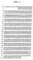
- the amplified 728-basepair fragment was inserted to the cloning vector pCR4 (manufactured by Invitrogen). Subsequently, the base sequence of the inserted region in the plasmid obtained (SEQ ID NO:23) was confirmed. As a result, the base sequence of the inserted region (SEQ ID NO:23) was confirmed as having base substitution at three sites, insertion of two bases at one site, and insertion of one base at one site, as shown in FIG.
- the plasmid pGL3 IL2 Pro was obtained, which has a 731-basepair sequence (SEQ ID NO:23) corresponding to the -675 to +56 region in the vicinity of the transcription initiation site of the human IL-2 gene upstream of the firefly luciferase gene.
- a 435-basepair fragment corresponding to the -379 to +56 region in the vicinity of the transcription initiation site of the human IL-2 gene was obtained by the PCR method with pGL3 IL2 Pro as a template.
- PCR was performed using the primer shown by SEQ ID NO:30 below and the primer shown by SEQ ID NO:28 above.
- the SEQ ID NO:30 primer was designed on the basis of the human IL-2 gene sequence inserted in pGL3 IL2 Pro. Additionally, a restriction endonuclease recognition site was added to the end of each primer to insert to a vector for reporter gene assay.
- the 435-basepair fragment obtained has an NheI recognition site upstream of the promoter, and a HindIII recognition site downstream of the promoter.
- the amplified 435-basepair fragment was inserted to the cloning vector pCR4.
- the base sequence of the inserted region in the plasmid obtained (SEQ ID NO:24) was confirmed.
- the base sequence of the inserted region (SEQ ID NO:24) was confirmed as having base substitution at two sites and insertion of two bases at one site as shown in FIG. 2 compared to the promoter sequence of IL-2 listed as GenBank Locus code: HSIL05 (SEQ ID NO:31).
- the plasmid obtained was cleaved at the NheI recognition site and the HindIII recognition site.
- the fragment obtained was inserted to the NheI/HindIII site of pGL3, a vector for reporter gene assay comprising the firefly luciferase gene.
- the plasmid pGL3 IL2 Pro43 was obtained, which has a 435-basepair promoter sequence corresponding to the -379 to +56 region in the vicinity of the transcription initiation site of the human IL-2 gene upstream of the firefly luciferase gene.
- An 821-basepair fragment corresponding to the -789 to +32 region in the vicinity of the transcription initiation site of the human GATA-1 gene was obtained by the PCR method with human genomic DNA as a template. PCR was performed using the primers shown by SEQ ID NO:32 and SEQ ID NO:33 below. These primers were designed on the basis of the human GATA-1 gene sequence listed as gene database GenBank accession number AF196971. Additionally, a restriction endonuclease recognition site was added to the end of each primer to insert to a vector for reporter gene assay.
- the 821-basepair fragment obtained has a BglII recognition site upstream of the promoter, and a HindIII recognition site downstream of the promoter.
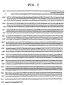
- the amplified 821-basepair fragment was inserted to the cloning vector pCR4. Subsequently, the base sequence of the inserted region in the plasmid obtained (SEQ ID NO:25) was confirmed. As a result, the base sequence of the inserted region (SEQ ID NO:25) was consistent with the promoter sequence of GATA-1 listed as GenBank accession number AF196971 (FIG. 3). Subsequently, the plasmid obtained was cleaved at the NheI recognition site and the HindIII recognition site. The fragment obtained was inserted to the NheI/HindIII site of pGL3 basic, a vector for reporter gene assay comprising the firefly luciferase gene. Thus, the plasmid pGL3-IE was obtained, which has an 821-basepair sequence corresponding to the -789 to +32 region of the promoter region of the GATA-1 gene upstream of the firefly luciferase gene.
- a 637-basepair fragment corresponding to the -3769 to -3133 region upstream of the transcription initiation site of human GATA-1 gene was obtained by the PCR method with human genomic DNA as a template. PCR was performed using the primers shown by SEQ ID NO:34 and SEQ ID NO:35 below. These primers were designed on the basis of the human GATA-1 gene sequence listed as gene database GenBank accession number AF196971. Additionally, a restriction endonuclease recognition site was added to the end of each primer to insert to a vector for reporter gene assay.
- the 637-basepair fragment obtained has a KpnI recognition site upstream of the promoter, and an NheI recognition site downstream of the promoter.
- the amplified 637-basepair fragment was inserted to the cloning vector pCR4. Subsequently, the base sequence of the inserted region in the plasmid obtained (SEQ ID NO:26) was confirmed. As a result, the base sequence of the inserted region (SEQ ID NO:26) was consistent with the promoter sequence of GATA-1 listed as GenBank accession number AF196971 (FIG. 4). Furthermore, the plasmid obtained was cleaved at the KpnI recognition site and the NheI recognition site. The fragment obtained was inserted to the KpnI/NheI site of the pGL3-IE promoter.
- Full-length HDAC3 was amplified by PCR with pMH108 as a template using the following primers 108E (SEQ ID NO : 52) and 108KFN (SEQ ID NO : 53). Meanwhile, the primers were designed to add a Flag sequence (DYKDDDDK) (SEQ ID NO:54) to the C-terminal.
- the PCR fragment obtained was subcloned into pGEM-T (Promega) to yield pGEM-HDAC3-Flag (E108KFN).
- E108KFN was inserted to pEAK10 (Edge BioSystems) via EcoRI/NotI (about 1.2 kbp fragment) treatment to yield pEAK-HDAC3 (pMH118).
- HDAC1-E SEQ ID NO:36
- 107K SEQ ID NO:55
- the primers were designed by modifying the C-terminal stop codon to insert a KpnI site.
- the PCR fragment obtained was subcloned into pGEM-T (Promega) to yield pGEM-HDAC1 (E107K).
- E107K was inserted to pMH118 (EcoRI/KpnI, about 6 kbp) via EcoRI/KpnI (about 1.5 kbp fragment) treatment to yield pEAK-HDAC1 (pMH119).
- HDAC2 Full-length HDAC2 was amplified by PCR with pMH111 as a template using the primers HDAC2-B (SEQ ID NO:38) and 111K (SEQ ID NO:56). Meanwhile, the primers were designed by modifying the C-terminal stop codon to insert a KpnI site.
- the PCR fragment obtained was subcloned into pGEM-T (Promega) to yield pGEM-HDAC2.
- pGEM-HDAC2 was inserted to pFLAG-CMV-5a (SIGMA) (BamHI/KpnI) via BamHI/KpnI treatment to yield pFLAG-CMV-5a-HDAC2 (pMH113).
- pMH113 was inserted to pMH118 (EcoRI/KpnI, about 6 kbp) via EcoRI/KpnI (about 1.5 kbp fragment) treatment to yield pEAK-HDAC2 (pMH121).
- HDAC4-E SEQ ID NO:42
- 106B SEQ ID NO:57
- the primers were designed by modifying the C-terminal stop codon to insert a BglII site.
- the PCR fragment obtained was subcloned into pGEM-T (Promega) to yield pGEM-HDAC4 (E106B).
- a linker oligo having a BglII site and a FLAG sequence (DYKDDDDK) (FLAG-E (SEQ ID NO:58), prepared by annealing FLAG-N (SEQ ID NO:59)) was inserted to an EcoRI/NotI-treated fragment (about 3 kbp) of pBluescriptII KS(+) to yield pBlue-Flag.
- E106B was inserted to pBlue-Flag via EcoRI/BglII treatment to yield pBKS-HDAC4 (E106BN).
- a HindIII/NotI fragment (about 3.3 kbp) of pMH109 was inserted to p3XFLAG-CMV-10 (SIGMA) (HindIII/NotI, about 6.3 kbp) to yield p3XFLAG-CMV-10-HDACS (pMH144).
- a HindIII/NotI fragment (about 3.6 kbp) of pMH110 was inserted to p3XFLAG-CMV-10 (SIGMA) (HindIII/NotI, about 6.3 kbp) to yield p3XFLAG-CMV-10-HDAC6 (pMH145).
- HDAC1-8 expression plasmid HDAC Expression plasmid used Position of Flag tag (DYKDDDK) for Each HDAC Name of plasmid prepared HDAC1 pEAK10 (Edge Bio Systems) C-terminal pMH119 HDAC2 pEAK10 (Edge Bio Systems) C-terminal pMH121 HDAC3 pEAK10 (Edge Bio Systems) C-terminal pMH118 HDAC4 pEAK10 (Edge Bio Systems) C-terminal pMH122 HDAC5 p3XFLAG-CMV-10 (SIGMA) N-terminal pMH144 HDAC6 p3XFLAG-CMV-10 (SIGMA) N-terminal pMH145 HDAC7 pEAK10 (Edge Bio Systems) C-terminal pMH141 HDAC8 pEAK10 (Edge Bio Systems) C-terminal pMH140
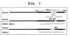
- each of the PCR products obtained ((3) for H802K and H803L variants) was subcloned into the TOPO cloning vector for sequence (pCR4-HDAC4 (H802K, H803L) and pCR4-HDAC4 (H863L)).
- pCR4-HDAC4 H802K, H803L
- pCR4-HDAC4 H863L
- HDAC1 dominant negative variants were prepared in the same manner as the preparation of the HDAC4 dominant negative variants.
- the human HDAC1 CDS base sequence was obtained from GenBank accession number D50405, and primers for mutagenesis were designed (for the base sequences of the individual primers, see Table 4; for the base sequence of HDAC1 CDS, see SEQ ID NO:9; for the amino acid sequence encoded by the HDAC1 CDS base sequence, see SEQ ID NO:10).
- each of the PCR products obtained (3) was subcloned into the TOPO cloning vector for sequence (pCR4-HDAC1 (H140K, H141L) and pCR4-HDAC1 (H199L)).
- a PCR fragment confirmed as being mutated by sequencing was inserted to an HDAC1 expression plasmid (pMH119) using the procedures shown below.
- E107K is a plasmid prepared by inserting full-length HDAC1 CDS to pGEM-T (Promega) (EcoRI/KpnI, the initiation codon is located on the EcoRI side).
- HDAC1 dominant negative variants H140K, H141L
- HDAC3 dominant negative variants were prepared in the same manner as the preparation of the HDAC4 dominant negative variants.
- the human HDAC3 CDS base sequence was obtained from GenBank accession number U66914, and primers for mutagenesis were designed (for the base sequences of the individual primers, see Table 5; for the HDAC3 CDS base sequence, see SEQ ID NO:15; for the amino acid sequence encoded by the HDAC3 CDS base sequence, see SEQ ID NO:16).
- PCR reaction was conducted according to the protocol of Expand (High fidelity PCR System (Roche)).
- pEAK-HDAC3 (pMH118), which was used as a template, is a plasmid prepared by inserting full-length HDAC3 CDS to pEAK10.
- pBKS-HDAC3 (H134K, H135L) (EcoRI/KpnI, about 1.3 kbp) was inserted to an EcoRI/KpnI-treated fragment (about 5.7 kbp) of pMH118 to yield pEAK-HDAC3 (H134K, H135L).
- the base sequence in the coding region of HDAC3 dominant negative variants (H134K, H135L) is shown by SEQ ID NO:17, and the amino acid sequence thereof is shown by SEQ ID NO:18.
- HDAC3 dominant negative variant H193L
- the plasmids for the expression of wild-type HDAC4 or each of HDAC4 dominant negative variants (H802K, H803L, H863L) prepared in Example 3 was mixed in various ratios (1, 3 and 10 ⁇ g), and 1 x 10 7 Jurkat cells (ATCC, TIB-152) were transformed by the electroporation method (BIO-RAD, Gene Pulser II, voltage 300 V, charge 975 ⁇ F, 400 ⁇ L).
- RPMI 1640 containing 10% FBS (MOREGATE) (10% FBS RPMI 1640) was added, and this mixture was dispensed to a 96-well white plate in a ratio of 50 ⁇ L/well.
- a liquid mixture of Phorbol 12-Myristate 13-Acetate (PMA, SIGMA), Ionomycin (SIGMA) and Anti-CD28 antibody (Pharmingen) in 10% FBS RPMI 1640 was added at 50 ⁇ L/well (the final concentrations were 50 ng/mL, 1 ⁇ g/mL and 75 ng/mL, respectively) to stimulate the Jurkat cells.
- luciferase activity in the cells was measured according to the manual of the Bright-Glo Luciferase Assay System (Promega) using a multi-label counter (1420 MULTILABEL COUNTER ARVO SX, WALLAC).
- HDAC4 dominant negative variants H802K, H803L, H863L
- FIG. 8 The expression level of the transferred HDAC was confirmed by Western blotting using an anti-Flag antibody (SIGMA, anti-Flag M2).
- 293 cells (ATCC, CRL-1573) were transfected by the calcium phosphate method (a mixture of DNA and calcium phosphate was added to 293 cells in culture using a DMEM (SIGMA) medium containing 10% FBS, and the medium was replaced with a fresh one 6 hours later); after cultivation under conditions of 37°C, 5% CO 2 and saturated humidity for 48 hours, the number of cell was confirmed using the Cell Counting Kit-8 (DOJIN).
- SIGMA DMEM
- DOJIN Cell Counting Kit-8
- HDAC4 dominant negative variant H863L
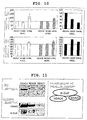
- FIG. 9 cell proliferation inhibition specific for Jurkat cells due to the expression of HDAC4 dominant negative variant (H863L) was observed (FIG. 9).
- HDAC4 dominant negative variant H863L
- SIGMA anti-Flag antibody
- IL-2 and GATA-1 transcription activities were measured with the expression of the HDAC1, 3 and 4 dominant negative variants prepared in Examples 6, 7 and 8.
- luciferase activity in cells were measured according to the manual of the Bright-Glo Luciferase Assay System (Promega), and IL-2 transcriptional activity was measured using a multi-label counter (1420 MULTILABEL COUNTER ARVO SX, WALLAC).
- luciferase activity in cells were measured according to the manual of the Bright-Glo Luciferase Assay System, and GATA-1 transcriptional activity was measured using a multi-label counter.
- HDAC4 dominant negative variants H802K, H803L, H863L
- GATA-1 transcription suppression was not observed at that time (FIG. 10).
- SIGMA anti-Flag antibody
- HDAC4 dominant negative variant H863L
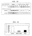
- HDAC4 and HDAC4 dominant negative variant were confirmed (FIG. 12). It was also confirmed by Western blotting (using an anti-HDAC3 antibody of SIGMA Company and an anti-N-CoR antibody of Santa Cruz Company) that the crudely purified fraction of HDAC4 contained HDAC3 and N-CoR, and that the crudely purified fraction of HDAC4 dominant negative variant (H863L) contained almost no HDAC3 or N-CoR.
- HDAC4 dominant negative variant H863L has a decreased complex forming ability with N-CoR and HDAC3 compared to wild-type HDAC4.
- HDAC4 variants which decreased complex forming ability with N-CoR and HDAC3 shows decrease of HDAC activity with histone as a substrate.
- HDAC activities of HDAC4 dominant negative variants H802K, H803L, H863L with histone as a substrate were measured.
- H802K, H803L, H863L wild-type HDAC4 and HDAC4 dominant negative variants
- 293T cells 293T cells by the calcium phosphate method; 48 hours later, whole cell lysate (cells were disrupted in 25mM Tris, pH 7.4, 150mM NaCl, 1mM CaCl 2 , 1% Triton X-100, and cell debris was removed by centrifugation) was crudely purified using an anti-Flag antibody column (SIGMA, anti-Flag M2-agarose) (washed with TBS, then eluted with 0.1M Glysine-HCl, pH 3.5), and used for the measurement of HDAC activity.
- SIGMA anti-Flag antibody column
- the reaction substrate used was a [ 3 H]-labeled acetylated histone prepared by cultivating Jurkat cells (2 ⁇ 10 8 cells) in 40 mL of an RPMI (SIGMA) medium containing 300 MBq [ 3 H] sodium acetate (NENTM Life Science Products, Inc.) for 2 hours, disrupting the cells in Lysis buffer, pH 6.5 (10mM Tris-HCl, 50mM Sodium Hydrogen sulfate, 1% Triton X-100, 10mM MgCl 2 -6H 2 O, 8.6% Sucrose), washing the cells with Wash buffer (10mM Tris-HCl, 13mM EDTA), and then adding 0.4N H 2 SO 4 .
- HDAC4 dominant negative variants H802K, H803L, H863L had decreased HDAC activity compared to wild-type HDAC4 (FIG. 13).
- HDAC4 dominant negative variants H802K, H803L, H863L have decreased complex forming ability with N-CoR and HDAC3 (Example 13), and hence decreased HDAC activity with histone as a substrate.
- HDAC4 dominant negative variant H863L
- N-CoR N-CoR
- the site corresponding to the RD3 sequence of human N-CoR was determined (1005-1498a.a.).
- the human N-CoR CDS base sequence was obtained from GenBank accession number AF044209, and human N-CoR(RD3) cDNA was amplified from human-lung-derived cDNA by the PCR method.
- the primers used were RD3-Bam-FW (SEQ ID NO:82) and RD3-Not-RV (SEQ ID NO:83) below.
- N-CoR(RD3) For the base sequence of human N-CoR(RD3), see SEQ ID NO:21; for the amino acid sequence encoded by the base sequence of human N-CoR(RD3), see SEQ ID NO:22.
- the PCR fragment obtained was confirmed by sequencing (for N-CoR(RD3), a variation of L1014V was observed, but this was adopted since it was reported as a variant in SWISS-PROT), and it was inserted to pBIND (Promega) via treatment with restriction endonuclease (BamHI/NotI) to yield a GAL4-N-CoR(RD3) expression plasmid.
- HDAC4 or an HDAC4 dominant negative variant (H863L) in full length was inserted to pACT (Promega) via restriction endonuclease treatment (XbaI/NotI) to yield HDAC4-VP16 and HDAC4(H863L)-VP16 expression plasmids.
- 10 ⁇ g of pG5-Luc (Promega), 3 ⁇ g of pBND and 3 ⁇ g of pACT, or 10 ⁇ g of pG5-Luc, 3 ⁇ g of pBND-N-CoR(RD3) and 3 ⁇ g of pACT-HDAC4, or 10 ⁇ g of pG5-Luc, 3 ⁇ g of pBND-N-CoR(RD3) and 3 ⁇ g of pACT-HDAC4 (H863L), were mixed respectively, and each mixture was transfected to 1 x 10 7 Jurkat cells (ATCC, TIB-152) by the electroporation method (BIO-RAD, Gene Pulser II, voltage 300 V, charge 975 ⁇ F, 400 ⁇ L).
- BIO-RAD Gene Pulser II, voltage 300 V, charge 975 ⁇ F, 400 ⁇ L
- luciferase activity in the cells was measured according to the manual of the Bright-Glo Luciferase Assay System (Promega) using a multi-label counter (1420 MULTILABEL COUNTER ARVO SX, WALLAC).
- HDAC4-targeted siRNA i.e., siGENE HDAC4 SMARTpool (manufactured by Dharmacon Inc., marketed by B-Bridge international Inc., code number M-003497-00-05), or Non-specific Control Duplex IX (47% GC content) (manufactured by Dharmacon Inc., marketed by B-Bride international Inc., code number D-001206-09-20), and pGL3 IL2 Pro43 were transfected to Jurkat cells by the electroporation method using the same conditions as those mentioned above, and IL-2 transcriptional activity was measured.
- siGENE HDAC4 SMARTpool manufactured by Dharmacon Inc., marketed by B-Bridge international Inc., code number M-003497-00-05
- Non-specific Control Duplex IX 47% GC content
- pGL3 IL2 Pro43 pGL3 IL2 Pro43
- siGENE HDAC4 SMARTpool or Non-specific Control Duplex IX, and pGL3-HSI-IE Pro were transfected to HEL cells, and GATA-1 transcriptional activity was measured.
- the conditions of siRNA transfection and their effects in Jurkat cells and HEL cells were previously confirmed using pGL3-control (Promega) and Luciferase GL3 Duplex (B-Bride: D-001400-01-20).
- the present invention has shown that immunity can be suppressed, IL-2 production can be inhibited, or immunocyte proliferation can be suppressed, without exhibiting a noticeable thrombocytopenic activity and/or GATA-1 production inhibitory activity, by selectively suppressing HDAC4/HDAC8, out of various HDACs. Therefore, by selecting a compound that selectively suppresses HDAC4/HDAC8, it is possible to select an immunosuppressant having low thrombocytopenic activity or an IL-2 production inhibitor or an immunocyte proliferation inhibitor having low GATA-1 production inhibitory activity more conveniently and in a shorter time than conventional. Hence, the method of the present invention is very useful for research for developing new drug.
Landscapes
- Health & Medical Sciences (AREA)
- Life Sciences & Earth Sciences (AREA)
- Chemical & Material Sciences (AREA)
- Engineering & Computer Science (AREA)
- Immunology (AREA)
- Organic Chemistry (AREA)
- General Health & Medical Sciences (AREA)
- Bioinformatics & Cheminformatics (AREA)
- Molecular Biology (AREA)
- Medicinal Chemistry (AREA)
- Hematology (AREA)
- Physics & Mathematics (AREA)
- Wood Science & Technology (AREA)
- Animal Behavior & Ethology (AREA)
- General Chemical & Material Sciences (AREA)
- Public Health (AREA)
- Veterinary Medicine (AREA)
- Biomedical Technology (AREA)
- Biotechnology (AREA)
- Zoology (AREA)
- Chemical Kinetics & Catalysis (AREA)
- Microbiology (AREA)
- Pharmacology & Pharmacy (AREA)
- Urology & Nephrology (AREA)
- Proteomics, Peptides & Aminoacids (AREA)
- Nuclear Medicine, Radiotherapy & Molecular Imaging (AREA)
- Biochemistry (AREA)
- Analytical Chemistry (AREA)
- Cell Biology (AREA)
- Biophysics (AREA)
- Food Science & Technology (AREA)
- General Physics & Mathematics (AREA)
- Genetics & Genomics (AREA)
- Pathology (AREA)
- General Engineering & Computer Science (AREA)
- Diabetes (AREA)
- Transplantation (AREA)
- Medicines That Contain Protein Lipid Enzymes And Other Medicines (AREA)
- Pharmaceuticals Containing Other Organic And Inorganic Compounds (AREA)
- Measuring Or Testing Involving Enzymes Or Micro-Organisms (AREA)
Abstract
The present invention provides a method of
selecting an IL-2 production inhibitor and/or an
immunocyte proliferation inhibitor having low GATA-1
production inhibitory activity, which comprises
measuring the HDAC4 and/or HDAC8 inhibitory activity of
a test substance, and a method of selecting an
immunosuppressant having low thrombocytopenic activity,
which comprises measuring the HDAC4 and/or HDAC8
inhibitory activity of a test HDAC inhibitor.
Description
The present invention relates to a method of
selecting an immunosuppressant having low
thrombocytopenic activity, and an IL-2 production
inhibitor and/or immunocyte proliferation inhibitor
having low GATA-1 production inhibitory activity, which
selectively suppress HDAC4 and/or HDAC8 only, as well as
an agent for suppressing immunity having low
thrombocytopenic activity, and an agent for inhibiting
IL-2 production and/or an agent for inhibiting
immunocyte proliferation having low GATA-1 production
inhibitory activity, which are obtained by the method, a
kit used for the method, and the like.
Cyclosporin A (CsA) and tacrolimus (FK506), which
are major immunosuppressants that are today widely used
in clinical settings to suppress acute graft rejection
reactions following organ transplantation, inhibit the
activity of calcineurin, which is a Ca2+/calmodulin-dependent
protein phosphatase, by binding to respective
specific immunophilins (for example, cyclophilin for CsA,
and FKBP12 for FK506). It is known that as a result, the
dephosphorylation reaction of NF-AT (Nuclear Factor of
Activated T-cell) is inhibited, which in turn inhibits
the transfer of NF-AT into nucleus and suppresses the
transcriptional activity of the IL-2 gene. Research into
this action mechanism has shown that the specific
suppression of the expression of the IL-2 gene at the
transcription level in activated T-cells is of paramount
importance to the suppression of organ graft rejection
and the therapeutic efficacy for various autoimmune
diseases and the like.
The cell nuclear DNA of eukaryotic organisms has a
chromatin structure with nucleosome as the structural
unit, and is folded in strata. Each nucleosome comprises
two molecules of each of the H2A, H2B, H3 and H4
histones, and one molecule of the H1 histone. A
particular lysine residue present on the N-terminal side
of these histone proteins is known to often undergo
modification by acetylation and to be associated with
the expression regulation of various genes.
The level of the acetylation on the histone N-terminal
side is exquisitely controlled in a balance of
the activities of two mutually counteracting series of
enzymes, that is, histone acetyltransferase (histone
acetylation enzyme, HAT) and histone deacetylase
(histone deacetylation enzyme, hereinafter abbreviated
HDAC as required). Of these enzymes, histone deacetylase,
a metal enzyme having zinc coordinated at the active
center thereof, is responsible for the function of
removing the acetyl group from acetylated histone; this
acetyl group removal is considered to cause aggregation
of the chromatin structure and lead to suppressed
transcription.
A series of studies have shown that human histone
deacetylase occurs in at least 11 kinds of isoforms,
from HDAC1 to HDAC11 (see, for example, Pandey R et al.,
Nucleic Acids Research, 30(23): 5036-55 (2002); L. Gao
et al., Journal of Biological Chemistry, 277(28): 25748-25755
(2002); Joseph J. BUGGY et al., Biochemical
Journal, 350: 199-205 (2000); International Patent
Publication No. WO00/10583). As such, each isoform has
been shown not to function as a single enzyme, but to
interact with many other proteins to form a larger
complex. Of these HDAC isoforms, HDAC1, HDAC2, HDAC3 and
HDAC8 are structurally highly homologous to the yeast
Rpd3 protein, and are classified under class I. HDAC1
and HDAC2 are components of a complex comprising a large
number of subunit structures, and are found in, for
example, the SIN3 complex and the NURD/Mi2 complex.
HDAC3 is known to form a complex with N-CoR (nuclear
receptor corepressor) or SMRT (silencing mediator of
retinoid and thyroid) and mediate the transcription
suppression by, for example, thyroid hormone receptor
(TR) or v-ErbA. On the other hand, a group of enzymes
that are homologous to the yeast Hda1 protein are
classified under class II, and HDAC4, HDAC5, HDAC6 and
HDAC7 are known to belong to this group. HDAC4, HDAC5
and HDAC7, which are class II histone deacetylases, are
known to be capable of interacting with BCoR, which is a
supplementary co-repressor that mediates the suppression
by N-CoR, SMRT or BCL-6.
Although the recombinant HDAC3 itself is inactive
as a single substance, data suggestive of its capability
of functioning as an active enzyme with the addition of
SMRT or N-CoR have been reported (see, for example,
Matthew G. Guenther et al., Molecular and Cellular
Biology, 21(18): 6091-6101 (2001)), and HDAC4, which is
a class II enzyme, has been reported to interact with
HDAC3, which is a class I enzyme, via SMRT or N-CoR in
vivo (see, for example, Wolfgang Fischie et al.,
Molecular Cell, 9: 45-57 (2002)). However, much remains
unknown concerning the accurate mechanism by which these
HDAC isoforms are activated.
To date, extensive screening has discovered a large
number of compounds that have HDAC inhibitory activity,
including compounds that have remarkable IL-2 production
inhibitory activity (see, for example, I. Takahashi et
al., The Journal of Antibiotics, 49: 453-457 (1995)),
which are attracting attention as candidates for
immunosuppressants that supplement cyclosporin and
tacrolimus. In fact, among the compounds thus selected,
those that exhibit excellent immunosuppressive action in
vivo have been found. FR225497 has been found to be
excellently effective as a therapeutic agent or
prophylactic agent for organ transplant rejection and
autoimmune diseases with its potent immunosuppressive
action (see, for example, International Patent
Publication No. WO00/08048) and, in addition, has been
suggested to be useful as a therapeutic agent or
prophylactic agent for many other diseases considered to
develop due to an abnormality in gene expression.
Examples of such diseases include inflammatory disorders,
diabetes, diabetic complications, homozygous thalassemia,
fibrosis, liver cirrhosis, acute promyelocytic leukemia
(APL), protozoal infection, cancers (tumors) and the
like. However, despite this utility, many of these
compounds that have HDAC inhibitory activity pose the
problem of being likely to cause serious
thrombocytopenia as a side effect when administered to
the body, and this aspect has made it difficult to use
such compounds as actual therapeutic drugs.
The reason why many of immunosuppressive HDAC
inhibitors are likely to cause thrombocytopenia as a
side effect has not yet fully been elucidated. However,
the present inventors previously showed that the
suppression of the expression of the gene for GATA-1
(also called GATA-1 binding protein, GF-1, NF-E1 or Eryf
1) is profoundly involved in the thrombocytopenic
activity of HDAC inhibitors as a serious side effect,
and that compounds with more potent platelet suppressive
action tend to more strongly suppress the transcription
of the GATA-1 gene (see Japanese Patent Application No.
2002-203901).
GATA-1 is a DNA-binding protein that recognizes the
(A/T)GATA(A/G) consensus sequence, which is
characteristically present in the transcription control
regions of hematopoietic genes. This GATA motif sequence
is found in a variety of control regions such as
enhancer regions of various globin genes, locus control
regions (LCRs) of the β-globin gene, enhancer regions of
the T-cell receptor α-chain and δ-chain genes, and in
promoters. Also, GATA-1 mRNA is expressed at high levels
in mature erythrocytes, mast cells, megakaryocyte and
the like, with slight expression observed in
multifunctional precursor cells and juvenile mouse
testis. Furthermore, mice having the GATA-1 gene knocked
out by an ordinary method are lethal due to primary
hematopoietic cell hypoplasia in the stage of
embryogenesis (see, for example, Y. Fujiwara et al.,
Proceedings of the National Academy of Sciences of the
USA, 93: 12355-12358 (1996)). On the other hand, it is
possible to prepare mice having only the expression of
the GATA-1 gene in megakaryocytic lineage knocked out
selectively, and these mice have been shown to suffer
dramatically decreased platelet counts and to lack
normal maturation of megakaryocytic lineage (see, for
example, R.A. Shivdasani et al. The EMBO Journal, 16:
3965-3973 (1997)).
In HDAC, a large number of isozymes exist, some of
which have a side effect serious to the body when
inhibited. For example, administering a conventional
HDAC inhibitor, which has no isozyme selectivity, causes
thrombocytopenia as a side effect, and the present
inventors showed that this action is possibly
attributable to the transcription suppressive action of
the GATA-1 gene, which plays a decisive role in the
differentiation and maturation of erythrocytic lineage
cells and megakaryocytic lineage, and provided a method
of screening for an immunosuppressant having low
thrombocytopenic activity based on this action (see
Japanese Patent Application No. 2002-203901). However,
in this screening method, to screen for an
immunosuppressant having low thrombocytopenic activity,
it is necessary to separately evaluate the platelet
suppressive action of a compound possessing HDAC
inhibitory action after the compound is screened for, so
that much labor and time are taken and the screening
method is not always satisfactory. Accordingly, to
select an immunosuppressant from among HDAC inhibitors,
there is a strong demand for the provision of an
excellent method of screening for an immunosuppressant
that inhibits a particular isozyme involved in
immunosuppressive effects, but does not inhibit other
isozymes involved in side effects such as
thrombocytopenic activity, conveniently and in a short
time, and the present invention is directed to resolve
these problems.
The present inventors diligently studied the
association of the transcription suppressive activities
for the IL-2 gene and the GATA-1 gene and various HDACs,
and found that if either or both of HDAC4 or HDAC8 out
of the various HDACs, are selectively suppressed, IL-2
production can be inhibited without noticeably
suppressing GATA-1 production inhibitory activity, the
proliferation of immunocytes can be inhibited, and
immunity can be suppressed without significantly
decreasing the platelet count. The present inventors
confirmed that HDAC4 forms a complex with N-CoR, and the
complex of HDAC4 and N-CoR further forms a complex with
HDAC3. Accordingly, the present inventors realized that
an IL-2 production inhibitor having low GATA-1
production inhibitory activity, an immunocyte
proliferation inhibitor, or an immunosuppressant having
low thrombocytopenic activity, can be obtained by
selecting a compound that selectively suppresses HDAC4
and/or HDAC8 out of various HDACs, and inhibiting the
formation of a complex of HDAC4 and N-CoR and, as
required, the formation of a higher complex of HDAC4 and
the N-CoR-HDAC3 complex, and developed the present
invention.
Accordingly, the present invention relates to the
inventions shown below.
The present invention is hereinafter described in
detail.
The present invention relates to an agent for
inhibiting IL-2 production (IL-2 production inhibitor)
and/or an agent for inhibiting immunocyte proliferation
(immunocyte proliferation inhibitor), each of which
having low GATA-1 production inhibitory activity,
comprising a selective HDAC4 and/or HDAC8 inhibitor. An
IL-2 production inhibitor and/or an immunocyte
proliferation inhibitor, each of which having low GATA-1
production inhibitory activity, comprising a selective
HDAC4 and/or HDAC8 inhibitor can be obtained by, for
example, the selection method described below. It is
also possible to obtain an IL-2 production inhibitor
and/or an immunocyte proliferation inhibitor, each of
which having low GATA-1 production inhibitory activity,
comprising a selective HDAC4 and/or HDAC8 inhibitor, by
using the selection method described below and a method
known in the art in combination.
A "selective HDAC4 and/or HDAC8 inhibitor" refers
to an inhibitor that suppresses the HDAC enzyme activity
of HDAC4 and/or HDAC8 more than suppresses the HDAC
enzyme activity of HDAC1-3 and HDAC5-7, and is
preferably an inhibitor that does not or substantially
does not suppress the HDAC enzyme activity of HDAC1-3
and HDAC5-7, and that suppresses the HDAC enzyme
activity of HDAC4 and/or HDAC8. As examples of the
selective HDAC4 and/or HDAC8 inhibitor, an antisense
nucleic acid, ribozyme, decoy nucleic acid, siRNA and
antibody (for example, single-chain antibody) that are
specific for HDAC4 and/or HDAC8, a dominant negative
variant of HDAC4 and/or HDAC8, an expression vector
carrying a nucleic acid that encodes one of them, and a
substance obtained by the selection method described
below, can be mentioned.
"GATA-1 production inhibitory activity" may be any
activity to suppress GATA-1 production. As examples of
the mechanism of suppression of GATA-1 production,
suppression at the transcription level, promotion of
GATA-1 mRNA decomposition, suppression at the
translation level and the like can be mentioned. GATA-1
production inhibitory activity can be evaluated by a
method known in the art. Specifically, GATA-1 production
inhibitory activity can be evaluated by a reporter assay
using the GATA-1 reporter gene as described below, or by
measuring the expression level of GATA-1 mRNA or GATA-1
protein in a cell line, primary culture cells and the
like that are naturally expressing GATA-1. "Low" GATA-1
production inhibitory activity as evaluated with the
addition of a compound means that the activity thereof
to inhibit GATA-1 production is lower than that obtained
when a compound for a reference control is added; for
example, whether or not the GATA-1 production inhibitory
activity is low can be evaluated by an assay using the
GATA-1 reporter gene. As an example of the compound used
as a reference control, tricostatin (TSA), which is a
non-specific HDAC inhibitor, can be mentioned.
The "immunocyte proliferation inhibitor" may have
any kind of activity, as long as it inhibits the
proliferation of immunocytes. As examples of the
mechanism of cell proliferation inhibition, cell cycle
termination, apoptosis induction and the like can be
mentioned. Immunocyte proliferation inhibitory activity
can be evaluated by a method known in the art.
Specifically, immunocyte proliferation inhibitory
activity can be evaluated by thymidine uptake activity,
the MTT method and the like. Here, "immunocytes" refer
to cells responsible for immune function in the body.
Preferably, "immunocytes" refer to lymphocytes,
dendritic cells and the like that are capable of produce
an antibody or of expressing a cellular immune reaction,
more preferably T-cells.
The "IL-2 production inhibitor" may have any kind
of activity, as long as it has IL-2 production
inhibitory activity, that is, an activity to suppress
IL-2 production. As examples of the mechanism of IL-2
production inhibition, suppression at the transcription
level, promotion of IL-2 mRNA decomposition, suppression
at the translation level and the like can be mentioned.
IL-2 production inhibitory activity can be evaluated by
a method known in the art. Specifically, IL-2 production
inhibitory activity can be evaluated by a reporter assay
using the IL-2 reporter gene as described in Examples
below, or by measuring the expression level of IL-2 mRNA
or IL-2 protein in a cell line, primary culture cells
and the like that are naturally expressing the IL-2.
The present invention also relates to an agent for
suppressing immunity (immunosuppressant) having low
thrombocytopenic activity, comprising a selective HDAC4
and/or HDAC8 inhibitor. An immunosuppressant having low
thrombocytopenic activity, comprising a selective HDAC4
and/or HDAC8 inhibitor can be obtained by, for example,
the selection method described below. It is also
possible to obtain an immunosuppressant having low
thrombocytopenic activity, comprising a selective HDAC4
and/or HDAC8 inhibitor, by using the selection method
described below and a method known in the art in
combination.
"Thrombocytopenic activity" refers to an activity
to decrease the platelet count, and this
thrombocytopenic activity can be directly confirmed by,
for example, a method known in the art, such as
administering a compound to a laboratory animal. Also,
because it is known that there is a correlation between
thrombocytopenic activity and GATA-1 production
inhibitory activity, the thrombocytopenic activity can
also be indirectly confirmed with GATA-1 production
inhibitory activity as an index. "Low" thrombocytopenic
activity of a compound or a drug means that the action
to decrease the platelet count is lower when the
compound or the drug is administered to a human or a
laboratory animal and the like, than when a compound
used as a reference control is administered, and it is
also possible to confirm whether or not the
thrombocytopenic activity is low, by, for example,
determining whether or not the GATA-1 production
inhibitory activity is low by a method described above.
As an example of the compound used as a reference
control, Oxamflatin can be mentioned.
An "immunosuppressant" refers to an agent that
induces immunosuppression; as an example of the
immunosuppressant, an immunosuppressant having an
activity to suppress IL-2 production can be mentioned.
Therefore, the immunosuppressant can be obtained by, for
example, determining whether or not it has IL-2
transcription suppressive activity using a method
described above.
The present invention also relates to a method of
selecting an IL-2 production inhibitor and/or an
immunocyte proliferation inhibitor, each of which having
low GATA-1 production inhibitory activity, or an
immunosuppressant having low thrombocytopenic activity,
which comprises measuring the HDAC4 and/or HDAC8 enzyme
inhibitory activity of a test substance.
Specifically, the present invention relates to a
method of selecting an IL-2 production inhibitor and/or
an immunocyte proliferation inhibitor, each of which
having low GATA-1 production inhibitory activity, or an
immunosuppressant having low thrombocytopenic activity,
which comprises (i) measuring the HDAC4 and/or HDAC8
enzyme inhibitory activity of a test substance, and (ii)
measuring the HDAC enzyme inhibitory activity of the
test substance (here, the HDAC enzyme inhibitory
activity is one or more HDAC enzyme inhibitory
activities selected from a group consisting of HDAC1,
HDAC2, HDAC3, HDAC5, HDAC6 and HDAC7 enzyme inhibitory
activities). In (ii) of this method, one or more HDAC
enzyme inhibitory activities selected from a group
consisting of HDAC1, HDAC2, HDAC3, HDAC5, HDAC6 and
HDAC7 enzyme inhibitory activities are measured in the
presence of the test substance; preferably all of HDAC1,
HDAC2, HDAC3, HDAC5, HDAC6 and HDAC7 enzyme inhibitory
activities are measured. This method may further
comprises (iii) determining whether or not the test
substance selectively inhibits HDAC4 and/or HDAC8 enzyme
activity, on the basis of the results of (i) and (ii).
More specifically, the present invention relates to
a method of selecting an IL-2 production inhibitor
and/or an immunocyte proliferation inhibitor, each of
which having low GATA-1 production inhibitory activity,
or an immunosuppressant having low thrombocytopenic
activity, wherein the IL-2 production inhibitor and/or
the immunocyte proliferation inhibitor selectively
inhibits HDAC4 and/or HDAC8 enzyme activity. In one
embodiment of the present invention, the above-described
method comprises (i) expressing the HDAC1, HDAC2, HDAC3,
HDAC4, HDAC5, HDAC6, HDAC7 and HDAC8 genes to obtain
respective enzyme solutions, then (ii) measuring HDAC
enzyme activity for each of the enzyme solutions in the
presence of a test substance, and (iii) selecting a test
substance that selectively suppresses HDAC enzyme
activity only when using an enzyme solution obtained by
expressing the HDAC4 gene or the HDAC8 gene.
In another embodiment, the above-described method
comprises (i) measuring HDAC enzyme activity for an
enzyme solution partially purified from human cells, in
the presence of a test substance, (ii) measuring HDAC
enzyme activity for an enzyme solution obtained by
expressing the HDAC4 gene and/or the HDAC8 gene, in the
presence of the test substance, and (iii) selecting a
test substance that selectively suppresses HDAC enzyme
activity only when using an enzyme solution obtained by
expressing the HDAC4 gene and/or the HDAC8 gene, by
comparing the enzyme activities measured in (i) and (ii).
The human cells used in (i) in this method are not
subject to limitation, as long as they are of a human-derived
cell line; preferably, a cell lines derived from
the above-described immunocytes are used, and most
preferably, Jurkat cells (ATCC accession number TIB-152)
are used.
The above-described "test substance" is not subject
to limitation, as long as it is a candidate substance
that can be applied to an assay system according to the
method of the present invention, and includes all
compounds such as low-molecular organic compounds, low-molecular
inorganic compounds, high-molecular compounds
including proteins and nucleic acids, saccharides and
the like, and also includes all of their mixtures,
natural products, synthetic products, and extracts from
animals, plants, fungi, algae and microorganisms. As
examples of the "test substance", HDAC inhibitors (see,
for example, International Patent Publication Nos.
WO02/085883, WO02/085400, WO02/076941, WO02/062773,
WO02/46129, WO02/07722, WO02/06307, WO01/70675,
WO01/38322, WO01/16106, WO00/71703, WO00/21979 and
WO00/08048; Japanese Patent Publication Nos. 2001-348340
and HEI-11-302173; US Patent Publications US
2002/0120099 and US 2002/0115826; US Patent Nos.
6,638,530, 6,541,661 and 6,399,568) can be used. The
"test substance" may be a known compound or a newly
discovered or synthesized compound.
An "HDAC inhibitor" refers to a compound that
inhibits HDAC enzyme activity, that is, a compound
having HDAC enzyme inhibitory activity.
"HDAC enzyme activity" refers to an activity to
remove the acetyl group from the lysine residue of an
acetylated protein, for example, a histone (i.e.,
deacetylation activity), and may be the deacetylation
activity of a particular HDAC isozyme, or the
deacetylation activity of one or two or more unspecified
HDAC isozymes. On the other hand, "HDAC4 enzyme
activity" refers to the deacetylation activity of HDAC4,
and "HDAC8 enzyme activity" refers to the deacetylation
activity of HDAC8. HDAC1 enzyme activity, HDAC2 enzyme
activity, HDAC3 enzyme activity, HDAC4 enzyme activity,
HDAC5 enzyme activity, HDAC6 enzyme activity, and HDAC7
enzyme activity are also defined similarly. HDAC enzyme
activity can be measured by reacting an enzyme solution
containing each HDAC enzyme (hereinafter abbreviated
"enzyme solution" as required) and a substrate of the
HDAC enzyme in the presence/absence of a test substance
by a method known in the art.
"HDAC enzyme inhibitory activity" refers to an
activity to suppress the deacetylation of the lysine
residue of a protein, and may be an activity to
specifically or non-specifically suppress the
deacetylation by a particular HDAC isozyme. On the other
hand, "HDAC4 enzyme inhibitory activity" refers to an
activity to suppress the deacetylation by HDAC4, and
"HDAC8 enzyme inhibitory activity" refers to an activity
to suppress the deacetylation by HDAC8. HDAC1 enzyme
inhibitory activity, HDAC2 enzyme inhibitory activity,
HDAC3 enzyme inhibitory activity, HDAC4 enzyme
inhibitory activity, HDAC5 enzyme inhibitory activity,
HDAC6 enzyme inhibitory activity, and HDAC7 enzyme
inhibitory activity are also defined similarly.
As the substrate used in the measurement of HDAC
enzyme activity, a substrate known in the art can be
used, and representatively, a [3H]acetyl-labeled histone,
a fluorescently labeled acetylated peptide and the like
can be used. For example, when a [3H]acetyl-labeled
histone is used as a substrate, the [3H]acetyl-labeled
histone can be obtained from Jurkat cells. Specifically,
the following procedure is followed. 1 × 10⁸ Jurkat cells
are incubated with 300 MBq [3H] sodium acetate in an
RPMI-1640 medium supplemented with 10% FBS, penicillin
(50 units/ml) and streptomycin (50 µg/ml), in the
presence of 5mM sodium butyrate, for 30 minutes (5% CO2,
37°C), the cells are recovered in a centrifugal tube (50
ml), collected via centrifugation (1000 rpm, 10 minutes),
and once washed with phosphate-buffered saline. The
washed cells are suspended in 15 ml of an ice-cooled
lytic buffer (10mM Tris-HCl, 50mM sodium hydrogen
sulfite, 1% Triton X-100, 10mM MgCl2, 8.6% sucrose, pH
6.5). After Dounce homogenization (30 strokes), nuclei
are collected via centrifugation (1000 rpm, 10 minutes),
washed with 15 ml of the lytic buffer three times, and
then once washed with 15 ml of an ice-cooled washing
buffer (10mM Tris-HCl, 13mM EDTA, pH 7.4). The pellet is
suspended in 6 ml of ice-cooled water using a mixer, and
68 ml of H2SO4 is added to this suspension to obtain a
concentration of 0.4N. After incubation at 4°C for 1
hour, the suspension is centrifuged (15,000 rpm, 5
minutes), and the supernatant is mixed with 60 ml of
acetone. Finally, overnight incubation at -20°C is
followed by recovery of the aggregate to yield a
[3H]acetyl-labeled histone. For the method of obtaining
a [3H]acetyl-labeled histone, see, for example,
International Patent Publication No. WO00/08048, Yoshida,
M. et al., J. Biol. Chem., 265: 17174-17179 (1990) and
the like. The [3H]acetyl-labeled histone thus obtained
may be used in solution in an appropriate buffer and
immediately used as is, or if it is not used immediately,
it may be stored under freezing or refrigeration in a
solution state, or may be stored under freezing in a dry
solid state.
To "express each gene" can be performed by
incorporating the gene to an expression vector having a
promoter sequence, transferring the expression vector
incorporating the gene to cells, and cultivating the
cells, and the like. The promoter, expression vector and
the like used differ variously depending on the host
cells used for expression, and those skilled in the art
having ordinary expertise can easily find the best
combination in commercially available products.
Alternatively, to "express each gene" can also be
performed by transferring the gene to an expression
vector having a promoter sequence, such as the SP6 phage
or the T7 phage, and allowing the reaction in vitro by
the in vitro transcription/translation method. Those
skilled in the art having ordinary experimental
knowledge can easily synthesize the transcription
product and protein of each of the genes by this method.
As the method of transferring a gene used in the
present invention to cells, a method of transformation
known in the art, such as the calcium phosphate method,
the liposome method, the lipofection method, the
electroporation method and the like, can be used, which
are not to be construed as limiting.
The cells to which a gene used in the present
invention is transferred are not subject to limitation
unless otherwise specified, as long as they are cells
wherein a gene inserted in an expression vector is
expressed, and mammalian cells including the above-described
human cells, insect cells, yeast, Escherichia
coli and the like can be used.
An "enzyme solution" refers to a fraction having
HDAC enzyme activity extracted from a cell disruption
solution or reaction solution having HDAC enzyme
activity, wherein the HDAC enzyme may have been
partially purified or purified to high purity, as long
as the HDAC enzyme has been purified to the extent that
enables a measurement of HDAC enzyme activity. A cell
disruption solution having HDAC enzyme activity can be
obtained from cells that are naturally expressing an
HDAC enzyme, or cells wherein each HDAC gene is
overexpressed. An enzyme solution can be obtained from
cells that are naturally expressing an HDAC enzyme by,
for example, cultivating the cells, and extracting a
fraction having HDAC enzyme activity from a disruption
solution of the cultured cells. Also, an enzyme solution
can be obtained from cells allowed to overexpress each
HDAC gene by, for example, inserting each HDAC gene in
an expression vector, incorporating the expression
vector to cells, cultivating the cells obtained, and
extracting a fraction having HDAC enzyme activity from a
disruption solution of the cultured cells. On the other
hand, a reaction solution having HDAC enzyme activity
can be prepared by, for example, synthesizing the
transcription product and protein of each HDAC from each
HDAC gene joined to an appropriate vector using the in
vitro transcription/in vitro translation method.
"Partial purification" of an HDAC enzyme refers to
purifying the HDAC enzyme to the extent that enables a
measurement of HDAC enzyme activity, and can be
conducted by a method known in the art, for example, a
method described in International Patent Publication No.
WO00/08048, Yoshida, M. et al., J. Biol. Chem. 265:
p.17174-17179 (1990), and the like. Also, the partially
purified HDAC enzyme can be purified to higher purity by
another method of purification known in the art. For
this partial purification, the following method of
purification, for example, can be used. First, 5×10⁸
Jurkat cells are suspended in 40 ml of HDA buffer
(composition: 15mM potassium phosphate, 5% glycerol and
0.2mM EDTA, pH 7.5). Subsequently, the suspension is
homogenized, then nuclei are recovered via
centrifugation (35,000 x g, 10 minutes), and the nuclei
obtained are homogenized in 20 ml of an HDA buffer
supplemented with 1M (NH4)2SO4. Subsequently, the
homogenate is subjected to sonication and clarified by
centrifugation (35,000 x g, 10 minutes). Subsequently,
deacetylase is precipitated by raising the (NH4)2SO4
concentration to 3.5M. Subsequently, the precipitated
protein is dissolved in 10 ml of HDA buffer, and this
solution is dialyzed in 4 L of HDA buffer. After
dialysis, the sample solution is loaded to a DEAE-cellulose
(Whatman DE52) column (25×85mm), previously
equilibrated with HDA buffer, and eluted with 300 ml of
NaCl solution on a linear gradient (0 to 0.6M). As a
result, HDAC enzyme activity appears as a single peak in
the fraction eluted at 0.3 to 0.4M NaCl.
HDAC enzyme activity is measured by a method known
in the art. For example, when a [3H]acetyl-labeled
histone is used as a substrate, 10 µl of the [3H]acetyl-labeled
histone is added to 90 µl of enzyme solution,
and this mixture is incubated at 25°C for 30 minutes.
The reaction is stopped by the addition of 10 µl of
hydrochloric acid, and the released [3H] acetic acid is
extracted with 1 ml of ethyl acetate. Finally, 0.9 ml of
the solvent layer is transferred to a toluene
scintillation solution, and the radioactivity of this
solution is measured, thereby HDAC enzyme activity can
be determined. When the HDAC enzyme activity of each
enzyme solution is measured in the presence of a test
substance, the test substance may be added around the
time of preparation of a mixture of 10 µl of the
[3H]acetyl-labeled histone and 90 µl of the enzyme
solution, and the reaction may be initiated. For details
about the measurement of HDAC enzyme activity, see, for
example, International Patent Publication No.
WO00/08048; Yoshida, M. et al., J. Biol. Chem., 265:
17174-17179 (1990) and the like, when a [3H] acetyl-labeled
histone is used as a substrate, and see, for
example, International Patent Publication No.
WO01/040506 and the like, when a fluorescently labeled
acetylated peptide is used as a substrate.
In the above-described method, a test substance
that selectively suppresses HDAC enzyme activity only
when using an enzyme solution obtained by expressing the
HDAC4 gene or the HDAC8 gene is preferably selected, and
a test substance that suppresses the HDAC enzyme
activity of HDAC4 and/or HDAC8 more than suppresses the
HDAC enzyme activity of HDAC1-3 and HDAC5-7 may be
selected. More preferably, a test substance that does
not suppress or substantially does not suppress the HDAC
enzyme activity of HDAC1-3 and HDAC5-7, and that
suppresses the HDAC enzyme activity of HDAC4 and/or
HDAC8, is selected.
The present invention also relates to a method of
selecting an IL-2 production inhibitor and/or an
immunocyte proliferation inhibitor, each of which having
low GATA-1 production inhibitory activity, or an
immunosuppressant having low thrombocytopenic activity,
which comprises selecting an inhibitor that specifically
inhibits the formation of a complex of HDAC4 and N-CoR
from among test HDAC inhibitors. For example, this
method comprises (i) expressing the HDAC4 gene and the
N-CoR gene to obtain an enzyme solution, then (ii)
allowing the enzyme solution to coexist with a test HDAC
inhibitor, and (iii) determining whether or not a
complex of HDAC4 and N-CoR is formed in the mixture of
enzyme solution and test HDAC inhibitor obtained in (ii).
In this method, an enzyme solution obtained by
expressing the HDAC3 gene, in addition to the HDAC4 gene
and the N-CoR gene, may also be used. In this case,
whether or not a higher complex is formed between HDAC4
and N-CoR and HDAC3 in the presence of a test HDAC
inhibitor is determined. In (iii) in the above-described
method, as examples of the method of determining whether
or not a complex is formed, a method known in the art
such as an analytical method using surface plasmon
resonance, an analytical method using an antibody (for
example, Western blotting) and the like can be mentioned.
As the labeled substance used to label the
secondary antibody in an analytical method using an
antibody, for example, Western blotting, an enzyme such
as horseradish peroxidase and alkaline phosphatase, a
fluorescent substance such as fluorescein isocyanate and
rhodamine, a radioactive substance such as 32P or 125I, a
chemical luminescent substance and the like can be
mentioned.
To select an IL-2 production inhibitor and/or an
immunocyte proliferation inhibitor, each of which having
low GATA-1 production inhibitory activity, or an
immunosuppressant having low thrombocytopenic activity,
that specifically inhibits the formation of a complex of
HDAC4 and N-CoR, a method based on a two-hybrid system
known in the art can also be used. For example, when a
two-hybrid system is used for this purpose, a test
substance that specifically inhibits the formation of a
complex of an HDAC4 fusion protein comprising at least a
portion of the HDAC4 protein and an N-CoR fusion protein
comprising at least a portion of the N-CoR protein is
selected by (i) expressing a gene that encodes the HDAC4
fusion protein and a gene that encodes the N-CoR fusion
protein in cells, and (ii) determining whether or not a
test substance (preferably a test HDAC inhibitor)
inhibits the formation of a complex of the HDAC4 fusion
protein and the N-CoR fusion protein.
The HDAC4 fusion protein may be any one comprising
a region that interacts with N-CoR in the HDAC4 protein,
and may also comprise the entire HDAC4 protein. Likewise,
the N-CoR fusion protein may be any one comprising a
region that interacts with HDAC4 in the N-CoR protein
(for example, RD3 described below), and may also
comprise the entire N-CoR protein. Each of the HDAC4
fusion protein and the N-CoR fusion protein further
comprises, in addition to a portion or the entire region
of the HDAC4 protein and the N-CoR protein, a DNA-binding
domain (for example, GAL4 DNA-binding domain and
the like) or a transcription activation domain (for
example, VP16 activation domain and the like) derived
from transcription regulatory factor. However, if the
HDAC4 fusion protein comprises a DNA-binding domain, the
N-CoR fusion protein need to comprise a transcription
activation domain; if the HDAC4 fusion protein comprises
a transcription activation domain, the N-CoR fusion
protein need to comprise a DNA-binding domain.
Whether or not a complex is formed between the two
fusion proteins can be confirmed by the expression level
of a gene located downstream of the promoter to which
the DNA-binding region in a fusion protein binds.
Accordingly, when a complex is formed between the fusion
proteins, the expression level of the mRNA or protein
encoded by the gene, and the expression level of a
reporter gene comprising a promoter located upstream of
the gene, increase, whereas when a complex is not formed
between the fusion proteins, the expression level of the
mRNA or protein encoded by the gene, and the expression
level of the above-described reporter gene comprising a
promoter remain unchanged.
Whether or not a test substance (preferably a test
HDAC inhibitor) inhibits the formation of a complex of
an HDAC4 fusion protein and an N-CoR fusion protein can
be determined by determining whether or not the
expression level of a gene located downstream of the
promoter to which the DNA-binding region in a fusion
protein binds is decreased in the presence of the test
substance. Accordingly, (a) the expression level of the
mRNA or protein encoded by the gene or the expression
level of a reporter gene comprising a promoter located
upstream of the gene is measured in the absence of a
test substance, (b) the expression level of the mRNA,
protein or reporter gene is measured in the presence of
the test substance, and (c) comparing the expression
level measured in (a) and the expression level measured
in (b); if the expression level measured in (b) is lower
than the expression level measured in (a), the test
substance is selected as a substance that inhibits the
formation of a complex of an HDAC4 fusion protein and an
N-CoR fusion protein. It is preferable, from the
viewpoint of assay simplicity, sensitivity and the like,
that whether or not the test substance inhibits the
formation of a complex of an HDAC4 fusion protein and an
N-CoR fusion protein be evaluated by measuring the
expression level of a reporter gene.
The present invention also relates to a method of
selecting a compound having low GATA-1 production
inhibitory activity, and having IL-2 production
inhibitory activity and/or immunocyte proliferation
inhibitory activity, or a compound having low
thrombocytopenic activity and having immunosuppressive
activity, which comprises selecting a compound that
suppresses the expression level of HDAC4 and/or HDAC8
from among test HDAC inhibitors. This method comprises
(i) allowing cells that express HDAC4 and/or HDAC8 to
coexist with a test HDAC inhibitor, and (ii) measuring
the HDAC4 and/or HDAC8 expression level in the cells.
Specifically, this method can be conducted in the same
manner as the method described below of measuring the
expression level of GATA-1 mRNA, GATA-1 protein or GATA-1
reporter gene.
The present invention also relates to a method of
selecting a compound having low GATA-1 production
inhibitory activity and having IL-2 production
inhibitory activity, or a compound having low
thrombocytopenic activity and having immunosuppressive
activity, wherein an HDAC inhibitor that selectively
inhibits the binding of HDAC4 and/or HDAC8 and an HDAC4-
and/or HDAC8-specific ligand is selected from among test
HDAC inhibitors. This method comprises (i) expressing
the HDAC4 and/or the HDAC8 gene to obtain an enzyme
solution, and then (ii) measuring the binding activity
of HDAC4 and/or HDAC8 enzyme and the HDAC4- and/or
HDAC8-specific ligand for the enzyme solution in the
presence of an HDAC4- and/or HDAC8-specific ligand and a
test HDAC inhibitor.
An "HDAC4- and/or HDAC8-specific ligand" refers to
a substance showing stronger binding activity to HDAC4
and/or HDAC8 than to HDAC1-3 and HDAC5-7, out of
substances that specifically bind to HDAC4 and/or HDAC8.
These substances include, but are not limited to,
substances that promote the activity of each enzyme,
those that inhibit the same, and the like. As "the
HDAC4- and/or HDAC8-specific ligand", a low-molecular
substance that shows weak binding activity to HDAC1-3
and HDAC5-7 or is substantially free from binding
activity, and that shows strong binding activity to
HDAC4 and/or HDAC8, is preferred.
In step (ii) in the above-described method, an
"HDAC4- and/or HDAC8-specific ligand" is preferably used,
but an "HDAC4- and/or HDAC8-nonspecific ligand" can also
be used. An "HDAC4- and/or HDAC8-nonspecific ligand"
refers to a substance that shows stronger binding
activity to HDACl-3 and HDAC5-7 than to HDAC4 and/or
HDAC8, or a substance that shows nearly equivalent
binding activity to HDAC4 and/or HDAC8 and to HDAC1-3
and HDAC5-7. In the present invention, an "HDAC4- and/or
HDAC8-specific ligand" and an "HDAC4- and/or HDAC8-
nonspecific ligand" are also integrally referred to as
an "HDAC4 and/or HDAC8 ligand".
In step (ii) in the above-described method, the
binding activity of HDAC4 and/or HDAC8 enzyme and an
HDAC4- and/or HDAC8-specific ligand is measured in the
presence of a test HDAC inhibitor. Binding activity can
be measured using, for example, a method known in the
art, such as binding assay or mass analysis. By the
above-described method, a substance that competitively
inhibits an HDAC4- and/or HDAC8-specific ligand can be
obtained.
In the present invention, human-derived HDAC4 is
preferably used as HDAC4. Specifically, HDAC4 refers to,
for example, a protein that comprises the amino acid
sequence shown by SEQ ID NO:2 or an analogue thereof
that has HDAC enzyme activity (for example, protein
encoded by the base sequence of GenBank accession number
AF132607). HDAC4 analogues include a variant having one
or two or more amino acids substituted, deleted or added
in the amino acid sequence shown by SEQ ID NO:2, a sugar
chain added form and the like. As the HDAC4 analogue, a
protein that comprises an amino acid sequence having a
homology of 70% or more, preferably 80% or more, more
preferably 90% or more, still more preferably 95% or
more, to the amino acid sequence shown by SEQ ID NO:2,
can also be used. The degree of amino acid sequence
homology can be determined by, for example, using BLAST
in the default settings between the amino acid sequence
of interest and a reference amino acid sequence in NCBI
(The National Center for Biotechnology Information). For
details about HDAC4, see, for example, International
Patent Publication No. WO00/10583.
Likewise, in the present invention, human-derived
HDAC8 is preferably used as HDAC8. Specifically, HDAC8
refers to, for example, a protein that comprises the
amino acid sequence shown by SEQ ID NO:8, or an analogue
thereof that has HDAC enzyme activity (for example,
protein encoded by the base sequence of GenBank
accession number HSA277724). HDAC8 analogues include a
variant having one or two or more amino acids
substituted, deleted or added in the amino acid sequence
shown by SEQ ID NO:8, a sugar chain added form and the
like. As the HDAC8 analogue, a protein that comprises an
amino acid sequence having a homology of 70% or more,
preferably 80% or more, more preferably 90% or more,
still more preferably 95% or more, to the amino acid
sequence shown by SEQ ID NO:8 can also be used. For
details about HDAC8, see, for example, Joseph J. BUGGY
et al., Biochemical Journal 350: 199-205 (2000).
In the present invention, human-derived N-CoR is
preferably used as N-CoR. Specifically, N-CoR refers to,
for example, a protein that comprises the amino acid
sequence shown by SEQ ID NO:22, or an analogue thereof
that is capable of forming a complex with HDAC4 or HDAC3
(for example, protein encoded by the base sequence of
GenBank accession number AF044209). N-CoR analogues
include a variant having one or two or more amino acids
substituted, deleted or added in the amino acid sequence
shown by SEQ ID NO:22, a sugar chain added form and the
like. As the N-CoR analogue, a protein that comprises an
amino acid sequence having a homology of 70% or more,
preferably 80% or more, more preferably 90% or more,
still more preferably 95% or more, to the amino acid
sequence shown by SEQ ID NO:22 can also be used. In the
present invention, N-CoR may be any one, as long as it
is capable of forming a complex with HDAC4 (both HDAC4
and HDAC3 as required). As such N-CoR, the N-CoR
variants disclosed in, for example, Matthew G. Guenther
et al., Molecular and Cellular Biology, 21(18): 6091-6101
(2001) and elsewhere, and the like can also be used,
in addition to those mentioned above.
In the present invention, human-derived HDAC3 is
preferably used as HDAC3. Specifically, HDAC3 refers to,
for example, a protein that comprises the amino acid
sequence shown by SEQ ID NO:16, or an analogue thereof
that has HDAC enzyme activity or is capable of binding
to HDAC4 (HDAC4 - N-CoR complex as required) (for
example, protein encoded by the base sequence of GenBank
accession number U66914). HDAC3 analogues include a
variant having one or two or more amino acids
substituted, deleted or added in the amino acid sequence
shown by SEQ ID NO:16, a sugar chain added form and the
like. As the HDAC3 analogue, a protein that comprises an
amino acid sequence having a homology of 70% or more,
preferably 80% or more, more preferably 90% or more,
still more preferably 95% or more, to the amino acid
sequence shown by SEQ ID NO:16 can also be used. In the
analysis of a complex of HDAC3 and N-CoR (a higher
complex of HDAC3 and HDAC4 - N-CoR complex as required),
HDAC3 need not always have HDAC enzyme activity, as long
as it retains the capability of binding to HDAC4 (HDAC4
- N-CoR complex as required). For details about HDAC3,
see, for example, Molecular Cell, 9: 45-57 (January
2002), Matthew G. Guenther et al., Molecular and
Cellular Biology, 21(18): 6091-6101 (2001) and the like.
Furthermore, in the present invention, human-derived
HDAC1-2 and HDAC5-7 are preferably used as
HDAC1-2 and HDAC5-7. Specifically, each of HDAC1-2 and
HDAC5-7 is a protein encoded by the base sequence of
GenBank accession number D50405 (HDAC1), the base
sequence of GenBank accession number HSU31814 (HDAC2),
the base sequence of GenBank accession number AF132608
(HDAC5), the base sequence of GenBank accession number
AF132609 (HDAC6), or the base sequence of GenBank
accession number AF239243 (HDAC7), or an analogue
thereof that has HDAC enzyme activity. HDAC1-2 and
HDAC5-7 analogues include a variant having one or two or
more amino acids substituted, deleted or added in the
amino acid sequence corresponding to each of the above-described
HDAC1-2, HDAC5-7, a sugar chain added form and
the like. As the HDAC1-2 and HDAC5-7 analogues, a
protein that comprises an amino acid sequence having a
homology of 70% or more, preferably 80% or more, more
preferably 90% or more, still more preferably 95% or
more, to the amino acid sequence corresponding to each
of the above-described HDAC1-2 and HDAC5-7, can also be
used
Furthermore, the present invention relates to a
method of selecting a selective HDAC4 and/or HDAC8
inhibitor having low thrombocytopenic activity by
further confirming the degree of GATA-1 production
inhibitory activity of an immunosuppressant and/or an
immunocyte proliferation inhibitor, each of which having
low thrombocytopenic activity selected as described
above. Although the degree of GATA-1 production
inhibitory activity can be evaluated by, for example,
measuring the expression level of GATA-1 mRNA, GATA-1
protein or GATA-1 reporter gene in a cell such as cell
line or primary culture cells that are naturally
expressing GATA-1, in the presence and/or absence of an
immunosuppressant having low thrombocytopenic activity,
it is preferable, from the viewpoint of assay simplicity,
sensitivity and the like, that the expression level of
the GATA-1 reporter gene be measured.
In the above-described method, as the test cells
used to measure GATA-1 production inhibitory activity, a
cell line, primary culture cells and the like that are
naturally expressing GATA-1 can be used. Whether or not
a particular cell line or primary culture cells are
naturally expressing GATA-1 can be determined by
confirming the expression of GATA-1 mRNA or protein in
these cells by a method known in the art. Also, as the
test cells used in the above-described method, cells
that substantially reflect the state of megakaryocytic
lineage cell are also preferred. The cell line that
substantially reflects the state of megakaryocytic
lineage cell is not subject to limitation, as long as it
is derived from megakaryocytic lineage cell; for example,
human-derived HEL cells (accession number JCRB0062,
Japanese Collection of Research Bioresources) can be
used.
When the GATA-1 production inhibitory activity of
an HDAC4 and/or HDAC8 inhibitor is evaluated by the
expression level of GATA-1 mRNA or GATA-1 protein, test
cells can be allowed to coexist with the HDAC4 and/or
HDAC8 inhibitor by adding the HDAC4 and/or HDAC8
inhibitor to a cell culture, and cultivating the cells.
When the GATA-1 production inhibitory activity of
an HDAC4 and/or HDAC8 inhibitor is evaluated by a
reporter assay using a GATA-1 reporter gene, test cells
can be allowed to coexist with the HDAC4 and/or HDAC8
inhibitor by adding the HDAC4 and/or HDAC8 inhibitor to
a cell culture, around the time of transferring the
GATA-1 reporter gene to the cells by a transfection
method such as electroporation. Preferably, the GATA-1
reporter gene is transferred, then the HDAC4 and/or
HDAC8 inhibitor is added to the culture, and further
cultivate the cells.
The expression level of GATA-1 mRNA can be
confirmed by, for example, preparing each total RNA from
test cells, and comparing the expression level of GATA-1
mRNA by quantitative RT-PCR and the like known in the
art, such as competitive RT-PCR or real-time RT-PCR and
the like.
The expression level of GATA-1 protein can be
measured by, for example, preparing an extract from
cells allowed to exist in the presence and absence of an
HDAC4 and/or HDAC8 inhibitor, and using an anti-GATA-1
antibody and a labeled secondary antibody. As the method
for this measurement, various methods in use for general
immunochemical assays, such as radioisotope immunoassay
(RIA), ELISA [Engvall, E., Methods in Enzymol., 70, 419-439
(1980)], fluorescent antibody method, plaque method,
spot method, agglutination method, and Ouchterlony
method ("Hybridoma Method and Monoclonal Antibodies",
published by R&D Planning, pp. 30-53, March 5, 1982) can
be used, and these methods can be easily performed by
those skilled in the art. Although one of the above-described
methods can be appropriately selected from
various viewpoints, ELISA is preferred from the
viewpoint of sensitivity, ease of operation and the like.
As the labeling substance used to label the above-described
secondary antibody, the same as those
mentioned above can be used.
The expression level of a GATA-1 reporter gene can
be evaluated by a reporter assay using the GATA-1
reporter gene. The "GATA-1 reporter gene" refers to a
DNA construct prepared by artificially joining the
transcription control region of the GATA-1 gene and a
reporter gene. As the transcription control region of
the GATA-1 gene, a DNA fragment comprising both the IE
promoter region of the human GATA-1 gene (GenBank
accession number: AF196971) and the HSI region (highly-sensitive-to-DNase
I region) located at about 3.5 kb
apart upstream thereof is used. Regarding the scope of
transcription control region essential to the objects of
the present invention, the region is desirably one that
substantially reflects the mode of transcription control
of the natural GATA-1 gene in megakaryocyte lineage
cells, rather than simply retains transcriptional
activity.
For promoters of the GATA-1 gene, at least two
kinds, i.e., the IE promoter and the IT promoter, are
known to exist. In relation to the thrombocytopenic
activity of HDAC inhibitors, because the function of the
IE promoter, which has been shown to be closely
associated with transcription control in megakaryocyte
lineage cells, is stipulated to be important, its
control region is preferably used.
For the transcription control by the IE promoter,
both a sequence located within about 0.7 kb just
upstream of the transcription initiation site and the
HSI region located further upstream thereof (highly-sensitive-to-DNase
I region) are important. On the 5'-side
of the HSI region, two CACC sites are present, and
apart therefrom by about 50 base pairs, a GATA binding
site and the E-box motif are adjacent to each other by a
distance of about 10 base pairs. It is known that if a
mutation that destroys the GATA-binding site is
introduced, erythrocyte- or megakaryocyte-specific
expression is no longer observed (P. Vyas et al.,
Development 126; 2799-2811 (1999)). Also, further on the
3'-side of this GATA-E-box motif, the CTGTGGCCACAG
sequence (SEQ ID NO:86), which is a characteristic
palindrome sequence, and a GC-rich region are present.
In the GC-rich region, the GGAA sequence, which is seen in the binding site of the transcription factor ETS, is present. Even if the CACC site on the 5'-side is deleted from the GATA-1 HSI region comprising these sequences, erythrocyte- or megakaryocyte-specific expression is seen, but if the GATA-E-box motif is also deleted, this expression disappears. Regarding the 3'-side, it has been reported that the GATA-1 HSI region can function as an enhancer, provided that up to about 250 base pairs downstream of the E-box are included. Therefore, provided that all the essential sequence sites described above are contained, the sequence may actually be of any length; for example, a sequence prepared by artificially joining a region comprising 819 base pairs in the vicinity of the transcription initiation site by the IE promoter (-789 to +30: transcription initiation site taken as +1) of the human the GATA-1 gene, and a region comprising 637 base pairs (-3769 to -3133) as the HIS region upstream of the transcription initiation site, can be used.
In the GC-rich region, the GGAA sequence, which is seen in the binding site of the transcription factor ETS, is present. Even if the CACC site on the 5'-side is deleted from the GATA-1 HSI region comprising these sequences, erythrocyte- or megakaryocyte-specific expression is seen, but if the GATA-E-box motif is also deleted, this expression disappears. Regarding the 3'-side, it has been reported that the GATA-1 HSI region can function as an enhancer, provided that up to about 250 base pairs downstream of the E-box are included. Therefore, provided that all the essential sequence sites described above are contained, the sequence may actually be of any length; for example, a sequence prepared by artificially joining a region comprising 819 base pairs in the vicinity of the transcription initiation site by the IE promoter (-789 to +30: transcription initiation site taken as +1) of the human the GATA-1 gene, and a region comprising 637 base pairs (-3769 to -3133) as the HIS region upstream of the transcription initiation site, can be used.
For the essential sequence sites such as the GATA-E-box
motif, it is possible to prepare an artificial DNA
construct wherein the essential sequences are multiplied
tandem in a plurality of sets by an ordinary technique
of recombinant DNA experiments. In some cases, the
transcription induction activity of the transcription
control region can be enhanced by using such an
artificial construct in place of the natural sequence.
Note that a "reporter gene" refers to a gene that
comprises a structural gene region that encodes a
specific protein serving as an index of gene expression
and a downstream untranslated region (3'-non-coding
region), and that lacks all the transcription control
region originally present on the upstream side of the
structural gene. The structural gene region that encodes
a specific protein serving as an index of gene
expression may be any one, as long as the gene
expression from the transcription control region in an
exogenous gene fragment can easily be measured from the
function of the index protein encoded by the reporter
gene by artificially joining the structural gene region
and the exogenous gene fragment, and transferring the
structural gene region and exogenous gene fragment to
cells. More preferably, the index protein is desirably
one that stably occurs in ordinary mammalian cells, and
that has no endogenous protein having similar activity
in the cells, or has such an endogenous protein that can
be measured with easy distinguishment. More preferably,
the mRNA that encodes the index protein is desirably one
that stably occurs in cells. Also, the substrate for the
index protein is desirably one that need not be newly
added, or can easily be incorporated in cells in a
sufficient amount if need to be added, in measuring the
activity. The assay system composed thereof desirably
has both a broad range of linearity and high sensitivity.
It is possible to use the firefly-derived luciferase
gene, the Renilla-derived luciferase gene, the alkaline
phosphatase gene, the β-galactosidase gene, the green
fluorescent protein (GFP) gene, the enhanced green
fluorescent protein (EGFP) gene, the β-glucuronidase gene,
the chloramphenicol acetyltransferase (CAT) gene, the
horseradish peroxidase (HRP) gene and the like. More
preferably, the firefly-derived luciferase gene is used.
For the firefly-derived luciferase gene, the detection
sensitivity of the measurement system can be increased
by using an improved type of the luciferase gene (luc+).
For the untranslated region downstream of the structural
gene, a sequence derived from the SV40 virus genome late
gene, for example, can be used.
Each of the above-described GATA-1 reporter genes
is joined to a cloning vector and amplified with
Escherichia coli as a host. The cloning vector used is
not subject to limitation, as long as it has a
replication origin amplifiable in Escherichia coli and a
selection marker; for example, using pGL3-Basic (Promega
Corporation), which encodes an ampicillin resistance
marker and an improved type of the luciferase structural
gene (luc+) and the like, the above-described DNA
construct can easily be prepared. Subsequently, the
amplified vector is transiently transferred to cells and
reporter activity is measured.
The present invention also relates to a kit for the
selection of an immunosuppressant having low
thrombocytopenic activity, or an IL-2 production
inhibitor and/or an immunocyte proliferation inhibitor,
each of which having low GATA-1 production inhibitory
activity and the like, which includes all or part of the
above-described components necessary to perform the
above-described method. As an example of the assay kit
for the selection of an immunosuppressant having low
thrombocytopenic activity comprising a selective HDAC4
and/or HDAC8 inhibitor, a kit including (i) enzyme
solutions obtained by expressing the HDAC1, HDAC2, HDAC3,
HDAC5, HDAC6 and HDAC7 genes, respectively, or (ii) an
enzyme solution obtained by expressing each of the HDAC4
and/or HDAC8 gene can be mentioned. Furthermore, the
above-described kit may comprise other components, an
instruction manual and the like.
The present invention encompasses a compound such
as an immunosuppressant having low thrombocytopenic
activity, or an IL-2 production inhibitor and/or an
immunocyte proliferation inhibitor, each of which having
low GATA-1 production inhibitory activity, obtained by
the above-described method, or using the above-described
kit.
Furthermore, the compound obtained by the above-described
method, or using the above-described kit, is
useful as an immunosuppressant having low
thrombocytopenic activity, an IL-2 production inhibitor
having low GATA-1 production inhibitory activity, and a
selective HDAC4 and/or HDAC8 inhibitor having low
thrombocytopenic activity, in the treatment or
prophylaxis of diseases such as inflammatory diseases,
diabetes, diabetic complications, homozygous thalassemia,
fibrosis, liver cirrhosis, acute promyelocytic leukemia,
protozoan infections, graft rejection during the
transplantation of organs or tissues such as heart,
kidney, liver, bone marrow, skin, cornea, lung, pancreas,
small intestine, hand and foot, muscle, nerve,
intervertebral disc, trachea, myeloblasts, and cartilage,
graft-versus-host reactions in bone marrow
transplantation, autoimmune diseases, cancers and the
like. The present invention relates to a therapeutic
and/or prophylactic pharmaceutical for the above-described
diseases, which comprises such a compound as
an active ingredient, and a therapeutic and/or
prophylactic method for the above-described diseases,
which comprises administering an effective amount of
such a compound.
When the compound obtained by the above-described
method, or using the above-described kit, is used as a
pharmaceutical, it can be used as a pharmaceutical as is,
and can also be used after being prepared as a
formulation by a publicly known method of pharmaceutical
making. For example, it can be used in the form of a
pharmaceutical formulation which is appropriately
combined with a publicly known pharmacologically
acceptable carrier or medium, specifically distilled
water, saline, vegetable oil, emulsifier, suspending
agent and the like, for example, a solid, semi-solid or
liquid formulation (for example, tablets, pills, troches,
capsules, suppositories, creams, ointments, aerosols,
powders, solutions, emulsions, suspensions and the like).
Regarding administration to patients, the
pharmaceutical formulation of the present invention is
suitable for nasal, ophthalmic, external (topical),
rectal, pulmonary (nasal or intraoral injection), oral
or non-oral (including subcutaneous, intravenous and
intramuscular) administration or inhalation.
Administration of an injection can be conducted by a
publicly known method, for example, intra-arterial
injection, intravenous injection, subcutaneous injection
and the like. Dosage is variable depending on body
weight, age and symptoms of patient, and the method of
administration used and the like, and those skilled in
the art can appropriately select the appropriate dosage.
The present invention also relates to a variant
that lacks the function of normal HDAC4 enzyme (i.e., an
HDAC4 dominant negative variant). As an example of the
HDAC4 dominant negative variant, an HDAC4 variant that
has (consists of) the amino acid sequence shown by SEQ
ID NO:4 or SEQ ID NO:6 can be mentioned. Furthermore,
the present invention also encompasses a variant that
has one or more amino acids deleted, substituted or
added in the amino acid sequence shown by SEQ ID NO:4 or
SEQ ID NO:6, and that lacks the function of normal HDAC4
enzyme.
Furthermore, the present invention relates to a DNA
that has (consists of) the base sequence that encodes
the above-described HDAC4 dominant negative variant.
More specifically, the present invention relates to a
DNA that has (consists of) the base sequence shown by
SEQ ID NO:3 or SEQ ID NO:5. The present invention also
encompasses a DNA that has the base sequence that
encodes a variant that has one or more amino acids
deleted, substituted or added in the amino acid sequence
shown by SEQ ID NO:4 or SEQ ID NO:6, and that lacks the
function of normal HDAC4 enzyme.
The present invention also relates to a variant
that lacks the function of normal HDAC1 enzyme (i.e., an
HDAC1 dominant negative variant). As an example of the
HDAC1 dominant negative variant, an HDAC1 variant that
has (consists of) the amino acid sequence shown by SEQ
ID NO:12 or SEQ ID NO:14 can be mentioned. Furthermore,
the present invention also encompasses a variant that
has one or more amino acids deleted, substituted or
added in the amino acid sequence shown by SEQ ID NO:12
or SEQ ID NO:14, and that lacks the function of normal
HDAC1 enzyme.
Furthermore, the present invention relates to a DNA
that has (consists of) the base sequence that encodes
the above-described HDAC1 dominant negative variant.
More specifically, the present invention relates to a
DNA that has (consists of) the base sequence shown by
SEQ ID NO:11 or SEQ ID NO:13. The present invention also
encompasses a DNA that has the base sequence that
encodes a variant that has one or more amino acids
deleted, substituted or added in the amino acid sequence
shown by SEQ ID NO:12 or SEQ ID NO:14, and that lacks
the function of normal HDAC1 enzyme.
The present invention also relates to a variant
that lacks the function of normal HDAC3 enzyme (i.e., an
HDAC3 dominant negative variant). As an example of the
HDAC3 dominant negative variant, an HDAC3 variant that
has (consists of) the amino acid sequence shown by SEQ
ID NO:18 or SEQ ID NO:20, can be mentioned. Furthermore,
the present invention also encompasses a variant that
has one or more amino acids deleted, substituted or
added in the amino acid sequence shown by SEQ ID NO:18
or SEQ ID NO:20, and that lacks the function of normal
HDAC3 enzyme.
Furthermore, the present invention relates to a DNA
that has (consists of) the base sequence that encodes
the above-described HDAC3 dominant negative variant.
More specifically, the present invention relates to a
DNA that has (consists of) the base sequence shown by
SEQ ID NO:17 or SEQ ID NO:19. The present invention also
encompasses a DNA that has the base sequence that
encodes a variant that has one or more amino acids
deleted, substituted or added in the amino acid sequence
shown by SEQ ID NO:18 or SEQ ID NO:20, and that lacks
the function of normal HDAC3 enzyme.
HDAC1, HDAC3 and HDAC4 dominant negative variants,
enzyme solutions containing these variants, and DNAs
that encode these variants can, for example, be used as
negative controls in the above-described screening. The
kit for performing the screening method of the present
invention can also include these HDAC1, HDAC3 and HDAC4
dominant negative variants, enzyme solutions containing
these variants, or DNAs that encode these variants.
The present invention is hereinafter described in
more detail by means of the following Examples, which
examples, however, are not to be construed as limiting
the scope of the present invention.
A 728-basepair fragment corresponding to the -674
to +54 region in the vicinity of the transcription
initiation site of the human IL-2 gene was obtained by
the PCR method with a genomic DNA isolated from Jurkat
cells derived from human T cell (ATCC, TIB-152) as a
template. PCR was performed using the primers shown by
SEQ ID NO:27 and SEQ ID NO:28 below. These primers were
designed on the basis of the human IL-2 gene sequence
listed as Locus code: HSIL05 in the gene database
GenBank. Additionally, a restriction endonuclease
recognition site was added to the end of each primer to
insert to a vector for reporter gene assay. The 728-basepair
fragment obtained has an NheI recognition site
upstream of the promoter, and a HindIII recognition site
downstream of the promoter. The amplified 728-basepair
fragment was inserted to the cloning vector pCR4
(manufactured by Invitrogen). Subsequently, the base
sequence of the inserted region in the plasmid obtained
(SEQ ID NO:23) was confirmed. As a result, the base
sequence of the inserted region (SEQ ID NO:23) was
confirmed as having base substitution at three sites,
insertion of two bases at one site, and insertion of one
base at one site, as shown in FIG. 1, compared to the
promoter sequence of IL-2 listed as GenBank Locus code:
HSIL05 (SEQ ID NO:29). Subsequently, the plasmid
obtained was cleaved at the NheI recognition site and
the HindIII recognition site. The fragment obtained was
inserted to the NheI/HindIII site of pGL3 basic, a
vector for reporter gene assay comprising the firefly
luciferase gene (manufactured by Promega Company). Thus,
the plasmid pGL3 IL2 Pro was obtained, which has a 731-basepair
sequence (SEQ ID NO:23) corresponding to the
-675 to +56 region in the vicinity of the transcription
initiation site of the human IL-2 gene upstream of the
firefly luciferase gene.


Subsequently, a 435-basepair fragment corresponding
to the -379 to +56 region in the vicinity of the
transcription initiation site of the human IL-2 gene was
obtained by the PCR method with pGL3 IL2 Pro as a
template. PCR was performed using the primer shown by
SEQ ID NO:30 below and the primer shown by SEQ ID NO:28
above. The SEQ ID NO:30 primer was designed on the basis
of the human IL-2 gene sequence inserted in pGL3 IL2 Pro.
Additionally, a restriction endonuclease recognition
site was added to the end of each primer to insert to a
vector for reporter gene assay. The 435-basepair
fragment obtained has an NheI recognition site upstream
of the promoter, and a HindIII recognition site
downstream of the promoter. The amplified 435-basepair
fragment was inserted to the cloning vector pCR4.
Subsequently, the base sequence of the inserted region
in the plasmid obtained (SEQ ID NO:24) was confirmed. As
a result, the base sequence of the inserted region (SEQ
ID NO:24) was confirmed as having base substitution at
two sites and insertion of two bases at one site as
shown in FIG. 2 compared to the promoter sequence of IL-2
listed as GenBank Locus code: HSIL05 (SEQ ID NO:31).
Subsequently, the plasmid obtained was cleaved at the
NheI recognition site and the HindIII recognition site.
The fragment obtained was inserted to the NheI/HindIII
site of pGL3, a vector for reporter gene assay
comprising the firefly luciferase gene. Thus, the
plasmid pGL3 IL2 Pro43 was obtained, which has a 435-basepair
promoter sequence corresponding to the -379 to
+56 region in the vicinity of the transcription
initiation site of the human IL-2 gene upstream of the
firefly luciferase gene.

An 821-basepair fragment corresponding to the -789
to +32 region in the vicinity of the transcription
initiation site of the human GATA-1 gene was obtained by
the PCR method with human genomic DNA as a template. PCR
was performed using the primers shown by SEQ ID NO:32
and SEQ ID NO:33 below. These primers were designed on
the basis of the human GATA-1 gene sequence listed as
gene database GenBank accession number AF196971.
Additionally, a restriction endonuclease recognition
site was added to the end of each primer to insert to a
vector for reporter gene assay. The 821-basepair
fragment obtained has a BglII recognition site upstream
of the promoter, and a HindIII recognition site
downstream of the promoter. The amplified 821-basepair
fragment was inserted to the cloning vector pCR4.
Subsequently, the base sequence of the inserted region
in the plasmid obtained (SEQ ID NO:25) was confirmed. As
a result, the base sequence of the inserted region (SEQ
ID NO:25) was consistent with the promoter sequence of
GATA-1 listed as GenBank accession number AF196971 (FIG.
3). Subsequently, the plasmid obtained was cleaved at
the NheI recognition site and the HindIII recognition
site. The fragment obtained was inserted to the
NheI/HindIII site of pGL3 basic, a vector for reporter
gene assay comprising the firefly luciferase gene. Thus,
the plasmid pGL3-IE was obtained, which has an 821-basepair
sequence corresponding to the -789 to +32
region of the promoter region of the GATA-1 gene
upstream of the firefly luciferase gene.


Subsequently, a 637-basepair fragment corresponding
to the -3769 to -3133 region upstream of the
transcription initiation site of human GATA-1 gene was
obtained by the PCR method with human genomic DNA as a
template. PCR was performed using the primers shown by
SEQ ID NO:34 and SEQ ID NO:35 below. These primers were
designed on the basis of the human GATA-1 gene sequence
listed as gene database GenBank accession number
AF196971. Additionally, a restriction endonuclease
recognition site was added to the end of each primer to
insert to a vector for reporter gene assay. The 637-basepair
fragment obtained has a KpnI recognition site
upstream of the promoter, and an NheI recognition site
downstream of the promoter. The amplified 637-basepair
fragment was inserted to the cloning vector pCR4.
Subsequently, the base sequence of the inserted region
in the plasmid obtained (SEQ ID NO:26) was confirmed. As
a result, the base sequence of the inserted region (SEQ
ID NO:26) was consistent with the promoter sequence of
GATA-1 listed as GenBank accession number AF196971 (FIG.
4). Furthermore, the plasmid obtained was cleaved at the
KpnI recognition site and the NheI recognition site. The
fragment obtained was inserted to the KpnI/NheI site of
the pGL3-IE promoter. Thus, the plasmid pGL3-HSI-IE Pro
was obtained, which has a 637-basepair sequence
corresponding to the -3769 to -3133 region upstream of
the transcription initiation site of the GATA-1 gene,
and an 821-basepair sequence corresponding to the -789
to +32 region, upstream of the firefly luciferase gene.


Using the templates and primers shown in Table 1,
various full-length HDAC isozymes were amplified by PCR
and once subcloned into pGEM-T (Promega). Subsequently,
each of full-length HDAC1 to 6 was inserted to
pBluescriptII KS(+) (TOYOBO) via treatment with a
restriction endonuclease (EcoRI/NotI for HDAC1,
BamHI/NotI for HDAC2, EcoRI/NotI for HDAC3, EcoRI/NotI
for HDAC4, HindIII/NotI for HDAC5, HindIII/NotI for
HDAC6). Each of full-length HDAC7 and 8 was subcloned
into pUC18 (TaKaRa) (SmaI treatment). For details, see
Table 1.
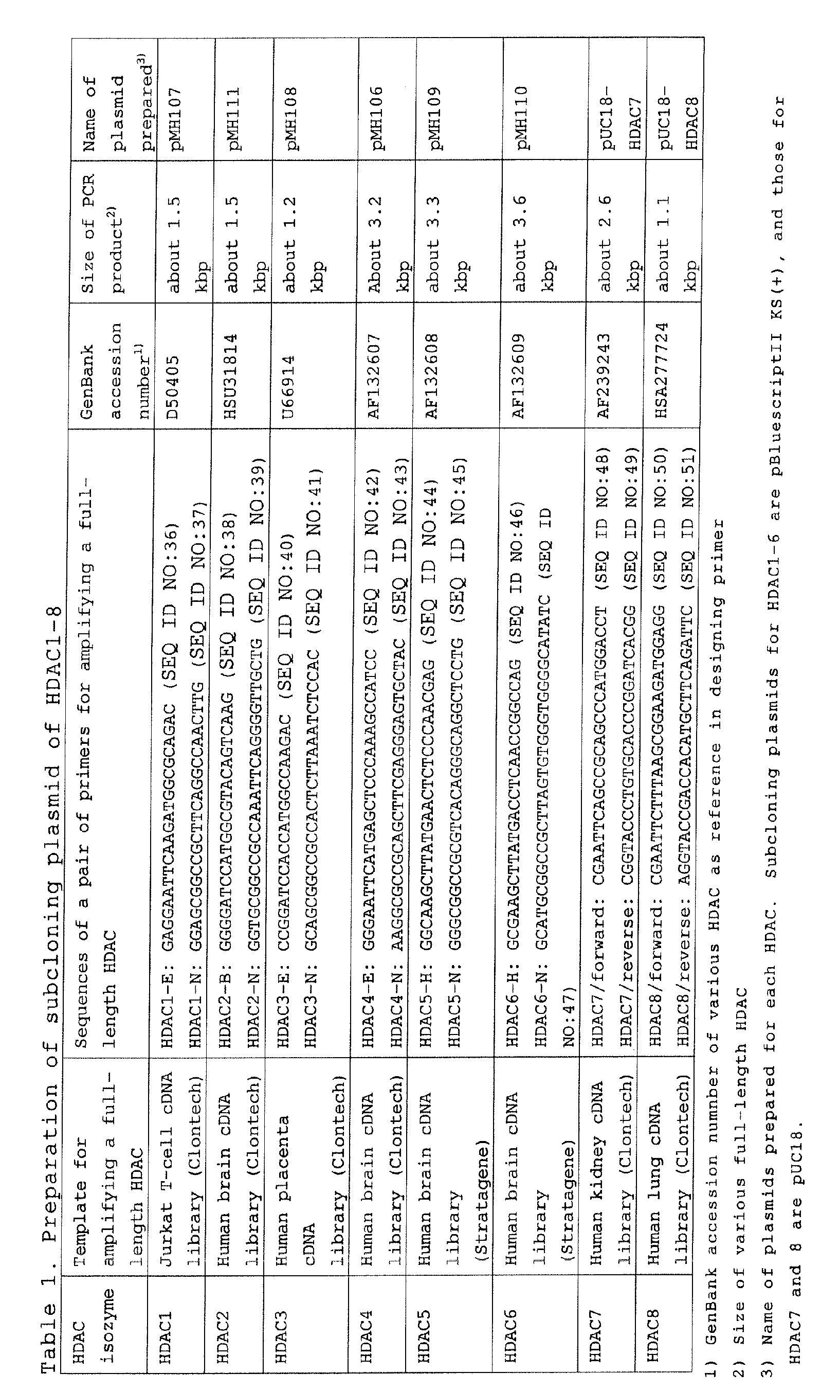
Subsequently, plasmids for expression having a Flag
sequence added to the C-terminal of each of HDAC1 to 8
were prepared. Details are shown below.
Full-length HDAC3 was amplified by PCR with pMH108
as a template using the following primers 108E (SEQ ID
NO : 52) and 108KFN (SEQ ID NO : 53). Meanwhile, the primers
were designed to add a Flag sequence (DYKDDDDK) (SEQ ID
NO:54) to the C-terminal. The PCR fragment obtained was
subcloned into pGEM-T (Promega) to yield pGEM-HDAC3-Flag
(E108KFN). E108KFN was inserted to pEAK10 (Edge
BioSystems) via EcoRI/NotI (about 1.2 kbp fragment)
treatment to yield pEAK-HDAC3 (pMH118).


Full-length HDAC1 was amplified by PCR with pMH107
as a template using the primers HDAC1-E (SEQ ID NO:36)
and 107K (SEQ ID NO:55). Meanwhile, the primers were
designed by modifying the C-terminal stop codon to
insert a KpnI site. The PCR fragment obtained was
subcloned into pGEM-T (Promega) to yield pGEM-HDAC1
(E107K). E107K was inserted to pMH118 (EcoRI/KpnI, about
6 kbp) via EcoRI/KpnI (about 1.5 kbp fragment) treatment
to yield pEAK-HDAC1 (pMH119).

Full-length HDAC2 was amplified by PCR with pMH111
as a template using the primers HDAC2-B (SEQ ID NO:38)
and 111K (SEQ ID NO:56). Meanwhile, the primers were
designed by modifying the C-terminal stop codon to
insert a KpnI site. The PCR fragment obtained was
subcloned into pGEM-T (Promega) to yield pGEM-HDAC2.
pGEM-HDAC2 was inserted to pFLAG-CMV-5a (SIGMA)
(BamHI/KpnI) via BamHI/KpnI treatment to yield pFLAG-CMV-5a-HDAC2
(pMH113). pMH113 was inserted to pMH118
(EcoRI/KpnI, about 6 kbp) via EcoRI/KpnI (about 1.5 kbp
fragment) treatment to yield pEAK-HDAC2 (pMH121).

Full-length HDAC4 was amplified by PCR with pMH106
as a template using the primers HDAC4-E (SEQ ID NO:42)
and 106B (SEQ ID NO:57). Meanwhile, the primers were
designed by modifying the C-terminal stop codon to
insert a BglII site. The PCR fragment obtained was
subcloned into pGEM-T (Promega) to yield pGEM-HDAC4
(E106B). A linker oligo having a BglII site and a FLAG
sequence (DYKDDDDK) (FLAG-E (SEQ ID NO:58), prepared by
annealing FLAG-N (SEQ ID NO:59)) was inserted to an
EcoRI/NotI-treated fragment (about 3 kbp) of
pBluescriptII KS(+) to yield pBlue-Flag. E106B was
inserted to pBlue-Flag via EcoRI/BglII treatment to
yield pBKS-HDAC4 (E106BN). E106BN was inserted to about
6 kbp pEAK10 (Edge Byo Systems) (EcoRI/NotI) via
EcoRI/NotI (about 3.2 kbp fragment) treatment to yield
pEAK-HDAC4 (pMH122).



A HindIII/NotI fragment (about 3.3 kbp) of pMH109
was inserted to p3XFLAG-CMV-10 (SIGMA) (HindIII/NotI,
about 6.3 kbp) to yield p3XFLAG-CMV-10-HDACS (pMH144).
A HindIII/NotI fragment (about 3.6 kbp) of pMH110
was inserted to p3XFLAG-CMV-10 (SIGMA) (HindIII/NotI,
about 6.3 kbp) to yield p3XFLAG-CMV-10-HDAC6 (pMH145).
A EcoRI/KpnI fragment (about 2.6 kbp) of pUC18-HDAC7
was inserted to PMH118 (EcoRI/KpnI, about 6 kbp)
to yield pEAK-HDAC7 (pMH141).
A EcoRI/KpnI fragment (about 1.1 kbp) of pUC18-HDAC8
was inserted to PMH118 (EcoRI/KpnI, about 6 kbp)
to yield pEAK-HDAC8 (pMH140).
For details about the individual HDAC expression
vectors prepared, see Table 2.
| Preparation of HDAC1-8 expression plasmid | |||
| HDAC | Expression plasmid used | Position of Flag tag (DYKDDDK) for Each HDAC | Name of plasmid prepared |
| HDAC1 | pEAK10 (Edge Bio Systems) | C-terminal | pMH119 |
| HDAC2 | pEAK10 (Edge Bio Systems) | C-terminal | pMH121 |
| HDAC3 | pEAK10 (Edge Bio Systems) | C-terminal | pMH118 |
| HDAC4 | pEAK10 (Edge Bio Systems) | C-terminal | pMH122 |
| HDAC5 | p3XFLAG-CMV-10 (SIGMA) | N-terminal | pMH144 |
| HDAC6 | p3XFLAG-CMV-10 (SIGMA) | N-terminal | pMH145 |
| HDAC7 | pEAK10 (Edge Bio Systems) | C-terminal | pMH141 |
| HDAC8 | pEAK10 (Edge Bio Systems) | C-terminal | pMH140 |
1 µg of the plasmid pGL3 IL2 Pro43, which has a
human IL-2 promoter sequence (the -674 to +54 region in
the vicinity of the transcription initiation site)
upstream of the firefly luciferase gene, 6 µg of pRL-TK
(Promega), and 10 µg of each of various HDAC isozyme
expression plasmids or a mock plasmid (the mock plasmid
was pEAK10, and the various HDAC expression plasmids
were pMH119 for HDAC1, pMH121 for HDAC2, pMH118 for
HDAC3, pMH122 for HDAC4, pMH144 for HDAC5, pMH145 for
HDAC6, pMH141 for HDAC7, and pMH140 for HDAC8,
respectively) were mixed, and 1 x 10⁷ Jurkat cells (ATCC,
TIB-152) were transformed by the electroporation method
(BIO-RAD, Gene Pulser II, voltage 300 V, charge 975 µF,
400 µL).
After transformation, 2.5 mL of RPMI 1640 (SIGMA)
containing 10% FBS (MOREGATE) (10% FBS RPMI 1640) was
added, and this mixture was dispensed to a 96-well white
plate in a ratio of 50 µL/well. After cultivation under
conditions of 37°C, 5% CO2 and saturated humidity for 10
hours, a 10% FBS RPMI 1640 solution was added at 50
µL/well, and a liquid mixture of Phorbol 12-Myristate
13-Acetate (PMA, SIGMA), Ionomycin (SIGMA) and Anti-CD28
antibody (Pharmingen) in 10% FBS RPMI 1640 was added at
50 µL/well (the final concentrations were 50 ng/mL, 1
µg/mL and 75 ng/mL, respectively) to stimulate the
Jurkat cells. After cultivation under conditions of 37°C,
5% CO2 and saturated humidity for 12 hours, luciferase
activity in the cells was measured according to the
manual of the Dual Luciferase Assay System (Promega)
using a multi-label counter (1420 MULTILABEL COUNTER
ARVO SX, WALLAC).
As a result, it was found that luciferase was
induced from pGL3 IL2 Pro43 in response to the
stimulation of the Jarkat cells by PMA, Ionomycin, and
Anti-CD28 antibody, and that the expression level
thereof increased with the overexpression of HDAC4 and 8
(FIG. 5). It was also confirmed using the Cell Counting
Kit-8 (DOJIN) that the overexpression of various HDAC
isozymes did not significantly affect the degree of cell
proliferation (FIG. 5).
These results demonstrate that HDAC4 and 8 are HDAC
isozymes involved in the IL-2 transcription system.
5 µg of the plasmid pGL3-HSI-IE Pro, which has a
human GATA-1 promoter sequence (the -3769 to -3133 and
-789 to +32 regions in the vicinity of the transcription
initiation site) upstream of the firefly luciferase gene,
and 10 µg of each of various HDAC isozyme expression
plasmids or a mock plasmid (the mock plasmid was pEAK10,
and HDAC1 to 4, 7 and 8 were under the control of the
EF-1α promoter, HDAC5 and 6 were under the control of
the CMV promoter, and the various HDAC isozymes had a
Flag tag at the C-terminal or N-terminal thereof) were
mixed, and 8.75 x 10⁶ HEL cells (accession number
JCRB0062, JCRB) were transformed by the electroporation
method (BIO-RAD, Gene Pulser II, voltage 1750 V, charge
10 µF, 365 µL). After transformation, 2 mL of 10% FBS
RPMI 1640 was added, and this mixture was dispensed to a
96-well white plate in a ratio of 50 µL/well. After
cultivation under conditions of 37°C, 5% CO2 and
saturated humidity for 3 hours, a 10% FBS RPMI 1640
medium was added at 100 µL/well. After cultivation under
conditions of 37°C, 5% CO2 and saturated humidity for 8
hours, luciferase activity in the cells was measured
according to the manual of the Dual Luciferase Assay
System using a multi-label counter.
As a result, it was found that luciferase depending
on the GATA-1 promoter was induced from pGL3-HSI-IE Pro,
and that the expression level of the induced luciferase
increased slightly with the overexpression of HDAC1, 3
and 7 (FIG. 6).
These results demonstrate that HDAC1, 3 and 7 are
involved in the GATA-1 transcription system, and that
HDAC4 and 8, which are involved in the IL-2
transcription system, are not significantly involved in
the GATA-1 transcription system.
For HDAC4, which seems to be largely involved in
IL-2 transcription, HDAC4 dominant negative variants
were prepared for the purpose of losing the function of
endogenous HDAC4. Hence, from the literature information
in PNAS, 1998, Vol. 95, p.3519-3524 & Mol. Cell. Biol.,
1999, Vol. 19 (11), p.7816-7827, the sites for
mutagenesis to reduce HDAC activity were determined
(H802K, H803L, H863L) (FIG. 7). The human HDAC4 CDS base
sequence was obtained from GenBank accession number
AF132607, and primers for mutagenesis were designed (for
the base sequences of the individual primers, see Table
3; for the HDAC4 CDS base sequence, see SEQ ID NO:1; for
the amino acid sequence encoded by the HDAC4 CDS base
sequence, see SEQ ID NO:2).
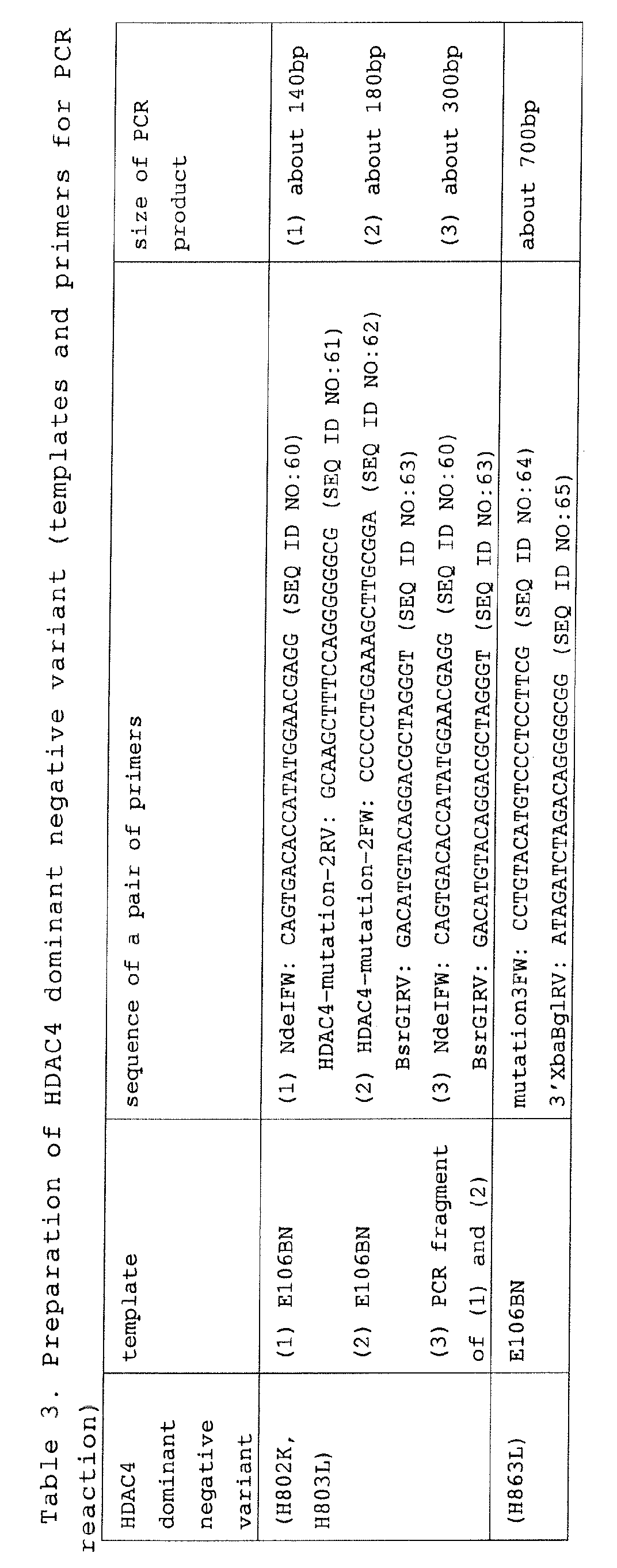
A PCR reaction was conducted according to the
protocol of Expand (High fidelity PCR System (Roche)).
E106BN, which was used as a template, is a plasmid
prepared by inserting full-length HDAC4 CDS to
pBluescriptII KS(+) (TOYOBO) (EcoRI/NotI, the initiation
codon was located on the EcoRI side).
Using the Topo TA cloning kit for sequence
(Invitrogen Company), each of the PCR products obtained
((3) for H802K and H803L variants) was subcloned into
the TOPO cloning vector for sequence (pCR4-HDAC4 (H802K,
H803L) and pCR4-HDAC4 (H863L)). Subsequently, a PCR
fragment confirmed as being mutated by sequencing was
inserted to an HDAC4 expression plasmid (pMH122) by the
procedures shown below.
An NdeI/BsrGI-treated fragment (about 300 bp) of
pCR4-HDAC4 (H802K, H803L) was inserted to E106BN
(NdeI/BsrGI) to yield pBKS-HDAC4 (H802K, H803L).
Subsequently, an EcoRI/NotI-treated fragment (about 3.3
kbp) of pBKS-HDAC4 (H802K, H803L) was inserted to pMH122
(EcoRI/NotI, about 6 kbp) to yield pEAK-HDAC4 (H802K,
H803L). The base sequence in the coding region of HDAC4
dominant negative variants (H802K, H803L) is shown by
SEQ ID NO:3, and the amino acid sequence thereof is
shown by SEQ ID NO:4.
A BsrGI/BglII-treated fragment (about 750 bp) of
pCR4-HDAC4 (H863L) was inserted to E106BN (BsrGI/BglII,
about 3 kbp) to yield pBKS-HDAC4 (H863L). Subsequently,
an EcoRI/NotI-treated fragment (about 3.3 kbp) of pBKS-HDAC4
(H863L) was inserted to pMH122 (EcoRI/NotI, about
6 kbp) to yield pEAK-HDAC4 (H863L). The base sequence in
the coding region of HDAC4 dominant negative variant
(H863L) is shown by SEQ ID NO:5, and the amino acid
sequence thereof is shown by SEQ ID NO:6.
HDAC1 dominant negative variants were prepared in
the same manner as the preparation of the HDAC4 dominant
negative variants. The human HDAC1 CDS base sequence was
obtained from GenBank accession number D50405, and
primers for mutagenesis were designed (for the base
sequences of the individual primers, see Table 4; for
the base sequence of HDAC1 CDS, see SEQ ID NO:9; for the
amino acid sequence encoded by the HDAC1 CDS base
sequence, see SEQ ID NO:10).
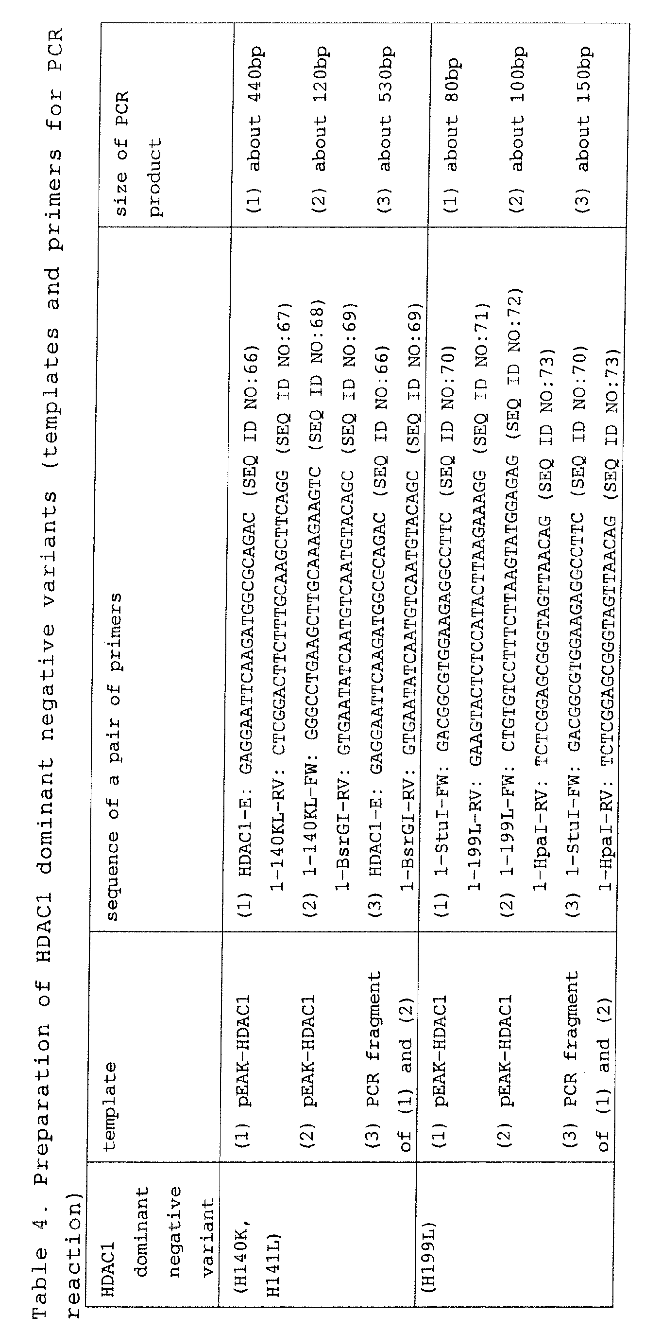
A PCR reaction was conducted according to the
protocol of Expand (High fidelity PCR System (Roche)).
pEAK-HDAC1 (pMH119), which was used as a template, is a
plasmid prepared by inserting full-length HDAC1 CDS to
pEAK10.
Using the Topo TA cloning kit for sequence
(Invitrogen Company), each of the PCR products obtained
(3) was subcloned into the TOPO cloning vector for
sequence (pCR4-HDAC1 (H140K, H141L) and pCR4-HDAC1
(H199L)). A PCR fragment confirmed as being mutated by
sequencing was inserted to an HDAC1 expression plasmid
(pMH119) using the procedures shown below. E107K is a
plasmid prepared by inserting full-length HDAC1 CDS to
pGEM-T (Promega) (EcoRI/KpnI, the initiation codon is
located on the EcoRI side).
An EcoRI/BsrGI-treated fragment (about 530 bp) of
pCR4-HDAC1 (H140K, H141L) was inserted to E107K
(EcoRI/BsrGI, about 3 kbp) to yield pGEM-HDAC1 (H140K,
H141L). Subsequently, pGEM-HDAC1 (H140K, H141L)
(EcoRI/KpnI, about 1.5 kbp) was inserted to an
EcoRI/KpnI-treated fragment (about 5.9 kbp) of pMH119 to
yield pEAK-HDAC1 (H140K, H141L). The base sequence in
the coding region of HDAC1 dominant negative variants
(H140K, H141L) is shown by SEQ ID NO:11, and the amino
acid sequence thereof is shown by SEQ ID NO:12.
An StuI/HpaI-treated fragment (about 150 bp) of
pCR4-HDAC1 (H199L) was inserted to E107K (StuI/HpaI)
(about 3 kbp) to yield pGEM-HDAC1 (H199L). Subsequently,
pGEM-HDAC1 (H199L) (EcoRI/KpnI, about 1.5 kbp) was
inserted to an EcoRI/KpnI-treated fragment (about 5.9
kbp) of pMH119 to yield pEAK-HDAC1 (H199L). The base
sequence in the coding region of HDAC1 dominant negative
variant (H199L) is shown by SEQ ID NO:13, and the amino
acid sequence thereof is shown by SEQ ID NO:14.
HDAC3 dominant negative variants were prepared in
the same manner as the preparation of the HDAC4 dominant
negative variants. The human HDAC3 CDS base sequence was
obtained from GenBank accession number U66914, and
primers for mutagenesis were designed (for the base
sequences of the individual primers, see Table 5; for
the HDAC3 CDS base sequence, see SEQ ID NO:15; for the
amino acid sequence encoded by the HDAC3 CDS base
sequence, see SEQ ID NO:16).
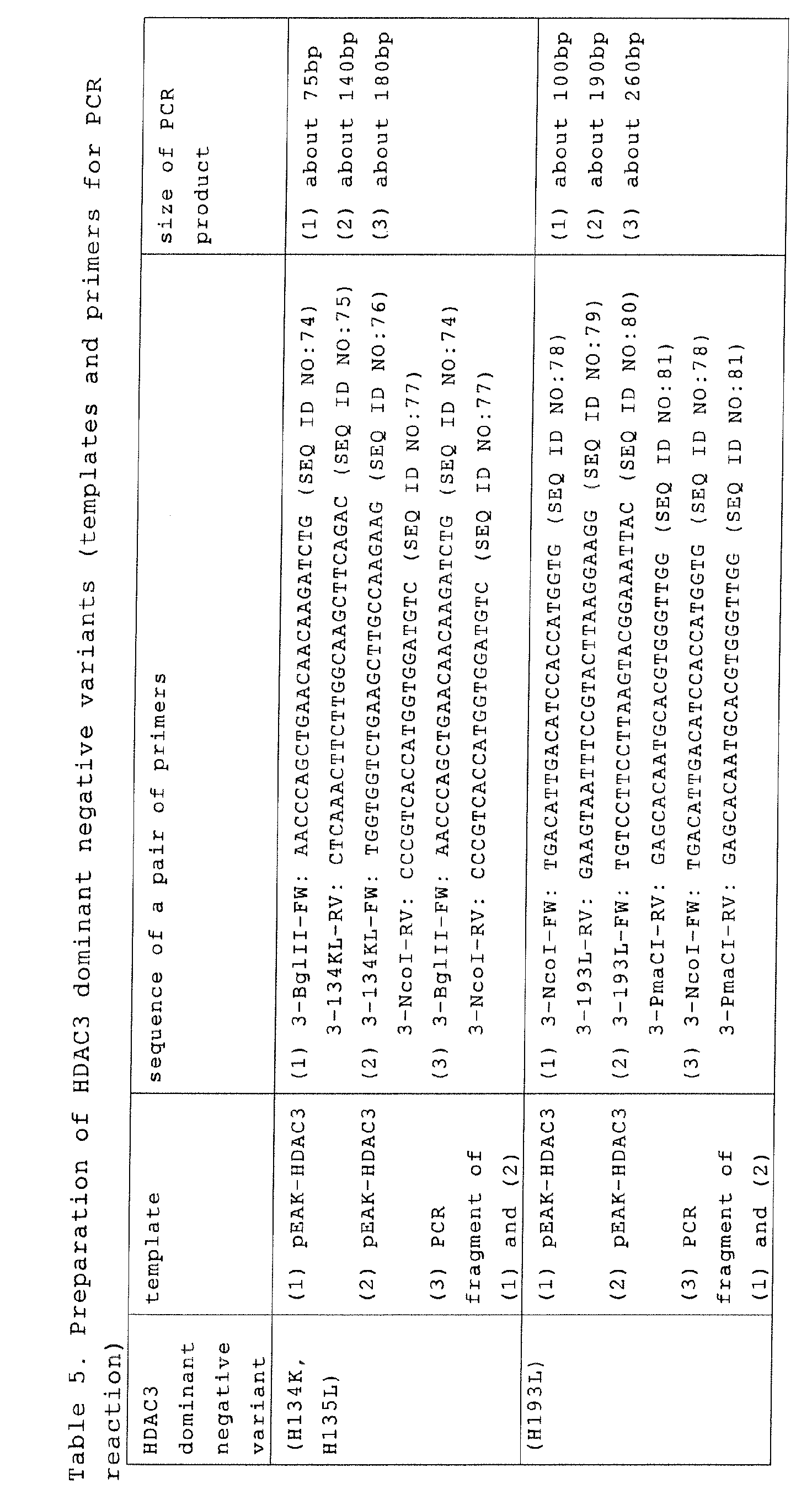
A PCR reaction was conducted according to the
protocol of Expand (High fidelity PCR System (Roche)).
pEAK-HDAC3 (pMH118), which was used as a template, is a
plasmid prepared by inserting full-length HDAC3 CDS to
pEAK10.
Using the Topo TA cloning kit for sequence
(Invitrogen Company), each of the PCR products (3)
obtained was subcloned into the TOPO cloning vector for
sequence (pCR4-HDAC3 (H134K, H135L) and pCR4-HDAC3
(H193L)). A PCR fragment confirmed as being mutated by
sequencing was inserted to an HDAC3 expression plasmid
(pMH118) by the procedures shown below.
An EcoRI/KpnI-treated fragment (about 1.3 kbp) of
pMH118 was inserted to pBluescriptII KS(-) (TOYOBO)
(EcoRI/KpnI) (about 3 kbp) to yield pBKS-HDAC3.
Subsequently, pCR4-HDAC3 (H134K, H135L) (BglII/NcoI,
about 180 bp) was inserted to pBKS-HDAC3 (BgIII/NcoI,
about 3 kbp) to yield pBKS-HDAC3 (H134K, H135L). Finally,
pBKS-HDAC3 (H134K, H135L) (EcoRI/KpnI, about 1.3 kbp)
was inserted to an EcoRI/KpnI-treated fragment (about
5.7 kbp) of pMH118 to yield pEAK-HDAC3 (H134K, H135L).
The base sequence in the coding region of HDAC3 dominant
negative variants (H134K, H135L) is shown by SEQ ID
NO:17, and the amino acid sequence thereof is shown by
SEQ ID NO:18.
A pCR4-HDAC3 (H134K, H135L) (NcoI/PmaCI, About 260
bp) was inserted to pMH118 (NcoI/PmaCI, about 7 kbp) to
yield pEAK-HDAC3 (H193L). The base sequence in the
coding region of HDAC3 dominant negative variant (H193L)
is shown by SEQ ID NO:19, and the amino acid sequence
thereof is shown by SEQ ID NO:20.
To 1 µg of the plasmid pGL3 IL2 Pro43, which has a
human IL-2 promoter sequence (the -674 to +54 region in
the vicinity of the transcription initiation site)
upstream of the firefly luciferase gene, and 6 µg of
pRL-TK (Promega), the plasmids for the expression of
wild-type HDAC4 or each of HDAC4 dominant negative
variants (H802K, H803L, H863L) prepared in Example 3 was
mixed in various ratios (1, 3 and 10 µg), and 1 x 10⁷
Jurkat cells (ATCC, TIB-152) were transformed by the
electroporation method (BIO-RAD, Gene Pulser II, voltage
300 V, charge 975 µF, 400 µL).
After transformation, 2.5 mL of RPMI 1640
containing 10% FBS (MOREGATE) (10% FBS RPMI 1640) was
added, and this mixture was dispensed to a 96-well white
plate in a ratio of 50 µL/well. After cultivation under
conditions of 37°C, 5% CO2 and saturated humidity for 10
hours, a liquid mixture of Phorbol 12-Myristate 13-Acetate
(PMA, SIGMA), Ionomycin (SIGMA) and Anti-CD28
antibody (Pharmingen) in 10% FBS RPMI 1640 was added at
50 µL/well (the final concentrations were 50 ng/mL, 1
µg/mL and 75 ng/mL, respectively) to stimulate the
Jurkat cells. After cultivation under conditions of 37°C,
5% CO2 and saturated humidity for 12 hours, luciferase
activity in the cells was measured according to the
manual of the Bright-Glo Luciferase Assay System
(Promega) using a multi-label counter (1420 MULTILABEL
COUNTER ARVO SX, WALLAC).
As a result, IL-2 transcription suppression
depending on the expression level of HDAC4 dominant
negative variants (H802K, H803L, H863L) was observed
(FIG. 8). The expression level of the transferred HDAC
was confirmed by Western blotting using an anti-Flag
antibody (SIGMA, anti-Flag M2).
These results suggest that IL-2 transcription may
be suppressed by selectively suppressing the function of
HDAC4.
10 µg of a plasmid for the expression of wild-type
HDAC4 and the HDAC4 dominant negative variant (H863L)
prepared in Example 6 was transfected to Jurkat cells,
HEL cells and 293 cells.
1 x 10⁷ Jurkat cells (ATCC, TIB-152) were
transformed by the electroporation method (BIO-RAD, Gene
Pulser II, voltage 300 V, charge 975 µF, 400 µL). After
transformation, 2.5 mL of RPMI 1640 containing 10% FBS
(MOREGATE) (10% FBS RPMI 1640) was added, and this
mixture was dispensed to a 96-well white plate in a
ratio of 50 µL/well. After cultivation under conditions
of 37°C, 5% CO2 and saturated humidity for 5 hours, a
liquid mixture of Phorbol 12-Myristate 13-Acetate (PMA,
SIGMA), Ionomycin (SIGMA) and Anti-CD28 antibody
(Pharmingen) in 10% FBS RPMI 1640 was added at 50
µL/well (the final concentrations were 50 ng/mL, 1 µg/mL
and 75 ng/mL, respectively) to stimulate the Jurkat
cells. After cultivation under conditions of 37°C, 5%
CO2 and saturated humidity for 4 hours, the number of
cell was confirmed using the Cell Counting Kit-8 (DOJIN).
8.75 x 10⁶ HEL cells (accession number JCRB0062,
JCRB) were transformed by the electroporation method
(BIO-RAD, Gene Pulser II, voltage 1750 V, charge 10 µF,
365 µL). After transformation, 2 mL of 10% FBS RPMI 1640
was added, and this mixture was dispensed to a 96-well
white plate in a ratio of 50 µL/well. After cultivation
under conditions of 37°C, 5% CO2 and saturated humidity
for 10 hours, 10% FBS RPMI 1640 medium was added at 50
µL/well, further cultivation was conducted for 12 hours,
and the number of cell was confirmed using the Cell
Counting Kit-8 (DOJIN).
293 cells (ATCC, CRL-1573) were transfected by the
calcium phosphate method (a mixture of DNA and calcium
phosphate was added to 293 cells in culture using a DMEM
(SIGMA) medium containing 10% FBS, and the medium was
replaced with a fresh one 6 hours later); after
cultivation under conditions of 37°C, 5% CO2 and
saturated humidity for 48 hours, the number of cell was
confirmed using the Cell Counting Kit-8 (DOJIN).
As a result, cell proliferation inhibition specific
for Jurkat cells due to the expression of HDAC4 dominant
negative variant (H863L) was observed (FIG. 9). For each
type of cells, the expression level of the transferred
HDAC was confirmed by Western blotting using an anti-Flag
antibody (SIGMA, anti-Flag M2).
These results demonstrate that the proliferation of
T cell lineage is selectively suppressed by selectively
inhibiting the function of HDAC4.
IL-2 and GATA-1 transcription activities were
measured with the expression of the HDAC1, 3 and 4
dominant negative variants prepared in Examples 6, 7 and
8.
1 x 10⁷ Jurkat cells (ATCC, TIB-152) were
transformed with 10 µg of a plasmid for the expression
of various HDACs or HDAC variants by the electroporation
method (BIO-RAD, Gene Pulser II, voltage 300 V, charge
975 µF, 400 µL). After transformation, 2.5 mL of RPMI
1640 (SIGMA) containing 10% FBS (MOREGATE) (10% FBS RPMI
1640) was added, and this mixture was dispensed to a 96-well
white plate in a ratio of 50 µL/well. After
cultivation under conditions of 37°C, 5% CO2 and
saturated humidity for 10 hours, a liquid mixture of
Phorbol 12-Myristate 13-Acetate (PMA, SIGMA), Ionomycin
(SIGMA) and Anti-CD28 antibody (Pharmingen) in 10% FBS
RPMI 1640 was added at 50 µL/well (the final
concentrations were 50 ng/mL, 1 µg/mL and 75 ng/mL,
respectively) to stimulate the Jurkat cells. After
cultivation under conditions of 37°C, 5% CO2 and
saturated humidity for 12 hours, luciferase activity in
cells were measured according to the manual of the
Bright-Glo Luciferase Assay System (Promega), and IL-2
transcriptional activity was measured using a multi-label
counter (1420 MULTILABEL COUNTER ARVO SX, WALLAC).
Using 15 µg of pGL3-HSI-IE Pro and 10 µg of a
plasmid for the expression of various HDACs or HDAC
variants, 8.75 x 10⁶ HEL cells (accession number JCRB0062,
JCRB) were transformed by the electroporation method
(BIO-RAD, Gene Pulser II, voltage 1750 V, charge 10 µF,
365 µL). After transformation, 2 mL of 10% FBS RPMI 1640
was added, and this mixture was dispensed to a 96-well
white plate in a ratio of 50 µL/well. After cultivation
under conditions of 37°C, 5% CO2 and saturated humidity
for 3 hours, 10% FBS RPMI 1640 medium was added at 50
µL/well. After cultivation under conditions of 37°C, 5%
CO2 and saturated humidity for 8 hours, luciferase
activity in cells were measured according to the manual
of the Bright-Glo Luciferase Assay System, and GATA-1
transcriptional activity was measured using a multi-label
counter.
As a result, it was found that IL-2 transcription
suppression was observed only with the expression of
HDAC4 dominant negative variants (H802K, H803L, H863L),
and that GATA-1 transcription suppression was not
observed at that time (FIG. 10). For each type of cells,
the expression level of the transferred HDAC was
confirmed by Western blotting using an anti-Flag
antibody (SIGMA, anti-Flag M2).
These results suggest that immune function can be
suppressed, without suffering thrombocytopenia, by
selectively inhibiting the function of HDAC4.
In Mol. Cell, Vol. 9, p.45-57, 2002, it is reported
that Class II HDACs have formed a complex with N-CoR and
HDAC3, and that HDAC activity with histone as a
substrate is much more portent for HDAC3 than for Class
II HDACs. Hence, the formation of complexes of crudely
purified Class II HDACs was confirmed.
7 µg of each of HDAC4, 5 and 7 expression plasmids
was transfected to PEAK Rapid cells (Edge BioSystems) by
the calcium phosphate method; 48 hours later, whole cell
lysate (cells were disrupted in 25mM Tris, pH 7.4, 150mM
NaCl, 1mM CaCl2, 1% Triton X-100, and cell debris was
removed by centrifugation) was crudely purified using an
anti-Flag antibody column (SIGMA, anti-Flag M2-agarose)
(washed with TBS, then eluted with 0.1M Glysine-HCl, pH
3.5). Subsequently, the crudely purified fraction was
subjected to SDS-PAGE and then stained with CBB.
As a result, the expression of the transferred
HDAC4, 5 and 7 was confirmed (FIG. 11). It was also
confirmed by Western blotting that each of the crudely
purified fractions of HDAC4, 5 and 7 contained HDAC3 and
N-CoR (using an anti-HDAC3 antibody of SIGMA Company and
an anti-N-CoR antibody of Santa Cruz Company).
These results confirm that the crudely purified
HDAC4, 5 and 7 have formed a complex with N-CoR and
HDAC3.
In Mol. Cell, Vol. 9, p.45-57, 2002, it is reported
that some HDAC4 variants do not form a complex with N-CoR
and HDAC3. Hence, the complex forming ability of
HDAC4 dominant negative variant (H863L) was examined.
7 µg of a plasmid for the expression of wild-type
HDAC4 and HDAC4 dominant negative variant (H863L) was
transfected to PEAK Rapid cells (Edge BioSystems) by the
calcium phosphate method; 48 hours later, whole cell
lysate (cells were disrupted in 25mM Tris, pH 7.4, 150mM
NaCl, 1mM CaCl2, 1% Triton X-100, and cell debris was
removed by centrifugation) was crudely purified using an
anti-Flag antibody column (SIGMA, anti-Flag M2-agarose)
(washed with TBS, then eluted with 0.1M Glysine-HCl, pH
3.5). Subsequently, the crudely purified fraction was
subjected to SDS-PAGE and then stained with CBB.
As a result, the expression of the transferred
HDAC4 and HDAC4 dominant negative variant (H863L) was
confirmed (FIG. 12). It was also confirmed by Western
blotting (using an anti-HDAC3 antibody of SIGMA Company
and an anti-N-CoR antibody of Santa Cruz Company) that
the crudely purified fraction of HDAC4 contained HDAC3
and N-CoR, and that the crudely purified fraction of
HDAC4 dominant negative variant (H863L) contained almost
no HDAC3 or N-CoR.
These results demonstrate that HDAC4 dominant
negative variant (H863L) has a decreased complex forming
ability with N-CoR and HDAC3 compared to wild-type HDAC4.
In Mol. Cell, Vol. 9, p.45-57, 2002, it is reported
that HDAC4 variants which decreased complex forming
ability with N-CoR and HDAC3 shows decrease of HDAC
activity with histone as a substrate. Hence, the HDAC
activities of HDAC4 dominant negative variants (H802K,
H803L, H863L) with histone as a substrate were measured.
7 µg of a plasmid for the expression of wild-type
HDAC4 and HDAC4 dominant negative variants (H802K, H803L,
H863L) was transfected to 293T cells by the calcium
phosphate method; 48 hours later, whole cell lysate
(cells were disrupted in 25mM Tris, pH 7.4, 150mM NaCl,
1mM CaCl2, 1% Triton X-100, and cell debris was removed
by centrifugation) was crudely purified using an anti-Flag
antibody column (SIGMA, anti-Flag M2-agarose)
(washed with TBS, then eluted with 0.1M Glysine-HCl, pH
3.5), and used for the measurement of HDAC activity. The
reaction substrate used was a [3H]-labeled acetylated
histone prepared by cultivating Jurkat cells (2×10⁸
cells) in 40 mL of an RPMI (SIGMA) medium containing 300
MBq [3H] sodium acetate (NENTM Life Science Products,
Inc.) for 2 hours, disrupting the cells in Lysis buffer,
pH 6.5 (10mM Tris-HCl, 50mM Sodium Hydrogen sulfate, 1%
Triton X-100, 10mM MgCl2-6H2O, 8.6% Sucrose), washing the
cells with Wash buffer (10mM Tris-HCl, 13mM EDTA), and
then adding 0.4N H2SO4.
As a result, the HDAC4 dominant negative variants
(H802K, H803L, H863L) had decreased HDAC activity
compared to wild-type HDAC4 (FIG. 13).
These results confirm that the HDAC4 dominant
negative variants (H802K, H803L, H863L) have decreased
complex forming ability with N-CoR and HDAC3 (Example
13), and hence decreased HDAC activity with histone as a
substrate.
Since it was found that an HDAC4 dominant negative
variant (H863L) had decreased complex forming ability
with N-CoR compared to wild-type HDAC4, it was
considered that the same IL-2 transcription suppressive
action as that obtained with the expression of HDAC4
dominant negative variant is exhibited by inhibiting the
interaction of HDAC4 and N-CoR.
Hence, to construct a screening system for a drug
that inhibits the interaction of HDAC4 and N-CoR, the
CheckMate Mammalian Two-Hybrid System (Promega) was used
(FIG. 14).
First, from the literature information in Mol. Cell,
2002, Vol. 9, p.45-57, the site corresponding to the RD3
sequence of human N-CoR was determined (1005-1498a.a.).
The human N-CoR CDS base sequence was obtained from
GenBank accession number AF044209, and human N-CoR(RD3)
cDNA was amplified from human-lung-derived cDNA by the
PCR method. The primers used were RD3-Bam-FW (SEQ ID
NO:82) and RD3-Not-RV (SEQ ID NO:83) below. For the base
sequence of human N-CoR(RD3), see SEQ ID NO:21; for the
amino acid sequence encoded by the base sequence of
human N-CoR(RD3), see SEQ ID NO:22. The PCR fragment
obtained was confirmed by sequencing (for N-CoR(RD3), a
variation of L1014V was observed, but this was adopted
since it was reported as a variant in SWISS-PROT), and
it was inserted to pBIND (Promega) via treatment with
restriction endonuclease (BamHI/NotI) to yield a GAL4-N-CoR(RD3)
expression plasmid. Also, HDAC4 or an HDAC4
dominant negative variant (H863L) in full length was
inserted to pACT (Promega) via restriction endonuclease
treatment (XbaI/NotI) to yield HDAC4-VP16 and
HDAC4(H863L)-VP16 expression plasmids.


Subsequently, 10 µg of pG5-Luc (Promega), 3 µg of
pBND and 3 µg of pACT, or 10 µg of pG5-Luc, 3 µg of pBND-N-CoR(RD3)
and 3 µg of pACT-HDAC4, or 10 µg of pG5-Luc,
3 µg of pBND-N-CoR(RD3) and 3 µg of pACT-HDAC4 (H863L),
were mixed respectively, and each mixture was
transfected to 1 x 10⁷ Jurkat cells (ATCC, TIB-152) by
the electroporation method (BIO-RAD, Gene Pulser II,
voltage 300 V, charge 975 µF, 400 µL). Twenty-two hours
after transfection, luciferase activity in the cells was
measured according to the manual of the Bright-Glo
Luciferase Assay System (Promega) using a multi-label
counter (1420 MULTILABEL COUNTER ARVO SX, WALLAC).
As a result, GAL4 transcription activation due to
the interaction of N-CoR (RD3) and HDAC4 was detected,
and it was confirmed that the HDAC4 dominant negative
variant (H863L) did not interact with N-CoR(RD3) (FIG.
15).
From these results, it was evaluated that a
detection system for the interaction of HDAC4 and N-CoR(RD3)
was constructed.
A mixture of four kinds of HDAC4-targeted siRNA,
i.e., siGENE HDAC4 SMARTpool (manufactured by Dharmacon
Inc., marketed by B-Bridge international Inc., code
number M-003497-00-05), or Non-specific Control Duplex
IX (47% GC content) (manufactured by Dharmacon Inc.,
marketed by B-Bride international Inc., code number D-001206-09-20),
and pGL3 IL2 Pro43 were transfected to
Jurkat cells by the electroporation method using the
same conditions as those mentioned above, and IL-2
transcriptional activity was measured. Likewise, siGENE
HDAC4 SMARTpool or Non-specific Control Duplex IX, and
pGL3-HSI-IE Pro were transfected to HEL cells, and GATA-1
transcriptional activity was measured. The conditions
of siRNA transfection and their effects in Jurkat cells
and HEL cells were previously confirmed using pGL3-control
(Promega) and Luciferase GL3 Duplex (B-Bride: D-001400-01-20).
As a result, IL-2 transcription suppression due to
the selective suppression of the expression of the HDAC4
gene was observed, but GATA-1 transcription suppression
was not observed (FIG. 16).
These results again suggest that immune function
can be suppressed, without suffering thrombocytopenia,
by selectively inhibiting the function of HDAC4.
The present invention has shown that immunity can
be suppressed, IL-2 production can be inhibited, or
immunocyte proliferation can be suppressed, without
exhibiting a noticeable thrombocytopenic activity and/or
GATA-1 production inhibitory activity, by selectively
suppressing HDAC4/HDAC8, out of various HDACs. Therefore,
by selecting a compound that selectively suppresses
HDAC4/HDAC8, it is possible to select an
immunosuppressant having low thrombocytopenic activity
or an IL-2 production inhibitor or an immunocyte
proliferation inhibitor having low GATA-1 production
inhibitory activity more conveniently and in a shorter
time than conventional. Hence, the method of the present
invention is very useful for research for developing new
drug.
The present appliation is base on Japanese patent
application no. 2002-378800 filed in Japan on December
27, 2002 and its content is herein incorporated by
reference.
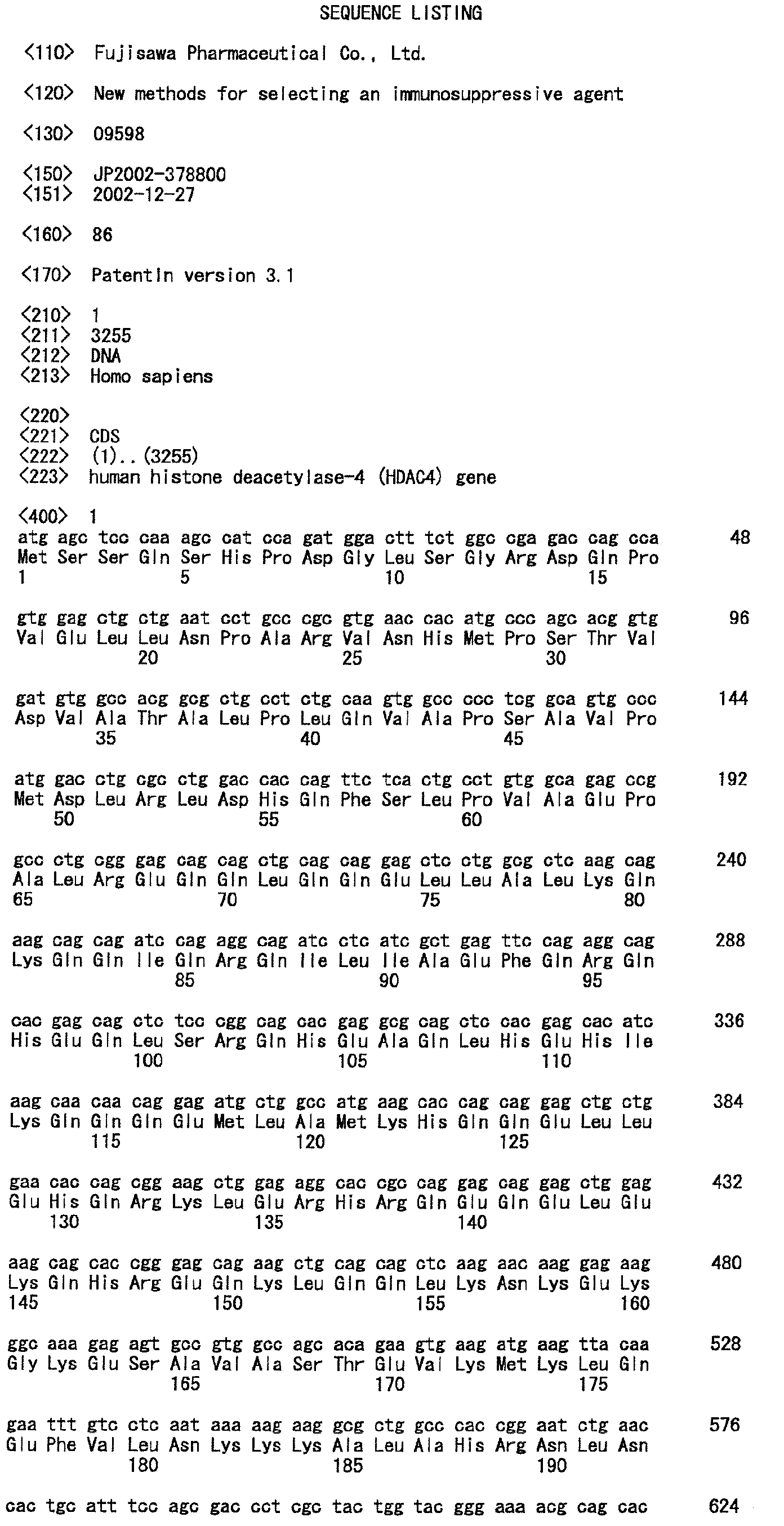
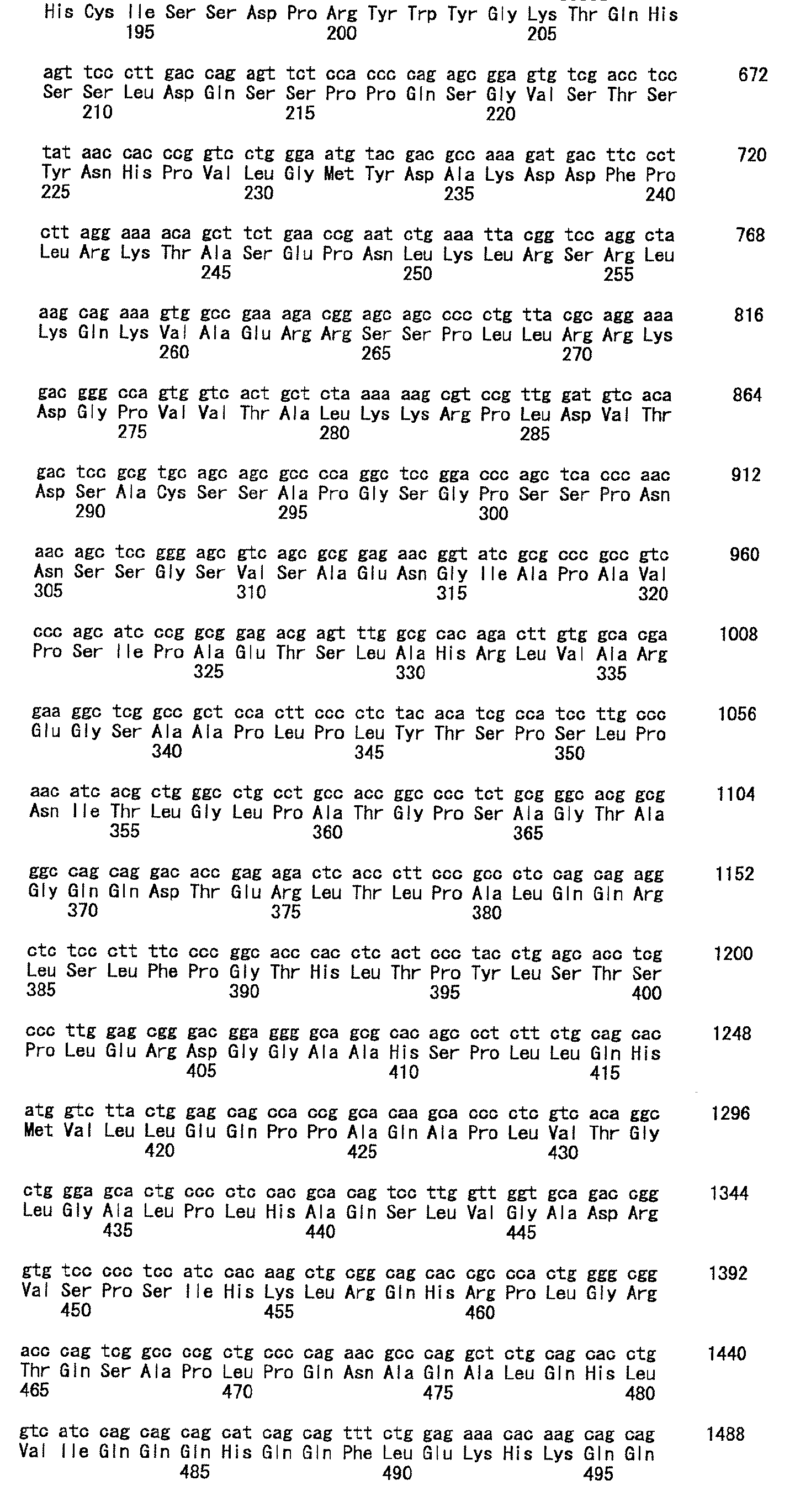
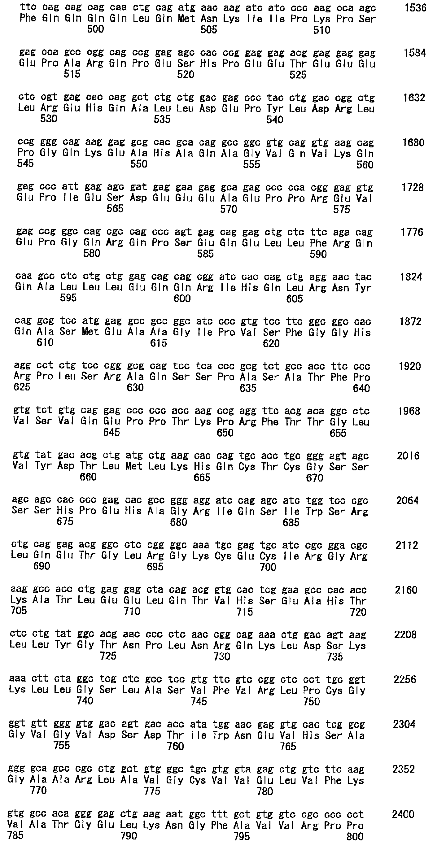


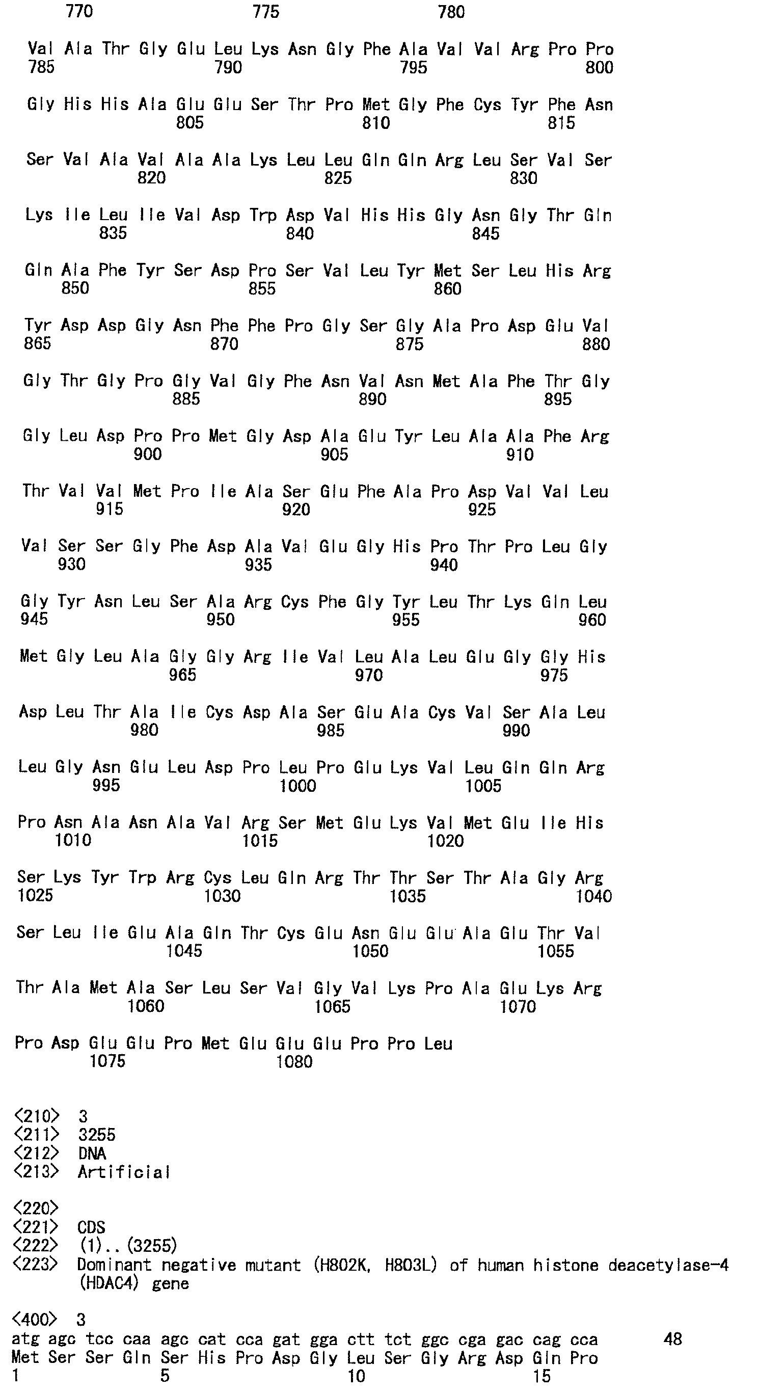

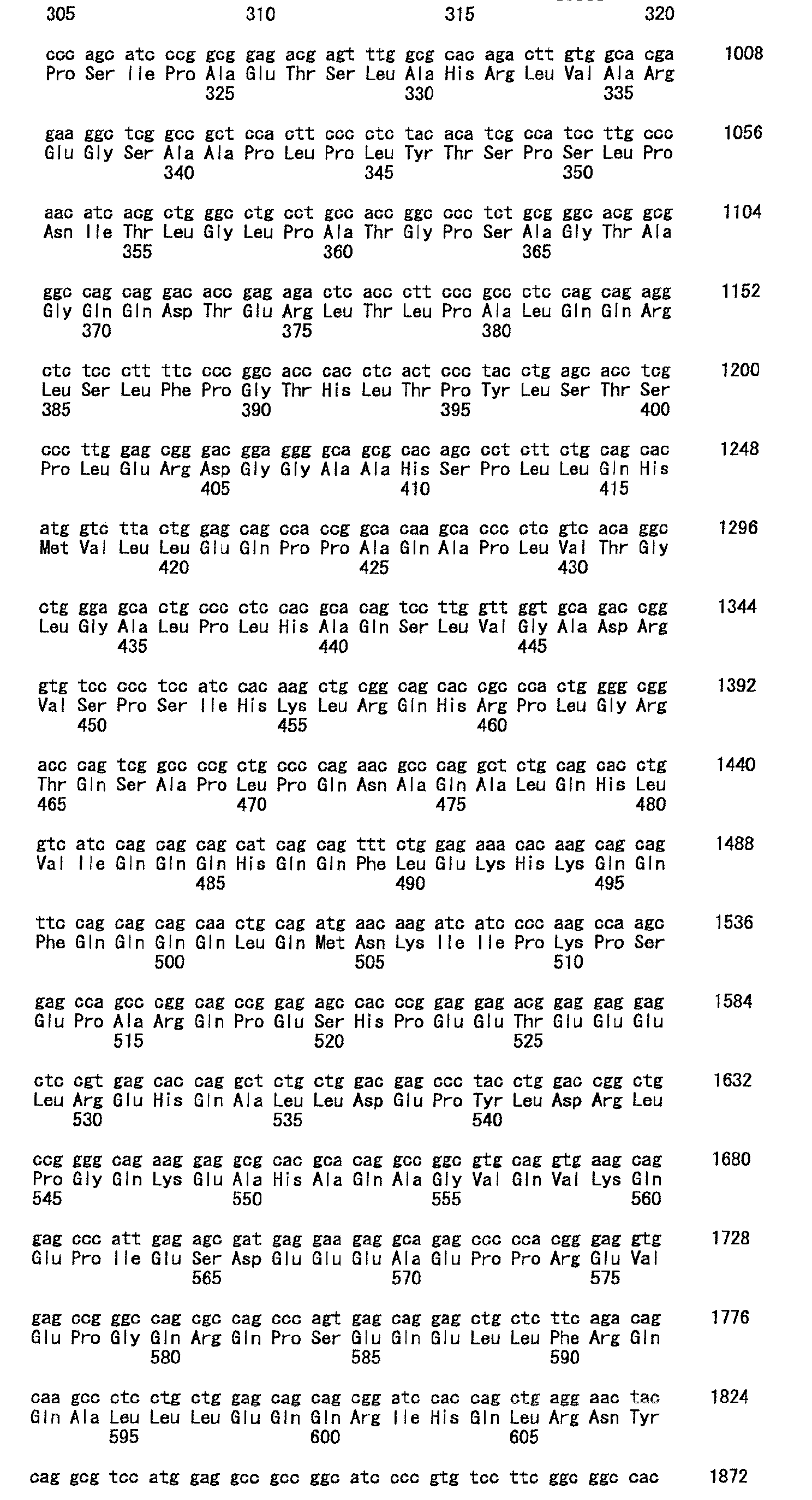


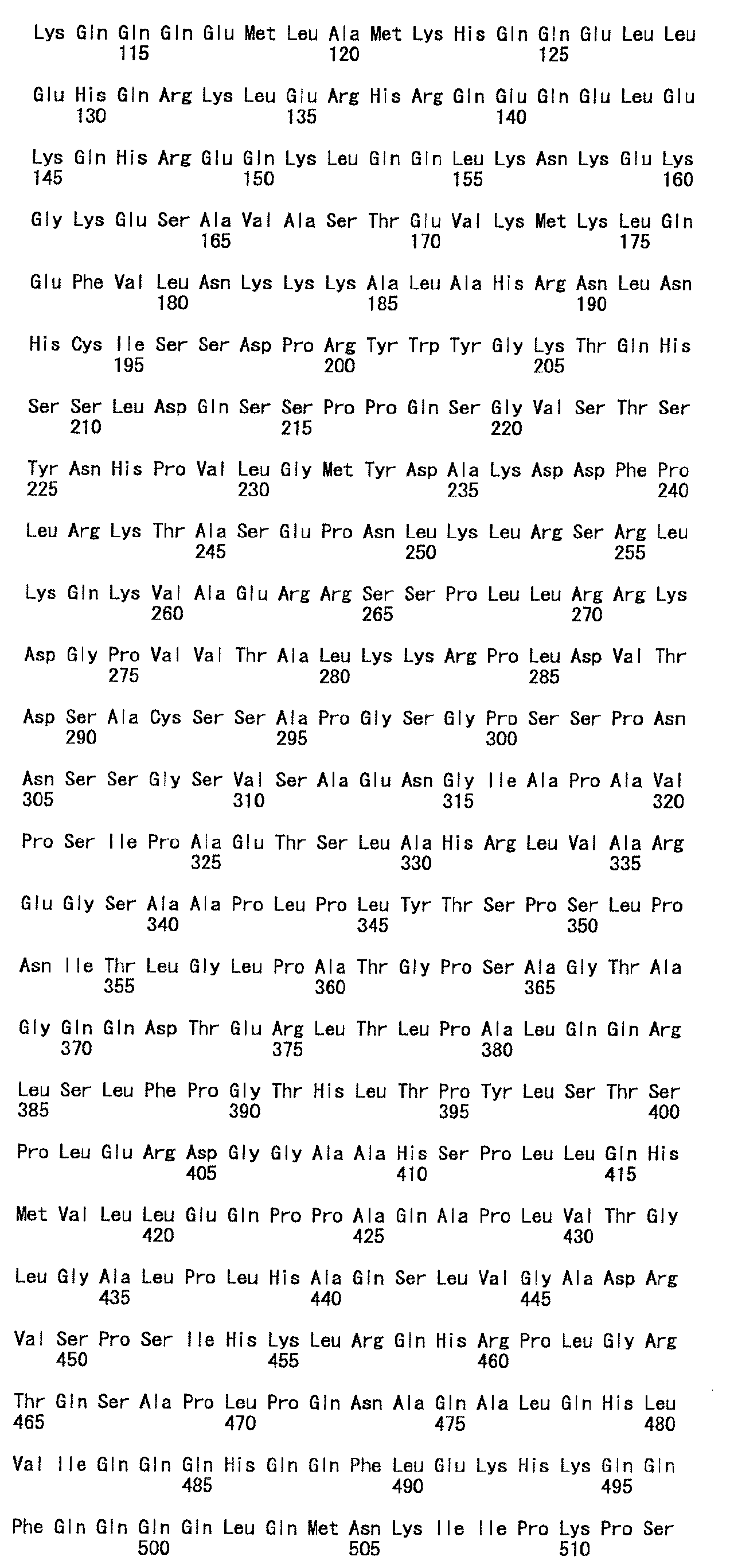
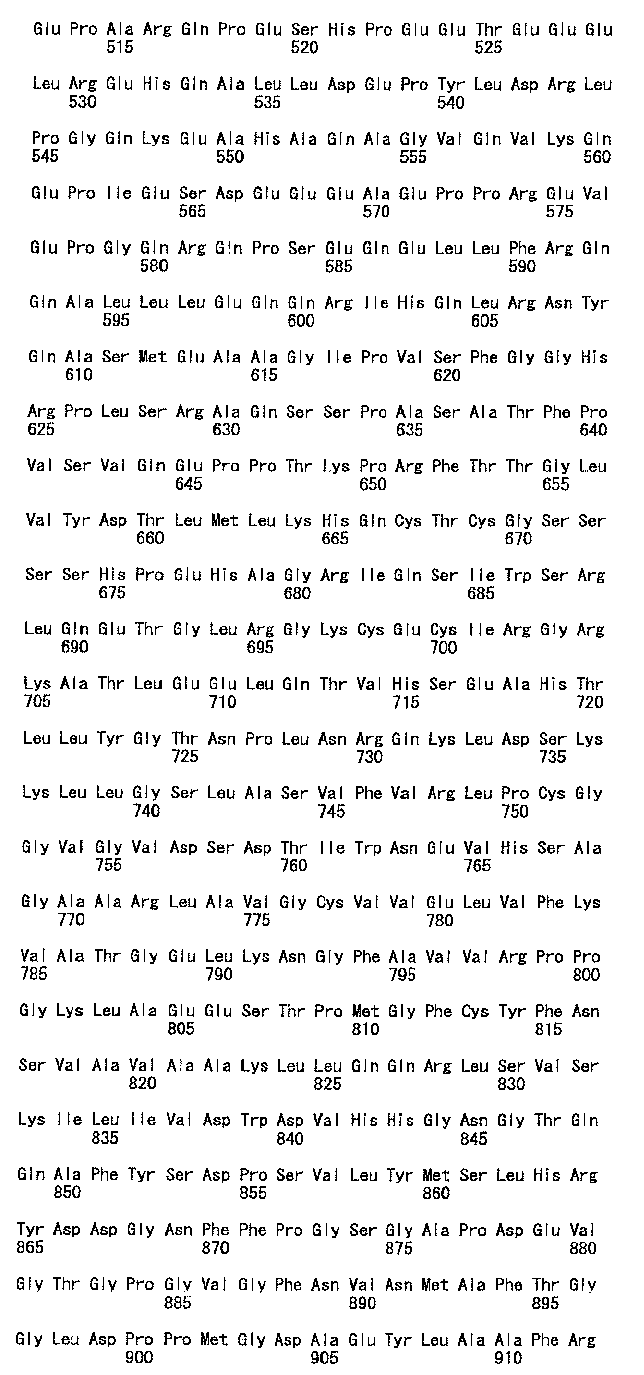


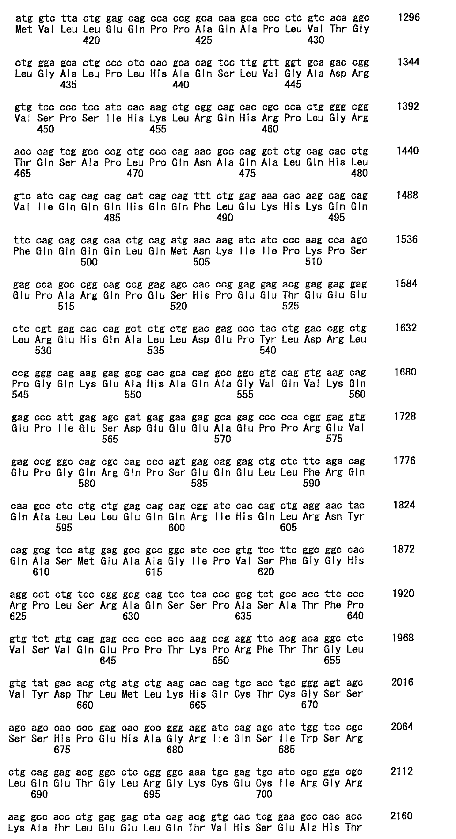
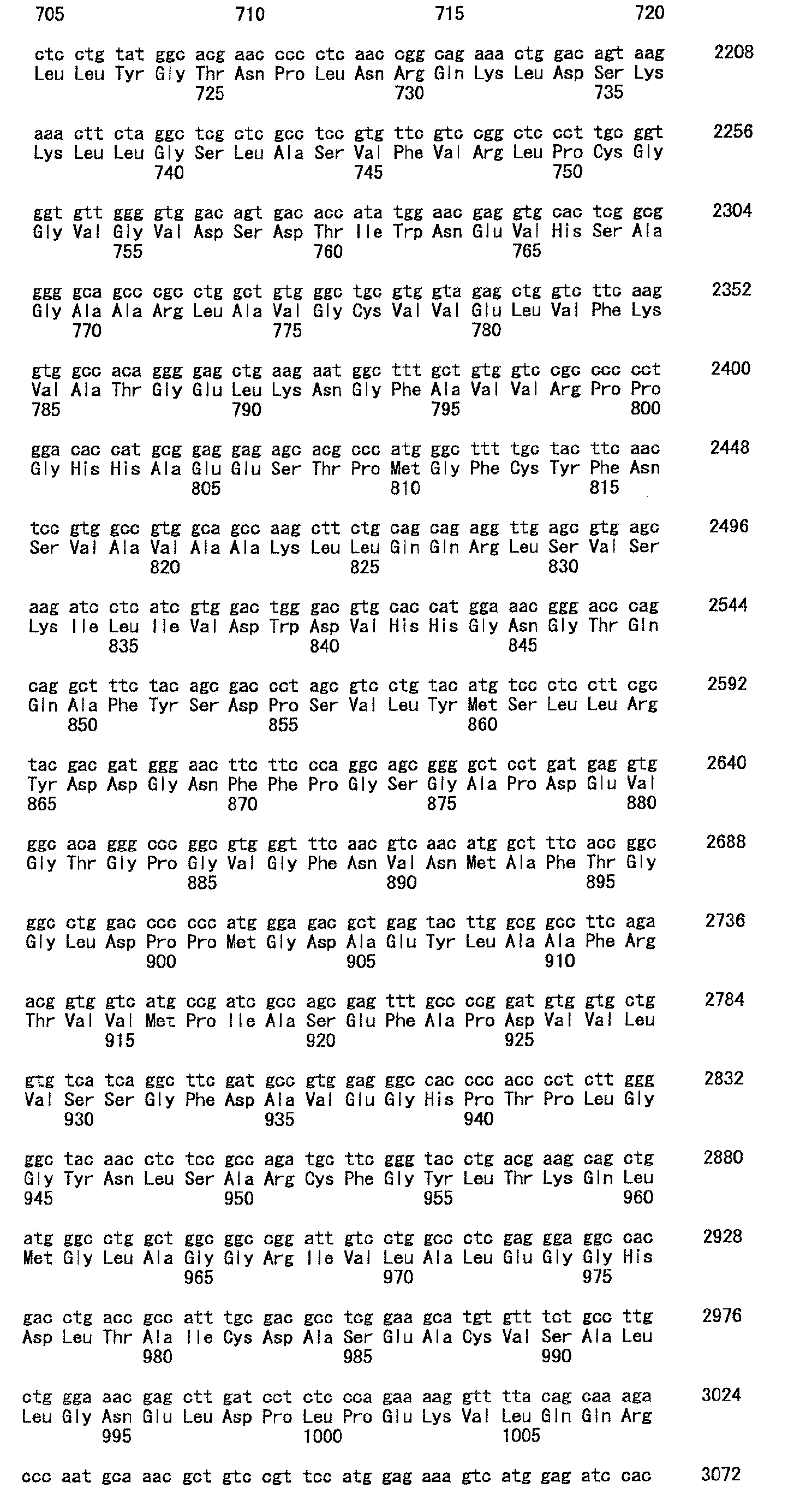


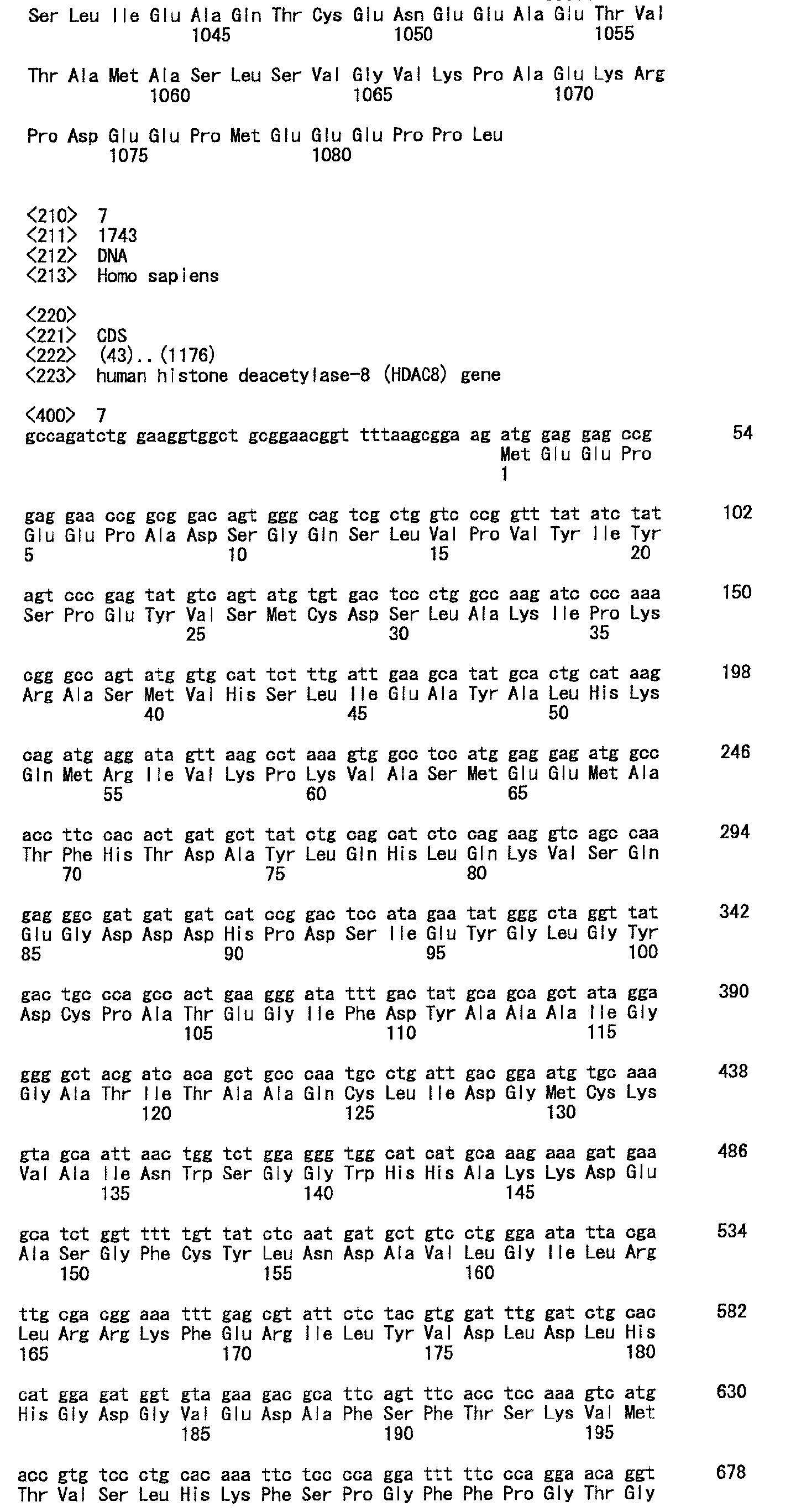


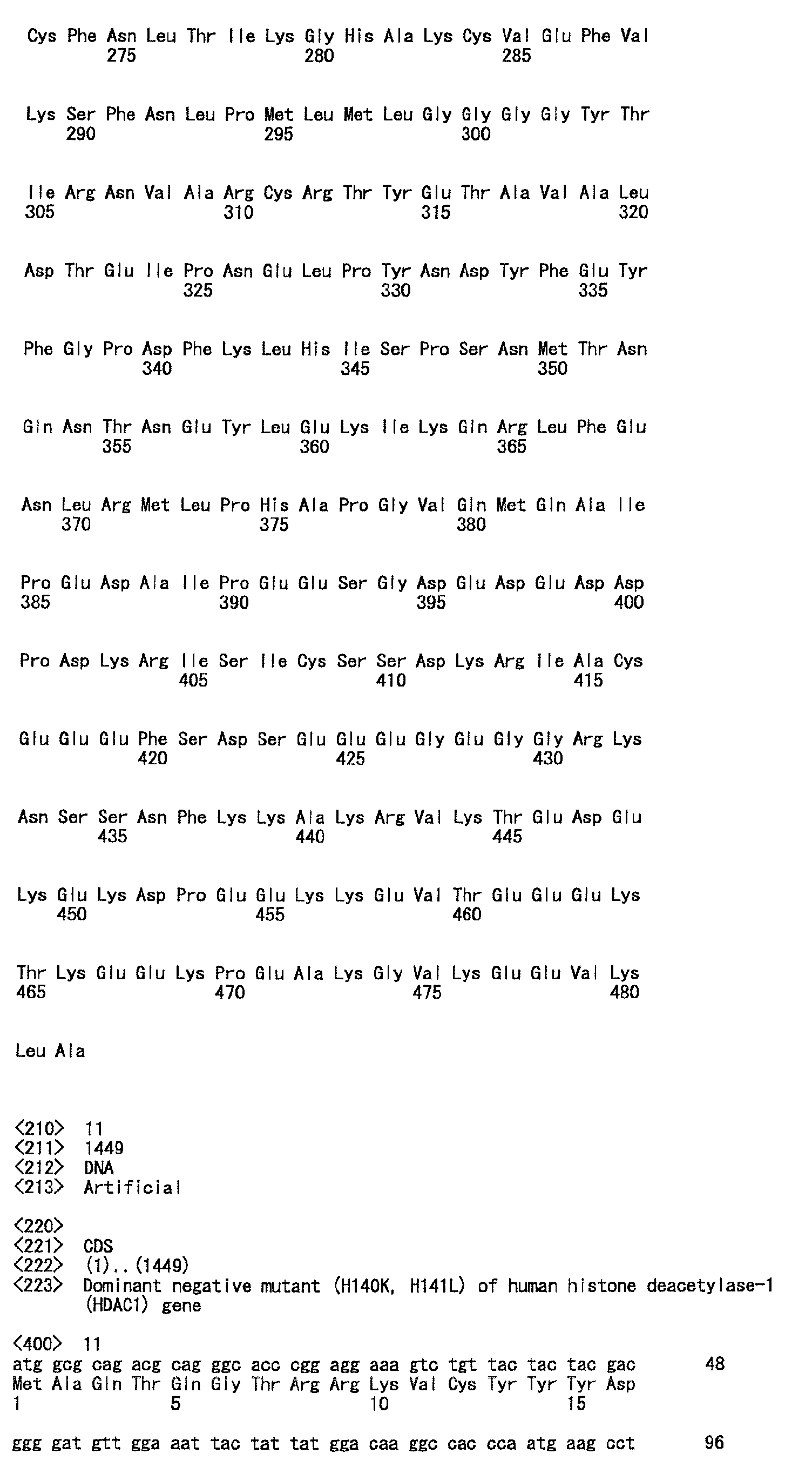
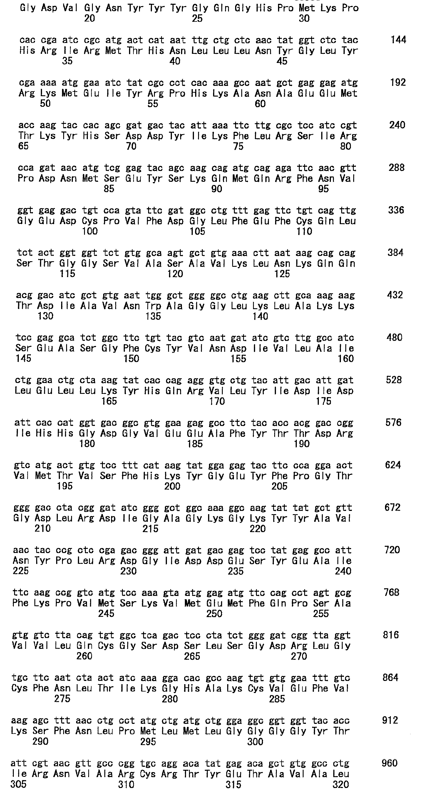
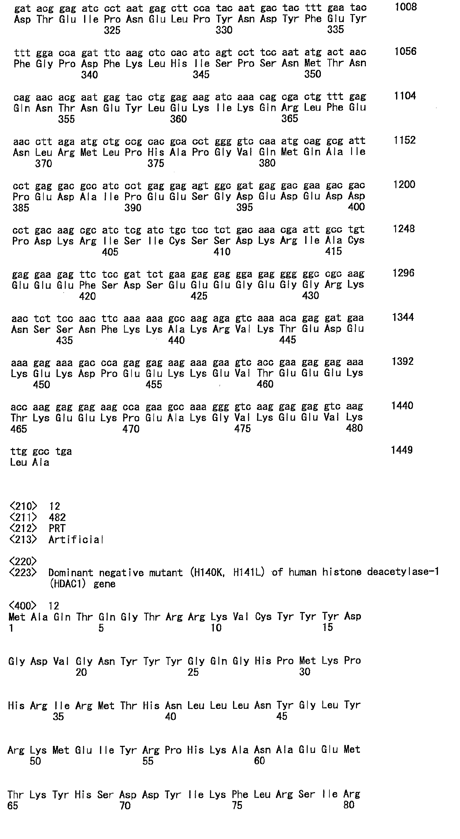
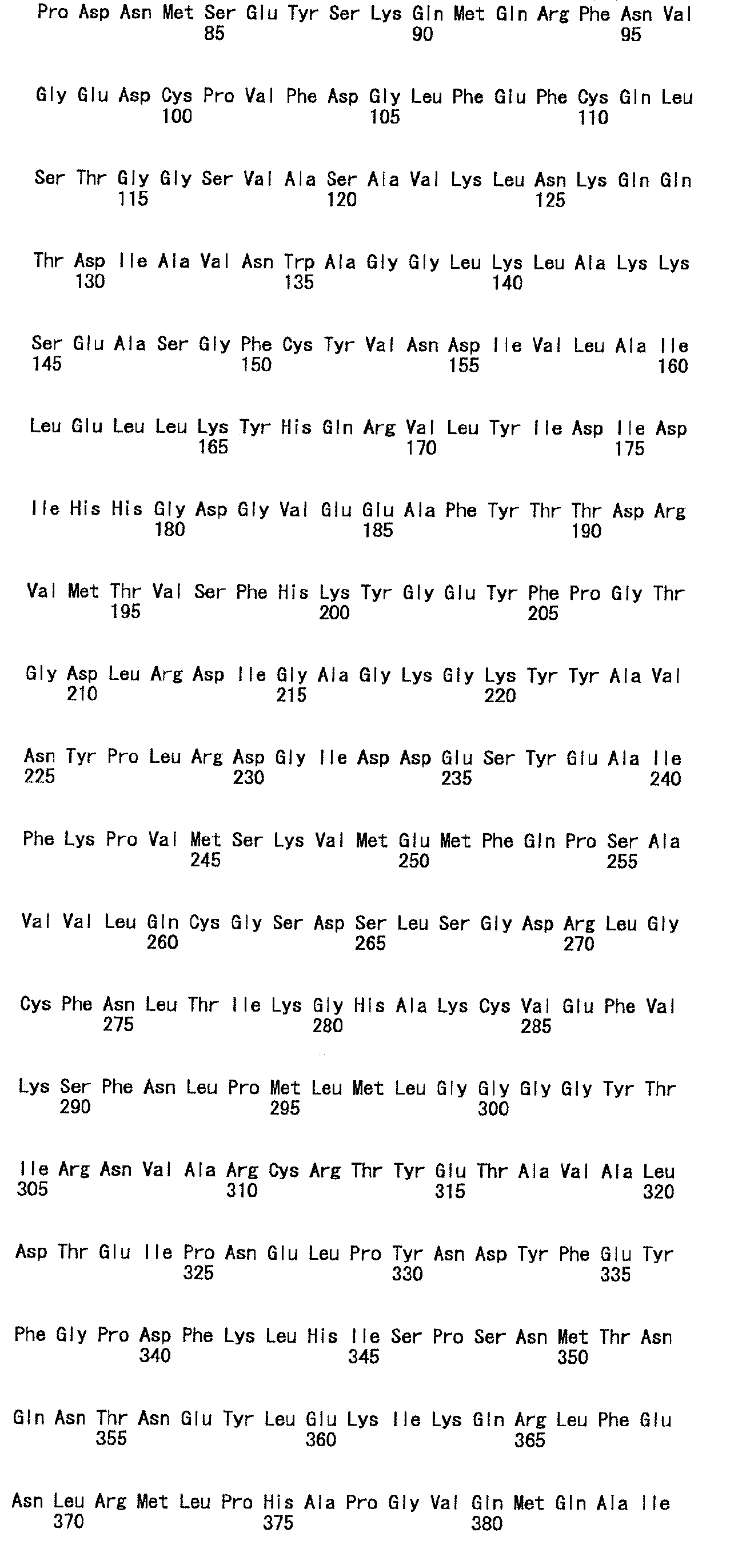
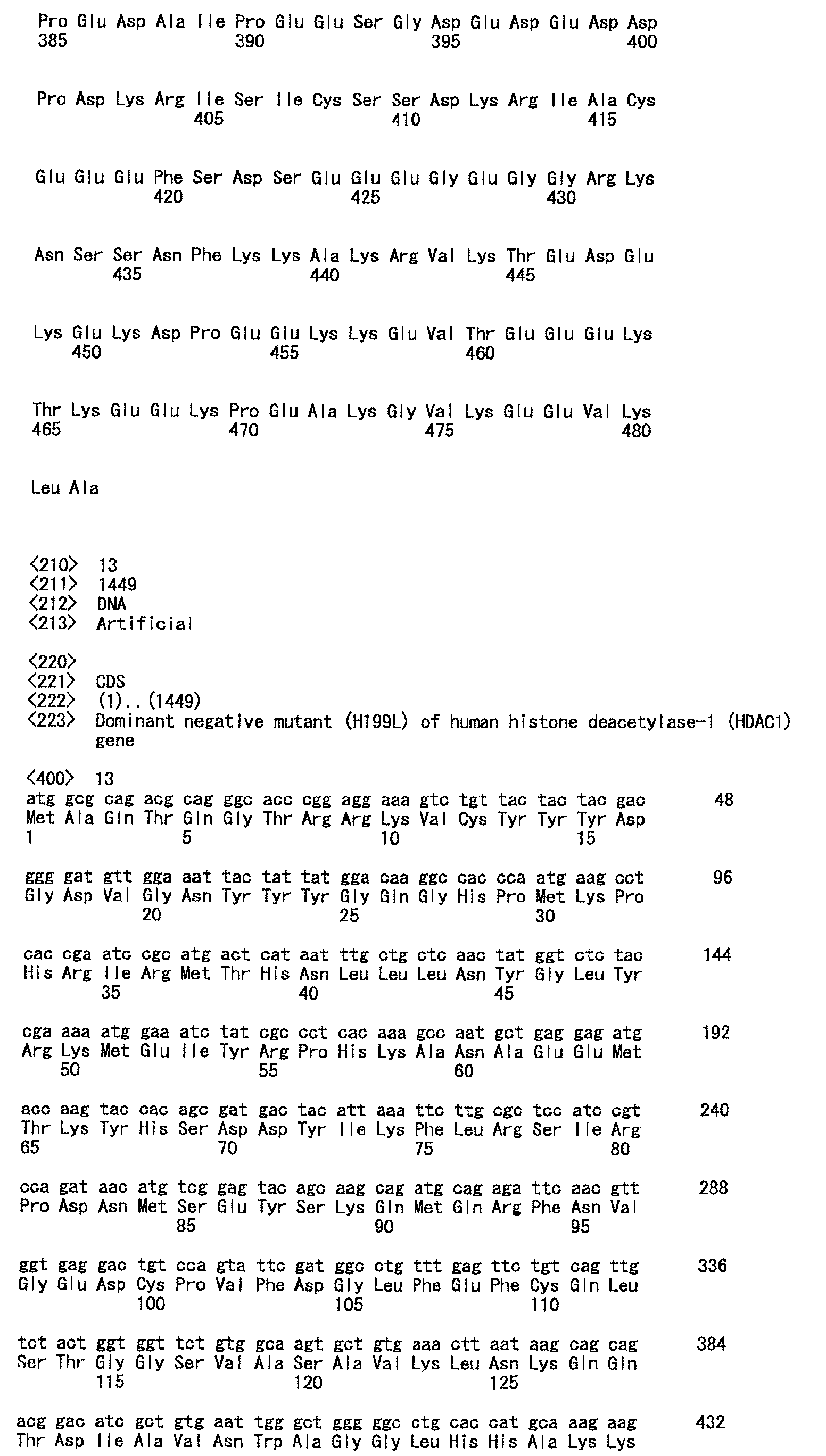


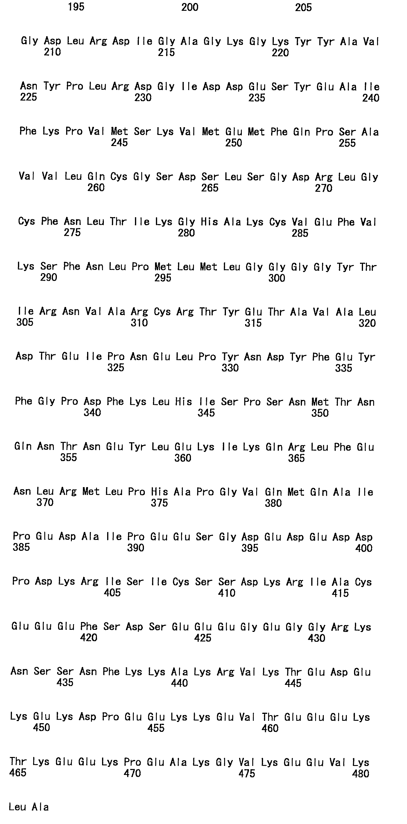
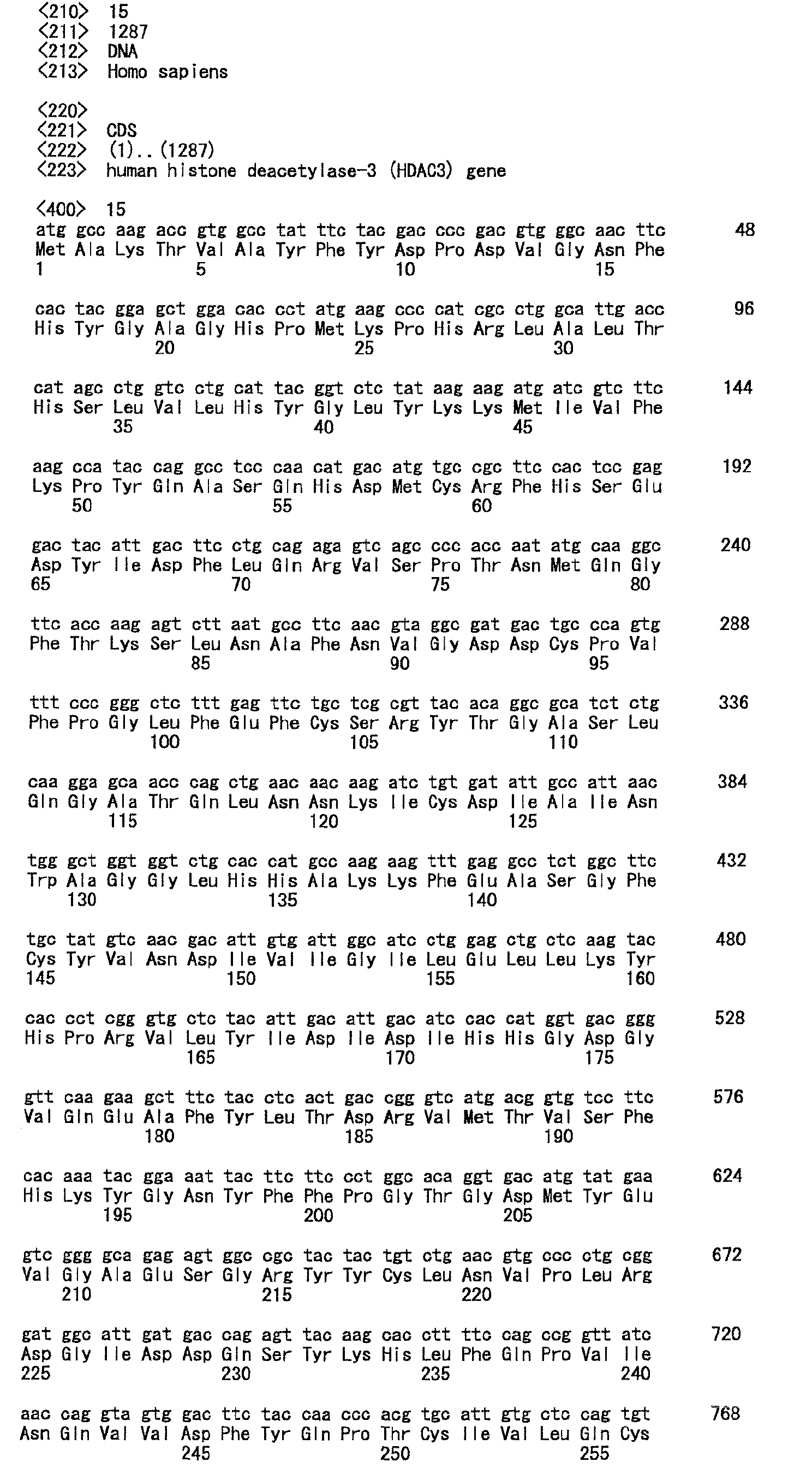


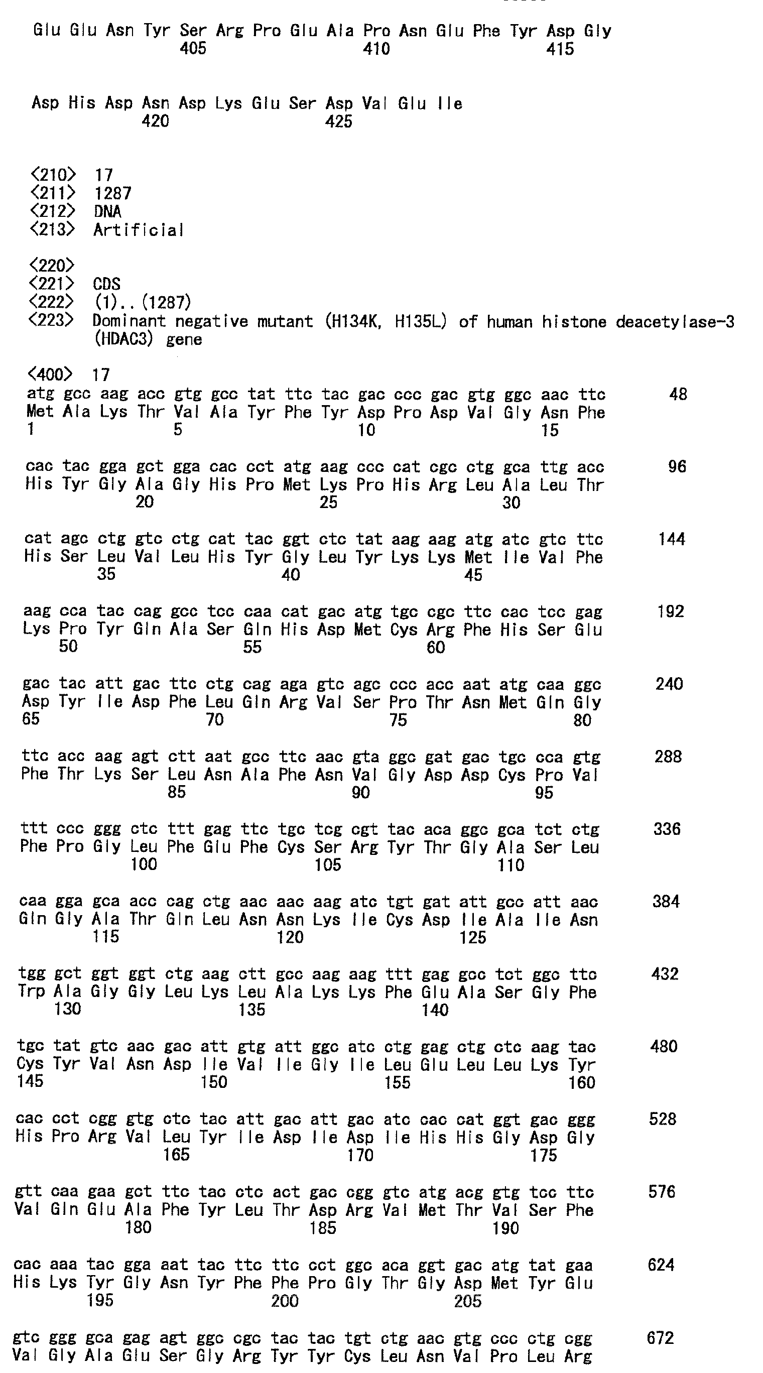


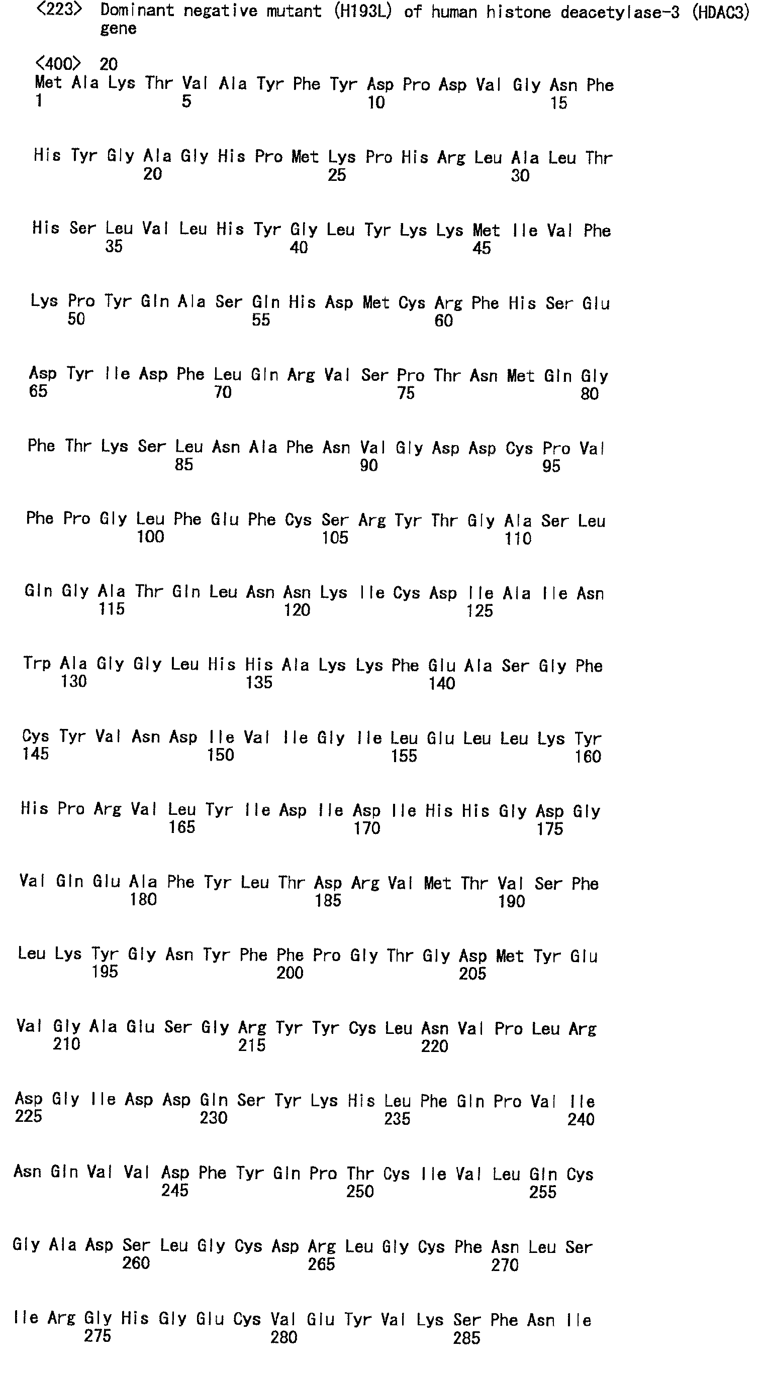
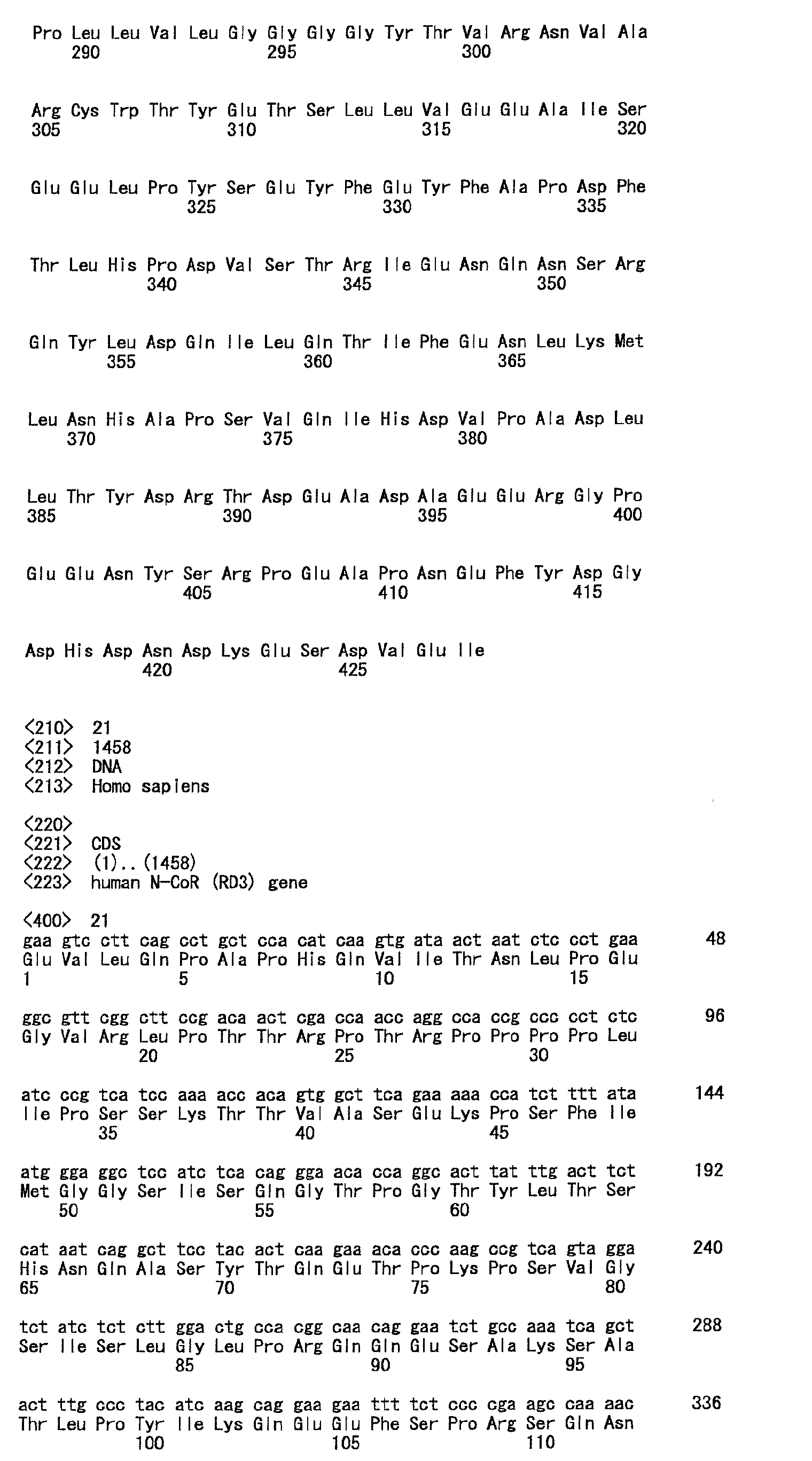


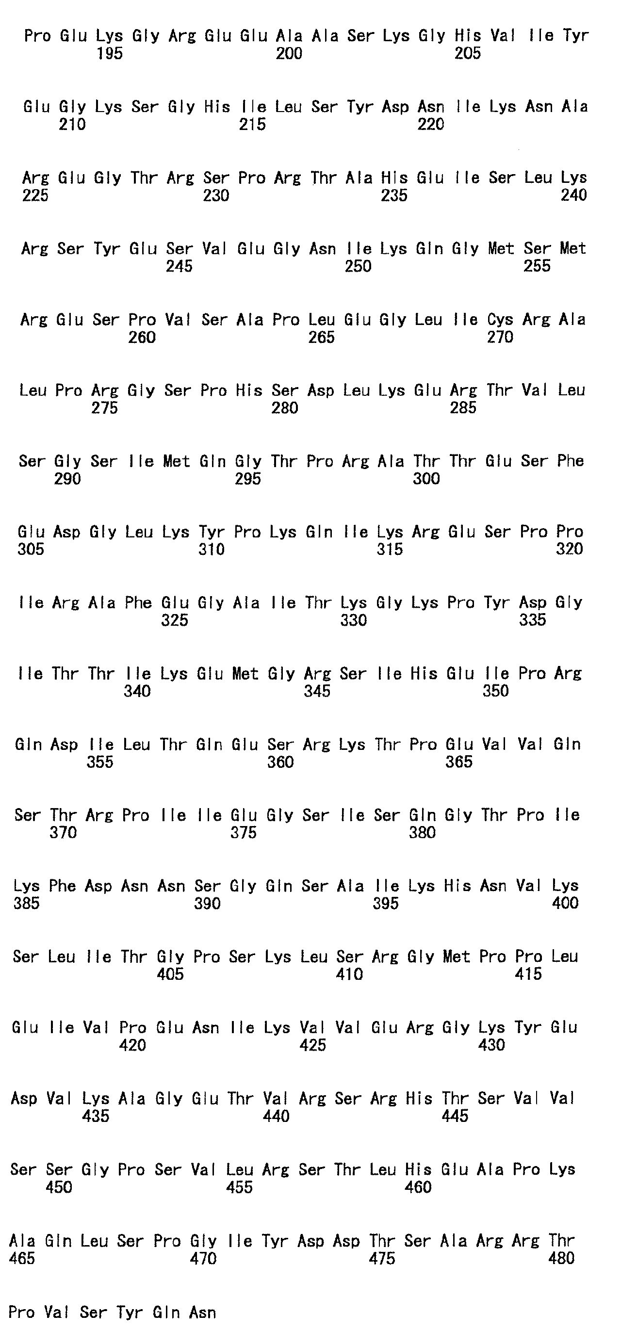
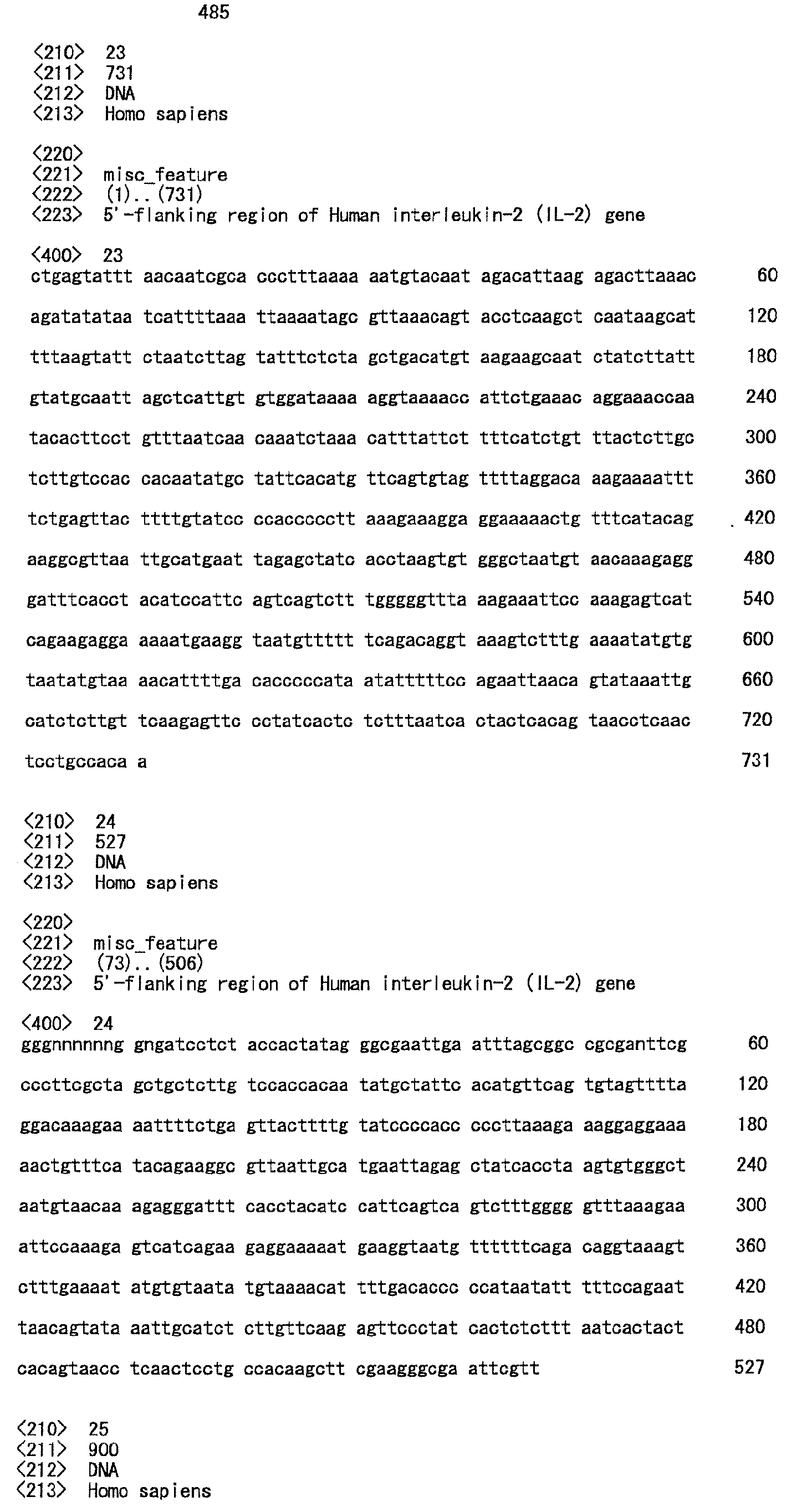
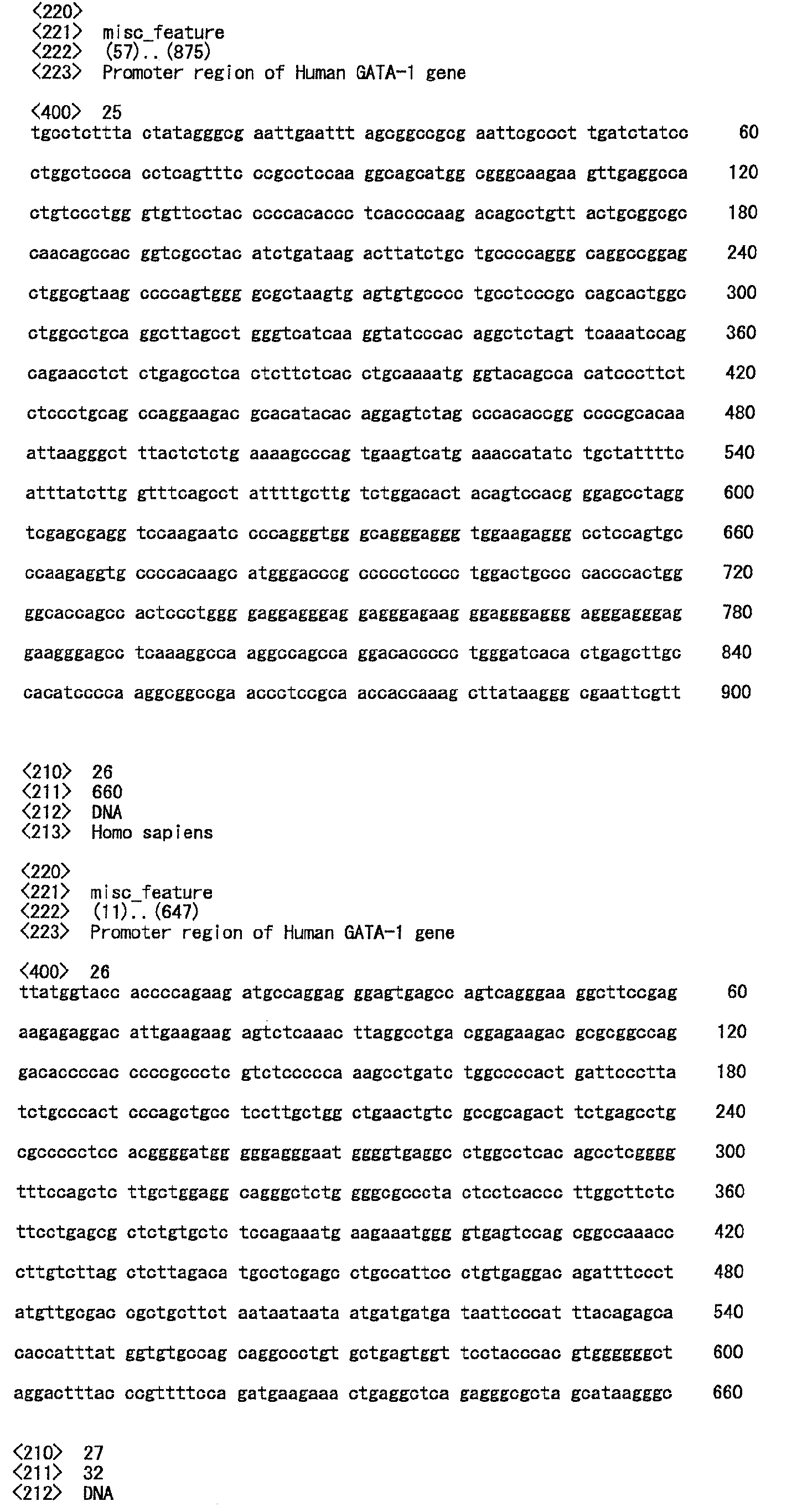

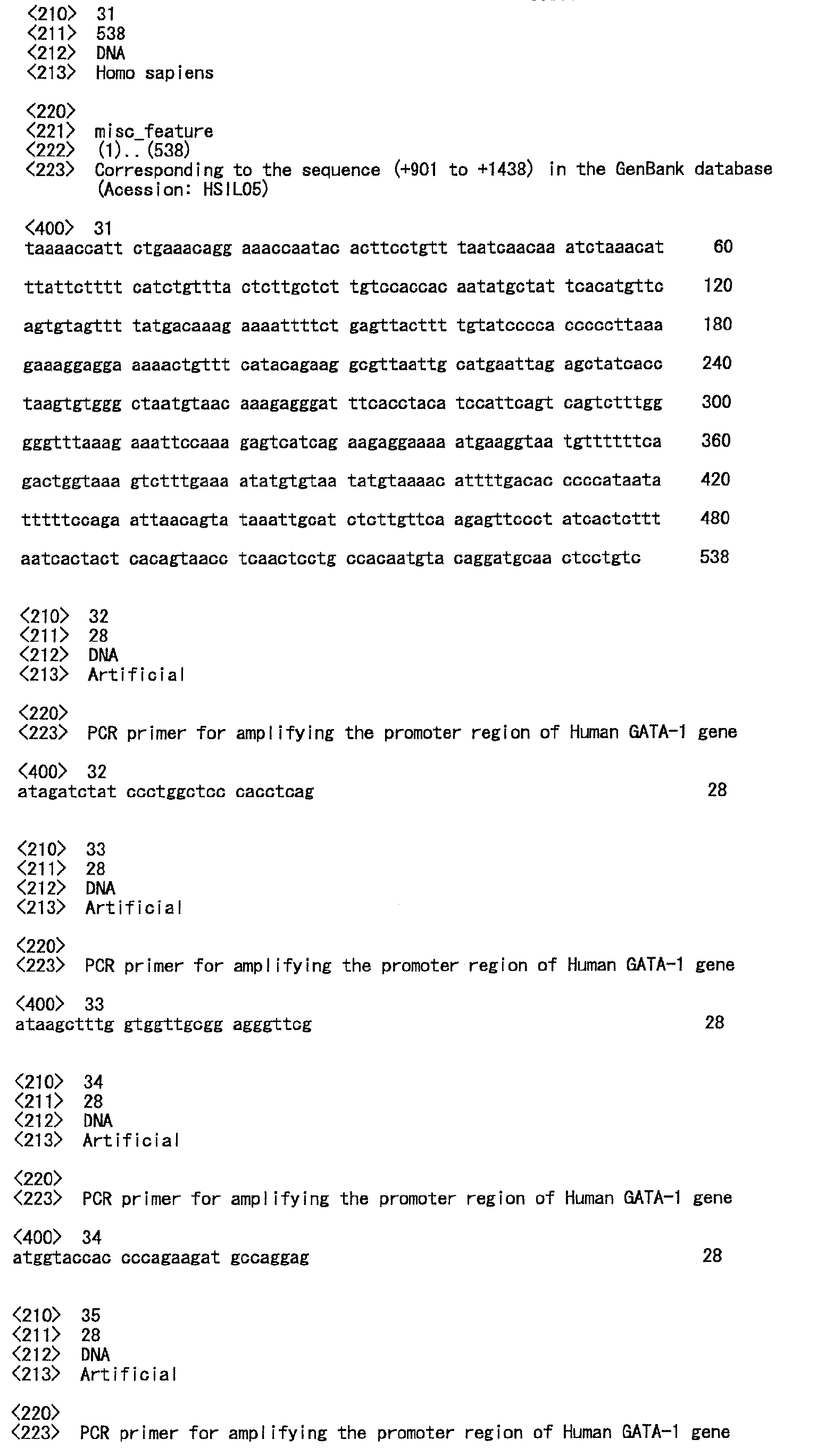


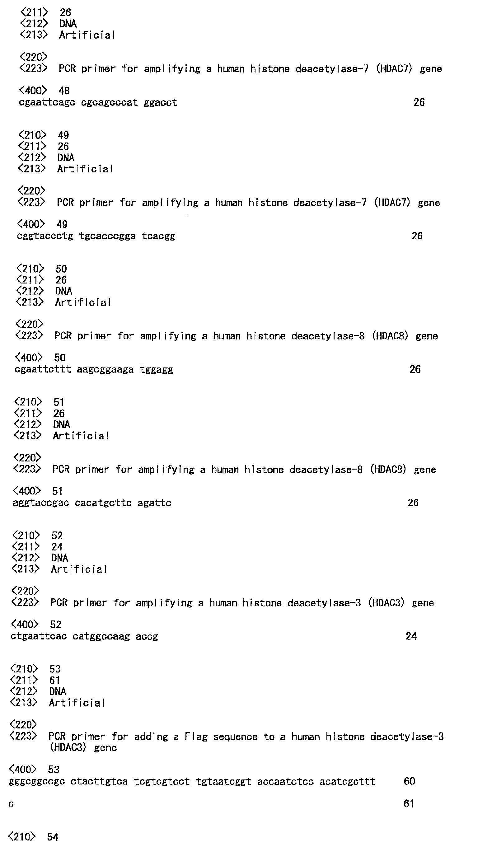


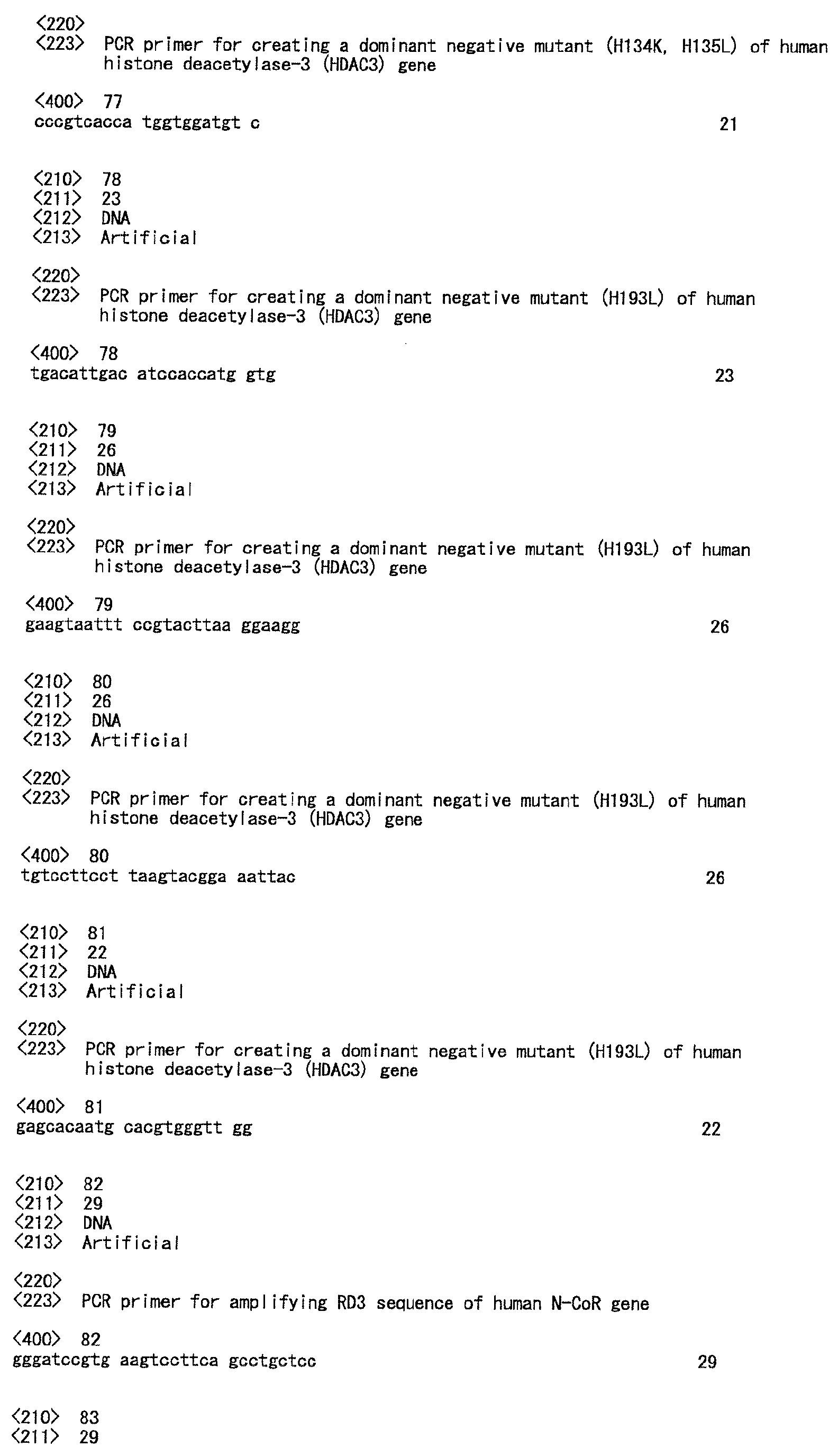
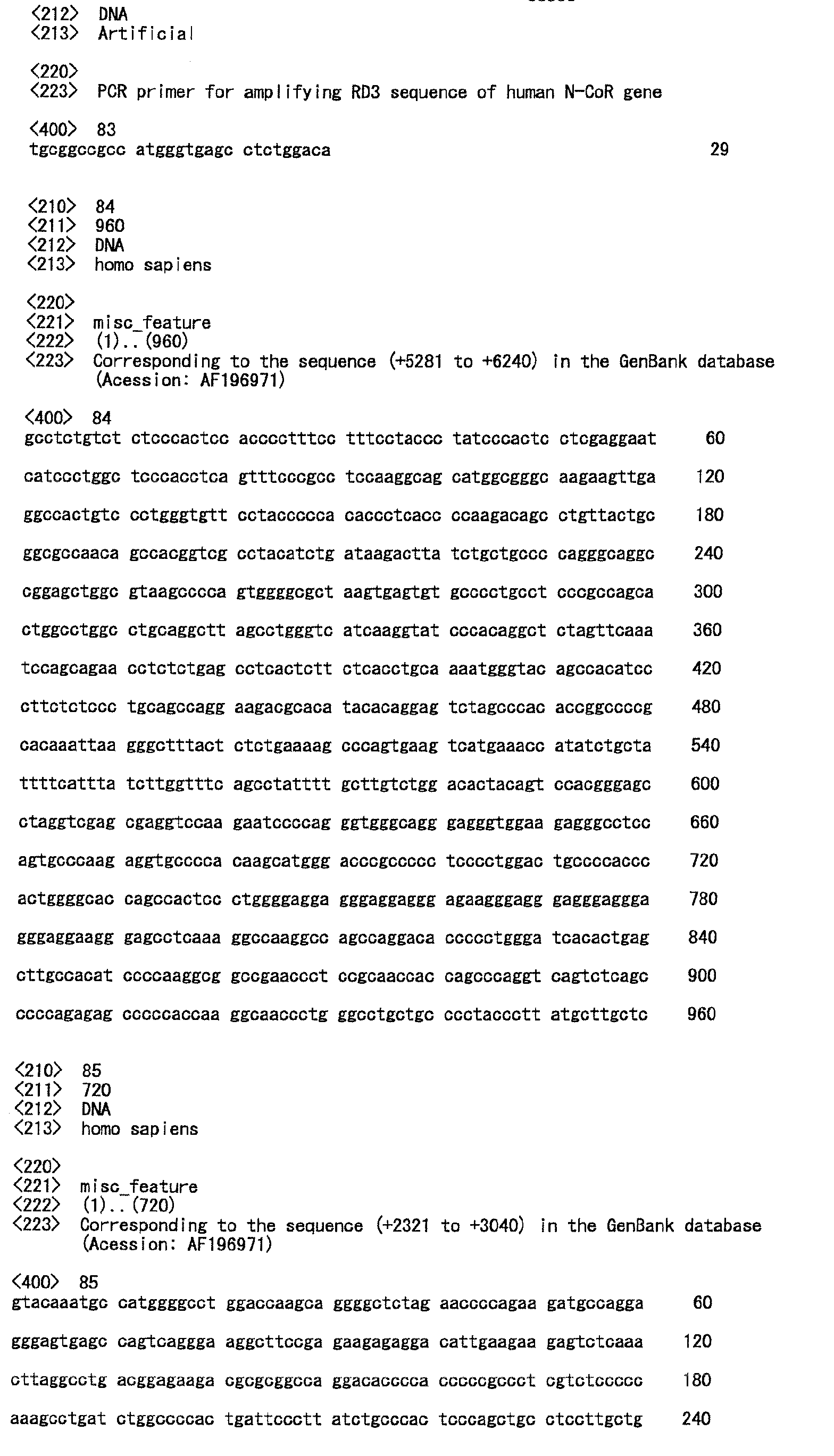
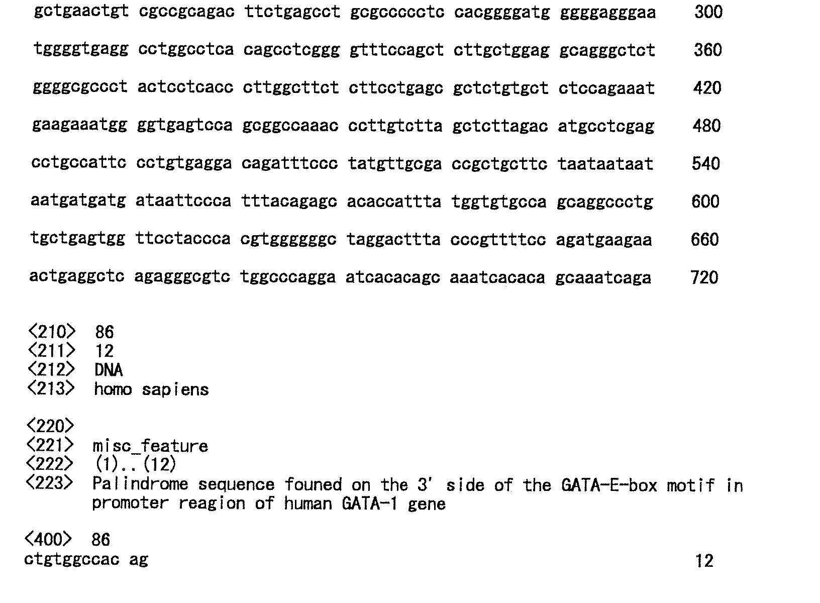
Claims (19)
- An agent for inhibiting IL-2 production and/or an agent for inhibiting immunocyte proliferation, each of which having low GATA-1 production inhibitory activity, comprising a selective HDAC4 and/or HDAC8 inhibitor.
- An agent for suppressing immunity having low thrombocytopenic activity, comprising a selective HDAC4 and/or HDAC8 inhibitor.
- A method of selecting an IL-2 production inhibitor and/or an immunocyte proliferation inhibitor which have low GATA-1 production inhibitory activity, which comprises measuring the HDAC4 and/or HDAC8 enzyme inhibitory activity of a test substance.
- A method of selecting an immunosuppressant having low thrombocytopenic activity, which comprises measuring the HDAC4 and/or HDAC8 enzyme inhibitory activity of a test substance.
- A method of selecting an immunosuppressant having low thrombocytopenic activity, which comprises steps (i) and (ii) below:(i) measuring the HDAC4 and/or HDAC8 enzyme inhibitory activity of a test substance, and(ii) measuring the HDAC enzyme inhibitory activity of the test substance, wherein the HDAC enzyme inhibitory activity is one or more HDAC enzyme inhibitory activities selected from a group consisting of HDAC1, HDAC2, HDAC3, HDAC5, HDAC6 and HDAC7 enzyme inhibitory activities.
- The method of claim 3, which is a method of selecting an IL-2 production inhibitor and/or an immunocyte proliferation inhibitor which have low GATA-1 production inhibitory activity, and which comprises steps (i) to (iii) below, wherein the IL-2 production inhibitor and/or the immunocyte proliferation inhibitor selectively inhibits HDAC4 and/or HDAC8 enzyme activity:(i) expressing each of the HDAC1, HDAC2, HDAC3, HDAC4, HDAC5, HDAC6, HDAC7 and HDAC8 genes to obtain respective enzyme solutions;(ii) measuring HDAC enzyme activity for each of the enzyme solutions in the presence of a test substance;(iii) selecting a test substance that selectively suppresses HDAC enzyme activity only when using an enzyme solution obtained by expressing the HDAC4 gene or the HDAC8 gene.
- The method of claim 4, which is a method of selecting an immunosuppressant having low thrombocytopenic activity, and which comprises steps (i) to (iii) below, wherein the immunosuppressant selectively inhibits HDAC4 and/or HDAC8 enzyme activity:(i) expressing each of the HDAC1, HDAC2, HDAC3, HDAC4, HDAC5, HDAC6, HDAC7 and HDAC8 genes to obtain respective enzyme solutions;(ii) measuring HDAC enzyme activity for each of the enzyme solutions in the presence of a test substance;(iii) selecting a test substance that selectively suppresses HDAC enzyme activity only when using an enzyme solution obtained by expressing the HDAC4 gene or the HDAC8 gene.
- The method of claim 4, which is a method of selecting an immunosuppressant having low thrombocytopenic activity, and which comprises steps (i) to (iii) below, wherein the immunosuppressant selectively inhibits HDAC4 and/or HDAC8 enzyme activity:(i) measuring HDAC enzyme activity for an enzyme solution partially purified from human cells, in the presence of a test substance;(ii) measuring HDAC enzyme activity for an enzyme solution obtained by expressing the HDAC4 gene and/or the HDAC8 gene, in the presence of a test substance;(iii) selecting a test substance that selectively suppresses HDAC enzyme activity only when using an enzyme solution obtained by expressing the HDAC4 gene and/or the HDAC8 gene, by comparing the enzyme activities measured in (i) and (ii).
- A method of selecting an immunosuppressant having low thrombocytopenic activity, which comprises steps (i) to (iii) below, wherein an HDAC inhibitor that specifically inhibits the formation of a complex of HDAC4 and N-CoR is selected from among test HDAC inhibitors:(i) expressing the HDAC4 gene and the N-CoR gene to obtain an enzyme solution;(ii) allowing the enzyme solution to coexist with a test HDAC inhibitor;(iii) determining whether or not a complex of HDAC4 and N-CoR is formed in the mixture of enzyme solution and test HDAC inhibitor obtained in (ii).
- A method of selecting an immunosuppressant having low thrombocytopenic activity, which comprises steps (i) and (ii) below, wherein an HDAC inhibitor that specifically inhibits the formation of a complex of the HDAC4 fusion protein comprising at least a portion of the HDAC4 protein, and the N-CoR fusion protein comprising at least a portion of the N-CoR protein, is selected from among test HDAC inhibitors:(i) expressing the gene that encodes the HDAC4 fusion protein and the gene that encodes the N-CoR fusion protein in cells;(ii) determining whether or not the test HDAC inhibitor inhibits the formation of a complex of the HDAC4 fusion protein and the N-CoR fusion protein.
- A method of selecting a compound having low thrombocytopenic activity and having immunosuppressive activity, which comprises steps (i) and (ii) below, wherein an HDAC inhibitor that suppresses the expression level of HDAC4 and/or HDAC8 is selected from among test HDAC inhibitors:(i) allowing cells that express HDAC4 and/or HDAC8 to coexist with a test HDAC inhibitors;(ii) measuring the HDAC4 and/or HDAC8 expression level in the cells.
- A method of selecting a compound having low thrombocytopenic activity and having immunosuppressive activity, which comprises steps (i) and (ii) below, wherein an HDAC inhibitor that selectively inhibits the binding of HDAC4 and/or HDAC8 and an HDAC4- and/or HDAC8-specific ligand is selected from among test HDAC inhibitors:(i) expressing the HDAC4 and/or HDAC8 gene to obtain an enzyme solution;(ii) measuring the binding activity of the HDAC4 and/or HDAC8 enzyme and the HDAC4- and/or HDAC8-specific ligand for the enzyme solution in the presence of an HDAC4- and/or HDAC8-specific ligand and a test HDAC inhibitor.
- An assay kit for the selection of an immunosuppressant having low thrombocytopenic activity including a selective HDAC4 and/or HDAC8 inhibitor, which includes at least (i) and (ii) below:(i) enzyme solutions prepared by expressing each of the HDAC1, HDAC2, HDAC3, HDAC5, HDAC6 and HDAC7 genes;(ii) an enzyme solution prepared by expressing each of the HDAC4 and/or HDAC8 gene;
- A DNA having the base sequence that encodes the amino acid sequence shown by SEQ ID NO:4 or SEQ ID NO:6.
- An HDAC4 variant that is the amino acid sequence shown by SEQ ID NO:4 or SEQ ID NO:6.
- A method of inhibiting IL-2 production and/or immunocyte proliferation with low GATA-1 production inhibitory activity, which comprises administering an effective amount of selective HDAC4 and/or HDAC8 inhibitor to a subject.
- A method of suppressing immunity with low thrombocytopenic activity, which comprises administering an effective amount of selective HDAC4 and/or HDAC8 inhibitor to a subject.
- Use of a selective HDAC4 and/or HDAC8 inhibitor for the production of an agent for inhibiting IL-2 production and/or an agent for inhibiting immunocyte proliferation which have low GATA-1 production inhibitory activity.
- Use of a selective HDAC4 and/or HDAC8 inhibitor for the production of an agent for suppressing immunity having low thrombocytopenic activity.
Applications Claiming Priority (3)
| Application Number | Priority Date | Filing Date | Title |
|---|---|---|---|
| JP2002378800 | 2002-12-27 | ||
| JP2002378800 | 2002-12-27 | ||
| PCT/JP2003/016895 WO2004061101A1 (en) | 2002-12-27 | 2003-12-26 | Method of selecting novel immunosuppressant |
Publications (2)
| Publication Number | Publication Date |
|---|---|
| EP1582585A1 true EP1582585A1 (en) | 2005-10-05 |
| EP1582585A4 EP1582585A4 (en) | 2006-06-07 |
Family
ID=32708346
Family Applications (1)
| Application Number | Title | Priority Date | Filing Date |
|---|---|---|---|
| EP03768314A Withdrawn EP1582585A4 (en) | 2002-12-27 | 2003-12-26 | METHOD OF SELECTING A NEW IMMUNOSUPPRESSOR |
Country Status (6)
| Country | Link |
|---|---|
| US (1) | US20070123452A1 (en) |
| EP (1) | EP1582585A4 (en) |
| JP (1) | JPWO2004061101A1 (en) |
| AU (1) | AU2003292680A1 (en) |
| CA (1) | CA2512002A1 (en) |
| WO (1) | WO2004061101A1 (en) |
Cited By (2)
| Publication number | Priority date | Publication date | Assignee | Title |
|---|---|---|---|---|
| WO2009156853A3 (en) * | 2008-06-26 | 2010-03-25 | Istituto Di Ricerche Di Biologia Molecolare P. Angeletti Spa | Compounds, compositions, and methods useful for modulating the expression of histone deacetylase (hdac) gene expression and/or activity |
| WO2012098006A1 (en) * | 2011-01-21 | 2012-07-26 | Cellzome Ag | Compounds and methods for the identification and characterization of hdac interacting compounds |
Families Citing this family (4)
| Publication number | Priority date | Publication date | Assignee | Title |
|---|---|---|---|---|
| WO2008060721A1 (en) | 2006-11-14 | 2008-05-22 | Pharmacyclics, Inc. | Uses of selective inhibitors of hdac8 for treatment of t-cell proliferative disorders |
| US20110150825A1 (en) * | 2006-11-14 | 2011-06-23 | Pharmacyclics, Inc. | Uses of selective inhibitors of hdac8 for treatment of inflammatory conditions |
| AU2008316667A1 (en) * | 2007-10-26 | 2009-04-30 | Repligen Corporation | Methods of identifying histone deacetylase inhibitors useful for neurological disorders |
| KR101075848B1 (en) | 2008-11-06 | 2011-10-25 | 가톨릭대학교 산학협력단 | Markers for Screening Inhibitors of HDAC |
Family Cites Families (2)
| Publication number | Priority date | Publication date | Assignee | Title |
|---|---|---|---|---|
| AUPP505798A0 (en) * | 1998-08-04 | 1998-08-27 | Fujisawa Pharmaceutical Co., Ltd. | Novel compound fr225497 substance |
| CA2493088A1 (en) * | 2002-07-12 | 2004-01-22 | Takao Fujimura | Novel method of selecting immunosuppressant having little thrombocytopenic effect |
-
2003
- 2003-12-26 CA CA002512002A patent/CA2512002A1/en not_active Abandoned
- 2003-12-26 AU AU2003292680A patent/AU2003292680A1/en not_active Abandoned
- 2003-12-26 US US10/540,546 patent/US20070123452A1/en not_active Abandoned
- 2003-12-26 WO PCT/JP2003/016895 patent/WO2004061101A1/en not_active Ceased
- 2003-12-26 JP JP2004564542A patent/JPWO2004061101A1/en not_active Withdrawn
- 2003-12-26 EP EP03768314A patent/EP1582585A4/en not_active Withdrawn
Non-Patent Citations (2)
| Title |
|---|
| No further relevant documents disclosed * |
| See also references of WO2004061101A1 * |
Cited By (2)
| Publication number | Priority date | Publication date | Assignee | Title |
|---|---|---|---|---|
| WO2009156853A3 (en) * | 2008-06-26 | 2010-03-25 | Istituto Di Ricerche Di Biologia Molecolare P. Angeletti Spa | Compounds, compositions, and methods useful for modulating the expression of histone deacetylase (hdac) gene expression and/or activity |
| WO2012098006A1 (en) * | 2011-01-21 | 2012-07-26 | Cellzome Ag | Compounds and methods for the identification and characterization of hdac interacting compounds |
Also Published As
| Publication number | Publication date |
|---|---|
| WO2004061101A1 (en) | 2004-07-22 |
| US20070123452A1 (en) | 2007-05-31 |
| EP1582585A4 (en) | 2006-06-07 |
| AU2003292680A1 (en) | 2004-07-29 |
| JPWO2004061101A1 (en) | 2006-05-11 |
| CA2512002A1 (en) | 2004-07-22 |
Similar Documents
| Publication | Publication Date | Title |
|---|---|---|
| Matsui et al. | The Rab interacting lysosomal protein (RILP) homology domain functions as a novel effector domain for small GTPase Rab36: Rab36 regulates retrograde melanosome transport in melanocytes | |
| US7208157B2 (en) | Proteolysis targeting chimeric pharmaceutical | |
| Wein et al. | SIKs control osteocyte responses to parathyroid hormone | |
| US7041298B2 (en) | Proteolysis targeting chimeric pharmaceutical | |
| Narang et al. | Reduced histone biotinylation in multiple carboxylase deficiency patients: a nuclear role for holocarboxylase synthetase | |
| Wang et al. | C/EBPα arrests cell proliferation through direct inhibition of Cdk2 and Cdk4 | |
| Sureban et al. | Functional antagonism between RNA binding proteins HuR and CUGBP2 determines the fate of COX-2 mRNA translation | |
| Greene et al. | Regulation of NF‐κB action by reversible acetylation | |
| KR101573009B1 (en) | Compositions and methods to modulate cell membrane resealing | |
| Woods et al. | Differential activities of murine single minded 1 (SIM1) and SIM2 on a hypoxic response element: cross-talk between basic helix-loop-helix/per-Arnt-Sim homology transcription factors | |
| US12110522B2 (en) | Engineered promiscuous biotin ligases for efficient proximity labeling | |
| Lee et al. | RNA-binding protein muscleblind-like 3 (MBNL3) disrupts myocyte enhancer factor 2 (Mef2) β-exon splicing | |
| Yuan et al. | Fibroblast-localized lncRNA CFIRL promotes cardiac fibrosis and dysfunction in dilated cardiomyopathy | |
| Palatini et al. | Mutational analysis of the GXXXG/A motifs in the human Na+/Taurocholate Co-transporting polypeptide NTCP on its bile acid transport function and hepatitis B/D virus receptor function | |
| Xu et al. | Smurf1 polyubiquitinates on K285/K282 of the kinases Mst1/2 to attenuate their tumor-suppressor functions | |
| Bechor et al. | p67 phox binds to a newly identified site in Nox2 following the disengagement of an intramolecular bond—Canaan sighted? | |
| US20070123452A1 (en) | Method of selecting novel immunosuppressant | |
| Clemente-Olivo et al. | Early adipogenesis is repressed through the newly identified FHL2-NFAT5 signaling complex | |
| CN114423444A (en) | CAMKII-delta 9 antagonists and uses thereof | |
| JP2010501827A (en) | Screening method for agents that inhibit binding of MPHOSPH1 and PRC1 | |
| CN115607675B (en) | Nav1.9 interaction protein PRMT7 and application of down regulator thereof in preparing analgesic drugs | |
| US20070054259A1 (en) | Modulation of hnRNP H and treatment of DM1 | |
| JP2007282628A (en) | Method for screening anti-cancer agent | |
| JP2010195684A (en) | REGULATION OF HIF-1 BY MTI-MMP, iFIH AND FIH-1 | |
| Samoylenko et al. | The adaptor protein Ruk/CIN85 activates plasminogen activator inhibitor-1 (PAI-1) expression via hypoxia-inducible factor-1α |
Legal Events
| Date | Code | Title | Description |
|---|---|---|---|
| PUAI | Public reference made under article 153(3) epc to a published international application that has entered the european phase |
Free format text: ORIGINAL CODE: 0009012 |
|
| 17P | Request for examination filed |
Effective date: 20050712 |
|
| AK | Designated contracting states |
Kind code of ref document: A1 Designated state(s): AT BE BG CH CY CZ DE DK EE ES FI FR GB GR HU IE IT LI LU MC NL PT RO SE SI SK TR |
|
| AX | Request for extension of the european patent |
Extension state: AL LT LV MK |
|
| DAX | Request for extension of the european patent (deleted) | ||
| A4 | Supplementary search report drawn up and despatched |
Effective date: 20060502 |
|
| STAA | Information on the status of an ep patent application or granted ep patent |
Free format text: STATUS: THE APPLICATION HAS BEEN WITHDRAWN |
|
| 18W | Application withdrawn |
Effective date: 20070831 |