EP1581645B1 - Method for using division arrested cells in screening assays - Google Patents
Method for using division arrested cells in screening assays Download PDFInfo
- Publication number
- EP1581645B1 EP1581645B1 EP03751966A EP03751966A EP1581645B1 EP 1581645 B1 EP1581645 B1 EP 1581645B1 EP 03751966 A EP03751966 A EP 03751966A EP 03751966 A EP03751966 A EP 03751966A EP 1581645 B1 EP1581645 B1 EP 1581645B1
- Authority
- EP
- European Patent Office
- Prior art keywords
- cell
- cells
- interest
- transfected
- transformed
- Prior art date
- Legal status (The legal status is an assumption and is not a legal conclusion. Google has not performed a legal analysis and makes no representation as to the accuracy of the status listed.)
- Expired - Lifetime
Links
- 238000000034 method Methods 0.000 title claims description 30
- 238000007423 screening assay Methods 0.000 title abstract description 12
- 230000000694 effects Effects 0.000 claims abstract description 21
- 239000000126 substance Substances 0.000 claims abstract description 21
- 210000004027 cell Anatomy 0.000 claims description 145
- 102000003688 G-Protein-Coupled Receptors Human genes 0.000 claims description 8
- 108090000045 G-Protein-Coupled Receptors Proteins 0.000 claims description 8
- 230000001275 ca(2+)-mobilization Effects 0.000 claims description 8
- 108020004707 nucleic acids Proteins 0.000 claims description 7
- 102000039446 nucleic acids Human genes 0.000 claims description 7
- 150000007523 nucleic acids Chemical class 0.000 claims description 7
- 230000008859 change Effects 0.000 claims description 6
- 150000001875 compounds Chemical class 0.000 claims description 6
- 102000004169 proteins and genes Human genes 0.000 claims description 6
- 108090000623 proteins and genes Proteins 0.000 claims description 6
- 230000004913 activation Effects 0.000 claims description 5
- 230000001413 cellular effect Effects 0.000 claims description 4
- 210000003527 eukaryotic cell Anatomy 0.000 claims description 4
- 230000006698 induction Effects 0.000 claims description 4
- 102100039705 Beta-2 adrenergic receptor Human genes 0.000 claims description 3
- 108010014499 beta-2 Adrenergic Receptors Proteins 0.000 claims description 3
- 230000000394 mitotic effect Effects 0.000 claims description 3
- 108010009685 Cholinergic Receptors Proteins 0.000 claims description 2
- 108010043121 Green Fluorescent Proteins Proteins 0.000 claims description 2
- 102000004144 Green Fluorescent Proteins Human genes 0.000 claims description 2
- 108060001084 Luciferase Proteins 0.000 claims description 2
- 239000005089 Luciferase Substances 0.000 claims description 2
- 102000034337 acetylcholine receptors Human genes 0.000 claims description 2
- 230000000692 anti-sense effect Effects 0.000 claims description 2
- 210000004978 chinese hamster ovary cell Anatomy 0.000 claims description 2
- 238000012875 competitive assay Methods 0.000 claims description 2
- 239000005090 green fluorescent protein Substances 0.000 claims description 2
- 108090000765 processed proteins & peptides Proteins 0.000 claims description 2
- 102000004310 Ion Channels Human genes 0.000 claims 1
- 241000700605 Viruses Species 0.000 claims 1
- 238000012203 high throughput assay Methods 0.000 abstract description 2
- 238000003567 signal transduction assay Methods 0.000 abstract description 2
- 238000007878 drug screening assay Methods 0.000 abstract 1
- 238000003556 assay Methods 0.000 description 20
- NWIBSHFKIJFRCO-WUDYKRTCSA-N Mytomycin Chemical compound C1N2C(C(C(C)=C(N)C3=O)=O)=C3[C@@H](COC(N)=O)[C@@]2(OC)[C@@H]2[C@H]1N2 NWIBSHFKIJFRCO-WUDYKRTCSA-N 0.000 description 15
- QZAYGJVTTNCVMB-UHFFFAOYSA-N serotonin Chemical compound C1=C(O)C=C2C(CCN)=CNC2=C1 QZAYGJVTTNCVMB-UHFFFAOYSA-N 0.000 description 14
- 230000006870 function Effects 0.000 description 12
- 102000005962 receptors Human genes 0.000 description 12
- 108020003175 receptors Proteins 0.000 description 12
- 238000002474 experimental method Methods 0.000 description 11
- JWZZKOKVBUJMES-UHFFFAOYSA-N (+-)-Isoprenaline Chemical compound CC(C)NCC(O)C1=CC=C(O)C(O)=C1 JWZZKOKVBUJMES-UHFFFAOYSA-N 0.000 description 8
- 230000012010 growth Effects 0.000 description 8
- 229940039009 isoproterenol Drugs 0.000 description 8
- 229960004857 mitomycin Drugs 0.000 description 8
- 230000004044 response Effects 0.000 description 8
- 229940076279 serotonin Drugs 0.000 description 7
- 239000005557 antagonist Substances 0.000 description 5
- 238000000423 cell based assay Methods 0.000 description 5
- 239000003446 ligand Substances 0.000 description 5
- 238000012216 screening Methods 0.000 description 5
- 239000000556 agonist Substances 0.000 description 4
- AIXAANGOTKPUOY-UHFFFAOYSA-N carbachol Chemical compound [Cl-].C[N+](C)(C)CCOC(N)=O AIXAANGOTKPUOY-UHFFFAOYSA-N 0.000 description 4
- 229960004484 carbachol Drugs 0.000 description 4
- 230000032823 cell division Effects 0.000 description 4
- 238000001890 transfection Methods 0.000 description 4
- 108091003079 Bovine Serum Albumin Proteins 0.000 description 3
- IAZDPXIOMUYVGZ-UHFFFAOYSA-N Dimethylsulphoxide Chemical compound CS(C)=O IAZDPXIOMUYVGZ-UHFFFAOYSA-N 0.000 description 3
- 102000004190 Enzymes Human genes 0.000 description 3
- 108090000790 Enzymes Proteins 0.000 description 3
- CAWBRCOBJNWRLK-UHFFFAOYSA-N acetyloxymethyl 2-[4-[bis[2-(acetyloxymethoxy)-2-oxoethyl]amino]-3-[2-[2-[bis[2-(acetyloxymethoxy)-2-oxoethyl]amino]-5-methylphenoxy]ethoxy]phenyl]-1h-indole-6-carboxylate Chemical compound CC(=O)OCOC(=O)CN(CC(=O)OCOC(C)=O)C1=CC=C(C)C=C1OCCOC1=CC(C=2NC3=CC(=CC=C3C=2)C(=O)OCOC(C)=O)=CC=C1N(CC(=O)OCOC(C)=O)CC(=O)OCOC(C)=O CAWBRCOBJNWRLK-UHFFFAOYSA-N 0.000 description 3
- 230000008901 benefit Effects 0.000 description 3
- 239000012091 fetal bovine serum Substances 0.000 description 3
- YFHXZQPUBCBNIP-UHFFFAOYSA-N fura-2 Chemical compound CC1=CC=C(N(CC(O)=O)CC(O)=O)C(OCCOC=2C(=CC=3OC(=CC=3C=2)C=2OC(=CN=2)C(O)=O)N(CC(O)=O)CC(O)=O)=C1 YFHXZQPUBCBNIP-UHFFFAOYSA-N 0.000 description 3
- 238000003384 imaging method Methods 0.000 description 3
- 239000000463 material Substances 0.000 description 3
- 235000018102 proteins Nutrition 0.000 description 3
- 238000012360 testing method Methods 0.000 description 3
- 239000006144 Dulbecco’s modified Eagle's medium Substances 0.000 description 2
- JLVHTNZNKOSCNB-YSVLISHTSA-N Mesulergine Chemical compound C1=CC([C@H]2C[C@@H](CN(C)[C@@H]2C2)NS(=O)(=O)N(C)C)=C3C2=CN(C)C3=C1 JLVHTNZNKOSCNB-YSVLISHTSA-N 0.000 description 2
- 230000031018 biological processes and functions Effects 0.000 description 2
- 229910052791 calcium Inorganic materials 0.000 description 2
- 239000011575 calcium Substances 0.000 description 2
- 238000004113 cell culture Methods 0.000 description 2
- 238000010494 dissociation reaction Methods 0.000 description 2
- 230000005593 dissociations Effects 0.000 description 2
- 239000003814 drug Substances 0.000 description 2
- 239000013604 expression vector Substances 0.000 description 2
- 238000003306 harvesting Methods 0.000 description 2
- PNDZEEPOYCVIIY-UHFFFAOYSA-N indo-1 Chemical compound CC1=CC=C(N(CC(O)=O)CC(O)=O)C(OCCOC=2C(=CC=C(C=2)C=2N=C3[CH]C(=CC=C3C=2)C(O)=O)N(CC(O)=O)CC(O)=O)=C1 PNDZEEPOYCVIIY-UHFFFAOYSA-N 0.000 description 2
- 230000003834 intracellular effect Effects 0.000 description 2
- 239000012160 loading buffer Substances 0.000 description 2
- 229950008693 mesulergine Drugs 0.000 description 2
- 239000013612 plasmid Substances 0.000 description 2
- 238000002360 preparation method Methods 0.000 description 2
- 230000008569 process Effects 0.000 description 2
- 238000010561 standard procedure Methods 0.000 description 2
- 230000000638 stimulation Effects 0.000 description 2
- 231100000419 toxicity Toxicity 0.000 description 2
- 230000001988 toxicity Effects 0.000 description 2
- 108091032973 (ribonucleotides)n+m Proteins 0.000 description 1
- 102000002260 Alkaline Phosphatase Human genes 0.000 description 1
- 108020004774 Alkaline Phosphatase Proteins 0.000 description 1
- 108020004491 Antisense DNA Proteins 0.000 description 1
- 108020005544 Antisense RNA Proteins 0.000 description 1
- OYPRJOBELJOOCE-UHFFFAOYSA-N Calcium Chemical compound [Ca] OYPRJOBELJOOCE-UHFFFAOYSA-N 0.000 description 1
- 108020004414 DNA Proteins 0.000 description 1
- KCXVZYZYPLLWCC-UHFFFAOYSA-N EDTA Chemical compound OC(=O)CN(CC(O)=O)CCN(CC(O)=O)CC(O)=O KCXVZYZYPLLWCC-UHFFFAOYSA-N 0.000 description 1
- OUVXYXNWSVIOSJ-UHFFFAOYSA-N Fluo-4 Chemical compound CC1=CC=C(N(CC(O)=O)CC(O)=O)C(OCCOC=2C(=CC=C(C=2)C2=C3C=C(F)C(=O)C=C3OC3=CC(O)=C(F)C=C32)N(CC(O)=O)CC(O)=O)=C1 OUVXYXNWSVIOSJ-UHFFFAOYSA-N 0.000 description 1
- 238000012413 Fluorescence activated cell sorting analysis Methods 0.000 description 1
- 108091006027 G proteins Proteins 0.000 description 1
- 230000005526 G1 to G0 transition Effects 0.000 description 1
- 102000030782 GTP binding Human genes 0.000 description 1
- 108091000058 GTP-Binding Proteins 0.000 description 1
- 102000003886 Glycoproteins Human genes 0.000 description 1
- 108090000288 Glycoproteins Proteins 0.000 description 1
- WQZGKKKJIJFFOK-ZZWDRFIYSA-N L-glucose Chemical compound OC[C@@H]1OC(O)[C@@H](O)[C@H](O)[C@H]1O WQZGKKKJIJFFOK-ZZWDRFIYSA-N 0.000 description 1
- ZDXPYRJPNDTMRX-VKHMYHEASA-N L-glutamine Chemical compound OC(=O)[C@@H](N)CCC(N)=O ZDXPYRJPNDTMRX-VKHMYHEASA-N 0.000 description 1
- 229930182816 L-glutamine Natural products 0.000 description 1
- 108090001030 Lipoproteins Proteins 0.000 description 1
- 102000004895 Lipoproteins Human genes 0.000 description 1
- 102000029749 Microtubule Human genes 0.000 description 1
- 108091022875 Microtubule Proteins 0.000 description 1
- 229930192392 Mitomycin Natural products 0.000 description 1
- 230000010799 Receptor Interactions Effects 0.000 description 1
- 108700008625 Reporter Genes Proteins 0.000 description 1
- 230000003698 anagen phase Effects 0.000 description 1
- 238000004458 analytical method Methods 0.000 description 1
- 238000010171 animal model Methods 0.000 description 1
- 239000003816 antisense DNA Substances 0.000 description 1
- 238000004166 bioassay Methods 0.000 description 1
- 238000010256 biochemical assay Methods 0.000 description 1
- 230000000903 blocking effect Effects 0.000 description 1
- 239000000872 buffer Substances 0.000 description 1
- 230000010261 cell growth Effects 0.000 description 1
- 230000004663 cell proliferation Effects 0.000 description 1
- 239000006285 cell suspension Substances 0.000 description 1
- 230000033077 cellular process Effects 0.000 description 1
- 238000012512 characterization method Methods 0.000 description 1
- 239000003153 chemical reaction reagent Substances 0.000 description 1
- 239000003795 chemical substances by application Substances 0.000 description 1
- 239000002299 complementary DNA Substances 0.000 description 1
- 239000003184 complementary RNA Substances 0.000 description 1
- 238000011161 development Methods 0.000 description 1
- 230000018109 developmental process Effects 0.000 description 1
- 229940079593 drug Drugs 0.000 description 1
- 229940000406 drug candidate Drugs 0.000 description 1
- 238000007876 drug discovery Methods 0.000 description 1
- 238000005516 engineering process Methods 0.000 description 1
- 230000005284 excitation Effects 0.000 description 1
- 238000001943 fluorescence-activated cell sorting Methods 0.000 description 1
- 239000003112 inhibitor Substances 0.000 description 1
- 230000003993 interaction Effects 0.000 description 1
- 108020004084 membrane receptors Proteins 0.000 description 1
- 102000006240 membrane receptors Human genes 0.000 description 1
- 210000004688 microtubule Anatomy 0.000 description 1
- 230000011278 mitosis Effects 0.000 description 1
- 238000002156 mixing Methods 0.000 description 1
- 238000010899 nucleation Methods 0.000 description 1
- 230000037361 pathway Effects 0.000 description 1
- 239000012466 permeate Substances 0.000 description 1
- 239000000047 product Substances 0.000 description 1
- 230000002250 progressing effect Effects 0.000 description 1
- 210000001236 prokaryotic cell Anatomy 0.000 description 1
- 235000004252 protein component Nutrition 0.000 description 1
- 238000011084 recovery Methods 0.000 description 1
- 230000010076 replication Effects 0.000 description 1
- 238000012552 review Methods 0.000 description 1
- 230000000630 rising effect Effects 0.000 description 1
- 230000035945 sensitivity Effects 0.000 description 1
- 230000019491 signal transduction Effects 0.000 description 1
- 239000000725 suspension Substances 0.000 description 1
- 238000010257 thawing Methods 0.000 description 1
- 231100000331 toxic Toxicity 0.000 description 1
- 231100000167 toxic agent Toxicity 0.000 description 1
- 230000002588 toxic effect Effects 0.000 description 1
- 238000013518 transcription Methods 0.000 description 1
- 230000035897 transcription Effects 0.000 description 1
- 230000009466 transformation Effects 0.000 description 1
- 239000013598 vector Substances 0.000 description 1
- 239000013603 viral vector Substances 0.000 description 1
Images

Classifications
-
- G—PHYSICS
- G01—MEASURING; TESTING
- G01N—INVESTIGATING OR ANALYSING MATERIALS BY DETERMINING THEIR CHEMICAL OR PHYSICAL PROPERTIES
- G01N33/00—Investigating or analysing materials by specific methods not covered by groups G01N1/00 - G01N31/00
- G01N33/48—Biological material, e.g. blood, urine; Haemocytometers
- G01N33/50—Chemical analysis of biological material, e.g. blood, urine; Testing involving biospecific ligand binding methods; Immunological testing
- G01N33/68—Chemical analysis of biological material, e.g. blood, urine; Testing involving biospecific ligand binding methods; Immunological testing involving proteins, peptides or amino acids
- G01N33/6872—Intracellular protein regulatory factors and their receptors, e.g. including ion channels
-
- C—CHEMISTRY; METALLURGY
- C12—BIOCHEMISTRY; BEER; SPIRITS; WINE; VINEGAR; MICROBIOLOGY; ENZYMOLOGY; MUTATION OR GENETIC ENGINEERING
- C12Q—MEASURING OR TESTING PROCESSES INVOLVING ENZYMES, NUCLEIC ACIDS OR MICROORGANISMS; COMPOSITIONS OR TEST PAPERS THEREFOR; PROCESSES OF PREPARING SUCH COMPOSITIONS; CONDITION-RESPONSIVE CONTROL IN MICROBIOLOGICAL OR ENZYMOLOGICAL PROCESSES
- C12Q1/00—Measuring or testing processes involving enzymes, nucleic acids or microorganisms; Compositions therefor; Processes of preparing such compositions
- C12Q1/02—Measuring or testing processes involving enzymes, nucleic acids or microorganisms; Compositions therefor; Processes of preparing such compositions involving viable microorganisms
- C12Q1/025—Measuring or testing processes involving enzymes, nucleic acids or microorganisms; Compositions therefor; Processes of preparing such compositions involving viable microorganisms for testing or evaluating the effect of chemical or biological compounds, e.g. drugs, cosmetics
-
- G—PHYSICS
- G01—MEASURING; TESTING
- G01N—INVESTIGATING OR ANALYSING MATERIALS BY DETERMINING THEIR CHEMICAL OR PHYSICAL PROPERTIES
- G01N33/00—Investigating or analysing materials by specific methods not covered by groups G01N1/00 - G01N31/00
- G01N33/48—Biological material, e.g. blood, urine; Haemocytometers
- G01N33/50—Chemical analysis of biological material, e.g. blood, urine; Testing involving biospecific ligand binding methods; Immunological testing
- G01N33/5005—Chemical analysis of biological material, e.g. blood, urine; Testing involving biospecific ligand binding methods; Immunological testing involving human or animal cells
- G01N33/5008—Chemical analysis of biological material, e.g. blood, urine; Testing involving biospecific ligand binding methods; Immunological testing involving human or animal cells for testing or evaluating the effect of chemical or biological compounds, e.g. drugs, cosmetics
-
- G—PHYSICS
- G01—MEASURING; TESTING
- G01N—INVESTIGATING OR ANALYSING MATERIALS BY DETERMINING THEIR CHEMICAL OR PHYSICAL PROPERTIES
- G01N33/00—Investigating or analysing materials by specific methods not covered by groups G01N1/00 - G01N31/00
- G01N33/48—Biological material, e.g. blood, urine; Haemocytometers
- G01N33/50—Chemical analysis of biological material, e.g. blood, urine; Testing involving biospecific ligand binding methods; Immunological testing
- G01N33/5005—Chemical analysis of biological material, e.g. blood, urine; Testing involving biospecific ligand binding methods; Immunological testing involving human or animal cells
- G01N33/5008—Chemical analysis of biological material, e.g. blood, urine; Testing involving biospecific ligand binding methods; Immunological testing involving human or animal cells for testing or evaluating the effect of chemical or biological compounds, e.g. drugs, cosmetics
- G01N33/5011—Chemical analysis of biological material, e.g. blood, urine; Testing involving biospecific ligand binding methods; Immunological testing involving human or animal cells for testing or evaluating the effect of chemical or biological compounds, e.g. drugs, cosmetics for testing antineoplastic activity
-
- G—PHYSICS
- G01—MEASURING; TESTING
- G01N—INVESTIGATING OR ANALYSING MATERIALS BY DETERMINING THEIR CHEMICAL OR PHYSICAL PROPERTIES
- G01N33/00—Investigating or analysing materials by specific methods not covered by groups G01N1/00 - G01N31/00
- G01N33/48—Biological material, e.g. blood, urine; Haemocytometers
- G01N33/50—Chemical analysis of biological material, e.g. blood, urine; Testing involving biospecific ligand binding methods; Immunological testing
- G01N33/5005—Chemical analysis of biological material, e.g. blood, urine; Testing involving biospecific ligand binding methods; Immunological testing involving human or animal cells
- G01N33/5008—Chemical analysis of biological material, e.g. blood, urine; Testing involving biospecific ligand binding methods; Immunological testing involving human or animal cells for testing or evaluating the effect of chemical or biological compounds, e.g. drugs, cosmetics
- G01N33/502—Chemical analysis of biological material, e.g. blood, urine; Testing involving biospecific ligand binding methods; Immunological testing involving human or animal cells for testing or evaluating the effect of chemical or biological compounds, e.g. drugs, cosmetics for testing non-proliferative effects
-
- G—PHYSICS
- G01—MEASURING; TESTING
- G01N—INVESTIGATING OR ANALYSING MATERIALS BY DETERMINING THEIR CHEMICAL OR PHYSICAL PROPERTIES
- G01N33/00—Investigating or analysing materials by specific methods not covered by groups G01N1/00 - G01N31/00
- G01N33/48—Biological material, e.g. blood, urine; Haemocytometers
- G01N33/50—Chemical analysis of biological material, e.g. blood, urine; Testing involving biospecific ligand binding methods; Immunological testing
- G01N33/5005—Chemical analysis of biological material, e.g. blood, urine; Testing involving biospecific ligand binding methods; Immunological testing involving human or animal cells
- G01N33/5008—Chemical analysis of biological material, e.g. blood, urine; Testing involving biospecific ligand binding methods; Immunological testing involving human or animal cells for testing or evaluating the effect of chemical or biological compounds, e.g. drugs, cosmetics
- G01N33/502—Chemical analysis of biological material, e.g. blood, urine; Testing involving biospecific ligand binding methods; Immunological testing involving human or animal cells for testing or evaluating the effect of chemical or biological compounds, e.g. drugs, cosmetics for testing non-proliferative effects
- G01N33/5041—Chemical analysis of biological material, e.g. blood, urine; Testing involving biospecific ligand binding methods; Immunological testing involving human or animal cells for testing or evaluating the effect of chemical or biological compounds, e.g. drugs, cosmetics for testing non-proliferative effects involving analysis of members of signalling pathways
Definitions
- This invention relates to methods for screening for substances of interest. More particularly, it relates to screening assays which utilize cells in a division arrested state which, nonetheless, function effectively in assays where dividing cells would normally be used.
- screening assays which utilize cells in a division arrested state which, nonetheless, function effectively in assays where dividing cells would normally be used.
- Cell based screening assays are tools well known to biologists. In the assays, one investigates compounds of interest to determine, e.g. if the compounds modulate one or more biological processes of interest.
- cell based systems which are used are those which measure reporter activity, calcium activity assay, and so forth. These, and all other cell based assays are encompassed by the invention.
- cell based screening assays One area where cell based screening assays have become widely accepted is the high throughput analysis of materials for use as pharmaceuticals. These assays are useful and desirable because compounds which are identified initially in biochemical assays have been known to fail as drug candidates later in the development process. The reasons for this are many. In some cases, the compound does not permeate the cell readily. In others, target binding capability is not predictive of the target modulating function, a feature that is, ultimately, a requirement of drug functions.
- Cell based screening assays are useful in that they address a number of problems associated with animal model testing (e.g., high expanse, intensive labor, long assay period). High throughput cell based screening assays can be scaled up via technologies such as "FLIPR,” “Leedseeker,” “VIPR,” and fluorescent, high speed cell-imaging.
- cell based assays involve enzyme-reporter systems, cell activity assays with a fluorescent or colorimetric readout, and so forth.
- An example of such an assay is a Ca 2+ mobilization assay to measure G-protein coupled receptor activity with the dye "Fluo-4.”
- cell based assays have a distinct advantage in that they permit the user to determine the functional outcome of the use of compounds. Properly designed assays also permit the artisan to select against the toxic compounds, when screening for active ones.
- Actively managed cell culture cycles have a recovery phase, when they are split from near confluent cultures, followed by an early log growth phase, then a mid log, and a late log phase, leading to a stationary phase if the culture is allowed to become confluent.
- Variances of the cellular processes and protein components at these different stages of growth and replication occur constantly during these cell cultures as cells cycle through mitosis. These variances must be expected to affect biological assays, and be a factor in the common phenomenon of variability in high throughput assays.
- One, but by no means the only example of this, relates to the length of time over which assays are run. There is generally an 8-36 hour period following the seeding of cells during which the assay is run. The cells in the particular culture go through different phases of their culture cycle during this time, and it is not usual for the cells to be at the same point in the cycle at the same time.
- the critical factors of a good cell based screening assay are (i) a well validated target, (ii) a sensitive readout, and (iii) extremely high consistency of the cells that are used.
- the invention which is set forth in the disclosure which follows addresses this third issue.
- the consistent performance of the cells in an assay can be greatly affected by changes in the level of target expression as a result of increasing cell passage number.
- a well characterized population of cells can be division arrested, cryopreserved as a cell bank, and plated for an assay without any additional passaging of the cells.
- division arrested cells may be plated and used over a period of up to five (5) days without any further handling of the cells and without a significant change in cell based responses.
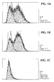
- Figure 1 sets forth FACS scans depicting serotonin induction of Ca ++ mobilization on division arrested cells, which are set forth in example 1.
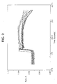
- Figure 2 shows the result of experiments, described in detail in example 2. It shows carbachol induced, Ca 2+ mobilization, observed via changes in fluorescent emission ratios of Fura2 loaded into cells that were induced.
- Figure 3 summarizes the result of experiments set forth in example 3, involving cells where division was arrested by irradiation, and which were treated with isoproterenol and reporter gene changes were measured.
- the invention relates to a method for determining the effect of a substance of interest on a cell, wherein said cell is an eukaryotic cell and wherein either the mitotic or meiotic cycle of said cell has been stopped and cellular division can no longer take place, the method comprising the steps:
- NIH3T3 cells were stably transfected with cDNA encoding the known receptor "5HT2c" using standard methods. For information on the receptor, see Julius, et al., Proc. Natl. Acad. Sci USA 87(3):928-32 (1990 ). After the transfection, and a preliminary screen to make sure that the transfection was successful, NIH3T3 cells which expressed the receptor were grown in DMEM, with L-glutamine without NaPyruvate, and with L-glucose (4500mg/L), together with 1X penicillin-streptomycin solution and 10% (v/v) fetal bovine serum (FBS). This cell line is referred to as "NIH3T3-POP.”
- Mitomycin C is well known for its ability to arrest cell growth by blocking microtubule mobility, thereby arresting cell division.
- the cells were frozen, 2.5 hours after treatment, using standard protocols.
- Cells were loaded with Indo-1 AM dye by adding 2 mg/ml of Indo-1 AM stock, in DMSO, to the cell suspensions to a final concentration of 10 ug/1.2mls. Cells were exposed to Indo-1 AM for seven (7) minutes at room temperature and then diluted up to a final volume of 10 mls with Indo-1 loading buffer and pelleted.
- Cells were resuspended in a buffer and loaded into a syringe. Cells were injected continuously into the flow cytometer to be sampled and to provide a baseline value. A substance such as an antagonist can be used to pretreat the cells, with a second material, such as an agonist being injected continuously and simultaneously through, e.g., a second syringe, via a correcting means, such as a standard Y-connector. Cells are then exposed to the test substance, analyzed, and changes from baseline measured.
- a substance such as an antagonist can be used to pretreat the cells, with a second material, such as an agonist being injected continuously and simultaneously through, e.g., a second syringe, via a correcting means, such as a standard Y-connector. Cells are then exposed to the test substance, analyzed, and changes from baseline measured.
- cell line M1WT3 (ATCC CRL 1985) was chosen. This cell line expresses muscaranic acetylcholine receptor. Experiments were designed to determine if know agonists induce Ca 2+ mobilization in these cells after growth arrest.
- M1WT3 cells were grown and treated, as set forth in example 1, supra . They were also treated with mitomycin C as described, and controls were prepared in exactly the same way.
- a Ca 2+ imaging device was used to inspect Ca 2+ mobilization visually. To do this, cells were loaded with "Fura-2" a fluorescent, Ca 2+ indicator. Cells were excited at 340 and 380nm wavelengths, and emission ratios were monitored at 450nm. Carbachol induced, Ca 2+ mobilization was observed, in M1WT3 cells, via changes in fluorescent emission ratios, rising from approximately 0.94 to 1.03 in individual cells ( Figure 2 ). Comparing to cells not division-arrested, Mitomycin C pretreatment had no negative effect on the carbachol induced Ca 2+ response of the cells (data not shown). It was shown that a larger percentage of division arrested cells responded to carbachol (data not shown), which is consistent with a more uniform cell population, resulting from the arrested division.
- G-protein coupled receptors elicit different pathways, depending on the G protein to which they couple.
- the experiments which follow were designed to show that seven-transmembrane receptors other than Gq coupled receptors function normally in growth arrested cells.
- HEK293 cells which had been transfected stably and overexpressed the ⁇ 2 adrenergic receptor (" ⁇ 2AR") (which is a Gs coupled, seven-transmembrane receptor) was used, in experiments designed to determine if isoproterenol would induce CRE-SEAP reporter activity.
- ⁇ 2AR ⁇ 2 adrenergic receptor
- the stably transfected cells were grown in DMEM with 10% FBS, and then were transiently transfected with a reporter plasmid, i.e., pCRE-SEAP.
- the cells were treated with mitomycin for 2 hours, 24 hours past transfection.
- the plasmid contained a CRE promoter, activity of which is elevated by cAMP, and which expresses higher levels of secreted, alkaline phosphatase ("SEAP") upon activation of GPCRs, which use cAMP as a second messenger.
- SEAP alkaline phosphatase
- ⁇ 2ARs were activated with 100 ⁇ M of isoproterenol , and the level of SEAP activity was measured using commercially available products, 24 hours later.
- ⁇ 2 adrenergic receptor expressing cells responded to overnight treatment with 100 ⁇ M of isoproterenol, as measured by increased SEAP activity (approximately 25%).
- Growth arrested, ⁇ 2AR expressing cells displayed much lower background SEAP activity, which may be attributable to mitomycin C toxicity.
- overnight stimulation with isproternol induced a 2.5 fold increase in SEAP activity, thus demonstrating that growth arrested cells still conduct largely intact signal transduction pathways down to the transcription response, and enzyme reporter assays can be carried out in division arrested cells.
- Miyotmycin treatment caused significant toxicity to the ⁇ 2AR expressing cells.
- a different method for arresting cell division i.e., gamma irradiation.
- Cells were either non-irradiated, and served as a control, or were irradiated at does ranging from 2Gy(Gray) or 8Gy. They were then treated with 100uM isoproterenol, as described, supra . Reporter SEAP activity was measured, and compared to baseline activity (i.e., cells not treated with isoproterenol).
- the foregoing discussion sets forth features of the invention, which relates, inter alia, to a method for screening for a substance of interest.
- the method comprises contacting the substance of interest with a sample of division arrested cells, and determining interaction between the division arrested cells and the substance of interest to determine one or more properties thereof. In this way, one can determine whether a substance of interest has efficacy as an antagonist, an agonist, an inhibitor, a stimulator, or a modulator of cells, is toxic to the cells, and so forth.
- division arrested as used herein is meant that the cells being used have been treated, by means know in the art, so that either their mitotic or meiotic cycle has been stopped, and cellular division can no longer take place.
- any prokaryotic or eukaryotic cell may be used, in any cell based assay to determine the effect of a substance of interest on a cell type of interest.
- cells which express a particular molecule or molecules naturally, or cells transfected or transformed to express the molecule or molecules of interest may be used.
- eukaryotic cells such as NIH3T3 cells, HEK293 cells, CHO cells, and so forth
- Other types of nucleic acid molecules may be used, including DNA encoding any protein of interest, RNA and antisense molecules, including antisense DNA and antisense RNA.
- Many methods are known for introducing the nucleic acid molecules to the host cells, such as via the use of recombinant viral vectors or other vectors that are adapted for the cell type of interest.
- the cells may be cells which have been transduced with a molecule of interest, such as a peptide, and/or a protein containing a molecule such as a protein glycoprotein, lipoprotein, and so forth.
- the cell to be used is transformed or transfected with a nucleic acid molecule which performs a reporter function, such as SEAP, luciferase, green fluorescent protein, and so forth.
- a reporter function such as SEAP, luciferase, green fluorescent protein, and so forth.
- a reporter function such as SEAP, luciferase, green fluorescent protein, and so forth.
- the substance of interest may be tested directly, or it may be tested in a competitive assay, using a known antagonist or agonist of a receptor or other molecule of interest.
- an antibody can be tested for its efficacy as an antagonist of a molecule by mixing it with a known ligand for the molecule, and comparing a property of the target molecule with and without the presence of the antibody.
- the converse of this type of assay can also be carried out, where the antibody function is know, and the molecule of interest is not an antibody, or is in fact a second antibody.
- kits useful in screening for a substance of interest.
- kits may contain, e.g., a separate portion of each of (i) a substance which causes arrested division of a cell, and a substance known to interact with a target molecule of interest.
- the kit may also include cells transformed or transfected with the molecule of interest, or cells to be transformed or transfected and the agent used for transformation/transfection (e.g., an expression vector), or cells naturally expressing the target molecule of interest or other items. All of the variations set forth supra can be used in these kits. In cases where an additional function of the cells is to be described, that material can be included in the kit as well.
Landscapes
- Health & Medical Sciences (AREA)
- Life Sciences & Earth Sciences (AREA)
- Engineering & Computer Science (AREA)
- Immunology (AREA)
- Chemical & Material Sciences (AREA)
- Biomedical Technology (AREA)
- Molecular Biology (AREA)
- Urology & Nephrology (AREA)
- Hematology (AREA)
- Analytical Chemistry (AREA)
- Bioinformatics & Cheminformatics (AREA)
- Biotechnology (AREA)
- Medicinal Chemistry (AREA)
- Microbiology (AREA)
- Physics & Mathematics (AREA)
- Cell Biology (AREA)
- General Health & Medical Sciences (AREA)
- Biochemistry (AREA)
- General Physics & Mathematics (AREA)
- Toxicology (AREA)
- Pathology (AREA)
- Food Science & Technology (AREA)
- Tropical Medicine & Parasitology (AREA)
- Organic Chemistry (AREA)
- Proteomics, Peptides & Aminoacids (AREA)
- Wood Science & Technology (AREA)
- Zoology (AREA)
- Genetics & Genomics (AREA)
- Biophysics (AREA)
- General Engineering & Computer Science (AREA)
- Measuring Or Testing Involving Enzymes Or Micro-Organisms (AREA)
- Investigating Or Analysing Biological Materials (AREA)
Abstract
Description
- This invention relates to methods for screening for substances of interest. More particularly, it relates to screening assays which utilize cells in a division arrested state which, nonetheless, function effectively in assays where dividing cells would normally be used. The advantages of the system will be seen in the disclosure which follows.
- Cell based screening assays are tools well known to biologists. In the assays, one investigates compounds of interest to determine, e.g. if the compounds modulate one or more biological processes of interest.
- Among the cell based systems which are used are those which measure reporter activity, calcium activity assay, and so forth. These, and all other cell based assays are encompassed by the invention.
- One area where cell based screening assays have become widely accepted is the high throughput analysis of materials for use as pharmaceuticals. These assays are useful and desirable because compounds which are identified initially in biochemical assays have been known to fail as drug candidates later in the development process. The reasons for this are many. In some cases, the compound does not permeate the cell readily. In others, target binding capability is not predictive of the target modulating function, a feature that is, ultimately, a requirement of drug functions. Cell based screening assays are useful in that they address a number of problems associated with animal model testing (e.g., high expanse, intensive labor, long assay period). High throughput cell based screening assays can be scaled up via technologies such as "FLIPR," "Leedseeker," "VIPR," and fluorescent, high speed cell-imaging.
- In addition to assays such as those discussed supra, other commonly used, cell based assays involve enzyme-reporter systems, cell activity assays with a fluorescent or colorimetric readout, and so forth. An example of such an assay is a Ca2+ mobilization assay to measure G-protein coupled receptor activity with the dye "Fluo-4."
- In addition to the advantages set forth supra, cell based assays have a distinct advantage in that they permit the user to determine the functional outcome of the use of compounds. Properly designed assays also permit the artisan to select against the toxic compounds, when screening for active ones.
- Carrying out high throughput, cell based assays present unique challenges to users. Unlike pure biochemical reagent like enzymes, proteins, and membrane receptor preparations, cells are live dynamic entities. Preparation and cultivation have to be tied to the actual screening process.
- Actively managed cell culture cycles have a recovery phase, when they are split from near confluent cultures, followed by an early log growth phase, then a mid log, and a late log phase, leading to a stationary phase if the culture is allowed to become confluent. Variances of the cellular processes and protein components at these different stages of growth and replication occur constantly during these cell cultures as cells cycle through mitosis. These variances must be expected to affect biological assays, and be a factor in the common phenomenon of variability in high throughput assays. One, but by no means the only example of this, relates to the length of time over which assays are run. There is generally an 8-36 hour period following the seeding of cells during which the assay is run. The cells in the particular culture go through different phases of their culture cycle during this time, and it is not usual for the cells to be at the same point in the cycle at the same time.
- Further, the miniaturization of cell based screening assays is progressing, with smaller and smaller numbers of cells being used. As this occurs, sensitivity of the assay to variability increases rapidly and dramatically.
- The critical factors of a good cell based screening assay are (i) a well validated target, (ii) a sensitive readout, and (iii) extremely high consistency of the cells that are used. The invention which is set forth in the disclosure which follows addresses this third issue. The consistent performance of the cells in an assay can be greatly affected by changes in the level of target expression as a result of increasing cell passage number. In addition, a well characterized population of cells can be division arrested, cryopreserved as a cell bank, and plated for an assay without any additional passaging of the cells. In fact, division arrested cells may be plated and used over a period of up to five (5) days without any further handling of the cells and without a significant change in cell based responses.
- Division arrested cells have been used in the art. Exemplary of this are Cho, et al, Biochem. Mol. Biol. Int. 42(5):949-56 (1997); Yao, et al, Mol. Pharmacol 57(3):422-30 (1997); Fueger, et al, J. Nucl. Med. 42(12):1856-62 (2001); Muller, et al, J. Exp. Med. 188(11):2033-45 (1998), and Kharbanda, et al, Nature 376 (6543):785-8 (1995). Review of these, as well as other references will show that these studies concern characterization of the cells, rather than their use in assays of the type described herein, such as drug discovery, screening assays, and/or signal transduction assays, especially when these are carried out on a large scale, high throughput basis as is required for industrial application.
- These, as well as other features of the invention will be elaborated upon in the disclosure which follows.
-
Figure 1 sets forth FACS scans depicting serotonin induction of Ca++ mobilization on division arrested cells, which are set forth in example 1. -
Figure 2 shows the result of experiments, described in detail in example 2. It shows carbachol induced, Ca2+ mobilization, observed via changes in fluorescent emission ratios of Fura2 loaded into cells that were induced. -
Figure 3 summarizes the result of experiments set forth in example 3, involving cells where division was arrested by irradiation, and which were treated with isoproterenol and reporter gene changes were measured. - The invention relates to a method for determining the effect of a substance of interest on a cell, wherein said cell is an eukaryotic cell and wherein either the mitotic or meiotic cycle of said cell has been stopped and cellular division can no longer take place, the method comprising the steps:
- (a) contacting the substance of interest with said division-arrested cell, and
- (b) determining a change in said division-arrested cell as in independent claim 1.
- Subject-matter of claims 2 to 14 relates to preferred embodiments of the invention.
- These experiments describe screening assays designed to measure the induction of Ca2+ mobilization by serotonin in growth arrested, NIH3T3 cells.
- NIH3T3 cells were stably transfected with cDNA encoding the known receptor "5HT2c" using standard methods. For information on the receptor, see Julius, et al., Proc. Natl. Acad. Sci USA 87(3):928-32 (1990). After the transfection, and a preliminary screen to make sure that the transfection was successful, NIH3T3 cells which expressed the receptor were grown in DMEM, with L-glutamine without NaPyruvate, and with L-glucose (4500mg/L), together with 1X penicillin-streptomycin solution and 10% (v/v) fetal bovine serum (FBS). This cell line is referred to as "NIH3T3-POP."
- In order to arrest the growth of the cells, they were exposed to 10µg/ml of mitomycin C, for 2.5 hours. Mitomycin C is well known for its ability to arrest cell growth by blocking microtubule mobility, thereby arresting cell division. The cells were frozen, 2.5 hours after treatment, using standard protocols.
- As a control, cells which were not exposed to mitomycin C were frozen using the same procedure. Samples of both treated and control cells were thawed, using standard methods, and plated for 24 hours, prior to harvest. Harvesting was accomplished by adding 7ml of an enzyme free, cell dissociation solution, (e.g., EnzymeFree Cell Dissociation Solution, Specialty Media, catalog number S-014) or with 0.5 mM EDTA, to cells. The number of cells used in each experiment was 4x10⁵ cells/ml or about 4-4.5x10⁶ total cells. Such solutions are widely available, and are well known to the skilled artisan. This treatment dissociated cells from the flask, and aggregates were then broken up by repeated pipetting of the suspensions, up and down in the flasks, so as to provide a good proportion of single cells, as this is necessary for the FACS analysis which followed.
- Cells were pelleted, and then resuspended in 6ml of Indo-1 loading buffer.
- Cells were loaded with Indo-1 AM dye by adding 2 mg/ml of Indo-1 AM stock, in DMSO, to the cell suspensions to a final concentration of 10 ug/1.2mls. Cells were exposed to Indo-1 AM for seven (7) minutes at room temperature and then diluted up to a final volume of 10 mls with Indo-1 loading buffer and pelleted.
- In order to measure and to analyze Ca2+ mobilization, a commercially available cell sorter was used. Excitation was set at 360nm, and emissions were set at both 400 (±5 nm) and 500 (±20)nm. The emissions were monitored simultaneously, and the emission ratio at 400nm/500nm was used to report the intracellular rise of Ca2+ concentration. Untreated cells were used to set a baseline ratio.
- Cells were resuspended in a buffer and loaded into a syringe. Cells were injected continuously into the flow cytometer to be sampled and to provide a baseline value. A substance such as an antagonist can be used to pretreat the cells, with a second material, such as an agonist being injected continuously and simultaneously through, e.g., a second syringe, via a correcting means, such as a standard Y-connector. Cells are then exposed to the test substance, analyzed, and changes from baseline measured.
- The data of the experiment are summarized in
Figure 1 . Cells which had been division arrested (POP Ar 10uM 5HT panel inFigure 1 ) responded in a manner similar to those which were not (POP Gr 10uM 5HT panel inFigure 1 ). In brief, "% positive" was calculated by taking the percentage in the positive region (i.e., cells demonstrating an increase in intracellular Ca++ concentration that is greater than the baseline Ca++ concentration of unstimulated cells) The "% positive" value was used to measure the extent of activation induced by serotonin. - Actively growing NIH3T3 cells which expressed 5HT2c showed a 50% Ca2+ response, while growth arrested cells showed a comparable 61.9% Ca response. Control experiments indicated that the response was receptor specific. To elaborate, parental NIH3T3 cells which were not transfected showed no fluorescence change when treated with serotonin (data not shown), while pretreatment with antagonist (10 uM Mesulergine or "MES" in the table) blocked serotonin induction completely. (POP Ar 10uM 5HT, 10uM Mes panel in
Figure 1 ). Only 0.1% of Mesulergine pretreated cells responded to serotonin in Ca++ mobilization. - These experiments were carried out to determine if the principles proven in example 1, supra, were applicable to other receptors, such as other Gq coupled receptors. To test this, cell line M1WT3 (ATCC CRL 1985) was chosen. This cell line expresses muscaranic acetylcholine receptor. Experiments were designed to determine if know agonists induce Ca2+ mobilization in these cells after growth arrest.
- M1WT3 cells were grown and treated, as set forth in example 1, supra. They were also treated with mitomycin C as described, and controls were prepared in exactly the same way.
- A Ca2+ imaging device was used to inspect Ca2+ mobilization visually. To do this, cells were loaded with "Fura-2" a fluorescent, Ca2+ indicator. Cells were excited at 340 and 380nm wavelengths, and emission ratios were monitored at 450nm. Carbachol induced, Ca2+ mobilization was observed, in M1WT3 cells, via changes in fluorescent emission ratios, rising from approximately 0.94 to 1.03 in individual cells (
Figure 2 ). Comparing to cells not division-arrested, Mitomycin C pretreatment had no negative effect on the carbachol induced Ca2+ response of the cells (data not shown). It was shown that a larger percentage of division arrested cells responded to carbachol (data not shown), which is consistent with a more uniform cell population, resulting from the arrested division. - The results reported supra suggested that division arrested cells may be more consistent, over time, in screening assays. This was tested via a time-course cell imaging experiment. Division arrested and frozen cells were imaged as stimulated by serotonin 1, 3, 5 and 7 days following thawing of cells. Comparable percentage, and extent of Ca2+ response were found, as measured by a Fura 2 fluorescence 340/380 ratio change, on these different days, while significant changes in Ca2+ levels were found in growing populations on these different days.
- It is well known that G-protein coupled receptors elicit different pathways, depending on the G protein to which they couple. The experiments which follow were designed to show that seven-transmembrane receptors other than Gq coupled receptors function normally in growth arrested cells.
- To do this, HEK293 cells which had been transfected stably and overexpressed the β2 adrenergic receptor ("β2AR") (which is a Gs coupled, seven-transmembrane receptor) was used, in experiments designed to determine if isoproterenol would induce CRE-SEAP reporter activity.
- The stably transfected cells were grown in DMEM with 10% FBS, and then were transiently transfected with a reporter plasmid, i.e., pCRE-SEAP. The cells were treated with mitomycin for 2 hours, 24 hours past transfection. The plasmid contained a CRE promoter, activity of which is elevated by cAMP, and which expresses higher levels of secreted, alkaline phosphatase ("SEAP") upon activation of GPCRs, which use cAMP as a second messenger.
- Twenty-four hours after mitomycin C treatment, β2ARs were activated with 100µM of isoproterenol, and the level of SEAP activity was measured using commercially available products, 24 hours later.
- Actively growing β2 adrenergic receptor expressing cells responded to overnight treatment with 100µM of isoproterenol, as measured by increased SEAP activity (approximately 25%). Growth arrested, β2AR expressing cells displayed much lower background SEAP activity, which may be attributable to mitomycin C toxicity. Notwithstanding the lower background levels, overnight stimulation with isproternol induced a 2.5 fold increase in SEAP activity, thus demonstrating that growth arrested cells still conduct largely intact signal transduction pathways down to the transcription response, and enzyme reporter assays can be carried out in division arrested cells.
- Miyotmycin treatment caused significant toxicity to the β2AR expressing cells. As such, a different method for arresting cell division was tested, i.e., gamma irradiation.
- Cells were either non-irradiated, and served as a control, or were irradiated at does ranging from 2Gy(Gray) or 8Gy. They were then treated with 100uM isoproterenol, as described, supra. Reporter SEAP activity was measured, and compared to baseline activity (i.e., cells not treated with isoproterenol).
- The results are depicted in
Figure 3 . Cells which were treated with 4gry or more gamma irradiation showed far greater division arrest, with no noticeable cell proliferation for about a week. These cells show normal cell morphology and, when stimulated with isoproterenol, SEAP responses ranged from 4 to 6 fold over the baseline levels. These results were comparable to the cells that had not been division arrested, which responded 5.2 fold over baseline upon stimulation with isoproterenol. - The foregoing discussion sets forth features of the invention, which relates, inter alia, to a method for screening for a substance of interest. The method comprises contacting the substance of interest with a sample of division arrested cells, and determining interaction between the division arrested cells and the substance of interest to determine one or more properties thereof. In this way, one can determine whether a substance of interest has efficacy as an antagonist, an agonist, an inhibitor, a stimulator, or a modulator of cells, is toxic to the cells, and so forth.
- By division arrested as used herein is meant that the cells being used have been treated, by means know in the art, so that either their mitotic or meiotic cycle has been stopped, and cellular division can no longer take place. There are many chemical, radiological, and other methods which can be used to accomplish the arrest of cellular division, and these need not be reiterated here, as the crux of the invention is not the act of causing the arrest of cell division, but the use of the arrested cells in assays as described.
- While it is possible to treat the cells in additional ways to arrest one or more additional biological processes, this is not necessary and, indeed, in many applications it will be desirable to have the cells function normally in all other ways but for the arrest in cell division.
- It will be seen by the skilled artisan that the type of cell used may vary. Any prokaryotic or eukaryotic cell may be used, in any cell based assay to determine the effect of a substance of interest on a cell type of interest.
- The nature of the cell type used will depend upon the particular type of assay to be run. To this end, cells which express a particular molecule or molecules naturally, or cells transfected or transformed to express the molecule or molecules of interest may be used. For example eukaryotic cells such as NIH3T3 cells, HEK293 cells, CHO cells, and so forth, can all be used. Other types of nucleic acid molecules may be used, including DNA encoding any protein of interest, RNA and antisense molecules, including antisense DNA and antisense RNA. Many methods are known for introducing the nucleic acid molecules to the host cells, such as via the use of recombinant viral vectors or other vectors that are adapted for the cell type of interest. Further, the cells may be cells which have been transduced with a molecule of interest, such as a peptide, and/or a protein containing a molecule such as a protein glycoprotein, lipoprotein, and so forth.
- In one embodiment of the invention, the cell to be used is transformed or transfected with a nucleic acid molecule which performs a reporter function, such as SEAP, luciferase, green fluorescent protein, and so forth. It is well known that one of ordinary skill in the art can transform or transfect cells with expression vectors which require activation of, e.g., a receptor to cause the promoter to which the reporter molecule is operably linked, to function. Since activation of the receptor molecule depends upon ligand receptor interaction, one can determine the effect of a putalive ligand or "anti-ligand" by measuring the reporter molecule function, and comparing it to a control.
- Of course, it will be clear to the skilled artisan that it is also possible to measure receptor function directly, as was shown by the examples, supra. There are legions of receptors that are known, as is their effect when linked to a ligand molecule. Determination of one or more of these functions can be used as a determination of the effect of a substance of interest.
- The substance of interest may be tested directly, or it may be tested in a competitive assay, using a known antagonist or agonist of a receptor or other molecule of interest. For example, an antibody can be tested for its efficacy as an antagonist of a molecule by mixing it with a known ligand for the molecule, and comparing a property of the target molecule with and without the presence of the antibody. The converse of this type of assay can also be carried out, where the antibody function is know, and the molecule of interest is not an antibody, or is in fact a second antibody.
- The features of this disclosure also afford the user a kit useful in screening for a substance of interest. Such kits may contain, e.g., a separate portion of each of (i) a substance which causes arrested division of a cell, and a substance known to interact with a target molecule of interest. The kit may also include cells transformed or transfected with the molecule of interest, or cells to be transformed or transfected and the agent used for transformation/transfection (e.g., an expression vector), or cells naturally expressing the target molecule of interest or other items. All of the variations set forth supra can be used in these kits. In cases where an additional function of the cells is to be described, that material can be included in the kit as well.
Claims (14)
- A method for determining the effect of a substance of interest on a cell, wherein said cell is an eukaryotic cell and wherein either the mitotic or meiotic cycle of said cell has been stopped and cellular division can no longer take place, the method comprising the steps:(a) contacting the substance of interest with said division-arrested cell, and(b) determining a change in said division-arrested cell.
- The method of claim 1, wherein said cell has been transformed or transfected with a nucleic acid molecule which expresses a protein of interest.
- The method of claim 2, wherein said cell has been transformed or transfected with a recombinant virus, or has been transformed or transfected with RNA, or has been transformed or transfected with an antisense nucleic acid molecule.
- The method of claim 1, wherein said cell has been transduced with a protein, peptide, or protein-containing molecule.
- The method of claim 1, wherein said cell is a NIH3T3 cell, a M1WT3 cell, a HEK293 cell or a CHO cell.
- The method of claim 1, wherein said method comprises measuring induction of Ca2+ mobilization.
- The method of claim 1, wherein said method comprises measuring activity of a G-protein coupled receptor.
- The method of claim 1, wherein said cell has been transformed or transfected to produce 5HT2c.
- The method of claim 1, wherein said cell expresses a G-protein coupled receptor, or said cell expresses ion channels or said cell expresses a ß2 adrenergic receptor.
- The method of claim 9, wherein said G-protein coupled receptor is muscaranic acetylcholine receptor.
- The method of claim 1, wherein said cell has been transformed or transfected with a nucleic acid molecule which encodes a reporter molecule.
- The method of claim 11, wherein said nucleic acid molecule is activated via activation of a molecule expressed in said cell.
- The method of claim 11, wherein said reporter molecule is luciferase, or green fluorescent protein.
- The method of claim 1, said method comprising a competitive assay wherein said substance of interest is admixed with a compound known to affect to said cell, and determining any change in the known affect as a determination that said substance of interest affects said cell.
Applications Claiming Priority (3)
| Application Number | Priority Date | Filing Date | Title |
|---|---|---|---|
| US10/251,467 US7045281B2 (en) | 2002-09-20 | 2002-09-20 | Method for using division arrested cells in screening assays |
| US251467 | 2002-09-20 | ||
| PCT/US2003/027537 WO2004027374A2 (en) | 2002-09-20 | 2003-09-04 | Method for using division arrested cells in screening assays |
Publications (3)
| Publication Number | Publication Date |
|---|---|
| EP1581645A2 EP1581645A2 (en) | 2005-10-05 |
| EP1581645A4 EP1581645A4 (en) | 2006-11-29 |
| EP1581645B1 true EP1581645B1 (en) | 2008-08-06 |
Family
ID=31992746
Family Applications (1)
| Application Number | Title | Priority Date | Filing Date |
|---|---|---|---|
| EP03751966A Expired - Lifetime EP1581645B1 (en) | 2002-09-20 | 2003-09-04 | Method for using division arrested cells in screening assays |
Country Status (8)
| Country | Link |
|---|---|
| US (7) | US7045281B2 (en) |
| EP (1) | EP1581645B1 (en) |
| JP (1) | JP4317922B2 (en) |
| AT (1) | ATE403739T1 (en) |
| AU (1) | AU2003270073B2 (en) |
| CA (1) | CA2499621A1 (en) |
| DE (1) | DE60322743D1 (en) |
| WO (1) | WO2004027374A2 (en) |
Families Citing this family (5)
| Publication number | Priority date | Publication date | Assignee | Title |
|---|---|---|---|---|
| US7045281B2 (en) * | 2002-09-20 | 2006-05-16 | Cell & Molecular Technologies, Inc. | Method for using division arrested cells in screening assays |
| JP2006507817A (en) * | 2002-10-04 | 2006-03-09 | ニューテクバイオ・リミテッド | Gene reporter assays, kits, and cells for determining the presence and / or level of molecules that activate cell surface protein signaling activity |
| ES2641271T3 (en) * | 2006-10-30 | 2017-11-08 | Le Centre National De La Recherche Scientifique (Cnrs) | Test gene, kit and cell assay with improved sensitivity and / or specificity to determine the level of TNF-alpha |
| ES2409356T3 (en) * | 2007-10-30 | 2013-06-26 | Le Centre National De La Recherche Scientifique (Cnrs) | Method of conducting an assay to neutralize antibodies |
| WO2009111572A2 (en) * | 2008-03-04 | 2009-09-11 | Le Centre Nationale De La Recherche Scientifique | Cell, method and kit for conducting an assay for neutralizing antibodies |
Family Cites Families (7)
| Publication number | Priority date | Publication date | Assignee | Title |
|---|---|---|---|---|
| US5998136A (en) * | 1996-08-19 | 1999-12-07 | Arcaris, Inc. | Selection systems and methods for identifying genes and gene products involved in cell proliferation |
| US20010046497A1 (en) * | 2000-04-21 | 2001-11-29 | Zhang Fang Liang | G-protein coupled receptor and methods |
| US6897067B2 (en) * | 2000-11-03 | 2005-05-24 | Regents Of The University Of Michigan | Surface transfection and expression procedure |
| EP1330516B1 (en) | 2000-11-03 | 2007-01-17 | The Regents Of The University Of Michigan | Surface transfection and expression procedure |
| CA2437576A1 (en) | 2001-02-15 | 2002-08-22 | Kosan Biosciences, Inc. | Method for evaluating therapeutic efficacy |
| US6723527B2 (en) * | 2001-10-26 | 2004-04-20 | Board Of Regents, The University Of Texas System | Methods for determining toxicity reversing agents |
| US7045281B2 (en) * | 2002-09-20 | 2006-05-16 | Cell & Molecular Technologies, Inc. | Method for using division arrested cells in screening assays |
-
2002
- 2002-09-20 US US10/251,467 patent/US7045281B2/en not_active Expired - Lifetime
-
2003
- 2003-09-04 AT AT03751966T patent/ATE403739T1/en not_active IP Right Cessation
- 2003-09-04 JP JP2004537712A patent/JP4317922B2/en not_active Expired - Fee Related
- 2003-09-04 WO PCT/US2003/027537 patent/WO2004027374A2/en not_active Ceased
- 2003-09-04 EP EP03751966A patent/EP1581645B1/en not_active Expired - Lifetime
- 2003-09-04 DE DE60322743T patent/DE60322743D1/en not_active Expired - Lifetime
- 2003-09-04 CA CA002499621A patent/CA2499621A1/en not_active Abandoned
- 2003-09-04 AU AU2003270073A patent/AU2003270073B2/en not_active Expired
-
2006
- 2006-02-27 US US11/363,983 patent/US20060148007A1/en not_active Abandoned
-
2007
- 2007-07-10 US US11/775,708 patent/US20080081327A1/en not_active Abandoned
-
2008
- 2008-06-12 US US12/138,218 patent/US20080248516A1/en not_active Abandoned
-
2010
- 2010-01-11 US US12/685,223 patent/US7960101B2/en not_active Expired - Fee Related
-
2011
- 2011-05-06 US US13/102,917 patent/US20110275101A1/en not_active Abandoned
-
2012
- 2012-05-01 US US13/461,649 patent/US20120276545A1/en not_active Abandoned
Also Published As
| Publication number | Publication date |
|---|---|
| US20110275101A1 (en) | 2011-11-10 |
| US20120276545A1 (en) | 2012-11-01 |
| WO2004027374A2 (en) | 2004-04-01 |
| US7045281B2 (en) | 2006-05-16 |
| US20040058329A1 (en) | 2004-03-25 |
| US20080081327A1 (en) | 2008-04-03 |
| JP4317922B2 (en) | 2009-08-19 |
| US20060148007A1 (en) | 2006-07-06 |
| EP1581645A4 (en) | 2006-11-29 |
| AU2003270073B2 (en) | 2006-09-07 |
| US20080248516A1 (en) | 2008-10-09 |
| CA2499621A1 (en) | 2004-04-01 |
| EP1581645A2 (en) | 2005-10-05 |
| ATE403739T1 (en) | 2008-08-15 |
| WO2004027374A3 (en) | 2005-08-04 |
| US20100203561A1 (en) | 2010-08-12 |
| DE60322743D1 (en) | 2008-09-18 |
| US7960101B2 (en) | 2011-06-14 |
| AU2003270073A1 (en) | 2004-04-08 |
| JP2006500025A (en) | 2006-01-05 |
Similar Documents
| Publication | Publication Date | Title |
|---|---|---|
| RU2476891C2 (en) | Identification of molecules modulating protein-protein interaction | |
| WO2003027634A2 (en) | Identification of modulatory molecules using inducible promoters | |
| US7960101B2 (en) | Method for using division arrested cells in screening assays | |
| EP2187213B1 (en) | Methods for detecting interactions between two and more polypeptides | |
| US20030170744A1 (en) | Multiplexed analysis of cellular responses using endogenous reporter genes | |
| KR102597698B1 (en) | A method for analysis of protein-protein interaction in cell based on p-body | |
| Miret et al. | Multiplexed G-Protein–Coupled Receptor Ca2+ Flux Assays for High-Throughput Screening | |
| Lin et al. | TimeSTAMP tagging of newly synthesized proteins | |
| CA2435810A1 (en) | Cell fusion assays for the identification of antiviral compounds, and systems and kits for practicing the same | |
| AU778648B2 (en) | Cells and assays | |
| JP2005172460A (en) | Method for detecting reaction of protein and sample | |
| US20050158813A1 (en) | Universal G-protein coupled receptor reporter constructs | |
| US7435546B2 (en) | System to detect inducible translocation | |
| Lin et al. | Current Protocols in Protein Science Unit 26.5 (ms# CP-09-0158) TimeSTAMP tagging of newly synthesized proteins | |
| US20060099643A1 (en) | Method for determining the regulation of xenobiotic removal |
Legal Events
| Date | Code | Title | Description |
|---|---|---|---|
| PUAI | Public reference made under article 153(3) epc to a published international application that has entered the european phase |
Free format text: ORIGINAL CODE: 0009012 |
|
| 17P | Request for examination filed |
Effective date: 20050318 |
|
| AK | Designated contracting states |
Kind code of ref document: A2 Designated state(s): AT BE BG CH CY CZ DE DK EE ES FI FR GB GR HU IE IT LI LU MC NL PT RO SE SI SK TR |
|
| AX | Request for extension of the european patent |
Extension state: AL LT LV MK |
|
| RIC1 | Information provided on ipc code assigned before grant |
Ipc: 7C 07K 7/00 B Ipc: 7C 12N 5/02 B Ipc: 7G 01N 33/53 B Ipc: 7C 12Q 1/00 A Ipc: 7C 12P 27/00 B Ipc: 7C 12Q 1/66 B Ipc: 7C 07K 17/00 B |
|
| DAX | Request for extension of the european patent (deleted) | ||
| RIN1 | Information on inventor provided before grant (corrected) |
Inventor name: ZHONG, ZHONG Inventor name: FEDERICI, MARK Inventor name: CONG, MEI Inventor name: LIVELLI, THOMAS |
|
| REG | Reference to a national code |
Ref country code: HK Ref legal event code: DE Ref document number: 1081603 Country of ref document: HK |
|
| RIN1 | Information on inventor provided before grant (corrected) |
Inventor name: CONG, MEI Inventor name: FEDERICI, MARK Inventor name: ZHONG, ZHONG Inventor name: LIVELLI, THOMAS |
|
| RAP1 | Party data changed (applicant data changed or rights of an application transferred) |
Owner name: CELL & MOLECULAR TECHNOLOGIES INC. |
|
| A4 | Supplementary search report drawn up and despatched |
Effective date: 20061102 |
|
| RIC1 | Information provided on ipc code assigned before grant |
Ipc: C07K 7/00 20060101ALI20061026BHEP Ipc: G01N 33/53 20060101ALI20061026BHEP Ipc: C07K 17/00 20060101ALI20061026BHEP Ipc: C12Q 1/66 20060101ALI20061026BHEP Ipc: G01N 33/50 20060101ALI20061026BHEP Ipc: C12Q 1/00 20060101AFI20051101BHEP Ipc: C12N 5/02 20060101ALI20061026BHEP Ipc: C12P 27/00 20060101ALI20061026BHEP |
|
| 17Q | First examination report despatched |
Effective date: 20070412 |
|
| GRAP | Despatch of communication of intention to grant a patent |
Free format text: ORIGINAL CODE: EPIDOSNIGR1 |
|
| GRAS | Grant fee paid |
Free format text: ORIGINAL CODE: EPIDOSNIGR3 |
|
| GRAA | (expected) grant |
Free format text: ORIGINAL CODE: 0009210 |
|
| RAP1 | Party data changed (applicant data changed or rights of an application transferred) |
Owner name: INVITROGEN CORPORATION |
|
| AK | Designated contracting states |
Kind code of ref document: B1 Designated state(s): AT BE BG CH CY CZ DE DK EE ES FI FR GB GR HU IE IT LI LU MC NL PT RO SE SI SK TR |
|
| REG | Reference to a national code |
Ref country code: GB Ref legal event code: FG4D |
|
| REG | Reference to a national code |
Ref country code: CH Ref legal event code: EP |
|
| REG | Reference to a national code |
Ref country code: IE Ref legal event code: FG4D |
|
| REF | Corresponds to: |
Ref document number: 60322743 Country of ref document: DE Date of ref document: 20080918 Kind code of ref document: P |
|
| PG25 | Lapsed in a contracting state [announced via postgrant information from national office to epo] |
Ref country code: ES Free format text: LAPSE BECAUSE OF FAILURE TO SUBMIT A TRANSLATION OF THE DESCRIPTION OR TO PAY THE FEE WITHIN THE PRESCRIBED TIME-LIMIT Effective date: 20081117 Ref country code: NL Free format text: LAPSE BECAUSE OF FAILURE TO SUBMIT A TRANSLATION OF THE DESCRIPTION OR TO PAY THE FEE WITHIN THE PRESCRIBED TIME-LIMIT Effective date: 20080806 |
|
| PG25 | Lapsed in a contracting state [announced via postgrant information from national office to epo] |
Ref country code: AT Free format text: LAPSE BECAUSE OF FAILURE TO SUBMIT A TRANSLATION OF THE DESCRIPTION OR TO PAY THE FEE WITHIN THE PRESCRIBED TIME-LIMIT Effective date: 20080806 Ref country code: FI Free format text: LAPSE BECAUSE OF FAILURE TO SUBMIT A TRANSLATION OF THE DESCRIPTION OR TO PAY THE FEE WITHIN THE PRESCRIBED TIME-LIMIT Effective date: 20080806 Ref country code: SI Free format text: LAPSE BECAUSE OF FAILURE TO SUBMIT A TRANSLATION OF THE DESCRIPTION OR TO PAY THE FEE WITHIN THE PRESCRIBED TIME-LIMIT Effective date: 20080806 |
|
| PG25 | Lapsed in a contracting state [announced via postgrant information from national office to epo] |
Ref country code: BE Free format text: LAPSE BECAUSE OF FAILURE TO SUBMIT A TRANSLATION OF THE DESCRIPTION OR TO PAY THE FEE WITHIN THE PRESCRIBED TIME-LIMIT Effective date: 20080806 |
|
| PG25 | Lapsed in a contracting state [announced via postgrant information from national office to epo] |
Ref country code: BG Free format text: LAPSE BECAUSE OF FAILURE TO SUBMIT A TRANSLATION OF THE DESCRIPTION OR TO PAY THE FEE WITHIN THE PRESCRIBED TIME-LIMIT Effective date: 20081106 Ref country code: DK Free format text: LAPSE BECAUSE OF FAILURE TO SUBMIT A TRANSLATION OF THE DESCRIPTION OR TO PAY THE FEE WITHIN THE PRESCRIBED TIME-LIMIT Effective date: 20080806 Ref country code: MC Free format text: LAPSE BECAUSE OF NON-PAYMENT OF DUE FEES Effective date: 20080930 |
|
| PG25 | Lapsed in a contracting state [announced via postgrant information from national office to epo] |
Ref country code: PT Free format text: LAPSE BECAUSE OF FAILURE TO SUBMIT A TRANSLATION OF THE DESCRIPTION OR TO PAY THE FEE WITHIN THE PRESCRIBED TIME-LIMIT Effective date: 20090106 Ref country code: RO Free format text: LAPSE BECAUSE OF FAILURE TO SUBMIT A TRANSLATION OF THE DESCRIPTION OR TO PAY THE FEE WITHIN THE PRESCRIBED TIME-LIMIT Effective date: 20080806 Ref country code: CZ Free format text: LAPSE BECAUSE OF FAILURE TO SUBMIT A TRANSLATION OF THE DESCRIPTION OR TO PAY THE FEE WITHIN THE PRESCRIBED TIME-LIMIT Effective date: 20080806 Ref country code: SK Free format text: LAPSE BECAUSE OF FAILURE TO SUBMIT A TRANSLATION OF THE DESCRIPTION OR TO PAY THE FEE WITHIN THE PRESCRIBED TIME-LIMIT Effective date: 20080806 |
|
| PLBE | No opposition filed within time limit |
Free format text: ORIGINAL CODE: 0009261 |
|
| STAA | Information on the status of an ep patent application or granted ep patent |
Free format text: STATUS: NO OPPOSITION FILED WITHIN TIME LIMIT |
|
| 26N | No opposition filed |
Effective date: 20090507 |
|
| PG25 | Lapsed in a contracting state [announced via postgrant information from national office to epo] |
Ref country code: EE Free format text: LAPSE BECAUSE OF FAILURE TO SUBMIT A TRANSLATION OF THE DESCRIPTION OR TO PAY THE FEE WITHIN THE PRESCRIBED TIME-LIMIT Effective date: 20080806 |
|
| PG25 | Lapsed in a contracting state [announced via postgrant information from national office to epo] |
Ref country code: IT Free format text: LAPSE BECAUSE OF FAILURE TO SUBMIT A TRANSLATION OF THE DESCRIPTION OR TO PAY THE FEE WITHIN THE PRESCRIBED TIME-LIMIT Effective date: 20080806 |
|
| REG | Reference to a national code |
Ref country code: GB Ref legal event code: 732E Free format text: REGISTERED BETWEEN 20090903 AND 20090909 |
|
| PG25 | Lapsed in a contracting state [announced via postgrant information from national office to epo] |
Ref country code: SE Free format text: LAPSE BECAUSE OF FAILURE TO SUBMIT A TRANSLATION OF THE DESCRIPTION OR TO PAY THE FEE WITHIN THE PRESCRIBED TIME-LIMIT Effective date: 20081106 |
|
| PG25 | Lapsed in a contracting state [announced via postgrant information from national office to epo] |
Ref country code: CY Free format text: LAPSE BECAUSE OF FAILURE TO SUBMIT A TRANSLATION OF THE DESCRIPTION OR TO PAY THE FEE WITHIN THE PRESCRIBED TIME-LIMIT Effective date: 20080806 Ref country code: HU Free format text: LAPSE BECAUSE OF FAILURE TO SUBMIT A TRANSLATION OF THE DESCRIPTION OR TO PAY THE FEE WITHIN THE PRESCRIBED TIME-LIMIT Effective date: 20090207 Ref country code: LU Free format text: LAPSE BECAUSE OF NON-PAYMENT OF DUE FEES Effective date: 20080904 |
|
| PG25 | Lapsed in a contracting state [announced via postgrant information from national office to epo] |
Ref country code: TR Free format text: LAPSE BECAUSE OF FAILURE TO SUBMIT A TRANSLATION OF THE DESCRIPTION OR TO PAY THE FEE WITHIN THE PRESCRIBED TIME-LIMIT Effective date: 20080806 |
|
| PG25 | Lapsed in a contracting state [announced via postgrant information from national office to epo] |
Ref country code: GR Free format text: LAPSE BECAUSE OF FAILURE TO SUBMIT A TRANSLATION OF THE DESCRIPTION OR TO PAY THE FEE WITHIN THE PRESCRIBED TIME-LIMIT Effective date: 20081107 |
|
| PGFP | Annual fee paid to national office [announced via postgrant information from national office to epo] |
Ref country code: IE Payment date: 20100927 Year of fee payment: 8 |
|
| REG | Reference to a national code |
Ref country code: HK Ref legal event code: WD Ref document number: 1081603 Country of ref document: HK |
|
| REG | Reference to a national code |
Ref country code: IE Ref legal event code: MM4A |
|
| PG25 | Lapsed in a contracting state [announced via postgrant information from national office to epo] |
Ref country code: IE Free format text: LAPSE BECAUSE OF NON-PAYMENT OF DUE FEES Effective date: 20110904 |
|
| REG | Reference to a national code |
Ref country code: FR Ref legal event code: PLFP Year of fee payment: 14 |
|
| REG | Reference to a national code |
Ref country code: FR Ref legal event code: PLFP Year of fee payment: 15 |
|
| REG | Reference to a national code |
Ref country code: FR Ref legal event code: PLFP Year of fee payment: 16 |
|
| PGFP | Annual fee paid to national office [announced via postgrant information from national office to epo] |
Ref country code: GB Payment date: 20220929 Year of fee payment: 20 Ref country code: DE Payment date: 20220920 Year of fee payment: 20 |
|
| PGFP | Annual fee paid to national office [announced via postgrant information from national office to epo] |
Ref country code: FR Payment date: 20220921 Year of fee payment: 20 |
|
| PGFP | Annual fee paid to national office [announced via postgrant information from national office to epo] |
Ref country code: CH Payment date: 20220926 Year of fee payment: 20 |
|
| REG | Reference to a national code |
Ref country code: DE Ref legal event code: R071 Ref document number: 60322743 Country of ref document: DE |
|
| REG | Reference to a national code |
Ref country code: CH Ref legal event code: PL |
|
| REG | Reference to a national code |
Ref country code: GB Ref legal event code: PE20 Expiry date: 20230903 |
|
| PG25 | Lapsed in a contracting state [announced via postgrant information from national office to epo] |
Ref country code: GB Free format text: LAPSE BECAUSE OF EXPIRATION OF PROTECTION Effective date: 20230903 |