EP1580269A1 - Method of detecting gene mutation - Google Patents
Method of detecting gene mutation Download PDFInfo
- Publication number
- EP1580269A1 EP1580269A1 EP03810655A EP03810655A EP1580269A1 EP 1580269 A1 EP1580269 A1 EP 1580269A1 EP 03810655 A EP03810655 A EP 03810655A EP 03810655 A EP03810655 A EP 03810655A EP 1580269 A1 EP1580269 A1 EP 1580269A1
- Authority
- EP
- European Patent Office
- Prior art keywords
- base sequence
- dna
- hybridization
- labeled
- mutation
- Prior art date
- Legal status (The legal status is an assumption and is not a legal conclusion. Google has not performed a legal analysis and makes no representation as to the accuracy of the status listed.)
- Granted
Links
Images




Classifications
-
- C—CHEMISTRY; METALLURGY
- C12—BIOCHEMISTRY; BEER; SPIRITS; WINE; VINEGAR; MICROBIOLOGY; ENZYMOLOGY; MUTATION OR GENETIC ENGINEERING
- C12Q—MEASURING OR TESTING PROCESSES INVOLVING ENZYMES, NUCLEIC ACIDS OR MICROORGANISMS; COMPOSITIONS OR TEST PAPERS THEREFOR; PROCESSES OF PREPARING SUCH COMPOSITIONS; CONDITION-RESPONSIVE CONTROL IN MICROBIOLOGICAL OR ENZYMOLOGICAL PROCESSES
- C12Q1/00—Measuring or testing processes involving enzymes, nucleic acids or microorganisms; Compositions therefor; Processes of preparing such compositions
- C12Q1/68—Measuring or testing processes involving enzymes, nucleic acids or microorganisms; Compositions therefor; Processes of preparing such compositions involving nucleic acids
- C12Q1/6813—Hybridisation assays
- C12Q1/6827—Hybridisation assays for detection of mutation or polymorphism
-
- G—PHYSICS
- G01—MEASURING; TESTING
- G01N—INVESTIGATING OR ANALYSING MATERIALS BY DETERMINING THEIR CHEMICAL OR PHYSICAL PROPERTIES
- G01N33/00—Investigating or analysing materials by specific methods not covered by groups G01N1/00 - G01N31/00
- G01N33/48—Biological material, e.g. blood, urine; Haemocytometers
- G01N33/50—Chemical analysis of biological material, e.g. blood, urine; Testing involving biospecific ligand binding methods; Immunological testing
- G01N33/53—Immunoassay; Biospecific binding assay; Materials therefor
- G01N33/5308—Immunoassay; Biospecific binding assay; Materials therefor for analytes not provided for elsewhere, e.g. nucleic acids, uric acid, worms, mites
-
- G—PHYSICS
- G01—MEASURING; TESTING
- G01N—INVESTIGATING OR ANALYSING MATERIALS BY DETERMINING THEIR CHEMICAL OR PHYSICAL PROPERTIES
- G01N33/00—Investigating or analysing materials by specific methods not covered by groups G01N1/00 - G01N31/00
- G01N33/48—Biological material, e.g. blood, urine; Haemocytometers
- G01N33/50—Chemical analysis of biological material, e.g. blood, urine; Testing involving biospecific ligand binding methods; Immunological testing
- G01N33/58—Chemical analysis of biological material, e.g. blood, urine; Testing involving biospecific ligand binding methods; Immunological testing involving labelled substances
Definitions
- the present invention relates to a method of detecting a base sequence, and more particularly to a method of detecting a base sequence containing a mutation site such as a point mutation, thereby detecting a gene mutation.
- the gene polymorphism and gene mutation include, for example, a base substitution, deletion, insertion, and variations in the number of repetitive sequences, and among them, a point mutation caused by substitution of a single base makes up the overwhelming majority.
- a method of simply and quickly detecting a point mutation is indispensable for applying the outcomes of human genome research to clinical purposes.
- Typical methods include the allele specific oligonucleotide hybridization (ASO) method, allele specific amplification method, restriction enzyme digestion method, ligase chain reaction, and minisequencing method. These methods require complicated procedures including hybridization or electrophoresis after DNA amplification.
- ASO allele specific oligonucleotide hybridization
- the TaqMan method, invader assay, DNA microarray (DNA chip) assay, TOF-MASS method with the use of a mass spectrometer, and the like which have been recently developed for promoting the human genome analysis and research, are suited to deal with a large number of samples.
- these methods require high-priced, specialized instruments and cannot be easily performed at clinical laboratories.
- the SSCP method, chemical cleavage method, and DHPLC method are widely used for screening of gene mutations, and are highly effective for broad screening of unknown gene mutations; but are inadequate to reliable detection of a known mutation.
- Probes used in the ASO method has been conventionally 15 to 25 mer (see Saiki RK, Erlich HA. Detection of mutations by hybridization with sequence-specific oligonucleotide probes. In: Mutation Detection: A Practical Approach. pp. 113-129, IRL Press, Oxford, 1998). Moreover, it is known that the specificity of a labeled probe for hybridization is enhanced using an oligonucleotide that competes with the probe (see Nozari G, Rahbar S, Wallace RB. Discrimination among the transcripts of the allelic human ⁇ -globin genes ⁇ A , ⁇ S and ⁇ C using oligodeoxynucleotide hybridization probes. Gene 43: 23-28, 1986).
- An object of the present invention is to provide a method of simply and quickly detecting a gene mutation.
- the present inventors have used to achieve the present invention the findings that the use of certain primers and probes under a certain condition enables both amplification and hybridization of nucleic acids in one reaction system, and also enables a easy detection of a hybrid formed by the hybridization.
- the present invention provides the following:
- a method of detecting a base sequence which comprises the steps of: amplifying DNA containing a target base sequence to be detected having a mutation site using DNA polymerase; hybridizing the amplified DNA to a hybridization probe having a base sequence complementary to the target base sequence to be detected; and detecting a hybrid formed by the hybridization; characterized in that at least one of primers to be used in the DNA amplification is labeled with a first labeling agent so that the amplified DNA will be labeled with the first labeling agent, the hybridization probe is labeled with a second labeling agent and contained in a reaction solution for effecting the DNA amplification, the base sequence of the hybridization probe is designed not to inhibit the DNA amplification, and the hybrid is detected by affinity chromatography with the use of the first and second labeling agents.
- primers to be used in the DNA amplification is labeled with a first labeling agent so that the amplified DNA will be labeled with the first labeling agent
- the hybridization probe is labeled
- the DNA amplification is carried out, if using DNA polymerase, without any particular limitation. Any amplification methods comprising the step of synthesizing DNA with the use of DNA polymerase can be employed. Examples of the DNA amplification method include PCR, TMA, NASBA, and LAMP methods.
- primers are designed by a method known in the art depending on an amplification method to be used and a target base sequence to be detected.
- at least one of primers to be used in DNA amplification is labeled with a first labeling agent so that the amplified DNA will be labeled with the first labeling agent.
- a pair of primers are used and at least one thereof is labeled so that the amplified DNA can be labeled.
- a primer that functions at the stage of DNA synthesis in DNA amplification by the NASBA and TMA methods or at least one of inner primers in DNA amplification by the LAMP method is labeled, thereby allowing the labeling of the amplified DNA.
- primers are carried out so as not to inhibit DNA synthesis reaction.
- labeling can be carried out according to a method known in the art, and a primer is usually labeled at its 5' end.
- a labeling agent to be used in the labeling may be those to which a corresponding substance can be biospecifically bound.
- a pair of the labeling agent and the substance biospecifically bound thereto includes an antigen and an antibody, an enzyme and an inhibitor, a sugar chain and lectin, a hormone and a receptor, and a metal-binding protein and a metal element.
- a pair of digoxigenin and an anti-digoxigenin antibody and a pair of biotin and streptavidin may be used. In these pairs, either of the two may be given as a labeling agent.
- the smaller molecular weight partner is generally used as a labeling agent.
- Primers and DNA amplification conditions to be used are appropriately adjusted on the basis of type of an amplification method and a target base sequence to be detected.
- Molecular Cloning A Laboratory Manual (3rd ed.), Volume 2, Chapter 8, pp. 8.1 - 8.126, Cold Spring Harbor Laboratory Press, Cold Spring Harbor, 2001 on the PCR method; PCR Methods and Applications, 1, 25-33 (1991) on the NASBA method; and Nucleic Acids Research, Vol. 28, No. 12, pp. i-vii (2000) on the LAMP method.
- Test DNA that functions as a template in DNA amplification can be prepared from a test sample by a conventional method.
- the target base sequence to be detected is appropriately selected depending on the type of an amplification method so that the target base sequence having a mutation site can be specifically amplified.
- the mutation site contained in the target base sequence to be detected is known as a site of gene mutation or gene polymorphism.
- the mutation of the site may be a point mutation, an insertion mutation or a deletion mutation.
- Gene mutation and gene polymorphism to be analyzed by the detection method of the present invention include, but not limited to: g727t mutation observed with high frequency in Japanese patients with glycogen storage disease type Ia; a985g mutation (Lys329Glu mutation) observed with high frequency in Caucasian patients with medium-chain acyl-CoA dehydrogenase deficiency; g1691t mutation (Ser564Ile mutation) of GLDC gene observed with high frequency in Finnish patients with hyperglycinemia; gene polymorphism (CYP2C19*2, g681a) in drug metabolizing enzyme gene CYP2C19; gene polymorphism (E487K) of an aldehyde dehydrogenase 2 determining individual variations in alcohol metabolism; deltaF508 deletion mutation in the gene of cystic fibrosis transmembrane regulator protein; 1277insTATC insertion mutation in HEXA gene associated with Tay-Sachs disease; 5382insC insertion
- Glycogen storage disease type Ia is a congenital disorder of carbohydrate metabolism, inherited in an autosomal recessive manner, caused by deficiency of glucose-6-phosphatase in the glycogen metabolic pathway, and leads to an excess accumulation of glycogen mainly in the liver.
- Patients with glycogen storage disease type Ia are found to have hypoglycemia, hepatomegaly, short statue, renal damage, hyperlipidemia, hyperuricemia, or the like.
- Mutation g727t in the gene of this enzyme is a highly frequent mutation making up approximately 90% of pathogenic mutations in Japanese cases, and generates aberrant splicing of its mRNA.
- Non-ketotic hyperglycinemia is a congenital disorder of amino acid metabolism (autosomal recessive inheritance) caused by deficiency of an enzyme of the glycine cleavage system, and exhibits severe neurological symptoms including neonatal convulsion during neonatal period.
- Mutation g1691t in GLDC gene of the enzymes in the glycine cleavage system is observed with high frequency (approximately 70% of mutated genes) in Finnish patients. This mutation causes an amino acid substitution of Ser564Ile.
- Medium-chain acyl-CoA dehydrogenase deficiency is a congenital disorder of organic acid metabolism (autosomal recessive inheritance) caused by deficiency of the enzyme (medium-chain acyl-CoA dehydrogenase, MCAD) playing a key role in fatty acid ⁇ oxidation pathway, and brings about hypoglycemia and consciousness disturbance at fasting and infection. It is known that medium-chain acyl-CoA dehydrogenase deficiency is often misdiagnosed as sudden infant death syndrome or acute encephalopathy (Reye syndrome).
- Mutation a985g in the gene of this enzyme is a highly frequent mutation making up approximately 90% of pathogenic mutations in Caucasian cases, and produces an amino acid substitution of Lys329Glu. Moreover, carriers having this gene mutation are found with high frequency in Caucasian population (one in 40 people in the U.K.). In U.S.A. and European countries, the genetic diagnosis of detecting this a985g mutation is widely used for diagnosis of this disease.
- CYP2C19 gene plays a key role in the metabolism of omeprazole (inhibitor of gastric acid secretion) or the like.
- CYP2C19*2 a SNP in the gene, shows 681A>G mutation in exon 5, leading to aberrant splicing, and thus finally decreases in the metabolic activity to this drug.
- An individual having such a polymorphism needs a decreased amount of the drug to be administrated to the subject. Therefore, it is clinically advantageous to determine the genotype of a patient before the medication. This gene polymorphism is found in approximately 23% of the gene in Japanese population.
- the gene polymorphism (Glu487Lys) of aldehyde dehydrogenase 2 is a SNP, observed largely in oriental population, to determine individual variations in alcohol metabolism. Because the enzyme having the gene polymorphism is less active to slow down the metabolism of acetaldehyde generated from alcohol, an individual having this polymorphism shows a constitutional "low tolerance for alcohol". Approximately 30% in Japanese population have a heterozygote of this gene polymorphism and approximately 5% have a homozygote thereof.
- hybridization of the amplified DNA to a hybridization probe having a base sequence complementary to the target base sequence to be detected may be carried out in the same manner as general hybridization except that a particular hybridization probe is used.
- the hybridization probe used in the present invention is labeled with a second labeling agent and contained in a reaction solution for effecting the DNA amplification, and the base sequence of the hybridization probe is designed not to inhibit the DNA amplification.
- the second labeling agent is defined as described in the first labeling agent; provided that the substance used for it must be different from the first labeling agent.
- the labeling of the hybridization probe can be carried out by a method known in the art so as not to inhibit the hybridization.
- the labeling of the hybridization probe is preferably carried out at its 3' end. This is because such labeling prevents the oligonucleotide chain from being extended during the DNA amplification reaction. If the chain is extended in length, the Tm value thereof is increased and thus hybridization may occur even though there are mismatches.
- the design of the base sequence of the hybridization probe so as not to inhibit the DNA amplification can be generally selected by adjusting the chain length or the like of the hybridization probe so that the hybridization of the hybridization probe will not occur under the DNA amplification condition.
- the hybridization probe used in the present invention has a base sequence designed so as not to inhibit the DNA amplification, and can be thus previously contained in a reaction solution for effecting the DNA amplification. Therefore, the reaction solution after the DNA amplification is completed is placed directly under such a condition that the amplified DNA can be hybridized to the hybridization probe, thereby allowing the hybridization thereof.
- the chain length of the hybridization probe and the condition of hybridization thereof are appropriately selected depending on a method used in the DNA amplification.
- the chain length is selected so that the hybridization will not occur at this temperature.
- the temperature at which the hybridization occurs is not particularly limited as long as DNA amplification is not inhibited, but is preferably selected so that the generated hybrid may not be dissociated even at room temperature.
- a specific condition under which the base sequence of the probe does not inhibit the DNA amplification includes a condition that the Tm of the probe is designed to be 25 to 40°C (preferably 30 to 35°C) lower than the Tm of primers.
- the probe should be typically 10-mers to 13-mers. This is much shorter than 15-mer to 25-mer probes (see the above-mentioned non-patent reference 2) that have been conventionally used as a probe for allele specific oligonucleotide hybridization. In the context of that longer probes have been extensively used heretofore, there has been the theory that a sequence of at least about the 15th power of 4 is required for constructing a probe having specificity by combinations of four different bases in the whole genome sequence (3 billion base pairs). However, this holds true for the case where hybridization is directed toward the whole genome sequences. When hybridization is directed to a PCR-amplified DNA fragment having several hundreds of bases, such length or specificity is not considered necessary for probes that the specificity of hybridization is sufficiently maintained with the former probe as shown above.
- the hybridization probe When using the detection method of the present invention for the detection of any gene mutation or polymorphism, the hybridization probe is required to be adjusted to an optimum chain length.
- the optimum length can be determined by a standard experimentation, as described in Examples below. Because an extremely short length of a probe is usually used in the detection method of the present invention, formation of a diagnostic line is found to dramatically vary depending on the length variations by a single base. When the emergence of false positive or weak positive reaction is observed, it is preferred that a probe having a shorter or longer length than that of the probe designed based on its Tm value may be constructed to choose the most suitable one. In this respect, because a probe having a normal base sequence and a probe having a mutant base sequence are different in Tm value due to the base substitution even though they have the same chain length, each optimum chain length should be designed independently.
- the base sequence of the hybridization probe it is preferred to design the base sequence of the hybridization probe so that the mutation site will be positioned in approximately middle of the base sequence.
- Hybridization is usually carried out by increasing a temperature until double-stranded DNA is denatured, followed by gradually lowering the temperature.
- the hybridization can be carried out by only a procedure of changing the temperature of a reaction solution in which DNA amplification is completed, without the need of any other procedures.
- a temperature condition for hybridization can be programmed in addition to a temperature condition requisite to the DNA amplification, thereby effecting the amplification and the hybridization as a series of reactions, after a sample is loaded in the thermal cycler.
- a short length of the probe designed as described above provides the following three advantages: 1) the difference in Tm values between the case where there is a mismatch of a single base and the case where there is no mismatch can be rendered larger than that of a longer length of a probe, and thus the specificity of the probe can be relatively increased; 2) the hybridization temperature of the probe can be given as low as 25°C in the detection method of the present invention, although conventionally 37 to 65°C, and thus a subsequent series of procedures can be carried out at room temperature; and 3) a short length of the probe has a reduced Tm value and does not hybridize during PCR reaction, and thus the probe does not affect the PCR reaction even though it is previously mixed in the PCR reaction solution.
- This probe enables the procedures PCR ⁇ heat denaturation ⁇ hybridization to be carried out as a series of reactions, without performance of additional procedures such as the addition of a reagent during the reactions. These advantages can be similarly obtained in other DNA amplification methods with the use of an extension reaction by DNA polymerase, as in the PCR method.
- a hybrid formed by the hybridization has both the first labeling agent and the second labeling agent.
- the hybrid is detected by affinity chromatography with the use of the first and second labeling agents.
- the affinity chromatography can be carried out with a test strip constructed for this purpose.
- the detection of a hybrid by affinity chromatography with the use of two different labeling agents can be carried out according to a method known in the art, and a test strip used in this method can be constructed according to a general method.
- test strip is constructed so that a hybrid will be reacted with a substance which is capable to be specifically bound to the first labeling agent and is coupled with a labeling agent (e.g., gold colloid) to become visible when accumulated; and transferred onto a chromatography support on which a substance capable to be specifically bound to the second labeling agent is immobilized, to allow of the observation of the visible labeling agent when accumulated on that immobilization site.
- a labeling agent e.g., gold colloid
- the first labeling agent is digoxigenin
- the second labeling agent is biotin
- the labeling agent that is visible when accumulated is gold colloid.
- the following sites are placed in the order named in the migration direction of a chromatography solvent (generally, a buffer solution): an immersion site that is immersed in a chromatography solvent to provide the chromatography solvent to the strip of the chromatography support; a complex-carrying site that has a pad carrying an anti-digoxigenin antibody conjugated with gold colloid (a complex) in a manner that this antibody can be released into the chromatography solvent; a sample-applying site to which the reaction solution containing a hybrid is applied; a streptavidin-immobilized site on which a band of streptavidin is immobilized perpendicularly to the migration direction of the chromatography solvent; an antibody-immobilized site on which an antibody against the anti-digoxigenin antibody is immobilized; and an absorption site that
- This test strip is used as described below. After the reaction solution containing the hybrid is applied to the sample-applying site and the immersion site is immersed in the chromatography solvent, the test strip is removed from the chromatography solvent and left to stand. When the chromatography solvent migrates through the chromatography support by capillary action and reaches the complex-carrying site, from which the chromatography solvent containing the complex will migrate forward. When this chromatography solvent reaches the sample-applied site, the digoxigenin of the hybrid in the applied reaction solution will bind to the anti-digoxigenin antibody of the complex to form the hybrid having the gold colloid, which further migrates forward through the chromatography support by the chromatography solvent.
- the hybrid When the hybrid reaches the streptavidin-immobilized site, this hybrid will be accumulated on the streptavidin-immobilized site through the binding of biotin and streptavidin; consequently a visible signal shall be seen when the hybrid is present.
- the complex that has passed through the streptavidin-immobilized site is accumulated on the antibody-immobilized site to generate a visible signal showing that the chromatogram has proceeded normally.
- the chromatography solvent further migrating will be absorbed and held in the absorption site.
- the reaction solution for effecting DNA amplification further contains, along with the hybridization probe, an unlabeled oligonucleotide (hereinafter, also referred to as a "competing probe") having a base sequence different in a single base at the position of the point mutation from the base sequence of the labeled hybridization probe, in an amount sufficient to enhance the specificity of the hybridization of the amplified DNA to the labeled hybridization probe.
- an unlabeled oligonucleotide hereinafter, also referred to as a "competing probe” having a base sequence different in a single base at the position of the point mutation from the base sequence of the labeled hybridization probe, in an amount sufficient to enhance the specificity of the hybridization of the amplified DNA to the labeled hybridization probe.
- the competing probe is designed in the same way as the hybridization probe except that it differs from the hybridization probe in a single base at the position of the point mutation.
- the length of the competing probe may be different from that of the hybridization probe.
- the amount of the competing probe sufficient to enhance the specificity of the hybridization of the amplified DNA to the labeled hybridization probe varies depending on conditions such as the target base sequence to be detected and the base sequence of the hybridization probe, whereas in principle, the competing probe may be usually contained in the range from an equal amount to 5-fold amount (molar ratio) with respect to the amount of the hybridization probe. Nevertheless, when positive reaction is significantly reduced, the omission of the competing probe, if it is confirmed not to cause false positives, may sometimes produce the best result. Because the formation of a diagnostic line is significantly affected by the chain length of the hybridization probe and the presence or absence of the competing probe, an optimum reaction condition will be relatively easily found.
- the specificity of the hybridization probe can be enhanced and non-specific hybridization can be suppressed by adding an unlabeled competing oligonucleotide in the hybridization.
- different labeling agents may be used for labeling a hybridization probe for detecting a normal base sequence and a hybridization probe for detecting a mutant base sequence, to integrate two reaction systems for detecting a normal base sequence and for detecting a mutant base sequence into one reaction system. That is, the hybridization probes for detecting a normal base sequence and for detecting a mutant base sequence can be differently labeled and mixed together in the ratio of 1:1 to integrate the reaction systems into one while these hybridization probes are allowed to compete with each other. After the reaction, using the complexes of substances which are capable of specifically binding to the labeling agents, respectively, and a labeling agent that becomes visible when accumulated, affinity chromatography is carried out to determine a genotype.
- the detection method of the present invention has the following advantages: (1) versatility: the method is based on allele specific oligonucleotide hybridization that has been widely used as a detection method for a long time, and therefore adaptable to detection of a wide range of base sequence mutations such as an insertion mutation, a substitution mutation, and a point mutation; (2) rapidity: the determination of a genotype can be carried out within 10 minutes after the amplification and hybridization reactions have been completed in a thermal cycler, and the use of a capillary-type PCR amplification device in the nucleic acid amplification also enables all steps to be completed within 1 hour, if a DNA sample is ready; and (3) simplicity: after PCR reaction, a genotype can be macroscopically determined, thereby eliminating the need for an instrument such as a gel electrophoresis device or a fluorescence detector.
- a thermal cycler for effecting PCR reaction is a general-purpose instrument for clinical examination, for example, an examination for infectious diseases, and has been already placed in many hospitals. Moreover, the reaction procedure is simple without the need for special technical skills. The above-described advantages can be also obtained when nucleic acid reactions (TMA, NASBA, LAMP, etc.) other than PCR are used.
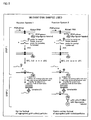
- Fig. 1 shows a reaction with the use of normal DNA as a sample.
- Reaction system 1 is a system to which is added a hybridization probe for detecting a normal base sequence
- reaction system 2 is a system to which is added a hybridization probe for detecting a mutant base sequence.
- the black circle represents a normal base
- the black triangle represents a mutant base
- Dig represents a digoxigenin label
- B represents a biotin label
- GP represents a gold particle.
- a gene site having a point mutation (the target base sequence to be detected) is amplified by PCR.
- One primer of the PCR primer pair used in this case has been previously labeled at its 5' end with digoxigenin.
- two oligonucleotides hybridization probe and competing probe
- hybridization probe and competing probe have been mixed with typical components. In this combination of the oligonucleotides, there exist two combinations for detecting a normal base sequence and for detecting a mutant base sequence.
- one is an oligonucleotide (normal probe) having a normal base sequence with the point mutation site located in the middle portion thereof and labeled with biotin at its 3' end; and the other is an unlabeled competing oligonucleotide (mutant probe) having a mutant base sequence with the point mutation site located in the middle portion thereof.
- normal probe normal probe
- mutant probe unlabeled competing oligonucleotide
- oligonucleotide having a mutant base sequence with the point mutation site located in the middle portion thereof and labeled with biotin at its 3' end and the other is an unlabeled competing oligonucleotide (normal probe) having a normal base sequence with the point mutation site located in the middle portion thereof.
- normal probe unlabeled competing oligonucleotide
- Any of these oligonucleotides are designed to be a reverse strand relative to the PCR primer labeled with digoxigenin.
- the composition of the PCR reaction solution is, for example, 50 to 100 ng of sample DNA, 10 mM Tris-HCl (pH 8.3), 50 mM KCl, 1.5 mM MgCl 2 , 250 ⁇ M each dNTPs, 1 ⁇ M PCR forward primer (labeled with digoxigenin at its 5' end), 1 ⁇ M PCR reverse primer, 600 nM hybridization probe (labeled with biotin at its 3' end), 3 ⁇ M unlabeled competing oligonucleotide, and 1.25 U Taq DNA polymerase, and the amount of the PCR reaction solution is 20 ⁇ l.
- the PCR condition is: for example, heating at 94°C for 2 minutes; 35 cycles of 98°C for 10 seconds, 55°C for 30 seconds, and 72°C for 30 seconds; followed by 72°C for 3 minutes; 98°C for 3 minutes; 65°C for 1 minute; 55°C for 1 minute; 45°C for 1 minute; 35°C for 1 minute; and 25°C for 1 minute.
- a PCR product labeled with digoxigenin is hybridized with oligonucleotide having a base sequence completely complementary to the base sequence of the PCR product.
- a PCR product labeled with digoxigenin and an oligonucleotide labeled with biotin form a hybrid (Fig. 1, reaction system 1).
- An aliquot (5 ⁇ l) of this solution is spotted onto the sample-applying site in a test strip of affinity chromatography, such as DNA detection test strip (Roche, #1-965-484), on which streptavidin is immobilized and in which an anti-digoxigenin antibody labeled with gold colloid is held in a manner that the antibody can migrate, and the bottom end of the test strip is immersed in a buffer for 5 seconds.
- the anti-digoxigenin antibody labeled with gold colloid binds to the hybrid of the PCR product labeled with digoxigenin and the oligonucleotide labeled with biotin. This hybrid is further captured by streptavidin immobilized on the test strip to form a red line that can be macroscopically observed.
- streptavidin immobilized on the test strip to form a red line that can be macroscopically observed.
- oligonucleotides for detecting a mutant base sequence is used with the DNA having a normal base sequence in combination, a PCR product labeled with digoxigenin and an unlabeled oligonucleotide form a hybrid.
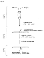
- a DNA as a sample is mixed with a reaction reagent in a PCR tube and heated/cooled with a thermal cycler according to the program to effect the DNA amplification and the formation of a hybrid (Step 1).
- An aliquot (5 ⁇ l) of the reaction solution is spotted to the sample-applying site of the test strip and the bottom end of the test strip is immersed in a buffer, followed by standing at room temperature (Step 2).
- the diagnosis is conducted on the basis of the presence or absence of the diagnostic line to determine a genotype (Step 3). Whether the affinity chromatography is normally completed or not can be confirmed by examining the presence or absence of a control line.
- the detection method of the present invention is a method capable of quickly and simply determining the presence or absence of a gene mutation with accuracy and without the use of a special device, and is suitable to conduct a genetic test in a hospital outpatient clinic or at bed side. That is, the detection method allows of a genetic diagnosis as a Point of Care. More particularly, the gene polymorphism of drug-metabolizing enzymes including CYP2C19 will be decided, which made it possible to determine on the spot whether or not a certain drug is suitable for a patient and to assist the adjustment of the dosage. In this case, an important advantage is that a test result can be obtained in a short time.
- the kit of the present invention comprises: primers for amplifying DNA containing a target base sequence to be detected having a mutation site using DNA polymerase; a hybridization probe having a base sequence complementary to the target base sequence to be detected; and a test strip for affinity chromatography; characterized in that at least one of the primers to be used in the DNA amplification is labeled with a first labeling agent so that the amplified DNA will be labeled with the first labeling agent, the hybridization probe is labeled with a second labeling agent, the base sequence of the hybridization probe is designed not to inhibit the DNA amplification, and the test strip allows of detection of a hybrid of the amplified DNA and the hybridization probe with the use of the first and second labeling agents.
- the kit of the present invention can be used for carrying out the detection method of the present invention.
- the primers, the hybridization probe, and the test strip for affinity chromatography are as described above in the detection method of the present invention.
- the kit of the present invention further comprises an unlabeled oligonucleotide (competing probe) having a base sequence different in a single base at the position of the point mutation from the base sequence of the labeled hybridization probe.
- This oligonucleotide is as described above in the detection method of the present invention.
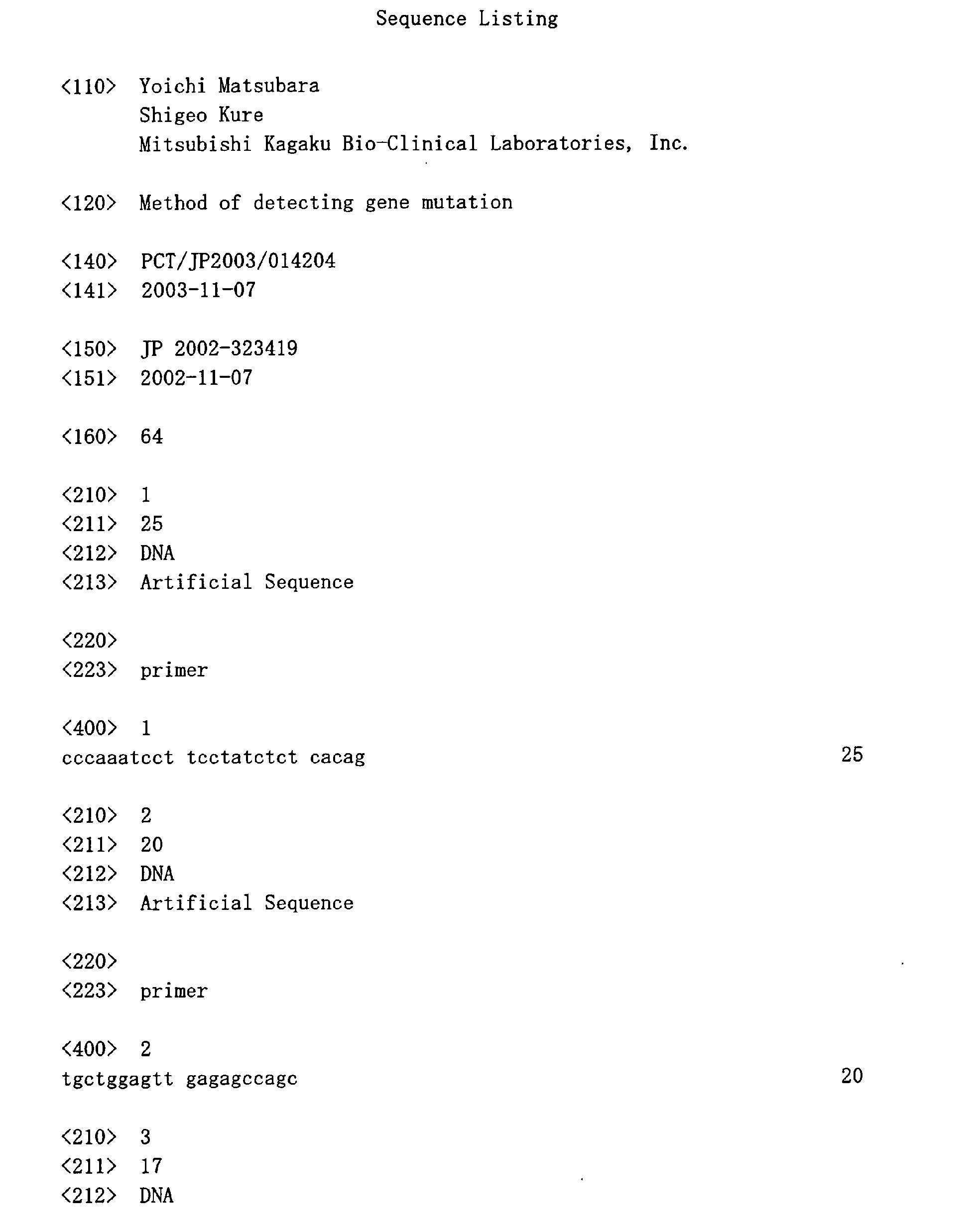
- primers listed in Table 1 were prepared on the basis of known base sequences around the mutation site.
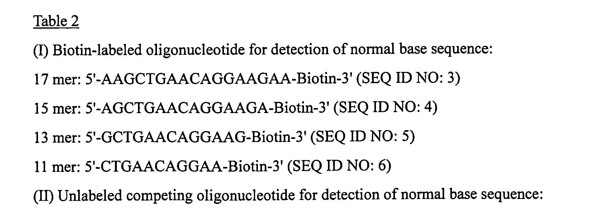
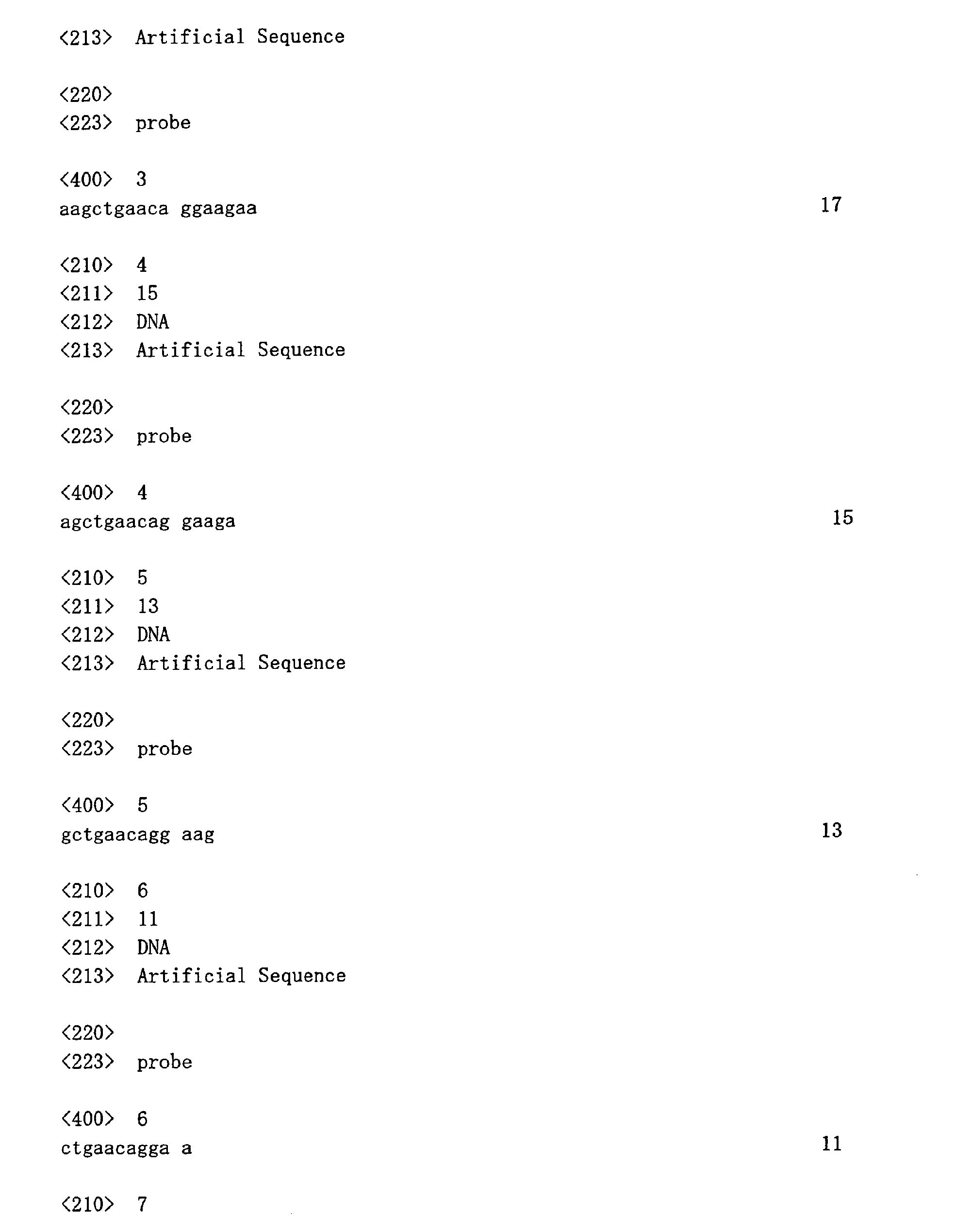
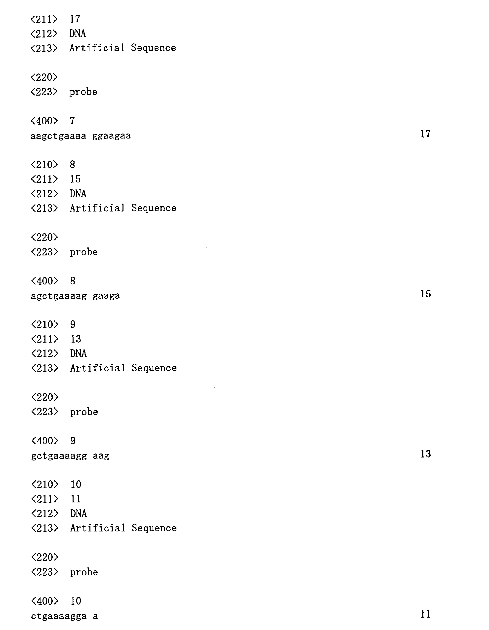
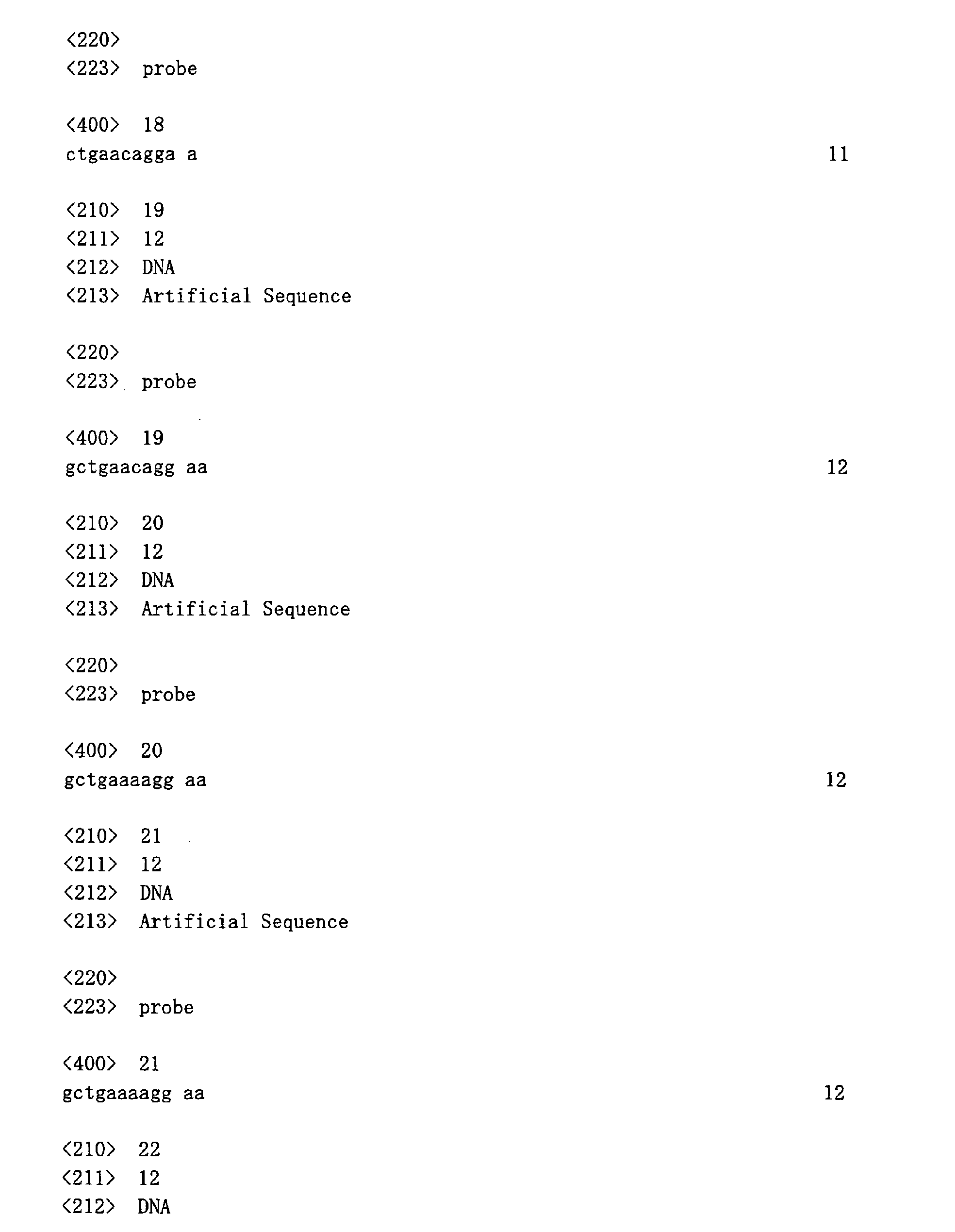
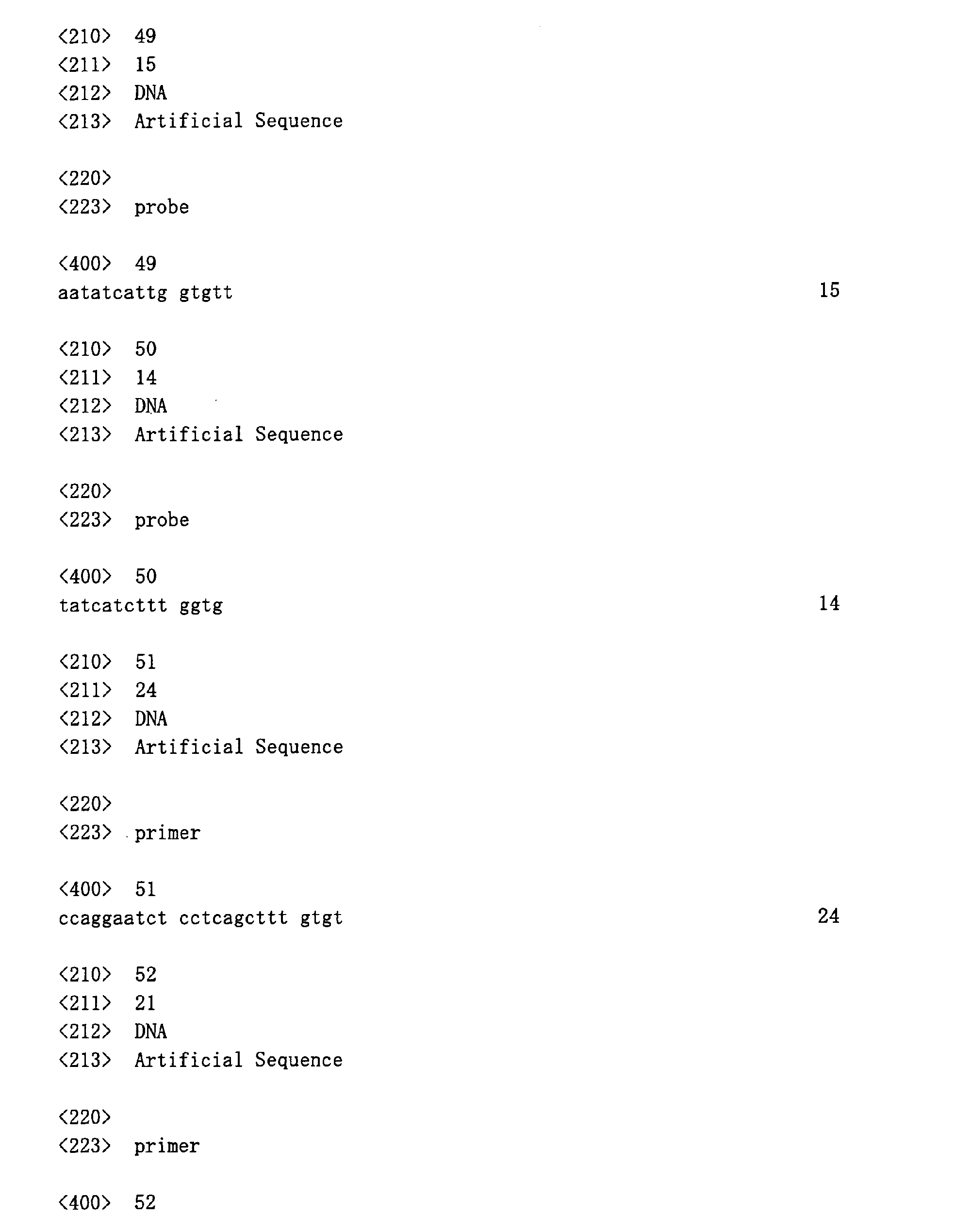
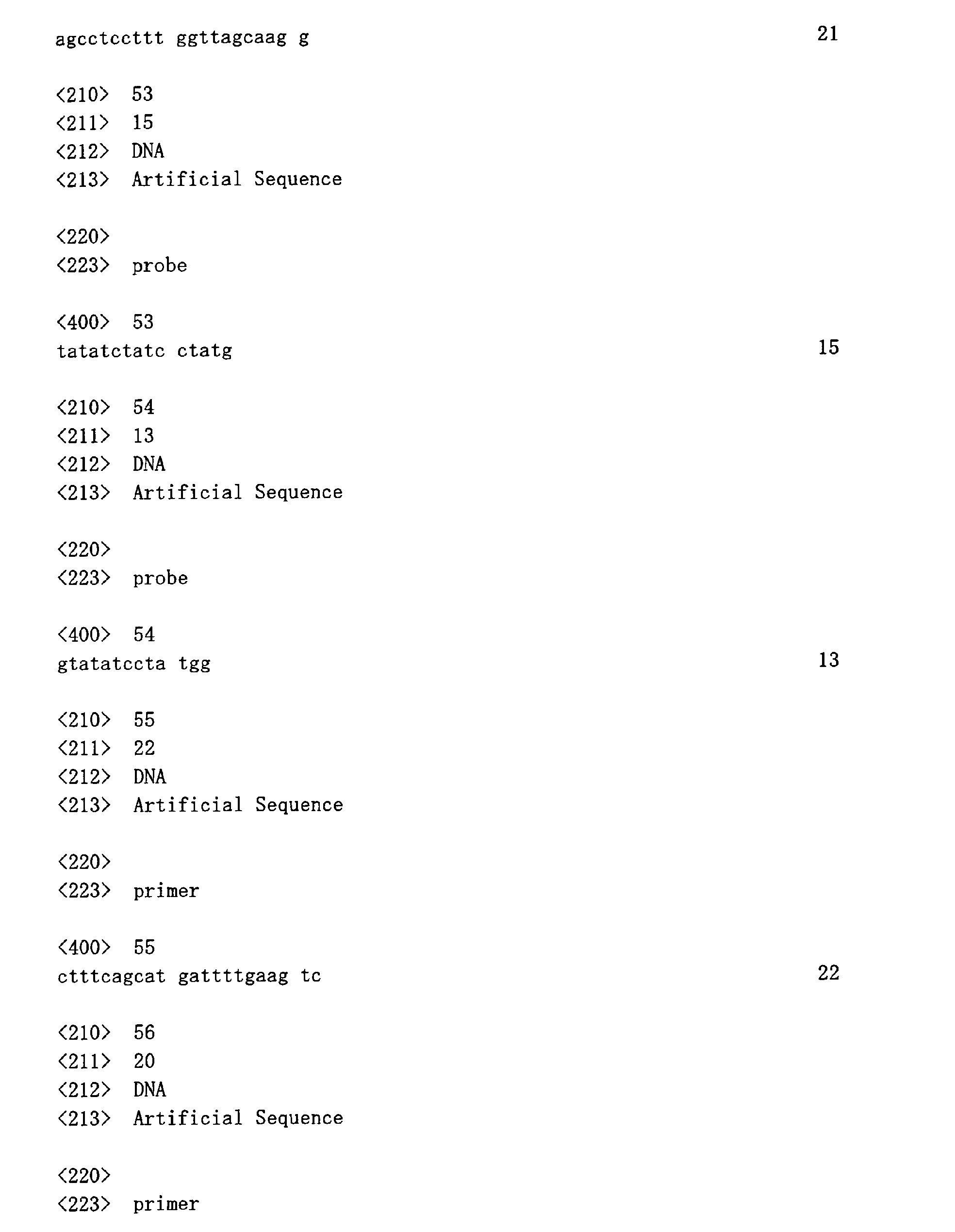
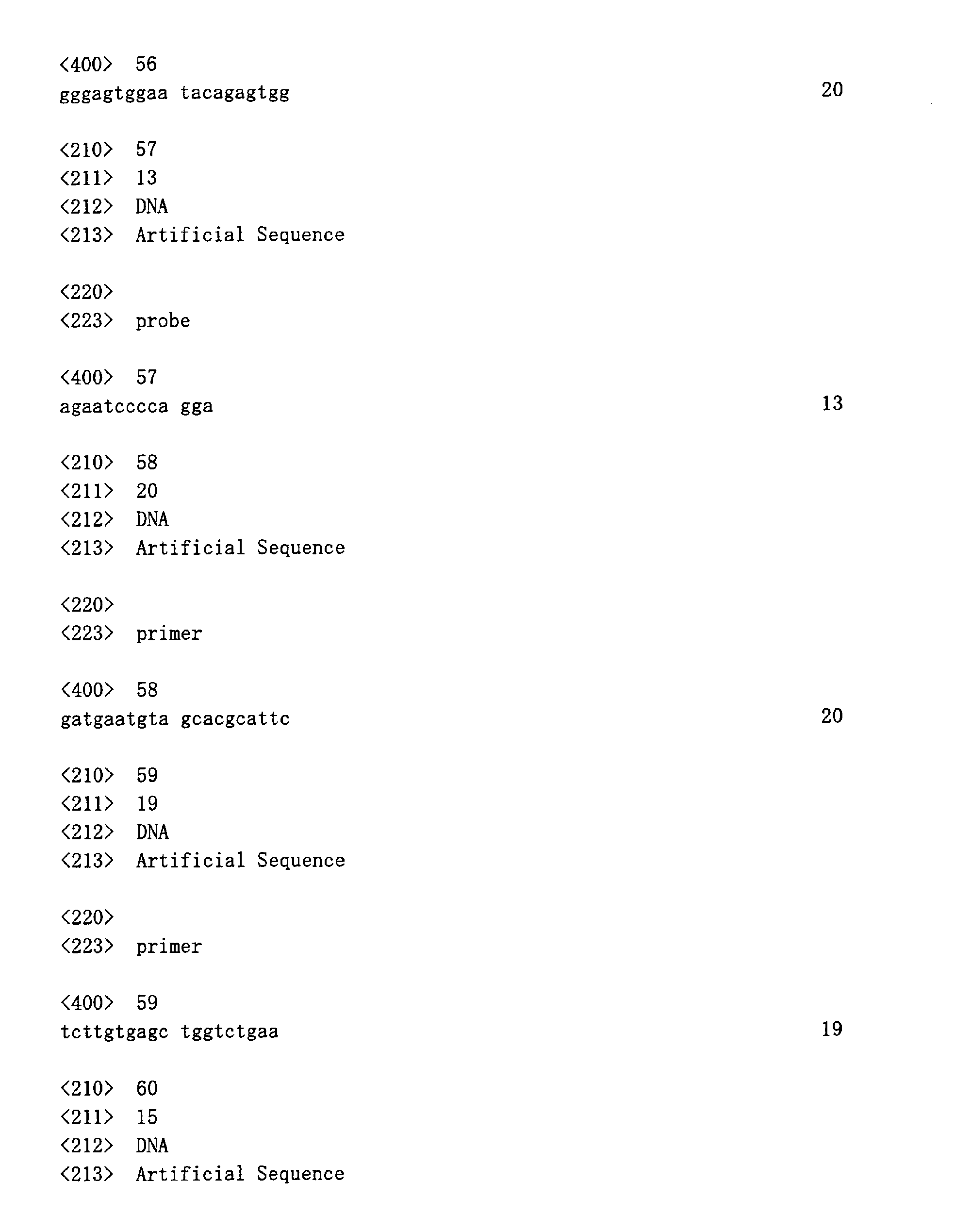
- oligonucleotides listed in Table 2 were prepared as hybridization probes and competing probes.
- the PCR reaction solution consists of 50-100 ng of sample DNA, 10 mM Tris-HCl (pH 8.3), 50 mM KCl, 1.5 mM MgCl 2 , 250 ⁇ M each dNTPs, 1 ⁇ M of PCR forward primer, 1 ⁇ M of PCR reverse primer (labeled with digoxigenin at the 5' end), 600 nM of hybridization probe (labeled with biotin at the 3' end), unlabeled competing oligonucleotide at a predetermined concentration, and 1.25 U Taq DNA polymerase in a fmal volume of 20 ⁇ l.
- the PCR was carried out by heating at 94°C for 2 minutes and repeating 35 times a cycle of 98°C for 10 seconds, 55°C for 30 seconds and 72°C for 30 seconds, followed by 72°C for 3 minutes, 98°C for 3 minutes, 65°C for 1 minute, 55°C for 1 minute, 45°C for 1 minute, 35°C for 1 minute, and 25°C for 1 minute.
- test strip DNA Detection Test Strip, Roche Co., Ltd., #1-965-484, an affinity chromatographic test strip on which streptavidin is immobilized and in which an anti-digoxigenin antibody labeled with gold colloid is held in a movable manner
- test strip DNA Detection Test Strip, Roche Co., Ltd., #1-965-484, an affinity chromatographic test strip on which streptavidin is immobilized and in which an anti-digoxigenin antibody labeled with gold colloid is held in a movable manner
- the bottom end of the strip was immersed in a buffer for 5 seconds.
- the test strip was left to stand for 5 minutes at room temperature to allow the buffer to move through the strip. After the keeping, the presence or absence of a genotype diagnostic line was macroscopically determined.
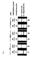
- the labeled hybridization probe used was of 17 mers and the detection was then carried out without the addition of a competing probe in a reaction solution.
- the DNA samples used were a homozygote for g727 allele (normal DNA) and a homozygote for t727 allele (mutant DNA), and the hybridization probes used were those for the detection of a normal base sequence and for the detection of a mutant base sequence. The results are shown in Fig. 4.
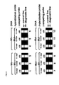
- the indications "Wt” and “Mut” with respect to “DNA” represent the normal DNA and the mutant DNA, respectively, while the indications “Wt” and “Mut” with respect to “hybridization probe” represent the hybridization probes for the detection of a normal base sequence and for the detection of a mutant base sequence, respectively (the same holds for Fig. 5 to Fig. 7 described below).
- the hybridization probes and competing probes used in this study were of 17 mers, 15 mers, 13 mers, and 11 mers.
- the amount of the competing probes added to the reaction solution was fixed to 30 times more than that of the hybridization probe. The results are shown in Fig. 6.
- the detection method of the present invention was carried out to detect a point mutation, including mutation a985g of medium-chain acyl-CoA dehydrogenase deficiency, mutation g1691t of GLDC gene in hyperglycinemia, mutation g681a of drug-metabolizing enzyme gene CYP2C19, and point mutation of Glu487Lys of aldehyde dehydrogenase 2 polymorphism.
- the PCR primers for amplifying base sequences containing the respective point mutation sites were adjusted in chain length so as to carry out PCR reactions with setting of an annealing temperature of 55°C.
- the hybridization probes were designed to have Tm values in the range of 35 to 40°C. As a result, the chain lengths thereof were 10 mers to 15 mers.
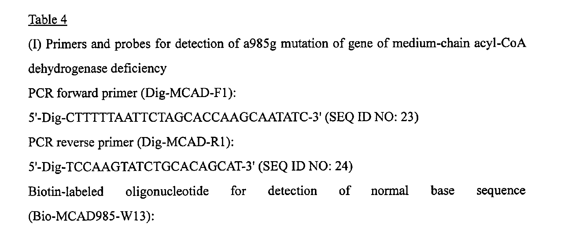
- the base sequences of primers, hybridization probes, and competing probes are listed in Table 4.
- the detections showed the correct determination of genotypes in all of the detection systems (Fig. 8, a, b, c, and d).
- the reaction line obtained was weak in the reaction system for detecting a mutant base sequence.
- unlabeled competing oligonucleotide was not mixed in this reaction system, the formation of distinct reaction line was observed. In each of the reactions, no false positive was observed.
- the determination of genotype was completed within 10 minutes after the completion of reaction in the thermal cycler. Even at least two years after the test strip was dried without any treatment and stored at room temperature, a macroscopic determination thereof was possible.
- the detection method of the invention allows of the simple and quick detection of each mutation or polymorphism of the five genes to determine the genotype of sample DNA, even though the design of primers and hybridization probes and competing probes, and the reaction conditions are required to be adjusted slightly for and depending on the respective gene mutations. Therefore, it is concluded that the detection method of the present invention can be used for many purposes.
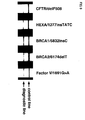
- the detection method of the present invention was carried out to detect a mutation, including deltaF508 deletion mutation in the gene of cystic fibrosis transmembrane regulator protein; 1277insTATC insertion mutation in HEXA gene of Tay-Sachs disease; 5382insC insertion mutation in BRCA1 gene of breast cancer; 6174delT deletion mutation in BRCA2 gene of breast cancer; and G1691A point mutation in Blood Coagulation Factor V gene of thrombosis.
- the PCR primers for amplifying base sequences containing the respective mutation sites were adjusted in chain length so as to carry out PCR reactions with setting of an annealing temperature of 55°C.
- the hybridization probes were designed to have Tm values in the range of 35 to 40°C. As a result, the chain lengths thereof were 10 mers to 15 mers.
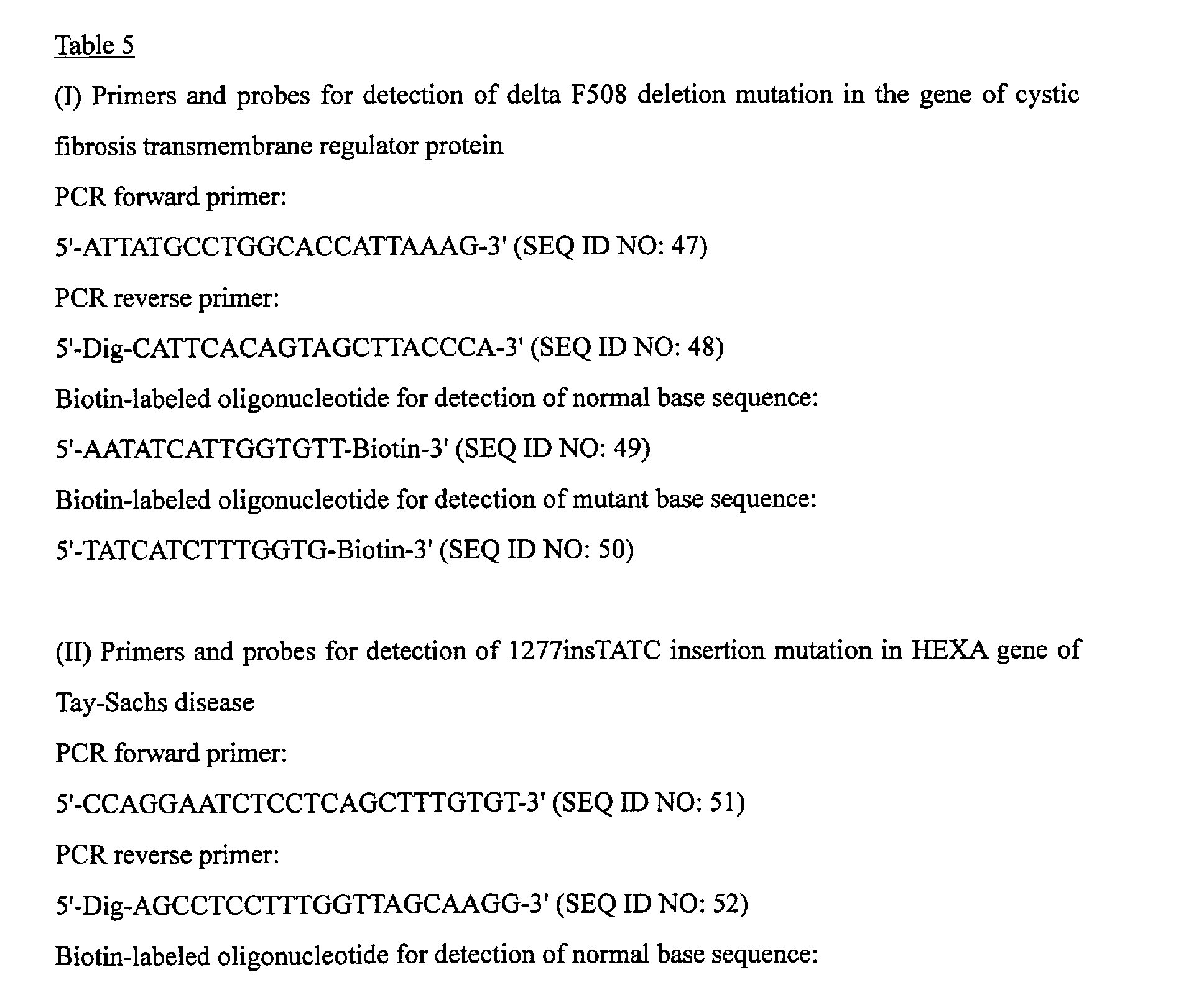
- the base sequences of primers, hybridization probes, and competing probes are listed in Table 5.
- the target mutations to be detected were base deletions or insertions and thus no competing probe was used.
- no probe for detecting the normal base sequence was used because even a patient of heterozygote of the mutation in question shows the symptom, indicating no clinical need of investigating the presence or absence of the gene having the normal base sequence.
- the determination of genotype was completed within 10 minutes after the completion of reaction in the thermal cycler. Even at least two years after the test strip was dried without any treatment and stored at room temperature, a macroscopic determination thereof was possible.
- the detection method of the invention enables the simple and quick detection of mutations including insertion and deletion mutations to determine the genotype of sample DNA, even though the design of primers and hybridization probes and competing probes and the reaction conditions are required to be adjusted slightly for and depending on the respective gene mutations. Therefore, it is concluded that the detection method of the present invention can be used for many purposes.
- the identification of pathogenic gene mutation and the detection of polymorphisms of disease-related genes and drug metabolism enzyme genes can be carried out in a simple, quick and accurate manner without use of other special devices and equipments than a conventional thermal cycler.
- the detection method of the present invention allows of the detection at bed side and is thus considered to facilitate the tailor-made medicine.
Landscapes
- Life Sciences & Earth Sciences (AREA)
- Chemical & Material Sciences (AREA)
- Health & Medical Sciences (AREA)
- Engineering & Computer Science (AREA)
- Organic Chemistry (AREA)
- Molecular Biology (AREA)
- Immunology (AREA)
- Zoology (AREA)
- Wood Science & Technology (AREA)
- Proteomics, Peptides & Aminoacids (AREA)
- Physics & Mathematics (AREA)
- Microbiology (AREA)
- Biotechnology (AREA)
- General Health & Medical Sciences (AREA)
- Analytical Chemistry (AREA)
- Biochemistry (AREA)
- Urology & Nephrology (AREA)
- Hematology (AREA)
- Biomedical Technology (AREA)
- Bioinformatics & Cheminformatics (AREA)
- Genetics & Genomics (AREA)
- General Engineering & Computer Science (AREA)
- Biophysics (AREA)
- Food Science & Technology (AREA)
- Cell Biology (AREA)
- Medicinal Chemistry (AREA)
- General Physics & Mathematics (AREA)
- Pathology (AREA)
- Tropical Medicine & Parasitology (AREA)
- Measuring Or Testing Involving Enzymes Or Micro-Organisms (AREA)
- Saccharide Compounds (AREA)
- Iron Core Of Rotating Electric Machines (AREA)
- Automatic Analysis And Handling Materials Therefor (AREA)
Abstract
Description
wherein at least one of primers to be used in the DNA amplification is labeled with a first labeling agent so that the amplified DNA will be labeled with the first labeling agent, the hybridization probe is labeled with a second labeling agent and contained in a reaction solution for effecting the DNA amplification, the base sequence of the hybridization probe is designed not to inhibit the DNA amplification, and the hybrid is detected by affinity chromatography with the use of the first and second labeling agents.
wherein at least one of the primers to be used in the DNA amplification is labeled with a first labeling agent so that the amplified DNA will be labeled with the first labeling agent, the hybridization probe is labeled with a second labeling agent, the base sequence of the hybridization probe is designed not to inhibit the DNA amplification, and the test strip allows of detection of a hybrid of the amplified DNA and the hybridization probe with the use of the first and second labeling agents.
characterized in that at least one of primers to be used in the DNA amplification is labeled with a first labeling agent so that the amplified DNA will be labeled with the first labeling agent, the hybridization probe is labeled with a second labeling agent and contained in a reaction solution for effecting the DNA amplification, the base sequence of the hybridization probe is designed not to inhibit the DNA amplification, and the hybrid is detected by affinity chromatography with the use of the first and second labeling agents. Hereinafter, each of the steps will be described.
characterized in that at least one of the primers to be used in the DNA amplification is labeled with a first labeling agent so that the amplified DNA will be labeled with the first labeling agent, the hybridization probe is labeled with a second labeling agent, the base sequence of the hybridization probe is designed not to inhibit the DNA amplification, and the test strip allows of detection of a hybrid of the amplified DNA and the hybridization probe with the use of the first and second labeling agents. The kit of the present invention can be used for carrying out the detection method of the present invention.














Claims (6)
- A method of detecting a base sequence, comprising the steps of: amplifying DNA containing a target base sequence to be detected having a mutation site using DNA polymerase; hybridizing the amplified DNA to a hybridization probe having a base sequence complementary to the target base sequence to be detected; and detecting a hybrid formed by the hybridization,
wherein at least one of primers to be used in the DNA amplification is labeled with a first labeling agent so that the amplified DNA will be labeled with the first labeling agent, the hybridization probe is labeled with a second labeling agent and contained in a reaction solution for effecting the DNA amplification, the base sequence of the hybridization probe is designed not to inhibit the DNA amplification, and the hybrid is detected by affinity chromatography with the use of the first and second labeling agents. - The method according to claim 1, wherein the mutation site is a point mutation, and the reaction solution for effecting the DNA amplification further contains an unlabeled oligonucleotide having a base sequence different in a single base at the position of the point mutation from the base sequence of the labeled hybridization probe, in an amount sufficient to enhance the specificity of hybridization of the amplified DNA to the hybridization probe.
- The method according to claim 1 or 2, wherein the DNA amplification is carried out by PCR.
- A kit comprising: primers for amplifying DNA containing a target base sequence to be detected having a mutation site using DNA polymerase; a hybridization probe having a base sequence complementary to the target base sequence to be detected; and a test strip for affinity chromatography,
wherein at least one of the primers to be used in the DNA amplification is labeled with a first labeling agent so that the amplified DNA will be labeled with the first labeling agent, the hybridization probe is labeled with a second labeling agent, the base sequence of the hybridization probe is designed not to inhibit the DNA amplification, and the test strip allows of detection of a hybrid of the amplified DNA and the hybridization probe with the use of the first and second labeling agents. - The kit according to claim 4, wherein the mutation site is a point mutation and the kit further comprises an unlabeled oligonucleotide having a base sequence different in a single base at the position of the point mutation from the base sequence of the labeled hybridization probe.
- The kit according to claim 4 or 5, wherein the primers are primers for PCR.
Applications Claiming Priority (3)
| Application Number | Priority Date | Filing Date | Title |
|---|---|---|---|
| JP2002323419 | 2002-11-07 | ||
| JP2002323419 | 2002-11-07 | ||
| PCT/JP2003/014204 WO2004042057A1 (en) | 2002-11-07 | 2003-11-07 | Method of detecting gene mutation |
Publications (3)
| Publication Number | Publication Date |
|---|---|
| EP1580269A1 true EP1580269A1 (en) | 2005-09-28 |
| EP1580269A4 EP1580269A4 (en) | 2006-01-11 |
| EP1580269B1 EP1580269B1 (en) | 2008-07-02 |
Family
ID=32310418
Family Applications (1)
| Application Number | Title | Priority Date | Filing Date |
|---|---|---|---|
| EP03810655A Expired - Lifetime EP1580269B1 (en) | 2002-11-07 | 2003-11-07 | Method of detecting gene mutation |
Country Status (11)
| Country | Link |
|---|---|
| US (2) | US20060127907A1 (en) |
| EP (1) | EP1580269B1 (en) |
| JP (1) | JP4425142B2 (en) |
| KR (1) | KR101078977B1 (en) |
| CN (1) | CN100343389C (en) |
| AT (1) | ATE399882T1 (en) |
| AU (1) | AU2003277612A1 (en) |
| CA (1) | CA2506654C (en) |
| DE (1) | DE60321961D1 (en) |
| NO (1) | NO338640B1 (en) |
| WO (1) | WO2004042057A1 (en) |
Cited By (2)
| Publication number | Priority date | Publication date | Assignee | Title |
|---|---|---|---|---|
| WO2010054254A1 (en) | 2008-11-07 | 2010-05-14 | University Of Utah Research Foundation | Allele amplification bias |
| US11111526B2 (en) * | 2016-08-03 | 2021-09-07 | Toyo Kohan Co., Ltd. | Buffer composition for hybridization and hybridization method |
Families Citing this family (32)
| Publication number | Priority date | Publication date | Assignee | Title |
|---|---|---|---|---|
| US8338109B2 (en) | 2006-11-02 | 2012-12-25 | Mayo Foundation For Medical Education And Research | Predicting cancer outcome |
| JP5593582B2 (en) * | 2007-06-12 | 2014-09-24 | 東洋紡株式会社 | Rapid detection method of nucleic acid |
| US9063130B2 (en) * | 2007-09-11 | 2015-06-23 | Kaneka Corporation | Nucleic acid detection method and nucleic acid detection kit |
| CA2725978A1 (en) | 2008-05-28 | 2009-12-03 | Genomedx Biosciences, Inc. | Systems and methods for expression-based discrimination of distinct clinical disease states in prostate cancer |
| US10407731B2 (en) | 2008-05-30 | 2019-09-10 | Mayo Foundation For Medical Education And Research | Biomarker panels for predicting prostate cancer outcomes |
| DE102008028908B3 (en) * | 2008-06-18 | 2009-12-31 | IfP Privates Institut für Produktqualität GmbH | Detection of an analyte in an aqueous medium |
| US9495515B1 (en) | 2009-12-09 | 2016-11-15 | Veracyte, Inc. | Algorithms for disease diagnostics |
| GB2512153B (en) * | 2008-11-17 | 2014-11-12 | Veracyte Inc | Methods and compositions of molecular profiling for disease diagnostics |
| US10236078B2 (en) | 2008-11-17 | 2019-03-19 | Veracyte, Inc. | Methods for processing or analyzing a sample of thyroid tissue |
| US9074258B2 (en) | 2009-03-04 | 2015-07-07 | Genomedx Biosciences Inc. | Compositions and methods for classifying thyroid nodule disease |
| US8669057B2 (en) | 2009-05-07 | 2014-03-11 | Veracyte, Inc. | Methods and compositions for diagnosis of thyroid conditions |
| JP5597939B2 (en) * | 2009-06-01 | 2014-10-01 | 凸版印刷株式会社 | Method for detecting a target nucleotide sequence using a partially competitive probe |
| JP2011062088A (en) * | 2009-09-15 | 2011-03-31 | Ihi Corp | Method for detecting legionella bacteria |
| US10446272B2 (en) | 2009-12-09 | 2019-10-15 | Veracyte, Inc. | Methods and compositions for classification of samples |
| CN103038635B (en) | 2010-05-11 | 2016-12-28 | 威拉赛特公司 | Methods and compositions for diagnosing conditions |
| US20130267443A1 (en) | 2010-11-19 | 2013-10-10 | The Regents Of The University Of Michigan | ncRNA AND USES THEREOF |
| US10513737B2 (en) | 2011-12-13 | 2019-12-24 | Decipher Biosciences, Inc. | Cancer diagnostics using non-coding transcripts |
| ES3015226T3 (en) | 2012-08-16 | 2025-04-30 | Veracyte Sd Inc | Prostate cancer prognosis using biomarkers |
| US20140244556A1 (en) * | 2013-02-27 | 2014-08-28 | Abdul Saleh | Methods for and apparatus generating automated pharmaco genetics correlation |
| BR112015022490A2 (en) | 2013-03-15 | 2017-07-18 | Veracyte Inc | methods and compositions for sample classification |
| US11976329B2 (en) | 2013-03-15 | 2024-05-07 | Veracyte, Inc. | Methods and systems for detecting usual interstitial pneumonia |
| US12297505B2 (en) | 2014-07-14 | 2025-05-13 | Veracyte, Inc. | Algorithms for disease diagnostics |
| CN114606309A (en) | 2014-11-05 | 2022-06-10 | 威拉赛特公司 | Diagnostic system and method using machine learning and high-dimensional transcription data |
| CN104531883B (en) * | 2015-01-14 | 2018-02-02 | 北京圣谷同创科技发展有限公司 | The detection kit and detection method of PKD1 gene mutations |
| US11414708B2 (en) | 2016-08-24 | 2022-08-16 | Decipher Biosciences, Inc. | Use of genomic signatures to predict responsiveness of patients with prostate cancer to post-operative radiation therapy |
| AU2018210695B2 (en) | 2017-01-20 | 2024-07-18 | The University Of British Columbia | Molecular subtyping, prognosis, and treatment of bladder cancer |
| WO2018165600A1 (en) | 2017-03-09 | 2018-09-13 | Genomedx Biosciences, Inc. | Subtyping prostate cancer to predict response to hormone therapy |
| US11078542B2 (en) | 2017-05-12 | 2021-08-03 | Decipher Biosciences, Inc. | Genetic signatures to predict prostate cancer metastasis and identify tumor aggressiveness |
| US11217329B1 (en) | 2017-06-23 | 2022-01-04 | Veracyte, Inc. | Methods and systems for determining biological sample integrity |
| JP2019012035A (en) * | 2017-06-30 | 2019-01-24 | キヤノン株式会社 | Kit for thin layer chromatography, developing solution for thin layer chromatography, and thin layer chromatography |
| AU2018310987A1 (en) | 2017-08-04 | 2020-02-27 | Decipher Biosciences, Inc. | Use of immune cell-specific gene expression for prognosis of prostate cancer and prediction of responsiveness to radiation therapy |
| CN108315408A (en) * | 2018-04-26 | 2018-07-24 | 宁波美丽人生医学检验所有限公司 | Primer composition and kit for detecting nitroglycerin pharmaceutical relevant gene |
Family Cites Families (25)
| Publication number | Priority date | Publication date | Assignee | Title |
|---|---|---|---|---|
| US5753433A (en) * | 1909-12-05 | 1998-05-19 | Boehringer Mannheim Gmbh | Method for the sensitive detection of nucleic acids |
| US6040166A (en) * | 1985-03-28 | 2000-03-21 | Roche Molecular Systems, Inc. | Kits for amplifying and detecting nucleic acid sequences, including a probe |
| CA1284931C (en) | 1986-03-13 | 1991-06-18 | Henry A. Erlich | Process for detecting specific nucleotide variations and genetic polymorphisms present in nucleic acids |
| US5635347A (en) * | 1986-04-30 | 1997-06-03 | Igen, Inc. | Rapid assays for amplification products |
| IE61148B1 (en) * | 1988-03-10 | 1994-10-05 | Ici Plc | Method of detecting nucleotide sequences |
| CA1341584C (en) * | 1988-04-06 | 2008-11-18 | Bruce Wallace | Method of amplifying and detecting nucleic acid sequences |
| KR920700360A (en) * | 1989-03-22 | 1992-02-19 | 하리크 프리드리히 | Sliding bearing |
| JP2715651B2 (en) * | 1990-10-08 | 1998-02-18 | 東洋紡績株式会社 | Oligonucleotide for detecting toxinogenic Escherichia coli, detection method and kit for detecting toxinogenic Escherichia coli |
| NZ240079A (en) | 1990-10-09 | 1993-07-27 | Boehringer Mannheim Gmbh | Method for the detection of a nucleic acid or part thereof |
| DE4129653A1 (en) * | 1991-09-06 | 1993-03-11 | Boehringer Mannheim Gmbh | PROCESS FOR DETECTION OF SIMILAR NUCLEIC ACIDS |
| US5650277A (en) * | 1992-07-02 | 1997-07-22 | Diagenetics Ltd. | Method of determining the presence and quantifying the number of di- and trinucleotide repeats |
| EP0648222B1 (en) * | 1992-07-02 | 2003-01-29 | Savyon Diagnostics Ltd. | Methods of single nucleotide primer extension to detect specific alleles and kits therefor |
| JPH0779779A (en) * | 1993-09-13 | 1995-03-28 | Toyobo Co Ltd | Oligonucleotide for detecting enterotoxigenic e.coli and its use |
| WO1995025538A1 (en) * | 1994-03-18 | 1995-09-28 | The General Hospital Corporation | Cleaved amplified rflp detection methods |
| CA2195562A1 (en) * | 1994-08-19 | 1996-02-29 | Pe Corporation (Ny) | Coupled amplification and ligation method |
| US6090620A (en) * | 1995-12-29 | 2000-07-18 | University Of Washington | Genes and gene products related to Werner's syndrome |
| US6613508B1 (en) * | 1996-01-23 | 2003-09-02 | Qiagen Genomics, Inc. | Methods and compositions for analyzing nucleic acid molecules utilizing sizing techniques |
| JPH11502727A (en) * | 1996-01-26 | 1999-03-09 | イノジェネティックス・ナムローゼ・フェンノートシャップ | Methods for detecting drug-induced mutations in the reverse transcriptase gene |
| US6013440A (en) * | 1996-03-11 | 2000-01-11 | Affymetrix, Inc. | Nucleic acid affinity columns |
| DE19730359A1 (en) * | 1997-07-15 | 1999-01-21 | Boehringer Mannheim Gmbh | Integrated method and system for amplification and detection of nucleic acids |
| DE19732086C2 (en) * | 1997-07-25 | 2002-11-21 | Univ Leipzig | Method for the quantitative determination of eubacteria |
| US5969123A (en) * | 1998-03-06 | 1999-10-19 | Millennium Biotherapeutics, Inc. | Nucleic acid molecules derived from a brain tissue library |
| EP1124990B1 (en) * | 1998-10-27 | 2006-01-18 | Affymetrix, Inc. | Complexity management and analysis of genomic dna |
| US7582420B2 (en) * | 2001-07-12 | 2009-09-01 | Illumina, Inc. | Multiplex nucleic acid reactions |
| AU2003206869A1 (en) * | 2002-02-08 | 2003-09-02 | Evotec Technologies Gmbh | Specific multiplex analysis of nucleic acids |
-
2003
- 2003-11-07 KR KR1020057008179A patent/KR101078977B1/en not_active Expired - Fee Related
- 2003-11-07 AU AU2003277612A patent/AU2003277612A1/en not_active Abandoned
- 2003-11-07 WO PCT/JP2003/014204 patent/WO2004042057A1/en not_active Ceased
- 2003-11-07 US US10/533,750 patent/US20060127907A1/en not_active Abandoned
- 2003-11-07 CN CNB2003801070393A patent/CN100343389C/en not_active Expired - Lifetime
- 2003-11-07 CA CA2506654A patent/CA2506654C/en not_active Expired - Lifetime
- 2003-11-07 AT AT03810655T patent/ATE399882T1/en not_active IP Right Cessation
- 2003-11-07 EP EP03810655A patent/EP1580269B1/en not_active Expired - Lifetime
- 2003-11-07 JP JP2004549640A patent/JP4425142B2/en not_active Expired - Lifetime
- 2003-11-07 DE DE60321961T patent/DE60321961D1/en not_active Expired - Lifetime
-
2005
- 2005-06-06 NO NO20052692A patent/NO338640B1/en not_active IP Right Cessation
-
2008
- 2008-05-12 US US12/119,141 patent/US9677127B2/en not_active Expired - Fee Related
Cited By (5)
| Publication number | Priority date | Publication date | Assignee | Title |
|---|---|---|---|---|
| WO2010054254A1 (en) | 2008-11-07 | 2010-05-14 | University Of Utah Research Foundation | Allele amplification bias |
| EP2356245A4 (en) * | 2008-11-07 | 2013-02-20 | Univ Utah Res Found | BALL OF AMPLIFICATION OF ALLELLELELELE AMPLIFICATION BIAS |
| US9422597B2 (en) | 2008-11-07 | 2016-08-23 | Biofire Diagnostics, Inc. | Allele amplification bias |
| US10351903B2 (en) | 2008-11-07 | 2019-07-16 | University Of Utah Research Foundation | Allele amplification bias |
| US11111526B2 (en) * | 2016-08-03 | 2021-09-07 | Toyo Kohan Co., Ltd. | Buffer composition for hybridization and hybridization method |
Also Published As
| Publication number | Publication date |
|---|---|
| US20060127907A1 (en) | 2006-06-15 |
| KR20050086431A (en) | 2005-08-30 |
| JP4425142B2 (en) | 2010-03-03 |
| CN1729289A (en) | 2006-02-01 |
| CA2506654A1 (en) | 2004-05-21 |
| CN100343389C (en) | 2007-10-17 |
| US9677127B2 (en) | 2017-06-13 |
| EP1580269B1 (en) | 2008-07-02 |
| CA2506654C (en) | 2014-02-25 |
| NO338640B1 (en) | 2016-09-26 |
| ATE399882T1 (en) | 2008-07-15 |
| US20080318238A1 (en) | 2008-12-25 |
| NO20052692L (en) | 2005-06-06 |
| WO2004042057A1 (en) | 2004-05-21 |
| KR101078977B1 (en) | 2011-11-01 |
| EP1580269A4 (en) | 2006-01-11 |
| NO20052692D0 (en) | 2005-06-06 |
| DE60321961D1 (en) | 2008-08-14 |
| JPWO2004042057A1 (en) | 2006-03-09 |
| AU2003277612A1 (en) | 2004-06-07 |
Similar Documents
| Publication | Publication Date | Title |
|---|---|---|
| US9677127B2 (en) | Method of detecting gene mutation | |
| US20230340564A1 (en) | Accurate and massively parallel quantification of nucleic acid | |
| JP3937136B2 (en) | Nucleotide polymorphism detection method | |
| US6027877A (en) | Use of immobilized mismatch binding protein for detection of mutations and polymorphisms, purification of amplified DNA samples and allele identification | |
| Tombelli et al. | Coupling of a DNA piezoelectric biosensor and polymerase chain reaction to detect apolipoprotein E polymorphisms | |
| US20030148301A1 (en) | Method of detecting nucleotide polymorphism | |
| US7252946B2 (en) | Nucleic acid detection | |
| JPWO2001042498A1 (en) | Nucleotide polymorphism detection method | |
| JP2022145606A (en) | Highly sensitive methods for accurate parallel quantification of nucleic acids | |
| Breveglieri et al. | A novel and efficient protocol for Surface Plasmon Resonance based detection of four β-thalassemia point mutations in blood samples and salivary swabs | |
| EP2984183B1 (en) | Nanoprobe-based genetic testing | |
| US20010039012A1 (en) | Methods for diagnostic screening | |
| WO2011092596A2 (en) | Genotyping cytochrome p450 2d6 (cyp2d6) | |
| JPH10513055A (en) | Methods for detecting changes in nucleic acid sequence | |
| Bhardwaj et al. | Neonatal hemoglobinopathy screening: molecular genetic technologies | |
| JP2982304B2 (en) | Method for identifying nucleic acid and test set for identifying nucleic acid | |
| RU2738752C1 (en) | Method of identifying person and establishing affinity using indel polymorphisms and set of synthetic oligonucleotides for genotyping thereof | |
| Absalan et al. | 3C based DNA hybridization method for chromosomal translocation screening | |
| KR20250088845A (en) | Method for diagnosis of mutant genotype of genes related to therapeutic response or side effects of tuberculosis treatment | |
| HK40126982A (en) | Highly sensitive methods for accurate parallel quantification of nucleic acids | |
| RU2402612C2 (en) | Method of screening newborns for monogenic diseases and biochip for said method realisation | |
| JP2002209584A (en) | Method for detecting nucleotide polymorphism | |
| Brickell | DNA probes in human diseases | |
| JP2003135069A (en) | Method for identifying nucleotide polymorphism |
Legal Events
| Date | Code | Title | Description |
|---|---|---|---|
| PUAI | Public reference made under article 153(3) epc to a published international application that has entered the european phase |
Free format text: ORIGINAL CODE: 0009012 |
|
| 17P | Request for examination filed |
Effective date: 20050520 |
|
| AK | Designated contracting states |
Kind code of ref document: A1 Designated state(s): AT BE BG CH CY CZ DE DK EE ES FI FR GB GR HU IE IT LI LU MC NL PT RO SE SI SK TR |
|
| AX | Request for extension of the european patent |
Extension state: AL LT LV MK |
|
| A4 | Supplementary search report drawn up and despatched |
Effective date: 20051130 |
|
| RIC1 | Information provided on ipc code assigned before grant |
Ipc: C12Q 1/68 19800101AFI20051124BHEP Ipc: G01N 33/58 19800101ALI20051124BHEP Ipc: G01N 33/53 19850101ALI20051124BHEP |
|
| DAX | Request for extension of the european patent (deleted) | ||
| GRAP | Despatch of communication of intention to grant a patent |
Free format text: ORIGINAL CODE: EPIDOSNIGR1 |
|
| RIN1 | Information on inventor provided before grant (corrected) |
Inventor name: MATSUBARA, YOICHI Inventor name: KURE, SHIGEO |
|
| GRAS | Grant fee paid |
Free format text: ORIGINAL CODE: EPIDOSNIGR3 |
|
| GRAA | (expected) grant |
Free format text: ORIGINAL CODE: 0009210 |
|
| AK | Designated contracting states |
Kind code of ref document: B1 Designated state(s): AT BE BG CH CY CZ DE DK EE ES FI FR GB GR HU IE IT LI LU MC NL PT RO SE SI SK TR |
|
| REG | Reference to a national code |
Ref country code: GB Ref legal event code: FG4D |
|
| REG | Reference to a national code |
Ref country code: CH Ref legal event code: EP |
|
| REF | Corresponds to: |
Ref document number: 60321961 Country of ref document: DE Date of ref document: 20080814 Kind code of ref document: P |
|
| REG | Reference to a national code |
Ref country code: IE Ref legal event code: FG4D |
|
| PG25 | Lapsed in a contracting state [announced via postgrant information from national office to epo] |
Ref country code: SI Free format text: LAPSE BECAUSE OF FAILURE TO SUBMIT A TRANSLATION OF THE DESCRIPTION OR TO PAY THE FEE WITHIN THE PRESCRIBED TIME-LIMIT Effective date: 20080702 |
|
| REG | Reference to a national code |
Ref country code: CH Ref legal event code: NV Representative=s name: E. BLUM & CO. AG PATENT- UND MARKENANWAELTE VSP |
|
| PG25 | Lapsed in a contracting state [announced via postgrant information from national office to epo] |
Ref country code: NL Free format text: LAPSE BECAUSE OF FAILURE TO SUBMIT A TRANSLATION OF THE DESCRIPTION OR TO PAY THE FEE WITHIN THE PRESCRIBED TIME-LIMIT Effective date: 20080702 |
|
| REG | Reference to a national code |
Ref country code: CH Ref legal event code: PFA Owner name: MATSUBARA, YOICHI Free format text: DAIICHI PURE CHEMICALS CO., LTD.#13-5, NIHONBASHI 3-CHOME#CHUO-KU TOKYO 103-0027 (JP) $ MATSUBARA, YOICHI#13-10, KEIWA-CHO, TAIHAKU-KU#SENDAI-SHI, MIYAGI 982-0823 (JP) $ KURE, SHIGEO#3-20-312, HAYAMA-CHO, AOBA-KU#SENDAI-SHI, MIYAGI 981-0917 (JP) -TRANSFER TO- MATSUBARA, YOICHI#13-10, KEIWA-CHO, TAIHAKU-KU#SENDAI-SHI, MIYAGI 982-0823 (JP) $ KURE, SHIGEO#3-20-312, HAYAMA-CHO, AOBA-KU#SENDAI-SHI, MIYAGI 981-0917 (JP) $ SEKISUI MEDICAL CO., LTD.#13-5, NIHONBASHI 3-CHOME CHUO-KU#TOKYO 103-0027 (JP) |
|
| NLV1 | Nl: lapsed or annulled due to failure to fulfill the requirements of art. 29p and 29m of the patents act | ||
| RAP2 | Party data changed (patent owner data changed or rights of a patent transferred) |
Owner name: KURE, SHIGEO Owner name: SEKISUI MEDICAL CO., LTD. Owner name: MATSUBARA, YOICHI |
|
| PG25 | Lapsed in a contracting state [announced via postgrant information from national office to epo] |
Ref country code: PT Free format text: LAPSE BECAUSE OF FAILURE TO SUBMIT A TRANSLATION OF THE DESCRIPTION OR TO PAY THE FEE WITHIN THE PRESCRIBED TIME-LIMIT Effective date: 20081202 Ref country code: ES Free format text: LAPSE BECAUSE OF FAILURE TO SUBMIT A TRANSLATION OF THE DESCRIPTION OR TO PAY THE FEE WITHIN THE PRESCRIBED TIME-LIMIT Effective date: 20081013 |
|
| PG25 | Lapsed in a contracting state [announced via postgrant information from national office to epo] |
Ref country code: BG Free format text: LAPSE BECAUSE OF FAILURE TO SUBMIT A TRANSLATION OF THE DESCRIPTION OR TO PAY THE FEE WITHIN THE PRESCRIBED TIME-LIMIT Effective date: 20081002 Ref country code: AT Free format text: LAPSE BECAUSE OF FAILURE TO SUBMIT A TRANSLATION OF THE DESCRIPTION OR TO PAY THE FEE WITHIN THE PRESCRIBED TIME-LIMIT Effective date: 20080702 Ref country code: FI Free format text: LAPSE BECAUSE OF FAILURE TO SUBMIT A TRANSLATION OF THE DESCRIPTION OR TO PAY THE FEE WITHIN THE PRESCRIBED TIME-LIMIT Effective date: 20080702 |
|
| PG25 | Lapsed in a contracting state [announced via postgrant information from national office to epo] |
Ref country code: BE Free format text: LAPSE BECAUSE OF FAILURE TO SUBMIT A TRANSLATION OF THE DESCRIPTION OR TO PAY THE FEE WITHIN THE PRESCRIBED TIME-LIMIT Effective date: 20080702 |
|
| PG25 | Lapsed in a contracting state [announced via postgrant information from national office to epo] |
Ref country code: DK Free format text: LAPSE BECAUSE OF FAILURE TO SUBMIT A TRANSLATION OF THE DESCRIPTION OR TO PAY THE FEE WITHIN THE PRESCRIBED TIME-LIMIT Effective date: 20080702 Ref country code: EE Free format text: LAPSE BECAUSE OF FAILURE TO SUBMIT A TRANSLATION OF THE DESCRIPTION OR TO PAY THE FEE WITHIN THE PRESCRIBED TIME-LIMIT Effective date: 20080702 |
|
| PLBE | No opposition filed within time limit |
Free format text: ORIGINAL CODE: 0009261 |
|
| STAA | Information on the status of an ep patent application or granted ep patent |
Free format text: STATUS: NO OPPOSITION FILED WITHIN TIME LIMIT |
|
| REG | Reference to a national code |
Ref country code: FR Ref legal event code: CD |
|
| PG25 | Lapsed in a contracting state [announced via postgrant information from national office to epo] |
Ref country code: SK Free format text: LAPSE BECAUSE OF FAILURE TO SUBMIT A TRANSLATION OF THE DESCRIPTION OR TO PAY THE FEE WITHIN THE PRESCRIBED TIME-LIMIT Effective date: 20080702 Ref country code: CZ Free format text: LAPSE BECAUSE OF FAILURE TO SUBMIT A TRANSLATION OF THE DESCRIPTION OR TO PAY THE FEE WITHIN THE PRESCRIBED TIME-LIMIT Effective date: 20080702 Ref country code: RO Free format text: LAPSE BECAUSE OF FAILURE TO SUBMIT A TRANSLATION OF THE DESCRIPTION OR TO PAY THE FEE WITHIN THE PRESCRIBED TIME-LIMIT Effective date: 20080702 |
|
| 26N | No opposition filed |
Effective date: 20090403 |
|
| PG25 | Lapsed in a contracting state [announced via postgrant information from national office to epo] |
Ref country code: MC Free format text: LAPSE BECAUSE OF NON-PAYMENT OF DUE FEES Effective date: 20081130 |
|
| PG25 | Lapsed in a contracting state [announced via postgrant information from national office to epo] |
Ref country code: IT Free format text: LAPSE BECAUSE OF FAILURE TO SUBMIT A TRANSLATION OF THE DESCRIPTION OR TO PAY THE FEE WITHIN THE PRESCRIBED TIME-LIMIT Effective date: 20080702 |
|
| PG25 | Lapsed in a contracting state [announced via postgrant information from national office to epo] |
Ref country code: IE Free format text: LAPSE BECAUSE OF NON-PAYMENT OF DUE FEES Effective date: 20081107 |
|
| PG25 | Lapsed in a contracting state [announced via postgrant information from national office to epo] |
Ref country code: SE Free format text: LAPSE BECAUSE OF FAILURE TO SUBMIT A TRANSLATION OF THE DESCRIPTION OR TO PAY THE FEE WITHIN THE PRESCRIBED TIME-LIMIT Effective date: 20081002 |
|
| PG25 | Lapsed in a contracting state [announced via postgrant information from national office to epo] |
Ref country code: LU Free format text: LAPSE BECAUSE OF NON-PAYMENT OF DUE FEES Effective date: 20081107 Ref country code: HU Free format text: LAPSE BECAUSE OF FAILURE TO SUBMIT A TRANSLATION OF THE DESCRIPTION OR TO PAY THE FEE WITHIN THE PRESCRIBED TIME-LIMIT Effective date: 20090103 Ref country code: CY Free format text: LAPSE BECAUSE OF FAILURE TO SUBMIT A TRANSLATION OF THE DESCRIPTION OR TO PAY THE FEE WITHIN THE PRESCRIBED TIME-LIMIT Effective date: 20080702 |
|
| PG25 | Lapsed in a contracting state [announced via postgrant information from national office to epo] |
Ref country code: TR Free format text: LAPSE BECAUSE OF FAILURE TO SUBMIT A TRANSLATION OF THE DESCRIPTION OR TO PAY THE FEE WITHIN THE PRESCRIBED TIME-LIMIT Effective date: 20080702 |
|
| PG25 | Lapsed in a contracting state [announced via postgrant information from national office to epo] |
Ref country code: GR Free format text: LAPSE BECAUSE OF FAILURE TO SUBMIT A TRANSLATION OF THE DESCRIPTION OR TO PAY THE FEE WITHIN THE PRESCRIBED TIME-LIMIT Effective date: 20081003 |
|
| REG | Reference to a national code |
Ref country code: FR Ref legal event code: PLFP Year of fee payment: 13 |
|
| REG | Reference to a national code |
Ref country code: FR Ref legal event code: PLFP Year of fee payment: 14 |
|
| REG | Reference to a national code |
Ref country code: FR Ref legal event code: PLFP Year of fee payment: 15 |
|
| REG | Reference to a national code |
Ref country code: FR Ref legal event code: PLFP Year of fee payment: 16 |
|
| PGFP | Annual fee paid to national office [announced via postgrant information from national office to epo] |
Ref country code: GB Payment date: 20220930 Year of fee payment: 20 |
|
| PGFP | Annual fee paid to national office [announced via postgrant information from national office to epo] |
Ref country code: FR Payment date: 20221010 Year of fee payment: 20 |
|
| PGFP | Annual fee paid to national office [announced via postgrant information from national office to epo] |
Ref country code: DE Payment date: 20220930 Year of fee payment: 20 |
|
| PGFP | Annual fee paid to national office [announced via postgrant information from national office to epo] |
Ref country code: CH Payment date: 20221103 Year of fee payment: 20 |
|
| REG | Reference to a national code |
Ref country code: DE Ref legal event code: R071 Ref document number: 60321961 Country of ref document: DE |
|
| REG | Reference to a national code |
Ref country code: CH Ref legal event code: PL |
|
| REG | Reference to a national code |
Ref country code: GB Ref legal event code: PE20 Expiry date: 20231106 |
|
| PG25 | Lapsed in a contracting state [announced via postgrant information from national office to epo] |
Ref country code: GB Free format text: LAPSE BECAUSE OF EXPIRATION OF PROTECTION Effective date: 20231106 |
|
| PG25 | Lapsed in a contracting state [announced via postgrant information from national office to epo] |
Ref country code: GB Free format text: LAPSE BECAUSE OF EXPIRATION OF PROTECTION Effective date: 20231106 |