EP1580265A1 - Nucleic acids isolated from neuroblastoma at stage 4s - Google Patents
Nucleic acids isolated from neuroblastoma at stage 4s Download PDFInfo
- Publication number
- EP1580265A1 EP1580265A1 EP03770029A EP03770029A EP1580265A1 EP 1580265 A1 EP1580265 A1 EP 1580265A1 EP 03770029 A EP03770029 A EP 03770029A EP 03770029 A EP03770029 A EP 03770029A EP 1580265 A1 EP1580265 A1 EP 1580265A1
- Authority
- EP
- European Patent Office
- Prior art keywords
- seq
- nucleic acid
- dna
- set forth
- neuroblastoma
- Prior art date
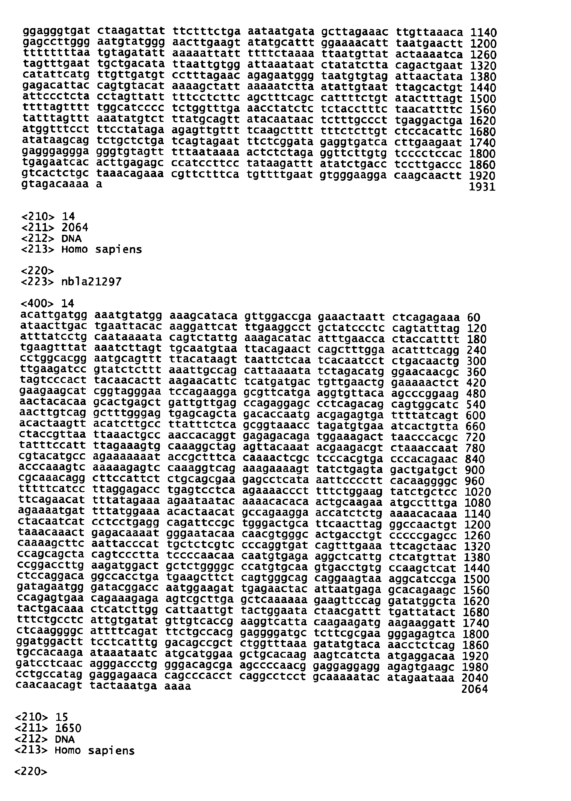
- Legal status (The legal status is an assumption and is not a legal conclusion. Google has not performed a legal analysis and makes no representation as to the accuracy of the status listed.)
- Granted
Links
Classifications
-
- G—PHYSICS
- G01—MEASURING; TESTING
- G01N—INVESTIGATING OR ANALYSING MATERIALS BY DETERMINING THEIR CHEMICAL OR PHYSICAL PROPERTIES
- G01N33/00—Investigating or analysing materials by specific methods not covered by groups G01N1/00 - G01N31/00
- G01N33/48—Biological material, e.g. blood, urine; Haemocytometers
- G01N33/50—Chemical analysis of biological material, e.g. blood, urine; Testing involving biospecific ligand binding methods; Immunological testing
- G01N33/53—Immunoassay; Biospecific binding assay; Materials therefor
- G01N33/575—Immunoassay; Biospecific binding assay; Materials therefor for cancer
- G01N33/5758—Immunoassay; Biospecific binding assay; Materials therefor for cancer involving compounds serving as markers for tumours, cancers or neoplasias, e.g. cellular determinants, receptors, heat shock/stress proteins, A-protein, oligosaccharides or metabolites
-
- C—CHEMISTRY; METALLURGY
- C12—BIOCHEMISTRY; BEER; SPIRITS; WINE; VINEGAR; MICROBIOLOGY; ENZYMOLOGY; MUTATION OR GENETIC ENGINEERING
- C12Q—MEASURING OR TESTING PROCESSES INVOLVING ENZYMES, NUCLEIC ACIDS OR MICROORGANISMS; COMPOSITIONS OR TEST PAPERS THEREFOR; PROCESSES OF PREPARING SUCH COMPOSITIONS; CONDITION-RESPONSIVE CONTROL IN MICROBIOLOGICAL OR ENZYMOLOGICAL PROCESSES
- C12Q1/00—Measuring or testing processes involving enzymes, nucleic acids or microorganisms; Compositions therefor; Processes of preparing such compositions
- C12Q1/68—Measuring or testing processes involving enzymes, nucleic acids or microorganisms; Compositions therefor; Processes of preparing such compositions involving nucleic acids
- C12Q1/6876—Nucleic acid products used in the analysis of nucleic acids, e.g. primers or probes
- C12Q1/6883—Nucleic acid products used in the analysis of nucleic acids, e.g. primers or probes for diseases caused by alterations of genetic material
- C12Q1/6886—Nucleic acid products used in the analysis of nucleic acids, e.g. primers or probes for diseases caused by alterations of genetic material for cancer
-
- C—CHEMISTRY; METALLURGY
- C12—BIOCHEMISTRY; BEER; SPIRITS; WINE; VINEGAR; MICROBIOLOGY; ENZYMOLOGY; MUTATION OR GENETIC ENGINEERING
- C12Q—MEASURING OR TESTING PROCESSES INVOLVING ENZYMES, NUCLEIC ACIDS OR MICROORGANISMS; COMPOSITIONS OR TEST PAPERS THEREFOR; PROCESSES OF PREPARING SUCH COMPOSITIONS; CONDITION-RESPONSIVE CONTROL IN MICROBIOLOGICAL OR ENZYMOLOGICAL PROCESSES
- C12Q2600/00—Oligonucleotides characterized by their use
- C12Q2600/112—Disease subtyping, staging or classification
-
- C—CHEMISTRY; METALLURGY
- C12—BIOCHEMISTRY; BEER; SPIRITS; WINE; VINEGAR; MICROBIOLOGY; ENZYMOLOGY; MUTATION OR GENETIC ENGINEERING
- C12Q—MEASURING OR TESTING PROCESSES INVOLVING ENZYMES, NUCLEIC ACIDS OR MICROORGANISMS; COMPOSITIONS OR TEST PAPERS THEREFOR; PROCESSES OF PREPARING SUCH COMPOSITIONS; CONDITION-RESPONSIVE CONTROL IN MICROBIOLOGICAL OR ENZYMOLOGICAL PROCESSES
- C12Q2600/00—Oligonucleotides characterized by their use
- C12Q2600/118—Prognosis of disease development
-
- C—CHEMISTRY; METALLURGY
- C12—BIOCHEMISTRY; BEER; SPIRITS; WINE; VINEGAR; MICROBIOLOGY; ENZYMOLOGY; MUTATION OR GENETIC ENGINEERING
- C12Q—MEASURING OR TESTING PROCESSES INVOLVING ENZYMES, NUCLEIC ACIDS OR MICROORGANISMS; COMPOSITIONS OR TEST PAPERS THEREFOR; PROCESSES OF PREPARING SUCH COMPOSITIONS; CONDITION-RESPONSIVE CONTROL IN MICROBIOLOGICAL OR ENZYMOLOGICAL PROCESSES
- C12Q2600/00—Oligonucleotides characterized by their use
- C12Q2600/158—Expression markers
Definitions
- This invention relates to nucleic acids derived from genes expressed in human neuroblastoma. More particularly, the invention relates to nucleic acids derived from genes expressed in human stage 4s neuroblastoma. Further, this invention relates to diagnostic agents and diagnostic kits for stage 4s neuroblastoma comprising nucleic acid probes, nucleic acid primers or nucleic acid microarrays utilizing those nucleic acids, their fragments, or the combinations of the foregoing, as well as to the elucidation of the mechanism of the programmed cell death of cancer cells based on the nucleic acid sequence data from the abovementioned genes.
- Neuroblastoma is a pediatric cancer occurring in sympathetic gangliocytes and adrenal medullary cells which originate from cells of the peripheral sympathetic nervous system.
- sympathetic neurons neural crest cells in the initial stage of development migrate to the abdomen, differentiating and maturing at sites where sympathetic ganglia are formed. Some of these cells migrate further to the adrenal bodies, penetrating through the adrenal cortex which is already in the process of formation, and reaching the medulla and forming medullary substance there.
- the neural crest cells also serve as a source of other peripheral nerve cells, differentiating into dorsal root ganglia (sensory nerves), skin pigment cells, thyroid C cells, some pulmonary cells, intestinal gangliocytes, and the like.
- Neuroblastoma is characterized by a varied clinical profile (Nakagawara, Shinkeigashu no Hassei to Sono Bunshi Kiko [Neuroblastoma Development and Molecular Mechanism], Shoni Naika [Japanese Journal of Pediatric Medicine], Vol. 30, p. 143, 1998).
- neuroblastoma occurring at less than one year of age has very favorable prognosis, with the majority undergoing differentiation and cell death, and results in spontaneous regression (which may be referred to as "favorable prognosis type").
- neuroblastoma occurring at age 1 or higher is highly malignant and leads to death of the infant in the majority of cases despite therapy (which may be referred to as "unfavorable prognosis type"). It is also hypothesized that a somatic mutation occurs in highly malignant neuroblastomas in infants older than one year of age, which are of monoclonal nature, whereas in naturally regressing neuroblastomas, the genetic mutation remains at only a germ line mutation. See Knudson AG, et al.: Regression of neuroblastoma IV-S: A genetic hypothesis, N. Engl. J. Med., Vol. 302, p. 1254, 1980. In addition, there are known neuroblastomas of the intermediate type that are clinically positioned between those two types.
- the neuroblastomas of the favorable prognosis type are tumors at stages 1, 2, and 4s, while the neuroblastomas of the unfavorable and intermediate types are tumors at stages 3 and 4.
- the tumor at stage 4s is peculiar and normally occurs in an infant at several months after birth. Although the tumor grows and metastasizes quickly, it suddenly stops growing and then disappears spontaneously. Thus, the tumors that regress spontaneously and the tumors that grow malignantly are clearly distinct, with respect to the age of onset, the site of metastasis, and the progress.
- TrkA nerve growth factor receptor TrkA
- NGF nerve growth factor
- Trk family receptors play an important role in specific nerve cell differentiation and survival in the central nervous and peripheral nervous systems (see Nakagawara, et al., Shinkeigasaiboushu ni Okeru Neurotrophin Juyoutai no Hatsugen to Yogo [Expression of Neurotrophin Receptors and Prognosis in Neuroblastoma], Shoni Geka (Japanese Journal of Pediatric Surgery), Vol. 29, pp. 425-432, 1997).
- the survival and differentiation of tumor cells is controlled by signals from Trk tyrosine kinase and Ret tyrosine kinase.
- TrkA receptor the role of TrkA receptor is most significant, with TrkA expression being notably high in neuroblastomas of the favorable prognosis type, and its signals exerting a powerful control over the survival and differentiation of tumor cells, and cell death (apoptosis).
- TrkA expression is significantly suppressed, while tumor development is aided by a mechanism in which survival is promoted by signals from TrkB and Ret, instead.
- CD44, PTN, caspase and others are known as the gene whose expression is high in neuroblastomas of the favorable prognosis type, whereas SVV (survivin), MK (midkine) and others are known as the gene whose expression is high in neuroblastomas of the unfavorable prognosis type.
- the present inventors found that a group of novel genes was highly expressed in neuroblastomas of the favorable prognosis type (International Publication PCT/JP01631 pamphlet). By contrast, the present inventors found that a different group of novel genes was highly expressed in neuroblastomas of the unfavorable prognosis type (International Publication PCT/JP01629 pamphlet).
- stage 4s neuroblastoma Up till the present time, however, there has hardly been any information concerning the genes which are expressed in stage 4s neuroblastoma, particularly with specificity. Further, since stage 4s neuroblastoma regresses spontaneously, there is an urgent need to identify the causative genes.
- This invention has been accomplished in light of the problems inherent in the above-described prior art, and its object is to identify the nucleic acid sequences of genes which are generally related to the favorable or unfavorable prognosis of neuroblastoma, and to allow for providing such genetic information and for diagnosis for the prognosis of neuroblastoma (whether favorable or unfavorable).
- This invention specifically aims at diagnosing the prognosis of neuroblastoma, classifying the progress of the neuroblastoma, and enabling the determination of stage 4s neuroblastoma.
- the present inventors have examined the prognoses of neuroblastomas and have succeeded in constructing cDNA libraries from the respective clinical tissues of the favorable and unfavorable prognosis types. Approximately 2,400 clones were respectively cloned from these two types of cDNA libraries and were classified according to the prognoses of neuroblastomas (whether favorable or unfavorable). Profiling of the genes in the respective subsets was then carried out.
- the present inventors found that a group of genes showed differential expression levels among the abovementioned subsets and showed enhanced expression levels only in the clinical tissues of the favorable prognosis type. Moreover, the present inventors found that a group of genes showed enhanced expression levels only in the clinical tissues of the unfavorable prognosis type. Based on such finding, the present inventors made it possible to provide nucleic acid sequence data which would allow the detection and cloning of genes whose expression levels are at least enhanced either only in the clinical tissues of the favorable prognosis type or only in the clinical tissues of the unfavorable prognosis type.
- the present inventors have succeeded in constructing cDNA libraries similarly from the clinical tissues of stage 4s neuroblastoma. Approximately 2,700 clones were cloned from this library. The subset of this library and the subsets of the libraries from the clinical tissues of the favorable and unfavorable prognosis types were analyzed and the profiling of approximately 16,000 genes that were expressed among these subsets was carried out. Consequently, 452 genes were identified that showed differential expression levels among the abovementioned subsets. When these genes were sequenced, they were found to comprise 308 novel genes and 144 known genes as the reminder. The genes were classified according to their expression patterns among the respective subsets, and grouped into seven groups.
- the present inventors made it possible to provide genetic information (such as nucleic acid sequence data) which would allow the detection and cloning of genes exhibiting expression patterns characteristic of stage 4s neuroblastoma. Further based on the nucleic acid sequence data, the present inventors made it possible to provide diagnostic agents and diagnostic kits which allow for methods of diagnosis for the prognosis of neuroblastoma (particularly, the classification of the progress), including determination on stage 4s neuroblastoma, upon which this invention has been completed.
- genetic information such as nucleic acid sequence data
- diagnostic agents and diagnostic kits which allow for methods of diagnosis for the prognosis of neuroblastoma (particularly, the classification of the progress), including determination on stage 4s neuroblastoma, upon which this invention has been completed.
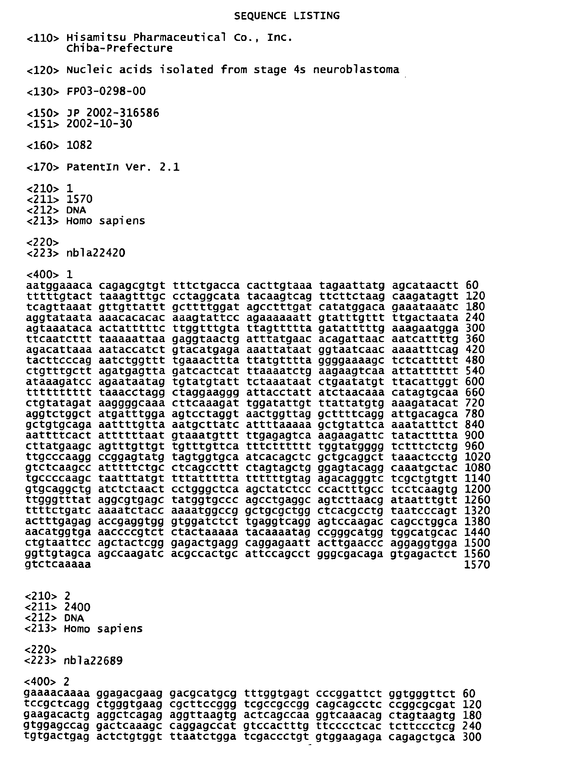
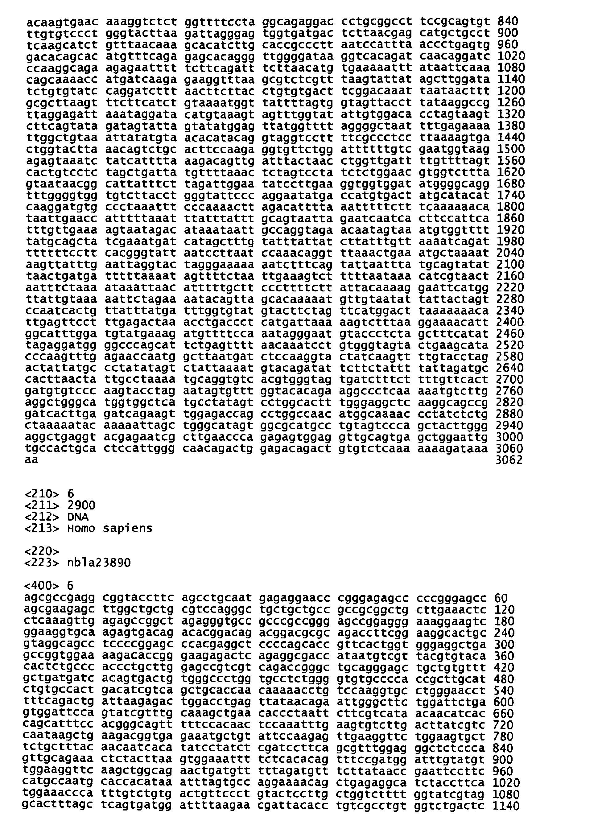
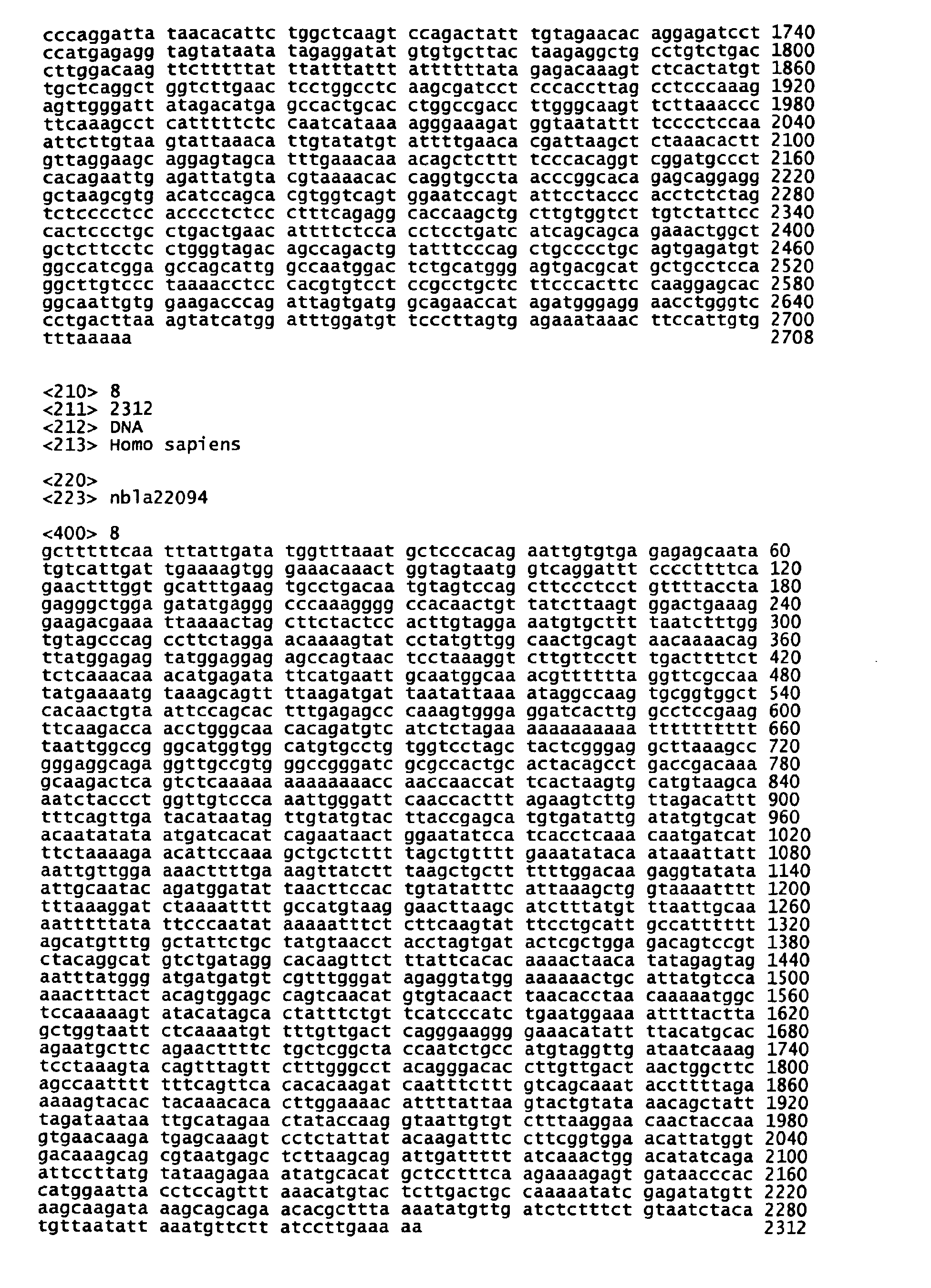
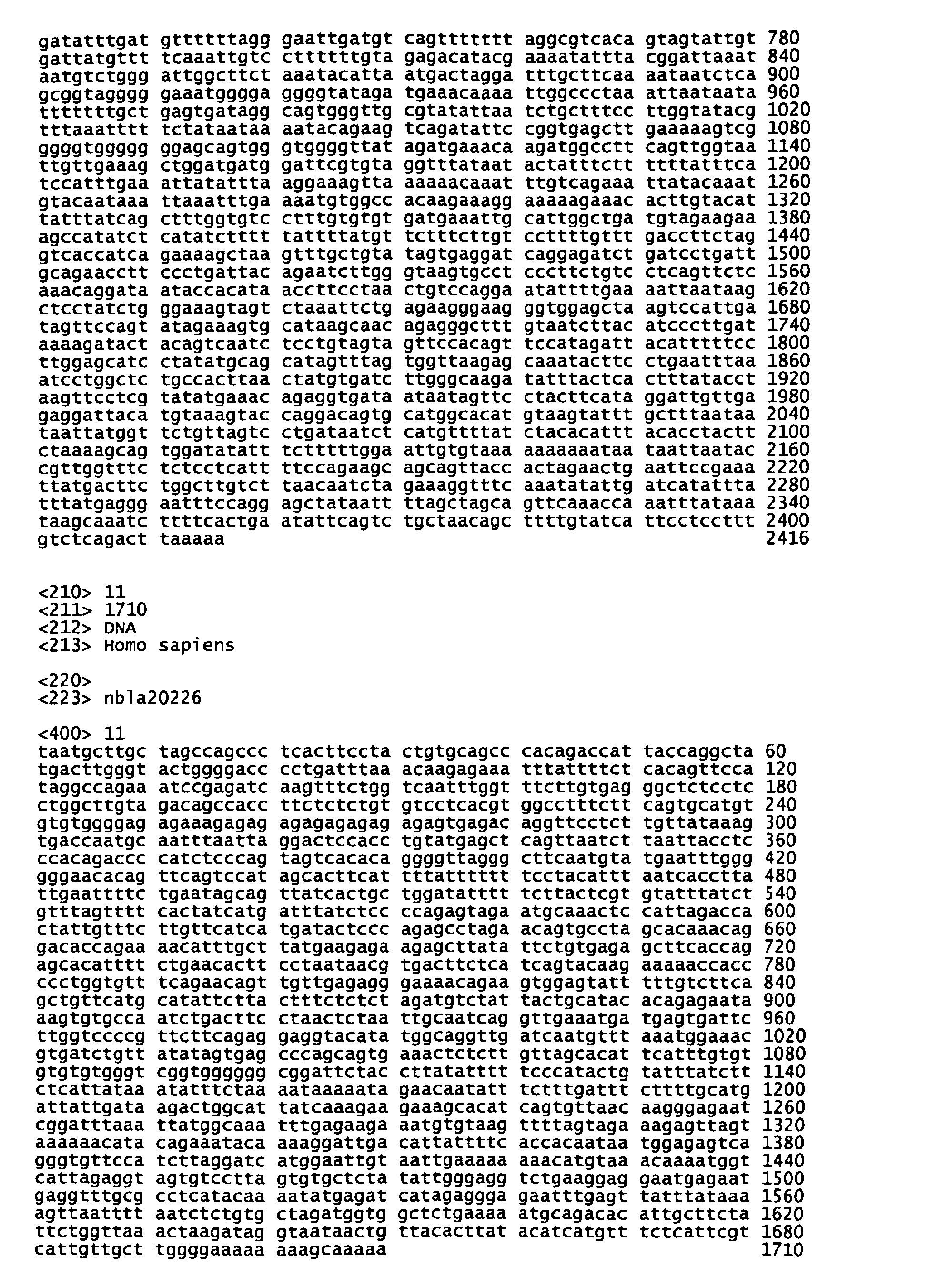
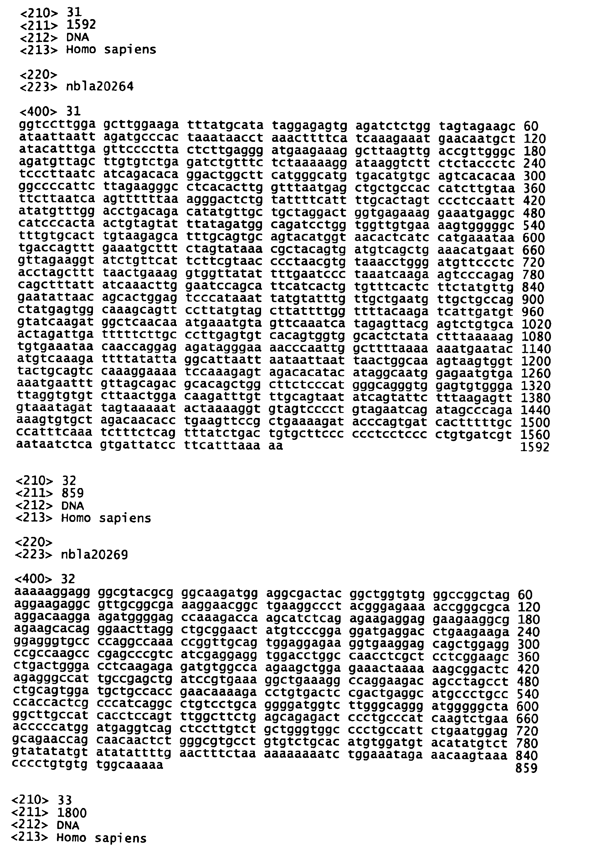
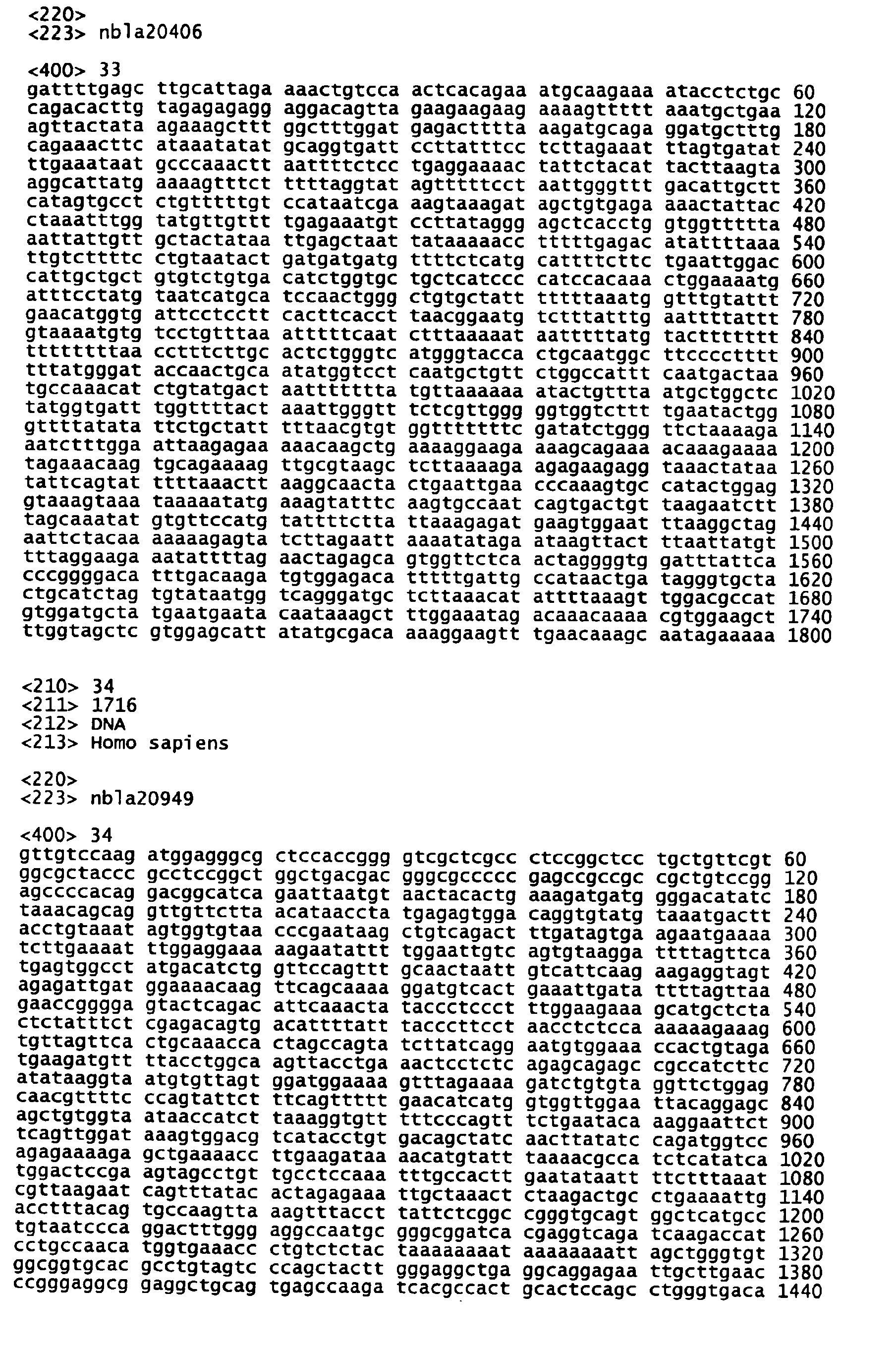
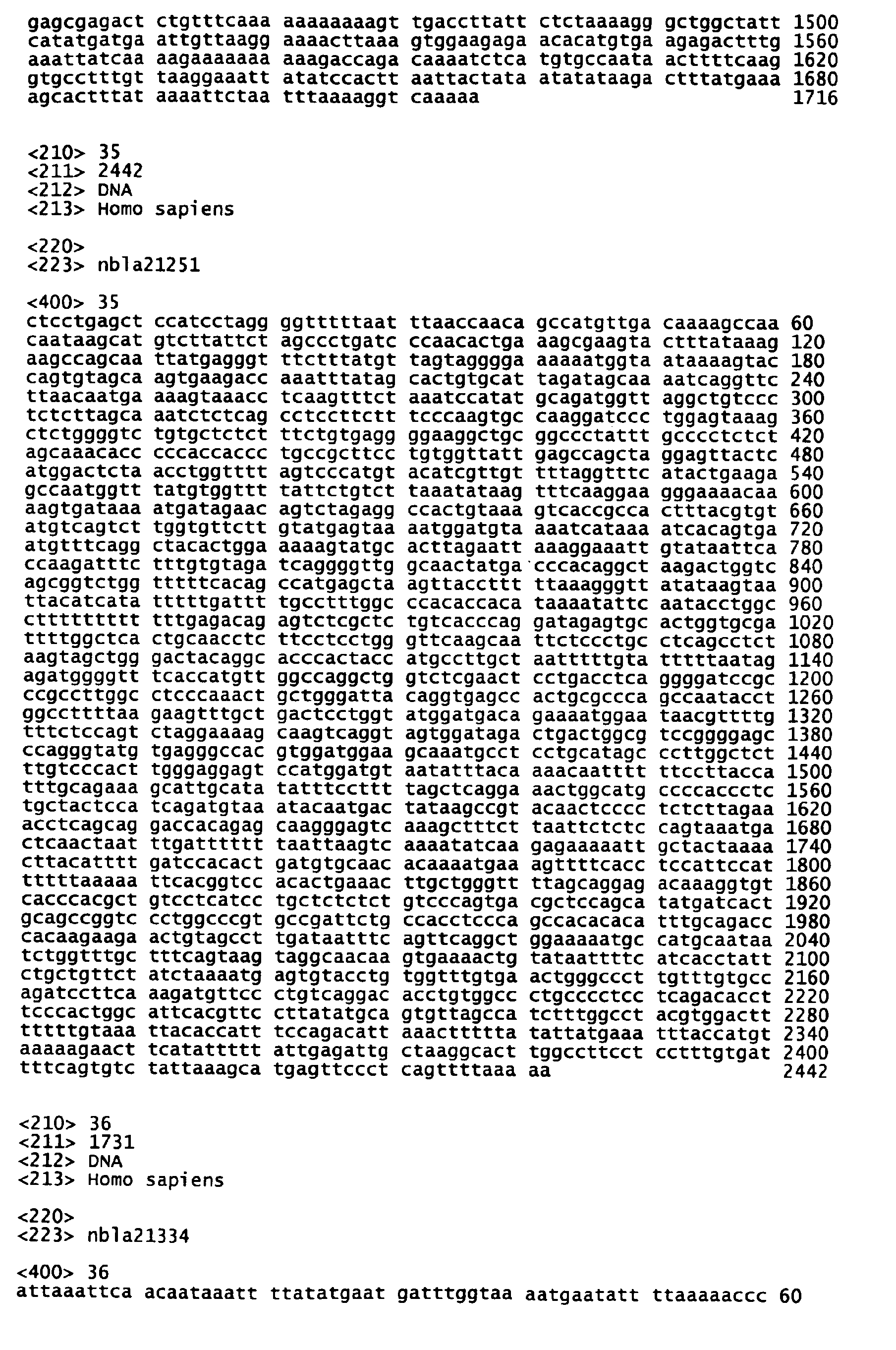
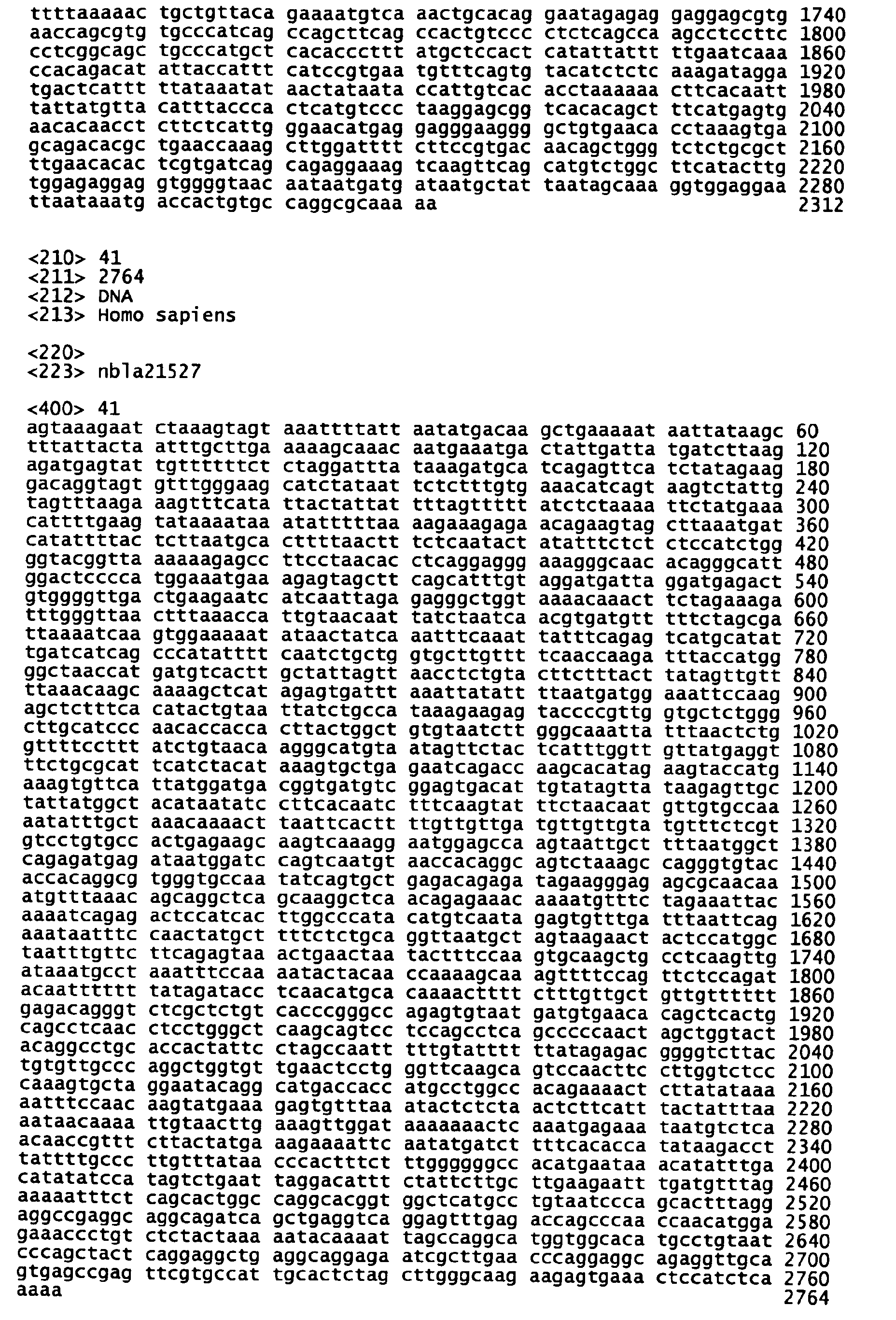
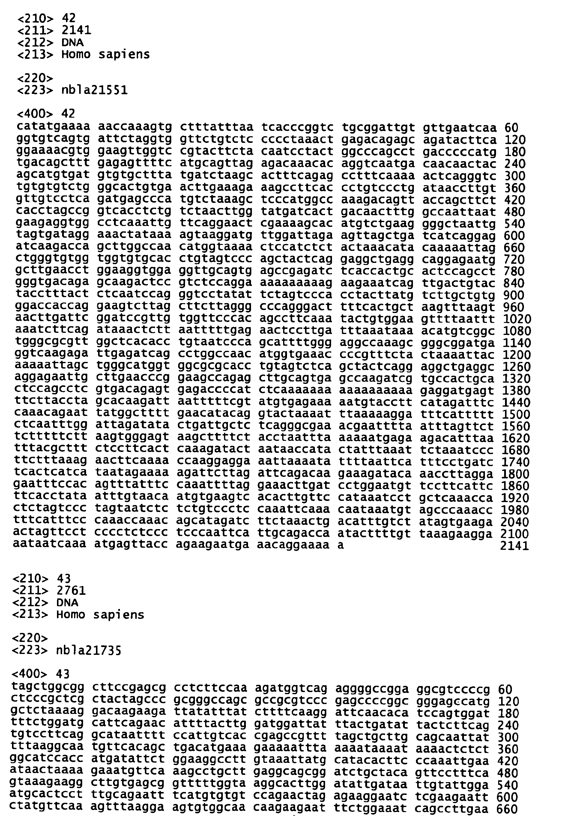
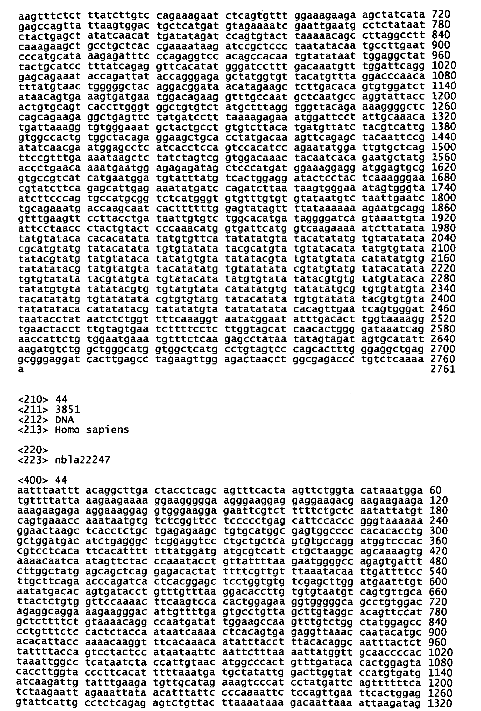
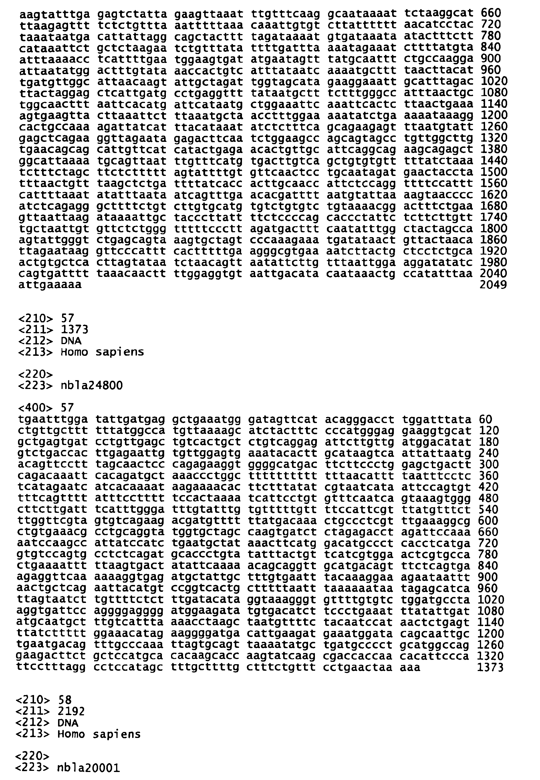
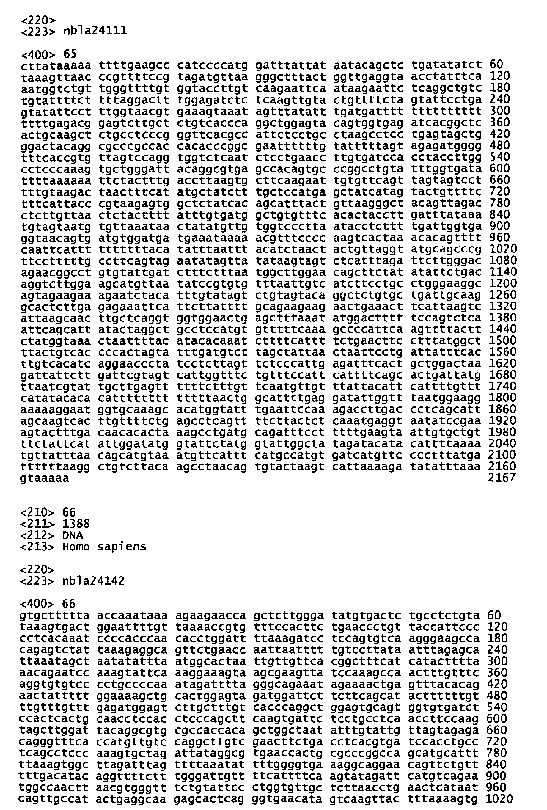
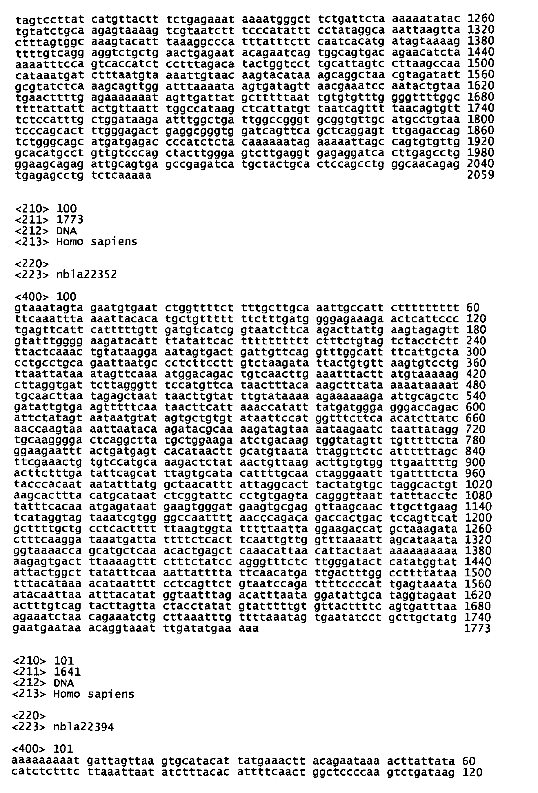
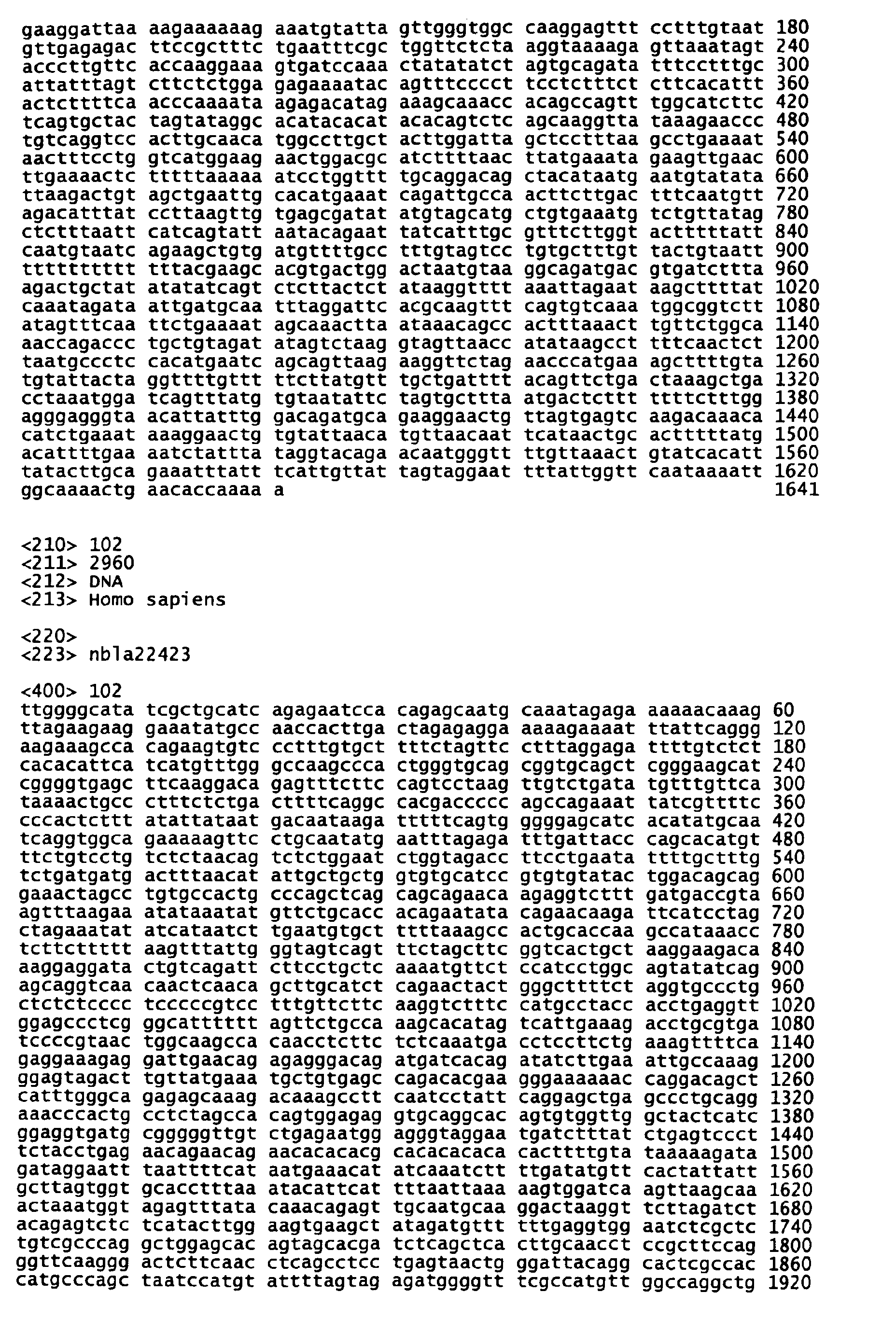
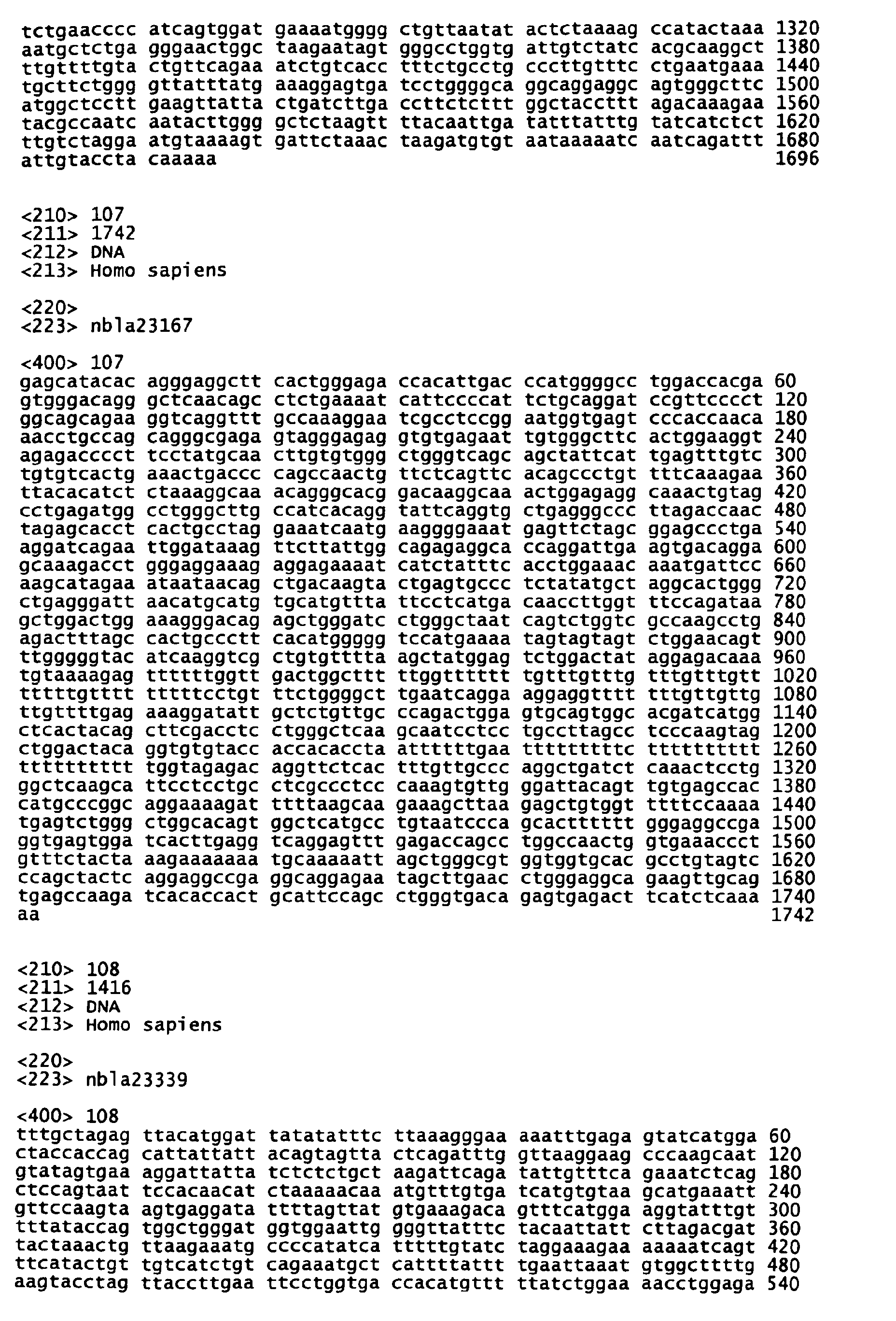
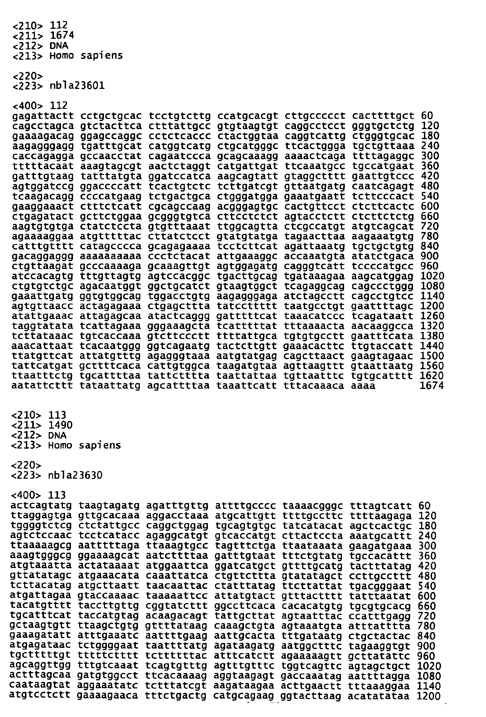
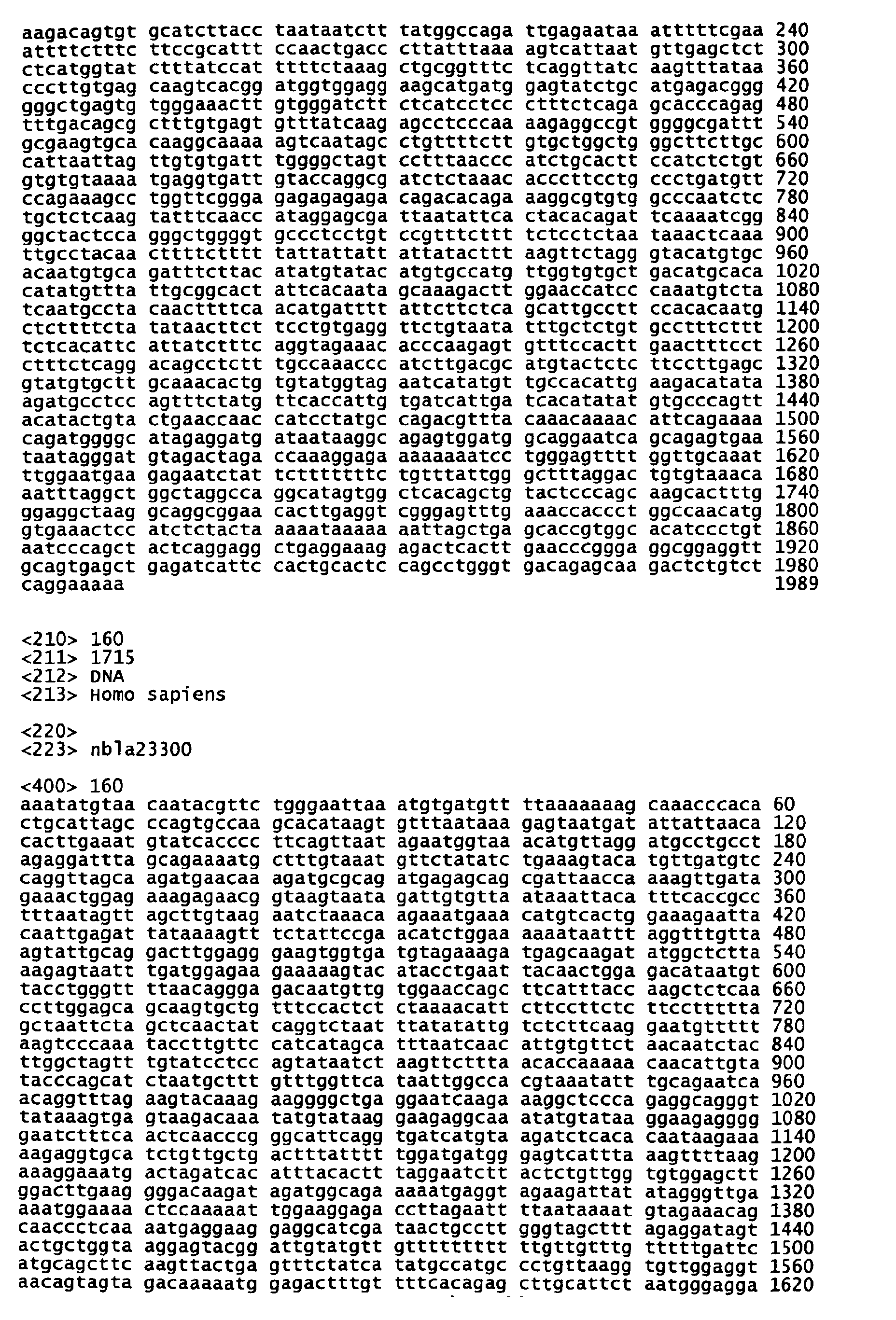
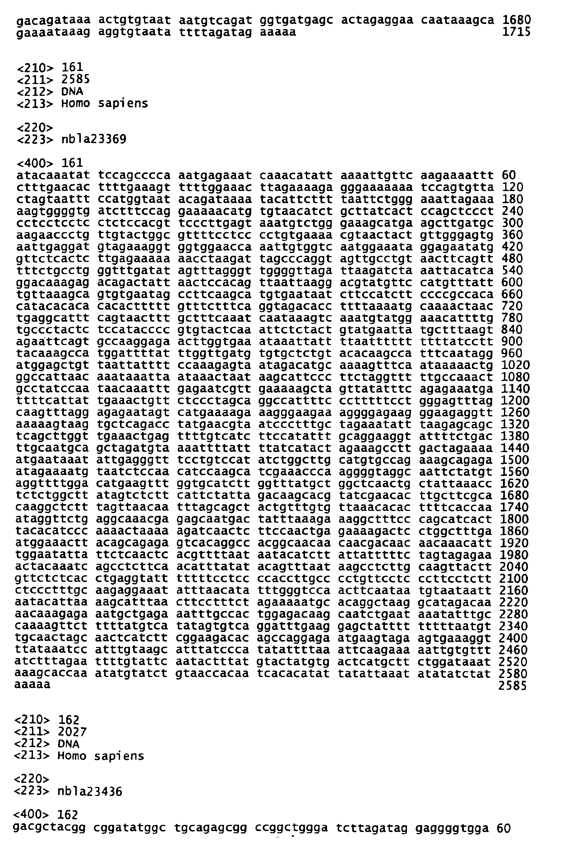
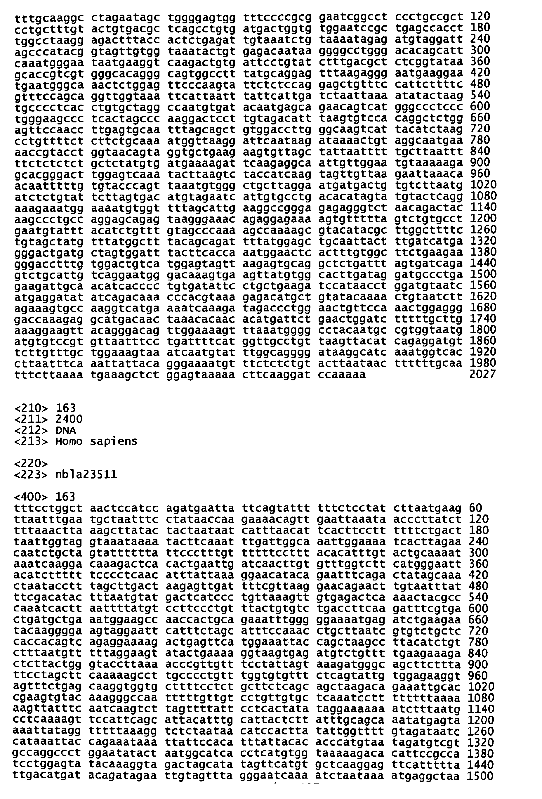
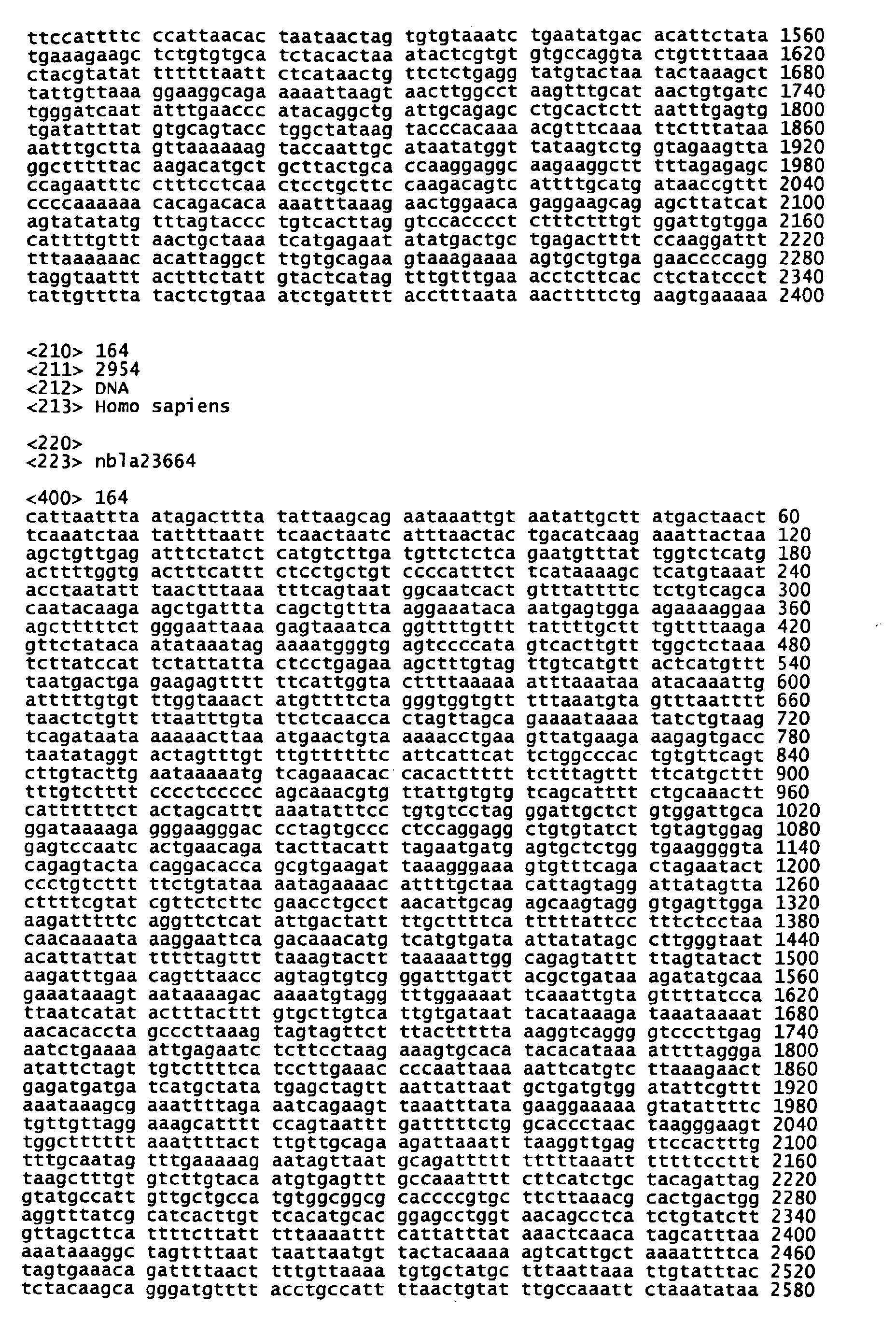
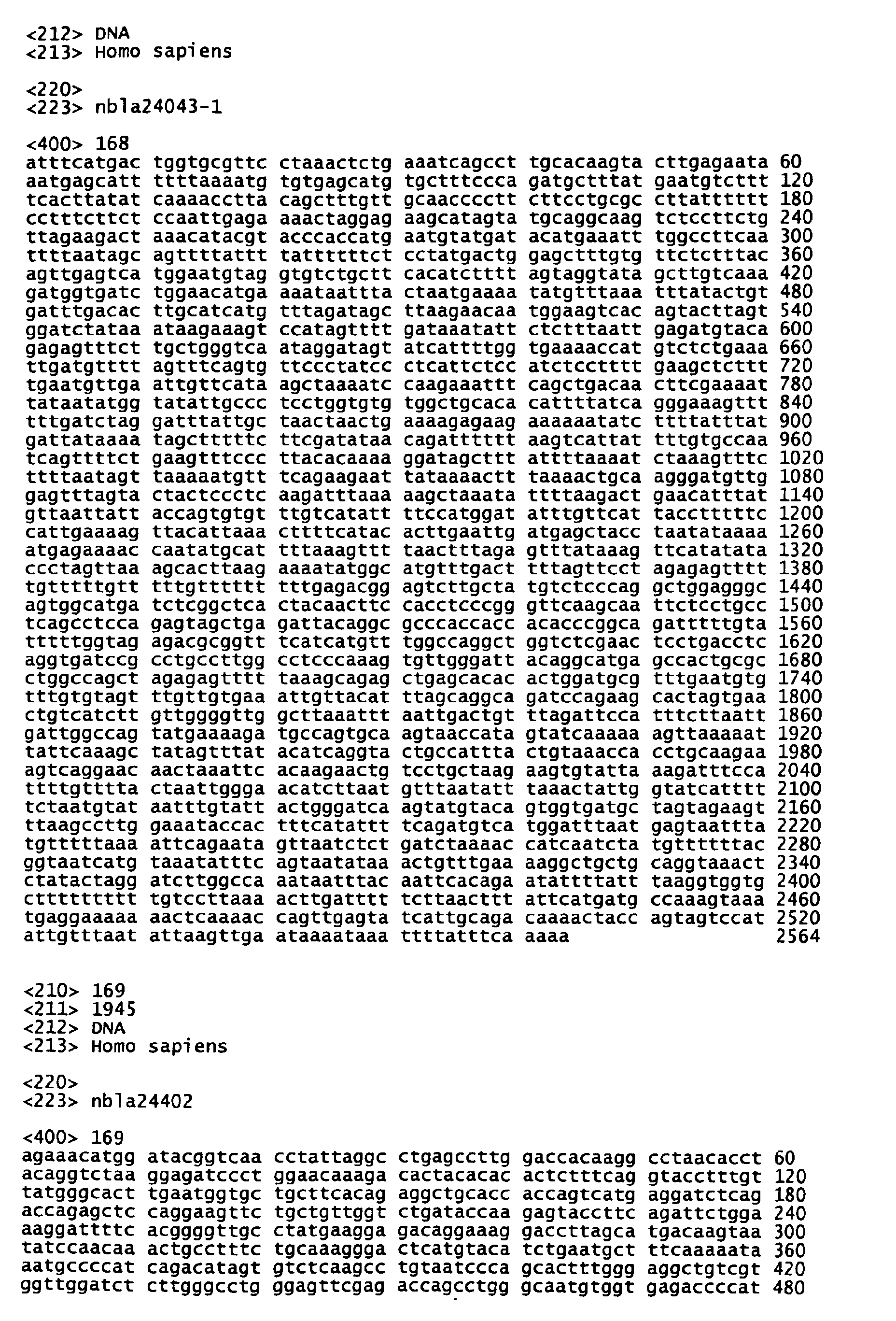
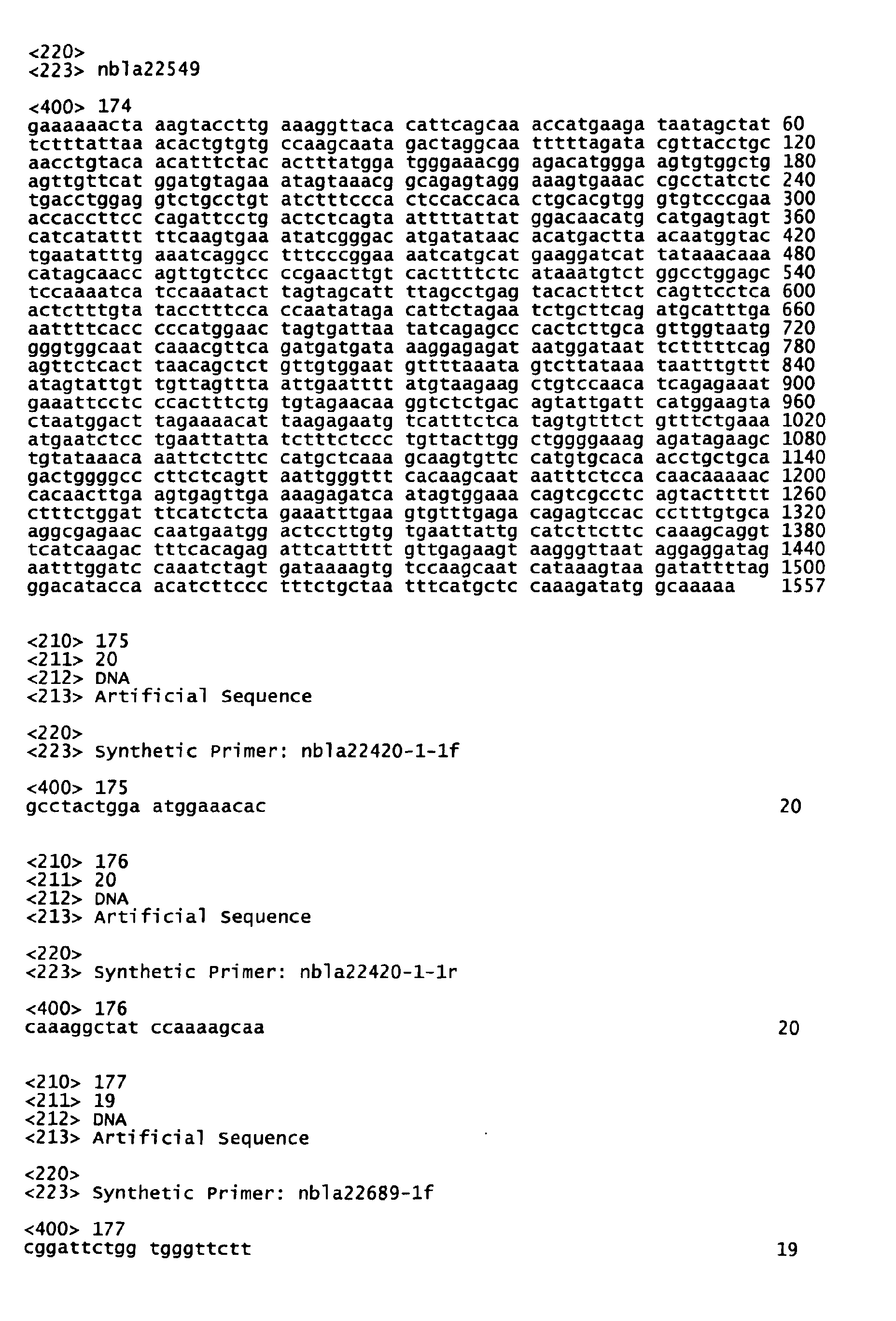
- this invention provides a nucleic acid comprising one sequence selected from the group consisting of nucleic acid sequences set forth in SEQ ID NO:1 to SEQ ID NO:174 in the
- the preferable nucleic acid is one that comprises a nucleic acid sequence of any one of nucleic acid sequences set forth in SEQ ID NO:1 to SEQ ID NO:14 among the nucleic acid sequences set forth in SEQ ID NO:1 to SEQ ID NO:174 in the Sequence Listing.
- This invention also provides nucleic acids complementary to those nucleic acids described above.
- This invention further provides nucleic acids capable of hybridizing to the nucleic acids described above or their complementary nucleic acids under stringent conditions.
- nucleic acid probe comprising nucleic acid (a) or nucleic acid (b) described below:
- the nucleic acid (a) or the nucleic acid (b) is DNA.
- the nucleic acid (a) or the nucleic acid (b) is a nucleic acid comprising one sequence selected from the group consisting of nucleic acid sequences set forth in SEQ ID NO:1 to SEQ ID NO:14 in the Sequence Listing.
- This invention also provides a diagnostic agent for stage 4s neuroblastoma comprising the nucleic acid probe described above as the active ingredient.
- This invention further provides a primer containing DNA
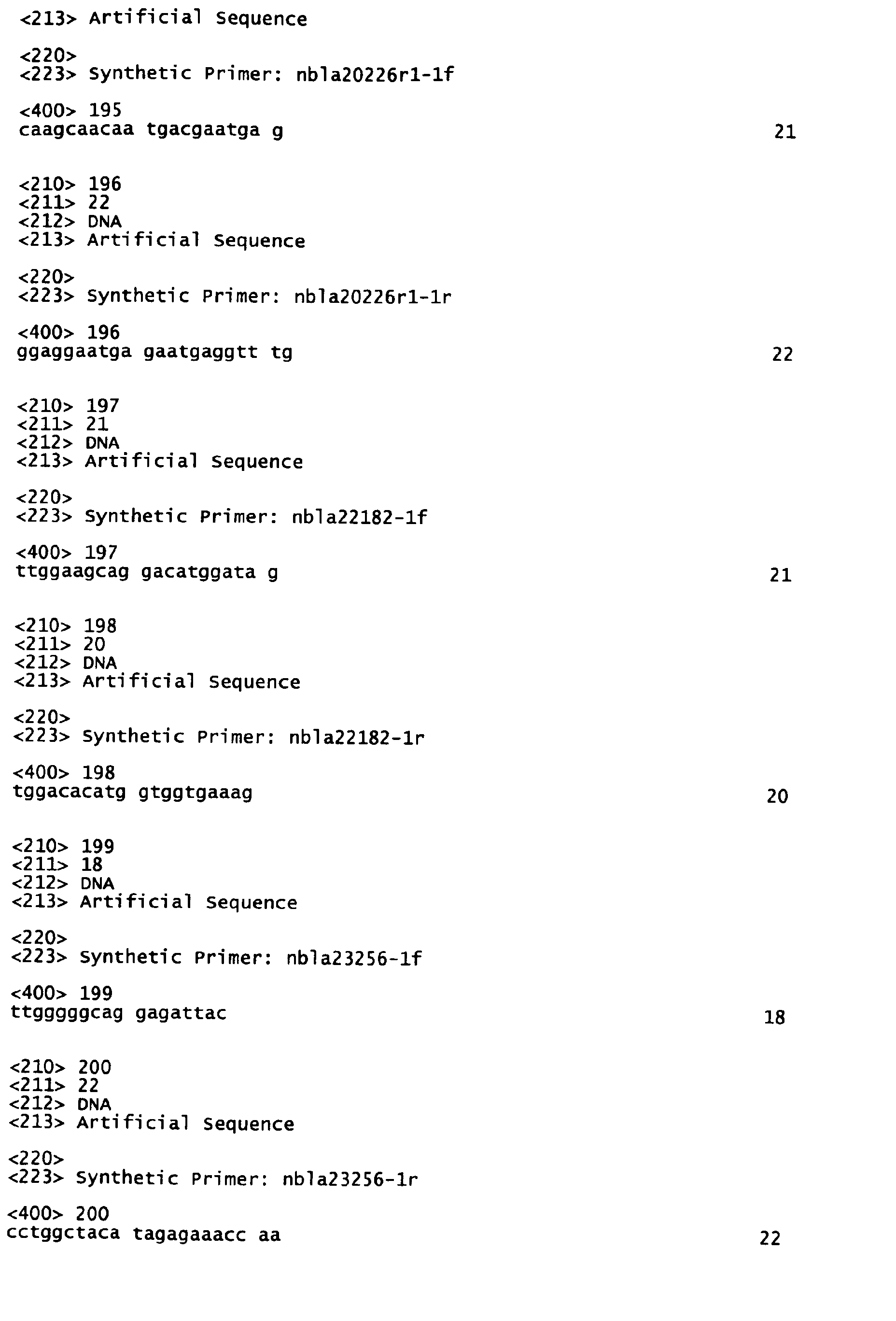
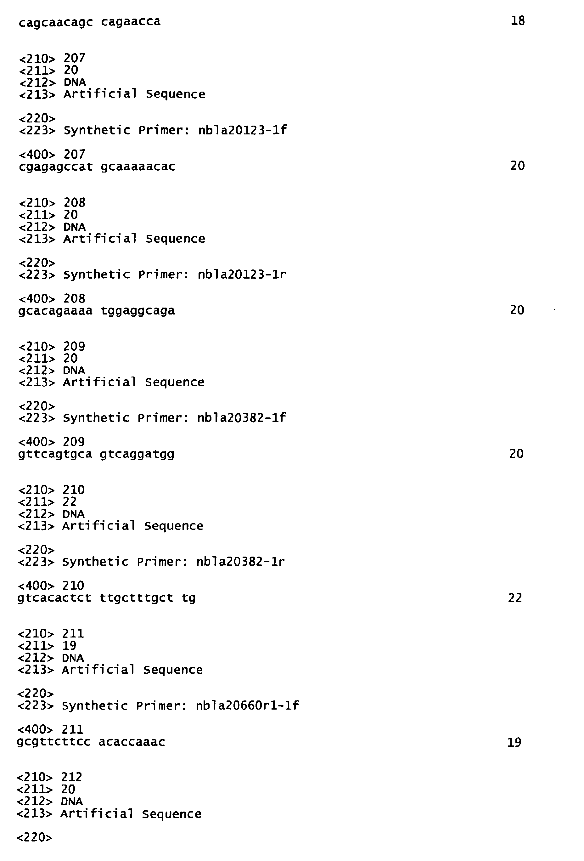
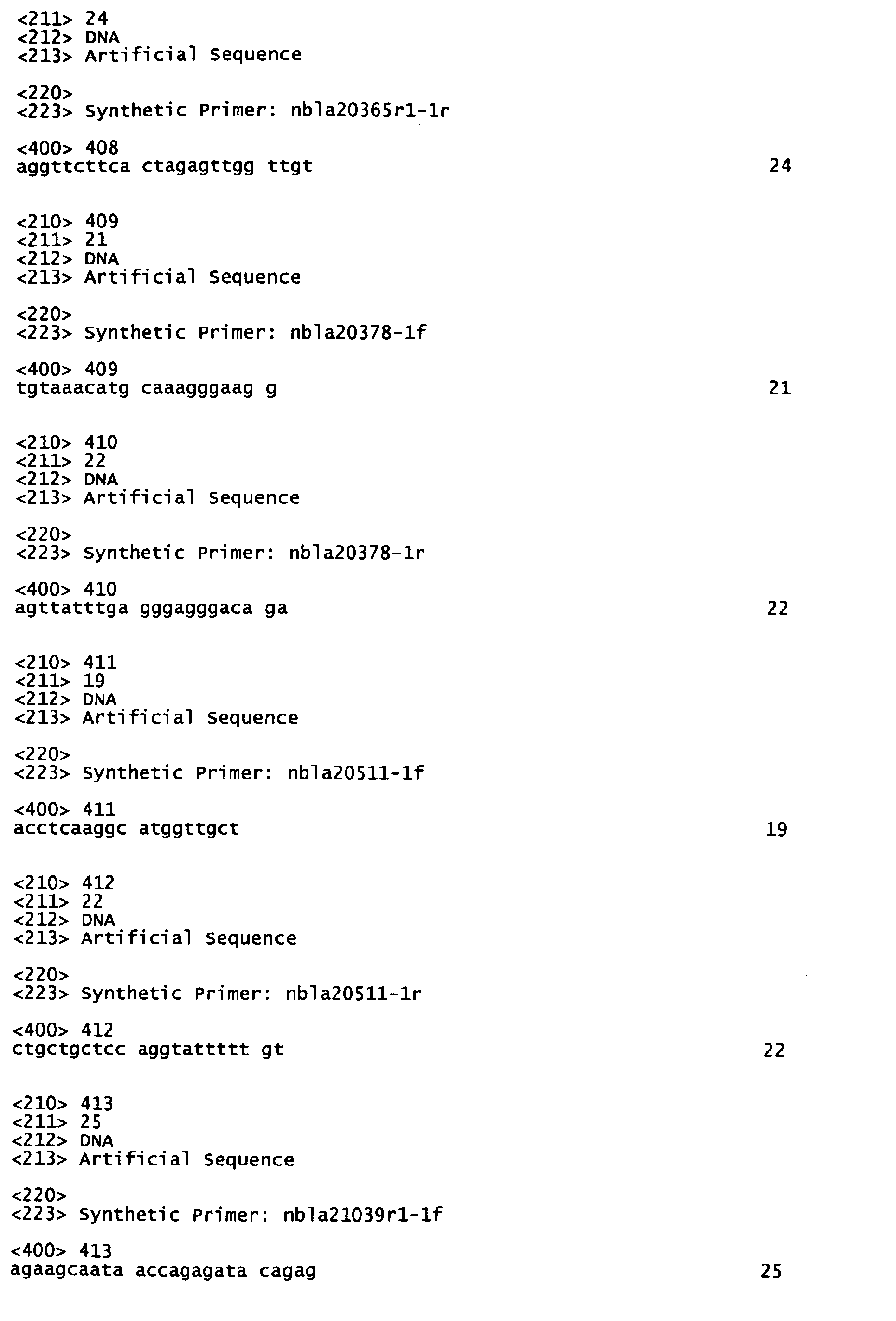
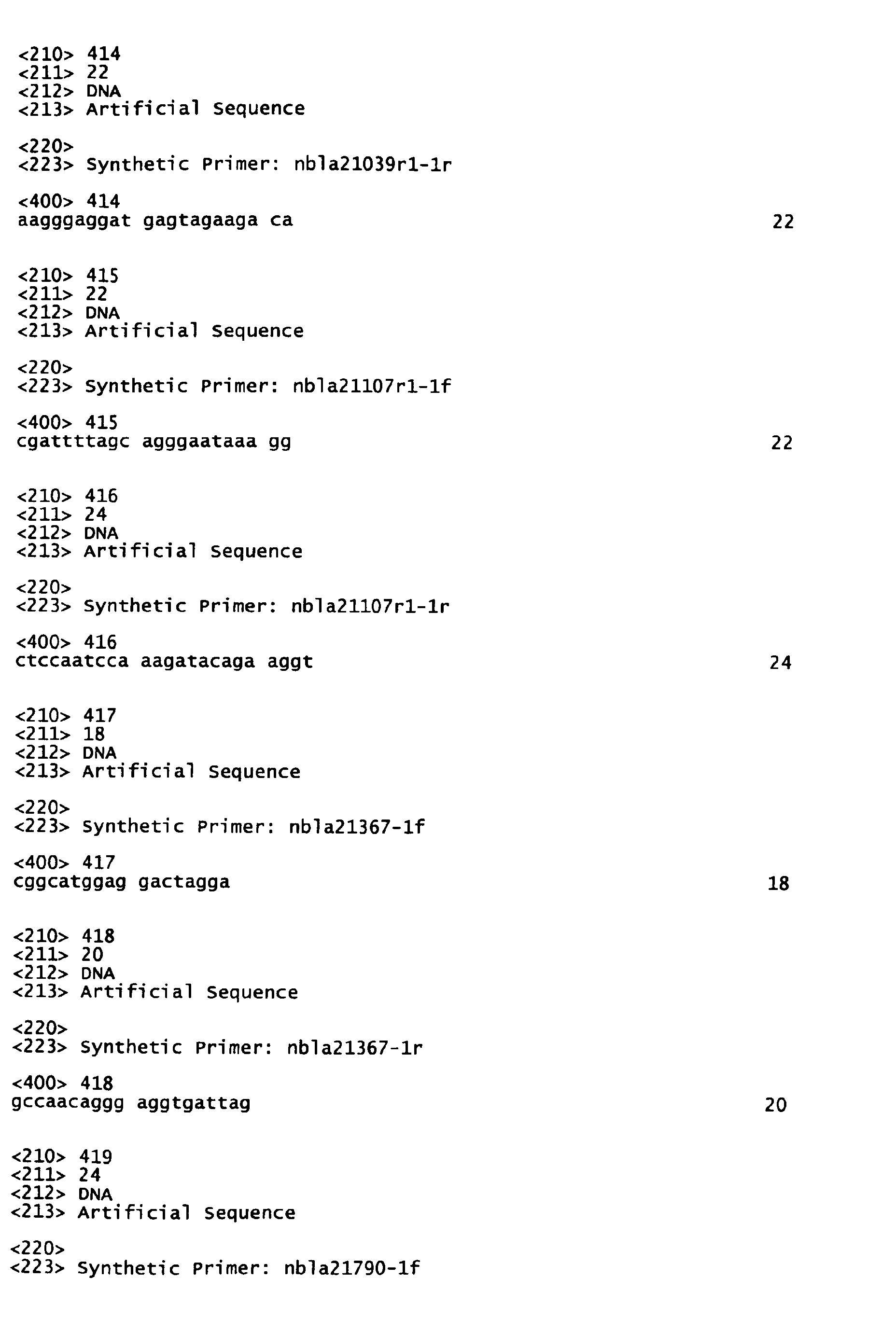
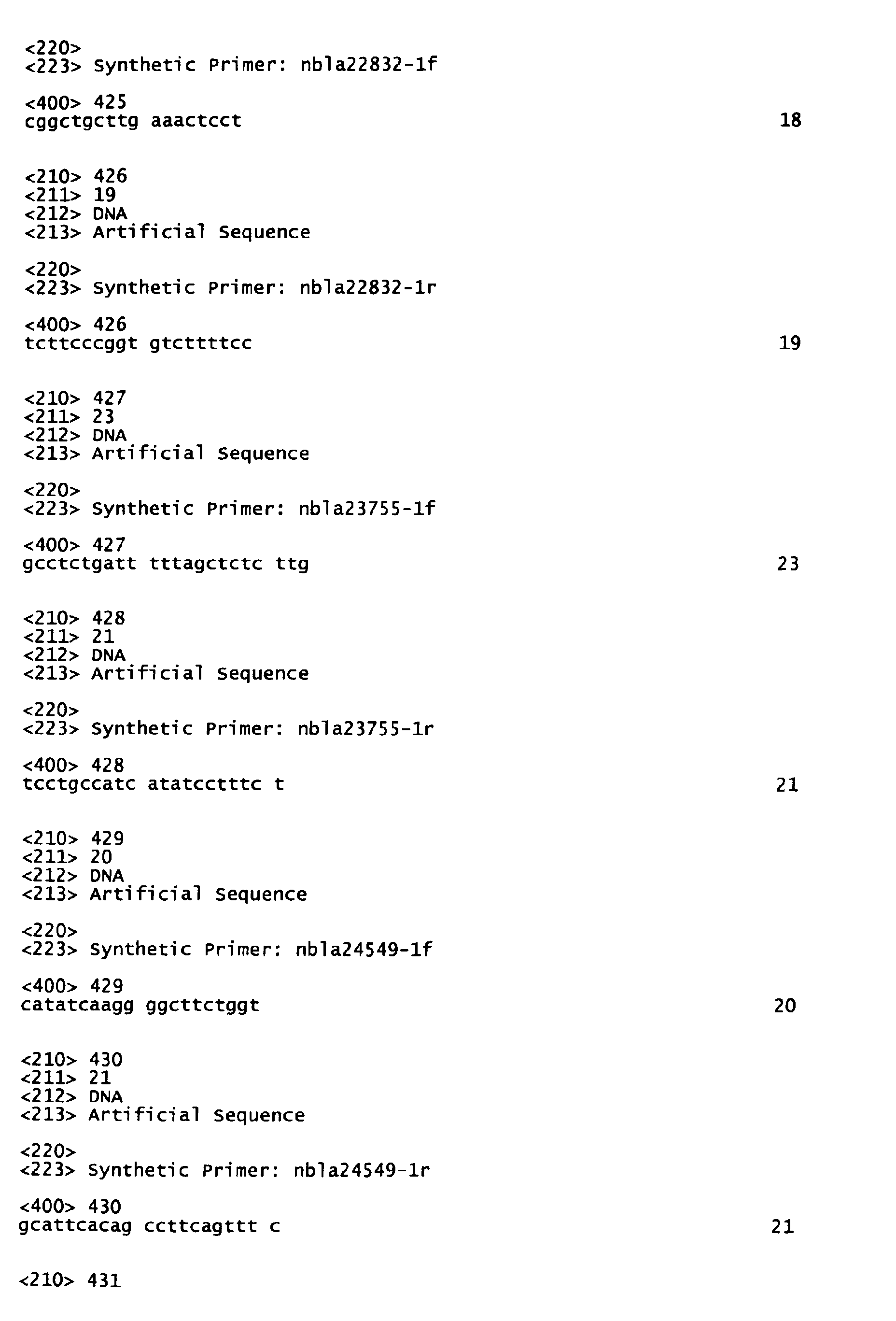
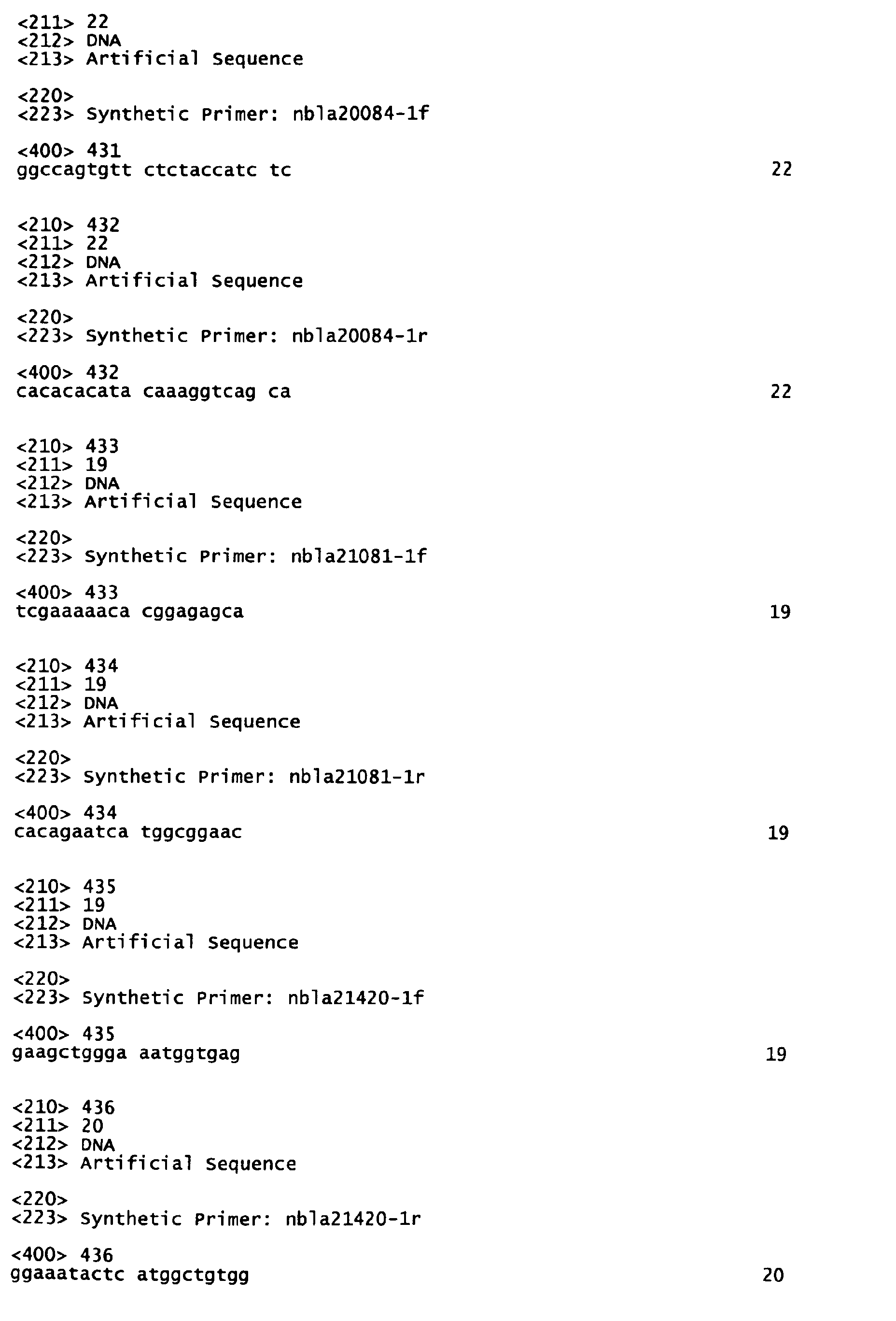
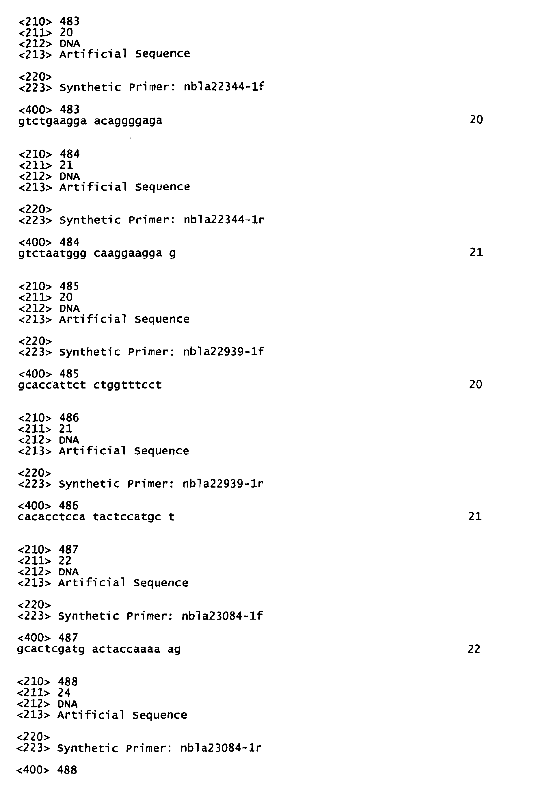
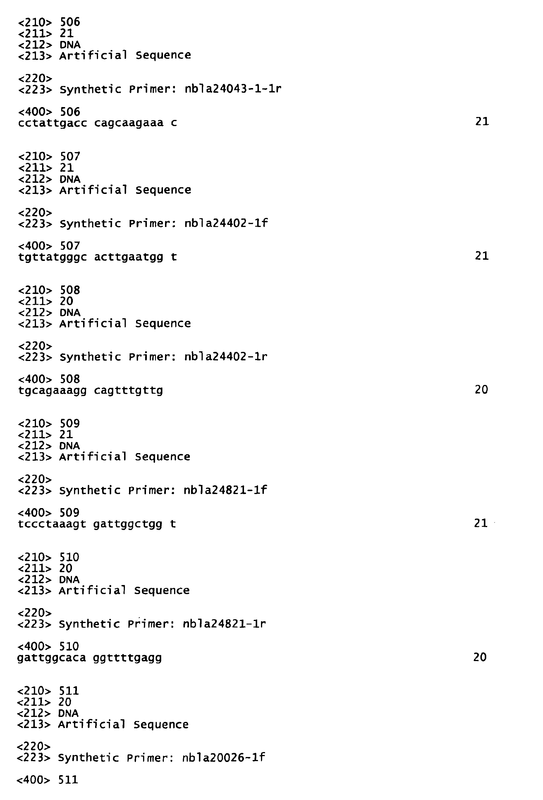
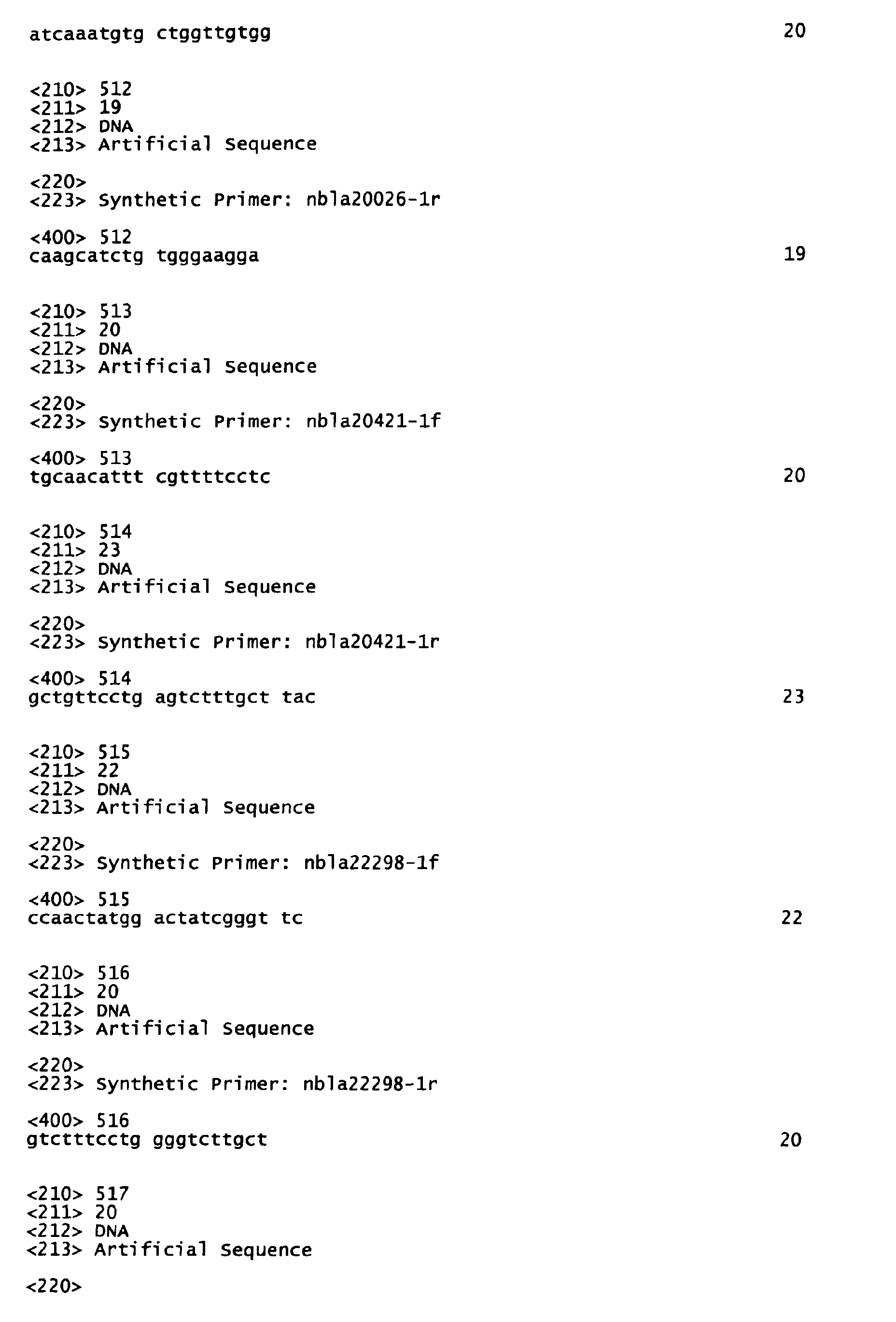
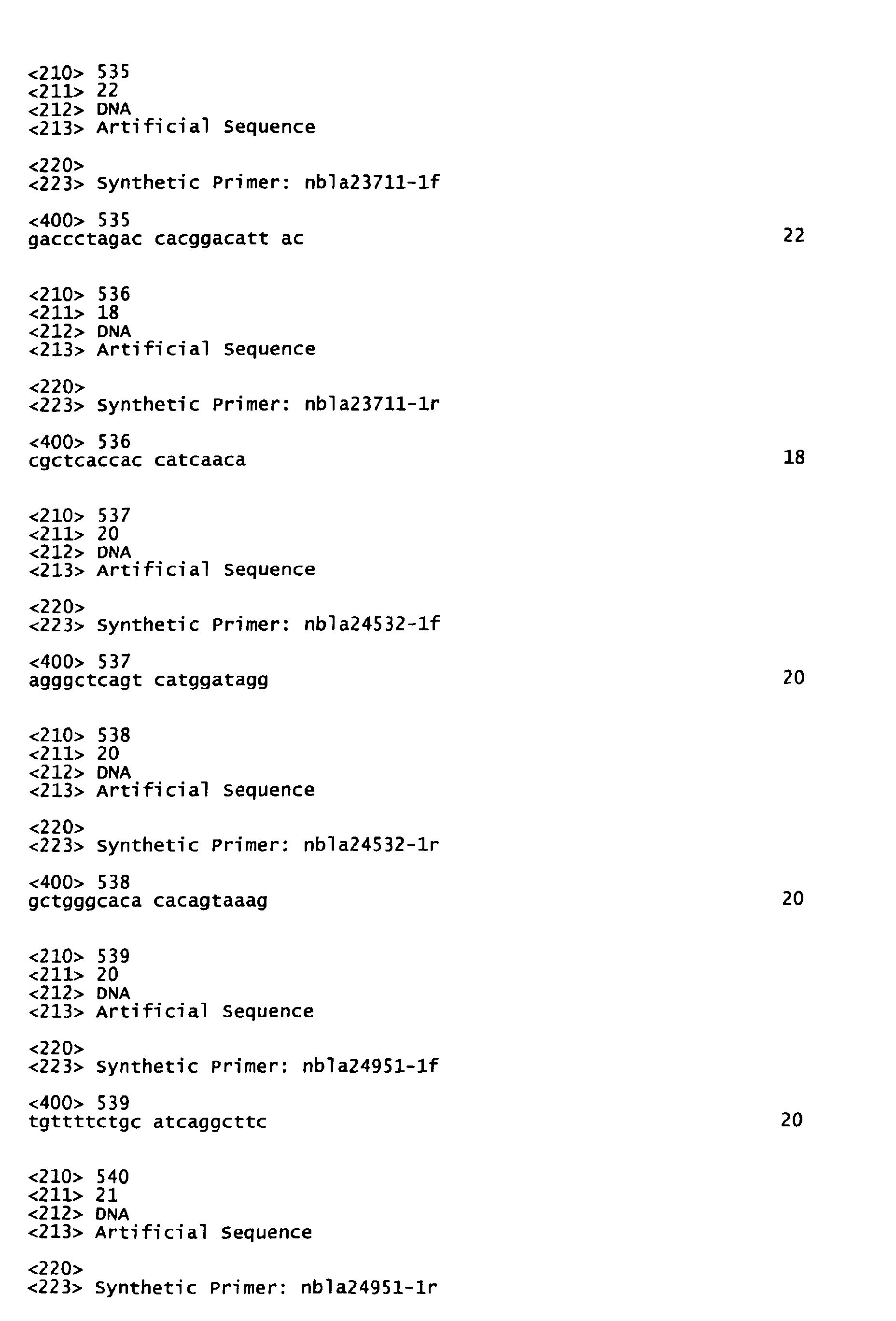
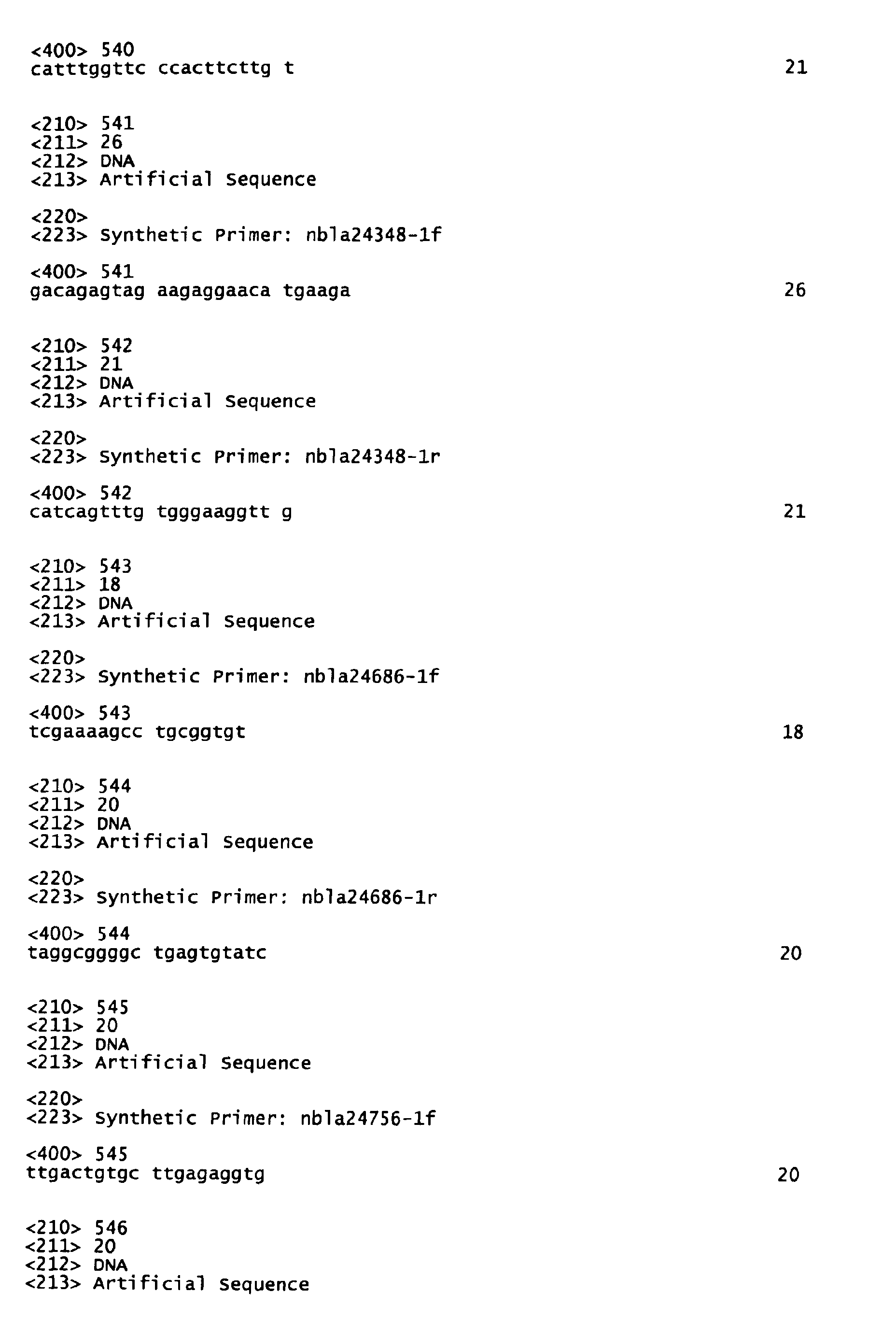
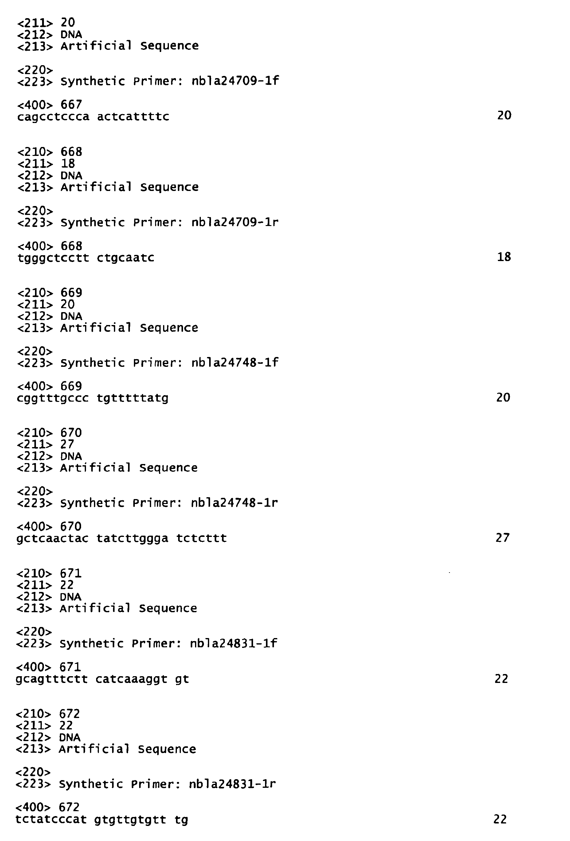
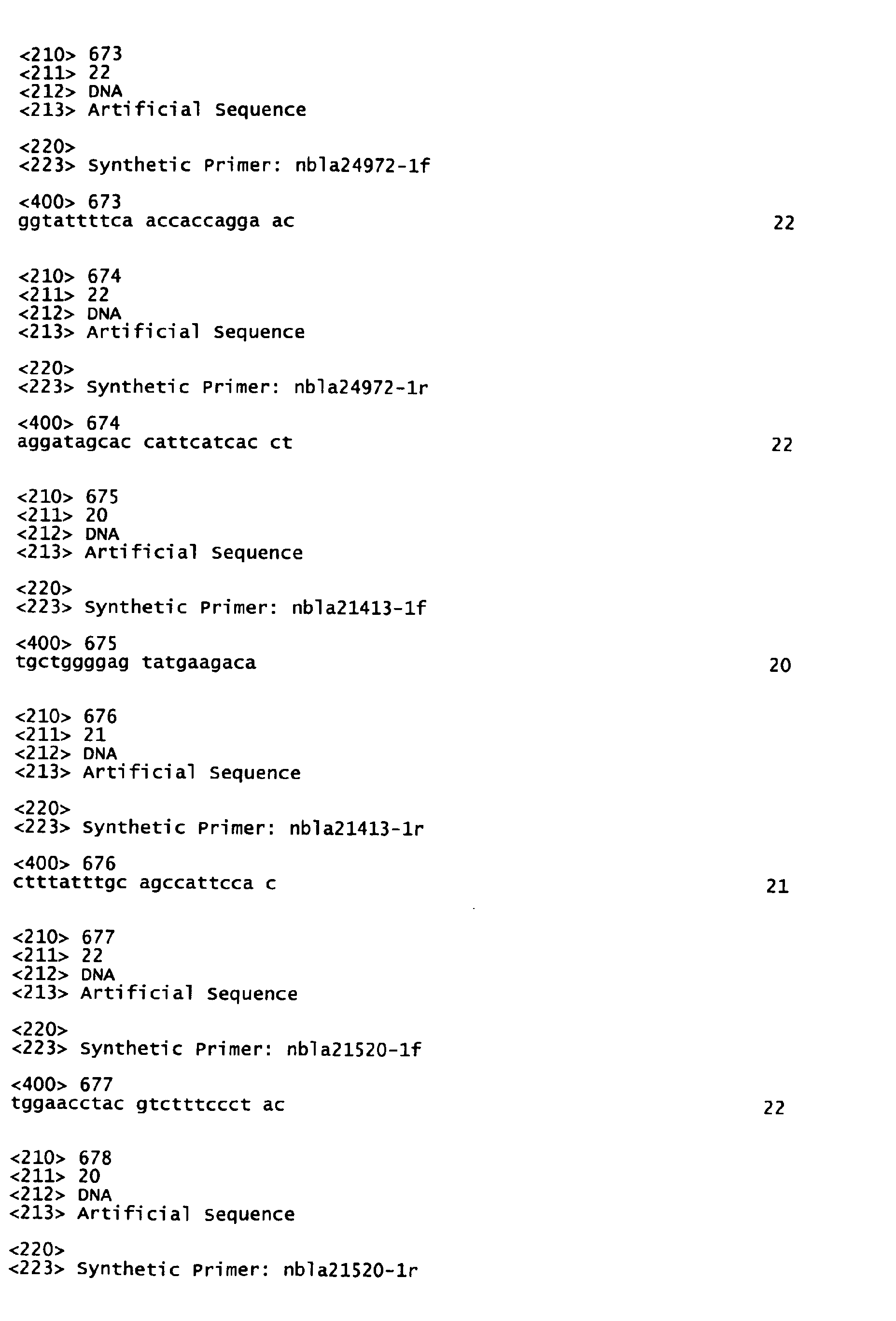
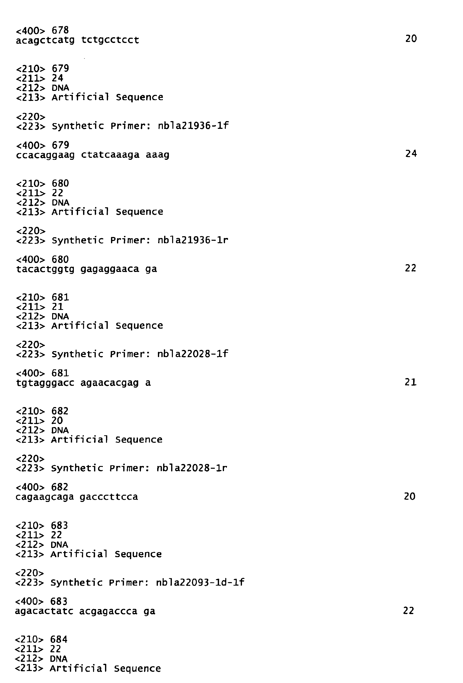
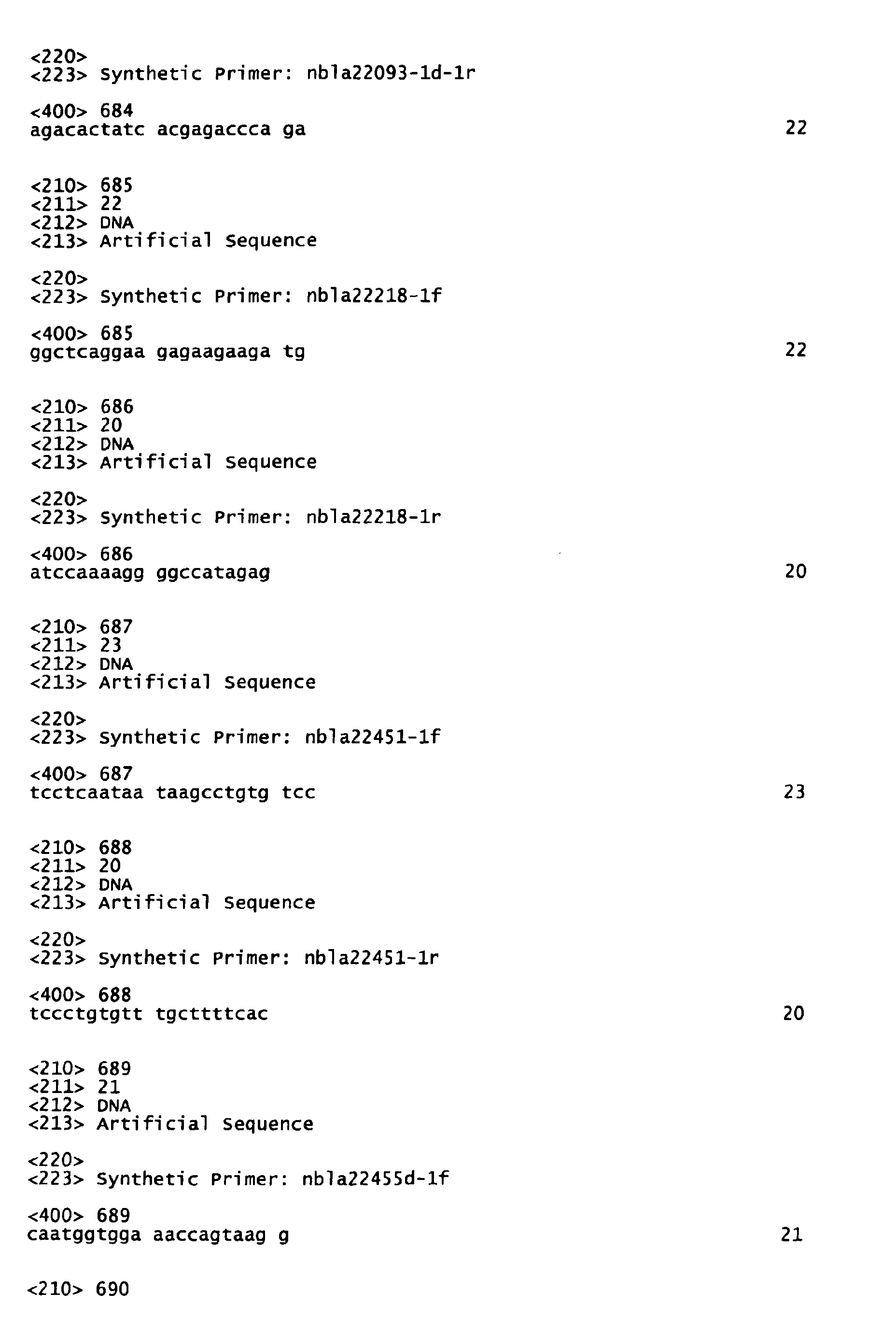
- the DNA (a) or the DNA (b) is a DNA comprising one sequence selected from the group consisting of nucleic acid sequences set forth in SEQ ID NO:175 to SEQ ID NO:202 and SEQ ID NO:519 to SEQ ID NO:540, or a DNA comprising one sequence selected from the group consisting of nucleic acid sequences set forth in SEQ ID NO:785 to SEQ ID NO:798 in the Sequence Listing.
- This invention also provides a diagnostic kit for stage 4s neuroblastoma comprising one pair of the primers described above as the active component.
- This invention further provides a method for determining stage 4s neuroblastoma, the method comprising detecting the presence or absence of a nucleic acid comprising one sequence selected from the group consisting of nucleic acid sequences set forth in SEQ ID NO:1 to SEQ ID NO:14 in the Sequence Listing from a clinical tissue sample of neuroblastoma.
- this invention provides a nucleic acid microarray comprising a solid phase support and a combination of plural nucleic acids each comprising a partial length- or the full length-nucleic acid comprising a nucleic acid sequence set forth in one of SEQ ID NO:1 to SEQ ID NO:174 in the Sequence Listing immobilized on the solid phase support.
- This invention also provides a nucleic acid microarray comprising a solid phase support and a combination of plural nucleic acids each comprising a nucleic acid sequence set forth in one of SEQ ID NO:175 to SEQ ID NO:200, a nucleic acid sequence set forth in one of SEQ ID NO:519 to SEQ ID NO:540, or a nucleic acid sequence set forth in one of SEQ ID NO:785 to SEQ ID NO:798 immobilized on the solid phase support.
- nucleic acids which will be referred to as “the nucleic acid(s) of this invention"
- the gene(s) of this invention derived from the gene which is expressed in neuroblastoma according to this invention (which will be referred to as “the gene(s) of this invention") including their utility, will be described in detail by referring to the preferred embodiments of the invention.
- the nucleic acids of this invention are derived from the gene of the invention and they make up the gene or are obtained from the gene by an in vivo or in vitro process.
- the chain lengths of the nucleic acids There are no limitations on the chain lengths of the nucleic acids, and herein, they will be referred to as "the nucleic acid(s) of the invention," which include nucleic acid fragments corresponding to portions of the gene.
- the nucleic acid chain length is short, it can be synthesized by chemical techniques.
- nucleic acid(s) refers to, for example, DNA or RNA, or polynucleotides derived therefrom which are active as DNA or RNA, and preferably refers to DNA or RNA.
- the particularly preferred nucleic acid has a DNA sequence that is identical with the human cDNA sequence disclosed in this specification or that is complementary to the sequence.
- hybridize under stringent conditions means that two nucleic acid (or fragments thereof) hybridize to each other under the hybridization conditions as described in Sambrook, J. et al. in "Expression of cloned genes in E . coli", Molecular Cloning: A Laboratory Manual (1989), Cold Spring Harbor Laboratory Press, New York, USA, pp. 9.47-9.62 and pp. 11.45-11.61.
- the "stringent conditions” refers to hybridization at approximately 45 °C with 6.0 x SSC, followed by washing at 50 °C with 2.0 x SSC.
- the stringency may be selected by choosing a salt concentration in the washing step from approximately 2.0 x SSC, 50 °C as low stringency to approximately 0.2 x SSC, 50 °C as high stringency.
- the temperature in the washing step may be increased from room temperature, or approximately 22 °C as low stringency conditions, to approximately 65 °C as high stringency conditions.
- nucleic acid(s) refers to an isolated nucleic acid(s) and to a nucleic acid or a polynucleotide containing substantially no cellular substances or culture medium, if prepared by recombinant DNA techniques, or containing substantially no precursor chemical substances or other chemical substances, if prepared by chemical synthesis.
- favorable prognosis type refers to a condition of human neuroblastoma in which the tumor exists with confinement or has become a regressing or benign sympathetic ganglion neoplasm, and is judged to have low malignancy based on N-myc or other tumor markers (TrkA, chromosomal aberration or the like) by the practicing physician.
- N-myc or other tumor markers TrkA, chromosomal aberration or the like
- neuroblastoma of the favorable prognosis type is considered a case of stage 1 or 2, with an onset age of less than one year and survival without recurrence for 5 or more years after surgery, and with no noted amplification of N-myc in the clinical tissue; however, it is not limited to such a specific case.
- unfavorable prognosis type refers to conditions of human neuroblastoma in which progression of the tumor has been observed, and it is judged to have high malignancy based on N-myc or other tumor markers by the practicing physician.
- neuroblastoma of the unfavorable prognosis type is considered a case of stage 4, with an onset age of greater than one year, death within 3 years after surgery and noted amplification of N-myc in the clinical tissue; however, it is not limited to such a specific case.
- stage 4s neuroblastoma is classified as the favorable prognosis type in accordance with the clinical molecular biology classification as described above, it will be treated differently from “the favorable prognosis type” for the sake of convenience in this specification.
- Neuroblastoma is a tumor consisting of actual neurons, of which only two types are known in humans, and analysis of the genes expressed therein is expected to provide very useful knowledge for understanding the biology of neurons. Specifically, it is extremely difficult, and practically impossible, to obtain a site-specific homogeneous tissue from the brain or peripheral nerves. On the other hand, neuroblastoma consists of an almost homogeneous population of neurons (though tumorized) derived from peripheral sympathetic neurons, and thus offers a high possibility of obtaining homogeneous expression of neuro-related genes. Furthermore, since neuroblastoma is a type of cancer, it is characteristically pointed out that there are many important genes expressed in the immature stage of neurogenesis.
- neuroblastoma can be further distinctly classified into the favorable prognosis type and the unfavorable prognosis types.
- Cancer cells from neuroblastoma of the favorable prognosis type are characterized by having a very slow rate of proliferation, with spontaneous regression beginning at some point.
- Findings to date have confirmed that neuronal differentiation and apoptosis (neuronal cell death) occur in the spontaneous regression, and that the differentiation which occurs in the maturation stages of normal neurons and programmed cell death are phenomena very closely resembling each other. Consequently, it is highly probable that the analysis of genes expressed in such tumors will lead to obtaining important genetic information relating to neuronal differentiation and apoptosis.
- the nucleic acids of this invention originating in the gene of the invention from which the useful genetic information described above can be obtained have been found in clinical tissues of stage 4s neuroblastomas (which may be referred to as "4s" hereafter).
- 4s neuroblastomas
- the expression levels of these genes are compared between a clinical tissue of the favorable prognosis type (which may be abbreviated as "F(favorable)” hereafter) and a clinical tissue of the unfavorable prognosis type (which may be abbreviated as "U(unfavorable)” hereafter), the genes have the characteristics described below.
- genes belonging to this group have expression levels (4s) that are on the same order as those of UF, but are lower than those of F. These genes are further classified into subgroups, resulting in I-1, I-2 and I-3. Table 1 should be consulted on the gene expression pattern of each subgroup.
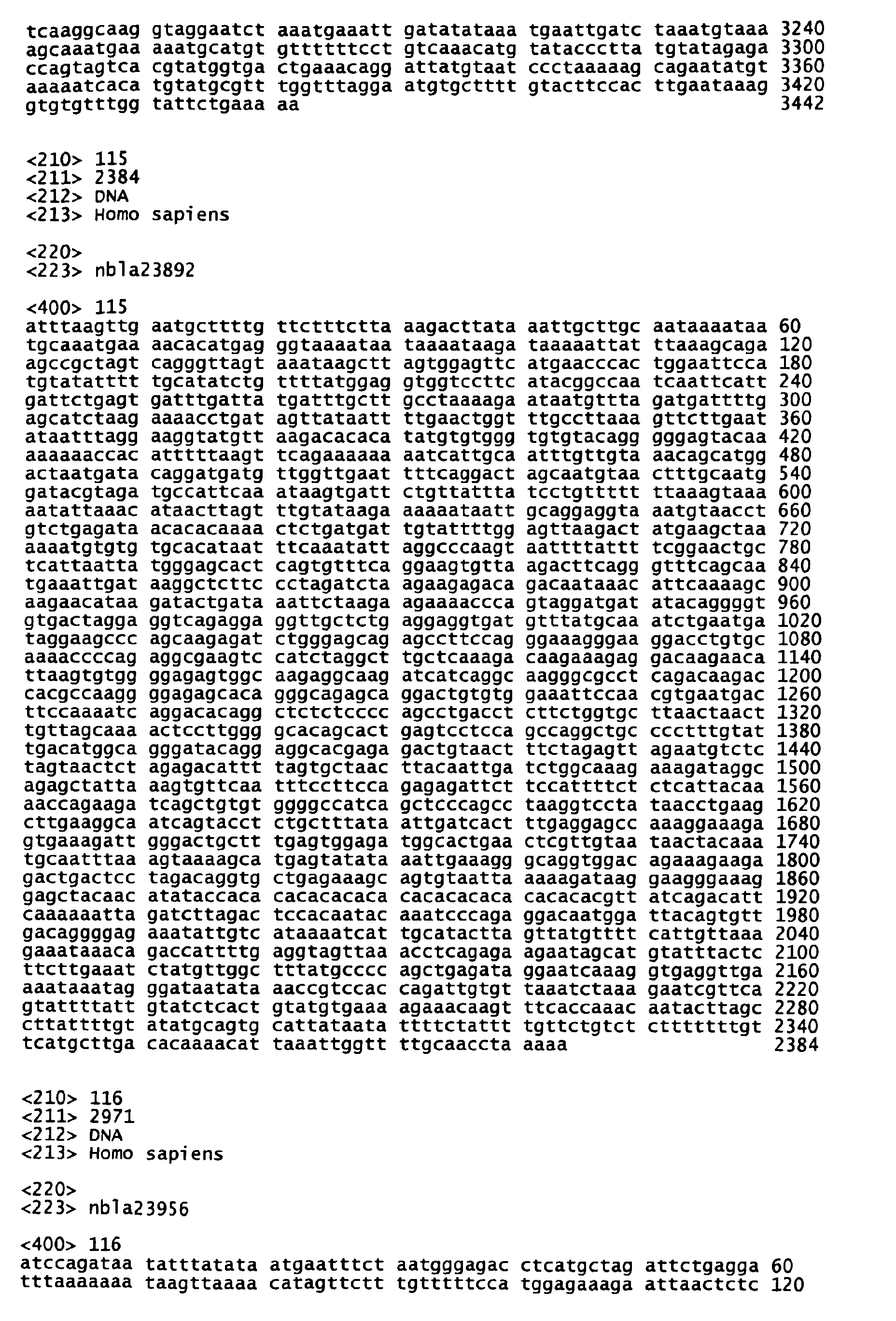
- nbla20026 SEQ ID NO:171
- nbla20421 SEQ ID NO:172
- nbla22298 SEQ ID NO:173
- nbla22549 SEQ ID NO:174
- nbla23020 all of which are novel genes.
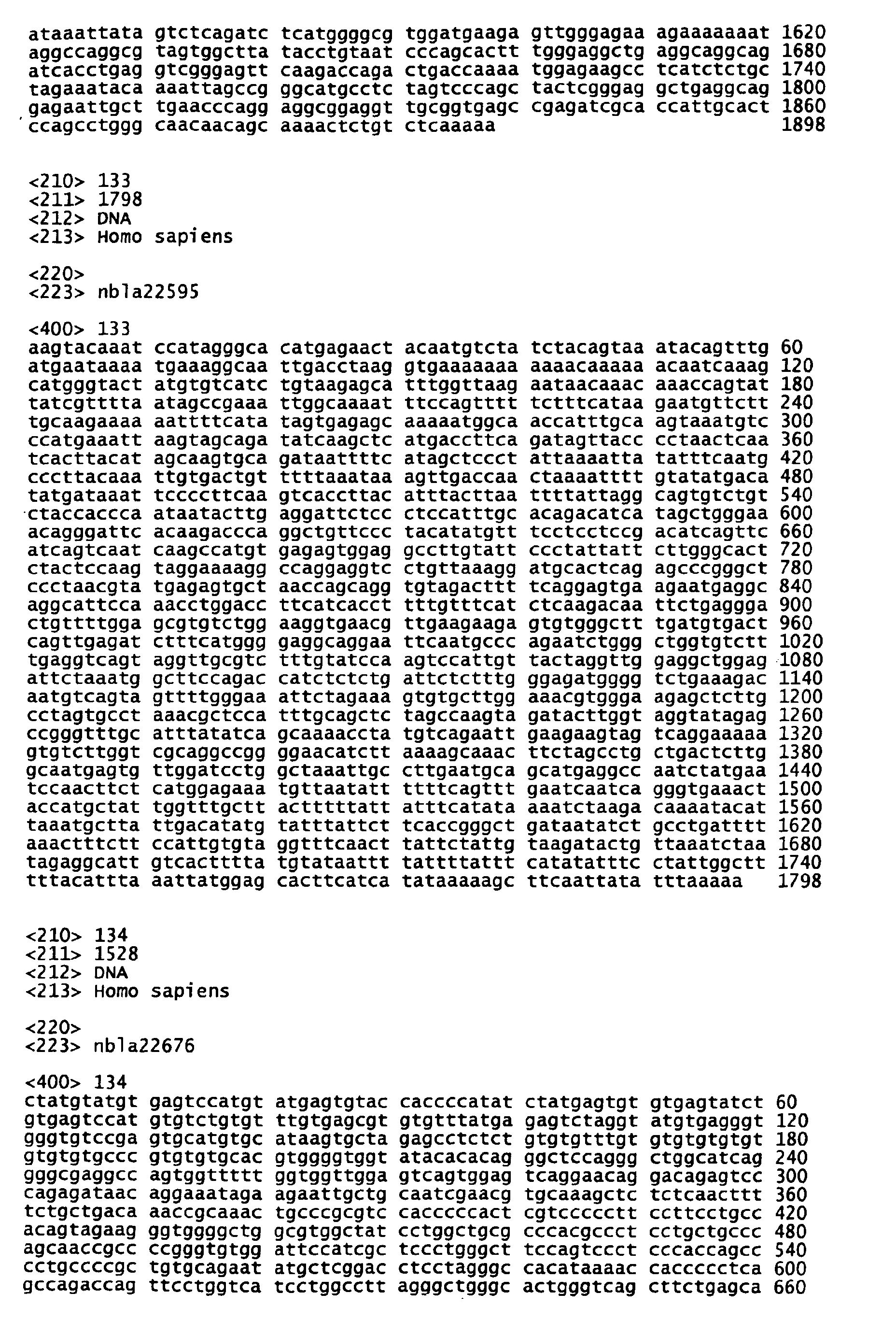
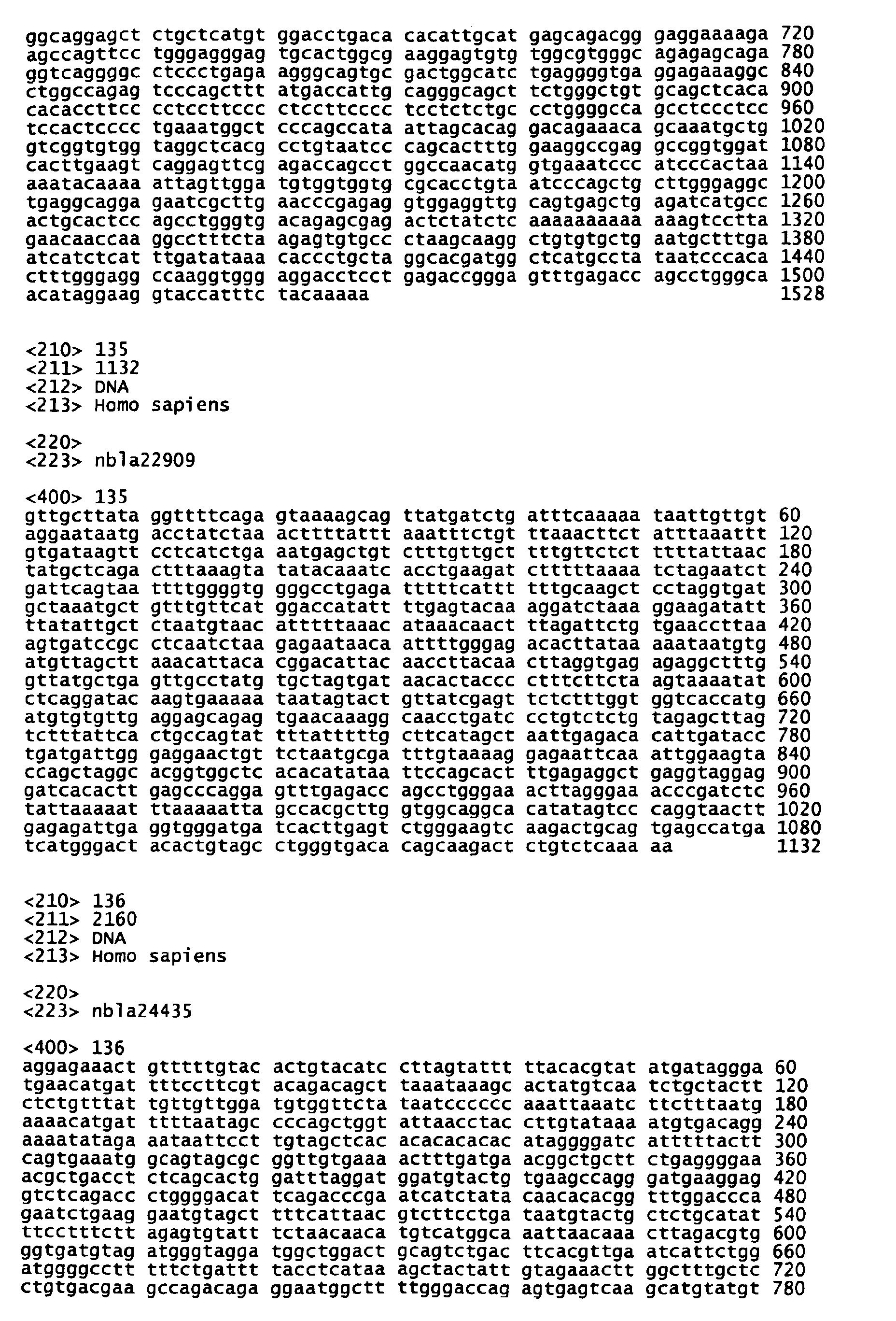
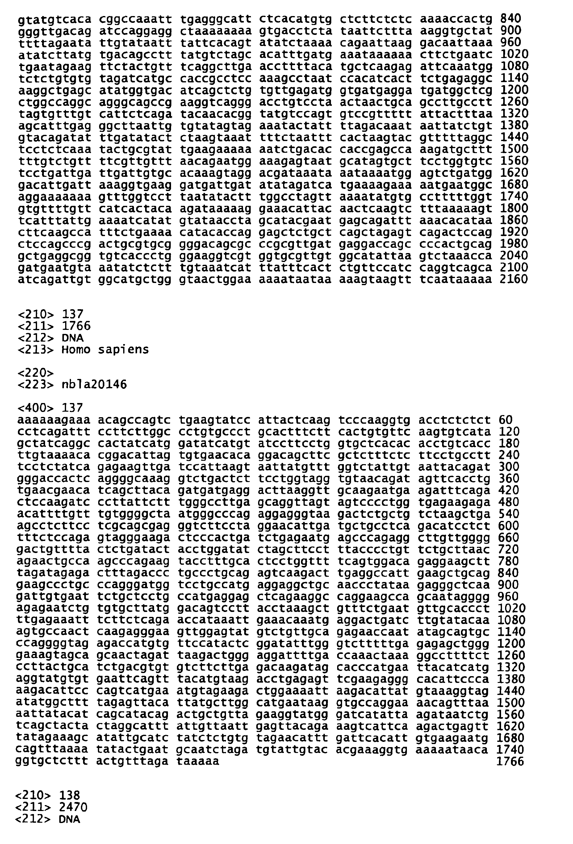
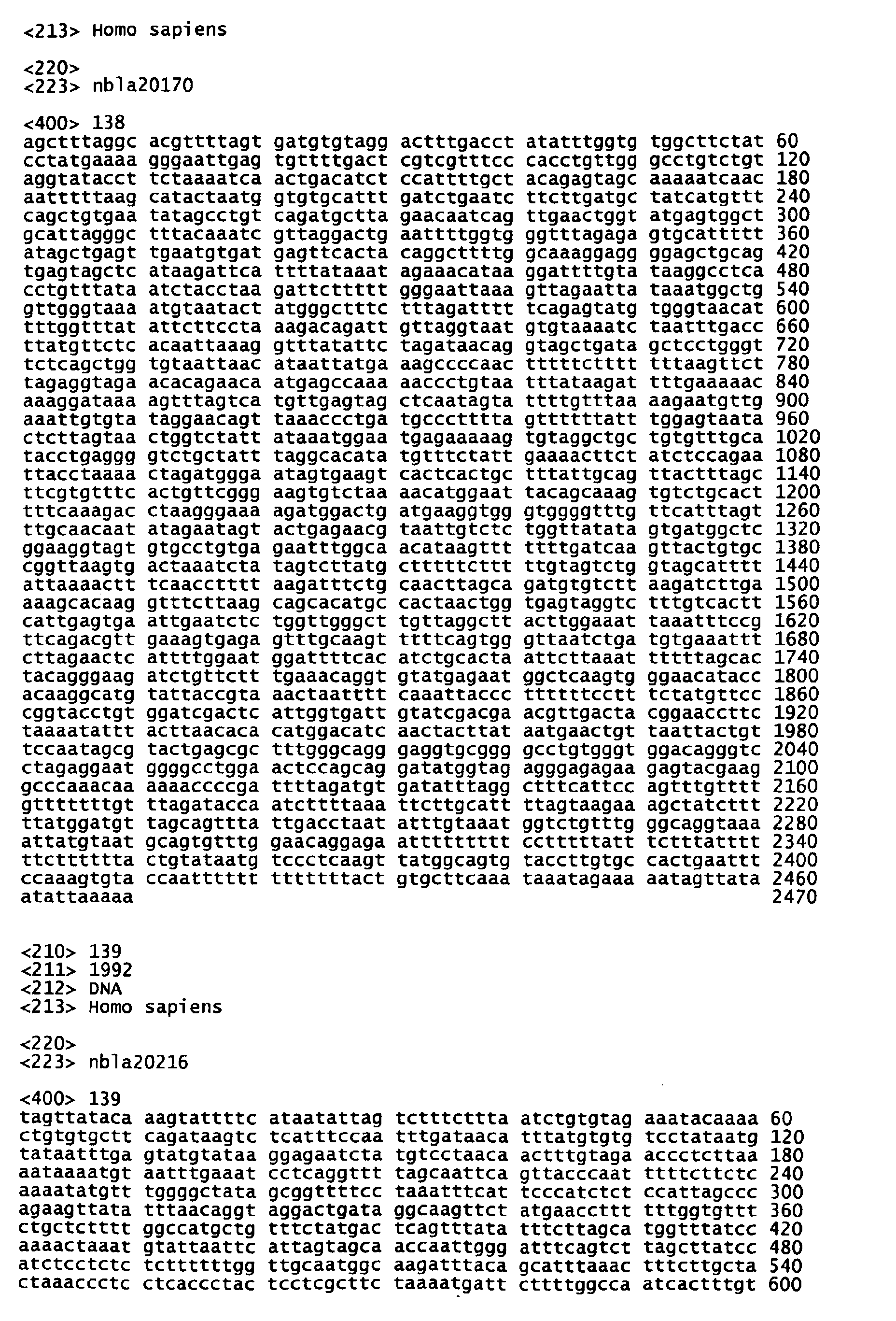
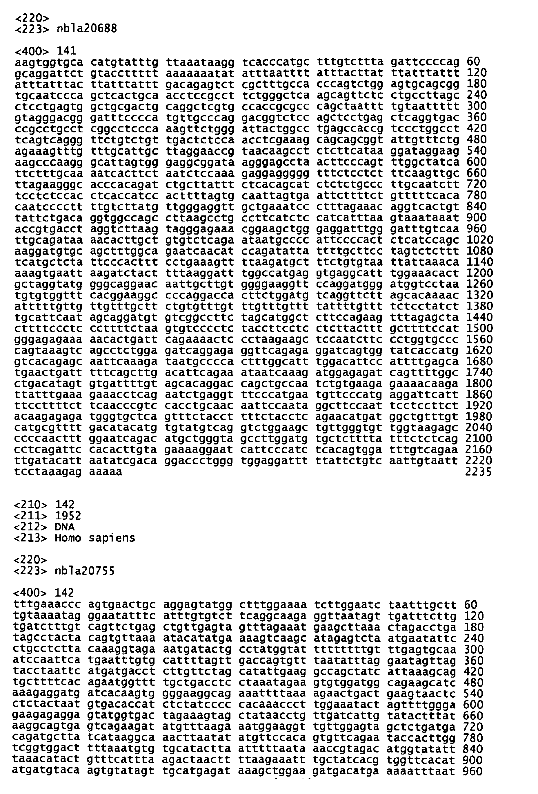
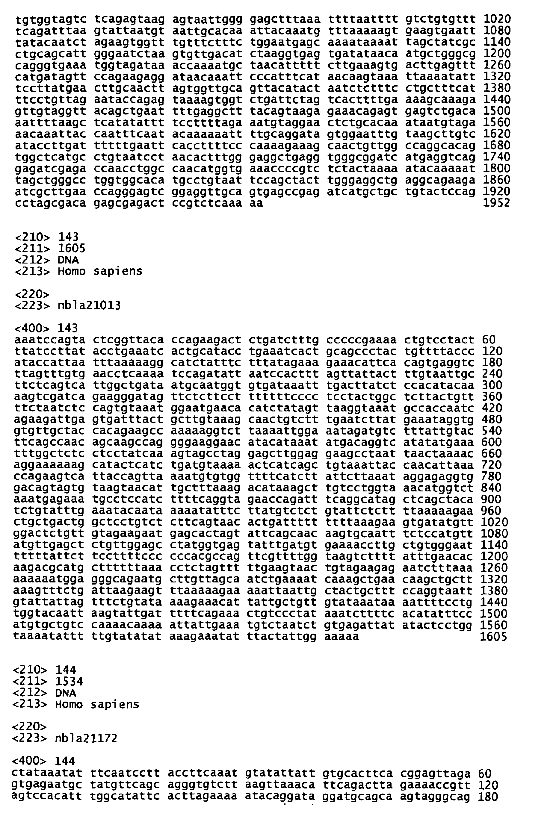
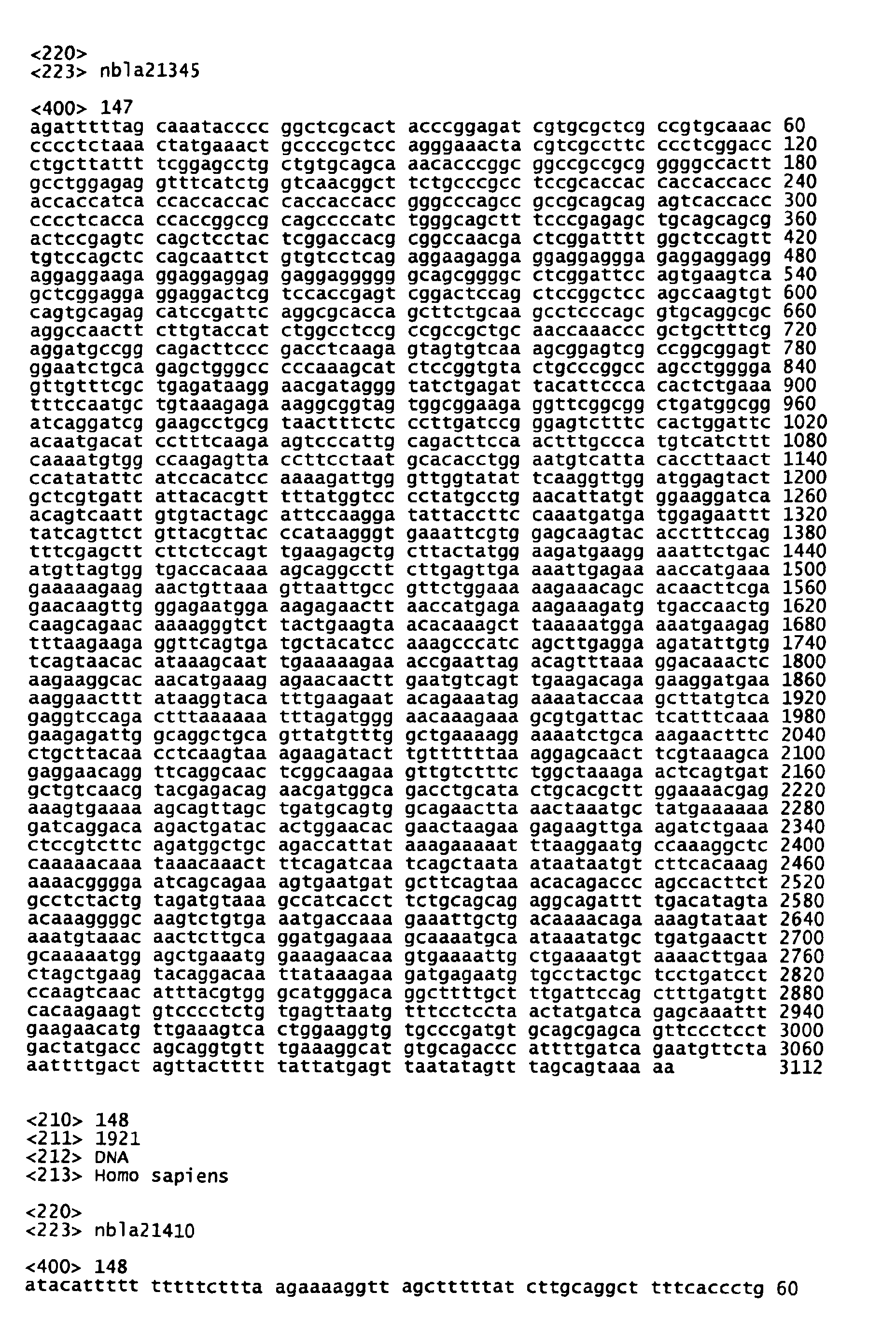
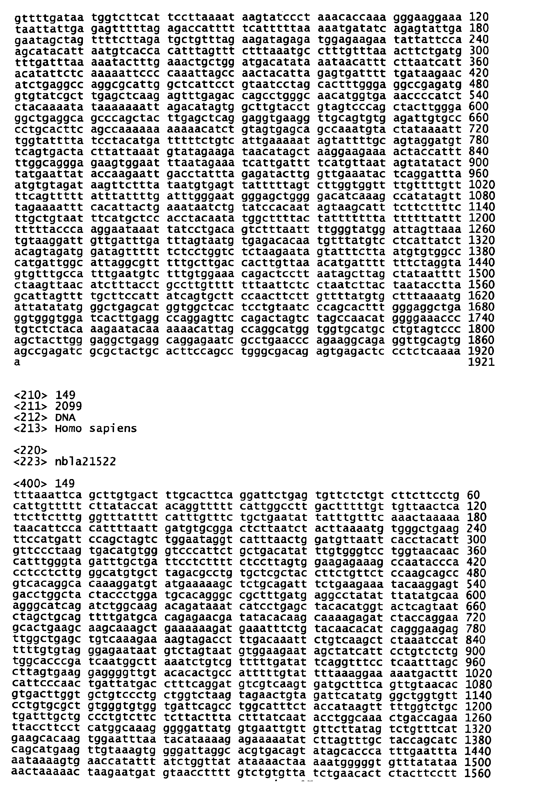
- nbla20113, nbla20146 (SEQ ID NO:137), nbla20170 (SEQ ID NO:138), nbla20216 (SEQ ID NO:139), nbla20253, nbla20549, nbla20657 (SEQ ID NO:140), nbla20688 (SEQ ID NO:141), nbla20755 (SEQ ID NO:142), nbla20835, nbla20968, nbla21013 (SEQ ID NO:143), nbla21087, nbla21172 (SEQ ID NO:144), nbla21189, nbla21200 (SEQ ID NO:145), nbla21214, nbla21255 (SEQ ID NO:146), nbla21337, nbla21344, nbla21345 (SEQ ID NO:147), nbla21410 (SEQ ID NO:148), nbla21522 (SEQ ID NO:149), nb1a21631 (SEQ ID NO:
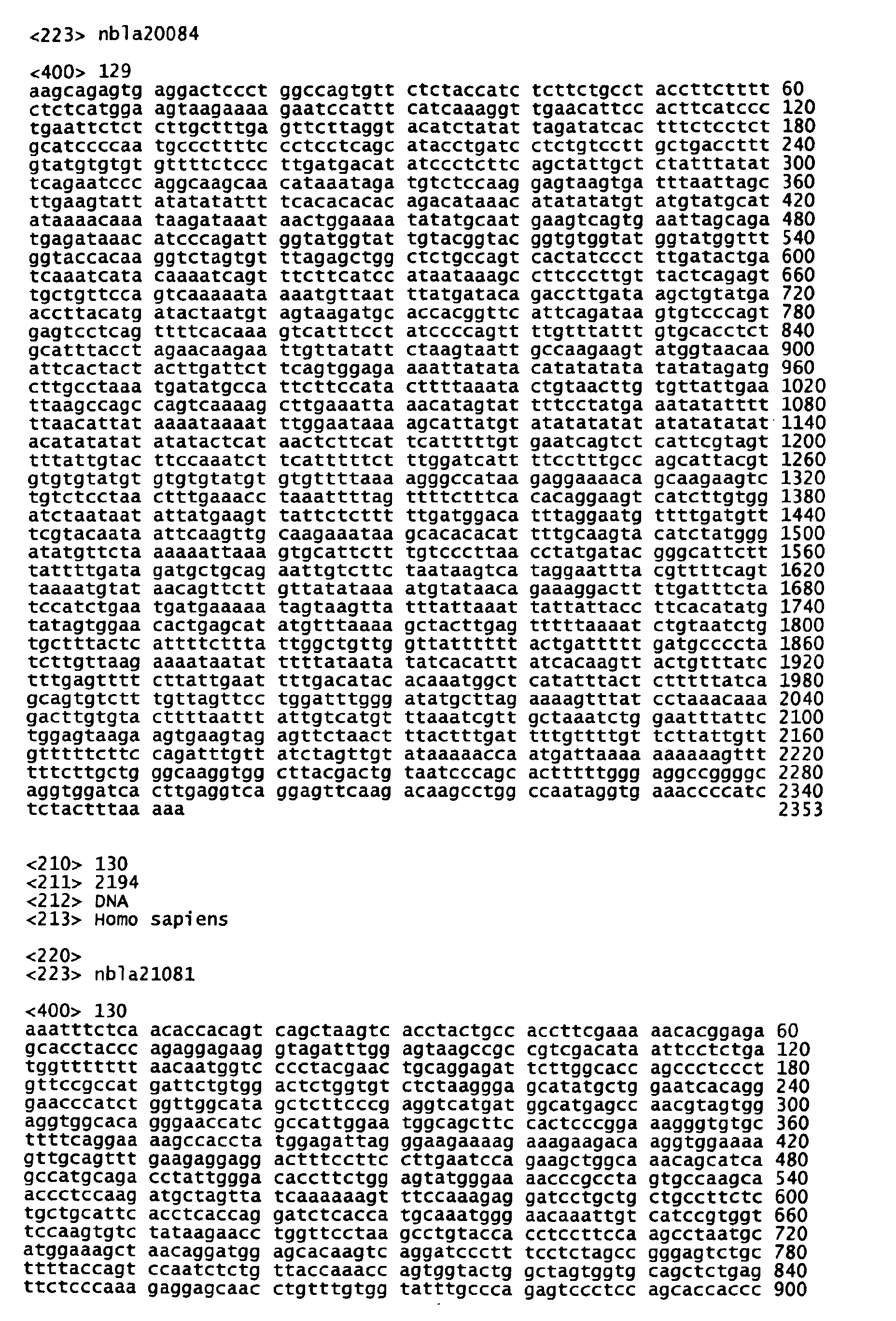
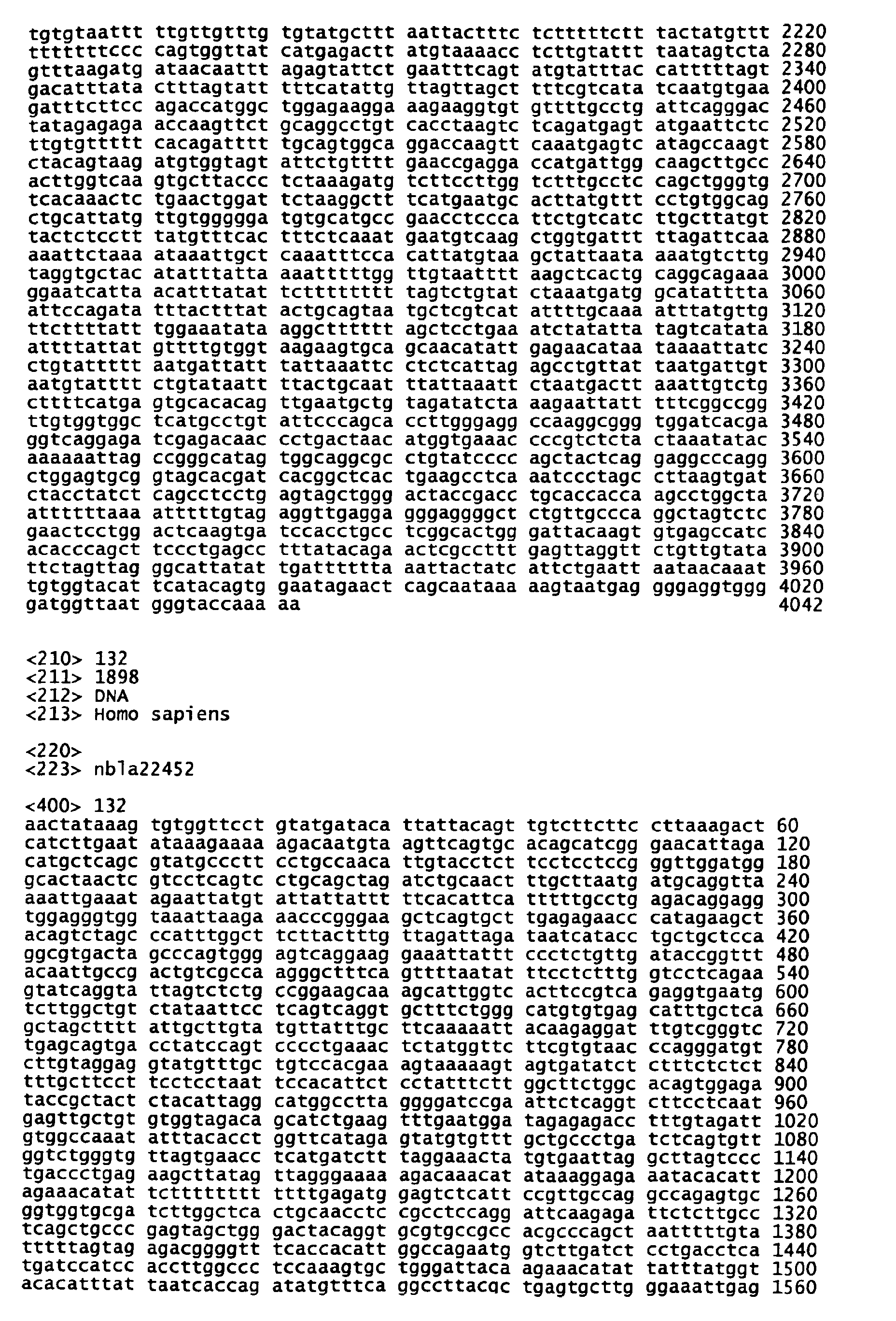
- nbla20084 (SEQ ID NO:129), nbla21081 (SEQ ID NO:130), nbla21420 (SEQ ID NO:131), nbla21761, nbla22452 (SEQ ID NO:132), nbla22595 (SEQ ID NO:133), nbla22676 (SEQ ID NO:134), nbla22909 (SEQ ID NO:135), nbla23456, nbla24297, nbla24435 (SEQ ID NO:136), and nbla24719, all of which are novel genes; and nbla20117, nbla20238, nbla20904, nbla23293, nbla23297, nbla23311, nbla23589, nbla23629, nbla23862, nbla2413, and nbla24761, all of which are known genes.
- genes belonging to this group have expression levels (4s) that are on the same order as those of F, but are higher than those of UF. These genes are further classified into subgroups, resulting in II-1, II-2 and II-3. Table 1 should be consulted on the gene expression pattern of each subgroup.
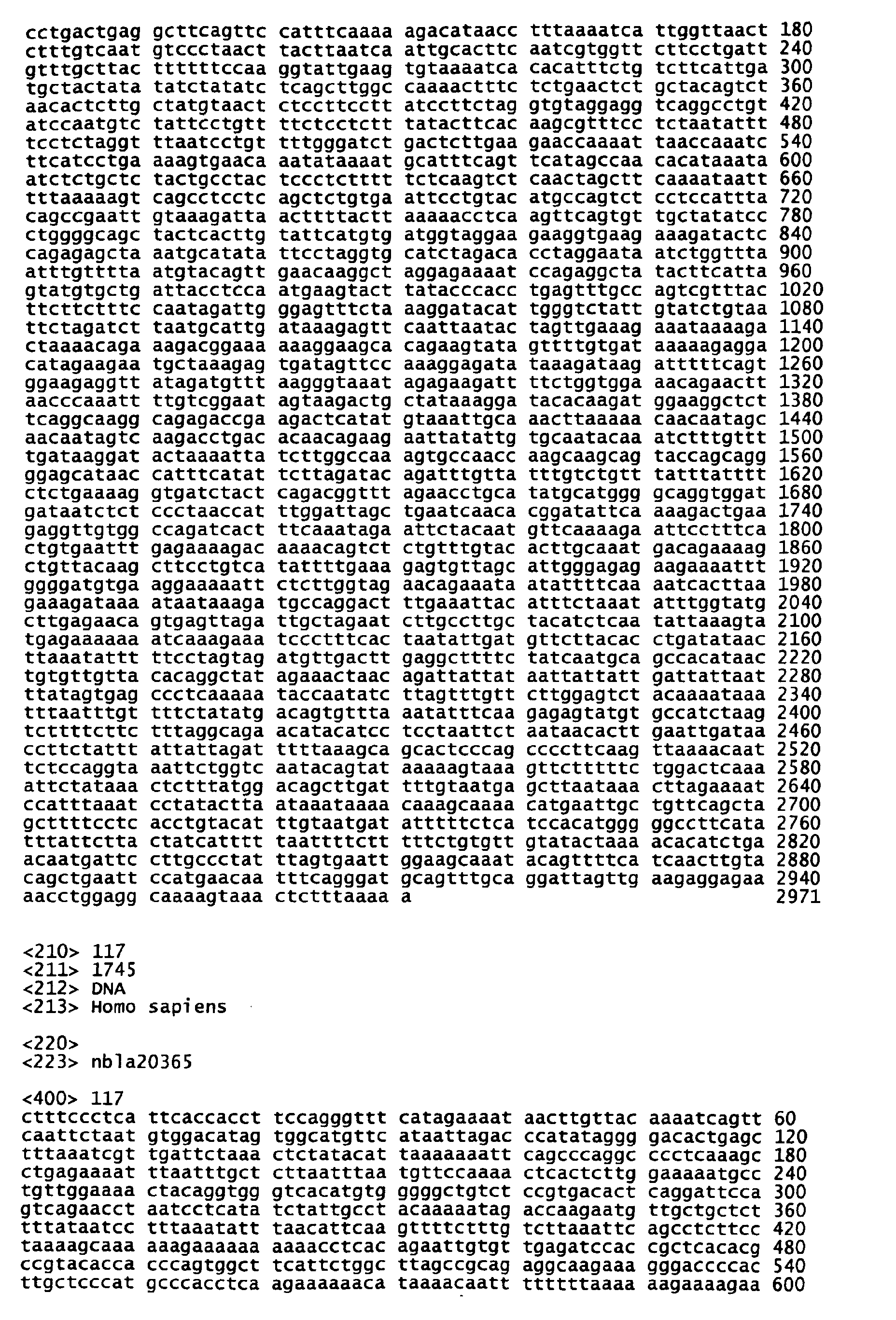
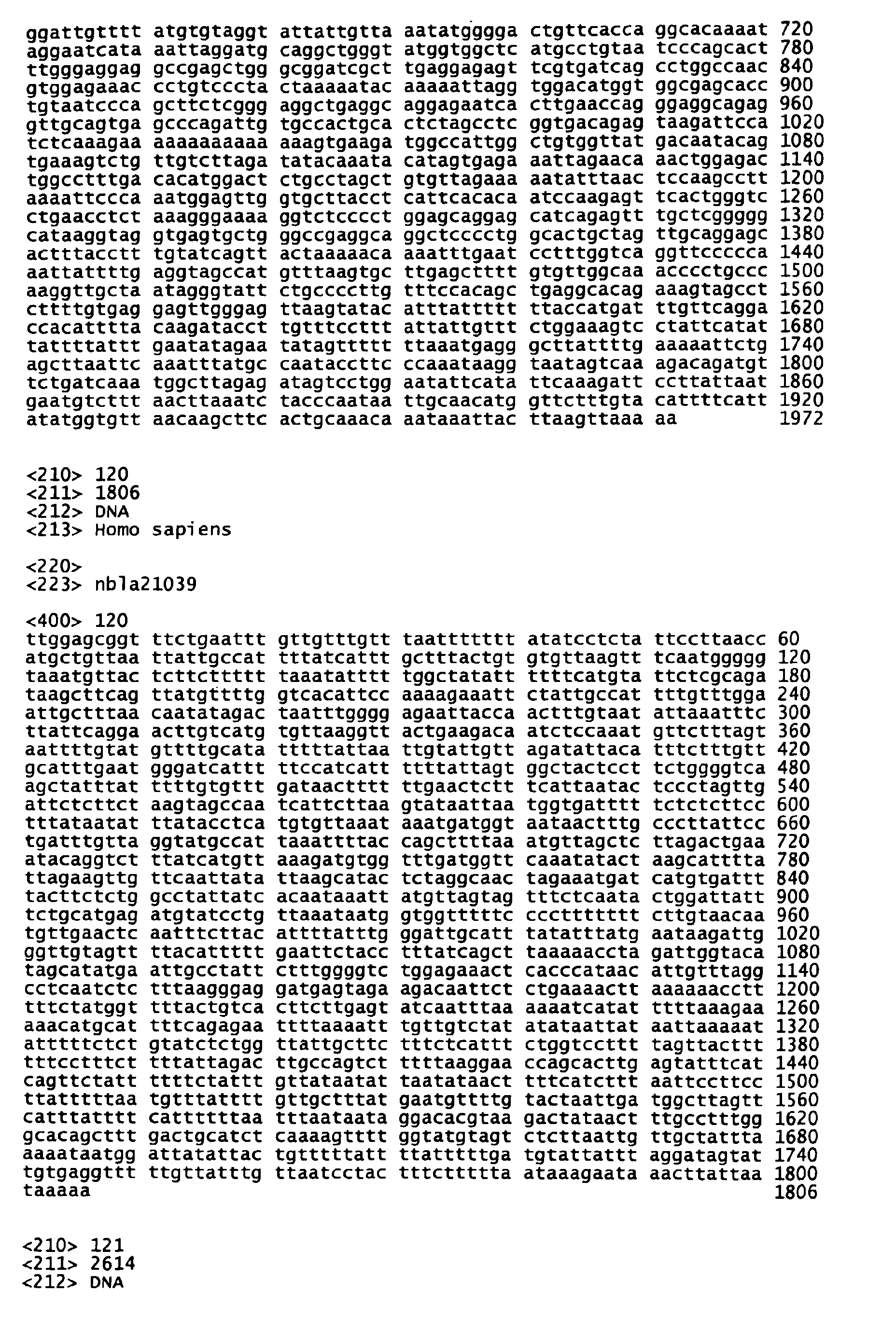
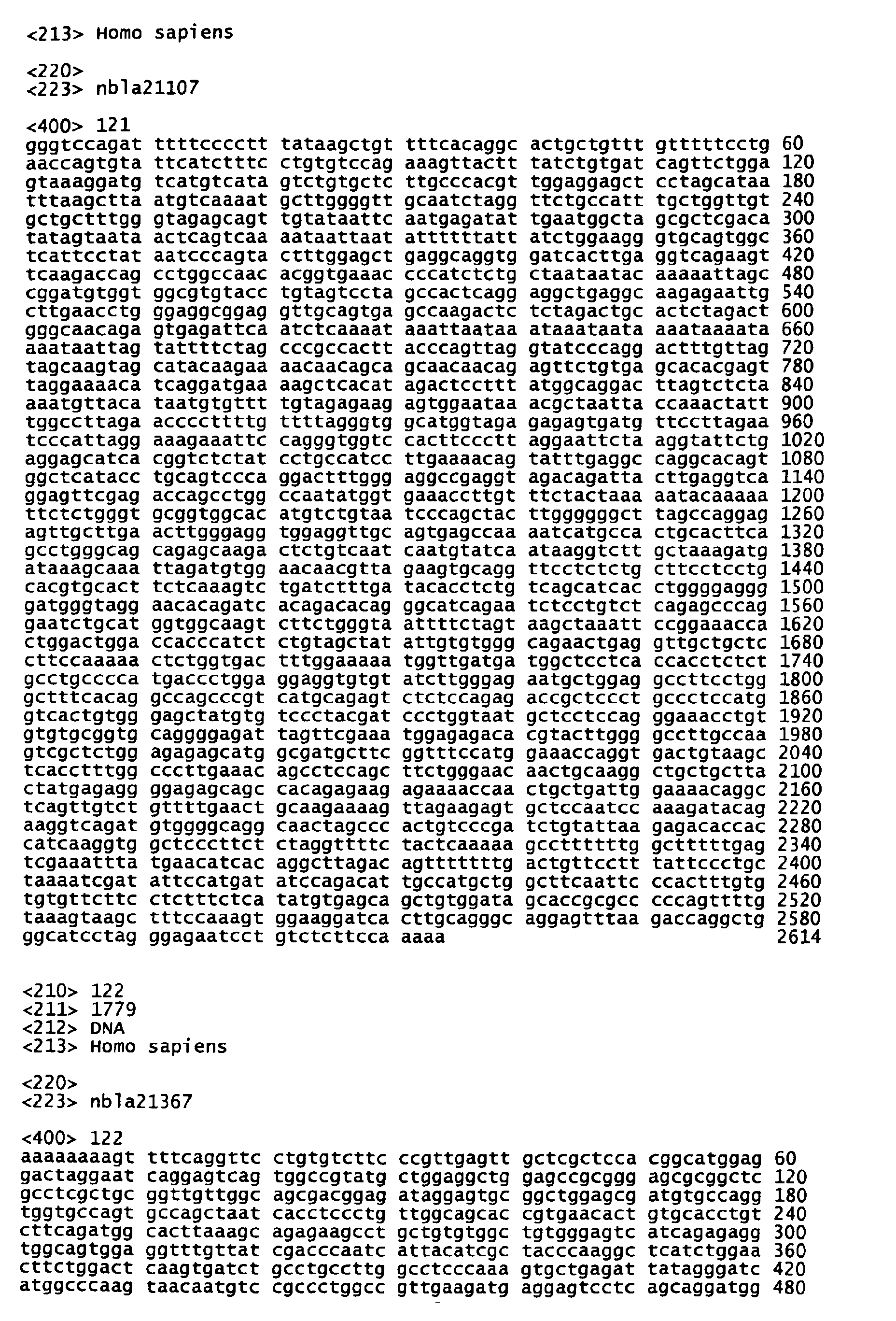
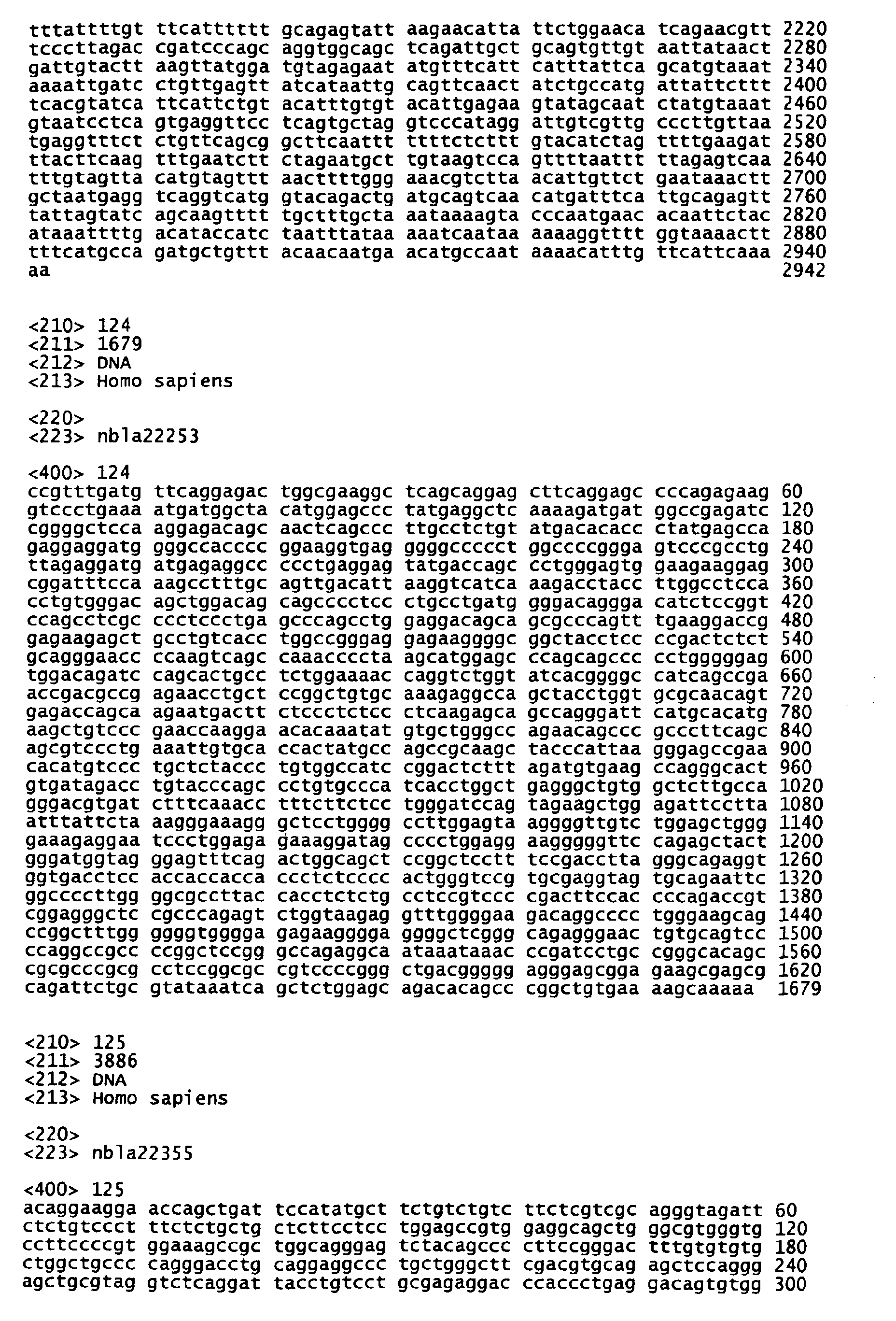
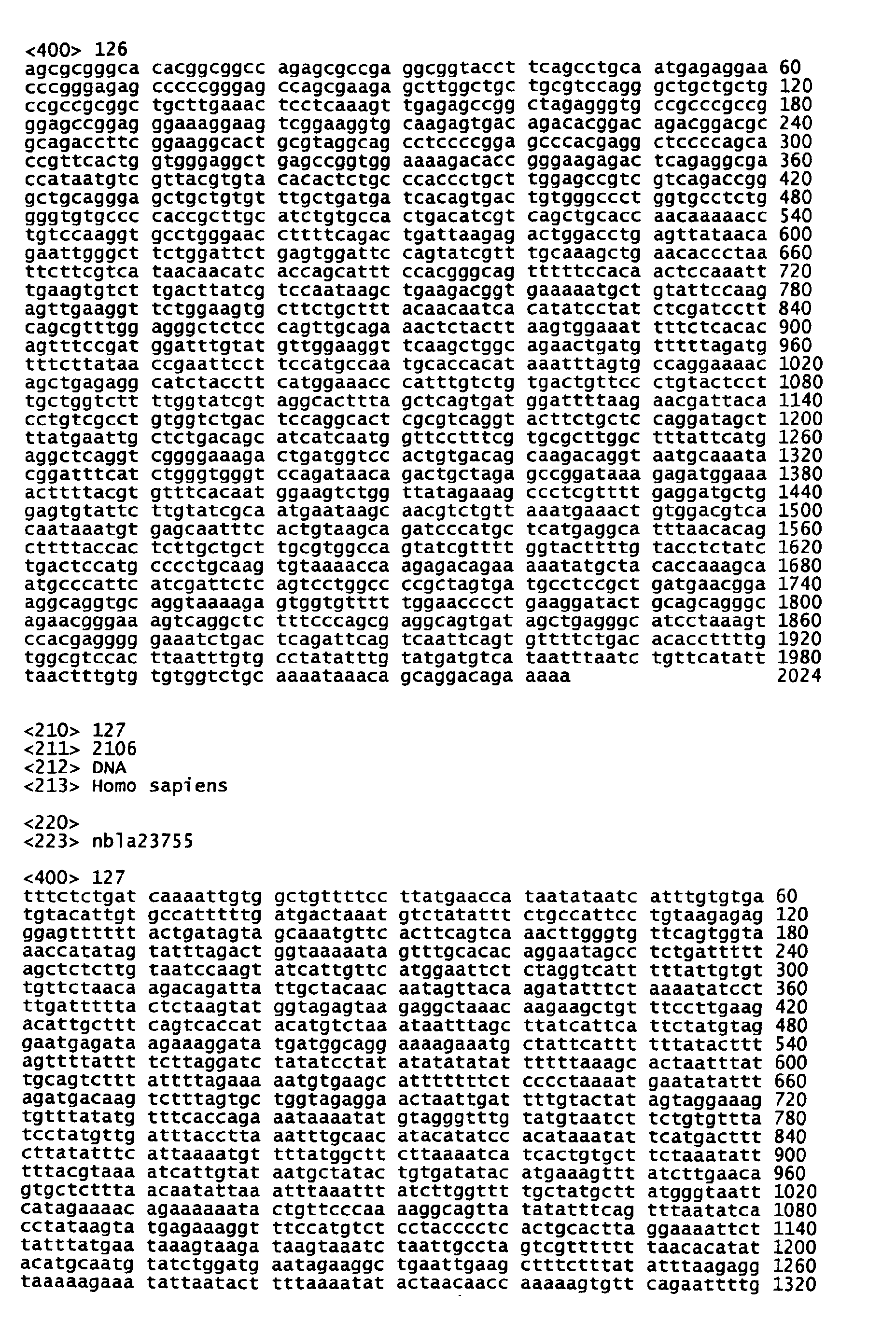
- nbla20365 (SEQ ID NO:117), nbla20378 (SEQ ID NO:118), nbla20511 (SEQ ID NO:119), nbla21039 (SEQ ID NO:120), nbla21107 (SEQ ID NO:121), nbla21367 (SEQ ID NO:122), nbla21790 (SEQ ID NO:123), nbla21855, nbla22253 (SEQ ID NO:124), nbla22355 (SEQ ID NO:125), nbla22704, nbla22832 (SEQ ID NO:126), nbla23394, nbla23512, nbla23755 (SEQ ID NO:127), nbla24084, nbla24376, and nbla24549 (SEQ ID NO:128), all of which are novel genes; and nbla20624, nbla22029, nbla22424, nbla22594 and nbla22622, all of which are novel genes; and nbla20624,
- nbla20001 (SEQ ID NO:58), nbla20083 (SEQ ID NO:59), nbla20125, nbla20182 (SEQ ID NO:60), nbla20231, nbla20248 (SEQ ID NO:61), nbla20250 (SEQ ID NO:62), nbla20268, nbla20330 (SEQ ID NO:63), nbla20395, nbla23973, nbla23983 (SEQ ID NO:64), nbla24041, nbla24082, nbla24104, nbla24111 (SEQ ID NO:65), nbla24142 (SEQ ID NO:66), nbla24157 (SEQ ID NO:67), nbla24230 (SEQ ID NO:68), nbla24239, nbla20541 (SEQ ID NO:69), nbla20555 (SEQ ID NO:70), nbla20638, nbla20645 (SEQ ID NO:71
- genes belonging to this group have expression levels (4s) that are on the same order as those of F, but are lower than those of UF. These genes are further classified into subgroups, resulting in III-1, III-2 and III-3. Table 1 should be consulted on the gene expression pattern of each subgroup.
- nbla20874 novel gene
- nbla23262 known gene
- nbla20604 nbla21226, nbla21908 (SEQ ID NO:27), nbla21928, nbla22027 (SEQ ID NO:28), nbla22082 (SEQ ID NO:29), nbla22643, nbla23303(SEQ ID NO:30), nbla23649, and nbla24468, all of which are novel genes; and nbla20141, nbla20446, nbla21538, nbla21558, nbla21623, nbla21969, nbla22219, nbla23272, nbla23307 and nbla24117, all of which are known genes.
- nbla20578 SEQ ID NO:26
- nbla21212 both of which are novel genes
- nbla23478, nbla23896 and nbla24920 all of which are known genes.
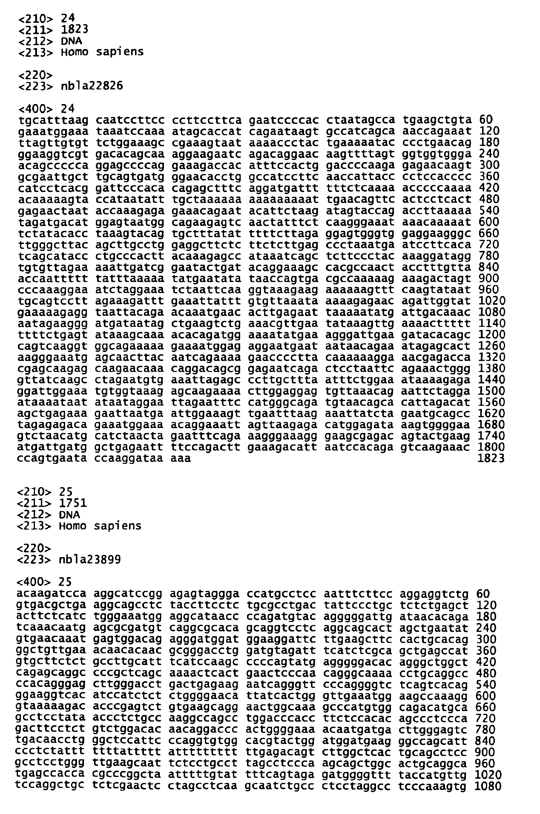
- the specific genes belonging to this group are nbla23899 (SEQ ID NO:25) and nbla24526, both of which are novel genes.
- genes belonging to this group have expression levels (4s) that are lower than those of F, but are higher than those of UF. These genes are further classified into subgroups, resulting in V-1, V-2, V-3, V-4 and V-5. Table 1 should be consulted on the gene expression pattern of each subgroup.
- the specific clone belonging to V-1 is nbla22031 (known gene).
- the specific clone belonging to V-2 is nbla22305 (known gene).
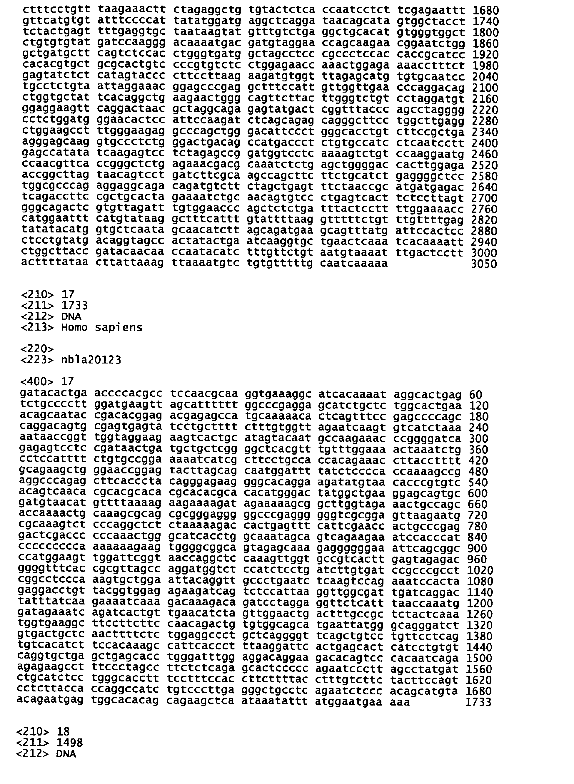
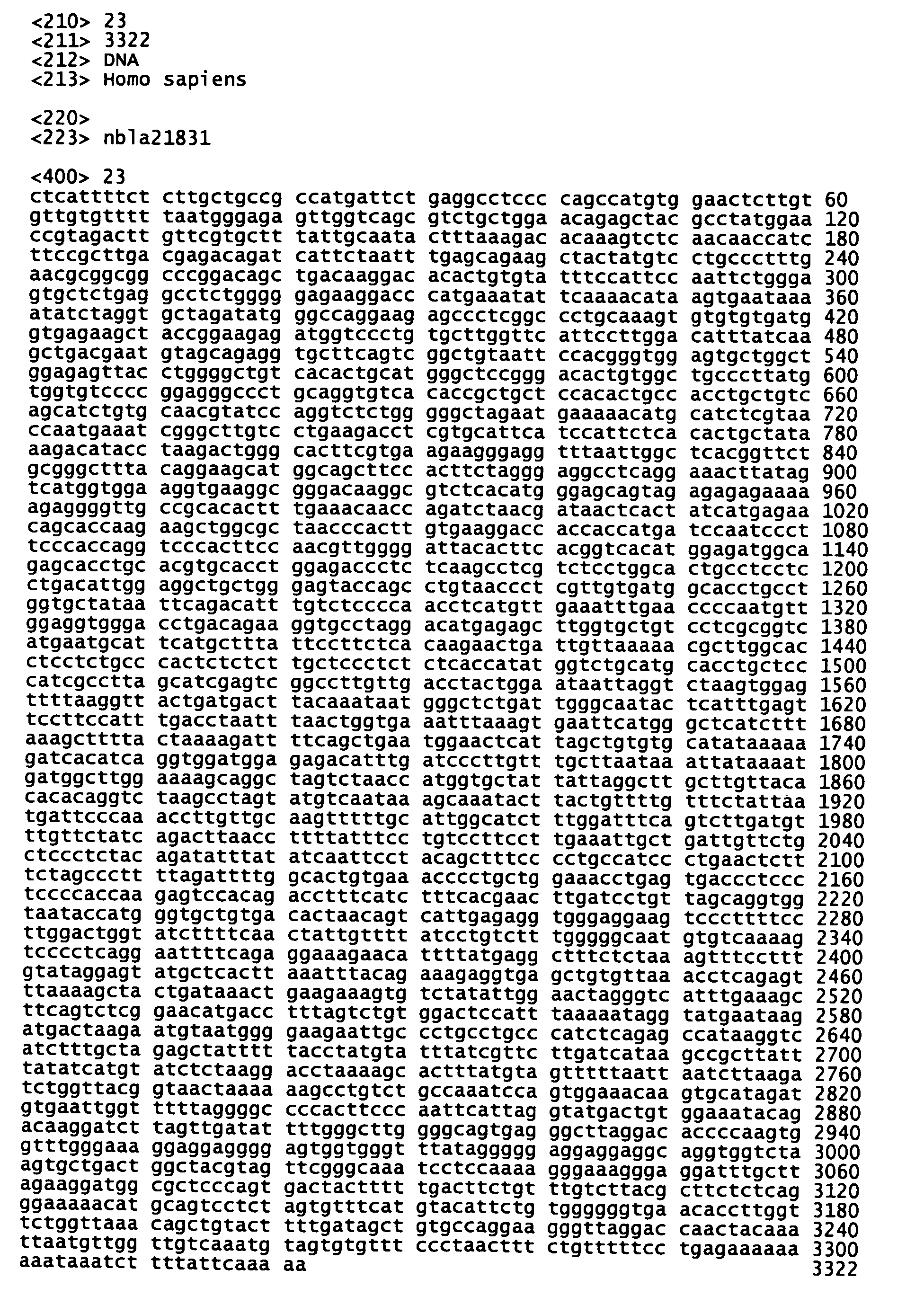
- nbla20123 (SEQ ID NO:17), nbla20382 (SEQ ID NO:18), nbla20660 (SEQ ID NO:19), nbla20666 (SEQ ID NO:20), nbla21239 (SEQ ID NO:21), nbla21729 (SEQ ID NO:22), nbla21831 (SEQ ID NO:23), nbla22826 (SEQ ID NO:24), and nbla24521, all of which are novel genes; and nbla20235 and nbla22607, both of which are known genes.
- nbla20787 SEQ ID NO:15
- nbla22284 SEQ ID NO:16
- nbla24756 all of which are novel genes.
- nbla24348 and nbla24686, both of which are novel genes are novel genes.
- genes belonging to this group have expression levels (4s) that are lower than either of F and UF, or are higher than either of F and UF. These genes are further classified into subgroups, resulting in VI-1, VI-2, VI-3, VI-4, VI-5, VI-6, VI-7 and VI-8. Table 1 should be consulted on the gene expression pattern of each subgroup.
- nbla21297 (SEQ ID NO:14) (novel gene) and nbla22443 (known gene).
- nbla20226 SEQ ID NO:11 (novel gene).
- nbla21650 SEQ ID NO:7
- nbla22094 SEQ ID NO:8
- nbla22739 SEQ ID NO:9
- nbla23525 SEQ ID NO:10
- nbla23701 SEQ ID NO:5
- nbla23890 SEQ ID NO:6
- nbla20087 The specific clone belonging to VI-6 is nbla20087 (known gene).
- nbla22689 SEQ ID NO:2
- nbla22968 nbla24079
- nbla24135 SEQ ID NO:3
- nbla24350 SEQ ID NO:4
- nbla22256 novel gene
- nbla22420 SQ ID NO:1 (novel gene).
- the gene groups have been divided into novel genes and known genes with respect to each group. A summary is shown in Table 1.
- the gene belonging to Group VII is detected only in a clinical tissue of stage 4s neuroblastoma. Therefore, if the presence of this gene is detected in a clinical tissue sample, it can be judged that there is a high probability of the sample being stage 4s neuroblastoma.
- the expression patterns as described above can be found. Therefore, if plural expression patterns of those genes are detected and analyzed, it can be judged as to whether the clinical tissue sample to be assayed is stage 4s neuroblastoma.
- a nucleic acid microarray which will be described below
- nucleic acids of this invention are useful as tumor markers to diagnose the favorable or unfavorable prognosis of neuroblastoma.
- this invention will allow for providing various genetic information on or relating to the prognosis of human neuroblastoma through the following means.
- the nucleic acid of the invention can be used as a probe (the nucleic acid probe of this invention) for hybridization to detect the gene of the invention expressed in neuroblastoma.
- the nucleic acid of this invention can also be used as a probe for hybridization in order to determine gene expression in various tumors and normal tissues, to identify the distribution of the gene expression.
- nucleic acid of this invention When the nucleic acid of this invention is used as a probe for hybridization, there are no particular limitations on the actual method of hybridization. As preferred methods there may be mentioned, for example, Northern hybridization, Southern hybridization, colony hybridization, dot hybridization, fluorescence in situ hybridization (FISH), in situ hybridization (ISH), DNA chip methods, and microarray methods.
- FISH fluorescence in situ hybridization
- ISH in situ hybridization
- DNA chip methods DNA chip methods, and microarray methods.
- the nucleic acid of this invention can be used as a probe for Northern hybridization to measure the length of mRNA or to quantitatively detect gene expression in a clinical tissue sample to be assayed.
- the nucleic acid of this invention can be used as a probe for Southern hybridization to detect the presence or absence of a DNA sequence in the genomic DNA of a clinical tissue sample to be assayed.
- the nucleic acid of this invention can also be used as a probe for the FISH method to identify the location of a gene of the invention on a chromosome.
- the nucleic acid of this invention can also be used as a probe for the ISH method to identify the tissue distribution of expression of a gene of the invention.
- nucleic acid of this invention When the nucleic acid of this invention is used as a probe for hybridization, a base length of at least 20 is necessary; and among the nucleic acids of this invention, a nucleic acid comprising 20 or more contiguous bases is preferably used. More preferably, the nucleic acid comprising 40 or more contiguous bases is used and most preferably a nucleic acid comprising 60 or more contiguous bases is used. Further, there may be used a nucleic acid comprising the full-length of any of the nucleic acid sequences set forth in SEQ ID NO:1 to SEQ ID NO:174 in the Sequence Listing.
- nucleic acid probe techniques in the various hybridization methods described above are well known to one skilled in the art, and for example, the conditions suitable for hybridization between a probe of this invention with each individual base length and the target polynucleotide may be readily determined.
- hybridization conditions optimal to probes containing varying base lengths Sambrook et al. "Molecular Cloning: A Laboratory Manual” (loc. cit.) may be consulted and followed for such manipulations which are well known to one skilled in the art.
- the probe of this invention may preferably be labeled for use in an easily detectable fashion.
- the detectable label may be any type and any element or compound which can be detected either visually or using devices.
- detectable labels there may be mentioned radioactive isotopes, avidin and biotin and fluorescent substances (FITC or Rhodamins).
- the radioactive isotopes are 32 P, 14 C, 125 I, 3 H, 35 S etc.
- Biotin-labeled nucleotides may be incorporated into nucleic acids by nick translation, or chemical or enzymatic means.
- the biotin-labeled probes are detected after hybridization using labeling means such as avidin/streptavidin, fluorescent labels, enzymes, gold colloidal complexes or the like.
- the nucleic acid probe of this invention may also be labeled by binding with a protein.
- a radioactive or fluorescent histone single-stranded binding protein may be used, for example.
- a suitably labeled probe constitutes a diagnostic agent of this invention.
- primers can be designed after any nucleic acid (DNA) sequence contained in the nucleic acid of the invention and the polymerase chain reaction (PCR) method can be used.
- DNA nucleic acid
- PCR polymerase chain reaction
- mRNA may be extracted from a clinical tissue sample to be assayed, and the gene expression can be semi-quantitatively measured by RT-PCR.
- Such method may be carried out by a method well known to one skilled in the art. For example, "Molecular Cloning: A Laboratory Manual,” (loc. cit.) and or Idenshibyo Nyumon [Introduction to Genetic Diseases] (Takahisa, S.: Nankodo Publishing) may be consulted.
- the nucleic acid (DNA) of this invention is used as a PCR primer (i.e., the primer of the invention)
- a base length of 10 to 60 is necessary; and among portions of the nucleic acid sequences according to the invention, the nucleic acid having 10 to 60 contiguous bases is preferably used. More preferably, one having 15 to 30 bases is used.
- a primer sequence with a GC content of 40-60% is preferred.
- the nucleic acid of this invention can also be used to detect the expression (or the distribution) of a gene of the invention which is expressed in various tissues or cells, non-limiting to neuroblastoma. This can be accomplished, for example, by using the nucleic acid of this invention as a probe for hybridization or as a primer for PCR as described above.
- the expression distribution of the gene can also be detected using a DNA chip, a nucleic acid microarray or the like. That is, the nucleic acid of the invention may be directly attached to the chip or array.
- a method by which nucleic acids (DNA) are spotted to a substrate for the purpose of attaching them to a chip or array by using a high precision dispenser see, for example, United State Patent No. 5,807,522.
- mRNA extracted from a clinical tissue sample may be labeled with a fluorescent substance or the like, hybridized thereto, and an analysis can be made of the type of tissue cells with high expression of the gene.
- the DNA attached to the chip or the array may be the reaction product of PCR using the nucleic acid or its fragment of this invention.
- the nucleic acid fragment of this invention (DNA fragment) may be directly synthesized on a substrate to form a DNA chip or a DNA array (see, for example, United State Patent No. 5,424,186).
- the nucleic acid of this invention can be used for cloning a gene which is expressed in human neuroblastoma.
- cloning of a gene containing the nucleic acid of the invention is possible.
- the genes capable of being cloned include, among others, genes displaying differential expression levels between neuroblastomas of the unfavorable prognosis type and neuroblastomas of the unfavorable prognosis type, genes expressed in stage 4s neuroblastoma, genes displaying expression in a pattern different from the expression patterns in other tissues or cancer cells, genes expressed in a cell cycle dependent manner, genes induced by nerve differentiation, and genes whose expression is regulated by oncogenes or cancer suppressor genes.
- the nucleic acid of this invention can be used as a probe for hybridization, or as a primer for PCR to investigate the expression pattern of the gene in a clinical tissue sample to be assayed taken from the subject, which enables the diagnosis for prognosis (i.e., determining stage 4s neuroblastoma).
- the methods of detecting the gene include Northern blotting hybridization, in situ hybridization and RT-PCR, as mentioned above among others.
- the amount of the nucleic acid hybridizing to the nucleic acid probe mentioned above in a clinical tissue sample to be assayed is compared with the control sample (e.g., clinical tissue samples from neuroblastomas of the favorable and unfavorable prognosis types) and the gene expression is determined.
- the control sample e.g., clinical tissue samples from neuroblastomas of the favorable and unfavorable prognosis types
- their expression patterns may then be, for example, compared with those listed in Table 1 and analyzed to enable the diagnosis for prognosis.
- the nucleic acid microarray mentioned above may desirably be used.
- RNA is extracted from the sample and reverse-transcribed into DNA, amplification is performed using the aforementioned primers, and the gene expression is semi-quantitatively measured.
- the diagnosis for prognosis can be made.
- the diagnostic kit containing a pair of the primers as essential components.
- the diagnostic kit also includes known components such as PCR buffer, detergent solution and enzymes.
- antisense oligonucleotides to the nucleic acids of the invention.
- the antisense oligonucleotides are capable of hybridizing to the nucleic acids of this invention, and include antisense DNAs and antisense RNAs.
- Antisense DNA inhibits transcription of mRNA from DNA, while antisense RNA inhibits translation of mRNA.
- These antisense oligonucleotides may be synthesized using an automated synthesizer or by PCR using the nucleic acids of this invention as templates.
- antisense oligonucleotides also encompass antisense oligonucleotide derivatives having improved binding affinity for DNA or mRNA, tissue selectivity, cell permeability, nuclease resistance and intracellular stability. These derivatives may be synthesized using the antisense technology known in the art.
- Antisense oligonucleotides having sequences complementary to the sequences near the translation initiation codon of the mRNA, those of the ribosome-binding site, and those of the capping site or the splicing site are capable of inhibiting synthesis of the RNA, and therefore, will exhibit a particularly notable inhibitory effect on gene expression.
- This invention therefore, encompasses such antisense oligonucleotides.
- nucleic acid sequences encoding the therapeutic genes to be used in gene therapy there are provided nucleic acid sequences encoding the therapeutic genes to be used in gene therapy.
- the nucleic acid of this invention can be transferred into a vector for use in gene transportation, whereby the transgene (i.e., the gene of the invention) can be expressed by an arbitrary expression promoter and can be used for the gene therapy.
- the transferable viral vectors may be prepared from DNA or RNA viruses.
- Such vectors may be any type of viral vectors from an MoMLV vector, a herpes virus vector, an Adenovirus vector, an AAV vector, a HIV vector, a SIV vector, a Sendai virus vector and the like.
- One or more proteins among the constituent protein group of a viral vector are substituted by the constituent proteins of a different species of virus, or alternatively a part of the nucleic acid sequence constituting genetic information is substituted by the nucleic acid sequence of a different species of virus to form a viral vector of the pseudo-type, which can also be used in this invention.
- VSV-G protein an envelop protein of vesicular stomatitis virus or VSV
- VSV vesicular stomatitis virus
- viruses having a host spectrum other than humans are usable as the viral vector insofar as they are efficacious.
- the vectors other than those of viral origin there may be used complexes of calcium phosphate and nucleic acid, ribosomes, cation-lipid complexes, Sendai virus liposomes, polymer carriers having polycation as the backbone main chain and others.
- methods such as electroporation and gene guns may be used as a gene transfer system.
- any cassettes without any particular limitations may be used insofar as they can cause genes to express in the target cells.
- One skilled in the art can readily select such expression cassettes.
- they are expression cassettes capable of gene expression in the cells derived from an animal, more preferably, expression cassettes capable of gene expression in the cells derived from a mammal, and most preferably expression cassettes capable of gene expression in the cells derived from human.
- the gene promoters that can be used as expression cassettes include: for example, virus-derived promoters from an Adenovirus, a cytomegalovirus, a human immunodeficiency virus, a simian virus 40, a Rous sarcoma virus, a herpes simplex virus, a murine leukemia virus, a sinbis virus, a hepatitis type A virus, a hepatitis type B virus, a hepatitis type C virus, a papilloma virus, a human T cell leukemia virus, an influenza virus, a Japanese encephalitis virus, a JC virus, parbovirus B19, a poliovirus, and the like; mammal-derived promoters such as albumin, SR ⁇ , a heat shock protein, and an elongation factor; chimera type promoters such as a CAG promoter; and the promoters whose expression can be induced by tetracyclines, steroids and
- the drugs to be used in the gene therapy may be prepared as a recombinant viral vector containing the therapeutic gene that is designed for therapeutic purposes as described above. More specifically, a recombinant virus vector containing the gene of this invention may be prepared by dissolving it in an appropriate solvent such as water, physiological saline or an isotonized buffer solution. Here, polyethylene glycol, glucose, various amino acids, collagen, albumin or the like can be then added as protective materials to form preparations.
- the daily dosage is preferably administered at about 0.1 ⁇ g/kg to 1,000 mg/kg per day, and more preferably at about 1 ⁇ g/kg to 100 mg/kg per day.
- Clinical tissue samples of human neuroblastoma were quasi-aseptically frozen immediately after surgical extraction and then preserved at -80 °C.
- RNA Extraction Kit QIAGEN Inc.
- the extracted total RNA was purified using an oligo dT cellulose column (Collaborative Research, Inc.) to obtain a pool of mRNA with a polyA structure.
- a cDNA library was prepared according to the oligo capping method (Y. Suzuki et al., Gene, U.S.A., Vol. 200, pp.149-156, 1997).
- DEPC diethyl pyrocarbonate
- the mixture was reacted at 37 °C for 1 hour to effect dephosphorylation of the 5' end of the mRNA. This was followed by phenol/chloroform treatment twice, and finally by ethanol precipitation to obtain a purified dephosphorylated mRNA pool.
- 5'-oligo RNA 5'-oligo RNA
- the DNA(-) oligo-capped mRNA pool prepared in 7 above was reverse-transcribed using Super Script II (kit manufactured by Life Tech Oriental, Inc.) to obtain a pool of 1st strand cDNA.
- the pool of DNA(-) oligo-capped mRNA was dissolved in 21 ⁇ l of sterile distilled water, and then 10 ⁇ l of 10 x First Strand buffer (kit accessory), 8 ⁇ l of dNTP mix (5 mM, kit accessory), 6 ⁇ l of DTT (0.1 M, kit accessory), 2.5 ⁇ l of oligo-dT adapter primer (5 pmol/ ⁇ l, 5'-GCGGCTGAAGACGGCCTATGTGGCCTTTTTTTTTTTTTTTTTTTTTTTTTTT-3': SEQ ID NO:1080), 2.0 ⁇ l of RNasin (40 unit/ ⁇ l) and 2 ⁇ l of Super Script II RTase (kit accessory) were added. The mixture was reacted at 42 °C for 3 hours to effect reverse transcription. This was followed by phenol/chloroform treatment, alkali treatment and neutralization treatment to digest all the RNA and purification was carried out by ethanol precipitation.
- the 1st strand cDNA pool prepared in 7 above was subjected to PCR amplification using Gene Amp (kit manufactured by Perkin Elmer Inc.).
- the pool of 1st strand cDNA was dissolved in 52.4 ⁇ l of sterile distilled water, and then 30 ⁇ l of 3.3 x Reaction buffer (kit accessory), 8 ⁇ l of dNTP mix (2.5 mM, kit accessory), 4.4 ⁇ l of magnesium acetate (25 mM, kit accessory), 1.6 ⁇ l of Primer F (10 pmol/ ⁇ l, 5'-AGCATCGAGTCGGCCTTGTTG-3': SEQ ID NO:1081), 1.6 ⁇ l of Primer R (10 pmol/ ⁇ l, 5'-GCGCTGAAGACGGCCTATGT-3': SEQ ID NO:1082) and 2 ⁇ l of rTth (kit accessory) were added.
- the 2nd strand cDNA pool prepared in 8 above was dissolved in 87 ⁇ l of sterile distilled water, and then 10 x NEB buffer (NEB Inc.), 100 x BSA (bovine serum albumin manufactured by NEB Inc.) and 2 ⁇ l of SfiI (restriction endonuclease, 20 unit/ ⁇ l, manufactured by NEB Inc.) were added. The mixture was reacted overnight at 50 °C to effect SfiI restriction endonuclease treatment. This was followed by purification with phenol/chloroform treatment and ethanol precipitation to obtain a pool of cDNA which had been SfiI-treated at both ends.
- NEB buffer NEB Inc.
- BSA bovine serum albumin manufactured by NEB Inc.
- SfiI restriction endonuclease
- the SfiI-treated cDNA pool prepared in 9 above was electrophoresed on 1% agarose gel and a fraction with >2 kb was purified using Geneclean II (Bio101 Inc.).
- the purified cDNA pool was dissolved in 100 ⁇ l of sterile distilled water and allowed to stand at 37 °C for 6 hours. This was followed by purification with phenol/chloroform treatment and ethanol precipitation to obtain a long-chain cDNA pool.
- the long-chain cDNA pool prepared in 10 above was ligated into the cloning vector pME18S-FL3 (provided by Prof. Sumio Kanno of the Institute of Medical Science, Tokyo University) using a DNA Ligation Kit ver.1 (kit manufactured by Takara Shuzo Co. Ltd.).
- the long-chain cDNA pool was dissolved in 8 ⁇ l of sterile distilled water, and then 1 ⁇ l of pME18S-FL3 pretreated with restriction endonuclease DraIII, 80 ⁇ l of Solution A (kit accessory) and 10 ⁇ l of Solution B (kit accessory) were added and reaction was conducted at 16 °C for 3 hours. This was followed by purification with phenol/chloroform treatment and ethanol precipitation to obtain a cDNA library.
- the cDNA library prepared in Preparation Example 1, Item 12 above was used for transformation into E. coli (TOP-10: Invitrogen Corporation). Specifically, the cDNA library was dissolved in 10 ⁇ l of sterile distilled water and mixed with TOP-10. The mixture was then incubated on ice for 30 minutes, at 40 °C for 1 minute and on ice for 5 minutes. After adding 500 ⁇ l of SOB medium, shake culturing was performed at 37 °C for 60 minutes. Appropriate amounts thereof were seeded onto an ampicillin-containing agar media and culturing was continued at 37 °C for 24 hours to obtain E. coli clones. There, 5075 clones were picked up randomly.
- the E . coli clones on agar media obtained in 1 above were collected with toothpick and suspended in 120 ⁇ l of LB medium prepared in a 96-well plate. The 96-well plate was then allowed to stand overnight at 37 °C for culturing of the E . coli. 60% Glycerol solution (72 ⁇ l) was then added and preserved at -20 °C (glycerol stock).
- the glycerol stock (10 ⁇ l) prepared in Example 1, Item 2 above was transferred to a 15 ml-centrifugation tube, and then 3 ml of LB medium and 50 ⁇ g/ml of ampicillin were added and shaking was carried out overnight at 37 °C for culturing of the E. coli.
- a QIAprep Spin Miniprep Kit (QIAGEN Inc.) was then used to extract and purify a plasmid DNA from the E . coli.
- Both end sequences of the plasmid DNA prepared in 1 above were determined using a DNA Sequencing Kit (kit manufactured by ABI). There were combined 600 ng of plasmid DNA, 8 ⁇ l of premix (kit accessory) and 3.2 pmol of primers, and sterile distilled water was added to a total of 20 ⁇ l. After denaturing the mixture at 96 °C for 2 minutes, a cycle of 96 °C for 10 seconds, 50 °C for 5 seconds and 60 °C for 4 minutes was repeated 25 times for reaction. The product was then purified by ethanol precipitation. Sequence determination was carried out by polyacrylamide gel electrophoresis under denaturing conditions, using ABI377 (ABI).
- Clinical tissue samples of human neuroblastoma were quasi-aseptically frozen immediately after surgical extraction and then preserved at -80 °C. These samples were provided in 8 specimens. In a similar manner, clinical tissue samples of human neuroblastomas of the favorable and unfavorable prognosis types were provided in 12 specimens each.
- Prognosis assay of the neuroblastoma samples of the favorable and unfavorable prognosis types was carried out based on the following criteria:
- RNA from the specimen was reverse-transcribed into cDNA using Super Script II reverse transcriptase (GIBCO). Specifically, 48 ⁇ l of a solution comprising 20 ⁇ g of the total RNA, 8 ⁇ l of random primers (1 ⁇ g/ ⁇ l) (Takara Shuzo Co., Ltd.), and sterile ultra-purified water containing DEPC in an amount as necessary was prepared. This solution was incubated at 65 °C for 15 minutes and was placed on ice after the reaction was complete.
- GEBCO Super Script II reverse transcriptase
- each sample with its concentration adjusted by dilution with DDW was subjected to PCR as described below.
- PCR was performed with rTaq polymerase (Takara Shuzo Co., Ltd.).
- Appropriate primers were designed against the genes (whether novel or known) identified in the cDNA library from the stage 4s neuroblastoma.
- Differential screening of three pairs of cDNA sample populations with their concentrations adjusted was performed. Specifically, 2 ⁇ l of cDNA, 5 ⁇ l of sterile distilled water, 1 ⁇ l of 10 x rTaq buffer, 1 ⁇ l of 2 mM dNTPs, 0.5 ⁇ l each of the synthesized primer set (forward and reverse), and 0.5 ⁇ l of rTaq were combined.
- PCR products under thus determined conditions were electrophoresed on 1.5% agarose gel for 20 minutes, stained with ethidium bromide.
- concentrations of the bands were compared among three pairs of specimens (i.e., stage 4s neuroblastoma, neuroblastomas of the favorable and unfavorable prognosis types).
- Table 1 summarizes the obtained patterns according to the specimen subsets. Results from the expressed pattern analysis are as previously discussed.
- the primers used were selected under suitable primer selection conditions (base number, Tm, GC%) by inputting into a Primer3 software, the end sequences of the genes to be detected (Example 3).
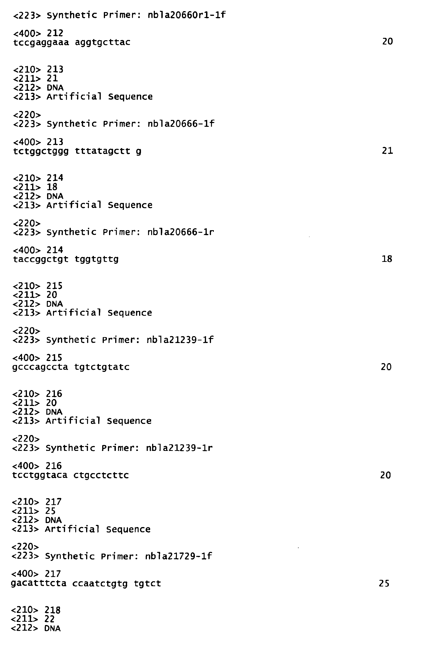
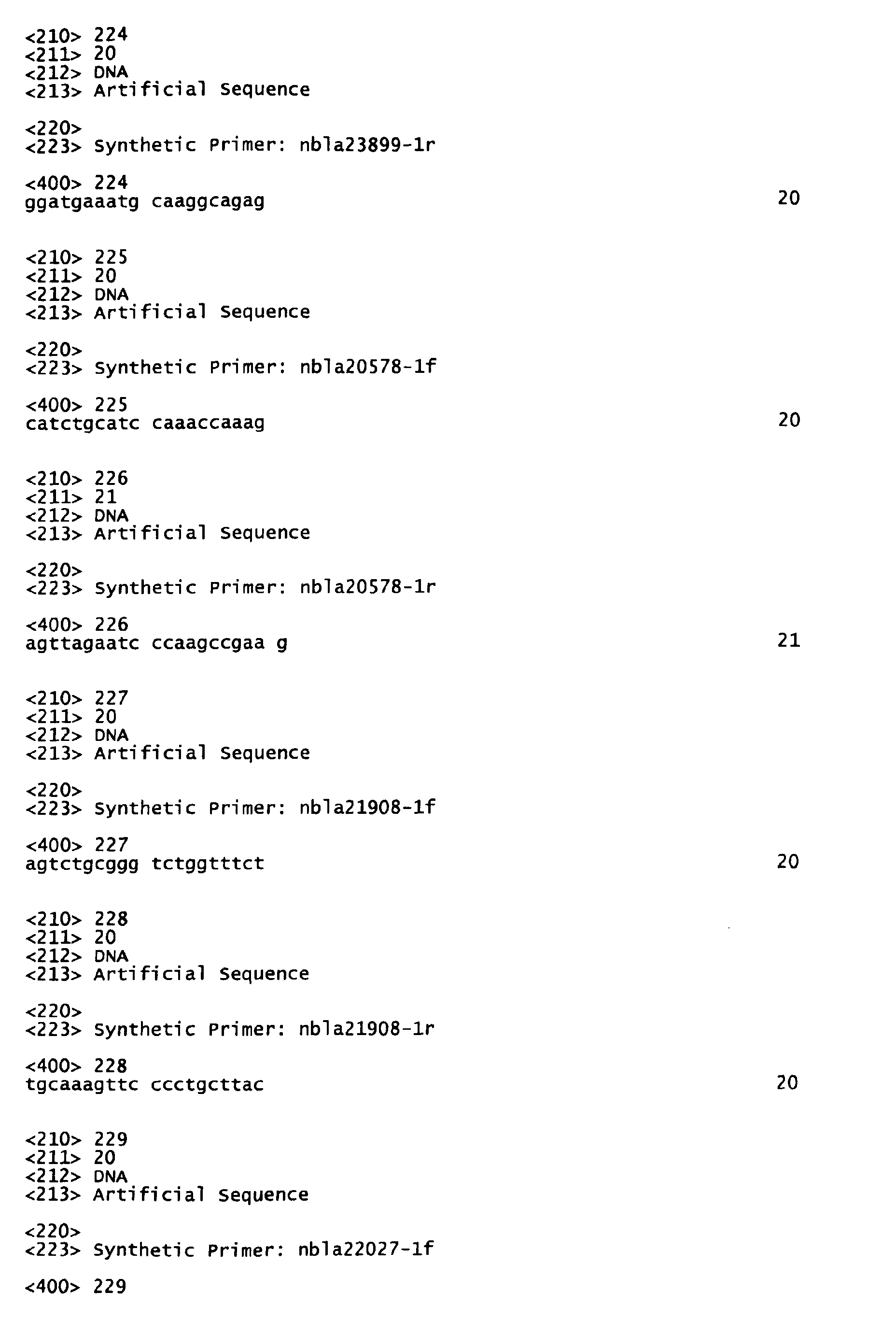
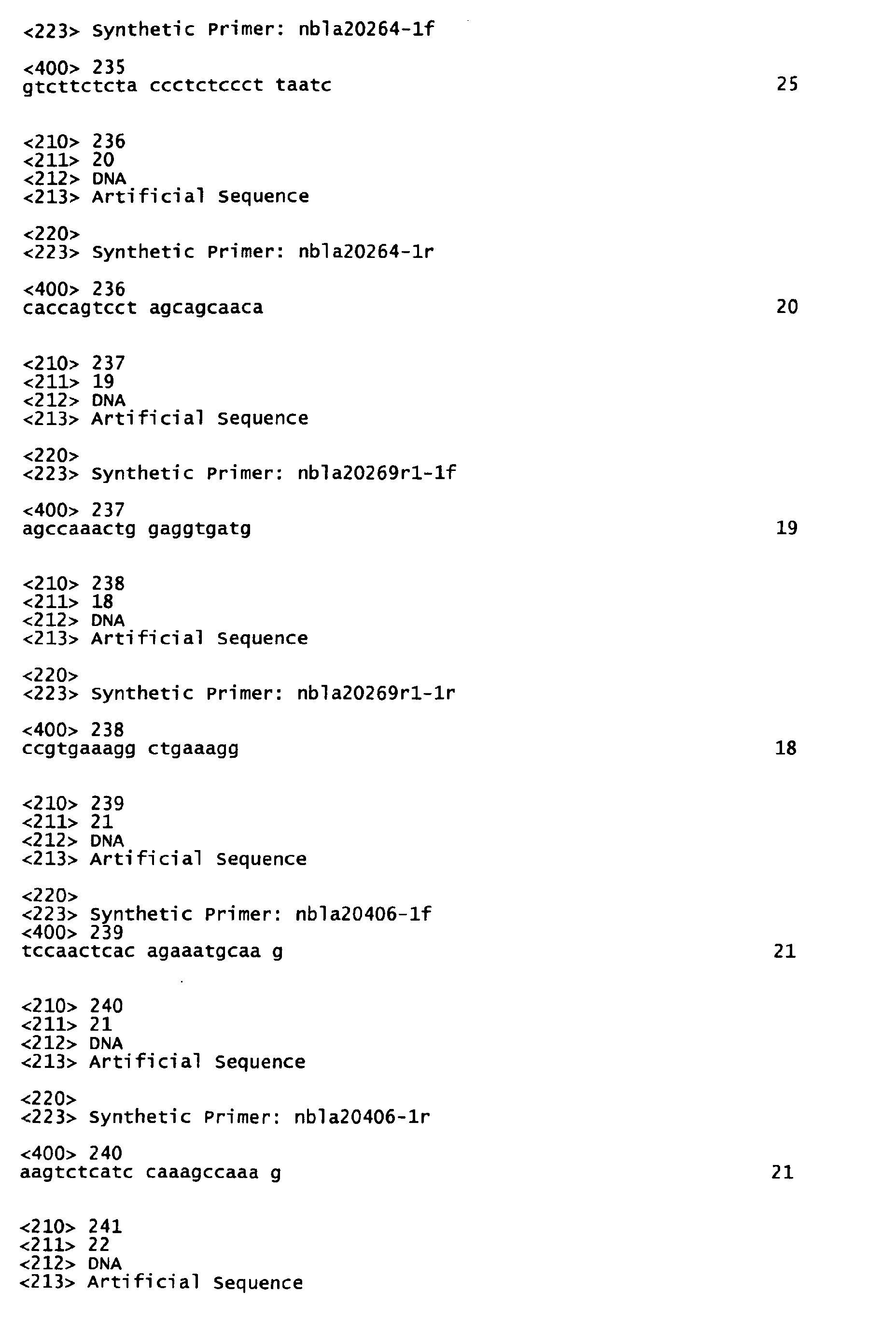
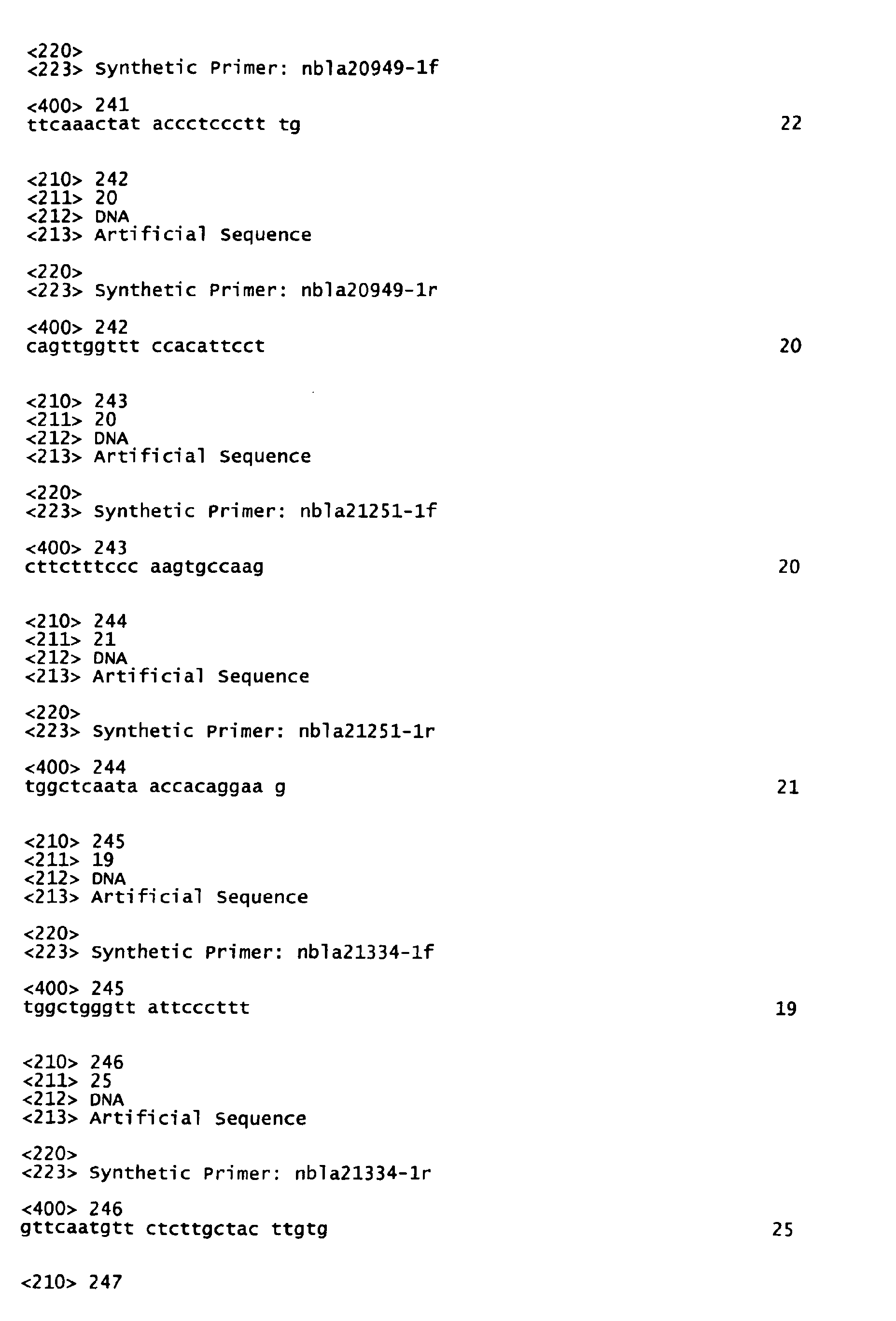
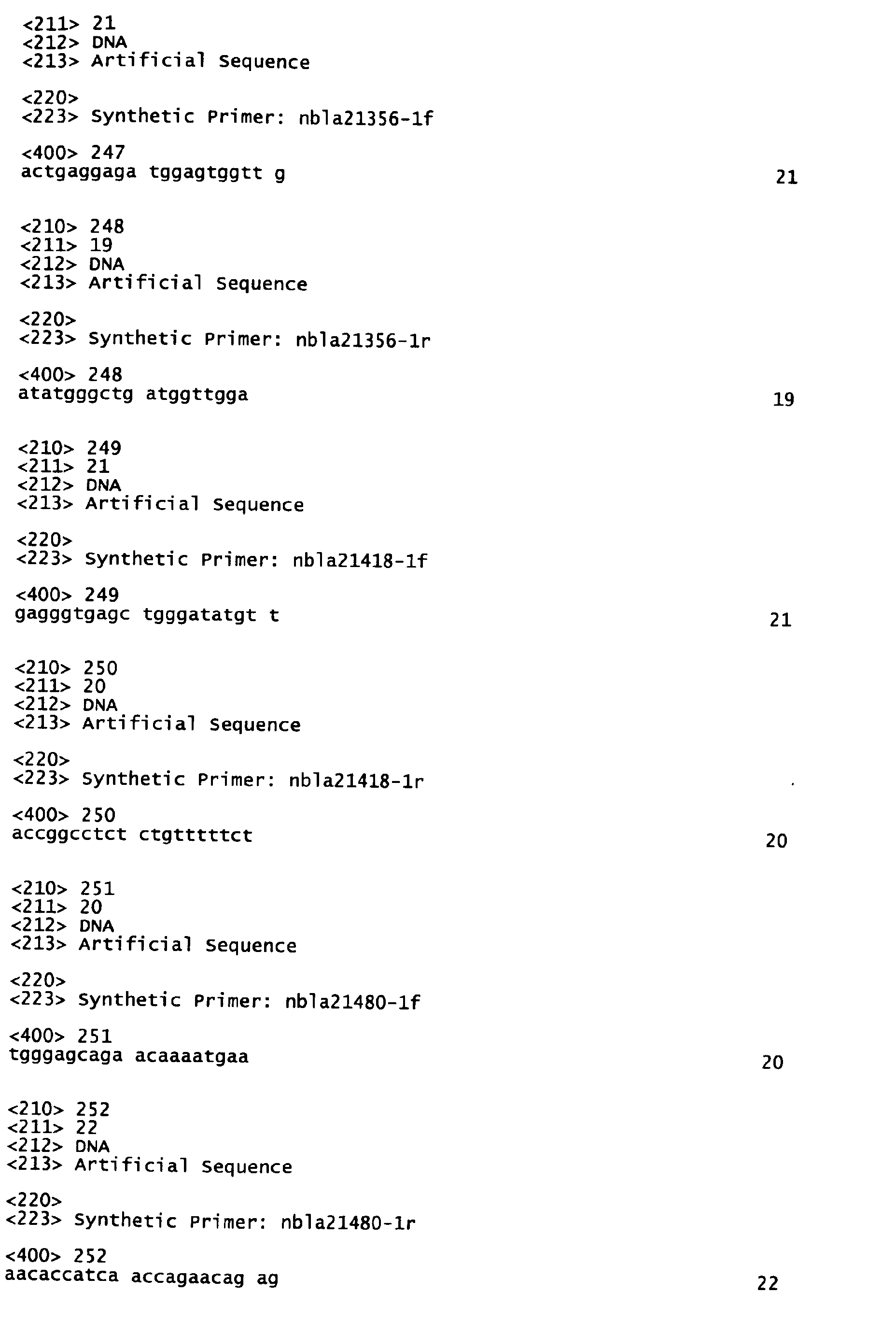
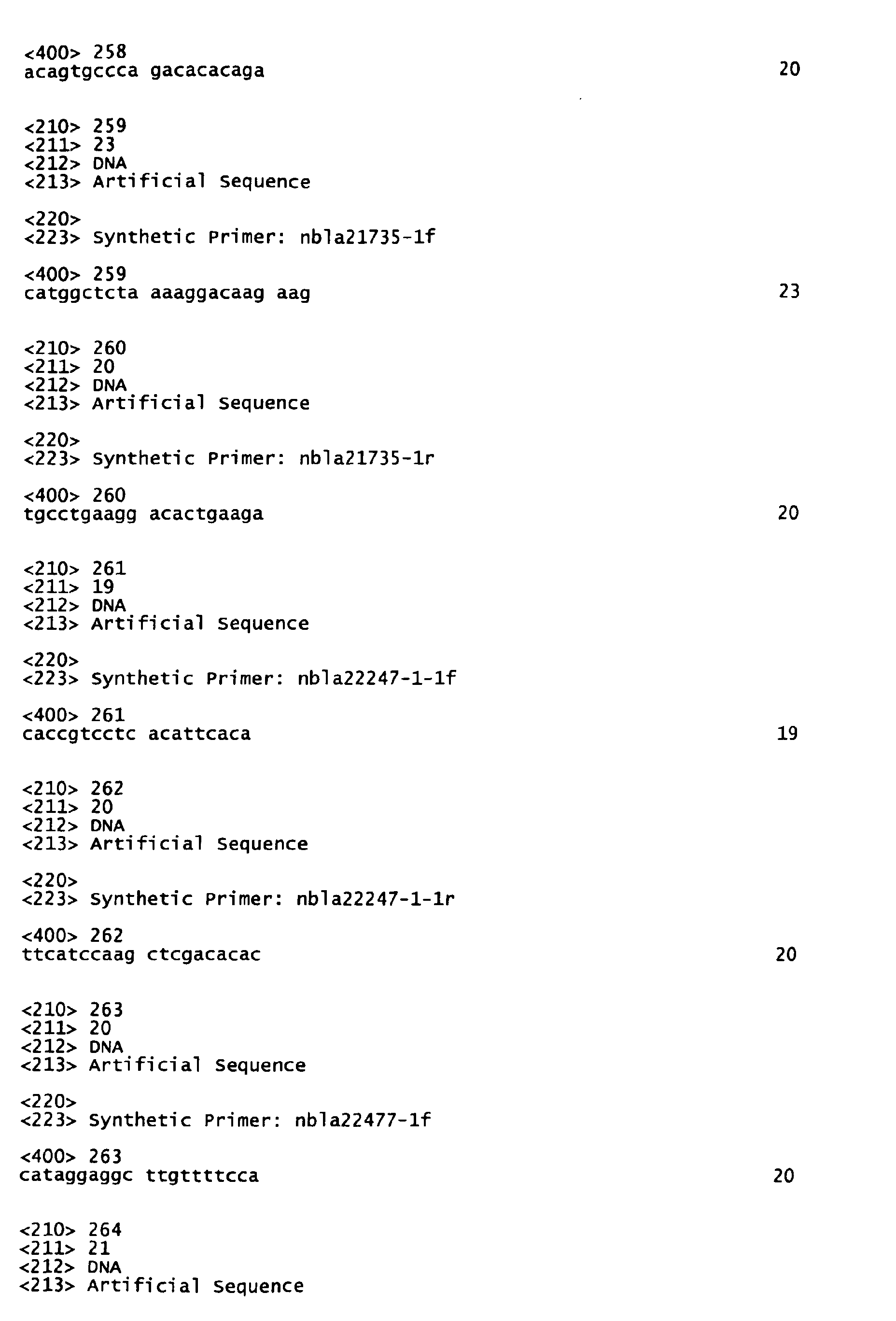
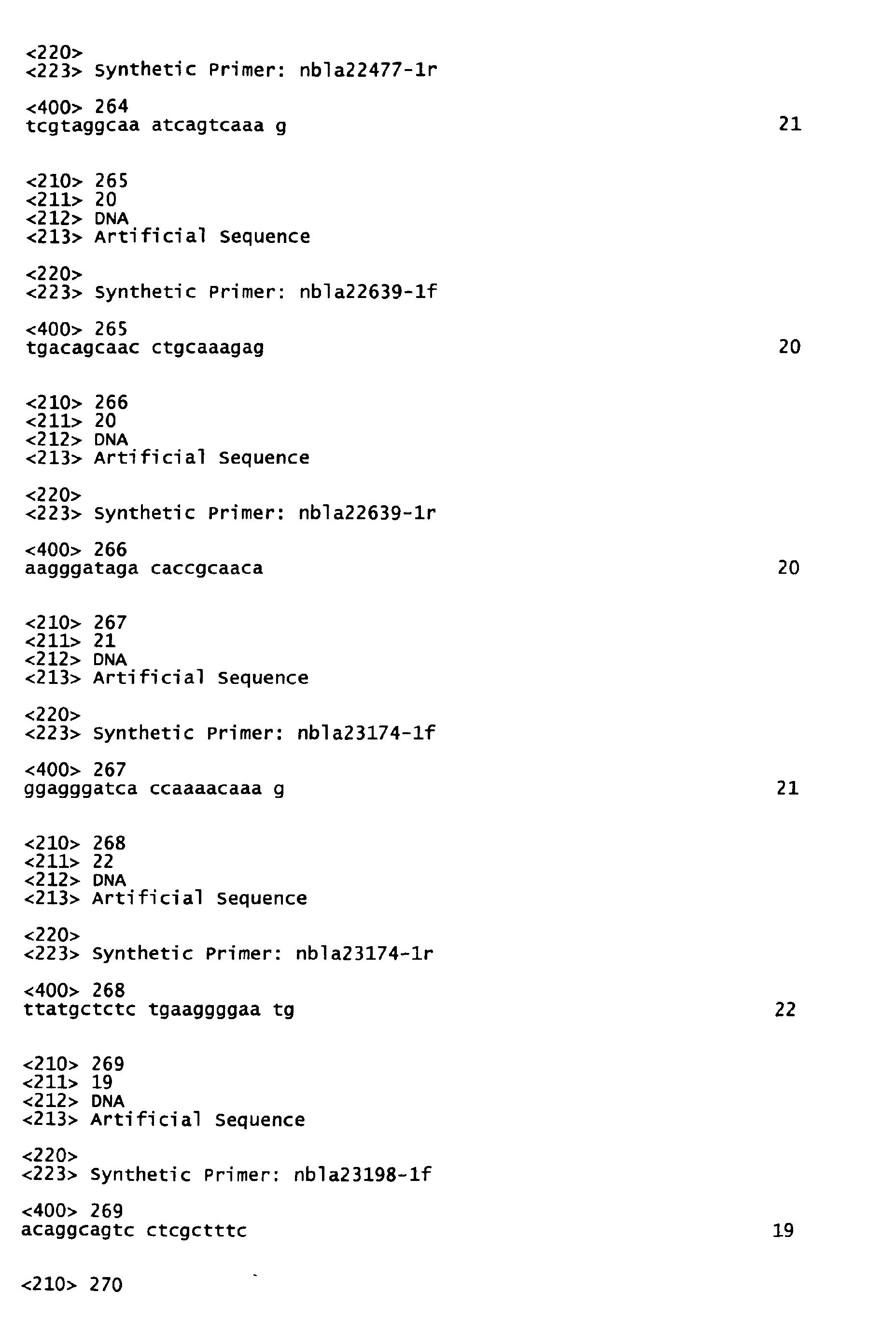
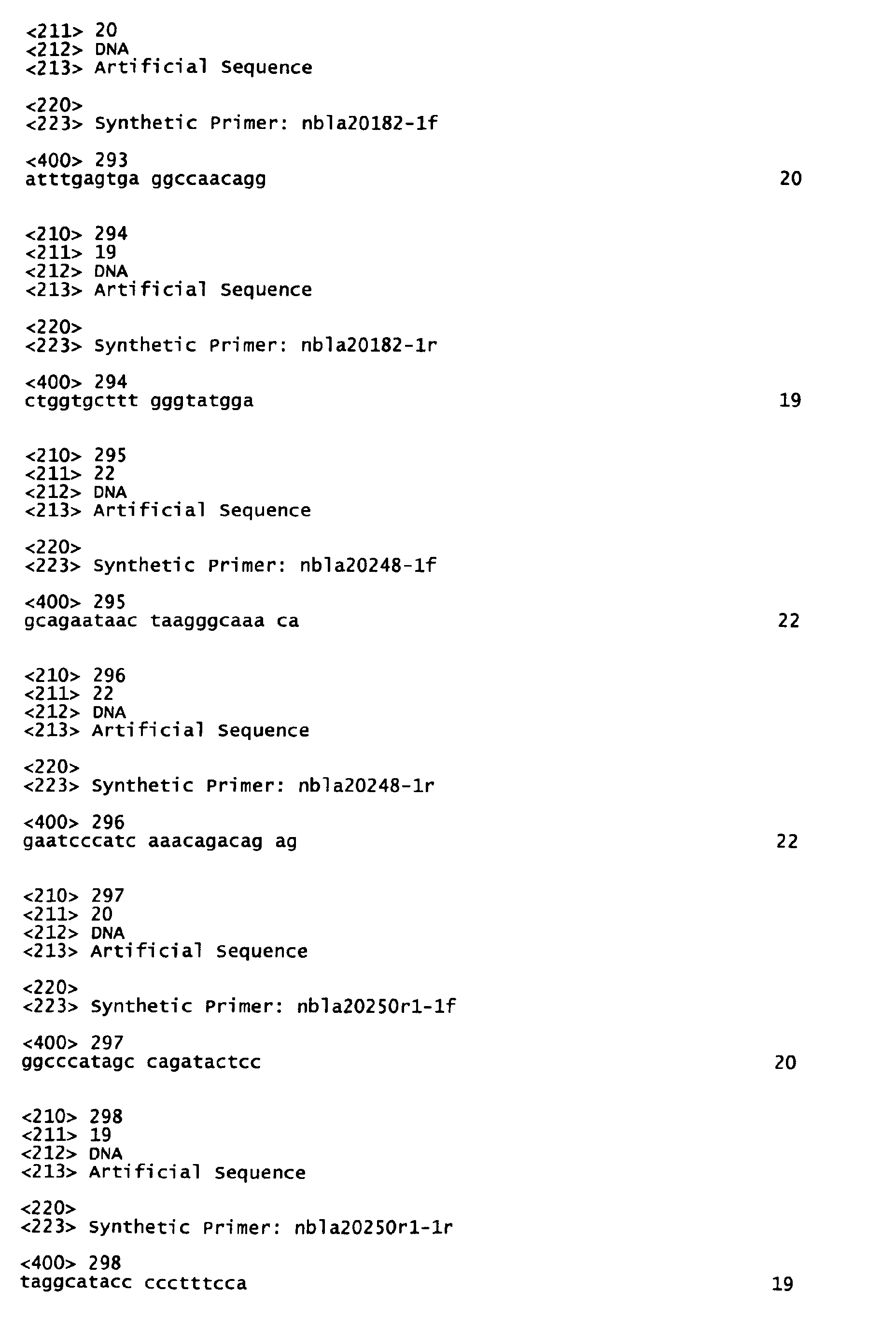
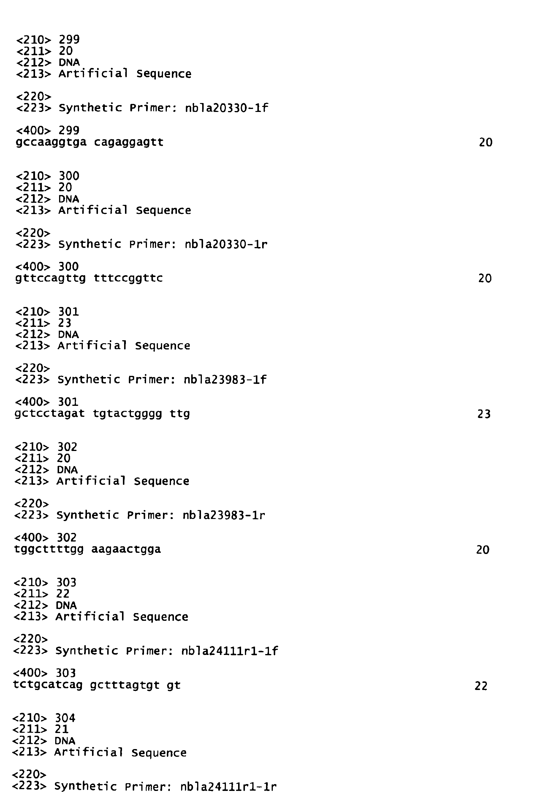
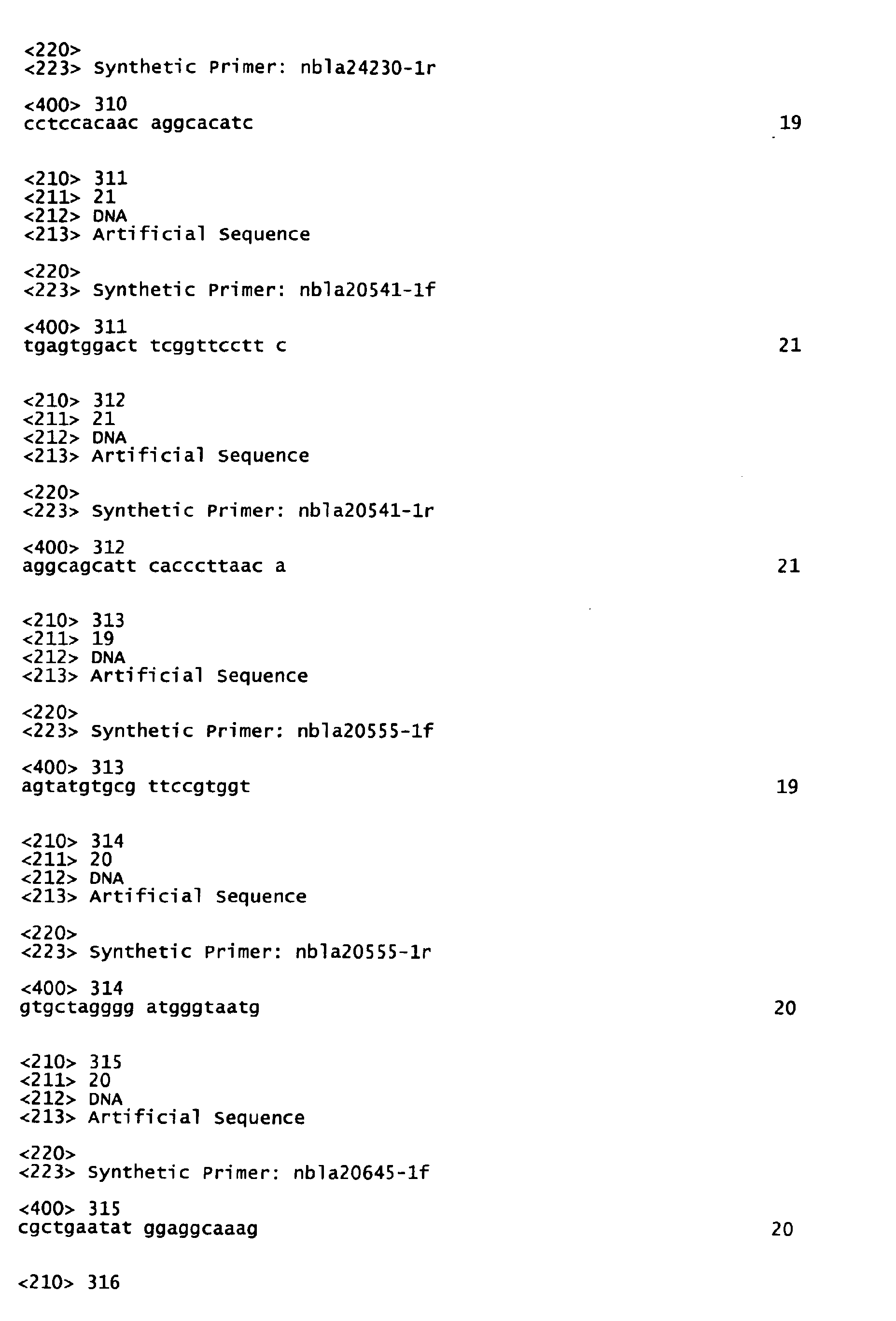
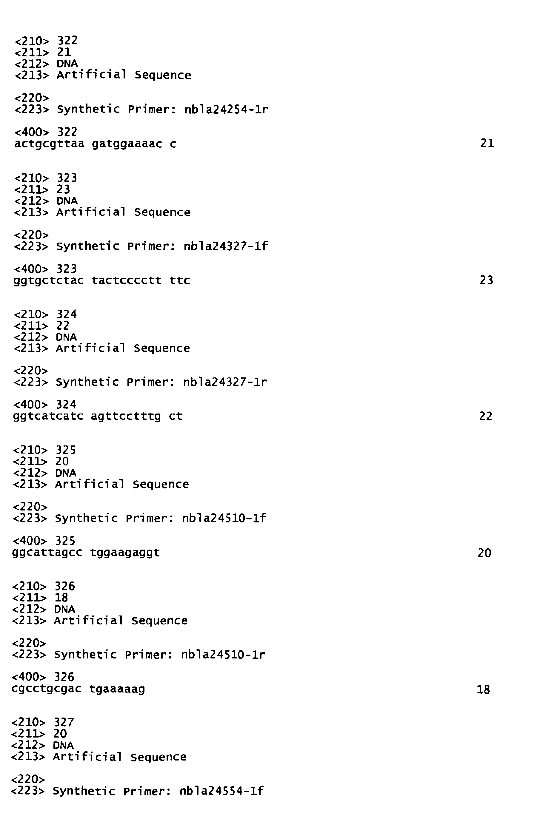
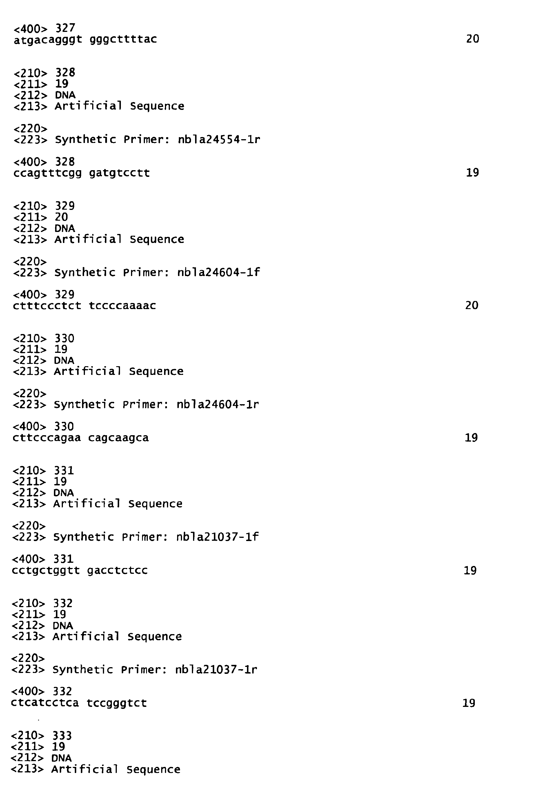
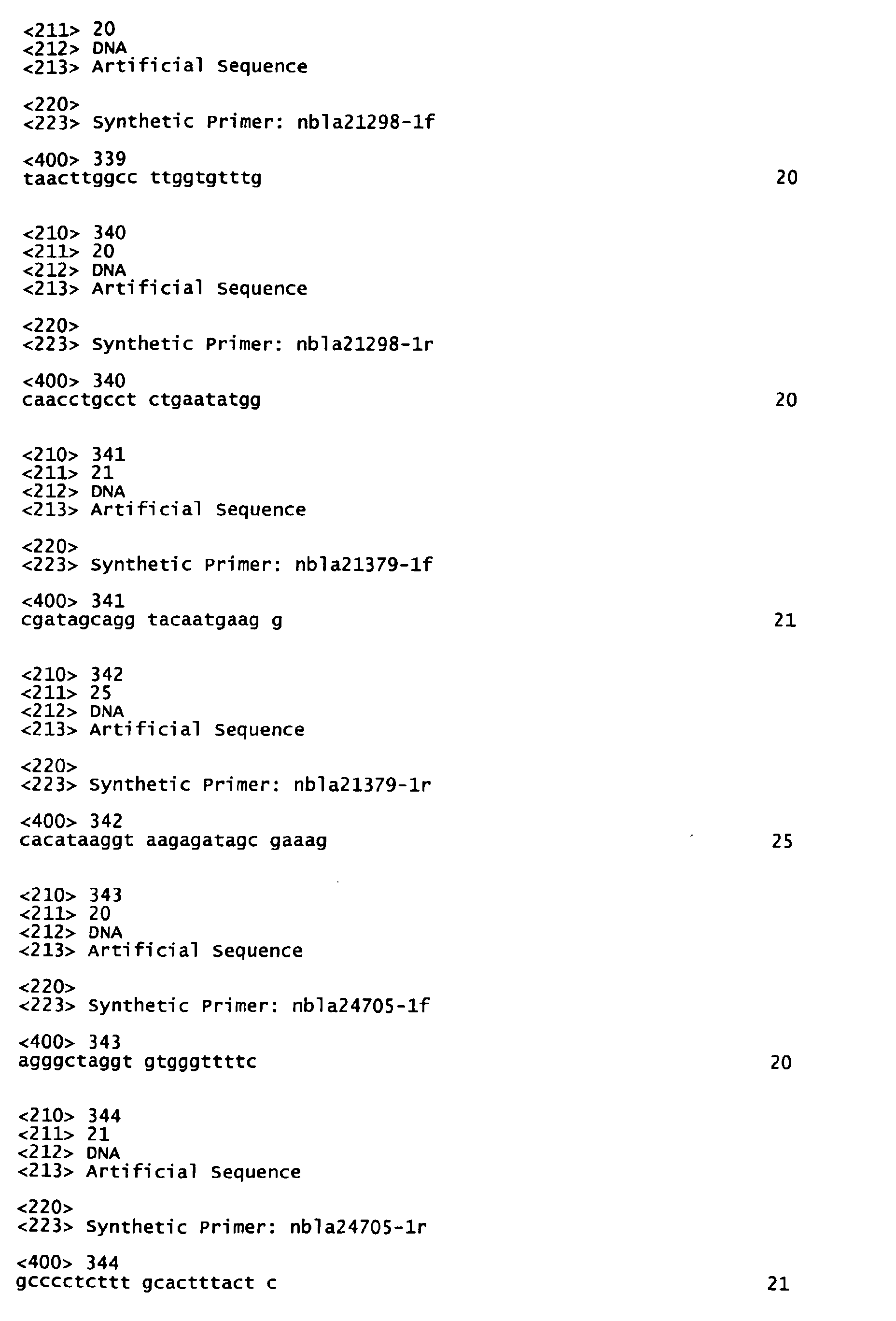
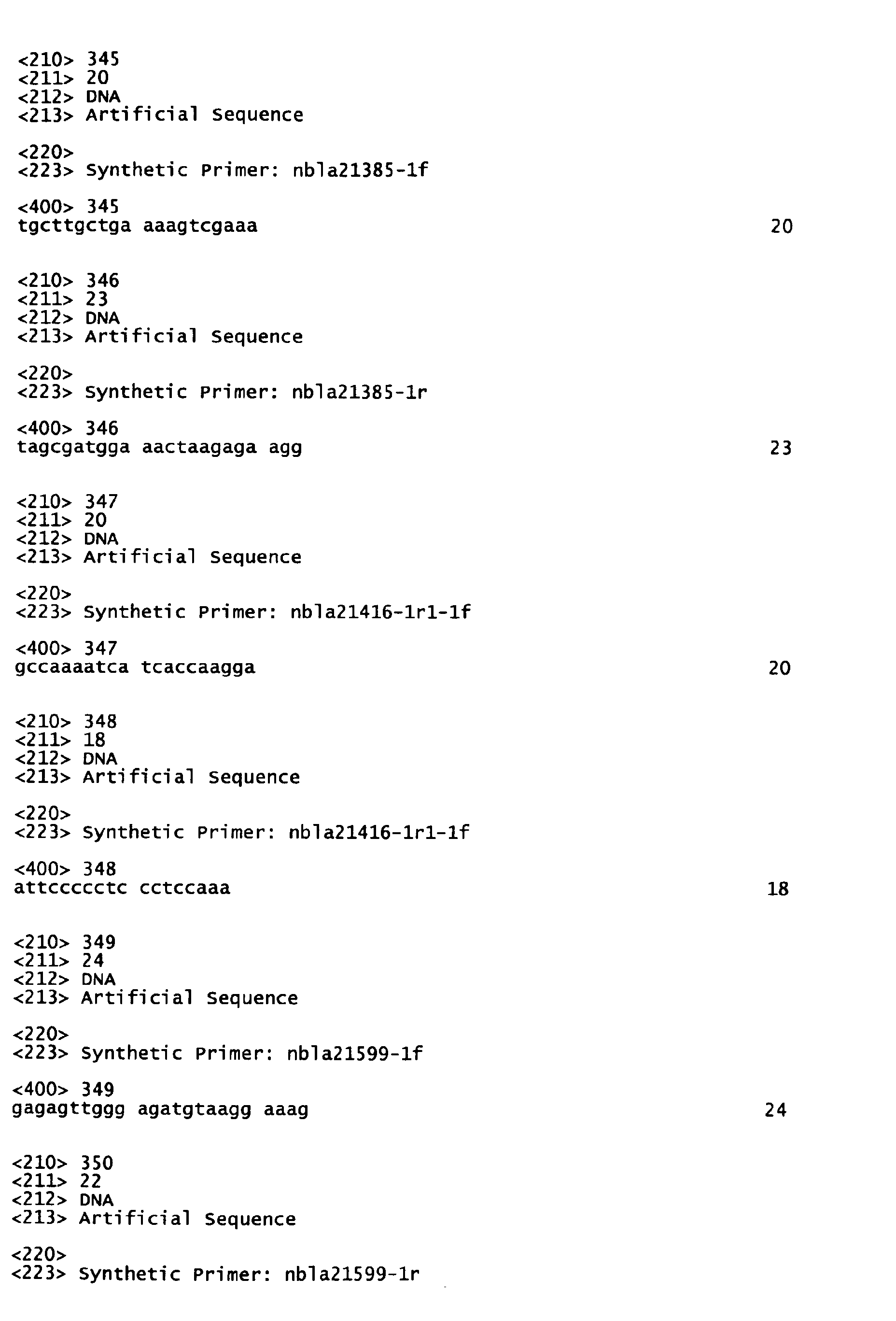
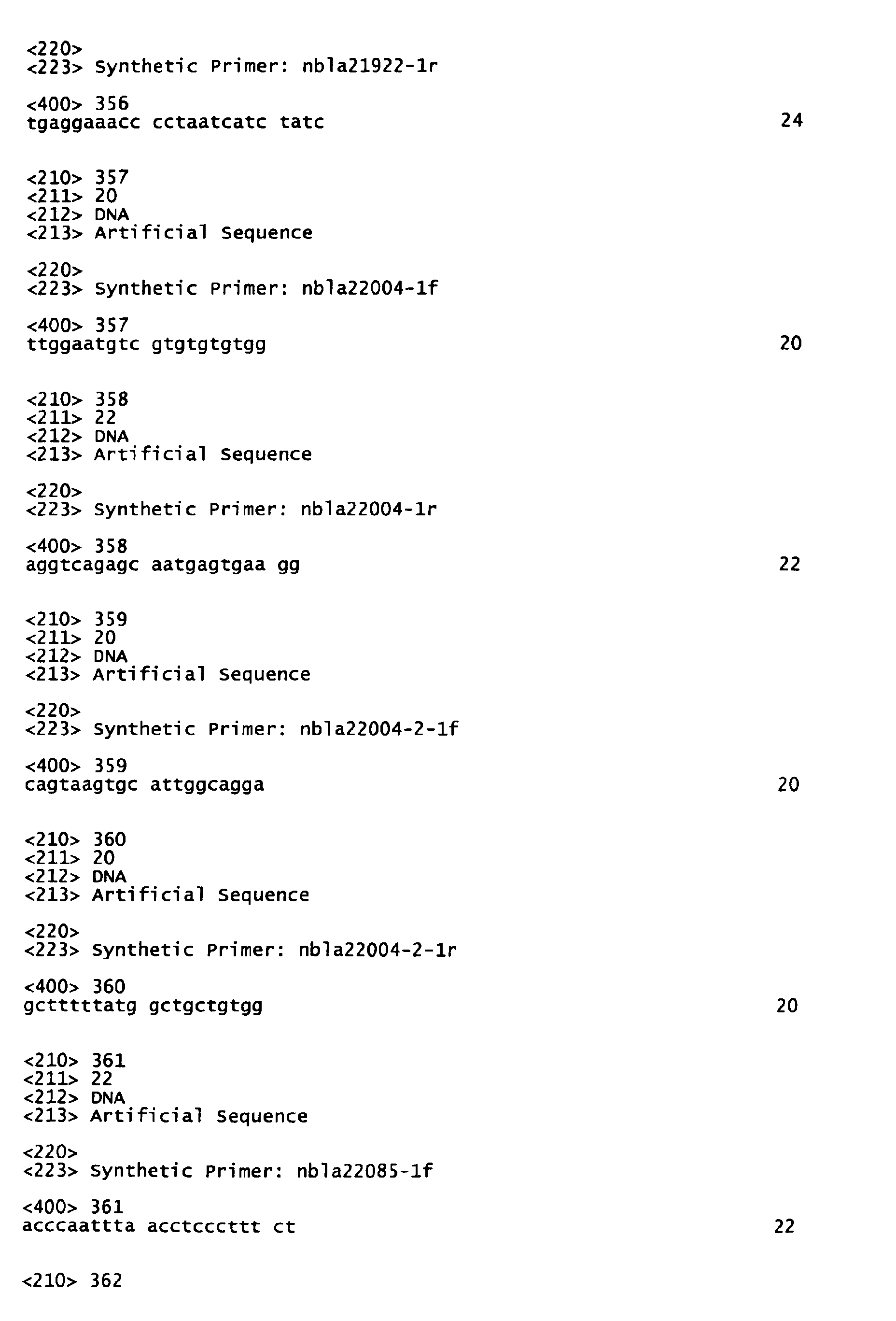
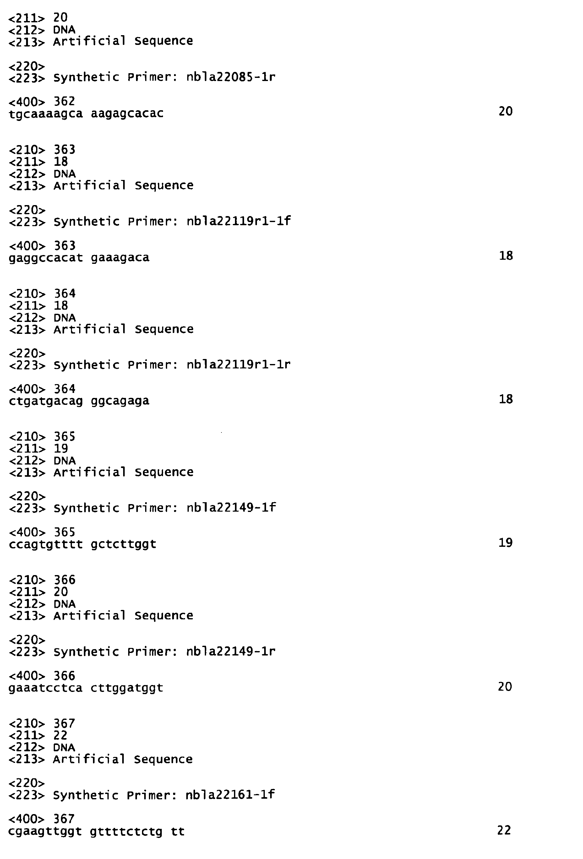
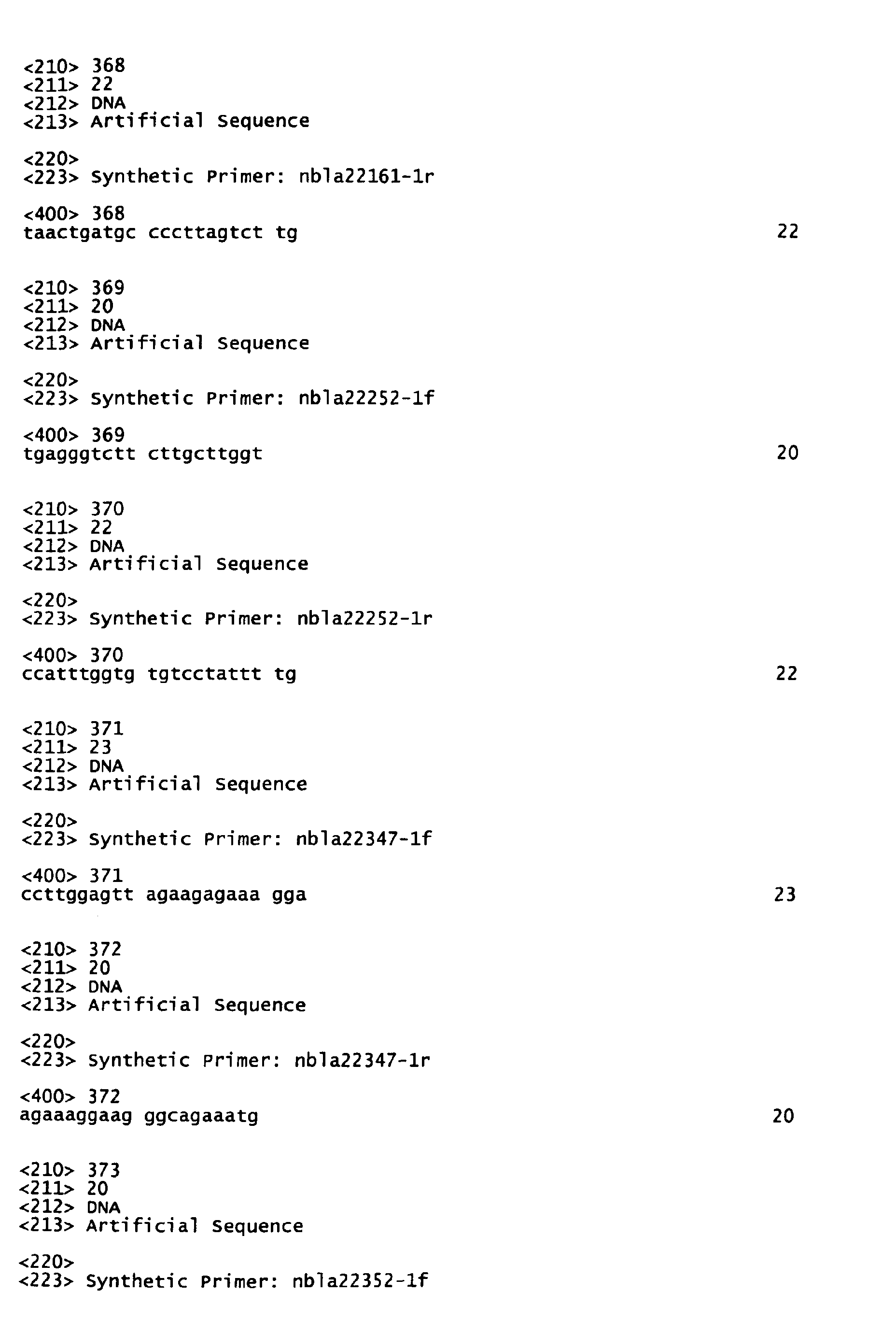
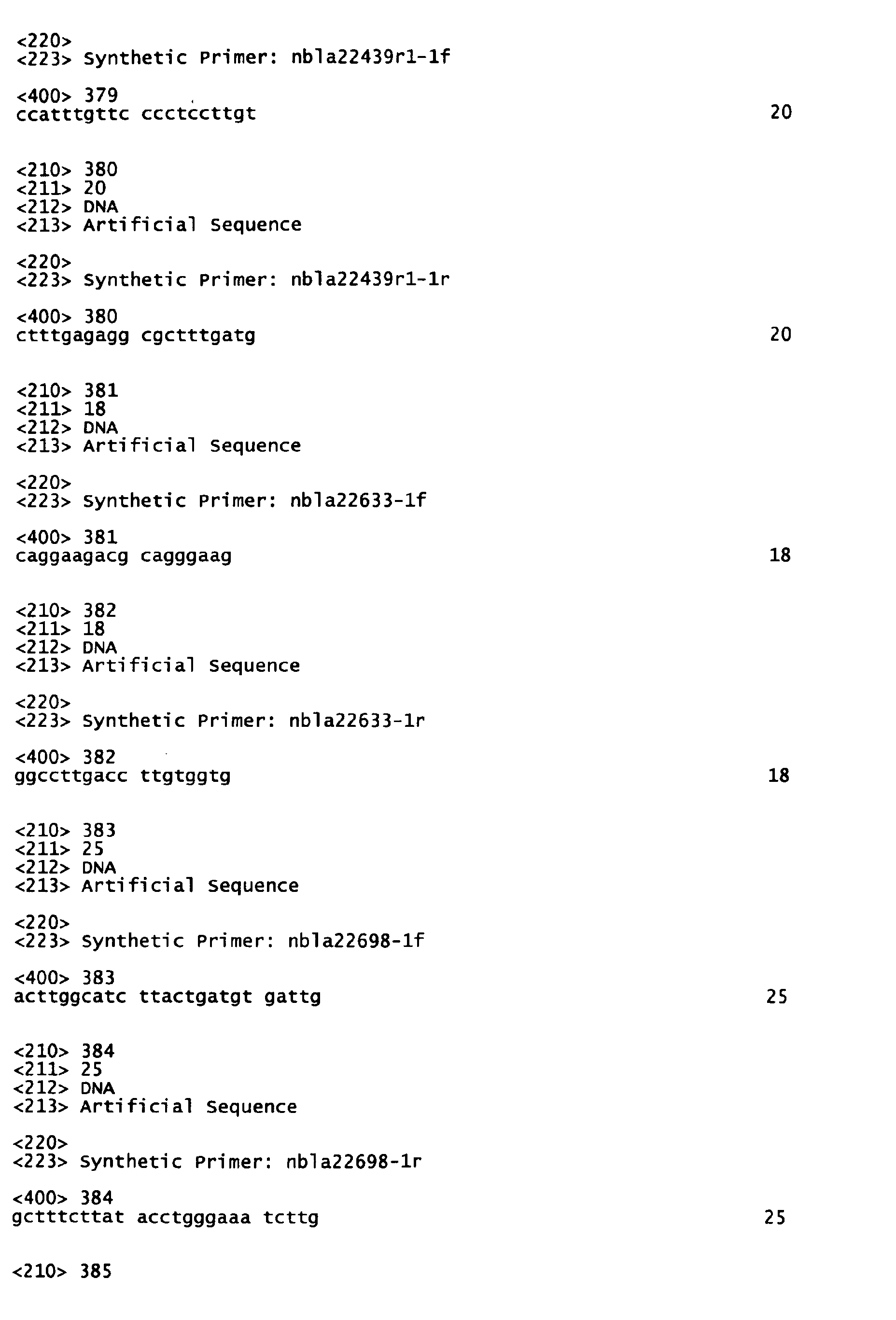
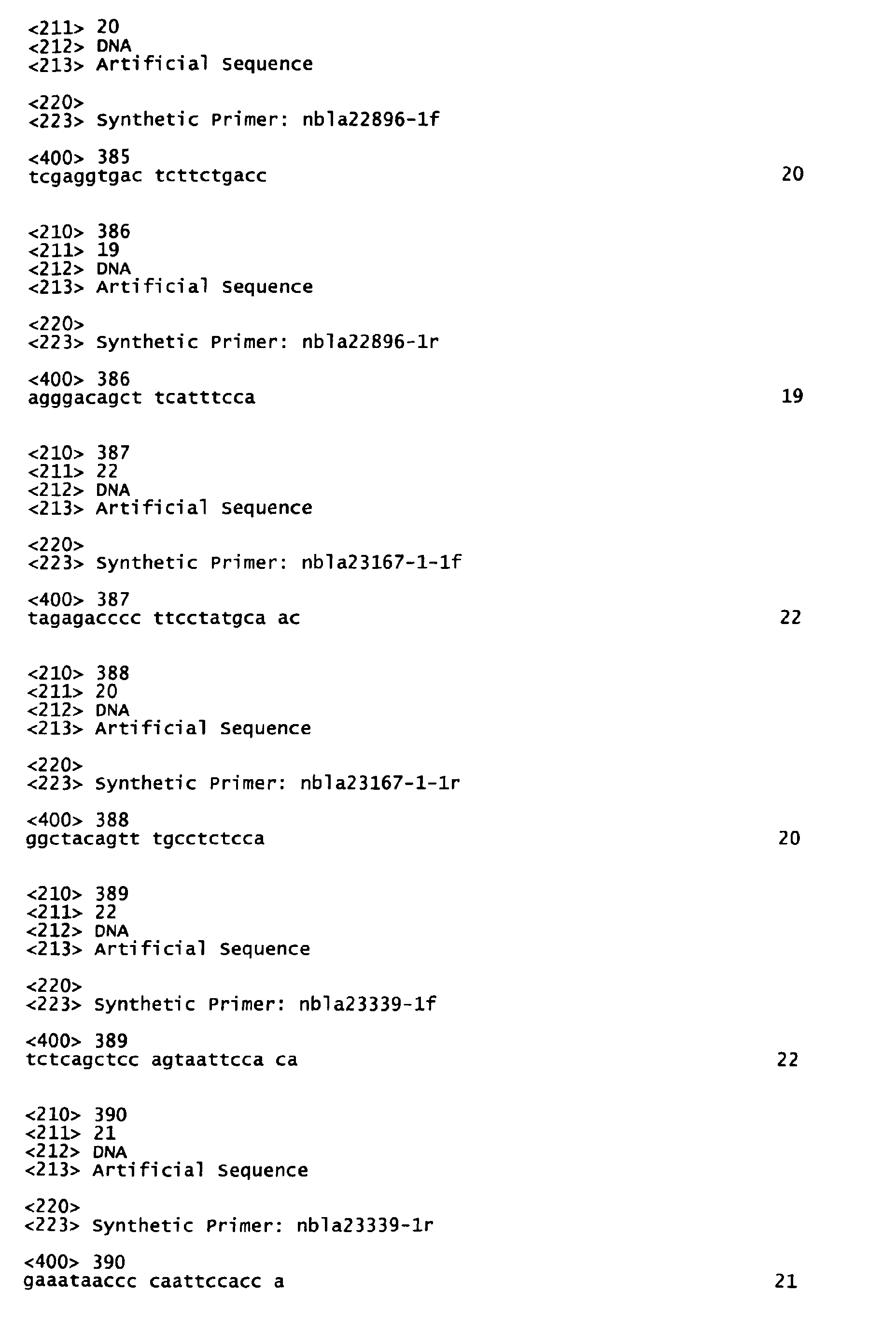
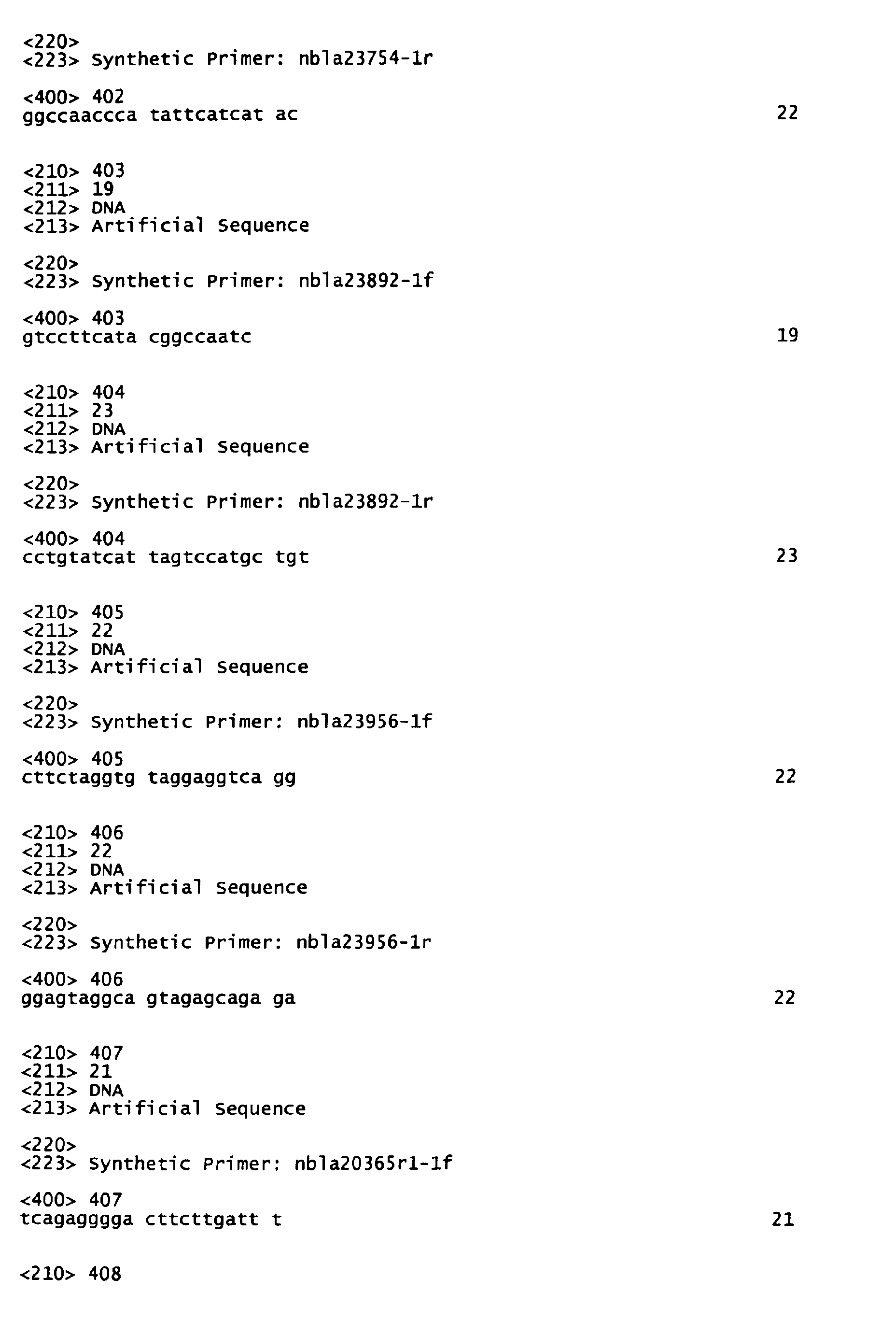
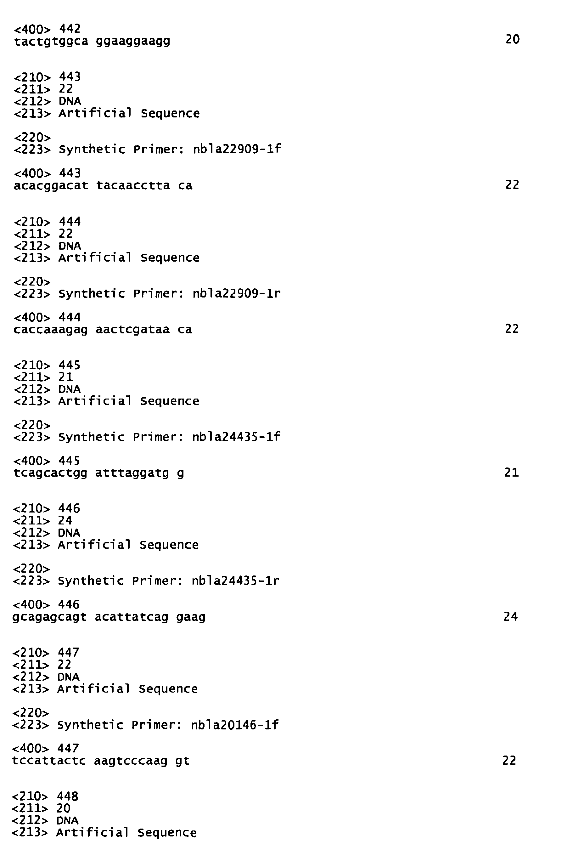
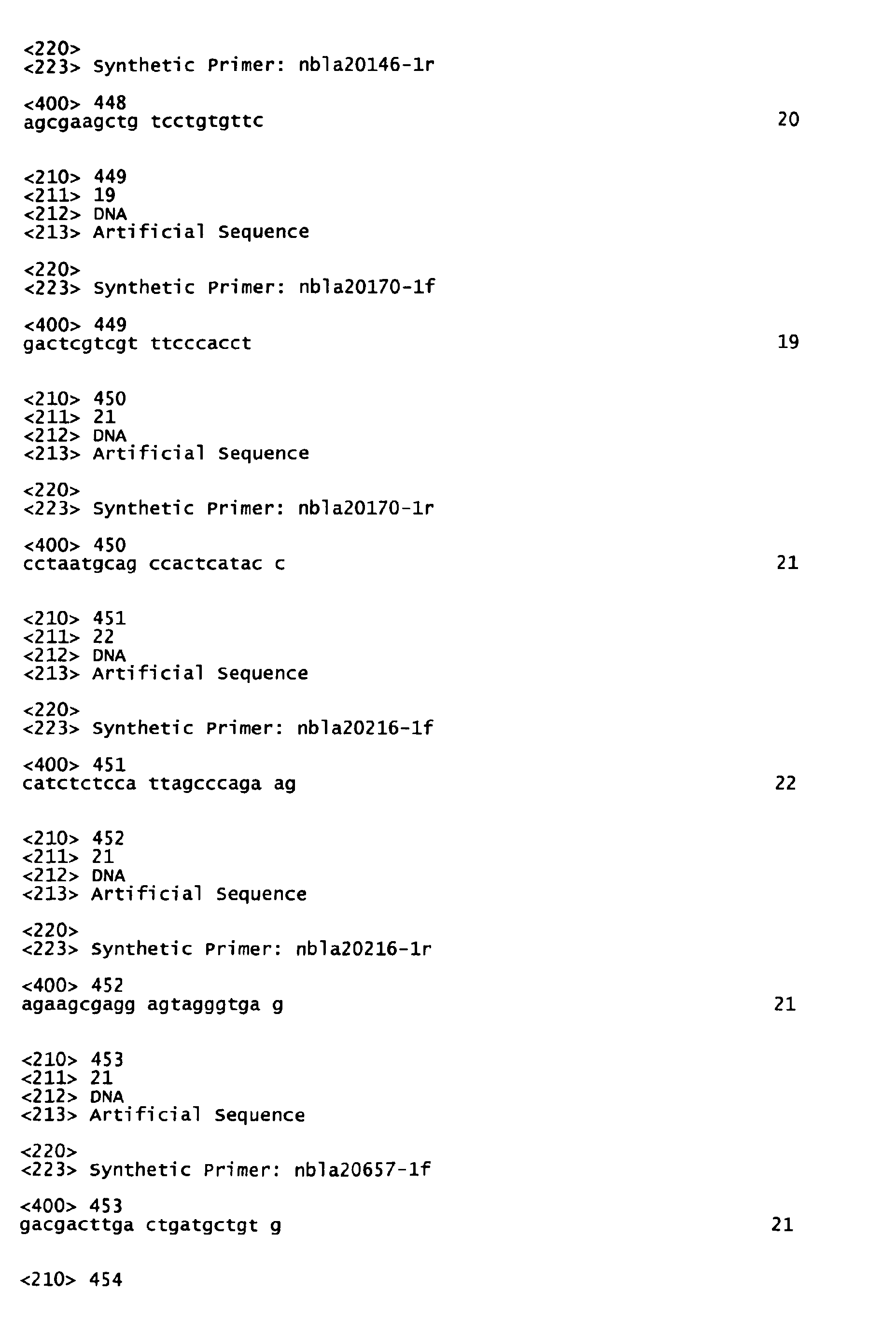
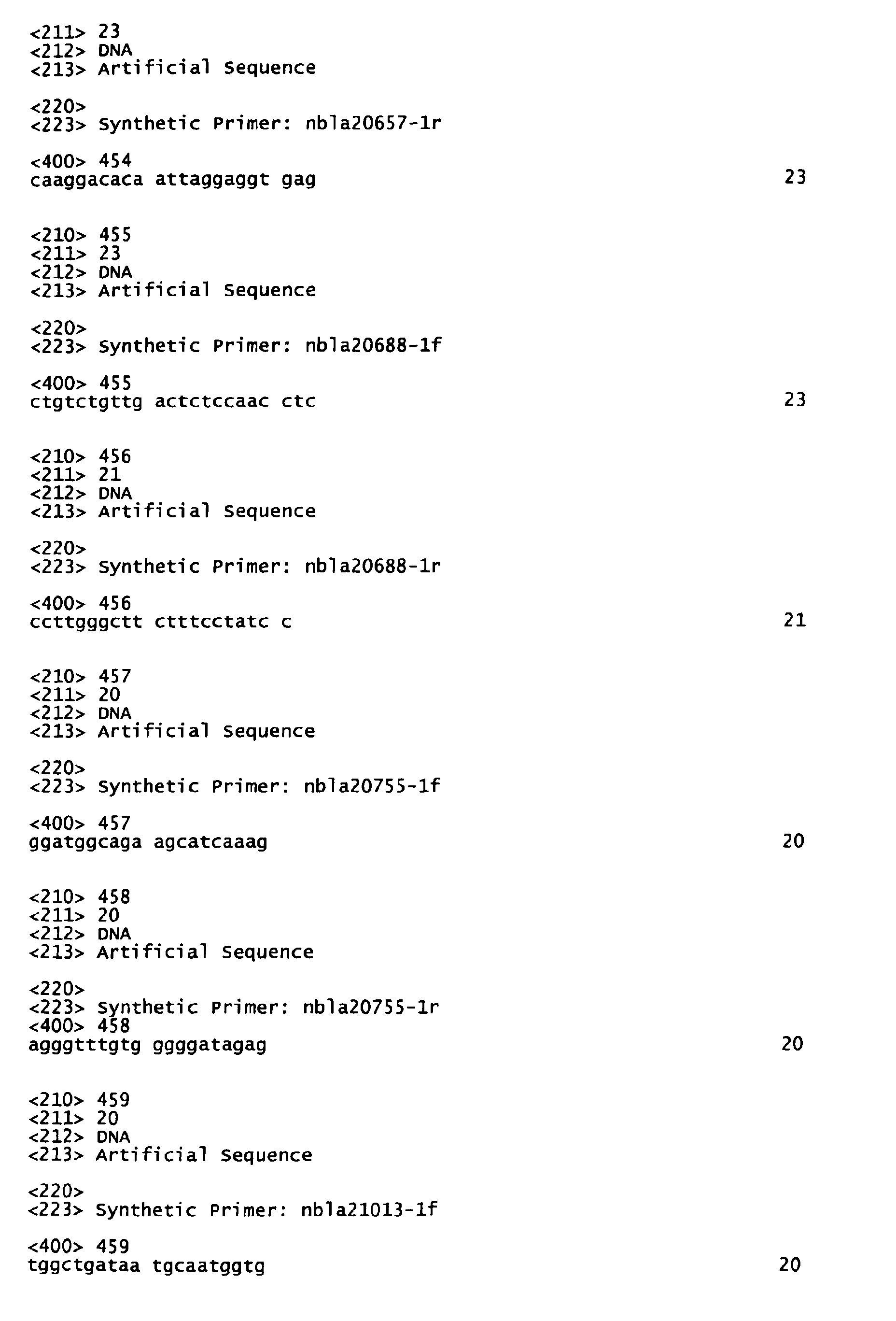
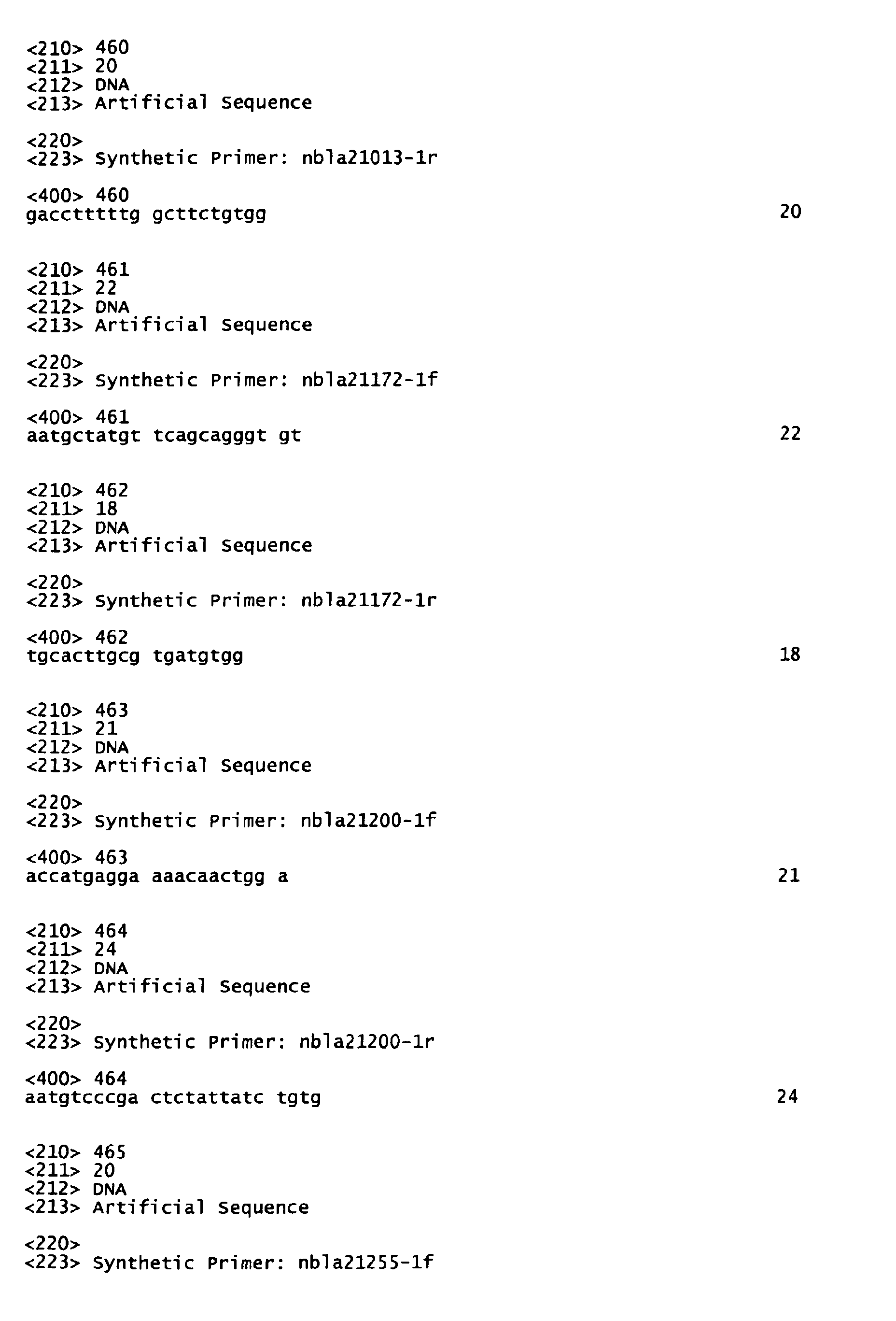
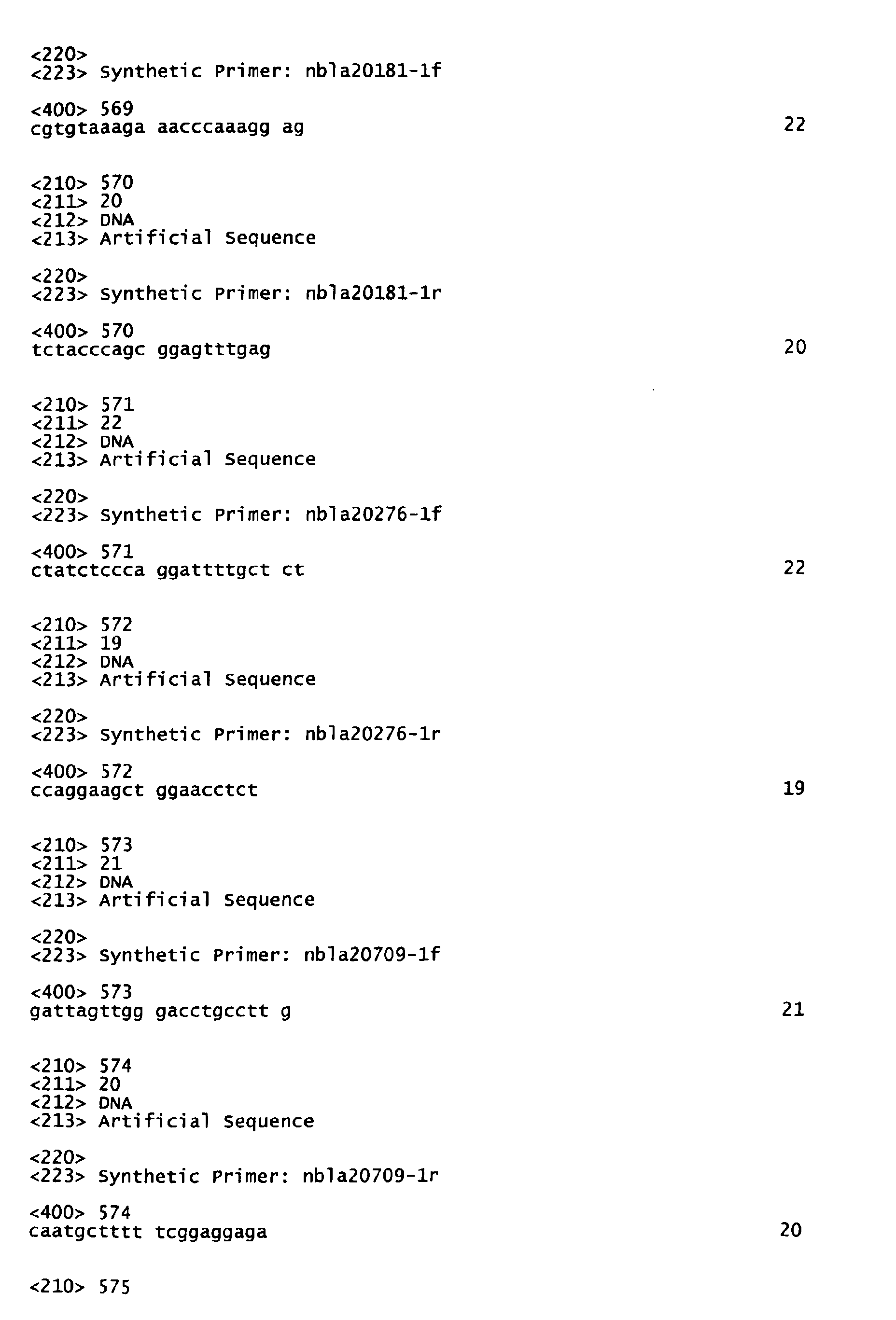
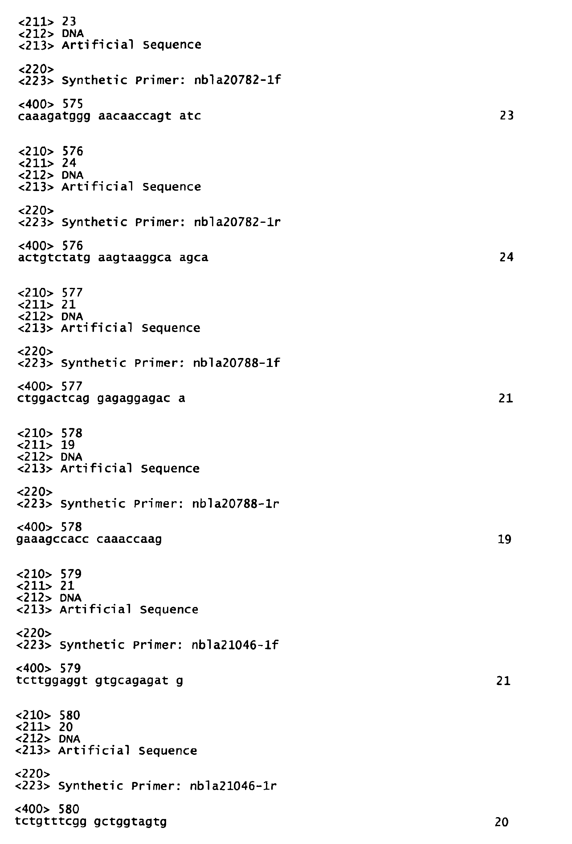
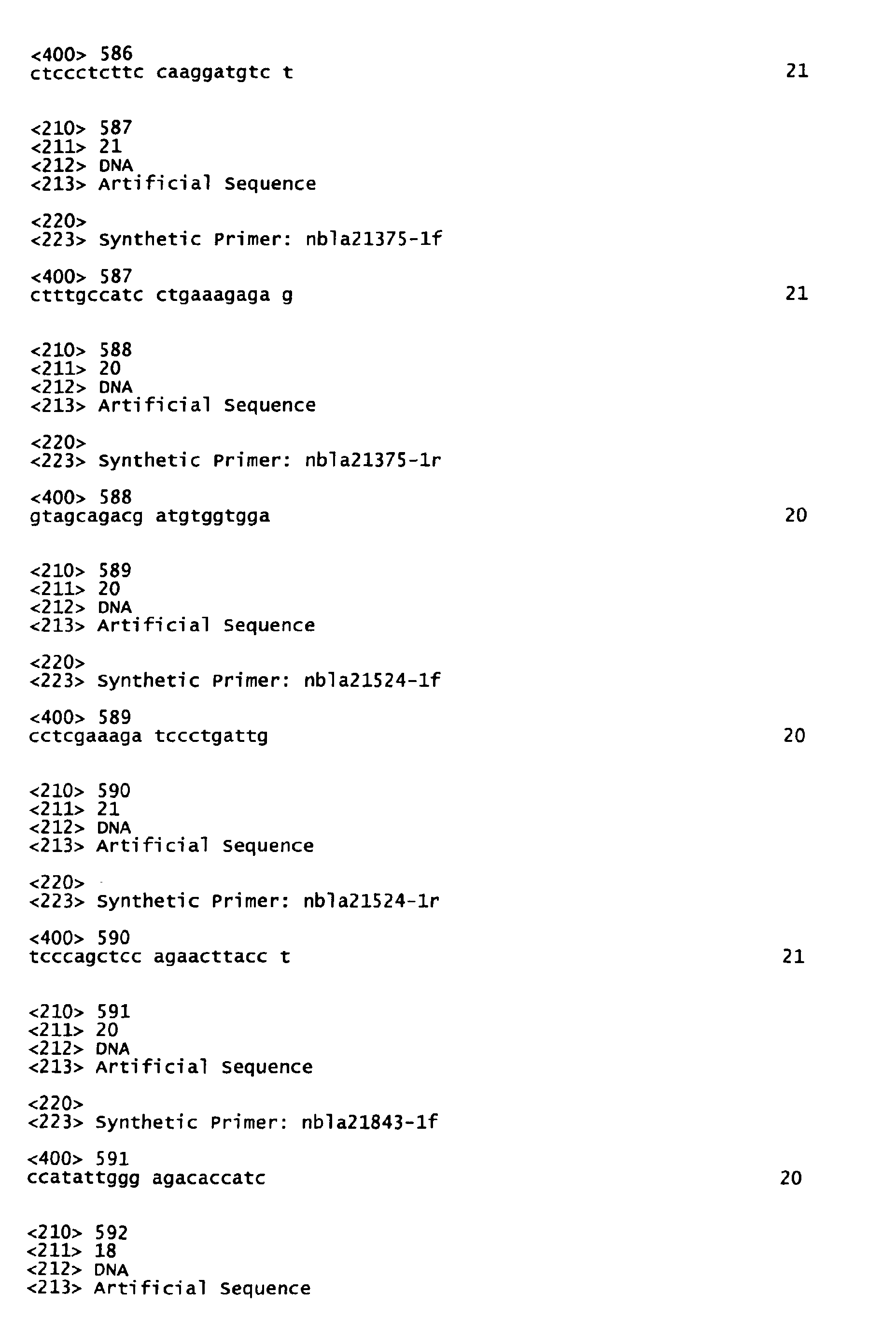
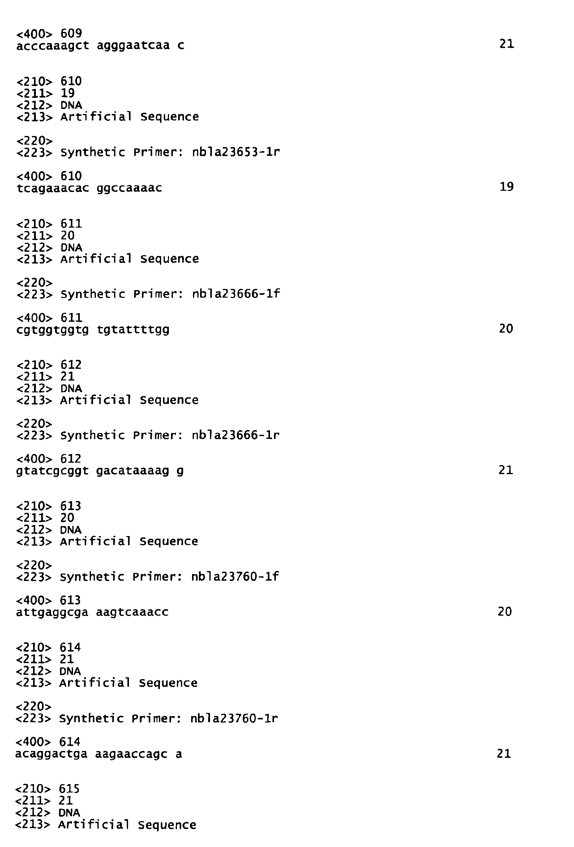
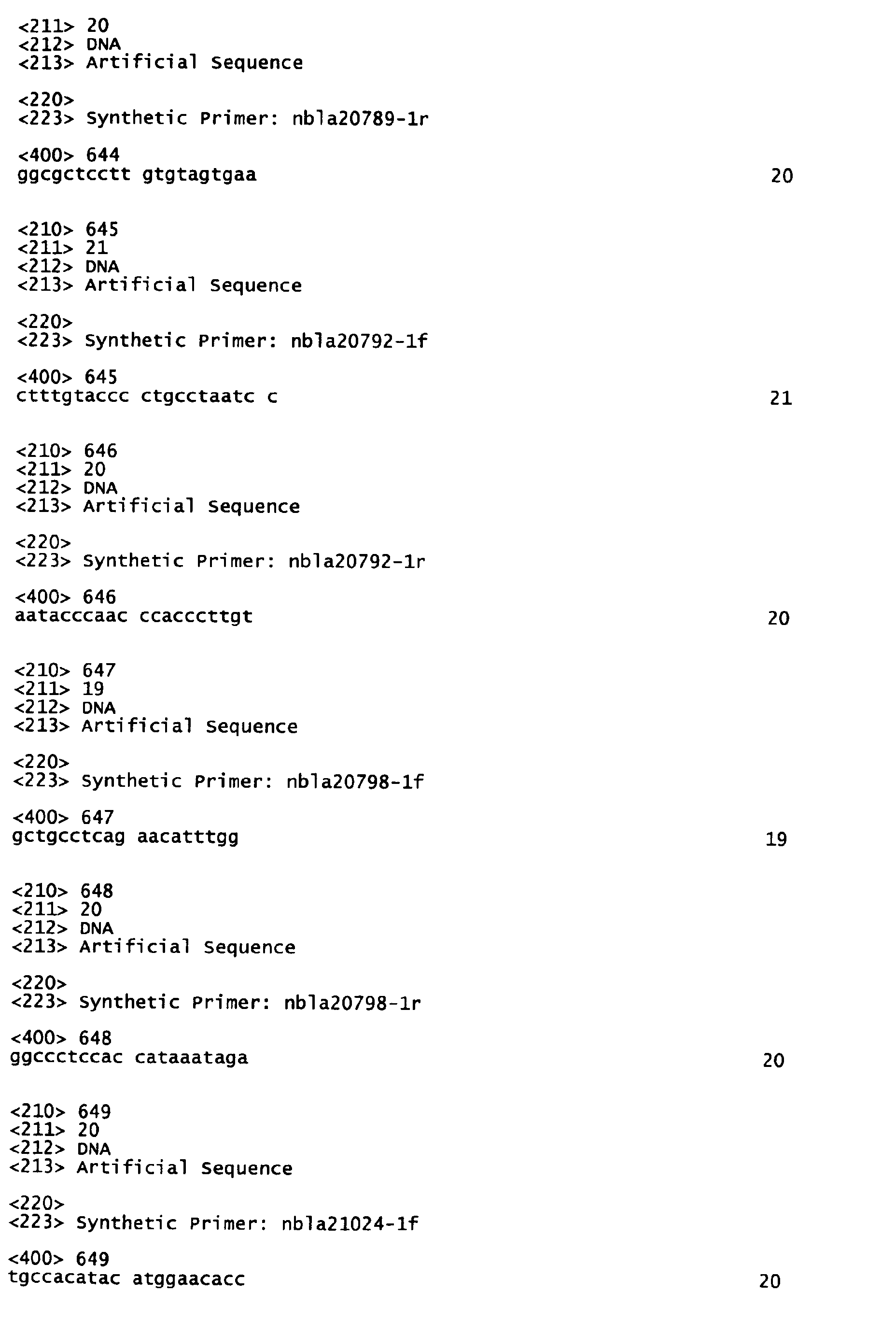
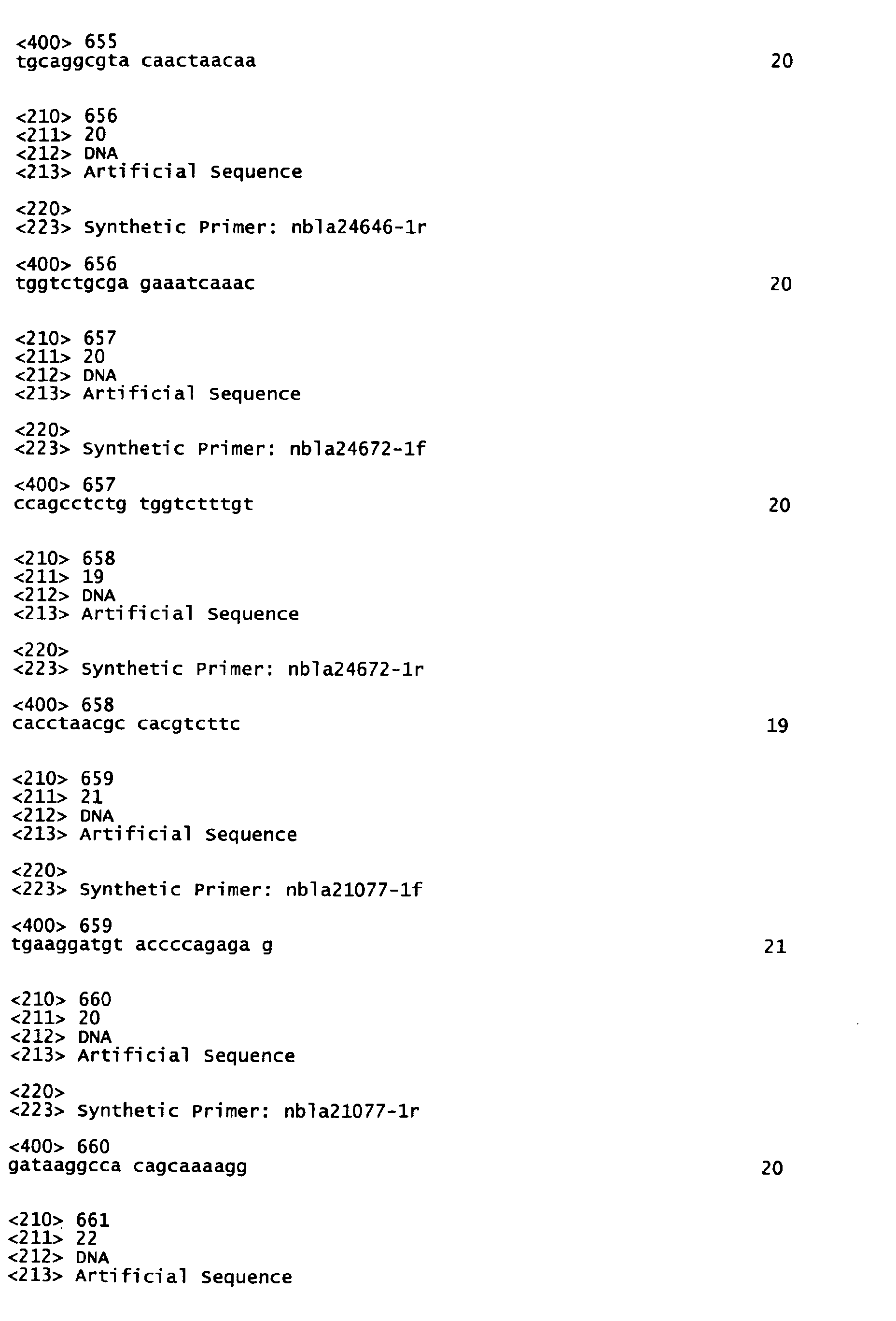
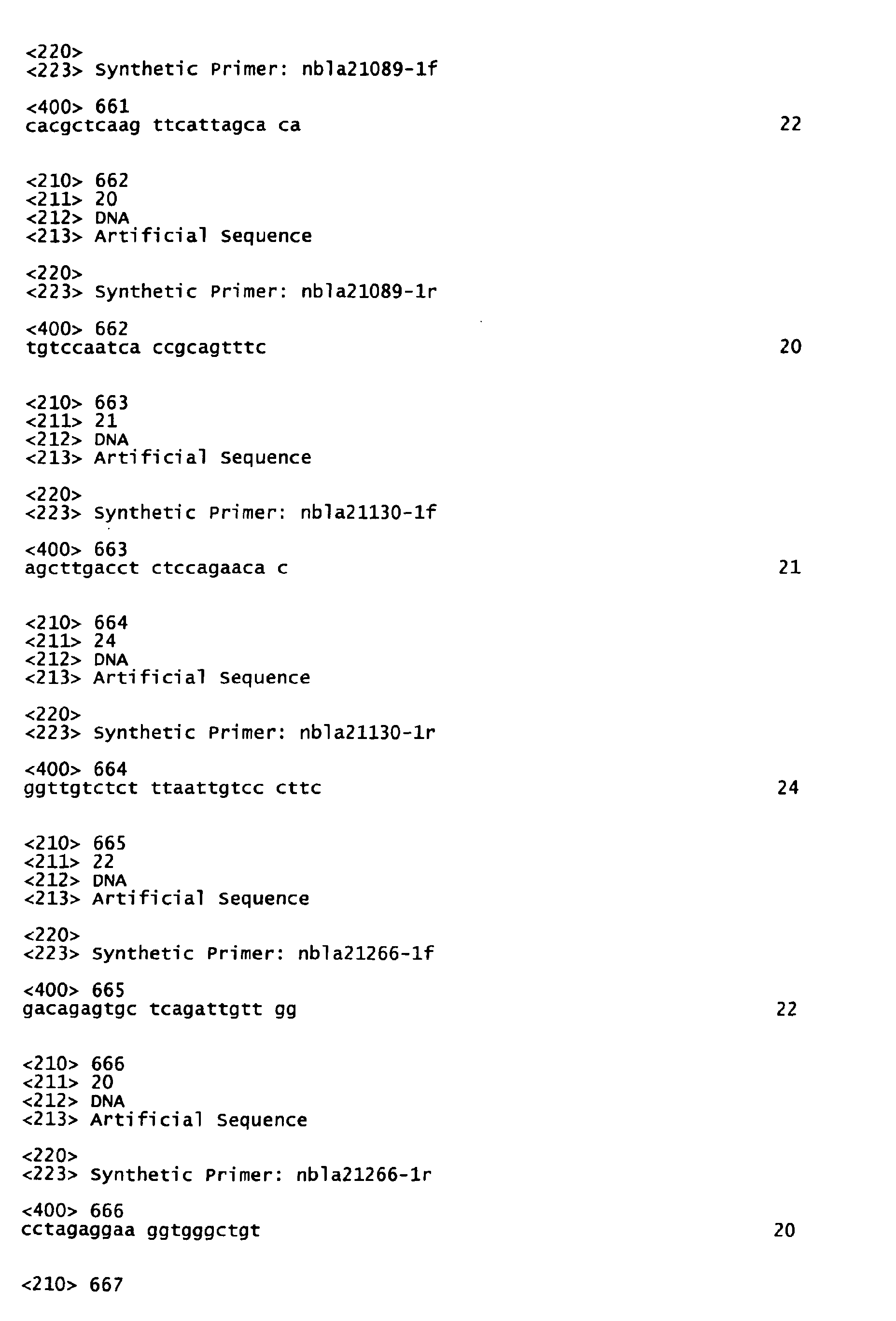
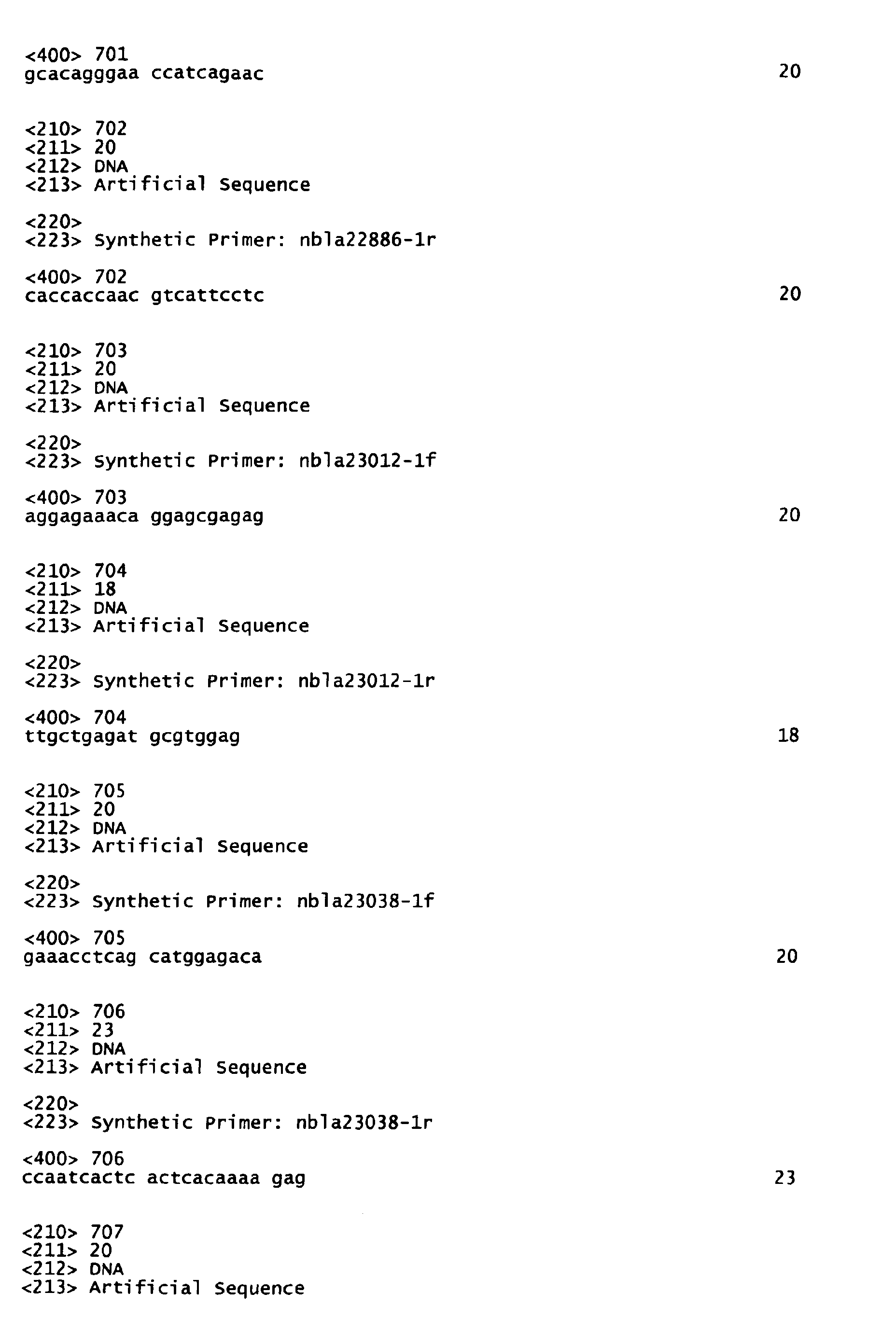
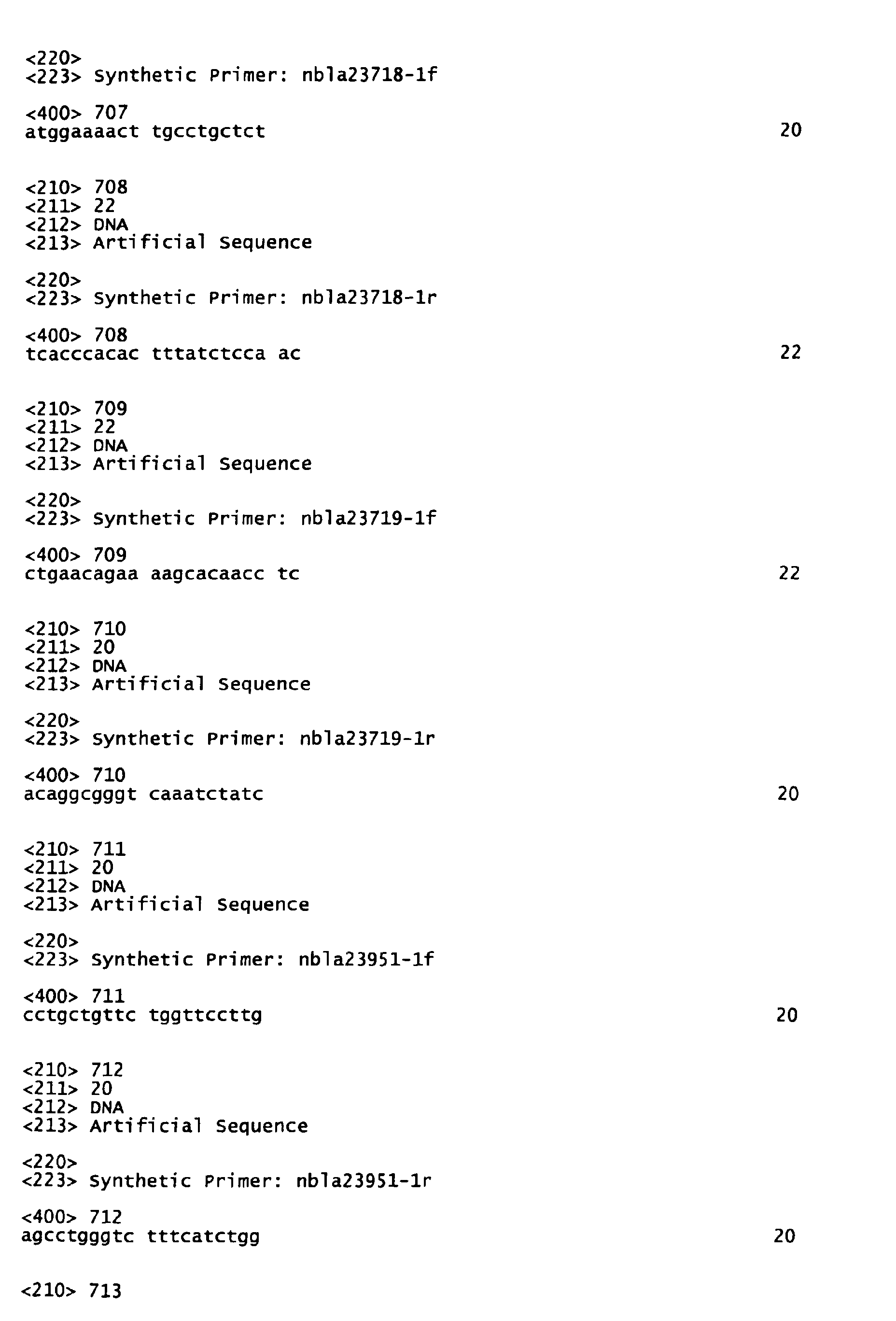
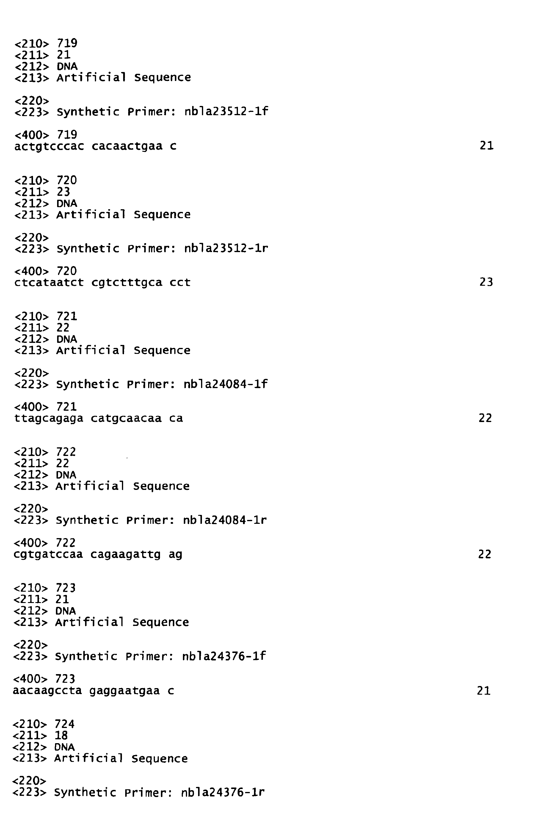
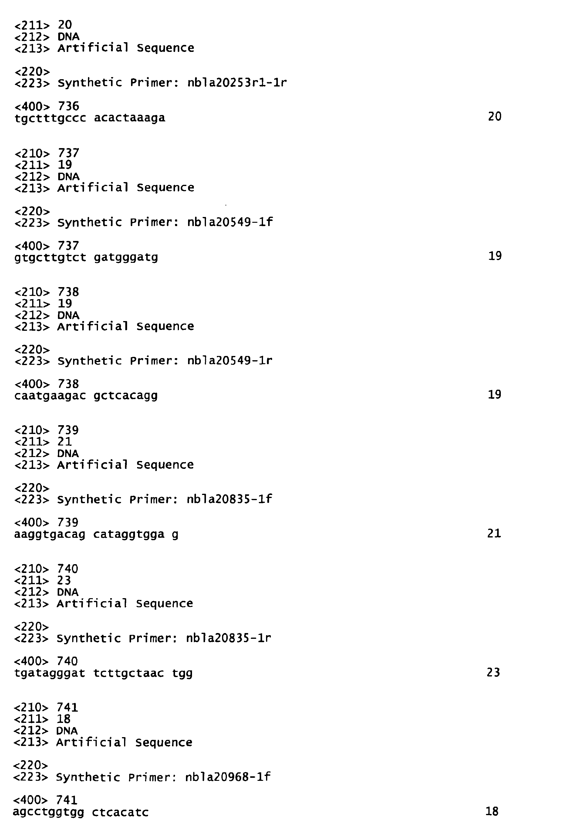
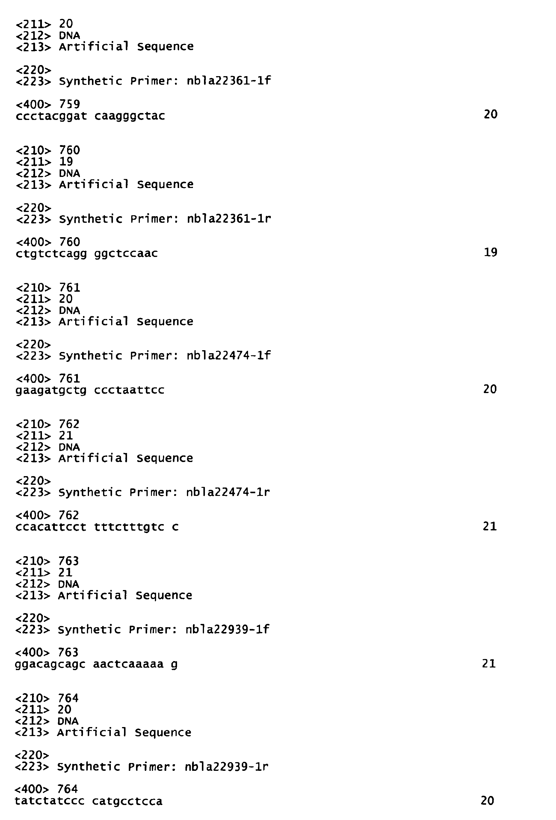
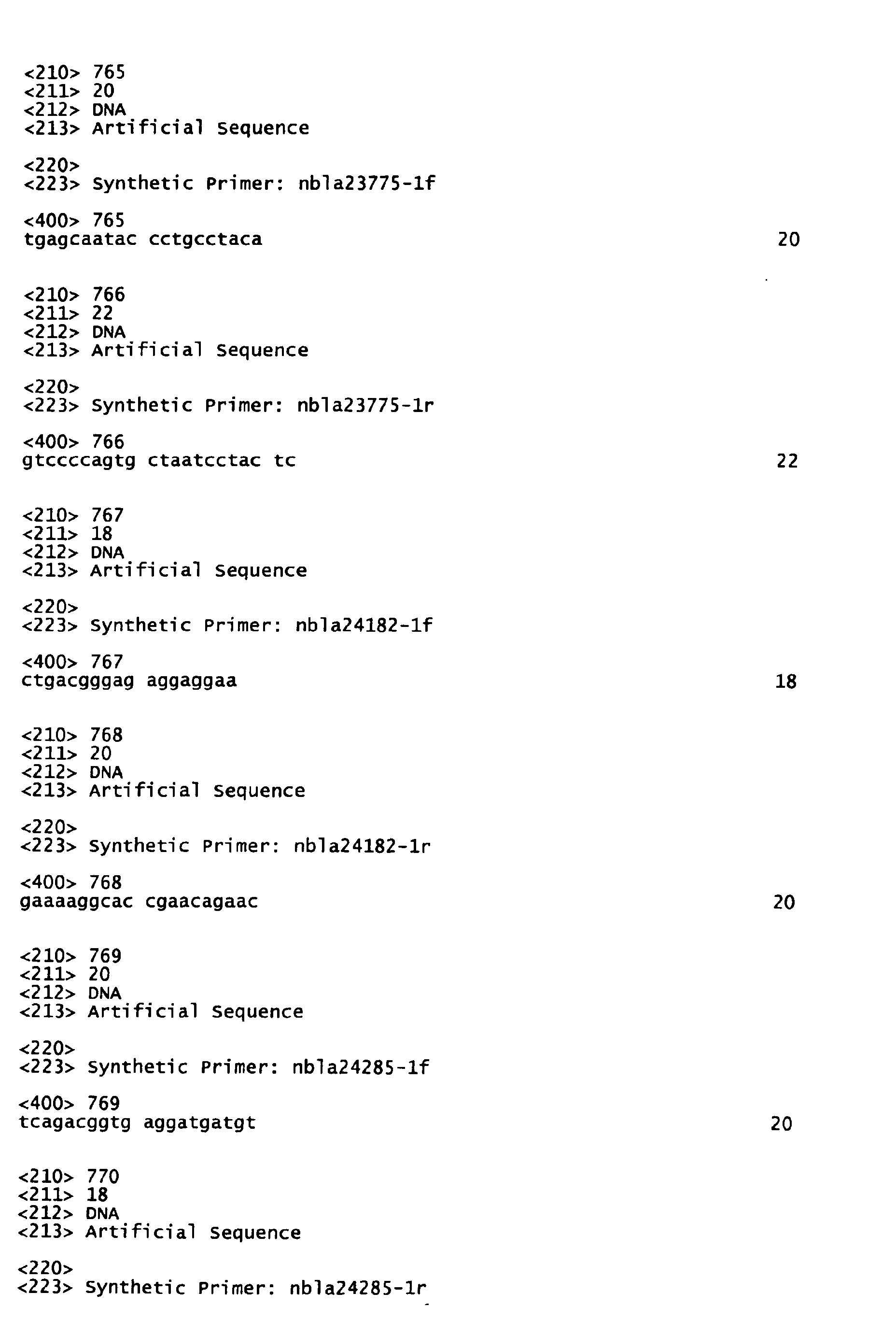
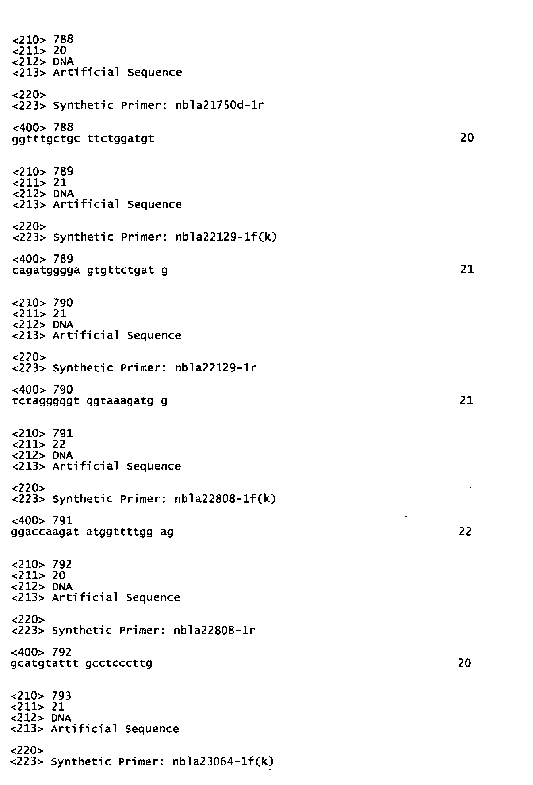
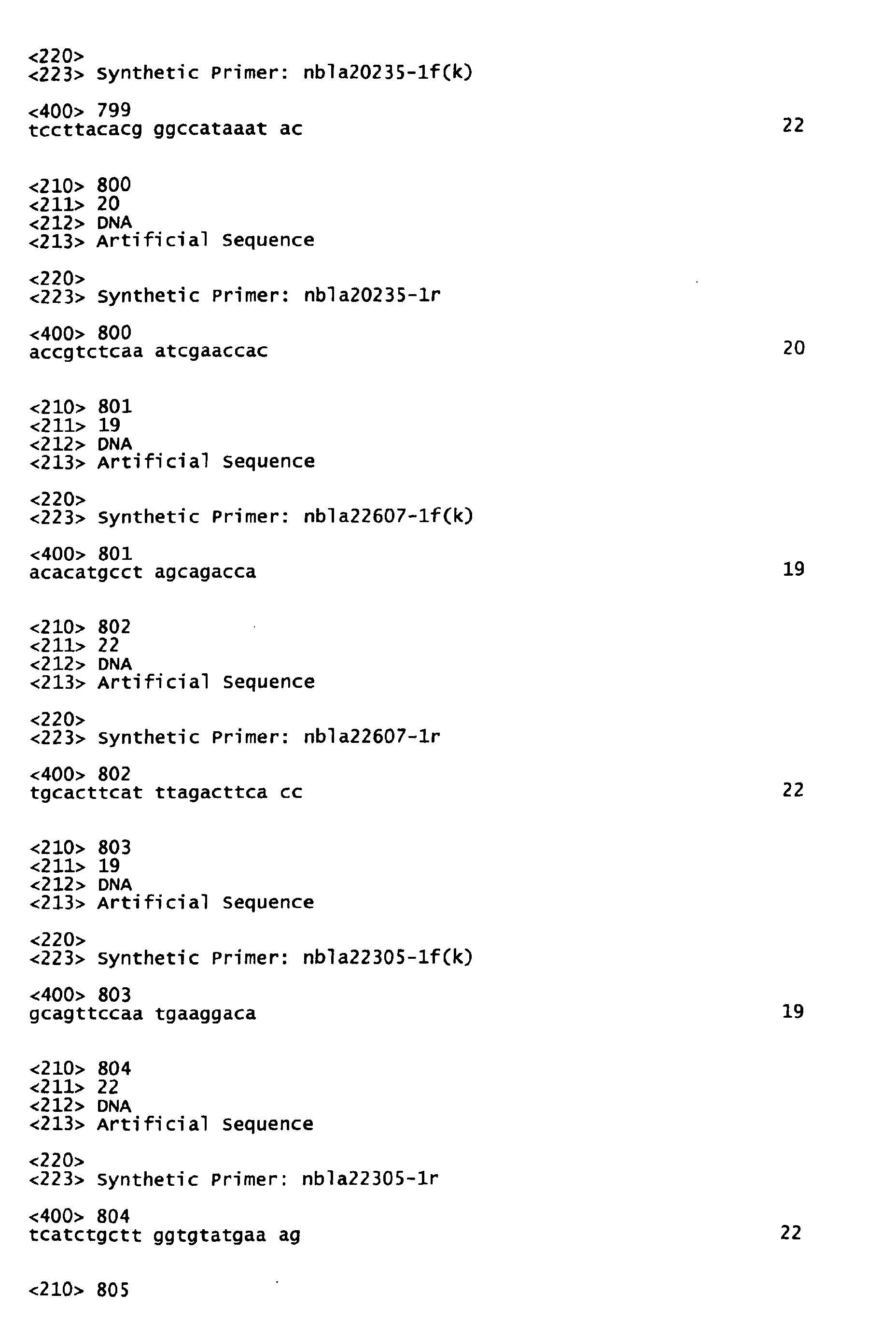
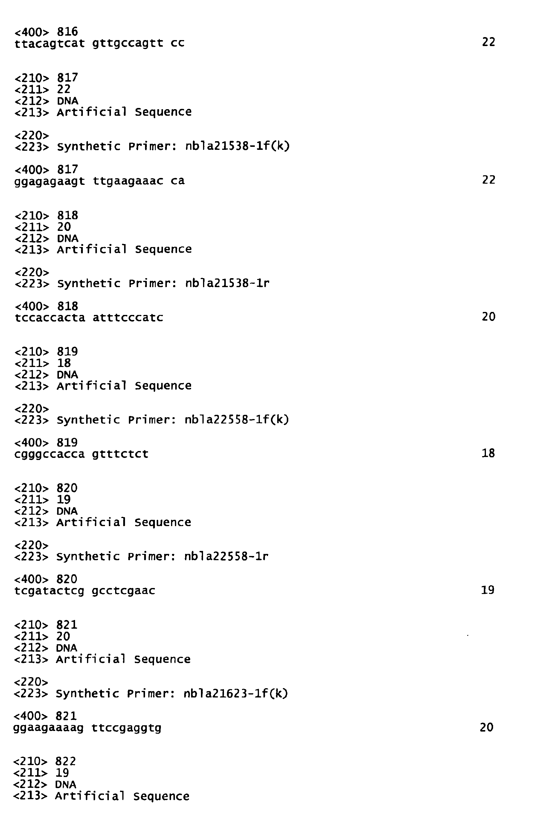
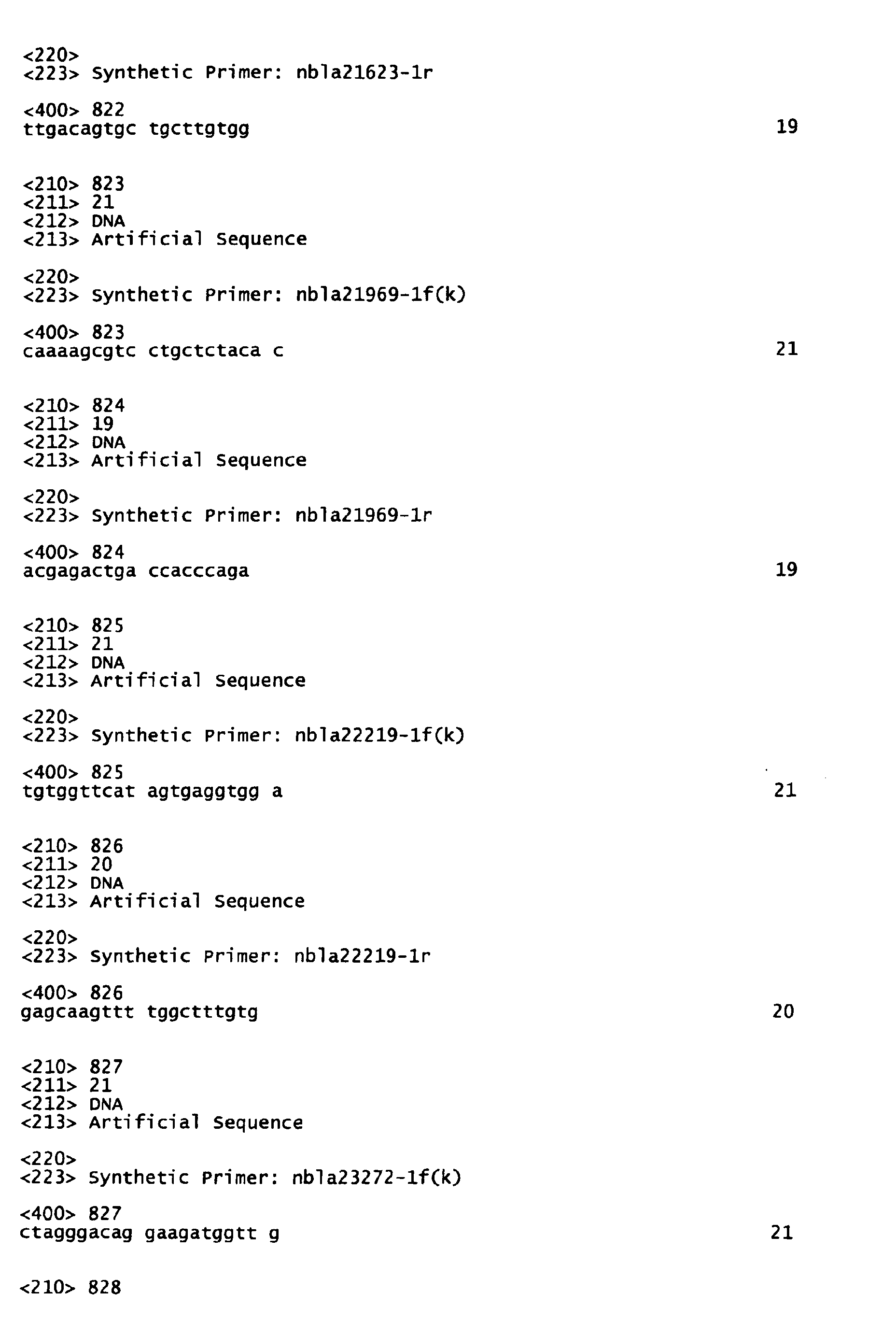
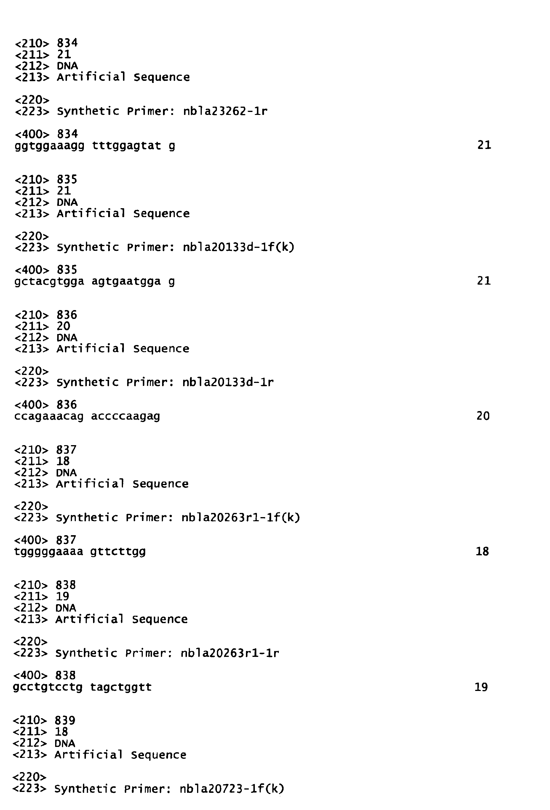
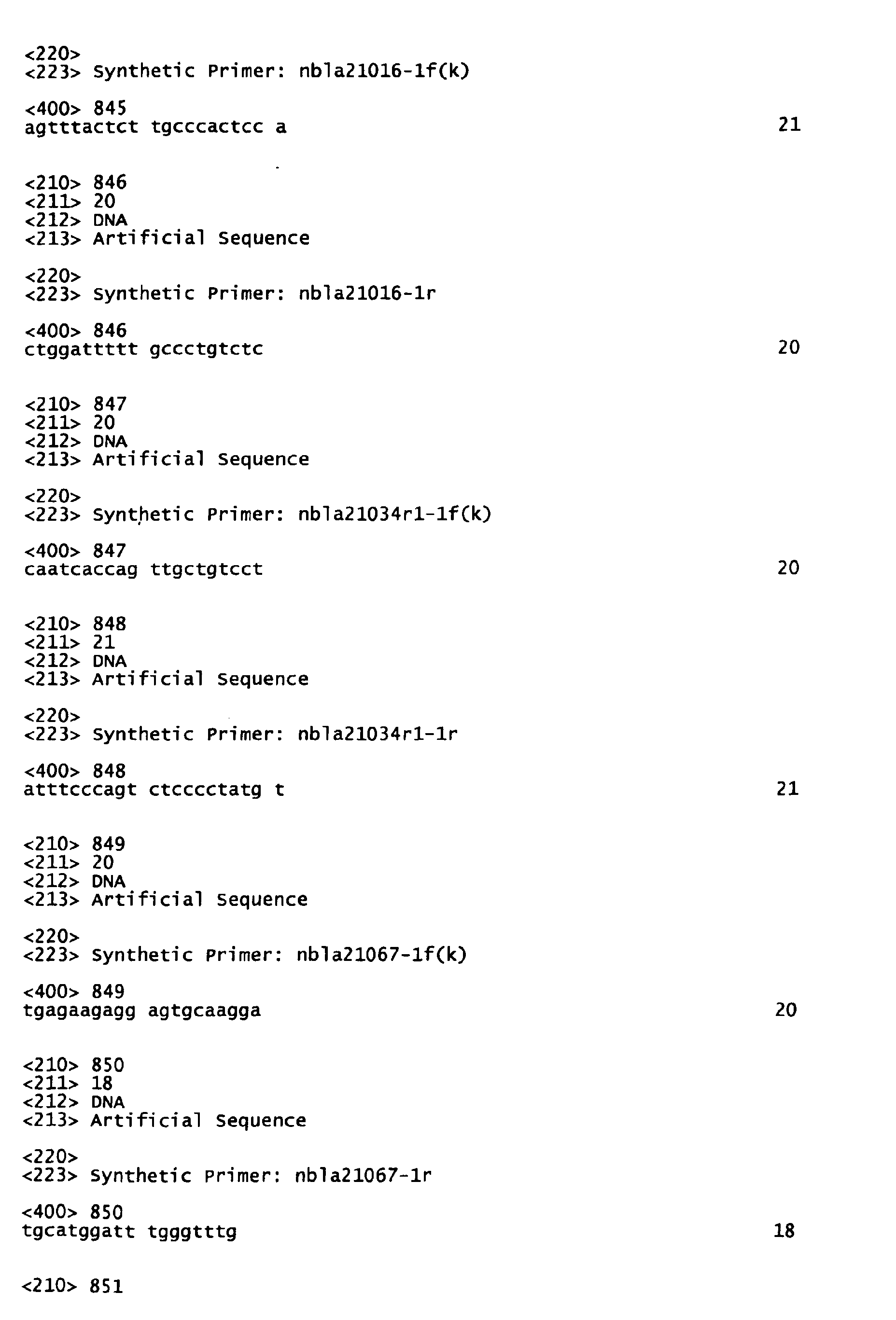
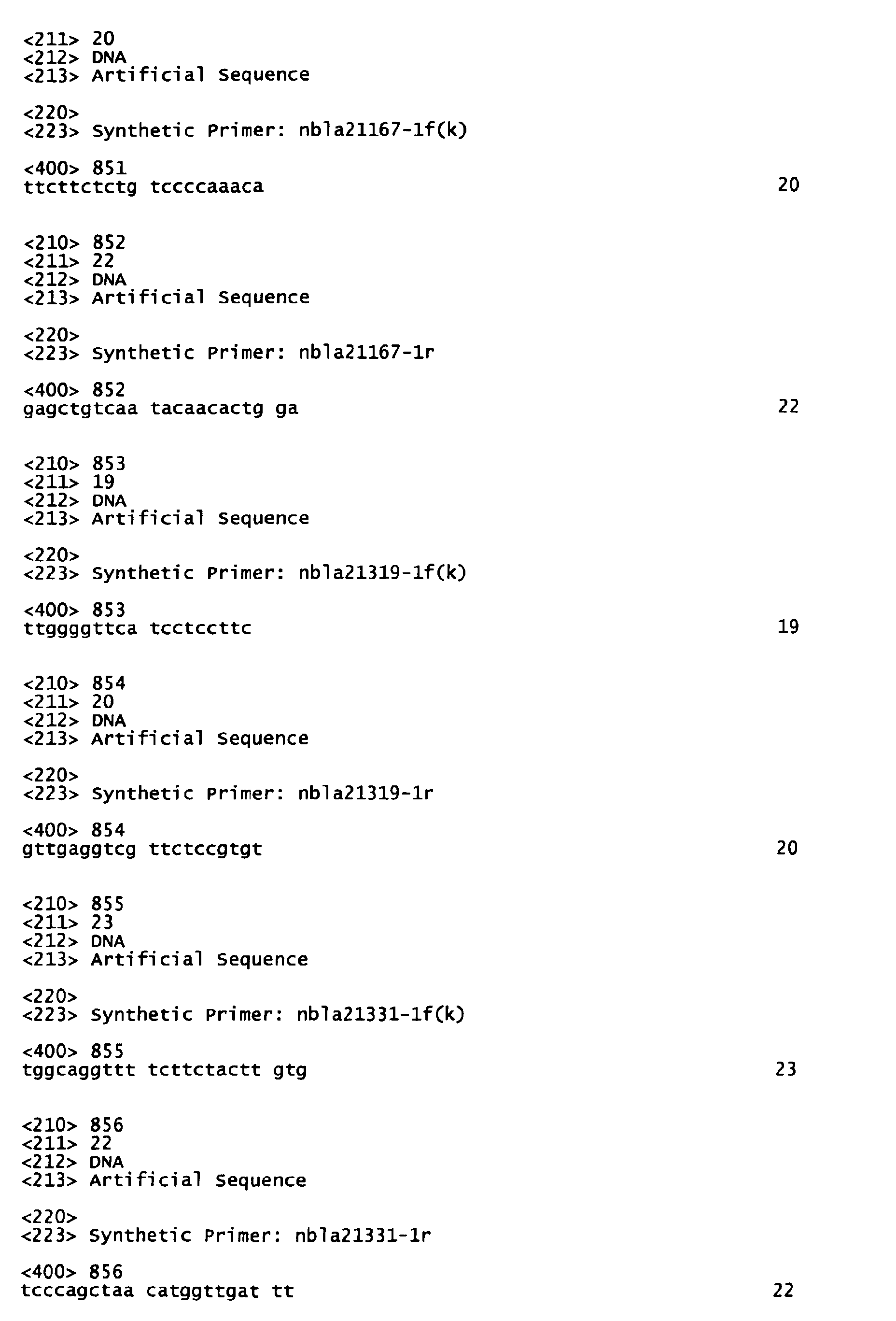
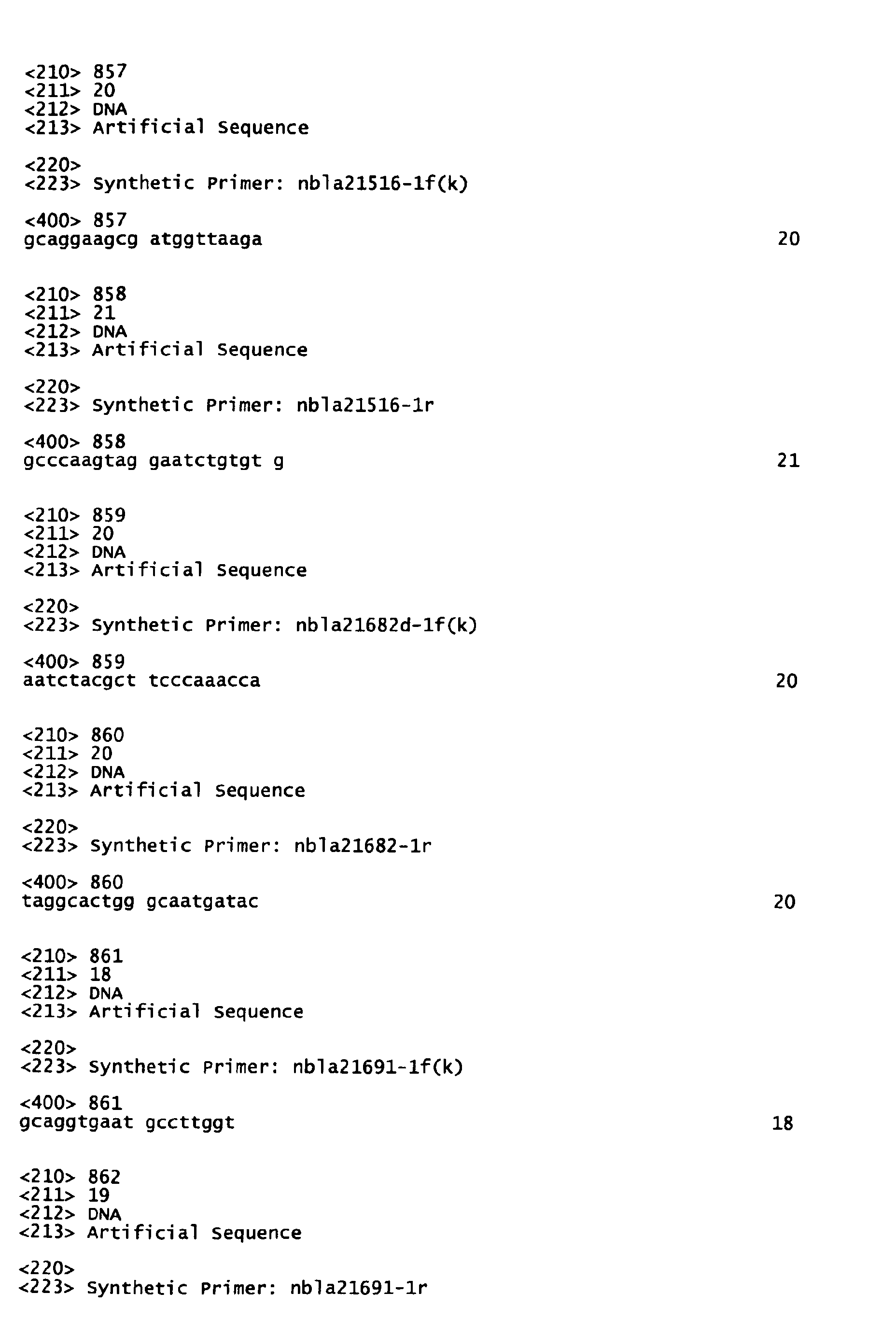
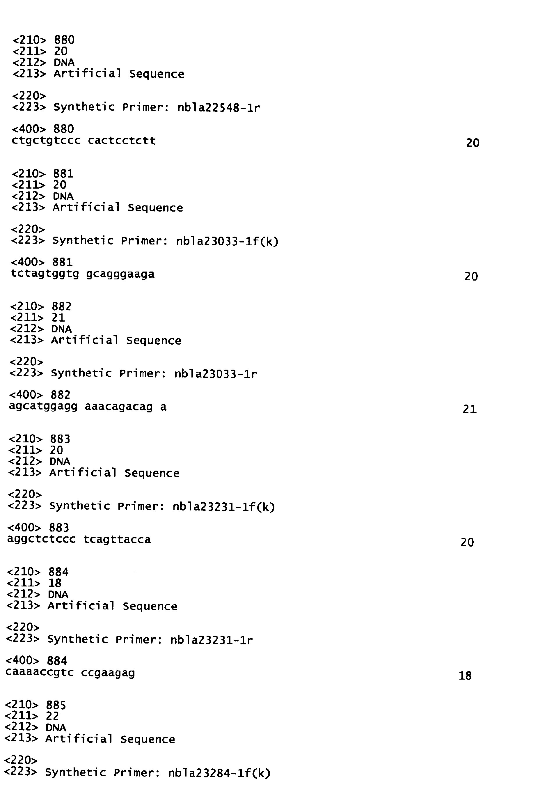
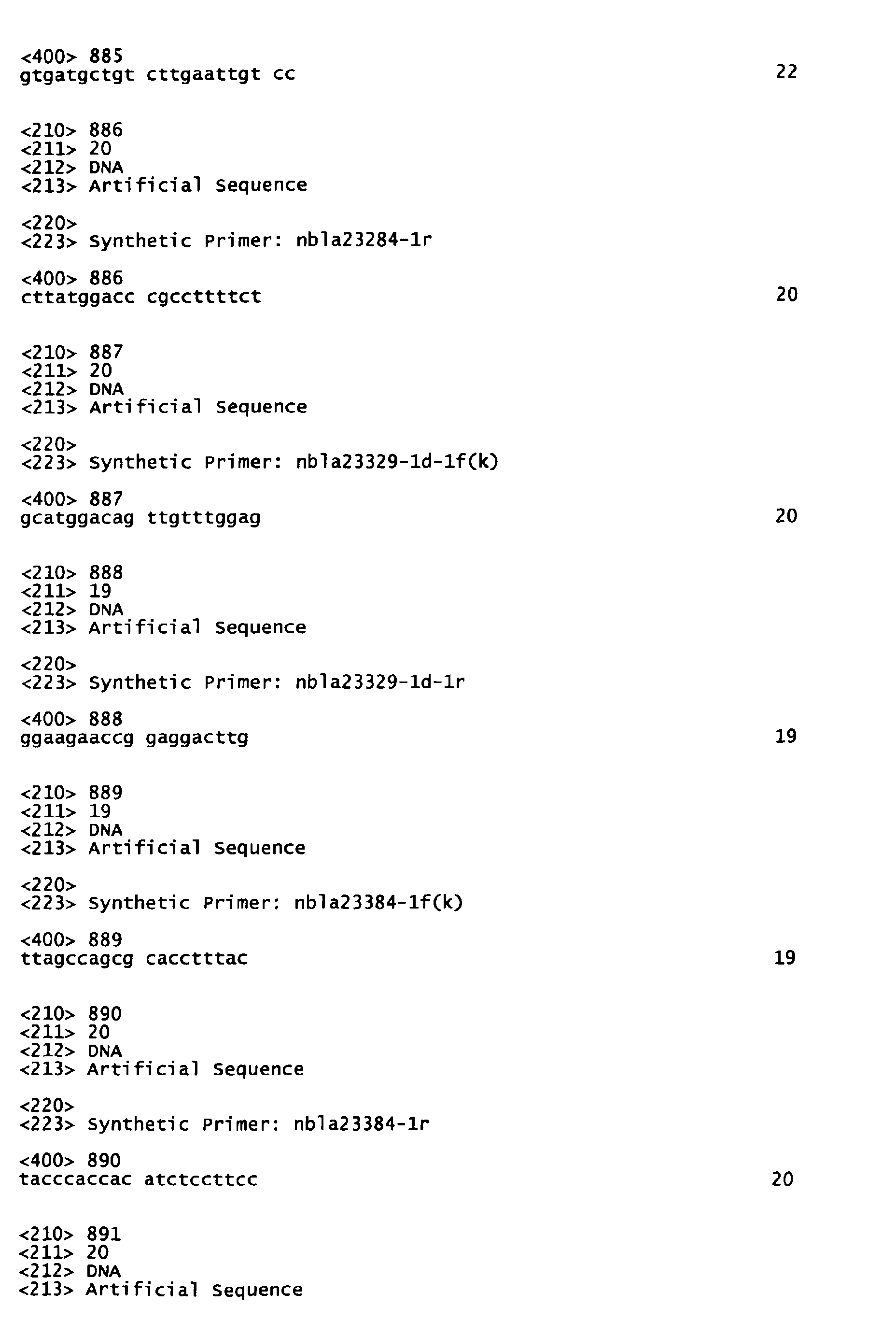
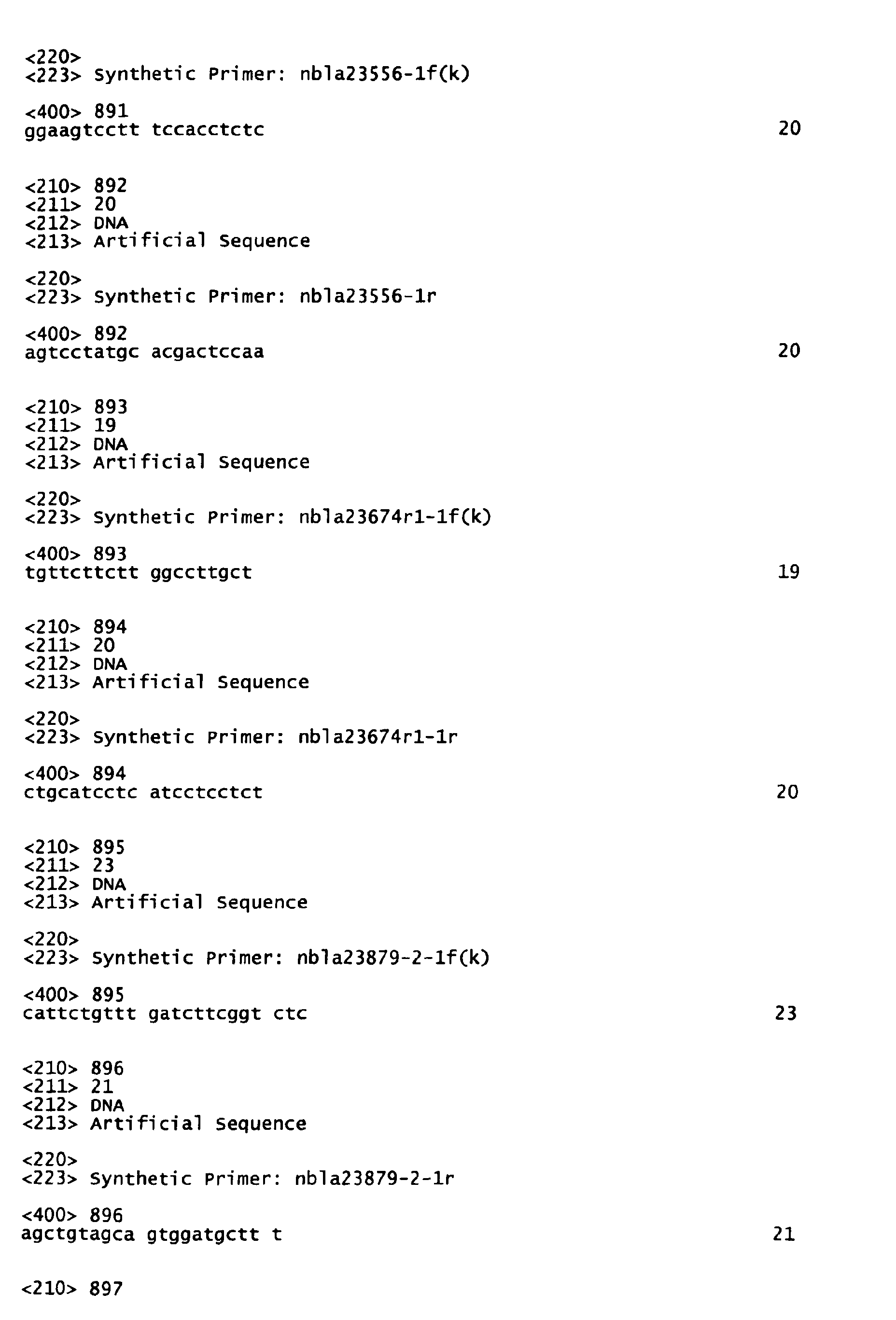
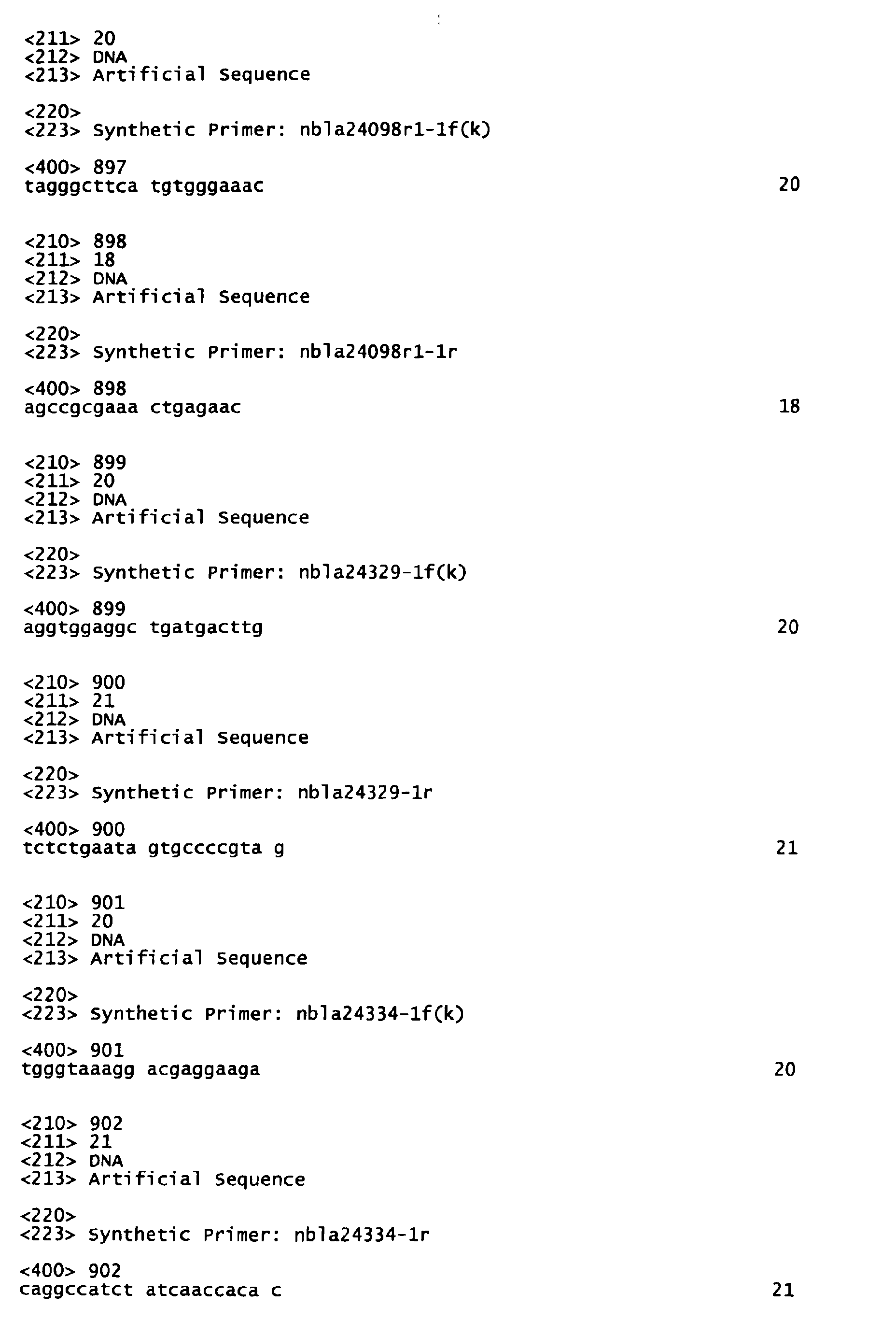
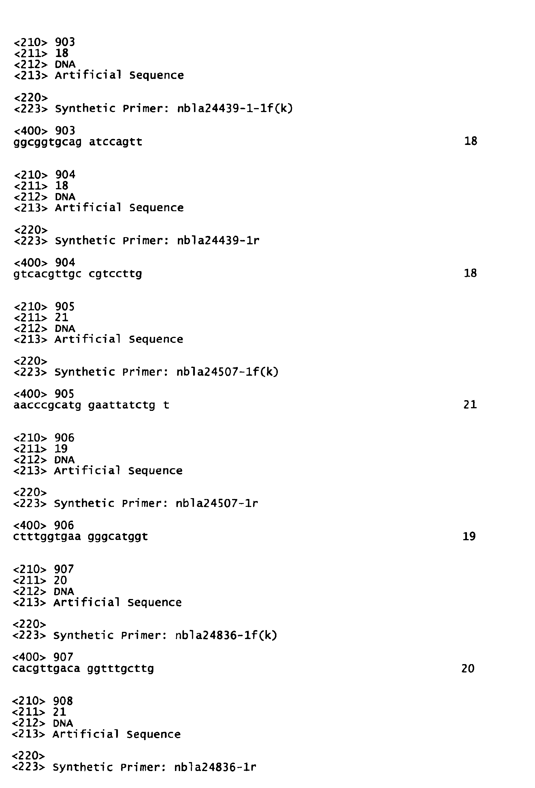
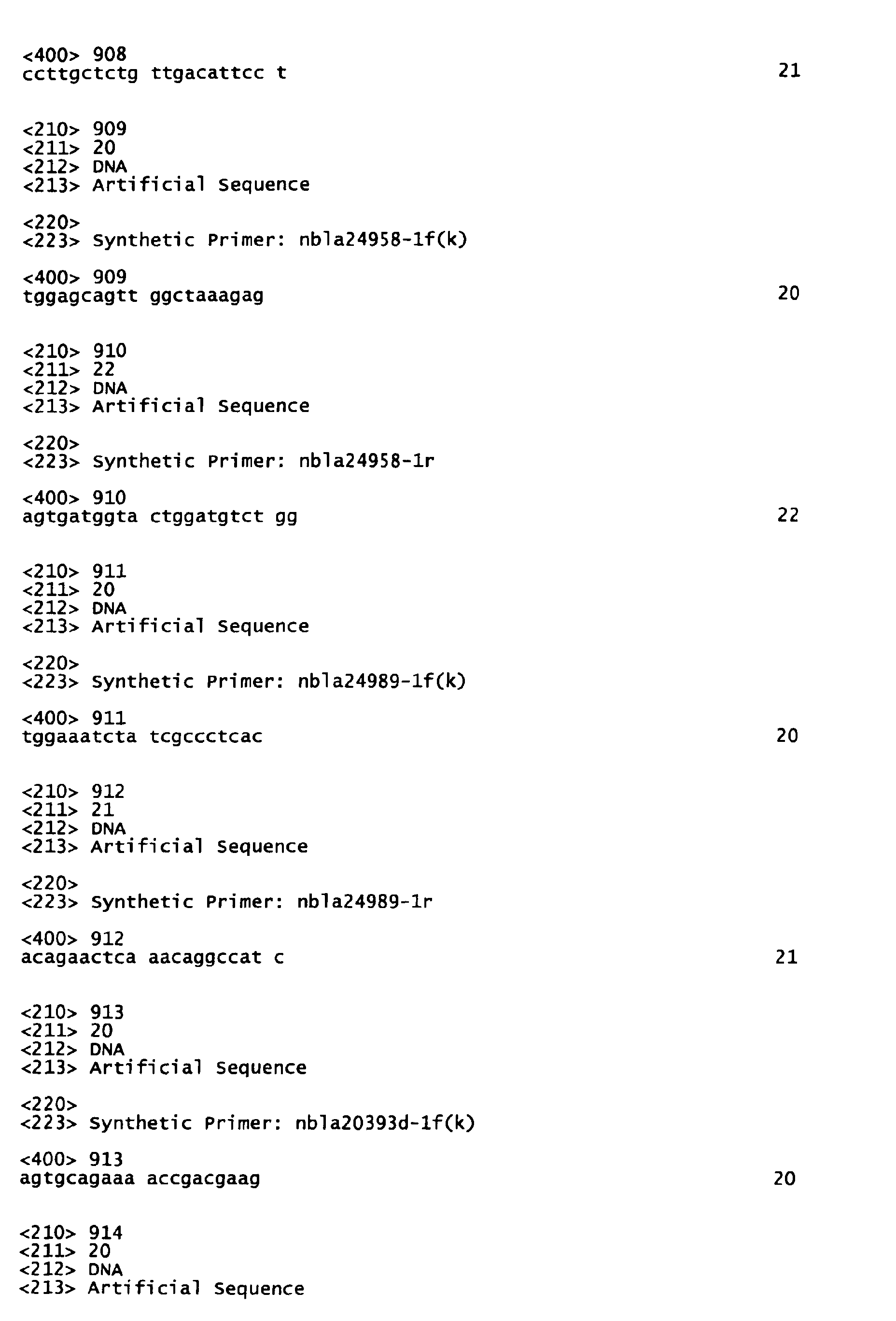
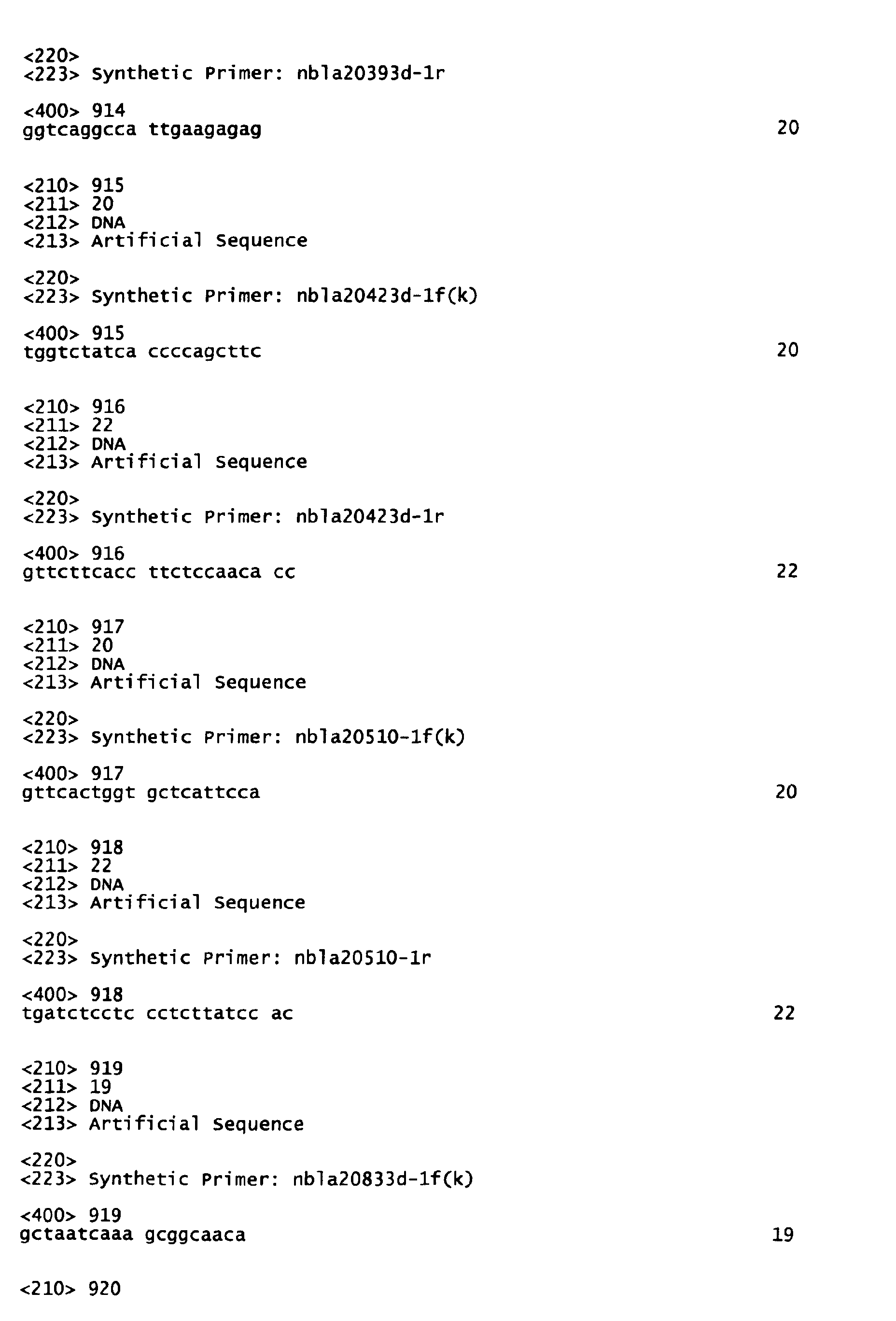
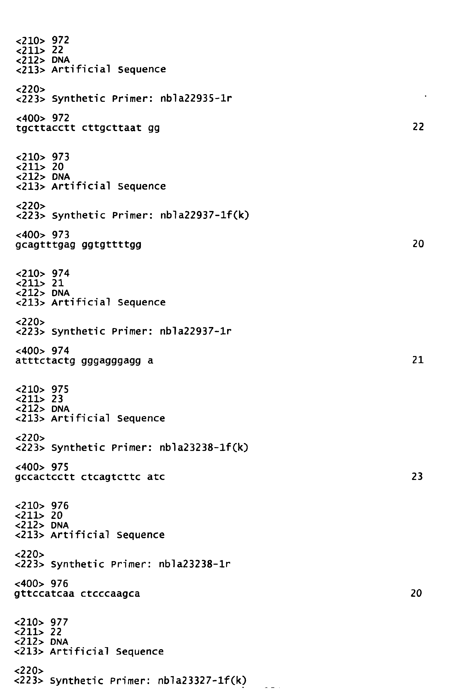
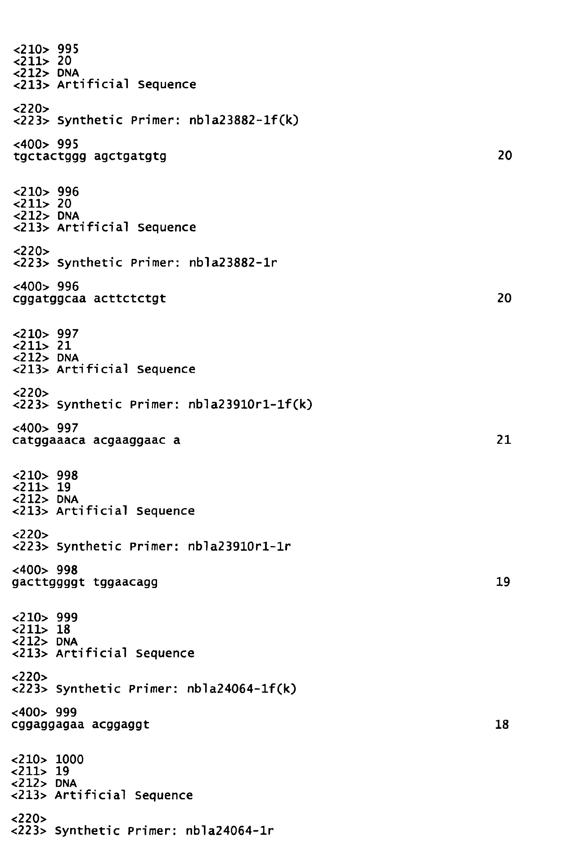
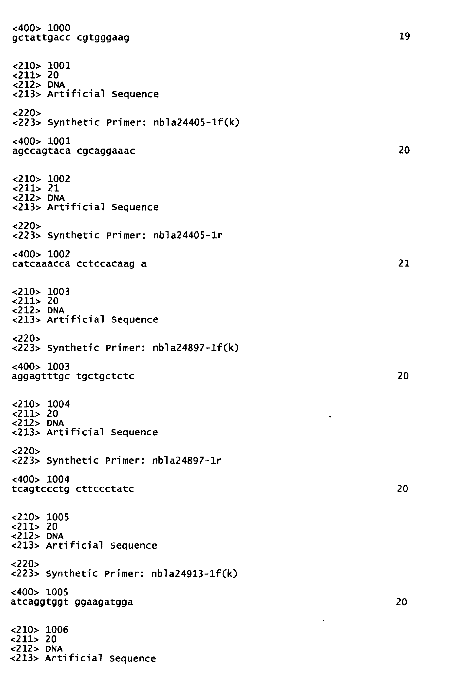
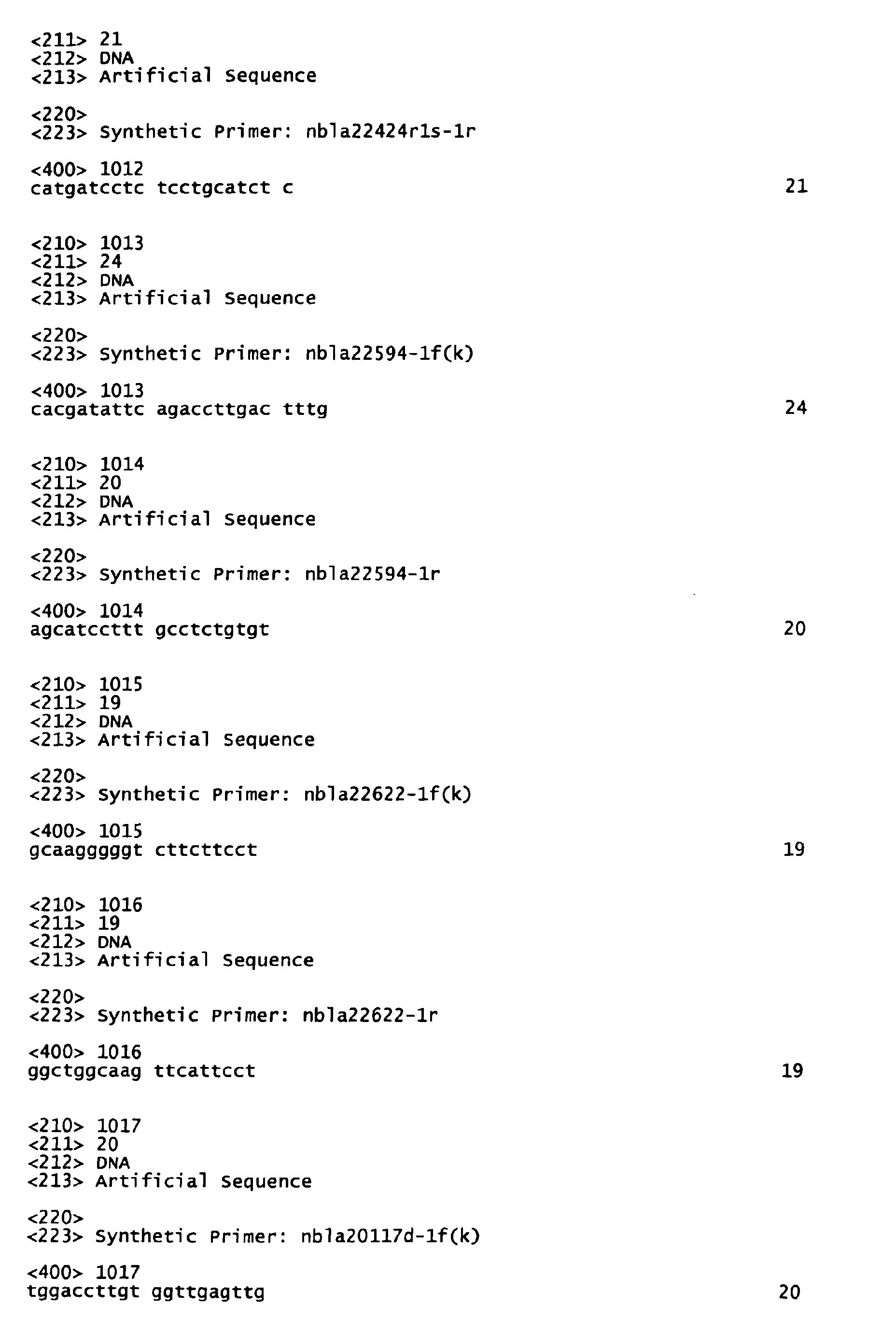
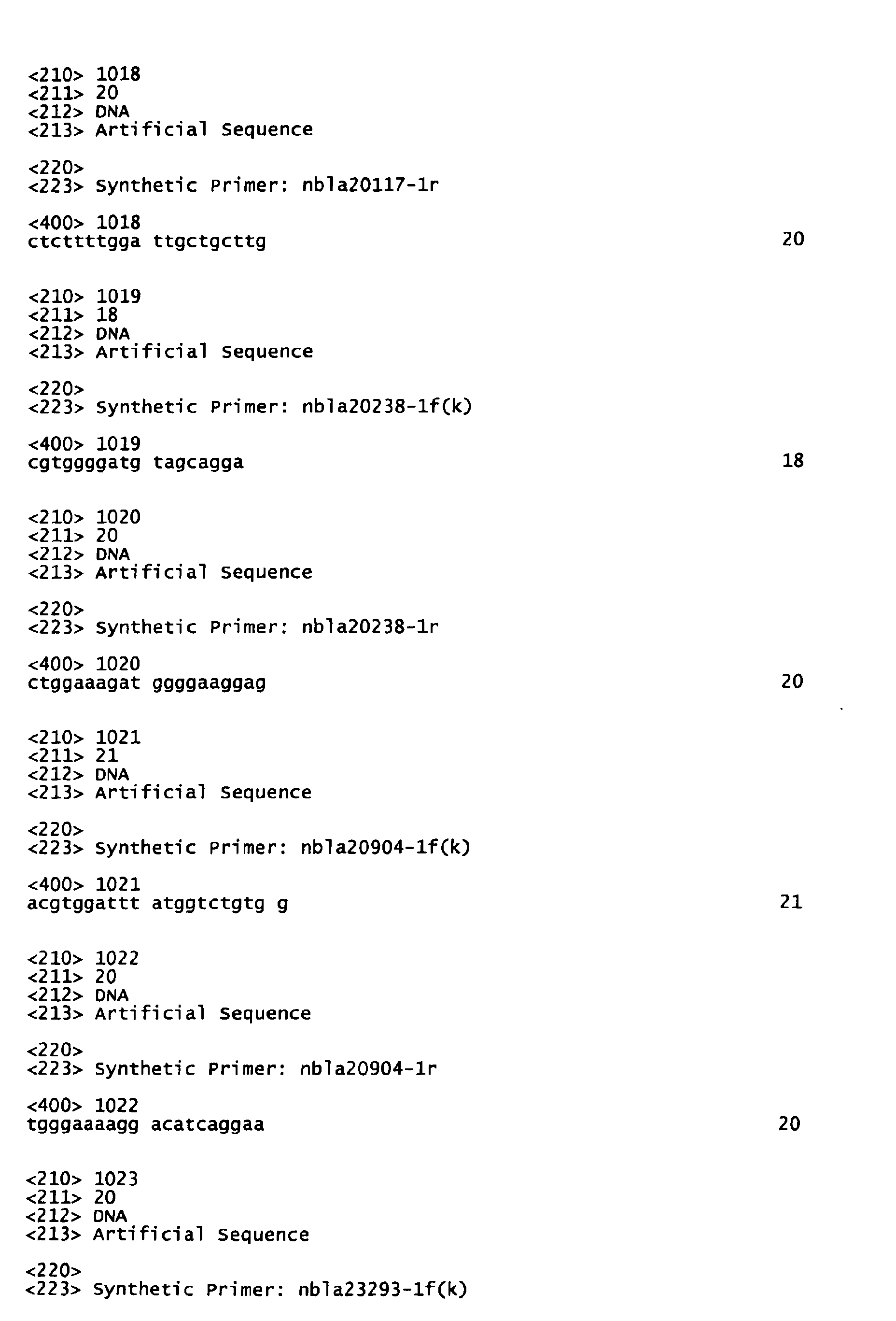
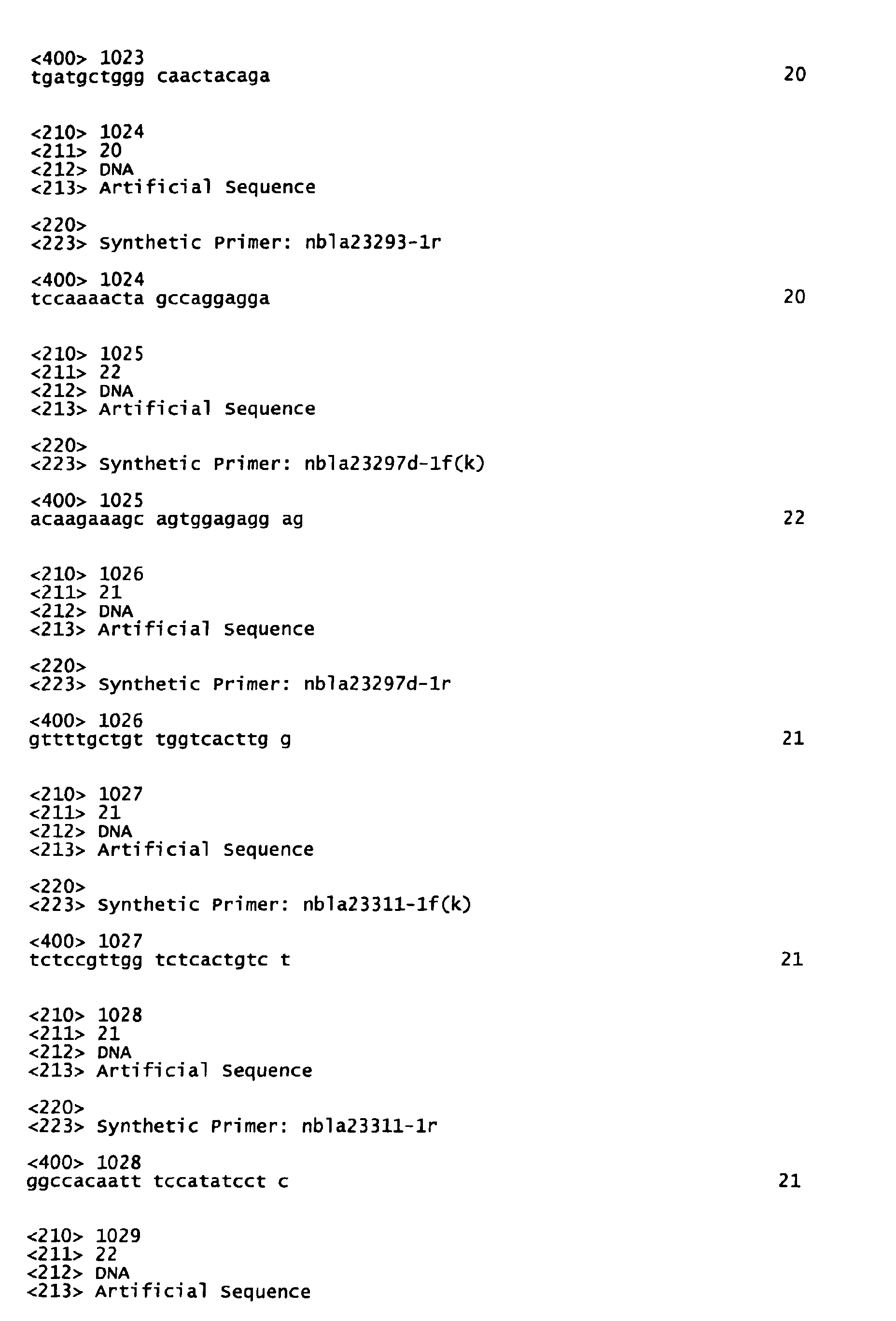
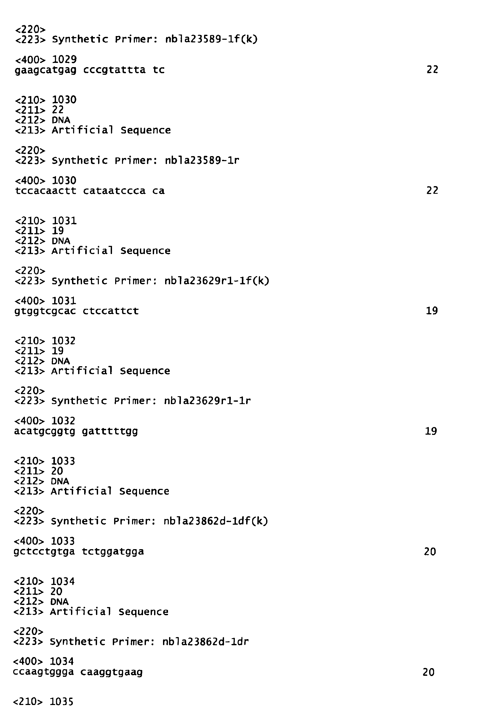
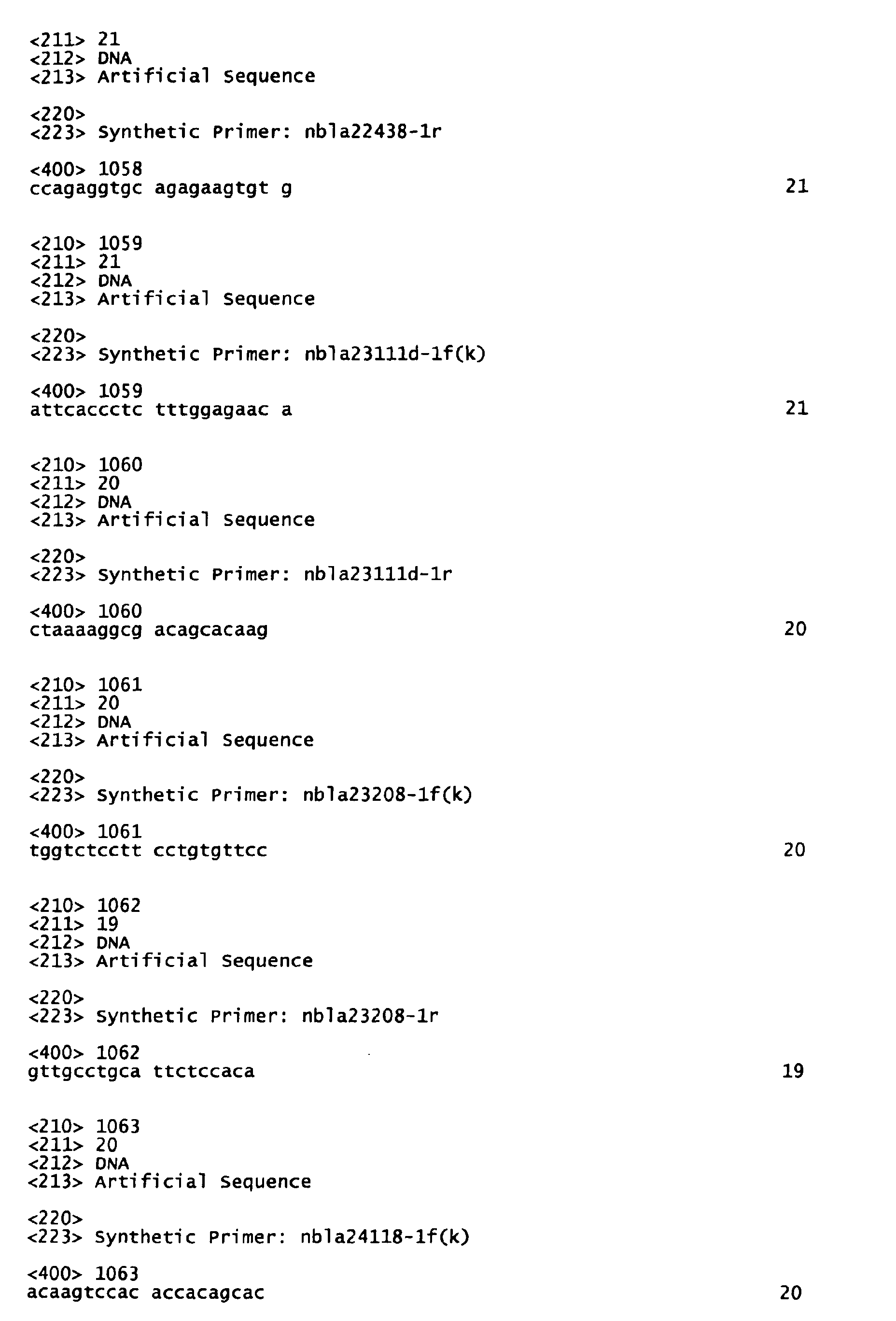
- the primer sequences corresponding to the specific clones previously described are provided in the Sequence Listing (SEQ ID NO:175 to SEQ ID NO:1076).
- the genes are detected in a clinical tissue sample to be assayed thereby to allow the diagnosis for prognosis of neuroblastoma (principally for determining stage 4s neuroblastoma).
- the information obtainable form the genes or the nucleic acids is utilized as tumor markers to allow the preparation of diagnostic agents as well as the design of diagnostic microarrays, both of which can be used for the diagnosis of prognosis.
- stage 4s neuroblastoma If the accurate diagnosis of stage 4s neuroblastoma can be made, it will be important information in deciding whether or not the patient in question needs treatment. Where the case warrants, an unnecessary surgical operation may be avoided.
Landscapes
- Health & Medical Sciences (AREA)
- Life Sciences & Earth Sciences (AREA)
- Chemical & Material Sciences (AREA)
- Engineering & Computer Science (AREA)
- Immunology (AREA)
- Organic Chemistry (AREA)
- Pathology (AREA)
- Molecular Biology (AREA)
- Analytical Chemistry (AREA)
- Proteomics, Peptides & Aminoacids (AREA)
- Zoology (AREA)
- Biomedical Technology (AREA)
- Physics & Mathematics (AREA)
- Biotechnology (AREA)
- Microbiology (AREA)
- Genetics & Genomics (AREA)
- Urology & Nephrology (AREA)
- Wood Science & Technology (AREA)
- Hematology (AREA)
- Biochemistry (AREA)
- General Health & Medical Sciences (AREA)
- Food Science & Technology (AREA)
- Medicinal Chemistry (AREA)
- Biophysics (AREA)
- Cell Biology (AREA)
- Hospice & Palliative Care (AREA)
- Oncology (AREA)
- Bioinformatics & Cheminformatics (AREA)
- General Physics & Mathematics (AREA)
- General Engineering & Computer Science (AREA)
- Measuring Or Testing Involving Enzymes Or Micro-Organisms (AREA)
- Apparatus Associated With Microorganisms And Enzymes (AREA)
- Pharmaceuticals Containing Other Organic And Inorganic Compounds (AREA)
- Image Processing (AREA)
- Medicines Containing Material From Animals Or Micro-Organisms (AREA)
- Micro-Organisms Or Cultivation Processes Thereof (AREA)
Abstract
Description
- (a) a DNA comprising one sequence selected from the group consisting of nucleic acid sequences set forth in SEQ ID NO:175 to SEQ ID NO:1076 in the Sequence Listing, or a DNA complementary thereto;
- (b) a DNA capable of hybridizing under stringent conditions to the DNA comprising one sequence selected from the group consisting of nucleic acid sequences set forth in SEQ ID NO:175 to SEQ ID NO:1076 in the Sequence Listing, or a DNA complementary thereto.
| Group | Expression pattern | Novel genes | Known genes | Total |
| I-1 | F»4s=UF | 5 | 0 | 5 |
| 1-2 | F>4s=UF | 59 | 16 | 75 |
| I-3 | F≥4s=UF | 12 | 11 | 23 |
| II-1 | F=4s»UF | 18 | 5 | 23 |
| II-2 | F=4s>UF | 105 | 47 | 152 |
| II-3 | F=4s≥UF | 55 | 40 | 95 |
| III-1 | F=4s«UF | 1 | 1 | 2 |
| III-2 | F=4s<UF | 10 | 10 | 20 |
| III-3 | F=4s<UF | 2 | 3 | 5 |
| IV | F<4s=UF | 2 | 0 | 2 |
| V-1 | F>4s»UF | 0 | 1 | 1 |
| V-2 | F≥4s»UF | 0 | 1 | 1 |
| V-3 | F>4s>UF | 9 | 2 | 11 |
| V-4 | F≥4s>UF | 3 | 0 | 3 |
| V-5 | F≥4s≥UF | 2 | 0 | 2 |
| VI-1 | F»4s<UF | 1 | 1 | 2 |
| VI-2 | F>4s<UF | 10 | 5 | 15 |
| VI-3 | F>4s≤UF | 1 | 0 | 1 |
| VI-4 | F≥4s≤UF | 4 | 0 | 4 |
| VI-5 | F<4s»UF | 2 | 0 | 2 |
| VI-6 | F≤4s»UF | 0 | 1 | 1 |
| VI-7 | F<4s>UF | 5 | 0 | 5 |
| VI-8 | F≤4s≥UF | 1 | 0 | 1 |
| VII | 4s only | 1 | 0 | 1 |
| Total clone number | 308 | 144 | 452 |







































































































Claims (13)
- A nucleic acid comprising one sequence selected from the group consisting of nucleic acid sequences set forth in SEQ ID NO:1 to SEQ ID NO:174 in the Sequence Listing.
- The nucleic acid according to claim 1, comprising one sequence selected from the group consisting of nucleic acid sequences set forth in SEQ ID NO:1 to SEQ ID NO:14 in the Sequence Listing.
- A nucleic acid complementary to the nucleic acid according to claim 1 or 2.
- A nucleic acid capable of hybridizing to the nucleic acid according to any of claims 1-3 under stringent conditions.
- A nucleic acid probe comprising nucleic acid (a) or nucleic acid (b):(a) a nucleic acid comprising a partial length or the full length of one sequence selected from the group consisting of nucleic acid sequences set forth in SEQ ID NO:1 to SEQ ID NO:174 in the Sequence Listing, or a nucleic acid complementary thereto;(b) a nucleic acid capable of hybridizing under stringent conditions to a nucleic acid comprising a partial length or the full length of one sequence selected from the group consisting of nucleic acid sequences set forth in SEQ ID NO:1 to SEQ ID NO:174 in the Sequence Listing, or a nucleic acid complementary thereto.
- The nucleic acid probe according to claim 5, comprising nucleic acid (a) or nucleic acid (b):(a) a nucleic acid comprising a partial length or the full length of one sequence selected from the group consisting of nucleic acid sequences set forth in SEQ ID NO:1 to SEQ ID NO:14 in the Sequence Listing, or a nucleic acid complementary thereto;(b) a nucleic acid capable of hybridizing under stringent conditions to a nucleic acid comprising a partial length or the full length of one sequence selected from the group consisting of nucleic acid sequences set forth in SEQ ID NO:1 to SEQ ID NO:14 in the Sequence Listing, or a nucleic acid complementary thereto.
- A diagnostic agent for stage 4s neuroblastoma comprising the nucleic acid probe according to claim 5 or 6 as the active ingredient.
- A primer containing DNA (a) or DNA (b):(a) a DNA comprising one sequence selected from the group consisting of nucleic acid sequences set forth in SEQ ID NO:175 to SEQ ID NO:1076 in the Sequence Listing, or a DNA complementary thereto;(b) a DNA capable of hybridizing under stringent conditions to the DNA comprising one sequence selected from the group consisting of nucleic acid sequences set forth in SEQ ID NO:175 to SEQ ID NO:1076 in the Sequence Listing, or a DNA complementary thereto.
- A primer containing DNA (a) or DNA (b):(a) a DNA comprising one sequence selected from the group consisting of nucleic acid sequences set forth in SEQ ID NO:175 to SEQ ID NO:202 and SEQ ID NO:519 to SEQ ID NO:540 in the Sequence Listing, or a DNA complementary thereto, or a DNA comprising one sequence selected from the group consisting of nucleic acid sequences set forth in SEQ ID NO:785 to SEQ ID NO:798 in the Sequence Listing, or a DNA complementary thereto;(b) a DNA capable of hybridizing under stringent conditions to the DNA comprising one sequence selected from the group consisting of nucleic acid sequences set forth in SEQ ID NO:175 to SEQ ID NO:202 and SEQ ID NO:519 to SEQ ID NO:540 in the Sequence Listing or a DNA comprising one sequence selected from the group consisting of nucleic acid sequences set forth in SEQ ID NO:785 to SEQ ID NO:798 in the Sequence Listing, or a DNA complementary thereto.
- A diagnostic kit for stage 4s neuroblastoma comprising one pair of the primers according to claims 8 or 9 as the active component.
- A method for determining stage 4s neuroblastoma, the method comprising detecting the presence or absence of a nucleic acid comprising one sequence selected from the group consisting of nucleic acid sequences set forth in SEQ ID NO:1 to SEQ ID NO:14 in the Sequence Listing from a clinical tissue sample of neuroblastoma.
- A nucleic acid microarray comprising a solid phase support and a combination of plural nucleic acids each comprising a partial length- or the full length-nucleic acid comprising a nucleic acid sequence set forth in one of SEQ ID NO:1 to SEQ ID NO:174 in the Sequence Listing immobilized on the solid phase support.
- A nucleic acid microarray comprising a solid phase support and a combination of plural nucleic acids each comprising a nucleic acid sequence set forth in one of SEQ ID NO:175 to SEQ ID NO:200, a nucleic acid sequence set forth in one of SEQ ID NO:519 to SEQ ID NO:540 or a nucleic acid sequence set forth in one of SEQ ID NO:785 to SEQ ID NO:798 immobilized on the solid phase support.
Applications Claiming Priority (3)
| Application Number | Priority Date | Filing Date | Title |
|---|---|---|---|
| JP2002316586 | 2002-10-30 | ||
| JP2002316586A JP4122205B2 (en) | 2002-10-30 | 2002-10-30 | Nucleic acid isolated from 4s stage neuroblastoma |
| PCT/JP2003/013932 WO2004039975A1 (en) | 2002-10-30 | 2003-10-30 | Nucleic acids isolated from neuroblastoma at stage 4s |
Publications (3)
| Publication Number | Publication Date |
|---|---|
| EP1580265A1 true EP1580265A1 (en) | 2005-09-28 |
| EP1580265A4 EP1580265A4 (en) | 2006-08-30 |
| EP1580265B1 EP1580265B1 (en) | 2010-09-29 |
Family
ID=32211688
Family Applications (1)
| Application Number | Title | Priority Date | Filing Date |
|---|---|---|---|
| EP20030770029 Expired - Lifetime EP1580265B1 (en) | 2002-10-30 | 2003-10-30 | Nucleic acids isolated from neuroblastoma at stage 4s |
Country Status (6)
| Country | Link |
|---|---|
| US (1) | US7429451B2 (en) |
| EP (1) | EP1580265B1 (en) |
| JP (1) | JP4122205B2 (en) |
| AT (1) | ATE483019T1 (en) |
| DE (1) | DE60334408D1 (en) |
| WO (1) | WO2004039975A1 (en) |
Cited By (1)
| Publication number | Priority date | Publication date | Assignee | Title |
|---|---|---|---|---|
| WO2007128984A3 (en) * | 2006-04-06 | 2008-09-25 | Randox Lab Ltd | Breast cancer makers |
Families Citing this family (5)
| Publication number | Priority date | Publication date | Assignee | Title |
|---|---|---|---|---|
| US8106172B2 (en) * | 2003-06-10 | 2012-01-31 | Biomerieux, B.V. | Nucleic acid sequences that can be used as primers and probes in the amplification and detection of SARS coronavirus |
| US7601532B2 (en) | 2003-09-25 | 2009-10-13 | Hisamitsu Pharmaceutical Co., Inc. | Microarray for predicting the prognosis of neuroblastoma and method for predicting the prognosis of neuroblastoma |
| WO2006059769A1 (en) * | 2004-12-03 | 2006-06-08 | Aichi Prefecture | Method of diagnosing malignant lymphoma and estimating the prognosis thereof |
| JP2008029296A (en) * | 2006-07-31 | 2008-02-14 | Hisamitsu Pharmaceut Co Inc | Neuroblastic therapeutic agent, screening method thereof, and method of determining prognosis of neuroblastic |
| WO2010145839A2 (en) * | 2009-06-19 | 2010-12-23 | Max-Delbrück-Centrum Für Molekulare Medizin Berlin-Buch | Polynucleotides for medical use |
Family Cites Families (3)
| Publication number | Priority date | Publication date | Assignee | Title |
|---|---|---|---|---|
| AU3605901A (en) | 2000-03-07 | 2001-09-17 | Chiba Prefecture | Nucleic acid sequences showing enhanced expression in benign neuroblastoma compared with acritical human neuroblastoma |
| JP2001245671A (en) | 2000-03-07 | 2001-09-11 | Chiba Prefecture | Novel genes and novel gene fragments cloned in human neuroblastoma |
| USH2191H1 (en) * | 2000-10-24 | 2007-06-05 | Snp Consortium | Identification and mapping of single nucleotide polymorphisms in the human genome |
-
2002
- 2002-10-30 JP JP2002316586A patent/JP4122205B2/en not_active Expired - Fee Related
-
2003
- 2003-10-30 AT AT03770029T patent/ATE483019T1/en not_active IP Right Cessation
- 2003-10-30 EP EP20030770029 patent/EP1580265B1/en not_active Expired - Lifetime
- 2003-10-30 DE DE60334408T patent/DE60334408D1/en not_active Expired - Lifetime
- 2003-10-30 US US10/533,158 patent/US7429451B2/en not_active Expired - Fee Related
- 2003-10-30 WO PCT/JP2003/013932 patent/WO2004039975A1/en not_active Ceased
Non-Patent Citations (6)
| Title |
|---|
| AOYAMA M ET AL: "High expression of human RIM gene in neuroblastomas with favorable biologies" SOCIETY FOR NEUROSCIENCE ABSTRACTS, vol. 27, no. 1, 2001, page 715, XP009065593 & 31ST ANNUAL MEETING OF THE SOCIETY FOR NEUROSCIENCE; SAN DIEGO, CALIFORNIA, USA; NOVEMBER 10-15, 2001 ISSN: 0190-5295 * |
| DATABASE EMBL [Online] 11 September 2001 (2001-09-11), "Homo sapiens BAC clone RP11-650J17 from 4, complete sequence." XP002377844 retrieved from EBI accession no. EM_PRO:AC093879 Database accession no. AC093879 * |
| ISLAM ASHRAFUL ET AL: "High expression of Survivin, mapped to 17q25, is significantly associated with poor prognostic factors and promotes cell survival in human neuroblastoma" ONCOGENE, vol. 19, no. 5, 3 February 2000 (2000-02-03), pages 617-623, XP002377836 ISSN: 0950-9232 * |
| KAWAMOTO T ET AL: "ASSOCIATION BETWEEN FAVORABLE NEUROBLASTOMA AND HIGH EXPRESSION OF THE NOVEL METALLOPROTEINASE GENE, NBLA3145/XCE, CLONED BY DIFFERENTIAL SCREENING OF THE FULL-LENGTH-ENRICHED OLIGO-CAPPING NEUROBLASTOMA CDNA LIBRARIES" MEDICAL AND PEDIATRIC ONCOLOGY, LISS, NEW YORK, NY, US, vol. 35, no. 6, December 2000 (2000-12), pages 628-631, XP009053429 ISSN: 0740-8226 * |
| See also references of WO2004039975A1 * |
| TANG X X ET AL: "Implications of EPHB6, EFNB2, and EFNB3 expressions in human neuroblastoma." PROCEEDINGS OF THE NATIONAL ACADEMY OF SCIENCES OF THE UNITED STATES OF AMERICA. 26 SEP 2000, vol. 97, no. 20, 26 September 2000 (2000-09-26), pages 10936-10941, XP002377837 ISSN: 0027-8424 * |
Cited By (1)
| Publication number | Priority date | Publication date | Assignee | Title |
|---|---|---|---|---|
| WO2007128984A3 (en) * | 2006-04-06 | 2008-09-25 | Randox Lab Ltd | Breast cancer makers |
Also Published As
| Publication number | Publication date |
|---|---|
| EP1580265B1 (en) | 2010-09-29 |
| DE60334408D1 (en) | 2010-11-11 |
| JP4122205B2 (en) | 2008-07-23 |
| US7429451B2 (en) | 2008-09-30 |
| ATE483019T1 (en) | 2010-10-15 |
| US20070281296A1 (en) | 2007-12-06 |
| EP1580265A4 (en) | 2006-08-30 |
| WO2004039975A1 (en) | 2004-05-13 |
| JP2004147563A (en) | 2004-05-27 |
Similar Documents
| Publication | Publication Date | Title |
|---|---|---|
| US20070243176A1 (en) | Human genes and gene expression products | |
| JPH0728759B2 (en) | Method of detecting tumorigenicity | |
| WO2001066719A1 (en) | Novel gene cloned in human neuroblastoma and novel gene fragments | |
| KR20130141395A (en) | Composition for treating or preventing osteoporotic fracture or osteoporosis comprising slit-robo system | |
| US7087383B2 (en) | Nucleic acid sequences showing enhanced expression in benign neuroblastoma compared with acritical human neuroblastoma | |
| US7429451B2 (en) | Nucleic acids isolated from stage 4 neuroblastoma | |
| EP1795597B1 (en) | Nucleic acids isolated from a neuroblastoma | |
| EP1792990B1 (en) | Nucleid acids isolated from neuroblastomas | |
| EP1428891A1 (en) | Novel gene nedl-1 | |
| JP2003528630A (en) | Human genes and expression products | |
| JP4565702B2 (en) | Detection kit for detecting nucleic acid sequences whose expression is enhanced in human neuroblastoma with good prognosis in comparison with human neuroblastoma with good prognosis and poor prognosis | |
| JP4098236B2 (en) | Nucleic acids with differential expression between hepatoblastoma and normal liver | |
| EP1426443B1 (en) | Human bmcc1 gene | |
| KR20090031557A (en) | Genes related to immortalization of human cancer cells and uses thereof | |
| WO1997012973A1 (en) | Human cyclin i and gene encoding the same | |
| US20030104418A1 (en) | Diagnostic markers for breast cancer | |
| KR20070027659A (en) | Breast cancer specific protein, gene encoding the same, and method for diagnosing breast cancer using the protein or gene | |
| US20040138161A1 (en) | Methods of modulating proliferative conditions | |
| KR20060108892A (en) | Anticancer Enhancers for Head and Neck Cancers Resistant to Cisplatin Using Microarray Technology | |
| CA2434042A1 (en) | Asip-related proteins |
Legal Events
| Date | Code | Title | Description |
|---|---|---|---|
| PUAI | Public reference made under article 153(3) epc to a published international application that has entered the european phase |
Free format text: ORIGINAL CODE: 0009012 |
|
| 17P | Request for examination filed |
Effective date: 20050519 |
|
| AK | Designated contracting states |
Kind code of ref document: A1 Designated state(s): AT BE BG CH CY CZ DE DK EE ES FI FR GB GR HU IE IT LI LU MC NL PT RO SE SI SK TR |
|
| A4 | Supplementary search report drawn up and despatched |
Effective date: 20060802 |
|
| 17Q | First examination report despatched |
Effective date: 20070314 |
|
| GRAP | Despatch of communication of intention to grant a patent |
Free format text: ORIGINAL CODE: EPIDOSNIGR1 |
|
| GRAS | Grant fee paid |
Free format text: ORIGINAL CODE: EPIDOSNIGR3 |
|
| GRAA | (expected) grant |
Free format text: ORIGINAL CODE: 0009210 |
|
| AK | Designated contracting states |
Kind code of ref document: B1 Designated state(s): AT BE BG CH CY CZ DE DK EE ES FI FR GB GR HU IE IT LI LU MC NL PT RO SE SI SK TR |
|
| REG | Reference to a national code |
Ref country code: GB Ref legal event code: FG4D |
|
| REG | Reference to a national code |
Ref country code: CH Ref legal event code: EP |
|
| REG | Reference to a national code |
Ref country code: IE Ref legal event code: FG4D |
|
| REF | Corresponds to: |
Ref document number: 60334408 Country of ref document: DE Date of ref document: 20101111 Kind code of ref document: P |
|
| PG25 | Lapsed in a contracting state [announced via postgrant information from national office to epo] |
Ref country code: FI Free format text: LAPSE BECAUSE OF FAILURE TO SUBMIT A TRANSLATION OF THE DESCRIPTION OR TO PAY THE FEE WITHIN THE PRESCRIBED TIME-LIMIT Effective date: 20100929 Ref country code: AT Free format text: LAPSE BECAUSE OF FAILURE TO SUBMIT A TRANSLATION OF THE DESCRIPTION OR TO PAY THE FEE WITHIN THE PRESCRIBED TIME-LIMIT Effective date: 20100929 |
|
| REG | Reference to a national code |
Ref country code: NL Ref legal event code: VDEP Effective date: 20100929 |
|
| PG25 | Lapsed in a contracting state [announced via postgrant information from national office to epo] |
Ref country code: SI Free format text: LAPSE BECAUSE OF FAILURE TO SUBMIT A TRANSLATION OF THE DESCRIPTION OR TO PAY THE FEE WITHIN THE PRESCRIBED TIME-LIMIT Effective date: 20100929 |
|
| PG25 | Lapsed in a contracting state [announced via postgrant information from national office to epo] |
Ref country code: GR Free format text: LAPSE BECAUSE OF FAILURE TO SUBMIT A TRANSLATION OF THE DESCRIPTION OR TO PAY THE FEE WITHIN THE PRESCRIBED TIME-LIMIT Effective date: 20101230 Ref country code: SE Free format text: LAPSE BECAUSE OF FAILURE TO SUBMIT A TRANSLATION OF THE DESCRIPTION OR TO PAY THE FEE WITHIN THE PRESCRIBED TIME-LIMIT Effective date: 20100929 |
|
| PG25 | Lapsed in a contracting state [announced via postgrant information from national office to epo] |
Ref country code: EE Free format text: LAPSE BECAUSE OF FAILURE TO SUBMIT A TRANSLATION OF THE DESCRIPTION OR TO PAY THE FEE WITHIN THE PRESCRIBED TIME-LIMIT Effective date: 20100929 Ref country code: PT Free format text: LAPSE BECAUSE OF FAILURE TO SUBMIT A TRANSLATION OF THE DESCRIPTION OR TO PAY THE FEE WITHIN THE PRESCRIBED TIME-LIMIT Effective date: 20110131 Ref country code: MC Free format text: LAPSE BECAUSE OF NON-PAYMENT OF DUE FEES Effective date: 20101031 Ref country code: RO Free format text: LAPSE BECAUSE OF FAILURE TO SUBMIT A TRANSLATION OF THE DESCRIPTION OR TO PAY THE FEE WITHIN THE PRESCRIBED TIME-LIMIT Effective date: 20100929 Ref country code: CZ Free format text: LAPSE BECAUSE OF FAILURE TO SUBMIT A TRANSLATION OF THE DESCRIPTION OR TO PAY THE FEE WITHIN THE PRESCRIBED TIME-LIMIT Effective date: 20100929 Ref country code: NL Free format text: LAPSE BECAUSE OF FAILURE TO SUBMIT A TRANSLATION OF THE DESCRIPTION OR TO PAY THE FEE WITHIN THE PRESCRIBED TIME-LIMIT Effective date: 20100929 Ref country code: SK Free format text: LAPSE BECAUSE OF FAILURE TO SUBMIT A TRANSLATION OF THE DESCRIPTION OR TO PAY THE FEE WITHIN THE PRESCRIBED TIME-LIMIT Effective date: 20100929 |
|
| REG | Reference to a national code |
Ref country code: CH Ref legal event code: PL |
|
| PG25 | Lapsed in a contracting state [announced via postgrant information from national office to epo] |
Ref country code: BE Free format text: LAPSE BECAUSE OF FAILURE TO SUBMIT A TRANSLATION OF THE DESCRIPTION OR TO PAY THE FEE WITHIN THE PRESCRIBED TIME-LIMIT Effective date: 20100929 |
|
| PG25 | Lapsed in a contracting state [announced via postgrant information from national office to epo] |
Ref country code: LI Free format text: LAPSE BECAUSE OF NON-PAYMENT OF DUE FEES Effective date: 20101031 Ref country code: CH Free format text: LAPSE BECAUSE OF NON-PAYMENT OF DUE FEES Effective date: 20101031 Ref country code: ES Free format text: LAPSE BECAUSE OF FAILURE TO SUBMIT A TRANSLATION OF THE DESCRIPTION OR TO PAY THE FEE WITHIN THE PRESCRIBED TIME-LIMIT Effective date: 20110109 |
|
| PLBE | No opposition filed within time limit |
Free format text: ORIGINAL CODE: 0009261 |
|
| STAA | Information on the status of an ep patent application or granted ep patent |
Free format text: STATUS: NO OPPOSITION FILED WITHIN TIME LIMIT |
|
| PG25 | Lapsed in a contracting state [announced via postgrant information from national office to epo] |
Ref country code: DK Free format text: LAPSE BECAUSE OF FAILURE TO SUBMIT A TRANSLATION OF THE DESCRIPTION OR TO PAY THE FEE WITHIN THE PRESCRIBED TIME-LIMIT Effective date: 20100929 |
|
| 26N | No opposition filed |
Effective date: 20110630 |
|
| REG | Reference to a national code |
Ref country code: DE Ref legal event code: R097 Ref document number: 60334408 Country of ref document: DE Effective date: 20110630 |
|
| PG25 | Lapsed in a contracting state [announced via postgrant information from national office to epo] |
Ref country code: IE Free format text: LAPSE BECAUSE OF NON-PAYMENT OF DUE FEES Effective date: 20101030 |
|
| PG25 | Lapsed in a contracting state [announced via postgrant information from national office to epo] |
Ref country code: CY Free format text: LAPSE BECAUSE OF FAILURE TO SUBMIT A TRANSLATION OF THE DESCRIPTION OR TO PAY THE FEE WITHIN THE PRESCRIBED TIME-LIMIT Effective date: 20100929 |
|
| PG25 | Lapsed in a contracting state [announced via postgrant information from national office to epo] |
Ref country code: BG Free format text: LAPSE BECAUSE OF FAILURE TO SUBMIT A TRANSLATION OF THE DESCRIPTION OR TO PAY THE FEE WITHIN THE PRESCRIBED TIME-LIMIT Effective date: 20100929 Ref country code: HU Free format text: LAPSE BECAUSE OF FAILURE TO SUBMIT A TRANSLATION OF THE DESCRIPTION OR TO PAY THE FEE WITHIN THE PRESCRIBED TIME-LIMIT Effective date: 20110330 Ref country code: LU Free format text: LAPSE BECAUSE OF NON-PAYMENT OF DUE FEES Effective date: 20101030 |
|
| PG25 | Lapsed in a contracting state [announced via postgrant information from national office to epo] |
Ref country code: TR Free format text: LAPSE BECAUSE OF FAILURE TO SUBMIT A TRANSLATION OF THE DESCRIPTION OR TO PAY THE FEE WITHIN THE PRESCRIBED TIME-LIMIT Effective date: 20100929 |
|
| PG25 | Lapsed in a contracting state [announced via postgrant information from national office to epo] |
Ref country code: BG Free format text: LAPSE BECAUSE OF FAILURE TO SUBMIT A TRANSLATION OF THE DESCRIPTION OR TO PAY THE FEE WITHIN THE PRESCRIBED TIME-LIMIT Effective date: 20101229 |
|
| REG | Reference to a national code |
Ref country code: FR Ref legal event code: PLFP Year of fee payment: 14 |
|
| REG | Reference to a national code |
Ref country code: FR Ref legal event code: PLFP Year of fee payment: 15 |
|
| PGFP | Annual fee paid to national office [announced via postgrant information from national office to epo] |
Ref country code: FR Payment date: 20170918 Year of fee payment: 15 |
|
| PGFP | Annual fee paid to national office [announced via postgrant information from national office to epo] |
Ref country code: DE Payment date: 20171025 Year of fee payment: 15 |
|
| PGFP | Annual fee paid to national office [announced via postgrant information from national office to epo] |
Ref country code: IT Payment date: 20171024 Year of fee payment: 15 Ref country code: GB Payment date: 20171025 Year of fee payment: 15 |
|
| REG | Reference to a national code |
Ref country code: DE Ref legal event code: R119 Ref document number: 60334408 Country of ref document: DE |
|
| GBPC | Gb: european patent ceased through non-payment of renewal fee |
Effective date: 20181030 |
|
| PG25 | Lapsed in a contracting state [announced via postgrant information from national office to epo] |
Ref country code: DE Free format text: LAPSE BECAUSE OF NON-PAYMENT OF DUE FEES Effective date: 20190501 |
|
| PG25 | Lapsed in a contracting state [announced via postgrant information from national office to epo] |
Ref country code: FR Free format text: LAPSE BECAUSE OF NON-PAYMENT OF DUE FEES Effective date: 20181031 |
|
| PG25 | Lapsed in a contracting state [announced via postgrant information from national office to epo] |
Ref country code: GB Free format text: LAPSE BECAUSE OF NON-PAYMENT OF DUE FEES Effective date: 20181030 Ref country code: IT Free format text: LAPSE BECAUSE OF NON-PAYMENT OF DUE FEES Effective date: 20181030 |