EP1579826B1 - Implantable cross-pin for anterior cruciate ligament repair - Google Patents
Implantable cross-pin for anterior cruciate ligament repair Download PDFInfo
- Publication number
- EP1579826B1 EP1579826B1 EP05251849A EP05251849A EP1579826B1 EP 1579826 B1 EP1579826 B1 EP 1579826B1 EP 05251849 A EP05251849 A EP 05251849A EP 05251849 A EP05251849 A EP 05251849A EP 1579826 B1 EP1579826 B1 EP 1579826B1
- Authority
- EP
- European Patent Office
- Prior art keywords
- cross
- pin
- guide wire
- distal end
- trough
- Prior art date
- Legal status (The legal status is an assumption and is not a legal conclusion. Google has not performed a legal analysis and makes no representation as to the accuracy of the status listed.)
- Expired - Fee Related
Links
- 210000001264 anterior cruciate ligament Anatomy 0.000 title description 24
- -1 polyethylene Polymers 0.000 claims description 13
- 239000000463 material Substances 0.000 claims description 9
- 238000004891 communication Methods 0.000 claims description 4
- 239000000560 biocompatible material Substances 0.000 claims description 3
- 239000000919 ceramic Substances 0.000 claims description 3
- 229910001000 nickel titanium Inorganic materials 0.000 claims description 3
- 229910000811 surgical stainless steel Inorganic materials 0.000 claims description 3
- 239000010966 surgical stainless steel Substances 0.000 claims description 3
- 239000004698 Polyethylene Substances 0.000 claims description 2
- 229920009382 Polyoxymethylene Homopolymer Polymers 0.000 claims description 2
- 239000004743 Polypropylene Substances 0.000 claims description 2
- HZEWFHLRYVTOIW-UHFFFAOYSA-N [Ti].[Ni] Chemical compound [Ti].[Ni] HZEWFHLRYVTOIW-UHFFFAOYSA-N 0.000 claims description 2
- DHKHKXVYLBGOIT-UHFFFAOYSA-N acetaldehyde Diethyl Acetal Natural products CCOC(C)OCC DHKHKXVYLBGOIT-UHFFFAOYSA-N 0.000 claims description 2
- 229910045601 alloy Inorganic materials 0.000 claims description 2
- 239000000956 alloy Substances 0.000 claims description 2
- 229920001577 copolymer Polymers 0.000 claims description 2
- 229920000573 polyethylene Polymers 0.000 claims description 2
- 229920001155 polypropylene Polymers 0.000 claims description 2
- 125000002777 acetyl group Chemical class [H]C([H])([H])C(*)=O 0.000 claims 1
- 238000000034 method Methods 0.000 description 13
- 210000003127 knee Anatomy 0.000 description 10
- 210000000689 upper leg Anatomy 0.000 description 9
- 229920000642 polymer Polymers 0.000 description 6
- 239000007943 implant Substances 0.000 description 5
- 238000003780 insertion Methods 0.000 description 5
- 230000037431 insertion Effects 0.000 description 5
- 210000003041 ligament Anatomy 0.000 description 5
- 210000000988 bone and bone Anatomy 0.000 description 4
- 238000001356 surgical procedure Methods 0.000 description 4
- 229920001059 synthetic polymer Polymers 0.000 description 4
- 230000000399 orthopedic effect Effects 0.000 description 3
- 210000002303 tibia Anatomy 0.000 description 3
- 108010014258 Elastin Proteins 0.000 description 2
- 238000007796 conventional method Methods 0.000 description 2
- 239000000203 mixture Substances 0.000 description 2
- 229920005615 natural polymer Polymers 0.000 description 2
- 229920001308 poly(aminoacid) Polymers 0.000 description 2
- 239000004633 polyglycolic acid Substances 0.000 description 2
- 229950008885 polyglycolic acid Drugs 0.000 description 2
- 210000002967 posterior cruciate ligament Anatomy 0.000 description 2
- 210000004872 soft tissue Anatomy 0.000 description 2
- 210000002435 tendon Anatomy 0.000 description 2
- KIUKXJAPPMFGSW-DNGZLQJQSA-N (2S,3S,4S,5R,6R)-6-[(2S,3R,4R,5S,6R)-3-Acetamido-2-[(2S,3S,4R,5R,6R)-6-[(2R,3R,4R,5S,6R)-3-acetamido-2,5-dihydroxy-6-(hydroxymethyl)oxan-4-yl]oxy-2-carboxy-4,5-dihydroxyoxan-3-yl]oxy-5-hydroxy-6-(hydroxymethyl)oxan-4-yl]oxy-3,4,5-trihydroxyoxane-2-carboxylic acid Chemical compound CC(=O)N[C@H]1[C@H](O)O[C@H](CO)[C@@H](O)[C@@H]1O[C@H]1[C@H](O)[C@@H](O)[C@H](O[C@H]2[C@@H]([C@@H](O[C@H]3[C@@H]([C@@H](O)[C@H](O)[C@H](O3)C(O)=O)O)[C@H](O)[C@@H](CO)O2)NC(C)=O)[C@@H](C(O)=O)O1 KIUKXJAPPMFGSW-DNGZLQJQSA-N 0.000 description 1
- FHVDTGUDJYJELY-UHFFFAOYSA-N 6-{[2-carboxy-4,5-dihydroxy-6-(phosphanyloxy)oxan-3-yl]oxy}-4,5-dihydroxy-3-phosphanyloxane-2-carboxylic acid Chemical compound O1C(C(O)=O)C(P)C(O)C(O)C1OC1C(C(O)=O)OC(OP)C(O)C1O FHVDTGUDJYJELY-UHFFFAOYSA-N 0.000 description 1
- 229920002101 Chitin Polymers 0.000 description 1
- 229920001661 Chitosan Polymers 0.000 description 1
- 102000008186 Collagen Human genes 0.000 description 1
- 108010035532 Collagen Proteins 0.000 description 1
- 102000053602 DNA Human genes 0.000 description 1
- 108020004414 DNA Proteins 0.000 description 1
- 229920004943 Delrin® Polymers 0.000 description 1
- 102000016942 Elastin Human genes 0.000 description 1
- 102100033167 Elastin Human genes 0.000 description 1
- 102000009123 Fibrin Human genes 0.000 description 1
- 108010073385 Fibrin Proteins 0.000 description 1
- BWGVNKXGVNDBDI-UHFFFAOYSA-N Fibrin monomer Chemical compound CNC(=O)CNC(=O)CN BWGVNKXGVNDBDI-UHFFFAOYSA-N 0.000 description 1
- 102000016359 Fibronectins Human genes 0.000 description 1
- 108010067306 Fibronectins Proteins 0.000 description 1
- OUYCCCASQSFEME-QMMMGPOBSA-N L-tyrosine Chemical compound OC(=O)[C@@H](N)CC1=CC=C(O)C=C1 OUYCCCASQSFEME-QMMMGPOBSA-N 0.000 description 1
- 239000004677 Nylon Substances 0.000 description 1
- 229920002201 Oxidized cellulose Polymers 0.000 description 1
- 239000004952 Polyamide Substances 0.000 description 1
- 229920002732 Polyanhydride Polymers 0.000 description 1
- 229920001710 Polyorthoester Polymers 0.000 description 1
- 229920002472 Starch Polymers 0.000 description 1
- 108090000190 Thrombin Proteins 0.000 description 1
- 150000001241 acetals Chemical class 0.000 description 1
- 229940072056 alginate Drugs 0.000 description 1
- 229920000615 alginic acid Polymers 0.000 description 1
- 235000010443 alginic acid Nutrition 0.000 description 1
- 229920003232 aliphatic polyester Polymers 0.000 description 1
- 125000003277 amino group Chemical group 0.000 description 1
- 230000001851 biosynthetic effect Effects 0.000 description 1
- 239000001506 calcium phosphate Substances 0.000 description 1
- 229920001436 collagen Polymers 0.000 description 1
- 210000004439 collateral ligament Anatomy 0.000 description 1
- 238000009826 distribution Methods 0.000 description 1
- 238000005553 drilling Methods 0.000 description 1
- 229920002549 elastin Polymers 0.000 description 1
- RTZKZFJDLAIYFH-UHFFFAOYSA-N ether Substances CCOCC RTZKZFJDLAIYFH-UHFFFAOYSA-N 0.000 description 1
- 229950003499 fibrin Drugs 0.000 description 1
- 150000004676 glycans Chemical class 0.000 description 1
- 229920002674 hyaluronan Polymers 0.000 description 1
- 229960003160 hyaluronic acid Drugs 0.000 description 1
- 230000003100 immobilizing effect Effects 0.000 description 1
- 238000002513 implantation Methods 0.000 description 1
- 208000014674 injury Diseases 0.000 description 1
- 238000003754 machining Methods 0.000 description 1
- 238000004519 manufacturing process Methods 0.000 description 1
- 238000005259 measurement Methods 0.000 description 1
- 238000000465 moulding Methods 0.000 description 1
- HLXZNVUGXRDIFK-UHFFFAOYSA-N nickel titanium Chemical compound [Ti].[Ti].[Ti].[Ti].[Ti].[Ti].[Ti].[Ti].[Ti].[Ti].[Ti].[Ni].[Ni].[Ni].[Ni].[Ni].[Ni].[Ni].[Ni].[Ni].[Ni].[Ni].[Ni].[Ni].[Ni] HLXZNVUGXRDIFK-UHFFFAOYSA-N 0.000 description 1
- 229920001778 nylon Polymers 0.000 description 1
- 230000000278 osteoconductive effect Effects 0.000 description 1
- 230000002138 osteoinductive effect Effects 0.000 description 1
- 229940107304 oxidized cellulose Drugs 0.000 description 1
- 210000000426 patellar ligament Anatomy 0.000 description 1
- 229920001277 pectin Polymers 0.000 description 1
- 239000001814 pectin Substances 0.000 description 1
- 235000010987 pectin Nutrition 0.000 description 1
- 229960000292 pectin Drugs 0.000 description 1
- XYJRXVWERLGGKC-UHFFFAOYSA-D pentacalcium;hydroxide;triphosphate Chemical compound [OH-].[Ca+2].[Ca+2].[Ca+2].[Ca+2].[Ca+2].[O-]P([O-])([O-])=O.[O-]P([O-])([O-])=O.[O-]P([O-])([O-])=O XYJRXVWERLGGKC-UHFFFAOYSA-D 0.000 description 1
- 230000037081 physical activity Effects 0.000 description 1
- 229920001982 poly(ester urethane) Polymers 0.000 description 1
- 229920002463 poly(p-dioxanone) polymer Polymers 0.000 description 1
- 229920002627 poly(phosphazenes) Polymers 0.000 description 1
- 229920001281 polyalkylene Polymers 0.000 description 1
- 229920002647 polyamide Polymers 0.000 description 1
- 229920001610 polycaprolactone Polymers 0.000 description 1
- 239000004632 polycaprolactone Substances 0.000 description 1
- 229920000515 polycarbonate Polymers 0.000 description 1
- 239000004417 polycarbonate Substances 0.000 description 1
- 239000000622 polydioxanone Substances 0.000 description 1
- 239000004626 polylactic acid Substances 0.000 description 1
- 102000040430 polynucleotide Human genes 0.000 description 1
- 108091033319 polynucleotide Proteins 0.000 description 1
- 239000002157 polynucleotide Substances 0.000 description 1
- 229920001184 polypeptide Polymers 0.000 description 1
- 229920001299 polypropylene fumarate Polymers 0.000 description 1
- 229920001282 polysaccharide Polymers 0.000 description 1
- 239000005017 polysaccharide Substances 0.000 description 1
- 229920002635 polyurethane Polymers 0.000 description 1
- 239000004814 polyurethane Substances 0.000 description 1
- 102000004196 processed proteins & peptides Human genes 0.000 description 1
- 108090000765 processed proteins & peptides Proteins 0.000 description 1
- 102000004169 proteins and genes Human genes 0.000 description 1
- 108090000623 proteins and genes Proteins 0.000 description 1
- 229920002477 rna polymer Polymers 0.000 description 1
- 235000019698 starch Nutrition 0.000 description 1
- 229960004072 thrombin Drugs 0.000 description 1
- 230000017423 tissue regeneration Effects 0.000 description 1
- 230000008733 trauma Effects 0.000 description 1
- QORWJWZARLRLPR-UHFFFAOYSA-H tricalcium bis(phosphate) Chemical compound [Ca+2].[Ca+2].[Ca+2].[O-]P([O-])([O-])=O.[O-]P([O-])([O-])=O QORWJWZARLRLPR-UHFFFAOYSA-H 0.000 description 1
- 229940078499 tricalcium phosphate Drugs 0.000 description 1
- 229910000391 tricalcium phosphate Inorganic materials 0.000 description 1
- 235000019731 tricalcium phosphate Nutrition 0.000 description 1
- OUYCCCASQSFEME-UHFFFAOYSA-N tyrosine Natural products OC(=O)C(N)CC1=CC=C(O)C=C1 OUYCCCASQSFEME-UHFFFAOYSA-N 0.000 description 1
Images


Classifications
-
- A—HUMAN NECESSITIES
- A61—MEDICAL OR VETERINARY SCIENCE; HYGIENE
- A61F—FILTERS IMPLANTABLE INTO BLOOD VESSELS; PROSTHESES; DEVICES PROVIDING PATENCY TO, OR PREVENTING COLLAPSING OF, TUBULAR STRUCTURES OF THE BODY, e.g. STENTS; ORTHOPAEDIC, NURSING OR CONTRACEPTIVE DEVICES; FOMENTATION; TREATMENT OR PROTECTION OF EYES OR EARS; BANDAGES, DRESSINGS OR ABSORBENT PADS; FIRST-AID KITS
- A61F2/00—Filters implantable into blood vessels; Prostheses, i.e. artificial substitutes or replacements for parts of the body; Appliances for connecting them with the body; Devices providing patency to, or preventing collapsing of, tubular structures of the body, e.g. stents
- A61F2/02—Prostheses implantable into the body
- A61F2/08—Muscles; Tendons; Ligaments
- A61F2/0811—Fixation devices for tendons or ligaments
-
- A—HUMAN NECESSITIES
- A61—MEDICAL OR VETERINARY SCIENCE; HYGIENE
- A61B—DIAGNOSIS; SURGERY; IDENTIFICATION
- A61B17/00—Surgical instruments, devices or methods, e.g. tourniquets
- A61B17/16—Bone cutting, breaking or removal means other than saws, e.g. Osteoclasts; Drills or chisels for bones; Trepans
- A61B17/17—Guides or aligning means for drills, mills, pins or wires
- A61B17/1739—Guides or aligning means for drills, mills, pins or wires specially adapted for particular parts of the body
- A61B17/1764—Guides or aligning means for drills, mills, pins or wires specially adapted for particular parts of the body for the knee
-
- A—HUMAN NECESSITIES
- A61—MEDICAL OR VETERINARY SCIENCE; HYGIENE
- A61B—DIAGNOSIS; SURGERY; IDENTIFICATION
- A61B17/00—Surgical instruments, devices or methods, e.g. tourniquets
- A61B17/16—Bone cutting, breaking or removal means other than saws, e.g. Osteoclasts; Drills or chisels for bones; Trepans
- A61B17/17—Guides or aligning means for drills, mills, pins or wires
- A61B17/1714—Guides or aligning means for drills, mills, pins or wires for applying tendons or ligaments
-
- A—HUMAN NECESSITIES
- A61—MEDICAL OR VETERINARY SCIENCE; HYGIENE
- A61B—DIAGNOSIS; SURGERY; IDENTIFICATION
- A61B17/00—Surgical instruments, devices or methods, e.g. tourniquets
- A61B17/56—Surgical instruments or methods for treatment of bones or joints; Devices specially adapted therefor
- A61B17/58—Surgical instruments or methods for treatment of bones or joints; Devices specially adapted therefor for osteosynthesis, e.g. bone plates, screws, setting implements or the like
- A61B17/88—Osteosynthesis instruments; Methods or means for implanting or extracting internal or external fixation devices
- A61B17/8897—Guide wires or guide pins
-
- A—HUMAN NECESSITIES
- A61—MEDICAL OR VETERINARY SCIENCE; HYGIENE
- A61F—FILTERS IMPLANTABLE INTO BLOOD VESSELS; PROSTHESES; DEVICES PROVIDING PATENCY TO, OR PREVENTING COLLAPSING OF, TUBULAR STRUCTURES OF THE BODY, e.g. STENTS; ORTHOPAEDIC, NURSING OR CONTRACEPTIVE DEVICES; FOMENTATION; TREATMENT OR PROTECTION OF EYES OR EARS; BANDAGES, DRESSINGS OR ABSORBENT PADS; FIRST-AID KITS
- A61F2/00—Filters implantable into blood vessels; Prostheses, i.e. artificial substitutes or replacements for parts of the body; Appliances for connecting them with the body; Devices providing patency to, or preventing collapsing of, tubular structures of the body, e.g. stents
- A61F2/02—Prostheses implantable into the body
- A61F2/08—Muscles; Tendons; Ligaments
- A61F2/0811—Fixation devices for tendons or ligaments
- A61F2002/0847—Mode of fixation of anchor to tendon or ligament
- A61F2002/0852—Fixation of a loop or U-turn, e.g. eyelets, anchor having multiple holes
-
- A—HUMAN NECESSITIES
- A61—MEDICAL OR VETERINARY SCIENCE; HYGIENE
- A61F—FILTERS IMPLANTABLE INTO BLOOD VESSELS; PROSTHESES; DEVICES PROVIDING PATENCY TO, OR PREVENTING COLLAPSING OF, TUBULAR STRUCTURES OF THE BODY, e.g. STENTS; ORTHOPAEDIC, NURSING OR CONTRACEPTIVE DEVICES; FOMENTATION; TREATMENT OR PROTECTION OF EYES OR EARS; BANDAGES, DRESSINGS OR ABSORBENT PADS; FIRST-AID KITS
- A61F2/00—Filters implantable into blood vessels; Prostheses, i.e. artificial substitutes or replacements for parts of the body; Appliances for connecting them with the body; Devices providing patency to, or preventing collapsing of, tubular structures of the body, e.g. stents
- A61F2/02—Prostheses implantable into the body
- A61F2/08—Muscles; Tendons; Ligaments
- A61F2/0811—Fixation devices for tendons or ligaments
- A61F2002/0876—Position of anchor in respect to the bone
- A61F2002/0882—Anchor in or on top of a bone tunnel, i.e. a hole running through the entire bone
Definitions
- the technical field to which this invention relates is orthopedic implants, in particular, orthopedic implants useful for anterior cruciate ligament repair procedures.
- the anterior cruciate ligament is a major component of the soft tissue in a human knee that is responsible for stability of the knee.
- several other ligaments provide stability including the posterior cruciate ligament (PCL) and the medial and lateral collateral ligaments (MCL/LCL).
- PCL posterior cruciate ligament
- MCL/LCL medial and lateral collateral ligaments
- Typically a tom or ruptured ACL cannot be repaired using conventional soft tissue repair procedures such as suturing, stapling, etc. It is necessary to replace the ACL with a graft.
- the graft may be an autograph harvested, for example, from the patient's patellar tendon or hamstring tendon, an allograft harvested from a cadaver, a xenograft, or an artificial man-made tendon. Tissue-engineered ligaments may also be available.
- axial tunnels are drilled into the patient's tibia and femur by the surgeon using conventional surgical drills, drill guides and instruments. Once the knee is prepared, the graft is then inserted by the surgeon into the tibial and femoral tunnels, such that one end of the graft resides in each tunnel. The graft is adjusted by the surgeon to provide the desired range of motion.
- the graft is secured at both ends in a conventional manner to complete the ACL repair or reconstruction.
- the graft ends may be secured with conventional interference screws, etc.
- An alternate method of securement is to use a cross-pin, in particular a femoral cross-pin.
- a transverse hole is drilled into the end of femur such that it intersects the femoral tunnel, and a guide wire is threaded through the transverse tunnel.
- a cannulated cross-pin is then inserted into the transverse tunnel over the guide wire and underneath a looped end of the graft in order to secure the graft in the femoral tunnel.
- the guide wire is then removed.
- the other end of the graft may be secured in the tibial tunnel by a tibial cross-pin in a similar manner.
- cross-pins known in this art are sufficient and adequate for their intended purposes, there is a continuing need in this art for improved cross-pins and surgical techniques.
- novel cross-pins that provide uni-cortical fixation and intraoperative removal or revision, eliminate or reduce the need for multiple size (length) implants, and simplify the need to make measurements and calculations in order to determine appropriate length.
- Prior art document EP 0 829 233 A discloses a bone anchor that could be threaded over a guide wire and used as a cross-pin.
- US 6132433 discloses a cross-pin with a symmetrical bore.
- a cross-pin for use in ACL reconstruction procedures has an elongated member having a proximal end, a distal end, an outer surface, and a longitudinal axis.
- a nose member extends out from the distal end.
- the nose member has a proximal end and a distal end.
- the trough has a proximal end, a distal end, a bottom, an open top and a passageway.
- the cross-pins of the present invention can be made from a variety of conventional biocompatible materials useful in implants.
- the materials may be absorbable or non-absorbable.
- conventional non-absorbable materials include surgical stainless steel, nickel titanium alloys, ceramics, polyoxymethylene-homopolymer (Delrin), polyethylene, and other non-absorbable polymers including, but not limited to, polypropylene, and Acetal.
- bioabsorbable materials include poly-lactic acid (PLA), poly-glycolic acid (PGA), polydioxanone, polycaprolactone, copolymers thereof, and the like.
- PHA poly-lactic acid
- PGA poly-glycolic acid
- polydioxanone polycaprolactone
- copolymers thereof and the like.
- naturally polymer refers to polymers that are naturally occurring, as opposed to synthetic polymers.
- suitable biocompatible synthetic polymers can include polymers selected from the group consisting of aliphatic polyesters, poly(amino acids), copoly(etheresters), polyalkylenes oxalaes, polyamides, tyrosine derived polycarbonates, poly(iminocarbonates), polyorthoesters, polyoxaesters, polyamidoesters, polyoxaesters containing amine groups, poly(anhydrides), polyphosphazenes, polyurethanes, poly(ether urethanes), poly(ester urethane) and blends thereof.
- Suitable synthetic polymers for use in the present invention can also include biosynthetic polymers based on sequences found in collagen, elastin, thrombin, fibronectin, starches, poly(amino acid), poly(propylene fumarate), geletin, alginate, pectin, fibrin, oxidized cellulose, chitin, chitosan, tropoelastin, hyaluronic acid, ribonucleic acids, deoxyribonucleic acids, polypeptides, proteins, polysaccharides, polynucleotides and combination thereof.
- the devices of the present invention may also be manufactured from conventional biocompatible natural polymers. If desired, the bioabsorbable materials may contain osteoinductive or osteoconductive materials, polymers and blends of polymers including but not limited to calcium hydroxyapatite, tricalcium phosphate, and the like.
- cross-pins of the present invention may be made using a variety of conventional manufacturing processes including machining, molding, etc., and combinations thereof.
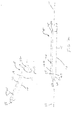
- a cross-pin 5 of the present invention has an elongated member 10.
- the member 10 has proximal end 20 and distal end 40.
- the member 10 is seen to have a longitudinal axis 11.
- Extending from the distal end 40 is the nose member 50 having proximal end 55 and distal end 60.
- Member 50 may have a variety of geometric configurations including tapered, conical, frustoconical, bullet-shaped, rounded, stepped, etc., and combinations thereof.
- Extending into the outer surface 15 of the member 10 is the axial trough 70. Trough 70 is seen to have open proximal end 72, distal end 74, bottom 75, opposed sides 77.
- Trough 70 may have a variety of cross-sections, including U-shaped, circular, arcuate, square, rectangular, etc. and combinations thereof. Trough 70 is also seen to have open top 79 extending through surface 15. The trough 70 also has passage 80.
- the nose member 50 is seen to have a guide wire opening 62 at its distal end 55, which is preferably located concentrically about the longitudinal axis 11. Nose end member 50 is seen to contain a tunnel 100 extending through to trough 70. Tunnel 100 is seen to have passage 102 that is in communication with the guide wire opening 62 in the nose end member 50 and also in communcation with the trough passage 80 at distal end 74.
- the cross-pin of the present invention is used in combination with a conventional guide wire in order to secure an ACL replacement ligament graft into a femoral tunnel.
- a variety of methods of securing ACL replacement ligament grafts in femoral tunnels using conventional cannulated cross-pins are known in the art.
- US 2004 230302 A1 discloses a method of moving an ACL graft into a femoral tunnel and securing it with a cannulated cross-pin moved over a guide wire.
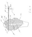
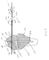
- FIGS. 3 -7 the novel cross-pin 5 of the present invention is seen used to secure an ACL graft 200 in a femoral bone tunnel 190 contained in a femur 180 of a knee 150.
- a patient is prepared by the orthopedic surgeon in a conventional manner by immobilizing the patient's knee 150 in a desired configuration.
- longitudinal or axial bone tunnels are drilled in the ends of the tibia 170 and the femur 180 adjacent to patient's knee 150 (See FIG. 3 ).
- tibial tunnel 175 and the femoral tunnel 190 are seen to be in substantial alignment.
- a transverse tunnel 270 is drilled into the femur 190 such that the traverse tunnel 270 intersects the femoral tunnel 190 toward the top 198 of the femoral tunnel 190.
- a conventional trocar drill member 210 is affixed with a pin 211 in a conventional cannula sleeve 220.
- the trocar drill member 210 has pointed distal end 212 and flat proximal end 215.
- Cannula sleeve 220 has proximal end 222, distal end 225 and passage way 227.
- the proximal end 215 is mounted to a conventional surgical drill and the distal end 212 drills through the femur 180 to create the transverse tunnel 270, while emplacing at least a portion of the distal end 225 of cannula sleeve 220 therein.
- Other conventional methods of drilling the transverse tunnel and emplacing the cannula sleeve 220 may be used as well.
- Transverse tunnel 270 has opposed open ends 272 and 273, and passage 275. After the trocar or drill has been removed from tunnel 270, an ACL replacement graft 160 is moved into the femoral tunnel 190 in a conventional manner, for example, by attaching to sutures that are pulled through suture tunnel extension 197.
- a conventional guide wire 120 is threaded through the transverse tunnel 270 through such that a portion of the guide wire 120 is beneath the top 168 of the upper end 166 of the looped ACL graft 160.
- Conventional techniques and equivalents thereof may be used to thread the guide wire 120 and move the graft 160 into place.
- Guide wires used in this art are conventionally known and may be made of a variety of biocompatible materials including surgical stainless steel, Nylon, Nitinol, etc.
- the cross-pin 5 is threaded onto a guide wire 120 by inserting a first end 122 of the guide wire 120 into the guide wire opening 62 in nose member 55 and threading the guide wire 120 through the tunnel 100 out of passage 102 and into passage 80 of the trough 70.
- the end 122 of the guide wire 120 exits the proximal end 72 of the trough 70 through an opening 22 in the proximal end 20 of the cross-pin 5.
- the guide wire 120 is then threaded through the insertion instrument 300.
- the instrument 300 is seen to have a proximal handle 305 and an elongated member 310 having a distal end 315.
- the insertion instrument 300 has a lumen or longitudinal passage 302 running the length of the instrument.
- the instrument 300 is seen to have set screw 306 having distal end 307 that is moveable into passage 302 in handle 305 to optionally engage guide wire 120.
- the distal end 315 of instrument 300 is located to engage the proximal end 20 of cross-pin 5.
- the cross-pin 5 of the present is moved by the insertion tool 300 (via pushing, hammering, etc.) through the cannula sleeve and into tunnel 270 such that it is positioned in the femoral tunnel 190 underneath the top 166 of ACL graft 160 in the transverse tunnel 270 and upper femoral tunnel 198.
- the insertion instrument is maintained in a fixed position relative to the guide wire 120 by the set screw 306, although it may also be slid along the wire 120. This implantation of the cross-pin 5 secures the end 166 of the ACL graft 160 in the femoral tunnel 190.
- the guide wire 120 is then removed along with the cannula sleeve 220 and this portion of the surgical procedure is completed.
- the other end 162 of ACL graft is secured in the tibial tunnel 175 using conventional securing procedures and techniques such as the use of interference screws or cross-pins (not shown). This then completes the surgical procedure and the reconstruction of the knee150 with the ACL graft 160 secured in place.
- a knot may be placed in the guide wire 120 distal to the nose member 50. The knot will have an overall dimension larger than the maximum dimension of the guide wire opening 62. Then, if the surgeon desires to remove the cross-pin 5 after emplacement in transverse tunnel 270 for any reason during any stage of the procedure, the surgeon pulls proximally on the guidewire 120 to back-out and remove the cross-pin 5.
- the cross-pin 5 of the present invention have many advantages.
- the advantages include the ability to remove pin at time of surgery, and, no uni-cortical fixation (equal distribution of load across the device).
- the pin is centered in the femoral tunnel and a single size implant may be used.
Description
- The technical field to which this invention relates is orthopedic implants, in particular, orthopedic implants useful for anterior cruciate ligament repair procedures.
- The anterior cruciate ligament (ACL) is a major component of the soft tissue in a human knee that is responsible for stability of the knee. In addition to the ACL, several other ligaments provide stability including the posterior cruciate ligament (PCL) and the medial and lateral collateral ligaments (MCL/LCL). It is not uncommon for a person to rupture or tear the ACL during various types of physical activities including sports, work, and the like. The tear or rupture can be caused by trauma such as impact, or by abrupt stopping or turning movements which cause exceptional forces to be transmitted to the ACL. Typically a tom or ruptured ACL cannot be repaired using conventional soft tissue repair procedures such as suturing, stapling, etc. It is necessary to replace the ACL with a graft. The graft may be an autograph harvested, for example, from the patient's patellar tendon or hamstring tendon, an allograft harvested from a cadaver, a xenograft, or an artificial man-made tendon. Tissue-engineered ligaments may also be available. In a typical ACL reconstruction, axial tunnels are drilled into the patient's tibia and femur by the surgeon using conventional surgical drills, drill guides and instruments. Once the knee is prepared, the graft is then inserted by the surgeon into the tibial and femoral tunnels, such that one end of the graft resides in each tunnel. The graft is adjusted by the surgeon to provide the desired range of motion. Finally, the graft is secured at both ends in a conventional manner to complete the ACL repair or reconstruction. For example, the graft ends may be secured with conventional interference screws, etc. An alternate method of securement is to use a cross-pin, in particular a femoral cross-pin. In this type of procedure, a transverse hole is drilled into the end of femur such that it intersects the femoral tunnel, and a guide wire is threaded through the transverse tunnel. A cannulated cross-pin is then inserted into the transverse tunnel over the guide wire and underneath a looped end of the graft in order to secure the graft in the femoral tunnel. The guide wire is then removed. If desired, the other end of the graft may be secured in the tibial tunnel by a tibial cross-pin in a similar manner.
- Although the cannulated cross-pins known in this art are sufficient and adequate for their intended purposes, there is a continuing need in this art for improved cross-pins and surgical techniques. For example, there is a need for novel cross-pins that provide uni-cortical fixation and intraoperative removal or revision, eliminate or reduce the need for multiple size (length) implants, and simplify the need to make measurements and calculations in order to determine appropriate length.
- Accordingly, there is a need in this art for novel cannulated cross-pins for use in ACL reconstruction procedures.
- Prior art document
EP 0 829 233 A discloses a bone anchor that could be threaded over a guide wire and used as a cross-pin.US 6132433 discloses a cross-pin with a symmetrical bore. - A cross-pin for use in ACL reconstruction procedures is disclosed. The cross-pin has an elongated member having a proximal end, a distal end, an outer surface, and a longitudinal axis. A nose member extends out from the distal end. The nose member has a proximal end and a distal end. There is an axial trough in the member extending through the outer surface. The trough has a proximal end, a distal end, a bottom, an open top and a passageway. There is a guide-wire opening in the distal end of the nose member. There is also an interior passage in the nose member that extends from the guide-wire opening through to the trough such that the passage is in communication with the guide wire opening and the trough.
- These and other aspects and characteristics of the present invention will become more apparent from the following description and accompanying drawings.
-
-
FIG. 1 is a perspective view of the cross-pin of the present invention. -
FIG. 2 is a top view of the cross-pin ofFIG 1 . -
FIG. 3 is a partial cross-section of a knee illustrating a cross-section of the bottom of a femur in the knee having a femoral tunnel with a transverse tunnel; a cannula sleeve is illustrated in the transverse tunnel along with a trocar/drill used to drill the transverse tunnel and emplace the cannula sleeve. Also illustrated is a cross-section of the top of a tibia in the knee having a tibial tunnel . -
FIG. 4 illustrates the femur of the knee ofFIG. 3 after the transverse tunnel has been drilled and the trocar/drill has been removed, and further illustrates a flexible guide wire member threaded through the transverse tunnel and the femoral tunnel; a partial cut-away view of the upper end of a looped ligament graft is shown emplaced in the femoral tunnel. -
FIG. 5 illustrates a cross-pin of the present invention threaded onto the guide wire extending through a transverse tunnel in a femur. -
FIG. 6 illustrates an insertion rod threaded onto the guide wire and in contact with the proximal end of the cross-pin immediately prior to emplacement in the transverse tunnel. -
FIG 7 illustrates the cross-pin of the present invention threaded over a guide wire and implanted in the transverse tunnel such that the cross-pin is beneath the top of an ACL graft; fixation of the lower end of the graft in the tibial tunnel is not illustrated. - The cross-pins of the present invention can be made from a variety of conventional biocompatible materials useful in implants. The materials may be absorbable or non-absorbable. Examples of conventional non-absorbable materials include surgical stainless steel, nickel titanium alloys, ceramics, polyoxymethylene-homopolymer (Delrin), polyethylene, and other non-absorbable polymers including, but not limited to, polypropylene, and Acetal. Examples of bioabsorbable materials include poly-lactic acid (PLA), poly-glycolic acid (PGA), polydioxanone, polycaprolactone, copolymers thereof, and the like. The term "natural polymer" refers to polymers that are naturally occurring, as opposed to synthetic polymers. In embodiments where the device includes at least one synthetic polymer, suitable biocompatible synthetic polymers can include polymers selected from the group consisting of aliphatic polyesters, poly(amino acids), copoly(etheresters), polyalkylenes oxalaes, polyamides, tyrosine derived polycarbonates, poly(iminocarbonates), polyorthoesters, polyoxaesters, polyamidoesters, polyoxaesters containing amine groups, poly(anhydrides), polyphosphazenes, polyurethanes, poly(ether urethanes), poly(ester urethane) and blends thereof. Suitable synthetic polymers for use in the present invention can also include biosynthetic polymers based on sequences found in collagen, elastin, thrombin, fibronectin, starches, poly(amino acid), poly(propylene fumarate), geletin, alginate, pectin, fibrin, oxidized cellulose, chitin, chitosan, tropoelastin, hyaluronic acid, ribonucleic acids, deoxyribonucleic acids, polypeptides, proteins, polysaccharides, polynucleotides and combination thereof. The devices of the present invention may also be manufactured from conventional biocompatible natural polymers. If desired, the bioabsorbable materials may contain osteoinductive or osteoconductive materials, polymers and blends of polymers including but not limited to calcium hydroxyapatite, tricalcium phosphate, and the like.
- The cross-pins of the present invention may be made using a variety of conventional manufacturing processes including machining, molding, etc., and combinations thereof.
- As seen in
FIG 1 , across-pin 5 of the present invention has anelongated member 10. Themember 10 has proximal end 20 anddistal end 40. Themember 10 is seen to have a longitudinal axis 11. Extending from thedistal end 40 is thenose member 50 havingproximal end 55 and distal end 60.Member 50 may have a variety of geometric configurations including tapered, conical, frustoconical, bullet-shaped, rounded, stepped, etc., and combinations thereof. Extending into theouter surface 15 of themember 10 is theaxial trough 70. Trough 70 is seen to have openproximal end 72,distal end 74,bottom 75, opposedsides 77. Trough 70 may have a variety of cross-sections, including U-shaped, circular, arcuate, square, rectangular, etc. and combinations thereof. Trough 70 is also seen to have open top 79 extending throughsurface 15. Thetrough 70 also haspassage 80. Thenose member 50 is seen to have aguide wire opening 62 at itsdistal end 55, which is preferably located concentrically about the longitudinal axis 11.Nose end member 50 is seen to contain atunnel 100 extending through totrough 70.Tunnel 100 is seen to havepassage 102 that is in communication with theguide wire opening 62 in thenose end member 50 and also in communcation with thetrough passage 80 atdistal end 74. - The cross-pin of the present invention is used in combination with a conventional guide wire in order to secure an ACL replacement ligament graft into a femoral tunnel. A variety of methods of securing ACL replacement ligament grafts in femoral tunnels using conventional cannulated cross-pins are known in the art. For example
US 2004 230302 A1 discloses a method of moving an ACL graft into a femoral tunnel and securing it with a cannulated cross-pin moved over a guide wire. - A method of using the novel cross-pins of the present invention to secure the end of a graft in a bone tunnel is now described. Referring to now to
FIGS. 3 -7 , thenovel cross-pin 5 of the present invention is seen used to secure an ACL graft 200 in afemoral bone tunnel 190 contained in afemur 180 of aknee 150. Typically in such a procedure, a patient is prepared by the orthopedic surgeon in a conventional manner by immobilizing the patient'sknee 150 in a desired configuration. Then, in a conventional manner longitudinal or axial bone tunnels are drilled in the ends of thetibia 170 and thefemur 180 adjacent to patient's knee 150 (SeeFIG. 3 ). Thetibial tunnel 175 and thefemoral tunnel 190 are seen to be in substantial alignment. In addition, atransverse tunnel 270 is drilled into thefemur 190 such that thetraverse tunnel 270 intersects thefemoral tunnel 190 toward the top 198 of thefemoral tunnel 190. This can be done in a variety of conventional manners. For example as seen inFIG. 3 , a conventionaltrocar drill member 210 is affixed with apin 211 in aconventional cannula sleeve 220. Thetrocar drill member 210 has pointeddistal end 212 and flatproximal end 215.Cannula sleeve 220 hasproximal end 222,distal end 225 andpassage way 227. Theproximal end 215 is mounted to a conventional surgical drill and thedistal end 212 drills through thefemur 180 to create thetransverse tunnel 270, while emplacing at least a portion of thedistal end 225 ofcannula sleeve 220 therein. Other conventional methods of drilling the transverse tunnel and emplacing thecannula sleeve 220 may be used as well.Transverse tunnel 270 has opposed open ends 272 and 273, andpassage 275. After the trocar or drill has been removed fromtunnel 270, anACL replacement graft 160 is moved into thefemoral tunnel 190 in a conventional manner, for example, by attaching to sutures that are pulled throughsuture tunnel extension 197. Aconventional guide wire 120 is threaded through thetransverse tunnel 270 through such that a portion of theguide wire 120 is beneath the top 168 of theupper end 166 of the loopedACL graft 160. Conventional techniques and equivalents thereof may be used to thread theguide wire 120 and move thegraft 160 into place. Guide wires used in this art are conventionally known and may be made of a variety of biocompatible materials including surgical stainless steel, Nylon, Nitinol, etc. Initially, thecross-pin 5 is threaded onto aguide wire 120 by inserting afirst end 122 of theguide wire 120 into theguide wire opening 62 innose member 55 and threading theguide wire 120 through thetunnel 100 out ofpassage 102 and intopassage 80 of thetrough 70. Theend 122 of theguide wire 120 exits theproximal end 72 of thetrough 70 through anopening 22 in the proximal end 20 of thecross-pin 5. Theguide wire 120 is then threaded through theinsertion instrument 300. Theinstrument 300 is seen to have aproximal handle 305 and anelongated member 310 having adistal end 315. Theinsertion instrument 300 has a lumen orlongitudinal passage 302 running the length of the instrument. Theinstrument 300 is seen to have set screw 306 havingdistal end 307 that is moveable intopassage 302 inhandle 305 to optionally engageguide wire 120. Thedistal end 315 ofinstrument 300 is located to engage the proximal end 20 ofcross-pin 5. Then, thecross-pin 5 of the present is moved by the insertion tool 300 (via pushing, hammering, etc.) through the cannula sleeve and intotunnel 270 such that it is positioned in thefemoral tunnel 190 underneath the top 166 ofACL graft 160 in thetransverse tunnel 270 and upperfemoral tunnel 198. Optionally, the insertion instrument is maintained in a fixed position relative to theguide wire 120 by the set screw 306, although it may also be slid along thewire 120. This implantation of thecross-pin 5 secures theend 166 of theACL graft 160 in thefemoral tunnel 190. Theguide wire 120 is then removed along with thecannula sleeve 220 and this portion of the surgical procedure is completed. Theother end 162 of ACL graft is secured in thetibial tunnel 175 using conventional securing procedures and techniques such as the use of interference screws or cross-pins (not shown). This then completes the surgical procedure and the reconstruction of the knee150 with theACL graft 160 secured in place. Although not shown, a knot may be placed in theguide wire 120 distal to thenose member 50. The knot will have an overall dimension larger than the maximum dimension of theguide wire opening 62. Then, if the surgeon desires to remove thecross-pin 5 after emplacement intransverse tunnel 270 for any reason during any stage of the procedure, the surgeon pulls proximally on theguidewire 120 to back-out and remove thecross-pin 5. - The
cross-pin 5 of the present invention have many advantages. The advantages include the ability to remove pin at time of surgery, and, no uni-cortical fixation (equal distribution of load across the device). In addition the pin is centered in the femoral tunnel and a single size implant may be used. - Although this invention has been shown and described with respect to detailed embodiments thereof, it will be understood by those skilled in the art that various changes in form and detail thereof may be made without departing from the scope of the claimed invention.
Claims (10)
- An implantable cross-pin, comprising:an elongated member having a proximal end, a distal end, a longitudinal axis, and an outer surface;a nose member extending out from the distal end of said elongated member having a proximal end and a distal end ;a guide wire opening in the distal end of the nose member;
characterised byan axial trough in the elongated member extending though the outer surface, said trough having a proximal end , a distal end, a bottom, opposed ends, an open top and a passageway; andan interior passage in the nose member extending from the guide wire opening and extending into the trough such that the passage is in communication with the guide wire opening and the trough. - The cross-pin of claim 1, wherein the cross-pin comprises a biocompatible material.
- The cross-pin of claim 2, wherein the material is bioabsorbable.
- The cross-pin of claim 2 wherein the material is non-absorbable.
- The cross-pin of claim 3, wherein the bioabsorbable material is selected from the group consisting of PLA, PGA, and copolymers thereof.
- The cross-pin of claim 4, wherein the non-absorbable material is selected from the group consisting of surgical stainless steel, nickel titanium alloys, ceramics, polyoxymethylene-homopolymer, polyethylene, polypropylene, acetal, and ceramics.
- The cross-pin of claim 1, wherein the proximal end of the cross-pin comprises an opening in communication with the proximal end of the trough.
- The cross-pin of claim 1, wherein the guide wire opening is concentric with the longitudinal axis.
- The cross-pin of claim 1, wherein the nose member has a bullet shape.
- The cross pin of claim 1, wherein the nose member has a frustoconical shape.
Applications Claiming Priority (2)
| Application Number | Priority Date | Filing Date | Title |
|---|---|---|---|
| US808764 | 2004-03-25 | ||
| US10/808,764 US8088128B2 (en) | 2004-03-25 | 2004-03-25 | Implantable cross-pin for anterior cruciate ligament repair |
Publications (3)
| Publication Number | Publication Date |
|---|---|
| EP1579826A2 EP1579826A2 (en) | 2005-09-28 |
| EP1579826A3 EP1579826A3 (en) | 2006-03-15 |
| EP1579826B1 true EP1579826B1 (en) | 2008-05-28 |
Family
ID=34862079
Family Applications (1)
| Application Number | Title | Priority Date | Filing Date |
|---|---|---|---|
| EP05251849A Expired - Fee Related EP1579826B1 (en) | 2004-03-25 | 2005-03-24 | Implantable cross-pin for anterior cruciate ligament repair |
Country Status (6)
| Country | Link |
|---|---|
| US (2) | US8088128B2 (en) |
| EP (1) | EP1579826B1 (en) |
| JP (1) | JP4602133B2 (en) |
| AU (1) | AU2005200992A1 (en) |
| CA (1) | CA2500927C (en) |
| DE (1) | DE602005007083D1 (en) |
Families Citing this family (26)
| Publication number | Priority date | Publication date | Assignee | Title |
|---|---|---|---|---|
| US8088128B2 (en) | 2004-03-25 | 2012-01-03 | Depuy Mitek, Inc. | Implantable cross-pin for anterior cruciate ligament repair |
| DE102004053471A1 (en) * | 2004-11-03 | 2006-05-04 | Karl Storz Gmbh & Co. Kg | Securing pin in particular to be used in tendon transplantation, comprising specifically shaped cross section |
| US7825083B2 (en) * | 2005-02-10 | 2010-11-02 | Spine Wave, Inc. | Synovial fluid barrier |
| GB2443795B (en) * | 2006-10-06 | 2011-04-13 | Dalmatic As | Leads for use in orthopaedic surgery |
| US7942914B2 (en) | 2006-10-17 | 2011-05-17 | Arthroscopic Innovations Llc | Method and apparatus for surgical repair |
| US8551123B2 (en) * | 2008-11-13 | 2013-10-08 | Rajiv D. Pandya | Device for the intraosteal seizing of sutures |
| US8545503B2 (en) * | 2009-06-16 | 2013-10-01 | Marshall Ephraim Stauber | Surgical tool and method |
| JP6256833B2 (en) * | 2011-01-05 | 2018-01-10 | インプランティカ・パテント・リミテッド | Knee joint device and method |
| US9445803B2 (en) | 2011-11-23 | 2016-09-20 | Howmedica Osteonics Corp. | Filamentary suture anchor |
| US9277945B2 (en) * | 2012-02-07 | 2016-03-08 | Mnr Device Corporation | Method and apparatus for treating a bone fracture |
| US9808242B2 (en) | 2012-04-06 | 2017-11-07 | Howmedica Osteonics Corp. | Knotless filament anchor for soft tissue repair |
| US8821494B2 (en) | 2012-08-03 | 2014-09-02 | Howmedica Osteonics Corp. | Surgical instruments and methods of use |
| US9078740B2 (en) * | 2013-01-21 | 2015-07-14 | Howmedica Osteonics Corp. | Instrumentation and method for positioning and securing a graft |
| US9402620B2 (en) | 2013-03-04 | 2016-08-02 | Howmedica Osteonics Corp. | Knotless filamentary fixation devices, assemblies and systems and methods of assembly and use |
| US9788826B2 (en) | 2013-03-11 | 2017-10-17 | Howmedica Osteonics Corp. | Filamentary fixation device and assembly and method of assembly, manufacture and use |
| US9386997B2 (en) * | 2013-03-29 | 2016-07-12 | Smith & Nephew, Inc. | Tunnel gage |
| US10292694B2 (en) | 2013-04-22 | 2019-05-21 | Pivot Medical, Inc. | Method and apparatus for attaching tissue to bone |
| US10610211B2 (en) | 2013-12-12 | 2020-04-07 | Howmedica Osteonics Corp. | Filament engagement system and methods of use |
| US9986992B2 (en) | 2014-10-28 | 2018-06-05 | Stryker Corporation | Suture anchor and associated methods of use |
| US10568616B2 (en) | 2014-12-17 | 2020-02-25 | Howmedica Osteonics Corp. | Instruments and methods of soft tissue fixation |
| CN104665918B (en) * | 2015-02-15 | 2017-08-25 | 陆爱清 | Guide pin entry point position developer |
| US11376079B2 (en) | 2016-02-19 | 2022-07-05 | Rajiv D. Pandya | System and technique for accessing extra articular lesions or abnormalities or intra osseous lesions or bone marrow lesions |
| US11419684B2 (en) | 2016-02-19 | 2022-08-23 | Rajiv D. Pandya | System and technique for accessing extra articular lesions or abnormalities or intra osseous lesions or bone marrow lesions |
| US9925010B2 (en) | 2016-02-19 | 2018-03-27 | Rajiv D. Pandya | System and technique for accessing extra articular lesions or abnormalities or intra osseous lesions or bone marrow lesions |
| US10064633B2 (en) | 2016-02-19 | 2018-09-04 | Rajiv D. Pandya | System and technique for accessing extra articular lesions or abnormalities or intra osseous lesions or bone marrow lesions |
| USD902405S1 (en) | 2018-02-22 | 2020-11-17 | Stryker Corporation | Self-punching bone anchor inserter |
Family Cites Families (32)
| Publication number | Priority date | Publication date | Assignee | Title |
|---|---|---|---|---|
| US4968315A (en) * | 1987-12-15 | 1990-11-06 | Mitek Surgical Products, Inc. | Suture anchor and suture anchor installation tool |
| JPH02154751A (en) * | 1988-04-12 | 1990-06-14 | Olympus Optical Co Ltd | Laser probe |
| US5102421A (en) * | 1990-06-14 | 1992-04-07 | Wm. E. Anpach, III | Suture anchor and method of forming |
| US5269809A (en) * | 1990-07-02 | 1993-12-14 | American Cyanamid Company | Locking mechanism for use with a slotted suture anchor |
| US5098435A (en) * | 1990-11-21 | 1992-03-24 | Alphatec Manufacturing Inc. | Cannula |
| US5257996A (en) | 1991-12-13 | 1993-11-02 | Mcguire David A | Surgical pin passer |
| US5480403A (en) * | 1991-03-22 | 1996-01-02 | United States Surgical Corporation | Suture anchoring device and method |
| US5211647A (en) * | 1992-02-19 | 1993-05-18 | Arthrex Inc. | Interference screw and cannulated sheath for endosteal fixation of ligaments |
| US5474554A (en) * | 1994-07-27 | 1995-12-12 | Ku; Ming-Chou | Method for fixation of avulsion fracture |
| US5702397A (en) * | 1996-02-20 | 1997-12-30 | Medicinelodge, Inc. | Ligament bone anchor and method for its use |
| US5921971A (en) * | 1996-09-13 | 1999-07-13 | Boston Scientific Corporation | Single operator exchange biliary catheter |
| US5733307A (en) * | 1996-09-17 | 1998-03-31 | Amei Technologies, Inc. | Bone anchor having a suture trough |
| CA2217406C (en) * | 1996-10-04 | 2006-05-30 | United States Surgical Corporation | Suture anchor installation system with disposable loading unit |
| US6113604A (en) | 1997-01-14 | 2000-09-05 | Ethicon, Inc. | Method and apparatus for fixing a graft in a bone tunnel |
| US5849013A (en) | 1997-01-14 | 1998-12-15 | Whittaker; Gregory R. | Method and apparatus for fixing a bone block in a bone tunnel |
| US5918604A (en) | 1997-02-12 | 1999-07-06 | Arthrex, Inc. | Method of loading tendons into the knee |
| US5885294A (en) * | 1997-09-22 | 1999-03-23 | Ethicon, Inc. | Apparatus and method for anchoring a cord-like element to a workpiece |
| CA2303853C (en) * | 1997-09-24 | 2007-01-30 | Depuy Orthopaedics, Inc. | Acl fixation pin and method |
| US6066173A (en) | 1998-01-28 | 2000-05-23 | Ethicon, Inc. | Method and apparatus for fixing a graft in a bone tunnel |
| US6499486B1 (en) * | 1999-07-29 | 2002-12-31 | Ethicon, Inc. | Method for reconstructing a ligament |
| US6623524B2 (en) | 2000-06-09 | 2003-09-23 | Arthrex, Inc. | Method for anterior cruciate ligament reconstruction using cross-pin implant with eyelet |
| US6325804B1 (en) | 2000-06-28 | 2001-12-04 | Ethicon, Inc. | Method for fixing a graft in a bone tunnel |
| US6878166B2 (en) | 2000-08-28 | 2005-04-12 | Ron Clark | Method and implant for securing ligament replacement into the knee |
| US6579295B1 (en) * | 2000-09-25 | 2003-06-17 | Robert S. Supinski | Tendon anchors |
| US6517546B2 (en) | 2001-03-13 | 2003-02-11 | Gregory R. Whittaker | Method and apparatus for fixing a graft in a bone tunnel |
| US6508830B2 (en) * | 2001-04-30 | 2003-01-21 | Musculoskeletal Transplant Foundation | Suture anchor |
| US7338492B2 (en) * | 2002-05-15 | 2008-03-04 | Linvatec Corporation | Cross-pin graft fixation, instruments, and methods |
| US7270666B2 (en) | 2002-05-15 | 2007-09-18 | Linvatec Corporation | Cross-pin graft fixation, instruments, and methods |
| US7713286B2 (en) * | 2002-11-15 | 2010-05-11 | Linvatec Corporation | Knotless suture anchor |
| US7032599B2 (en) | 2003-05-15 | 2006-04-25 | Mitek Surgical Products Div. Of Ethicon, Inc. | Method of replacing an anterior cruciate ligament in the knee |
| JP2007517635A (en) * | 2004-01-16 | 2007-07-05 | オステオバイオロジックス, インコーポレイテッド | Bone-tendon-bone implant |
| US8088128B2 (en) | 2004-03-25 | 2012-01-03 | Depuy Mitek, Inc. | Implantable cross-pin for anterior cruciate ligament repair |
-
2004
- 2004-03-25 US US10/808,764 patent/US8088128B2/en active Active
-
2005
- 2005-03-04 AU AU2005200992A patent/AU2005200992A1/en not_active Abandoned
- 2005-03-15 CA CA2500927A patent/CA2500927C/en not_active Expired - Fee Related
- 2005-03-24 EP EP05251849A patent/EP1579826B1/en not_active Expired - Fee Related
- 2005-03-24 DE DE602005007083T patent/DE602005007083D1/en active Active
- 2005-03-24 JP JP2005086791A patent/JP4602133B2/en not_active Expired - Fee Related
-
2011
- 2011-11-22 US US13/301,975 patent/US8721653B2/en not_active Expired - Lifetime
Also Published As
| Publication number | Publication date |
|---|---|
| CA2500927A1 (en) | 2005-09-25 |
| AU2005200992A1 (en) | 2005-10-13 |
| DE602005007083D1 (en) | 2008-07-10 |
| US20120071976A1 (en) | 2012-03-22 |
| EP1579826A3 (en) | 2006-03-15 |
| US20050216014A1 (en) | 2005-09-29 |
| JP2005270668A (en) | 2005-10-06 |
| CA2500927C (en) | 2013-05-28 |
| JP4602133B2 (en) | 2010-12-22 |
| EP1579826A2 (en) | 2005-09-28 |
| US8721653B2 (en) | 2014-05-13 |
| US8088128B2 (en) | 2012-01-03 |
Similar Documents
| Publication | Publication Date | Title |
|---|---|---|
| EP1579826B1 (en) | Implantable cross-pin for anterior cruciate ligament repair | |
| US20210298906A1 (en) | Methods, systems and devices for repairing anatomical joint conditions | |
| US7458975B2 (en) | Method of replacing an anterior cruciate ligament in the knee | |
| US7527648B2 (en) | Method of replacing an anterior cruciate ligament in the knee | |
| US8016867B2 (en) | Graft fixation device and method | |
| US5674224A (en) | Bone mulch screw assembly for endosteal fixation of soft tissue grafts and method for using same | |
| EP1720459B1 (en) | Articular cartilage fixation device | |
| US5968045A (en) | Intra-articular tendon sling fixation screw | |
| US5306301A (en) | Graft attachment device and method of using same | |
| US6423073B2 (en) | Instrument for inserting graft fixation device | |
| EP1169979B1 (en) | Use of an adhesive substance for the manufacture of a bone adhesive for fixing a bone plug in a bone tunnel | |
| JP4637506B2 (en) | Tissue fixing device | |
| EP1652484B1 (en) | Instruments and kit for suture management | |
| US20020052628A1 (en) | Graft fixation device and method | |
| US20010029381A1 (en) | Graft fixation device combination | |
| US20010029382A1 (en) | Method of securing a graft using a graft fixation device | |
| AU2011236077B2 (en) | Implantable cross-pin for anterior cruciate ligament repair |
Legal Events
| Date | Code | Title | Description |
|---|---|---|---|
| PUAI | Public reference made under article 153(3) epc to a published international application that has entered the european phase |
Free format text: ORIGINAL CODE: 0009012 |
|
| AK | Designated contracting states |
Kind code of ref document: A2 Designated state(s): AT BE BG CH CY CZ DE DK EE ES FI FR GB GR HU IE IS IT LI LT LU MC NL PL PT RO SE SI SK TR |
|
| AX | Request for extension of the european patent |
Extension state: AL BA HR LV MK YU |
|
| PUAL | Search report despatched |
Free format text: ORIGINAL CODE: 0009013 |
|
| AK | Designated contracting states |
Kind code of ref document: A3 Designated state(s): AT BE BG CH CY CZ DE DK EE ES FI FR GB GR HU IE IS IT LI LT LU MC NL PL PT RO SE SI SK TR |
|
| AX | Request for extension of the european patent |
Extension state: AL BA HR LV MK YU |
|
| 17P | Request for examination filed |
Effective date: 20060904 |
|
| AKX | Designation fees paid |
Designated state(s): DE FR GB IT |
|
| GRAP | Despatch of communication of intention to grant a patent |
Free format text: ORIGINAL CODE: EPIDOSNIGR1 |
|
| RAP1 | Party data changed (applicant data changed or rights of an application transferred) |
Owner name: ETHICON, INC. |
|
| GRAS | Grant fee paid |
Free format text: ORIGINAL CODE: EPIDOSNIGR3 |
|
| GRAA | (expected) grant |
Free format text: ORIGINAL CODE: 0009210 |
|
| AK | Designated contracting states |
Kind code of ref document: B1 Designated state(s): DE FR GB IT |
|
| REG | Reference to a national code |
Ref country code: GB Ref legal event code: FG4D |
|
| RAP2 | Party data changed (patent owner data changed or rights of a patent transferred) |
Owner name: DEPUY MITEK, INC. |
|
| REF | Corresponds to: |
Ref document number: 602005007083 Country of ref document: DE Date of ref document: 20080710 Kind code of ref document: P |
|
| PLBE | No opposition filed within time limit |
Free format text: ORIGINAL CODE: 0009261 |
|
| STAA | Information on the status of an ep patent application or granted ep patent |
Free format text: STATUS: NO OPPOSITION FILED WITHIN TIME LIMIT |
|
| 26N | No opposition filed |
Effective date: 20090303 |
|
| REG | Reference to a national code |
Ref country code: FR Ref legal event code: PLFP Year of fee payment: 12 |
|
| REG | Reference to a national code |
Ref country code: FR Ref legal event code: PLFP Year of fee payment: 13 |
|
| REG | Reference to a national code |
Ref country code: FR Ref legal event code: PLFP Year of fee payment: 14 |
|
| PGFP | Annual fee paid to national office [announced via postgrant information from national office to epo] |
Ref country code: DE Payment date: 20200310 Year of fee payment: 16 Ref country code: IT Payment date: 20200221 Year of fee payment: 16 Ref country code: GB Payment date: 20200311 Year of fee payment: 16 |
|
| PGFP | Annual fee paid to national office [announced via postgrant information from national office to epo] |
Ref country code: FR Payment date: 20200214 Year of fee payment: 16 |
|
| REG | Reference to a national code |
Ref country code: DE Ref legal event code: R119 Ref document number: 602005007083 Country of ref document: DE |
|
| GBPC | Gb: european patent ceased through non-payment of renewal fee |
Effective date: 20210324 |
|
| PG25 | Lapsed in a contracting state [announced via postgrant information from national office to epo] |
Ref country code: DE Free format text: LAPSE BECAUSE OF NON-PAYMENT OF DUE FEES Effective date: 20211001 Ref country code: GB Free format text: LAPSE BECAUSE OF NON-PAYMENT OF DUE FEES Effective date: 20210324 Ref country code: FR Free format text: LAPSE BECAUSE OF NON-PAYMENT OF DUE FEES Effective date: 20210331 |
|
| PG25 | Lapsed in a contracting state [announced via postgrant information from national office to epo] |
Ref country code: IT Free format text: LAPSE BECAUSE OF NON-PAYMENT OF DUE FEES Effective date: 20210324 |