-
Glycine is the major inhibitory neurotransmitter in the spinal cord and brainstem
and is also a co-agonist at the NMDA receptor. The extracellular concentration of glycine
is regulated by at least two Na+/Cl--dependent glycine transporters (GLYT1 and GLYT2)
which play an important role in the termination of post-synaptic glycinergic actions and
maintenance of low extracellular glycine concentration by re-uptake of glycine into
presynaptic nerve terminals and surrounding fine glial processes. GLYT2 is expressed at
high levels in the rodent spinal cord, brainstem and cerebellum where its expression
correlates very well with the presence of strychnine-sensitive glycine receptors (Zafra, F.,
et al., J Neurosci, 1995. 15(5 Pt 2): p. 3952-69; Luque, J.M., N. Nelson, and J.G. Richards,
Neuroscience, 1995. 64(2): p. 525-35 and Jursky, F. and N. Nelson, J Neurochem, 1995.
64(3): p. 1026-33]. Immunohistochemical analysis suggests a predominantly pre-synaptic
localization in presumptive glycinergic synapses (Zafra, F., et al., J Neurosci, 1995. 15(5 Pt
2): p. 3952-69; Spike, R.C., et al., Neuroscience, 1997. 77(2): p. 543-51), strongly
suggesting a role in the termination of glycinergic inhibitory synaptic transmission.
Human GLYT2 has been cloned and appears to exhibit a similar expression pattern
(Morrow, J.A., et al., FEBS Lett, 1998. 439(3): p. 334-40). GLYT2 has some degree of
heterogeneity. Indeed two GLYT2 isoforms (2a and 2b) have been identified in rodent
brains.
-
GLYT1 can be distinguished pharmacologically from GLYT2 by its sensitivity to be
blockaded by sarcosine and N-methylated derivative of glycine (Liu, Q.R., et al., J Biol
Chem, 1993. 268(30): p. 22802-8, Kim, K.M., et al., Mol Pharmacol, 1994. 45(4): p. 608-17).
The human glyt1 gene has been cloned and encodes four isoforms GLYT1a, 1b, 1c
and 1d (Kim, K.M., et al., Mol Pharmacol, 1994. 45(4): p. 608-17) whereas only two rat
isoforms, GLYT1a and 1b, have been identified (Guastella, J., et al., Proc Natl Acad Sci
USA, 1992. 89(15): p. 7189-93; Smith, K.E., et al., Neuron, 1992. 8(5): p. 927-35;
Borowsky, B., E. Mezey, and B.J. Hoffman, Neuron, 1993. 10(5): p. 851-63). GLYT1
appears to be expressed in both glia and neurons in the rat CNS (Zafra, F., et al., J
Neurosci, 1995. 15(5 Pt 2): p. 3952-69; Smith, K.E., et al., Neuron, 1992. 8(5): p. 927-35;
Borowsky, B., E. Mezey, and B.J. Hoffman, Neuron, 1993. 10(5): p. 851-63; Zafra, F., et
al., Eur J Neurosci, 1995. 7(6): p. 1342-52), with GLYT1a apparently expressed in the gray
matter as well as in some peripheral tissues whilst GLYT1b is expressed only in the white
matter of the CNS (Borowsky, B., E. Mezey, and B.J. Hoffman, Neuron, 1993. 10(5): p.
851-63). In humans, a probe common to all GLYT1 isoforms revealed expression in
several peripheral tissues, most notably the kidney, whereas GLYT1c seems to be brain
specific (Kim, K.M., et al., Mol Pharmacol, 1994. 45(4): p. 608-17). The GLYT1 isoforms
differ only in their amino termini and 5' non-coding regions (Kim, K.M., et al., Mol
Pharmacol, 1994. 45(4): p. 608-17; Borowsky, B., E. Mezey, and B.J. Hoffman, Neuron,
1993. 10(5): p. 851-63). GLYT1a and GLYT1b originate from transcription directed from
alternate promoters whereas human GLYT1c is a splice variant of the GLYT1b transcript
(Kim, K.M., et al., Mol Pharmacol, 1994. 45(4): p. 608-17; Adams, R.H., et al., J Neurosci,
1995. 15(3 Pt 2): p. 2524-32; Borowsky, B. and B.J. Hoffman, J Biol Chem, 1998. 273(44):
p. 29077-85). The peripheral expression of GLYT1 and the differential CNS expression
patterns of the isoforms are somewhat controversial with major discrepancies evident
between the published studies. GLYT1 is expressed together with GLYT2 in the spinal
cord, brainstem and diencephalon. Interestingly GLYT1 is expressed in forebrain areas
such as the cortex, hippocampus and olfactory bulb where no functional inhibitory
glycinergic neurons have been found (Zafra, F., et al., J Neurosci, 1995. 15(5 Pt 2): p.
3952-69; Guastella, J., et al., Proc Natl Acad Sci U S A, 1992. 89(15): p. 7189-93; Smith,
K.E., et al., Neuron, 1992. 8(5): p. 927-35; Borowsky, B., E. Mezey, and B.J. Hoffman,
Neuron, 1993 Eur J Neurosci, 1995. 7(6): p. 1342-52) thus, suggesting additional roles for
GLYT1, which might include regulation of NMDA receptor-mediated neurotransmission
(Smith, K.E., et al., Neuron, 1992. 8(5): p. 927-35).
-
Binding of both glutamate and glycine is necessary for NMDA receptor activation.
Whilst glutamate is released in an activity-dependent manner from pre-synaptic
terminals, glycine is apparently present at a more constant level, indicating a more
modulatory function. Measurements of glycine concentration in the extracellular and
cerebrospinal fluids suggest that it is present at low micromolar levels (Westergren, I. et
al., J Neurochem, 1994. 62(1): p. 159-65). However glycine transporters might reduce the
glycine concentration markedly in the local microenvironment of NMDA receptors.
Indeed, expression of GLYT1b in Xenopus oocytes has been shown to reduce the glycine
concentration at co-expressed NMDA receptors (Supplisson, S. and C. Bergman, J
Neurosci, 1997. 17(12): p. 4580-90). Additionally, recent studies have suggested that
glycine uptake mechanisms can regulate synaptic NMDA receptor activity (Berger, A.J., S.
Dieudonne, and P. Ascher, J Neurophysiol, 1998. 80(6): p. 3336-40; Bergeron, R., et al.,
Proc Natl Acad Sci U S A, 1998. 95(26): p. 15730-4). NMDA receptor glycine affinity is
influenced by the identity of the receptor NR2 subunit and in recombinant systems,
receptors containing NR2A exhibit a markedly reduced affinity for glycine relative to
those containing NR2B, C or D (Ikeda, K., et al., FEBS Lett, 1992. 313(1): p. 34-8;
Kutsuwada, T., et al., Nature, 1992. 358(6381): p. 36-41; Priestley, T., et al., Mol
Pharmacol, 1995. 48(5): p. 841-8). A population of NMDA receptors with a markedly
lower affinity for glycine appears during maturation, paralleling the developmental
increase in expression of NR2A (Kew, J.N., et al., J Neurosci, 1998. 18(6): p. 1935-43)
suggesting the existence of a population of NMDA receptors not saturated by glycine
under normal physiological conditions
-
Glutamate neurotransmission, in particular NMDA receptor activity, plays a critical
role in synaptic plasticity, learning and memory, such as the NMDA receptors appears to
serve as a graded switch for gating the threshold of synaptic plasticity and memory
formation (Hebb, D., The organization of behavior. 1949, New York: Wiley; Bliss, T.V. and
G.L. Collingridge, Nature, 1993. 361(6407): p. 31-9). Transgenic mice overexpressing the
NMDA NR2B subunit exhibit enhanced synaptic plasticity and superior ability in
learning and memory (Tang, Y.P., et al., Nature, 1999. 401(6748): p. 63-9).
-
NMDA receptor hypofunction has been implicated in the pathophysiology of
schizophrenia (Olney, J.W. and N.B. Farber, Arch Gen Psychiatry, 1995. 52(12): p. 998-1007;
Hirsch, S.R., et al., Pharmacol Biochem Behav, 1997. 56(4): p. 797-802). Non-competitive
NMDA receptor antagonists such as PCP and ketamine can induce
schizophrenia-like psychosis (Allen, R.M. and S.J. Young, Am J Psychiatry, 1978. 135(9):
p. 1081-4; Javitt, D.C. and S.R. Zukin, Am J Psychiatry, 1991. 148(10): p. 1301-8; Krystal,
J.H., et al., Arch Gen Psychiatry, 1994. 51(3): p. 199-214), which incorporates positive
and negative symptoms as well as cognitive dysfunction, thus closely resembling
schizophrenia in patients (Javitt, D.C., et al., Biol Psychiatry, 1999. 45(6): p. 668-79).
Transgenic mice expressing reduced levels of the NMDAR1 subunit displays behavioral
abnormalities similar to those observed in pharmacologically induced models of
schizophrenia, supporting a model in which reduced NMDA receptor activity results in
schizophrenia-like behavior (Mohn, A.R., et al., Cell, 1999. 98(4): p. 427-36).
Furthermore mice lacking the NMDA receptor 2A subunit exhibit an increased
spontaneous locomotor activity in a novel environments and an impairment of latent
learning in a water-finding task besides deficit in hippocampal LTP and spatial learning
(Miyamoto Y, Yamada K, Noda Y, Mori H, Mishina M and Nabeshima T, J. Neurosci.
2001 21(2): 750-757).
-
A mouse overproducing GLYT1 provides a valuable tool to assess the physiological
function of GLYT1. These mice should exhibit decreased levels of glycine in the forebrain
and they can be useful in addressing the question of whether active regulation of NMDA
receptor glycine site occupancy is important for physiological NMDA receptor function.
-
The present invention provides genetic constructs and methods for producing
transgenic non-human animals comprising within their genome transgenic DNA
encoding GLYT1. These transgenic animals can be further used to generate transgenic
animals which overexpress active GLYT1. Moreover, said transgenic animals overexpress
GLYT1 protein, as well as the methods of producing them are also provided. The
invention also relates to the use of these animals as a model for analyzing the effects of
depressing synaptic NMDA receptor function and studying the ability of compounds to
reduce symptoms of psychotic behavior.
-
The present invention therefore provides a genetic construct comprising a DNA
sequence encoding GLYT1, operatively linked to a promoter. The sequence of glyt1 gene
may encode an isoform of GLYT1. Preferably, the sequence of glyt1 gene encodes
GLYT1b.
-
Preferably, the sequence of glyt1 gene is a cDNA sequence. More preferably, the
cDNA incorporates at least one intron sequence. Most preferably, the at least one intron
sequence comprises a polyadenylation site.
-
The sequence of glyt1 gene can be derived from any animal, preferably the sequence
of glyt1 derives from a mammal, more preferably the sequence of glyt1 gene is a human
sequence.
-
The promoter may be a neuronal promoter. In one embodiment the promoter is a
tissue-specific promoter. A tissue-specific promoter may be any promoter, which controls
and directs expression of a gene in a tissue-specific manner, e.g., in brain tissue, in muscle
tissue, in liver tissue, in kidney tissue, etc. Preferably, the promoter provides specific
expression in the forebrain. Most preferably, the promoter is the mouse CamKαII-promoter.
-
The promoter may also be a controllable promoter. A controllable promoter may
be any promoter, which controls the expression of a transgene in a regulatable and/or
inducible fashion, e.g., by addition of specific inducer or repressor substances. Several
inducible bacterial promoter are known in the art (Schultze N, Burki Y, Lang Y, Certa U,
Bluethmann H; Nat Biotechnol 1996; 14(4): 499-503; van der Neut R; Targeted gene
disruption: applications in neurobiology; J Neurosci Methods 1997; 71(1): 19-27; Liu HS,
Lee CH, Lee CF, Su IJ, Chang TY; Lac/Tet dual-inducible system functions in mammalian
cell lines. Biotechniques. 1998; 24(4): 624-8, 630-2).
-
Preferably, the genetic construct is as depicted in Fig. 1.
-
The term "transgenic DNA" as used herein describes DNA artificially introduced
and incorporated into an organism.
-
The term "sequence of glyt1 gene" as used herein, describes the DNA sequence
encoding GLYT1.
-
The present invention further provides a method of producing a non-human
transgenic animal whose genome comprises transgenic DNA encoding GLYT1,
comprising
- a) introducing a genetic construct as described above into a non-human zygote or an
non-human embryonic stem cell,
- b) generating a transgenic non-human animal from said zygote or an embryonic
stem cell, and thereby,
- c) producing a transgenic non-human animal whose genome comprises transgenic
DNA encoding GLYT1.
-
-
A further embodiment of the invention provides a method of producing a non-human
transgenic animal expressing transgenic GLYT1 comprising
- a) introducing a genetic construct as described above into a non-human zygote or an
non-human embryonic stem cell derived from a non-human animal
- b) generating a transgenic animal from said zygote or embryonic stem cell, and
thereby,
- c) producing a transgenic non-human animal expressing transgenic GLYT1.
-
-
Preferably, the zygote used in the methods described above is a C57BL/6J zygote.
Zygotes used in the art which may also be used in the methods of this invention
comprise, but not limited to, FVB/N zygotes, BALB/c zygotes, DBA/1 zygotes and DBA/2
zygotes.
-
Preferably, the embryonic stem cell used in the methods described above is a
C57BL/6J embryonic stem cell. Stem cells used in the art which may also be used in the
methods of this invention comprise, but are not limited to, BALB/c embryonic stem cells,
DBA/2J embryonic stem cells, CBA/J embryonic stem cells and embryonic stem cell lines
of mouse strains 129.
-
The zygote or embryonic stem cell may derive from any non-human animal.
Preferably, the zygote or embryonic stem cell derives from a rodent. More preferably, the
zygote or embryonic stem cell derives from a mouse.
-
The introduction of the genetic construct in the zygote may be by microinjection of
the DNA. The introduction of the genetic construct in the embryonic stem cell may be by
viral infection.
-
For example, the transgenic non-human animals of the above-described methods
may be generated by culturing the zygotes after microinjection, transferring the cultured
zygotes into a pseudo-pregnant non-human animal and breeding transgenic non-human
animals.
-
The term "transgenic GLYT1" as used herein describes GLYT1 protein originating
from DNA artificially introduced and incorporated into an organism.
-
The term "transgenic animal" as used herein describes an animal comprising
transgenic DNA in their genome. This transgenic DNA may be incorporated somewhere
in the genome.
-
The present invention further provides the transgenic non-human animal
produced by any of the above described methods.
-
In one embodiment of the invention, transgenic non-human animals whose
genome comprises transgenic DNA encoding GLYT1 are provided. In a preferred
embodiment the transgenic non-human animal comprises a genetic construct as depicted
in Fig. 1.
-
In another embodiment transgenic non-human animals expressing transgenic
GLYT1 are provided. In a preferred embodiment, the transgenic GLYT1 is tissue-specific
expressed, i.e., in the brain. In a more preferred embodiment, the transgenic GLYT1 is
specifically expressed in the forebrain of the transgenic non-human animal. In another
embodiment, the expression of the transgenic GLYT1 may be controlled, e.g., by addition
of specific inducer or repressor substances.
-
In the described transgenic non-human animals overproducing GLYT1 protein a
modulation of NMDA receptor activity is expected in vivo by alteration of the
endogenous glycine level. Due to the lack of GLYT2 receptors in hippocampal and
cerebral cortex regions, an overexpression of GLYT1 in these regions leads to decreased
levels of glycine in glutamatergic synapse and thus depressing NMDA receptor function.
Therefore a mutant mouse overexpressing GLYT1 is expected to develop behavioral
alterations and abnormalities respectively to schizophrenia and cognitive impairments.
-
The transgenic non-human animal may be any non-human animal known in the
art, which may be used for the methods of the invention. Preferably, the transgenic non-human
animal is a mammal, more preferably the transgenic non-human animal is a
rodent. Most preferably, the transgenic animal of the invention is a mouse.
-
The transgenic non-human animals described above may be analyzed genetically,
molecularly and behaviorally.
-
The present invention also relates to descendants of the transgenic non-human
animals as provided by the invention, obtained by breeding with the same or with
another genotype.
-
The present invention further provides a cell line or primary cell culture, tissue as
well as an organotypic brain slice culture derived from the transgenic non-human
animals as provided by the invention or its descendants.
-
Cell culture based models can be prepared by two methods. Cell cultures can be
isolated from the non-human transgenic animals or prepared from established cell
cultures using the same constructs with standard cell transfection techniques.
-
Integration of the genetic construct comprising transgenic DNA encoding GLYT1,
can be detected by various methods comprising genomic Southern blot and PCR analysis
using DNA isolated from tail biopsies of two to three weeks old mice.
-
It will be apparent to the person skilled in the art that there are a large number of
analytical procedures which may be used to detect the expression of the transgenic DNA
comprising methods at the RNA level comprising mRNA quantification by reverse
transcriptase polymerase chain reaction (RT-PCR) or by Northern blot, in situ
hybridization, as well as methods at the protein level comprising histochemistry,
immunoblot analysis and in vitro binding studies. Quantification of the expression levels
of the targeted gene can moreover be determined by the ELISA technology, which is
common to those knowledgeable in the art.
-
Quantitative measurement can be accomplished using many standard assays. For
example, transcript levels can be measured using RT-PCR and hybridization methods
including RNase protection, Northern blot analysis, and RNA dot analysis.
Immunohistochemical staining as well as Western blot analysis can also be used to assess
the presence or absence of the transgenic GLYT1 protein.
-
The transgenic animals of the invention may be further characterized by methods
known in the art, comprising immunohistochemistry, electron microscopy, Magnetic
Resonance Imaging (MRI) and by behavioral studies addressing neurological and
cognitive functions. Examples of behavioral tests are: spontaneous behavior, behavior
related to cognitive functions, pharmacologically-disrupted behavior, grip strength, wire
manoeuvre, swim test, rotarod, locomotor activity, Morris water maze, Y-maze, lightdark
preference, passive and active avoidance tests.
-
A further objective of the present invention is the use of the transgenic non-human
animal as described, or a cell line or tissue or an organotypic brain slice culture as derived
thereof, as a model for studying the ability of compounds to reduce psychotic behavior.
Additionally, these transgenic animals, cells or tissue or organotypic brain slice culture as
derived thereof, may be used as a model for studying the effects of depressing synaptic
NMDA receptor function.
-
In a further embodiment, a method for evaluating the in vivo effects of GLYT1
function on NMDA receptor activation is provided, comprising determining NMDA
receptor activity, synaptic plasticity and behavior comprising learning and memory in a
transgenic non-human animal whose genome contains a transgenic sequence of glyt1
gene in a way that active GLYT1 protein is overexpressed, and comparing the NMDA
receptor activity, synaptic plasticity and behavior to those in a control.
-
Controls may comprise any non-human animal, wherein transgenic DNA encoding
GLYT1 is not introduced in a way that active GLYT1 protein is overexpressed, or wherein
the animal comprises exclusively native glyt1 genes. Assessment of the behavior may
comprise spontaneous behavior, behavior related to cognitive functions comprising
spatial short- and long-term memory, object recognition memory, associative emotional
memory, conditioned fear extinction, and pharmacologically-disrupted behavior
comprising drug-induced hyperlocomotion, drug-induced social withdrawal, drug-induced
deficits in prepulse inhibition and drug-induced memory loss.
-
In another embodiment, a method of testing GLYT1 inhibitor compounds for the
capability to enhance the NMDA receptor function, which method comprises
administering a GLYT1 inhibitor compound to a transgenic non-human animal whose
genome contains a transgenic sequence of glyt1 gene in a way that active GLYT1 protein
is overexpressed, or a cell line or primary cell culture or an organotypic brain slice culture
derived thereof, and determining the effect of the compound comprising assessing
behavior, electrophysiology and histology, and comparing the behavior,
electrophysiology and histology to those of a control.
GLYT1 inhibitor compounds which maybe used in the method of the invention are any
GLYT1 inhibitor compounds known in the art comprising, but not limited to, ALX-5407(NPS
Pharmaceuticals) and ORG-24598 (Organon).
-
Controls may comprise any animal, cell line or primary cell culture or organotypic
brain slice culture or tissue, wherein transgenic DNA encoding GLYT1 is not introduced
in a way that active GLYT1 protein is overexpressed, or wherein the animal, cell line or
primary cell culture or organotypic brain slice culture comprises exclusively native glyt1
genes. Assessment of the behavior may comprise spontaneous behavior, behavior related
to cognitive functions comprising spatial short- and long-term memory, object
recognition memory, associative emotional memory, conditioned fear extinction, and
pharmacologically-disrupted behavior comprising drug-induced hyperlocomotion, drug-induced
social withdrawal, drug-induced deficits in prepulse inhibition and drug-induced
memory loss.
-
The present invention further relates to a kit for testing compounds for capability
to enhance the NMDA receptor activity comprising transgenic non-human animal whose
genome contains a transgenic sequence of glyt1 gene in a way that active GLYT1 protein
is overexpressed, or a cell line or primary cell culture or tissue or an organotypic brain
slice culture or tissue derived thereof, and a means for determining whether a compound
exhibits the capability to enhance the NMDA receptor activity.
-
Furthermore, the use of a transgenic non-human animal, whose genome contains a
transgenic sequence of glyt1 gene so that active GLYT1 protein is overexpressed, or a cell
line or primary cell culture or tissue or an organotypic brain slice culture or tissue derived
thereof is provided as model for studying the effect of compounds on the psychotic
behaviour and for testing of compounds for GLYT1-specific inhibitory effects.
-
The invention further provides the transgenic animals, methods, compositions,
kits, and uses substantially as described herein before especially with reference to the
foregoing examples.
-
Having now generally described this invention, the same will become better
understood by reference to the specific examples, which are included herein for purpose
of illustration only and are not intended to be limiting unless otherwise specified, in
connection with the following figures.
-
Figure 1 shows a schematic diagram of the genetic construct comprising the mouse
CamKαII-Promoter, cDNA encoding human GLYT1b (hGlyt1b) with incorporated
introns (I), wherein one intron comprises a polyadenylation site (pA). Restriction sites
are given above the diagram.
-
Figure 2: Schematic diagram of the primer pairs used for cloning of the glyt1b-cDNA
(SEQ. ID NOs: 3 to 8) and for the verification of recombination events (SEQ. ID
NOs: 9 and 10).
-
Figure 3: PCR for transgenic cassette 3' of CamKaII-promoter to 5' of glyt1b gene
(SEQ. ID NOs: 9 and 10). The amplicon resulting form the genetic construct has a size of
1600bp. The endogenous gene does not give an amplicon. 17-22: F1-mice, M: Marker.
Examples
-
Commercially available reagents referred to in the examples were used according to
manufacturer's instructions unless otherwise indicated.
Example 1: Generation of mice:
A) Cloning of human GLYT1b cDNA:
-
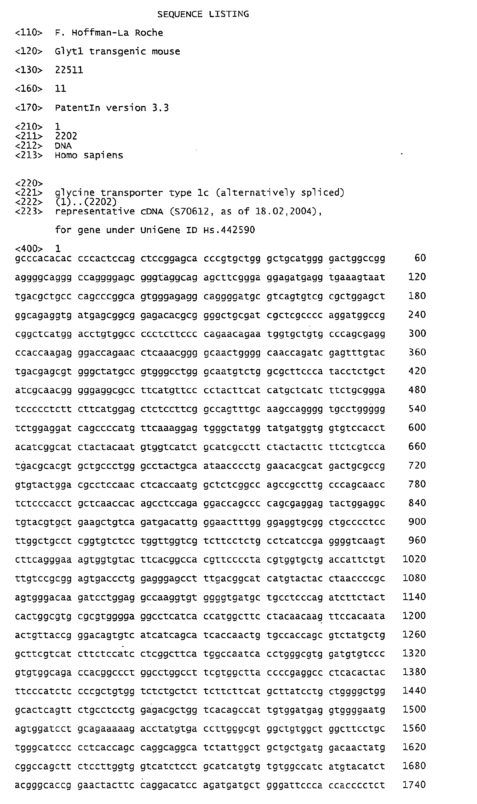
Based on sequence information from the published human glyt1b-cDNA-sequence
(note: the GLYT1b -sequence is published as sequence of 1c; SEQ. ID NO: 1) primers
(SEQ. ID NOs: 3 to 6)) were derived for cloning of the glyt1b-cDNA from a pACT2-cDNA
library of whole human brain (Clontech) by a nested PCR. The amplified cDNA
was subcloned into the NheI and EcoRI restriction-sites of the cloning vector pCI
(Promega; SEQ. ID NO: 10).
B) Cloning of hGlyt1b-transgene:
-
The human glyt1b cDNA was reamplified from the above vector using the primers
huGlyt1b-2147FLAG-PvuII-rev (SEQ. ID NO: 7) and huGlyt1b-234c (SEQ. ID NO: 8).
The amplicon was cut with PvuII, purified and cloned into the EcoRV-site of the vector
pNN265 (M. Mayford, E. Kandel, Columbia University, New York, USA; Choi, T., et al.,
Mol Cell Biol, 1991. 11(6): p. 3070-4) for the addition of introns and a polyA-sequence
resulting in the vector pNN265-hGlyt1b-FLAG. The cDNA-part was sequenced and
compared to the published sequence to verify the correct amplification by the used Pwo-polymerase.
No mutation was found.
-
To generate a forebrain-specific neuronal expression pattern of the transgene, the
hGlyt1b-cDNA was subsequently cloned into a vector containing the mouse CamKαII-promoter
(Mayford, M., et al., Science, 1996. 274(5293): p. 1678-83). For this, the
multicloning site of a p Bluescript II SK+ plasmid (Stratagene) was substituted with a
minimal cloning site containing the restriction-sites KpnI, HindIII and NotI, only. After
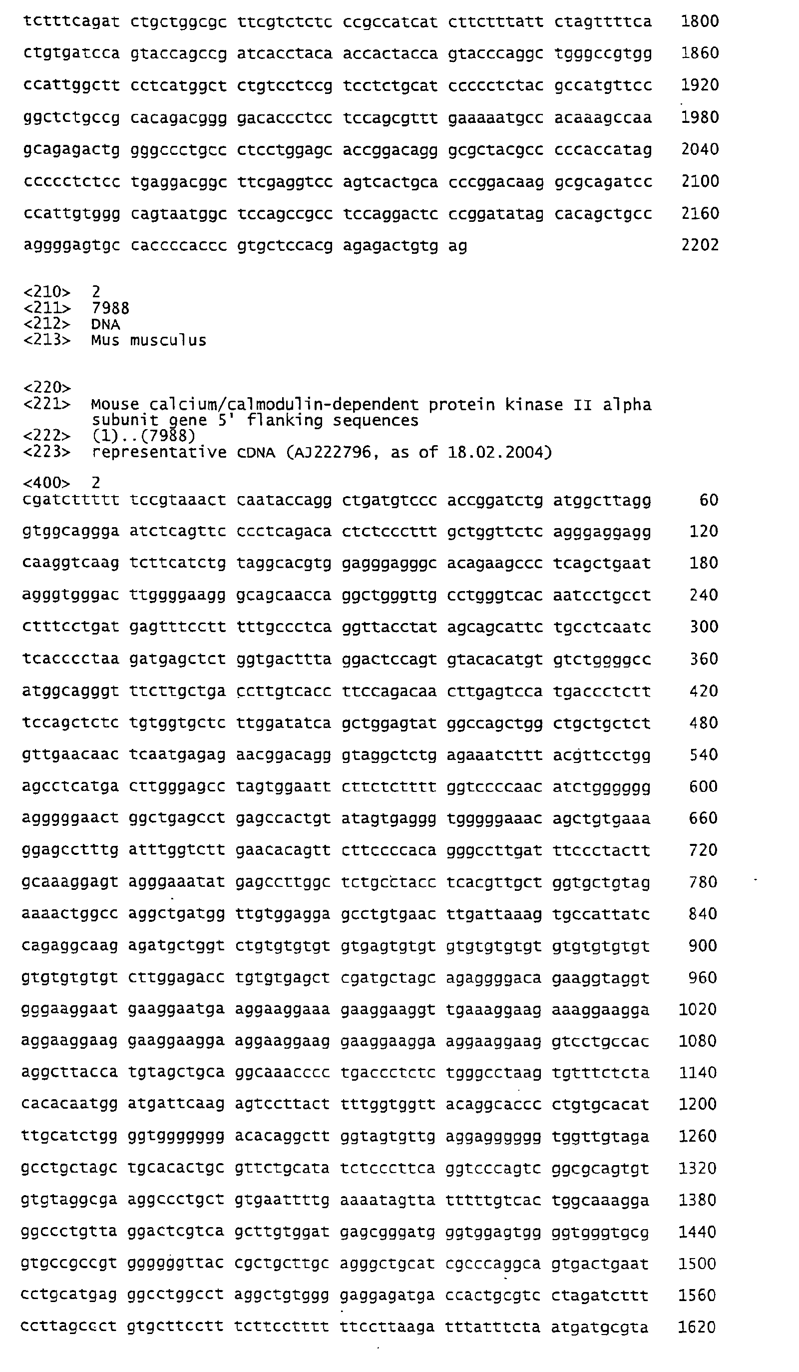
that, the promoter-cassette (SEQ. ID NO: 2) was removed from the vector pNN279
(Mayford, M., et al., Science, 1996. 274(5293): p. 1678-83) by NotI and HindIII and
cloned into the modified pBluescript II SK+ vector. The hGlyt1b-cDNA together with
surrounding introns was removed from pNN265-hGlyt1b-FLAG by NotI-digest and
cloned behind the mCamKαII-promoter into the unique NotI-site (Fig. 1).
C) Generation of transgenic mice:
-
The transgenic cassette was excised from the vector backbone by BssHII-digest and
purified. The DNA was injected into C57BL/6J zygotes (available from: The Jackson
Laboratory, 600 Main Street, Bar Harbor, Maine 04609 USA) at a concentration of 3
ng/µl to generate transgenic mice according to established procedures (Hogan, B.C., F;
Lacy, E, 1986, New York: Cold Spring Harbor Laboratory Press). Genomic DNA of
subsequent offspring was screened by PCR with the primers pNN279-7431c (SEQ. ID
NO: 9) and huGlyt1b-786nc (SEQ. ID NO: 10), which amplify a 1600 bp fragment of the
transgenic cassette, for the presence of the transgene (Fig. 2 and 3). Founders identified in
this screening were mated to C57BL/6J mice to establish the line.
Example 2: Molecular analysis of GLYT1b transgenic mice
A) Histological analysis of GLYT1b mutant mice
-
The overexpression of GLYT1b in the brains of mutant mice was confirmed by
immunohistochemistry and by Western blot analysis using the GLYT1-specific antibodies
raised in rabbits and guinea pigs.
B) Electrophysiological analysis of GLYT1b mutant mice
-
NMDA receptor activation is required for induction of certain forms of long term
potentiation (LTP) (Bliss, T.V. and G.L. Collingridge, Nature, 1993. 361(6407): p. 31-9).
Potentiation induced by theta burst stimulation in hippocampal slices of GLYT1b-transgenics
was compared with wild-type controls throughout the post-tetanus period as
described previously (Kew, J.N., et al., J Neurosci, 2000. 20(11): p. 4037-49). It was
determined whether GLYTlb-transgenic mice exhibit a different level of LTP-potentiation
compared to wild-type controls.
C) Preparation of forebrain and brainstem synaptosomes
-
Mice were sacrified and brain tissue were dissected on ice and subsequent
procedures performed at 4°C. Tissues were homogenized in 10 vol (w/v) of 10 mM Tris-HCl
pH 7.4 containing 0.32 M sucrose and 1 mM Pefabloc (cocktail of protease
inhibitors) (buffer A) using a glass/teflon homogeniser (800 rpm 10 times). The
homogenate was centrifuge 5 minutes at 1300 x g. The supernatant was carefully decanted
and kept on ice, while the pellet was suspended in 5 vol (of the original weight) of buffer
A, homogenised and centrifuged as described. The second supernatant was added to the
first and centrifuged at 17,000 x g for 20 minutes. The resulting pellet (crude
synaptosomal fraction) was suspended in 5 vol (of the original weight) of Krebs-Ringer
solution, pH 7.4 containing 10 mM glucose (KRB).
D) Glycine uptake
-
The assays were performed in 96-well plates. Aliquots of mice forebrain (0.1 mg)
and brainstem (0.05 mg) synaptosomal preparations were incubated at 22°C in KRB
together with 120 nM [3H] glycine in a total volume of 250 µl for 30 minutes. Incubation
was stopped by rapid filtration onto 96 well Packard GF/B unifilter plates, followed by 3
washes with ice-cold KRB. After addition of scintillation solution the radioactivity
content of the wells was measured.
E) Saturation binding for MK801
-
The assays were performed in 96-well deep plates. Aliquots of mice forebrain and
brainstem synaptosomal preparations (0.07 mg) were incubated at 22°C in 20 mM
Hepes-KOH, pH 7.4 containing 100 µM glutamate and 30µM glycine together with
increasing concentration(0.03nM- 300 nM) of [3H] MK801 in a total volume of 0.5 ml
for 1 hour. Non-specific binding was defined with 10 µM MK801. Incubation was
stopped by rapid filtration onto 96 well Packard GF/C unifilter plates, followed by 3
washes with ice-cold 20 mM of Hepes-KOH, pH 7.4. After addition of scintillation
solution the radioactivity content of the wells was measured.
F) Extracellular glycine levels in vivo
-
To assess the impact of increased GLYT1b expression on the extracellular level of
glycine a microdialysis study was performed. Adult wild-type and mutant mice were
anesthetized with isoflurane and a microdialysis vertical probe (CMA7 4/2, cuprophane -
membrane custome made) was inserted in the striatum (CPu, bregma A: +0.9; L: -1.8; V:
-4.6). The animals were allowed to recover for three to four days before the experiment.
Dialysate glycine levels were quantified according to the method of Smith and Sharp with
minor modification).
Example 3: Behavioral analysis of GLYT1 mutant mice
A) Neurological assessment
-
Neurological assessment includes a number of neurological tests like flexion
reflex, grip strength (g) and time (sec) spent on a rotarod at 16 and 32 rpm and body
weight.
B) Spontaneous behavior
-
The GLYT1b transgenic mice were observed for signs of natural exploratory
behavior including body posture, gait and sensory responses (Irwin, S.,
Psychopharmacologia, 1968. 13(3): p. 222-57). In addition, their spontaneous locomotor
activity was analyzed (activity box). Moreover, the state of anxiety was assessed by
exposing to the animals to naturally aversive stimuli (elevated plus maze test and the
light/dark choice test).
C) Auditory startle and prepulse inhibition of the acoustic startle reflex (behavior related
to schizophrenia)
-
Testing was conducted in eight startle devices each consisting of a Plexiglas cylinder
(5 cm in diameter) mounted on a Plexiglas platform in a ventilated sound attenuated
cubicle with a high-frequency loudspeaker producing all acoustic stimuli. The
background noise of each chamber was 68dB. Movements within the cylinder were
detected and transduced by a piezoelectric accelerometer attached to the Plexiglas base
and digitized and stored by a computer. Beginning at the stimulus onset, 65 x 1 msec
readings were recorded to obtain the animal's startle amplitude.
-
Each section was initiated with a 5 min acclimation period followed by five
successive 110 dB trials, which were not included in the analysis. Ten different trial types
were then presented: startle pulse alone (ST110, 110 dB/40 msec); eight different prepulse
trials in which either 20-msec-long 72, 78, 84, and 90 dB stimuli were presented alone
(P72, P78, P84, P90) or preceded the 110 dB pulse by 100 msec (PP72, PP78, PP84,
PP90); and finally one trial in which only the background noise was presented (NST) to
measure the baseline movements in the cylinder. All trials were presented in a
pseudorandom order, and the average intertrial interval (ITI) was 15 msec. The startle
data and percentage prepulse inhibition (PPI) were analyzed by two-ways ANOVA.
D) Nest building
-
To quantify the ability of mutant mice to make nests by shredding a tissue, folded
pieces of tissue paper were placed into each cage, and 24 hr later the nests were assessed.
E) Intracerebroventricular NMDA-induce convulsions.
-
Seizures were induced by injection of NMDA (5nM in 1 µl) into the lateral ventricle
of conscious mice. Immediately after injection, animals were placed in Plexiglas boxes
and observed for a period of 5 minutes. The latency (in seconds) for each mouse to
exhibit wild running phase and clonic convulsions was recorded for mutant mice versus
wild-type mice.
F) Behavior related to cognitive functions
Spatial short- and long-term memory
-
The delayed matching spatial working memory task was performed as described by
Durkin (Durkin, T.P., et al., Behav Brain Res, 2000. 116(1): p. 39-53). Working memory
was evaluated on the basis of the acquisition of a delayed matching rule in a 5-arm maze.
The basic learning task was comprised of two phases. Each trial begins with a presentation
phase during which the animal was exposed to a forced and rewarded visit to one arm
chosen quasi-randomly, the other four arms being closed. Once rewarded, the animal was
placed in a waiting cage. Following a retention interval of variable duration (2-sec, 20-sec
and 40-sec delay for working memory; 5-min, 1-hr, 4-hr, 24-hr delay for short- and long-term
memory), the retrieval test phase was performed during which the animal was
exposed to a situation of choice among the five open arms. A correct choice of the
previously visited arm was rewarded. The working memory retention capacity was
expressed as a function of the retention interval by the mean percent of correct accuracy
choices during successive trials with a fixed intertrial interval of 10 sec. The memory task
was monitored by an automated video-tracking system.
-
Alternatively spatial learning and memory was assessed in the water maze paradigm
described by Morris (Morris, R.G., et al., Nature, 1982. 297(5868): p. 681-3; Morris, R.G.,
et al., Nature, 1986. 319(6056): p. 774-6). Mice were placed in a circular pool (diameter
120 cm, height 30 cm) in which they learn to escape from milky water (20 cm depth, 20 ±
1°C) by locating a hidden platform. This target platform (7 cm diameter, 1 cm below the
water surface) was located in the center of a particular quadrant of the pool, and external
visual cues were positioned around the pool to facilitate navigation of the animals.
During a 4 d test period, mice were placed in the water facing the wall of the pool in one
of four fixed starting positions chosen randomly (3 trials per session, 3 sessions per day).
The time the mouse needs to locate the target (escape latency) and the swim path and
swim speed were measured using an automated video motility system. If an animal fails
to find the target within 60 seconds, it was placed on the platform by hand and was
allowed to remain there for an intertrial interval (10 to 20 sec). The interval between each
session was 1.5 to 2 hr. After the final trial on day 4, the platform was removed, and the
mice were allowed to swim freely for 60 sec. The time the mice spend in each quadrant
and their swim path were recorded.
Associative emotional memory
-
Associative emotional memory, which was NMDA receptor dependent, was
assessed in two behavioral tasks:
- i. Fear potentiated startle response: Mice were conditioned to respond to a light (CS)
which was paired with a footshock (aversive US). After a delay of 24 hours, emotional
memory was evaluated by the amplitude of the startle reflex elicited by different
acoustic stimuli (90-110 dB) with or without the conditioned light stimulus. The
presentation of the conditioned light stimulus leads to a potentiation of the acoustic
startle reflex (Davis, M., Psychopharmacology (Berl), 1979. 62(1): p. 1-7).
- ii. Contextual fear conditioning: Contextual fear conditioning was an implicit aversive
associative learning process by which an initially neutral context acquires aversive
properties after its repetitive association with an unconditioned aversive stimulus
(US). The animals were exposed to a new chamber where they were treated after few
minutes to successive electric foot shocks (US) that elicit unconditioned fear responses
(freezing behavior) (Phillips, R.G. and J.E. LeDoux, Behav Neurosci, 1992. 106(2): p.
274-85). Contextual fear conditioning was measured by the amount of freezing in
response to re-exposure to the context. The conditioned freezing response was tested
at different periods of time after training in order to evaluate short-term (1 to 3 hrs)
and long-term (1 to 10 days) contextual memory.
-
Conditioned fear extinction
-
Extinction of a learned fear response represents a form of behavioral plasticity that
was thought to rely on the formation of a new form of memory rather than an erasure of
the original learned association (Falls, W.A., M.J. Miserendino, and M. Davis, J Neurosci,
1992. 12(3): p. 854-63). Recently it has been shown that conditioned fear extinction
involves an NMDA receptor-dependent process (Tang, Y.P., et al., Nature, 1999.
401(6748): p. 63-9). Following a delay of 24-hr after training, extinction of conditioned
freezing can be evaluated by the time-dependent decrease of the amount of freezing in
response to repetitive exposure to the context during five consecutive days.
G) Pharmacologically disrupted behavior
-
A distinct range of schizophrenia-type symptoms, including hyperlocomotion,
deficits in prepulse inhibition (PPI) and memory deficit can be induced by the
administration of apomorphine, D-amphetamine or the non-competitive NMDA
receptor antagonist PCP. The minimally effective dose in disrupting behavior in wildtype
mice was used. It was then tested whether GLYT1b transgenic mice displayed a different
susceptibility to the pharmacological disruption of behavior.
Drug-induced hyperlocomotion
-
In the open field (activity box) the effect of D-amphetamine on locomotor activity
and stereotypic behavior was assessed by recording the horizontal locomotor activity,
vertical activity, stereotypic movements and the time spent in the center of the open field.
Drug-induced social withdrawal
-
To assess the effect of GLYT1b overexpression on the behavioral response to D-amphetamine
or PCP on social behavior a social exploration test was used (Crestani, F.,
F. Seguy, and R. Dantzer, Brain Res, 1991. 542(2): p. 330-5). An individually housed male
mouse was exposed for 5 minutes to a juvenile female mouse. Social interactions
including ano-genital and neck sniffing, heterogrooming and pursuits were recorded via a
video tracking system before and after drug administration. The drug-induced social
exploration deterioration was examined at different time intervals (30 min, 2 hours, 4
hours and 24 hours) after drug administration.
Drug-induced deficits in prepulse inhibition
-
Disruption of prepulse inhibition (PPI) by apomorphine or PCP was a frequently
used model of the sensorimotor gating deficits (Kretschmer, B.D., et al., Eur J Pharmacol,
1997. 331(2-3): p. 109-16; Bakshi, V.P. and M.A. Geyer, J Neurosci, 1998. 18(20): p.
8394-401; Bakshi, V.P., et al., J Pharmacol Exp Ther, 1999. 288(2): p. 643-52) by which
attention and sensorimotor gating can be evaluated. PPI was the attenuation of an
acoustic or tactile startle response following the presentation of a non-startle-inducing
prepulse stimulus. The animal's startle response following acoustic stimuli was assessed.
Drug-induced memory loss
-
PCP- or apomorphine-induced impairment of memory functions was assessed in
the delayed matching spatial memory task described above.
H) Reversal of in vitro and in vivo deficits/impairments by specific GLYT1 inhibitors in
GLYT1b mutant mice.
-
It was determined whether specific GLYT1 inhibitors were able to reverse
impairments/deficit observed in vitro (see example 2 paragraphs B, D and E) and in vivo
(see Example 2 paragraph F, Example 3, paragraphs A to F).
I) Reversal of in vitro and in vivo deficits/impairments by specific GLYT1 inhibitors in
pharmacologically challenged GLYT1b mutant mice.
-
It was determined whether specific GLYT1 inhibitors were able to reverse, in
GLYT1b mutant mice, their altered sensitivity to pharmacological disruption of behavior
(see example 3 paragraph G).