EP1572904B1 - Modified bace - Google Patents
Modified bace Download PDFInfo
- Publication number
- EP1572904B1 EP1572904B1 EP03709301A EP03709301A EP1572904B1 EP 1572904 B1 EP1572904 B1 EP 1572904B1 EP 03709301 A EP03709301 A EP 03709301A EP 03709301 A EP03709301 A EP 03709301A EP 1572904 B1 EP1572904 B1 EP 1572904B1
- Authority
- EP
- European Patent Office
- Prior art keywords
- bace
- polypeptide
- sequence
- seq
- pro
- Prior art date
- Legal status (The legal status is an assumption and is not a legal conclusion. Google has not performed a legal analysis and makes no representation as to the accuracy of the status listed.)
- Expired - Lifetime
Links
- 101150058765 BACE1 gene Proteins 0.000 title 1
- 101000894895 Homo sapiens Beta-secretase 1 Proteins 0.000 claims description 69
- 102100021257 Beta-secretase 1 Human genes 0.000 claims description 58
- 108090000765 processed proteins & peptides Proteins 0.000 claims description 58
- 102000004196 processed proteins & peptides Human genes 0.000 claims description 38
- 229920001184 polypeptide Polymers 0.000 claims description 34
- 108091033319 polynucleotide Proteins 0.000 claims description 30
- 102000040430 polynucleotide Human genes 0.000 claims description 30
- 239000002157 polynucleotide Substances 0.000 claims description 30
- 108010062466 Enzyme Precursors Proteins 0.000 claims description 23
- 102000010911 Enzyme Precursors Human genes 0.000 claims description 23
- 238000000034 method Methods 0.000 claims description 20
- 241000588724 Escherichia coli Species 0.000 claims description 19
- 239000013604 expression vector Substances 0.000 claims description 15
- XLYOFNOQVPJJNP-UHFFFAOYSA-N water Substances O XLYOFNOQVPJJNP-UHFFFAOYSA-N 0.000 claims description 14
- 102000044297 human BACE1 Human genes 0.000 claims description 12
- 239000002773 nucleotide Substances 0.000 claims description 12
- 125000003729 nucleotide group Chemical group 0.000 claims description 12
- FWMNVWWHGCHHJJ-SKKKGAJSSA-N 4-amino-1-[(2r)-6-amino-2-[[(2r)-2-[[(2r)-2-[[(2r)-2-amino-3-phenylpropanoyl]amino]-3-phenylpropanoyl]amino]-4-methylpentanoyl]amino]hexanoyl]piperidine-4-carboxylic acid Chemical compound C([C@H](C(=O)N[C@H](CC(C)C)C(=O)N[C@H](CCCCN)C(=O)N1CCC(N)(CC1)C(O)=O)NC(=O)[C@H](N)CC=1C=CC=CC=1)C1=CC=CC=C1 FWMNVWWHGCHHJJ-SKKKGAJSSA-N 0.000 claims description 8
- 238000004519 manufacturing process Methods 0.000 claims description 8
- 238000012986 modification Methods 0.000 claims description 6
- 230000004048 modification Effects 0.000 claims description 6
- 239000000203 mixture Substances 0.000 claims description 5
- 238000007865 diluting Methods 0.000 claims description 4
- 238000012258 culturing Methods 0.000 claims description 2
- 125000003275 alpha amino acid group Chemical group 0.000 claims 1
- 108090000623 proteins and genes Proteins 0.000 description 45
- 102000004169 proteins and genes Human genes 0.000 description 43
- 235000018102 proteins Nutrition 0.000 description 41
- 102000002659 Amyloid Precursor Protein Secretases Human genes 0.000 description 38
- 108010043324 Amyloid Precursor Protein Secretases Proteins 0.000 description 38
- 102000004190 Enzymes Human genes 0.000 description 30
- 108090000790 Enzymes Proteins 0.000 description 30
- 210000004027 cell Anatomy 0.000 description 30
- 230000000694 effects Effects 0.000 description 26
- 239000000758 substrate Substances 0.000 description 25
- 150000001413 amino acids Chemical group 0.000 description 24
- 238000003776 cleavage reaction Methods 0.000 description 19
- 230000007017 scission Effects 0.000 description 19
- 108020004705 Codon Proteins 0.000 description 17
- 210000003000 inclusion body Anatomy 0.000 description 17
- 235000001014 amino acid Nutrition 0.000 description 16
- 238000000746 purification Methods 0.000 description 15
- 208000024827 Alzheimer disease Diseases 0.000 description 14
- XSQUKJJJFZCRTK-UHFFFAOYSA-N Urea Chemical compound NC(N)=O XSQUKJJJFZCRTK-UHFFFAOYSA-N 0.000 description 14
- 102100022704 Amyloid-beta precursor protein Human genes 0.000 description 13
- DZHSAHHDTRWUTF-SIQRNXPUSA-N amyloid-beta polypeptide 42 Chemical compound C([C@@H](C(=O)N[C@@H](C)C(=O)N[C@@H](CCC(O)=O)C(=O)N[C@@H](CC(O)=O)C(=O)N[C@H](C(=O)NCC(=O)N[C@@H](CO)C(=O)N[C@@H](CC(N)=O)C(=O)N[C@@H](CCCCN)C(=O)NCC(=O)N[C@@H](C)C(=O)N[C@H](C(=O)N[C@@H]([C@@H](C)CC)C(=O)NCC(=O)N[C@@H](CC(C)C)C(=O)N[C@@H](CCSC)C(=O)N[C@@H](C(C)C)C(=O)NCC(=O)NCC(=O)N[C@@H](C(C)C)C(=O)N[C@@H](C(C)C)C(=O)N[C@@H]([C@@H](C)CC)C(=O)N[C@@H](C)C(O)=O)[C@@H](C)CC)C(C)C)NC(=O)[C@H](CC=1C=CC=CC=1)NC(=O)[C@@H](NC(=O)[C@H](CC(C)C)NC(=O)[C@H](CCCCN)NC(=O)[C@H](CCC(N)=O)NC(=O)[C@H](CC=1N=CNC=1)NC(=O)[C@H](CC=1N=CNC=1)NC(=O)[C@@H](NC(=O)[C@H](CCC(O)=O)NC(=O)[C@H](CC=1C=CC(O)=CC=1)NC(=O)CNC(=O)[C@H](CO)NC(=O)[C@H](CC(O)=O)NC(=O)[C@H](CC=1N=CNC=1)NC(=O)[C@H](CCCNC(N)=N)NC(=O)[C@H](CC=1C=CC=CC=1)NC(=O)[C@H](CCC(O)=O)NC(=O)[C@H](C)NC(=O)[C@@H](N)CC(O)=O)C(C)C)C(C)C)C1=CC=CC=C1 DZHSAHHDTRWUTF-SIQRNXPUSA-N 0.000 description 13
- 101710137189 Amyloid-beta A4 protein Proteins 0.000 description 12
- 101710151993 Amyloid-beta precursor protein Proteins 0.000 description 12
- 102000035195 Peptidases Human genes 0.000 description 12
- 108091005804 Peptidases Proteins 0.000 description 12
- 239000004365 Protease Substances 0.000 description 12
- 230000035772 mutation Effects 0.000 description 12
- 108010047320 Pepsinogen A Proteins 0.000 description 10
- 239000013615 primer Substances 0.000 description 10
- 235000019419 proteases Nutrition 0.000 description 10
- 238000003556 assay Methods 0.000 description 9
- RWSXRVCMGQZWBV-WDSKDSINSA-N glutathione Chemical compound OC(=O)[C@@H](N)CCC(=O)N[C@@H](CS)C(=O)NCC(O)=O RWSXRVCMGQZWBV-WDSKDSINSA-N 0.000 description 9
- DGVVWUTYPXICAM-UHFFFAOYSA-N β‐Mercaptoethanol Chemical compound OCCS DGVVWUTYPXICAM-UHFFFAOYSA-N 0.000 description 9
- 102000004580 Aspartic Acid Proteases Human genes 0.000 description 8
- 108010017640 Aspartic Acid Proteases Proteins 0.000 description 8
- DHMQDGOQFOQNFH-UHFFFAOYSA-N Glycine Chemical compound NCC(O)=O DHMQDGOQFOQNFH-UHFFFAOYSA-N 0.000 description 8
- 239000000047 product Substances 0.000 description 8
- 239000000523 sample Substances 0.000 description 8
- 239000013598 vector Substances 0.000 description 8
- 108010090849 Amyloid beta-Peptides Proteins 0.000 description 7
- 102000013455 Amyloid beta-Peptides Human genes 0.000 description 7
- KCXVZYZYPLLWCC-UHFFFAOYSA-N EDTA Chemical compound OC(=O)CN(CC(O)=O)CCN(CC(O)=O)CC(O)=O KCXVZYZYPLLWCC-UHFFFAOYSA-N 0.000 description 7
- 210000004556 brain Anatomy 0.000 description 7
- 239000000872 buffer Substances 0.000 description 7
- 239000004202 carbamide Substances 0.000 description 7
- 239000008188 pellet Substances 0.000 description 7
- 238000012545 processing Methods 0.000 description 7
- 230000002829 reductive effect Effects 0.000 description 7
- 239000000243 solution Substances 0.000 description 7
- ACERFIHBIWMFOR-UHFFFAOYSA-N 2-hydroxy-3-[(1-hydroxy-2-methylpropan-2-yl)azaniumyl]propane-1-sulfonate Chemical compound OCC(C)(C)NCC(O)CS(O)(=O)=O ACERFIHBIWMFOR-UHFFFAOYSA-N 0.000 description 6
- 108090000397 Caspase 3 Proteins 0.000 description 6
- KDXKERNSBIXSRK-UHFFFAOYSA-N Lysine Natural products NCCCCC(N)C(O)=O KDXKERNSBIXSRK-UHFFFAOYSA-N 0.000 description 6
- FAPWRFPIFSIZLT-UHFFFAOYSA-M Sodium chloride Chemical compound [Na+].[Cl-] FAPWRFPIFSIZLT-UHFFFAOYSA-M 0.000 description 6
- 230000003197 catalytic effect Effects 0.000 description 6
- 238000005119 centrifugation Methods 0.000 description 6
- 239000003638 chemical reducing agent Substances 0.000 description 6
- 102000004091 Caspase-8 Human genes 0.000 description 5
- 108090000538 Caspase-8 Proteins 0.000 description 5
- 108020004414 DNA Proteins 0.000 description 5
- 102100030875 Gastricsin Human genes 0.000 description 5
- 108090001072 Gastricsin Proteins 0.000 description 5
- 239000004472 Lysine Substances 0.000 description 5
- 108010076504 Protein Sorting Signals Proteins 0.000 description 5
- 210000004899 c-terminal region Anatomy 0.000 description 5
- 230000001413 cellular effect Effects 0.000 description 5
- 238000010790 dilution Methods 0.000 description 5
- 239000012895 dilution Substances 0.000 description 5
- VHJLVAABSRFDPM-QWWZWVQMSA-N dithiothreitol Chemical compound SC[C@@H](O)[C@H](O)CS VHJLVAABSRFDPM-QWWZWVQMSA-N 0.000 description 5
- 239000003550 marker Substances 0.000 description 5
- 239000000463 material Substances 0.000 description 5
- 230000002441 reversible effect Effects 0.000 description 5
- 102100029855 Caspase-3 Human genes 0.000 description 4
- 239000004471 Glycine Substances 0.000 description 4
- PXHVJJICTQNCMI-UHFFFAOYSA-N Nickel Chemical compound [Ni] PXHVJJICTQNCMI-UHFFFAOYSA-N 0.000 description 4
- 238000012408 PCR amplification Methods 0.000 description 4
- ONIBWKKTOPOVIA-UHFFFAOYSA-N Proline Natural products OC(=O)C1CCCN1 ONIBWKKTOPOVIA-UHFFFAOYSA-N 0.000 description 4
- 239000007983 Tris buffer Substances 0.000 description 4
- 230000001580 bacterial effect Effects 0.000 description 4
- 238000006243 chemical reaction Methods 0.000 description 4
- 210000004978 chinese hamster ovary cell Anatomy 0.000 description 4
- 239000002299 complementary DNA Substances 0.000 description 4
- 125000000151 cysteine group Chemical class N[C@@H](CS)C(=O)* 0.000 description 4
- 208000037265 diseases, disorders, signs and symptoms Diseases 0.000 description 4
- 229960003180 glutathione Drugs 0.000 description 4
- 230000008569 process Effects 0.000 description 4
- 238000002864 sequence alignment Methods 0.000 description 4
- LENZDBCJOHFCAS-UHFFFAOYSA-N tris Chemical compound OCC(N)(CO)CO LENZDBCJOHFCAS-UHFFFAOYSA-N 0.000 description 4
- WEVYAHXRMPXWCK-UHFFFAOYSA-N Acetonitrile Chemical compound CC#N WEVYAHXRMPXWCK-UHFFFAOYSA-N 0.000 description 3
- 208000037259 Amyloid Plaque Diseases 0.000 description 3
- PEDCQBHIVMGVHV-UHFFFAOYSA-N Glycerine Chemical compound OCC(O)CO PEDCQBHIVMGVHV-UHFFFAOYSA-N 0.000 description 3
- 239000006137 Luria-Bertani broth Substances 0.000 description 3
- 108091028043 Nucleic acid sequence Proteins 0.000 description 3
- 102000007056 Recombinant Fusion Proteins Human genes 0.000 description 3
- 108010008281 Recombinant Fusion Proteins Proteins 0.000 description 3
- 238000001042 affinity chromatography Methods 0.000 description 3
- 229960000723 ampicillin Drugs 0.000 description 3
- AVKUERGKIZMTKX-NJBDSQKTSA-N ampicillin Chemical compound C1([C@@H](N)C(=O)N[C@H]2[C@H]3SC([C@@H](N3C2=O)C(O)=O)(C)C)=CC=CC=C1 AVKUERGKIZMTKX-NJBDSQKTSA-N 0.000 description 3
- CKLJMWTZIZZHCS-REOHCLBHSA-L aspartate group Chemical group N[C@@H](CC(=O)[O-])C(=O)[O-] CKLJMWTZIZZHCS-REOHCLBHSA-L 0.000 description 3
- 239000003153 chemical reaction reagent Substances 0.000 description 3
- 238000010367 cloning Methods 0.000 description 3
- 239000013078 crystal Substances 0.000 description 3
- 238000001514 detection method Methods 0.000 description 3
- 239000003599 detergent Substances 0.000 description 3
- 239000012634 fragment Substances 0.000 description 3
- 230000002068 genetic effect Effects 0.000 description 3
- 229910052739 hydrogen Inorganic materials 0.000 description 3
- 239000001257 hydrogen Substances 0.000 description 3
- BPHPUYQFMNQIOC-NXRLNHOXSA-N isopropyl beta-D-thiogalactopyranoside Chemical compound CC(C)S[C@@H]1O[C@H](CO)[C@H](O)[C@H](O)[C@H]1O BPHPUYQFMNQIOC-NXRLNHOXSA-N 0.000 description 3
- 230000007246 mechanism Effects 0.000 description 3
- 229930182817 methionine Natural products 0.000 description 3
- 125000001360 methionine group Chemical group N[C@@H](CCSC)C(=O)* 0.000 description 3
- 210000002682 neurofibrillary tangle Anatomy 0.000 description 3
- 230000037361 pathway Effects 0.000 description 3
- 230000001105 regulatory effect Effects 0.000 description 3
- 230000000717 retained effect Effects 0.000 description 3
- 238000002741 site-directed mutagenesis Methods 0.000 description 3
- 239000011780 sodium chloride Substances 0.000 description 3
- 230000007928 solubilization Effects 0.000 description 3
- 238000005063 solubilization Methods 0.000 description 3
- YBJHBAHKTGYVGT-ZKWXMUAHSA-N (+)-Biotin Chemical compound N1C(=O)N[C@@H]2[C@H](CCCCC(=O)O)SC[C@@H]21 YBJHBAHKTGYVGT-ZKWXMUAHSA-N 0.000 description 2
- 206010059245 Angiopathy Diseases 0.000 description 2
- 101710150192 Beta-secretase 1 Proteins 0.000 description 2
- 201000010374 Down Syndrome Diseases 0.000 description 2
- 108010024636 Glutathione Proteins 0.000 description 2
- 108010053070 Glutathione Disulfide Proteins 0.000 description 2
- 108010002747 Pfu DNA polymerase Proteins 0.000 description 2
- 108010029485 Protein Isoforms Proteins 0.000 description 2
- 102000001708 Protein Isoforms Human genes 0.000 description 2
- 239000012614 Q-Sepharose Substances 0.000 description 2
- VMHLLURERBWHNL-UHFFFAOYSA-M Sodium acetate Chemical compound [Na+].CC([O-])=O VMHLLURERBWHNL-UHFFFAOYSA-M 0.000 description 2
- 206010044688 Trisomy 21 Diseases 0.000 description 2
- 238000005571 anion exchange chromatography Methods 0.000 description 2
- 238000006555 catalytic reaction Methods 0.000 description 2
- 238000004113 cell culture Methods 0.000 description 2
- 230000002490 cerebral effect Effects 0.000 description 2
- 210000001175 cerebrospinal fluid Anatomy 0.000 description 2
- 229960005091 chloramphenicol Drugs 0.000 description 2
- WIIZWVCIJKGZOK-RKDXNWHRSA-N chloramphenicol Chemical compound ClC(Cl)C(=O)N[C@H](CO)[C@H](O)C1=CC=C([N+]([O-])=O)C=C1 WIIZWVCIJKGZOK-RKDXNWHRSA-N 0.000 description 2
- 230000019771 cognition Effects 0.000 description 2
- 230000001149 cognitive effect Effects 0.000 description 2
- 239000000356 contaminant Substances 0.000 description 2
- 230000008878 coupling Effects 0.000 description 2
- 238000010168 coupling process Methods 0.000 description 2
- 238000005859 coupling reaction Methods 0.000 description 2
- 235000018417 cysteine Nutrition 0.000 description 2
- 230000001086 cytosolic effect Effects 0.000 description 2
- 230000006735 deficit Effects 0.000 description 2
- 238000004925 denaturation Methods 0.000 description 2
- 230000036425 denaturation Effects 0.000 description 2
- 230000008021 deposition Effects 0.000 description 2
- 238000011161 development Methods 0.000 description 2
- 230000018109 developmental process Effects 0.000 description 2
- 201000010099 disease Diseases 0.000 description 2
- 208000035475 disorder Diseases 0.000 description 2
- 238000010828 elution Methods 0.000 description 2
- 230000002255 enzymatic effect Effects 0.000 description 2
- 239000012467 final product Substances 0.000 description 2
- YPZRWBKMTBYPTK-BJDJZHNGSA-N glutathione disulfide Chemical compound OC(=O)[C@@H](N)CCC(=O)N[C@H](C(=O)NCC(O)=O)CSSC[C@@H](C(=O)NCC(O)=O)NC(=O)CC[C@H](N)C(O)=O YPZRWBKMTBYPTK-BJDJZHNGSA-N 0.000 description 2
- 230000006698 induction Effects 0.000 description 2
- 230000001939 inductive effect Effects 0.000 description 2
- 239000003112 inhibitor Substances 0.000 description 2
- 230000003993 interaction Effects 0.000 description 2
- 230000003902 lesion Effects 0.000 description 2
- 230000033001 locomotion Effects 0.000 description 2
- 239000012528 membrane Substances 0.000 description 2
- 210000002569 neuron Anatomy 0.000 description 2
- 229910052759 nickel Inorganic materials 0.000 description 2
- 102000039446 nucleic acids Human genes 0.000 description 2
- 108020004707 nucleic acids Proteins 0.000 description 2
- 150000007523 nucleic acids Chemical class 0.000 description 2
- 230000000750 progressive effect Effects 0.000 description 2
- 125000001500 prolyl group Chemical group [H]N1C([H])(C(=O)[*])C([H])([H])C([H])([H])C1([H])[H] 0.000 description 2
- 238000000926 separation method Methods 0.000 description 2
- 239000007974 sodium acetate buffer Substances 0.000 description 2
- 238000002415 sodium dodecyl sulfate polyacrylamide gel electrophoresis Methods 0.000 description 2
- 239000002904 solvent Substances 0.000 description 2
- 238000006467 substitution reaction Methods 0.000 description 2
- 239000006228 supernatant Substances 0.000 description 2
- 238000012360 testing method Methods 0.000 description 2
- 238000013518 transcription Methods 0.000 description 2
- 230000035897 transcription Effects 0.000 description 2
- 230000002103 transcriptional effect Effects 0.000 description 2
- VGIRNWJSIRVFRT-UHFFFAOYSA-N 2',7'-difluorofluorescein Chemical compound OC(=O)C1=CC=CC=C1C1=C2C=C(F)C(=O)C=C2OC2=CC(O)=C(F)C=C21 VGIRNWJSIRVFRT-UHFFFAOYSA-N 0.000 description 1
- UFBJCMHMOXMLKC-UHFFFAOYSA-N 2,4-dinitrophenol Chemical compound OC1=CC=C([N+]([O-])=O)C=C1[N+]([O-])=O UFBJCMHMOXMLKC-UHFFFAOYSA-N 0.000 description 1
- QKNYBSVHEMOAJP-UHFFFAOYSA-N 2-amino-2-(hydroxymethyl)propane-1,3-diol;hydron;chloride Chemical compound Cl.OCC(N)(CO)CO QKNYBSVHEMOAJP-UHFFFAOYSA-N 0.000 description 1
- 208000018282 ACys amyloidosis Diseases 0.000 description 1
- 108700028369 Alleles Proteins 0.000 description 1
- 208000000044 Amnesia Diseases 0.000 description 1
- 102000009091 Amyloidogenic Proteins Human genes 0.000 description 1
- 108010048112 Amyloidogenic Proteins Proteins 0.000 description 1
- 230000007351 Aβ plaque formation Effects 0.000 description 1
- 241000894006 Bacteria Species 0.000 description 1
- 125000001433 C-terminal amino-acid group Chemical group 0.000 description 1
- 102100035904 Caspase-1 Human genes 0.000 description 1
- 108090000426 Caspase-1 Proteins 0.000 description 1
- 208000005145 Cerebral amyloid angiopathy Diseases 0.000 description 1
- PJWWRFATQTVXHA-UHFFFAOYSA-N Cyclohexylaminopropanesulfonic acid Chemical compound OS(=O)(=O)CCCNC1CCCCC1 PJWWRFATQTVXHA-UHFFFAOYSA-N 0.000 description 1
- 238000007399 DNA isolation Methods 0.000 description 1
- 239000003155 DNA primer Substances 0.000 description 1
- 206010012289 Dementia Diseases 0.000 description 1
- 241000672609 Escherichia coli BL21 Species 0.000 description 1
- 241000620209 Escherichia coli DH5[alpha] Species 0.000 description 1
- 229910052693 Europium Inorganic materials 0.000 description 1
- 208000007487 Familial Cerebral Amyloid Angiopathy Diseases 0.000 description 1
- 241000282326 Felis catus Species 0.000 description 1
- 208000032849 Hereditary cerebral hemorrhage with amyloidosis Diseases 0.000 description 1
- 241000282412 Homo Species 0.000 description 1
- 101000823051 Homo sapiens Amyloid-beta precursor protein Proteins 0.000 description 1
- CKLJMWTZIZZHCS-REOHCLBHSA-N L-aspartic acid Chemical compound OC(=O)[C@@H](N)CC(O)=O CKLJMWTZIZZHCS-REOHCLBHSA-N 0.000 description 1
- FFEARJCKVFRZRR-BYPYZUCNSA-N L-methionine Chemical compound CSCC[C@H](N)C(O)=O FFEARJCKVFRZRR-BYPYZUCNSA-N 0.000 description 1
- QIVBCDIJIAJPQS-VIFPVBQESA-N L-tryptophane Chemical compound C1=CC=C2C(C[C@H](N)C(O)=O)=CNC2=C1 QIVBCDIJIAJPQS-VIFPVBQESA-N 0.000 description 1
- 108060001084 Luciferase Proteins 0.000 description 1
- 239000005089 Luciferase Substances 0.000 description 1
- 102220591686 Mediator of RNA polymerase II transcription subunit 13_P54K_mutation Human genes 0.000 description 1
- 125000001429 N-terminal alpha-amino-acid group Chemical group 0.000 description 1
- 125000000729 N-terminal amino-acid group Chemical group 0.000 description 1
- 208000009668 Neurobehavioral Manifestations Diseases 0.000 description 1
- 108010033276 Peptide Fragments Proteins 0.000 description 1
- 102000007079 Peptide Fragments Human genes 0.000 description 1
- 102000002933 Thioredoxin Human genes 0.000 description 1
- DTQVDTLACAAQTR-UHFFFAOYSA-N Trifluoroacetic acid Chemical compound OC(=O)C(F)(F)F DTQVDTLACAAQTR-UHFFFAOYSA-N 0.000 description 1
- QIVBCDIJIAJPQS-UHFFFAOYSA-N Tryptophan Natural products C1=CC=C2C(CC(N)C(O)=O)=CNC2=C1 QIVBCDIJIAJPQS-UHFFFAOYSA-N 0.000 description 1
- 238000002835 absorbance Methods 0.000 description 1
- 230000003213 activating effect Effects 0.000 description 1
- 238000001261 affinity purification Methods 0.000 description 1
- 230000002776 aggregation Effects 0.000 description 1
- 238000004220 aggregation Methods 0.000 description 1
- 230000032683 aging Effects 0.000 description 1
- 238000012870 ammonium sulfate precipitation Methods 0.000 description 1
- 230000003321 amplification Effects 0.000 description 1
- 230000003942 amyloidogenic effect Effects 0.000 description 1
- 238000000137 annealing Methods 0.000 description 1
- 238000013459 approach Methods 0.000 description 1
- 239000012062 aqueous buffer Substances 0.000 description 1
- 239000007864 aqueous solution Substances 0.000 description 1
- 125000004429 atom Chemical group 0.000 description 1
- 238000011888 autopsy Methods 0.000 description 1
- 230000003115 biocidal effect Effects 0.000 description 1
- 230000015572 biosynthetic process Effects 0.000 description 1
- 229960002685 biotin Drugs 0.000 description 1
- 235000020958 biotin Nutrition 0.000 description 1
- 239000011616 biotin Substances 0.000 description 1
- 210000004204 blood vessel Anatomy 0.000 description 1
- 229910021538 borax Inorganic materials 0.000 description 1
- 239000007853 buffer solution Substances 0.000 description 1
- 238000005277 cation exchange chromatography Methods 0.000 description 1
- 150000001768 cations Chemical class 0.000 description 1
- 230000006037 cell lysis Effects 0.000 description 1
- 239000000919 ceramic Substances 0.000 description 1
- 230000008859 change Effects 0.000 description 1
- 239000002738 chelating agent Substances 0.000 description 1
- 238000001311 chemical methods and process Methods 0.000 description 1
- 239000003795 chemical substances by application Substances 0.000 description 1
- 230000003920 cognitive function Effects 0.000 description 1
- 239000012141 concentrate Substances 0.000 description 1
- 239000003636 conditioned culture medium Substances 0.000 description 1
- 238000012790 confirmation Methods 0.000 description 1
- GLNDAGDHSLMOKX-UHFFFAOYSA-N coumarin 120 Chemical compound C1=C(N)C=CC2=C1OC(=O)C=C2C GLNDAGDHSLMOKX-UHFFFAOYSA-N 0.000 description 1
- 210000004748 cultured cell Anatomy 0.000 description 1
- 125000004122 cyclic group Chemical group 0.000 description 1
- 210000000805 cytoplasm Anatomy 0.000 description 1
- 210000005220 cytoplasmic tail Anatomy 0.000 description 1
- 230000034994 death Effects 0.000 description 1
- 238000000354 decomposition reaction Methods 0.000 description 1
- 238000013400 design of experiment Methods 0.000 description 1
- 238000000502 dialysis Methods 0.000 description 1
- VILAVOFMIJHSJA-UHFFFAOYSA-N dicarbon monoxide Chemical group [C]=C=O VILAVOFMIJHSJA-UHFFFAOYSA-N 0.000 description 1
- 239000000539 dimer Substances 0.000 description 1
- 238000009826 distribution Methods 0.000 description 1
- 238000007876 drug discovery Methods 0.000 description 1
- 239000000975 dye Substances 0.000 description 1
- 239000003623 enhancer Substances 0.000 description 1
- OGPBJKLSAFTDLK-UHFFFAOYSA-N europium atom Chemical compound [Eu] OGPBJKLSAFTDLK-UHFFFAOYSA-N 0.000 description 1
- 238000002474 experimental method Methods 0.000 description 1
- 239000007850 fluorescent dye Substances 0.000 description 1
- 239000012737 fresh medium Substances 0.000 description 1
- 230000006870 function Effects 0.000 description 1
- 102000038383 gamma-secretases Human genes 0.000 description 1
- 108091007739 gamma-secretases Proteins 0.000 description 1
- 239000001963 growth medium Substances 0.000 description 1
- 229960004198 guanidine Drugs 0.000 description 1
- PJJJBBJSCAKJQF-UHFFFAOYSA-N guanidinium chloride Chemical compound [Cl-].NC(N)=[NH2+] PJJJBBJSCAKJQF-UHFFFAOYSA-N 0.000 description 1
- ZJYYHGLJYGJLLN-UHFFFAOYSA-N guanidinium thiocyanate Chemical compound SC#N.NC(N)=N ZJYYHGLJYGJLLN-UHFFFAOYSA-N 0.000 description 1
- 238000003306 harvesting Methods 0.000 description 1
- 238000004128 high performance liquid chromatography Methods 0.000 description 1
- 230000007062 hydrolysis Effects 0.000 description 1
- 238000006460 hydrolysis reaction Methods 0.000 description 1
- 230000002209 hydrophobic effect Effects 0.000 description 1
- 229910052588 hydroxylapatite Inorganic materials 0.000 description 1
- 238000001727 in vivo Methods 0.000 description 1
- 230000002779 inactivation Effects 0.000 description 1
- 238000011534 incubation Methods 0.000 description 1
- 230000005764 inhibitory process Effects 0.000 description 1
- 239000002054 inoculum Substances 0.000 description 1
- 238000003780 insertion Methods 0.000 description 1
- 230000037431 insertion Effects 0.000 description 1
- 230000003834 intracellular effect Effects 0.000 description 1
- 238000002955 isolation Methods 0.000 description 1
- 238000005304 joining Methods 0.000 description 1
- IXHBTMCLRNMKHZ-LBPRGKRZSA-N levobunolol Chemical compound O=C1CCCC2=C1C=CC=C2OC[C@@H](O)CNC(C)(C)C IXHBTMCLRNMKHZ-LBPRGKRZSA-N 0.000 description 1
- 230000000670 limiting effect Effects 0.000 description 1
- 238000004811 liquid chromatography Methods 0.000 description 1
- 230000004807 localization Effects 0.000 description 1
- 231100000863 loss of memory Toxicity 0.000 description 1
- 125000003588 lysine group Chemical class [H]N([H])C([H])([H])C([H])([H])C([H])([H])C([H])([H])C([H])(N([H])[H])C(*)=O 0.000 description 1
- 230000002934 lysing effect Effects 0.000 description 1
- 230000014759 maintenance of location Effects 0.000 description 1
- 230000010534 mechanism of action Effects 0.000 description 1
- 108020004999 messenger RNA Proteins 0.000 description 1
- 230000003278 mimic effect Effects 0.000 description 1
- 239000003068 molecular probe Substances 0.000 description 1
- 238000002703 mutagenesis Methods 0.000 description 1
- 231100000350 mutagenesis Toxicity 0.000 description 1
- 210000004897 n-terminal region Anatomy 0.000 description 1
- 208000015122 neurodegenerative disease Diseases 0.000 description 1
- 238000003199 nucleic acid amplification method Methods 0.000 description 1
- 230000000269 nucleophilic effect Effects 0.000 description 1
- 230000003287 optical effect Effects 0.000 description 1
- 230000003647 oxidation Effects 0.000 description 1
- 238000007254 oxidation reaction Methods 0.000 description 1
- YPZRWBKMTBYPTK-UHFFFAOYSA-N oxidized gamma-L-glutamyl-L-cysteinylglycine Natural products OC(=O)C(N)CCC(=O)NC(C(=O)NCC(O)=O)CSSCC(C(=O)NCC(O)=O)NC(=O)CCC(N)C(O)=O YPZRWBKMTBYPTK-UHFFFAOYSA-N 0.000 description 1
- 230000008506 pathogenesis Effects 0.000 description 1
- 230000001575 pathological effect Effects 0.000 description 1
- XYJRXVWERLGGKC-UHFFFAOYSA-D pentacalcium;hydroxide;triphosphate Chemical compound [OH-].[Ca+2].[Ca+2].[Ca+2].[Ca+2].[Ca+2].[O-]P([O-])([O-])=O.[O-]P([O-])([O-])=O.[O-]P([O-])([O-])=O XYJRXVWERLGGKC-UHFFFAOYSA-D 0.000 description 1
- 230000000737 periodic effect Effects 0.000 description 1
- 230000001323 posttranslational effect Effects 0.000 description 1
- 239000002244 precipitate Substances 0.000 description 1
- 239000002243 precursor Substances 0.000 description 1
- 239000012460 protein solution Substances 0.000 description 1
- 230000017854 proteolysis Effects 0.000 description 1
- 230000002797 proteolythic effect Effects 0.000 description 1
- 238000012207 quantitative assay Methods 0.000 description 1
- 230000002285 radioactive effect Effects 0.000 description 1
- 238000009790 rate-determining step (RDS) Methods 0.000 description 1
- 239000011541 reaction mixture Substances 0.000 description 1
- 230000010076 replication Effects 0.000 description 1
- 239000011347 resin Substances 0.000 description 1
- 229920005989 resin Polymers 0.000 description 1
- 230000000284 resting effect Effects 0.000 description 1
- 108091008146 restriction endonucleases Proteins 0.000 description 1
- 210000003705 ribosome Anatomy 0.000 description 1
- 150000003839 salts Chemical class 0.000 description 1
- 230000003248 secreting effect Effects 0.000 description 1
- 230000001953 sensory effect Effects 0.000 description 1
- 238000001542 size-exclusion chromatography Methods 0.000 description 1
- 235000017281 sodium acetate Nutrition 0.000 description 1
- 239000001632 sodium acetate Substances 0.000 description 1
- -1 sodium chloride Chemical class 0.000 description 1
- 235000010339 sodium tetraborate Nutrition 0.000 description 1
- 238000000527 sonication Methods 0.000 description 1
- 125000006850 spacer group Chemical group 0.000 description 1
- 238000003860 storage Methods 0.000 description 1
- 239000000126 substance Substances 0.000 description 1
- 230000001225 therapeutic effect Effects 0.000 description 1
- 108060008226 thioredoxin Proteins 0.000 description 1
- 229940094937 thioredoxin Drugs 0.000 description 1
- 238000013519 translation Methods 0.000 description 1
- BSVBQGMMJUBVOD-UHFFFAOYSA-N trisodium borate Chemical compound [Na+].[Na+].[Na+].[O-]B([O-])[O-] BSVBQGMMJUBVOD-UHFFFAOYSA-N 0.000 description 1
- 108010087967 type I signal peptidase Proteins 0.000 description 1
- 229940045136 urea Drugs 0.000 description 1
- 230000002792 vascular Effects 0.000 description 1
- 238000012800 visualization Methods 0.000 description 1
- 238000005406 washing Methods 0.000 description 1
Images


Classifications
-
- C—CHEMISTRY; METALLURGY
- C12—BIOCHEMISTRY; BEER; SPIRITS; WINE; VINEGAR; MICROBIOLOGY; ENZYMOLOGY; MUTATION OR GENETIC ENGINEERING
- C12N—MICROORGANISMS OR ENZYMES; COMPOSITIONS THEREOF; PROPAGATING, PRESERVING, OR MAINTAINING MICROORGANISMS; MUTATION OR GENETIC ENGINEERING; CULTURE MEDIA
- C12N9/00—Enzymes; Proenzymes; Compositions thereof; Processes for preparing, activating, inhibiting, separating or purifying enzymes
- C12N9/14—Hydrolases (3)
- C12N9/48—Hydrolases (3) acting on peptide bonds (3.4)
- C12N9/50—Proteinases, e.g. Endopeptidases (3.4.21-3.4.25)
- C12N9/64—Proteinases, e.g. Endopeptidases (3.4.21-3.4.25) derived from animal tissue
- C12N9/6421—Proteinases, e.g. Endopeptidases (3.4.21-3.4.25) derived from animal tissue from mammals
- C12N9/6478—Aspartic endopeptidases (3.4.23)
Definitions
- the invention is related to a recombinant human BACE. More particularly, the invention is related to an active human BACE having a modification at amino acid position 33.
- AD Alzheimer's disease
- Clinical presentation of AD is characterized by loss of memory, cognition, reasoning, judgment, and orientation. As the disease progresses, motor, sensory, and linguistic abilities are also affected until there is global impairment of multiple cognitive functions. These cognitive losses occur gradually, but typically lead to severe impairment and eventual death in the range of four to twelve years.
- Alzheimer's disease is characterized by two major pathologic observations in the brain: neurofibrillary tangles and beta amyloid (or neuritic) plaques, comprised predominantly of an aggregate of a peptide fragment know as A beta.
- Individuals with AD exhibit characteristic beta-amyloid deposits in the brain (beta amyloid plaques) and in cerebral blood vessels (beta amyloid angiopathy) as well as neurofibrillary tangles.
- Neurofibrillary tangles occur not only in Alzheimer's disease but also in other dementia-inducing disorders. On autopsy, large numbers of these lesions are generally found in areas of the human brain important for memory and cognition.
- Amyloidogenic plaques and vascular amyloid angiopathy also characterize the brains of individuals with Trisomy 21 (Down's Syndrome), Hereditary Cerebral Hemorrhage with Amyloidosis of the Dutch-Type (HCHWA-D), and other neurogenerative disorders.
- Beta-amyloid is a defining feature of AD, now believed to be a causative precursor or factor in the development of the disease. Deposition of A beta in areas of the brain responsible for cognitive activities is a major factor in the development of AD. Beta-amyloid plaques are predominantly composed of amyloid beta peptide (A beta, also sometimes designated betaA4).
- a beta peptide is derived by proteolysis of the amyloid precursor protein (APP) and is comprised of 39-42 amino acids. Several proteases called secretases are involved in the processing of APP.
- Cleavage of APP at the N-terminus of the A beta peptide by beta-secretase and at the C-terminus by one or more gamma-secretases constitutes the beta-amyloidogenic pathway, i.e. the pathway by which A beta is formed.
- Cleavage of APP by alpha-secretase produces alpha-sAPP, a secreted form of APP that does not result in beta-amyloid plaque formation. This alternate pathway precludes the formation of A beta peptide.
- a description of the proteolytic processing fragments of APP is found, for example, in U.S. Patent Nos. 5,441,870 ; 5,721,130 ; and 5,942,400 .
- beta-secretase enzyme has been identified as the enzyme responsible for processing of APP at the beta-secretase cleavage site.
- the beta-secretase enzyme has been disclosed using varied nomenclature, including BACE, Asp, and Memapsin. See, for example, Sinha et.al., 1999, Nature 402:537-554 (p501 ) and published PCT application WO00/17369 .
- beta-amyloid peptide plays a seminal role in the pathogenesis of AD and can precede cognitive symptoms by years or decades. See, for example, Selkoe, 1991, Neuron 6:487 . Release of A beta from neuronal cells grown in culture and the presence of A beta in cerebrospinal fluid (CSF) of both normal individuals and AD patients has been demonstrated. See, for example, Seubert et al., 1992, Nature 359:325-327 .
- a beta peptide accumulates as a result of APP processing by beta-secretase, thus inhibition of this enzyme's activity is desirable for the treatment of AD.
- In vivo processing of APP at the beta-secretase cleavage site is thought to be a rate-limiting step in A beta production, and is thus a therapeutic target for the treatment of AD. See for example, Sabbagh, M., et al., 1997, Alz. Dis. Rev. 3, 1-19 .
- the present invention relates to an isolated polypeptide sequence comprising human BACE having the modification Pro33Lys.
- the invention also relates to a composition comprising an active human BACE enzyme comprising the pro-enzyme sequence of BACE having the modification Pro33Lys.
- the invention relates to an isolated polypeptide of SEQ ID NO: 2.
- the invention relates to an isolated polynucleotide comprising a polynucleotide sequence which, by virtue of the degeneracy of the genetic code, encodes P33K-BACE.
- the P33K-BACE can have the sequence of SEQ ID NO: 2.
- the isolated polynucleotide can comprise the nucleotide sequence of nucleotides 70 - 1365 of SEQ ID NO: 8.
- the invention also relates to an expression vector comprising the polynucleotide sequence encoding P33K-BACE.
- the expression vector produces a P33K-BACE polypeptide when said expression vector is present in a compatible host cell.
- the expression vector can comprise the polypeptide sequence of SEQ ID NO: 2.
- a recombinant host cell comprising the expression vector having the polynucleotide sequence encoding P33K-BACE.
- a method for producing a P33K-BACE polypeptide comprising culturing the recombinant host cell having an expression vector encoding P33K-BACE under conditions sufficient for the production of said polypeptide and recovering the polypeptide from the culture.
- the host cell can be E. Coli.
- a method of producing active P33K-BACE comprising recovering the P33K-BACE from the culture of host cells according and diluting the polypeptide 20-50 fold with water having a temperature of about 1 to 15° C.
- FIG. 1 shows the amino acid sequence of human BACE [SEQ ID NO: 1]
- FIG. 2 shows the amino acid sequence of an embodiment of the P33K-BACE polypeptide [SEQ ID NO: 2].
- FIG. 3 shows a sequence alignment of ⁇ -secretase zymogen (pbsz) [SEQ ID NO: 3], ⁇ -secretase (1fkn) [SEQ ID NO: 4], progastricsin (1htr) [SEQ ID NO: 5] and pepsinogen (3psg) [SEQ ID NO: 6].
- the lines indicate the residue pair involved in forming disulfide bond as observed in 1 fkn.
- the codes representing the conserved residues at the active site for the aspartyl protease family are residues 93-95 and residues 289-291 for pbsz , residues 91-93 and residues 274-276 for 3psg, and residues 91-93 and residues 276-278 for 1htr.
- the signal peptide segments (residues 1-21 for pbsz, residues 1-16 for 1htr, and residues 1-15 for 3psg) were not included for the alignment operation because they will be cleaved off by signal peptidase during the secretory process.
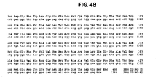
- FIGS. 4A and 4B are the DNA and predicted amino acid sequence of the modified recombinant BACE expressed from a pET11a-P33K-BACE construct: amino acid sequence [SEQ ID NO: 7]; DNA sequence [SEQ ID NO: 8].
- BACE beta-site APP-cleaving enzyme
- Asp2 beta-secretase
- Memapsin 2 beta-secretase 2
- BACE has been described, for example, in WO 00/17369 , WO 00/47618 and WO 01/23533 , .
- BACE comprises an aspartyl protease and contains the classical consensus aspartyl protease active site motif (DTG/DSG).
- FIG. 1 Features of the human BACE polypeptide shown in FIG. 1 include a 21 amino acid leader (signal or pre-) sequence shown in italics, and a 24 amino acid pro-sequence, shown in bold type. T 1 marks the start of the pro-sequence. A 27 amino acid transmembrane domain is underlined, and is followed by the cytosolic C-terminal tail. Disulphide bridges are formed by cysteines (Cys 195 -Cys 399 , Cys 257 -Cys 422 ; and Cys 309 -Cys 359 ).
- Pro33Lys-BACE refers to the enzyme including the polypeptide sequence of recombinant human BACE having a proline to lysine mutation at position 33 as shown in FIG. 2 . Accordingly, when appropriately refolded, recombinant P33K-BACE is an active BACE enzyme including a peptide sequence of at least amino acids 1-432 of BACE with the P33K mutation.
- BACE zymogen refers to the BACE which includes the 24 amino acid pro-sequence.
- an enzyme will be inactivated by the existence of its pro-peptide.
- the existence of the pro-peptide does not seem to have a significant impact on the activity of BACE.
- the three dimensional structure of BACE was elucidated. From the three dimensional structure, it has been possible to explore possible mutations in BACE which will inhibit enzyme activity.
- a sequence alignment was performed for the ⁇ -secretase zymogen as disclosed by Vassar et al., 1999, Science 286:735-741 (abbreviated as pbsz) [SEQ ID NO: 3], ⁇ -secretase (1fkn) [SEQ ID NO: 4], progastricsin (1htr) [SEQ ID NO: 5], and pepsinogen (3psg) [SEQ ID NO: 6] was performed using the PILEUP program in the GCG package (Genetic Computer Group, Madison, Wisconsin).
- Pepsinogen and progastricsin are pro-enzymes in the family of aspartyl proteases that includes ⁇ -secretase.
- the aligned result is given in FIG. 3 , where the pro-peptides are underlined.
- the signal peptide includes residues 1-21 (not shown)
- the pro-peptide includes residues 22- 45 ( Bennett, et al, 2000, Journal of Biological Chemistry 275:37712-37717 )
- the main-chain includes residues 46-446 (with the active site aspartates at 93 and 289).
- the transmembrane and intracellular domains that follow residue 446 are outside the scope of the study.
- the 3-D model of the ⁇ -secretase zymogen (pbsz) was constructed from (i) the X-ray coordinates of the protease domain of ⁇ -secretase (1fkn.pdb) recently determined by Hong et al., 2000, Science 290:150-153 , and (ii) the X-ray coordinates of pepsinogen (3psg.pdb) determined by Hartsuck et al, 1992, PROTEINS: Structure, Function and Genetics 13:1-25 .
- the pro-segment of the pepsinogen structure provided the basis for a homology model of the ⁇ -secretase pro-segment, which was grafted onto the ⁇ -secretase protease domain, using the procedure described below.
- the template structure 3psg.pdb was superimposed onto the template structure 1fkn.pdb, using the commercial software package, MOE (Chemical Computing Ltd.). During the superimposition process the entire structure of 3psg underwent a translational and rotational motion, and hence the coordinates of 3psg changed, although the coordinates of 1 fkn remained unchanged.
- the segment matching approach employs a database of known protein structures to build an unknown target structure based on an amino acid sequence alignment.
- the target structure was the ⁇ -secretase zymogen, i.e. the pro-segment plus the protease domain of ⁇ -secretase.
- the target structure was first broken into a set of short segments.
- the database was then searched for matching segments on the basis of amino acid sequence similarity and compatibility with the target structure. The process was repeated 10 times and an average model was generated, followed by energy minimization of the entire pro-enzyme to create the final model.
- the structure thus obtained uniquely defined the atomic coordinates of not only residues 22-45, the pro-segment of pbsz, but also the segment of residues 46-55 in the main chain that was missing in the crystal structure of 1fkn.pdb (Hong et al., 2000). Furthermore, although the majority of the protease domain (residues 75-446) of pbsz was almost identical to the corresponding sequence in 1 fkn, a small transition-linking part of the protease domain (residues 56-74) was affected owing to the existence of the pro-segment.
- the backbone traces a path from one end of the active site cleft, toward the center, then covers over the "flap" of the active site as described in Hong et al., 2000. It then continues toward the far end of the active site, makes a turn, and returns via two helices to near its origination point.
- Its overall structure is somewhat similar to the pepsinogen pro-segment from which it was derived, but with a key difference described below.
- An overlay of the ⁇ -secretase crystal structure with the pro-enzyme model shows some differences in side chain positioning induced by the presence of the pro-segment, and very minor differences in distal positioning, likely due to the energy minimization.
- Inactivation of an enzyme by its pro-peptide is generally thought to be due to physical blockage of the catalytic site, preventing access to substrate.
- a pro-segment could also disrupt the catalytically-required water molecule between the two aspartates.
- a comparison of the 3-D structures of pepsinogen, pro-gastricsin, and the ⁇ -secretase pro-enzyme model indicates that the pro-segments of all three cover up the catalytic site, and therefore should block access to substrate.
- the dynamics of protein motion could allow periodic unfolding of the pro-segments exposing the catalytic clefts to enable substrate processing. Yet only for the ⁇ -secretase pro-enzyme is substrate processing known to occur, so there is something unique about the positioning of its pro-segment.
- the catalytic reaction involves (i) the ⁇ -carboxyl groups of the two Asp residues (i.e., Asp-93 and Asp-289 for the case of ⁇ -secretase) at the active site being brought in to close proximity to activate a water molecule by forming hydrogen bonds with it; (ii) the nucleophilic attack of the activated water molecule on the carbonyl carbon atom of the scissile peptide bond to form the tetrahedral intermediate; (iii) the decomposition of the tetrahedral intermediate to yield the product of cleaved peptides and active enzyme. Accordingly, before a peptide bond is cleaved by an aspartyl protease, the two Asp residues at the active site must first activate a water molecule by forming four hydrogen bonds with it.
- the two Asp residues at the active site must first activate a water molecule by forming four hydrogen bonds with it.
- the microenvironment is much different in the ⁇ -secretase zymogen model, where no salt bridges are observed between the pro-peptide segment and the two active site Asp residues, i.e. , Asp-93 and Asp-289 ( FIG. 2 ).
- Pro-54 that corresponds to the Lys locations in the other two pro-enzyme structures from both sequence alignment ( FIG. 2 ) and 3-D structure.
- the numbering of the BACE (pbsz) in FIG. 2 assumes a 21 amino acid signal sequence
- Pro-54 in FIG. 2 is the same residue as Pro-33 in FIG. 1 .
- a proline side-chain cannot form a salt bridge.
- the "locking" mechanism is absent and there is no pro-segment side-chain in the location of the catalytic water position.
- the P33K-BACE has essentially the same activity of BACE. This suggests that confirmation of the hypothesis by experimental modification of the BACE pro-segment would need to involve more than just the Pro33Lys mutation, to include one or more additional residues that would enable the nearby pro-segment backbone to more closely mimic those of the comparator pro-enzymes.
- Recombinant BACE including recombinant P33K-BACE
- the construct can also contain additional nucleotide sequences that may, for example, assist in purification or expression of the recombinant polypeptide, as desired.
- the polynucleotide construct for expressing P33K-BACE may include nucleotides coding for the signal peptide, the transmembrane domain and/or the c-terminal tail or portions thereof. Such constructs may be assembled using routine methods by those skilled in the art.
- the complete polynucleotide sequence of BACE may be found, for example in Vassar et al, Science 286:7353-741 (1999 ) and the PCT publications that have been incorporated by reference herein.
- GenBank Accesion No. NM 012104 describes a number of known alleles of the BACE sequence.
- polypeptide sequence of BACE may be expressed from a vast number of polynucleotide sequences.
- the present invention is directed to any polynucleotide sequence encoding P33K-BACE.
- inclusion bodies When expressed in E. coli, recombinant P33K-BACE accumulates intracellularly in an insoluble form, resulting in phase-bright inclusions in the cytoplasm (inclusion bodies).
- the protein in the inclusion bodies can be a mixture of monomeric and multimeric forms of the protein, both reduced and oxidized.
- Processes designed to recover biologically active, soluble protein from the insoluble cellular material generally include the steps of: (1) cell lysis, (2) isolation of inclusion bodies, (3) solubilization of protein from inclusion bodies, (4) refolding of solubilized protein, and (5) purification of the active protein. Each of these steps will be described in relation to the invention below.
- Useful constructs for the production of P33K-BACE are designed to express a selected portion of the P33K-BACE polypeptide.
- the polynucleotide encoding the P33K-BACE polypeptide can be operably linked to suitable transcriptional or translational regulatory sequences in an expression construct. Regulatory sequences include transcriptional promoters, operators, enhancers, mRNA ribosomal binding sites, and other sequences that control transcription or translation.
- Nucleotide sequences are "operably linked" when the regulatory sequence functionally relates to the polynucleotide encoding P33K-BACE.
- a promoter nucleotide sequence is operably linked to a polynucleotide encoding P33K-BACE if the promoter nucleotide sequence directs the transcription of the P33K-BACE sequence.
- an expression vector will include a selectable marker and an origin of replication, for propagation in E. coli.
- Expression vectors generally comprise one or more phenotypic selectable marker genes. Such genes generally encode, for example, a protein that confers antibiotic resistance or that supplies an auxotrophic requirement.
- a polynucleotide can encode a P33K-BACE polypeptide having an N-terminal methionine to facilitate expression of the recombinant polypeptide in a prokaryotic host, for example, for expression in E. coli.
- the N-terminal methionine can optionally be cleaved from the expressed P33K-BACE polypeptide.
- the polynucleotide can also encode other N-terminal amino acids added to the P33K-BACE polypeptide that facilitate expression in E. coli.
- Such amino acids include, but are not limited to, a T7 leader sequence, a T7-caspase 8 leader sequence, and known tags for purification such as the T7-Tag MASMTGGQQMGR [SEQ ID NO: 9] that allows binding of antibodies, or a six-histidine tag (His) 6 that allows purification by binding to nickel.
- T7-Tag MASMTGGQQMGR [SEQ ID NO: 9] that allows binding of antibodies
- His six-histidine tag
- Other useful peptide tags include the thioredoxin tag, hemaglutinin tag, and GST tag. These and other amino acid tags can be encoded by polynucleotides added to either terminus of the polynucleotide encoding P33K-BACE.
- the polynucleotide of the expression construct can encode a P33K-BACE polypeptide that is truncated by removal of all or a portion of the C-terminal cytoplasmic tail, the transmembrane domain, the membrane proximal region, or any combination of these.
- the expression constructs can also encode cleavage sites for selected enzymes, to improve purification of the expressed protein or to assist in expression of the enzyme, when desired.
- active recombinant BACE protein can terminate at S 432 , lacking the transmembrane domain and cytosolic tail region. This provides BACE in a soluble form, that is, a form that is not membrane-bound. Accordingly, in the following examples, P33K-BACE was terminated at S 432 to compare activity with the known active recombinant BACE.
- one or more codon of the polynucleotide sequence encoding P33K-BACE can be modified, using such techniques as site directed mutagenesis, to eliminate GC-rich regions of strong secondary structure known to interfere with efficient cloning or expression of the recombinant protein. Codons can also be optimized for expression in E. coli, for example, according to published codon preferences. Underlined nucleotides in FIG. 4A show preferred codon changes.
- An expression construct containing a polynucleotide encoding P33K-BACE can be used to transform bacteria, for example E.coli, in order to produce P33K-BACE protein. Production of the protein can be inducible or constitutive, depending upon the control elements provided in the vectors.
- expression constructs are transfected into a bacterial host, such as E.coli BL21 codon plus (DE3) RP (Stratagene) and grown in suitable media, such as Luria broth supplemented with 100 micrograms/ml ampicillin and 34 micrograms/ml chloromphenicol.
- suitable media such as Luria broth supplemented with 100 micrograms/ml ampicillin and 34 micrograms/ml chloromphenicol.
- the T7 or T5 lac promoter promotes expression of the operably linked P33JK-BACE polynucleotide upon addition of IPTG (for example, to a final concentration of about 1 mM) to the culture media. After induction, for example, about three hours, the cell pellet is collected and can be stored, generally at -70° C, for later enzyme purification.
- the expressed recombinant enzyme accumulates intracellularly in an insoluble form, as inclusion bodies.
- bacterial cells are pelleted from the bacterial cell culture, lysed, and the inclusion bodies are isolated from the lysed cells.
- the recombinant enzyme can then be isolated from the isolated inclusion bodies.
- lysing of cells to obtain the protein inclusion bodies can be accomplished using a number of known methods, including mechanical and chemical techniques. Sonication and freeze-thaw techniques are generally not practical for the volume of cells being disrupted. However, any commercially available device that uses a pressure differential to disrupt the cells, such as a French Press or a Rannie apparatus, is acceptable, assuming the overall handling capacity is similar or greater than these instruments. Detergent solubilization is not generally a practical solution, since removal of the detergent can pose a difficult challenge and may influence subsequent refolding efforts. Detergents may solubilize contaminating proteins and nucleic acids together with some or all of the protein of interest from the inclusion bodies, and thus is not a desirable option. Once the cells have been lysed, the inclusion bodies may be washed to remove protein contaminants associated with or entrapped in the inclusion bodies. If not removed, such contaminants could interfere with or prevent refolding of the enzyme.
- bacterial cells can be suspended in a suitable buffer that may contain a salt such as sodium chloride, a chelating agent such as EDTA, or both. Suspended cells are then lysed using, for example, a French Press or a Rannie apparatus. The insoluble cellular material obtained is washed in buffer and can be stored and frozen at -20°C overnight.
- a suitable buffer that may contain a salt such as sodium chloride, a chelating agent such as EDTA, or both.
- Suspended cells are then lysed using, for example, a French Press or a Rannie apparatus.
- the insoluble cellular material obtained is washed in buffer and can be stored and frozen at -20°C overnight.
- Protein aggregates are solubilized and then refolded to obtain active protein.
- Reagents that can be used to solubilize P33K-BACE include urea, guanidine HCl, guanidine thiocyanate, and the like, generally at a concentration of about 6 to 8M.
- Reducing agents such as beta-mercaptoethanol (BME), glutathione (gamma-Glu-Cys-Gly; or GSH, Sigma Cat. No. G-6529); or DTT (dithiothreitol, Sigma Cat.No. D-0632), and the like can also be used. These reducing agents can be used separately or in combination to provide the isolated protein in a reduced form (random coil). These agents can reduce the presence of dimers and higher molecular weight multimers, as well as reduce improper folding, for example, as a result of cysteine residues within the protein, or reduce aggregation of the protein.
- Solubilization of P33K-BACE present in inclusion bodies can be achieved via treatment with a solubilizing agent at a high pH (about pH 10-11), and in the presence of a reducing agent such as BME.
- a solubilizing agent such as BME.
- the insoluble cellular material can be solubilized in 8 M urea, 1 mM EDTA, 1 mM glycine, 100 mM Tris base (pH 10.1-10.6), and 100 mM BME.
- An aliquot of sample is then diluted, for example, 10-fold, centrifuged, and the optical density (OD) at 280 nm is measured. Sample is diluted to adjust the OD to about 5.0 and pH to approximately 10.1.
- the sample is then diluted in 8 M urea buffer without a reducing agent. Thereafter, the reducing agent, for example, BME, is added to make the total molarity of the reducing agent about 10 mM.
- Dithiothreitol (DTT), reduced glutathione (GSH) and oxidized glutathione (GSSH) are added to the solution to obtain final concentrations of 10 mM DTT, 1 mM GSH, and 0.1 mM GSSG, and the pH of the solution is adjusted to 10.3-10.5. This procedure provides P33K-BACE in reduced form.
- DTT dithiothreitol
- GSH reduced glutathione
- GSSH oxidized glutathione
- insoluble cellular material can be solubilized and the enzyme provided in reduced form by washing in 10 mM Tris buffer (pH 8), 1 mM EDTA (TE). Inclusion bodies are then extracted with 8 M urea, 100 mM AMPSO (pH 10.5-10.8), 1 mM glycine, 1 mM EDTA, and 100 mM BME.
- AMPSO is 3-[(1,1-dimethyl-2-hydroxyethyl)amino]-2-hydroxypropanesulfonic acid (Sigma Cat. No. A1911). After centrifugation, the protein concentration of the supernatant can be adjusted by dilution with buffer to approximately 5.0 at A 280 .
- the protein is then diluted with 8 M urea, 100 mM AMPSO, 1 mM glycine, 1 mM EDTA, and BME at an adjusted concentration of 10 mM.
- Other buffer solutions can be substituted for AMPSO, such as CAPS or Tris.
- CAPS is (3-[cyclohexylamino]-1-propanesulfonic acid, Sigma Cat. No. C-2632).
- the protein Once the protein has been solubilized, it can be refolded into the correct conformation to provide active enzyme.
- refolding of an expressed recombinant enzyme can be accomplished by removing the solubilizing agent and replacing it with an aqueous buffer, for example, by dialysis or dilution.
- oxidation of the reduced protein occurs prior to or concomitant with refolding.
- reduced protein P33K-BACE is refolded by considerably diluting (20 to 50 fold, generally 20 to 30 fold) the enzyme in a cold, aqueous solution such as water, optionally to a final concentration of about 10 ⁇ g to 30 ⁇ g P33K-BACE per ml of solution.
- Water is preferred, generally at a temperature of about 4°C to 15°C.
- refolding of recombinantly expressed P33K-BACE is accomplished by permitting the diluted enzyme solution (at about pH 10-11) to rest at about 4°C-15°C in, for example, a coldroom or refrigerator for approximately 3-5 days.
- solubilized, recombinant P33K-BACE can be diluted in water (20-25 fold), optionally to a final concentration of approximately 10 micrograms to 30 micrograms P33K-BACE per ml of water, and generally at a pH of about 10.5-10.8.
- This mixture is maintained at temperatures of approximately 4°C to approximately 15°C for several days (3-5) and assayed periodically for enzymatic activity.
- Activity assays can be performed at this resting stage, starting at about 20 to 24 hours after the initial dilution step.
- the refolded enzyme can be purified using standard liquid chromatography techniques, such as, for example, cation or anion exchange chromatography (available, for example, from Amersham Pharmacia Biotech), hydrophobic interaction (available, for example, from Toso Haas), dye interaction (available, for example from Sigma), ceramic hydroxyapatite (available, for example, for Bio-Rad), affinity chromatography (for example, using an inhibitor that binds active enzyme), or size exclusion chromatography (for example, Sephacryl-S100 or S200 column purification as well as resins from BioRad, Toso Haas, Sigma, and Amersham Pharmacia Biotech).
- standard liquid chromatography techniques such as, for example, cation or anion exchange chromatography (available, for example, from Amersham Pharmacia Biotech), hydrophobic interaction (available, for example, from Toso Haas), dye interaction (available, for example from Sigma), ceramic hydroxyapatite (available, for example, for Bio-Rad
- One or a combination of these purification techniques can be used according to the invention to provide purified, recombinant P33K-BACE.
- Anion exchange chromatography using, for example, Q-sepharose, Mono-Q, or Resource Q column purification provides useful separation.
- Activity of the refolded, purified recombinant P33K-BACE can be determined by incubating the refolded enzyme with a suitable substrate under conditions to allow cleavage of the substrate.
- the substrate can be labeled with a detectable marker, such as a fluorescent label, to allow detection of cleavage events.
- Suitable substrates are peptides that include a P33K-BACE cleavage site.
- the synthetic peptides SEISY-EVEFRWKK (SEQ ID NO: 10) and (GLTNIKTEEISEISY-EVEFRWKK) (SEQ ID NO: 11) can be cleaved by the recombinant P33K-BACE (at the site marked by "-").
- Additional substrates suitable for BACE cleavage include the non-limiting examples, (SEVNL-DAEFRWKK) (SEQ ID NO:12) and (GLTNIKTEEISEVNL-DAEFRWKK)(SEQ ID NO:13), containing the APP Swedish Mutation.
- the substrate can be labeled with a suitable detectable marker to permit visualization of cleavage.
- Assays to detect activity of recombinantly produced P33K-BACE can measure retention or liberation of the detectable marker.
- Suitable detectable markers include, for example, radioactive, enzymatic, chemiluminescent, or fluorescent labels.
- the substrate can include internally quenched labels that result in increased detection after cleavage of the substrate.
- the substrate can be modified to include a paired fluorophore and quencher including, but not limited to, 7-amino-4-methyl coumarin and dinitrophenol, respectively, such that cleavage of the substrate by P33K-BACE results in increased fluorescence as a result of physical separation of the fluorophore and quencher.
- paired fluorophores and quenchers include bodipy-tetramethylrhodamine and QSY-5 (Molecular Probes, Inc.).
- biotin or another suitable tag can be placed on one end of the peptide to anchor the peptide to a substrate assay plate, and a fluorophore can be placed at the other end of the peptide.
- Useful fluorophores include those listed herein, as well as Europium labels such as W8044 (EG&G Wallac, Inc.).
- One exemplary label is Oregon green that can be coupled to a cysteine residue. Cleavage of the substrate by P33K-BACE will release the fluorophore or other tag from the plate, allowing detection of an increase in retained fluorescence.
- detectable markers include a reporter protein amino acid sequence coupled to the substrate.
- reporter proteins include a fluorescing protein (for example, green fluorescing proteins, luciferase, and the like) or an enzyme that is used to cleave a substrate to produce a colorimetric cleavage product.
- tag sequences that are commonly used as epitopes for quantitative assays.
- the detectable markers do not interfere with binding of P33K-BACE to the substrate, or subsequent cleavage of the substrate.
- detectable markers can be provided in a suitable size that does not interfere with P33K-BACE activity.
- detectable markers can be coupled to the substrate using spacers.
- An expression construct for producing recombinant P33K-BACE protein in E.coli was prepared by site-directed mutagenesis from an existing construct referred as pET11a-BACE.
- This construct contains nucleotides coding for the following sequence: T7 tag (MASMTGGQQMGR)-GSM-BACE (A -8 -S 432 ), where the expressed BACE fragment is truncated at both N-terminal and C-terminal regions as compared with the sequence shown on FIG. 1 .
- the insert encodes a protein lacking the transmembrane domain and the 13 N-terminal residues of the leader sequence.
- a methione codon was inserted adjacent to the first BACE codon, to facilitate removal of the BACE insert with BamHI and potential subcloning for expression of the BACE sequence without the T7 tag. However, this methionine residue is not necessary if the T7 tag is included.
- the BACE cDNA sequence contains preferred codons for expression in E. coli. These codon changes are underlined in FIG. 4A .
- pET11a-BACE can be prepared by techniques well known to one skilled in the art.
- the insert is obtained by PCR from a full length BACE cDNA.
- the PCR primers are designed to amplify the BACE sequence from Ala -8 to Ser 432 , including BamHI sites for insertion into the pET11a vector (Novagen, Madison, Wisconsin). Examples of PCR primers that can be used are:
- the PCR product is gel-purified, digested with restriction enzymes, and ligated to the corresponding sites of vector pET11a.
- the vector includes the T7 lac promoter, permitting induced expression on addition of IPTG.
- the codon changes (underlined on FIG. 4A ) can be introduced by site directed mutagenesis, using oligonucleotide primers and PCR and the method described below for mutagenesis of P33K. These changes are not necessary for successful expression of BACE in E. coli, but may improve yield of the re-foldable protein by eliminating rare codons.
- the pET11a-BACE construct was derived from two pre-existing clones.
- One clone referred to as pET11a-BACE-J, contained the desired codon changes but lacked the two C-terminal cysteines.
- the other clone referred as pQE80L-BACE (MRGS (H) 6 GS GSIGTD- BACE: T 1 -S 432 ) contains all the required cysteins (6), but lacks Ala -8 to Gly -1 (pQE80L, Qiagen). Two overlapping cDNA fragments were generated by PCR. BACE-encoding polynucleotides 37 to 880 as shown in FIGS.
- 4A and 4B were amplified from pET11a-BACE-J, overlapping 20 nucleotides with the remaining BACE sequence, which was amplified from pQE80L-BACE (nucleotides 861-1368 which includes a stop codon not present at this position in BACE).
- PCR primers for BACE-encoding polynucleotides 37 to 880 amplification of pET11a-BACE-J were:
- the overlapping segment from pQE80L-BACE including the rest of the c-terminal amino acids was amplified in a separate PCR reaction, using the primers:
- the PCR conditions were as follows: one initial cycle of denaturation at 95° C, 30 seconds, 30 cycles of 30 seconds denaturation at 95° C, 30 seconds annealing at 60° C, 2 minutes extension at 72° C, followed by one cycle of 5 minutes at 72° C.
- the reaction components were: 1X cloned Pfu polymerase buffer (Stratagene), 100 ⁇ M each dNTP, 100ng each primer, 10 ng template DNA, and 2T1 (20 units) of cloned Pfu DNA polymerase.
- the construct for expressing P33K-BACE was obtained by introducing the P33K mutation in pET11a-BACE by PCR using primers PF8 and PF9 as follows:
- E. coli DH5 ⁇ for propagation and DNA isolation.
- the resulting DNA was fully sequenced in both strands and then transformed into E. coli BL21 CodonPlus (DE3) Rp for expression.
- Cells were grown in Luria Broth (LB), pH 7.5, with 100 ⁇ g/ml ampicillin and 34 ⁇ g/ml chloramphenicol, at 37°C and 200 rpm (2.5 inch throw).
- the collected centrifuged cells (cell paste) was resuspended in TE (10mM Tris HCl pH8.0, 1mM EDTA) at 1/10 of the original culture volume and sonicated.
- the soluble protein fraction was separated from cell debris and insoluble proteins by centrifugation at 10,000 x g for 15 minutes. Protein in each of the fractions was analyzed by SDS-PAGE.
- cultured cells were centrifuged to pellet the cells.
- Cell pellets were weighed from 1.0 liters of cell culture. The wet weight of the cell pellet was 2.25g.
- the cell pellet was resuspended in 20 ml TE.
- the re-suspended cell pellet was subjected to 16,000 psi in a French press.
- the resulting solution was centrifuged at 6000 rpm for 30 minutes and then at 2900 rpm for 30 minutes in a Sorvall SS34 rotor.
- the pellet was then frozen at -20°C for storage and later resuspended in 4ml 8 M urea, 100 mM AMPSO, 1 mM glycine, 1 mM EDTA, and 100 mM BME, at pH 10.5-10.8. After centrifugation at 12,900 rpm in a Sorvall SS34 rotor for 40 minutes, the protein concentration of the supernatant was diluted 50 times with the above buffer (without BME) to read approximately 5.0-7.0 at A 280 .
- the P33K-BACE was refolded by diluting the resuspended protein 20-25 times with approximately 1700 ml of cold H 2 O and adjusting the pH to 10.1 with a few drops of HCl. This dilution was stored for approximately 3 weeks at 4-15°C prior to purification
- a first purification step involved a Q-SepharoseTM FAST FLOW columns to concentrate the enzyme sample and remove nucleic acids present in abundance at this stage.
- the 1700 ml enzyme sample was loaded onto a 10 ml Q-SepharoseTM Fast Flow column was pre-equilibrated with 10 mM Tris (pH 8.2), 0.4 M urea and NaCl to bring the conductivity to 0.9 mMhos (to match the ionic strength of the BACE protein solution).
- a linear gradient of 0-1.0 M NaCl was applied in the same buffer used to equilibrate the column. Fractions of 5.5 ml were collected. Elutions of protein fractions were stored in a cold room.
- a second purification step involved affinity purification.
- An affinity column was generated by coupling 1 mg of reduced Inhibitor I-1 (shown below) per ml of SulfoLinkTM Coupling Gel (Pierce, Cat. No. 204011) in an Econo column (BioRad) in an amount of 1 mg/ml of the gel, according to the manufacturer's instructions.
- the 36 ml BACE solution obtained from Q-Sepharose TM FAST FLOW purification was added to 9 ml of 1 M sodium acetate buffer, pH 4.5. No visible precipitate was observed.
- the sample was then loaded onto the affinity column pre-equilibrated with 200 mM sodium acetate buffer, pH 4.5. Elution from the affinity column was performed at pH 8.5 with 100 mM sodium borate. The eluted sample, 30 ml, was assayed for activity and analyzed on SDS-PAGE.
- a BACE construct (Asp2-2L-TM-His 6 ) encoding the amino acid sequence shown below in Table 2, was expressed in CHO cells and purified from about 75 liters of conditioned media.
- the purification process consisted of successive steps of tangential flow concentration, ammonium sulfate precipitation, Nickel affinity column, and affinity chromatography (I-1 affinity).
- the purified enzyme contains a 50:50 mixture of the isoforms starting at T 1 QHGIRL. and E 25 TDEEPEEPG Vietnamese, numbered as in FIG. 1 .
- the two isoforms are generated by post-translational cleavage by yet unknown proteases. It is readily expected that P33K-BACE can be expressed in CHO cells following site directed mutagenisis of the BACE polynucleotide.
- the 200 ⁇ l assay reaction was incubated for 1-2 hours at 37°C. Activity was expressed as fluorescence peak area generated by the liberation of a fluorescent (Tryptophan fluorescence) product upon cleavage of Substrate S-1 by recombinant BACE. The reaction was stopped with 100 ⁇ l of 4% TFA in H 2 O.
Landscapes
- Health & Medical Sciences (AREA)
- Engineering & Computer Science (AREA)
- Life Sciences & Earth Sciences (AREA)
- Chemical & Material Sciences (AREA)
- Biomedical Technology (AREA)
- Organic Chemistry (AREA)
- Zoology (AREA)
- Bioinformatics & Cheminformatics (AREA)
- Wood Science & Technology (AREA)
- Genetics & Genomics (AREA)
- Microbiology (AREA)
- Biotechnology (AREA)
- Molecular Biology (AREA)
- Biochemistry (AREA)
- General Engineering & Computer Science (AREA)
- General Health & Medical Sciences (AREA)
- Medicinal Chemistry (AREA)
- Enzymes And Modification Thereof (AREA)
- Micro-Organisms Or Cultivation Processes Thereof (AREA)
- Peptides Or Proteins (AREA)
- Preparation Of Compounds By Using Micro-Organisms (AREA)
Description
- The invention is related to a recombinant human BACE. More particularly, the invention is related to an active human BACE having a modification at amino acid position 33.
- Alzheimer's disease (AD) is a progressive degenerative disease of the brain primarily associated with aging. Clinical presentation of AD is characterized by loss of memory, cognition, reasoning, judgment, and orientation. As the disease progresses, motor, sensory, and linguistic abilities are also affected until there is global impairment of multiple cognitive functions. These cognitive losses occur gradually, but typically lead to severe impairment and eventual death in the range of four to twelve years.
- Alzheimer's disease is characterized by two major pathologic observations in the brain: neurofibrillary tangles and beta amyloid (or neuritic) plaques, comprised predominantly of an aggregate of a peptide fragment know as A beta. Individuals with AD exhibit characteristic beta-amyloid deposits in the brain (beta amyloid plaques) and in cerebral blood vessels (beta amyloid angiopathy) as well as neurofibrillary tangles. Neurofibrillary tangles occur not only in Alzheimer's disease but also in other dementia-inducing disorders. On autopsy, large numbers of these lesions are generally found in areas of the human brain important for memory and cognition.
- Smaller numbers of these lesions in a more restricted anatomical distribution are found in the brains of most aged humans who do not have clinical AD. Amyloidogenic plaques and vascular amyloid angiopathy also characterize the brains of individuals with Trisomy 21 (Down's Syndrome), Hereditary Cerebral Hemorrhage with Amyloidosis of the Dutch-Type (HCHWA-D), and other neurogenerative disorders. Beta-amyloid is a defining feature of AD, now believed to be a causative precursor or factor in the development of the disease. Deposition of A beta in areas of the brain responsible for cognitive activities is a major factor in the development of AD. Beta-amyloid plaques are predominantly composed of amyloid beta peptide (A beta, also sometimes designated betaA4). A beta peptide is derived by proteolysis of the amyloid precursor protein (APP) and is comprised of 39-42 amino acids. Several proteases called secretases are involved in the processing of APP.
- Cleavage of APP at the N-terminus of the A beta peptide by beta-secretase and at the C-terminus by one or more gamma-secretases constitutes the beta-amyloidogenic pathway, i.e. the pathway by which A beta is formed. Cleavage of APP by alpha-secretase produces alpha-sAPP, a secreted form of APP that does not result in beta-amyloid plaque formation. This alternate pathway precludes the formation of A beta peptide. A description of the proteolytic processing fragments of APP is found, for example, in
U.S. Patent Nos. 5,441,870 ;5,721,130 ; and5,942,400 . - An aspartyl protease has been identified as the enzyme responsible for processing of APP at the beta-secretase cleavage site. The beta-secretase enzyme has been disclosed using varied nomenclature, including BACE, Asp, and Memapsin. See, for example, Sinha et.al., 1999, Nature 402:537-554 (p501) and published
PCT application WO00/17369 - Several lines of evidence indicate that progressive cerebral deposition of beta-amyloid peptide (A beta) plays a seminal role in the pathogenesis of AD and can precede cognitive symptoms by years or decades. See, for example, Selkoe, 1991, Neuron 6:487. Release of A beta from neuronal cells grown in culture and the presence of A beta in cerebrospinal fluid (CSF) of both normal individuals and AD patients has been demonstrated. See, for example, Seubert et al., 1992, Nature 359:325-327.
- It has been proposed that A beta peptide accumulates as a result of APP processing by beta-secretase, thus inhibition of this enzyme's activity is desirable for the treatment of AD. In vivo processing of APP at the beta-secretase cleavage site is thought to be a rate-limiting step in A beta production, and is thus a therapeutic target for the treatment of AD. See for example, Sabbagh, M., et al., 1997, Alz. Dis. Rev. 3, 1-19.
- Published international patent applications
WO 00/47618 WO 01/23533 WO 00/17369 - The present invention relates to an isolated polypeptide sequence comprising human BACE having the modification Pro33Lys.
- The invention also relates to a composition comprising an active human BACE enzyme comprising the pro-enzyme sequence of BACE having the modification Pro33Lys.
- Further, the invention relates to an isolated polypeptide of SEQ ID NO: 2.
- The invention relates to an isolated polynucleotide comprising a polynucleotide sequence which, by virtue of the degeneracy of the genetic code, encodes P33K-BACE. The P33K-BACE can have the sequence of SEQ ID NO: 2. The isolated polynucleotide can comprise the nucleotide sequence of nucleotides 70 - 1365 of SEQ ID NO: 8.
- The invention also relates to an expression vector comprising the polynucleotide sequence encoding P33K-BACE. The expression vector produces a P33K-BACE polypeptide when said expression vector is present in a compatible host cell. The expression vector can comprise the polypeptide sequence of SEQ ID NO: 2.
- A recombinant host cell comprising the expression vector having the polynucleotide sequence encoding P33K-BACE.
- A method for producing a P33K-BACE polypeptide comprising culturing the recombinant host cell having an expression vector encoding P33K-BACE under conditions sufficient for the production of said polypeptide and recovering the polypeptide from the culture. The host cell can be E. Coli.
- A method of producing active P33K-BACE comprising recovering the P33K-BACE from the culture of host cells according and diluting the polypeptide 20-50 fold with water having a temperature of about 1 to 15° C.
-
FIG. 1 shows the amino acid sequence of human BACE [SEQ ID NO: 1] -
FIG. 2 shows the amino acid sequence of an embodiment of the P33K-BACE polypeptide [SEQ ID NO: 2]. -
FIG. 3 shows a sequence alignment of β-secretase zymogen (pbsz) [SEQ ID NO: 3], β-secretase (1fkn) [SEQ ID NO: 4], progastricsin (1htr) [SEQ ID NO: 5] and pepsinogen (3psg) [SEQ ID NO: 6]. The lines indicate the residue pair involved in forming disulfide bond as observed in 1 fkn. The codes representing the conserved residues at the active site for the aspartyl protease family are residues 93-95 and residues 289-291 for pbsz , residues 91-93 and residues 274-276 for 3psg, and residues 91-93 and residues 276-278 for 1htr. The signal peptide segments (residues 1-21 for pbsz, residues 1-16 for 1htr, and residues 1-15 for 3psg) were not included for the alignment operation because they will be cleaved off by signal peptidase during the secretory process. -
FIGS. 4A and4B are the DNA and predicted amino acid sequence of the modified recombinant BACE expressed from a pET11a-P33K-BACE construct: amino acid sequence [SEQ ID NO: 7]; DNA sequence [SEQ ID NO: 8]. - The invention provides for a human BACE polypeptide having a mutation at amino acid position 33 (
position 54 if the leader sequence is counted). "BACE" (beta-site APP-cleaving enzyme), refers to an enzyme that mediates cleavage at the beta-site of APP. This enzyme is also known as beta-secretase, Asp2, and Memapsin 2. BACE has been described, for example, inWO 00/17369 WO 00/47618 WO 01/23533 - Features of the human BACE polypeptide shown in
FIG. 1 include a 21 amino acid leader (signal or pre-) sequence shown in italics, and a 24 amino acid pro-sequence, shown in bold type. T1 marks the start of the pro-sequence. A 27 amino acid transmembrane domain is underlined, and is followed by the cytosolic C-terminal tail. Disulphide bridges are formed by cysteines (Cys195-Cys399, Cys257-Cys422; and Cys309 -Cys359). - "Pro33Lys-BACE," or "P33K-BACE", refers to the enzyme including the polypeptide sequence of recombinant human BACE having a proline to lysine mutation at position 33 as shown in
FIG. 2 . Accordingly, when appropriately refolded, recombinant P33K-BACE is an active BACE enzyme including a peptide sequence of at least amino acids 1-432 of BACE with the P33K mutation. - The "beta secretase zymogen" or the "BACE zymogen" refers to the BACE which includes the 24 amino acid pro-sequence. Generally, an enzyme will be inactivated by the existence of its pro-peptide. However, unlike most other zymogens, the existence of the pro-peptide does not seem to have a significant impact on the activity of BACE. To understand the effect of the pro-sequence in BACE, the three dimensional structure of BACE was elucidated. From the three dimensional structure, it has been possible to explore possible mutations in BACE which will inhibit enzyme activity.
- As a first step in the elucidation of the three-dimensional structure of the β-secretase zymogen, a sequence alignment was performed for the β-secretase zymogen as disclosed by Vassar et al., 1999, Science 286:735-741 (abbreviated as pbsz) [SEQ ID NO: 3], β-secretase (1fkn) [SEQ ID NO: 4], progastricsin (1htr) [SEQ ID NO: 5], and pepsinogen (3psg) [SEQ ID NO: 6] was performed using the PILEUP program in the GCG package (Genetic Computer Group, Madison, Wisconsin). Pepsinogen and progastricsin are pro-enzymes in the family of aspartyl proteases that includes β-secretase. The aligned result is given in
FIG. 3 , where the pro-peptides are underlined. For the case of pbsz, the signal peptide includes residues 1-21 (not shown), the pro-peptide includes residues 22- 45 (Bennett, et al, 2000, Journal of Biological Chemistry 275:37712-37717), and the main-chain includes residues 46-446 (with the active site aspartates at 93 and 289). The transmembrane and intracellular domains that follow residue 446 are outside the scope of the study. - The numbering of amino acids in sequence pbsz of
FIG. 3 , and the numbering of the corresponding amino acids inFIGS. 1 and2 differ since the numbering ofFIG. 3 assumes the 21 amino acid signal sequence that is shown in italics inFIG. 1 . Accordingly,position 54 of pbsz inFIG. 3 is same as position 33 inFIGS. 1 and2 . Thus, while P33K-refers to the proline to lysine mutation at position 33 of BACE as shown inFIG. 1 , it should be understood that, if referring to pbsz ofFIG. 3 , the same mutation could be referred to as P54K. - Using the alignment shown in
FIG. 3 , the 3-D model of the β-secretase zymogen (pbsz) was constructed from (i) the X-ray coordinates of the protease domain of β-secretase (1fkn.pdb) recently determined by Hong et al., 2000, Science 290:150-153, and (ii) the X-ray coordinates of pepsinogen (3psg.pdb) determined by Hartsuck et al, 1992, PROTEINS: Structure, Function and Genetics 13:1-25. The pro-segment of the pepsinogen structure provided the basis for a homology model of the β-secretase pro-segment, which was grafted onto the β-secretase protease domain, using the procedure described below. - Since the pro-peptide segment and the protease domain of the model were derived from two different templates, an operation for a smooth connection at a proper site for the two structures was needed. This was performed as follows. The template structure 3psg.pdb was superimposed onto the template structure 1fkn.pdb, using the commercial software package, MOE (Chemical Computing Ltd.). During the superimposition process the entire structure of 3psg underwent a translational and rotational motion, and hence the coordinates of 3psg changed, although the coordinates of 1 fkn remained unchanged. It was observed from the superimposed pair that, starting from Gly-74 and proceeding in the C-terminal direction (the residue number is counted based on the sequence of pbsz as shown in
FIG. 2 ), the backbone chain of 1fkn followed almost the same trajectory as that of the backbone chain of 3psg, for most of the N-terminal lobe of the bilobal structures. Moving in the N-terminal direction from Gly-74 of β-secretase, however, the structures diverged markedly. Accordingly,residue 74 of the β-secretase structure became the joining point for grafting on the pro-segment of pepsinogen (in the form of the actual β-secretase pro-segment sequence). A smooth connection between residues 16-72 of 3psg and residues 74-446 of 1 fkn (FIG. 2 ) was realized without causing any structural conflicts. The structure thus obtained was then used as a combined template to develop the final 3-D model of the β-secretase zymogen (pbsz) by the segment matching modeling method. Levitt, M., 1992, J. Mol. Biol. 226:507-533. - The segment matching approach (in the MOE software) employs a database of known protein structures to build an unknown target structure based on an amino acid sequence alignment. In this case the target structure was the β-secretase zymogen, i.e. the pro-segment plus the protease domain of β-secretase. The target structure was first broken into a set of short segments. The database was then searched for matching segments on the basis of amino acid sequence similarity and compatibility with the target structure. The process was repeated 10 times and an average model was generated, followed by energy minimization of the entire pro-enzyme to create the final model. The structure thus obtained uniquely defined the atomic coordinates of not only residues 22-45, the pro-segment of pbsz, but also the segment of residues 46-55 in the main chain that was missing in the crystal structure of 1fkn.pdb (Hong et al., 2000). Furthermore, although the majority of the protease domain (residues 75-446) of pbsz was almost identical to the corresponding sequence in 1 fkn, a small transition-linking part of the protease domain (residues 56-74) was affected owing to the existence of the pro-segment. This procedure was originally shown to be highly accurate for eight test proteins ranging in size from 46 to 323 residues, where the all-atom root-mean-square deviation (RMSD) of the modeled structures was between 0.93 angstrom and 1.73 angstrom (Levitt, M,. 1992, J. Mol. Biol. 226:507-533 ). This method was previously used to model the structure of the protease domain of caspase-8, at a time before the X-ray coordinates were released for caspase-3 (13). In that particular study, the atomic coordinates of the catalytic domain of caspase-3 were predicted based on the X-ray structure of caspase-1, and then the caspase-3 structure thus obtained served as a template to model the protease domain of caspase-8. After the X-ray coordinates of caspase-3 protease domain were finally released and the X-ray structure of the caspase-8 protease domain was determined (Watt, et al, 1999, Structure 7:1135-1143), it turned out that the RMSD for all the backbone atoms of the caspase-3 protease domain between the X-ray and predicted structures was 2.7 angstrom, while the corresponding RMSD was 3.1 angstrom for caspase-8, and only 1.2 angstrom for its core structure. This indicates that the computed structures of caspase-3 and -8 were quite close to the corresponding X-ray structures.
- Since the origins of the protease domain of the model came from crystallographic coordinates, it was expected that the final energy minimized model of that domain would retain most, if not all, of the experimental attributes, and that was the case. In particular, the model retained the three pairs of disulfide bonds, i.e. Cys216- Cys420, Cys278- Cys443, and Cys330-Cys380. This implies that the existence of the pro-peptide segment would not destroy the disulfide bonds but rather likely facilitate a proper folding for forming the three pairs disulfide bonds as observed in an active protease domain, Haniu, M. et al, 2000, Journal of Biological Chemistry 275:21099-21106. Proceeding in the N-terminal direction from the pro-segment attachment point, the backbone traces a path from one end of the active site cleft, toward the center, then covers over the "flap" of the active site as described in Hong et al., 2000. It then continues toward the far end of the active site, makes a turn, and returns via two helices to near its origination point. Its overall structure is somewhat similar to the pepsinogen pro-segment from which it was derived, but with a key difference described below. An overlay of the β-secretase crystal structure with the pro-enzyme model shows some differences in side chain positioning induced by the presence of the pro-segment, and very minor differences in distal positioning, likely due to the energy minimization.
- Inactivation of an enzyme by its pro-peptide is generally thought to be due to physical blockage of the catalytic site, preventing access to substrate. In the case of aspartyl proteases, a pro-segment could also disrupt the catalytically-required water molecule between the two aspartates. A comparison of the 3-D structures of pepsinogen, pro-gastricsin, and the β-secretase pro-enzyme model indicates that the pro-segments of all three cover up the catalytic site, and therefore should block access to substrate. The dynamics of protein motion, however, could allow periodic unfolding of the pro-segments exposing the catalytic clefts to enable substrate processing. Yet only for the β-secretase pro-enzyme is substrate processing known to occur, so there is something unique about the positioning of its pro-segment.
- As mentioned above, the substrate amide bond hydrolysis by aspartyl proteases requires the participation of a water molecule (Silverman, R. B., 2000, The Organic Chemistry of Enzyme-Catalyzed Reactions, ). The catalytic reaction involves (i) the β-carboxyl groups of the two Asp residues (i.e., Asp-93 and Asp-289 for the case of β-secretase) at the active site being brought in to close proximity to activate a water molecule by forming hydrogen bonds with it; (ii) the nucleophilic attack of the activated water molecule on the carbonyl carbon atom of the scissile peptide bond to form the tetrahedral intermediate; (iii) the decomposition of the tetrahedral intermediate to yield the product of cleaved peptides and active enzyme. Accordingly, before a peptide bond is cleaved by an aspartyl protease, the two Asp residues at the active site must first activate a water molecule by forming four hydrogen bonds with it.
- However, for the case of pepsinogen (3psg), the two active site Asp residues, i.e., Asp-91 and Asp-274 (
FIG. 2 ), have already formed bonds to Lys-51 of the pro-peptide by two salt bridges: one is between Oδ1 of Asp-91 and Nζ of Lys-51, and the other between Oδ2 of Asp-274 and Nζ of Lys-51, as clearly shown in the X-ray structure determined by Hartsuck et al. As is well known, salt-bridges are stronger than hydrogen bonds. This will certainly disrupt the two active site Asp residues in activating a water molecule, and hence the activity of the pepsinogen in cleaving a peptide bond is impeded by the existence of the pro-peptide segment. A similar situation also occurs in the case of progastricsin (1htr), where the two active site Asp residues, i.e. Asp-91 and Asp-276, have also formed two salt bridges with Lys-53 of the pro-peptide: one is between Oδ1 of Asp-91 and Nζ of Lys-53, and the other between Oδ2 of Asp-276 and Nζ of Lys-53, as shown by the X-ray structure determined by Ivanov et al, 1990, Biochim. Biophys. Acta, 1040:308-310. Accordingly, one could view the salt bridges to the aspartates as a "locking" mechanism that holds the pro-segment in place and prevents the proper positioning of a catalytic water molecule. - The microenvironment is much different in the β-secretase zymogen model, where no salt bridges are observed between the pro-peptide segment and the two active site Asp residues, i.e., Asp-93 and Asp-289 (
FIG. 2 ). According to the model, it is Pro-54 that corresponds to the Lys locations in the other two pro-enzyme structures from both sequence alignment (FIG. 2 ) and 3-D structure. Because the numbering of the BACE (pbsz) inFIG. 2 assumes a 21 amino acid signal sequence, Pro-54 inFIG. 2 is the same residue as Pro-33 inFIG. 1 . However, a proline side-chain cannot form a salt bridge. Thus, for the case of the β-secretase zymogen, the "locking" mechanism is absent and there is no pro-segment side-chain in the location of the catalytic water position. - According to this model, therefore, the existence of the pro-peptide segment should not completely reduce the activity of β-secretase. This structural observation is supported by the recent experimental observations from the following two independent groups. Shi et al., 2001, J. Biol. Chem. 276:10366-10373 observed that, when assayed with a polypeptide substrate, the k(cat)/K(m) of β-secretase with the pro-segment intact is only 2.3-fold less than β-secretase. They concluded that the pro-domain of β-secretase "does not suppress activity as in a strict zymogen but does appear to facilitate proper folding of an active protease domain." Benjannet et al., (2001 J. Biol. Chem. 276:10879-10887), observed that "pro-BACE can produce significant quantities of Swedish mutant βAPPsw β-secretase product C99," and hence the pro-domain has little effect on the BACE active site.
- While the absence of the "locking mechanism" in the pro-BACE model provides a possible explanation for the unusual retained activity of the pro-enzyme, the design of experiments to test the hypothesis is complicated by the fact that Pro54 (
FIG. 3 ), or Pro33 (FIG. 1 ), in the pro-BACE model imparts a substantially different backbone trajectory in the region of that residue, as compared to what is observed in the two comparator crystal structures (3psg and 1htr). This observation would be expected, due to the cyclic conformational constraints of a proline residue. Mutation of the proline to a lysine in pro-BACE would, correspondingly, also be expected to change the backbone characteristics in that region. - As shown in the following experiments, the P33K-BACE has essentially the same activity of BACE. This suggests that confirmation of the hypothesis by experimental modification of the BACE pro-segment would need to involve more than just the Pro33Lys mutation, to include one or more additional residues that would enable the nearby pro-segment backbone to more closely mimic those of the comparator pro-enzymes.
- Recombinant BACE, including recombinant P33K-BACE, can be produced, for example, in E. coli or other suitable host cells, by expressing a construct that contains at least a portion of a cDNA encoding P33K-BACE, for example, encoding at least a portion of the amino acid sequence shown in
FIG. 2 . The construct can also contain additional nucleotide sequences that may, for example, assist in purification or expression of the recombinant polypeptide, as desired. - The polynucleotide construct for expressing P33K-BACE may include nucleotides coding for the signal peptide, the transmembrane domain and/or the c-terminal tail or portions thereof. Such constructs may be assembled using routine methods by those skilled in the art. The complete polynucleotide sequence of BACE may be found, for example in Vassar et al, Science 286:7353-741 (1999) and the PCT publications that have been incorporated by reference herein. In addition, GenBank Accesion No. NM 012104 describes a number of known alleles of the BACE sequence. In addition, "silent" nucleotides substitutions may be introduced into the BACE construct sequence to enable better expression of the sequence in a desired organism, or for other reasons. Accordingly, due to the degeneracy of the genetic code, the polypeptide sequence of BACE may be expressed from a vast number of polynucleotide sequences. The present invention is directed to any polynucleotide sequence encoding P33K-BACE.
- When expressed in E. coli, recombinant P33K-BACE accumulates intracellularly in an insoluble form, resulting in phase-bright inclusions in the cytoplasm (inclusion bodies). The protein in the inclusion bodies can be a mixture of monomeric and multimeric forms of the protein, both reduced and oxidized.
- Processes designed to recover biologically active, soluble protein from the insoluble cellular material generally include the steps of: (1) cell lysis, (2) isolation of inclusion bodies, (3) solubilization of protein from inclusion bodies, (4) refolding of solubilized protein, and (5) purification of the active protein. Each of these steps will be described in relation to the invention below.
- Useful constructs for the production of P33K-BACE are designed to express a selected portion of the P33K-BACE polypeptide. The polynucleotide encoding the P33K-BACE polypeptide can be operably linked to suitable transcriptional or translational regulatory sequences in an expression construct. Regulatory sequences include transcriptional promoters, operators, enhancers, mRNA ribosomal binding sites, and other sequences that control transcription or translation. Nucleotide sequences are "operably linked" when the regulatory sequence functionally relates to the polynucleotide encoding P33K-BACE. Thus, a promoter nucleotide sequence is operably linked to a polynucleotide encoding P33K-BACE if the promoter nucleotide sequence directs the transcription of the P33K-BACE sequence.
- The polynucleotide is cloned into appropriate expression vectors for expression in E. coli. Generally, an expression vector will include a selectable marker and an origin of replication, for propagation in E. coli. Expression vectors generally comprise one or more phenotypic selectable marker genes. Such genes generally encode, for example, a protein that confers antibiotic resistance or that supplies an auxotrophic requirement.
- A polynucleotide can encode a P33K-BACE polypeptide having an N-terminal methionine to facilitate expression of the recombinant polypeptide in a prokaryotic host, for example, for expression in E. coli. The N-terminal methionine can optionally be cleaved from the expressed P33K-BACE polypeptide. The polynucleotide can also encode other N-terminal amino acids added to the P33K-BACE polypeptide that facilitate expression in E. coli. Such amino acids include, but are not limited to, a T7 leader sequence, a T7-
caspase 8 leader sequence, and known tags for purification such as the T7-Tag MASMTGGQQMGR [SEQ ID NO: 9] that allows binding of antibodies, or a six-histidine tag (His)6 that allows purification by binding to nickel. Other useful peptide tags include the thioredoxin tag, hemaglutinin tag, and GST tag. These and other amino acid tags can be encoded by polynucleotides added to either terminus of the polynucleotide encoding P33K-BACE. - The polynucleotide of the expression construct can encode a P33K-BACE polypeptide that is truncated by removal of all or a portion of the C-terminal cytoplasmic tail, the transmembrane domain, the membrane proximal region, or any combination of these. The expression constructs can also encode cleavage sites for selected enzymes, to improve purification of the expressed protein or to assist in expression of the enzyme, when desired.
- It has been found that active recombinant BACE protein can terminate at S432, lacking the transmembrane domain and cytosolic tail region. This provides BACE in a soluble form, that is, a form that is not membrane-bound. Accordingly, in the following examples, P33K-BACE was terminated at S432 to compare activity with the known active recombinant BACE.
- For efficient expression, one or more codon of the polynucleotide sequence encoding P33K-BACE can be modified, using such techniques as site directed mutagenesis, to eliminate GC-rich regions of strong secondary structure known to interfere with efficient cloning or expression of the recombinant protein. Codons can also be optimized for expression in E. coli, for example, according to published codon preferences. Underlined nucleotides in
FIG. 4A show preferred codon changes. - An expression construct containing a polynucleotide encoding P33K-BACE can be used to transform bacteria, for example E.coli, in order to produce P33K-BACE protein. Production of the protein can be inducible or constitutive, depending upon the control elements provided in the vectors. For example, expression constructs are transfected into a bacterial host, such as E.coli BL21 codon plus (DE3) RP (Stratagene) and grown in suitable media, such as Luria broth supplemented with 100 micrograms/ml ampicillin and 34 micrograms/ml chloromphenicol. When cells have grown to a desired density, in general, when the absorbance of the culture at 550 nm is between 0.5 and 0.6, expression is induced. For example, the T7 or T5 lac promoter promotes expression of the operably linked P33JK-BACE polynucleotide upon addition of IPTG (for example, to a final concentration of about 1 mM) to the culture media. After induction, for example, about three hours, the cell pellet is collected and can be stored, generally at -70° C, for later enzyme purification.
- The expressed recombinant enzyme accumulates intracellularly in an insoluble form, as inclusion bodies. To recover the enzyme from insoluble cellular material, bacterial cells are pelleted from the bacterial cell culture, lysed, and the inclusion bodies are isolated from the lysed cells. The recombinant enzyme can then be isolated from the isolated inclusion bodies.
- Generally, lysing of cells to obtain the protein inclusion bodies can be accomplished using a number of known methods, including mechanical and chemical techniques. Sonication and freeze-thaw techniques are generally not practical for the volume of cells being disrupted. However, any commercially available device that uses a pressure differential to disrupt the cells, such as a French Press or a Rannie apparatus, is acceptable, assuming the overall handling capacity is similar or greater than these instruments. Detergent solubilization is not generally a practical solution, since removal of the detergent can pose a difficult challenge and may influence subsequent refolding efforts. Detergents may solubilize contaminating proteins and nucleic acids together with some or all of the protein of interest from the inclusion bodies, and thus is not a desirable option. Once the cells have been lysed, the inclusion bodies may be washed to remove protein contaminants associated with or entrapped in the inclusion bodies. If not removed, such contaminants could interfere with or prevent refolding of the enzyme.
- For example, to obtain inclusion bodies, bacterial cells can be suspended in a suitable buffer that may contain a salt such as sodium chloride, a chelating agent such as EDTA, or both. Suspended cells are then lysed using, for example, a French Press or a Rannie apparatus. The insoluble cellular material obtained is washed in buffer and can be stored and frozen at -20°C overnight.
- Protein aggregates (inclusion bodies) are solubilized and then refolded to obtain active protein. Reagents that can be used to solubilize P33K-BACE include urea, guanidine HCl, guanidine thiocyanate, and the like, generally at a concentration of about 6 to 8M. Reducing agents, such as beta-mercaptoethanol (BME), glutathione (gamma-Glu-Cys-Gly; or GSH, Sigma Cat. No. G-6529); or DTT (dithiothreitol, Sigma Cat.No. D-0632), and the like can also be used. These reducing agents can be used separately or in combination to provide the isolated protein in a reduced form (random coil). These agents can reduce the presence of dimers and higher molecular weight multimers, as well as reduce improper folding, for example, as a result of cysteine residues within the protein, or reduce aggregation of the protein.
- Solubilization of P33K-BACE present in inclusion bodies can be achieved via treatment with a solubilizing agent at a high pH (about pH 10-11), and in the presence of a reducing agent such as BME. For example, the insoluble cellular material can be solubilized in 8 M urea, 1 mM EDTA, 1 mM glycine, 100 mM Tris base (pH 10.1-10.6), and 100 mM BME. An aliquot of sample is then diluted, for example, 10-fold, centrifuged, and the optical density (OD) at 280 nm is measured. Sample is diluted to adjust the OD to about 5.0 and pH to approximately 10.1. The sample is then diluted in 8 M urea buffer without a reducing agent. Thereafter, the reducing agent, for example, BME, is added to make the total molarity of the reducing agent about 10 mM. Dithiothreitol (DTT), reduced glutathione (GSH) and oxidized glutathione (GSSH) are added to the solution to obtain final concentrations of 10 mM DTT, 1 mM GSH, and 0.1 mM GSSG, and the pH of the solution is adjusted to 10.3-10.5. This procedure provides P33K-BACE in reduced form.
- Alternatively, insoluble cellular material can be solubilized and the enzyme provided in reduced form by washing in 10 mM Tris buffer (pH 8), 1 mM EDTA (TE). Inclusion bodies are then extracted with 8 M urea, 100 mM AMPSO (pH 10.5-10.8), 1 mM glycine, 1 mM EDTA, and 100 mM BME. AMPSO is 3-[(1,1-dimethyl-2-hydroxyethyl)amino]-2-hydroxypropanesulfonic acid (Sigma Cat. No. A1911). After centrifugation, the protein concentration of the supernatant can be adjusted by dilution with buffer to approximately 5.0 at A280. The protein is then diluted with 8 M urea, 100 mM AMPSO, 1 mM glycine, 1 mM EDTA, and BME at an adjusted concentration of 10 mM. Other buffer solutions can be substituted for AMPSO, such as CAPS or Tris. CAPS is (3-[cyclohexylamino]-1-propanesulfonic acid, Sigma Cat. No. C-2632).
- Once the protein has been solubilized, it can be refolded into the correct conformation to provide active enzyme. Typically, refolding of an expressed recombinant enzyme can be accomplished by removing the solubilizing agent and replacing it with an aqueous buffer, for example, by dialysis or dilution. Generally, for proteins with disulfide bridges, oxidation of the reduced protein occurs prior to or concomitant with refolding.
- According to the invention, reduced protein P33K-BACE is refolded by considerably diluting (20 to 50 fold, generally 20 to 30 fold) the enzyme in a cold, aqueous solution such as water, optionally to a final concentration of about 10µg to 30 µg P33K-BACE per ml of solution. Water is preferred, generally at a temperature of about 4°C to 15°C.
- Generally, refolding of recombinantly expressed P33K-BACE is accomplished by permitting the diluted enzyme solution (at about pH 10-11) to rest at about 4°C-15°C in, for example, a coldroom or refrigerator for approximately 3-5 days.
- For example, as shown in the Examples below, solubilized, recombinant P33K-BACE can be diluted in water (20-25 fold), optionally to a final concentration of approximately 10 micrograms to 30 micrograms P33K-BACE per ml of water, and generally at a pH of about 10.5-10.8. This mixture is maintained at temperatures of approximately 4°C to approximately 15°C for several days (3-5) and assayed periodically for enzymatic activity. Activity assays can be performed at this resting stage, starting at about 20 to 24 hours after the initial dilution step.
- The refolded enzyme can be purified using standard liquid chromatography techniques, such as, for example, cation or anion exchange chromatography (available, for example, from Amersham Pharmacia Biotech), hydrophobic interaction (available, for example, from Toso Haas), dye interaction (available, for example from Sigma), ceramic hydroxyapatite (available, for example, for Bio-Rad), affinity chromatography (for example, using an inhibitor that binds active enzyme), or size exclusion chromatography (for example, Sephacryl-S100 or S200 column purification as well as resins from BioRad, Toso Haas, Sigma, and Amersham Pharmacia Biotech). One or a combination of these purification techniques can be used according to the invention to provide purified, recombinant P33K-BACE. Anion exchange chromatography using, for example, Q-sepharose, Mono-Q, or Resource Q column purification provides useful separation.
- Activity of the refolded, purified recombinant P33K-BACE can be determined by incubating the refolded enzyme with a suitable substrate under conditions to allow cleavage of the substrate. The substrate can be labeled with a detectable marker, such as a fluorescent label, to allow detection of cleavage events.
- Suitable substrates are peptides that include a P33K-BACE cleavage site. For example, the synthetic peptides (SEISY-EVEFRWKK) (SEQ ID NO: 10) and (GLTNIKTEEISEISY-EVEFRWKK) (SEQ ID NO: 11) can be cleaved by the recombinant P33K-BACE (at the site marked by "-"). Additional substrates suitable for BACE cleavage include the non-limiting examples, (SEVNL-DAEFRWKK) (SEQ ID NO:12) and (GLTNIKTEEISEVNL-DAEFRWKK)(SEQ ID NO:13), containing the APP Swedish Mutation.
- The substrate can be labeled with a suitable detectable marker to permit visualization of cleavage. Assays to detect activity of recombinantly produced P33K-BACE can measure retention or liberation of the detectable marker. Suitable detectable markers include, for example, radioactive, enzymatic, chemiluminescent, or fluorescent labels. In some embodiments, the substrate can include internally quenched labels that result in increased detection after cleavage of the substrate. The substrate can be modified to include a paired fluorophore and quencher including, but not limited to, 7-amino-4-methyl coumarin and dinitrophenol, respectively, such that cleavage of the substrate by P33K-BACE results in increased fluorescence as a result of physical separation of the fluorophore and quencher. Other paired fluorophores and quenchers include bodipy-tetramethylrhodamine and QSY-5 (Molecular Probes, Inc.).
- In a variant of this embodiment, biotin or another suitable tag can be placed on one end of the peptide to anchor the peptide to a substrate assay plate, and a fluorophore can be placed at the other end of the peptide. Useful fluorophores include those listed herein, as well as Europium labels such as W8044 (EG&G Wallac, Inc.). One exemplary label is Oregon green that can be coupled to a cysteine residue. Cleavage of the substrate by P33K-BACE will release the fluorophore or other tag from the plate, allowing detection of an increase in retained fluorescence.
- Further examples of detectable markers include a reporter protein amino acid sequence coupled to the substrate. Exemplary reporter proteins include a fluorescing protein (for example, green fluorescing proteins, luciferase, and the like) or an enzyme that is used to cleave a substrate to produce a colorimetric cleavage product. Also contemplated are tag sequences that are commonly used as epitopes for quantitative assays. Preferably, the detectable markers do not interfere with binding of P33K-BACE to the substrate, or subsequent cleavage of the substrate. For example, detectable markers can be provided in a suitable size that does not interfere with P33K-BACE activity. In some embodiments, detectable markers can be coupled to the substrate using spacers.
- An expression construct for producing recombinant P33K-BACE protein in E.coli was prepared by site-directed mutagenesis from an existing construct referred as pET11a-BACE. This construct contains nucleotides coding for the following sequence: T7 tag (MASMTGGQQMGR)-GSM-BACE (A-8-S432), where the expressed BACE fragment is truncated at both N-terminal and C-terminal regions as compared with the sequence shown on
FIG. 1 . The insert encodes a protein lacking the transmembrane domain and the 13 N-terminal residues of the leader sequence. A methione codon was inserted adjacent to the first BACE codon, to facilitate removal of the BACE insert with BamHI and potential subcloning for expression of the BACE sequence without the T7 tag. However, this methionine residue is not necessary if the T7 tag is included. The BACE cDNA sequence contains preferred codons for expression in E. coli. These codon changes are underlined inFIG. 4A . - pET11a-BACE can be prepared by techniques well known to one skilled in the art. The insert is obtained by PCR from a full length BACE cDNA. The PCR primers are designed to amplify the BACE sequence from Ala-8 to Ser432, including BamHI sites for insertion into the pET11a vector (Novagen, Madison, Wisconsin). Examples of PCR primers that can be used are:
- PF15' - GGCA GGA TCC ATG GCG GGA GTG CTG CCT GCC CA (Forward)
[SEQ ID NO:14] - PF2 5' - GGC AGG ATC CTA TGA CTC ATC TGT CTG TGG AAT G (Reverse)
[SEQ ID NO: 15] - The PCR product is gel-purified, digested with restriction enzymes, and ligated to the corresponding sites of vector pET11a. The vector includes the T7 lac promoter, permitting induced expression on addition of IPTG.
- The codon changes (underlined on
FIG. 4A ) can be introduced by site directed mutagenesis, using oligonucleotide primers and PCR and the method described below for mutagenesis of P33K. These changes are not necessary for successful expression of BACE in E. coli, but may improve yield of the re-foldable protein by eliminating rare codons. - In this embodiment of the invention, the pET11a-BACE construct was derived from two pre-existing clones. One clone, referred to as pET11a-BACE-J, contained the desired codon changes but lacked the two C-terminal cysteines.. The other clone referred as pQE80L-BACE (MRGS (H)6 GS GSIGTD- BACE: T1 -S432) contains all the required cysteins (6), but lacks Ala-8 to Gly-1 (pQE80L, Qiagen). Two overlapping cDNA fragments were generated by PCR. BACE-
encoding polynucleotides 37 to 880 as shown inFIGS. 4A and4B were amplified from pET11a-BACE-J, overlapping 20 nucleotides with the remaining BACE sequence, which was amplified from pQE80L-BACE (nucleotides 861-1368 which includes a stop codon not present at this position in BACE). - The PCR primers for BACE-
encoding polynucleotides 37 to 880 amplification of pET11a-BACE-J were: - PF3 5'- GGCA GGA TCC ATG GCT GGT GTT CTG CCA GCT
[SEQ ID NO: 16] - PR4 5'- T GCC ACT GTC CAC AAT GCT C [SEQ ID NO: 17]
Primer PF3 includes preferred codon changes in addition to those shown inFIG. 4A . - The overlapping segment from pQE80L-BACE including the rest of the c-terminal amino acids was amplified in a separate PCR reaction, using the primers:
- PR5 - 5' GGCAGGATCCTA TGA CTC ATC TGT CTG TGG AAT 3' (reverse) [SEQ
ID NO: 18] - PF6 - 5' G AGC ATT GTG GAC AGT GGC A 3' (forward)
[SEQ ID NO: 19]. - The PCR conditions were as follows: one initial cycle of denaturation at 95° C, 30 seconds, 30 cycles of 30 seconds denaturation at 95° C, 30 seconds annealing at 60° C, 2 minutes extension at 72° C, followed by one cycle of 5 minutes at 72° C. The reaction components were: 1X cloned Pfu polymerase buffer (Stratagene), 100µM each dNTP, 100ng each primer, 10 ng template DNA, and 2T1 (20 units) of cloned Pfu DNA polymerase.
- The products obtained from these two PCR amplifications were joined together in a third PCR amplification using the external primers PF3 [SEQ ID NO: 16] and PR5 [SEQ ID NO: 18]. This final product was gel purified, digested with BamHI and ligated into the corresponding site of vector pET11a.
- The construct for expressing P33K-BACE was obtained by introducing the P33K mutation in pET11a-BACE by PCR using primers PF8 and PF9 as follows:
- PF7 - 5' CCGAGGAGAAAGGCCGGAGGG (forward) [SEQ ID NO: 20]
- PR8 - 5'CCCTCCGGCCTTTCTCCTCGG (reverse) [SEQ ID NO: 21]
The codons for the substitution of Lysine for Proline at amino acid position 33 are underlined. Lysine is also coded by AAG. Accordingly the forward and reverse codons could also be AAG and CTT, respectively. - The products obtained from these two PCR amplifications were joined together in a third PCR amplification using the external primers for the pET11a vector:
- PF9 - 5' TAATACGACTCACTATAGG (forward,T7 promoter)
[SEQ ID NO. 22] - PF10 - 5' GCTAGTTATTGCTCAGCGG (reverse,T7 terminator primer)
[SEQ ID NO. 23]
This final product was gel purified, digested with BamHI and ligated into the corresponding site of vector pET11a. The complete DNA and amino acid sequence for the pET11a-P33K-BACE construct is shown inFIGS. 4A and4B . The first fifteen amino acids (underlined) correspond to the vector's T7 tag and contain a BamHI cloning site as well as an additional methionine. Codon changes as preferred for expression in E. coli are shown in bold type. - Ligated DNA was transformed into E. coli DH5α for propagation and DNA isolation. The resulting DNA was fully sequenced in both strands and then transformed into E. coli BL21 CodonPlus (DE3) Rp for expression. Cells were grown in Luria Broth (LB), pH 7.5, with 100 µg/ml ampicillin and 34 µg/ml chloramphenicol, at 37°C and 200 rpm (2.5 inch throw). A loop of a glycerol stock of the construct was inoculated into the media and was incubated until the A550 = 0.5 -0.6. Cells were collected by centrifugation, resuspended in fresh media, and used as inoculum for a secondary culture at a 1:100 dilution. When cell density reached A550 = 0.5 - 0.6, cells were harvested by centrifugation at room temperature and then resuspended at the same concentration in fresh LB, again containing ampicillin and chloramphenicol.
- P33K-BACE expression was induced by the addition of IPTG to a final concentration of 1mM. Expression of the recombinant protein was continued for 3 hours after induction (A550 =1.8 - 2.0). Cells were collected by centrifugation and stored at -80° C.
- To determine the level of expression and localization of the recombinant protein, the collected centrifuged cells (cell paste) was resuspended in TE (10mM Tris HCl pH8.0, 1mM EDTA) at 1/10 of the original culture volume and sonicated. The soluble protein fraction was separated from cell debris and insoluble proteins by centrifugation at 10,000 x g for 15 minutes. Protein in each of the fractions was analyzed by SDS-PAGE.
- To obtain inclusion bodies, cultured cells were centrifuged to pellet the cells. Cell pellets were weighed from 1.0 liters of cell culture. The wet weight of the cell pellet was 2.25g. The cell pellet was resuspended in 20 ml TE. The re-suspended cell pellet was subjected to 16,000 psi in a French press. The resulting solution was centrifuged at 6000 rpm for 30 minutes and then at 2900 rpm for 30 minutes in a Sorvall SS34 rotor. The pellet was then frozen at -20°C for storage and later resuspended in 4ml 8 M urea, 100 mM AMPSO, 1 mM glycine, 1 mM EDTA, and 100 mM BME, at pH 10.5-10.8. After centrifugation at 12,900 rpm in a Sorvall SS34 rotor for 40 minutes, the protein concentration of the supernatant was diluted 50 times with the above buffer (without BME) to read approximately 5.0-7.0 at A280.
- The P33K-BACE was refolded by diluting the resuspended protein 20-25 times with approximately 1700 ml of cold H2O and adjusting the pH to 10.1 with a few drops of HCl. This dilution was stored for approximately 3 weeks at 4-15°C prior to purification
- A first purification step involved a Q-Sepharose™ FAST FLOW columns to concentrate the enzyme sample and remove nucleic acids present in abundance at this stage. The 1700 ml enzyme sample was loaded onto a 10 ml Q-Sepharose™ Fast Flow column was pre-equilibrated with 10 mM Tris (pH 8.2), 0.4 M urea and NaCl to bring the conductivity to 0.9 mMhos (to match the ionic strength of the BACE protein solution). A linear gradient of 0-1.0 M NaCl was applied in the same buffer used to equilibrate the column. Fractions of 5.5 ml were collected. Elutions of protein fractions were stored in a cold room.
- A second purification step involved affinity purification. An affinity column was generated by coupling 1 mg of reduced Inhibitor I-1 (shown below) per ml of SulfoLink™ Coupling Gel (Pierce, Cat. No. 204011) in an Econo column (BioRad) in an amount of 1 mg/ml of the gel, according to the manufacturer's instructions.

- The 36 ml BACE solution obtained from Q-Sepharose™ FAST FLOW purification was added to 9 ml of 1 M sodium acetate buffer, pH 4.5. No visible precipitate was observed. The sample was then loaded onto the affinity column pre-equilibrated with 200 mM sodium acetate buffer, pH 4.5. Elution from the affinity column was performed at pH 8.5 with 100 mM sodium borate. The eluted sample, 30 ml, was assayed for activity and analyzed on SDS-PAGE.
- In order to provide a control for activity assays for P33K-BACE expressed in E. Coli and prepared as in Examples 1-3, a BACE construct (Asp2-2L-TM-His6) encoding the amino acid sequence shown below in Table 2, was expressed in CHO cells and purified from about 75 liters of conditioned media. The purification process consisted of successive steps of tangential flow concentration, ammonium sulfate precipitation, Nickel affinity column, and affinity chromatography (I-1 affinity). However, the purified enzyme contains a 50:50 mixture of the isoforms starting at T1QHGIRL..... and E25TDEEPEEPG....., numbered as in
FIG. 1 . The two isoforms are generated by post-translational cleavage by yet unknown proteases. It is readily expected that P33K-BACE can be expressed in CHO cells following site directed mutagenisis of the BACE polynucleotide.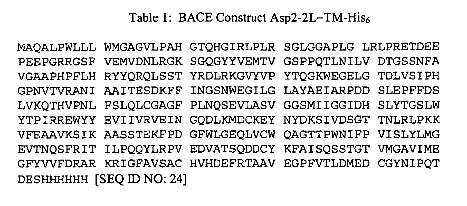
- The activity of P33K-BACE expressed in E. coli according the above Examples was compared to the activity of human BACE prepared as above without the P33K mutation, and to human BACE expressed in CHO cells. The results are provided in Table 2.
- To assay for BACE activity, the following activity assay conditions were utilized, unless specifically indicated otherwise: 20 µl 1M sodium acetate (NaOAc), pH 5.0; 125 µl H2O; 50 µl BACE sample containing 5-10 pmoles protein; and 5
µl 1 mM substrate S-1 (SEQ ID NO: 10). - The 200 µl assay reaction was incubated for 1-2 hours at 37°C. Activity was expressed as fluorescence peak area generated by the liberation of a fluorescent (Tryptophan fluorescence) product upon cleavage of Substrate S-1 by recombinant BACE. The reaction was stopped with 100 µl of 4% TFA in H2O. In order to separate the cleavage products and thereby detect activity of recombinant BACE, 50 microliters of the reaction mixture was injected into a Hewlett Packard Model 1100 HPLC equipped with an Alltech Rocket™ column (7 mm i.d., x 53 mm length, C18, 3 µm) pre-equilibrated with 88% Reagent A (0.1% TFA in water), 12% Reagent B (0.1 % TFA in acetonitrile). The flow rate over this column was 3 ml per minute.
Table 2 Activity Assays (Fluorescence Peak Area) E. Coli P33K-BACE E. Coli BACE CHO BACE 38.41 31.03 83.32 26.50 45.80 85.20 32.84 31.86 78.07
Claims (15)
- An isolated polypeptide comprising human BACE having the modification Pro33Lys.
- A composition comprising an active human BACE enzyme comprising the pro-enzyme sequence of BACE having the modification Pro33Lys.
- An isolated polypeptide comprising SEQ ID NO: 2.
- An isolated polypeptide consisting of SEQ ID NO: 2.
- An isolated polynucleotide comprising a sequence that encodes the polypeptide of claim 1.
- An isolated polynucleotide comprising a sequence that encodes the amino acid sequence set forth in SEQ ID NO: 2.
- An isolated polynucleotide comprising nucleotides 70 - 1365 of SEQ ID NO: 8.
- An isolated polynucleotide consisting of nucleotides 70 - 1365 of SEQ ID NO: 8.
- An expression vector comprising the polynucleotide of claim 5.
- An expression vector comprising a polynucleotide sequence encoding for a Pro33Lys-BACE polypeptide, wherein said expression vector can produce the Pro33Lys-BACE polypeptide when said expression vector is present in a compatible host cell, and when the host cell is cultured under conditions that allow for production.
- The expression vector of claim 10 wherein the Pro33Lys-BACE polypeptide that is produced comprises the polypeptide sequence of SEQ ID NO: 2.
- A recombinant host cell comprising the expression vector of claim 9.
- A method for producing a Pro33Lys-BACE polypeptide comprising the steps of: a) culturing the recombinant host cell of claim 12 under conditions that allow for the production of said polypeptide; and b) recovering the polypeptide from the culture.
- The method of claim 13 wherein the host cell is E. coli.
- A method of producing active Pro33Lys-BACE comprising recovering the Pro33Lys-BACE from the cultured host cell of claim 12 and diluting the polypeptide 20-50 fold with water having a temperature of about 1° C to 15° C.
Applications Claiming Priority (3)
| Application Number | Priority Date | Filing Date | Title |
|---|---|---|---|
| US35865102P | 2002-02-21 | 2002-02-21 | |
| US358651P | 2002-02-21 | ||
| PCT/US2003/005508 WO2003072733A2 (en) | 2002-02-21 | 2003-02-21 | Modified bace |
Publications (4)
| Publication Number | Publication Date |
|---|---|
| EP1572904A3 EP1572904A3 (en) | 2005-08-11 |
| EP1572904A2 EP1572904A2 (en) | 2005-09-14 |
| EP1572904A4 EP1572904A4 (en) | 2006-03-15 |
| EP1572904B1 true EP1572904B1 (en) | 2009-04-15 |
Family
ID=27765972
Family Applications (1)
| Application Number | Title | Priority Date | Filing Date |
|---|---|---|---|
| EP03709301A Expired - Lifetime EP1572904B1 (en) | 2002-02-21 | 2003-02-21 | Modified bace |
Country Status (7)
| Country | Link |
|---|---|
| US (2) | US7273743B2 (en) |
| EP (1) | EP1572904B1 (en) |
| JP (1) | JP4272070B2 (en) |
| AU (1) | AU2003213252A1 (en) |
| CA (1) | CA2477243A1 (en) |
| DE (1) | DE60327250D1 (en) |
| WO (1) | WO2003072733A2 (en) |
Families Citing this family (3)
| Publication number | Priority date | Publication date | Assignee | Title |
|---|---|---|---|---|
| US7763609B2 (en) | 2003-12-15 | 2010-07-27 | Schering Corporation | Heterocyclic aspartyl protease inhibitors |
| US7479538B2 (en) * | 2004-11-12 | 2009-01-20 | The Zhabilov Trust | Irreversibly-inactivated pepsinogen fragment and pharmaceutical compositions comprising the same for detecting, preventing, and treating HIV |
| MX2007011234A (en) * | 2005-03-14 | 2007-11-12 | Transtech Pharma Inc | Benzazole derivatives, compositions, and methods of use as b-secretase inhibitors. |
Family Cites Families (10)
| Publication number | Priority date | Publication date | Assignee | Title |
|---|---|---|---|---|
| US5319689A (en) * | 1992-03-12 | 1994-06-07 | General Electric Company | Compactable phase-separator assembly for dual-phase nuclear reactor |
| US5744346A (en) * | 1995-06-07 | 1998-04-28 | Athena Neurosciences, Inc. | β-secretase |
| EP0837883B1 (en) * | 1995-06-07 | 2005-08-10 | Chiron Corporation | Method of solubilizing, purifying, and refolding protein |
| US6225103B1 (en) | 1996-11-20 | 2001-05-01 | Oklahoma Medical Research Foundation | Cloning and characterization of napsin |
| GB9701684D0 (en) * | 1997-01-28 | 1997-03-19 | Smithkline Beecham Plc | Novel compounds |
| KR20070013361A (en) | 1998-09-24 | 2007-01-30 | 파마시아 앤드 업존 캄파니 엘엘씨 | Alzheimer's Disease Secretase |
| US6699671B1 (en) | 1998-09-24 | 2004-03-02 | Pharmacia & Upjohn Company | Alzheimer's disease secretase, APP substrates therefor, and uses therefor |
| JP4653318B2 (en) | 1999-02-10 | 2011-03-16 | エラン ファーマシューティカルズ,インコーポレイテッド | β-secretase enzyme composition and method |
| CA2374346C (en) | 1999-06-28 | 2010-06-22 | Jordan J. N. Tang | Inhibitors of memapsin 2 and use thereof |
| ATE431419T1 (en) | 1999-09-23 | 2009-05-15 | Pharmacia & Upjohn Co Llc | ALZHEIMER'S DISEASE SECRETASE, APP (AMYLOID PRECURSOR PROTEIN) SUBSTRATES THEREOF AND USES |
-
2003
- 2003-02-21 EP EP03709301A patent/EP1572904B1/en not_active Expired - Lifetime
- 2003-02-21 JP JP2003571421A patent/JP4272070B2/en not_active Expired - Lifetime
- 2003-02-21 US US10/372,473 patent/US7273743B2/en not_active Expired - Fee Related
- 2003-02-21 CA CA002477243A patent/CA2477243A1/en not_active Abandoned
- 2003-02-21 DE DE60327250T patent/DE60327250D1/en not_active Expired - Lifetime
- 2003-02-21 AU AU2003213252A patent/AU2003213252A1/en not_active Abandoned
- 2003-02-21 WO PCT/US2003/005508 patent/WO2003072733A2/en not_active Ceased
-
2005
- 2005-10-31 US US11/263,057 patent/US7244606B2/en not_active Expired - Fee Related
Also Published As
| Publication number | Publication date |
|---|---|
| US7273743B2 (en) | 2007-09-25 |
| US20040005691A1 (en) | 2004-01-08 |
| AU2003213252A1 (en) | 2003-09-09 |
| WO2003072733A2 (en) | 2003-09-04 |
| JP2005531292A (en) | 2005-10-20 |
| US20060094103A1 (en) | 2006-05-04 |
| EP1572904A2 (en) | 2005-09-14 |
| US7244606B2 (en) | 2007-07-17 |
| WO2003072733A3 (en) | 2005-08-11 |
| CA2477243A1 (en) | 2003-09-04 |
| JP4272070B2 (en) | 2009-06-03 |
| EP1572904A4 (en) | 2006-03-15 |
| DE60327250D1 (en) | 2009-05-28 |
| AU2003213252A8 (en) | 2003-09-09 |
Similar Documents
| Publication | Publication Date | Title |
|---|---|---|
| US5424205A (en) | Amyloidin protease and uses thereof | |
| Hartleib et al. | High-yield expression, purification, and characterization of the recombinant diisopropylfluorophosphatase from Loligo vulgaris | |
| CA2153254C (en) | Cloning of enterokinase and method of use | |
| UA129511C2 (en) | METHOD FOR PRODUCING SOLUBLE-CHAIN BoNT/A PROTEIN | |
| Welch et al. | Purification of Human Matrilysin Produced inEscherichia coliand Characterization Using a New Optimized Fluorogenic Peptide Substrate | |
| Dostál et al. | The precursor of secreted aspartic proteinase Sapp1p from Candida parapsilosis can be activated both autocatalytically and by a membrane-bound processing proteinase. | |
| US7732183B2 (en) | Method for refolding enzymes | |
| US7273743B2 (en) | Modified BACE | |
| US20020192754A1 (en) | Method for producing active serine proteases and inactive variants | |
| JP3915983B2 (en) | Protease, DNA encoding the protease, and method for producing protease | |
| AU2009307423B2 (en) | Process for producing protein capable of forming inclusion body | |
| Baker et al. | Autocatalytic processing of pro-papaya proteinase IV is prevented by crowding of the active-site cleft | |
| Goodfellow et al. | Characterization of a maize root proteinase | |
| US6833262B1 (en) | Polypeptide having amidolytic activity for a serpin | |
| JP4631436B2 (en) | Metalloendopeptidase | |
| Rank et al. | [W206R]-procaspase 3: an inactivatable substrate for caspase 8 | |
| US6312935B1 (en) | Cathepsin-L, the pre-proform thereof and the corresponding propeptide from ciliates | |
| US6372469B1 (en) | CDNA encoding plant-derived epoxide hydrolase, gene encoding same and transformant | |
| Flentke et al. | Purification and crystallization of rhizopuspepsin: the use of nickel chelation chromatography to select for catalytically active species | |
| Phlippen et al. | The glutathione synthetase of Schizosaccharomyces pombe is synthesized as a homodimer but retains full activity when present as a heterotetramer | |
| Francky et al. | A basic residue at position 36p of the propeptide is not essential for the correct folding and subsequent autocatalytic activation of prochymosin | |
| JP2004509973A (en) | Method for selective modification of peptides and proteins | |
| Emmons et al. | Large scale refolding and purification of the catalytic domain of Human BACE-2 produced in E. coli | |
| NZ275817A (en) | Endothelin converting enzymes, preparation and use | |
| KR100770665B1 (en) | Highly thermophilic prolylgigopeptidase enzyme and preparation method thereof |
Legal Events
| Date | Code | Title | Description |
|---|---|---|---|
| PUAI | Public reference made under article 153(3) epc to a published international application that has entered the european phase |
Free format text: ORIGINAL CODE: 0009012 |
|
| PUAK | Availability of information related to the publication of the international search report |
Free format text: ORIGINAL CODE: 0009015 |
|
| AK | Designated contracting states |
Kind code of ref document: A2 Designated state(s): AT BE BG CH CY CZ DE DK EE ES FI FR GB GR HU IE IT LI LU MC NL PT SE SI SK TR |
|
| AK | Designated contracting states |
Kind code of ref document: A3 Designated state(s): AT BE BG CH CY CZ DE DK EE ES FI FR GB GR HU IE IT LI LU MC NL PT SE SI SK TR |
|
| RIC1 | Information provided on ipc code assigned before grant |
Ipc: 7C 07H 21/04 B Ipc: 7C 12N 9/64 B Ipc: 7C 12N 15/52 B Ipc: 7C 12N 1/21 A |
|
| A4 | Supplementary search report drawn up and despatched |
Effective date: 20060126 |
|
| 17P | Request for examination filed |
Effective date: 20060213 |
|
| RBV | Designated contracting states (corrected) |
Designated state(s): AT BE BG CH CY CZ DE DK EE ES FI FR GB GR HU IE IT LI LU MC NL PT SE SI SK TR |
|
| RAX | Requested extension states of the european patent have changed |
Extension state: RO Payment date: 20060213 Extension state: MK Payment date: 20060213 Extension state: LV Payment date: 20060213 Extension state: LT Payment date: 20060213 Extension state: AL Payment date: 20060213 |
|
| RAP1 | Party data changed (applicant data changed or rights of an application transferred) |
Owner name: PHARMACIA & UPJOHN COMPANY LLC |
|
| 17Q | First examination report despatched |
Effective date: 20071213 |
|
| GRAP | Despatch of communication of intention to grant a patent |
Free format text: ORIGINAL CODE: EPIDOSNIGR1 |
|
| GRAS | Grant fee paid |
Free format text: ORIGINAL CODE: EPIDOSNIGR3 |
|
| GRAA | (expected) grant |
Free format text: ORIGINAL CODE: 0009210 |
|
| AK | Designated contracting states |
Kind code of ref document: B1 Designated state(s): CH DE FR GB IE LI |
|
| AX | Request for extension of the european patent |
Extension state: AL LT LV MK RO |
|
| REG | Reference to a national code |
Ref country code: GB Ref legal event code: FG4D Ref country code: CH Ref legal event code: EP |
|
| REG | Reference to a national code |
Ref country code: IE Ref legal event code: FG4D |
|
| REF | Corresponds to: |
Ref document number: 60327250 Country of ref document: DE Date of ref document: 20090528 Kind code of ref document: P |
|
| LTIE | Lt: invalidation of european patent or patent extension |
Effective date: 20090415 |
|
| PLBE | No opposition filed within time limit |
Free format text: ORIGINAL CODE: 0009261 |
|
| STAA | Information on the status of an ep patent application or granted ep patent |
Free format text: STATUS: NO OPPOSITION FILED WITHIN TIME LIMIT |
|
| 26N | No opposition filed |
Effective date: 20100118 |
|
| PGFP | Annual fee paid to national office [announced via postgrant information from national office to epo] |
Ref country code: IE Payment date: 20100217 Year of fee payment: 8 Ref country code: CH Payment date: 20100215 Year of fee payment: 8 |
|
| PGFP | Annual fee paid to national office [announced via postgrant information from national office to epo] |
Ref country code: FR Payment date: 20100223 Year of fee payment: 8 |
|
| PGFP | Annual fee paid to national office [announced via postgrant information from national office to epo] |
Ref country code: GB Payment date: 20100202 Year of fee payment: 8 Ref country code: DE Payment date: 20100303 Year of fee payment: 8 |
|
| REG | Reference to a national code |
Ref country code: CH Ref legal event code: PL |
|
| GBPC | Gb: european patent ceased through non-payment of renewal fee |
Effective date: 20110221 |
|
| PG25 | Lapsed in a contracting state [announced via postgrant information from national office to epo] |
Ref country code: LI Free format text: LAPSE BECAUSE OF NON-PAYMENT OF DUE FEES Effective date: 20110228 Ref country code: CH Free format text: LAPSE BECAUSE OF NON-PAYMENT OF DUE FEES Effective date: 20110228 |
|
| REG | Reference to a national code |
Ref country code: FR Ref legal event code: ST Effective date: 20111102 |
|
| REG | Reference to a national code |
Ref country code: IE Ref legal event code: MM4A |
|
| REG | Reference to a national code |
Ref country code: DE Ref legal event code: R119 Ref document number: 60327250 Country of ref document: DE Effective date: 20110901 |
|
| PG25 | Lapsed in a contracting state [announced via postgrant information from national office to epo] |
Ref country code: IE Free format text: LAPSE BECAUSE OF NON-PAYMENT OF DUE FEES Effective date: 20110221 Ref country code: FR Free format text: LAPSE BECAUSE OF NON-PAYMENT OF DUE FEES Effective date: 20110228 |
|
| PG25 | Lapsed in a contracting state [announced via postgrant information from national office to epo] |
Ref country code: GB Free format text: LAPSE BECAUSE OF NON-PAYMENT OF DUE FEES Effective date: 20110221 |
|
| PG25 | Lapsed in a contracting state [announced via postgrant information from national office to epo] |
Ref country code: DE Free format text: LAPSE BECAUSE OF NON-PAYMENT OF DUE FEES Effective date: 20110901 |