EP1571211A1 - Antibodies against lesion tissue - Google Patents
Antibodies against lesion tissue Download PDFInfo
- Publication number
- EP1571211A1 EP1571211A1 EP03775858A EP03775858A EP1571211A1 EP 1571211 A1 EP1571211 A1 EP 1571211A1 EP 03775858 A EP03775858 A EP 03775858A EP 03775858 A EP03775858 A EP 03775858A EP 1571211 A1 EP1571211 A1 EP 1571211A1
- Authority
- EP
- European Patent Office
- Prior art keywords
- cells
- antibody
- antibodies
- cell
- cancer
- Prior art date
- Legal status (The legal status is an assumption and is not a legal conclusion. Google has not performed a legal analysis and makes no representation as to the accuracy of the status listed.)
- Granted
Links
Images





Classifications
-
- C—CHEMISTRY; METALLURGY
- C07—ORGANIC CHEMISTRY
- C07K—PEPTIDES
- C07K16/00—Immunoglobulins [IG], e.g. monoclonal or polyclonal antibodies
-
- A—HUMAN NECESSITIES
- A61—MEDICAL OR VETERINARY SCIENCE; HYGIENE
- A61P—SPECIFIC THERAPEUTIC ACTIVITY OF CHEMICAL COMPOUNDS OR MEDICINAL PREPARATIONS
- A61P35/00—Antineoplastic agents
-
- C—CHEMISTRY; METALLURGY
- C07—ORGANIC CHEMISTRY
- C07K—PEPTIDES
- C07K16/00—Immunoglobulins [IG], e.g. monoclonal or polyclonal antibodies
- C07K16/18—Immunoglobulins [IG], e.g. monoclonal or polyclonal antibodies against material from animals or humans
- C07K16/28—Immunoglobulins [IG], e.g. monoclonal or polyclonal antibodies against material from animals or humans against receptors, cell surface antigens or cell surface determinants
- C07K16/30—Immunoglobulins [IG], e.g. monoclonal or polyclonal antibodies against material from animals or humans against receptors, cell surface antigens or cell surface determinants from tumour cells
- C07K16/3015—Breast
-
- C—CHEMISTRY; METALLURGY
- C07—ORGANIC CHEMISTRY
- C07K—PEPTIDES
- C07K2317/00—Immunoglobulins specific features
- C07K2317/20—Immunoglobulins specific features characterized by taxonomic origin
- C07K2317/21—Immunoglobulins specific features characterized by taxonomic origin from primates, e.g. man
-
- C—CHEMISTRY; METALLURGY
- C07—ORGANIC CHEMISTRY
- C07K—PEPTIDES
- C07K2317/00—Immunoglobulins specific features
- C07K2317/60—Immunoglobulins specific features characterized by non-natural combinations of immunoglobulin fragments
- C07K2317/62—Immunoglobulins specific features characterized by non-natural combinations of immunoglobulin fragments comprising only variable region components
- C07K2317/622—Single chain antibody (scFv)
Definitions
- the present invention relates to antibodies against lesional tissues and methods for producing the same.
- Lymphocytes are widely known to infiltrate cancer tissues (Document 1/ Hurliamnn et al . (1985) Int J Cancer 35:753; Document 2/ Whiteside et al . (1986) Cancer Immunol Immunother 23:169; Document 3/ Wolf et al . (1986) Otolaryngol Head Neck Surg 95:142; Document 4/ Husby et al . (1976) J Clin Invest 57:1471; Document 5/ Vose et al . (1979) Int J Cancer 24:579). Experimental and clinical data suggest that lymphocyte infiltration of cancer tissues is associated with host immunoreactions against cancer (Document 5/ Rosenberg et al . (1988) New Engl J Med 319:1676; Document 6/ Van Pel et al. (1995) Immunol Reviews 145:229; Document 7/ Kreider et al . (1984) Cancer Metastasis Rev 3:53).
- cytotoxic T cells are the effector cells that directly kill cancer cells (Document 8/ Nobholz and MacDonald (1983) Annu Rev Immunol 1:273). Furthermore, some of the antibodies produced by plasma cells, which are the final form of differentiated B cells, are assumed to have the ability to bind to cancer cells (Document 9/ Roitt et al . (1969) Lancet 2: 367; Document 10/ Borsos (1971) Progress in Immunology: p841. New York, Academic Press; Document 11/ Kodera and Bean (1975) Int J Cancer 16:579).
- B cells which infiltrate cancer tissues express antibodies, and that these antibodies bind selectively to antigens on cancer cells (Document 12 / Punt et al . (1994) Cancer Immunol Immunother 38:225; Document 13/ Zhang et al . (1995) Cancer Res 55:3584).
- cancer antigens can be identified by using antibodies expressed in infiltrating B cells. Antibodies having such reactivity will be useful for cancer treatment and diagnosis if they can be obtained.
- Antibodies that bind to cancer cells destroy cancer cells by activating the complement system or by triggering antibody-dependent cytotoxicity.
- cancer cells destroy cancer cells by activating the complement system or by triggering antibody-dependent cytotoxicity.
- the hybridoma method devised for establishing murine antibody-producing cell lines is another technique for cloning antibody-producing cells.
- the hybridoma method comprises the step of fusing an antigen-specific B cell with an immortalized myeloma cell to establish an antibody-expressing cell line.
- Murine myeloma cells are effective in the case of murine hybridomas; however, when used as a fusion partner for human B cells, characteristics of human hybridomas become unstable due to the preferential occurrence of deletions in human chromosomes, thus failing to lead to the establishment of antibody-producing cell lines (Document 16/ Winter and Milstein (1991) Nature 349:293). At present, it is thus technically difficult to establish human-derived antibody-producing cell lines.
- the smallest recombinant antibody molecule, scFv has a structure in which the heavy chain variable region and the light chain variable region are connected through a linker.
- B cells express a single antibody gene. Accordingly, antibodies having an activity similar to that of antibodies produced by B cells can be reconstructed by cloning the heavy and light chain variable regions from these cells.
- B cells present in peripheral blood and those infiltrating cancer tissues are (polyclonal) cell populations which produce various antibodies (Document 22/ Kotlan et al . (1999) Immunol Lett 65:143; Document 23/ Hansen et al . (2001) Pro Natl Acad Sci USA 98:12659). Therefore, it would not be easy to clone antibody genes from such cell populations and reconstruct them as human antibodies.
- An objective of the present invention is to provide methods for obtaining polynucleotides encoding antibodies having a specific reactivity from antibody-producing cells comprised in a polyclonal cell population.
- B cells cloned by some technique are required. This limitation poses difficulties for, for example, obtaining antibodies produced by B cells which have infiltrated cancer tissues.
- the present inventors searched for methods that would enable one to obtain antibody genes without having to rely on B cell cloning, and conceived that it may be possible to utilize cellular mRNAs isolated from cell populations as a cloning source.
- B cells in peripheral blood are used to clone human antibody genes.
- B cell populations in peripheral blood are polyclonal cell populations producing antibodies with various reactivities, these B cells were thought to be inappropriate for selectively isolating antibodies with a specific reactivity.
- Microdissection is a technique for excising and isolating specific cells from a specimen composed of heterogeneous cell populations such as a tissue section.
- a system for isolating cells in which the periphery of target cells is excised with an ultraviolet laser has been put into practical use. This system is referred to as laser microdissection (LMD), and is commercially available.
- LMD has spread as a technique that can be used to obtain target cells with high precision and little damage to cells. By using LMD, a specific cellular gene can be obtained and amplified by PCR.
- the present inventors thought that B cells which have infiltrated cancer tissues are very likely to produce antibodies having reactivities to cancer cells, and therefore, polynucleotides encoding antibodies which bind to cancer cells may be efficiently isolated by isolating infiltrating B cells using the LMD system or such. Then, the present inventors showed that polynucleotides encoding the antibodies can be obtained by utilizing, as a cloning source, B cells which have actually infiltrated a lesional tissue, and thereby completed the present invention. That is, the present invention, as described below, relates to methods for isolating antibody genes and preparing antibodies encoded by these genes, and antibodies obtained by these methods.
- the present invention relates to methods for isolating a polynucleotide encoding an antibody against a lesion, wherein the methods comprise the steps of:
- B cells which have infiltrated a lesion are very likely to produce antibodies against that lesion.
- lesions have accumulations of B cells that produce antibodies recognizing the lesion. Therefore, antibody genes derived from B cells isolated from a cell population that has infiltrated a lesion is useful for generating antibodies against that lesion.
- the term "antibodies against a lesion” refers to antibodies that recognize the antigens which constitute the lesion or antigenic substances produced at the lesion. Such antibodies are useful for diagnosing and treating the lesion.
- such antibodies provide important information for epitope analysis in the autoimmune disease.
- any lesion recognized as non-self by the immune system can be utilized.
- the following lesions are preferable for obtaining B cells. These lesions can be referred to as naturally occurring lesions, which can also be obtained from human subjects being treated for said lesions.
- solid tumor lesions arteriosclerosis lesions inflammatory disease lesions lesions generated by infectious pathogens autoimmune disease lesions
- artificially prepared lesions can also be utilized in this invention.
- the following lesions are artificially prepared lesions, and can be obtained from, for example, immune animals.
- polynucleotides encoding antibodies against any antigen can be obtained.
- Cancer tissues can be exemplified as preferable lesions of the present invention.
- the present invention relates to methods for isolating a polynucleotide encoding an antibody against a cancer cell, wherein the method comprises the steps of:
- Cancer tissues to be used in this invention are not limited. Specifically, breast cancer tissues, lung cancer tissues, liver cancer tissues, colon cancer tissues, pancreas cancer tissues, prostate cancer tissues, and such can be used. Among them, cancers comprising many B cell infiltrations are preferable cancer tissues of this invention. Examples of cancers comprising many B cell infiltrations include breast cancers, lung cancers, and melanomas. Cancer tissues can be removed by surgical excision. For example, cancer tissues removed for biopsy can be used as cancer tissues for the present invention. Furthermore, tissues taken out from patients by surgical removal are also useful as cancer tissues. These tissues can be those removed for obtaining antibody genes; alternatively, tissues removed for histopathological examination or surgical treatment can also be used.
- any method may be used for isolating B cells which infiltrate cancer tissues.
- the preferable methods for isolating B cells include microdissection, a technique for excising specific cells from a tissue section.
- target cells can be isolated from frozen tissue sections using the Laser Microdissection (LMD) system.
- LMD Laser Microdissection
- a system for excising tissue sections with an ultraviolet laser is commercially available. The use of this system, which involves specifying the region to be excised on a computer image under microscopic observation, makes it possible to excise any region from a tissue section.
- a large number of B cells can be isolated by observing a specimen under a microscope and selecting a portion enriched with B cells.
- a small number of B cells can be easily obtained by excising a region with low B cell density. As shown in the Examples, it is even possible to obtain a single cell.
- B cells can be isolated from any specimen prepared for pathological analysis.
- thin-section frozen specimens are preferable pathological specimens in this invention.
- a pathological specimen not only fresh tissues but also specimens fixed with paraformaldehyde (PFA) or such can be used.
- PFA paraformaldehyde
- a broad range of cloning sources can be selected for the methods of this invention. That is, the present invention provides methods that can be conveniently used to obtain a variety of antibody genes.
- Microdissection is a system also utilized in gene analysis of specialized cells in tissues using PCR or such.
- the present inventors focused on the fact that it is possible to use lesion-infiltrating B cell populations as cell populations that are very likely to produce antibodies with a desired reactivity.
- the present inventors thus made it possible to obtain antibody genes.
- B cells or plasma cells which infiltrate cancer tissues and produce antibodies can be extracted based on pathological analysis under microscopic observation.
- B cells and plasma cells can be distinguished by staining with toluidine blue or such.
- this method makes it possible to isolate genes of antibodies that recognize cancer cells with a much higher probability.
- the term "cell isolation” refers to the separation of B cells from a cell population comprising mixtures of heterogeneous cells.
- the “cell isolation” of the present invention includes isolation of cells harboring antigen genes when they coexist with other cells that do not carry an antibody gene.
- carrier cells that are clearly incapable of producing antibodies may be added to the antigen-producing cells.
- Carrier cells are added to facilitate the extraction of target mRNAs. That is, even if only a limited number of antibody-producing cells are coexisting with other cells which do not carry antibody genes, these antibody-producing cells can be said to be in an isolated state.
- the number of B cells to be isolated is not limited. Specifically, for example, one to 100 cells, usually one to 50 cells, preferably 20 cells or less, more preferably five cells or less, and yet more preferably one cell is isolated.
- Isolation of single cells enhances the possibility of obtaining an antibody gene in a state that maintains its heavy and light chain combinations.
- it is an important condition to obtain an antibody gene in such a state.
- cloning antibody genes the use of monoclonal antibody-producing cells as a cloning source is an essential condition for accurately reconstructing combinations of heavy and light chains.
- an antibody gene obtained from a single cell is used, repeated cloning of the gene would be required when comparing multiple antibodies with different reactivities. Comparison of multiple antibodies is useful for obtaining a suitable antibody with purpose-oriented characteristics.
- any lesion-infiltrating antibody-producing cell can be used as a cloning source.
- B cells circulating in peripheral blood have various levels of differentiation. At the early stages of differentiation, B-cells have ⁇ -chains on their surfaces as an antigen receptor. B cells mature and differentiate into IgG-secreting cells through differentiation and activation by antigen stimulation. B cells at the final stage of differentiation are referred to as plasma cells (or plasmocytes). Plasma cells produce 2,000 molecules of IgG per second. Therefore, by isolating plasma cells, more mRNAs can be obtained.
- lesion-infiltrating B cells are found to be at an advanced stage of differentiation. Further, it is highly likely that the infiltrating B cells produce antibodies against the lesion. Therefore, by using B cells isolated from a lesion as the cloning source, the specificity of the resulting antibody will be naturally centered on the lesion. Furthermore, antibody genes are expressed at an extremely high level in plasma cells and in B cells at an advanced stage of differentiation. Accordingly, utilization of such cells as the cloning source leads to increased possibility of obtaining antibody genes. By utilizing lesion-infiltrating B cells, antibody genes of interest can be obtained with a high probability using relatively few cells.
- Antibody genes can be obtained from isolated B cells by amplifying the antibody genes. Methods for amplifying genes are known in the art. For example, the PCR method is preferred as a method for amplifying antibody genes. A method for isolating antibody genes using the PCR method is described below.
- mRNAs are extracted from isolated B cells.
- cDNAs are synthesized by using the extracted mRNAs as a template to obtain a cDNA library.
- kits are conveniently used for extracting mRNAs and for constructing the cDNA library.
- mRNAs derived from a small number of B cells are used.
- mRNAs obtained from only few cells are extremely small in amount, and have low yields when directly purified. Therefore, mRNAs are usually purified after the addition of carrier RNAs that clearly contain no antibody genes.
- the RNAs of antibody-producing cells themselves can also be efficiently extracted.
- the addition of carrier RNAs may not be required for extracting RNAs from, for example, 10 or more, 30 or more, preferably 50 or more antibody-producing cells.
- antibody genes can be amplified by the PCR method.
- Primers for amplifying antibody genes by the PCR method are known in the art.
- primers that allow the amplification of genes encoding ⁇ 1 to ⁇ 5 as the heavy chain, and ⁇ -chain and ⁇ -chain as the light chains, can be used.
- a primer capable of annealing to a portion that corresponds to the hinge region is generally used as the 3' primer.
- a primer corresponding to each subclass can be used as the 5' primer.
- PCR products obtained using primers for amplifying genes of the respective heavy and light chain subclasses are considered as independent libraries.
- Using thus synthesized libraries it is possible to reconstitute immunoglobulins comprising combinations of heavy chains and light chains.
- the lesion-binding activity of the reconstituted immunoglobulin can be used as an index to screen for antibodies of interest.
- cancer-binding antibodies can be screened by, for example, the following steps of:
- an immobilized cancer specimen is reacted with a test antibody, and then with a labeled secondary antibody which recognizes the test antibody.
- the binding of the test antibody to cancer cells can be confirmed by detecting labeled antibody on the immobilized specimen after washing.
- Enzymatically active proteins such as peroxidase and ⁇ -galactosidase, and fluorescent substances such as FITC can be used as the label.
- Cancer tissues to be analyzed for antibody-binding activity may be cancer tissues forming the lesion from which B cells are obtained. Alternatively, cancer tissues of the same organ which have been removed from different individuals, and cell lines derived from these cancer tissues, can be also used. Furthermore, cancer tissues of different organs, or cell lines derived therefrom, can also be used to screen for antibodies which commonly react with different types of cancers.
- an antibody whose reactivity towards a cancer tissue is significantly high compared to the normal tissue can be said to bind specifically to the cancer.
- the same type of tissue is generally used. That is, the reactivity of an antibody is compared between a cancer tissue and a normal tissue of the organ from which the cancer tissue is derived. Under conditions in which an antibody's reactivity towards a cancer tissue can be confirmed, the antibody is said to have a specific reactivity towards the cancer tissue when no binding activity towards the normal tissue can be detected.
- Methods of screening for antibodies using binding activity as an index include the panning method which utilizes phage vectors.
- the obtained antibody genes are gene libraries of heavy and light chain subclasses as described above, screening methods that use phage vectors are advantageous.
- genes encoding variable regions of the heavy and light chains can be made into single chain Fv (scFv) through conjugation with a suitable linker sequence.
- Phages expressing scFv on their surface can be obtained by inserting a scFv-encoding gene into a phage vector.
- DNAs encoding scFv with the desired binding activity can be recovered.
- scFv having the desired binding activity can be concentrated by repeating this procedure as necessary.
- An antibody-encoding polynucleotide of the present invention may encode a whole antibody or a portion of the antibody.
- the term "a portion of the antibody” means any portion of an antibody molecule.
- a portion of an antibody may be referred to as "an antibody fragment”.
- Preferred antibody fragments of the present invention comprise the complementarity determination region (CDR) of an antibody. More preferably, antibody fragments of the present invention comprise all three CDRs that constitute the variable region.
- polynucleotides encoding the variable region of an antibody are preferred antibody fragments of the present invention.
- a complete immunoglobulin molecule can be reconstituted by obtaining a polynucleotide encoding a variable region and linking the polynucleotide with a polynucleotide encoding a constant region.
- Constant regions of antibodies within the same class have approximately the same structure. That is, the structure of the constant region has no effect on the antigen-binding activity. Accordingly, if the structure of the variable region of an antibody can be elucidated, antibodies having similar activity can be reconstituted through conjugation with the constant region that is already obtained.
- Antibodies of the present invention include genetically recombinant antibodies whose structures have been artificially modified.
- mouse-human chimeric antibodies can be prepared by: conjugating a human constant region gene with an antibody gene obtained not from a human but from a non-human animal such as a mouse. Methods for humanizing a mouse variable region by transferring CDRs which constitute the variable region of a non-human animal such as mouse to a human variable region are also known in the art.
- Polynucleotides encoding the antibodies of the present invention can be DNAs, RNAs, or chimeric molecules thereof. Furthermore, artificial structures such as PNAs can also be included, as long as the nucleotide sequence is maintained. Methods for synthesizing polynucleotides having the same nucleotide sequence, based on the nucleotide sequence of a gene that encodes an antibody isolated from B cells, are known in the art.
- polynucleotides of the present invention can comprise a sequence identical or highly homologous to the gene encoding an antibody isolated from B cells.
- highly homologous generally refers to a homology of 70% or more, preferably 80% or more, more preferably 90% or more, and even more preferably 95% or more.
- the present invention relates to polynucleotides encoding antibodies obtained by the above-described methods.
- the polynucleotides of this invention can be incorporated into any expression vector.
- Appropriate host cells can be transformed with the expression vector to obtain antibody-expressing cells.
- By culturing the antibody-expressing cells and then recovering expression products therefrom, antibodies encoded by the genes can be obtained.
- the following describes expressing antibody genes isolated by the above-described methods.
- hosts and expression vectors can be used in appropriate combinations to produce the antibodies.
- animal cells include: (1) mammalian cells such as CHO, COS, myeloma, baby hamster kidney (BHK), HeLa, and Vero cells; (2) amphibian cells such as Xenopus oocytes; or (3) insect cells such as sf9, sf21, and Tn5.
- Known plant cells include cells derived from the Nicotiana genus such as Nicotiana tabacum , which can be callus cultured.
- Known fungal cells include yeasts such as the Saccharomyces genus, for example Saccharomyces cerevisiae , and filamentous fungi such as the Aspergillus genus, for example Aspergillus niger.
- Prokaryotic cells can also be used in production systems that utilize bacterial cells.
- Known bacterial cells include E. coli and Bacillus subtilis .
- the antibodies can be obtained by transferring the antibody genes of interest into these cells using transformation, and then culturing the transformed cells in vitro .
- the antibodies obtained by the methods of the present invention may be antibody fragments or modified antibodies thereof.
- the antibody fragments may be Fab, F(ab')2, Fv, or single chain Fv (scFv) in which Fv from H or L chains are ligated by an appropriate linker.
- the antibody fragments can be obtained by treating the antibodies with enzymes such as papain and pepsin to produce antibody fragments.
- genes encoding these antibody fragments can be prepared and introduced into expression vectors for expression in appropriate host cells (see, for example, Co, M. S. et al ., J. Immunol. (1994) 152, 2968-2976; Better, M. & Horwitz, A. H.
- scFv can be obtained by ligating the V regions of the antibody H-chain and L-chain.
- the V regions of the H chain and L chain are ligated via a linker, and preferably via a peptide linker (Huston, J. S. et al ., Proc. Natl. Acad. Sci. U.S.A (1988) 85, 5879-5883).
- the V regions of the scFv H chain and L chain may be derived from any of the antibodies described herein.
- the peptide linker used to ligate the V regions may be any single-chain peptide consisting of 12 to 19 residues.
- DNA encoding scFv can be amplified by PCR using as a template either the whole DNA, or a partial DNA encoding a desired DNA, selected from a DNA encoding the H chain or the V region of the H chain of the above antibody, and a DNA encoding the L chain or the V region of the L chain of the above antibody; and using a primer pair that defines the two ends. Further amplification can be subsequently conducted using the combination of DNA encoding the peptide linker portion, and the primer pair that defines both ends of the DNA to be ligated to the H chain and the L chain respectively.
- expression vectors containing the DNAs, and hosts transformed by these expression vectors can be obtained according to conventional methods.
- scFvs can be obtained according to conventional methods using the resulting hosts.
- antibody fragments can be produced in hosts by obtaining genes encoding the antibody fragments and expressing them in a similar manner to that outlined above.
- Antibodies bound to various types of molecules such as polyethylene glycol (PEG) may be used as modified antibodies.
- antibodies may bind to radioisotopes, chemotherapeutics, cytotoxic substances such as bacteria-derived toxin, and labeling substances.
- modified antibodies can be obtained by chemically modifying the resulting antibodies. Methods for modifying antibodies are already established in the art.
- the term "antibody” in the present invention also encompasses the modified antibodies as described above.
- the antibodies of the present invention may be bispecific antibodies.
- the bispecific antibodies may have antigen-binding sites recognizing different epitopes on the antigen molecules, or may have one antigen-binding site recognizing antigens and the other recognizing a cytotoxic substance such as radioactive substance, chemotherapeutic agent, and cell-derived toxin. In this case, it is possible to inhibit the growth of cancer cells by directly applying the cytotoxic substance to the cells expressing antigens to specifically damage them.
- Bispecific antibodies can be prepared by linking HL pairs of two kinds of antibodies, or obtained by fusing hybridomas that produce different monoclonal antibodies to prepare fused cells generating bispecific antibodies. Furthermore, bispecific antibodies can be generated by using genetic engineering techniques.
- Antibodies expressed and produced as described above can be purified by conventional methods for purifying normal proteins. Antibodies can be separated and purified by, appropriately selecting and/or combining affinity columns such as protein A columns or chromatography columns, filtration, ultrafiltration, salt precipitation, dialysis, and such (Antibodies A Laboratory Manual. Ed Harlow, David Lane, Cold Spring Harbor Laboratory, 1988).
- ELISAs enzyme linked immunosorbent assays
- EIAs enzyme immunoassays
- RIAs radioimmunoassays
- fluoroimmunoassays fluoroimmunoassays
- Host cells may be prokaryotic cells such as bacteria, and eukaryotic cells such as yeast, animal cells, insect cells, and plant cells, as long as the cells are capable of expressing the light chains of the present invention or the antibodies comprising the light chains. Mammalian cells are particularly preferred in view of glycosylation.
- Expression vectors need to comprise units that regulate the transcription and translation of genetic information, such as promoters and terminators.
- units that regulate the transcription and translation of genetic information such as promoters and terminators.
- Escherichia microorganisms such as E. coli are used as hosts
- plasmids of the pBR or pUC series can be used as plasmid vectors, and any promoters selected from those such as lac, trp, tac, trc, ⁇ phage PL, and PR can be used.
- Terminators may originate from trpA, phage, and rmB ribosomal RNA.
- plasmids such as those of the pUB110 and pC194 series can be used, and genes may be integrated into chromosomes in some cases. Promoters and terminators may be derived from apr, npr, amy, and such.
- prokaryotic cells include microorganisms such as Pseudomonas (e.g. P. putida, P. cepacia ; pKT240 vectors, and such), Brevibacteria (e.g. B. lactofermentum ; pAJ43), Corynebacteria (e.g. C. glutamicu m; pCS11, pCB101), Streptococcus (e.g. pHV1301, pGK1), Lactobatcillaceae (e.g. pAM ⁇ 1), Rhodcoccus (e.g. plasmids isolated from R. rhodochrous (J. Gen. Microbiol.
- Pseudomonas e.g. P. putida, P. cepacia ; pKT240 vectors, and such
- Brevibacteria e.g. B. lactofermentum ; pAJ43
- Corynebacteria
- Streptomyces e.g. S. lividans, S.virginiae ; pIJ486, pKC1064, pUWL-KS
- Enterobacter Erwinia, Klebsiella, Proteus, Salmonella (e.g. S. typhimurium ), Serratia (e.g. S. marcescans ), and Shigella.
- a system using Saccharomyces cerevisiae as a host, and plasmids from YRp, YEp, YCp, and YIp series is known.
- promoters and terminators such as ADH, GAPDH, PHO, GAL, PGK, and ENO can be used.
- Other microorganisms used in the expression vector systems of the present invention include Kluyveromyces (e.g. K. lactis ; plasmids of the 2 ⁇ m, pKD1, pGKI1, and KARS series, and such), Schizosaccharomyces (e.g. S.
- pombe pombe; pAUR224), Zygosaccharomyces (e.g. Z. rouxii ; pSB3 and PH05 promoters from S . cerevisiae ), Hansenula (e.g. H. polymorpha ), Pichia (e.g. P. pastoris ), Candida (e.g. C. maltosa, C. tropicalis, C. utilis, and C . albicans ), Aspergillus (e.g. A. oryzae, A. niger), and Trichoderma (e.g. T. reesei ).
- Zygosaccharomyces e.g. Z. rouxii ; pSB3 and PH05 promoters from S . cerevisiae
- Hansenula e.g. H. polymorpha
- Pichia e.g. P. pastoris
- Candida e.g. C. maltosa,
- plant cells may be used as hosts.
- host cells may be those from cotton, corn, potato, tomato, soybean, petunia, and tobacco.
- a particularly well-known system uses cells from Nicotina tabacum , which are cultured as a callus.
- expression vectors such as pMON530 are introduced into bacteria such as Agrobacterium tumefaciens . By infecting these bacteria into tobacco ( Nicotina tabacum ), desired polypeptides can be obtained from the tobacco leaves.
- Cells from insects such as silkworms ( Bombyx mori ), mosquitoes (e.g. Aede aegypti , Aedes albopictus ) and fruit flies (Drosophila melanogaster ) can be used as hosts.
- silkworms DNAs encoding antibodies may be inserted into baculovirus vectors, these vectors may be used to infect silkworms, and desired polypeptides can be obtained from the silkworm body fluids (Nature 315: 592-594 (1985)).
- Examples of expression vectors when using animal cells as hosts include pME18S (Med. Immunol. 20: 27-32 (1990)), pEF-BOS (Nucleic Acids Res. 18: 5322 (1990)), pCDM8 (Nature 329: 840-842 (1987)), pRSVneo, pSV2-neo, pcDNAI/Amp (Invitrogen), pcDNAI, pAMoERC3Sc, pCDM8 (Nature 329: 840 (1987)), pAGE107 (Cytotechnology 3: 133 (1990)), pREP4 (Invitrogen), pAGE103 (J. Biochem.
- Promoters may be cytomegalovirus IE gene promoter and enhancer, SV40 early promoter, a retrovirus LTR such as those from RSV, HIV, and MMLV, and gene promoters from animal cells such as metallothionein, ⁇ -actin, elongation factor-1, HSP genes, and such.
- viral vectors such as those mentioned above may also be used.
- Viral vectors may be derived from DNA viruses and RNA viruses such as retroviruses, adenoviruses, adeno-associated viruses, herpes viruses, vaccinia viruses, poxviruses, Simbu viruses, Sendai viruses, SV40, and HIV.
- Host animal cells may be mouse myeloma cells (e.g. SP2/0, NSO), rat myeloma cells (e.g. YB2/0), mouse hybridoma cells, Namalwa cells (including KJM-1 cells), human embryonic kidney cells (e.g. 293 cells), human leukemia cells (e.g. BALL-1), CHO cells, COS cells (e.g. COS-1, COS-7), hamster embryonic kidney cells (e.g. BHK), mouse Sertoli cells (e.g. TM4), African green monkey kidney cells (e.g. VERO-76), HBT637 cells, HeLa cells, rabbit kidney cells (e.g. MDCK), human liver cells (e.g. HepG2), mouse mammary tumor cells (e.g. MMT060562), TRI cells, MRC cells, FS3 cells, etc.
- mouse myeloma cells e.g. SP2/0, NSO
- Vectors can be introduced into prokaryotic cells by methods utilizing calcium ions (Proc. Natl. Acad. Sci. USA 69: 2110 (1972)), protoplasts (Unexamined Published Japanese Patent Application No. (JP-A) Sho 63-24829), electroporation (Gene 17: 107 (1982); Molecular & General Genetics 168: 111 (1979)), and such; introduced into yeast cells by using electroporation (Methods in Enzymology, 194: 182 (1990)), spheroplasts (Proc. Natl. Acad. Sci.
- Transformants obtained as described above can be cultured, for example, by the following methods:
- a medium such as the TNM-FH medium (Pharmingen), Sf-900 II SFM (Life Technologies), ExCell400 and ExCell405 (JRH Biosciences), and Grace's Insect Medium (Nature 195: 788 (1962)) can be used. If necessary, antibiotics such as gentamicin may be added to the medium.
- RPMI1640 The Journal of American Medical Association 199: 519 (1967)
- Eagle's MEM Science 122: 501 (1952)
- DMEM DMEM
- 199 medium Proceeding of the Society for the Biological Medicine 73: 1 (1950)
- antibiotics such as kanamycin and penicillin may be added to the medium.
- the antibodies of the present invention obtained as above can be isolated from within host cells, or from the culture medium if secreted into the extracellular space using signal sequences. They can then be purified as substantially pure polypeptides.
- the antibodies of the present invention can be separated or purified by appropriately selecting, or combining as necessary, methods generally used in separation and purification. Such methods can be chromatography, filtration, ultrafiltration, salting out, solvent precipitation, solvent extraction, distillation, immunoprecipitation, SDS-polyacrylamide gel electrophoresis, isoelectric point focusing, dialysis, and recrystallization.
- Chromatography includes affinity chromatography, ion exchange chromatography, hydrophobic chromatography, gel filtration, reverse phase chromatography, absorption chromatography, and the like (Strategies for Protein Purification and Characterization: A Laboratory Course Manual, Daniel R Marshak et al. eds., Cold Spring Harbor Laboratory Press (1996); Antibodies: A Laboratory Course Manual, Harlow and David Lane eds., Cold Spring Harbor Laboratory Press (1988)).
- Such chromatographies may be performed using liquid chromatographies such as HPLC, FPLC, and the like.
- the antibodies of the present invention may be purified by making use of their affinities towards antigens.
- Example 1 LMD isolation of a single cancer tissue-infiltrating B cell
- a fresh human tissue (breast cancer tissue) was sliced into a suitable size to prepare frozen blocks using an OCT compound (Tissue-tek), and fixed prior to freezing with fixatives such as paraformaldehyde-lysine-periodate, as necessary.
- fixatives such as paraformaldehyde-lysine-periodate
- thin specimen sections were prepared from each frozen block and attached onto LMD slides (Matsunami Glass Co.). Resulting thin-section frozen specimens were dried in air, fixed with fixatives such as acetone, and stained with toluidine blue (Muto Pure Chemicals Co., Ltd) or such.
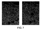
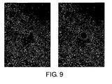
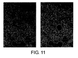
- plasma cells were excised with a laser microdissection system (Leica AS-LMD), and recovered using a recovery buffer (RLT solution attached to QIAGEN RNeasy Mini Kit).
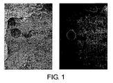
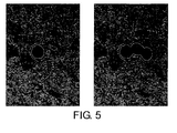
- Plasma cells before and after the excision are shown in Figs. 1, 5, 7, 9, 11, 13, and 15.
- the left photograph shows the state of the cells before excision
- the right photograph shows the state of the cells after excision.
- the cell(s) to be excised can be specified, they are indicated with an arrow or such in the "before excision" photographs (left).
- RNAs were prepared from the resultant mixture, using the RNeasy Mini Kit (QIAGEN) according to the manufacturer's instructions. When 50 cells or more were excised from the thin-section specimen, total RNAs were prepared without the addition of carrier cells. cDNA was synthesized using all 35 ⁇ l of the RNA eluate fraction as a template, and Sensiscript Reverse Transcriptase (QIAGEN) according to the manufacturer's instructions.
- cDNA synthesis reaction was performed on an 80- ⁇ l scale using 40 ng of Oligo dT primers (Promega) and 0.8 ⁇ g of random hexamers (Invitrogen) as the reverse transcription primers at 37°C for 1 h.
- Oligo dT primers Promega
- Invitrogen random hexamers
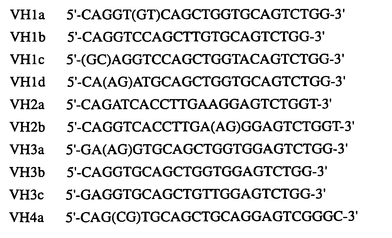
- Primers for cloning heavy chain variable regions are designated with VH and JH as the initial letters.
- Primers for amplifying the light chain ⁇ -chain or the light chain ⁇ -chain are designated with VK and JK, or VL and JL as the initial letters.
- PCR amplifications were carried out using combinations of the Taq DNA polymerase Core Kit (QIAGEN) and primer mixtures for each gene subset.
- QIAGEN Taq DNA polymerase Core Kit
- primer mixtures for amplifying the heavy chain and ⁇ -chain, five different primer mixtures were prepared for each. Ten kinds of reaction solutions were prepared using these primer mixtures. Combinations of primers in the mixtures are shown in Table 1.
- Prepared reaction mixtures (20 ⁇ l) had a final concentration of 1x reaction buffer containing 4 ⁇ l of the template cDNA, 1x Q solution (QIAGEN), 0.4 mM dNTP, 0.4 ⁇ M each of the forward and reverse primers, and 2 U Taq DNA polymerase.
- the reaction mixture was subjected to 40 cycles of amplification reaction on Applied Biosystems PE9700.
- the amplification cycle was carried out under conditions of denaturation at 94°C for 10 sec, annealing at 50°C for 30 sec, and elongation at 72°C for 30 sec.
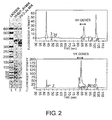
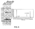
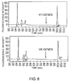
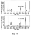
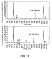
- amplification products were analyzed using DNA 7500 Labchip and Agilent 2100. Results of the amplification were shown in Figs. 2, 6, 8, 10, 12, 14, and 16. Amplification products were purified using the QIAGEN PCR Purification Kit. When the amount of PCR-amplified products was small, the products were electrophoresed on an agarose gel, and the region corresponding to the molecular weight of the antibody variable region gene was excised and then amplified again. DNA fragments thus obtained were cloned into pGEM-T Easy (Promega), and then introduced into E. coli DH5 ⁇ .
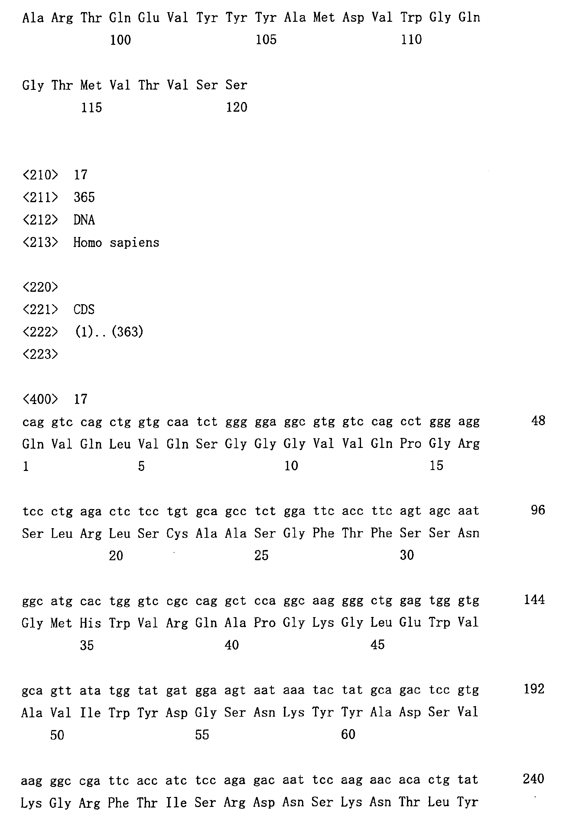
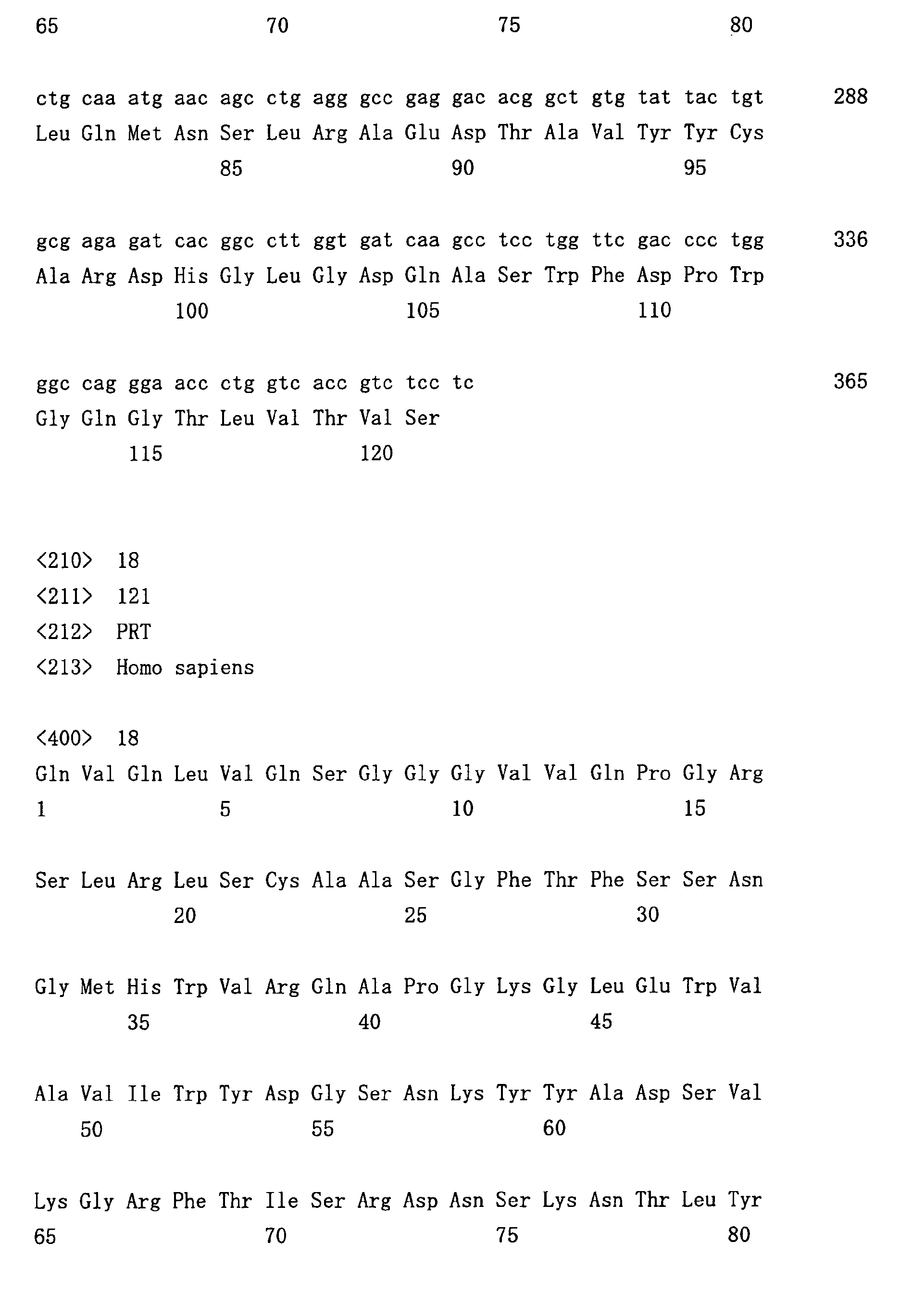
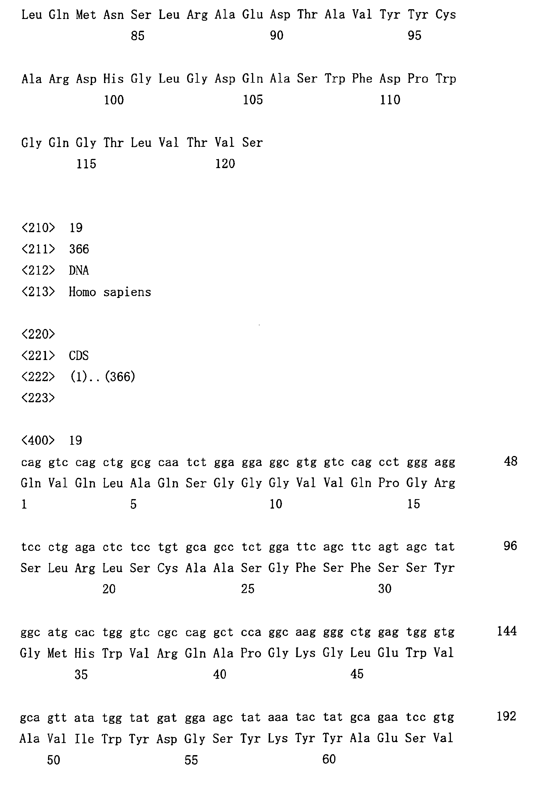
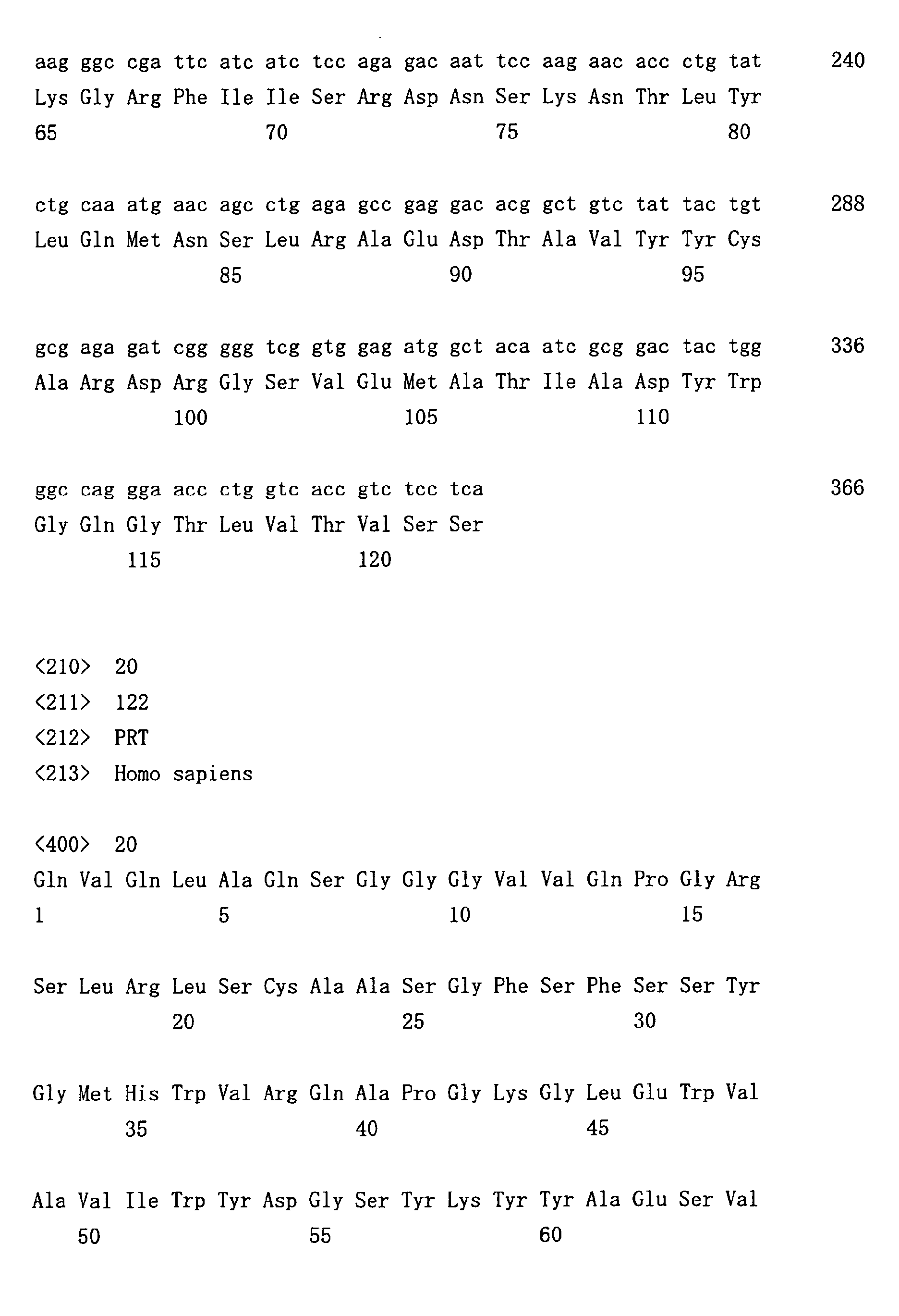
- Nucleotide sequences of the insert sequences in the recombinant plasmids were determined and it was confirmed that antibody genes were amplified.
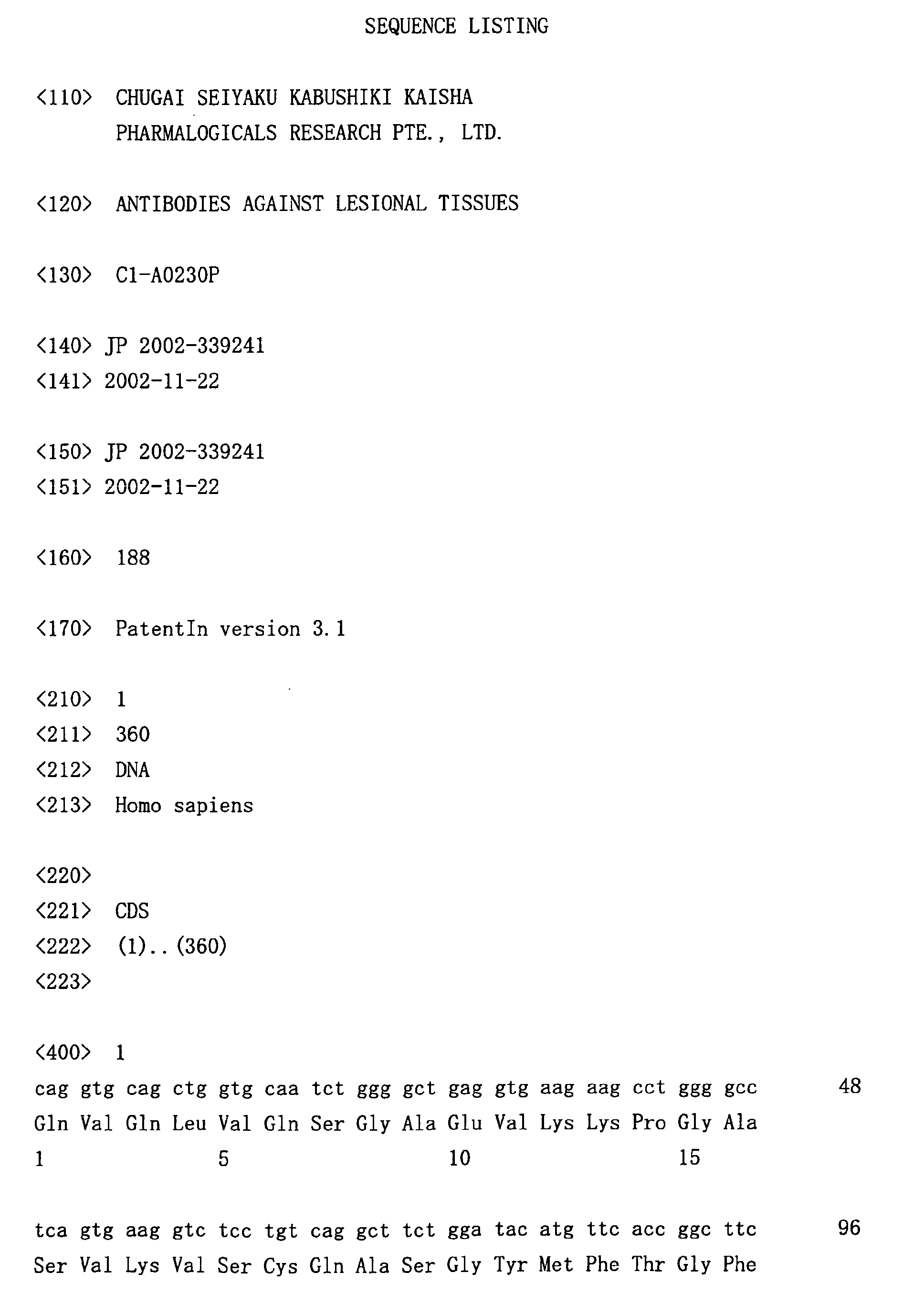
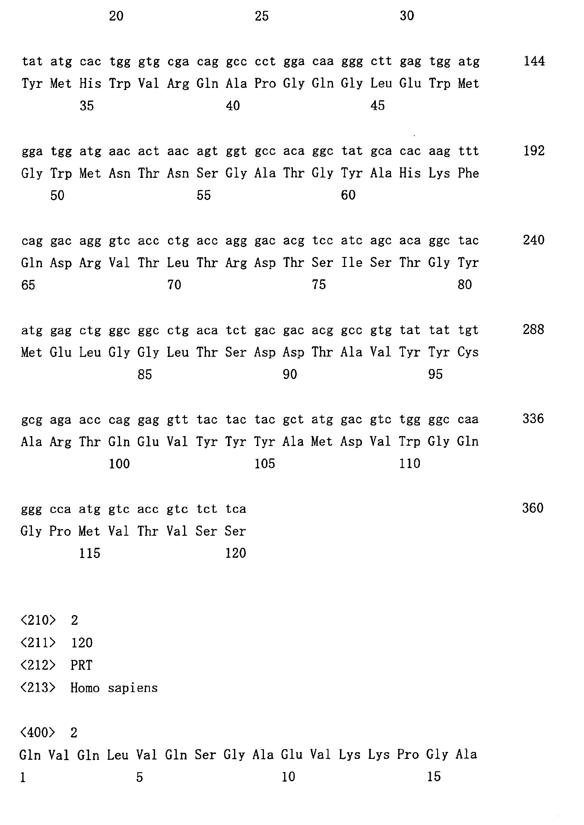
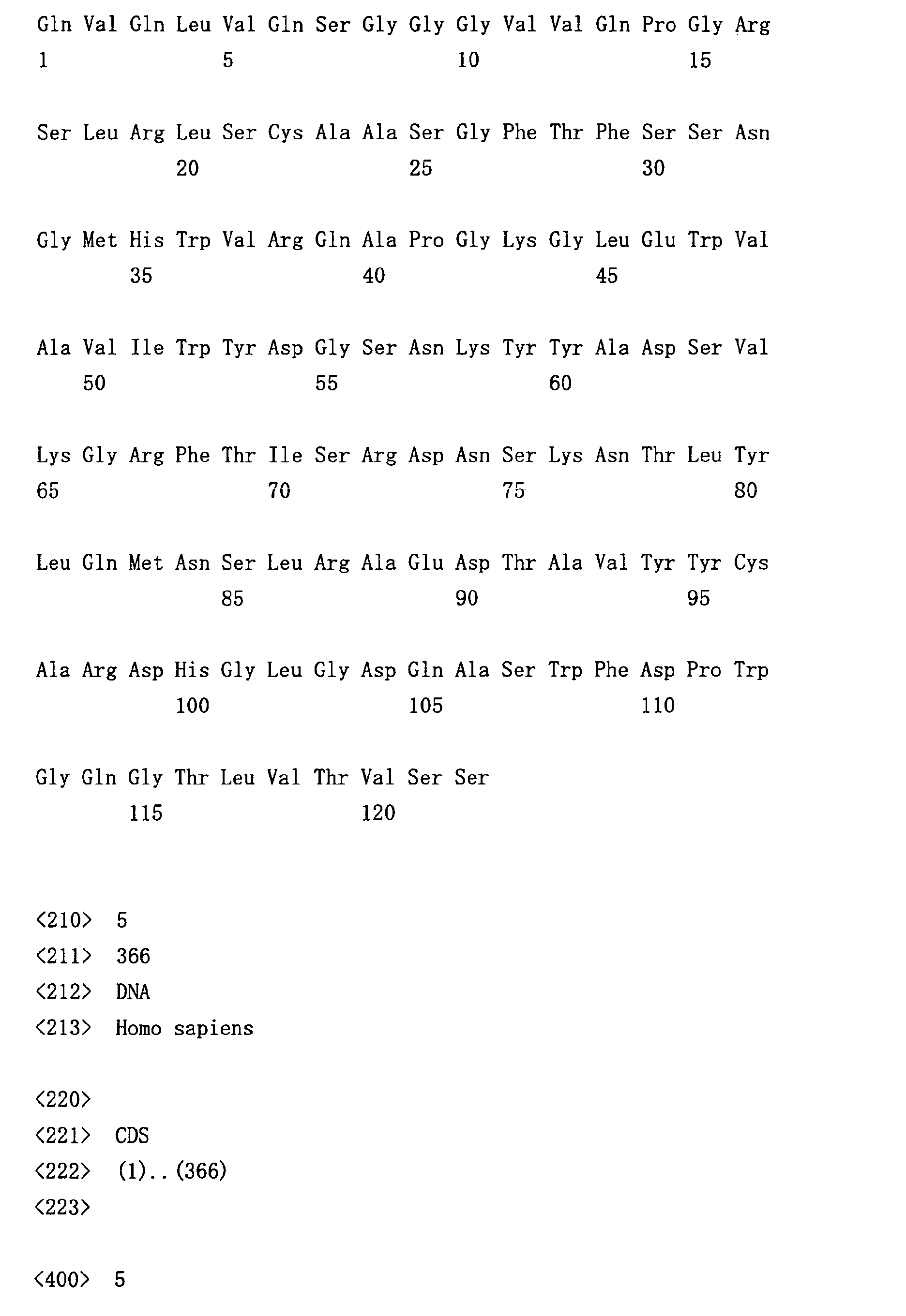
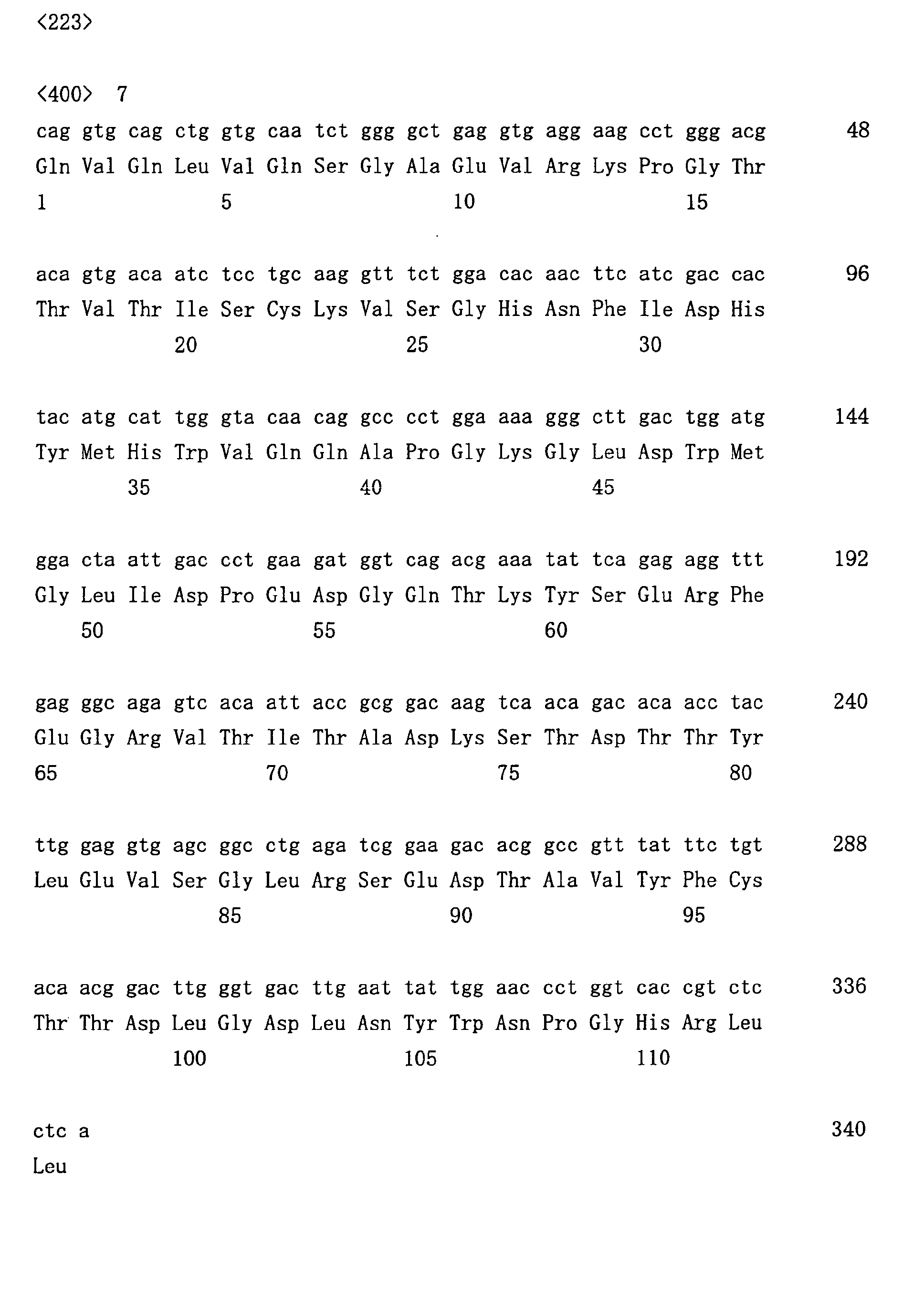
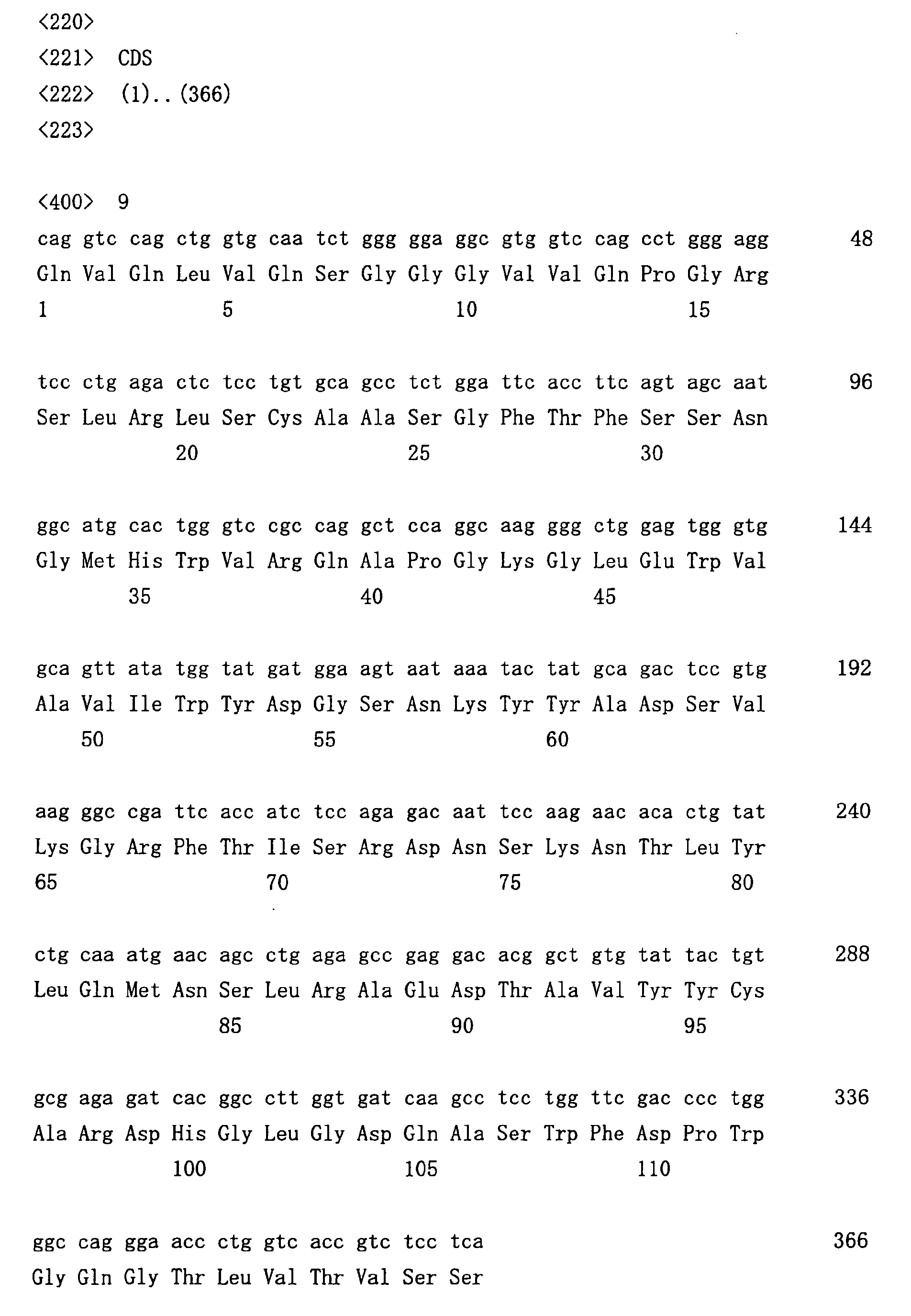
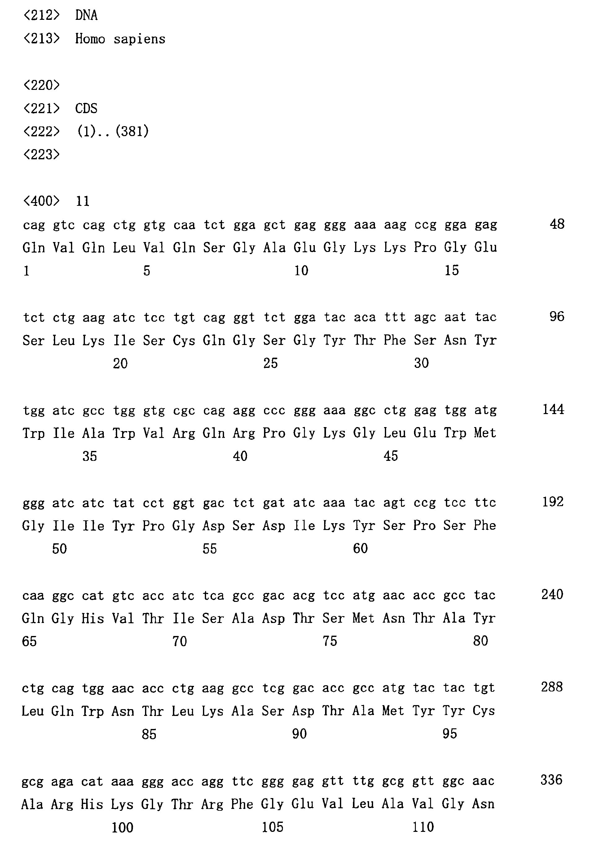
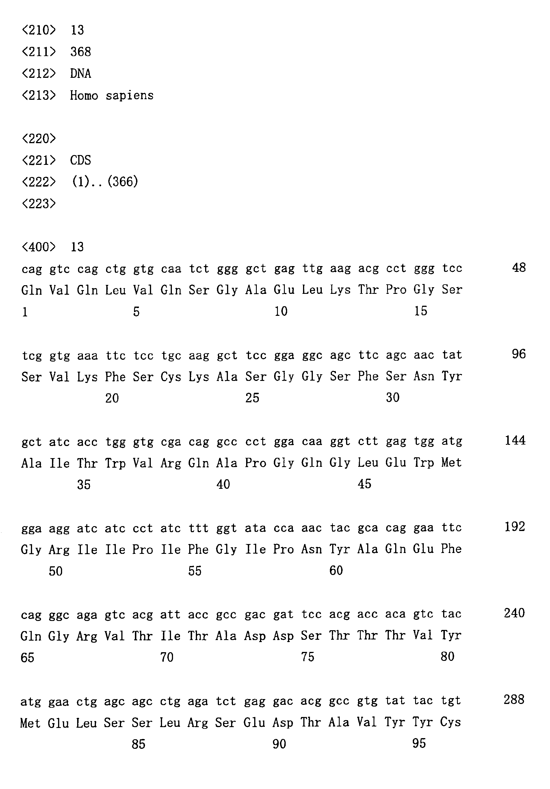
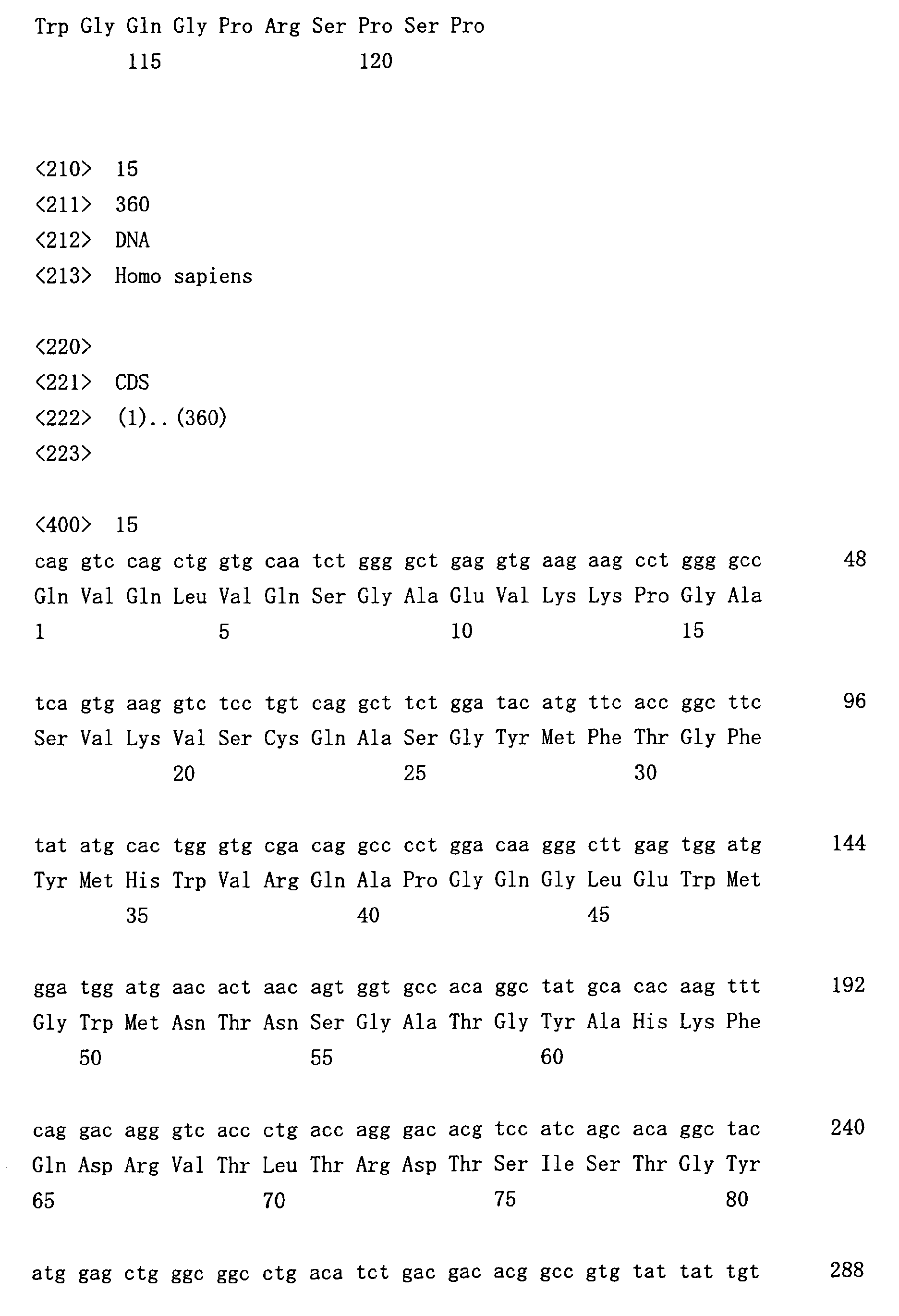
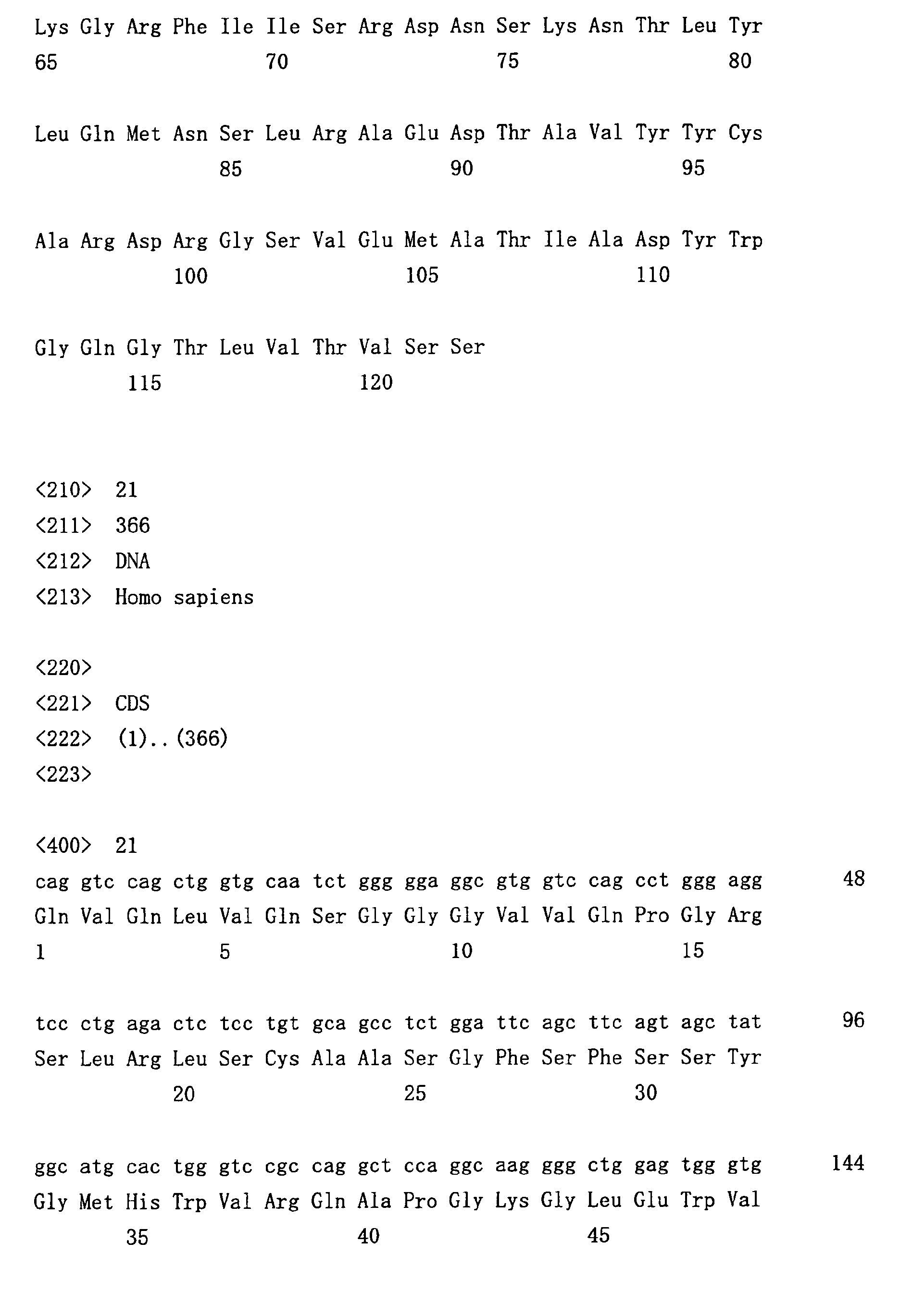
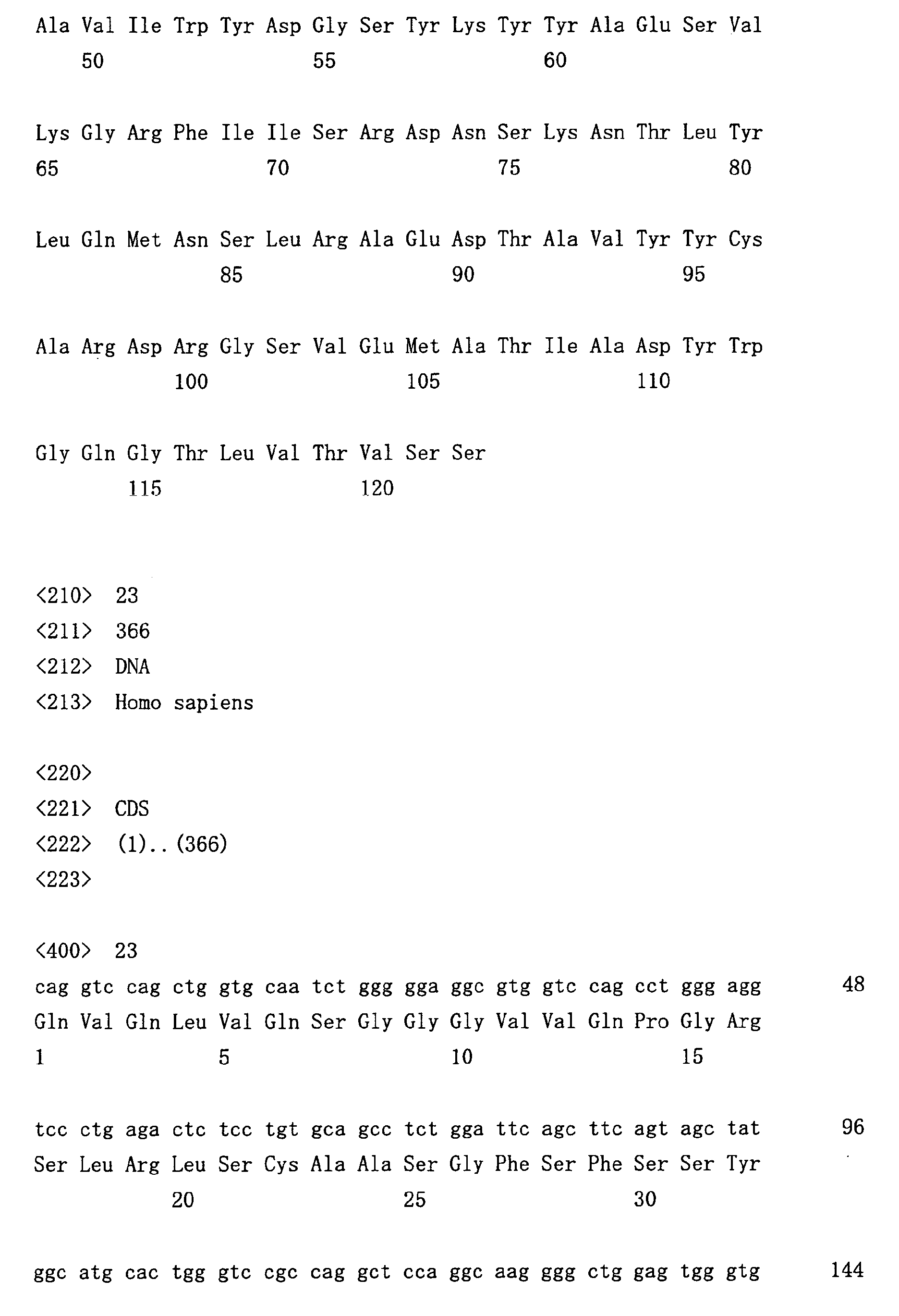
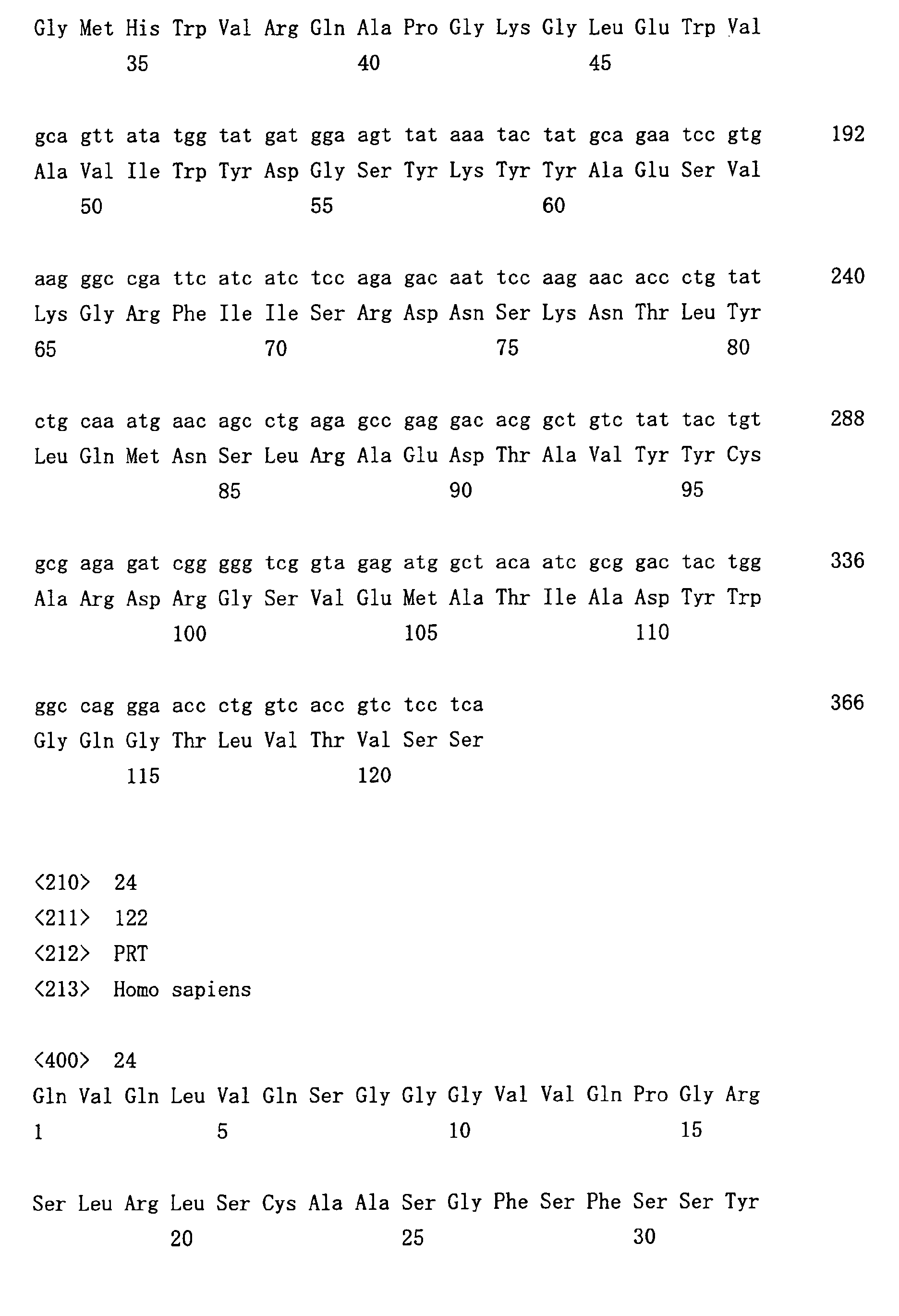
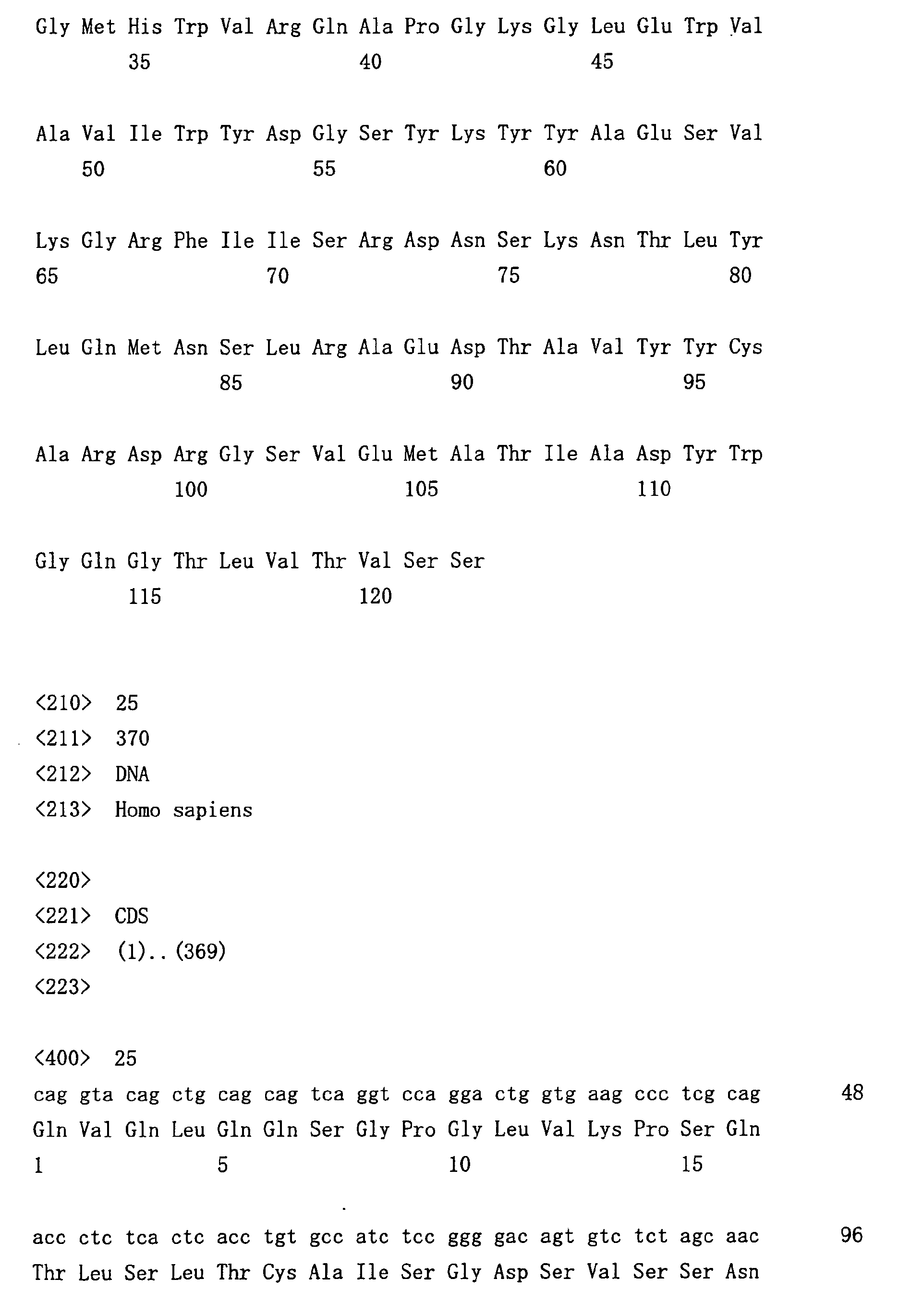
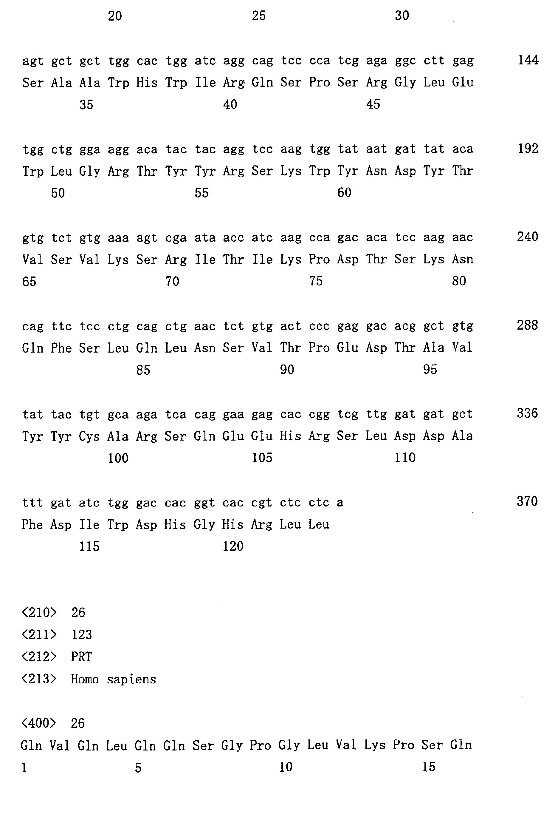
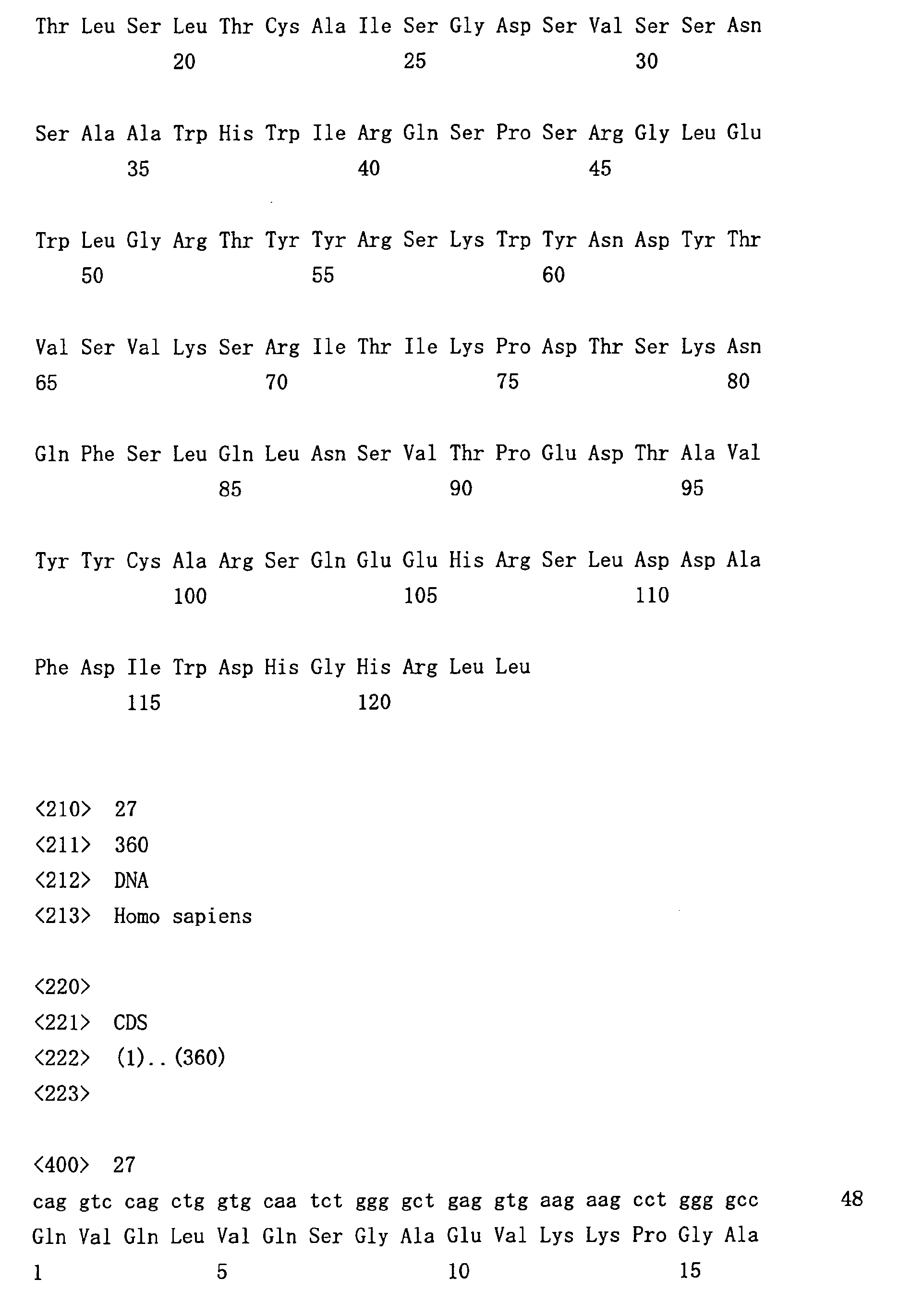
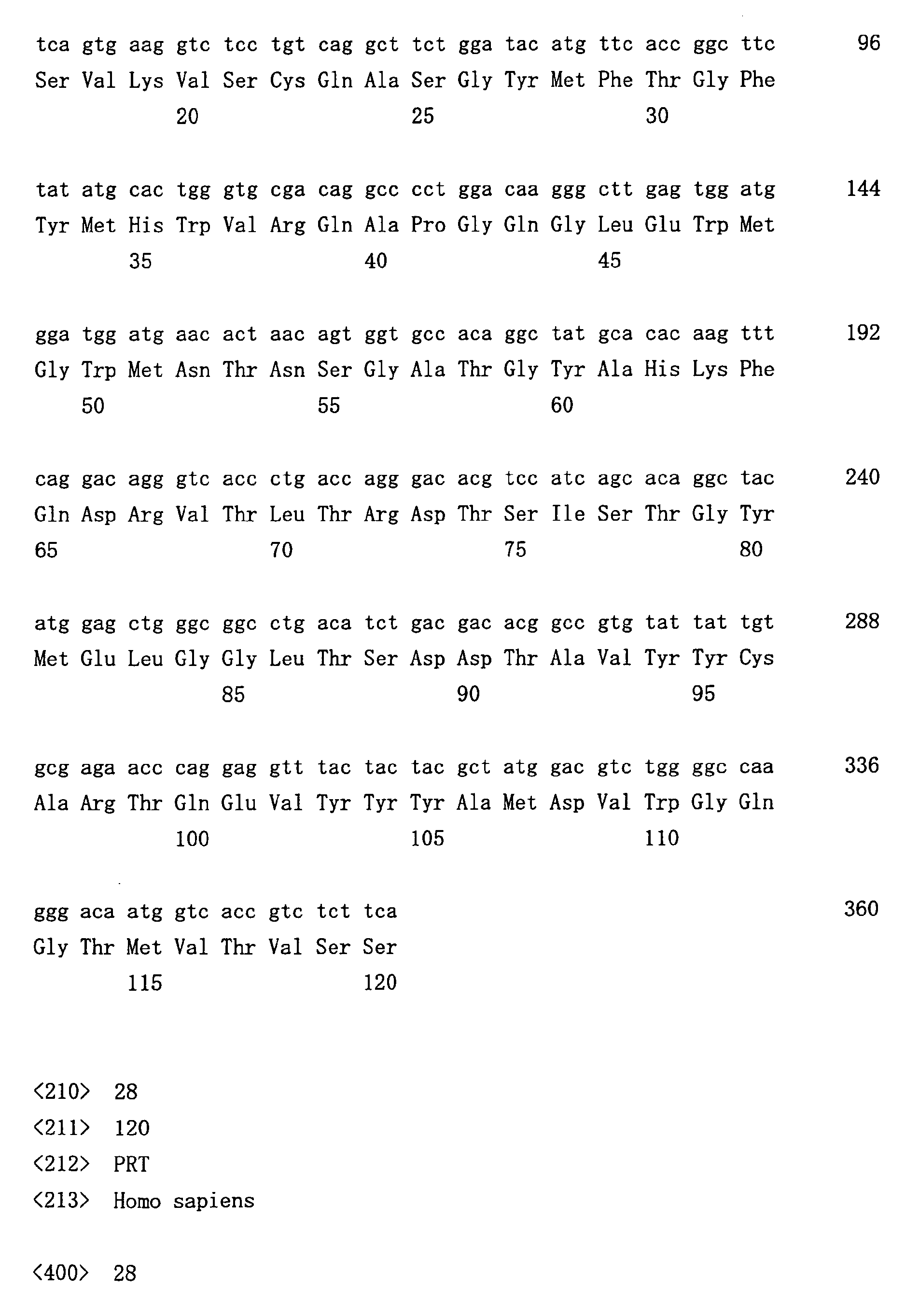
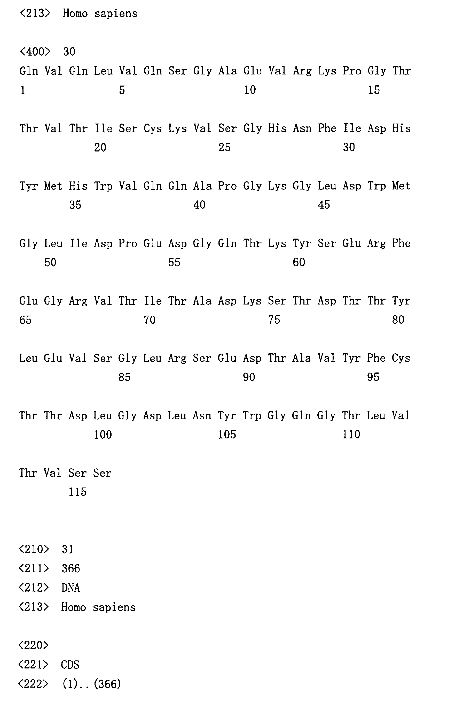
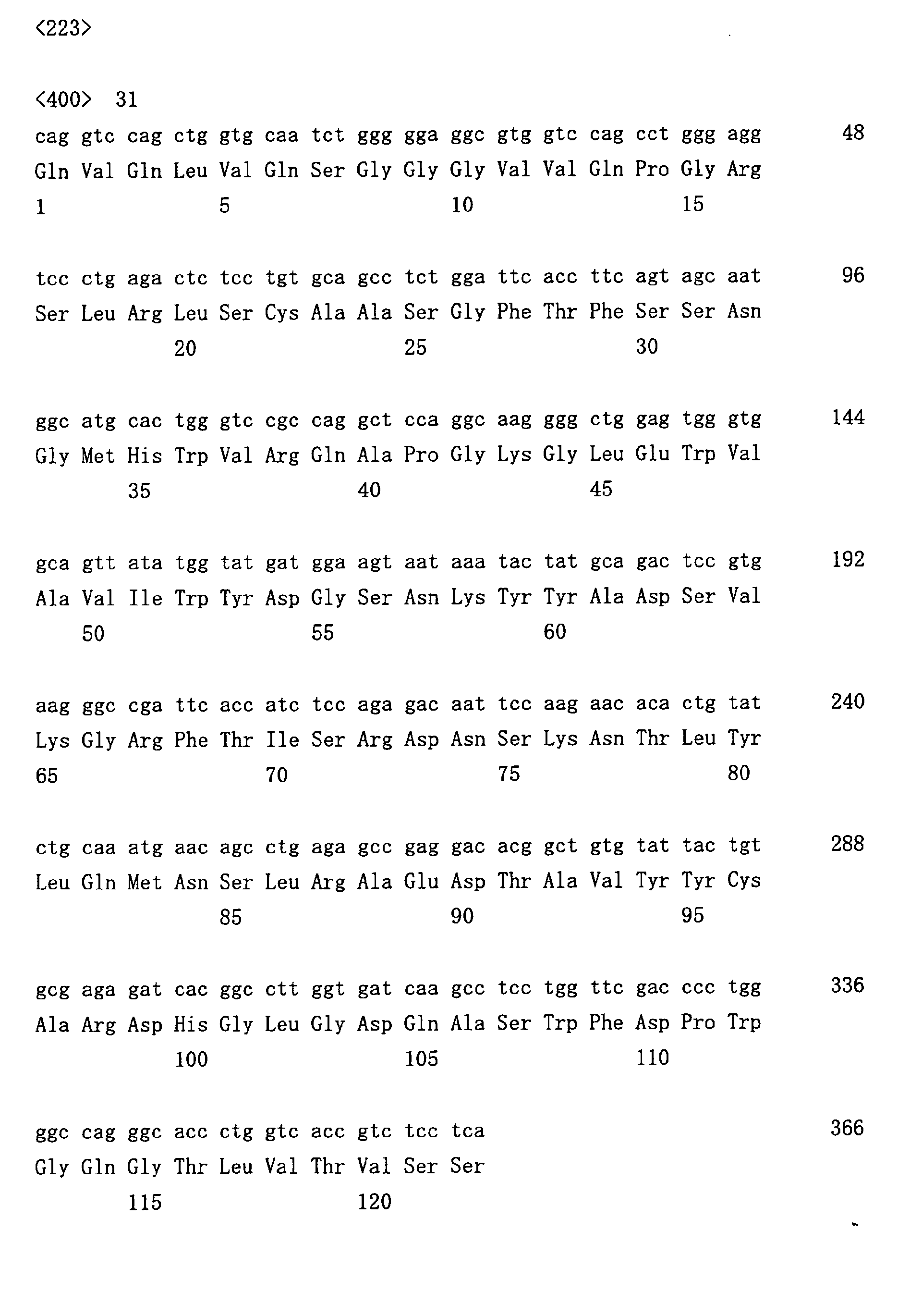
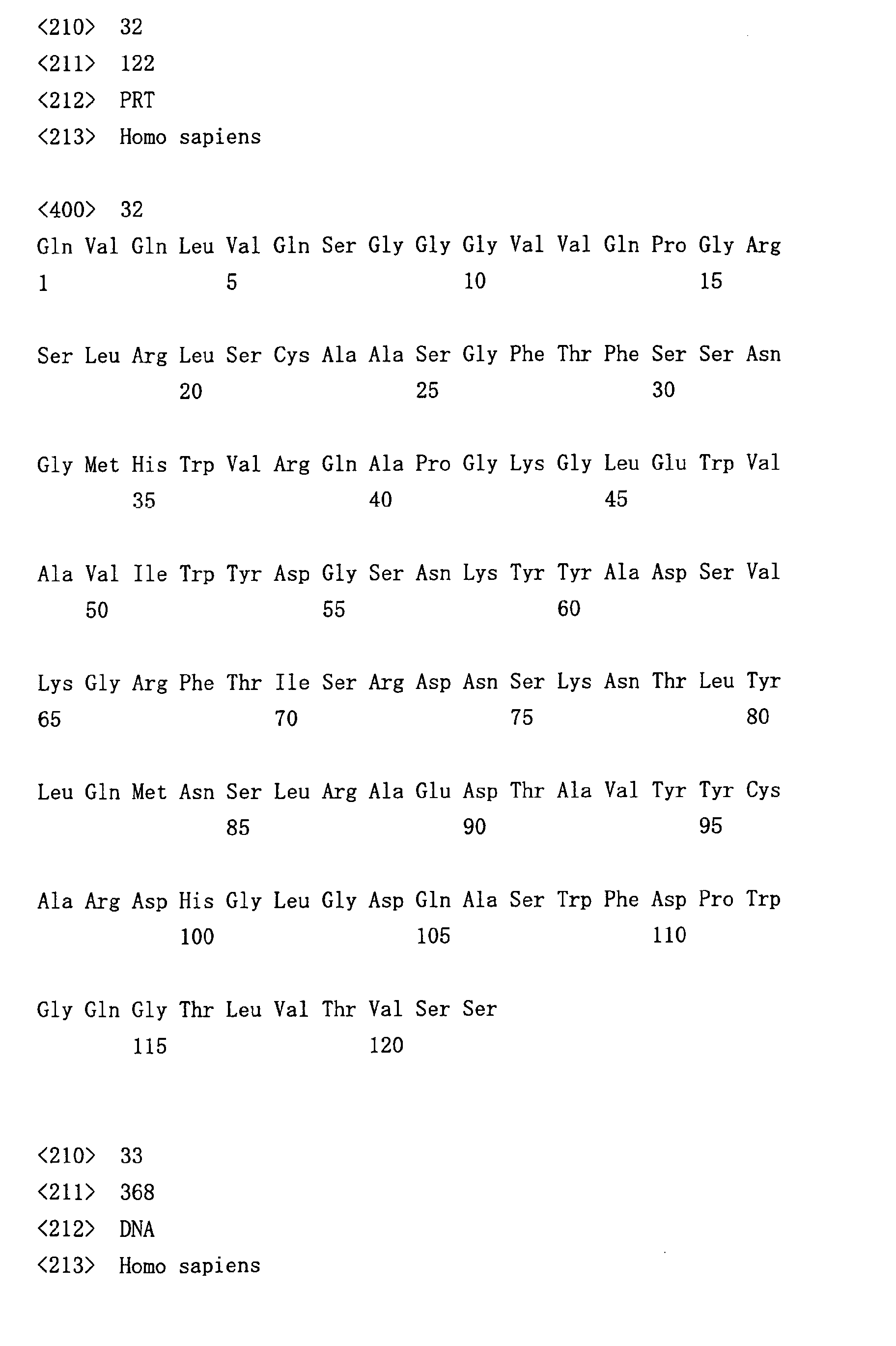
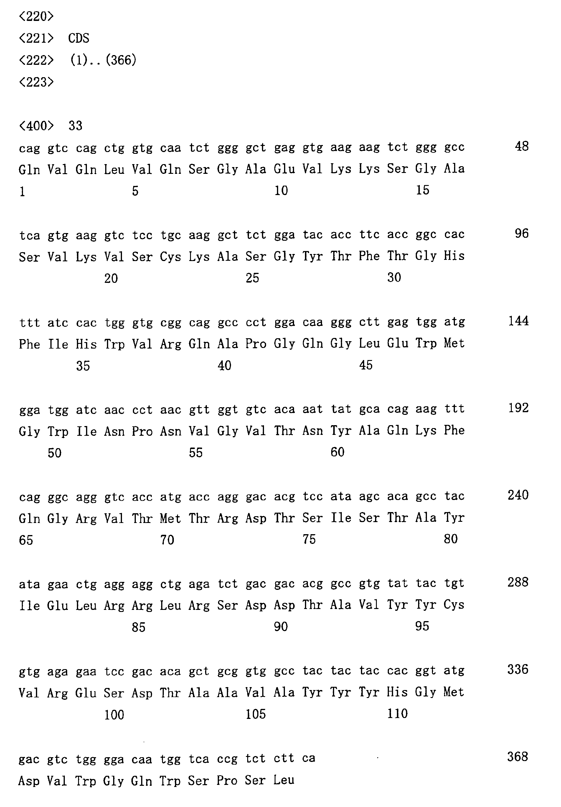
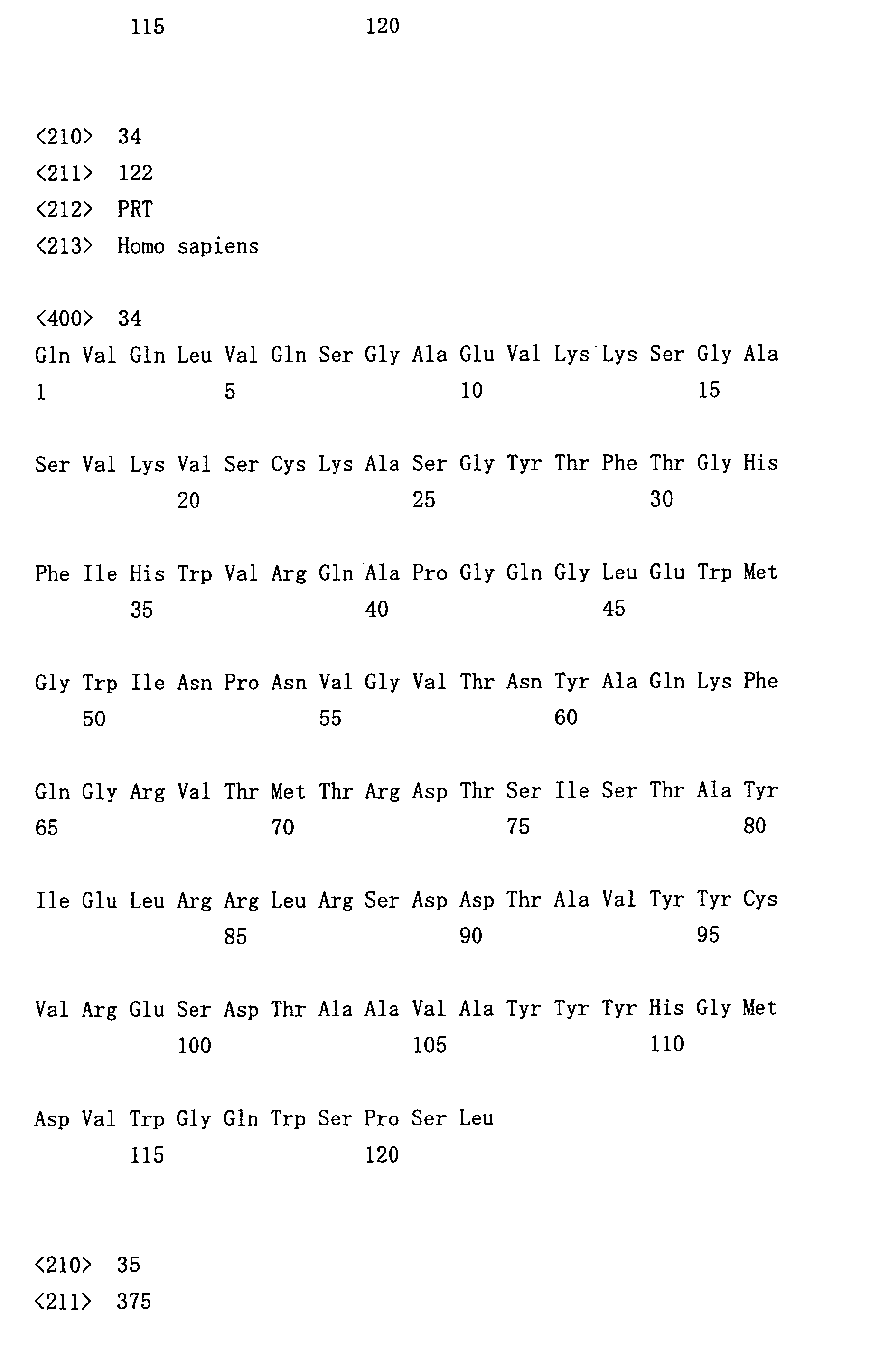
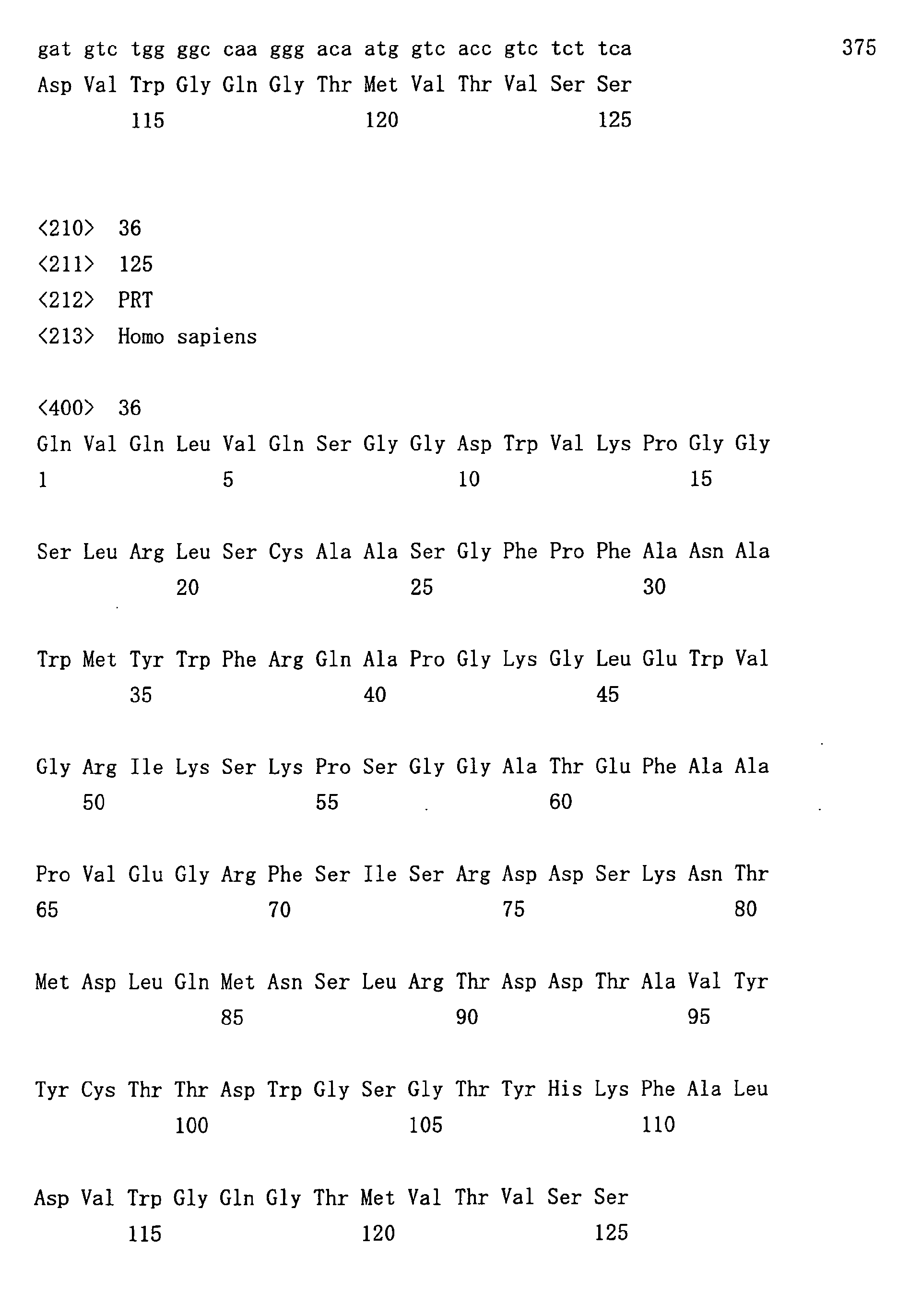
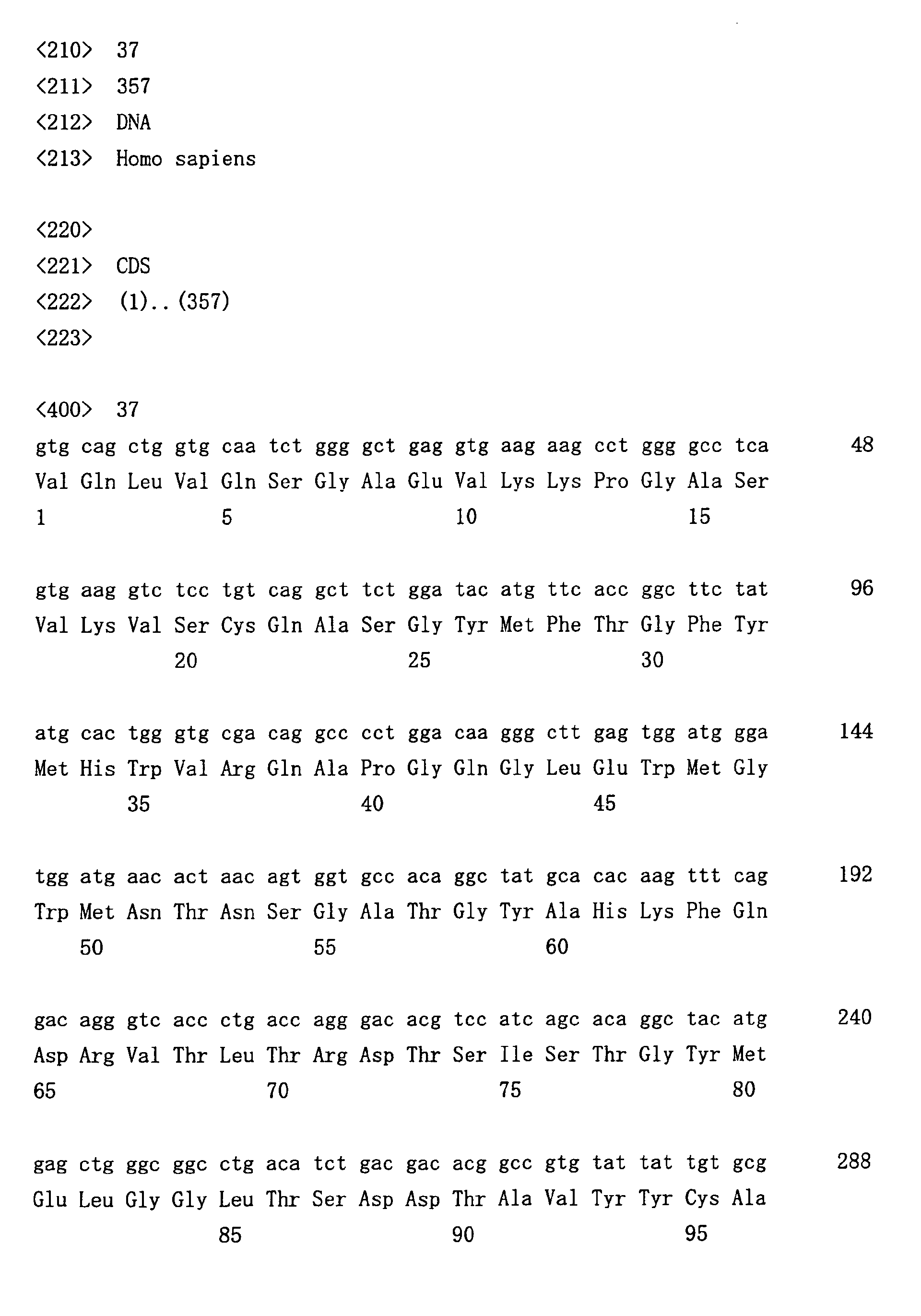
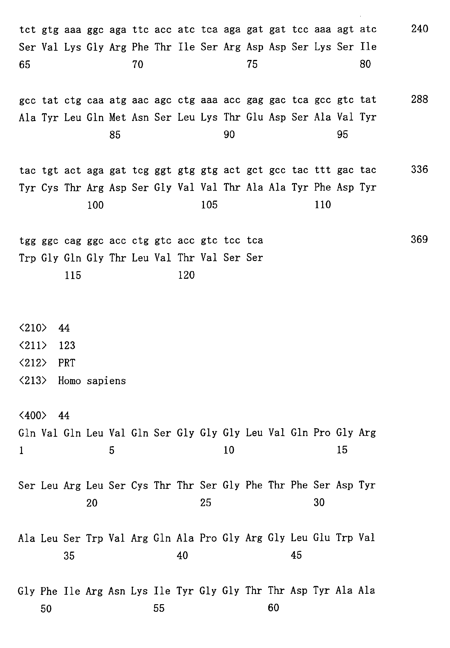
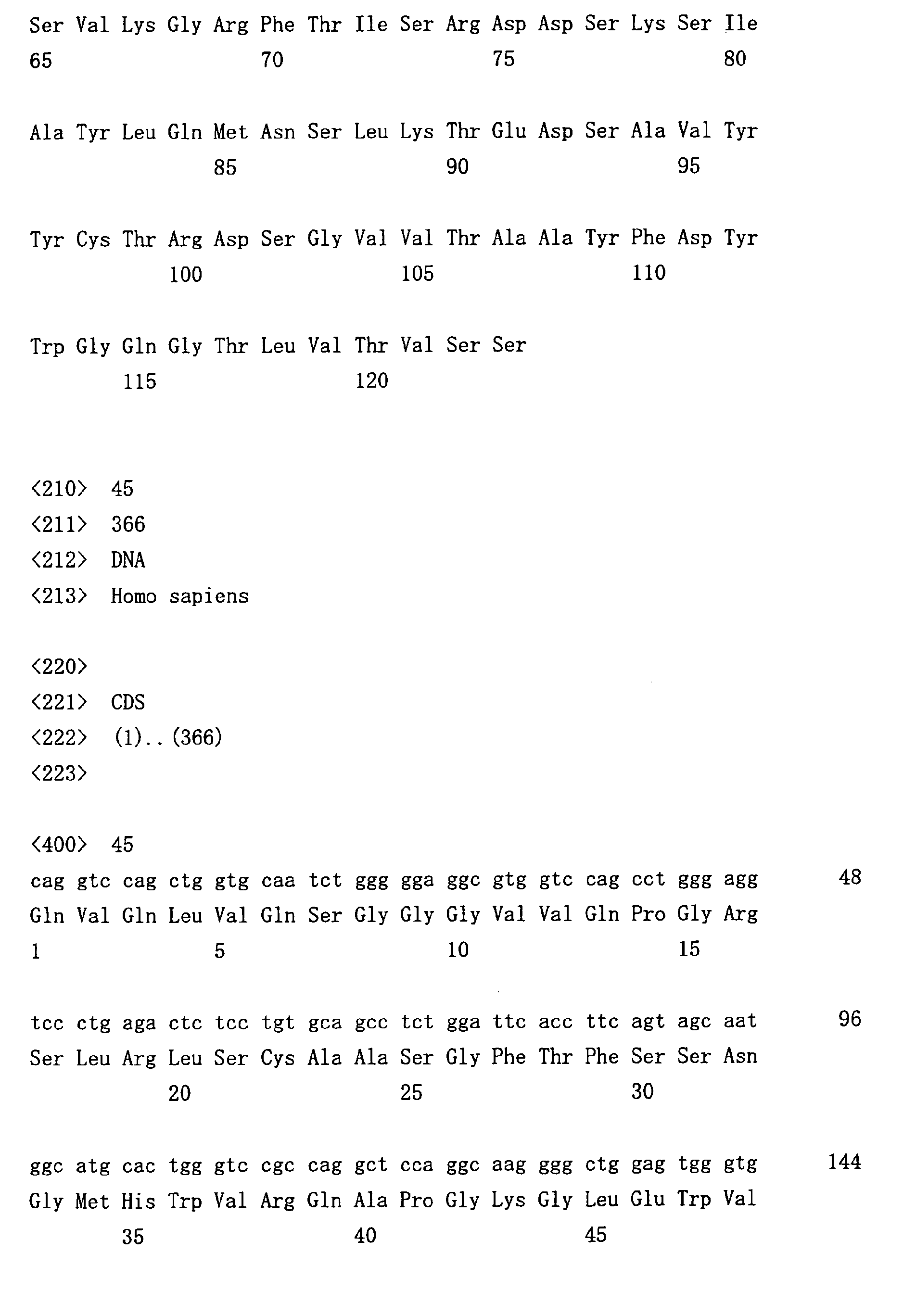
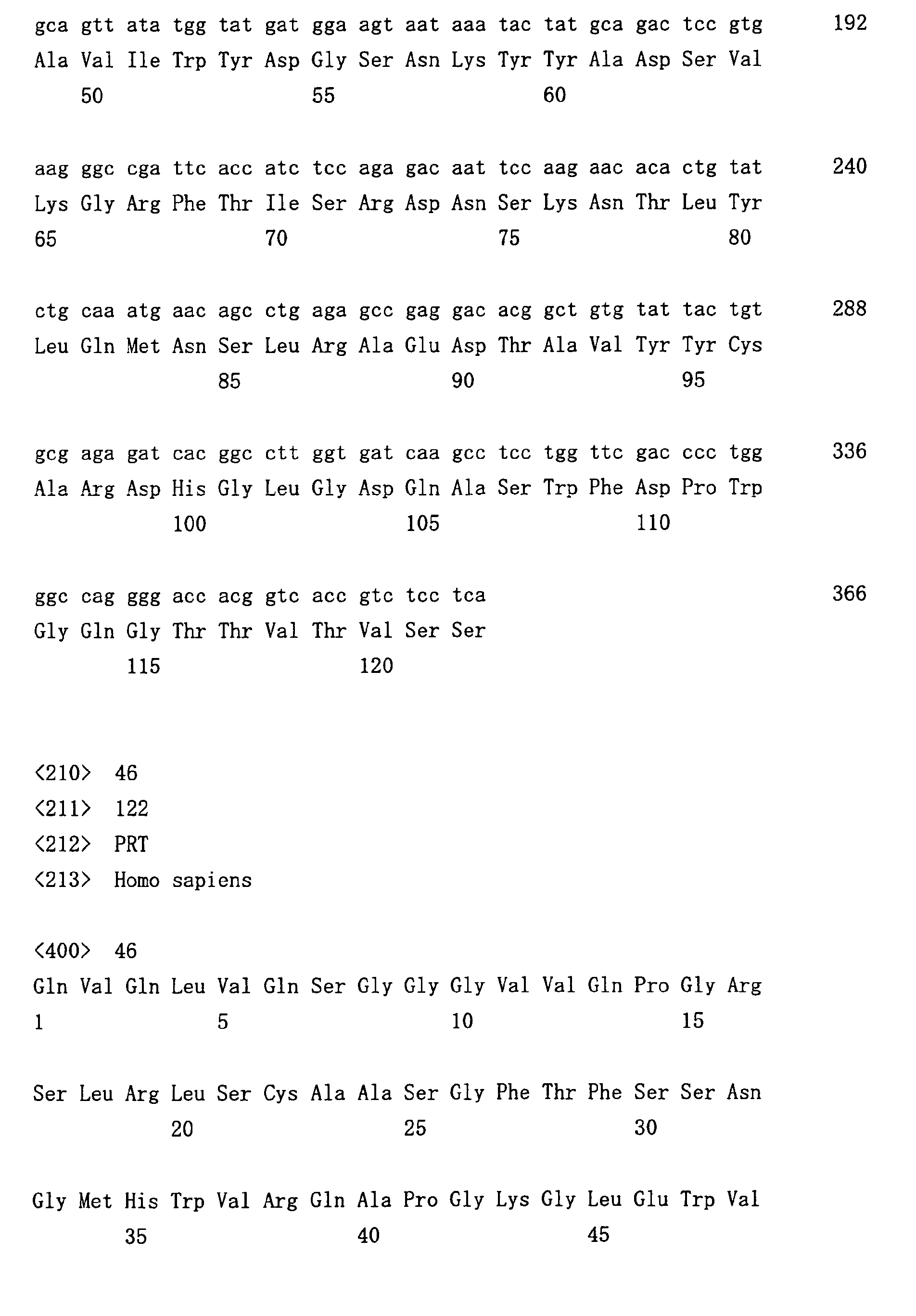
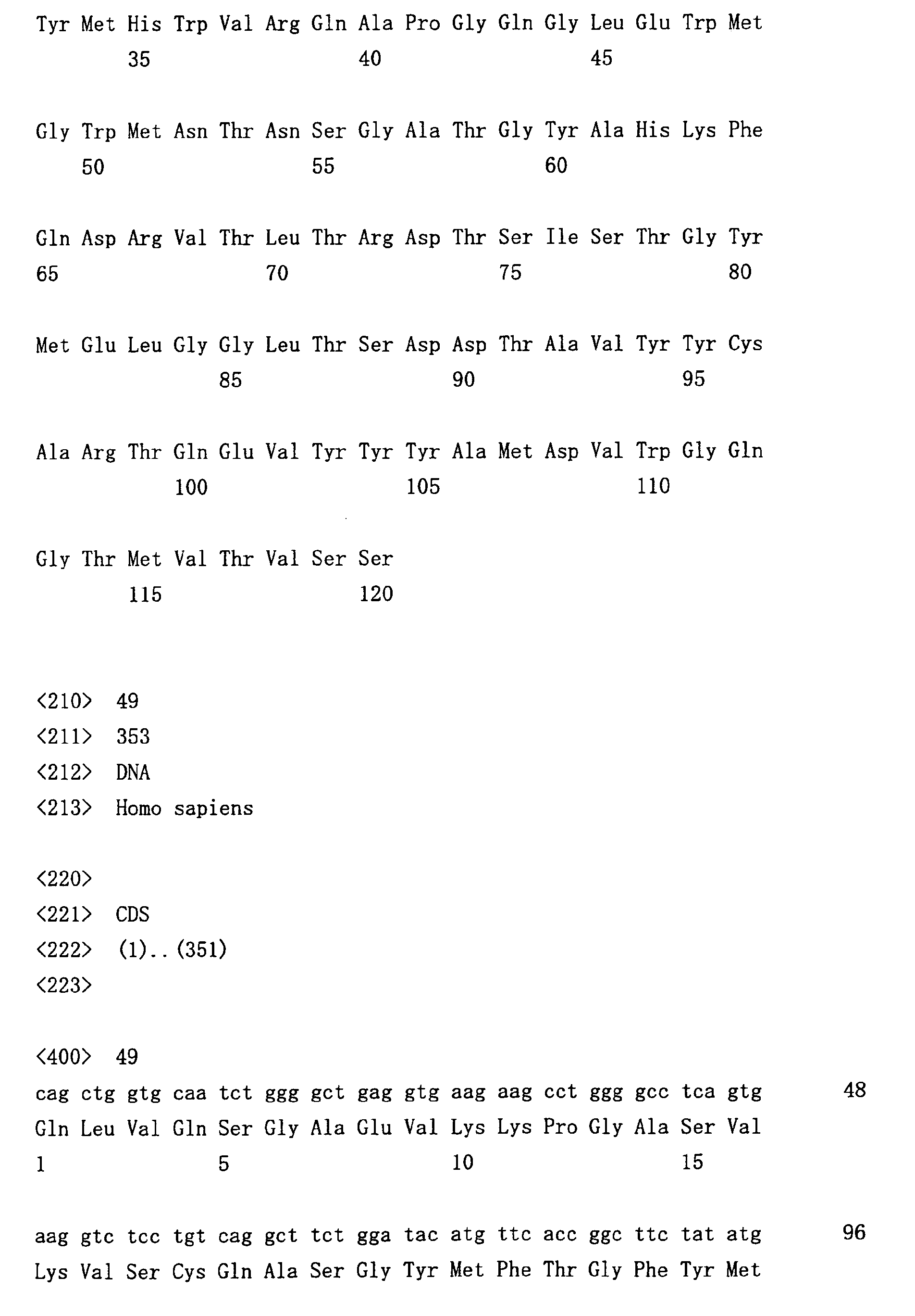
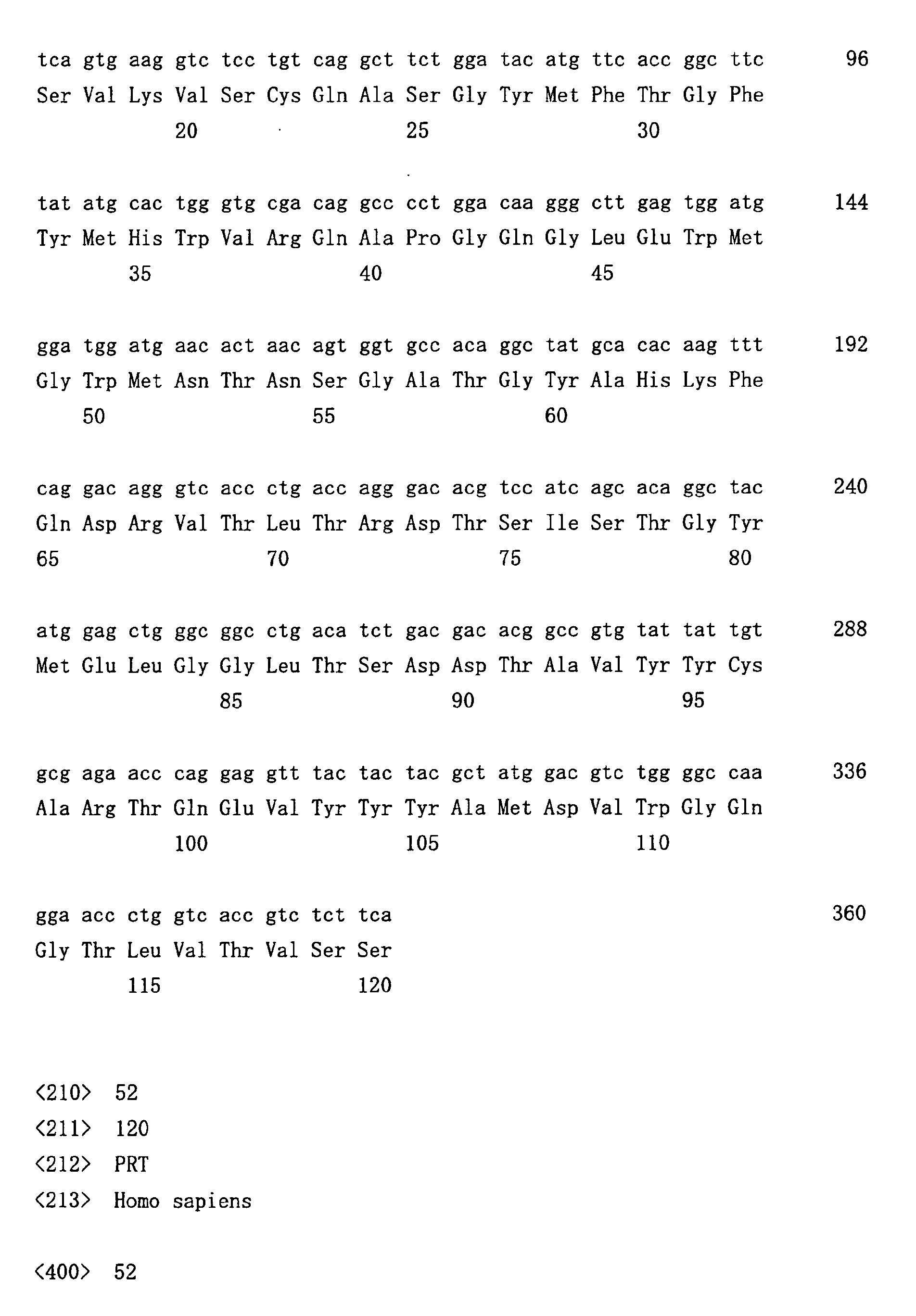
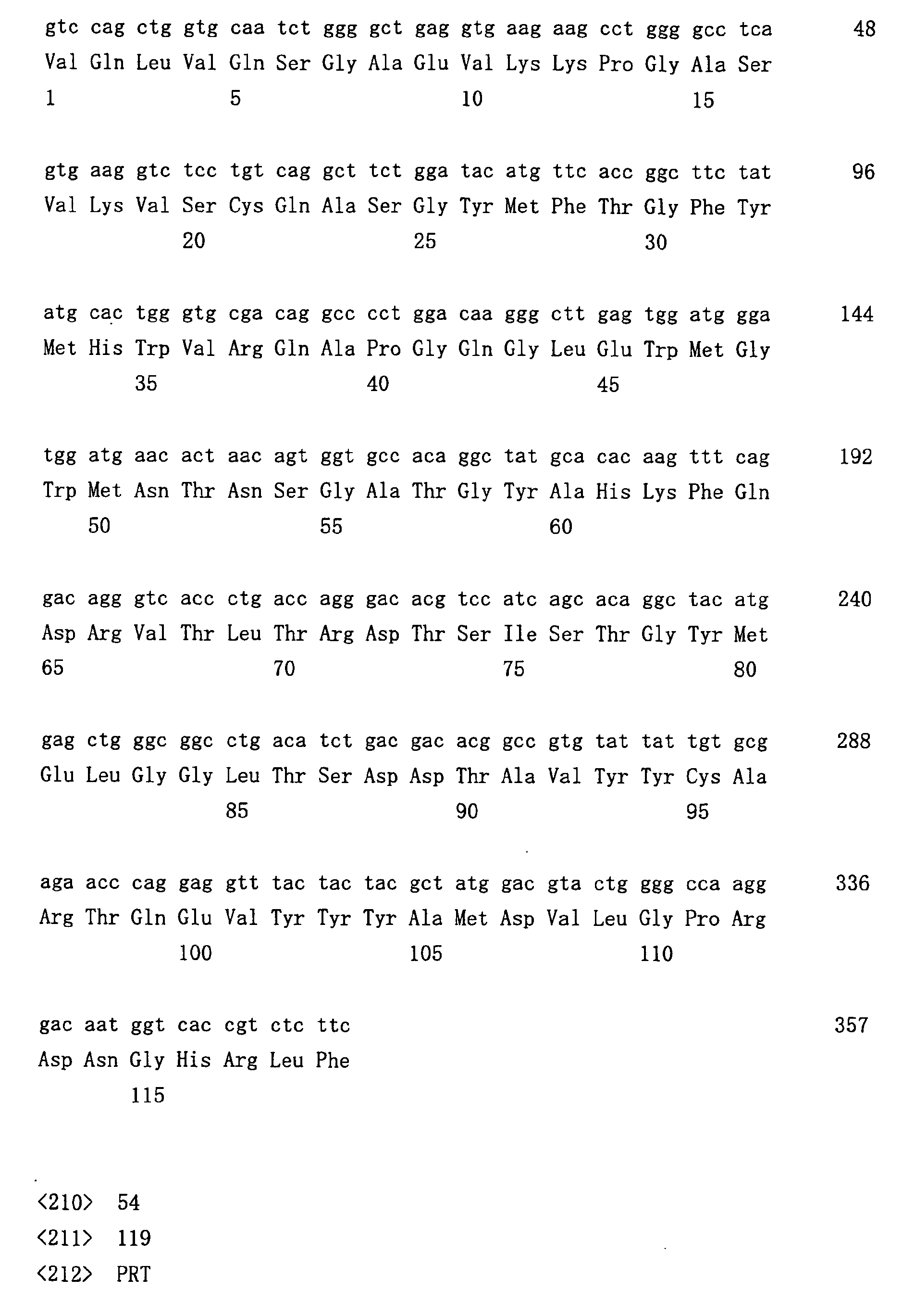
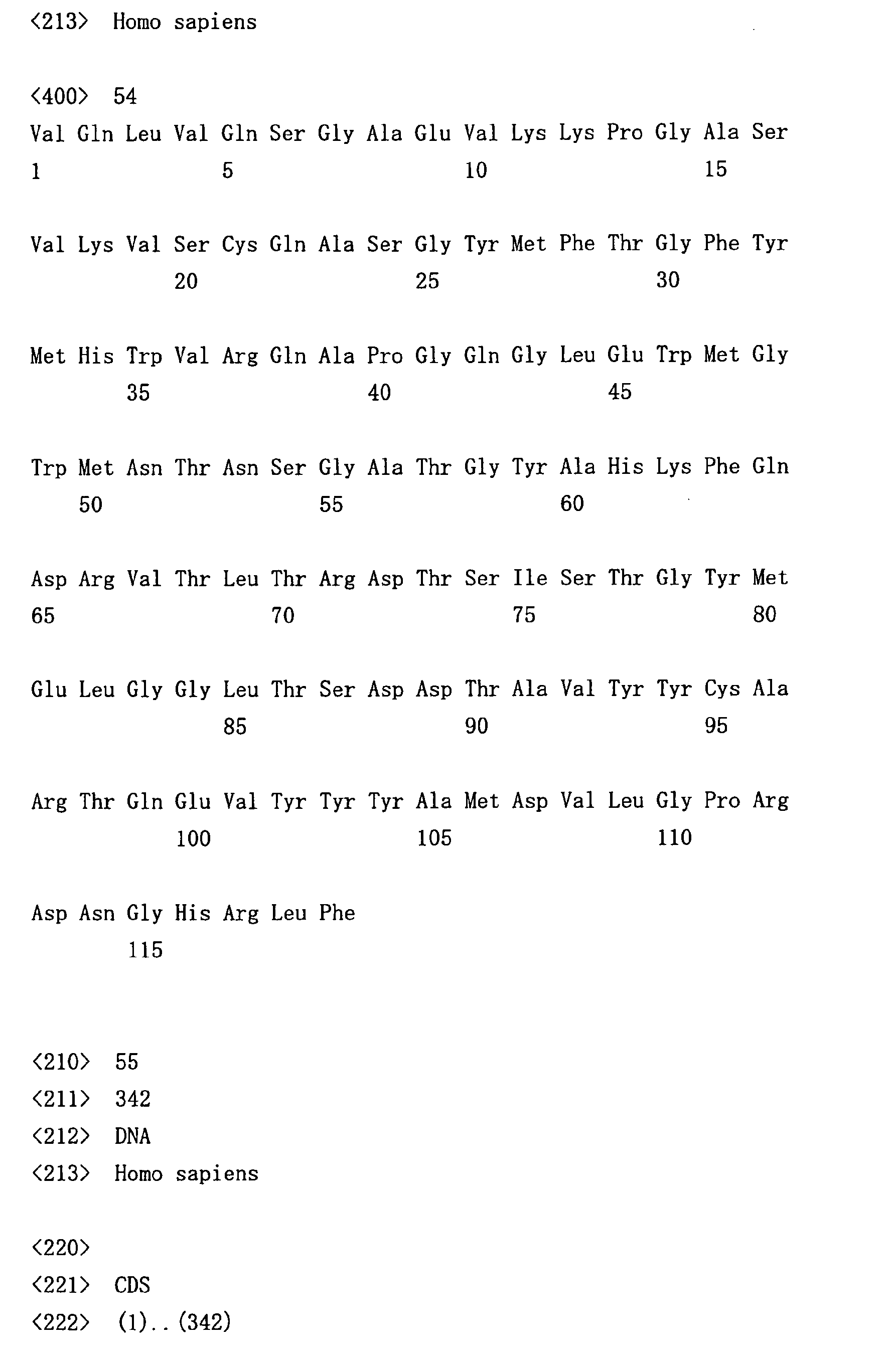
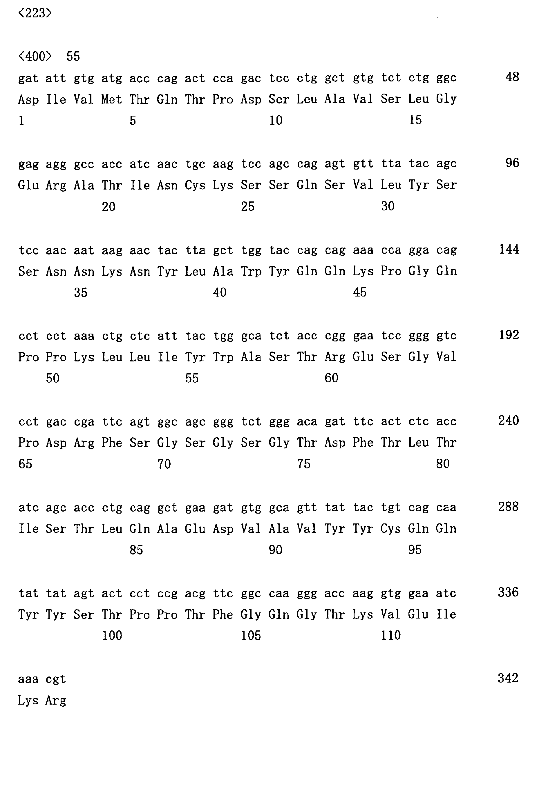
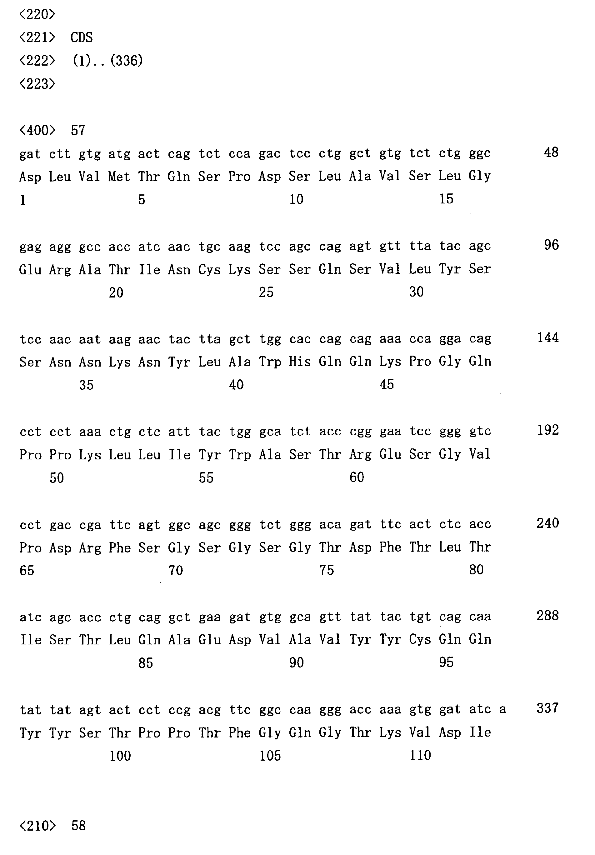
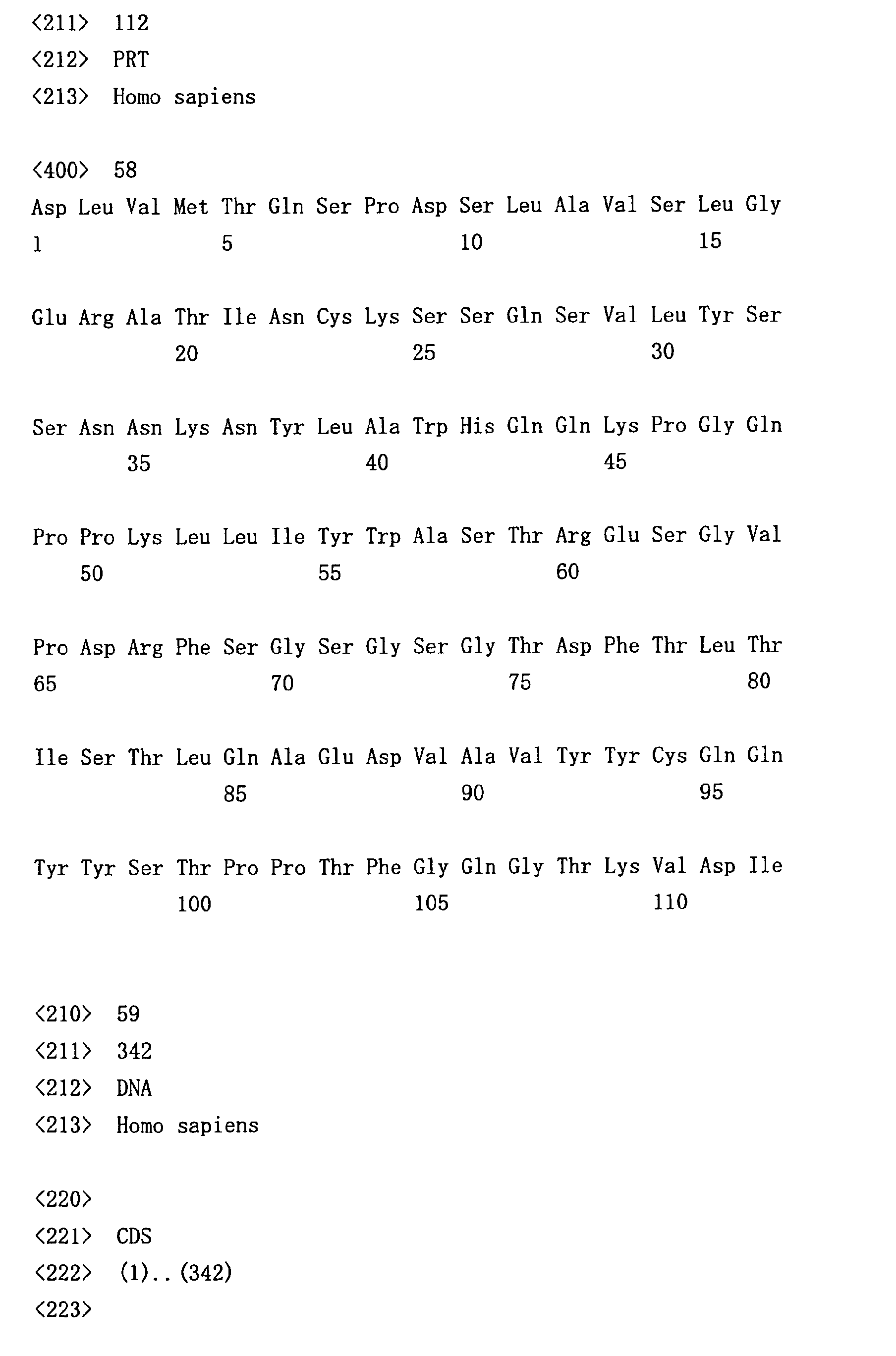
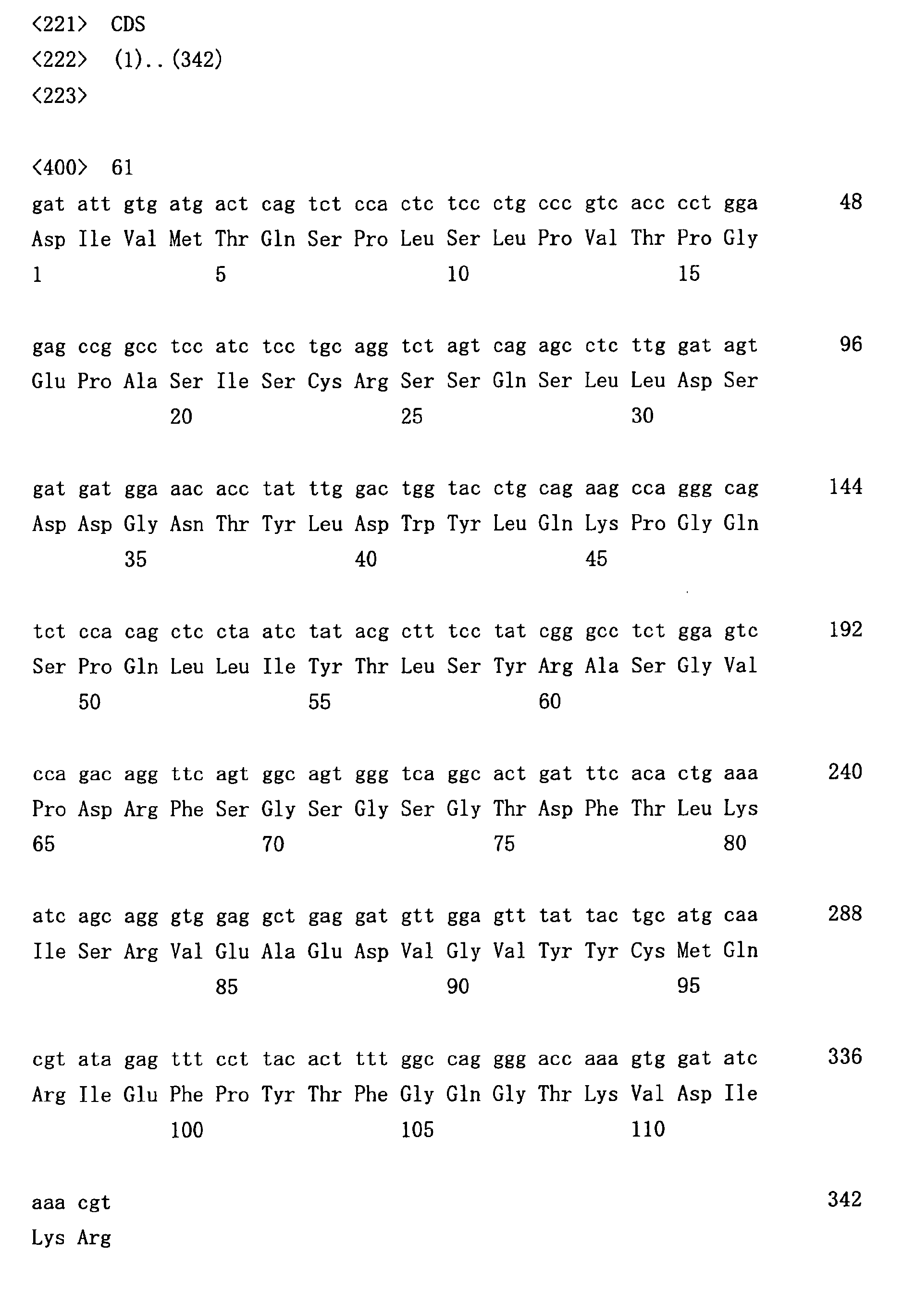
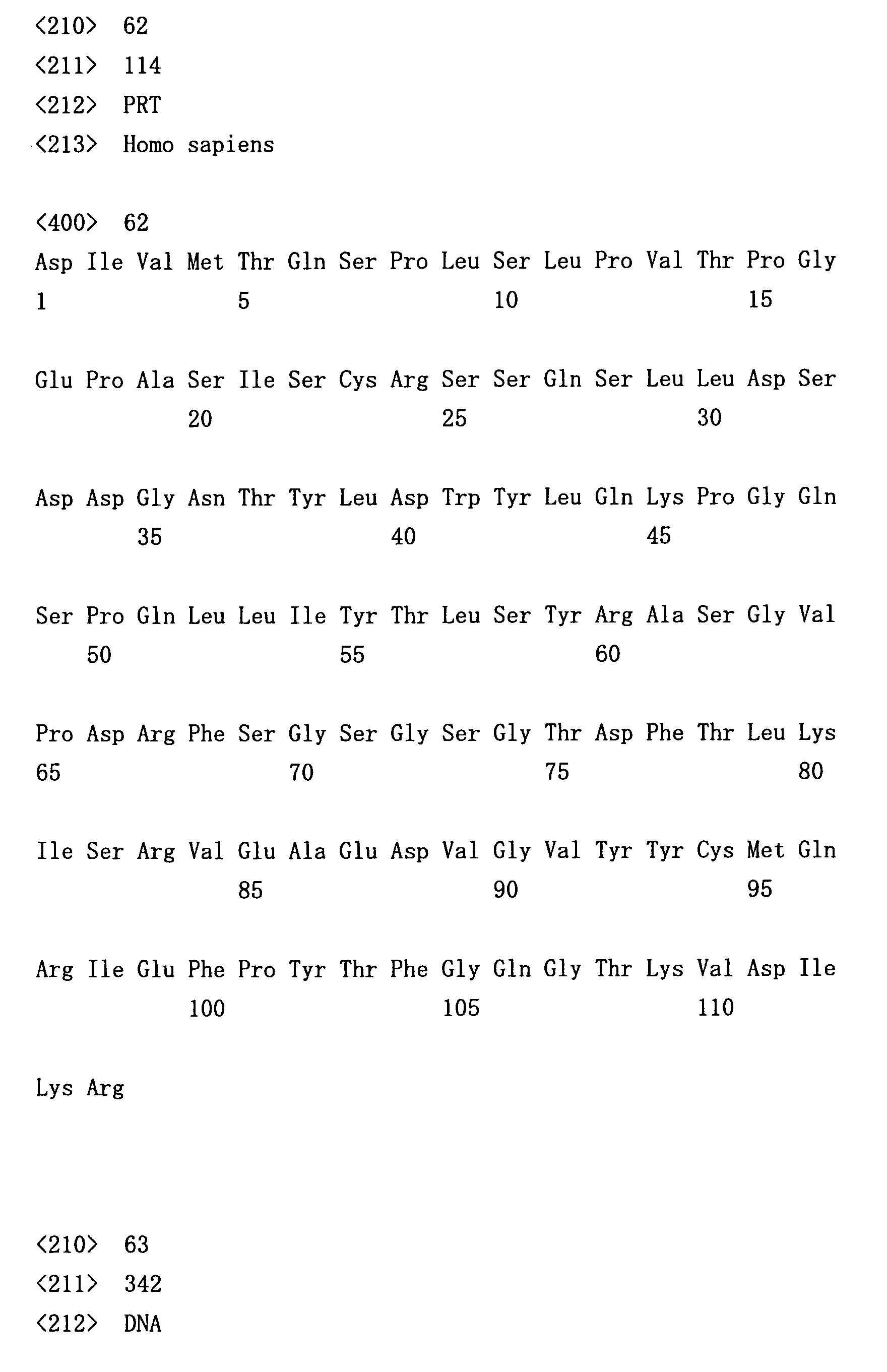
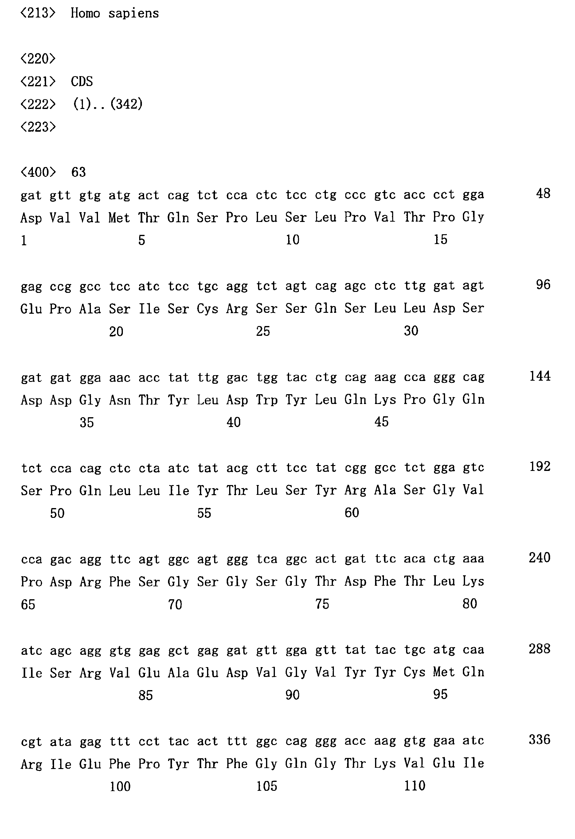
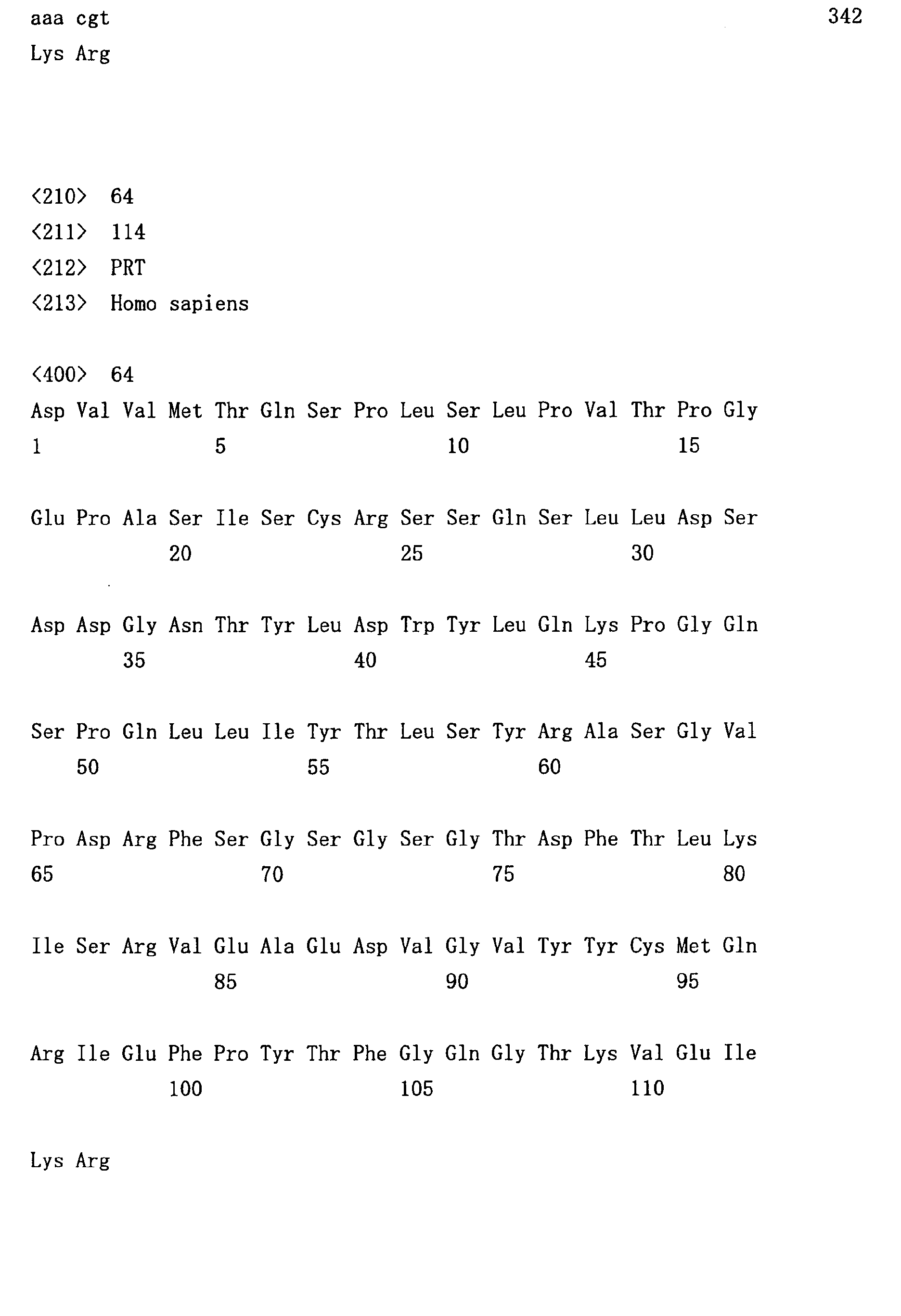
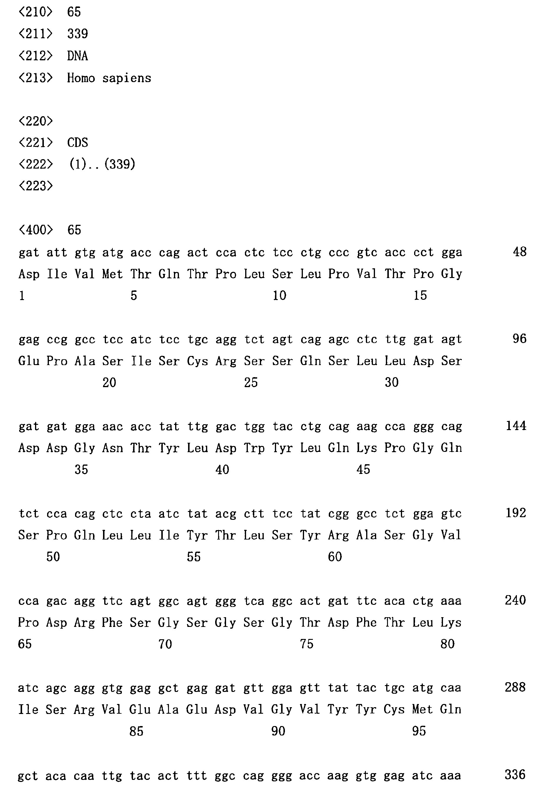
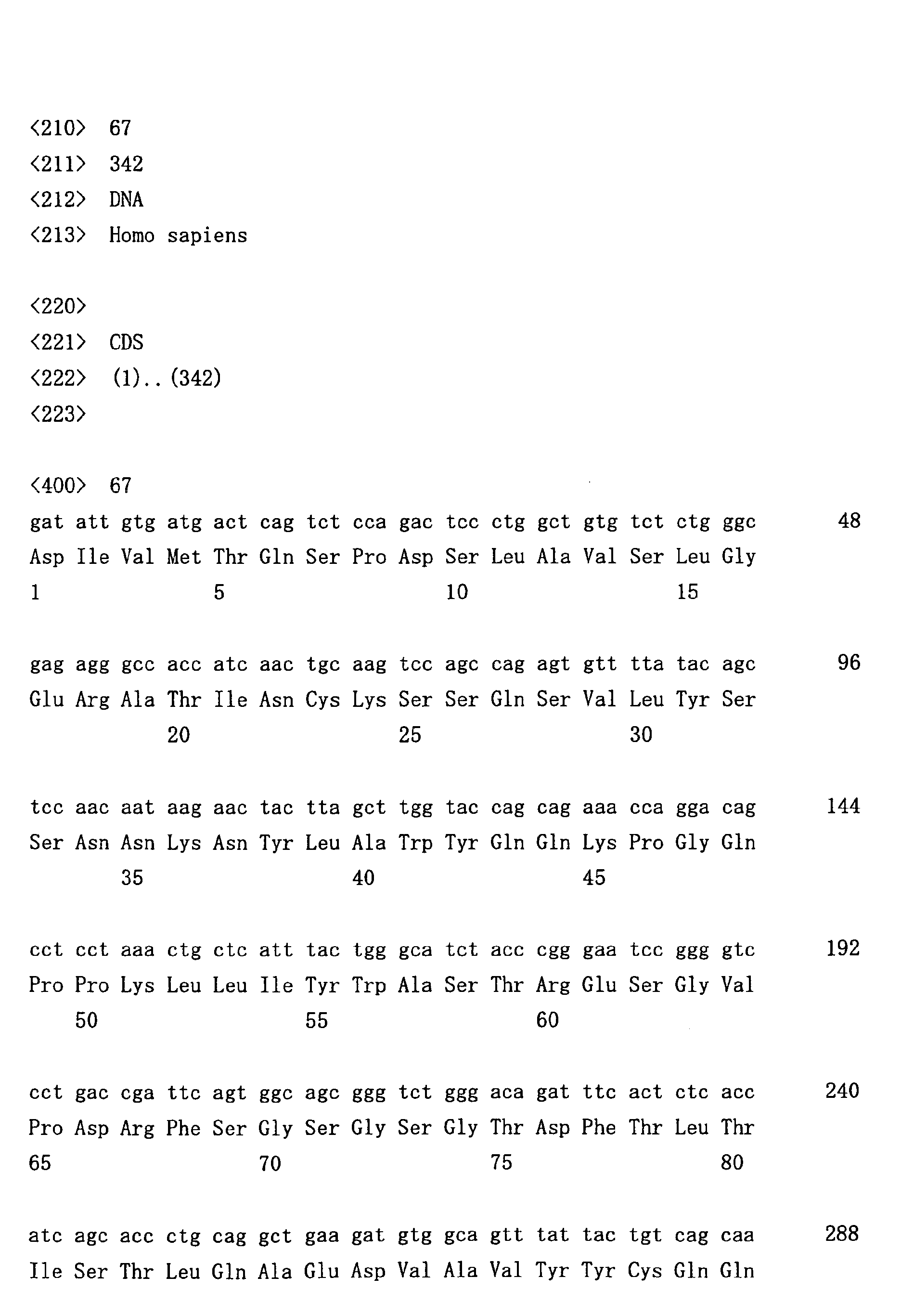
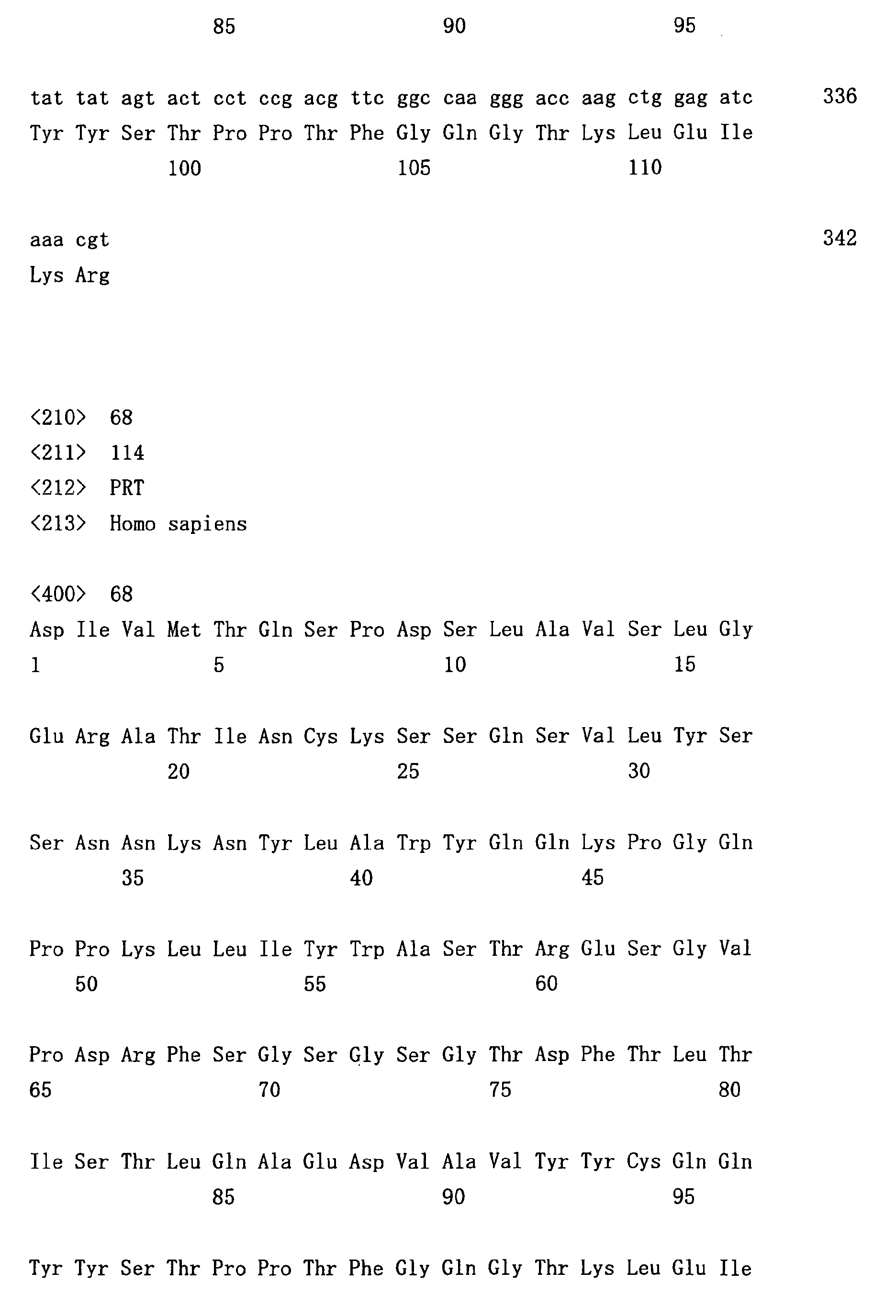
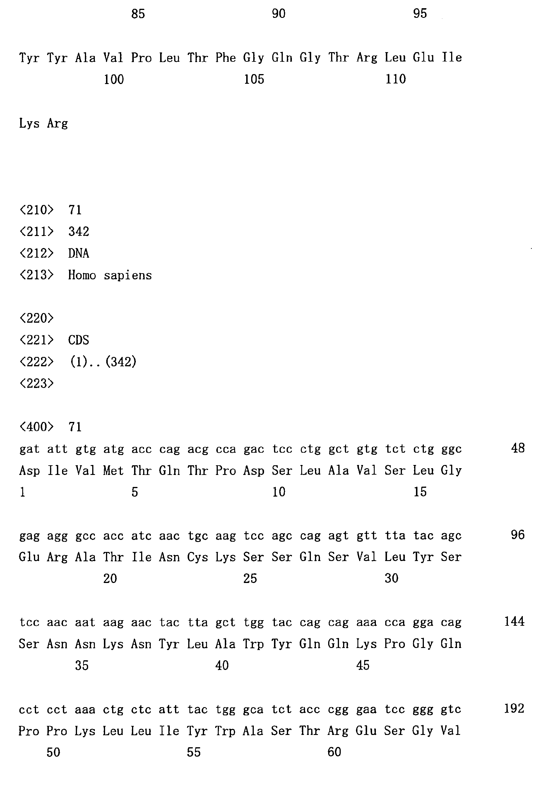
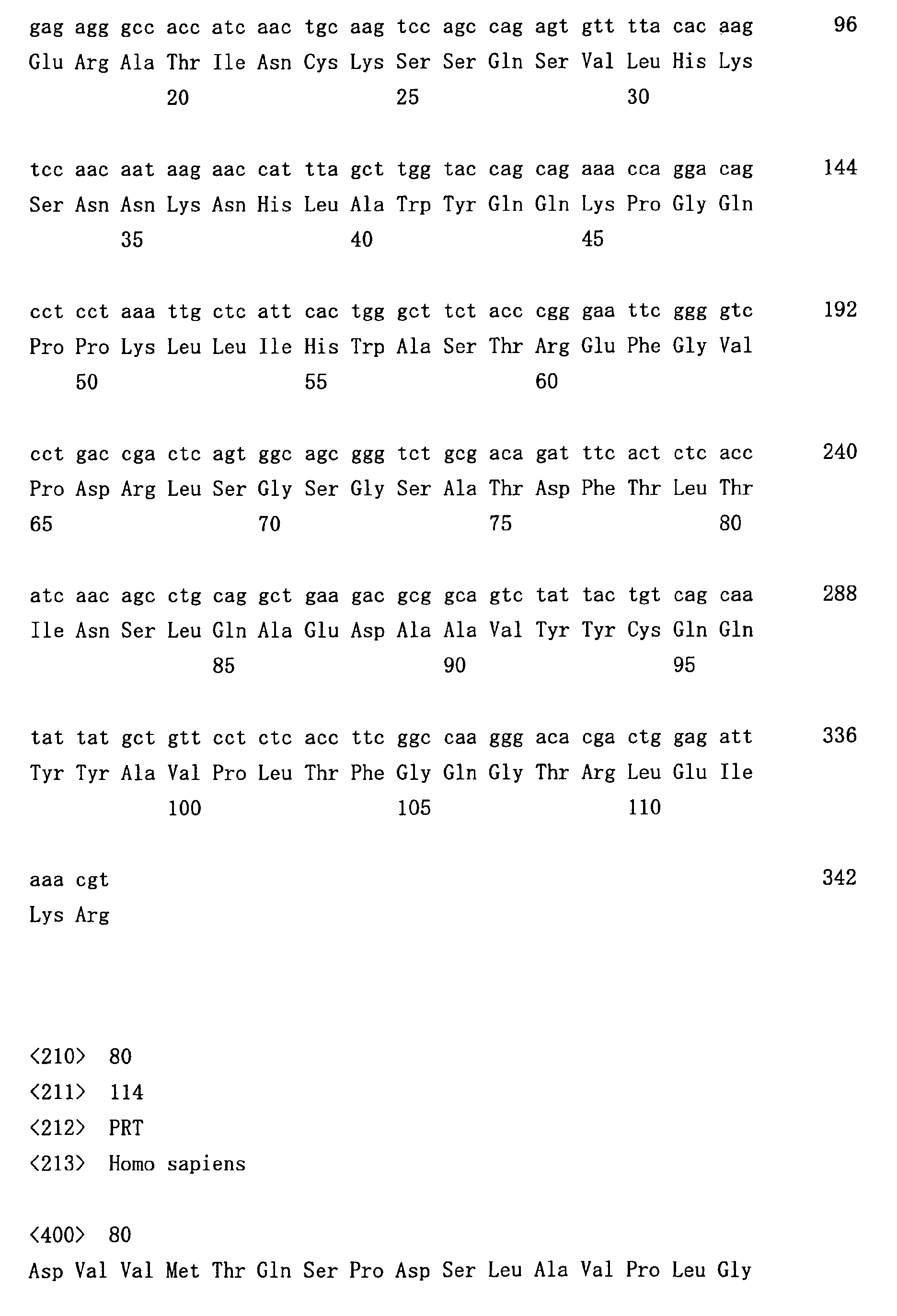
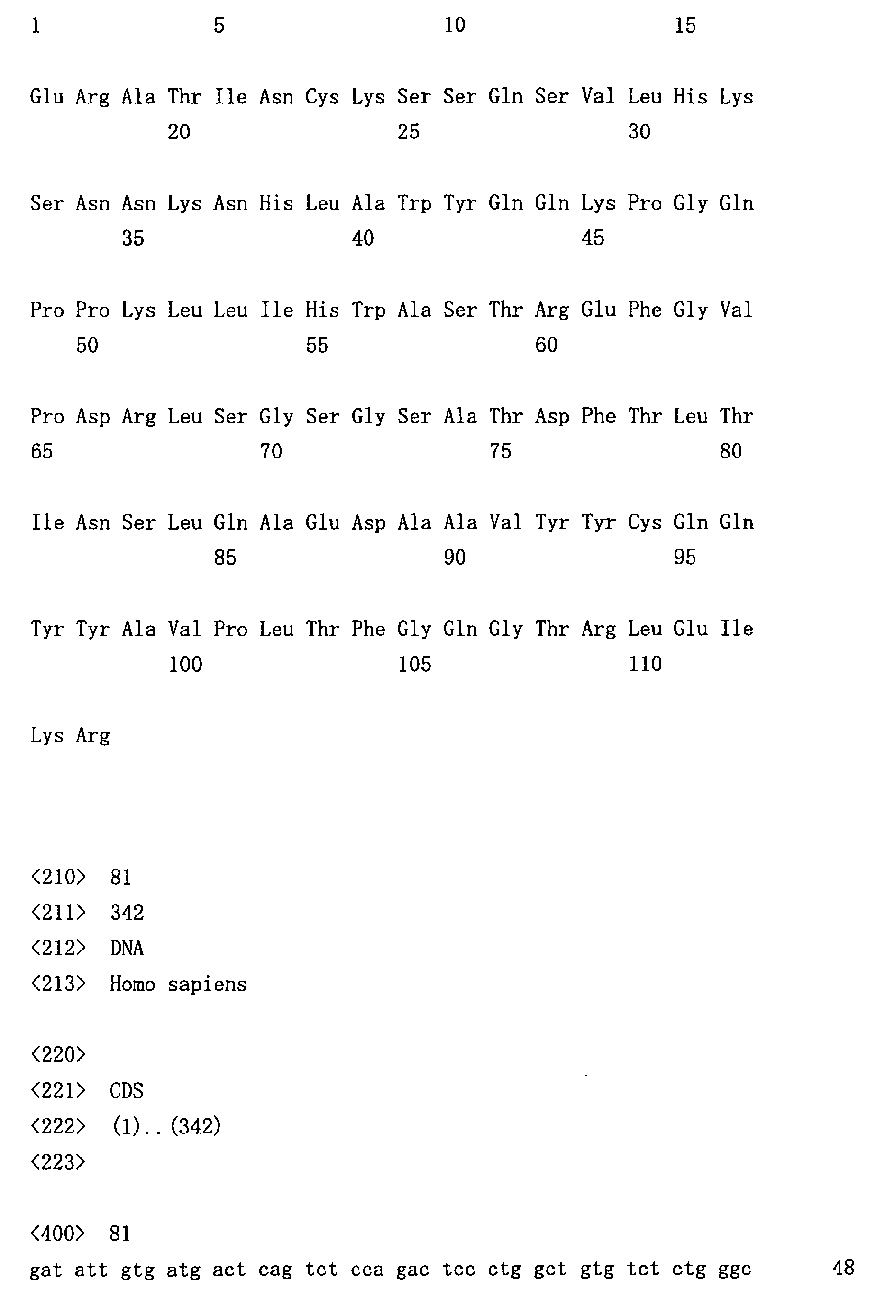
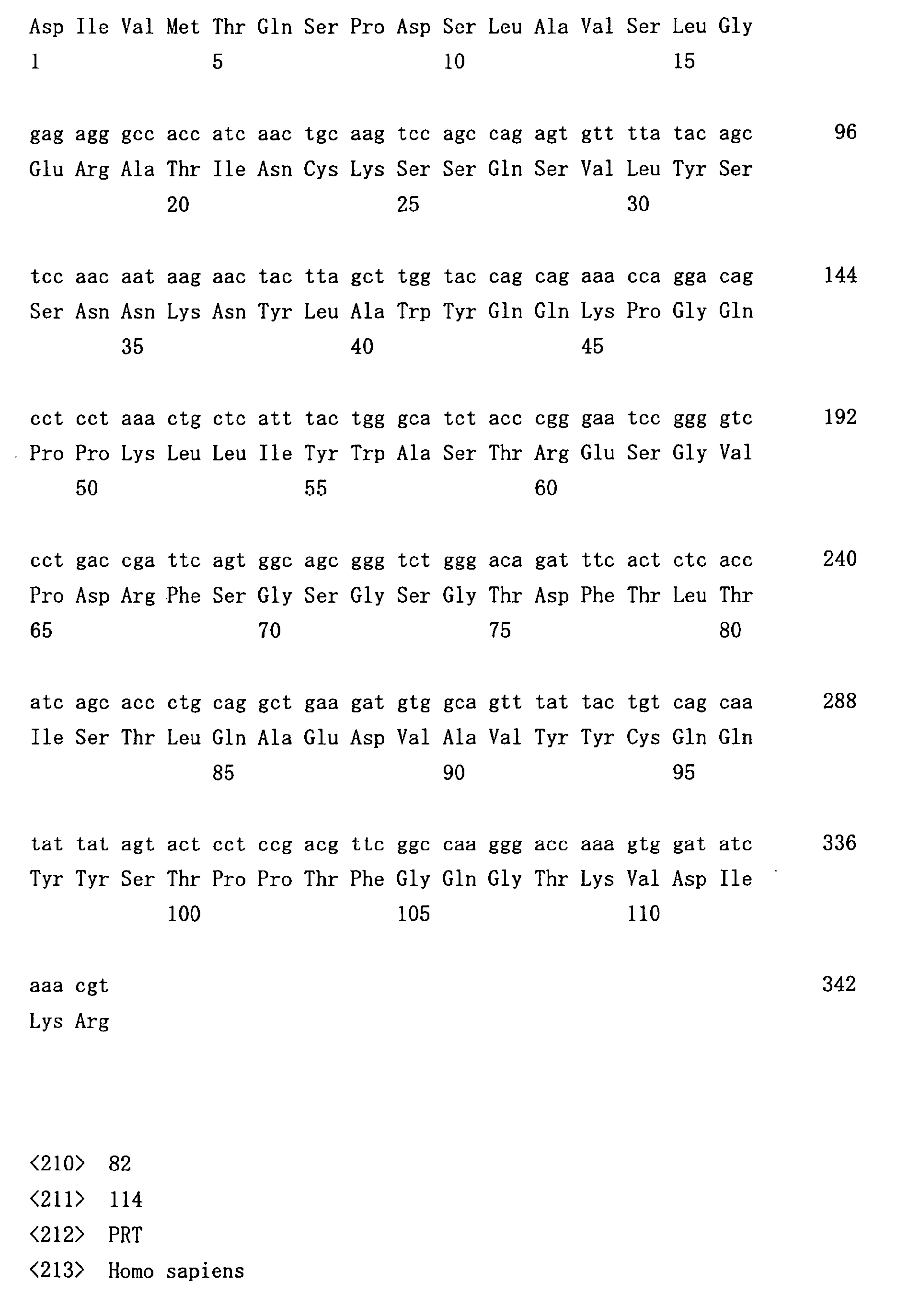
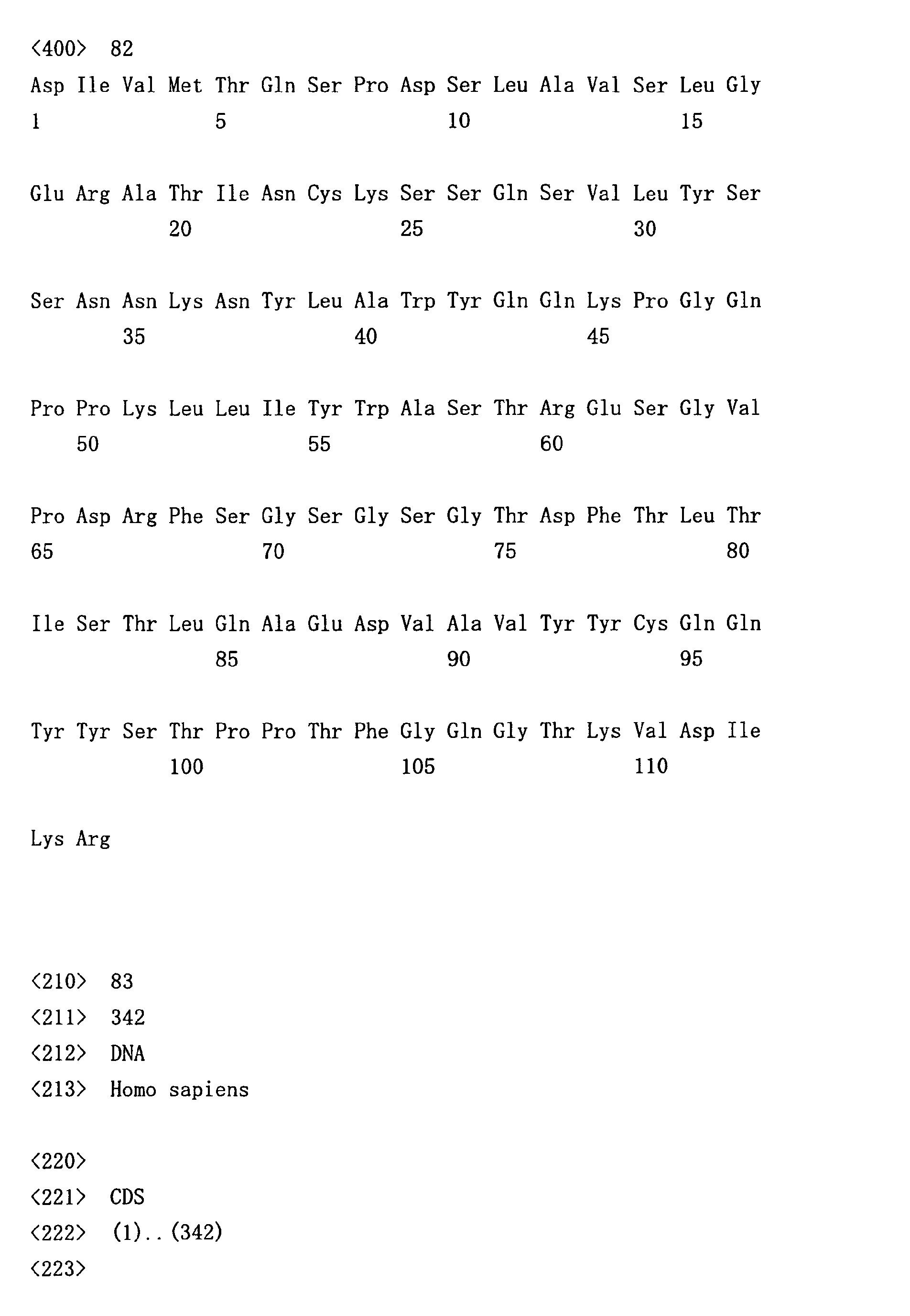
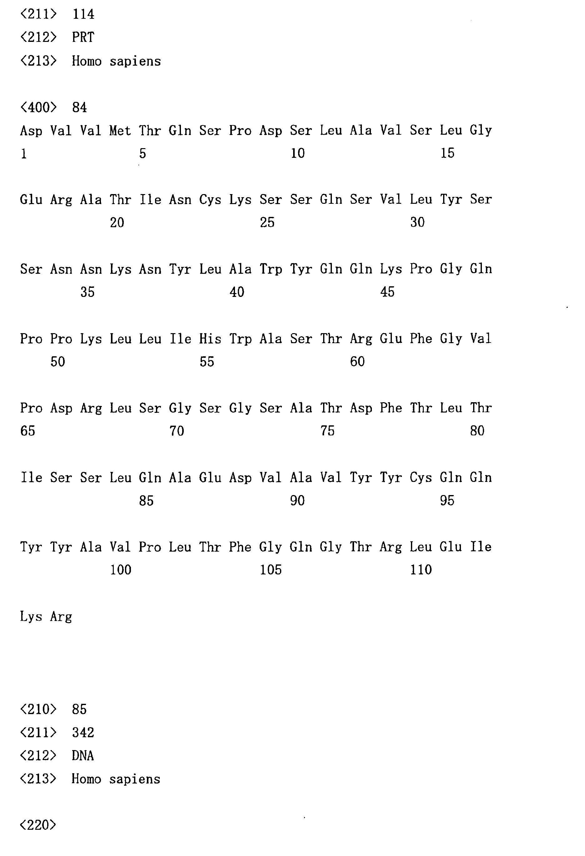
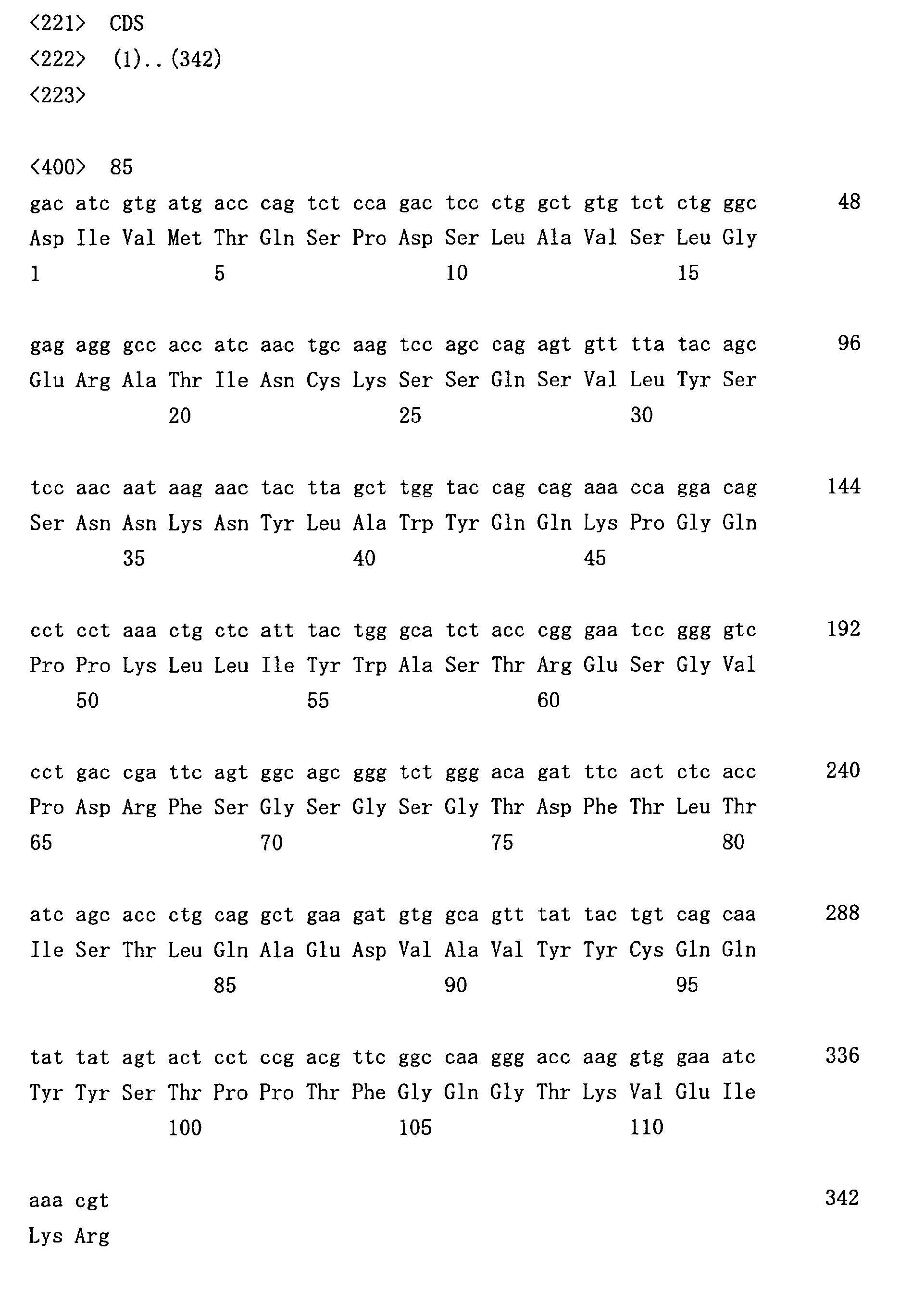
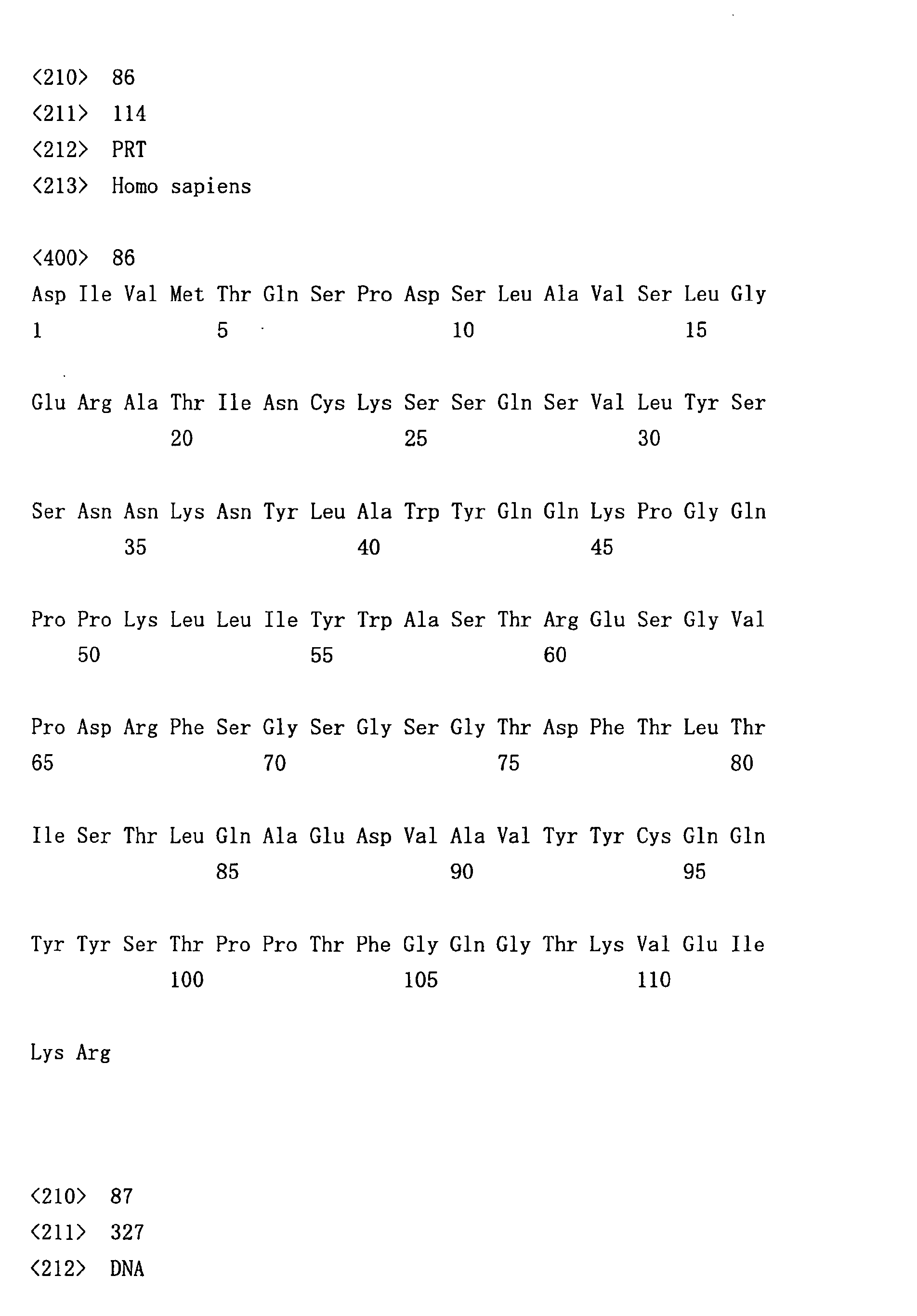
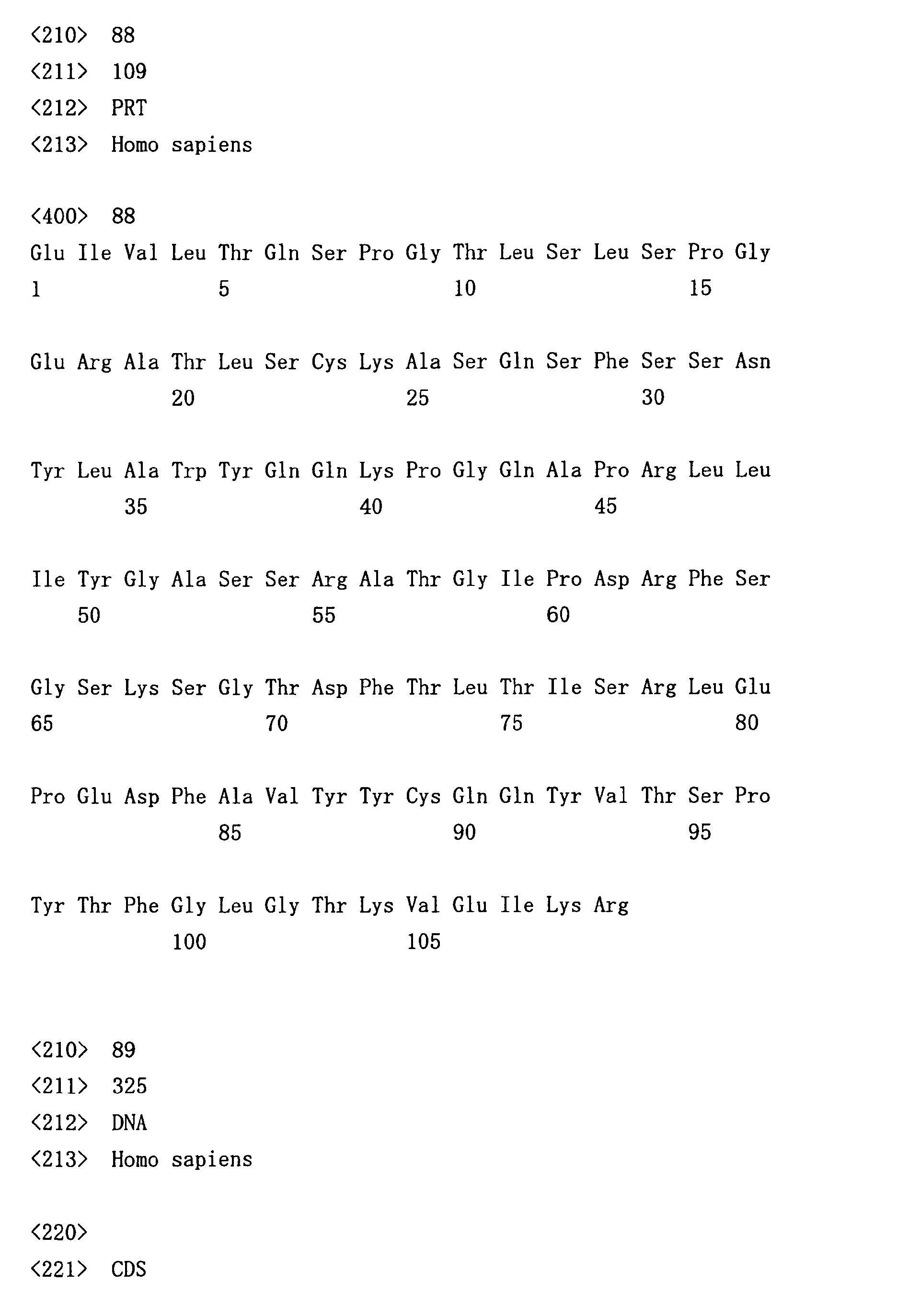
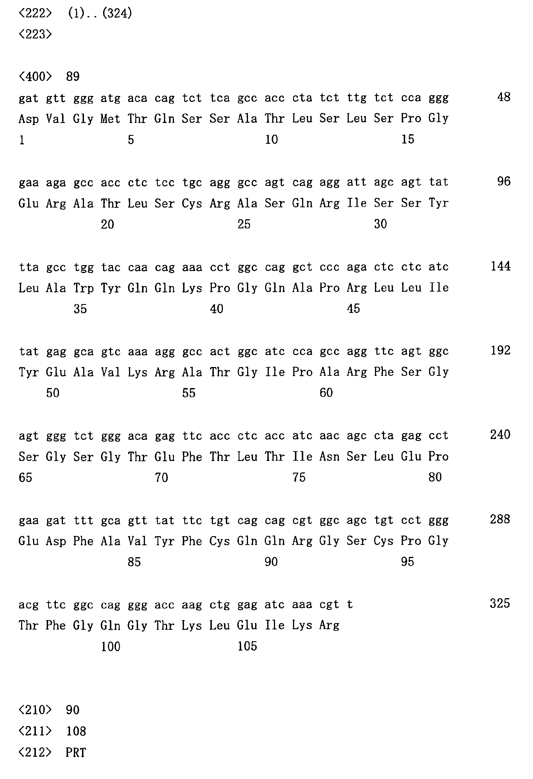
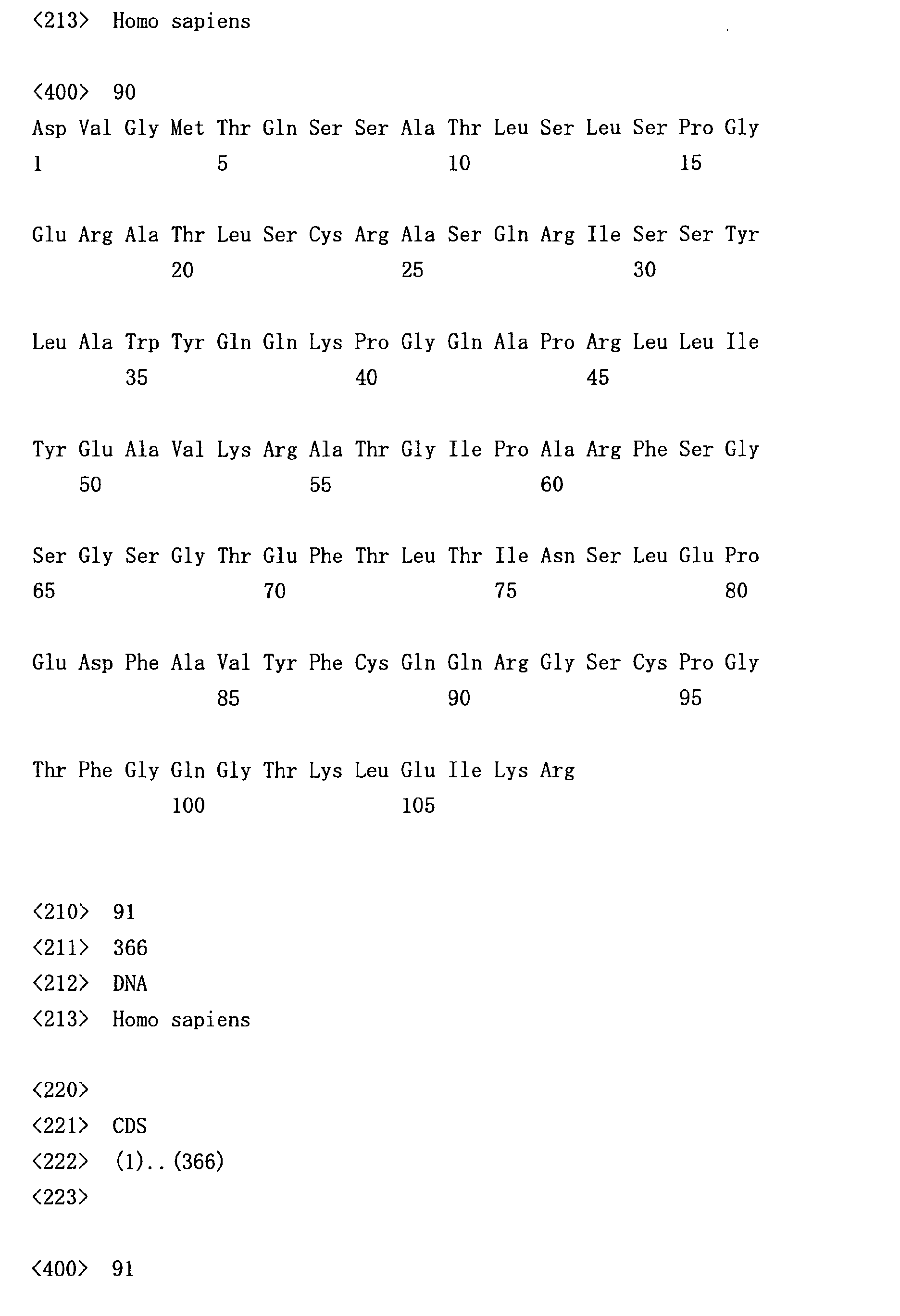
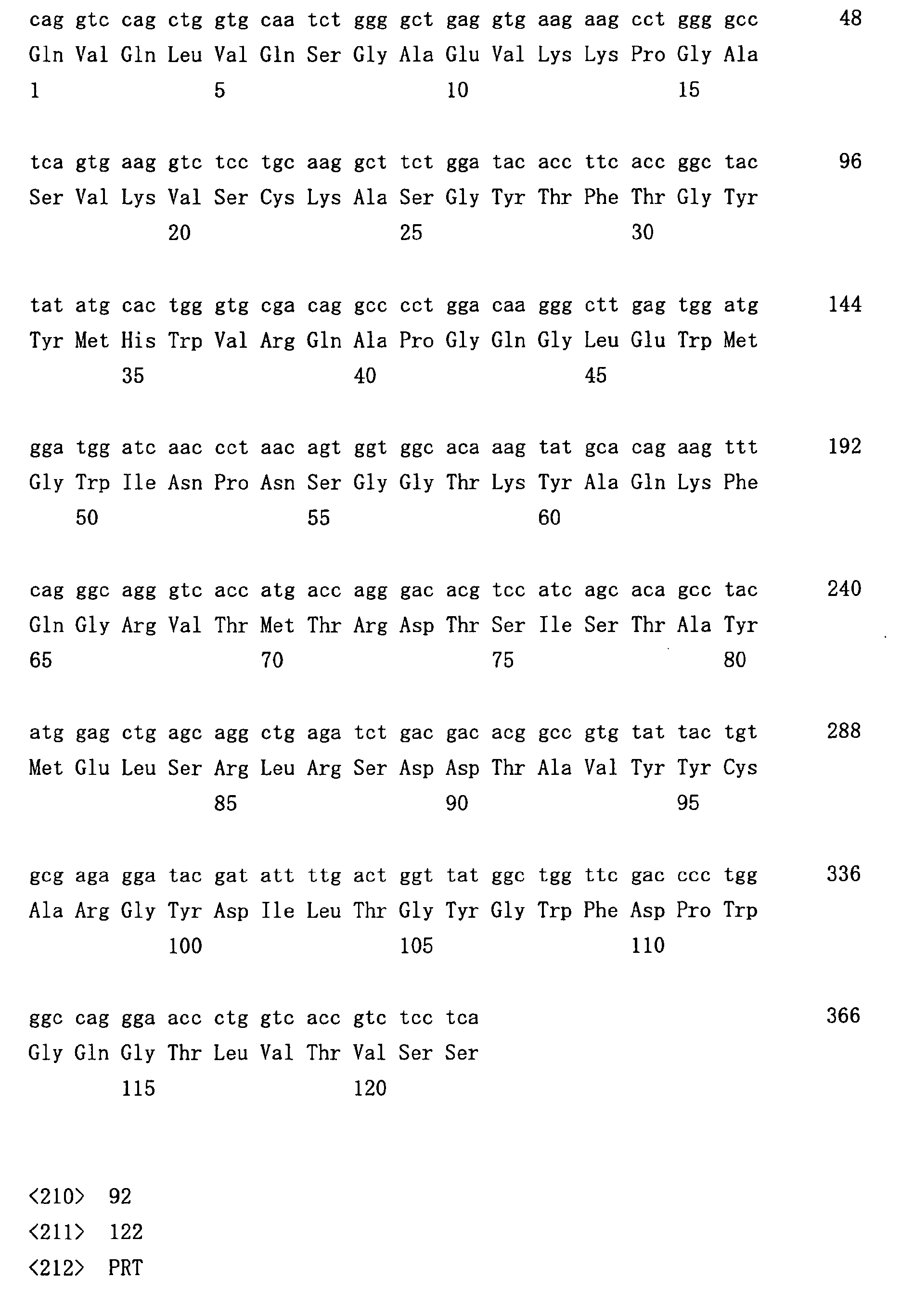
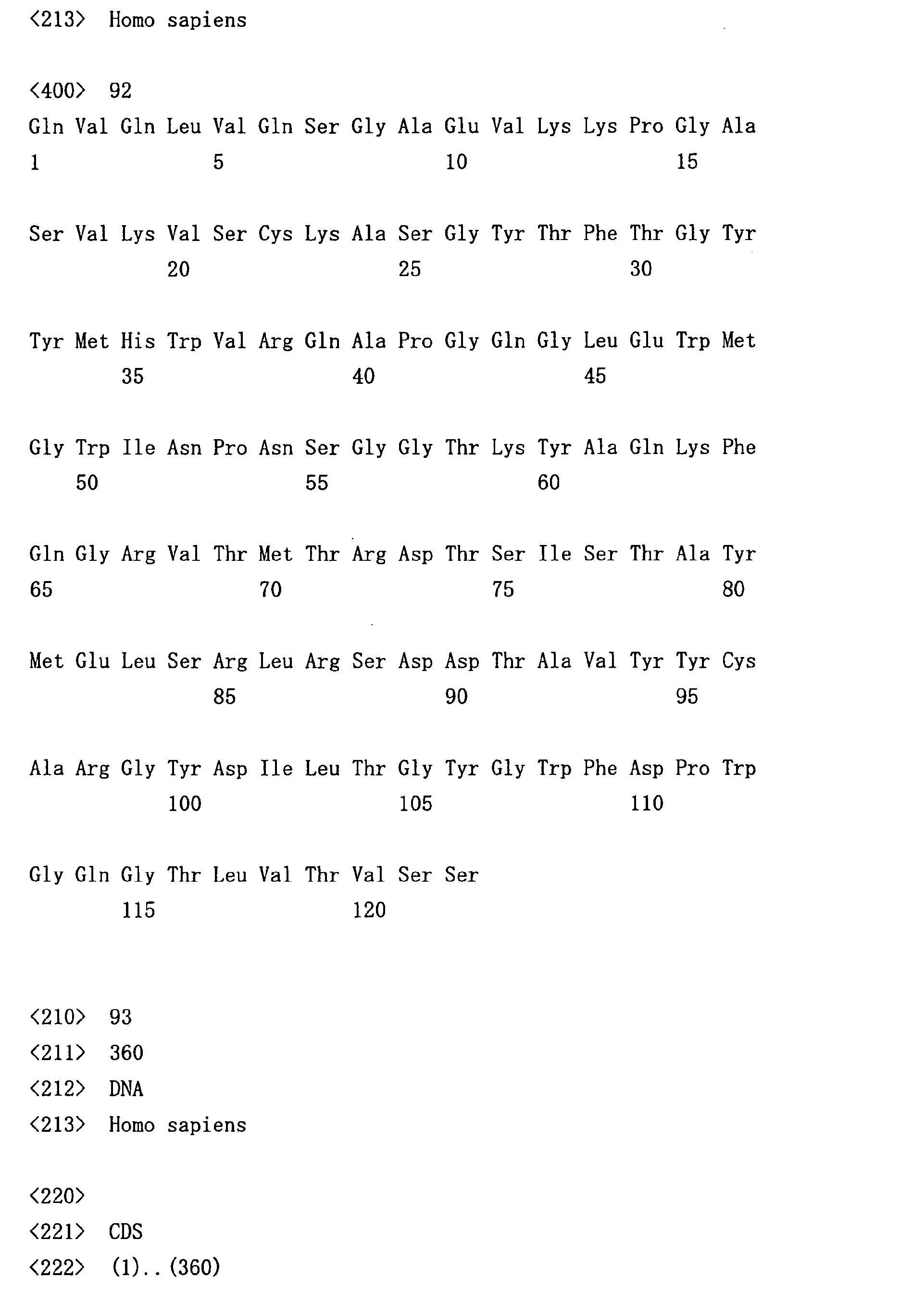
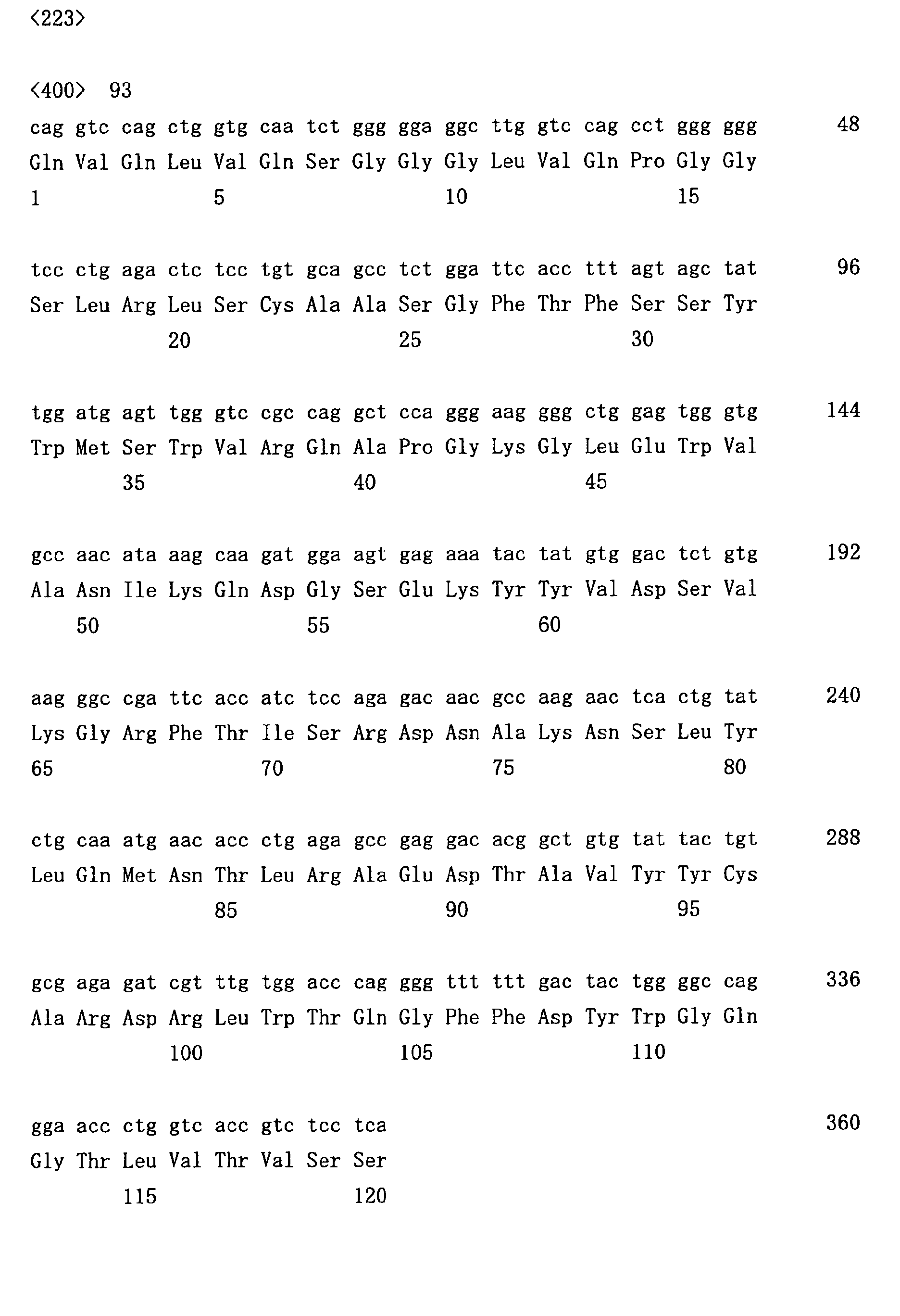
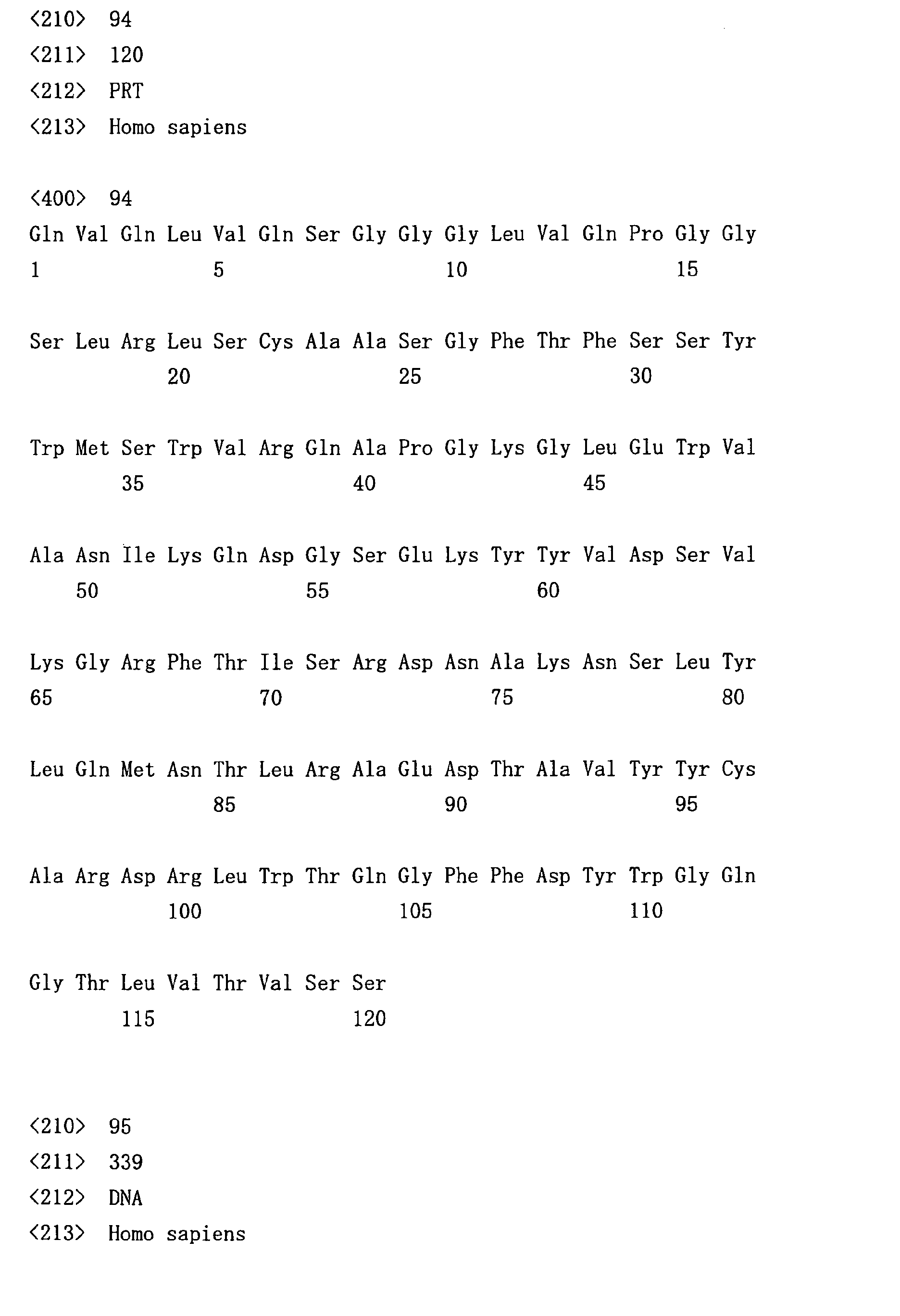
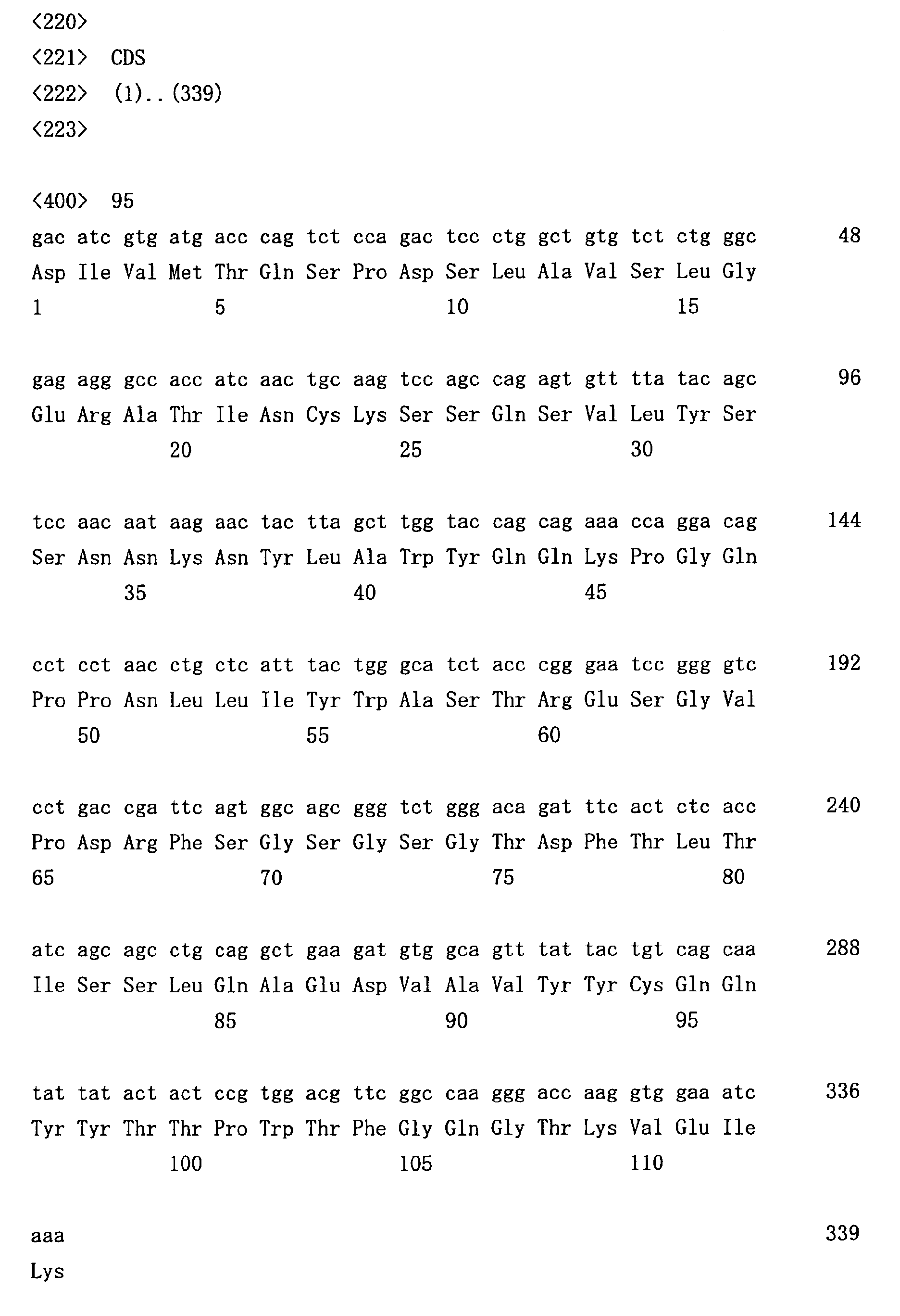
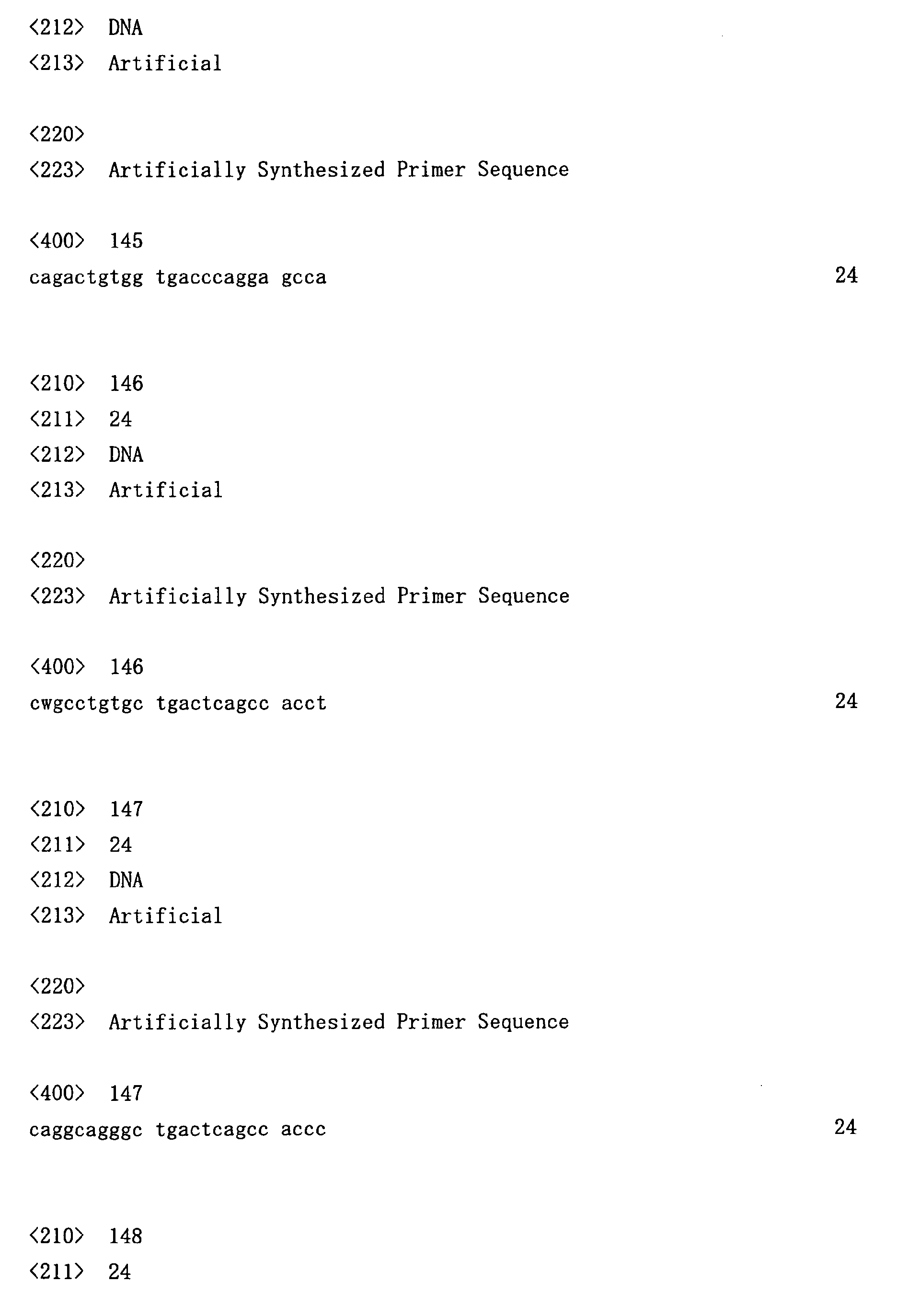
- the nucleotide sequences thus determined are shown in SEQ ID NOs: 1 to 54 (heavy chains) and SEQ ID NOs: 55 to 84 (light chains).
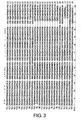
- the determined amino acid sequences were aligned as shown in Figs. 3 and 4.
- the alignment results confirmed that multiple clones of the antibody genes were obtained by the methods of this invention.
- B cells present in the specimens may very likely have been cell populations that had multiplied on specific antigen stimulations.
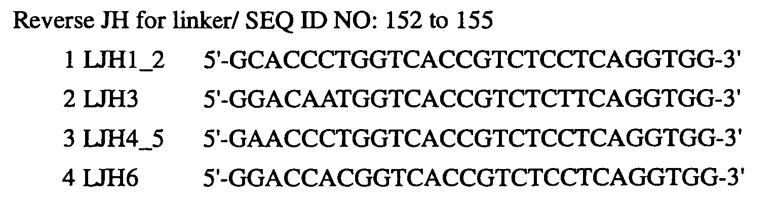
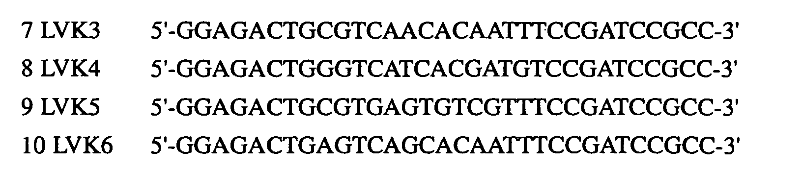
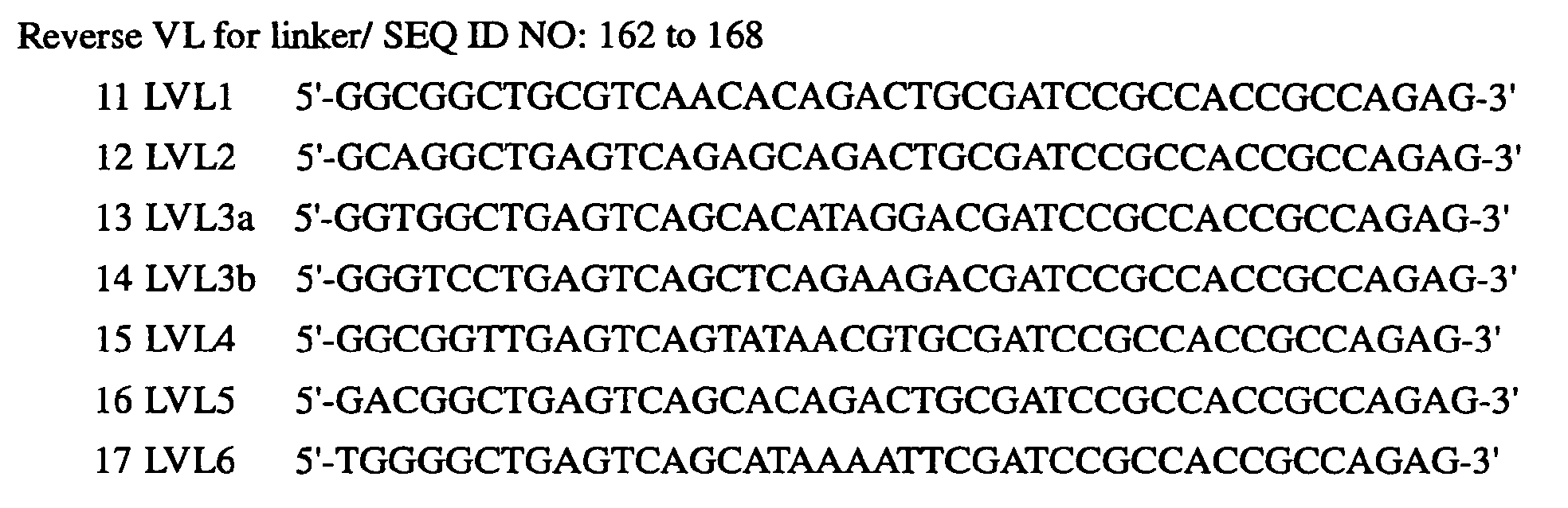
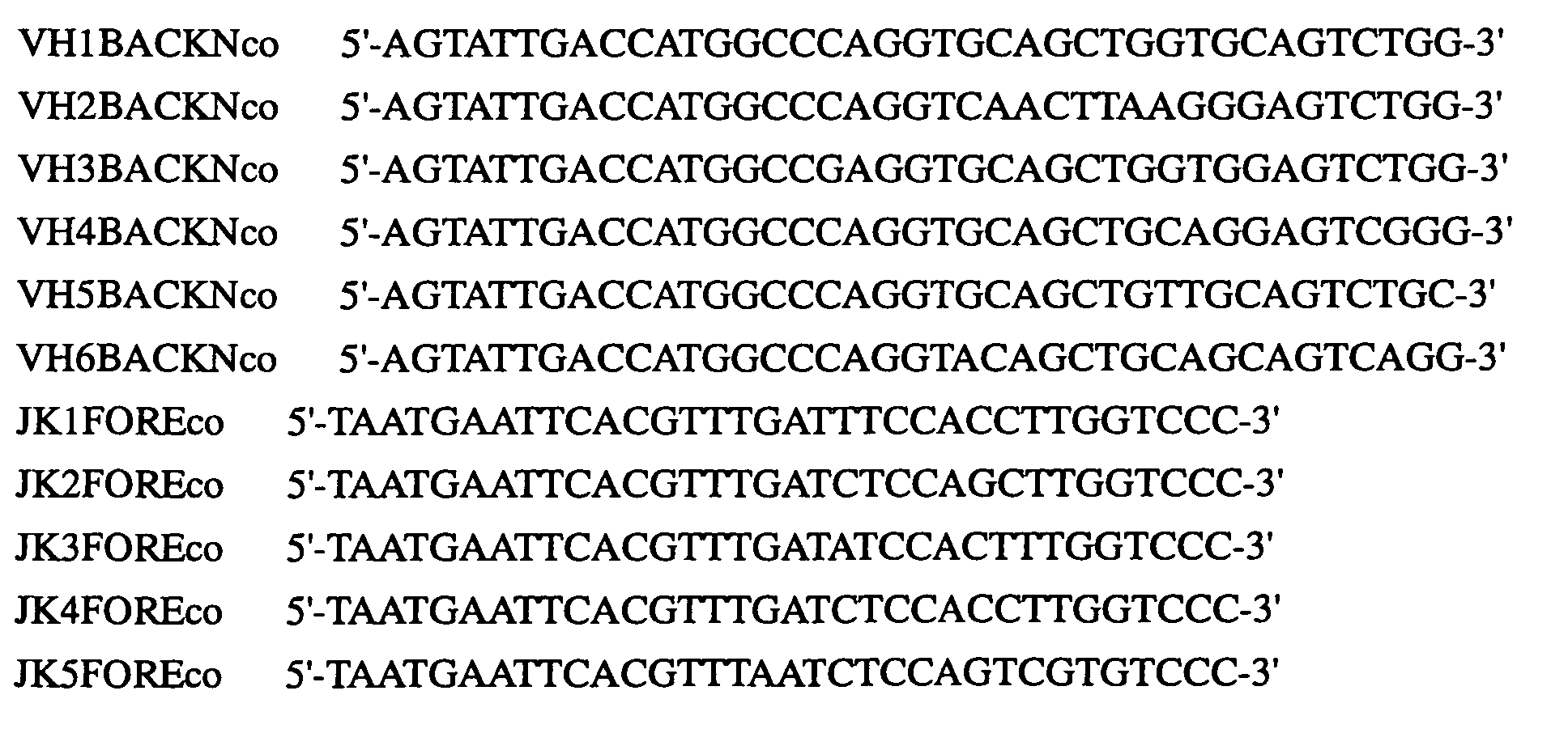
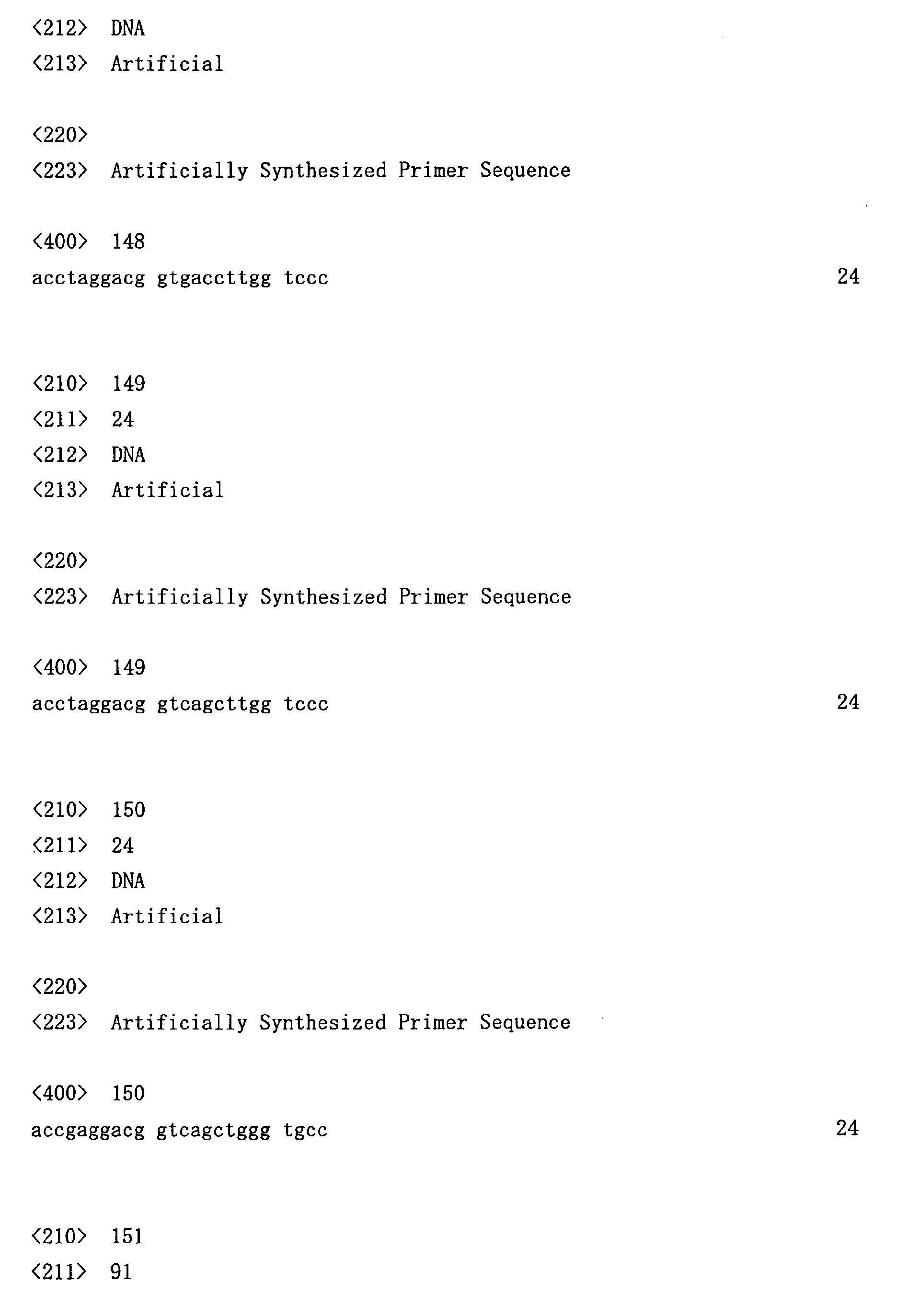
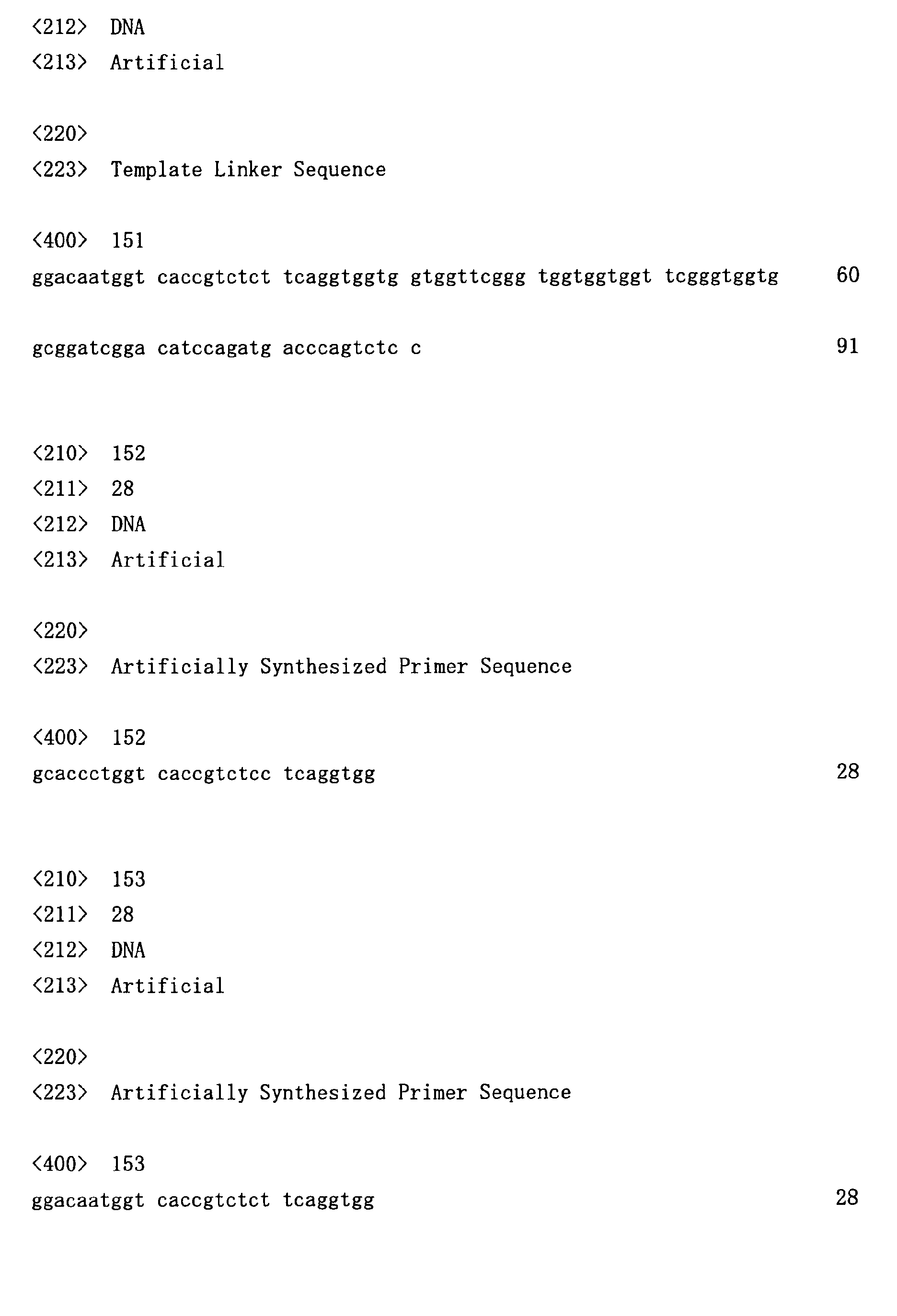
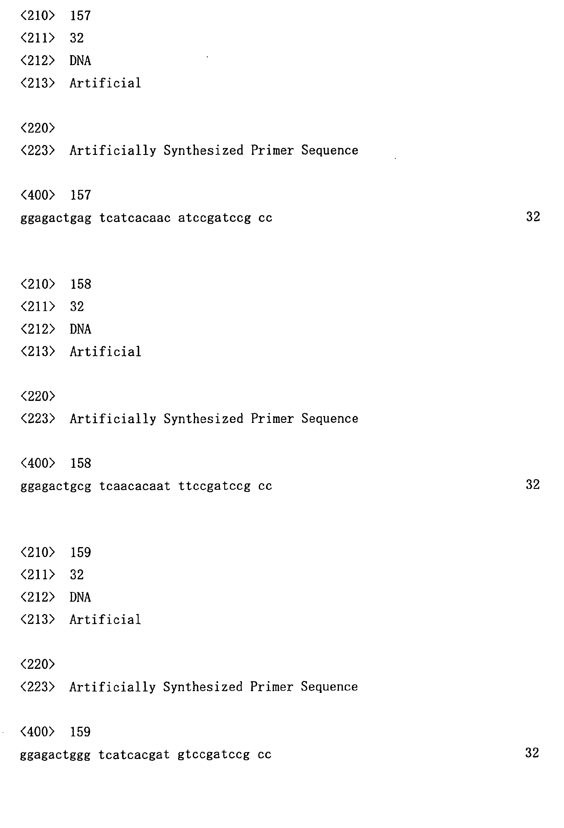
- linker sequences to be used for preparing single-stranded antibody genes were produced according to the method of Marks et al . (J. Mol. Biol. (1991) 222, 581-597). The template DNA sequences and the nucleotide sequences of primers used in the preparation are shown below. Linker fragments synthesized using PCR amplification were confirmed by agarose gel electrophoresis, and bands containing the respective fragments were excised and purified.
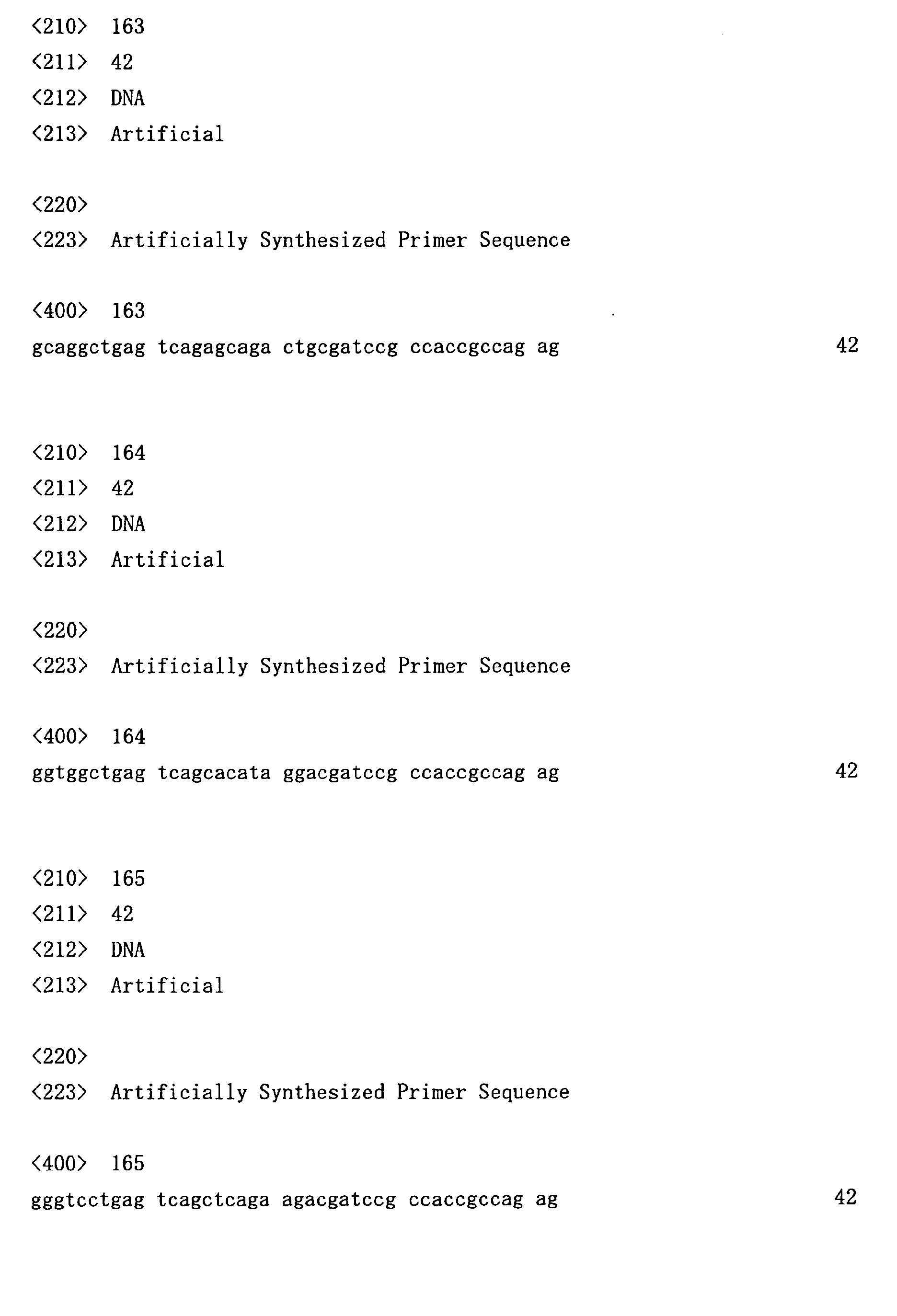
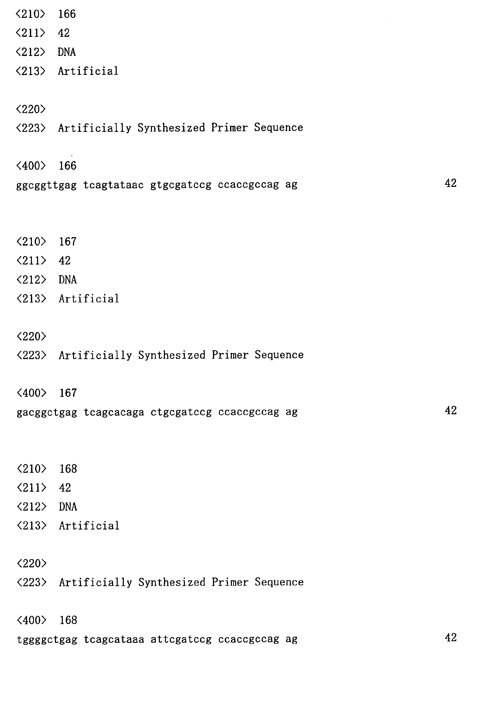
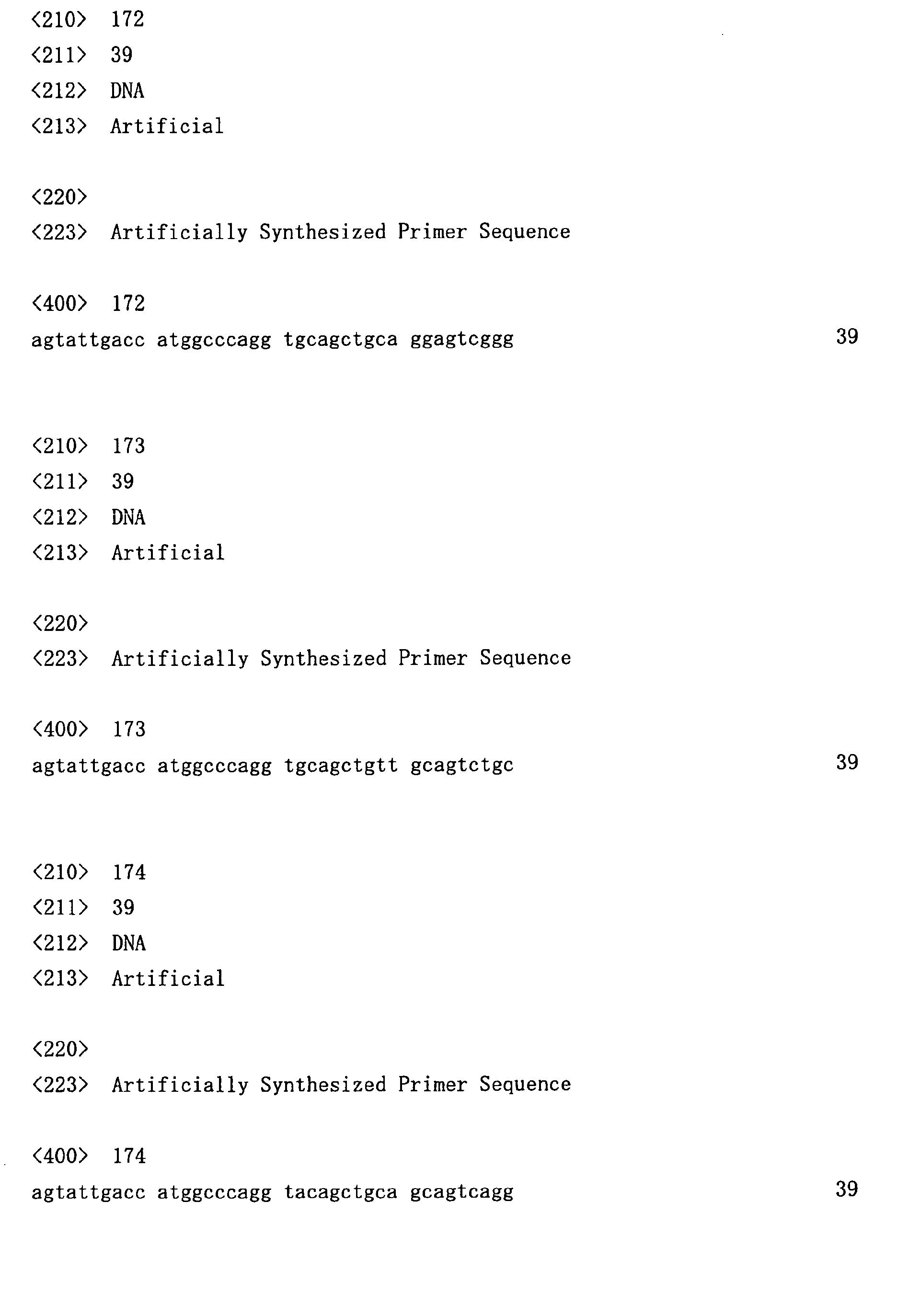
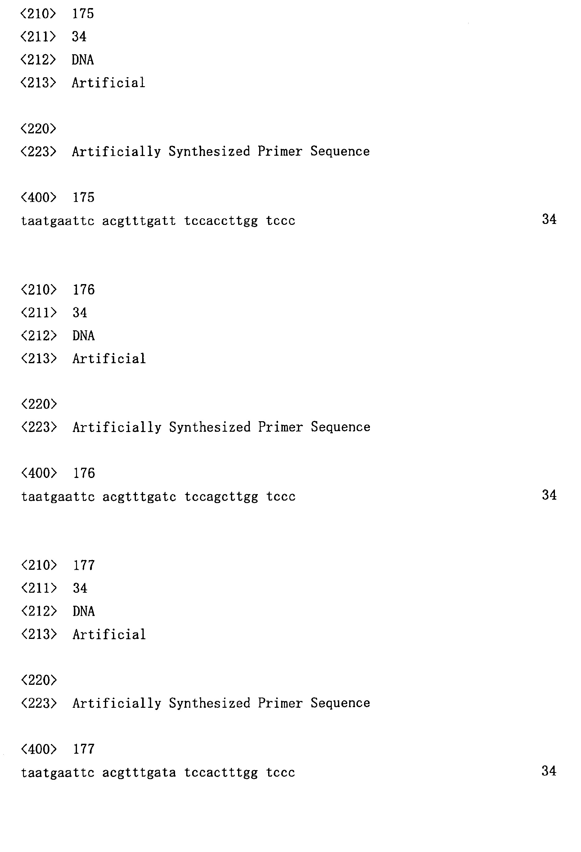
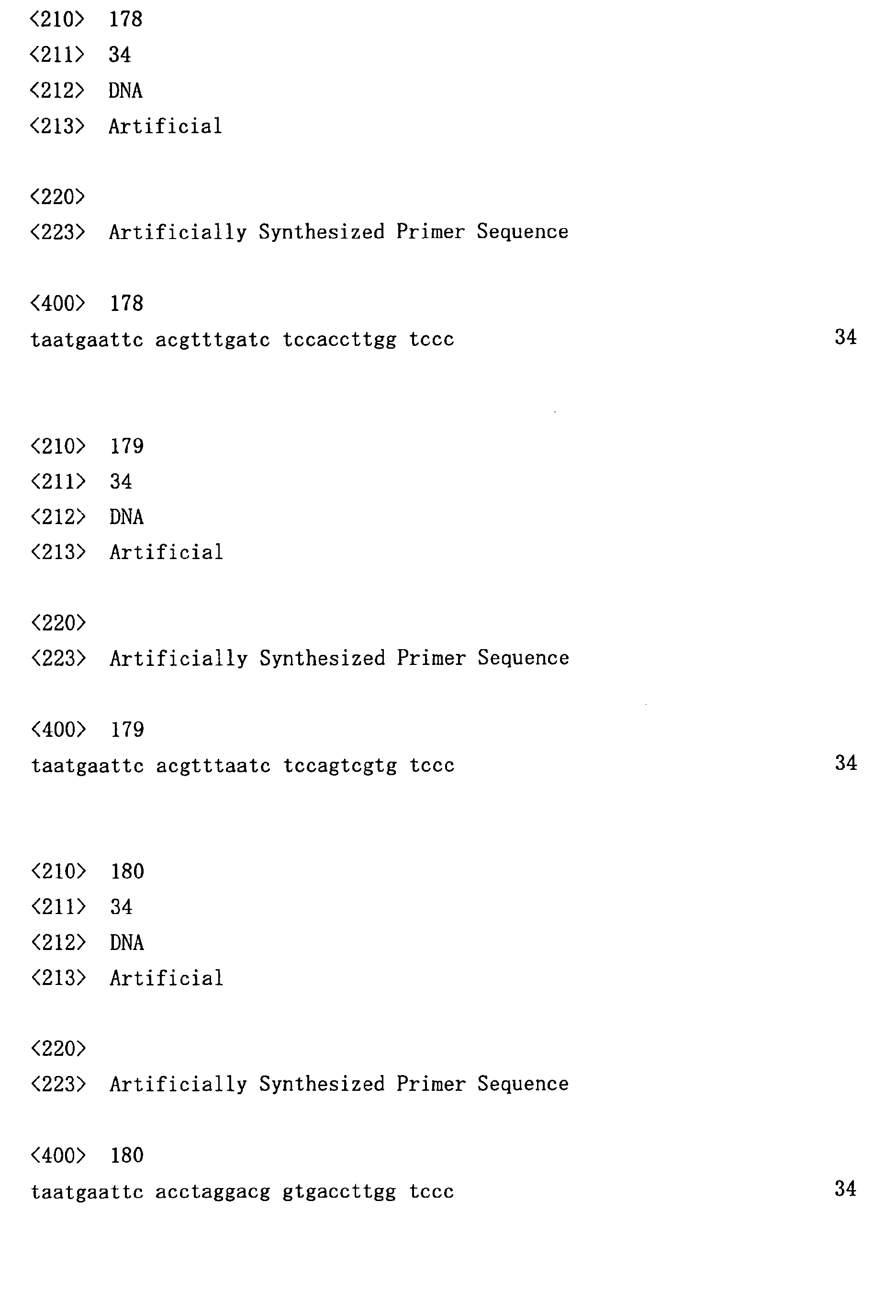
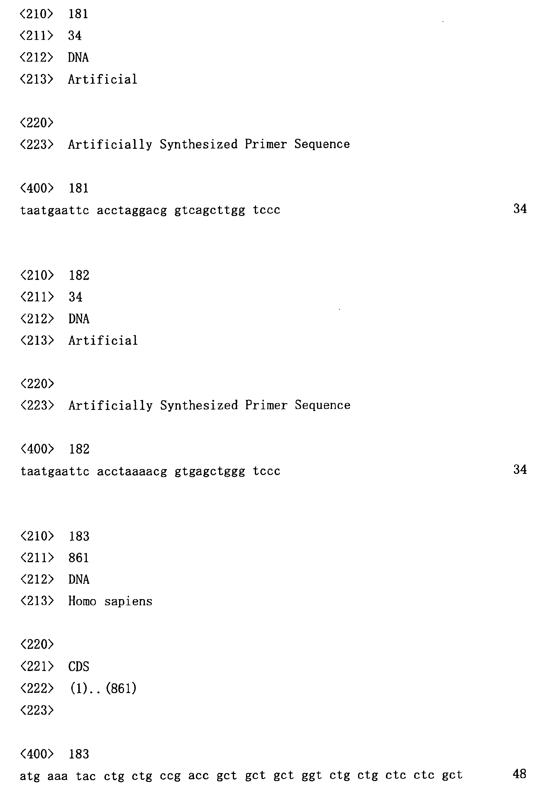
- PCR using the primer sets (SEQ ID NOs: 169 to 182) as shown below was performed by mixing the heavy chain variable region, ⁇ -chain variable region or ⁇ -chain variable region, and linker sequence, which were PCR-amplified using the cDNA synthesized from mRNA extracts of a single B cell as a template.
- reaction solutions were prepared using a combination of VH and JK primers.
- reaction solutions were prepared with a combination of VH and JL primers.
- These reaction mixtures were prepared using KOD plus DNA polymerase (TOYOBO) according to manufacturer's instructions.
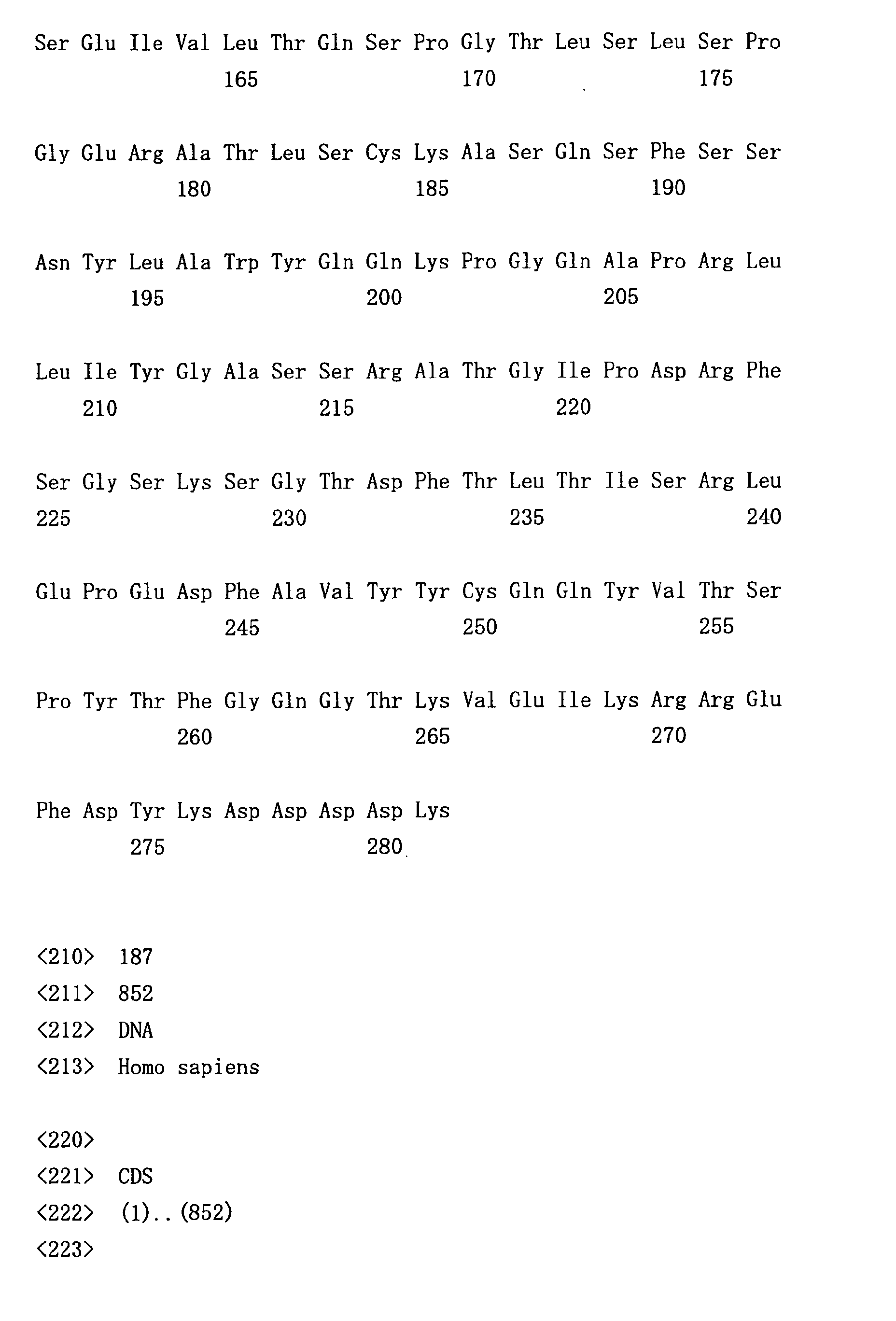
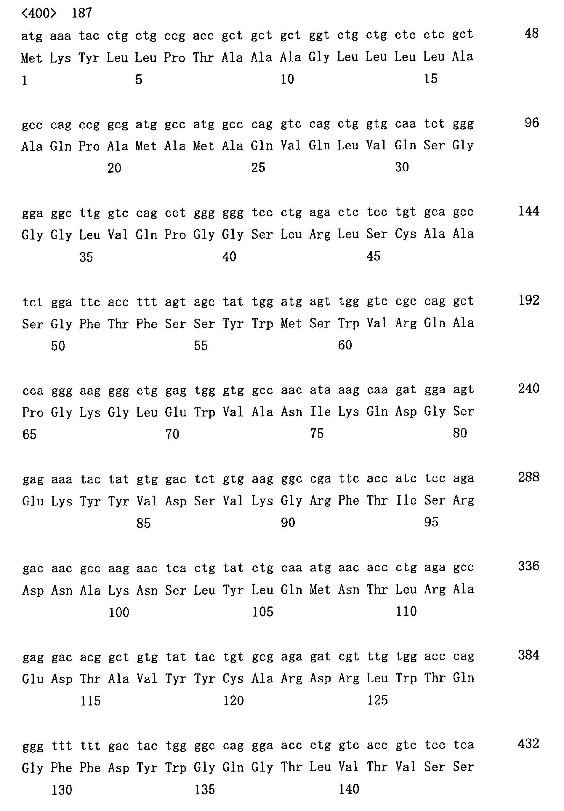
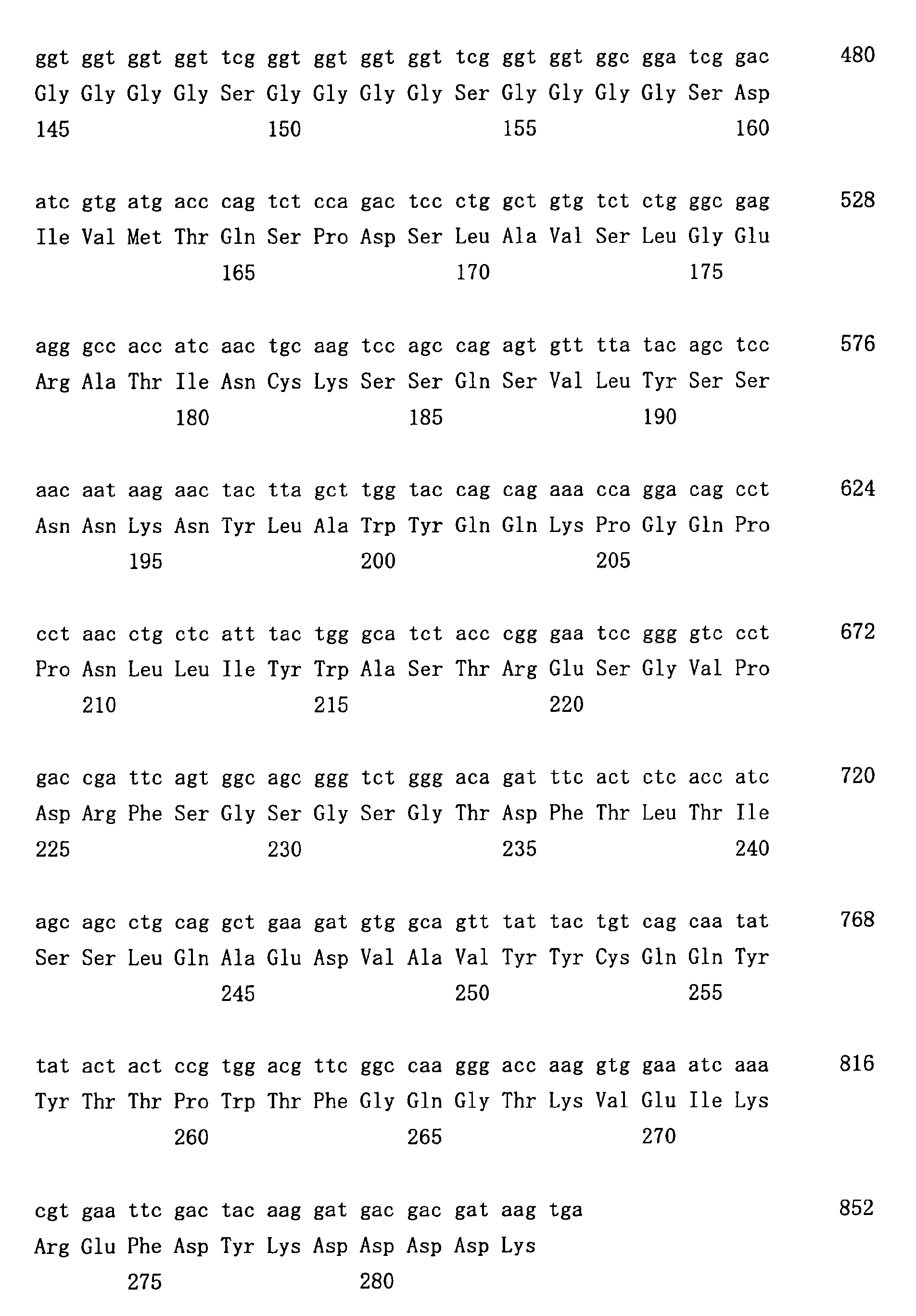
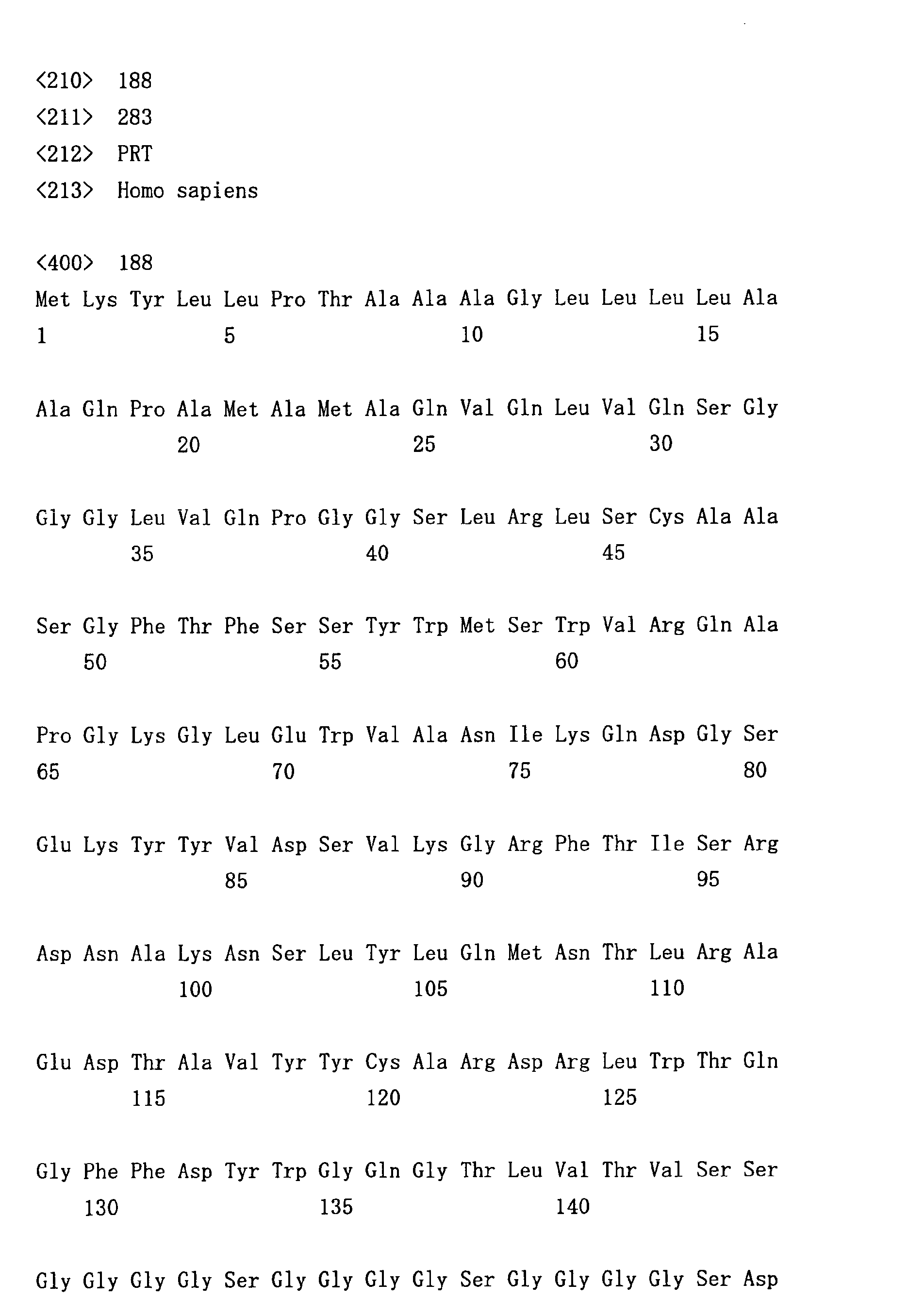
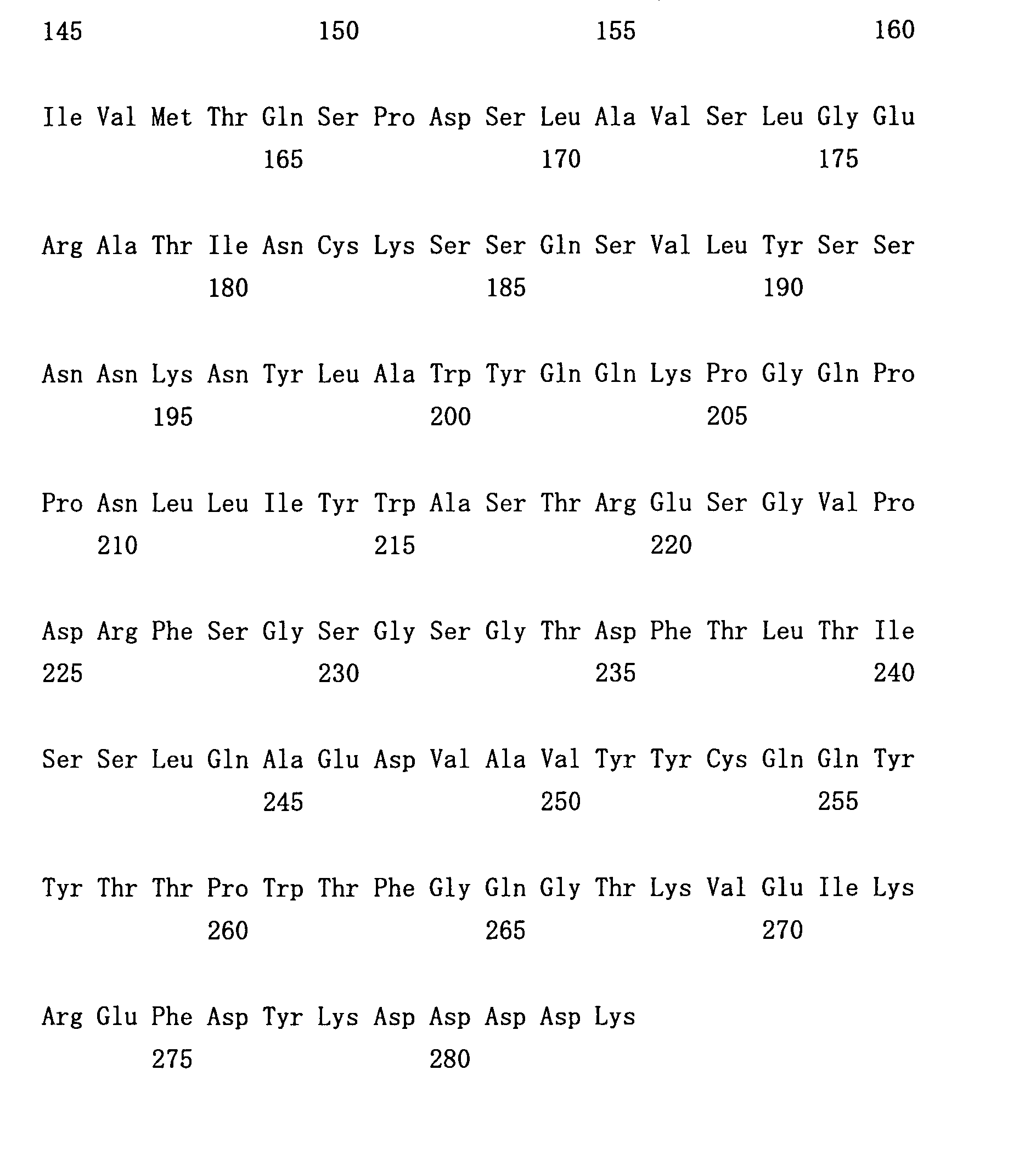
- 70-6scFV (SEQ ID NOs: 187-188) is a single-stranded antibody prepared from a heavy chain (SEQ ID NOs: 93-94) and a light chain (SEQ ID NOs: 95-96) isolated from a single B cell.
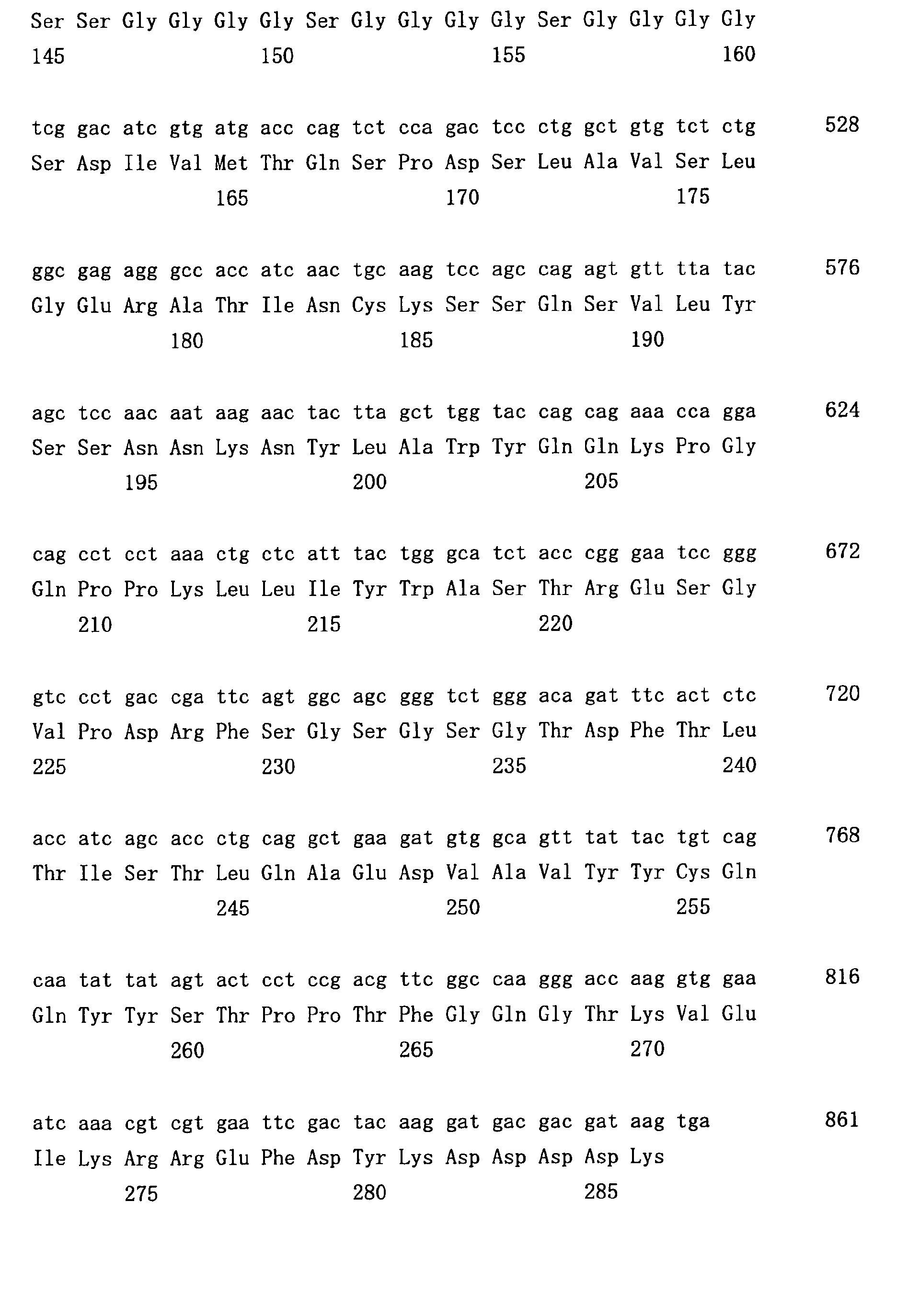
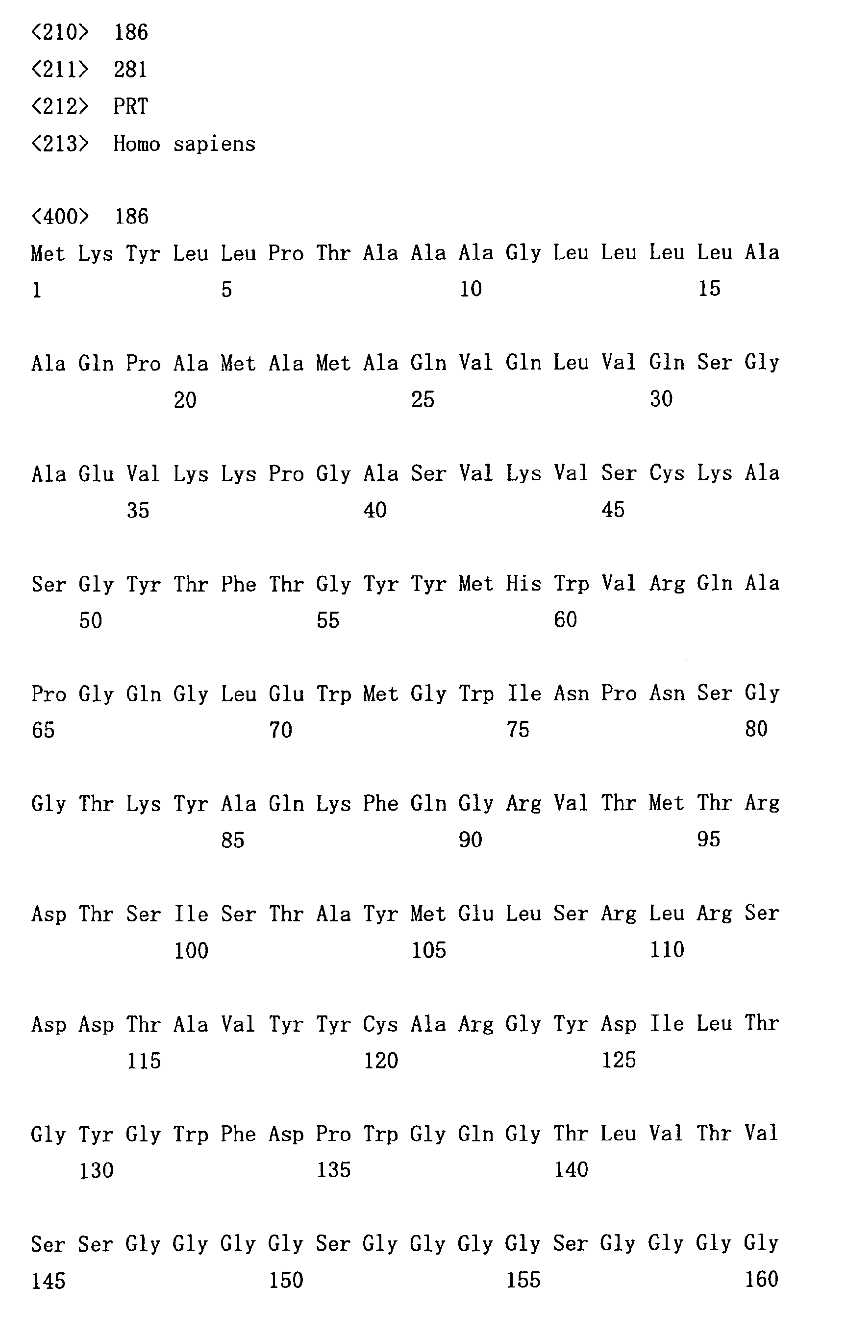
- 70-5AscFv (SEQ ID NOs: 183-184) and 70-5BscFv (SEQ ID NOs: 185-186) are single-stranded antibodies formed by combining heavy chains and light chains obtained from five B cells.
- nucleotide sequences and amino acid sequences that constitute the heavy chains comprised in 70-5AscFv and 70-5BscFv are shown in SEQ ID NOs: 91-92, and the nucleotide sequences and amino acid sequences which constitute the light chains are shown in SEQ ID NOs: 85-88.
- Recombinant single-stranded antibodies were prepared from E. coli culture supematants. During the logarithmic growth phase of transformed E. coli cells, expression of the recombinant antibodies was induced by the addition of 0.5 mM isopropyl- ⁇ -thiogalactopyranoside at 30°C. After overnight culture, the culture solution was centrifuged to separate the culture supernatant from cells. Following filtration, the resulting supernatant was applied to Anti-FLAG M2 affinity column (Sigma) and the recombinant protein was absorbed thereto. After washing the column, the recombinant protein was eluted with 0.1 M glycine (pH3.5).
- the eluate was applied to PD10 column (Amersham Pharmacia Biotech), and the buffer was immediately replaced with PBS containing 0.01% Tween-20. Presence of the protein was confirmed by Coomassie staining or Western blotting with anti-FLAG antibodies following SDS-PAGE.
- the bound recombinant antibodies were detected by the conversion of 3,3-diamino-benzidine-tetrahydrochloride with the peroxidase-conjugated anti-FLAG antibody (FLAG) into a brown precipitate in the presence of hydrogen peroxide. Before and after the addition of antibodies, washing was carried out three times with PBS containing 0.1 % Tween-20 at room temperature for 5 min. Prior to mounting, tissue sections were counterstained with hematoxylin, and dehydrated using ethanol and xylene. As a negative control for antibody staining, similar procedures were performed without adding the recombinant antibodies.
- the present invention enables the isolation of polynucleotides encoding antibodies against a lesion without depending on B cell cloning. Since the methods of this invention do not rely on B cell cloning, they enable easy acquisition of genes derived from human antibody-producing cells which are difficult to clone.
- genes encoding cancer tissue-recognizing antibodies can be isolated from B cells that infiltrate the cancer tissue.
- Antibodies recognizing cancer cells are useful for the diagnosis and treatment of cancer.
- antibody genes can be easily obtained from human antibody-producing cells as well. Acquisition of genes for cancer tissue-recognizing human antibodies is of great significance in cancer diagnosis and treatment.
- antibodies are administered to humans.
- antibody molecules with a traceable label are administered so as to indicate the presence of cancer at the region where the antibodies are localized.
- antibodies are utilized in target therapy. That is, antibodies conjugated with an anticancer agent are administered to patients.
- Human antibodies can be expected to be highly safe when administered to humans.
- the concentration can be stably maintained in blood over a long period of time.
Landscapes
- Chemical & Material Sciences (AREA)
- Health & Medical Sciences (AREA)
- Organic Chemistry (AREA)
- Life Sciences & Earth Sciences (AREA)
- Immunology (AREA)
- General Health & Medical Sciences (AREA)
- Medicinal Chemistry (AREA)
- Biochemistry (AREA)
- Biophysics (AREA)
- Genetics & Genomics (AREA)
- Molecular Biology (AREA)
- Proteomics, Peptides & Aminoacids (AREA)
- Cell Biology (AREA)
- Chemical Kinetics & Catalysis (AREA)
- General Chemical & Material Sciences (AREA)
- Nuclear Medicine, Radiotherapy & Molecular Imaging (AREA)
- Pharmacology & Pharmacy (AREA)
- Animal Behavior & Ethology (AREA)
- Public Health (AREA)
- Veterinary Medicine (AREA)
- Peptides Or Proteins (AREA)
- Preparation Of Compounds By Using Micro-Organisms (AREA)
- Medicines Containing Antibodies Or Antigens For Use As Internal Diagnostic Agents (AREA)
- Micro-Organisms Or Cultivation Processes Thereof (AREA)
- Acyclic And Carbocyclic Compounds In Medicinal Compositions (AREA)
Abstract
Description
solid tumor lesions
arteriosclerosis lesions
inflammatory disease lesions
lesions generated by infectious pathogens
autoimmune disease lesions
heterogenous cells and tissues artificially transplanted into immune animals
cells and tissues that express non-self genes artificially transplanted into immune animals
The graph on the right shows electrophoresis time measured using Agilent 2100. The vertical axis shows fluorescence intensity, and the horizontal axis shows electrophoresis time (seconds).
The graph on the right shows electrophoresis time measured using Agilent 2100. The vertical axis shows fluorescence intensity, and the horizontal axis shows electrophoresis time (seconds).


| PRIMER SET | FORWARD | REVERSE | ||||||
| VH1-JH MIX | VH1a | VH1b | VH1c | VH1d | JH1245 | JH3 | JH6 | |
| VH2-JH MIX | VH2a | VH2b | JH1245 | JH3 | JH6 | |||
| VH3/5-JH MIX | VH3a | VH3b | VH3c | VH3d | JH1245 | JH3 | JH6 | |
| VN4-JH MIX | VH4a | VH4b | JH1245 | JH3 | JH6 | |||
| VH6/7-JH MIX | VH6a | VH7a | JH1245 | JH3 | JH6 | |||
| VK1-JK MIX | VK1a | VK1b | VK1c | VK1d | JK1 | JK24 | JK3 | JK5 |
| VK2-JK MIX | VK2a | VK2b | JK1 | JK24 | JK3 | JK5 | ||
| VK3-JK MIX | VK3a | VK3b | VK3c | JK1 | JK24 | JK3 | JK5 | |
| VK4/5-JK MIX | VK4a | VK5a | JK1 | JK24 | JK3 | JK5 | ||
| VK6-JK MIX | VK6a | VK6b | JK1 | JK24 | JK3 | JK5 |













































Claims (12)
- A method for isolating a polynucleotide encoding an antibody against a lesional tissue, wherein the method comprises the steps of:(a) isolating a lesional tissue-infiltrating B cell; and(b) obtaining a polynucleotide encoding an antibody from the isolated B cell.
- The method of claim 1, wherein the lesional tissue is a cancer tissue.
- The method of claim 1, wherein step (a) of isolating a lesional tissue-infiltrating B cell comprises the step of excising a region comprising a B cell from a section of said lesional tissue.
- The method of claim 1, wherein step (b) of obtaining a polynucleotide encoding an antibody comprises the step of amplifying a gene encoding an antibody variable region.
- An antibody-encoding polynucleotide isolated by the method of claim 1.
- The polynucleotide of claim 5, wherein the antibody-encoding polynucleotide comprises a polynucleotide encoding an antibody variable region.
- An expression vector comprising the polynucleotide of claim 5.
- A host cell comprising the polynucleotide of claim 5 or the expression vector of claim 7.
- A method for producing an antibody, wherein the method comprises the steps of:culturing the host cell of claim 8; andrecovering an antibody which is the expression product.
- An antibody produced by the method of claim 9.
- An antibody encoded by the polynucleotide of claim 5.
- The antibody production method of claim 9, wherein the method further comprises the steps of:(1) contacting the antibody obtained by the method of claim 9 with a lesional tissue;(2) detecting the binding between the antibody and the lesional tissue; and(3) selecting an antibody that binds to the lesional tissue.
Applications Claiming Priority (3)
| Application Number | Priority Date | Filing Date | Title |
|---|---|---|---|
| JP2002339241 | 2002-11-22 | ||
| JP2002339241 | 2002-11-22 | ||
| PCT/JP2003/014919 WO2004048571A1 (en) | 2002-11-22 | 2003-11-21 | Antibody against lesion tissue |
Publications (3)
| Publication Number | Publication Date |
|---|---|
| EP1571211A1 true EP1571211A1 (en) | 2005-09-07 |
| EP1571211A4 EP1571211A4 (en) | 2006-03-22 |
| EP1571211B1 EP1571211B1 (en) | 2010-01-20 |
Family
ID=32375769
Family Applications (1)
| Application Number | Title | Priority Date | Filing Date |
|---|---|---|---|
| EP03775858A Expired - Lifetime EP1571211B1 (en) | 2002-11-22 | 2003-11-21 | Antibodies against lesional tissues |
Country Status (7)
| Country | Link |
|---|---|
| US (1) | US20060235207A1 (en) |
| EP (1) | EP1571211B1 (en) |
| JP (1) | JP4690046B2 (en) |
| AT (1) | ATE455853T1 (en) |
| AU (1) | AU2003284427A1 (en) |
| DE (1) | DE60331101D1 (en) |
| WO (1) | WO2004048571A1 (en) |
Families Citing this family (22)
| Publication number | Priority date | Publication date | Assignee | Title |
|---|---|---|---|---|
| US7658924B2 (en) * | 2001-10-11 | 2010-02-09 | Amgen Inc. | Angiopoietin-2 specific binding agents |
| JP5314242B2 (en) | 2004-04-27 | 2013-10-16 | 中外製薬株式会社 | Plasma cell growth method |
| AU2013204620B2 (en) * | 2007-01-05 | 2015-09-17 | University Of Zurich | Method of Providing Disease-Specific Binding Molecules and Targets |
| AU2011265453B9 (en) * | 2007-01-05 | 2013-09-12 | University Of Zurich | Method of providing disease-specific binding molecules and targets |
| HRP20140049T1 (en) | 2007-01-05 | 2014-02-28 | University Of Zürich | Anti-beta-amyloid antibody and uses thereof |
| HUE033325T2 (en) * | 2007-01-05 | 2017-11-28 | Univ Zuerich | Method of providing disease-specific binding molecules and targets |
| PL2457928T3 (en) * | 2007-03-13 | 2017-10-31 | Univ Zuerich | Monoclonal human tumor-specific antibody |
| US8557575B2 (en) * | 2008-01-28 | 2013-10-15 | Thomas Jefferson University | Fusion partner cell line for preparation of hybrid cells expressing human antibodies |
| JO2913B1 (en) | 2008-02-20 | 2015-09-15 | امجين إنك, | Antibodies directed to angiopoietin-1 and angiopoietin-2 and uses thereof |
| WO2010004434A2 (en) * | 2008-07-09 | 2010-01-14 | University Of Zurich | Method of promoting neurogenesis |
| HRP20150831T1 (en) | 2008-12-19 | 2015-10-09 | Biogen International Neuroscience Gmbh | Human anti-alpha-synuclein autoantibodies |
| US20120034626A1 (en) * | 2010-03-03 | 2012-02-09 | Morre D James | Neoplasia-Specific Autoantibodies and Methods |
| ES2912934T3 (en) | 2010-04-16 | 2022-05-30 | Biogen Ma Inc | VLA-4 antibodies |
| PL2723379T3 (en) | 2011-06-23 | 2019-05-31 | Biogen Int Neuroscience Gmbh | Anti-alpha synuclein binding molecules |
| MA41115A (en) | 2014-12-02 | 2017-10-10 | Biogen Int Neuroscience Gmbh | ALZHEIMER'S DISEASE TREATMENT PROCESS |
| MD3672631T3 (en) | 2017-08-22 | 2023-09-30 | Biogen Ma Inc | Pharmaceutical compositions containing anti-beta amyloid antibodies |
| CN112867394B9 (en) | 2018-06-04 | 2024-12-06 | 马萨诸塞州渤健公司 | Anti-VLA-4 antibodies with reduced effector function |
| CA3156776A1 (en) * | 2019-11-08 | 2021-05-14 | xCella Biosciences, Inc. | Apparatus and methods for laser-based single cell recovery from microcapillary arrays |
| WO2021154530A1 (en) * | 2020-01-27 | 2021-08-05 | Vanderbilt University | Human anti-dengue antibodies and methods of use therefor |
| JP7852926B2 (en) * | 2020-06-26 | 2026-04-28 | ファイム ファーマ リミテッド | Antigen-immune modulator (AIM) therapy platform for psoriasis and other autoimmune diseases |
| EP4208176A4 (en) * | 2020-09-04 | 2025-06-25 | Gavish-Galilee Bio Applications Ltd. | INHIBITORY CHIMERIC ANTIGEN RECEPTOR (ICAR)/ACTIVATING ANTIGEN RECEPTOR (ACAR) CONSTRUCTS FOR USE IN CANCER THERAPIES |
| WO2022183185A1 (en) * | 2021-02-23 | 2022-09-01 | Pandion Operations, Inc. | Pd-1 antibodies, polypeptides and uses thereof |
Family Cites Families (6)
| Publication number | Priority date | Publication date | Assignee | Title |
|---|---|---|---|---|
| US5495002A (en) * | 1984-01-31 | 1996-02-27 | Akzo N.V. | Tumor associated monoclonal antibody 123AV16 |
| JPH06141884A (en) * | 1992-11-06 | 1994-05-24 | Hagiwara Yoshihide | Amino acid sequence of anti-cancer human monoclonal antibody and DNA base sequence encoding the same |
| US20020193296A1 (en) * | 1997-02-25 | 2002-12-19 | Jiangchun Xu | Compositions and methods for the therapy and diagnosis of prostate cancer |
| US6676927B1 (en) * | 1999-01-20 | 2004-01-13 | The Rockefeller University | Animal model and methods for its use in the selection of cytotoxic antibodies |
| WO2001068682A1 (en) * | 2000-03-10 | 2001-09-20 | Large Scale Biology Corporation | Self antigen vaccines for treating b cell lymphomas and other cancers |
| JP5314242B2 (en) * | 2004-04-27 | 2013-10-16 | 中外製薬株式会社 | Plasma cell growth method |
-
2003
- 2003-11-21 AU AU2003284427A patent/AU2003284427A1/en not_active Abandoned
- 2003-11-21 WO PCT/JP2003/014919 patent/WO2004048571A1/en not_active Ceased
- 2003-11-21 AT AT03775858T patent/ATE455853T1/en not_active IP Right Cessation
- 2003-11-21 EP EP03775858A patent/EP1571211B1/en not_active Expired - Lifetime
- 2003-11-21 JP JP2004555009A patent/JP4690046B2/en not_active Expired - Fee Related
- 2003-11-21 US US10/535,764 patent/US20060235207A1/en not_active Abandoned
- 2003-11-21 DE DE60331101T patent/DE60331101D1/en not_active Expired - Lifetime
Also Published As
| Publication number | Publication date |
|---|---|
| AU2003284427A1 (en) | 2004-06-18 |
| JP4690046B2 (en) | 2011-06-01 |
| EP1571211A4 (en) | 2006-03-22 |
| ATE455853T1 (en) | 2010-02-15 |
| US20060235207A1 (en) | 2006-10-19 |
| WO2004048571A1 (en) | 2004-06-10 |
| JPWO2004048571A1 (en) | 2006-04-06 |
| AU2003284427A8 (en) | 2004-06-18 |
| DE60331101D1 (en) | 2010-03-11 |
| EP1571211B1 (en) | 2010-01-20 |
Similar Documents
| Publication | Publication Date | Title |
|---|---|---|
| US20060235207A1 (en) | Antibodies against lesion tissue | |
| AU2010202634B2 (en) | Immunoglobulin frameworks which demonstrate enhanced stability in the intracellular environment and methods of identifying same | |
| EP0934526B1 (en) | Methods and means for selecting peptides and proteins having specific affinity for a target | |
| US8343761B2 (en) | Methods of generating and using antibody-producing cells | |
| US20210403601A1 (en) | Anti-taq dna polymerase antibody and use thereof | |
| CN108218993A (en) | It is a kind of using ROBO1 as the bispecific antibody of target spot and its preparation and application | |
| KR102139388B1 (en) | Identifying affinity-matured human antibodies | |
| CN115785270A (en) | Monoclonal antibody for human CD20 protein, preparation method and application thereof | |
| CN107987169B (en) | A bispecific antibody scFv targeting ROBO1 and its preparation and application | |
| JP4938263B2 (en) | Method for inhibiting cytosolic protein function using single-chain antibody and use thereof | |
| KR20010021624A (en) | Antibodies and scFv immunotoxins specific to imported fire ants, and their application | |
| TWI698643B (en) | Antibody and antibody fragments, kit and method for detecting miltenberger blood group antigen | |
| JP5105468B2 (en) | Creation of antibodies that function in cells and identification of proteins that control cell phenotypes | |
| AU2013203288B2 (en) | Immunoglobulin frameworks which demonstrate enhanced stability in the intracellular environment and methods of identifying same | |
| CN117866086B (en) | Monoclonal antibody binding to human caveolin-1 and its application | |
| AU2012211497C1 (en) | Immunoglobulin frameworks which demonstrate enhanced stability in the intracellular environment and methods of identifying same | |
| SK9262002A3 (en) | Method for identifying substances which mimic mammal epitopes | |
| TW202535955A (en) | Antibodies and methods for ptk7 detection | |
| HK1192265B (en) | Identifying affinity-matured human antibodies | |
| HK1192265A (en) | Identifying affinity-matured human antibodies | |
| AU2015202410A1 (en) | Immunoglobulin frameworks which demonstrate enhanced stability in the intracellular environment and methods of identifying same |
Legal Events
| Date | Code | Title | Description |
|---|---|---|---|
| PUAI | Public reference made under article 153(3) epc to a published international application that has entered the european phase |
Free format text: ORIGINAL CODE: 0009012 |
|
| 17P | Request for examination filed |
Effective date: 20050622 |
|
| AK | Designated contracting states |
Kind code of ref document: A1 Designated state(s): AT BE BG CH CY CZ DE DK EE ES FI FR GB GR HU IE IT LI LU MC NL PT RO SE SI SK TR |
|
| AX | Request for extension of the european patent |
Extension state: AL LT LV MK |
|
| RTI1 | Title (correction) |
Free format text: ANTIBODIES AGAINST LESIONAL TISSUE |
|
| DAX | Request for extension of the european patent (deleted) | ||
| A4 | Supplementary search report drawn up and despatched |
Effective date: 20060208 |
|
| RIC1 | Information provided on ipc code assigned before grant |
Ipc: C07K 16/18 20060101ALI20060127BHEP Ipc: G01N 33/547 20060101ALI20060127BHEP Ipc: C12N 1/21 20060101ALI20060127BHEP Ipc: A61K 39/395 20060101ALI20060127BHEP Ipc: C12N 15/12 20060101AFI20040615BHEP Ipc: A61P 35/00 20060101ALI20060127BHEP Ipc: C07K 16/30 20060101ALI20060127BHEP Ipc: C07K 16/00 20060101ALI20060127BHEP Ipc: C12N 15/70 20060101ALI20060127BHEP Ipc: C12N 15/09 20060101ALI20060127BHEP Ipc: C07K 16/32 20060101ALI20060127BHEP Ipc: C12N 15/13 20060101ALI20060127BHEP Ipc: G01N 33/53 20060101ALI20060127BHEP Ipc: G01N 33/577 20060101ALI20060127BHEP |
|
| 17Q | First examination report despatched |
Effective date: 20061009 |
|
| 17Q | First examination report despatched |
Effective date: 20061009 |
|
| GRAP | Despatch of communication of intention to grant a patent |
Free format text: ORIGINAL CODE: EPIDOSNIGR1 |
|
| GRAS | Grant fee paid |
Free format text: ORIGINAL CODE: EPIDOSNIGR3 |
|
| GRAA | (expected) grant |
Free format text: ORIGINAL CODE: 0009210 |
|
| RTI1 | Title (correction) |
Free format text: ANTIBODIES AGAINST LESIONAL TISSUES |
|
| AK | Designated contracting states |
Kind code of ref document: B1 Designated state(s): AT BE BG CH CY CZ DE DK EE ES FI FR GB GR HU IE IT LI LU MC NL PT RO SE SI SK TR |
|
| REG | Reference to a national code |
Ref country code: GB Ref legal event code: FG4D |
|
| REG | Reference to a national code |
Ref country code: CH Ref legal event code: EP |
|
| REG | Reference to a national code |
Ref country code: CH Ref legal event code: NV Representative=s name: VOSSIUS & PARTNER |
|
| REG | Reference to a national code |
Ref country code: IE Ref legal event code: FG4D |
|
| REF | Corresponds to: |
Ref document number: 60331101 Country of ref document: DE Date of ref document: 20100311 Kind code of ref document: P |
|
| REG | Reference to a national code |
Ref country code: NL Ref legal event code: VDEP Effective date: 20100120 |
|
| PG25 | Lapsed in a contracting state [announced via postgrant information from national office to epo] |
Ref country code: AT Free format text: LAPSE BECAUSE OF FAILURE TO SUBMIT A TRANSLATION OF THE DESCRIPTION OR TO PAY THE FEE WITHIN THE PRESCRIBED TIME-LIMIT Effective date: 20100120 |
|
| REG | Reference to a national code |
Ref country code: CH Ref legal event code: PCOW Free format text: CHUGAI SEIYAKU KABUSHIKI KAISHA;5-1, UKIMA 5-CHOME, KITA-KU;TOKYO, 115-8543 (JP) $ PHARMALOGICALS RESEARCH PTE. LTD.;11 BIOPOLIS WAY NO. 05-08/09 HELIOS;SINGAPORE 138667 (SG) |
|
| PG25 | Lapsed in a contracting state [announced via postgrant information from national office to epo] |
Ref country code: ES Free format text: LAPSE BECAUSE OF FAILURE TO SUBMIT A TRANSLATION OF THE DESCRIPTION OR TO PAY THE FEE WITHIN THE PRESCRIBED TIME-LIMIT Effective date: 20100501 Ref country code: NL Free format text: LAPSE BECAUSE OF FAILURE TO SUBMIT A TRANSLATION OF THE DESCRIPTION OR TO PAY THE FEE WITHIN THE PRESCRIBED TIME-LIMIT Effective date: 20100120 Ref country code: PT Free format text: LAPSE BECAUSE OF FAILURE TO SUBMIT A TRANSLATION OF THE DESCRIPTION OR TO PAY THE FEE WITHIN THE PRESCRIBED TIME-LIMIT Effective date: 20100520 |
|
| PG25 | Lapsed in a contracting state [announced via postgrant information from national office to epo] |
Ref country code: FI Free format text: LAPSE BECAUSE OF FAILURE TO SUBMIT A TRANSLATION OF THE DESCRIPTION OR TO PAY THE FEE WITHIN THE PRESCRIBED TIME-LIMIT Effective date: 20100120 Ref country code: SI Free format text: LAPSE BECAUSE OF FAILURE TO SUBMIT A TRANSLATION OF THE DESCRIPTION OR TO PAY THE FEE WITHIN THE PRESCRIBED TIME-LIMIT Effective date: 20100120 |
|
| PG25 | Lapsed in a contracting state [announced via postgrant information from national office to epo] |
Ref country code: CY Free format text: LAPSE BECAUSE OF FAILURE TO SUBMIT A TRANSLATION OF THE DESCRIPTION OR TO PAY THE FEE WITHIN THE PRESCRIBED TIME-LIMIT Effective date: 20100120 Ref country code: SE Free format text: LAPSE BECAUSE OF FAILURE TO SUBMIT A TRANSLATION OF THE DESCRIPTION OR TO PAY THE FEE WITHIN THE PRESCRIBED TIME-LIMIT Effective date: 20100120 Ref country code: RO Free format text: LAPSE BECAUSE OF FAILURE TO SUBMIT A TRANSLATION OF THE DESCRIPTION OR TO PAY THE FEE WITHIN THE PRESCRIBED TIME-LIMIT Effective date: 20100120 Ref country code: GR Free format text: LAPSE BECAUSE OF FAILURE TO SUBMIT A TRANSLATION OF THE DESCRIPTION OR TO PAY THE FEE WITHIN THE PRESCRIBED TIME-LIMIT Effective date: 20100421 Ref country code: EE Free format text: LAPSE BECAUSE OF FAILURE TO SUBMIT A TRANSLATION OF THE DESCRIPTION OR TO PAY THE FEE WITHIN THE PRESCRIBED TIME-LIMIT Effective date: 20100120 Ref country code: BE Free format text: LAPSE BECAUSE OF FAILURE TO SUBMIT A TRANSLATION OF THE DESCRIPTION OR TO PAY THE FEE WITHIN THE PRESCRIBED TIME-LIMIT Effective date: 20100120 |
|
| PLBE | No opposition filed within time limit |
Free format text: ORIGINAL CODE: 0009261 |
|
| STAA | Information on the status of an ep patent application or granted ep patent |
Free format text: STATUS: NO OPPOSITION FILED WITHIN TIME LIMIT |
|
| PG25 | Lapsed in a contracting state [announced via postgrant information from national office to epo] |
Ref country code: BG Free format text: LAPSE BECAUSE OF FAILURE TO SUBMIT A TRANSLATION OF THE DESCRIPTION OR TO PAY THE FEE WITHIN THE PRESCRIBED TIME-LIMIT Effective date: 20100420 Ref country code: CZ Free format text: LAPSE BECAUSE OF FAILURE TO SUBMIT A TRANSLATION OF THE DESCRIPTION OR TO PAY THE FEE WITHIN THE PRESCRIBED TIME-LIMIT Effective date: 20100120 Ref country code: SK Free format text: LAPSE BECAUSE OF FAILURE TO SUBMIT A TRANSLATION OF THE DESCRIPTION OR TO PAY THE FEE WITHIN THE PRESCRIBED TIME-LIMIT Effective date: 20100120 |
|
| 26N | No opposition filed |
Effective date: 20101021 |
|
| PG25 | Lapsed in a contracting state [announced via postgrant information from national office to epo] |
Ref country code: DK Free format text: LAPSE BECAUSE OF FAILURE TO SUBMIT A TRANSLATION OF THE DESCRIPTION OR TO PAY THE FEE WITHIN THE PRESCRIBED TIME-LIMIT Effective date: 20100120 |
|
| PG25 | Lapsed in a contracting state [announced via postgrant information from national office to epo] |
Ref country code: IT Free format text: LAPSE BECAUSE OF FAILURE TO SUBMIT A TRANSLATION OF THE DESCRIPTION OR TO PAY THE FEE WITHIN THE PRESCRIBED TIME-LIMIT Effective date: 20100120 |
|
| PG25 | Lapsed in a contracting state [announced via postgrant information from national office to epo] |
Ref country code: MC Free format text: LAPSE BECAUSE OF NON-PAYMENT OF DUE FEES Effective date: 20101130 |
|
| PG25 | Lapsed in a contracting state [announced via postgrant information from national office to epo] |
Ref country code: IE Free format text: LAPSE BECAUSE OF NON-PAYMENT OF DUE FEES Effective date: 20101121 |
|
| PG25 | Lapsed in a contracting state [announced via postgrant information from national office to epo] |
Ref country code: HU Free format text: LAPSE BECAUSE OF FAILURE TO SUBMIT A TRANSLATION OF THE DESCRIPTION OR TO PAY THE FEE WITHIN THE PRESCRIBED TIME-LIMIT Effective date: 20100721 Ref country code: LU Free format text: LAPSE BECAUSE OF NON-PAYMENT OF DUE FEES Effective date: 20101121 |
|
| PG25 | Lapsed in a contracting state [announced via postgrant information from national office to epo] |
Ref country code: TR Free format text: LAPSE BECAUSE OF FAILURE TO SUBMIT A TRANSLATION OF THE DESCRIPTION OR TO PAY THE FEE WITHIN THE PRESCRIBED TIME-LIMIT Effective date: 20100120 |
|
| REG | Reference to a national code |
Ref country code: DE Ref legal event code: R082 Ref document number: 60331101 Country of ref document: DE Representative=s name: VOSSIUS & PARTNER PATENTANWAELTE RECHTSANWAELT, DE |
|
| REG | Reference to a national code |
Ref country code: CH Ref legal event code: PUE Owner name: CHUGAI SEIYAKU KABUSHIKI KAISHA, JP Free format text: FORMER OWNER: CHUGAI SEIYAKU KABUSHIKI KAISHA, SG |
|
| REG | Reference to a national code |
Ref country code: GB Ref legal event code: 732E Free format text: REGISTERED BETWEEN 20140807 AND 20140813 |
|
| REG | Reference to a national code |
Ref country code: CH Ref legal event code: PK Free format text: DIE VERTRETERLOESCHUNG VOM 25.08.2014 ERFOLGTE IRRTUEMLICH. Ref country code: CH Ref legal event code: NV Representative=s name: VOSSIUS AND PARTNER, CH Ref country code: CH Ref legal event code: PK Free format text: BERICHTIGUNG INHABER |
|
| REG | Reference to a national code |
Ref country code: DE Ref legal event code: R082 Ref document number: 60331101 Country of ref document: DE Representative=s name: VOSSIUS & PARTNER PATENTANWAELTE RECHTSANWAELT, DE Effective date: 20140808 Ref country code: DE Ref legal event code: R081 Ref document number: 60331101 Country of ref document: DE Owner name: CHUGAI SEIYAKU KABUSHIKI KAISHA, JP Free format text: FORMER OWNER: CHUGAI SEIYAKU K.K., PHARMALOGICALS RESEARCH PTE. LT, , SG Effective date: 20140808 Ref country code: DE Ref legal event code: R081 Ref document number: 60331101 Country of ref document: DE Owner name: CHUGAI SEIYAKU KABUSHIKI KAISHA, JP Free format text: FORMER OWNERS: CHUGAI SEIYAKU K.K., TOKIO/TOKYO, JP; PHARMALOGICALS RESEARCH PTE. LTD., SINGAPORE, SG Effective date: 20140808 |
|
| REG | Reference to a national code |
Ref country code: FR Ref legal event code: CA Effective date: 20140926 Ref country code: FR Ref legal event code: TP Owner name: CHUGAI SEIYAKU KABUSHIKI KAISHA, JP Effective date: 20140926 |
|
| REG | Reference to a national code |
Ref country code: FR Ref legal event code: PLFP Year of fee payment: 13 |
|
| REG | Reference to a national code |
Ref country code: CH Ref legal event code: PFA Owner name: CHUGAI SEIYAKU KABUSHIKI KAISHA, JP Free format text: FORMER OWNER: CHUGAI SEIYAKU KABUSHIKI KAISHA, JP |
|
| REG | Reference to a national code |
Ref country code: FR Ref legal event code: PLFP Year of fee payment: 14 |
|
| PGFP | Annual fee paid to national office [announced via postgrant information from national office to epo] |
Ref country code: DE Payment date: 20161116 Year of fee payment: 14 Ref country code: FR Payment date: 20161014 Year of fee payment: 14 Ref country code: GB Payment date: 20161116 Year of fee payment: 14 Ref country code: CH Payment date: 20161115 Year of fee payment: 14 |
|
| REG | Reference to a national code |
Ref country code: DE Ref legal event code: R119 Ref document number: 60331101 Country of ref document: DE |
|
| GBPC | Gb: european patent ceased through non-payment of renewal fee |
Effective date: 20171121 |
|
| PG25 | Lapsed in a contracting state [announced via postgrant information from national office to epo] |
Ref country code: LI Free format text: LAPSE BECAUSE OF NON-PAYMENT OF DUE FEES Effective date: 20171130 Ref country code: CH Free format text: LAPSE BECAUSE OF NON-PAYMENT OF DUE FEES Effective date: 20171130 |
|
| REG | Reference to a national code |
Ref country code: FR Ref legal event code: ST Effective date: 20180731 |
|
| PG25 | Lapsed in a contracting state [announced via postgrant information from national office to epo] |
Ref country code: FR Free format text: LAPSE BECAUSE OF NON-PAYMENT OF DUE FEES Effective date: 20171130 Ref country code: DE Free format text: LAPSE BECAUSE OF NON-PAYMENT OF DUE FEES Effective date: 20180602 |
|
| PG25 | Lapsed in a contracting state [announced via postgrant information from national office to epo] |
Ref country code: GB Free format text: LAPSE BECAUSE OF NON-PAYMENT OF DUE FEES Effective date: 20171121 |