EP1564557A1 - Method for cancer detection, progression analysis and malign tumour prognosis based on the study of metabolic markers of the cell - Google Patents
Method for cancer detection, progression analysis and malign tumour prognosis based on the study of metabolic markers of the cell Download PDFInfo
- Publication number
- EP1564557A1 EP1564557A1 EP03727509A EP03727509A EP1564557A1 EP 1564557 A1 EP1564557 A1 EP 1564557A1 EP 03727509 A EP03727509 A EP 03727509A EP 03727509 A EP03727509 A EP 03727509A EP 1564557 A1 EP1564557 A1 EP 1564557A1
- Authority
- EP
- European Patent Office
- Prior art keywords
- atpase
- cell
- cancer
- cox
- expression
- Prior art date
- Legal status (The legal status is an assumption and is not a legal conclusion. Google has not performed a legal analysis and makes no representation as to the accuracy of the status listed.)
- Granted
Links
Images


Classifications
-
- C—CHEMISTRY; METALLURGY
- C12—BIOCHEMISTRY; BEER; SPIRITS; WINE; VINEGAR; MICROBIOLOGY; ENZYMOLOGY; MUTATION OR GENETIC ENGINEERING
- C12N—MICROORGANISMS OR ENZYMES; COMPOSITIONS THEREOF; PROPAGATING, PRESERVING, OR MAINTAINING MICROORGANISMS; MUTATION OR GENETIC ENGINEERING; CULTURE MEDIA
- C12N9/00—Enzymes; Proenzymes; Compositions thereof; Processes for preparing, activating, inhibiting, separating or purifying enzymes
- C12N9/14—Hydrolases (3)
-
- C—CHEMISTRY; METALLURGY
- C07—ORGANIC CHEMISTRY
- C07K—PEPTIDES
- C07K14/00—Peptides having more than 20 amino acids; Gastrins; Somatostatins; Melanotropins; Derivatives thereof
- C07K14/435—Peptides having more than 20 amino acids; Gastrins; Somatostatins; Melanotropins; Derivatives thereof from animals; from humans
- C07K14/46—Peptides having more than 20 amino acids; Gastrins; Somatostatins; Melanotropins; Derivatives thereof from animals; from humans from vertebrates
- C07K14/47—Peptides having more than 20 amino acids; Gastrins; Somatostatins; Melanotropins; Derivatives thereof from animals; from humans from vertebrates from mammals
-
- G—PHYSICS
- G01—MEASURING; TESTING
- G01N—INVESTIGATING OR ANALYSING MATERIALS BY DETERMINING THEIR CHEMICAL OR PHYSICAL PROPERTIES
- G01N33/00—Investigating or analysing materials by specific methods not covered by groups G01N1/00 - G01N31/00
- G01N33/48—Biological material, e.g. blood, urine; Haemocytometers
- G01N33/50—Chemical analysis of biological material, e.g. blood, urine; Testing involving biospecific ligand binding methods; Immunological testing
- G01N33/53—Immunoassay; Biospecific binding assay; Materials therefor
- G01N33/575—Immunoassay; Biospecific binding assay; Materials therefor for cancer
- G01N33/57515—Immunoassay; Biospecific binding assay; Materials therefor for cancer of the breast
-
- G—PHYSICS
- G01—MEASURING; TESTING
- G01N—INVESTIGATING OR ANALYSING MATERIALS BY DETERMINING THEIR CHEMICAL OR PHYSICAL PROPERTIES
- G01N33/00—Investigating or analysing materials by specific methods not covered by groups G01N1/00 - G01N31/00
- G01N33/48—Biological material, e.g. blood, urine; Haemocytometers
- G01N33/50—Chemical analysis of biological material, e.g. blood, urine; Testing involving biospecific ligand binding methods; Immunological testing
- G01N33/53—Immunoassay; Biospecific binding assay; Materials therefor
- G01N33/575—Immunoassay; Biospecific binding assay; Materials therefor for cancer
- G01N33/5752—Immunoassay; Biospecific binding assay; Materials therefor for cancer of the lungs
-
- G—PHYSICS
- G01—MEASURING; TESTING
- G01N—INVESTIGATING OR ANALYSING MATERIALS BY DETERMINING THEIR CHEMICAL OR PHYSICAL PROPERTIES
- G01N33/00—Investigating or analysing materials by specific methods not covered by groups G01N1/00 - G01N31/00
- G01N33/48—Biological material, e.g. blood, urine; Haemocytometers
- G01N33/50—Chemical analysis of biological material, e.g. blood, urine; Testing involving biospecific ligand binding methods; Immunological testing
- G01N33/53—Immunoassay; Biospecific binding assay; Materials therefor
- G01N33/575—Immunoassay; Biospecific binding assay; Materials therefor for cancer
- G01N33/57525—Immunoassay; Biospecific binding assay; Materials therefor for cancer of the liver or pancreas
-
- G—PHYSICS
- G01—MEASURING; TESTING
- G01N—INVESTIGATING OR ANALYSING MATERIALS BY DETERMINING THEIR CHEMICAL OR PHYSICAL PROPERTIES
- G01N33/00—Investigating or analysing materials by specific methods not covered by groups G01N1/00 - G01N31/00
- G01N33/48—Biological material, e.g. blood, urine; Haemocytometers
- G01N33/50—Chemical analysis of biological material, e.g. blood, urine; Testing involving biospecific ligand binding methods; Immunological testing
- G01N33/53—Immunoassay; Biospecific binding assay; Materials therefor
- G01N33/575—Immunoassay; Biospecific binding assay; Materials therefor for cancer
- G01N33/5753—Immunoassay; Biospecific binding assay; Materials therefor for cancer of the stomach or small intestine
-
- G—PHYSICS
- G01—MEASURING; TESTING
- G01N—INVESTIGATING OR ANALYSING MATERIALS BY DETERMINING THEIR CHEMICAL OR PHYSICAL PROPERTIES
- G01N33/00—Investigating or analysing materials by specific methods not covered by groups G01N1/00 - G01N31/00
- G01N33/48—Biological material, e.g. blood, urine; Haemocytometers
- G01N33/50—Chemical analysis of biological material, e.g. blood, urine; Testing involving biospecific ligand binding methods; Immunological testing
- G01N33/53—Immunoassay; Biospecific binding assay; Materials therefor
- G01N33/575—Immunoassay; Biospecific binding assay; Materials therefor for cancer
- G01N33/57535—Immunoassay; Biospecific binding assay; Materials therefor for cancer of the large intestine, e.g. colon, rectum or anus
-
- G—PHYSICS
- G01—MEASURING; TESTING
- G01N—INVESTIGATING OR ANALYSING MATERIALS BY DETERMINING THEIR CHEMICAL OR PHYSICAL PROPERTIES
- G01N33/00—Investigating or analysing materials by specific methods not covered by groups G01N1/00 - G01N31/00
- G01N33/48—Biological material, e.g. blood, urine; Haemocytometers
- G01N33/50—Chemical analysis of biological material, e.g. blood, urine; Testing involving biospecific ligand binding methods; Immunological testing
- G01N33/53—Immunoassay; Biospecific binding assay; Materials therefor
- G01N33/575—Immunoassay; Biospecific binding assay; Materials therefor for cancer
- G01N33/5758—Immunoassay; Biospecific binding assay; Materials therefor for cancer involving compounds serving as markers for tumours, cancers or neoplasias, e.g. cellular determinants, receptors, heat shock/stress proteins, A-protein, oligosaccharides or metabolites
-
- G—PHYSICS
- G01—MEASURING; TESTING
- G01N—INVESTIGATING OR ANALYSING MATERIALS BY DETERMINING THEIR CHEMICAL OR PHYSICAL PROPERTIES
- G01N33/00—Investigating or analysing materials by specific methods not covered by groups G01N1/00 - G01N31/00
- G01N33/48—Biological material, e.g. blood, urine; Haemocytometers
- G01N33/50—Chemical analysis of biological material, e.g. blood, urine; Testing involving biospecific ligand binding methods; Immunological testing
- G01N33/53—Immunoassay; Biospecific binding assay; Materials therefor
- G01N33/575—Immunoassay; Biospecific binding assay; Materials therefor for cancer
- G01N33/5758—Immunoassay; Biospecific binding assay; Materials therefor for cancer involving compounds serving as markers for tumours, cancers or neoplasias, e.g. cellular determinants, receptors, heat shock/stress proteins, A-protein, oligosaccharides or metabolites
- G01N33/57595—Immunoassay; Biospecific binding assay; Materials therefor for cancer involving compounds serving as markers for tumours, cancers or neoplasias, e.g. cellular determinants, receptors, heat shock/stress proteins, A-protein, oligosaccharides or metabolites involving intracellular compounds
-
- G—PHYSICS
- G01—MEASURING; TESTING
- G01N—INVESTIGATING OR ANALYSING MATERIALS BY DETERMINING THEIR CHEMICAL OR PHYSICAL PROPERTIES
- G01N2333/00—Assays involving biological materials from specific organisms or of a specific nature
- G01N2333/90—Enzymes; Proenzymes
- G01N2333/914—Hydrolases (3)
Definitions
- the present descriptive memory deals with a patent of invention application that relates to a method for cancer detection, progression analysis and malign tumour prognosis based on the study of metabolic markers of the cell, whose purpose lies in being established as a procedure for cancer detection, analysis of tumour progression and its prognosis, based in the study of the functional capacity of the mitochondrion with respect to the glycolytic capacity of the cell.
- This invention has its application in the Medical and Veterinary Oncology, for detection and tracking of malign tumours, particularly inscribed in the field of Biotechnology.
- the catalytic subunit ⁇ of the mitochondrial complex of the H(+)-ATP synthase could also be of use in the fields of the Neurosciencies and the Cardiovascular diseases, and in general in all those pathologies that deal with hipoxia and/or anoxia of tissues, as well as other pathologies associated to dysfunctions of the mitochondrial cell activity.
- the mitochondrion is the organule of the eukaryotic cell responsible for the energy supply.
- the mitochondrion has come to take an important role in studies related to Biomedicine due to its obvious implication in multiple manifestations of the human pathology.
- the apoptosis is a genetic program of programmed cell death, that requires energy and is essential for the normal development of the organism. Alterations in the apoptosis program contribute to the progression of certain pathologies such as cancer and neurodegenerative diseases [10, 11].
- kidney carcinomas ( Figure 2) and colon carcinomas ( Figure 3)
- the lower expression of the catalytic subunit ⁇ of the mitochondrial complex of the H(+)-ATP synthase is produced in absence of significative changes in the expression of Hsp 60, what indicates that in this type of carcinomas it is being affected the molecular mechanism that determines the grade of functional differentiation of the mitochondrion, that comes expressed by the ratio between the bioenergetic marker and the structural marker, that is, by the proportion ⁇ -F1-ATPase/ Hsp 60.
- ⁇ -F1-ATPase decreases very strongly with respect to the expression of the respiratory enzimes cytochrome oxidase I and IV ( ⁇ -F1-ATPase/COX I and ⁇ -F1-ATPase/COX IV).
- BEC index a bioenergetic index of the cell that relates the bioenergetic potentiality of the mitochondrion with the cellular glycolytic capacity
- the BEC index also decreases in tumour cells with respect to the observed
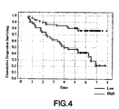
- the ⁇ -F1-ATPase ( Figure 4) as well as the BEC index, are indicators of the patient survival.
- the method for cancer detection, progression analysis and malign tumour prognosis based on the study of metabolic markers of the cell is configured as a method for the detection of malignant tumours based in the determination of the relative expression of the catalytic subunit ⁇ of the mitochondrial complex of the H(+)-ATP synthase, as only marker or in combination with the mitochondrial markers Hsp 60, COX I and COX IV (proportions ⁇ -F1-ATFase/ Hsp 60, ⁇ -F1-ATPase/COX I and/or ⁇ -F1-ATPase/COX IV), and this proportion is at the same time expressed with respect to the expression of a glycolytic marker (GAPDH, HK, PK) of the cell.
- a glycolytic marker GPDH, HK, PK
- the method that the invention proposes is configured in himself as an evident novelty in his specific field of application, as is the first time that is claimed and shown that the bioenergetic function of the mitochondrion brings a generic marker of cancer that we can call " bioenergetic sign or mark of cancer".
- the method for the cancer detection, progression analysis and malign tumour prognosis based on the study of metabolic markers of the cell object of the invention is created starting from the previous study of hepatocarcinomas, hepatoblastomas and adenocarcinomas of human kidney, mamma, lung, stomach and colon by means of immunological techniques of Western blot and immunocytochemistry using specific antibodies against the proteins used as markers.
- the method for cancer detection, progression analysis and malign tumour prognosis based on the study of metabolic markers of the cell that is foreseen consists of a method to determine the relative expresion of ⁇ -F1-ATPase in the tumour cell to show his lower expression in cancer.
- the rabbit polyclonal antibody specific against the subunit ⁇ of the H(+)-ATP synthase of rat liver has been generated in the laboratory by several techniques. In one case, using as immunogen the purified protein of SDS polyacrylamide gel [3]. In another case, using as immunogen the protein synthesized in bacteria, by means of the cDNA expression of rat liver ⁇ -F1-ATPase [5], and later purification of the protein expressed through affinity techniques. The remaining antibodies that have been used have been commercial.
- the immunological detection of the proteins was carried out using the following primary antibodies: anti- ⁇ -F1-ATPase (1:20.000), anti-Hsp 60 (1:2.000), anti-COX I (1:250), anti-COX IV (1:100) and anti-GAPDH (1:10.000), with the corresponding secondary antibody diluted in peroxidase conjugate at 1:3.000 and using the chemiluminescence method.
- the quantification of the intensity in the immunoreactivity signal of the different proteins was realized using an image digital analysis system.
- the relative cell content of mitochondrial DNA was also determined.
- the cellular DNA was extracted by phenol from the homogenized samples in buffer 10mM Tris HCI pH 7.5 that contains 10mM EDTA, 0,5% SDS and 0,8 ⁇ g/ ⁇ l proteinase K.
- the DNA obtained was precipitated with 2,5 volumes ethanol and 0,1 volumes sodium acetate 3M.
- DNA was resuspended in medium 10mM Tris HCI pH 8.0, that contains 100mM NaCl and 0,1 ⁇ g/ ⁇ l RNAse.
- the hybridization conditions were 5x Denhardt's solution, 50% formamide, 0.5% SDS, 6X SSC and 0,132 mg/ml of salmon semen DNA.
- the membranes were washed following the next protocol: 10 minutes with 6X SSC at room temperature, 10 minutes with 2X SDS containing 0,1 SDS at 65 ° C and 10 minutes with 1X SSC containing 0,1 SDS at 65 ° C.
- the membranes thus washed were exposed to autoradiographic films at -70° C and the intensity of the bars corresponding to the different genes were quantified by densitometry.
- hepatocarcinomas In the case of hepatocarcinomas, it was shown this decrease in the expression of both markers in relation to the tubulin expression by western-blot techniques. Furthermore, it was shown that in hepatocarcinomas the relative content of mitochondrial DNA is much lower than that of the healthy liver, approximately 2 -3 less.
- the ⁇ -F1-ATPase expression is 40% lower than this protein expression in normal mammal tissue.
- Hsp 60 and cytochrome oxidase expressions are 700% and 300% higher respectively, than the observed in the normal mammal tissue.
- lung adenocarcinomas the relative expression of ⁇ -F1-ATPase is 50% lower than the observed in normal lung tissue. Moreover, in lung adenocarcinomas, the expression of ⁇ -F1-ATPase is very decreased with respect to the expression in the respiratory enzimes cytochrome oxidase I and IV, which presents levels of expression 300% higher than the observed in normal lung tissue. In lung squamous adenocarcinomas, the relative expression of ⁇ -F1-ATPase is very decreased with respect to the Hsp 60 expression, being the ratio ⁇ -F1-ATPase/ Hsp 60 a 20% of the observed in normal lung tissue.
- GAPDH glyceraldehyde-3-phosphate dehydrogenase
- GAPDH increases 600% in kidney tumours, 200% in colon tumours, 900% in mammal tumours and 400% in lung tumours, when compared with the GAPDH expression in their respective normal tissues.
- the PK expression in mammal and lung tumours is 500% and 250% higher, respectively, than the observed in normal mammal and lung tissue.
- the BEC index ( ⁇ -F1-ATPase/ Hsp 60/ GAPDH or ⁇ -F1-ATPase/ Hsp 60/PK) for tumours of kidney, mamma, stomach, lung and colon is, therefore, much lower than in the respective healthy tissues.
- the BEC index estimated by the ratio ⁇ -F1-ATPase/ Hsp 60/GAPDH in kidney tumours is a 10% of the value found in normal kidney
- in colon tumours is a 25% of the value found in normal colon
- in stomach tumours is a 50% of the value found in normal stomach
- in mammal adenocarcinomas is a 5% of the value found in normal mammal tissue
- in lung adenocarcinomas is a 20% of the value found in normal lung
- lung squamous carcinomas is a 10% of the value found in normal lung.
- the BEC index estimated by the ratio ⁇ -F1-ATPase/ Hsp 60/PK in stomach adenocarcinomas is a 50% of the value found in normal stomach, in mammal adenocarcinomas is a 5% of the value found in normal mammal tissue, in lung adenocarcinomas is a 25% of the value found in normal lung and in lung squamous carcinomas is a 30% of the value found in normal lung .
Landscapes
- Health & Medical Sciences (AREA)
- Life Sciences & Earth Sciences (AREA)
- Engineering & Computer Science (AREA)
- Immunology (AREA)
- Chemical & Material Sciences (AREA)
- Molecular Biology (AREA)
- Biomedical Technology (AREA)
- Hematology (AREA)
- Urology & Nephrology (AREA)
- Medicinal Chemistry (AREA)
- General Health & Medical Sciences (AREA)
- Biochemistry (AREA)
- Microbiology (AREA)
- Biotechnology (AREA)
- Cell Biology (AREA)
- Physics & Mathematics (AREA)
- Analytical Chemistry (AREA)
- General Physics & Mathematics (AREA)
- Pathology (AREA)
- Food Science & Technology (AREA)
- Organic Chemistry (AREA)
- Genetics & Genomics (AREA)
- Zoology (AREA)
- Wood Science & Technology (AREA)
- Bioinformatics & Cheminformatics (AREA)
- General Engineering & Computer Science (AREA)
- Biophysics (AREA)
- Proteomics, Peptides & Aminoacids (AREA)
- Gastroenterology & Hepatology (AREA)
- Toxicology (AREA)
- Measuring Or Testing Involving Enzymes Or Micro-Organisms (AREA)
- Medicines That Contain Protein Lipid Enzymes And Other Medicines (AREA)
- Pharmaceuticals Containing Other Organic And Inorganic Compounds (AREA)
- Steroid Compounds (AREA)
Abstract
Description
- The present descriptive memory deals with a patent of invention application that relates to a method for cancer detection, progression analysis and malign tumour prognosis based on the study of metabolic markers of the cell, whose purpose lies in being established as a procedure for cancer detection, analysis of tumour progression and its prognosis, based in the study of the functional capacity of the mitochondrion with respect to the glycolytic capacity of the cell.
- This invention has its application in the Medical and Veterinary Oncology, for detection and tracking of malign tumours, particularly inscribed in the field of Biotechnology. From a conceptual point of view, the catalytic subunit β of the mitochondrial complex of the H(+)-ATP synthase, as well as the bioenergetic mitochondrial index and the bioenergetic index of the cell, could also be of use in the fields of the Neurosciencies and the Cardiovascular diseases, and in general in all those pathologies that deal with hipoxia and/or anoxia of tissues, as well as other pathologies associated to dysfunctions of the mitochondrial cell activity.
- The mitochondrion is the organule of the eukaryotic cell responsible for the energy supply. The enzymatic machinery responsible in the last step of the synthesis of the cellular ATP into the H(+)-ATP synthase, a rotary molecular engine that is located in the inner membrane of the mitochondrion [12]. In the last years, the mitochondrion has come to take an important role in studies related to Biomedicine due to its obvious implication in multiple manifestations of the human pathology. Specifically, in the case of cancer, because is the subcellular organule sensor and executor of the apoptosis program. The apoptosis is a genetic program of programmed cell death, that requires energy and is essential for the normal development of the organism. Alterations in the apoptosis program contribute to the progression of certain pathologies such as cancer and neurodegenerative diseases [10, 11].
- The study of the energetic metabolism of cancer cells was a central issue in the cancer investigation until the beginning of 1970, decade in which was a relegated topic by the scientific community, due to the approach and development of the molecular biology of this disease. However, it was in 1930 when Otto Warburg formulated the hypothesis that tumour cells should have an altered mitochondrial function and this dysfunction had to explain the high aerobic glycolisis that characterize the majority of the tumour cells. While the glycolytic fenotype of many cancer cells and tumours have been demonstrated from the biochemical and molecular study, dysfunction of the mitochondrial function of the cancer cell was never established in the biology of cancer. In fact, we still are ignorant of the role that the mitochondrion plays in the neoplasic transformation, as well as in the support or promotion of the transformed state of the cell. Solely, and in the case of highly glycolytic rat hepatomas, his aberrant energetic metabolism has been explained by the decrease of the relative cell content of mitochondrias
[6]. In this concrete case, it has been described that the mitochondrial fenotype of the hepatomas is similar to the mitochondrial fenotype of the fetal liver, where an specific program of mitochondrial biogenesis limits the mitochondrial content of the cell [1]. - On the other hand, it has recently been revealed that the execution of the apoptosis program requires the molecular components of the H(+)-ATP synthase of the mitochondrion [7, 8], as well as a functional oxidative phosphorylation in the cell [2, 4]. These results suggest that cells with a relatively low expression of H(+)-ATP synthase and/or with a deficient functionality of oxidative phosphorylation, of which is a bottleneck the same H(+)-ATP synthase, must be more resistant to the apoptosis, and therefore, should have favoured his clonal expansion in the context of the organism.
- In the case of the present invention, there have been used monoclonal and polyclonal antibodies that specifically recognize mitochondrial proteins and proteins of the glycolytic pathway in "'Western blot" and immunocytochemical techniques, with the aim to quantify the expression of these proteins in normal tissues and its corresponding tumours, to use them, alone in combination, as markers of the progression and prognosis of cancer.
- In this way, it has been attained to show for the first time, that the catalytic subunit β of the mitochondrial complex of the H(+)-ATP synthase (β-F1-ATPase) has his expression decreased in liver, kidney and colon tumours, what implies to show that the mitochondrial function of the tumour cell is affected, confirming the hypothesis that Otto Warburg had stated in the beginning of the last century, and that had not been demonstrated in human carcinomas up to this moment. These results allow his extrapolation to mamma, lung, stomach, prostate and endometrium adenocarcinomas; to lung and larynx squamous carcinomas, as well as melanomas and lymphomas.
- In liver carcinomas, the affection of the mitochondrial function is produced by repression of the mitochondrial biogenesis, as in a parallel way it is produced the lower expression of structural components of the mitochondrion, as are the
marker Hsp 60 and the proper mitochondrial DNA (Figure 1). - On the contrary, in kidney carcinomas (Figure 2) and colon carcinomas (Figure 3), the lower expression of the catalytic subunit β of the mitochondrial complex of the H(+)-ATP synthase is produced in absence of significative changes in the expression of
Hsp 60, what indicates that in this type of carcinomas it is being affected the molecular mechanism that determines the grade of functional differentiation of the mitochondrion, that comes expressed by the ratio between the bioenergetic marker and the structural marker, that is, by the proportion β-F1-ATPase/Hsp 60. In mammal adenocarcinomas the lower expression of β-F1-ATPase is produced in a simultaneus way with a higher expression ofHsp 60 and cytochrome oxidase I, what means a very pronounced decrease of the proportion β-F1-ATPase/Hsp 60 and β-F1-ATPase/COX I in the tumour tissue. - In lung adenocarcinomas, the relative expression of β-F1-ATPase decreases very strongly with respect to the expression of the respiratory enzimes cytochrome oxidase I and IV (β-F1-ATPase/COX I and β-F1-ATPase/COX IV).
- In lung squamous carcinomas, the relative expression of β-F1-ATPase decreases significantly with respect to the
Hsp 60 expression (β-F1-ATPase/ Hsp 60). - Moreover, we have shown that in a parallel manner that a decrease in the ratio β-F1-ATPase/
Hsp 60, β-F1-ATPase/COX I and/or β-F1-ATPase/COX IV in cancer is produced, it is produced an increase in the expression of the glyceraldehyde-3-phosphate dehydrogenase (GAPDH) (Figures 2 and 3) and/or the pyruvate kinase isoform M (PK), markers of the glycolytic pathway. - Because of this reason, we define a bioenergetic index of the cell (BEC index) that relates the bioenergetic potentiality of the mitochondrion with the cellular glycolytic capacity, an index that expresses any of the proportions β-F1-ATPase/
Hsp 60, β-F1-ATPase/COX I and/or β-F1-ATPase/COX IV with respect to the cellular expression of GAPDH (ratio β-F1-ATPase/Hsp 60/ GAPDH, β-F1-ATPase/COX I/ GAPDH, β-F1-ATPase/COX IV/ GAPDH and/or PK (ratios β-F1-ATPase/Hsp 60/ PK, β-F1-ATPase/COX I/ PK, β-F1-ATPase/COX IV/ PK). The BEC index also decreases in tumour cells with respect to the observed in healthy cells of the kidney (Figure 2), colon (Figure 3), mamma, stomach and lung. - Moreover, in the case of colon carcinomas, the β-F1-ATPase (Figure 4) as well as the BEC index, are indicators of the patient survival.
-
- 1. Cuezva J.M., Ostronoff L.K., Ricart J., Lopez de Heredia M. , Di LIegro C.M., and IzquierdoJ.M.. Mitochondrial biogenesis in the liver during development and oncogenesis. J. Bioenerg. Biomembr. 29, 365-377 (1997).
- 2. Dey, R. and Moraes, C.T. Lack of Oxidative Phosphorylation and Low Mitochondrial Membrane Potential Decrease Susceptibility to Apoptosis and Do not Modulate the Protective Effect of Bc1-xL in Osteosarcoma Cells. J. Biol. Chem., 275:7087-7094 (2000)
- 3. Egea G., Izquierdo, J.M. Ricart, J. San Martin C.and Cuezva J.M. mRNA encoding the beta-subunit of the mitochondrial F1-ATPase complex is a localized mRNA in rat hepatocytes. Biochem. J. 322, 557-656 (1997).
- 4. Harris, MH, Vander Heiden, MG, Kron, SJ, and Thompson, CB (2000) Role of oxidative phosphorylation in Bax toxicity. Mol. Cell. Biol. 20: 3590-3596.
- 5. IzquierdoJ.M. and Cuezva J.M. Control of the translational efficiency of beta-F1-ATPase mRNA depends on the regulation of a protein that binds the 3'-untranslated region of the mRNA . Mol. Cell Biol. 17, 5255-5268 (1997).
- 6. Lopez de Heredia, J. M. Izquierdo, and J. M. Cuezva A Conserved Mechanism for Controlling the Translation of beta -F1-ATPase mRNA between the Fetal Liver and Cancer Cells J. Biol. Chem., March 15, 2000; 275(10): 7430 - 7437.
- 7. Matsuyama S, Llopis J, Deveraux QL, Tsien RY, Reed JC. Changes in intramitochondrial and cytosolic pH: early events that modulate caspase activation during apoptosis. Nat Cell Biol. 2000, 2:318-25.
- 8. Matsuyama S, Xu Q, Velours J, Reed JC. The Mitochondrial F0F1-ATPase proton pump is required for function of theproapoptotic protein Bax in yeast and mammalian cells. Mol Cell. 1998 Feb;1(3):327-36.
- 9. Ostronoff LK, Izquierdo JM, Enriquez JA, Montoya J, Cuezva JM. Transient activation of mitochondrial translation regulates the expression of the mitochondrial genome during mammalian mitochondrial differentiation. 1996. Biochem. J. 316:183-91.
- 10. Reed JC: Mechanisms of apoptosis avoidance in cancer. Curr. Opin. Oncol. 1999;11:68-75.
- 11. Thompson CB. Apoptosis in the pathogenesis and treatment of disease.Science. 1995.267:1456-62.
- 12. Yoshida,M., Muneyuki,E. and Hisabori,T.ATP synthase--a marvellous rotary engine of the cell Nat. Rev. Mol. Cell Biol. 2 (9), 669-677 (2001)
-
- The method for cancer detection, progression analysis and malign tumour prognosis based on the study of metabolic markers of the cell, is configured as a method for the detection of malignant tumours based in the determination of the relative expression of the catalytic subunit β of the mitochondrial complex of the H(+)-ATP synthase, as only marker or in combination with the
mitochondrial markers Hsp 60, COX I and COX IV (proportions β-F1-ATFase/Hsp 60, β-F1-ATPase/COX I and/or β-F1-ATPase/COX IV), and this proportion is at the same time expressed with respect to the expression of a glycolytic marker (GAPDH, HK, PK) of the cell. The method that the invention proposes is configured in himself as an evident novelty in his specific field of application, as is the first time that is claimed and shown that the bioenergetic function of the mitochondrion brings a generic marker of cancer that we can call " bioenergetic sign or mark of cancer". - To specify, the method for the cancer detection, progression analysis and malign tumour prognosis based on the study of metabolic markers of the cell object of the invention, is created starting from the previous study of hepatocarcinomas, hepatoblastomas and adenocarcinomas of human kidney, mamma, lung, stomach and colon by means of immunological techniques of Western blot and immunocytochemistry using specific antibodies against the proteins used as markers.
- It must be point out that as controls, there have been used non-tumour samples of human liver, kidney, mamma, lung, stomach and colon coming from autopsies and/or biopsies or from the non-tumour part adjacent to the tumour.
- To complete the description that is being made and with the aim to help for a better understanding of the characteristics of the invention, it is accompanied to the present descriptive memory, as an integral part thereof, a set of planes for illustrative and non-limitative purposes, where has been described the following:
- Figure 1.- Represents the repression of the mitochondrial biogenesis in
human hepatocarcinomas. a, Analysis of the expression of β-F1-ATPase
(β-F1),
Hsp 60 and tubulin in normal liver (C1-C3) and in hepatocarcinomas (T1-T2). The histogram shows the relative cell content of β-F1 andHsp 60 with respect to the expression of tubulin in normal liver (white bars) and in hepatocarcinomas (black bars). b, Analysis of the mitochondrial DNA content (mtDNA) in normal liver(C1-C3) and in hepatocarcinomas (T1-T2). The histogram shows the relative cell content of the mitochondrial genes 12S and A6-8, with respect to the nuclear gen β-F1 in normal liver (white bars) and in hepatocarcinomas (black bars). - Figure 2.- Corresponds to the analysis of the expression of β-F1-ATPase(β-F1),
Hsp 60 and GAPDH, as well as to the bioenergetic index of the mitochondrion (β-F1/ Hsp 60), and of the cell (BEC indexβ-F1/
Hsp 60/ GAPDH) in normal renal epithelium (white bars) and in kidney carcinomas (black bars). - Figure 3.- Shows the analysis of the bioenergetic index of the
mitochondrion(β-F1/ Hsp 60) and of the GAPDH expression, as well as
of the bioenergetic index of the cell (BEC index
Hsp 60/GAPDH) in normal colon samples (white bars) and in colon carcinomas (black bars). - Figure 4.- Shows finally the Kaplan-Meier analysis, where the β-F1-ATPase expression in colorectal carcinomas is correlated with the survival of patients. The broken line (in the upper part of the Figure), corresponds to tumours with a high level of expression of β-F1-ATPase. The solid line (in the lower part of the Figure) corresponds to tumours with a low level of expression of β-F1-ATPase.
-
- The method for cancer detection, progression analysis and malign tumour prognosis based on the study of metabolic markers of the cell that is foreseen, consists of a method to determine the relative expresion of β-F1-ATPase in the tumour cell to show his lower expression in cancer.
- The analysis of the expression of
Hsp 60, COX I and COX IV in the same sample and by means of the same technique, brings an inner control of the measurement system used, whether is by loading and/or by transference of the mitochondrial proteins to the membrane, at the same time allowing the calculus of the mitochondrial bioenergetic index (proportions β-F1-ATpase/Hsp 60, β-F1-ATPase/COX I , β-F1-ATPase/COX IV) of the sample. - The analysis of the expression of GAPDH and/or PK in the same sample and by means of the same technique, brings an inner control of the measurement system used, whether is by loading and/or by transference of the cell proteins to the membrane, at the same time allowing the calculus of the bioenergetic index of the cell (proportions β-F1-ATPase/ COX I/ GAPDH; β-F1-ATPase/COX IV/ GAPDH; β-F1-ATPase/
Hsp 60/PK; β-F1-ATPase/ COX I/ PK; β-F1-ATPase/ COX IV/ PK). - The hypothesis is that both the expression of β-F1-ATPase and the bioenergetic indexes of the mitochondrion and the cell (BEC index), are decreased in cancer, what affects negatively the apoptosis program, and permits the clonal expansion of the tumour cell. Although it is ignored exactly the mechanism thereby in cancer it is produced the inhibition of the β-F1-ATPase expression, this does not seem to be due to a lower expression of his mRNA, as the mRNA expression of β-F1-ATPase is increased in cancer. Possibly, the lower expression β-F1-ATPase in cancer is due to a control of the coresponding mRNA translation by the interaction of this with cell proteins.
- The rabbit polyclonal antibody specific against the subunit β of the H(+)-ATP synthase of rat liver has been generated in the laboratory by several techniques. In one case, using as immunogen the purified protein of SDS polyacrylamide gel [3]. In another case, using as immunogen the protein synthesized in bacteria, by means of the cDNA expression of rat liver β-F1-ATPase [5], and later purification of the protein expressed through affinity techniques. The remaining antibodies that have been used have been commercial.
- The expression of metabolic markers in tumours of liver, kidney, mamma, stomach, lung and colon, as well as the corresponding healthy tissues was made by Western blot and immunocytochemical techniques.
- For the determination of markers by Western blot techniques 20 cuts of 17 micrometer thick of normal and tumour tissue of the same patient were extracted and processed, respectively, in 300 µl of a 50 mM Tris-
HCI pH 8 buffer, that contains 150 mM Na Cl, 0,02% Sodium acide, 0,1% SDS, 1 %Nonidet P-40, 0,5% sodium deoxycholate,1µg/µl aprotinin, 1µg/µl antitrypsin, 0,4 mM EDTA, 10 mM NaF, and 0,75 mM PMSF at 4 ° C for thirty minutes. After the extraction of the proteins, samples were centrifuged at 14.000 rpm at 4 ° C for 25 minutes, and in the resulting supernatants the protein concentration was determined using the Bradford reactive. The fractionation of 15 - 30 µg of the proteins extracted from the normal and tumour tissue of the same patient was made in SDS gel 10% or 15% PAGE. Later on, the fractioned proteins in the gel were transferred to PVDF membranes for 1 hour at constant voltage. The immunological detection of the proteins was carried out using the following primary antibodies: anti-β-F1-ATPase (1:20.000), anti-Hsp 60 (1:2.000), anti-COX I (1:250), anti-COX IV (1:100) and anti-GAPDH (1:10.000), with the corresponding secondary antibody diluted in peroxidase conjugate at 1:3.000 and using the chemiluminescence method. The quantification of the intensity in the immunoreactivity signal of the different proteins was realized using an image digital analysis system. - For the determination of the expression in the protein markers by immunohistochemical techniques, sections of 4 - 5 µM thick correponding to the samples of normal or pathological tissue, previously fixed in neutral formalin solution and paraffin-embedded, were incubated with the primary antibodies anti-β-F1-ATPase (1:3.000), anti-Hsp 60 (1:800) or anti-GAPDH (1:1.000). For the development of the different immunohistochemicals a detection method based in the diaminobenzidine was followed, using the Envision-Plus horseradish peroxidase system of DAKO Corporation. Afterwards, the cuts were stained with hematoxylin to highlight cells nuclei. The quantification of the chromogen intensity deposited in the cell was carried out by means of densitometric analysis of the chromogen intensity deposited per unit area of cytoplasm (30µm2) obtained starting from the digital magnified sample images (1.000x).
- In the case of hepatocarcinomas and healthy liver there was also determined the relative cell content of mitochondrial DNA through western-blot techniques [9]. For this purpose, the cellular DNA was extracted by phenol from the homogenized samples in buffer 10mM Tris HCI pH 7.5 that contains 10mM EDTA, 0,5% SDS and 0,8 µg/µl proteinase K. The DNA obtained was precipitated with 2,5 volumes ethanol and 0,1 volumes sodium acetate 3M. DNA was resuspended in medium 10mM Tris HCI pH 8.0, that contains 100mM NaCl and 0,1 µg/µl RNAse. After incubation of one hour at room temperature SDS is added to 0,5% final concentration and it is extracted with phenolchloroform-isoamilic (25:24:1) and the precipitation of DNA with ethanol and sodium acetate is repeated. The DNA was directed with the restriction enzyme BamHI and was fractioned on 0,8% agarose gel. The DNA fragments were transferred to nitrocellulose membranes under vacuum conditions for 4 hours at partial pressure of 10mm mercury. The DNA fragments are fixed to the membrane by incubation of the same at 80 ° C for 2 hours. The membranes were incubated with DNA sondes marked with 32P for the nuclear gen of the β-F1-ATPase and the mitochondrial genes of the ATPase 6 - 8 and 12 S. The hybridization conditions were 5x Denhardt's solution, 50% formamide, 0.5% SDS, 6X SSC and 0,132 mg/ml of salmon semen DNA. The membranes were washed following the next protocol: 10 minutes with 6X SSC at room temperature, 10 minutes with 2X SDS containing 0,1 SDS at 65 ° C and 10 minutes with 1X SSC containing 0,1 SDS at 65 ° C. The membranes thus washed were exposed to autoradiographic films at -70° C and the intensity of the bars corresponding to the different genes were quantified by densitometry.
- The analysis of the samples of hepatocarcinomas, hepatoblastomas and focal nodular hyperplasias by immunocytochemical techniques showed a very significant decrease of the β-F1-ATPase and
Hsp 60 expressions when compared with the expression of these markers in normal liver. Particularly, the expression of these two markers in hepatocarcinomas was of 13% for β-F1-ATPase and 30% forHsp 60 in relation to the expression observed in normal liver. - In the case of hepatocarcinomas, it was shown this decrease in the expression of both markers in relation to the tubulin expression by western-blot techniques. Furthermore, it was shown that in hepatocarcinomas the relative content of mitochondrial DNA is much lower than that of the healthy liver, approximately 2 -3 less.
- This set of results indicate that in liver cancer it is produced a repression of the mitochondrial biogenesis of the cell.
- The analysis of the
Hsp 60 expression in adenocarcinomas of kidney, stomach and colon did not show significant variations with regard to the respective non-tumour tissue. On the contrary, the β-F1-ATPase expression is intensely decreased in kidney, stomach and colon tumours. Particularly, in carcinomas of these three tissues the decrease in the β-F1-ATPase expression is a 50% of the one observed in the respective normal tissues. - In mammal adenocarcinomas, the β-F1-ATPase expression is 40% lower than this protein expression in normal mammal tissue. Simultaneously, in mammal adenocarcinomas it is observed that
Hsp 60 and cytochrome oxidase expressions are 700% and 300% higher respectively, than the observed in the normal mammal tissue. - In lung adenocarcinomas, the relative expression of β-F1-ATPase is 50% lower than the observed in normal lung tissue. Moreover, in lung adenocarcinomas, the expression of β-F1-ATPase is very decreased with respect to the expression in the respiratory enzimes cytochrome oxidase I and IV, which presents levels of expression 300% higher than the observed in normal lung tissue. In lung squamous adenocarcinomas, the relative expression of β-F1-ATPase is very decreased with respect to the
Hsp 60 expression, being the ratio β-F1-ATPase/ Hsp 60 a 20% of the observed in normal lung tissue. - Consistent with a lower activity of the bioenergetic function of the mitochondrion in cancer, it was observed that the activity of the glyceraldehyde-3-phosphate dehydrogenase (GAPDH) is strongly increased in tumours of kidney, mamma, lung and colon, finding a highly significant inverse relation between the bioenergetic index of the mitochondrion (ratio β-F1-ATPase/ Hsp 60) and the glucolitic capacity of the cell, expressed these latter by the quantity of GAPDH. Then, it has been shown that GAPDH increases 600% in kidney tumours, 200% in colon tumours, 900% in mammal tumours and 400% in lung tumours, when compared with the GAPDH expression in their respective normal tissues. The PK expression in mammal and lung tumours is 500% and 250% higher, respectively, than the observed in normal mammal and lung tissue.
- The BEC index (β-F1-ATPase/
Hsp 60/ GAPDH or β-F1-ATPase/Hsp 60/PK) for tumours of kidney, mamma, stomach, lung and colon is, therefore, much lower than in the respective healthy tissues. In this sense, the BEC index estimated by the ratio β-F1-ATPase/Hsp 60/GAPDH in kidney tumours is a 10% of the value found in normal kidney, in colon tumours is a 25% of the value found in normal colon, in stomach tumours is a 50% of the value found in normal stomach, in mammal adenocarcinomas is a 5% of the value found in normal mammal tissue, in lung adenocarcinomas is a 20% of the value found in normal lung and in lung squamous carcinomas is a 10% of the value found in normal lung. The BEC index estimated by the ratio β-F1-ATPase/Hsp 60/PK in stomach adenocarcinomas is a 50% of the value found in normal stomach, in mammal adenocarcinomas is a 5% of the value found in normal mammal tissue, in lung adenocarcinomas is a 25% of the value found in normal lung and in lung squamous carcinomas is a 30% of the value found in normal lung . - The study of the expressions of β-F1-ATPase,
Hsp 60 and GAPDH as well as the mitochondrial bioenergetic index (β-F1-ATPase/ Hsp 60) and the cellular bioenergetic index (β-F1-ATPase/Hsp 60/ GAPDH) in colorectal tumours for which there is known the survival of patients, shows the existence of a significant relation between the level of expression of the β-F1-ATPase and of the mitochondrial and cellular bioenergetic indexes with the survival and prognosis of patients.
Claims (5)
- Method for cancer detection, progression analysis and malign tumour prognosis based on the study of metabolic markers of the cell, characterized because it uses the subunit β of the H(+)-ATP synthase of the mitochondrion as a marker of expression, where the type of cancer is the kidney, colon, stomach, lung and mamma cancer.
- Method for cancer detection, progression analysis and malign tumour prognosis based on the study of metabolic markers of the cell, according to claim 1, characterized by the use of immunological techniques, such as western-blot, immunohistochemistry, etcetera...
- Method for cancer detection, progression analysis and malign tumour prognosis based on the study of metabolic markers of the cell, according to both previous claims characterized by the use of the mitochondrial bioenergetic index which is expressed by the ratio β-F1-ATPase/ Hsp 60; β-F1-ATPase/ COX I and/or β-F1-ATPase/ COX IV, where the type of cancer is the kidney, colon, stomach, lung and mamma cancer.
- Method for cancer detection, progression analysis and malign tumour prognosis based on the study of metabolic markers of the cell, according to all the previous claims characterized by the use of the bioenergetic index of the cell that is expressed by the ratio β-F1-ATPase/ Hsp 60/GAPDH; β-F1-ATPase/ COX I/ GAPDH and/or β-F1-ATPase/ COX IV/ GAPDH where the type of cancer is the kidney, colon, stomach, lung and mamma cancer.
- Method for cancer detection, progression analysis and malign tumour prognosis based on the study of metabolic markers of the cell, according to claim 3, characterized by the use of the bioenergetic index of the cell that is expressed by the ratio β-F1-ATPase/ Hsp 60/PK; β-F1-ATPase/ COX I/PK; β-F1-ATPase/ COX TV/PK GAPDH where the type of cancer is the kidney, colon, stomach, lung and mamma cancer.
Applications Claiming Priority (3)
| Application Number | Priority Date | Filing Date | Title |
|---|---|---|---|
| ES200201190A ES2195791B2 (en) | 2002-05-24 | 2002-05-24 | PROCEDURE FOR THE DETECTION OF CANCER, ANALYSIS OF THE PROGRESSION AND PROGNOSIS OF MALIGMAN TUMORS BASED ON THE STUDY OF METABOLIC MARKERS OF THE CELL. |
| ES200201190 | 2002-05-24 | ||
| PCT/ES2003/000228 WO2003100430A1 (en) | 2002-05-24 | 2003-05-20 | Method for cancer detection, progression analysis and malign tumour prognosis based on the study of metabolic markers of the cell |
Publications (2)
| Publication Number | Publication Date |
|---|---|
| EP1564557A1 true EP1564557A1 (en) | 2005-08-17 |
| EP1564557B1 EP1564557B1 (en) | 2008-12-24 |
Family
ID=29558508
Family Applications (1)
| Application Number | Title | Priority Date | Filing Date |
|---|---|---|---|
| EP03727509A Expired - Lifetime EP1564557B1 (en) | 2002-05-24 | 2003-05-20 | Method for cancer detection, progression analysis and malign tumour prognosis based on the study of metabolic markers of the cell |
Country Status (8)
| Country | Link |
|---|---|
| US (1) | US7608403B2 (en) |
| EP (1) | EP1564557B1 (en) |
| JP (1) | JP4235610B2 (en) |
| AT (1) | ATE418736T1 (en) |
| CA (1) | CA2487176C (en) |
| DE (1) | DE60325508D1 (en) |
| ES (2) | ES2195791B2 (en) |
| WO (1) | WO2003100430A1 (en) |
Families Citing this family (2)
| Publication number | Priority date | Publication date | Assignee | Title |
|---|---|---|---|---|
| EP2821790A1 (en) * | 2013-07-04 | 2015-01-07 | Universität Heidelberg | Tumor energy metabolism profiling |
| DE102016221867A1 (en) | 2016-11-08 | 2018-05-09 | BSH Hausgeräte GmbH | System for producing an alcoholic beverage |
Family Cites Families (2)
| Publication number | Priority date | Publication date | Assignee | Title |
|---|---|---|---|---|
| US5972628A (en) * | 1989-11-17 | 1999-10-26 | Schebo Tech Medizinisch-Biologische Forschungsgesellschaft M.B.H. | Pyruvatekinase-iosenzyme typ-M2 (Tumor-M2-PK)-specific antibody/process for the preparation and use thereof |
| US5434046A (en) * | 1991-03-01 | 1995-07-18 | The Regents Of The University Of California | Diagnostic assay based on cis-plain drug resistance gene |
-
2002
- 2002-05-24 ES ES200201190A patent/ES2195791B2/en not_active Expired - Fee Related
-
2003
- 2003-05-20 CA CA002487176A patent/CA2487176C/en not_active Expired - Fee Related
- 2003-05-20 WO PCT/ES2003/000228 patent/WO2003100430A1/en not_active Ceased
- 2003-05-20 AT AT03727509T patent/ATE418736T1/en not_active IP Right Cessation
- 2003-05-20 DE DE60325508T patent/DE60325508D1/en not_active Expired - Lifetime
- 2003-05-20 ES ES03727509T patent/ES2318134T3/en not_active Expired - Lifetime
- 2003-05-20 US US10/514,771 patent/US7608403B2/en not_active Expired - Fee Related
- 2003-05-20 EP EP03727509A patent/EP1564557B1/en not_active Expired - Lifetime
- 2003-05-20 JP JP2004507836A patent/JP4235610B2/en not_active Expired - Fee Related
Non-Patent Citations (1)
| Title |
|---|
| See references of WO03100430A1 * |
Also Published As
| Publication number | Publication date |
|---|---|
| ES2318134T3 (en) | 2009-05-01 |
| EP1564557B1 (en) | 2008-12-24 |
| CA2487176A1 (en) | 2003-12-04 |
| JP4235610B2 (en) | 2009-03-11 |
| US7608403B2 (en) | 2009-10-27 |
| US20070003988A1 (en) | 2007-01-04 |
| ES2195791B2 (en) | 2005-02-01 |
| JP2005526980A (en) | 2005-09-08 |
| ATE418736T1 (en) | 2009-01-15 |
| WO2003100430A1 (en) | 2003-12-04 |
| DE60325508D1 (en) | 2009-02-05 |
| ES2195791A1 (en) | 2003-12-01 |
| CA2487176C (en) | 2008-12-09 |
Similar Documents
| Publication | Publication Date | Title |
|---|---|---|
| Balasubramani et al. | Altered expression and localization of creatine kinase B, heterogeneous nuclear ribonucleoprotein F, and high mobility group box 1 protein in the nuclear matrix associated with colon cancer | |
| US20070053896A1 (en) | Diagnostic marker for ovarian cancer | |
| JPWO2009028417A1 (en) | Gynecologic cancer detection method | |
| Lee et al. | Identification of cystatin B as a potential serum marker in hepatocellular carcinoma | |
| Yang et al. | Ubiquitin-specific protease 22: a novel molecular biomarker in cervical cancer prognosis and therapeutics | |
| EP2585488A2 (en) | Novel biomarkers of liver cancer | |
| EP2553458A2 (en) | Serum and tissue biomarkers of human hcc | |
| Wang et al. | Expression and clinical significance of BIRC6 in human epithelial ovarian cancer | |
| Müller et al. | Proteomic characterization of prostate cancer to distinguish nonmetastasizing and metastasizing primary tumors and lymph node metastases | |
| CN111505292A (en) | Use of PCK 1-based modulation of lipid metabolism as a target for cancer therapy, diagnosis and prognosis prediction | |
| Zhang et al. | KMT2A regulates cervical cancer cell growth through targeting VDAC1 | |
| Kaneko et al. | The active form of Helicobacter pylori vacuolating cytotoxin induces decay‐accelerating factor CD55 in association with intestinal metaplasia in the human gastric mucosa | |
| He et al. | A decision tree–based combination of ezrin-interacting proteins to estimate the prognostic risk of patients with esophageal squamous cell carcinoma | |
| EP1564557B1 (en) | Method for cancer detection, progression analysis and malign tumour prognosis based on the study of metabolic markers of the cell | |
| CN111273011A (en) | Application of FKBP38 protein in the diagnosis of breast cancer | |
| Zhang et al. | Comparative analysis of the human urinary proteome by 1D SDS-PAGE and chip-HPLC-MS/MS identification of the AACT putative urinary biomarker | |
| WO2013022789A2 (en) | Compositions and methods for assessing and determining the presence of liver cancer in a mammalian subject | |
| JP2008502891A (en) | Use of protein PDX1 as a marker for breast cancer | |
| CN117192123A (en) | Biomarker for evaluating prognosis of nasopharyngeal carcinoma and application thereof | |
| CN103543266B (en) | Applications of reagent for detecting PGRMC1 to preparation of kidney cancer diagnosis reagent and kit | |
| Codarin et al. | Differential proteomic analysis of subfractioned human hepatocellular carcinoma tissues | |
| Robinson-Bennett et al. | Implications of tyrosine phosphoproteomics in cervical carcinogenesis | |
| CN1920570B (en) | Thioredoxin-like protein 2 applications | |
| US20130203059A1 (en) | Method for Diagnosis of Bladder Cancer and Related Kits | |
| Yu et al. | RIPK4 driven by TP53 mutations promotes resistance to redox stress of CRC by phosphorylating MTHFD1 |
Legal Events
| Date | Code | Title | Description |
|---|---|---|---|
| PUAI | Public reference made under article 153(3) epc to a published international application that has entered the european phase |
Free format text: ORIGINAL CODE: 0009012 |
|
| 17P | Request for examination filed |
Effective date: 20050408 |
|
| AK | Designated contracting states |
Kind code of ref document: A1 Designated state(s): AT BE BG CH CY CZ DE DK EE ES FI FR GB GR HU IE IT LI LU MC NL PT RO SE SI SK TR |
|
| GRAP | Despatch of communication of intention to grant a patent |
Free format text: ORIGINAL CODE: EPIDOSNIGR1 |
|
| GRAS | Grant fee paid |
Free format text: ORIGINAL CODE: EPIDOSNIGR3 |
|
| GRAA | (expected) grant |
Free format text: ORIGINAL CODE: 0009210 |
|
| AK | Designated contracting states |
Kind code of ref document: B1 Designated state(s): AT BE BG CH CY CZ DE DK EE ES FI FR GB GR HU IE IT LI LU MC NL PT RO SE SI SK TR |
|
| REG | Reference to a national code |
Ref country code: GB Ref legal event code: FG4D |
|
| REG | Reference to a national code |
Ref country code: CH Ref legal event code: EP |
|
| REG | Reference to a national code |
Ref country code: CH Ref legal event code: NV Representative=s name: TROESCH SCHEIDEGGER WERNER AG |
|
| REG | Reference to a national code |
Ref country code: IE Ref legal event code: FG4D |
|
| REF | Corresponds to: |
Ref document number: 60325508 Country of ref document: DE Date of ref document: 20090205 Kind code of ref document: P |
|
| REG | Reference to a national code |
Ref country code: SE Ref legal event code: TRGR |
|
| REG | Reference to a national code |
Ref country code: ES Ref legal event code: FG2A Ref document number: 2318134 Country of ref document: ES Kind code of ref document: T3 |
|
| PG25 | Lapsed in a contracting state [announced via postgrant information from national office to epo] |
Ref country code: SI Free format text: LAPSE BECAUSE OF FAILURE TO SUBMIT A TRANSLATION OF THE DESCRIPTION OR TO PAY THE FEE WITHIN THE PRESCRIBED TIME-LIMIT Effective date: 20081224 Ref country code: FI Free format text: LAPSE BECAUSE OF FAILURE TO SUBMIT A TRANSLATION OF THE DESCRIPTION OR TO PAY THE FEE WITHIN THE PRESCRIBED TIME-LIMIT Effective date: 20081224 |
|
| PG25 | Lapsed in a contracting state [announced via postgrant information from national office to epo] |
Ref country code: BG Free format text: LAPSE BECAUSE OF FAILURE TO SUBMIT A TRANSLATION OF THE DESCRIPTION OR TO PAY THE FEE WITHIN THE PRESCRIBED TIME-LIMIT Effective date: 20090324 Ref country code: RO Free format text: LAPSE BECAUSE OF FAILURE TO SUBMIT A TRANSLATION OF THE DESCRIPTION OR TO PAY THE FEE WITHIN THE PRESCRIBED TIME-LIMIT Effective date: 20081224 Ref country code: BE Free format text: LAPSE BECAUSE OF FAILURE TO SUBMIT A TRANSLATION OF THE DESCRIPTION OR TO PAY THE FEE WITHIN THE PRESCRIBED TIME-LIMIT Effective date: 20081224 Ref country code: EE Free format text: LAPSE BECAUSE OF FAILURE TO SUBMIT A TRANSLATION OF THE DESCRIPTION OR TO PAY THE FEE WITHIN THE PRESCRIBED TIME-LIMIT Effective date: 20081224 |
|
| PG25 | Lapsed in a contracting state [announced via postgrant information from national office to epo] |
Ref country code: CZ Free format text: LAPSE BECAUSE OF FAILURE TO SUBMIT A TRANSLATION OF THE DESCRIPTION OR TO PAY THE FEE WITHIN THE PRESCRIBED TIME-LIMIT Effective date: 20081224 Ref country code: PT Free format text: LAPSE BECAUSE OF FAILURE TO SUBMIT A TRANSLATION OF THE DESCRIPTION OR TO PAY THE FEE WITHIN THE PRESCRIBED TIME-LIMIT Effective date: 20090525 Ref country code: AT Free format text: LAPSE BECAUSE OF FAILURE TO SUBMIT A TRANSLATION OF THE DESCRIPTION OR TO PAY THE FEE WITHIN THE PRESCRIBED TIME-LIMIT Effective date: 20081224 |
|
| PG25 | Lapsed in a contracting state [announced via postgrant information from national office to epo] |
Ref country code: SK Free format text: LAPSE BECAUSE OF FAILURE TO SUBMIT A TRANSLATION OF THE DESCRIPTION OR TO PAY THE FEE WITHIN THE PRESCRIBED TIME-LIMIT Effective date: 20081224 |
|
| PG25 | Lapsed in a contracting state [announced via postgrant information from national office to epo] |
Ref country code: DK Free format text: LAPSE BECAUSE OF FAILURE TO SUBMIT A TRANSLATION OF THE DESCRIPTION OR TO PAY THE FEE WITHIN THE PRESCRIBED TIME-LIMIT Effective date: 20081224 |
|
| PLBE | No opposition filed within time limit |
Free format text: ORIGINAL CODE: 0009261 |
|
| STAA | Information on the status of an ep patent application or granted ep patent |
Free format text: STATUS: NO OPPOSITION FILED WITHIN TIME LIMIT |
|
| 26N | No opposition filed |
Effective date: 20090925 |
|
| PG25 | Lapsed in a contracting state [announced via postgrant information from national office to epo] |
Ref country code: MC Free format text: LAPSE BECAUSE OF NON-PAYMENT OF DUE FEES Effective date: 20090531 |
|
| REG | Reference to a national code |
Ref country code: IE Ref legal event code: MM4A |
|
| PG25 | Lapsed in a contracting state [announced via postgrant information from national office to epo] |
Ref country code: IE Free format text: LAPSE BECAUSE OF NON-PAYMENT OF DUE FEES Effective date: 20090520 |
|
| PG25 | Lapsed in a contracting state [announced via postgrant information from national office to epo] |
Ref country code: GR Free format text: LAPSE BECAUSE OF FAILURE TO SUBMIT A TRANSLATION OF THE DESCRIPTION OR TO PAY THE FEE WITHIN THE PRESCRIBED TIME-LIMIT Effective date: 20090325 |
|
| PG25 | Lapsed in a contracting state [announced via postgrant information from national office to epo] |
Ref country code: LU Free format text: LAPSE BECAUSE OF NON-PAYMENT OF DUE FEES Effective date: 20090520 |
|
| PG25 | Lapsed in a contracting state [announced via postgrant information from national office to epo] |
Ref country code: HU Free format text: LAPSE BECAUSE OF FAILURE TO SUBMIT A TRANSLATION OF THE DESCRIPTION OR TO PAY THE FEE WITHIN THE PRESCRIBED TIME-LIMIT Effective date: 20090625 |
|
| PG25 | Lapsed in a contracting state [announced via postgrant information from national office to epo] |
Ref country code: TR Free format text: LAPSE BECAUSE OF FAILURE TO SUBMIT A TRANSLATION OF THE DESCRIPTION OR TO PAY THE FEE WITHIN THE PRESCRIBED TIME-LIMIT Effective date: 20081224 |
|
| PG25 | Lapsed in a contracting state [announced via postgrant information from national office to epo] |
Ref country code: CY Free format text: LAPSE BECAUSE OF FAILURE TO SUBMIT A TRANSLATION OF THE DESCRIPTION OR TO PAY THE FEE WITHIN THE PRESCRIBED TIME-LIMIT Effective date: 20081224 |
|
| PGFP | Annual fee paid to national office [announced via postgrant information from national office to epo] |
Ref country code: IT Payment date: 20120529 Year of fee payment: 10 |
|
| PGFP | Annual fee paid to national office [announced via postgrant information from national office to epo] |
Ref country code: CH Payment date: 20130522 Year of fee payment: 11 Ref country code: SE Payment date: 20130522 Year of fee payment: 11 Ref country code: DE Payment date: 20130520 Year of fee payment: 11 Ref country code: GB Payment date: 20130523 Year of fee payment: 11 |
|
| PGFP | Annual fee paid to national office [announced via postgrant information from national office to epo] |
Ref country code: FR Payment date: 20130618 Year of fee payment: 11 |
|
| PGFP | Annual fee paid to national office [announced via postgrant information from national office to epo] |
Ref country code: NL Payment date: 20130520 Year of fee payment: 11 |
|
| PG25 | Lapsed in a contracting state [announced via postgrant information from national office to epo] |
Ref country code: IT Free format text: LAPSE BECAUSE OF NON-PAYMENT OF DUE FEES Effective date: 20130520 |
|
| REG | Reference to a national code |
Ref country code: DE Ref legal event code: R119 Ref document number: 60325508 Country of ref document: DE |
|
| REG | Reference to a national code |
Ref country code: NL Ref legal event code: V1 Effective date: 20141201 |
|
| REG | Reference to a national code |
Ref country code: CH Ref legal event code: PL |
|
| GBPC | Gb: european patent ceased through non-payment of renewal fee |
Effective date: 20140520 |
|
| PG25 | Lapsed in a contracting state [announced via postgrant information from national office to epo] |
Ref country code: CH Free format text: LAPSE BECAUSE OF NON-PAYMENT OF DUE FEES Effective date: 20140531 Ref country code: SE Free format text: LAPSE BECAUSE OF NON-PAYMENT OF DUE FEES Effective date: 20140521 Ref country code: LI Free format text: LAPSE BECAUSE OF NON-PAYMENT OF DUE FEES Effective date: 20140531 |
|
| REG | Reference to a national code |
Ref country code: SE Ref legal event code: EUG |
|
| REG | Reference to a national code |
Ref country code: DE Ref legal event code: R119 Ref document number: 60325508 Country of ref document: DE Effective date: 20141202 |
|
| PG25 | Lapsed in a contracting state [announced via postgrant information from national office to epo] |
Ref country code: NL Free format text: LAPSE BECAUSE OF NON-PAYMENT OF DUE FEES Effective date: 20141201 |
|
| REG | Reference to a national code |
Ref country code: FR Ref legal event code: ST Effective date: 20150130 |
|
| PG25 | Lapsed in a contracting state [announced via postgrant information from national office to epo] |
Ref country code: DE Free format text: LAPSE BECAUSE OF NON-PAYMENT OF DUE FEES Effective date: 20141202 |
|
| PG25 | Lapsed in a contracting state [announced via postgrant information from national office to epo] |
Ref country code: FR Free format text: LAPSE BECAUSE OF NON-PAYMENT OF DUE FEES Effective date: 20140602 Ref country code: GB Free format text: LAPSE BECAUSE OF NON-PAYMENT OF DUE FEES Effective date: 20140520 |
|
| PG25 | Lapsed in a contracting state [announced via postgrant information from national office to epo] |
Ref country code: ES Free format text: LAPSE BECAUSE OF NON-PAYMENT OF DUE FEES Effective date: 20090521 |
|
| PGFP | Annual fee paid to national office [announced via postgrant information from national office to epo] |
Ref country code: ES Payment date: 20220531 Year of fee payment: 20 |
|
| REG | Reference to a national code |
Ref country code: ES Ref legal event code: FD2A Effective date: 20230526 |
|
| PG25 | Lapsed in a contracting state [announced via postgrant information from national office to epo] |
Ref country code: ES Free format text: LAPSE BECAUSE OF EXPIRATION OF PROTECTION Effective date: 20230521 |