EP1557474A1 - Method for determination of protein modifying or demodifying activity and suitable materials therefor - Google Patents
Method for determination of protein modifying or demodifying activity and suitable materials therefor Download PDFInfo
- Publication number
- EP1557474A1 EP1557474A1 EP04075185A EP04075185A EP1557474A1 EP 1557474 A1 EP1557474 A1 EP 1557474A1 EP 04075185 A EP04075185 A EP 04075185A EP 04075185 A EP04075185 A EP 04075185A EP 1557474 A1 EP1557474 A1 EP 1557474A1
- Authority
- EP
- European Patent Office
- Prior art keywords
- protease
- site
- activation
- activity
- detection enzyme
- Prior art date
- Legal status (The legal status is an assumption and is not a legal conclusion. Google has not performed a legal analysis and makes no representation as to the accuracy of the status listed.)
- Withdrawn
Links
- 230000000694 effects Effects 0.000 title claims abstract description 196
- 108090000623 proteins and genes Proteins 0.000 title claims abstract description 82
- 102000004169 proteins and genes Human genes 0.000 title claims abstract description 81
- 230000000051 modifying effect Effects 0.000 title claims abstract description 55
- 238000000034 method Methods 0.000 title claims abstract description 54
- 239000000463 material Substances 0.000 title description 3
- 108090000790 Enzymes Proteins 0.000 claims abstract description 325
- 102000004190 Enzymes Human genes 0.000 claims abstract description 325
- 229940088598 enzyme Drugs 0.000 claims abstract description 325
- 238000001514 detection method Methods 0.000 claims abstract description 304
- 230000004048 modification Effects 0.000 claims abstract description 177
- 238000012986 modification Methods 0.000 claims abstract description 177
- 230000004913 activation Effects 0.000 claims abstract description 172
- 108091005804 Peptidases Proteins 0.000 claims abstract description 151
- 239000004365 Protease Substances 0.000 claims abstract description 137
- 238000003776 cleavage reaction Methods 0.000 claims abstract description 101
- 230000007017 scission Effects 0.000 claims abstract description 101
- 230000002779 inactivation Effects 0.000 claims abstract description 82
- 108010073863 saruplase Proteins 0.000 claims abstract description 52
- 239000000758 substrate Substances 0.000 claims abstract description 33
- 238000010830 demodification reaction Methods 0.000 claims abstract description 23
- 238000006243 chemical reaction Methods 0.000 claims abstract description 18
- 239000007795 chemical reaction product Substances 0.000 claims abstract description 4
- 102000035195 Peptidases Human genes 0.000 claims description 146
- 235000018102 proteins Nutrition 0.000 claims description 75
- 108010062466 Enzyme Precursors Proteins 0.000 claims description 45
- 102000010911 Enzyme Precursors Human genes 0.000 claims description 45
- 239000000872 buffer Substances 0.000 claims description 31
- 102000004400 Aminopeptidases Human genes 0.000 claims description 22
- 108090000915 Aminopeptidases Proteins 0.000 claims description 22
- 102000003952 Caspase 3 Human genes 0.000 claims description 15
- 108090000397 Caspase 3 Proteins 0.000 claims description 15
- 230000002255 enzymatic effect Effects 0.000 claims description 15
- 102000001253 Protein Kinase Human genes 0.000 claims description 14
- 108060006633 protein kinase Proteins 0.000 claims description 14
- 102000005367 Carboxypeptidases Human genes 0.000 claims description 13
- 108010006303 Carboxypeptidases Proteins 0.000 claims description 13
- 108700019535 Phosphoprotein Phosphatases Proteins 0.000 claims description 12
- 102000045595 Phosphoprotein Phosphatases Human genes 0.000 claims description 11
- 102000005927 Cysteine Proteases Human genes 0.000 claims description 10
- 108010005843 Cysteine Proteases Proteins 0.000 claims description 10
- 108010022999 Serine Proteases Proteins 0.000 claims description 10
- 102000012479 Serine Proteases Human genes 0.000 claims description 10
- 239000000203 mixture Substances 0.000 claims description 9
- 238000003556 assay Methods 0.000 claims description 7
- 239000001963 growth medium Substances 0.000 claims description 7
- 239000013604 expression vector Substances 0.000 claims description 5
- 102000004041 Caspase 7 Human genes 0.000 claims description 4
- 108090000567 Caspase 7 Proteins 0.000 claims description 4
- 102000005454 Dimethylallyltranstransferase Human genes 0.000 claims description 3
- 108010006731 Dimethylallyltranstransferase Proteins 0.000 claims description 3
- 108700023372 Glycosyltransferases Proteins 0.000 claims description 3
- 102000051366 Glycosyltransferases Human genes 0.000 claims description 3
- 108060005986 Granzyme Proteins 0.000 claims description 3
- 102000001398 Granzyme Human genes 0.000 claims description 3
- 102000008109 Mixed Function Oxygenases Human genes 0.000 claims description 3
- 108010074633 Mixed Function Oxygenases Proteins 0.000 claims description 3
- 230000009849 deactivation Effects 0.000 claims description 3
- 230000000087 stabilizing effect Effects 0.000 claims description 2
- 102000011727 Caspases Human genes 0.000 abstract description 27
- 108010076667 Caspases Proteins 0.000 abstract description 27
- 102100037486 Reverse transcriptase/ribonuclease H Human genes 0.000 abstract 5
- 238000001994 activation Methods 0.000 description 119
- 235000019419 proteases Nutrition 0.000 description 70
- 102000008130 Cyclic AMP-Dependent Protein Kinases Human genes 0.000 description 30
- 108010049894 Cyclic AMP-Dependent Protein Kinases Proteins 0.000 description 30
- 210000004027 cell Anatomy 0.000 description 30
- 239000000523 sample Substances 0.000 description 24
- 108010088842 Fibrinolysin Proteins 0.000 description 22
- FAPWRFPIFSIZLT-UHFFFAOYSA-M Sodium chloride Chemical compound [Na+].[Cl-] FAPWRFPIFSIZLT-UHFFFAOYSA-M 0.000 description 22
- 229940012957 plasmin Drugs 0.000 description 22
- 238000013459 approach Methods 0.000 description 20
- 102000008490 2-Oxoglutarate 5-Dioxygenase Procollagen-Lysine Human genes 0.000 description 18
- 108010020504 2-Oxoglutarate 5-Dioxygenase Procollagen-Lysine Proteins 0.000 description 18
- 102000004160 Phosphoric Monoester Hydrolases Human genes 0.000 description 14
- UQLDLKMNUJERMK-UHFFFAOYSA-L di(octadecanoyloxy)lead Chemical compound [Pb+2].CCCCCCCCCCCCCCCCCC([O-])=O.CCCCCCCCCCCCCCCCCC([O-])=O UQLDLKMNUJERMK-UHFFFAOYSA-L 0.000 description 14
- QKNYBSVHEMOAJP-UHFFFAOYSA-N 2-amino-2-(hydroxymethyl)propane-1,3-diol;hydron;chloride Chemical compound Cl.OCC(N)(CO)CO QKNYBSVHEMOAJP-UHFFFAOYSA-N 0.000 description 13
- 102000029816 Collagenase Human genes 0.000 description 13
- 108060005980 Collagenase Proteins 0.000 description 13
- 108090000608 Phosphoric Monoester Hydrolases Proteins 0.000 description 13
- 230000003213 activating effect Effects 0.000 description 13
- 125000000539 amino acid group Chemical group 0.000 description 13
- 238000005406 washing Methods 0.000 description 13
- 108091000080 Phosphotransferase Proteins 0.000 description 12
- 102000020233 phosphotransferase Human genes 0.000 description 12
- 102000003902 Cathepsin C Human genes 0.000 description 11
- 108090000267 Cathepsin C Proteins 0.000 description 11
- 210000004899 c-terminal region Anatomy 0.000 description 11
- 229960002424 collagenase Drugs 0.000 description 11
- LOKCTEFSRHRXRJ-UHFFFAOYSA-I dipotassium trisodium dihydrogen phosphate hydrogen phosphate dichloride Chemical compound P(=O)(O)(O)[O-].[K+].P(=O)(O)([O-])[O-].[Na+].[Na+].[Cl-].[K+].[Cl-].[Na+] LOKCTEFSRHRXRJ-UHFFFAOYSA-I 0.000 description 11
- 239000002953 phosphate buffered saline Substances 0.000 description 11
- 238000006366 phosphorylation reaction Methods 0.000 description 11
- 239000011780 sodium chloride Substances 0.000 description 11
- 230000015572 biosynthetic process Effects 0.000 description 10
- 230000026731 phosphorylation Effects 0.000 description 10
- 108090000765 processed proteins & peptides Proteins 0.000 description 10
- 230000009145 protein modification Effects 0.000 description 10
- 238000002360 preparation method Methods 0.000 description 9
- 108090000435 Urokinase-type plasminogen activator Proteins 0.000 description 8
- 102000003990 Urokinase-type plasminogen activator Human genes 0.000 description 8
- 238000010276 construction Methods 0.000 description 8
- 238000005259 measurement Methods 0.000 description 8
- 229960005356 urokinase Drugs 0.000 description 8
- RAXXELZNTBOGNW-UHFFFAOYSA-N 1H-imidazole Chemical compound C1=CNC=N1 RAXXELZNTBOGNW-UHFFFAOYSA-N 0.000 description 7
- 102000002260 Alkaline Phosphatase Human genes 0.000 description 7
- 108020004774 Alkaline Phosphatase Proteins 0.000 description 7
- TWRXJAOTZQYOKJ-UHFFFAOYSA-L Magnesium chloride Chemical compound [Mg+2].[Cl-].[Cl-] TWRXJAOTZQYOKJ-UHFFFAOYSA-L 0.000 description 7
- 108091034117 Oligonucleotide Proteins 0.000 description 7
- 238000011534 incubation Methods 0.000 description 7
- 239000013612 plasmid Substances 0.000 description 7
- 230000008569 process Effects 0.000 description 7
- 230000006337 proteolytic cleavage Effects 0.000 description 7
- IEQAICDLOKRSRL-UHFFFAOYSA-N 2-[2-[2-[2-[2-[2-[2-[2-[2-[2-[2-[2-[2-[2-[2-[2-[2-[2-[2-[2-[2-[2-(2-dodecoxyethoxy)ethoxy]ethoxy]ethoxy]ethoxy]ethoxy]ethoxy]ethoxy]ethoxy]ethoxy]ethoxy]ethoxy]ethoxy]ethoxy]ethoxy]ethoxy]ethoxy]ethoxy]ethoxy]ethoxy]ethoxy]ethoxy]ethanol Chemical compound CCCCCCCCCCCCOCCOCCOCCOCCOCCOCCOCCOCCOCCOCCOCCOCCOCCOCCOCCOCCOCCOCCOCCOCCOCCOCCOCCO IEQAICDLOKRSRL-UHFFFAOYSA-N 0.000 description 6
- KDXKERNSBIXSRK-UHFFFAOYSA-N Lysine Natural products NCCCCC(N)C(O)=O KDXKERNSBIXSRK-UHFFFAOYSA-N 0.000 description 6
- JLCPHMBAVCMARE-UHFFFAOYSA-N [3-[[3-[[3-[[3-[[3-[[3-[[3-[[3-[[3-[[3-[[3-[[5-(2-amino-6-oxo-1H-purin-9-yl)-3-[[3-[[3-[[3-[[3-[[3-[[5-(2-amino-6-oxo-1H-purin-9-yl)-3-[[5-(2-amino-6-oxo-1H-purin-9-yl)-3-hydroxyoxolan-2-yl]methoxy-hydroxyphosphoryl]oxyoxolan-2-yl]methoxy-hydroxyphosphoryl]oxy-5-(5-methyl-2,4-dioxopyrimidin-1-yl)oxolan-2-yl]methoxy-hydroxyphosphoryl]oxy-5-(6-aminopurin-9-yl)oxolan-2-yl]methoxy-hydroxyphosphoryl]oxy-5-(6-aminopurin-9-yl)oxolan-2-yl]methoxy-hydroxyphosphoryl]oxy-5-(6-aminopurin-9-yl)oxolan-2-yl]methoxy-hydroxyphosphoryl]oxy-5-(6-aminopurin-9-yl)oxolan-2-yl]methoxy-hydroxyphosphoryl]oxyoxolan-2-yl]methoxy-hydroxyphosphoryl]oxy-5-(5-methyl-2,4-dioxopyrimidin-1-yl)oxolan-2-yl]methoxy-hydroxyphosphoryl]oxy-5-(4-amino-2-oxopyrimidin-1-yl)oxolan-2-yl]methoxy-hydroxyphosphoryl]oxy-5-(5-methyl-2,4-dioxopyrimidin-1-yl)oxolan-2-yl]methoxy-hydroxyphosphoryl]oxy-5-(5-methyl-2,4-dioxopyrimidin-1-yl)oxolan-2-yl]methoxy-hydroxyphosphoryl]oxy-5-(6-aminopurin-9-yl)oxolan-2-yl]methoxy-hydroxyphosphoryl]oxy-5-(6-aminopurin-9-yl)oxolan-2-yl]methoxy-hydroxyphosphoryl]oxy-5-(4-amino-2-oxopyrimidin-1-yl)oxolan-2-yl]methoxy-hydroxyphosphoryl]oxy-5-(4-amino-2-oxopyrimidin-1-yl)oxolan-2-yl]methoxy-hydroxyphosphoryl]oxy-5-(4-amino-2-oxopyrimidin-1-yl)oxolan-2-yl]methoxy-hydroxyphosphoryl]oxy-5-(6-aminopurin-9-yl)oxolan-2-yl]methoxy-hydroxyphosphoryl]oxy-5-(4-amino-2-oxopyrimidin-1-yl)oxolan-2-yl]methyl [5-(6-aminopurin-9-yl)-2-(hydroxymethyl)oxolan-3-yl] hydrogen phosphate Polymers Cc1cn(C2CC(OP(O)(=O)OCC3OC(CC3OP(O)(=O)OCC3OC(CC3O)n3cnc4c3nc(N)[nH]c4=O)n3cnc4c3nc(N)[nH]c4=O)C(COP(O)(=O)OC3CC(OC3COP(O)(=O)OC3CC(OC3COP(O)(=O)OC3CC(OC3COP(O)(=O)OC3CC(OC3COP(O)(=O)OC3CC(OC3COP(O)(=O)OC3CC(OC3COP(O)(=O)OC3CC(OC3COP(O)(=O)OC3CC(OC3COP(O)(=O)OC3CC(OC3COP(O)(=O)OC3CC(OC3COP(O)(=O)OC3CC(OC3COP(O)(=O)OC3CC(OC3COP(O)(=O)OC3CC(OC3COP(O)(=O)OC3CC(OC3COP(O)(=O)OC3CC(OC3COP(O)(=O)OC3CC(OC3COP(O)(=O)OC3CC(OC3CO)n3cnc4c(N)ncnc34)n3ccc(N)nc3=O)n3cnc4c(N)ncnc34)n3ccc(N)nc3=O)n3ccc(N)nc3=O)n3ccc(N)nc3=O)n3cnc4c(N)ncnc34)n3cnc4c(N)ncnc34)n3cc(C)c(=O)[nH]c3=O)n3cc(C)c(=O)[nH]c3=O)n3ccc(N)nc3=O)n3cc(C)c(=O)[nH]c3=O)n3cnc4c3nc(N)[nH]c4=O)n3cnc4c(N)ncnc34)n3cnc4c(N)ncnc34)n3cnc4c(N)ncnc34)n3cnc4c(N)ncnc34)O2)c(=O)[nH]c1=O JLCPHMBAVCMARE-UHFFFAOYSA-N 0.000 description 6
- 230000001419 dependent effect Effects 0.000 description 6
- 125000002467 phosphate group Chemical group [H]OP(=O)(O[H])O[*] 0.000 description 6
- 238000011002 quantification Methods 0.000 description 6
- 239000000243 solution Substances 0.000 description 6
- 230000009471 action Effects 0.000 description 5
- 239000000284 extract Substances 0.000 description 5
- 239000002609 medium Substances 0.000 description 5
- 235000010482 polyoxyethylene sorbitan monooleate Nutrition 0.000 description 5
- 229920000053 polysorbate 80 Polymers 0.000 description 5
- 239000000126 substance Substances 0.000 description 5
- UXVMQQNJUSDDNG-UHFFFAOYSA-L Calcium chloride Chemical compound [Cl-].[Cl-].[Ca+2] UXVMQQNJUSDDNG-UHFFFAOYSA-L 0.000 description 4
- 239000012190 activator Substances 0.000 description 4
- 230000001580 bacterial effect Effects 0.000 description 4
- 239000001110 calcium chloride Substances 0.000 description 4
- 229910001628 calcium chloride Inorganic materials 0.000 description 4
- 238000004519 manufacturing process Methods 0.000 description 4
- -1 phosphoryl group Chemical group 0.000 description 4
- 229920001184 polypeptide Polymers 0.000 description 4
- 102000004196 processed proteins & peptides Human genes 0.000 description 4
- 229940024999 proteolytic enzymes for treatment of wounds and ulcers Drugs 0.000 description 4
- 238000000746 purification Methods 0.000 description 4
- 239000011592 zinc chloride Substances 0.000 description 4
- JIAARYAFYJHUJI-UHFFFAOYSA-L zinc dichloride Chemical compound [Cl-].[Cl-].[Zn+2] JIAARYAFYJHUJI-UHFFFAOYSA-L 0.000 description 4
- NGYHSXDNNOFHNE-AVGNSLFASA-N Arg-Pro-Leu Chemical compound [H]N[C@@H](CCCNC(N)=N)C(=O)N1CCC[C@H]1C(=O)N[C@@H](CC(C)C)C(O)=O NGYHSXDNNOFHNE-AVGNSLFASA-N 0.000 description 3
- 241000699800 Cricetinae Species 0.000 description 3
- 108020004414 DNA Proteins 0.000 description 3
- PEDCQBHIVMGVHV-UHFFFAOYSA-N Glycerine Chemical compound OCC(O)CO PEDCQBHIVMGVHV-UHFFFAOYSA-N 0.000 description 3
- 101710183568 Serine/threonine-protein kinase PknK Proteins 0.000 description 3
- 125000003275 alpha amino acid group Chemical group 0.000 description 3
- 239000007853 buffer solution Substances 0.000 description 3
- 230000001413 cellular effect Effects 0.000 description 3
- 239000003795 chemical substances by application Substances 0.000 description 3
- 239000011248 coating agent Substances 0.000 description 3
- 238000000576 coating method Methods 0.000 description 3
- 239000002299 complementary DNA Substances 0.000 description 3
- 230000007423 decrease Effects 0.000 description 3
- 230000003247 decreasing effect Effects 0.000 description 3
- 239000013613 expression plasmid Substances 0.000 description 3
- 108010050848 glycylleucine Proteins 0.000 description 3
- 230000007062 hydrolysis Effects 0.000 description 3
- 238000006460 hydrolysis reaction Methods 0.000 description 3
- 230000000415 inactivating effect Effects 0.000 description 3
- 230000002452 interceptive effect Effects 0.000 description 3
- 229910001629 magnesium chloride Inorganic materials 0.000 description 3
- 230000007246 mechanism Effects 0.000 description 3
- 229940126619 mouse monoclonal antibody Drugs 0.000 description 3
- 230000002797 proteolythic effect Effects 0.000 description 3
- 238000011160 research Methods 0.000 description 3
- KPGXRSRHYNQIFN-UHFFFAOYSA-N 2-oxoglutaric acid Chemical compound OC(=O)CCC(=O)C(O)=O KPGXRSRHYNQIFN-UHFFFAOYSA-N 0.000 description 2
- CIWBSHSKHKDKBQ-JLAZNSOCSA-N Ascorbic acid Chemical compound OC[C@H](O)[C@H]1OC(=O)C(O)=C1O CIWBSHSKHKDKBQ-JLAZNSOCSA-N 0.000 description 2
- 108091003079 Bovine Serum Albumin Proteins 0.000 description 2
- KCXVZYZYPLLWCC-UHFFFAOYSA-N EDTA Chemical compound OC(=O)CN(CC(O)=O)CCN(CC(O)=O)CC(O)=O KCXVZYZYPLLWCC-UHFFFAOYSA-N 0.000 description 2
- 241000588724 Escherichia coli Species 0.000 description 2
- 101000930822 Giardia intestinalis Dipeptidyl-peptidase 4 Proteins 0.000 description 2
- HTTJABKRGRZYRN-UHFFFAOYSA-N Heparin Chemical compound OC1C(NC(=O)C)C(O)OC(COS(O)(=O)=O)C1OC1C(OS(O)(=O)=O)C(O)C(OC2C(C(OS(O)(=O)=O)C(OC3C(C(O)C(O)C(O3)C(O)=O)OS(O)(=O)=O)C(CO)O2)NS(O)(=O)=O)C(C(O)=O)O1 HTTJABKRGRZYRN-UHFFFAOYSA-N 0.000 description 2
- PARSHQDZROHERM-NHCYSSNCSA-N Ile-Lys-Gly Chemical compound CC[C@H](C)[C@@H](C(=O)N[C@@H](CCCCN)C(=O)NCC(=O)O)N PARSHQDZROHERM-NHCYSSNCSA-N 0.000 description 2
- 102000005741 Metalloproteases Human genes 0.000 description 2
- 108010006035 Metalloproteases Proteins 0.000 description 2
- 229930193140 Neomycin Natural products 0.000 description 2
- PXHVJJICTQNCMI-UHFFFAOYSA-N Nickel Chemical compound [Ni] PXHVJJICTQNCMI-UHFFFAOYSA-N 0.000 description 2
- 230000008901 benefit Effects 0.000 description 2
- 239000013060 biological fluid Substances 0.000 description 2
- 239000012472 biological sample Substances 0.000 description 2
- 229940098773 bovine serum albumin Drugs 0.000 description 2
- 102000043927 cAMP-dependent protein kinase catalytic subunit Human genes 0.000 description 2
- 108700038308 cAMP-dependent protein kinase catalytic subunit Proteins 0.000 description 2
- 230000008859 change Effects 0.000 description 2
- 239000003153 chemical reaction reagent Substances 0.000 description 2
- 239000003599 detergent Substances 0.000 description 2
- 231100000673 dose–response relationship Toxicity 0.000 description 2
- 238000002474 experimental method Methods 0.000 description 2
- 229960002897 heparin Drugs 0.000 description 2
- 229920000669 heparin Polymers 0.000 description 2
- 125000002887 hydroxy group Chemical group [H]O* 0.000 description 2
- 238000003312 immunocapture Methods 0.000 description 2
- 238000000338 in vitro Methods 0.000 description 2
- 238000001727 in vivo Methods 0.000 description 2
- 238000003780 insertion Methods 0.000 description 2
- 230000037431 insertion Effects 0.000 description 2
- 210000003292 kidney cell Anatomy 0.000 description 2
- 239000003446 ligand Substances 0.000 description 2
- 238000004020 luminiscence type Methods 0.000 description 2
- 125000003588 lysine group Chemical group [H]N([H])C([H])([H])C([H])([H])C([H])([H])C([H])([H])C([H])(N([H])[H])C(*)=O 0.000 description 2
- 239000003550 marker Substances 0.000 description 2
- 239000006225 natural substrate Substances 0.000 description 2
- 229960004927 neomycin Drugs 0.000 description 2
- 230000003287 optical effect Effects 0.000 description 2
- 230000036961 partial effect Effects 0.000 description 2
- 230000004952 protein activity Effects 0.000 description 2
- 239000000376 reactant Substances 0.000 description 2
- 239000003381 stabilizer Substances 0.000 description 2
- 238000003786 synthesis reaction Methods 0.000 description 2
- 239000013598 vector Substances 0.000 description 2
- 239000011534 wash buffer Substances 0.000 description 2
- XLYOFNOQVPJJNP-UHFFFAOYSA-N water Substances O XLYOFNOQVPJJNP-UHFFFAOYSA-N 0.000 description 2
- JKMHFZQWWAIEOD-UHFFFAOYSA-N 2-[4-(2-hydroxyethyl)piperazin-1-yl]ethanesulfonic acid Chemical compound OCC[NH+]1CCN(CCS([O-])(=O)=O)CC1 JKMHFZQWWAIEOD-UHFFFAOYSA-N 0.000 description 1
- PWKSKIMOESPYIA-UHFFFAOYSA-N 2-acetamido-3-sulfanylpropanoic acid Chemical compound CC(=O)NC(CS)C(O)=O PWKSKIMOESPYIA-UHFFFAOYSA-N 0.000 description 1
- UMCMPZBLKLEWAF-BCTGSCMUSA-N 3-[(3-cholamidopropyl)dimethylammonio]propane-1-sulfonate Chemical compound C([C@H]1C[C@H]2O)[C@H](O)CC[C@]1(C)[C@@H]1[C@@H]2[C@@H]2CC[C@H]([C@@H](CCC(=O)NCCC[N+](C)(C)CCCS([O-])(=O)=O)C)[C@@]2(C)[C@@H](O)C1 UMCMPZBLKLEWAF-BCTGSCMUSA-N 0.000 description 1
- JBNOVHJXQSHGRL-UHFFFAOYSA-N 7-amino-4-(trifluoromethyl)coumarin Chemical compound FC(F)(F)C1=CC(=O)OC2=CC(N)=CC=C21 JBNOVHJXQSHGRL-UHFFFAOYSA-N 0.000 description 1
- 101710186015 Acetyltransferase Pat Proteins 0.000 description 1
- 108010088751 Albumins Proteins 0.000 description 1
- 102000009027 Albumins Human genes 0.000 description 1
- 108010039627 Aprotinin Proteins 0.000 description 1
- 108091023037 Aptamer Proteins 0.000 description 1
- CPTXATAOUQJQRO-GUBZILKMSA-N Arg-Val-Ser Chemical compound [H]N[C@@H](CCCNC(N)=N)C(=O)N[C@@H](C(C)C)C(=O)N[C@@H](CO)C(O)=O CPTXATAOUQJQRO-GUBZILKMSA-N 0.000 description 1
- 102000035101 Aspartic proteases Human genes 0.000 description 1
- 108091005502 Aspartic proteases Proteins 0.000 description 1
- 108091028026 C-DNA Proteins 0.000 description 1
- 125000001433 C-terminal amino-acid group Chemical group 0.000 description 1
- 102000004657 Calcium-Calmodulin-Dependent Protein Kinase Type 2 Human genes 0.000 description 1
- 108010003721 Calcium-Calmodulin-Dependent Protein Kinase Type 2 Proteins 0.000 description 1
- 102000000584 Calmodulin Human genes 0.000 description 1
- 108010041952 Calmodulin Proteins 0.000 description 1
- 108090000426 Caspase-1 Proteins 0.000 description 1
- 102000004046 Caspase-2 Human genes 0.000 description 1
- 108090000552 Caspase-2 Proteins 0.000 description 1
- 108090000538 Caspase-8 Proteins 0.000 description 1
- 102000004091 Caspase-8 Human genes 0.000 description 1
- 108090000566 Caspase-9 Proteins 0.000 description 1
- 102000004039 Caspase-9 Human genes 0.000 description 1
- 102100035882 Catalase Human genes 0.000 description 1
- 108010053835 Catalase Proteins 0.000 description 1
- 108091026890 Coding region Proteins 0.000 description 1
- 241000699802 Cricetulus griseus Species 0.000 description 1
- IGXWBGJHJZYPQS-SSDOTTSWSA-N D-Luciferin Chemical compound OC(=O)[C@H]1CSC(C=2SC3=CC=C(O)C=C3N=2)=N1 IGXWBGJHJZYPQS-SSDOTTSWSA-N 0.000 description 1
- FBPFZTCFMRRESA-KVTDHHQDSA-N D-Mannitol Chemical compound OC[C@@H](O)[C@@H](O)[C@H](O)[C@H](O)CO FBPFZTCFMRRESA-KVTDHHQDSA-N 0.000 description 1
- CYCGRDQQIOGCKX-UHFFFAOYSA-N Dehydro-luciferin Natural products OC(=O)C1=CSC(C=2SC3=CC(O)=CC=C3N=2)=N1 CYCGRDQQIOGCKX-UHFFFAOYSA-N 0.000 description 1
- 208000002249 Diabetes Complications Diseases 0.000 description 1
- 206010012655 Diabetic complications Diseases 0.000 description 1
- BWGNESOTFCXPMA-UHFFFAOYSA-N Dihydrogen disulfide Chemical compound SS BWGNESOTFCXPMA-UHFFFAOYSA-N 0.000 description 1
- 101000944251 Emericella nidulans (strain FGSC A4 / ATCC 38163 / CBS 112.46 / NRRL 194 / M139) Calcium/calmodulin-dependent protein kinase cmkA Proteins 0.000 description 1
- BJGNCJDXODQBOB-UHFFFAOYSA-N Fivefly Luciferin Natural products OC(=O)C1CSC(C=2SC3=CC(O)=CC=C3N=2)=N1 BJGNCJDXODQBOB-UHFFFAOYSA-N 0.000 description 1
- 241000233866 Fungi Species 0.000 description 1
- 239000001828 Gelatine Substances 0.000 description 1
- 102000002254 Glycogen Synthase Kinase 3 Human genes 0.000 description 1
- 239000007995 HEPES buffer Substances 0.000 description 1
- CDGLBYSAZFIIJO-RCOVLWMOSA-N Ile-Gly-Gly Chemical compound CC[C@H](C)[C@H]([NH3+])C(=O)NCC(=O)NCC([O-])=O CDGLBYSAZFIIJO-RCOVLWMOSA-N 0.000 description 1
- 108060001084 Luciferase Proteins 0.000 description 1
- 239000005089 Luciferase Substances 0.000 description 1
- DDWFXDSYGUXRAY-UHFFFAOYSA-N Luciferin Natural products CCc1c(C)c(CC2NC(=O)C(=C2C=C)C)[nH]c1Cc3[nH]c4C(=C5/NC(CC(=O)O)C(C)C5CC(=O)O)CC(=O)c4c3C DDWFXDSYGUXRAY-UHFFFAOYSA-N 0.000 description 1
- HGNRJCINZYHNOU-LURJTMIESA-N Lys-Gly Chemical compound NCCCC[C@H](N)C(=O)NCC(O)=O HGNRJCINZYHNOU-LURJTMIESA-N 0.000 description 1
- 102000043136 MAP kinase family Human genes 0.000 description 1
- 108091054455 MAP kinase family Proteins 0.000 description 1
- 229930195725 Mannitol Natural products 0.000 description 1
- 241001465754 Metazoa Species 0.000 description 1
- 206010028980 Neoplasm Diseases 0.000 description 1
- 108091028043 Nucleic acid sequence Proteins 0.000 description 1
- 241000283973 Oryctolagus cuniculus Species 0.000 description 1
- 229910019142 PO4 Inorganic materials 0.000 description 1
- 108010026809 Peptide deformylase Proteins 0.000 description 1
- 102100021418 Peptide deformylase, mitochondrial Human genes 0.000 description 1
- 108010073135 Phosphorylases Proteins 0.000 description 1
- 102000009097 Phosphorylases Human genes 0.000 description 1
- 108010022233 Plasminogen Activator Inhibitor 1 Proteins 0.000 description 1
- 102100039418 Plasminogen activator inhibitor 1 Human genes 0.000 description 1
- 102100026534 Procathepsin L Human genes 0.000 description 1
- 206010036790 Productive cough Diseases 0.000 description 1
- 102000006010 Protein Disulfide-Isomerase Human genes 0.000 description 1
- 102000055027 Protein Methyltransferases Human genes 0.000 description 1
- 108700040121 Protein Methyltransferases Proteins 0.000 description 1
- 240000004808 Saccharomyces cerevisiae Species 0.000 description 1
- 229920002684 Sepharose Polymers 0.000 description 1
- NFDYGNFETJVMSE-BQBZGAKWSA-N Ser-Leu Chemical compound CC(C)C[C@@H](C(O)=O)NC(=O)[C@@H](N)CO NFDYGNFETJVMSE-BQBZGAKWSA-N 0.000 description 1
- 229920004890 Triton X-100 Polymers 0.000 description 1
- 239000013504 Triton X-100 Substances 0.000 description 1
- 102100038182 Vitamin K-dependent gamma-carboxylase Human genes 0.000 description 1
- 230000021736 acetylation Effects 0.000 description 1
- 238000006640 acetylation reaction Methods 0.000 description 1
- 239000002253 acid Substances 0.000 description 1
- 235000001014 amino acid Nutrition 0.000 description 1
- 150000001413 amino acids Chemical class 0.000 description 1
- 239000003963 antioxidant agent Substances 0.000 description 1
- 235000006708 antioxidants Nutrition 0.000 description 1
- 229960005070 ascorbic acid Drugs 0.000 description 1
- 235000010323 ascorbic acid Nutrition 0.000 description 1
- 239000011668 ascorbic acid Substances 0.000 description 1
- 108010078837 aspartyl-glutamyl-valyl-aspartyl-p-nitroanilide Proteins 0.000 description 1
- 239000012131 assay buffer Substances 0.000 description 1
- 238000003149 assay kit Methods 0.000 description 1
- QVGXLLKOCUKJST-UHFFFAOYSA-N atomic oxygen Chemical compound [O] QVGXLLKOCUKJST-UHFFFAOYSA-N 0.000 description 1
- 230000009286 beneficial effect Effects 0.000 description 1
- 238000005415 bioluminescence Methods 0.000 description 1
- 230000029918 bioluminescence Effects 0.000 description 1
- 210000004369 blood Anatomy 0.000 description 1
- 239000008280 blood Substances 0.000 description 1
- 210000001124 body fluid Anatomy 0.000 description 1
- 239000010839 body fluid Substances 0.000 description 1
- 201000011510 cancer Diseases 0.000 description 1
- 125000000837 carbohydrate group Chemical group 0.000 description 1
- 235000014633 carbohydrates Nutrition 0.000 description 1
- 150000001720 carbohydrates Chemical class 0.000 description 1
- 230000021523 carboxylation Effects 0.000 description 1
- 238000006473 carboxylation reaction Methods 0.000 description 1
- 230000003197 catalytic effect Effects 0.000 description 1
- 239000012930 cell culture fluid Substances 0.000 description 1
- 239000013522 chelant Substances 0.000 description 1
- 210000004978 chinese hamster ovary cell Anatomy 0.000 description 1
- 238000004587 chromatography analysis Methods 0.000 description 1
- 239000003593 chromogenic compound Substances 0.000 description 1
- 238000010367 cloning Methods 0.000 description 1
- 230000000295 complement effect Effects 0.000 description 1
- 230000009918 complex formation Effects 0.000 description 1
- 150000001875 compounds Chemical class 0.000 description 1
- 239000003636 conditioned culture medium Substances 0.000 description 1
- 238000005520 cutting process Methods 0.000 description 1
- 230000002939 deleterious effect Effects 0.000 description 1
- 238000012217 deletion Methods 0.000 description 1
- 230000037430 deletion Effects 0.000 description 1
- 230000030609 dephosphorylation Effects 0.000 description 1
- 238000006209 dephosphorylation reaction Methods 0.000 description 1
- 238000011161 development Methods 0.000 description 1
- 230000029087 digestion Effects 0.000 description 1
- 238000010790 dilution Methods 0.000 description 1
- 239000012895 dilution Substances 0.000 description 1
- 239000000539 dimer Substances 0.000 description 1
- 201000010099 disease Diseases 0.000 description 1
- 208000037265 diseases, disorders, signs and symptoms Diseases 0.000 description 1
- VHJLVAABSRFDPM-ZXZARUISSA-N dithioerythritol Chemical compound SC[C@H](O)[C@H](O)CS VHJLVAABSRFDPM-ZXZARUISSA-N 0.000 description 1
- VHJLVAABSRFDPM-QWWZWVQMSA-N dithiothreitol Chemical compound SC[C@@H](O)[C@H](O)CS VHJLVAABSRFDPM-QWWZWVQMSA-N 0.000 description 1
- 229940079593 drug Drugs 0.000 description 1
- 239000003814 drug Substances 0.000 description 1
- 238000009509 drug development Methods 0.000 description 1
- 238000007876 drug discovery Methods 0.000 description 1
- 239000012149 elution buffer Substances 0.000 description 1
- 238000005516 engineering process Methods 0.000 description 1
- 238000006911 enzymatic reaction Methods 0.000 description 1
- 239000002532 enzyme inhibitor Substances 0.000 description 1
- 230000020764 fibrinolysis Effects 0.000 description 1
- 235000013305 food Nutrition 0.000 description 1
- 230000022244 formylation Effects 0.000 description 1
- 238000006170 formylation reaction Methods 0.000 description 1
- 229920000159 gelatin Polymers 0.000 description 1
- 235000019322 gelatine Nutrition 0.000 description 1
- 108010013113 glutamyl carboxylase Proteins 0.000 description 1
- 230000036252 glycation Effects 0.000 description 1
- 230000013595 glycosylation Effects 0.000 description 1
- 238000006206 glycosylation reaction Methods 0.000 description 1
- 108700014210 glycosyltransferase activity proteins Proteins 0.000 description 1
- 238000004128 high performance liquid chromatography Methods 0.000 description 1
- 230000033444 hydroxylation Effects 0.000 description 1
- 238000005805 hydroxylation reaction Methods 0.000 description 1
- 238000003018 immunoassay Methods 0.000 description 1
- 208000027866 inflammatory disease Diseases 0.000 description 1
- 239000003112 inhibitor Substances 0.000 description 1
- 230000003993 interaction Effects 0.000 description 1
- BAUYGSIQEAFULO-UHFFFAOYSA-L iron(2+) sulfate (anhydrous) Chemical compound [Fe+2].[O-]S([O-])(=O)=O BAUYGSIQEAFULO-UHFFFAOYSA-L 0.000 description 1
- 229910000359 iron(II) sulfate Inorganic materials 0.000 description 1
- 238000002955 isolation Methods 0.000 description 1
- BPHPUYQFMNQIOC-NXRLNHOXSA-N isopropyl beta-D-thiogalactopyranoside Chemical compound CC(C)S[C@@H]1O[C@H](CO)[C@H](O)[C@H](O)[C@H]1O BPHPUYQFMNQIOC-NXRLNHOXSA-N 0.000 description 1
- 210000003734 kidney Anatomy 0.000 description 1
- 230000000670 limiting effect Effects 0.000 description 1
- 239000007788 liquid Substances 0.000 description 1
- 239000011344 liquid material Substances 0.000 description 1
- 239000012139 lysis buffer Substances 0.000 description 1
- 108010064235 lysylglycine Proteins 0.000 description 1
- 229920002521 macromolecule Polymers 0.000 description 1
- 239000000594 mannitol Substances 0.000 description 1
- 235000010355 mannitol Nutrition 0.000 description 1
- 238000004949 mass spectrometry Methods 0.000 description 1
- 239000011159 matrix material Substances 0.000 description 1
- 239000012528 membrane Substances 0.000 description 1
- 229910021645 metal ion Inorganic materials 0.000 description 1
- 125000002496 methyl group Chemical group [H]C([H])([H])* 0.000 description 1
- 230000011987 methylation Effects 0.000 description 1
- 238000007069 methylation reaction Methods 0.000 description 1
- 230000000813 microbial effect Effects 0.000 description 1
- 238000006011 modification reaction Methods 0.000 description 1
- 239000000178 monomer Substances 0.000 description 1
- 230000035772 mutation Effects 0.000 description 1
- 230000007935 neutral effect Effects 0.000 description 1
- 229910052759 nickel Inorganic materials 0.000 description 1
- 108020004707 nucleic acids Proteins 0.000 description 1
- 102000039446 nucleic acids Human genes 0.000 description 1
- 150000007523 nucleic acids Chemical class 0.000 description 1
- 235000015097 nutrients Nutrition 0.000 description 1
- 238000005457 optimization Methods 0.000 description 1
- 239000003960 organic solvent Substances 0.000 description 1
- 230000003647 oxidation Effects 0.000 description 1
- 238000007254 oxidation reaction Methods 0.000 description 1
- 239000001301 oxygen Substances 0.000 description 1
- 229910052760 oxygen Inorganic materials 0.000 description 1
- 239000008188 pellet Substances 0.000 description 1
- 239000010452 phosphate Substances 0.000 description 1
- 238000000053 physical method Methods 0.000 description 1
- 210000002381 plasma Anatomy 0.000 description 1
- 239000013600 plasmid vector Substances 0.000 description 1
- 238000003752 polymerase chain reaction Methods 0.000 description 1
- 239000002243 precursor Substances 0.000 description 1
- 230000013823 prenylation Effects 0.000 description 1
- 108010028069 procathepsin B Proteins 0.000 description 1
- 108010028016 procathepsin K Proteins 0.000 description 1
- 108010028075 procathepsin L Proteins 0.000 description 1
- 108010078894 propapain Proteins 0.000 description 1
- 108020003519 protein disulfide isomerase Proteins 0.000 description 1
- 230000033998 protein modification process Effects 0.000 description 1
- 238000001243 protein synthesis Methods 0.000 description 1
- 230000017854 proteolysis Effects 0.000 description 1
- 230000009257 reactivity Effects 0.000 description 1
- 230000001105 regulatory effect Effects 0.000 description 1
- 108091008146 restriction endonucleases Proteins 0.000 description 1
- 230000000717 retained effect Effects 0.000 description 1
- 206010039073 rheumatoid arthritis Diseases 0.000 description 1
- 150000003839 salts Chemical class 0.000 description 1
- 230000035945 sensitivity Effects 0.000 description 1
- 125000003607 serino group Chemical group [H]N([H])[C@]([H])(C(=O)[*])C(O[H])([H])[H] 0.000 description 1
- 230000019491 signal transduction Effects 0.000 description 1
- 239000007787 solid Substances 0.000 description 1
- 239000011343 solid material Substances 0.000 description 1
- 238000004611 spectroscopical analysis Methods 0.000 description 1
- 210000003802 sputum Anatomy 0.000 description 1
- 208000024794 sputum Diseases 0.000 description 1
- 238000003756 stirring Methods 0.000 description 1
- 238000003860 storage Methods 0.000 description 1
- 239000006228 supernatant Substances 0.000 description 1
- 238000002560 therapeutic procedure Methods 0.000 description 1
- 238000001890 transfection Methods 0.000 description 1
- 230000014616 translation Effects 0.000 description 1
- 229940108519 trasylol Drugs 0.000 description 1
- 210000002700 urine Anatomy 0.000 description 1
- 230000003612 virological effect Effects 0.000 description 1
Images





Classifications
-
- G—PHYSICS
- G01—MEASURING; TESTING
- G01N—INVESTIGATING OR ANALYSING MATERIALS BY DETERMINING THEIR CHEMICAL OR PHYSICAL PROPERTIES
- G01N33/00—Investigating or analysing materials by specific methods not covered by groups G01N1/00 - G01N31/00
- G01N33/48—Biological material, e.g. blood, urine; Haemocytometers
- G01N33/50—Chemical analysis of biological material, e.g. blood, urine; Testing involving biospecific ligand binding methods; Immunological testing
- G01N33/58—Chemical analysis of biological material, e.g. blood, urine; Testing involving biospecific ligand binding methods; Immunological testing involving labelled substances
-
- C—CHEMISTRY; METALLURGY
- C12—BIOCHEMISTRY; BEER; SPIRITS; WINE; VINEGAR; MICROBIOLOGY; ENZYMOLOGY; MUTATION OR GENETIC ENGINEERING
- C12Q—MEASURING OR TESTING PROCESSES INVOLVING ENZYMES, NUCLEIC ACIDS OR MICROORGANISMS; COMPOSITIONS OR TEST PAPERS THEREFOR; PROCESSES OF PREPARING SUCH COMPOSITIONS; CONDITION-RESPONSIVE CONTROL IN MICROBIOLOGICAL OR ENZYMOLOGICAL PROCESSES
- C12Q1/00—Measuring or testing processes involving enzymes, nucleic acids or microorganisms; Compositions therefor; Processes of preparing such compositions
- C12Q1/34—Measuring or testing processes involving enzymes, nucleic acids or microorganisms; Compositions therefor; Processes of preparing such compositions involving hydrolase
- C12Q1/37—Measuring or testing processes involving enzymes, nucleic acids or microorganisms; Compositions therefor; Processes of preparing such compositions involving hydrolase involving peptidase or proteinase
-
- G—PHYSICS
- G01—MEASURING; TESTING
- G01N—INVESTIGATING OR ANALYSING MATERIALS BY DETERMINING THEIR CHEMICAL OR PHYSICAL PROPERTIES
- G01N33/00—Investigating or analysing materials by specific methods not covered by groups G01N1/00 - G01N31/00
- G01N33/48—Biological material, e.g. blood, urine; Haemocytometers
- G01N33/50—Chemical analysis of biological material, e.g. blood, urine; Testing involving biospecific ligand binding methods; Immunological testing
- G01N33/53—Immunoassay; Biospecific binding assay; Materials therefor
- G01N33/573—Immunoassay; Biospecific binding assay; Materials therefor for enzymes or isoenzymes
-
- G—PHYSICS
- G01—MEASURING; TESTING
- G01N—INVESTIGATING OR ANALYSING MATERIALS BY DETERMINING THEIR CHEMICAL OR PHYSICAL PROPERTIES
- G01N33/00—Investigating or analysing materials by specific methods not covered by groups G01N1/00 - G01N31/00
- G01N33/48—Biological material, e.g. blood, urine; Haemocytometers
- G01N33/50—Chemical analysis of biological material, e.g. blood, urine; Testing involving biospecific ligand binding methods; Immunological testing
- G01N33/58—Chemical analysis of biological material, e.g. blood, urine; Testing involving biospecific ligand binding methods; Immunological testing involving labelled substances
- G01N33/581—Chemical analysis of biological material, e.g. blood, urine; Testing involving biospecific ligand binding methods; Immunological testing involving labelled substances with enzyme label (including co-enzymes, co-factors, enzyme inhibitors or substrates)
-
- G—PHYSICS
- G01—MEASURING; TESTING
- G01N—INVESTIGATING OR ANALYSING MATERIALS BY DETERMINING THEIR CHEMICAL OR PHYSICAL PROPERTIES
- G01N33/00—Investigating or analysing materials by specific methods not covered by groups G01N1/00 - G01N31/00
- G01N33/48—Biological material, e.g. blood, urine; Haemocytometers
- G01N33/50—Chemical analysis of biological material, e.g. blood, urine; Testing involving biospecific ligand binding methods; Immunological testing
- G01N33/68—Chemical analysis of biological material, e.g. blood, urine; Testing involving biospecific ligand binding methods; Immunological testing involving proteins, peptides or amino acids
Definitions
- This invention is in the field of determining, assaying or quantifying protein modifying activity or activity removing modifications from proteins (further referred to herein as protein demodification or demodifying activity).
- the invention relates to a method for determining presence or level of activity in a sample of an enzyme or enzymes catalyzing the modification of proteins or catalyzing the removal of modification of proteins.
- the invention also concerns a detection enzyme construct that can be modified by the activity to be determined or from which a modification can be removed by the protein demodification activity to be determined and in unmodified form or modified form respectively in modified form or demodified form can be activated by limited proteolysis into a measurable active enzyme.
- the invention also concerns an assay kit containing such a detection enzyme construct, as well as a cell expressing or containing the detection enzyme construct and an expression vector coding for the detection enzyme construct.
- Applicant's European patent EP 0 691 409 B1 discloses a method for determining a protease, or its precursor after activation, in a sample, comprising incubating the sample with a substrate of said protease and determining proteolytic cleavage of said substrate, wherein said substrate is a modified proenzyme containing a recognition site which is cleavable by said protease and derived from a proenzyme by providing it with a recognition site which is cleavable by a protease different from the one which activates the unmodified proenzyme.
- the method allows to determine a specific proteolytic activity in a sample, for example the activity of an aspartic protease or a metalloprotease.
- the modified proenzyme may for example be derived from a serine protease, such as pro-urokinase.
- the modification of the proenzyme may consist of a replacement of the natural activation site of pro-urokinase (which is recognized by plasmin, the natural activator of pro-urokinase) by a new activation site specifically recognized by the protease to be determined.
- the present invention addresses the problem of determining a protein modifying or demodifying activity.
- Examples of protein modifications and activities leading to modifications or removing modifications are summarized in Table I. They generally concern the introduction or removal of a chemical moiety, usually a small moiety like a methyl group, a phosphate group, a hydroxyl group, etc., but the chemical moiety may also be a large group, such as a carbohydrate side chain.
- the modification may comprise the formation of a complex with another protein (the same or different) or, more in general, with other (macro)molecules, including for example complex formation with nucleic acid or with cofactors of the protein in question. Therefore, the covalent attachment of a chemical moiety or molecule is not a prerequisite.
- modification or demodification is catalyzed by a specific enzyme that recognizes particular sites (sequences of aminoacid residues) and directs the modification or demodification to particular amino acid residues.
- a particularly interesting modification is phosphorylation of a protein.
- the introduction of one or more phosphate groups into proteins can greatly influence their properties such as enzymatic activity, affinity for receptors, physical form, monomer, dimer, multimer.
- Introduction and removal of phosphate groups into proteins appears to be an important mechanism of signal transduction involved in critical processes.
- Introduction of phosphate groups into proteins is catalyzed by specific enzymes called protein kinases of which over 500 are known at present. Removal of phosphate groups from proteins is brought about by enzymes known as protein phosphatases of which over 100 different examples have been described.
- Both protein kinases and protein phosphatases are very interesting targets for development of drugs to diseases such as cancer, inflammatory diseases such as rheumatoid arthritis and diabetic complication.
- Measurement of the presence or level of protein modifying or demodifying activities, particularly the activity of enzymes catalyzing protein modifying or demodifying reactions, is of considerable importance in basic academic research, drug discovery research, drug development and might also have applications in diagnostic or prognostic methods or as (surrogate) marker to follow or monitor (experimental) therapy.
- Measurement and quantification of protein modifying or demodifying activity is in general not easy and requires often a variety of complicated, time-consuming and expensive techniques.
- US 2002/0172991 A1 discloses a method for measuring protein kinase activity.
- the method uses the fact that the reaction catalyzed by the protein kinase uses ATP as a donor for the phosphate group.
- the method measures the change of ATP concentration using a bioluminescence reaction which involves luciferin and a luciferase.
- This method has the disadvantage that it is indirect, it does not measure the kinase activity itself, but the consumption of ATP. Further, the method is restricted to the measurement of ATP-dependent protein kinases and is not applicable to the measurement of other modifying or demodifying activities.
- This invention provides in a first aspect a method for determining a protein modifying activity or protein demodifying activity in a sample, the method comprising providing a detection enzyme construct comprising at least one site of modification and at least one site of activation or inactivation, wherein activation or inactivation of the detection enzyme construct results from cleavage of said site of activation or inactivation by a protease, wherein said site of modification of the detection enzyme construct is capable of being modified by the protein modifying activity to be determined or demodified by the protein demodifying activity to be determined, and wherein modification or demodification of said site of modification affects cleavage of the said site of activation or inactivation by the said protease, contacting the sample with the said detection enzyme construct under suitable conditions for the protein modifying or demodifying activity to be determined, contacting the detection enzyme construct with the said protease under suitable conditions for activity of the said protease, and determining the occurrence or level of detection enzyme activity.
- said detection enzyme construct comprises a site of modification and a site of activation, wherein activation of the detection enzyme construct results from cleavage of said site of activation by an activation protease to form active detection enzyme, wherein said site of modification of the detection enzyme construct is capable of being modified by the protein modifying activity to be determined or demodified by the protein demodifying activity to be determined, and wherein modification or demodification of said site of modification determines whether said activation protease can cleave the said site of activation, and the method comprises contacting the detection enzyme construct with the said activation protease under suitable conditions for activity of the said activation protease.
- said detection enzyme construct comprises a site of modification, a site of activation and a site of inactivation, wherein activation of the detection enzyme construct results from cleavage of said site of activation by an activation protease to form active detection enzyme, and inactivation of the detection enzyme construct or detection enzyme results from cleavage of said site of inactivation by a deactivation protease, wherein said site of modification of the detection enzyme construct is capable of being modified by the protein modifying activity to be determined or demodified by the protein demodifying activity to be determined, and wherein modification or demodification of said site of modification determines whether said activation protease can cleave the said site of activation or said inactivation protease can cleave the said site of inactivation, and the method comprises contacting the detection enzyme construct with the said activation protease under suitable conditions for activity of the said activation protease, and before, after, or simultaneously therewith, contacting the detection enzyme construct or detection
- modification or demodification of said site of modification determines whether said inactivation protease can cleave the said site of inactivation.
- active detection enzyme is detectable by its conversion of a substrate of the detection enzyme to a reaction product and detection enzyme activity is determined by contacting the detection enzyme construct or detection enzyme with said substrate of the detection enzyme and determining the occurrence or level of conversion of said substrate.
- said detection enzyme construct is derived from a protease enzyme or protease pro-enzyme. More preferably, said detection enzyme construct is derived from a serine protease enzyme or serine protease pro-enzyme or from a cysteine protease enzyme or cysteine protease pro-enzyme. In some particular preferred embodiments, said detection enzyme construct is derived from a pro-enzyme selected from the group consisting of pro-urokinase, pro-granzyme, pro-caspase-3 and pro-caspase-7. In some most preferred embodiments, said detection enzyme construct is derived from pro-urokinase. In some other most preferred embodiments, said detection enzyme construct is derived from a pro-caspase, in particular pro-caspase-3.
- said detection enzyme construct contains an artificial modification site, or an artificial activation cleavage site, or an artificial inactivation cleavage site, or any combination thereof, and optionally further changes, introduced into a protease enzyme or protease pro-enzyme in such a way that modification or demodification of said modification site affects activatability of the detection enzyme construct by treatment with said activation protease or inactivatability of the detection enzyme construct or detection enzyme by treatment with said inactivation protease.
- the activation protease may be selected from the group consisting of (a) the natural activation protease of said protease pro-enzyme, (b) a protease specifically cleaving an artificial activation cleavage site introduced into said protease pro-enzyme, (c) an aminopeptidase, and (d) a carboxypeptidase.
- the inactivation protease which may be the same or different from the activation protease, may be selected from the group consisting of (a) a natural inactivation protease of said protease enzyme, (b) a protease specifically cleaving an artificial inactivation cleavage site introduced into said protease pro-enzyme, (c) an aminopeptidase, and (d) a carboxypeptidase.
- said protein modifying activity or protein demodifying activity is an enzymatic activity.
- said enzymatic activity is selected from the group consisting of of protein kinase activity, protein phosphatase activity, protein hydroxylase activity, protein glycosyltransferase activity or protein prenyltransferase activity.
- the sample In methods according to the invention for determining a protein demodifying activity, the sample would usually be contacted with pre-modified detection enzyme construct.
- the sample may be contacted with a purified or partially purified detection enzyme construct, or with a cell expressing or containing the detection enzyme construct, or with the content of such a cell or of a specific compartment of such a cell, or with the culture medium of such a cell.
- the sample may be contacted with a cell expressing or containing the detection enzyme construct and then either the cell as a whole, or the content of the cell or of a specific compartment of the cell, or the culture medium of the cell is contacted with the activation and/or inactivation protease and detection enzyme substrate.
- the invention provides a detection enzyme construct useful for determining a protein modifying activity or protein demodifying activity in a sample, comprising at least one site of modification and at least one site of activation or inactivation, wherein activation or inactivation of the detection enzyme construct results from cleavage of said site of activation or inactivation by a protease, wherein said site of modification of the detection enzyme construct is capable of being modified by the protein modifying activity to be determined or demodified by the protein demodifying activity to be determined, and wherein modification or demodification of said site of modification affects cleavage of the said site of activation or inactivation by the said protease.
- the invention provides a cell expressing or containing a detection enzyme construct as defined herein.
- the invention provides a kit for determining a protein modifying activity or a protein demodifying activity in a sample, comprising a detection enzyme construct or a cell as defined herein, and further comprising at least one member of the group consisting of activation protease, inactivation protease, substrate of the detection enzyme, a standard composition, a buffer composition, an antibody composition, a stabilizing composition, an assay plate or container, and instructions for use.
- the invention also provides an expression vector coding for a detection enzyme construct as defined herein.
- the invention provides a method of determining a protein modifying activity in a sample, comprising incubating the sample with a substrate of said activity and determining the modification of said substrate, wherein said substrate is a detection enzyme construct containing a modification target sequence which is recognized and modified by said modifying activity.
- Said modification target sequence has in modified or non-modified form a different effect on activity or activatability of said detection enzyme construct.
- said detection enzyme construct also contains a site cleavable by a protease that upon cleavage results in activation or in-activation of said detection enzyme construct.
- the difference in activity or activatability after cleavage by a protease may be determined by observing or measuring the resulting activity of the detection enzyme using a suitable substrate of the activated detection enzyme.
- the sample will usually be a biological sample, such as a biological fluid, a fraction of a biological fluid, a biological tissue, an extract of a biological tissue and a fraction of an extract of a biological tissue.
- the detection enzyme construct may be derived from a (pro) enzyme by providing it with a modification target sequence if not already present in its sequence and with one or more suitably positioned protease cleavage sites if not already present in its sequence.
- the present invention also provides the detection enzyme construct per se, more particularly a modified (pro)enzyme derived from a (pro)enzyme by introduction therein of a suitably positioned modification target sequence if not already present and one or more suitably positioned protease cleavage sites if not already present.
- the invention further provides a kit for determining a protein modifying or demodifying activity in a sample, comprising a detection enzym e construct as defined herein, together with at least one member from the group consisting of substrates for active detection enzyme, (in)activating proteases, buffer solutions, standard preparations, specific antibodies, microtiter plates and instructions for use; and provides a device for determining a protein modifying or demodifying activity in a sample, comprising a detection enzyme construct as defined herein.
- the invention also provides DNA constructs coding for a suitable detection enzyme construct, wherein said DNA constructs can be used to modify cells or organisms in such a way that said cells or organisms express said detection enzyme construct; said modified cells or organisms expressing the detection enzyme construct can be used as a source for isolation of said detection enzyme construct in purified or partially purified form or can be used per se as parts of an in vitro or in vivo detection system.
- the invention uses a detection enzyme construct containing a sequence that can be modified by a particular protein modifying activity resulting in a modified form of the detection enzyme.
- the non-modified form and modified form of the detection enzyme construct differ in their behaviour upon contact with a particular proteolytic enzyme resulting in a difference of enzyme activity.
- the resulting enzyme activity can be a measure of modification or absence of modification depending on the structure and sequence of the detection enzyme construct.
- detection enzyme constructs can be suitable for use in the invention as long as a proteolytic cleavage step is required for conversion of the pro-enzyme form of the detection enzyme construct to the active form or of the active form of the detection enzyme to an inactive form and a suitable detection system exists for detection and/or quantification of the resulting enzymatic activity.
- detection enzyme constructs are detection enzymes derived from proteolytic enzymes since many proteolytic enzymes result from pro-enzymes activated by a proteolytic-cleavage, but also other pro-enzymes could be suitable as basis for such a detection enzyme construct.
- proteolytic enzymes many possible candidates exist to form a basis for construction of a suitable detection enzyme construct.
- pro-enzymes of the serine protease family see Table II for a number of examples
- cysteine protease family see Table III for a number of examples
- pro-urokinase and pro-caspases are of interest since cleavage of pro-enzyme to enzyme leads to an enormous increase in activity (high zymogenicity), the resulting active enzymes are sufficiently specific to prevent unwanted cleavages, the resulting enzyme activity is easily detected using well known mature technology, and the proteins are stable and easily expressed in eukaryotic or bacterial expression systems.
- these pro-enzymes may be modified by introduction therein of a new activation sequence.
- modification was required to allow specific measurement of the proteolytic activity recognizing said new activation sequence.
- replacement of the natural activation sequence by a new activation sequence is not an essential requirement, but it may be practical and will usually be preferable.
- a detection enzyme construct of the invention should contain at least one site that can be modified by a particular protein modification process or activity, such as a protein modifying enzyme.
- the exact sequence of such a site mostly depends on the protein modifying activity or process one wants to detect or quantify.
- the position within the sequence of the detection enzyme construct is chosen in such a way that the presence or absence of the protein modification either has effect on enzymatic activity or activatability of the pro-enzyme.
- modifications around the activation site or modifications forming a target for protein modification include modifications increasing (thermal) stability, conferring resistance against proteases, conferring resistance against enzyme inhibitors, increasing the enzymatic activity, conferring reactivity to certain antibodies or ligands, aiding expression or purification, etc.
- modifications are in parts of the detection enzyme construct not directly involved in the protein modification to be measured or the activation of the pro-enzyme construct to its active form.
- Another modification that might be useful is the introduction of an extra protease cleavage site N-terminal from the activation site. Cleavage at such a site would result in an inactive molecule that could be activated by an aminopeptidase.
- an extra protease cleavage site C-terminal from the activation site could be appropriate, when a carboxypeptidase is used for activation.
- Deletion mutants lacking parts of the original sequence not needed for activity may be a suitable alternative.
- Such constructs may be preferable to achieve more rapid activation (or inactivation) of the detection enzyme construct by an aminopeptidase or carboxypeptidase.
- Critical to the invention is a site that can be modified by the particular protein modifying activity one wants to detect or quantify.
- a site might be present naturally in the pro-enzyme construct, but more likely has to be introduced by adding or replacing a stretch of aminoacid residues expected or known to be a target for modification in proteins.
- the position of such a modification within the detection enzyme construct has to be chosen such that the absence or presence of the protein modification will interfere with activity or activation of the detection enzyme construct.
- a suitable position of such a modification target sequence should be determined. In general, but not necessarily, such modification target sequences are placed in the vicinity of the activation site.
- Natural pro-urokinase occurs as a single chain polypeptide, cleavage by its natural activator plasmin after Lysine residue 158 leads to a molecule consisting of an N-terminal light chain (residues 1-158) and a C-terminal heavy chain (residues 159-411) connected by a single disulfide bridge.
- the two chain molecule is enzymatically active. It has been shown that residues 1-158 have no function in enzymatic activity, the isolated chain 159-411 has equal or almost equal enzymatic activity as the natural two chain urokinase.
- the new N-terminus (I 159) in natural urokinase is very important for activity, cleavage at R 156 results in a C terminal chain starting with F 157 and no protease activity. Similarly cleavage at I 159 resulting in a C terminal chain starting with I 160 and no protease activity. So it appears that it is the newly formed N-terminus that is important for activity and not the way it is generated. It is possible to generate the new N-terminus with a single proteolytic cleavage at the right position with a regular protease or even by removing residues one-by-one with an amino peptidase.
- Activation with such an aminopeptidase is particularly effective when the number of aminoacid residues N-terminal to the activation sequence is limited. As soon as the I 159 becomes N-terminal, activity results.
- the inactive molecule starting with F 157 can be converted to an active molecule by removing F 157 and K 158 by an aminopeptidase.
- Pro-urokinase constructs suitable as detection enzyme in the present invention should contain a natural or modified C-terminal heavy chain and at least a short N-terminal chain connected by a peptide bond.
- the heavy chain should start with a sequence very similar to that of natural urokinase in order to retain enzymatic activity.
- the C- and N-terminal chains need not be connected by a disulfide bond like natural urokinase.
- the allowable length of the N-terminal chain may vary; it can be as short as 1 aminoacid residue or be as long or even longer than the N-terminal chain of natural urokinase.
- a sequence of residues immediately in front of the activation cleavage site determines which protease can activate the pro-urokinase construct to an active enzyme.
- a large variety of sequences has been shown to be compatible resulting in pro-urokinase constructs activatable by a variety of proteases other than plasmin, its natural activator. All such constructs could be employed in the invention and can have particular advantages for certain applications.
- pro-urokinase and actually of most of the members of the serine protease family and of the related cysteine protease family make these molecules particularly suitable to form the basis of a detection enzyme according to the present invention.
- pro-urokinase we use pro-urokinase as an example but the same principles hold for most of the members of its family.
- pro-urokinase related detection enzymes Although all examples given above are especially suitable for pro-urokinase related detection enzymes, the approach is more general and would work for basically almost all pro-enzymes of the serine- and cysteine protease family with an activation mechanism based on cleavage of a peptide bond in a specific position and the release of a specific new N-terminus. Other examples of suitable pro-enzymes can be found in Table II and Table III.
- pro-enzymes with a completely different activation mechanism than pro-urokinase and its family members are also suitable to be used as detection enzymes according to the invention.
- Particularly suitable are pro-caspases.
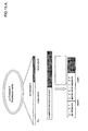
- Pro-caspases are synthesized as single polypeptide chains. A proteolytic cleavage in a certain area of the peptide chain leads to a molecule consisting of two chains non-covalently attached to each other. The two chain forms of the caspases are enzymatically active (Fig 5).
- pro-caspases In contrast to the activation of pro-urokinase and similar pro-enzymes, which require a proteolytic cleavage at a specific site and the release of a specific new N-terminus, activation of pro-caspases is not so stringent. Almost any cleavage in a certain area of the molecule is sufficient to result in activity.
- This property of pro-caspases has been employed in European patent application No. 03 075 319.8 to develop assay methods for a variety of proteolytic enzymes by introduction of a new cleavage sequence into the pro-caspase backbone. The length and sequence of this new cleavage sequence can be chosen with a considerable amount of freedom, giving much more flexibility than using pro-urokinase family members as a backbone.
- pro-caspases can be adapted to be used as detection enzymes to detect or quantify protein modifications.
- pro-caspase variant that has a sequence inserted in the activation area between the A and B chain sequences replacing the original activation sequence.
- This inserted sequence should contain a modification target sequence for the particular modification to be detected and a cleavage recognition sequence for a chosen activating protease.
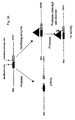
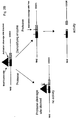
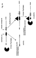
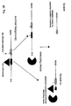
- the modification target sequence is placed N-terminal from the activation cleavage site (Fig. 6A). Upon cleavage of the activation cleavage site, activity results. Subsequently a carboxypeptidase is added, an enzyme that sequentially removes one or more aminoacid residues from the newly formed C-terminus. The presence of the modification interferes with carboxypeptidase activity and will prevent carboxypeptidase to degrade residues in the A-chain critical for activity. We have discovered that the C-terminal residues of the A-chain are critical for caspase activity.
- the activity leading to modification or removal of modification of a modification target site is most often caused by an enzymatic reaction of a specific enzyme.
- the activity of such an enzyme can be determined in a sample.
- a sample can consist of tissues, tissue extracts, cells, cell extracts, body fluids such as blood, plasma, urine, sputum etc., tissue- or cell-culture fluid, solutions of purified d'enzymes etc. All cells or tissues can be of plant, animal or human origin.
- the sample can also be of microbial origin (bacterial, yeast, fungus and the like or viral origin) or be taken from the medium on which the microogranism was cultured.
- the sample can also be taken from a nutrients.
- the sample can also consist of a purified or partially purified protein, or could be a food product.
- the sample can be any solid or liquid material in which one may detect or determine the presence or quantity of an activity leading to modification or removal of modification of a protein. In case the sample is taken from a solid it generally will be brought into solution by dissolving or suspending in a suitable liquid like water, buffer solutions, or organic solvents, detergents containing solutions etc.
- the sample will be brought into contact with the detection enzyme with a suitable modification target sequence according to the invention under conditions suitable for the activity of the modification or demodification process.
- Conditions mean presence of other reactants; temperature; pressure; pH, reaction duration or any other parameter relevant for the activity of the process.
- ATP ATP will be required to provide the phosphoryl group to be transferred to the detection enzyme.
- Metal ions like Mg 2+ or Ca 2+ might be required for the activity of certain modifying enzymes. Some enzymes might require co-factors for activity or reagents to stabilize their activity like sulphhydryl containing compounds such as dithiothreitol or dithioerythritol. For determination of e.g. lysylhydroxylase activity 2-oxoglutarate and oxygen are required as cofactor and donor for the hydroxyl group.
- temperatures in the range of 20°-37°C in general one will employ temperatures in the range of 20°-37°C but lower temperatures from 0°C to 20°C or higher temperatures than 37°C can also be used.
- the pH will generally be in the neutral range e.g. from pH 6-pH 8, but certain activities or enzymes have a higher activity or stability at more basic conditions to pH values of pH 10 or occasionally even higher, other activities prefer more acid conditions to pH values as low as 3 or in special cases even lower.
- the duration of the modifying or demodifying step can vary in a wide range and depends i.e. on the activity to be determined and the required sensitivity and detection limit.
- a substrate for this enzyme For this enzyme, the use of synthetic instead of natural substrates might be preferred. Particularly suitable are substrates that on conversion by the active detection enzyme yield a coloured, fluorescent or luminescent signal.
- the detection enzyme is a protease
- synthetic peptide substrates with a color, fluorescence or luminescence generating label are suitable.
- the detection system is not limited to the above mentioned substrates, many other detection systems such as those using natural substrates can be used. Ways of detection or quantification are known to those skilled in the art and would employ for example optical methods like turbidity and physical methods like HPLC, various forms of optical spectroscopy, mass spectroscopy etc.
- Assays based on the invention can have various formats dependent on the properties of the modifying activity to be detected and the detection enzyme used. If modifying enzyme and detection enzyme can be active in the same condition (buffer, pH, temperature etc.) the complete set of reactions can be performed in one single step. If this is not the case it can be advantageous to perform the modification reaction and detection reaction subsequently. This allows separate optimization of the conditions in these steps. Especially if one wants to measure modifying activities in biological samples with many potential interfering components a rapid partial purification of the modifying enzyme can be of help. One very convenient way to do this is to use the so called immunocapture format. In this format one makes use of antibodies or other agents specifically binding the modifying enzyme to a surface of e. g. a microtiter plate. This approach enables rapid purification (capture) of one component from a complicated mixture. All such approaches are particular embodiments of the invention.
- one does not use the detection enzyme in purified or partially purified form, but one uses cells or organisms expressing or containing the detection enzyme.
- Expression can be such that the detection enzyme remains intracellularly, even in particular cellular compartments, bound to membranes or excreted into the medium.
- Such cells or organisms can be used as in vitro or in vivo detection systems suitable for measuring protein modifying activity within, bound to or in the immediate cellular surrounding or in the organism. This approach is particularly useful if one wants to measure the modifying activity in its natural surroundings with all interactions with other cellular components retained.
- the detection enzyme can be isolated from the cells and its modification be analysed as described before. Alternatively the detection enzyme activity can be determined directly on the cell surface or in lysed cells or a cell extract by adding the necessary components.
- detection is preferentially by fluorescence or luminescence although other detection principles can also be used.
- a method of determination or quantification of a particular protein modification using a detection enzyme with a modification target sequence can be offered as a kit.
- such a kit comprises containers with sufficient quantities of detection enzyme, a suitable substrate to detect the activity of the detection enzyme, a suitable standard preparation to quantify the modifying enzyme activity and (materials to prepare) buffer solutions.
- a kit might also contain one or more proteases useful for activation or inactivation of the detection enzyme, or specific antibodies recognizing the enzyme to be determined to increase the specificity of the assay by interfering with activity of that enzyme, by specifically capture the enzyme or by interfering with activity of other enzymes.
- a kit might also contain one or more assay plates in e.g. the regular 96-well, 384-well or 1536-well format. It might be advantageous to add stabilizers to the detection enzyme to increase its stability during transport and storage. Such stabilizers could be other proteins like albumin or gelatine, carbohydrates like mannitol, anti-oxidants, detergents, or other organic chemicals. Furthermore inorganic salts might also have beneficial effects on stability.
- the detection enzyme is present in frozen solution or in lyophilized form that has to be reconstituted by adding buffer or water shortly before use.
- a detailed description how to perform the determination, preparing the samples and calculate the activity is included.
- a very convenient way to improve specificity as known in the art is to employ an immunocapture procedure: first the modifying enzyme to be measured is captured using a specific immobilized (monoclonal or polyclonal) antibody or other capturing device such as ligand, aptamer, etc. followed by measurement of the activity of the captured enzyme.
- a kit or device might be of the "dipstick" type where all necessary reagents are present in a dry form immobilized on a strip or dot of material.
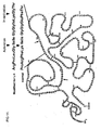
- This pro-urokinase variant contains an artificial site for modification by protein kinase A, Arg Val Ser Leu Ala (RVSLA in one-letter code) of which the Ser can be phosphorylated by PKA.
- This sequence overlaps with an artificial recognition sequence cleavable by various collagenases including matrix metallo protease -14 (MMP-14) Ser Leu Ala ⁇ Ile Ile Gly Gly. Collagenases can cleave at ⁇ between Ala and Ile. This cleavage leads to formation of an active urokinase variant.
- MMP-14 matrix metallo protease -14
- plasmid DNA pcDNA3PKA3 was mixed with medium and Fugene 6TM (Roche) and transfection was performed according to the manufacturer's instructions. To make stable transfectants, transfectants were selected by resistance to the neomycin analog G418. Transfected cells were cultured at various dilutions in medium containing selection agent G418 (1mg/ml) for 1-2 weeks. Remaining clones were selected, isolated with a pipette and cultured separately in medium containing selection agent.
- pro-urokinase variants were tested by immunoassay (van Bohemen et al. Fibrinolysis 9 (1995) 343-349).
- Clones with high production levels were expanded and used for production, in culture disks, flasks, cell factories or any other suitable system known to one skilled in the art.
- For production cells were routinely grown in triple flasks and conditioned culture medium was collected, centrifuged and stored at -20°C.
- the pro-urokinase based detection enzyme UK PKA3 was purified according to the methods described previously (JH Verheijen et al.Biochem J 323 (1997) 603-609).
- the modified pro-urokinase derived detection enzyme with a protein kinase A specific modification sequence UK PKA3 described in Example 1 was used in this example.
- Microtiter plates (96-wells) were coated with a mouse monoclonal antibody recognizing human pro-urokinase and its modified forms. Coating was performed in phosphate buffered saline (PBS) overnight at 4 °C followed by washing with PBST (PBS with 0.01% (v/v) Tween 80).
- PBS phosphate buffered saline
- Phosphorylation of the detection enzyme UK PKA3 was performed as follows: 50 ⁇ l buffer A (40 mM Tris HCl pH 7.5, 20 mM MgCl 2 , 0.1 mg/ml BSA), 25 ⁇ l cAMP dependent protein kinase catalytic subunit (PKA) (Promega, catalog number V5111) diluted in buffer A to 320 U/ml and 25 ⁇ l 0.2 mM ATP in buffer A were added and the plate was incubated during 7 h at 25°C or 37°C. After incubation the plate was washed with PBST.
- buffer A 40 mM Tris HCl pH 7.5, 20 mM MgCl 2 , 0.1 mg/ml BSA
- PKA cAMP dependent protein kinase catalytic subunit
- De-phosphorylation was achieved by alkaline phosphatase (Sigma, catalog number P-2276) at 200 U/ml in 50 mM Tris HCl pH 9.0, 10 mM NaCl, 0.01 % (v/v) Tween 80 overnight at 37°C. After incubation the plate was washed with PBST.
- alkaline phosphatase Sigma, catalog number P-2276
- Detection was performed by addition of 10 ⁇ l 6 mM pyro-Glu-Gly-Arg-pNA (S2444 Chromogenix) and incubation at 37°C. Color formation was monitored in a microtiterplate reader at 405 nm.
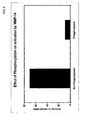
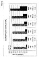
- Fig. 8 it is shown that the non-phosphorylated form of the detection enzyme UK PKA3 can be activated by MMP-14 resulting in color formation, whereas the phosphorylated form showed hardly any activity. This indicates that phosphorylation of the serine residue in the cleavage recognition sequence (see Fig. 7) interferes with activation.
- Fig. 9 it can be seen that the non-phosphorylated form of the detection enzyme (first bar) is active, and that kinase treatment leads to loss of activity (second bar). Treatment of the non-phosphorylated form of the detection enzyme with alkaline phosphatase has no effect (third bar) and alkaline phosphatase treatment of the phosphorylated form leads to reappearance of activity (fourth bar).
- Fig. 10 it is shown that the activity of the detection enzyme after MMP-14 cleavage decreases with PKA concentration indicating that increasing phosphorylation correlates with decreasing activity as expected in this approach (see also Fig. 2).
- a modified pro-urokinase derived detection enzyme was prepared with a collagenase specific cleavage activation site, (Arg Pro Leu Gly, collagenase cleaves C terminal from Gly) and overlapping lysylhydroxylase recognition and plasmin inactivation sites (Ile Lys Gly, Lys can be hydroxylated and plasmin cleaves C terminal from Lys). (Fig. 11).
- Oligonucleotides 5'-TCG AGG CGC CCC CTG GGC ATT ATT GGC GGG ATT AAG GGT ACC AT-3' (SEQ ID NO:5) and 5'- C GAT GGT ACC CTT AAT CCC GCC AAT AAT GCC CAG GGG GCG CC-3' (SEQ ID NO:6) were used for construction of the modification into the expression plasmid.
- Microtiterplates (96-well) were coated with a mouse monoclonal antibody recognizing human pro-urokinase and its modified forms. Coating was performed in phosphate buffered saline (PBS) overnight at 4°C followed by washing with PBST (PBS with 0.01% 1(v/v) Tween 80).
- PBS phosphate buffered saline
- the detection enzyme can be activated by collagenase, leading to activity. Plasmin treatment results in cleavage at the inactivation site and leads to loss of activity.
- a cDNA coding for pro-caspase-3 cloned into an expression vector having a sequence coding for a His-tag enabling rapid purification using Ni-chelate chromatography and regulatory sequences enabling expression in E.coli was obtained from the ATCC (number MGC-21241). This plasmid was used as a basis for construction of similar plasmids coding for pro-caspase-3 based detection enzymes.
- a detection enzyme for protein kinase A activity the following sequence was introduced with oligonucleotides 5'-GA TCC CTT CGA AGA GCA TCC CTT GGT C-3' (SEQ ID NO:9) and 5'-AA TTG ACC AAG GGA TGC TCT TCG AAG G-3' (SEQ ID NO:10).
- the resulting plasmid was used to transform E.coli (Rosetta DE3 pLys Novagen). Cultures were grown to an O.D. of 0.45-0.7 cooled to 30°C and induced with 1 mM IPTG.
- Bacterial pellets were mixed with lysis buffer (50 mM Tris HCl pH 8.0, 300 mM NaCl, 10 mM imidazol) and BugbusterTM (Novagen), incubated at 4°C while stirring for 30 min and centrifuged at ca. 5000 x g for 10 min at 4°C.
- lysis buffer 50 mM Tris HCl pH 8.0, 300 mM NaCl, 10 mM imidazol
- BugbusterTM BugbusterTM
- the supernatant was brought to a nickel Sepharose column (Amersham Biosciences) (2 ml column volume for 30-50 ml original culture volume) and subsequently washed with 20 volumes of 50 mM Tris HCl pH 8.0, 300 mM NaCl, 20 mM imidazole (wash buffer) and eluted with elution buffer (wash buffer with 250 mM imidazole instead of 20 mM).
- the peak-fractions were desalted using a PD-10 column (Amersham Bioscience) equilibrated with 10 mM Tris HCl pH 7.0, 1.5 mM NaCl, 0.01 (v/v) BRIJ 35 buffer.
- the pro-caspase-3 derived protein kinase A substrate (QZ PKA1 ) produced as described in example 5 was used in this experiment.
- Microtiter plates (96 wells) were coated with a 5 ⁇ g/ml solution of anti caspase-3 rabbit IgG in phosphate buffered saline (PBS) during 1 h at 37°C and subsequently washed with PBST.
- Purified QZ PKA1 100 ⁇ l in 50 mM HEPES pH 7.4, 100 mM NaCl, 1 mM EDTA, 0.1% (w/v) CHAPS, 0.1% (w/v) caseine, 0.05% (v/v) Triton-X-100 (capture buffer) was pipetted to wells of the coated microtiterplate and incubated for 2 h at 37°C followed by washing in PBST.
- Dephosphorylation was performed either with alkaline phosphatase (Sigma, catalog number P-2276) at 200 U/ml in buffer A (40 mM TrisHCl pH 7.5, 20 mM Mg Cl 2 , 0.1 mg/ml bovine serum albumin overnight at 37°C or with phosphatase 2 B (Calbiochem catalog number 539568) at 1.5 microgram/ml in buffer A in the presence of calmodulin at 37°C overnight, followed by washing with PBST.
- alkaline phosphatase Sigma, catalog number P-2276
- buffer A 40 mM TrisHCl pH 7.5, 20 mM Mg Cl 2 , 0.1 mg/ml bovine serum albumin overnight at 37°C
- phosphatase 2 B Calbiochem catalog number 539568
- Phosphorylation of the captured detection enzyme was performed as follows: to each well 50 ⁇ l of buffer A (40 mM Tris HCl pH 7.5, 20 mM MgCl 2 , 0.1 mg/ml BSA), 25 ⁇ l PKA (Promega, catalog number V5111) diluted in buffer A to 320 U/ml and 25 ⁇ l ATP 0.2 mM in buffer A were added and the plate was shaken for 20 seconds and incubated for 7 h at 37°C and washed with PBST.
- buffer A 40 mM Tris HCl pH 7.5, 20 mM MgCl 2 , 0.1 mg/ml BSA
- PKA Promega, catalog number V5111
- MMP-14 Activation by MMP-14 was done as follows: 80 ⁇ l of MMP-14 (Chemicon CC1043) at 24 ng/ml in 50 mM Tris HCl pH 7.6, 1.5 mM NaCl, 0.5 mM CaCl 2 and 1 ⁇ M ZnCl 2 , 0.01% (v/v) BRIJ 35, 2 IU/ml heparin was added to the wells and incubated for 3 h at 37°C.
- Fig. 16 shows the result of the experiment.
- the light bars show the activity of the non-kinase treated QZ PKA1 without treatment (left), with alkaline phosphatase treatment (middle), or with phosphatase 2B treatment (right).
- the results indicate that most of the QZ PKA1 is produced in the non-phosphorylated form and the effect of phosphatase treatment is small.
- Treatment with protein kinase A in the presence of ATP results in a significant drop in activity explainable by partial phosphorylation.
- the modified pro-urokinase derived detection enzyme with a protein kinase A specific modification sequence UK PKA3 described in Example 1 was used in this example.
- Microtiter plates (96-wells) were coated with a mouse monoclonal antibody recognizing human pro-urokinase and its modified forms. Coating was performed in phosphate buffered saline (PBS) overnight at 4 °C followed by washing with PBST (PBS with 0.01% (v/v) Tween 80).
- PBS phosphate buffered saline
- Phosphorylation of the detection enzyme UK PKA3 was performed as follows: 50 ⁇ l buffer A (40 mM Tris HCl pH 7.5, 20 mM MgCl 2 , 0.1 mg/ml BSA), 25 ⁇ l cAMP dependent protein kinase catalytic subunit (PKA) (Promega, catalog number V5111) diluted in buffer A to approximately 300 U/ml, 25 ⁇ l 0.2 mM ATP in buffer A were added and the plate was incubated overnight at 37°C. After incubation the plate was washed with PBST.
- buffer A 40 mM Tris HCl pH 7.5, 20 mM MgCl 2 , 0.1 mg/ml BSA
- PKA cAMP dependent protein kinase catalytic subunit
- Activation with the dipeptidyl aminopeptidase cathepsin C was performed in 100 ⁇ l cathepsin C buffer (50 mM Na-Phosphate pH 6.0, 100 mM NaCl, 2 mM EDTA, 10 mM L-cysteine, 0.1 % (w/v) bovine serum albumin, 0.01% (v/v) Tween 80 containing 3.3 ⁇ g/ml cathepsin C (Sigma D 7052) overnight at 37°C, followed by washing with PBST.
- cathepsin C buffer 50 mM Na-Phosphate pH 6.0, 100 mM NaCl, 2 mM EDTA, 10 mM L-cysteine, 0.1 % (w/v) bovine serum albumin, 0.01% (v/v) Tween 80 containing 3.3 ⁇ g/ml cathepsin C (Sigma D 7052) overnight at 37°C, followed by washing with PB
- Detection was performed by addition of 90 ⁇ l assay buffer (50 mM Tris HCl pH 7.6, 1.5 mM NaCl, 0.5 mM CaCl 2 and 1 ⁇ M ZnCl 2 , 0.01% (v/v) BRIJ 35), and 10 ⁇ l 6 mM pyro-Glu-Gly-Arg-pNA (S2444 Chromogenix) and incubated at 37°C. Color formation was monitored in a microtiterplate reader at 405 nm.
- Fig. 17 it is shown that the non-phosphorylated (-PKA-CC dark bar) form of the detection enzyme UK PKA3 has hardly any activity, but can be activated by the aminopeptidase cathepsin C resulting in an increase of activity (-PKA+CC light bar). If the detection enzyme is phosphorylated by protein kinase A, activity resulting after cathepsin C treatment is much lower (compare +PKA-CC with +PKA-CC).
- Modification Target residue Enzyme modifying activity
- Enzyme demodifying activity Phosphorylation Tyr, Ser, Thr, His Protein-kinases, phosphorylases Protein phosphatases Glycosylation Asn, Ser, Thr, Hyp Glycosyltransferases Not known Hydroxylation Pro, Lys Hydroxylases Not known Methylation Lys Protein methyl transferase Not known Acetylation Lys, N-terminus Protein acetyl transferase Not known Formylation N-terminal Met i.a.
Landscapes
- Life Sciences & Earth Sciences (AREA)
- Health & Medical Sciences (AREA)
- Engineering & Computer Science (AREA)
- Chemical & Material Sciences (AREA)
- Immunology (AREA)
- Molecular Biology (AREA)
- Hematology (AREA)
- Urology & Nephrology (AREA)
- Biomedical Technology (AREA)
- Analytical Chemistry (AREA)
- Biotechnology (AREA)
- Biochemistry (AREA)
- Microbiology (AREA)
- Physics & Mathematics (AREA)
- General Health & Medical Sciences (AREA)
- Cell Biology (AREA)
- Food Science & Technology (AREA)
- Pathology (AREA)
- General Physics & Mathematics (AREA)
- Medicinal Chemistry (AREA)
- Proteomics, Peptides & Aminoacids (AREA)
- Organic Chemistry (AREA)
- Wood Science & Technology (AREA)
- Zoology (AREA)
- Bioinformatics & Cheminformatics (AREA)
- Biophysics (AREA)
- Genetics & Genomics (AREA)
- General Engineering & Computer Science (AREA)
- Measuring Or Testing Involving Enzymes Or Micro-Organisms (AREA)
- Enzymes And Modification Thereof (AREA)
Abstract
Method for determining protein modifying activity or protein demodifying
activity in a sample by providing a detection enzyme construct comprising at
least one site of modification and at least one site of activation or inactivation,
wherein activation or inactivation of the detection enzyme construct results
from cleavage of the site of activation or inactivation by a protease, wherein
the site of modification of the detection enzyme construct is capable of being
modified by the protein modifying activity to be determined or demodified by
the protein demodifying activity to be determined, and wherein modification or
demodification of the site of modification affects cleavage of the site of
activation or inactivation by the protease, contacting the sample with the
detection enzyme construct under suitable conditions for the protein modifying
or demodifying activity to be determined, contacting the detection enzyme
construct with the protease under suitable conditions for activity of the
protease, and determining the occurrence or level of detection enzyme activity.
Active detection enzyme may be detectable by its conversion of a substrate of
the detection enzyme to a reaction product. The detection enzyme construct
may be derived from a protease (pro-)enzyme, like pro-urokinase or pro-caspase,
and may comprise artificial modification site, activation cleavage site,
and/or inactivation cleavage site.
Description
This invention is in the field of determining, assaying or quantifying
protein modifying activity or activity removing modifications from proteins
(further referred to herein as protein demodification or demodifying activity).
More particularly, the invention relates to a method for determining
presence or level of activity in a sample of an enzyme or enzymes catalyzing
the modification of proteins or catalyzing the removal of modification of
proteins.
The invention also concerns a detection enzyme construct that can be
modified by the activity to be determined or from which a modification can be
removed by the protein demodification activity to be determined and in
unmodified form or modified form respectively in modified form or demodified
form can be activated by limited proteolysis into a measurable active enzyme.
The invention also concerns an assay kit containing such a detection
enzyme construct, as well as a cell expressing or containing the detection
enzyme construct and an expression vector coding for the detection enzyme
construct.
Applicant's European patent EP 0 691 409 B1 discloses a method for
determining a protease, or its precursor after activation, in a sample,
comprising incubating the sample with a substrate of said protease and
determining proteolytic cleavage of said substrate, wherein said substrate is a
modified proenzyme containing a recognition site which is cleavable by said
protease and derived from a proenzyme by providing it with a recognition site
which is cleavable by a protease different from the one which activates the
unmodified proenzyme. The method allows to determine a specific proteolytic
activity in a sample, for example the activity of an aspartic protease or a
metalloprotease. The modified proenzyme may for example be derived from a
serine protease, such as pro-urokinase. The modification of the proenzyme may
consist of a replacement of the natural activation site of pro-urokinase (which
is recognized by plasmin, the natural activator of pro-urokinase) by a new
activation site specifically recognized by the protease to be determined.
Applicant's copending European patent application No. 03 075 319.8,
filed January 31, 2003, discloses a similar method for determining a protease,
wherein the modified proenzyme is specifically derived from a caspase.
Whereas the above mentioned documents are concerned with a method
of determining a proteolytic activity, the present invention addresses the
problem of determining a protein modifying or demodifying activity.
Many proteins have been found to contain modifications that result from
physical, chemical or enzymatic processes acting on its aminoacid residues
resulting in modified amino acid residues. Such modifications can be
introduced during protein synthesis on the growing peptide chain or be
introduced in the completed and properly folded protein after synthesis.
Examples of protein modifications and activities leading to modifications
or removing modifications are summarized in Table I. They generally concern
the introduction or removal of a chemical moiety, usually a small moiety like a
methyl group, a phosphate group, a hydroxyl group, etc., but the chemical
moiety may also be a large group, such as a carbohydrate side chain. The
modification may comprise the formation of a complex with another protein
(the same or different) or, more in general, with other (macro)molecules,
including for example complex formation with nucleic acid or with cofactors of
the protein in question. Therefore, the covalent attachment of a chemical
moiety or molecule is not a prerequisite. Usually, modification or
demodification is catalyzed by a specific enzyme that recognizes particular
sites (sequences of aminoacid residues) and directs the modification or
demodification to particular amino acid residues.
Recently is has become clear that modification of a protein can greatly
influence its properties.
A particularly interesting modification is phosphorylation of a protein.
The introduction of one or more phosphate groups into proteins can greatly
influence their properties such as enzymatic activity, affinity for receptors,
physical form, monomer, dimer, multimer. Introduction and removal of
phosphate groups into proteins appears to be an important mechanism of
signal transduction involved in critical processes. Introduction of phosphate
groups into proteins is catalyzed by specific enzymes called protein kinases of
which over 500 are known at present. Removal of phosphate groups from
proteins is brought about by enzymes known as protein phosphatases of which
over 100 different examples have been described. Both protein kinases and
protein phosphatases are very interesting targets for development of drugs to
diseases such as cancer, inflammatory diseases such as rheumatoid arthritis
and diabetic complication.
Measurement of the presence or level of protein modifying or
demodifying activities, particularly the activity of enzymes catalyzing protein
modifying or demodifying reactions, is of considerable importance in basic
academic research, drug discovery research, drug development and might also
have applications in diagnostic or prognostic methods or as (surrogate) marker
to follow or monitor (experimental) therapy.
Measurement and quantification of protein modifying or demodifying
activity is in general not easy and requires often a variety of complicated,
time-consuming and expensive techniques.
US 2002/0172991 A1 discloses a method for measuring protein kinase
activity. The method uses the fact that the reaction catalyzed by the protein
kinase uses ATP as a donor for the phosphate group. The method measures the
change of ATP concentration using a bioluminescence reaction which involves
luciferin and a luciferase. This method has the disadvantage that it is indirect,
it does not measure the kinase activity itself, but the consumption of ATP.
Further, the method is restricted to the measurement of ATP-dependent
protein kinases and is not applicable to the measurement of other modifying or
demodifying activities.
This invention provides in a first aspect a method for determining a
protein modifying activity or protein demodifying activity in a sample, the
method comprising
providing a detection enzyme construct comprising at least one site of modification and at least one site of activation or inactivation, wherein activation or inactivation of the detection enzyme construct results from cleavage of said site of activation or inactivation by a protease, wherein said site of modification of the detection enzyme construct is capable of being modified by the protein modifying activity to be determined or demodified by the protein demodifying activity to be determined, and wherein modification or demodification of said site of modification affects cleavage of the said site of activation or inactivation by the said protease,
contacting the sample with the said detection enzyme construct under suitable conditions for the protein modifying or demodifying activity to be determined,
contacting the detection enzyme construct with the said protease under suitable conditions for activity of the said protease, and
determining the occurrence or level of detection enzyme activity.
providing a detection enzyme construct comprising at least one site of modification and at least one site of activation or inactivation, wherein activation or inactivation of the detection enzyme construct results from cleavage of said site of activation or inactivation by a protease, wherein said site of modification of the detection enzyme construct is capable of being modified by the protein modifying activity to be determined or demodified by the protein demodifying activity to be determined, and wherein modification or demodification of said site of modification affects cleavage of the said site of activation or inactivation by the said protease,
contacting the sample with the said detection enzyme construct under suitable conditions for the protein modifying or demodifying activity to be determined,
contacting the detection enzyme construct with the said protease under suitable conditions for activity of the said protease, and
determining the occurrence or level of detection enzyme activity.
In one group of preferred embodiments of the invention, said detection
enzyme construct comprises a site of modification and a site of activation,
wherein activation of the detection enzyme construct results from cleavage of
said site of activation by an activation protease to form active detection
enzyme, wherein said site of modification of the detection enzyme construct is
capable of being modified by the protein modifying activity to be determined or
demodified by the protein demodifying activity to be determined, and wherein
modification or demodification of said site of modification determines whether
said activation protease can cleave the said site of activation,
and the method comprises contacting the detection enzyme construct with the said activation protease under suitable conditions for activity of the said activation protease.
and the method comprises contacting the detection enzyme construct with the said activation protease under suitable conditions for activity of the said activation protease.
In another group of preferred embodiments of the invention, said
detection enzyme construct comprises a site of modification, a site of activation
and a site of inactivation, wherein activation of the detection enzyme construct
results from cleavage of said site of activation by an activation protease to
form active detection enzyme, and inactivation of the detection enzyme
construct or detection enzyme results from cleavage of said site of inactivation
by a deactivation protease, wherein said site of modification of the detection
enzyme construct is capable of being modified by the protein modifying activity
to be determined or demodified by the protein demodifying activity to be
determined, and wherein modification or demodification of said site of
modification determines whether said activation protease can cleave the said
site of activation or said inactivation protease can cleave the said site of
inactivation,
and the method comprises contacting the detection enzyme construct with the said activation protease under suitable conditions for activity of the said activation protease, and before, after, or simultaneously therewith, contacting the detection enzyme construct or detection enzyme with the said inactivation protease under suitable conditions for activity of the said inactivation protease.
and the method comprises contacting the detection enzyme construct with the said activation protease under suitable conditions for activity of the said activation protease, and before, after, or simultaneously therewith, contacting the detection enzyme construct or detection enzyme with the said inactivation protease under suitable conditions for activity of the said inactivation protease.
In this group of embodiments of the invention, it is preferred that
modification or demodification of said site of modification determines whether
said inactivation protease can cleave the said site of inactivation.
In preferred embodiments of this invention, active detection enzyme is
detectable by its conversion of a substrate of the detection enzyme to a reaction
product and detection enzyme activity is determined by contacting the
detection enzyme construct or detection enzyme with said substrate of the
detection enzyme and determining the occurrence or level of conversion of said
substrate.
Preferably, said detection enzyme construct is derived from a protease
enzyme or protease pro-enzyme. More preferably, said detection enzyme
construct is derived from a serine protease enzyme or serine protease pro-enzyme
or from a cysteine protease enzyme or cysteine protease pro-enzyme.
In some particular preferred embodiments, said detection enzyme construct is
derived from a pro-enzyme selected from the group consisting of pro-urokinase,
pro-granzyme, pro-caspase-3 and pro-caspase-7. In some most preferred
embodiments, said detection enzyme construct is derived from pro-urokinase.
In some other most preferred embodiments, said detection enzyme construct is
derived from a pro-caspase, in particular pro-caspase-3.
Preferably, said detection enzyme construct contains an artificial
modification site, or an artificial activation cleavage site, or an artificial
inactivation cleavage site, or any combination thereof, and optionally further
changes, introduced into a protease enzyme or protease pro-enzyme in such a
way that modification or demodification of said modification site affects
activatability of the detection enzyme construct by treatment with said
activation protease or inactivatability of the detection enzyme construct or
detection enzyme by treatment with said inactivation protease.
The activation protease may be selected from the group consisting of (a)
the natural activation protease of said protease pro-enzyme, (b) a protease
specifically cleaving an artificial activation cleavage site introduced into said
protease pro-enzyme, (c) an aminopeptidase, and (d) a carboxypeptidase.
Similarly, the inactivation protease, which may be the same or different
from the activation protease, may be selected from the group consisting of (a) a
natural inactivation protease of said protease enzyme, (b) a protease
specifically cleaving an artificial inactivation cleavage site introduced into said
protease pro-enzyme, (c) an aminopeptidase, and (d) a carboxypeptidase.
Usually, said protein modifying activity or protein demodifying activity
is an enzymatic activity. Preferably, said enzymatic activity is selected from
the group consisting of of protein kinase activity, protein phosphatase activity,
protein hydroxylase activity, protein glycosyltransferase activity or protein
prenyltransferase activity.
In methods according to the invention for determining a protein
demodifying activity, the sample would usually be contacted with pre-modified
detection enzyme construct.
The sample may be contacted with a purified or partially purified
detection enzyme construct, or with a cell expressing or containing the
detection enzyme construct, or with the content of such a cell or of a specific
compartment of such a cell, or with the culture medium of such a cell.
The sample may be contacted with a cell expressing or containing the
detection enzyme construct and then either the cell as a whole, or the content
of the cell or of a specific compartment of the cell, or the culture medium of the
cell is contacted with the activation and/or inactivation protease and detection
enzyme substrate.
In another aspect, the invention provides a detection enzyme construct
useful for determining a protein modifying activity or protein demodifying
activity in a sample, comprising at least one site of modification and at least
one site of activation or inactivation, wherein activation or inactivation of the
detection enzyme construct results from cleavage of said site of activation or
inactivation by a protease, wherein said site of modification of the detection
enzyme construct is capable of being modified by the protein modifying activity
to be determined or demodified by the protein demodifying activity to be
determined, and wherein modification or demodification of said site of
modification affects cleavage of the said site of activation or inactivation by the
said protease.
In a further aspect, the invention provides a cell expressing or
containing a detection enzyme construct as defined herein.
In a further aspect, the invention provides a kit for determining a
protein modifying activity or a protein demodifying activity in a sample,
comprising a detection enzyme construct or a cell as defined herein, and
further comprising at least one member of the group consisting of activation
protease, inactivation protease, substrate of the detection enzyme, a standard
composition, a buffer composition, an antibody composition, a stabilizing
composition, an assay plate or container, and instructions for use.
The invention also provides an expression vector coding for a detection
enzyme construct as defined herein.
The invention provides a method of determining a protein modifying
activity in a sample, comprising incubating the sample with a substrate of said
activity and determining the modification of said substrate, wherein said
substrate is a detection enzyme construct containing a modification target
sequence which is recognized and modified by said modifying activity. Said
modification target sequence has in modified or non-modified form a different
effect on activity or activatability of said detection enzyme construct. Apart
from said modification target sequence said detection enzyme construct also
contains a site cleavable by a protease that upon cleavage results in activation
or in-activation of said detection enzyme construct. The difference in activity
or activatability after cleavage by a protease may be determined by observing
or measuring the resulting activity of the detection enzyme using a suitable
substrate of the activated detection enzyme. The sample will usually be a
biological sample, such as a biological fluid, a fraction of a biological fluid, a
biological tissue, an extract of a biological tissue and a fraction of an extract of
a biological tissue. The detection enzyme construct may be derived from a (pro)
enzyme by providing it with a modification target sequence if not already
present in its sequence and with one or more suitably positioned protease
cleavage sites if not already present in its sequence.
The present invention also provides the detection enzyme construct per
se, more particularly a modified (pro)enzyme derived from a (pro)enzyme by
introduction therein of a suitably positioned modification target sequence if
not already present and one or more suitably positioned protease cleavage
sites if not already present.
The invention further provides a kit for determining a protein modifying
or demodifying activity in a sample, comprising a detection enzym e construct
as defined herein, together with at least one member from the group consisting
of substrates for active detection enzyme, (in)activating proteases, buffer
solutions, standard preparations, specific antibodies, microtiter plates and
instructions for use; and provides a device for determining a protein modifying
or demodifying activity in a sample, comprising a detection enzyme construct
as defined herein.
The invention also provides DNA constructs coding for a suitable
detection enzyme construct, wherein said DNA constructs can be used to
modify cells or organisms in such a way that said cells or organisms express
said detection enzyme construct; said modified cells or organisms expressing
the detection enzyme construct can be used as a source for isolation of said
detection enzyme construct in purified or partially purified form or can be used
per se as parts of an in vitro or in vivo detection system.
The invention uses a detection enzyme construct containing a sequence
that can be modified by a particular protein modifying activity resulting in a
modified form of the detection enzyme. The non-modified form and modified
form of the detection enzyme construct differ in their behaviour upon contact
with a particular proteolytic enzyme resulting in a difference of enzyme
activity. The resulting enzyme activity can be a measure of modification or
absence of modification depending on the structure and sequence of the
detection enzyme construct.
A large variety of detection enzyme constructs can be suitable for use in
the invention as long as a proteolytic cleavage step is required for conversion
of the pro-enzyme form of the detection enzyme construct to the active form or
of the active form of the detection enzyme to an inactive form and a suitable
detection system exists for detection and/or quantification of the resulting
enzymatic activity. Particularly suitable as detection enzyme construct are
detection enzymes derived from proteolytic enzymes since many proteolytic
enzymes result from pro-enzymes activated by a proteolytic-cleavage, but also
other pro-enzymes could be suitable as basis for such a detection enzyme
construct. Within the group of proteolytic enzymes many possible candidates
exist to form a basis for construction of a suitable detection enzyme construct.
Particularly useful are pro-enzymes of the serine protease family (see Table II
for a number of examples) or cysteine protease family (see Table III for a
number of examples) since a lot of information is available on amino acid and
DNA sequence, activity, activation-mechanisms and structure and because
general detection methods for these enzymes are operational.
Within these families particularly pro-urokinase and pro-caspases are of
interest since cleavage of pro-enzyme to enzyme leads to an enormous increase
in activity (high zymogenicity), the resulting active enzymes are sufficiently
specific to prevent unwanted cleavages, the resulting enzyme activity is easily
detected using well known mature technology, and the proteins are stable and
easily expressed in eukaryotic or bacterial expression systems.
As demonstrated in EP 0 691 409 B1 and European patent application
No. 03 075 319.8, which have been mentioned above and are incorporated in
their entirety herein by reference, these pro-enzymes may be modified by
introduction therein of a new activation sequence. In those documents, such
modification was required to allow specific measurement of the proteolytic
activity recognizing said new activation sequence. Herein, replacement of the
natural activation sequence by a new activation sequence is not an essential
requirement, but it may be practical and will usually be preferable.
Apart from these elements, a detection enzyme construct of the
invention should contain at least one site that can be modified by a particular
protein modification process or activity, such as a protein modifying enzyme.
The exact sequence of such a site mostly depends on the protein modifying
activity or process one wants to detect or quantify. The position within the
sequence of the detection enzyme construct is chosen in such a way that the
presence or absence of the protein modification either has effect on enzymatic
activity or activatability of the pro-enzyme.
A non-limited list of possible sequences that can be modified by a
number of known protein modifying enzymes can be found in Table IV.
Apart from modifications around the activation site or modifications
forming a target for protein modification, also other modifications in the
sequence of the detection enzyme construct might be present. Such
modifications include modifications increasing (thermal) stability, conferring
resistance against proteases, conferring resistance against enzyme inhibitors,
increasing the enzymatic activity, conferring reactivity to certain antibodies or
ligands, aiding expression or purification, etc. In general such modifications
are in parts of the detection enzyme construct not directly involved in the
protein modification to be measured or the activation of the pro-enzyme
construct to its active form. Another modification that might be useful is the
introduction of an extra protease cleavage site N-terminal from the activation
site. Cleavage at such a site would result in an inactive molecule that could be
activated by an aminopeptidase. In some cases, an extra protease cleavage site
C-terminal from the activation site could be appropriate, when a
carboxypeptidase is used for activation. Deletion mutants lacking parts of the
original sequence not needed for activity may be a suitable alternative. Such
constructs may be preferable to achieve more rapid activation (or inactivation)
of the detection enzyme construct by an aminopeptidase or carboxypeptidase.
Critical to the invention is a site that can be modified by the particular
protein modifying activity one wants to detect or quantify. Such a site might
be present naturally in the pro-enzyme construct, but more likely has to be
introduced by adding or replacing a stretch of aminoacid residues expected or
known to be a target for modification in proteins. The position of such a
modification within the detection enzyme construct has to be chosen such that
the absence or presence of the protein modification will interfere with activity
or activation of the detection enzyme construct. Depending on the nature of the
modification, the particular aminoacid sequence of the site to be modified, the
detection enzyme and the protease used for activation or inactivation of the
detection enzyme construct, a suitable position of such a modification target
sequence should be determined. In general, but not necessarily, such
modification target sequences are placed in the vicinity of the activation site.
Natural pro-urokinase occurs as a single chain polypeptide, cleavage by
its natural activator plasmin after Lysine residue 158 leads to a molecule
consisting of an N-terminal light chain (residues 1-158) and a C-terminal
heavy chain (residues 159-411) connected by a single disulfide bridge. The two
chain molecule is enzymatically active. It has been shown that residues 1-158
have no function in enzymatic activity, the isolated chain 159-411 has equal or
almost equal enzymatic activity as the natural two chain urokinase. Further,
it has been shown that the new N-terminus (I 159) in natural urokinase is
very important for activity, cleavage at R 156 results in a C terminal chain
starting with F 157 and no protease activity. Similarly cleavage at I 159
resulting in a C terminal chain starting with I 160 and no protease activity. So
it appears that it is the newly formed N-terminus that is important for activity
and not the way it is generated. It is possible to generate the new N-terminus
with a single proteolytic cleavage at the right position with a regular protease
or even by removing residues one-by-one with an amino peptidase. Activation
with such an aminopeptidase is particularly effective when the number of
aminoacid residues N-terminal to the activation sequence is limited. As soon
as the I 159 becomes N-terminal, activity results. The inactive molecule
starting with F 157 can be converted to an active molecule by removing F 157
and K 158 by an aminopeptidase.
Further research has shown that variations in the new N-terminal part
of the heavy chain of cleaved pro-urokinase compatible with activity are very
limited. Even replacing I 159 by a very similar A residue greatly affects
activity. Modifications N-terminal from the natural cleavage position generally
have no significant influence on activity but might result in pro-enzymes
activatable by other proteins than the ones activating natural pro-urokinase.
Examples of this have been described before in EP 0 691 409 B1. Also
modifications in the aminoacid sequence in the heavy chain, more distant from
the activation cleavage site are not necessarily deleterious for activity.
Modifications around residue 180 have been described to lead to a molecule
that in its two-chain form is an active protease resistant against its naturally
occurring inhibitor PAI-1.
Pro-urokinase constructs suitable as detection enzyme in the present
invention should contain a natural or modified C-terminal heavy chain and at
least a short N-terminal chain connected by a peptide bond. The heavy chain
should start with a sequence very similar to that of natural urokinase in order
to retain enzymatic activity. The C- and N-terminal chains need not be
connected by a disulfide bond like natural urokinase. The allowable length of
the N-terminal chain may vary; it can be as short as 1 aminoacid residue or be
as long or even longer than the N-terminal chain of natural urokinase. A
sequence of residues immediately in front of the activation cleavage site
determines which protease can activate the pro-urokinase construct to an
active enzyme. A large variety of sequences has been shown to be compatible
resulting in pro-urokinase constructs activatable by a variety of proteases
other than plasmin, its natural activator. All such constructs could be
employed in the invention and can have particular advantages for certain
applications.
The above mentioned properties of pro-urokinase and actually of most of
the members of the serine protease family and of the related cysteine protease
family make these molecules particularly suitable to form the basis of a
detection enzyme according to the present invention. In the following we use
pro-urokinase as an example but the same principles hold for most of the
members of its family.
In the following part a number of specific embodiments of the invention
based on the use of detection enzymes with urokinase like properties will be
explained in more detail.
Although all examples given above are especially suitable for pro-urokinase
related detection enzymes, the approach is more general and would
work for basically almost all pro-enzymes of the serine- and cysteine protease
family with an activation mechanism based on cleavage of a peptide bond in a
specific position and the release of a specific new N-terminus. Other examples
of suitable pro-enzymes can be found in Table II and Table III.
Other pro-enzymes with a completely different activation mechanism
than pro-urokinase and its family members are also suitable to be used as
detection enzymes according to the invention. Particularly suitable are pro-caspases.
In the following part a number of specific embodiments of the
invention based on the use of detection enzymes with caspase like properties
will be explained in more detail.
Pro-caspases are synthesized as single polypeptide chains. A proteolytic
cleavage in a certain area of the peptide chain leads to a molecule consisting of
two chains non-covalently attached to each other. The two chain forms of the
caspases are enzymatically active (Fig 5).
In contrast to the activation of pro-urokinase and similar pro-enzymes,
which require a proteolytic cleavage at a specific site and the release of a
specific new N-terminus, activation of pro-caspases is not so stringent. Almost
any cleavage in a certain area of the molecule is sufficient to result in activity.
This property of pro-caspases has been employed in European patent
application No. 03 075 319.8 to develop assay methods for a variety of
proteolytic enzymes by introduction of a new cleavage sequence into the pro-caspase
backbone. The length and sequence of this new cleavage sequence can
be chosen with a considerable amount of freedom, giving much more flexibility
than using pro-urokinase family members as a backbone.
These properties make pro-caspases very suitable to form the basis for
detection enzymes according to the present invention.
In an analogous manner as described above for pro-urokinase, also pro-caspases
can be adapted to be used as detection enzymes to detect or quantify
protein modifications.
One possible approach is to construct a pro-caspase variant that has a
sequence inserted in the activation area between the A and B chain sequences
replacing the original activation sequence. This inserted sequence should
contain a modification target sequence for the particular modification to be
detected and a cleavage recognition sequence for a chosen activating protease.
As described for pro-urokinase derived detection enzymes, various
possible approaches can be discerned.
Two other approaches using pro-caspase based detection enzymes might
be suitable for certain applications. In both cases a sequence containing a
modification target site and an activation cleavage site will be introduced in
the activation area. In these cases there does not need to be overlap in these
two sequences.
In one approach the modification target sequence is placed N-terminal
from the activation cleavage site (Fig. 6A). Upon cleavage of the activation
cleavage site, activity results. Subsequently a carboxypeptidase is added, an
enzyme that sequentially removes one or more aminoacid residues from the
newly formed C-terminus. The presence of the modification interferes with
carboxypeptidase activity and will prevent carboxypeptidase to degrade
residues in the A-chain critical for activity. We have discovered that the C-terminal
residues of the A-chain are critical for caspase activity. In this way
the presence of the modification will prevent inactivation and result in
activity, whereas absence of the modification will allow carboxypeptidase to
cut into the C-terminal part of the caspase A chain, removing the critical
residues and lead to inactivation of the detection enzyme.
Another, very similar, approach requires an insertion with the
modification target sequence C-terminal from the activation site (Fig. 6B).
Cleaving the activation site will lead to activity. Subsequently an aminopeptidase
is added that will cut into the N-terminal part of the caspase B
chain again removing critical residues and leading to loss of activity. The
presence of the modification prevents aminopeptidase to damage the B chain
and will result in an active detection enzyme.
The activity leading to modification or removal of modification of a
modification target site is most often caused by an enzymatic reaction of a
specific enzyme. The activity of such an enzyme can be determined in a
sample. Such a sample can consist of tissues, tissue extracts, cells, cell
extracts, body fluids such as blood, plasma, urine, sputum etc., tissue- or cell-culture
fluid, solutions of purified d'enzymes etc. All cells or tissues can be of
plant, animal or human origin. The sample can also be of microbial origin
(bacterial, yeast, fungus and the like or viral origin) or be taken from the
medium on which the microogranism was cultured. The sample can also be
taken from a nutrients. The sample can also consist of a purified or partially
purified protein, or could be a food product. The sample can be any solid or
liquid material in which one may detect or determine the presence or quantity
of an activity leading to modification or removal of modification of a protein. In
case the sample is taken from a solid it generally will be brought into solution
by dissolving or suspending in a suitable liquid like water, buffer solutions, or
organic solvents, detergents containing solutions etc.
The sample will be brought into contact with the detection enzyme with
a suitable modification target sequence according to the invention under
conditions suitable for the activity of the modification or demodification
process. Conditions mean presence of other reactants; temperature; pressure;
pH, reaction duration or any other parameter relevant for the activity of the
process.
The presence and nature of other reactants depend on the specific
modification or demodification activity to be determined. For determination of
e.g. kinase activity, ATP will be required to provide the phosphoryl group to be
transferred to the detection enzyme.
Metal ions like Mg2+ or Ca2+ might be required for the activity of certain
modifying enzymes. Some enzymes might require co-factors for activity or
reagents to stabilize their activity like sulphhydryl containing compounds such
as dithiothreitol or dithioerythritol. For determination of e.g. lysylhydroxylase
activity 2-oxoglutarate and oxygen are required as cofactor and donor for the
hydroxyl group.
The conditions suitable for measurement of any modifying activity such
as temperature, pressure, pH, reaction duration and other parameters are
known to one skilled in the art.
In general one will employ temperatures in the range of 20°-37°C but
lower temperatures from 0°C to 20°C or higher temperatures than 37°C can
also be used. The pH will generally be in the neutral range e.g. from pH 6-pH
8, but certain activities or enzymes have a higher activity or stability at more
basic conditions to pH values of pH 10 or occasionally even higher, other
activities prefer more acid conditions to pH values as low as 3 or in special
cases even lower. The duration of the modifying or demodifying step can vary
in a wide range and depends i.e. on the activity to be determined and the
required sensitivity and detection limit.
To determine the activity of the detection enzyme one can use a
substrate for this enzyme. The use of synthetic instead of natural substrates
might be preferred. Particularly suitable are substrates that on conversion by
the active detection enzyme yield a coloured, fluorescent or luminescent signal.
When the detection enzyme is a protease, synthetic peptide substrates with a
color, fluorescence or luminescence generating label are suitable.
For urokinase, many such substrates are known and in general consist
of a short peptide sequence coupled to a color or fluorescence generating label
(Table V). Also for caspases such substrates are well known (Table VI). Also for
many other detection enzyme systems such synthetic substrates are known to
one skilled in the art. All enzyme systems with a suitable detection system can
be used in the invention.
The detection system is not limited to the above mentioned substrates,
many other detection systems such as those using natural substrates can be
used. Ways of detection or quantification are known to those skilled in the art
and would employ for example optical methods like turbidity and physical
methods like HPLC, various forms of optical spectroscopy, mass spectroscopy
etc.
Assays based on the invention can have various formats dependent on
the properties of the modifying activity to be detected and the detection
enzyme used. If modifying enzyme and detection enzyme can be active in the
same condition (buffer, pH, temperature etc.) the complete set of reactions can
be performed in one single step. If this is not the case it can be advantageous
to perform the modification reaction and detection reaction subsequently. This
allows separate optimization of the conditions in these steps. Especially if one
wants to measure modifying activities in biological samples with many
potential interfering components a rapid partial purification of the modifying
enzyme can be of help. One very convenient way to do this is to use the so
called immunocapture format. In this format one makes use of antibodies or
other agents specifically binding the modifying enzyme to a surface of e. g. a
microtiter plate. This approach enables rapid purification (capture) of one
component from a complicated mixture. All such approaches are particular
embodiments of the invention.
In another embodiment of the invention, one does not use the detection
enzyme in purified or partially purified form, but one uses cells or organisms
expressing or containing the detection enzyme. Expression can be such that
the detection enzyme remains intracellularly, even in particular cellular
compartments, bound to membranes or excreted into the medium. Such cells or
organisms can be used as in vitro or in vivo detection systems suitable for
measuring protein modifying activity within, bound to or in the immediate
cellular surrounding or in the organism. This approach is particularly useful if
one wants to measure the modifying activity in its natural surroundings with
all interactions with other cellular components retained. The detection enzyme
can be isolated from the cells and its modification be analysed as described
before. Alternatively the detection enzyme activity can be determined directly
on the cell surface or in lysed cells or a cell extract by adding the necessary
components.
In this embodiment of the invention, detection is preferentially by
fluorescence or luminescence although other detection principles can also be
used.
A method of determination or quantification of a particular protein
modification using a detection enzyme with a modification target sequence can
be offered as a kit.
Typically, such a kit comprises containers with sufficient quantities of
detection enzyme, a suitable substrate to detect the activity of the detection
enzyme, a suitable standard preparation to quantify the modifying enzyme
activity and (materials to prepare) buffer solutions. Furthermore, a kit might
also contain one or more proteases useful for activation or inactivation of the
detection enzyme, or specific antibodies recognizing the enzyme to be
determined to increase the specificity of the assay by interfering with activity
of that enzyme, by specifically capture the enzyme or by interfering with
activity of other enzymes. A kit might also contain one or more assay plates in
e.g. the regular 96-well, 384-well or 1536-well format. It might be
advantageous to add stabilizers to the detection enzyme to increase its
stability during transport and storage. Such stabilizers could be other proteins
like albumin or gelatine, carbohydrates like mannitol, anti-oxidants,
detergents, or other organic chemicals. Furthermore inorganic salts might also
have beneficial effects on stability.
Most conveniently the detection enzyme is present in frozen solution or
in lyophilized form that has to be reconstituted by adding buffer or water
shortly before use. In addition, a detailed description how to perform the
determination, preparing the samples and calculate the activity is included. A
very convenient way to improve specificity as known in the art is to employ an
immunocapture procedure: first the modifying enzyme to be measured is
captured using a specific immobilized (monoclonal or polyclonal) antibody or
other capturing device such as ligand, aptamer, etc. followed by measurement
of the activity of the captured enzyme. Alternatively a kit or device might be of
the "dipstick" type where all necessary reagents are present in a dry form
immobilized on a strip or dot of material.
The following examples are merely intended to illustrate the invention
and in no way can be considered limiting.
The complete cDNA coding for human pro-urokinase (UKcol) with a
collagenase specific cleavage site Arg Pro Leu Gly (RPLG in one-letter code)
was cut from the expression plasmid pEV2UKcol (see EP 0691409 B1 for
details) and cloned into the multiple cloning site of a new expression vector
containing a neomycin resistance marker pcDNA3 (from Invitrogen).
With polymerase chain reaction using primers 5'-ACC ATC GAt AAC
CAG CCC TGG-3' (SEQ ID NO:1) and 5'-CCG CCT cga gGT CTT TTG GCC-3'
(SEQ ID NO:2) (mutations indicated as lower case letters) two new unique
restriction sites ClaI and XhoI were introduced flanking the region coding for
the activation site. The resulting plasmid vector was used to construct pro-urokinase
based detection enzymes by cutting the plasmid with ClaI and XhoI
followed by insertion and ligation of two partially complementary oligonucleotides
coding for the required mutated amino acid sequence. For construction of
a detection enzyme with an overlapping protein kinase A phosphorylation and
a collagenase activation sequence the following oligonucleotides were inserted
in the ClaI XhoI cut plasmid: 5'-TCG AGG CGG CCG CTC GGG CGA AGA
GTG TCC CTT GCC ATT ATT GGG GGA GAA TTC ACC ACC AT-3' (SEQ ID
NO:3) and 5'-C GAT GGT GGT GAA TTC TCC CCC AAT AAT GGC AAG
GGA CAC TCT TCG CCC GAG CGG CCG CC-3' (SEQ ID NO:4) resulting in a
plasmid pcDNA3PKA3 coding for a pro-urokinase detection enzyme (UKPKA3)
with a sequence as shown in Fig. 7. This pro-urokinase variant contains an
artificial site for modification by protein kinase A, Arg Val Ser Leu Ala
(RVSLA in one-letter code) of which the Ser can be phosphorylated by PKA.
This sequence overlaps with an artificial recognition sequence cleavable by
various collagenases including matrix metallo protease -14 (MMP-14) Ser Leu
Ala^Ile Ile Gly Gly. Collagenases can cleave at ^ between Ala and Ile. This
cleavage leads to formation of an active urokinase variant.
Cells (Chinese Hamster Ovary, CHO or Hamster Embryonic Kidney,
HEK) were grown to 70-90% confluency in culture dishes. 1 µg of plasmid
DNA pcDNA3PKA3 was mixed with medium and Fugene 6™ (Roche) and
transfection was performed according to the manufacturer's instructions. To
make stable transfectants, transfectants were selected by resistance to the
neomycin analog G418. Transfected cells were cultured at various dilutions in
medium containing selection agent G418 (1mg/ml) for 1-2 weeks. Remaining
clones were selected, isolated with a pipette and cultured separately in
medium containing selection agent. Production of pro-urokinase variants by
these clones was tested by immunoassay (van Bohemen et al. Fibrinolysis 9
(1995) 343-349). Clones with high production levels were expanded and used
for production, in culture disks, flasks, cell factories or any other suitable
system known to one skilled in the art. For production, cells were routinely
grown in triple flasks and conditioned culture medium was collected,
centrifuged and stored at -20°C. The pro-urokinase based detection enzyme
UKPKA3 was purified according to the methods described previously (JH
Verheijen et al.Biochem J 323 (1997) 603-609).
The modified pro-urokinase derived detection enzyme with a protein
kinase A specific modification sequence UKPKA3 described in Example 1 was
used in this example.
Microtiter plates (96-wells) were coated with a mouse monoclonal
antibody recognizing human pro-urokinase and its modified forms. Coating
was performed in phosphate buffered saline (PBS) overnight at 4 °C followed
by washing with PBST (PBS with 0.01% (v/v) Tween 80).
Twenty microliters of culture medium from hamster embryonic kidney
cells expressing the detection enzyme UKPKA3 and 80 µl PBST were pipetted
into wells of the coated plate and incubated for 2 h at 37°C, followed by
washing with PBST.
Phosphorylation of the detection enzyme UKPKA3 was performed as
follows: 50 µl buffer A (40 mM Tris HCl pH 7.5, 20 mM MgCl2, 0.1 mg/ml
BSA), 25 µl cAMP dependent protein kinase catalytic subunit (PKA) (Promega,
catalog number V5111) diluted in buffer A to 320 U/ml and 25 µl 0.2 mM ATP
in buffer A were added and the plate was incubated during 7 h at 25°C or 37°C.
After incubation the plate was washed with PBST.
De-phosphorylation was achieved by alkaline phosphatase (Sigma,
catalog number P-2276) at 200 U/ml in 50 mM Tris HCl pH 9.0, 10 mM NaCl,
0.01 % (v/v) Tween 80 overnight at 37°C. After incubation the plate was
washed with PBST.
Activation of the detection enzyme UKPKA3 by the collagenase MMP-14
(Chemicon, CC1043) at 24 ng/ml was done in 100 µl per well of 50 mM Tris
HCl pH 7.6, 1.5 mM NaCl, 0.5 mM CaCl2 and 1 µM ZnCl2, 0.01% (v/v) BRIJ 35,
2 IU/ml heparin at 37°C.
Detection was performed by addition of 10 µl 6 mM pyro-Glu-Gly-Arg-pNA
(S2444 Chromogenix) and incubation at 37°C. Color formation was
monitored in a microtiterplate reader at 405 nm.
In Fig. 8 it is shown that the non-phosphorylated form of the detection
enzyme UKPKA3 can be activated by MMP-14 resulting in color formation,
whereas the phosphorylated form showed hardly any activity. This indicates
that phosphorylation of the serine residue in the cleavage recognition sequence
(see Fig. 7) interferes with activation.
In Fig. 9 it can be seen that the non-phosphorylated form of the
detection enzyme (first bar) is active, and that kinase treatment leads to loss of
activity (second bar). Treatment of the non-phosphorylated form of the
detection enzyme with alkaline phosphatase has no effect (third bar) and
alkaline phosphatase treatment of the phosphorylated form leads to reappearance
of activity (fourth bar).
In Fig. 10 it is shown that the activity of the detection enzyme after
MMP-14 cleavage decreases with PKA concentration indicating that
increasing phosphorylation correlates with decreasing activity as expected in
this approach (see also Fig. 2).
According to the procedure described in example 1, a modified pro-urokinase
derived detection enzyme was prepared with a collagenase specific
cleavage activation site, (Arg Pro Leu Gly, collagenase cleaves C terminal from
Gly) and overlapping lysylhydroxylase recognition and plasmin inactivation
sites (Ile Lys Gly, Lys can be hydroxylated and plasmin cleaves C terminal
from Lys). (Fig. 11).
Oligonucleotides 5'-TCG AGG CGC CCC CTG GGC ATT ATT GGC GGG
ATT AAG GGT ACC AT-3' (SEQ ID NO:5) and 5'- C GAT GGT ACC CTT AAT
CCC GCC AAT AAT GCC CAG GGG GCG CC-3' (SEQ ID NO:6) were used for
construction of the modification into the expression plasmid.
Microtiterplates (96-well) were coated with a mouse monoclonal
antibody recognizing human pro-urokinase and its modified forms. Coating
was performed in phosphate buffered saline (PBS) overnight at 4°C followed by
washing with PBST (PBS with 0.01% 1(v/v) Tween 80).
Twenty microliters of culture medium from CHO cells expressing
detection enzyme and 80 µl of 50 mM TrisHCl pH 7.6, 1.5 mM NaCl, 0.5 mM
CaCl2, 1 µM ZnCl2; 0.01% (v/v) BRIJ 35 (Buffer A) were pipetted to wells of the
antibody coated microtiterplate and incubated overnight at 4°C, followed by
washing with buffer A.
Subsequently 90 µl of 20 mM TrisHCl, 10 mM NaCl, 0.5 % (v/v) glycerol,
0.01 % (v/v) BRIJ 35 (buffer B) containing 1.12 mg/ml BSA, 82 µg/ml Catalase,
0.2 mM DTT, 1.1 mM ascorbic acid, 1.1 µM FeSO4, 1.1 mM 2-oxo-glutarate)
and 100 µl of a lysylhydroxylase preparation (lysylhydroxylase was expressed
in HEK cells and purified as described in US2003/0219852A1) were added.
After incubation for 3h or overnight at 30°C the plate was washed with buffer
A. Hundred microliter of Na-periodate (5 mM in buffer B) was added and
incubated at room temperature for 2h followed by washing with buffer A.
Hundred microliter of 50 nM plasmin in buffer A was added and
incubated for 1h at 37°C followed by washing with buffer A. Trasylol solution
100 µl 50 KIE/ml in buffer A was added to inhibit remaining plasmin activity
and incubated 30 min at 37°C followed by washing with buffer A. Chromogenic
substrate 10 µl S2444 (pyro-Glu-Gly-Arg-pNA) 6 mM and 90 µl buffer A were
added and incubated at 37°C. Color formation was monitored in a microtiter
plate reader at 405 nm during 6 hours. The results are presented in Fig. 12.
Without plasmin treatment (grey bars) a slightly variable signal was observed
showing no dependence on lysylhydroxylase concentration. With plasmin
treatment the signal was clearly dependent on lysylhydroxylase concentration
(Fig. 12, Fig. 13).
The various reaction steps in the determination are schematically
summarized in Fig. 14.
The detection enzyme can be activated by collagenase, leading to
activity. Plasmin treatment results in cleavage at the inactivation site and
leads to loss of activity.
Modification of the overlapping plasmin cleavage site and modification
site by lysylhydroxylase (partially) prevents inactivation by plasmin resulting
in a signal positively correlating with amount of modifying enzyme activity.
A cDNA coding for pro-caspase-3 cloned into an expression vector having
a sequence coding for a His-tag enabling rapid purification using Ni-chelate
chromatography and regulatory sequences enabling expression in E.coli was
obtained from the ATCC (number MGC-21241). This plasmid was used as a
basis for construction of similar plasmids coding for pro-caspase-3 based
detection enzymes. To enable rapid construction of variants two new silent
restriction sites (BamHI and EcoRI) were introduced by PCR using
oligonucleotides 5'-GGA TCC TGT CTC AAT GCC ACA GTC CAG-3' (SEQ ID
NO:7) and 5'-GAT ATC AAG GAA TTC AGT GGT GTT GAT GAT GAC ATG
GCG-3' (SEQ ID NO:8) as primers (Fig. 15). After digestion with BamHI and
EcoRI a novel coding sequence was introduced using two oligonucleotides. For
preparation of QZPKA, a detection enzyme for protein kinase A activity, the
following sequence was introduced with oligonucleotides 5'-GA TCC CTT CGA
AGA GCA TCC CTT GGT C-3' (SEQ ID NO:9) and 5'-AA TTG ACC AAG GGA
TGC TCT TCG AAG G-3' (SEQ ID NO:10). The resulting plasmid was used to
transform E.coli (Rosetta DE3 pLys Novagen). Cultures were grown to an O.D.
of 0.45-0.7 cooled to 30°C and induced with 1 mM IPTG. After shaking at 30°C,
cells were centrifuged for 10 min, at approximately 5000 xg at 4°C and either
immediately used or stored frozen at -20°C. Bacterial pellets were mixed with
lysis buffer (50 mM Tris HCl pH 8.0, 300 mM NaCl, 10 mM imidazol) and
Bugbuster™ (Novagen), incubated at 4°C while stirring for 30 min and
centrifuged at ca. 5000 x g for 10 min at 4°C. The supernatant was brought to
a nickel Sepharose column (Amersham Biosciences) (2 ml column volume for
30-50 ml original culture volume) and subsequently washed with 20 volumes
of 50 mM Tris HCl pH 8.0, 300 mM NaCl, 20 mM imidazole (wash buffer) and
eluted with elution buffer (wash buffer with 250 mM imidazole instead of
20 mM). The peak-fractions were desalted using a PD-10 column (Amersham
Bioscience) equilibrated with 10 mM Tris HCl pH 7.0, 1.5 mM NaCl, 0.01 (v/v)
BRIJ 35 buffer.
The pro-caspase-3 derived protein kinase A substrate (QZPKA1) produced
as described in example 5 was used in this experiment.
Microtiter plates (96 wells) were coated with a 5 µg/ml solution of anti
caspase-3 rabbit IgG in phosphate buffered saline (PBS) during 1 h at 37°C
and subsequently washed with PBST. Purified QZ PKA1 100 µl in 50 mM
HEPES pH 7.4, 100 mM NaCl, 1 mM EDTA, 0.1% (w/v) CHAPS, 0.1% (w/v)
caseine, 0.05% (v/v) Triton-X-100 (capture buffer) was pipetted to wells of the
coated microtiterplate and incubated for 2 h at 37°C followed by washing in
PBST.
Dephosphorylation was performed either with alkaline phosphatase
(Sigma, catalog number P-2276) at 200 U/ml in buffer A (40 mM TrisHCl pH
7.5, 20 mM Mg Cl2, 0.1 mg/ml bovine serum albumin overnight at 37°C or with
phosphatase 2 B (Calbiochem catalog number 539568) at 1.5 microgram/ml in
buffer A in the presence of calmodulin at 37°C overnight, followed by washing
with PBST.
Phosphorylation of the captured detection enzyme was performed as
follows: to each well 50 µl of buffer A (40 mM Tris HCl pH 7.5, 20 mM MgCl2,
0.1 mg/ml BSA), 25 µl PKA (Promega, catalog number V5111) diluted in
buffer A to 320 U/ml and 25 µl ATP 0.2 mM in buffer A were added and the
plate was shaken for 20 seconds and incubated for 7 h at 37°C and washed
with PBST.
Activation by MMP-14 was done as follows: 80 µl of MMP-14 (Chemicon
CC1043) at 24 ng/ml in 50 mM Tris HCl pH 7.6, 1.5 mM NaCl, 0.5 mM CaCl2
and 1 µM ZnCl2, 0.01% (v/v) BRIJ 35, 2 IU/ml heparin was added to the wells
and incubated for 3 h at 37°C.
Activity was measured after addition of 10 µl 8 mM Asp-Glu-Val-Asp-pNA
and 10 µl 100 mM DTT and color formation was followed at 405 nm with
a plate reader.
Fig. 16 shows the result of the experiment. The light bars show the
activity of the non-kinase treated QZPKA1 without treatment (left), with
alkaline phosphatase treatment (middle), or with phosphatase 2B treatment
(right). The results indicate that most of the QZPKA1 is produced in the non-phosphorylated
form and the effect of phosphatase treatment is small.
Treatment with protein kinase A in the presence of ATP (dark bars) results in
a significant drop in activity explainable by partial phosphorylation.
A schematic representation of the protocol can be found in Fig. 2A.
The modified pro-urokinase derived detection enzyme with a protein
kinase A specific modification sequence UKPKA3 described in Example 1 was
used in this example.
Microtiter plates (96-wells) were coated with a mouse monoclonal
antibody recognizing human pro-urokinase and its modified forms. Coating
was performed in phosphate buffered saline (PBS) overnight at 4 °C followed
by washing with PBST (PBS with 0.01% (v/v) Tween 80).
Hundred microliters of culture medium from hamster embryonic kidney
cells expressing the detection enzyme UKPKA3 were pipetted into wells of the
coated plate and incubated for 2 h at 37°C, followed by washing with PBST.
Phosphorylation of the detection enzyme UKPKA3 was performed as
follows: 50 µl buffer A (40 mM Tris HCl pH 7.5, 20 mM MgCl2, 0.1 mg/ml
BSA), 25 µl cAMP dependent protein kinase catalytic subunit (PKA) (Promega,
catalog number V5111) diluted in buffer A to approximately 300 U/ml, 25 µl
0.2 mM ATP in buffer A were added and the plate was incubated overnight at
37°C. After incubation the plate was washed with PBST.
Activation with the dipeptidyl aminopeptidase cathepsin C was
performed in 100 µl cathepsin C buffer (50 mM Na-Phosphate pH 6.0, 100 mM
NaCl, 2 mM EDTA, 10 mM L-cysteine, 0.1 % (w/v) bovine serum albumin,
0.01% (v/v) Tween 80 containing 3.3 µg/ml cathepsin C (Sigma D 7052)
overnight at 37°C, followed by washing with PBST.
Detection was performed by addition of 90 µl assay buffer (50 mM Tris
HCl pH 7.6, 1.5 mM NaCl, 0.5 mM CaCl2 and 1 µM ZnCl2, 0.01% (v/v)
BRIJ 35), and 10 µl 6 mM pyro-Glu-Gly-Arg-pNA (S2444 Chromogenix) and
incubated at 37°C. Color formation was monitored in a microtiterplate reader
at 405 nm.
In Fig. 17 it is shown that the non-phosphorylated (-PKA-CC dark bar)
form of the detection enzyme UKPKA3 has hardly any activity, but can be
activated by the aminopeptidase cathepsin C resulting in an increase of
activity (-PKA+CC light bar). If the detection enzyme is phosphorylated by
protein kinase A, activity resulting after cathepsin C treatment is much lower
(compare +PKA-CC with +PKA-CC).
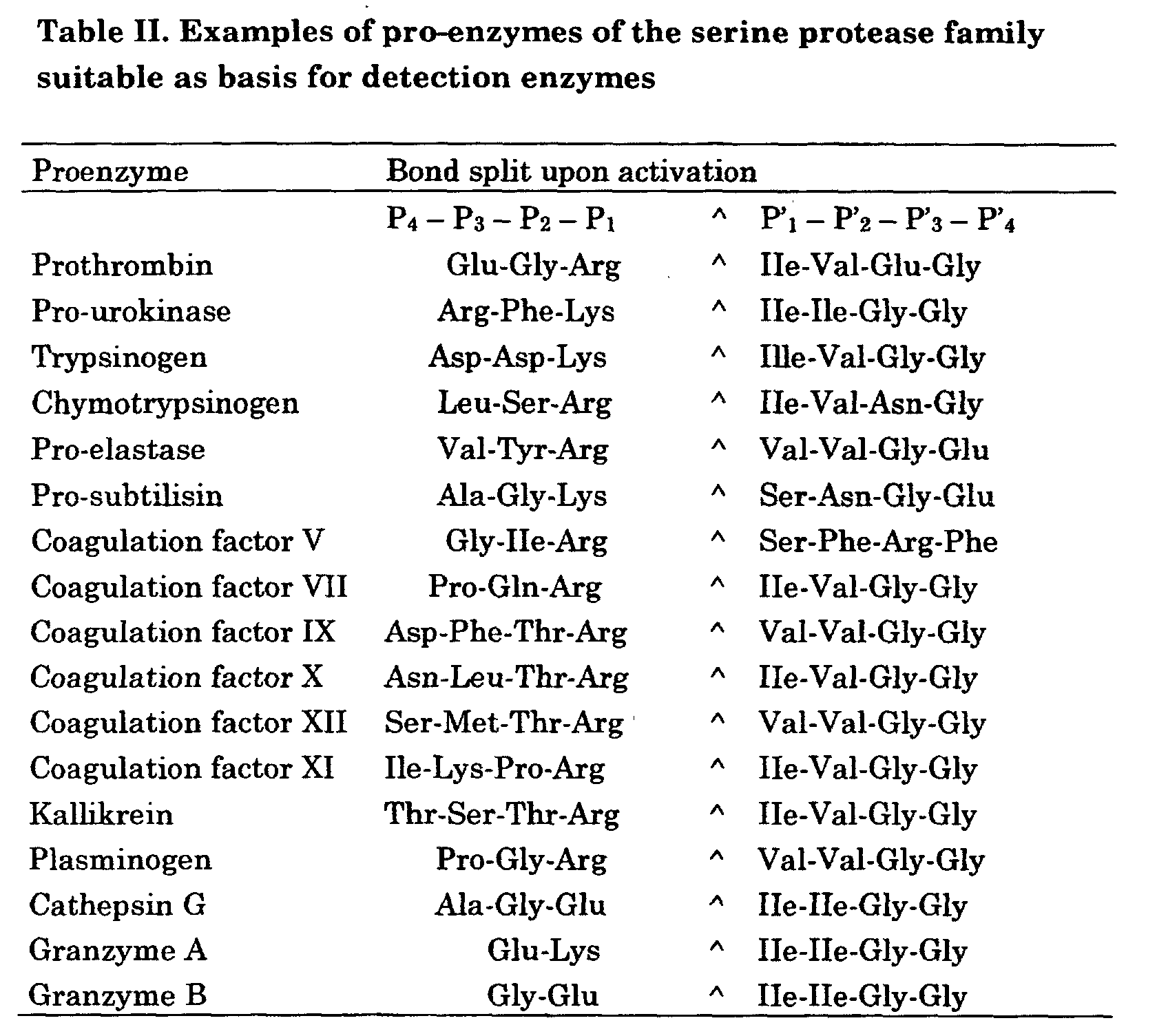
| Non limited list of protein modifications, target amino acid residues and activities leading to or removing modification. | |||
| Modification | Target residue | Enzyme modifying activity | Enzyme demodifying activity |
| Phosphorylation | Tyr, Ser, Thr, His | Protein-kinases, phosphorylases | Protein phosphatases |
| Glycosylation | Asn, Ser, Thr, Hyp | Glycosyltransferases | Not known |
| Hydroxylation | Pro, Lys | Hydroxylases | Not known |
| Methylation | Lys | Protein methyl transferase | Not known |
| Acetylation | Lys, N-terminus | Protein acetyl transferase | Not known |
| Formylation | N-terminal Met | i.a. Introduced during synthesis | Polypeptide deformylase |
| Carboxylation | Glu | Gamma glutamyl carboxylase | Not known |
| Prenylation | Cys | Prenyltransferase | Not known |
| Myristylation | Cys, His | Not known | Not known |
| Oxidation | Met | Non-enzymatic | Not known |
| Glycation | Lys | Non-enzymatic | Not known |
| Disulfide formation | Cys | Enzymatic/non-enzymatic | Protein disulfide isomerase |
Sequences obtained from SWISS-PROT, GenBank or PIR databases.
Davis RJ, J Biol Chem 268 (1993) 14553-14556
Pearson RB , Kemp BE, Methods in Enzymology 200 (1991) 62-81
Flotow H et al. J Biol Chem 265 (1990) 14264-14269
Fiol CL et al. J Biol Chem 265 (1990) 6061-6065
Russo GL et al. J Biol Chem 267 (1992) 20317-20325
Roach PJ J Biol Chem 266 (1991) 14139-14142
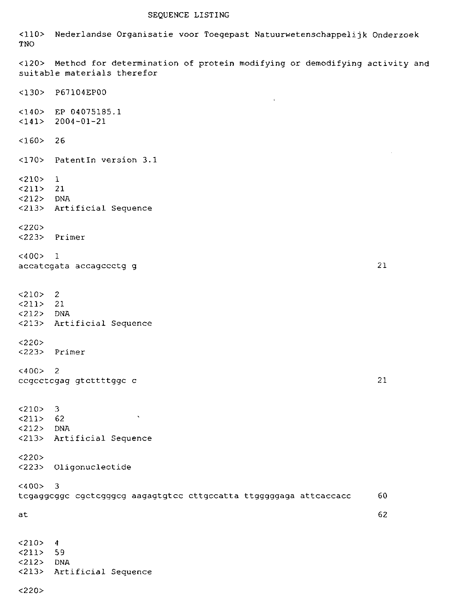
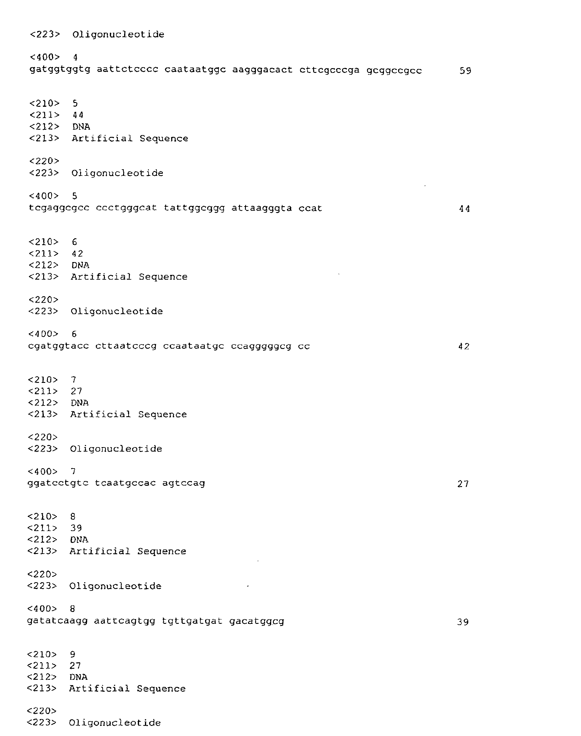


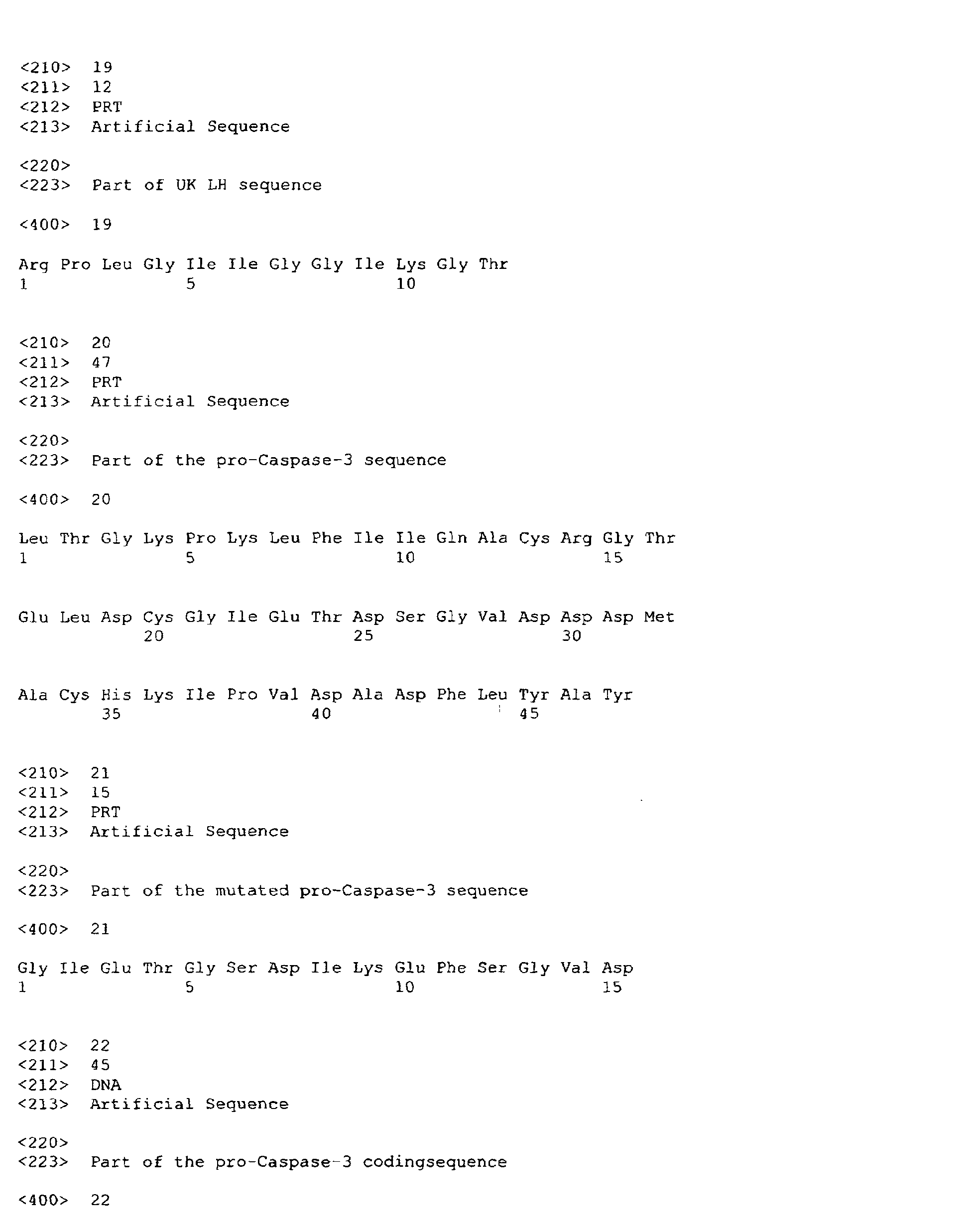

| Examples of proenzymes of the cysteine protease family suitable as basis for detection enzymes. | |
| Pro-Caspase | -1 |
| Pro-Caspase | -2 |
| Pro-Caspase | -3 |
| Pro-Caspase | -4 |
| Pro-Caspase | -5 |
| Pro-Caspase | -6 |
| Pro-Caspase | -7 |
| Pro-Caspase | -8 |
| Pro-Caspase | -9 |
| Pro-Papain | |
| Pro-cathepsin B | |
| Pro-cathepsin L | |
| Pro-cathepsin K |
| Recognition Motifs of Protein Kinases | ||
| Protein Kinase | Abbreviation(s) | Recognition Motifs |
| Glycogen synthase kinase | GSK-3 | S*-X-X-X-S(P) |
| Calmodulin dependent protein kinase II | CaMK II | R-X-X-S/T* R-X-X- S/T*-V |
| CdC2 | S/T*-P-X-R/K | |
| MAPK, Erk | P-X-S/T*-P | |
| CGPK | R/K-X-S/T* | |
| PKA, cAMPK | R-X-S/T* R-R/K-X-S/T* | |
| Caseine Kinase I | CK-1 | S(P)-X-X-S/T* |
| Caseine Kinase II | CK-2 | S/T*-X-X-E |
| PhK | K/R-X-X-S*-V/I | |
| PKC | S/T*-X-K/R K/R-X-X-S/T* K/R-X-S/T* | |
| AbI | I/V/L-Y*-X-X-P/F | |
| EGF-RK | E/D-Y*-X E/D-Y*-I/L/V |
Pearson RB , Kemp BE, Methods in Enzymology 200 (1991) 62-81
Flotow H et al. J Biol Chem 265 (1990) 14264-14269
Fiol CL et al. J Biol Chem 265 (1990) 6061-6065
Russo GL et al. J Biol Chem 267 (1992) 20317-20325
Roach PJ J Biol Chem 266 (1991) 14139-14142
| Peptide substrates for detection of caspase activity | |
| Substrate | Preferred caspase |
| VAD- | 1 |
| DEVD- | 3, 6, 7, 8 |
| VEID- | 6,8 |
| IETD- | 8, 9, 10 |
| WEHD- | 1, 4, 5 |
| YVAD- | 1, 4, 5 |
| VDVAD-X | 2 |
| X can be -pNA (para-nitro-anilide), -AFC (7-amino-4 trifluoro methyl coumarine), -AMC (amino methyl coumarine), or any other chromogenic or fluorogenic leaving group. |
Claims (34)
- A method for determining a protein modifying activity or protein demodifying activity in a sample comprising
providing a detection enzyme construct comprising at least one site of modification and at least one site of activation or inactivation, wherein activation or inactivation of the detection enzyme construct results from cleavage of said site of activation or inactivation by a protease, wherein said site of modification of the detection enzyme construct is capable of being modified by the protein modifying activity to be determined or demodified by the protein demodifying activity to be determined, and wherein modification or demodification of said site of modification affects cleavage of the said site of activation or inactivation by the said protease,
contacting the sample with the said detection enzyme construct under suitable conditions for the protein modifying or demodifying activity to be determined,
contacting the detection enzyme construct with the said protease under suitable conditions for activity of the said protease, and
determining the occurrence or level of detection enzyme activity. - A method according to claim 1, wherein said detection enzyme construct comprises a site of modification and a site of activation, wherein activation of the detection enzyme construct results from cleavage of said site of activation by an activation protease to form active detection enzyme, wherein said site of modification of the detection enzyme construct is capable of being modified by the protein modifying activity to be determined or demodified by the protein demodifying activity to be determined, and wherein modification or demodification of said site of modification determines whether said activation protease can cleave the said site of activation, and the method comprises contacting the detection enzyme construct with the said activation protease under suitable conditions for activity of the said activation protease.
- A method according to claim 1, wherein said detection enzyme construct comprises a site of modification, a site of activation and a site of inactivation, wherein activation of the detection enzyme construct results from cleavage of said site of activation by an activation protease to form active detection enzyme, and inactivation of the detection enzyme construct or detection enzyme results from cleavage of said site of inactivation by a deactivation protease, wherein said site of modification of the detection enzyme construct is capable of being modified by the protein modifying activity to be determined or demodified by the protein demodifying activity to be determined, and wherein modification or demodification of said site of modification determines whether said activation protease can cleave the said site of activation or said inactivation protease can cleave the said site of inactivation, and the method comprises contacting the detection enzyme construct with the said activation protease under suitable conditions for activity of the said activation protease, and before, after, or simultaneously therewith, contacting the detection enzyme construct or detection enzyme with the said inactivation protease under suitable conditions for activity of the said inactivation protease.
- A method according to claim 3, wherein modification or demodification of said site of modification determines whether said inactivation protease can cleave the said site of inactivation.
- A method according to any one of claims 1-4, wherein active detection enzyme is detectable by its conversion of a substrate of the detection enzyme to a reaction product and detection enzyme activity is determined by contacting the detection enzyme construct or detection enzyme with said substrate of the detection enzyme and determining the occurrence or level of conversion of said substrate.
- A method according to any one of claims 1-5, wherein said detection enzyme construct is derived from a protease enzyme or protease pro-enzyme.
- A method according to claim 6, wherein said detection enzyme construct is derived from a serine protease enzyme or serine protease pro-enzyme or from a cysteine protease enzyme or cysteine protease pro-enzyme.
- A method according to any one of claims 1-5, wherein said detection enzyme construct is derived from a pro-enzyme selected from the group consisting of pro-urokinase, pro-granzyme, pro-caspase-3 and pro-caspase-7.
- A method according to any one of claims 1-5, wherein said detection enzyme construct is derived from pro-urokinase.
- A method according to any one of claims 1-5, wherein said detection enzyme construct is derived from pro-caspase-3.
- A method according to any one of the previous claims, wherein said detection enzyme construct contains an artificial modification site, or an artificial activation cleavage site, or an artificial inactivation cleavage site, or any combination thereof, and optionally further changes, introduced into a protease enzyme or protease pro-enzyme in such a way that modification or demodification of said modification site affects activatability of the detection enzyme construct by treatment with said activation protease or inactivatability of the detection enzyme construct or detection enzyme by treatment with said inactivation protease.
- A method according to claim 11, wherein the activation protease is selected from the group consisting of (a) the natural activation protease of said protease pro-enzyme, (b) a protease specifically cleaving an artificial activation cleavage site introduced into said protease pro-enzyme, (c) an aminopeptidase, and (d) a carboxypeptidase.
- A method according to claim 11, wherein the inactivation protease is the same or different from the activation protease and is selected from the group consisting of (a) a natural inactivation protease of said protease enzyme, (b) a protease specifically cleaving an artificial inactivation cleavage site introduced into said protease pro-enzyme, (c) an aminopeptidase, and (d) a carboxypeptidase.
- A method according to any one of the previous claims, wherein said protein modifying activity or protein demodifying activity is an enzymatic activity.
- A method according to claim 14, wherein said enzymatic activity is selected from the group consisting of of protein kinase activity, protein phosphatase activity, protein hydroxylase activity, protein glycosyltransferase activity or protein prenyltransferase activity.
- A method according to any one of the previous claims for determining a protein demodifying activity, wherein the sample is contacted with pre-modified detection enzyme construct.
- A method according to any one of the previous claims, wherein the sample is contacted with a purified or partially purified detection enzyme construct, or with a cell expressing or containing the detection enzyme construct, or with the content of such a cell or of a specific compartment of such a cell, or with the culture medium of such a cell.
- A method according to claim 17, wherein the sample is contacted with a cell expressing or containing the detection enzyme construct and then either the cell as a whole, or the content of the cell or of a specific compartment of the cell, or the culture medium of the cell is contacted with the activation and/or inactivation protease and detection enzyme substrate.
- A detection enzyme construct useful for determining a protein modifying activity or protein demodifying activity in a sample, comprising at least one site of modification and at least one site of activation or inactivation, wherein activation or inactivation of the detection enzyme construct results from cleavage of said site of activation or inactivation by a protease, wherein said site of modification of the detection enzyme construct is capable of being modified by the protein modifying activity to be determined or demodified by the protein demodifying activity to be determined, and wherein modification or demodification of said site of modification affects cleavage of the said site of activation or inactivation by the said protease.
- A detection enzyme construct according to claim 19, comprising a site of modification and a site of activation, wherein activation of the detection enzyme construct results from cleavage of said site of activation by an activation protease to form active detection enzyme, wherein said site of modification of the detection enzyme construct is capable of being modified by the protein modifying activity to be determined or demodified by the protein demodifying activity to be determined, and wherein modification or demodification of said site of modification determines whether said activation protease can cleave the said site of activation.
- A detection enzyme construct according to claim 19, comprising a site of modification, a site of activation and a site of inactivation, wherein activation of the detection enzyme construct results from cleavage of said site of activation by an activation protease to form active detection enzyme, and inactivation of the detection enzyme construct or detection enzyme results from cleavage of said site of inactivation by a deactivation protease, wherein said site of modification of the detection enzyme construct is capable of being modified by the protein modifying activity to be determined or demodified by the protein demodifying activity to be determined, and wherein modification or demodification of said site of modification determines whether said activation protease can cleave the said site of activation or said inactivation protease can cleave the said site of inactivation.
- A detection enzyme construct according to claim 21, wherein modification or demodification of said site of modification determines whether said inactivation protease can cleave the said site of inactivation.
- A detection enzyme construct according to any one of claims 19-22, wherein active detection enzyme is detectable by its conversion of a substrate of the detection enzyme to a reaction product.
- A detection enzyme construct according to any one of claims 19-23, derived from a protease enzyme or protease pro-enzyme.
- A detection enzyme construct according to claim 24, derived from a serine protease enzyme or serine protease pro-enzyme or from a cysteine protease enzyme or cysteine protease pro-enzyme.
- A detection enzyme construct according to any one of claims 19-23, derived from a pro-enzyme selected from the group consisting of pro-urokinase, pro-granzyme, pro-caspase-3 and pro-caspase-7.
- A detection enzyme construct according to any one of claims 19-23, derived from pro-urokinase.
- A detection enzyme construct according to any one of claims 19-23, derived from pro-caspase-3.
- A detection enzyme construct according to any one of claims 19-28, which contains an artificial modification site, or an artificial activation cleavage site, or an artificial inactivation cleavage site, or any combination thereof, and optionally further changes, introduced into a protease enzyme or protease pro-enzyme in such a way that modification or demodification of said modification site affects activatability of the detection enzyme construct by treatment with said activation protease or inactivatability of the detection enzyme construct or detection enzyme by treatment with said inactivation protease.
- A detection enzyme construct according to claim 29, wherein the activation protease is selected from the group consisting of (a) the natural activation protease of said protease pro-enzyme, (b) a protease specifically cleaving an artificial activation cleavage site introduced into said protease pro-enzyme, (c) an aminopeptidase, and (d) a carboxypeptidase.
- A detection enzyme construct according to claim 29, wherein the inactivation protease is the same or different from the activation protease and is selected from the group consisting of (a) a natural inactivation protease of said protease enzyme, (b) a protease specifically cleaving an artificial inactivation cleavage site introduced into said protease pro-enzyme, (c) an aminopeptidase, and (d) a carboxypeptidase.
- A cell expressing or containing a detection enzyme construct as defined in any one of claims 19-31.
- A kit for determining a protein modifying activity or a protein demodifying activity in a sample, comprising a detection enzyme construct as defined in any one of claims 19-31, or a cell as defined in claim 32, and further comprising at least one member of the group consisting of activation protease, inactivation protease, substrate of the detection enzyme, a standard composition, a buffer composition, an antibody composition, a stabilizing composition, an assay plate or container, and instructions for use.
- An expression vector coding for a detection enzyme construct as defined in any one of claims 19-31.
Priority Applications (2)
| Application Number | Priority Date | Filing Date | Title |
|---|---|---|---|
| EP04075185A EP1557474A1 (en) | 2004-01-21 | 2004-01-21 | Method for determination of protein modifying or demodifying activity and suitable materials therefor |
| PCT/NL2005/000035 WO2005071100A2 (en) | 2004-01-21 | 2005-01-19 | Method for determination of protein modifying or demodifying activity and suitable materials thereof |
Applications Claiming Priority (1)
| Application Number | Priority Date | Filing Date | Title |
|---|---|---|---|
| EP04075185A EP1557474A1 (en) | 2004-01-21 | 2004-01-21 | Method for determination of protein modifying or demodifying activity and suitable materials therefor |
Publications (1)
| Publication Number | Publication Date |
|---|---|
| EP1557474A1 true EP1557474A1 (en) | 2005-07-27 |
Family
ID=34626523
Family Applications (1)
| Application Number | Title | Priority Date | Filing Date |
|---|---|---|---|
| EP04075185A Withdrawn EP1557474A1 (en) | 2004-01-21 | 2004-01-21 | Method for determination of protein modifying or demodifying activity and suitable materials therefor |
Country Status (2)
| Country | Link |
|---|---|
| EP (1) | EP1557474A1 (en) |
| WO (1) | WO2005071100A2 (en) |
Cited By (4)
| Publication number | Priority date | Publication date | Assignee | Title |
|---|---|---|---|---|
| WO2009024805A1 (en) * | 2007-08-23 | 2009-02-26 | Mologic Ltd | An enzyme detection device |
| US8241864B2 (en) | 2006-10-19 | 2012-08-14 | Apeiron Biologics Forschungs-und Entwicklungsgesellschaft m.b.H. | Method for determining ACE2 activity |
| CN101416057B (en) * | 2006-04-06 | 2013-06-05 | 金伯利-克拉克环球有限公司 | Enzyme Detection Technology |
| WO2013156794A1 (en) * | 2012-04-20 | 2013-10-24 | Mologic Limited | An enzyme detection device |
Families Citing this family (1)
| Publication number | Priority date | Publication date | Assignee | Title |
|---|---|---|---|---|
| US11624082B2 (en) * | 2018-06-21 | 2023-04-11 | Universiteit Utrecht Holding B.V. | Method for monitoring kinase activity in a sample |
Citations (4)
| Publication number | Priority date | Publication date | Assignee | Title |
|---|---|---|---|---|
| EP0691409A1 (en) * | 1994-07-07 | 1996-01-10 | Nederlandse Organisatie Voor Toegepast-Natuurwetenschappelijk Onderzoek Tno | Modified proenzymes as substrates for proteolytic enzymes |
| US5811252A (en) * | 1994-07-07 | 1998-09-22 | Nederlandse Organisatie Voor Toegepast-Natuurwetenschappelijk Onderzoek Tno | Modified proenzymes as substrates for proteolytic enzymes |
| EP1243658A1 (en) * | 1999-11-29 | 2002-09-25 | Cyclex Co. Ltd | Method of measuring deacetylase activity and method of screening inhibitor or promoter of these enzymes |
| WO2003066889A2 (en) * | 2002-02-07 | 2003-08-14 | Axys Pharmaceuticals, Inc. | Assay for acytyltransferase or deacetylase activity |
-
2004
- 2004-01-21 EP EP04075185A patent/EP1557474A1/en not_active Withdrawn
-
2005
- 2005-01-19 WO PCT/NL2005/000035 patent/WO2005071100A2/en not_active Ceased
Patent Citations (4)
| Publication number | Priority date | Publication date | Assignee | Title |
|---|---|---|---|---|
| EP0691409A1 (en) * | 1994-07-07 | 1996-01-10 | Nederlandse Organisatie Voor Toegepast-Natuurwetenschappelijk Onderzoek Tno | Modified proenzymes as substrates for proteolytic enzymes |
| US5811252A (en) * | 1994-07-07 | 1998-09-22 | Nederlandse Organisatie Voor Toegepast-Natuurwetenschappelijk Onderzoek Tno | Modified proenzymes as substrates for proteolytic enzymes |
| EP1243658A1 (en) * | 1999-11-29 | 2002-09-25 | Cyclex Co. Ltd | Method of measuring deacetylase activity and method of screening inhibitor or promoter of these enzymes |
| WO2003066889A2 (en) * | 2002-02-07 | 2003-08-14 | Axys Pharmaceuticals, Inc. | Assay for acytyltransferase or deacetylase activity |
Non-Patent Citations (1)
| Title |
|---|
| VERHEIJEN J H ET AL: "MODIFIED PROENZYMES AS ARTIFICIAL SUBSTRATES FOR PROTEOLYTIC ENZYMES: COLORIMETRIC ASSAY OF BACTERIAL COLLAGENASE AND MATRIX METALLOPROTEINASE ACTIVITY USING MODIFIED PRO-UROKINASE", BIOCHEMICAL JOURNAL, PORTLAND PRESS, LONDON, GB, vol. 323, no. 3, 1997, pages 603 - 609, XP008019534, ISSN: 0264-6021 * |
Cited By (9)
| Publication number | Priority date | Publication date | Assignee | Title |
|---|---|---|---|---|
| CN101416057B (en) * | 2006-04-06 | 2013-06-05 | 金伯利-克拉克环球有限公司 | Enzyme Detection Technology |
| US8241864B2 (en) | 2006-10-19 | 2012-08-14 | Apeiron Biologics Forschungs-und Entwicklungsgesellschaft m.b.H. | Method for determining ACE2 activity |
| WO2009024805A1 (en) * | 2007-08-23 | 2009-02-26 | Mologic Ltd | An enzyme detection device |
| JP2010537186A (en) * | 2007-08-23 | 2010-12-02 | モロジック リミテッド | Enzyme detection device |
| US8592167B2 (en) | 2007-08-23 | 2013-11-26 | Mologic Ltd | Enzyme detection device |
| CN101784671B (en) * | 2007-08-23 | 2014-04-02 | 莫洛迪克有限公司 | An enzyme detection device |
| US10502740B2 (en) | 2007-08-23 | 2019-12-10 | Mologic Limited | Enzyme detection device |
| WO2013156794A1 (en) * | 2012-04-20 | 2013-10-24 | Mologic Limited | An enzyme detection device |
| US10234457B2 (en) | 2012-04-20 | 2019-03-19 | Mologic Limited | Enzyme detection device |
Also Published As
| Publication number | Publication date |
|---|---|
| WO2005071100A3 (en) | 2005-11-17 |
| WO2005071100A2 (en) | 2005-08-04 |
Similar Documents
| Publication | Publication Date | Title |
|---|---|---|
| Wang et al. | Evidence for bidentate substrate binding as the basis for the K48 linkage specificity of otubain 1 | |
| Zhu et al. | Identification of key functional amino acids of the mouse fertilin β (ADAM2) disintegrin loop for cell-cell adhesion during fertilization | |
| Jones et al. | Intracellular trafficking of furin is modulated by the phosphorylation state of a casein kinase II site in its cytoplasmic tail. | |
| EP2308978A1 (en) | Luciferase biosensor | |
| JPH0856665A (en) | Modified zymogens as substrates for proteolytic enzymes | |
| US20080241867A1 (en) | Apparatuses and methods for determining protease activity | |
| Arnold et al. | The noncatalytic portion of human UDP-glucose: glycoprotein glucosyltransferase I confers UDP-glucose binding and transferase function to the catalytic domain | |
| IL180201A (en) | Method for assessing the activity of a proteolytic enzyme and a kit based thereon | |
| US20030219848A1 (en) | Short enzyme donor fragment | |
| US7166475B2 (en) | Compositions and methods for monitoring the modification state of a pair of polypeptides | |
| CA2447040C (en) | Assays, methods and means | |
| WO2005071100A2 (en) | Method for determination of protein modifying or demodifying activity and suitable materials thereof | |
| You et al. | Proteolytic targeting of transcriptional regulator TIP120B by a HECT domain E3 ligase | |
| US20080057527A1 (en) | Methods for carboxypeptidases | |
| JP4708326B2 (en) | Improved method for the detection of proteolytic enzymes | |
| ZA200300033B (en) | Metalloprotease peptide substrates and methods. | |
| EP1828775B1 (en) | Methods for the functional determination of mannan-binding-lectin associated serine proteases (masps) and complexes thereof | |
| WO2006044918A2 (en) | Compounds useful as substrates for enzymes that reverse nucleotidylylated peptide and protein modifications and methods for production and uses thereof |
Legal Events
| Date | Code | Title | Description |
|---|---|---|---|
| PUAI | Public reference made under article 153(3) epc to a published international application that has entered the european phase |
Free format text: ORIGINAL CODE: 0009012 |
|
| AK | Designated contracting states |
Kind code of ref document: A1 Designated state(s): AT BE BG CH CY CZ DE DK EE ES FI FR GB GR HU IE IT LI LU MC NL PT RO SE SI SK TR |
|
| AX | Request for extension of the european patent |
Extension state: AL LT LV MK |
|
| AKX | Designation fees paid | ||
| REG | Reference to a national code |
Ref country code: DE Ref legal event code: 8566 |
|
| STAA | Information on the status of an ep patent application or granted ep patent |
Free format text: STATUS: THE APPLICATION IS DEEMED TO BE WITHDRAWN |
|
| 18D | Application deemed to be withdrawn |
Effective date: 20060128 |