EP1546328B1 - High yield heterologous expression cell lines for expression of gene products with human glycosylation pattern - Google Patents
High yield heterologous expression cell lines for expression of gene products with human glycosylation pattern Download PDFInfo
- Publication number
- EP1546328B1 EP1546328B1 EP03776861A EP03776861A EP1546328B1 EP 1546328 B1 EP1546328 B1 EP 1546328B1 EP 03776861 A EP03776861 A EP 03776861A EP 03776861 A EP03776861 A EP 03776861A EP 1546328 B1 EP1546328 B1 EP 1546328B1
- Authority
- EP
- European Patent Office
- Prior art keywords
- cell
- gene
- functional
- gene product
- starting
- Prior art date
- Legal status (The legal status is an assumption and is not a legal conclusion. Google has not performed a legal analysis and makes no representation as to the accuracy of the status listed.)
- Expired - Lifetime
Links
- 108090000623 proteins and genes Proteins 0.000 title claims abstract description 225
- 230000014509 gene expression Effects 0.000 title claims abstract description 50
- 241000282414 Homo sapiens Species 0.000 title claims description 45
- 230000013595 glycosylation Effects 0.000 title claims description 15
- 238000006206 glycosylation reaction Methods 0.000 title claims description 15
- 238000000034 method Methods 0.000 claims abstract description 54
- 108091028043 Nucleic acid sequence Proteins 0.000 claims abstract description 36
- 102000018120 Recombinases Human genes 0.000 claims abstract description 22
- 108010091086 Recombinases Proteins 0.000 claims abstract description 22
- 239000002243 precursor Substances 0.000 claims abstract description 15
- 238000012216 screening Methods 0.000 claims abstract description 10
- 208000005229 Autosomal recessive Robinow syndrome Diseases 0.000 claims abstract 2
- 238000001945 resonance Rayleigh scattering spectroscopy Methods 0.000 claims abstract 2
- 210000004027 cell Anatomy 0.000 claims description 203
- 239000013598 vector Substances 0.000 claims description 93
- 102000004169 proteins and genes Human genes 0.000 claims description 38
- 230000010354 integration Effects 0.000 claims description 23
- 230000008569 process Effects 0.000 claims description 21
- 239000003550 marker Substances 0.000 claims description 15
- 238000004519 manufacturing process Methods 0.000 claims description 13
- 102000004190 Enzymes Human genes 0.000 claims description 10
- 108090000790 Enzymes Proteins 0.000 claims description 10
- 210000005260 human cell Anatomy 0.000 claims description 7
- 210000004754 hybrid cell Anatomy 0.000 claims description 7
- 210000004408 hybridoma Anatomy 0.000 claims description 7
- 108700005091 Immunoglobulin Genes Proteins 0.000 claims description 6
- 102100034343 Integrase Human genes 0.000 claims description 6
- 108010061833 Integrases Proteins 0.000 claims description 6
- 206010035226 Plasma cell myeloma Diseases 0.000 claims description 5
- 201000000050 myeloid neoplasm Diseases 0.000 claims description 5
- 230000008488 polyadenylation Effects 0.000 claims description 5
- 210000003719 b-lymphocyte Anatomy 0.000 claims description 4
- 239000003623 enhancer Substances 0.000 claims description 4
- 102000004196 processed proteins & peptides Human genes 0.000 claims description 4
- 108090000765 processed proteins & peptides Proteins 0.000 claims description 4
- 238000001262 western blot Methods 0.000 claims description 4
- 108090000695 Cytokines Proteins 0.000 claims description 3
- 102000004127 Cytokines Human genes 0.000 claims description 3
- 108060003951 Immunoglobulin Proteins 0.000 claims description 3
- 229940088597 hormone Drugs 0.000 claims description 3
- 239000005556 hormone Substances 0.000 claims description 3
- 102000018358 immunoglobulin Human genes 0.000 claims description 3
- 108020003175 receptors Proteins 0.000 claims description 3
- 102000005962 receptors Human genes 0.000 claims description 3
- 238000002965 ELISA Methods 0.000 claims description 2
- 238000000636 Northern blotting Methods 0.000 claims description 2
- 238000010195 expression analysis Methods 0.000 claims description 2
- 102000037865 fusion proteins Human genes 0.000 claims description 2
- 108020001507 fusion proteins Proteins 0.000 claims description 2
- 238000001502 gel electrophoresis Methods 0.000 claims description 2
- 238000002493 microarray Methods 0.000 claims description 2
- 229920001184 polypeptide Polymers 0.000 claims description 2
- 238000003753 real-time PCR Methods 0.000 claims description 2
- 230000028327 secretion Effects 0.000 claims 1
- 108020004414 DNA Proteins 0.000 description 63
- 239000000047 product Substances 0.000 description 47
- 239000012634 fragment Substances 0.000 description 33
- 230000008685 targeting Effects 0.000 description 32
- 238000006243 chemical reaction Methods 0.000 description 27
- 229930189065 blasticidin Natural products 0.000 description 26
- 239000013612 plasmid Substances 0.000 description 25
- 102000007056 Recombinant Fusion Proteins Human genes 0.000 description 17
- 108010008281 Recombinant Fusion Proteins Proteins 0.000 description 17
- YQYJSBFKSSDGFO-UHFFFAOYSA-N Epihygromycin Natural products OC1C(O)C(C(=O)C)OC1OC(C(=C1)O)=CC=C1C=C(C)C(=O)NC1C(O)C(O)C2OCOC2C1O YQYJSBFKSSDGFO-UHFFFAOYSA-N 0.000 description 16
- 229930193140 Neomycin Natural products 0.000 description 15
- 229960004927 neomycin Drugs 0.000 description 15
- 210000000349 chromosome Anatomy 0.000 description 14
- 230000006798 recombination Effects 0.000 description 14
- 238000005215 recombination Methods 0.000 description 14
- 230000006801 homologous recombination Effects 0.000 description 12
- 238000002744 homologous recombination Methods 0.000 description 12
- 239000000872 buffer Substances 0.000 description 11
- 239000002609 medium Substances 0.000 description 11
- 102000003886 Glycoproteins Human genes 0.000 description 10
- 108090000288 Glycoproteins Proteins 0.000 description 10
- BRZYSWJRSDMWLG-CAXSIQPQSA-N geneticin Natural products O1C[C@@](O)(C)[C@H](NC)[C@@H](O)[C@H]1O[C@@H]1[C@@H](O)[C@H](O[C@@H]2[C@@H]([C@@H](O)[C@H](O)[C@@H](C(C)O)O2)N)[C@@H](N)C[C@H]1N BRZYSWJRSDMWLG-CAXSIQPQSA-N 0.000 description 10
- 239000001963 growth medium Substances 0.000 description 10
- 239000006228 supernatant Substances 0.000 description 10
- LFQSCWFLJHTTHZ-UHFFFAOYSA-N Ethanol Chemical compound CCO LFQSCWFLJHTTHZ-UHFFFAOYSA-N 0.000 description 9
- 238000004520 electroporation Methods 0.000 description 8
- 230000009466 transformation Effects 0.000 description 8
- 230000001413 cellular effect Effects 0.000 description 7
- 238000003780 insertion Methods 0.000 description 7
- 239000012528 membrane Substances 0.000 description 7
- 230000004481 post-translational protein modification Effects 0.000 description 7
- MPLHNVLQVRSVEE-UHFFFAOYSA-N texas red Chemical compound [O-]S(=O)(=O)C1=CC(S(Cl)(=O)=O)=CC=C1C(C1=CC=2CCCN3CCCC(C=23)=C1O1)=C2C1=C(CCC1)C3=[N+]1CCCC3=C2 MPLHNVLQVRSVEE-UHFFFAOYSA-N 0.000 description 7
- IAZDPXIOMUYVGZ-UHFFFAOYSA-N Dimethylsulphoxide Chemical compound CS(C)=O IAZDPXIOMUYVGZ-UHFFFAOYSA-N 0.000 description 6
- 238000012181 QIAquick gel extraction kit Methods 0.000 description 6
- 108090000631 Trypsin Proteins 0.000 description 6
- 102000004142 Trypsin Human genes 0.000 description 6
- 238000004113 cell culture Methods 0.000 description 6
- 238000010367 cloning Methods 0.000 description 6
- 230000002779 inactivation Effects 0.000 description 6
- 230000037431 insertion Effects 0.000 description 6
- 238000000746 purification Methods 0.000 description 6
- 108091008146 restriction endonucleases Proteins 0.000 description 6
- 238000010186 staining Methods 0.000 description 6
- 230000001225 therapeutic effect Effects 0.000 description 6
- 238000001890 transfection Methods 0.000 description 6
- 239000012588 trypsin Substances 0.000 description 6
- SHZGCJCMOBCMKK-UHFFFAOYSA-N D-mannomethylose Natural products CC1OC(O)C(O)C(O)C1O SHZGCJCMOBCMKK-UHFFFAOYSA-N 0.000 description 5
- 241000588724 Escherichia coli Species 0.000 description 5
- SHZGCJCMOBCMKK-DHVFOXMCSA-N L-fucopyranose Chemical compound C[C@@H]1OC(O)[C@@H](O)[C@H](O)[C@@H]1O SHZGCJCMOBCMKK-DHVFOXMCSA-N 0.000 description 5
- GYDJEQRTZSCIOI-UHFFFAOYSA-N Tranexamic acid Chemical compound NCC1CCC(C(O)=O)CC1 GYDJEQRTZSCIOI-UHFFFAOYSA-N 0.000 description 5
- 238000004458 analytical method Methods 0.000 description 5
- SQVRNKJHWKZAKO-UHFFFAOYSA-N beta-N-Acetyl-D-neuraminic acid Natural products CC(=O)NC1C(O)CC(O)(C(O)=O)OC1C(O)C(O)CO SQVRNKJHWKZAKO-UHFFFAOYSA-N 0.000 description 5
- 230000003115 biocidal effect Effects 0.000 description 5
- 239000013553 cell monolayer Substances 0.000 description 5
- 238000001514 detection method Methods 0.000 description 5
- 230000029087 digestion Effects 0.000 description 5
- 238000010790 dilution Methods 0.000 description 5
- 239000012895 dilution Substances 0.000 description 5
- 230000004927 fusion Effects 0.000 description 5
- 210000003917 human chromosome Anatomy 0.000 description 5
- 238000012744 immunostaining Methods 0.000 description 5
- 238000010348 incorporation Methods 0.000 description 5
- 210000004962 mammalian cell Anatomy 0.000 description 5
- 239000000203 mixture Substances 0.000 description 5
- 239000008188 pellet Substances 0.000 description 5
- 238000012546 transfer Methods 0.000 description 5
- XLYOFNOQVPJJNP-UHFFFAOYSA-N water Substances O XLYOFNOQVPJJNP-UHFFFAOYSA-N 0.000 description 5
- PXBFMLJZNCDSMP-UHFFFAOYSA-N 2-Aminobenzamide Chemical compound NC(=O)C1=CC=CC=C1N PXBFMLJZNCDSMP-UHFFFAOYSA-N 0.000 description 4
- 229920000936 Agarose Polymers 0.000 description 4
- 108010046276 FLP recombinase Proteins 0.000 description 4
- PNNNRSAQSRJVSB-SLPGGIOYSA-N Fucose Natural products C[C@H](O)[C@@H](O)[C@H](O)[C@H](O)C=O PNNNRSAQSRJVSB-SLPGGIOYSA-N 0.000 description 4
- 108700026244 Open Reading Frames Proteins 0.000 description 4
- 239000004904 UV filter Substances 0.000 description 4
- 238000013459 approach Methods 0.000 description 4
- 210000004369 blood Anatomy 0.000 description 4
- 239000008280 blood Substances 0.000 description 4
- 239000006285 cell suspension Substances 0.000 description 4
- 238000005119 centrifugation Methods 0.000 description 4
- 239000012228 culture supernatant Substances 0.000 description 4
- 230000001419 dependent effect Effects 0.000 description 4
- 239000000499 gel Substances 0.000 description 4
- 238000007857 nested PCR Methods 0.000 description 4
- 125000005629 sialic acid group Chemical group 0.000 description 4
- 238000001228 spectrum Methods 0.000 description 4
- 238000011144 upstream manufacturing Methods 0.000 description 4
- 102000002260 Alkaline Phosphatase Human genes 0.000 description 3
- 108020004774 Alkaline Phosphatase Proteins 0.000 description 3
- 241000282412 Homo Species 0.000 description 3
- 108090000144 Human Proteins Proteins 0.000 description 3
- 102000003839 Human Proteins Human genes 0.000 description 3
- OKKJLVBELUTLKV-UHFFFAOYSA-N Methanol Chemical compound OC OKKJLVBELUTLKV-UHFFFAOYSA-N 0.000 description 3
- 241001045988 Neogene Species 0.000 description 3
- 101150117115 V gene Proteins 0.000 description 3
- 238000000137 annealing Methods 0.000 description 3
- 230000010056 antibody-dependent cellular cytotoxicity Effects 0.000 description 3
- 230000001580 bacterial effect Effects 0.000 description 3
- 230000008901 benefit Effects 0.000 description 3
- 108091092356 cellular DNA Proteins 0.000 description 3
- 239000007979 citrate buffer Substances 0.000 description 3
- 239000002299 complementary DNA Substances 0.000 description 3
- 150000004676 glycans Chemical group 0.000 description 3
- 238000004128 high performance liquid chromatography Methods 0.000 description 3
- 230000002163 immunogen Effects 0.000 description 3
- 238000011534 incubation Methods 0.000 description 3
- 238000001840 matrix-assisted laser desorption--ionisation time-of-flight mass spectrometry Methods 0.000 description 3
- 238000000386 microscopy Methods 0.000 description 3
- 101150091879 neo gene Proteins 0.000 description 3
- 238000002360 preparation method Methods 0.000 description 3
- QEVHRUUCFGRFIF-MDEJGZGSSA-N reserpine Chemical compound O([C@H]1[C@@H]([C@H]([C@H]2C[C@@H]3C4=C(C5=CC=C(OC)C=C5N4)CCN3C[C@H]2C1)C(=O)OC)OC)C(=O)C1=CC(OC)=C(OC)C(OC)=C1 QEVHRUUCFGRFIF-MDEJGZGSSA-N 0.000 description 3
- 239000000523 sample Substances 0.000 description 3
- 239000000243 solution Substances 0.000 description 3
- 239000000725 suspension Substances 0.000 description 3
- 230000010474 transient expression Effects 0.000 description 3
- IJGRMHOSHXDMSA-UHFFFAOYSA-N Atomic nitrogen Chemical compound N#N IJGRMHOSHXDMSA-UHFFFAOYSA-N 0.000 description 2
- 241000894006 Bacteria Species 0.000 description 2
- 241000283707 Capra Species 0.000 description 2
- 108010077544 Chromatin Proteins 0.000 description 2
- ZDXPYRJPNDTMRX-UHFFFAOYSA-N Glutamine Chemical compound OC(=O)C(N)CCC(N)=O ZDXPYRJPNDTMRX-UHFFFAOYSA-N 0.000 description 2
- 241000238631 Hexapoda Species 0.000 description 2
- 108091092195 Intron Proteins 0.000 description 2
- ZQISRDCJNBUVMM-UHFFFAOYSA-N L-Histidinol Natural products OCC(N)CC1=CN=CN1 ZQISRDCJNBUVMM-UHFFFAOYSA-N 0.000 description 2
- ZQISRDCJNBUVMM-YFKPBYRVSA-N L-histidinol Chemical compound OC[C@@H](N)CC1=CNC=N1 ZQISRDCJNBUVMM-YFKPBYRVSA-N 0.000 description 2
- 108090000364 Ligases Proteins 0.000 description 2
- 241000124008 Mammalia Species 0.000 description 2
- SQVRNKJHWKZAKO-PFQGKNLYSA-N N-acetyl-beta-neuraminic acid Chemical compound CC(=O)N[C@@H]1[C@@H](O)C[C@@](O)(C(O)=O)O[C@H]1[C@H](O)[C@H](O)CO SQVRNKJHWKZAKO-PFQGKNLYSA-N 0.000 description 2
- SUHQNCLNRUAGOO-UHFFFAOYSA-N N-glycoloyl-neuraminic acid Natural products OCC(O)C(O)C(O)C(NC(=O)CO)C(O)CC(=O)C(O)=O SUHQNCLNRUAGOO-UHFFFAOYSA-N 0.000 description 2
- FDJKUWYYUZCUJX-UHFFFAOYSA-N N-glycolyl-beta-neuraminic acid Natural products OCC(O)C(O)C1OC(O)(C(O)=O)CC(O)C1NC(=O)CO FDJKUWYYUZCUJX-UHFFFAOYSA-N 0.000 description 2
- FDJKUWYYUZCUJX-KVNVFURPSA-N N-glycolylneuraminic acid Chemical compound OC[C@H](O)[C@H](O)[C@@H]1O[C@](O)(C(O)=O)C[C@H](O)[C@H]1NC(=O)CO FDJKUWYYUZCUJX-KVNVFURPSA-N 0.000 description 2
- 102000000447 Peptide-N4-(N-acetyl-beta-glucosaminyl) Asparagine Amidase Human genes 0.000 description 2
- 108010055817 Peptide-N4-(N-acetyl-beta-glucosaminyl) Asparagine Amidase Proteins 0.000 description 2
- 241000235648 Pichia Species 0.000 description 2
- 241000283984 Rodentia Species 0.000 description 2
- FAPWRFPIFSIZLT-UHFFFAOYSA-M Sodium chloride Chemical compound [Na+].[Cl-] FAPWRFPIFSIZLT-UHFFFAOYSA-M 0.000 description 2
- 108091081024 Start codon Proteins 0.000 description 2
- 108010006785 Taq Polymerase Proteins 0.000 description 2
- 239000000853 adhesive Substances 0.000 description 2
- 230000001070 adhesive effect Effects 0.000 description 2
- 238000001042 affinity chromatography Methods 0.000 description 2
- 239000011543 agarose gel Substances 0.000 description 2
- 238000000246 agarose gel electrophoresis Methods 0.000 description 2
- 229960000723 ampicillin Drugs 0.000 description 2
- AVKUERGKIZMTKX-NJBDSQKTSA-N ampicillin Chemical compound C1([C@@H](N)C(=O)N[C@H]2[C@H]3SC([C@@H](N3C2=O)C(O)=O)(C)C)=CC=CC=C1 AVKUERGKIZMTKX-NJBDSQKTSA-N 0.000 description 2
- 239000003242 anti bacterial agent Substances 0.000 description 2
- -1 antibodies Proteins 0.000 description 2
- 230000015572 biosynthetic process Effects 0.000 description 2
- 239000006143 cell culture medium Substances 0.000 description 2
- 210000003483 chromatin Anatomy 0.000 description 2
- 239000013611 chromosomal DNA Substances 0.000 description 2
- 230000002759 chromosomal effect Effects 0.000 description 2
- OPTASPLRGRRNAP-UHFFFAOYSA-N cytosine Chemical compound NC=1C=CNC(=O)N=1 OPTASPLRGRRNAP-UHFFFAOYSA-N 0.000 description 2
- 230000002950 deficient Effects 0.000 description 2
- 238000001962 electrophoresis Methods 0.000 description 2
- ZMMJGEGLRURXTF-UHFFFAOYSA-N ethidium bromide Chemical compound [Br-].C12=CC(N)=CC=C2C2=CC=C(N)C=C2[N+](CC)=C1C1=CC=CC=C1 ZMMJGEGLRURXTF-UHFFFAOYSA-N 0.000 description 2
- 230000033581 fucosylation Effects 0.000 description 2
- 230000002538 fungal effect Effects 0.000 description 2
- 238000002523 gelfiltration Methods 0.000 description 2
- 238000012239 gene modification Methods 0.000 description 2
- 230000002068 genetic effect Effects 0.000 description 2
- 230000005017 genetic modification Effects 0.000 description 2
- 235000013617 genetically modified food Nutrition 0.000 description 2
- 125000005842 heteroatom Chemical group 0.000 description 2
- 238000009396 hybridization Methods 0.000 description 2
- 238000003125 immunofluorescent labeling Methods 0.000 description 2
- 230000003834 intracellular effect Effects 0.000 description 2
- 238000002955 isolation Methods 0.000 description 2
- 229960000318 kanamycin Drugs 0.000 description 2
- 229930027917 kanamycin Natural products 0.000 description 2
- SBUJHOSQTJFQJX-NOAMYHISSA-N kanamycin Chemical compound O[C@@H]1[C@@H](O)[C@H](O)[C@@H](CN)O[C@@H]1O[C@H]1[C@H](O)[C@@H](O[C@@H]2[C@@H]([C@@H](N)[C@H](O)[C@@H](CO)O2)O)[C@H](N)C[C@@H]1N SBUJHOSQTJFQJX-NOAMYHISSA-N 0.000 description 2
- 229930182823 kanamycin A Natural products 0.000 description 2
- 230000007774 longterm Effects 0.000 description 2
- 239000000463 material Substances 0.000 description 2
- 238000005374 membrane filtration Methods 0.000 description 2
- 229910052757 nitrogen Inorganic materials 0.000 description 2
- 108020004707 nucleic acids Proteins 0.000 description 2
- 102000039446 nucleic acids Human genes 0.000 description 2
- 150000007523 nucleic acids Chemical class 0.000 description 2
- 239000002244 precipitate Substances 0.000 description 2
- 230000035945 sensitivity Effects 0.000 description 2
- SQVRNKJHWKZAKO-OQPLDHBCSA-N sialic acid Chemical compound CC(=O)N[C@@H]1[C@@H](O)C[C@@](O)(C(O)=O)OC1[C@H](O)[C@H](O)CO SQVRNKJHWKZAKO-OQPLDHBCSA-N 0.000 description 2
- 230000009450 sialylation Effects 0.000 description 2
- 230000010473 stable expression Effects 0.000 description 2
- 238000003860 storage Methods 0.000 description 2
- LNAZSHAWQACDHT-XIYTZBAFSA-N (2r,3r,4s,5r,6s)-4,5-dimethoxy-2-(methoxymethyl)-3-[(2s,3r,4s,5r,6r)-3,4,5-trimethoxy-6-(methoxymethyl)oxan-2-yl]oxy-6-[(2r,3r,4s,5r,6r)-4,5,6-trimethoxy-2-(methoxymethyl)oxan-3-yl]oxyoxane Chemical compound CO[C@@H]1[C@@H](OC)[C@H](OC)[C@@H](COC)O[C@H]1O[C@H]1[C@H](OC)[C@@H](OC)[C@H](O[C@H]2[C@@H]([C@@H](OC)[C@H](OC)O[C@@H]2COC)OC)O[C@@H]1COC LNAZSHAWQACDHT-XIYTZBAFSA-N 0.000 description 1
- CERZMXAJYMMUDR-QBTAGHCHSA-N 5-amino-3,5-dideoxy-D-glycero-D-galacto-non-2-ulopyranosonic acid Chemical compound N[C@@H]1[C@@H](O)CC(O)(C(O)=O)O[C@H]1[C@H](O)[C@H](O)CO CERZMXAJYMMUDR-QBTAGHCHSA-N 0.000 description 1
- MPVDXIMFBOLMNW-ISLYRVAYSA-N 7-hydroxy-8-[(E)-phenyldiazenyl]naphthalene-1,3-disulfonic acid Chemical compound OC1=CC=C2C=C(S(O)(=O)=O)C=C(S(O)(=O)=O)C2=C1\N=N\C1=CC=CC=C1 MPVDXIMFBOLMNW-ISLYRVAYSA-N 0.000 description 1
- HRPVXLWXLXDGHG-UHFFFAOYSA-N Acrylamide Chemical compound NC(=O)C=C HRPVXLWXLXDGHG-UHFFFAOYSA-N 0.000 description 1
- 108700028369 Alleles Proteins 0.000 description 1
- 101710081722 Antitrypsin Proteins 0.000 description 1
- 241000228212 Aspergillus Species 0.000 description 1
- 108700003860 Bacterial Genes Proteins 0.000 description 1
- 101000583086 Bunodosoma granuliferum Delta-actitoxin-Bgr2b Proteins 0.000 description 1
- 0 CC1C*CC1 Chemical compound CC1C*CC1 0.000 description 1
- 241000222120 Candida <Saccharomycetales> Species 0.000 description 1
- 102000014914 Carrier Proteins Human genes 0.000 description 1
- 108010078791 Carrier Proteins Proteins 0.000 description 1
- 229920000298 Cellophane Polymers 0.000 description 1
- 241000282693 Cercopithecidae Species 0.000 description 1
- KRKNYBCHXYNGOX-UHFFFAOYSA-K Citrate Chemical compound [O-]C(=O)CC(O)(CC([O-])=O)C([O-])=O KRKNYBCHXYNGOX-UHFFFAOYSA-K 0.000 description 1
- 108020004705 Codon Proteins 0.000 description 1
- 241000699800 Cricetinae Species 0.000 description 1
- 101150097493 D gene Proteins 0.000 description 1
- 108010017826 DNA Polymerase I Proteins 0.000 description 1
- 102000004594 DNA Polymerase I Human genes 0.000 description 1
- 101100364969 Dictyostelium discoideum scai gene Proteins 0.000 description 1
- 108010067770 Endopeptidase K Proteins 0.000 description 1
- 101000798869 Escherichia phage Mu DDE-recombinase A Proteins 0.000 description 1
- 241000233866 Fungi Species 0.000 description 1
- 108700028146 Genetic Enhancer Elements Proteins 0.000 description 1
- 101001040800 Homo sapiens Integral membrane protein GPR180 Proteins 0.000 description 1
- 102000009490 IgG Receptors Human genes 0.000 description 1
- 108010073807 IgG Receptors Proteins 0.000 description 1
- 102000006496 Immunoglobulin Heavy Chains Human genes 0.000 description 1
- 108010019476 Immunoglobulin Heavy Chains Proteins 0.000 description 1
- 102100021244 Integral membrane protein GPR180 Human genes 0.000 description 1
- 241000235649 Kluyveromyces Species 0.000 description 1
- 102000003960 Ligases Human genes 0.000 description 1
- TWRXJAOTZQYOKJ-UHFFFAOYSA-L Magnesium chloride Chemical compound [Mg+2].[Cl-].[Cl-] TWRXJAOTZQYOKJ-UHFFFAOYSA-L 0.000 description 1
- 108010052285 Membrane Proteins Proteins 0.000 description 1
- 102000018697 Membrane Proteins Human genes 0.000 description 1
- 241001465754 Metazoa Species 0.000 description 1
- 108090000143 Mouse Proteins Proteins 0.000 description 1
- 101100364971 Mus musculus Scai gene Proteins 0.000 description 1
- OVRNDRQMDRJTHS-UHFFFAOYSA-N N-acelyl-D-glucosamine Natural products CC(=O)NC1C(O)OC(CO)C(O)C1O OVRNDRQMDRJTHS-UHFFFAOYSA-N 0.000 description 1
- OVRNDRQMDRJTHS-RTRLPJTCSA-N N-acetyl-D-glucosamine Chemical compound CC(=O)N[C@H]1C(O)O[C@H](CO)[C@@H](O)[C@@H]1O OVRNDRQMDRJTHS-RTRLPJTCSA-N 0.000 description 1
- MBLBDJOUHNCFQT-LXGUWJNJSA-N N-acetylglucosamine Natural products CC(=O)N[C@@H](C=O)[C@@H](O)[C@H](O)[C@H](O)CO MBLBDJOUHNCFQT-LXGUWJNJSA-N 0.000 description 1
- 230000004988 N-glycosylation Effects 0.000 description 1
- 102000005348 Neuraminidase Human genes 0.000 description 1
- 108010006232 Neuraminidase Proteins 0.000 description 1
- 239000012124 Opti-MEM Substances 0.000 description 1
- 238000009004 PCR Kit Methods 0.000 description 1
- 238000012408 PCR amplification Methods 0.000 description 1
- 238000002944 PCR assay Methods 0.000 description 1
- 108091005804 Peptidases Proteins 0.000 description 1
- 102000035195 Peptidases Human genes 0.000 description 1
- 102000002508 Peptide Elongation Factors Human genes 0.000 description 1
- 108010068204 Peptide Elongation Factors Proteins 0.000 description 1
- 102000004160 Phosphoric Monoester Hydrolases Human genes 0.000 description 1
- 108090000608 Phosphoric Monoester Hydrolases Proteins 0.000 description 1
- 208000020584 Polyploidy Diseases 0.000 description 1
- 239000004365 Protease Substances 0.000 description 1
- 101150050559 SOAT1 gene Proteins 0.000 description 1
- 101000718529 Saccharolobus solfataricus (strain ATCC 35092 / DSM 1617 / JCM 11322 / P2) Alpha-galactosidase Proteins 0.000 description 1
- 241000235070 Saccharomyces Species 0.000 description 1
- 240000004808 Saccharomyces cerevisiae Species 0.000 description 1
- 101710172711 Structural protein Proteins 0.000 description 1
- 108091023040 Transcription factor Proteins 0.000 description 1
- 102000040945 Transcription factor Human genes 0.000 description 1
- 102000004357 Transferases Human genes 0.000 description 1
- 108090000992 Transferases Proteins 0.000 description 1
- 108700019146 Transgenes Proteins 0.000 description 1
- 239000007984 Tris EDTA buffer Substances 0.000 description 1
- GLNADSQYFUSGOU-GPTZEZBUSA-J Trypan blue Chemical compound [Na+].[Na+].[Na+].[Na+].C1=C(S([O-])(=O)=O)C=C2C=C(S([O-])(=O)=O)C(/N=N/C3=CC=C(C=C3C)C=3C=C(C(=CC=3)\N=N\C=3C(=CC4=CC(=CC(N)=C4C=3O)S([O-])(=O)=O)S([O-])(=O)=O)C)=C(O)C2=C1N GLNADSQYFUSGOU-GPTZEZBUSA-J 0.000 description 1
- 238000002835 absorbance Methods 0.000 description 1
- 239000002253 acid Substances 0.000 description 1
- 102000015395 alpha 1-Antitrypsin Human genes 0.000 description 1
- 108010050122 alpha 1-Antitrypsin Proteins 0.000 description 1
- 229940024142 alpha 1-antitrypsin Drugs 0.000 description 1
- 101150087698 alpha gene Proteins 0.000 description 1
- NNISLDGFPWIBDF-MPRBLYSKSA-N alpha-D-Gal-(1->3)-beta-D-Gal-(1->4)-D-GlcNAc Chemical compound O[C@@H]1[C@@H](NC(=O)C)C(O)O[C@H](CO)[C@H]1O[C@H]1[C@H](O)[C@@H](O[C@@H]2[C@@H]([C@@H](O)[C@@H](O)[C@@H](CO)O2)O)[C@@H](O)[C@@H](CO)O1 NNISLDGFPWIBDF-MPRBLYSKSA-N 0.000 description 1
- 230000003321 amplification Effects 0.000 description 1
- 229940088710 antibiotic agent Drugs 0.000 description 1
- 238000003556 assay Methods 0.000 description 1
- 229960000074 biopharmaceutical Drugs 0.000 description 1
- 230000000903 blocking effect Effects 0.000 description 1
- 230000004663 cell proliferation Effects 0.000 description 1
- 239000003795 chemical substances by application Substances 0.000 description 1
- 238000012411 cloning technique Methods 0.000 description 1
- 230000035071 co-translational protein modification Effects 0.000 description 1
- 230000000295 complement effect Effects 0.000 description 1
- 239000003636 conditioned culture medium Substances 0.000 description 1
- 238000010276 construction Methods 0.000 description 1
- 238000005138 cryopreservation Methods 0.000 description 1
- 210000004748 cultured cell Anatomy 0.000 description 1
- 230000001351 cycling effect Effects 0.000 description 1
- 229940104302 cytosine Drugs 0.000 description 1
- 230000030609 dephosphorylation Effects 0.000 description 1
- 238000006209 dephosphorylation reaction Methods 0.000 description 1
- 238000013461 design Methods 0.000 description 1
- 230000003292 diminished effect Effects 0.000 description 1
- 229940042399 direct acting antivirals protease inhibitors Drugs 0.000 description 1
- 239000003814 drug Substances 0.000 description 1
- 229940079593 drug Drugs 0.000 description 1
- 238000009510 drug design Methods 0.000 description 1
- 238000001035 drying Methods 0.000 description 1
- 230000009977 dual effect Effects 0.000 description 1
- 230000000694 effects Effects 0.000 description 1
- 210000001671 embryonic stem cell Anatomy 0.000 description 1
- 238000001976 enzyme digestion Methods 0.000 description 1
- 229960005542 ethidium bromide Drugs 0.000 description 1
- 210000003527 eukaryotic cell Anatomy 0.000 description 1
- 238000000605 extraction Methods 0.000 description 1
- 230000002349 favourable effect Effects 0.000 description 1
- 238000007710 freezing Methods 0.000 description 1
- 230000008014 freezing Effects 0.000 description 1
- 239000012737 fresh medium Substances 0.000 description 1
- 210000000167 fungal chromosome Anatomy 0.000 description 1
- 229930182830 galactose Natural products 0.000 description 1
- 210000004602 germ cell Anatomy 0.000 description 1
- 101150118163 h gene Proteins 0.000 description 1
- 238000003119 immunoblot Methods 0.000 description 1
- 229940072221 immunoglobulins Drugs 0.000 description 1
- 230000001771 impaired effect Effects 0.000 description 1
- 230000000415 inactivating effect Effects 0.000 description 1
- 208000015181 infectious disease Diseases 0.000 description 1
- 210000003734 kidney Anatomy 0.000 description 1
- 238000002372 labelling Methods 0.000 description 1
- 239000003446 ligand Substances 0.000 description 1
- 239000007788 liquid Substances 0.000 description 1
- 239000012160 loading buffer Substances 0.000 description 1
- 239000011159 matrix material Substances 0.000 description 1
- 230000035800 maturation Effects 0.000 description 1
- 230000001404 mediated effect Effects 0.000 description 1
- 108020004999 messenger RNA Proteins 0.000 description 1
- 229920000609 methyl cellulose Polymers 0.000 description 1
- 239000001923 methylcellulose Substances 0.000 description 1
- 229960002900 methylcellulose Drugs 0.000 description 1
- 235000010981 methylcellulose Nutrition 0.000 description 1
- 244000005700 microbiome Species 0.000 description 1
- 230000004048 modification Effects 0.000 description 1
- 238000012986 modification Methods 0.000 description 1
- CERZMXAJYMMUDR-UHFFFAOYSA-N neuraminic acid Natural products NC1C(O)CC(O)(C(O)=O)OC1C(O)C(O)CO CERZMXAJYMMUDR-UHFFFAOYSA-N 0.000 description 1
- 108091027963 non-coding RNA Proteins 0.000 description 1
- 102000042567 non-coding RNA Human genes 0.000 description 1
- 238000010899 nucleation Methods 0.000 description 1
- 238000003199 nucleic acid amplification method Methods 0.000 description 1
- 230000031787 nutrient reservoir activity Effects 0.000 description 1
- 150000002482 oligosaccharides Polymers 0.000 description 1
- 239000000137 peptide hydrolase inhibitor Substances 0.000 description 1
- 230000000144 pharmacologic effect Effects 0.000 description 1
- 238000012545 processing Methods 0.000 description 1
- 230000035755 proliferation Effects 0.000 description 1
- 230000001915 proofreading effect Effects 0.000 description 1
- 238000011002 quantification Methods 0.000 description 1
- 239000011541 reaction mixture Substances 0.000 description 1
- 230000008707 rearrangement Effects 0.000 description 1
- 238000003259 recombinant expression Methods 0.000 description 1
- 230000008439 repair process Effects 0.000 description 1
- 238000011160 research Methods 0.000 description 1
- 230000001177 retroviral effect Effects 0.000 description 1
- 239000012146 running buffer Substances 0.000 description 1
- 239000012266 salt solution Substances 0.000 description 1
- 238000000926 separation method Methods 0.000 description 1
- 239000002356 single layer Substances 0.000 description 1
- 239000011780 sodium chloride Substances 0.000 description 1
- 210000001082 somatic cell Anatomy 0.000 description 1
- 241000894007 species Species 0.000 description 1
- 230000003595 spectral effect Effects 0.000 description 1
- 238000004611 spectroscopical analysis Methods 0.000 description 1
- 238000007447 staining method Methods 0.000 description 1
- 210000000352 storage cell Anatomy 0.000 description 1
- 235000000346 sugar Nutrition 0.000 description 1
- 150000008163 sugars Chemical class 0.000 description 1
- 238000012360 testing method Methods 0.000 description 1
- 210000001519 tissue Anatomy 0.000 description 1
- 230000002103 transcriptional effect Effects 0.000 description 1
- 239000012096 transfection reagent Substances 0.000 description 1
- 230000009261 transgenic effect Effects 0.000 description 1
- 230000014616 translation Effects 0.000 description 1
- 210000004881 tumor cell Anatomy 0.000 description 1
- 230000035899 viability Effects 0.000 description 1
- 108700026220 vif Genes Proteins 0.000 description 1
- 230000003612 virological effect Effects 0.000 description 1
- 238000012800 visualization Methods 0.000 description 1
Images








Classifications
-
- C—CHEMISTRY; METALLURGY
- C12—BIOCHEMISTRY; BEER; SPIRITS; WINE; VINEGAR; MICROBIOLOGY; ENZYMOLOGY; MUTATION OR GENETIC ENGINEERING
- C12N—MICROORGANISMS OR ENZYMES; COMPOSITIONS THEREOF; PROPAGATING, PRESERVING, OR MAINTAINING MICROORGANISMS; MUTATION OR GENETIC ENGINEERING; CULTURE MEDIA
- C12N15/00—Mutation or genetic engineering; DNA or RNA concerning genetic engineering, vectors, e.g. plasmids, or their isolation, preparation or purification; Use of hosts therefor
- C12N15/09—Recombinant DNA-technology
- C12N15/63—Introduction of foreign genetic material using vectors; Vectors; Use of hosts therefor; Regulation of expression
- C12N15/79—Vectors or expression systems specially adapted for eukaryotic hosts
- C12N15/85—Vectors or expression systems specially adapted for eukaryotic hosts for animal cells
-
- C—CHEMISTRY; METALLURGY
- C12—BIOCHEMISTRY; BEER; SPIRITS; WINE; VINEGAR; MICROBIOLOGY; ENZYMOLOGY; MUTATION OR GENETIC ENGINEERING
- C12N—MICROORGANISMS OR ENZYMES; COMPOSITIONS THEREOF; PROPAGATING, PRESERVING, OR MAINTAINING MICROORGANISMS; MUTATION OR GENETIC ENGINEERING; CULTURE MEDIA
- C12N15/00—Mutation or genetic engineering; DNA or RNA concerning genetic engineering, vectors, e.g. plasmids, or their isolation, preparation or purification; Use of hosts therefor
- C12N15/09—Recombinant DNA-technology
- C12N15/87—Introduction of foreign genetic material using processes not otherwise provided for, e.g. co-transformation
- C12N15/90—Stable introduction of foreign DNA into chromosome
- C12N15/902—Stable introduction of foreign DNA into chromosome using homologous recombination
- C12N15/907—Stable introduction of foreign DNA into chromosome using homologous recombination in mammalian cells
-
- C—CHEMISTRY; METALLURGY
- C12—BIOCHEMISTRY; BEER; SPIRITS; WINE; VINEGAR; MICROBIOLOGY; ENZYMOLOGY; MUTATION OR GENETIC ENGINEERING
- C12N—MICROORGANISMS OR ENZYMES; COMPOSITIONS THEREOF; PROPAGATING, PRESERVING, OR MAINTAINING MICROORGANISMS; MUTATION OR GENETIC ENGINEERING; CULTURE MEDIA
- C12N2800/00—Nucleic acids vectors
- C12N2800/30—Vector systems comprising sequences for excision in presence of a recombinase, e.g. loxP or FRT
-
- C—CHEMISTRY; METALLURGY
- C12—BIOCHEMISTRY; BEER; SPIRITS; WINE; VINEGAR; MICROBIOLOGY; ENZYMOLOGY; MUTATION OR GENETIC ENGINEERING
- C12N—MICROORGANISMS OR ENZYMES; COMPOSITIONS THEREOF; PROPAGATING, PRESERVING, OR MAINTAINING MICROORGANISMS; MUTATION OR GENETIC ENGINEERING; CULTURE MEDIA
- C12N2830/00—Vector systems having a special element relevant for transcription
-
- C—CHEMISTRY; METALLURGY
- C12—BIOCHEMISTRY; BEER; SPIRITS; WINE; VINEGAR; MICROBIOLOGY; ENZYMOLOGY; MUTATION OR GENETIC ENGINEERING
- C12N—MICROORGANISMS OR ENZYMES; COMPOSITIONS THEREOF; PROPAGATING, PRESERVING, OR MAINTAINING MICROORGANISMS; MUTATION OR GENETIC ENGINEERING; CULTURE MEDIA
- C12N2830/00—Vector systems having a special element relevant for transcription
- C12N2830/15—Vector systems having a special element relevant for transcription chimeric enhancer/promoter combination
-
- C—CHEMISTRY; METALLURGY
- C12—BIOCHEMISTRY; BEER; SPIRITS; WINE; VINEGAR; MICROBIOLOGY; ENZYMOLOGY; MUTATION OR GENETIC ENGINEERING
- C12N—MICROORGANISMS OR ENZYMES; COMPOSITIONS THEREOF; PROPAGATING, PRESERVING, OR MAINTAINING MICROORGANISMS; MUTATION OR GENETIC ENGINEERING; CULTURE MEDIA
- C12N2830/00—Vector systems having a special element relevant for transcription
- C12N2830/60—Vector systems having a special element relevant for transcription from viruses
-
- C—CHEMISTRY; METALLURGY
- C12—BIOCHEMISTRY; BEER; SPIRITS; WINE; VINEGAR; MICROBIOLOGY; ENZYMOLOGY; MUTATION OR GENETIC ENGINEERING
- C12N—MICROORGANISMS OR ENZYMES; COMPOSITIONS THEREOF; PROPAGATING, PRESERVING, OR MAINTAINING MICROORGANISMS; MUTATION OR GENETIC ENGINEERING; CULTURE MEDIA
- C12N2840/00—Vectors comprising a special translation-regulating system
- C12N2840/20—Vectors comprising a special translation-regulating system translation of more than one cistron
Definitions
- Recombinant protein production is of central importance for different applications. Structural studies of proteins (rational drug design and drug optimisation are based thereon (Antivir. Chem. Chemother., 12 Suppl. 1, 43-49 (200:1))), industrial applications of proteins (enzymes) and clinical use of recombinant proteins have increased the need for their efficient production. As of February 2000, according to a survey by the Pharmaceutical Research and Manufacturers of America, 122 biologics, including 20 monoclonal antibodies were either in phase III trials or awaiting FDA approval (K. Garber, 2001, Nature Biotech. 19, 184-185). Depending on the application, native conformation and correct posttranslational modifications (such as glycosylation) of the recombinant protein are essential.
- Prokaryots such as the biotechnology "pet” organism Escherichia coli ( E. coli ) lack the ability to introduce posttranslational modification. Only eukaryotic cells possess the cell machinery necessary for co-translational and post-translational modifications as they are often required to produce functionally active proteins. Various eukaryotic systems for the production of a variety of heterologous proteins exist. Fungal expression systems, using e.g. derived from the genus Saccharomyces, Candida, Pichia, Hansenula, Aspergillus or Kluyveromyces are well established (Hollenberg and Gelissen (1997), Current Opinion in Biotechnology 8, 554-560).
- heterologous proteins are ideally integrated into the fungal chromosome via homologous recombination. Further problems encountered with fungal expression systems are overglycosylation of heterologous proteins and incorrect folding such as incorrect oligomerisation and insufficient ligand incorporation. Expression of heterologous proteins in insect cells - the DNA encoding the heterologous protein can also become integrated into the chromosome via recombination - gets around these problems.
- insect cells lack the ability to produce sialic acid and sialic glycans. Terminal sialic acid residues play divers biological roles in many glycoconjugates. Plants can also be used for the production of recombinant proteins. However, in these heterologous expression system difficulties in extraction and purification prove real bottlenecks.
- Mammalian expression system cultured cells as well as transgenic animals have none of these disadvantages.
- the posttranslational modifications vary between the mammalian species.
- rodent cells e.g. as disclosed in Sun, W. et al., J. Immunol. 152, p. 695-704 (1994); Lang, P. and Mocikat, R., Mol. Gen. Genet. 424, p 528-538 (1994); WO 96/30498, and Fukuta, K. et al., Arch. Biochem. Biophys. 378, p. 142-150 (2001) results in a human protein with rodent glycosylation pattern.
- the gene encoding the recombinant protein is associated with a selection marker, cells carrying this cassette can be identified and isolated as stably transformed cells.
- Stable transformants have the advantage that the heterologous proteins are continuously produced.
- the expression titer is mainly determined by the strength of the promoter construct, the site of integration into the chromosome, the copy number and the type of recombinant protein in question. Many strong promoters are commercially available, however, their transcriptional activity varies depending on the cellular level of the relevant transcription factors and on the chromatin structure at the integration site.
- scaffod- or matrix attachment regions (S/MAR elements) of chromosomal DNA can augment the activity of promoters -and hence the expression of heterologous genes- and protect them from inactivation by the flanking chromatin. Therefore, it is highly desirable to chose a promoter highly active in a specific cell and to direct integration into an active part of the chromosome. Preferentially a single integration event is desirable, since heterologous genes at low copy number are in general expressed more stable than multicopy genes.
- the place holder gene can be exchanged with the gene of interest, encoding the recombinant protein, by site-specific recombination at the RRSs catalyzed by a suitable recombinase, giving rise to the final high-yield expression cell.
- the present invention provides
- the present invention provides a process to transform a mammalian starting cell, in particular a human cell or human hybrid cell into a stable high yield expressing cell.
- the gene encoding the recombinant product becomes integrated into the genomic cellular DNA. Expression levels are highly determined by the site of integration of the recombinant gene into the cellular DNA. Therefore, the here presented method comprises the integration of a recombinant gene into a transcriptionally highly active part of the genome of a cell.
- the gene of interest coding for the recombinant protein can either be under the control of very strong recombinant promoter, or be placed under the control of a highly active cellular promoter by integrating it downstream of the highly active cellular promoter.
- the starting cell secretes the starting gene product, preferably in an amount of at least 0.3 fmol/cell/d of a polypeptide chain (which equals 30pg/cell /day for a protein of approximately 90kd) and more preferably in an amount of more than 1fmol/cell/d (which equals 100pg/cell /day for a protein of approximately 90kd).
- a gene coding for a highly expressed preferably nonessential intracellular or membrane protein or a highly expressed noncoding RNA is selected.
- the starting cell is a primary, immortalized or fusionated cell or a genetic modification thereof.
- the starting cell may be selected from primary cells, immortalized cells (e.g. immortalized cells derived from B lymphocytes) or tumor cells or genetic modifications thereof, cell hybrids, cell lines used generally in protein manufacturing such as HEK293, PER.C6 human cell lines created from primary cells via genetic immortalization or fusion with immortal cell lines, preferably it is a human hybridoma or hetero-hybridoma cell (e.g. human-mouse, human-rat or the like) and most preferably is human-mouse hetero-hybridoma H-CB-P1 (DSM ACC 2104; previously referred to as ZIM517).
- DSM ACC 2104 previously referred to as ZIM517
- the starting gene product is preferably selected from secreted proteins such as antibodies, cytokines, hormones, enzymes, transport proteins, storage proteins, and structural proteins.
- the starting gene product is either known a main product of the chosen starting cell. So stable expression of IgM has been observed for H-CB-P1 or selected in a screening procedure. Screening may be based on individual or combined methods comprising microarray expression analysis, 2D protein gel electrophoresis, quantitative PCR, RNAse protection, northern blot, ELISA and western blot. The power and sensitivity of these individual methods is known to those skilled in the art.
- the method of the invention allows the production of any recombinant protein.
- Preferred target gene products include enzymes, in particular proteases, protease inhibitors, hormones, cytokines, receptors or soluble forms thereof (e.g. receptors lacking transmembrane or intracellular domains), full-length antibodies or antibody domains and fusion proteins combining domains of these protein classes.
- the replacement of the starting gene is effected by an one step replacement strategy, wherein the starting cell is contacted with a vector construct containing the first functional sequence, said first functional sequence inactivating and partially or completely replacing the gene coding for the starting gene product.
- the replacement is effected in a two or multistep strategy, wherein the gene coding for the starting gene product is deleted or inactivated and subsequently contacted with a vector containing the first functional sequence, said first functional sequence being incorporated at the site of the deleted/inactivated starting gene product.
- Examples for positive selection markers are the hygromycin, blasticidin, neomycin, or glutamin synthetase genes and the HSV tk or the Cytosine desaminase gene are negative selection markers. Markers and methods for their application are known to those skilled in the art. Cell clones resulting from homologous exchange are identified by the presence of elements of or gene products expressed from the first functional sequence and the inactivation of at least one allele of the starting gene. These cell clones represent the functionalised precursor cell.
- the first functional sequence comprises one or more RRS(s) selected from loxP, frt, att L and attR sites of lambdoid phages, recognition sites for resolvases or phage C31 integrase. It is preferred that said recognition sites provide for unidirectional integration, which is achieved, e.g. by modified loxP and frt sites as well as by the (wild-type) recognition sites of ⁇ C31 integrase.
- the first functional sequence may further comprise sequences selected from marker sequences, secreted protein genes, promoters, enhancers, splice signals, polyadenylation signals and IRES elements.
- the functionalized precursor cell (if not already a producer as obtained in step (c2) of second option of embodiment (1), see below), e.g.the PBG03 clone D3 (DSM ACC2577), is subsequently contacted with a second vector containing the second functional sequence.
- the second functional sequence comprises the target gene and RRS(s) for said recombinases present in the first functional sequence.
- the second functional sequence further comprises functional sequences selected from promoter sequences, marker sequences, splice donor and acceptor sequences and recombinase recognition sequences differing from RRS of the first functional sequence.
- the integration of the second functional DNA sequence is effected by recombinases recognising the RRS with or without accessory proteins (e.g., Cre, Flp, ⁇ C31 integrase and resolvase).
- accessory proteins e.g., Cre, Flp, ⁇ C31 integrase and resolvase.
- a pure population of clones containing the second functional sequence at the location of the starting gene is achieved by selection using a reconstituted functional selection marker gene.
- a reconstituted functional selection marker gene As an example an inactive ATG deleted selection marker gene introduced with the first functional sequence may be reconstituted by delivery of an active promoter and an in frame-ATG codon with the second functional sequence.
- the gene coding for the starting gene product may directly (i.e. without the provision of the precursor cell) be replaced with a functional DNA sequence containing a DNA sequence coding for the target gene product (hereinafter shortly referred as "third DNA sequence").
- Said third DNA sequence may be incorporated by a one- or multi-step strategy as described herein before.
- the third DNA sequence may further contain functional sequences (such as promoters and markers) as the first and second DNA sequence described herein before.
- This second option of embodiment (1) of the invention is particularly preferred, if only one target gene product is to be produced so that the generation of the precursor cell is not necessary.
- the starting cell preferably is a human-mouse hetero-hybridoma cell, preferably is hetero-hybridoma cell H-CB-P1 (DSM ACC2104).
- the integration of the functional DNA sequence is effected at a Ig locus, preferably at one of the human rearranged Ig loci (e.g. heavy chain or light chain ( ⁇ , or ⁇ )) of the hybridoma cell.
- the rearranged immunoglobin locus is the genomic sequence surrounding the functional Ig gene (heavy chain ⁇ or ⁇ ) modified from the germ line chromosomal configuration during maturation of the B-lymphocyte which gave rise to the hybridoma.
- the IgH locus is located at chromosome 14q32.33. In H-CB-P1 this locus is formed by the rearranged and affinity matured VH1-2 gene linked via a D-gene to the J H 6-gene linked via the ⁇ -intron to the C ⁇ sequences (DD 296 102 B3).
- the sequence of the rearranged VDJ region of the H-CB-P1-IgH locus is provided in SEQ ID NO:12.
- the cells of embodiments (3) and (5) of the invention are derived from H-CB-P1 (DSM ACC2104).
- the target gene product is an antibody.
- the cell is preferably PBG04 (DMS ACC2577).
- its light chains are inactivated (disrupted) or replaced with a gene coding for same or different target gene product.
- the target gene products obtainable by expression of a cell line derived from H-CB-P1 possesses a unique essentially human glycosylation pattern.
- N linked glycans are generated only in mammals and they have a substantial impact on pharmacological features of these proteins.
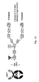
- a fully processed N glycan forms a biantennary structure with core fucose and terminal sialic acids (Fig 15).
- the majority of proteins, under physiologic conditions, carries only truncated versions of the full structure.
- the degree at which glycosylation is driven to completion depends on the cell type as well as on culture conditions. So the degree of sialinisation the addition of the terminal sialic acid to the glycan varies and does rich only 30-40 % for antibodies in human blood.
- sialinated proteins increases the half life of a therapeutic protein in blood.
- the cell lines derived from H-CB-P1, such as PBG04 generate highly sialinated glycoproteins in comparison to CHO and NS0 cells widely used in the manufacture of glycoproteins as can be seen from Example 5.
- a low content of glycans terminated before the addition of galactose (G0 structures) is advantageous. So G0 Glycoproteins tend to dimerize and the ability of antibodies to mediate complement dependent cytotoxicitiy is diminished.
- PBG04 allows a more complete processing with a low degree of G0 structures (4.3% on leptin-Fc in a roller bottle process)
- some specific structures linkages between individual sugars
- These structures affect biological features as well. Therefore, it is of advantage to use cells to manufacture glycoproteins for therapeutic applications which provide the necessary enzymes to generate human specific modifications and lack enzymes which are not present in human cells are responsible for atypical linkages.
- Such cells may be entirely human or contain a subset of human chromosomes. In the latter cells it is important that the human specific glycosylation enzymes dominate over those, not present in humans.
- So neuraminic acid may be added as N acetylneuraminic acid or N glycolyl-neuraminic acid, the latter being the major structure in mouse cells.
- N glycolyl-neuranminic acid is absent on glycans form old word monkeys and man. They are immunogenic and may lead to the formation of antibodies against the therapeutic protein.
- mouse cells contain an additional glycosylation enzyme, the alpha 1,3 galactosysltransferase. It mediates the transfer of gal residues to exposed gal residues of the glycan. Such linkage is also found in yeast and as a protection humans have pre-existing antibodies against this structure. Recognition may lead to the formation of immune complexes and kidney damage as a result of treatment.
- PBG04 derived leptin-Fc contains alpha 1,3 gal.
- a small percentage of human proteins contains bisecting N-acetylglycoseamine. It does not, per se, influence biologic features but the enzyme complex interferes with another one mediating core fucosylation.
- core fucose is missing, resulting in more efficient binding of the Fc-gamma Receptor and in enhancement of antibody dependent cellular cytotoxicity (ADCC). Therefore cells have been engineered to express (1,4)-N-acetylglucosaminyl transferase III to increase the percentage of non-core-fuccosylated proteins (US Patent 6,602,684).
- a high content of glycoproteins without core fucose can also be achieved in mouse cells.
- these proteins contain the disadvantageous features typical for mouse proteins.
- a specific hybrid cell of a human a and a mouse with the right chromosomal composition cell can combine the advantageous features of both.
- Cell lines derived from H-CB-P1 such as PBG04 are such cell lines.
- the cell line H-CB-P1 was deposited at the "Zentralinstitut für Molekularbiologie, Akademie dermaschineen der DDR", Robert-Rössle-Str. 10, Berlin Buch, DDR-1115 as ZIM-0517 on March 16, 1990 and was transferred to the DSMZ, Deutsche Sammlung von Mikroorganismen und Zellkulturen GmbH, Maschroder Weg 13, 38124 Braunschweig, Germany, on December 12, 2000 and here given the depositary number DSM ACC2104.
- the PBG03 clone D3 (pVHC ⁇ CES hobFcblas) was renamed PBG04 and was deposited at the DMSZ as DSM ACC2577 on September 18, 2002.
- Preparative PCR reactions (50 ⁇ l) were set up with the Expand High Fidelity PCR kit (Roche) according to the manufacturer's instruction (20-30 ng of template, 5 ⁇ l of 15 mM Mg Cl 2 buffer (10x), 5 ⁇ l dNTP mix, 0.5 ⁇ l of each primer (30 nM), 0.5 ⁇ l polymerase and filled to 50 ⁇ l with water).
- the PCR products were purified using a QIAquick PCR purification kit (QIAGEN).
- the PCR cycling program varied for each product according to the annealing temperature of the primer (see Table 2) and the length of the expected PCR products (determined elongation time and temperatur; see Table 1).
- Table 1 Length of the amplified fragment determines elongation time parameter length, time temperature length, time temperature length, time temperature length, time temperature length, time temperature length, time temperature length of fragment ⁇ 750 bp 1.5 kb ⁇ 3 kb > 3 kb 6 kb elongation time 45 s 1 min 2 min 3 min + 15 s after each step 4 min + 15 s after each step temperature 72°C 72°C 72°C 68°C 68°C
- Table 2 Annealing temperature for primers Primer/SEQ ID NO: Sequence (annealing temperature [°C]) Producer VHpromF/1 ATACTAGTCGGCCG CAGGCACATCCACAGTCAC (55) GIBCO BRL VHpromR/2 TCCCGGGTATCGA TGGAGCTCTCAGGGGATT
- E. coli transformants were grown in a 1 ml, 30 ml or 100 ml culture and plasmid DNA isolated using Mini-, Midi- or Maxi- plasmid purification kits (QIAGEN), respectively. The instruction of the manufacturer were followed. The DNA concentration was determined via spectroscopy, measuring the absorbance at 260 and 280 nm.
- Restriction Enzyme Digestion Plasmid DNA was digested with 1 unit of the appropriate restriction enzyme for 1 ⁇ g of DNA, using the buffer and temperature recommended by the supplier (see Table 3). If the analysis required the use of two or more restriction enzymes, the reactions were carried out simultaneous digestion if possible. Otherwise, sequential single digestions were performed with an intervening column purification step (QIAGEN) of the reaction mix. Table 3: Used Restriction Enzymes Enzyme Conc. Temp. Buffer Inact. Producer Art. No.
- End repair of DNA with 5' protruding termini To "blunt" 5' overhangs such as those produced by EcoRI, the digested DNA was treated with the Klenow fragment (Roche) according to the manufacturer's instruction. The endfilling reaction was stopped by a heat inactivation step (65°C, 20 min) and the DNA ethanol precipitated or directly subjected to gel purification.
- DNA was dephosphorylated using alkaline phosphatase (AP) (Roche) according to the manufacturer's instruction.
- AP alkaline phosphatase
- the AP was heat inactivated (65°C, 15 min) and the DNA gel purified (for electrophoresis see passage "Agarose Gel Electrophoresis” below) prior to use in a ligation reaction.
- TOPO Cloning PCR amplification products were cloned into TOPO vectors from Invitrogen. According to the instruction of the TOPO cloning kit, the purified PCR product (0.25-2 ⁇ l) was mixed with the salt solution (0.5 ⁇ l), water added to reach a volume of 2.5 ⁇ l and then the TOPO vector (0.5 ⁇ l) added. Following a 30 min incubation at room temperature, the reaction tube was transferred onto ice, 2 ⁇ l of the reaction added to "one shot chemically competent E. coli" and the cells incubated for 30 min on ice. The cells were heat-shocked (42°C, 30 s), immediately transferred back onto ice and 250 ⁇ l of room temperature SOC medium added. The transformation reaction was incubated for 1 h at 37°C with shaking (300 rpm) before the mixture was plated on LB plates containing either Kanamycin or ampicillin. The plates incubated of overnight at 37°C.
- Ligation All ligation reactions were carried out in 10 ⁇ l volumes with 0.1-1 ⁇ g of dephosphorylated vector and an excess of insert.
- the reaction contained 2 ⁇ l T4 ligation buffer (Gibco BRL) and 1 ⁇ l T4 ligase (Roche), and were incubated for two hours at 16°C or overnight at 4°C.
- the ligation reaction was transformed into bacteria.
- ligations could be post-digested with a suitable restriction enzyme if there was a unique restriction site in the self-ligated vector.
- the ligation reaction was ethanol precipitated in the presence of acrylamide (centrifugation at 4°C, 14,000 rpm, 15 min) before the re-dissolved DNA was used again in a transformation reaction
- Competent E . coli XL2 (stored at -70°C) were thawed on ice, mixed with either the ligation reaction (also kept on ice, see passage "Ligation” above) or with 1-100 ng plasmid DNA (re-transformation) and incubated on ice for 20 min. Subsequently the transformation reaction was heat-shocked (30-60 s, 42°C), the tube returned onto ice and 205 ⁇ l SOC medium (free of any antibiotic) added and the reaction incubated shaking (300 rpm) at 37°C for 45 min.
- the transformation reaction was plated on LB plates containing an antibiotic (either kanamycin (40-60 ⁇ g/ml) or ampicillin (50-100 ⁇ g/ml)) and incubated overnight at 37°C. The bacterial colonies were counted and the efficiency of the transformation reaction calculated.
- an antibiotic either kanamycin (40-60 ⁇ g/ml) or ampicillin (50-100 ⁇ g/ml)
- DNA fragments were separated according to their length on 0.7-1.5% agarose gels.
- the agarose was dissolved in 1xTAE buffer and 2 ⁇ l ethidiumbromide/100 ml agarose added. When the agarose had dissolved, it was poured into a tray and allowed to set.
- the DNA sample was mixed with the loading buffer Orange G, loaded onto a horizontal gel and run at 40-90 V with 1xTAE as running buffer. The DNA/ethidium bromide complexes were visualized under UV light.
- Trypsinisation Adhesive cells were harvested using trypsin. First the culture medium was removed and the cell monolayer washed with citrate buffer (pre-warmed to 37°C). A small amount of trypsin was added directly to the cell monolayer and incubated for 3-5 min at 37°C. Trypsinisation was stopped by addition of PBG 1.0 medium supplemented with 5% FCS (see Table 4). The cell suspension was transferred into a Falcon tube (50 ml) and centrifuged for 10 min (800 rpm, 30°C). The cell pellet was re-suspended in fresh medium and the cells used for electroporation or for further passaging.
- Table 4 Volumes of citrate, trypsin and PBG 1.0 supplemented with 5% FCS used for the trypsinisation of the cells growing in different flasks Tissue culture flask Citrate buffer (ml) Trypsin (ml) PBG 1.0/5%FCS (ml) T25 1.0 0.5 4.5 T85 2.0 1.0 9.0 T180 5.0 2.0 8.0
- the cells After the cells had been trypsinized they were counted in a Neubauer chamber (haematocytometer). A small volume of the cell suspension was introduced into the chamber and the chamber placed under a microscope. Only cells within one of the four squares of the chamber were counted, the cell number multiplied with the factor 10 4 to obtain the number of cells per ml. To differentiate between vital and dead cells, the cells were stained with trypan blue prior to counting. Dead cells appeared blue whereas vital cells did not take up the dye.
- Electroporation of H-CB-P1 Cells In a standard electroporation reaction 10 ⁇ g of linearised plasmid DNA were used. The culture medium was removed and the H-CB-P1 monolayer washed with citrate buffer and trypsinised (see passage "Trypsinisation” above). The cell pellet was re-suspended in Opti-MEM (pre-warmed to 37°C) to obtain 3 x 10 6 cells per ml. A volume of 700 ⁇ l of the cell suspension was transferred into the electroporation cuvette (peqLab; EQUBIO 4 mm) and the linearised DNA (10 ⁇ g) added. The cells were electroporated at 250 V, 1500 ⁇ F and immediately afterwards transferred into T75 bottles containing pre-warmed PBG 1.0 medium supplemented with 5% FCS, and incubated at 37°C and 5% CO 2 .
- peqLab electroporation cuvette
- H-CB-P1 Cells The electroporated H-CB-P1 cells were cultured for two days at 37°C and 5% CO 2 . On day 2, the culture medium was removed and non-adhesive cells harvested by centrifugation of the culture medium. 1 ml of the culture medium (supernatant) was frozen for later examination of transient expression. The trypsinized cell monolayer and the cells harvested from the culture medium by centrifugation were combined and pelleted. Cells were re-suspended in PBG 1.0 medium supplemented with 5% FCS to obtain dilutions of 1x10 6 , 1x10 5 and 1x10 4 cells/ml.
- Each dilution was supplemented with 5 ⁇ g/ml or 10 ⁇ g/ml blasticidin or with 200 ⁇ g/ml or 400 ⁇ g/ml hygromycin.
- the selection medium was changed on days 4, 7 and 10, and the growth of the H-CB-P1 clones controlled via microscopy. Between days 8 and 10 vital clones were visible by eye. On day 13 clones were harvested by trypsinisation, the cell pellet suspended in PBG 1.0 medium so that when cells were seeded into 96 well plates, a well contained either 5 cell or 1 cell.
- the selection pressure (either 5 or 10 ⁇ g/ml blasticidin or 200 or 400 ⁇ g/ml hygromycin) was maintained throughout.
- the cells were immuno-stained to differentiate between positive and negative cell clones.
- the cell culture medium was removed and replaced with standard medium supplemented with a fluorecently labeled antibody (2 ⁇ g/ml) recognizing the recombinant protein produced by the cells.
- the antibody suspension was left for 4 h on the cell monolayer before it was replaced with OptiMem 1 supplemented with 5% FCS.
- the cell monolayers were examined under a fluorescent microscope. When Texas red conjugated antibodies were used, the microscopy was done with a UV filter (WG) that is transmissible for 470-480 nm spectra.
- the excited Texas red labeled antibodies emit light in the spectra of 590 nm.
- a UV filter (WU) transmissible for spectra of 330-355 nm was used for visualization of antibodies conjugated to AMCA. Emission of excited AMCA occurred in the blue spectra (420 nm). Only clones that were large and strongly fluorescent were considered. Was there only a single clone in one well, the cells were further expanded. Was there more than only one clone in a well, the individual clones (cells) were picked with a microcapillar and transferred into a new 96 well plate for further expansion (see the following passage "Mircopillary Picking").
- the microcapillary picking device used a capillary attached to a movable arm that was controlled via a joystick.
- the microcapillar and the arm were inside the hood wheras the joystick was controlled from outside.
- an interesting clone was identified via the immunostaining technique (described in section "Selection of H-CB-P1" above)
- the microcapillary was placed over the clone, negative pressure induced within the capillary through a vacuum pump and the cell pile of interest sucked into the microcapillary.
- the arm was moved over the fresh well of a 96 well plate and the cells therein ejected.
- EC-Western-Blot (Enhanced Chemiluminscence): For detection of hobFC antibodies 20 ⁇ l of cell culture supernatant were mixed with 10 ⁇ l 5% SDS and incubated for 2 min at 97°C. Was the cell culture medium expected to contain IgM antibodies, the culture medium was not treated. A membrane (Amersham-Pharmacia; Hybond-P) was first rinsed in methanol (1 min), washed three times in water (1 min), and then soaked in plot transfer buffer before placing it on a piece of 3 MM paper also soaked with plot transfer buffer.
- Example 1 Preparation of a targeting vector specific for the IgM region of H-CB-P1 cells
- the recombinant gene was to be inserted into the IgM sequence region because it is well-known that antibodies are highly expressed and secreted proteins.
- the final targeting vector hence required sequences that had 100% homology to the targeted genomic IgM sequences.
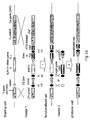
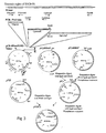
- the basic targeting vector pVHC ⁇ the VH region of 2 kb and the C ⁇ intron region of 7.4 kb in length were chosen (see Fig. 3). Both regions were isolated as PCR fragments using polymerases with proof-reading activity (proofstart polymerase (Qiagen)), subcloned into a pCR 4BluntTOPO vector (Invitrogen) and finally combined in one vector named pVHC ⁇ (see Fig. 3).
- the basic targeting vector pVHC ⁇ does not have an endogenous cassette yet.
- the endogenous cassette containing the CE promoter, the place holder gene hobFc, three FRT recombination sites as well as a hygromycin resistance gene and an ATG deleted neomycin gene was isolated from the vector pCESHhobFc as a BstDI-SwaI fragment. The fragment was then endfilled with Klenow polymerase and ligated into the basic targeting vector pVHC ⁇ that had been digested with PmeI and dephosphorylated. The resulting targeting vector was called pVHC ⁇ CESHhobFc.
- the vector pcDNATRD was used as donor for the blasticidin gene.
- the first step involved the exchange of the hygromycin gene with the blasticidin gene.
- the blasticidin gene was isolated from pcDNATRD as an EcoRI-SalI fragment.
- the endogenous cassette vector pcESHhobFc was also opened with EcoRI-SalI and thereby the hygromycin gene was removed as well as part of the ATG-deleted neomycin gene.
- the fragment containing the blastidin gene was ligated into the opened pCESHhobFc and the resulting vector named pCEShobFcblasdeleted.
- the second step was the re-insertion of the coincidentally removed ATG-deleted neomycin gene.
- a fragment encompassing the ATG-deleted neomycin gene was isolated from pCESHhobFc and inserted into the opened vector pCEShobFcblasdeleted.
- the resulting vector was named pCEShobFcblas.
- This vector now served as donor vector for the blasticidin gene for plasmids pVHC ⁇ CSHhobFc and pVHC ⁇ HhobFc.
- a BamHI-SalI fragment was isolated from pCESHhobFcblas and inserted into a BamHI-SalI opened vector pVHC ⁇ CESHhobFc, giving rise to pVHC ⁇ CEShobFcblas.
- the vector pVHC ⁇ HhobFc was also digested with BamHI-SalI and again the BamHI-SalI fragment isolated from pCEShobFcblas ligated into it.
- H-CB-P1 cells were electroporated with plasmids pVHC ⁇ CshobFcblas, pVHC ⁇ hobFcblas, pVHC ⁇ HhobFc and pCShobFcblas.
- plasmids pVHC ⁇ CshobFcblas, pVHC ⁇ hobFcblas, pVHC ⁇ HhobFc and pCShobFcblas.
- cells were seeded at 10 4 , 10 5 or 10 6 cells/20 ml into T75 flasks two days post-electroporation.
- the medium was supplemented with either 5 or at 10 ⁇ g/m blasticidin,or 200 or 400 ⁇ g/ml hygromycin.
- the number of clones per cm 2 was determined. The highest number of clones was obtained in flasks seeded with at 1 x 10 6 cells that had been transfected with the plasmids lacking the CES promoter (pVHC ⁇ hobFcblas). Three times fewer clones were obtained when cells had been transfected with plasmids carrying the CES promoter. Furthermore these clones did grow less well as those without the CES promoter.
- the culture medium contained either 5 or 10 ⁇ g/ml blasticidin or 200 or 400 ⁇ g/ml hygromycin.
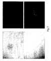
- cells were stained with Anti IgG antibodies conjugated with Texas red and AMCA labeled antibodies against IgM. The results obtained with cells cultured with blasticidin showed that with the higher antibiotic concentration far fewer positive clones were obtained.
- the rearranged immunoglobulin heavy chain locus was assembled based on the cDNA sequence of the antibody and human genome sequence information.
- the complete leader sequence including the ATG, the V, D and J genes were omitted from the targeting vector and deleted from the genome via a single homologous recombination event.
- the replacement type targeting vector contained isogenic sequences from the IgM locus to allow the directed cross over as well as the hobFc gene, blasticidin and ATG deleted neomycin resistance genes.
- PCR for detection of integrated targeting sequences To verify that the targeting cassette had become integrated at the IgM locus PCR reactions were set up with the forward primer V5 (SEQ ID NO:7) and reverse primers V6 (SEQ ID NO:8) or V7 (SEQ ID NO:9), and genomic DNA isolated from cell clones as template.
- Primer V5 binds to the genomic V gene promoter sequences outside of the fragment Vhprompresent in the targeting vector
- reverse primer V6 binds within the CES promoter sequences (and hence was only used for cells transfected with plasmids carrying the CES promoter) and primer V7 bind within the hobFc gene.
- Clone pVHC ⁇ CEShobFcblas D3 (PBG04) was transfected with vector 2 comprising a second functional sequence (frtwt, GFP ORF and polyadenylation signal, minimal promoter followed by ATG and frt5) and plasmid pflp comprising a functional expression unit for flp recombinase using the transfection reagent effectene (Qiagen).
- Vector 2 does not contain a promoter driving the GFP expression unit.
- the functionalised cell (PBG04) is sensitive to Geneticin selection from 200 ⁇ g/ml. Vector 2 misses a neomycin resistance gene sequence which could confer resistance to Geneticin. As expected no green fluorescence was detectable 1-4 days after transfection.
- GFP expression depends on integration in direct proximity to a functional promoter. Geneticin resistance is dependent on reconstitution of the neo resistance gene present in the cell line by the ATG from vector 2. We conclude that in all cases vector 2 has replaced sequences between the frtw and frt F5 sites and functionally linked the CES promoter and GFP as well as the ATG deleted neomycin gene with the ATG.
- Leptin Fc from PB604 was generated in roller bottle culture and purified by a generic process including affinity chromatography, gel filtration and membrane filtration.
- the protein was digested with trypsin and the resulting peptides were deglycosylated by PNGase F digestion.
- the glycans were labeled with 2-aminobenzamide and separated by HPLC on a Phenomenex Hypersil APS-2-column (Fig.16).
- MALDI-TOF-MS (BRUKER BIFLEX TM ) was used with the desialylated, labelled samples to further characterize the respective fractions shown in Table 5.
- the single N-glycosylation site on Fc carries complex oligosaccharide structures which are sialylated at 37% , a rate close to average sialylation on antibodies in human blood.
- Sialic acids were further characterized by sialidase treatment, DMB labelling and separation on a Bischoff Hypersil-ODS-column and compared with the Sialic Acid Reference Panel (Oxford GlycoSciences).
- N-acetylneuraminic acid was found. Only 2% were represented by N-glycolylneuraminic acid, the dominating form in mouse myeloma cells; which was shown to be immunogenic (Noguchi, A. et al., J. Biochem, 17(1):p.
- Example 6 Preparation of a targeting vector for the light chain lambda locus of PBG04
- the structure of the rearranged lambda chain locus was identified by alignment of the known cDNA of the lambda gene with human genomic sequences.
- the gene consists of a variable gene, J and H segment already joined together. V3-19 was identified as the variable gene. This approach was not suitable to identify the constant gene because the locus contains 100% identical gene copies.
- a PCR based on primers in the known leader sequence and in the unique intervening sequences between constant region genes. Primers V81, V83 (SEQ ID NOs:13 and 14, respectively) gave a correctly sized PCR product which showed the expected restriction pattern and therefore allowed to identify J2 and H2 as the genes constituting the rearranged lambda gene of H-CB-P1 (SEQ ID NO:21).
- a 5' flank upstream of the coding sequences of the variable gene V3-19 was created with Primers V89 and V94 (SEQ ID NOs:15 and 18, respectively) using Provestart Polymerase (Qiagen).
- a 4kb fragment was cloned in pPCR4blunttopo (Invitrogen).
- a 3 prime flank was amplified in two steps: overlapping PCR products were created using Primers V90 V91 and V115 V116 (SEQ ID NOs:16, 17, 19 and 20, respectively) and lined via a unique SphI site present in both fragments.
- flanking sequences were cloned into a single vector PVLCL (SEQ ID NO:22).
- PVLCL Vector-like cell line
- SEQ ID NO:22 The flanking sequences were cloned into a single vector PVLCL (SEQ ID NO:22).
- SEQ ID NO:22 The flanking sequences were cloned into a single vector PVLCL (SEQ ID NO:22).
- clones were isolated by dilution cloning and clones resulting from homologous exchange were identified by the absence of an immunoflourescence signal using a fluorescence-labelled anti human-lambda-chain antibody.
- the resulting cell clones are analyzed for the expression of alpha 1 antitrypsin.
- These clones are suitable for the coexpression of two independent transgenes which have to be expressed at high level.
- these genes are the heavy and light chain genes of an antibody.
- heavy and light chain genes can be directed to their respective locations.
Landscapes
- Health & Medical Sciences (AREA)
- Genetics & Genomics (AREA)
- Life Sciences & Earth Sciences (AREA)
- Engineering & Computer Science (AREA)
- Zoology (AREA)
- Chemical & Material Sciences (AREA)
- Bioinformatics & Cheminformatics (AREA)
- Biomedical Technology (AREA)
- Wood Science & Technology (AREA)
- Biotechnology (AREA)
- General Engineering & Computer Science (AREA)
- Organic Chemistry (AREA)
- Molecular Biology (AREA)
- Plant Pathology (AREA)
- Microbiology (AREA)
- Biophysics (AREA)
- Physics & Mathematics (AREA)
- Biochemistry (AREA)
- General Health & Medical Sciences (AREA)
- Mycology (AREA)
- Cell Biology (AREA)
- Micro-Organisms Or Cultivation Processes Thereof (AREA)
- Preparation Of Compounds By Using Micro-Organisms (AREA)
- Peptides Or Proteins (AREA)
- Measuring Or Testing Involving Enzymes Or Micro-Organisms (AREA)
Abstract
Description
- The present invention relates to ubiquitous/universal processes for establishing cells capable of stable high yield expression of a recombinant gene with human glycosylation pattern, and for establishing stable universal precursor cells available for insertion of arbitrary target genes. The invention further relates to the cells obtainable by said processes.
- Recombinant protein production is of central importance for different applications. Structural studies of proteins (rational drug design and drug optimisation are based thereon (Antivir. Chem. Chemother., 12 Suppl. 1, 43-49 (200:1))), industrial applications of proteins (enzymes) and clinical use of recombinant proteins have increased the need for their efficient production. As of February 2000, according to a survey by the Pharmaceutical Research and Manufacturers of America, 122 biologics, including 20 monoclonal antibodies were either in phase III trials or awaiting FDA approval (K. Garber, 2001, Nature Biotech. 19, 184-185).
Depending on the application, native conformation and correct posttranslational modifications (such as glycosylation) of the recombinant protein are essential. Prokaryots such as the biotechnology "pet" organism Escherichia coli (E. coli) lack the ability to introduce posttranslational modification. Only eukaryotic cells possess the cell machinery necessary for co-translational and post-translational modifications as they are often required to produce functionally active proteins. Various eukaryotic systems for the production of a variety of heterologous proteins exist. Fungal expression systems, using e.g. derived from the genus Saccharomyces, Candida, Pichia, Hansenula, Aspergillus or Kluyveromyces are well established (Hollenberg and Gelissen (1997), Current Opinion in Biotechnology 8, 554-560). To circumvent the problem of plasmid instability sometimes encountered in fungi, sequences coding for heterologous proteins are ideally integrated into the fungal chromosome via homologous recombination. Further problems encountered with fungal expression systems are overglycosylation of heterologous proteins and incorrect folding such as incorrect oligomerisation and insufficient ligand incorporation. Expression of heterologous proteins in insect cells - the DNA encoding the heterologous protein can also become integrated into the chromosome via recombination - gets around these problems. However, insect cells lack the ability to produce sialic acid and sialic glycans. Terminal sialic acid residues play divers biological roles in many glycoconjugates. Plants can also be used for the production of recombinant proteins. However, in these heterologous expression system difficulties in extraction and purification prove real bottlenecks. - Mammalian expression system, cultured cells as well as transgenic animals have none of these disadvantages. However, the posttranslational modifications vary between the mammalian species. Thus, the expression of human proteins in rodent cells (e.g. as disclosed in Sun, W. et al., J. Immunol. 152, p. 695-704 (1994); Lang, P. and Mocikat, R., Mol. Gen. Genet. 424, p 528-538 (1994); WO 96/30498, and Fukuta, K. et al., Arch. Biochem. Biophys. 378, p. 142-150 (2001) results in a human protein with rodent glycosylation pattern.
- Recombinant proteins can be produced in cultured mammalian cells either transiently or constitutively (stably). For transient expression of recombinant a vector DNA encoding the recombinant protein is introduced into the cell and in general is not integrated into the cellular DNA. Expression titers of the recombinant protein are at the beginning high. However, since the vector DNA is generally not replicated, the vector DNA becomes diluted with each cell proliferation and hence the expression titer drops. Only rarely a vector DNA or part of the vector DNA illegitimedly recombines with the cellular genomic DNA and the gene encoding recombinant protein is stably integrated into the genome. If the gene encoding the recombinant protein is associated with a selection marker, cells carrying this cassette can be identified and isolated as stably transformed cells. Stable transformants have the advantage that the heterologous proteins are continuously produced. The expression titer is mainly determined by the strength of the promoter construct, the site of integration into the chromosome, the copy number and the type of recombinant protein in question. Many strong promoters are commercially available, however, their transcriptional activity varies depending on the cellular level of the relevant transcription factors and on the chromatin structure at the integration site. For example integration within the scaffod- or matrix attachment regions (S/MAR elements) of chromosomal DNA can augment the activity of promoters -and hence the expression of heterologous genes- and protect them from inactivation by the flanking chromatin. Therefore, it is highly desirable to chose a promoter highly active in a specific cell and to direct integration into an active part of the chromosome. Preferentially a single integration event is desirable, since heterologous genes at low copy number are in general expressed more stable than multicopy genes.
- Integration at a single preselected highly active locus can be achieved via homologous recombination. This method, although typically applied to mouse embryonic stem cells, is extremely inefficient in somatic cells of human origin and requires a large scale screening effort. Moreover it is not applicable for most human permanent cell lines when it is desired to completely shut off the expression of a given target gene, because these cell lines are usually polyploid and targeting more than 2 identical loci is hardly feasible. Site specific recombination using recombinases, eg. Cre, flp, C13 and their respective target site (RRS) are a viable alternative (Feng, Y.Q. et al., Journal of Molecular Biology, vol. 292(4), p. 779-785 (1999); Schlake, T. et al., Biochemistry, Am. Chem. Soc., vol. 244(1-2), p. 185-193 (October 2000); Fussenegger, M. et al., Trends in Biotechnology, vol. 17(1), p. 35-42 (January 1999); Groth, A.C. et al., Proceedings of the National Academy of Sciences of USA, vol. 97(11), p. 5995-6000 (May 2000)). With this approach, a plasmid carrying a single RRS can be used to target a single RRS in the chromosome. This method, however, has certain limitations: Namely, it is quite inefficient because the reverse reaction, excision of the plasmid, is an intermolecular recombination and takes place at much higher speed than the integration. Secondly, the whole plasmid including bacterial genes are integrated. To solve the first problem unidirectional was established, e.g. by meains of heterospecific target sites for both flp and cre. These RRS are recognised by the respective recombinase but a successful recombination requires identical sites and the excision reaction is precluded (Karreman S. et al., Nucleic Acids Res., vol 24(9), p. 1616-1624 (1996); Trinh, K.R. et al., J. of Immunol. Methods, vol. 244, p. 185-193 (2000)). However, the targeting plasmid still has to be integrated into a single favourable position of the chromosome. A large scale screening effort is required to find such rare integrates. These clones often contain more than one copy of the plasmid, the may contain incomplete copies and bacterial sequences care not precluded from integration. These bacterial sequences are recognized by the mammalian cell often leading to inactivation of the targeted region. Alternatively, the targeting cassette may be integrated via retroviral vectors (Karreman S. et al., Nucleic Acids Res., vol 24(9), p. 1616-1624 (1996)). These vectors target active sites within chromosome, only full length cassettes are integrated and the infection dose can be adjusted to create single integration sites. However, expression units flanked by ITRs may also be subject to inactivation. In addition, the use of this system may be restricted by the governmental release agencies to exclude therapeutic applications of the expressed protein. Finally, Karpas A. et al., PNAS 98(4), 1799-1804 (2001) discloses a human myeloma cell line suitable for the generation of hybridomas that stably express immunoglobulins at high yield. On the other hand, is mentioned that myeloma are generally not suitable for that purpose.
- In view of the above, there is still a need for a method allowing the transformation/conversion of a cell line with an arbitrary gene coding for a product of interest to obtain a high yield recombinant human glycoprotein producing cell, especially for a method without or only little cumbersome screening procedures. It was surprisingly found that cells expressing recombinant glycoproteins with features of human posttranslational modification at high yield are obtainable by first identifying a non-essential highly expressed cellular gene (hereinafter shortly referred to as "starting gene") in a human or essentially human hybrid cell (hereinafter shortly referred to as "starting cell"); secondly directly replacing the starting gene via homologous recombination with a first functional DNA sequence (e.g. by utilizing an appropriate targeting cassette) containing recombinase recognition sites (RRSs) for site-directed integration and optionally a "place-holder" gene comprising various functional sequences and selecting/isolating a stable clone of this precursor expression cell (functionalized cell); thirdly introducing the gene of interest (from here on called "target gene") coding for the target gene product (protein) by site-directed integration using a recombinase recognizing the RRSs incorporated with the first targeting cassette; and finally selecting/isolating a stable expression cell capable of producing large amounts of the recombinant protein. Direct replacement of the starting gene with a functional DNA sequence containing a DNA sequence coding for the target gene product is also applicable.
- It was moreover found that suitable starting cells for the above method are specific mammalian cells such as human myeloma and hybridoma cells and human heterobybridoma cells (including human-mouse hetero-hybridoma cells such as H-CB-P1), which allow the production of proteins having an essentially human glycosylation pattern.
- Using the present invention it is possible to introduce stably any gene encoding a recombinant protein of interest into the specific mammalian cells set forth above. Using the present invention the gene of interest encoding the recombinant protein will become integrated into the locus of a highly expressed cellular gene and preferably in close proximity to a highly active cellular promoter residing in an active part of the chromosome. Using the present invention precursor cell lines of various origin can be created carrying a place holder gene surrounded by RRSs. Using the present invention the place holder gene can be exchanged with the gene of interest, encoding the recombinant protein, by site-specific recombination at the RRSs catalyzed by a suitable recombinase, giving rise to the final high-yield expression cell.
- Finally, it was found that the human-mouse heterohybridoma provides for a very distinct human glycosylation pattern.
- More specifically, the present invention provides
- (1) a process for preparing cells capable of stable high yield expression of a target gene product having essentially human glycosylation pattern which method comprises
- (a) selecting an immortalized human cell or human hybrid cell (hereinafter "starting cell") which is derived from B lymphocytes and is capable of stable high yield expression of a starting gene product being non-essential to the starting cell;
- (b) screening for the locus of the starting gene product within the genome of the starting cell;
- (c1) replacing the gene coding for the starting gene product with a first functional DNA sequence containing one or more recombinase recognition sites (RRS) to obtain a functionalized precursor cell; and
- (d) integrating a second functional DNA sequence containing a DNA sequence coding for the target gene product into the functionalized precursor cell obtained in step (c1) by use of a recombinase recognizing the RRSs incorporated with the first functional sequence, or
- (c2) directly replacing the gene coding for the starting gene product with a functional DNA sequence containing a DNA sequence coding for the target gene product;
- (2) in a preferred embodiment of the process of (1) above the starting cell is a human-mouse hetero-hybridoma such as hetero hybridoma H-CB-P1 (DSM ACC 2104)) and integration of the functional DNA sequence(s) is effected at a Ig locus (preferably at a rearranged human Ig locus of said cell);
- (3) a cell capable of high yield expression of a target gene product obtainable by the process of (1) or (2) above;
- (4) a process for preparing a functionalized cell comprising the steps (a) to (c1) as defined in (1) or (2) above;
- (5) a precursor cell obtainable by the process of (4) above; and
- (6) a method for high yield expression of a target gene product which comprises cultivating a cell as defined in (3) above.
-
- Fig. 1: Concept overview, multistep process to create high yield expression cell lines comprising site-specific integration of genes into an IgH locus at frt sites. The IgH locus of H-CB-P1 is represented in the upper graph of figures 1a and 1b. It contains the variable gene promoter followed by a protein leader sequence and specific V, D, and J genes rearranged and positioned next to the enhancer Eµ, the MAR and the Cµ coding sequences. Target sequences for homologous recombination are shown marmorate. Via homologous recombination between the flanking sequence elements "Vhprom" and "Cµ" of vector 1 (targeting vector) and the genomic DNA, first functionalized sequences located between the flanking sequences are introduced into the genomic DNA and a recombinant PBG03 genome is the result. The first functionalized sequence contains frt sites (frtF5 frtF3 and frt wt), an artifical strong promoter CES (Fig. 1a) or no additional promoter (Fig. 1b) upstream of the first expressed gene(hobFc). In addition a blasticidine or hygromycin resistance gene and an ATG deleted neomycin gene are part of the first functionalized sequence. The recombinant genome carries the first functional sequences integrated in the chromosomal DNA.
FLP recombinase catalyses recombination at frtF5 and frtF3 or at frt wt and frtF3 sites of the recombinant H-CB-P1 genome andvector 2, the actual gene of interest is introduced and expressed from the artificial or the endogenous VH promoter (Fig. 1a and 1b, respectively). Parts of the first functionalized sequence located between frtwt and frt F3 sites are replaced by a weak promoter followed by an ATG which after recombination is positioned in the same open reading frame as the ATG-deleted neomycin gene. The resulting genome has lost the hobFc gene and the blasticidin or hygromycin resistence genes and instead has the gene of interest (target gene) and a functional neo gene. - Figure 2: The endogenous cassette (CEShobFcblas) of the targeting vector.
The detailed structure of the endogenous cassette containing an CES promoter, a hobFC fusion gene, a blasticidin resistance gene and a start codon (ATG) deficient neomycin gene is shown. In more detail, the endogenous sequence contains a modified frt site (frtF5) followed by a hybrid promoter structure comprising the early CMV promoter/enhancer elements as well as the first intron of the elongation factor alpha gene. The next element is a frt wildtype site followed by the hobFC fusion gene (hobFC), and the SV40 polyadenylation signal (SV40PA). A weak SV40 promoter controlling the expression of the blasticidin resistance gene follows. The last elements are a modified frt site (frtF3) and next to it the ATG deficient neo gene. Modified frt sites F3 and F5 allow recombination with identical sites but not with wildtype frt sites and F5 or F3 sites respectively. The frt F3 site is positioned to upstream of the neo gene to form a contiguous open reading frame lacking the ATG. - Fig. 3: Cloning strategy for the intermediate vector pVCµ containing the flanking regions Vhprom and Cµ.
The 2 kb VH promoter sequence was amplified from PBG03 cell genomic DNA using forward primer VHpromF (SEQ ID NO:1) and reverse primer VHpromR (SEQ ID NO:2). The PCR product VHprom was cloned into the pCR 4BluntTOPO vector (Invitrogen) and the resulting vector named pVH. The 7.4 kb Cµ region was amplified from genomic H-CB-P1 DNA as two overlapping fragments, namely CµmitteR and CµMitteF. The primers CµintV (SEQ ID NO:3) and CµMitteR (SEQ ID NO:5) give rise to the product CµMitteR and the primers CµMitteF (SEQ ID NO:6) and the reverse primer CµintR (SEQ ID NO:4) produce the fragment CµMitteF. Both fragments were cloned into a pCR 4BluntTOPO vector (Invitrogen) and the resulting vectors called pCµMitteR and pCµMitteF. The full length Cµ sequence was re-established by opening both vectors with the restriction enzyme SpeI and DraIII, and ligating the SpeI-DraIII fragment from pCµMitteR into the opend pCµMitteF. The resulting vector pCµ carries the full-length Cµ region (Cµ intron). The VH promoter sequence and the Cµ sequence were combined in one vector pVHCµ, by digesting both pVH and pCµ with the restriction SpeI and PmeI, and inserting the isolated Vhprom PmeI-SpeI fragment into the phosphatase treated opend vector pCµ. - Fig. 4: Cloning strategy for the targeting vectors pCESHhobFc and pVHCµHhobFc. The targeting vector pVHCµCESHhobFc containging the highly active promoter CES and the hobFC fusion gene was created by ligating an end-filled SwaI-BstBI fragment isolated from pCESHhobFc into the pVHCµ vector that had been digested with PmeI and dephosphorylated. The targeting vector pVHCµHhobFc that has the hobFC fusion gene but no CES promoter, was prepared by ligating an endfilled Bst1107-BstBI fragment isolated from pCESHhobFc into an PmeI digested and dephosphorylated vector pVHCµ.
- Fig. 5: Cloning strategy for targeting vectors carrying a blasticidin resistant gene, pVHCµCEShobFcblas and pVHCµhobFcblas.
The plasmid pcDNATRD was used as donor for the blasticidin gene. To delete the hygromicin gene sequences as well as the FRT5 and neomycin sequences from the vector pCESHhobFc, the vector was digested with EcoRI and SalI and dephosphorylated. An EcoI-SalI fragment from pCDNATRD containing the blasticidin resistant gene sequence was ligated into the previously opened pCESHhobFc vector and the resulting plasmid named pCEShobFcblasdeleted. The Frt F5 sequence and the ATG-deleted neomycin sequence were isolated from pCESHhobFc as a SalI SalI fragment and re-inserted into the SalI site of pCEShobFcblasdeleted. The resulting plasmid pCEShobFcblas was used together with pVHCµCESHhobFc to create pVHCµCEShobFcblas. The BamHI-SalI fragment comprising the hobFc sequence and the blasticidin gene was isolated from pCEShobFcblas and inserted into vector pVHCµCESHhobFc opened with BamHI and SalI, giving rise to pVHCµCEShobFcbfas. The vector pVHCµhobFcblas was created by ligating a BamHI-SalI fragment containing the hobFc gene and the blasticidin gene into vector pVHCµHhobFc digested with BamHI and SalI. - Fig. 6: Immunostaining of H-CB-P1 clones
H-CB-P1 clones obtained through transfection of H-CB-P1 cells with pVHCµ CESHhobFcblas were immunostained with a Texas Red conjugated anti-human IgG, Fy fragments specific antibody isolated from goats or an AMCA conjugated anti-human IgM, Fc5µ specific antibody isolated from goats. The left column shows two H-CB-P1 clones stained with the Texas Red conjugated antibody and visualized with an UV WG filter. In the right column the same clones are shown after staining with the AMCA conjugated anti-IgM antibody and visualized under UV filter WU. Whereas for the clone in the upper panel only IgG staining is evident, staining with both antibodies is present for the clone in the lower panel. The first clone may result from homologous recombination whereas the other clone contains an illegitimate insertion of the functional sequences. - Fig. 7: Direct immunostaining of H-CB-P1 clones and further expansion of clones.
It is demonstrated that H-CB-P1 clones could be immunostained without jeopardizing the cells viability and that subsequent expansion of the stained clone was possible. A H-CB-P1 clone cultured for ten days in a 96 well plate was immunostained with an Texas Red conjugated anti-IgG antibody. A picture of the clone prior to immunostaining visualized with normal light microscopy, is shown in the top left panel. The same clone immunostained with the Texas Red conjugated anti-IgG antibody is shown in the top right hand panel. The bottom panels show pictures of the well post trypsinisation. No cells can be seen when the picture was taken under a normal light microscope, as shown in the bottom left hand panel. The right hand panel shows the same well examined under UV filter WU. The cells are completely removed from the well and the fluorescent antibody precipitate remained in the well and is not attached to the cell surface. - Fig. 8: Ant-IgM dot blot of cell culture supernatants from induvidual clones
Supernatant of the following clones
1: pVHCµhobFcblas-D6; 2: pVHCµhobFcblas-G8;
3: pCEShobFcblas-A3; 4: pVHCµCEShobFcblas-B4;
5: pVHCµCEShobFcblas-D3; 6: pVHCµCEShobFcblas-G8 were spotted onto a membrane and subjected to the ECR staining method. Since the starting cell population homogenously produces IgM, clones with no detectable IgM expression result from inactivation of the IgM H gene mediated by the targeting vector.
The clone A3 generated by transfection of pCESHhobFcblas which lacks homologous flanking sequences was unable to target the IgM locus and expresses IgM. - Fig. 9: Anti-IgG dot blot of cell culture supernatants from individual clones.
Supernatant from the following clones
1: pVHCµhobFcblas D6; 2: pVHCµhobFcblas D6 (1:2 diluted); 3: pVHCµhobFcblas-G8; 4: pVHCµhobFcblas G8 (1:2 diluted); 5: pCEShobFcblas A3; 6: pVHCµCEShobFcblas B4; 7: pVHCµCEShobFcblas D3; 8: pVHCµCEShobFcblas D3 (1:2 diluted);
9: pVHCµCEShobFcblas D3 (1:10 diluted); 10: pVHCµCEShobFcblas G8;11:hobFc standard 500 ng/ml; 12: hobFc standard 50 ng/ml IgG
were spotted onto a membrane and subjected to the ECR staining procedure using an anti-IgG antibody. - Fig. 10: Detection of homologue recombination via PCR
To test whether a homologue recombination event had occurred between the (targeting) vector and the Ig locus of H-CB-P1 cells, a PCR strategy was applied. Upon recombination the endogenous cassette of the vector containing the CES promoter, the hobFc gene and a resistance gene (hygromycin or blasticidin) followed by the ATG-deleted neomycin gene_ becomes integrated between the genomic V gene promoter sequences and the enhancer Eµ. The forward primer V5 (SEQ ID NO:7) which binds to the genomic V gene promoter sequences outside of the fragment Vhprom was combined with primers V6 or V7 (SEQ ID NOs:8 and 9, respectively), which bind specifically within the first functional sequence. The occurrence of PCR products is strictly dependent on co-localisation of both primer binding sequences and hence homologous recombination. To confirm any positives the putative positiv PCR product was used in a nested PCR reaction with the primers VHpromF and VHpromR (SEQ ID NOs:1 and 2, respectively).
a: primer position, b: electrophoretic analysis of PCR products- left: first PCR V5, V7
lane 1: 1kb ladder (Invitrogen); lane 2: clone pVHCµhobFcblas H6; lane 3: clone pVHCµCEShobFcblas B10; lane 4: clone pVHCµhobFcblas D4; lane 5: clone pVHCµhobFcblas D8; lane 6: clone pVHCµhobFcblas E11; lane 7: neg control H-CB-P1 - right: nested PCR
lane 1: 1kb ladder (Invitrogen); lane 2: neg control H-CB-P1; lane 3: clone pVHCµCEShobFcblas H6; lane 4: clone pVHCµhobFcblas D4;lane 5 clone pVHCµhobFcblas E11
- left: first PCR V5, V7
- Fig. 11: hobFc expression in the absence of selction pressure.
Cells were cultivated for 3 month in the absence of selection pressure. To determine expression cells were seeded at a density of 105 cells/ml. After 24h cell culture supernatants were harvested and subjected to a westen blot (dot blot) using an anti human Fc antibody. Supernatants were applied to the filter from left to right: undiluted, 1:2 dilution and 1:10 dilution.
Left: clone pCEShobFcblas A3 (random insertion),
Center top pVHCµhobFcblas G8; Center bottom pVHCµhobFcblas G8;
Right pVHCµCEShobFcblas D3.
Expression is stable in clones resulting from homologous insertion of the functionalised sequences including the hobFc gene. - Fig 12: Generation of a target cell clone using GFP as a model target gene.
Clone pVHCµCEShobFcblas D3 (renamed PBG04) was transfected withvector 2 comprising a second fuctional sequence (frtwt, GFP open reading frame and polyadenylation signal, minimal promoter followed by ATG and frtF5) and plasmid pflp comprising a functional expression unit for the flp recombinase (Note: the vector is unable to express GFP in a naive cell because it lacks a functional promoter driving expression). After two weeks of selection with G418 individual stable clones strongly expressing GFP are detectable. GFP expression as well as G418 resistance depends on the homologous recombination event. - Fig. 13: Identification of the rearrangements in the heterohybridoma H-CB-P1
The cDNA for the light and heavy chain genes form H-CB-P1 was sequenced and compared to database sequences. Genomic genes constituting the heavy gene genes were identified by homology with unrearranged genomic sequences. Based on the identification of V1-2, D1, J6 and µ a genomic map of the rearranged locus was constructed. PCR primers were designed using this information, respective fragments extending 5' to the variable gene V1-2 promoter and containing the D,J, and µ Intron sequences but leaving out the variable gene ATG were amplified and used to construct the targeting vector.
The lambda light chain variable gene V3-19 was identified via homology search as well. This approach was not suitable to identify the constant gene because the locus contains 100% identical gene copies. A PCR based on primers in the intervening sequences between constant region genes allowed to identify J2 and H2 as the genes constituting the rearranged lambda gene of H-CB-P1 - Fig. 14: Chromosome analysis of H-CB-P1. GTG Banding, left panel 68-94 chromosomes were fond. The majority were identified as mouse chromosomes Spectral Karyotype Analysis, middle and right panels. Human chromosomes within H-CB-P1 were identified by hybridisation with specifically labeled humanchromosome libraries. 8 intact
human chromosomes intact chromosome 14 - Fig 15: Schematic representation of a N linked oligosacharide structure of a mammalian glycoprotein.
The leptin-Fc molecule contains two N linked oligosacharides, one on each chain of the Fc domain. - Fig. 16: Aminophase-HPLC of leptinFc
LeptinFc from PBG-04 was generated in roller bottle culture and purified by a generic process including affinity chromatography, gel filtration and membrane filtration. The protein was digested with trypsin and the resulting peptides were deglycosylated by PNGase F digestion. The glycans were labeled with 2-aminobenzamide and separated by HPLC on a Phenomenex Hypersil APS-2-column. Peak numbers represent the fractions which were used in MALDI-TOF-MS analysis. - The present invention provides a process to transform a mammalian starting cell, in particular a human cell or human hybrid cell into a stable high yield expressing cell. To achieve continuous recombinant expression, the gene encoding the recombinant product becomes integrated into the genomic cellular DNA. Expression levels are highly determined by the site of integration of the recombinant gene into the cellular DNA. Therefore, the here presented method comprises the integration of a recombinant gene into a transcriptionally highly active part of the genome of a cell. The gene of interest coding for the recombinant protein can either be under the control of very strong recombinant promoter, or be placed under the control of a highly active cellular promoter by integrating it downstream of the highly active cellular promoter.
- In a preferred embodiment of the process (1) of the invention the starting cell secretes the starting gene product, preferably in an amount of at least 0.3 fmol/cell/d of a polypeptide chain (which equals 30pg/cell /day for a protein of approximately 90kd) and more preferably in an amount of more than 1fmol/cell/d (which equals 100pg/cell /day for a protein of approximately 90kd). Alternatively, in case the starting cell does not secrete the starting gene product a gene coding for a highly expressed preferably nonessential intracellular or membrane protein or a highly expressed noncoding RNA is selected.
- In another preferred embodiment, the starting cell is a primary, immortalized or fusionated cell or a genetic modification thereof. Thus, the starting cell may be selected from primary cells, immortalized cells (e.g. immortalized cells derived from B lymphocytes) or tumor cells or genetic modifications thereof, cell hybrids, cell lines used generally in protein manufacturing such as HEK293, PER.C6 human cell lines created from primary cells via genetic immortalization or fusion with immortal cell lines, preferably it is a human hybridoma or hetero-hybridoma cell (e.g. human-mouse, human-rat or the like) and most preferably is human-mouse hetero-hybridoma H-CB-P1 (DSM ACC 2104; previously referred to as ZIM517).
- If the starting cell is a human cell or human heterohybridoma (e.g. as defined above), it is preferred that said hybrid cell or heteor-hybridoma comprises at least one human chromosome and/or is capable of human post-translational modification. It is particularly preferred that the starting gene product is a human gene.
- The starting gene product is preferably selected from secreted proteins such as antibodies, cytokines, hormones, enzymes, transport proteins, storage proteins, and structural proteins. The starting gene product is either known a main product of the chosen starting cell. So stable expression of IgM has been observed for H-CB-P1 or selected in a screening procedure. Screening may be based on individual or combined methods comprising microarray expression analysis, 2D protein gel electrophoresis, quantitative PCR, RNAse protection, northern blot, ELISA and western blot. The power and sensitivity of these individual methods is known to those skilled in the art.
- The method of the invention allows the production of any recombinant protein. Preferred target gene products include enzymes, in particular proteases, protease inhibitors, hormones, cytokines, receptors or soluble forms thereof (e.g. receptors lacking transmembrane or intracellular domains), full-length antibodies or antibody domains and fusion proteins combining domains of these protein classes.
- In a first option of embodiment (1) comprising steps (a), (b), (c1) and (d), the replacement of the starting gene is effected by an one step replacement strategy, wherein the starting cell is contacted with a vector construct containing the first functional sequence, said first functional sequence inactivating and partially or completely replacing the gene coding for the starting gene product. Alternatively, the replacement is effected in a two or multistep strategy, wherein the gene coding for the starting gene product is deleted or inactivated and subsequently contacted with a vector containing the first functional sequence, said first functional sequence being incorporated at the site of the deleted/inactivated starting gene product.
Specific incorporation of the first functional sequence at the location of the starting genes is facilitated by sequences flanking the first functional sequence in the vector which are homologous to the target gene or adjacent sequences. These flanking sequences are obtained either from lambda, cosmid, pac or bac libraries of the starting cell or generated by PCR using starting cell DNA as a template.
The percentage of cell clones resulting from specific incorporation of the first functional, sequence at the location of the target gene may be further increased by employing a dual selection strategy, where a positive selection marker is contained as part of the first functional sequence and a negative selection marker separated from the first functional sequence by a homologous flank. Homologous exchange allows incorporation of the positive selection marker in the absence of the negative selection marker. Examples for positive selection markers are the hygromycin, blasticidin, neomycin, or glutamin synthetase genes and the HSV tk or the Cytosine desaminase gene are negative selection markers. Markers and methods for their application are known to those skilled in the art.
Cell clones resulting from homologous exchange are identified by the presence of elements of or gene products expressed from the first functional sequence and the inactivation of at least one allele of the starting gene. These cell clones represent the functionalised precursor cell. - The first functional sequence comprises one or more RRS(s) selected from loxP, frt, att L and attR sites of lambdoid phages, recognition sites for resolvases or phage C31 integrase. It is preferred that said recognition sites provide for unidirectional integration, which is achieved, e.g. by modified loxP and frt sites as well as by the (wild-type) recognition sites of ΦC31 integrase. The first functional sequence may further comprise sequences selected from marker sequences, secreted protein genes, promoters, enhancers, splice signals, polyadenylation signals and IRES elements.
- To create a producer cell for the target gene product, the functionalized precursor cell (if not already a producer as obtained in step (c2) of second option of embodiment (1), see below), e.g.the PBG03 clone D3 (DSM ACC2577), is subsequently contacted with a second vector containing the second functional sequence. The second functional sequence comprises the target gene and RRS(s) for said recombinases present in the first functional sequence. The second functional sequence further comprises functional sequences selected from promoter sequences, marker sequences, splice donor and acceptor sequences and recombinase recognition sequences differing from RRS of the first functional sequence.
- The integration of the second functional DNA sequence is effected by recombinases recognising the RRS with or without accessory proteins (e.g., Cre, Flp, φ C31 integrase and resolvase). These recombinase and accessory proteins, mRNA coding for these proteins or viral or nonviral vectors allowing there transient expression are delivered together with, shortly before or after delivery of the second functional sequence.
- A pure population of clones containing the second functional sequence at the location of the starting gene is achieved by selection using a reconstituted functional selection marker gene. As an example an inactive ATG deleted selection marker gene introduced with the first functional sequence may be reconstituted by delivery of an active promoter and an in frame-ATG codon with the second functional sequence.
- In a second option of embodiment (1) of the invention (comprising steps (a), (b) and (c2)) the gene coding for the starting gene product may directly (i.e. without the provision of the precursor cell) be replaced with a functional DNA sequence containing a DNA sequence coding for the target gene product (hereinafter shortly referred as "third DNA sequence"). Said third DNA sequence may be incorporated by a one- or multi-step strategy as described herein before. The third DNA sequence may further contain functional sequences (such as promoters and markers) as the first and second DNA sequence described herein before. This second option of embodiment (1) of the invention is particularly preferred, if only one target gene product is to be produced so that the generation of the precursor cell is not necessary.
- In preferred embodiment (2) of the invention, the starting cell preferably is a human-mouse hetero-hybridoma cell, preferably is hetero-hybridoma cell H-CB-P1 (DSM ACC2104). The integration of the functional DNA sequence is effected at a Ig locus, preferably at one of the human rearranged Ig loci (e.g. heavy chain or light chain (λ, or κ)) of the hybridoma cell. The rearranged immunoglobin locus is the genomic sequence surrounding the functional Ig gene (heavy chain λ or κ) modified from the germ line chromosomal configuration during maturation of the B-lymphocyte which gave rise to the hybridoma. The IgH locus is located at chromosome 14q32.33. In H-CB-P1 this locus is formed by the rearranged and affinity matured VH1-2 gene linked via a D-gene to the JH6-gene linked via the µ-intron to the Cµ sequences (DD 296 102 B3). The sequence of the rearranged VDJ region of the H-CB-P1-IgH locus is provided in SEQ ID NO:12.
- It is preferred that the cells of embodiments (3) and (5) of the invention are derived from H-CB-P1 (DSM ACC2104). Furthermore, it is preferred in embodiment (3) of the invention that the target gene product is an antibody. In such case the cell is preferably PBG04 (DMS ACC2577). In the above cells in particular utilized for the expression of antibodies it is feasible that its light chains are inactivated (disrupted) or replaced with a gene coding for same or different target gene product.
- Moreover, the target gene products obtainable by expression of a cell line derived from H-CB-P1 possesses a unique essentially human glycosylation pattern.
- Glycoproteins for therapeutic application, in particular antibodies are typically manufactured in mammalian cells because posttranslational modifications such as N linked glycans are generated only in mammals and they have a substantial impact on pharmacological features of these proteins. A fully processed N glycan forms a biantennary structure with core fucose and terminal sialic acids (Fig 15). The majority of proteins, under physiologic conditions, carries only truncated versions of the full structure. The degree at which glycosylation is driven to completion depends on the cell type as well as on culture conditions.
So the degree of sialinisation the addition of the terminal sialic acid to the glycan varies and does rich only 30-40 % for antibodies in human blood. However, a high percentage of sialinated proteins increases the half life of a therapeutic protein in blood. The cell lines derived from H-CB-P1, such as PBG04 generate highly sialinated glycoproteins in comparison to CHO and NS0 cells widely used in the manufacture of glycoproteins as can be seen from Example 5. For therapeutic glycoproteins a low content of glycans terminated before the addition of galactose (G0 structures) is advantageous. So G0 Glycoproteins tend to dimerize and the ability of antibodies to mediate complement dependent cytotoxicitiy is diminished. The genetic composition of PBG04 allows a more complete processing with a low degree of G0 structures (4.3% on leptin-Fc in a roller bottle process)
Whereas the general biantennary structure is formed by all mammals, some specific structures (linkages between individual sugars) are either specific to, or completely excluded in humans. These structures affect biological features as well. Therefore, it is of advantage to use cells to manufacture glycoproteins for therapeutic applications which provide the necessary enzymes to generate human specific modifications and lack enzymes which are not present in human cells are responsible for atypical linkages. Such cells may be entirely human or contain a subset of human chromosomes. In the latter cells it is important that the human specific glycosylation enzymes dominate over those, not present in humans.
So neuraminic acid may be added as N acetylneuraminic acid or N glycolyl-neuraminic acid, the latter being the major structure in mouse cells. N glycolyl-neuranminic acid is absent on glycans form old word monkeys and man. They are immunogenic and may lead to the formation of antibodies against the therapeutic protein.
In addition, mouse cells contain an additional glycosylation enzyme, thealpha alpha
A small percentage of human proteins contains bisecting N-acetylglycoseamine. It does not, per se, influence biologic features but the enzyme complex interferes with another one mediating core fucosylation. Often, in proteins with bisecting N-acetylglycoseamine, core fucose is missing, resulting in more efficient binding of the Fc-gamma Receptor and in enhancement of antibody dependent cellular cytotoxicity (ADCC). Therefore cells have been engineered to express (1,4)-N-acetylglucosaminyl transferase III to increase the percentage of non-core-fuccosylated proteins (US Patent 6,602,684).
A high content of glycoproteins without core fucose can also be achieved in mouse cells. However, these proteins contain the disadvantageous features typical for mouse proteins. A specific hybrid cell of a human a and a mouse with the right chromosomal composition cell can combine the advantageous features of both. Cell lines derived from H-CB-P1 such as PBG04 are such cell lines. - The present invention is further explained by the following examples.
- The cell line H-CB-P1 was deposited at the "Zentralinstitut für Molekularbiologie, Akademie der Wissenschaften der DDR", Robert-Rössle-Str. 10, Berlin Buch, DDR-1115 as ZIM-0517 on March 16, 1990 and was transferred to the DSMZ, Deutsche Sammlung von Mikroorganismen und Zellkulturen GmbH,
Maschroder Weg 13, 38124 Braunschweig, Germany, on December 12, 2000 and here given the depositary number DSM ACC2104. The PBG03 clone D3 (pVHCµCES hobFcblas) was renamed PBG04 and was deposited at the DMSZ as DSM ACC2577 on September 18, 2002. - Isolation of Genomic DNA: Cells from a T25 cm2 flask were trypsinized (see chapter "Trypsinisation" below), the resuspended cell pellet transferred into a 1.5 ml Eppendorf tube and 200 µl PBS added. The tube was centrifuged for 5 min at 13,200 rpm, the supernatant discarded and the pellet re-suspended in 2 ml of solution A. After transfer of the suspension into a Falcon tube (15 ml), 133
µl 10% SDS and 333 µl protease K were added. Either a 3 h incubation at 55°C, or an over night incubation at room temperature followed. The suspension was mixed with 607 µl 6 M NaCl and vortexed for 15 s before centrifuging it (4300 rpm, 4°C, 20 min). The supernatant was transferred into a Falcon tube (15 ml) and mixed with 2.5ml 100% Ethanol. A threadlike DNA precipitate formed at the interface and was removed with a pipette tip and re-suspended in ½ TE buffer. The DNA was allowed to completely dissolve at 56°C before it was stored at 4°C. - PCR: The PCR method was used to isolate genomic DNA sequences (preparative PCR) or to detect certain DNA sequences (analytical PCR).
- Preparative PCR: Preparative PCR reactions (50 µl) were set up with the Expand High Fidelity PCR kit (Roche) according to the manufacturer's instruction (20-30 ng of template, 5 µl of 15 mM Mg Cl2 buffer (10x), 5 µl dNTP mix, 0.5 µl of each primer (30 nM), 0.5 µl polymerase and filled to 50 µl with water). The PCR products were purified using a QIAquick PCR purification kit (QIAGEN).
- Analytical PCR: Analytical PCR reactions (10 µl) were prepared with Taq polymerase kit (QIAGEN) following the manufacturer's instruction (10 ng of template, 1 µl 10x buffer, 0.5 µl dNTP mix, 0.1 µl of each primer (30 pM), 0.1 µl Taq polymerase and filled to 10 µl with water).
- The PCR cycling program varied for each product according to the annealing temperature of the primer (see Table 2) and the length of the expected PCR products (determined elongation time and temperatur; see Table 1).
Table 1: Length of the amplified fragment determines elongation time parameter length, time temperature length, time temperature length, time temperature length, time temperature length, time temperature length of fragment < 750 bp 1.5 kb <3 kb > 3 kb 6 kb elongation time 45 s 1 min 2 min 3 min + 15 s after each step 4 min + 15 s after each step temperature 72°C 72°C 72°C 68°C 68°C Table 2: Annealing temperature for primers Primer/SEQ ID NO: Sequence (annealing temperature [°C]) Producer VHpromF/1 ATACTAGTCGGCCGCAGGCACATCCACAGTCAC (55) GIBCO BRL VHpromR/2 TCCCGGGTATCGATGGAGCTCTCAGGGGATTC (55) GIBCO BRL CµintV/3 CATCGATCCGCTACTACTACTACATGG (55) GIBCO BRL CµintR/4 CGGCCACGCTGCTCGTAT (55) GIBCO BRL CµMitteR/5 AGCTCACCTGGTGCAACT (54) GIBCO BRL CµMitteF/6 GACCTAAGCTGACCTAGAC (54) GIBCO BRL V5/7 TCCCTC-CAAAAGCTGTAG (52) TIB V6/8 ATGGCGGTAATGTTGGAC (52) TIB V7/9 CACAAGAATCCGCACAGG (54) TIB EBVtestR/10 CCTGATATTGCAGGTAGG (52) GIBCO BRL EBVtestF/11 TACCGACGAAGGAACTTG (52) GIBCO BRL - Amplification, isolation and quantification of plasmid DNA: E. coli transformants were grown in a 1 ml, 30 ml or 100 ml culture and plasmid DNA isolated using Mini-, Midi- or Maxi- plasmid purification kits (QIAGEN), respectively. The instruction of the manufacturer were followed. The DNA concentration was determined via spectroscopy, measuring the absorbance at 260 and 280 nm.
- Restriction Enzyme Digestion: Plasmid DNA was digested with 1 unit of the appropriate restriction enzyme for 1 µg of DNA, using the buffer and temperature recommended by the supplier (see Table 3). If the analysis required the use of two or more restriction enzymes, the reactions were carried out simultaneous digestion if possible. Otherwise, sequential single digestions were performed with an intervening column purification step (QIAGEN) of the reaction mix.
Table 3: Used Restriction Enzymes Enzyme Conc. Temp. Buffer Inact. Producer Art. No. BamHI 20 U/µl 37°C 2a 65°C BioLabs R0136L BgIII 40 U/µl 37°C M 65°C Boehringer 1175068 BsaBI 10 //µl 60°C 2 80°C BioLabs R0556S BsiWI 10 U/µl 55°C 3 80°C BioLabs R0136L Bst11071 5 U/µl 37°C 3 + BSA 80°C BioLabs R0553S BstBI 20 U/µl 65°C 4 + BSA 80°C BioLabs R0519S ClaI 10 U/µl 37°C H 65°C Roche 404217 DraIII 20 U/µl 37°C 3 + BSA 65°C BioLabs R0510L EagI 10 U/µl 37°C 3 + BSA 65°C BioLabs R0505S EcoRI 40 U/µl 37 °C H 65°C Roche 200310 HincII 10 U/µl 37°C 3 + BSA 65°C BioLabs R0103S HindIII 40 U/µl 37°C B 5°C Roche 798983 KpnI 10 U/µl 37°C L 65°C Roche 899186 Mfel 10 U/µl 37°C 4 65°C BioLabs R0589S NotI 10 U/µl 37°C 3 + BSA 65°C BioLabs R0189L PmeI 10 U/µl 37°C 4 + BSA 65°C BioLabs R0560L PstI 10 U/µl 37°C H 80°C Roche 621633 PvuII 5 U/µl 37°C 3 + BSA 65°C BioLabs R0150S SacII 20 U/µl 37°C 4 + BSA 65°C BioLabs R0157S SalI 10 U/µl 37°C H 65°C Boehringer 567663 ScaI 10 U/µl 37°C H 80°C Roche 775266 SmaI 20 U/µl 37°C 4 + BSA 65°C BioLabs R0141S SpeI 10 U/µl 37°C 2 + BSA 65°C BioLabs R0133L SspI 5 U/µl 37°C 2 + BSA 65 BioLabs R0182L StyI 10 U/µl 37°C H 65°C Roche 85111023 Swal 10 U/µl 25°C 3 + BSA 65°C BioLabs R0604L XbaI 10 U/µl 37°C H 65°C Boehringer 674265 a. specific buffer - End repair of DNA with 5' protruding termini: To "blunt" 5' overhangs such as those produced by EcoRI, the digested DNA was treated with the Klenow fragment (Roche) according to the manufacturer's instruction. The endfilling reaction was stopped by a heat inactivation step (65°C, 20 min) and the DNA ethanol precipitated or directly subjected to gel purification.
- Dephosphorylation of vector DNA: To prevent self-ligation of linearised vector DNA (see passage "Ligation" below) with compatible ends, DNA was dephosphorylated using alkaline phosphatase (AP) (Roche) according to the manufacturer's instruction. The AP was heat inactivated (65°C, 15 min) and the DNA gel purified (for electrophoresis see passage "Agarose Gel Electrophoresis" below) prior to use in a ligation reaction.
- TOPO Cloning: PCR amplification products were cloned into TOPO vectors from Invitrogen. According to the instruction of the TOPO cloning kit, the purified PCR product (0.25-2 µl) was mixed with the salt solution (0.5 µl), water added to reach a volume of 2.5 µl and then the TOPO vector (0.5 µl) added. Following a 30 min incubation at room temperature, the reaction tube was transferred onto ice, 2 µl of the reaction added to "one shot chemically competent E. coli" and the cells incubated for 30 min on ice. The cells were heat-shocked (42°C, 30 s), immediately transferred back onto ice and 250 µl of room temperature SOC medium added. The transformation reaction was incubated for 1 h at 37°C with shaking (300 rpm) before the mixture was plated on LB plates containing either Kanamycin or ampicillin. The plates incubated of overnight at 37°C.
- Ligation: All ligation reactions were carried out in 10 µl volumes with 0.1-1 µg of dephosphorylated vector and an excess of insert. The reaction contained 2 µl T4 ligation buffer (Gibco BRL) and 1 µl T4 ligase (Roche), and were incubated for two hours at 16°C or overnight at 4°C. The ligation reaction was transformed into bacteria.
To reduce the level of unwanted non-recombinants, ligations could be post-digested with a suitable restriction enzyme if there was a unique restriction site in the self-ligated vector. Following the digestion, the ligation reaction was ethanol precipitated in the presence of acrylamide (centrifugation at 4°C, 14,000 rpm, 15 min) before the re-dissolved DNA was used again in a transformation reaction - Transformation of competent bacteria: Competent E. coli XL2 (stored at -70°C) were thawed on ice, mixed with either the ligation reaction (also kept on ice, see passage "Ligation" above) or with 1-100 ng plasmid DNA (re-transformation) and incubated on ice for 20 min. Subsequently the transformation reaction was heat-shocked (30-60 s, 42°C), the tube returned onto ice and 205 µl SOC medium (free of any antibiotic) added and the reaction incubated shaking (300 rpm) at 37°C for 45 min. The transformation reaction was plated on LB plates containing an antibiotic (either kanamycin (40-60 µg/ml) or ampicillin (50-100 µg/ml)) and incubated overnight at 37°C. The bacterial colonies were counted and the efficiency of the transformation reaction calculated.
- Agarose Gel Electrophoresis: DNA fragments were separated according to their length on 0.7-1.5% agarose gels. The agarose was dissolved in 1xTAE buffer and 2 µl ethidiumbromide/100 ml agarose added. When the agarose had dissolved, it was poured into a tray and allowed to set. The DNA sample was mixed with the loading buffer Orange G, loaded onto a horizontal gel and run at 40-90 V with 1xTAE as running buffer. The DNA/ethidium bromide complexes were visualized under UV light.
- Gel Purification of DNA Fragments: The DNA was separated on an agarose gel (40-80 V) and DNA bands (visualised under UV light) of interest excised with a scalpel. Using a QIAquick gel extraction kit (QIAGEN), the DNA was extracted from the agarose block according to the manufacturer's instructions.
- Trypsinisation: Adhesive cells were harvested using trypsin. First the culture medium was removed and the cell monolayer washed with citrate buffer (pre-warmed to 37°C). A small amount of trypsin was added directly to the cell monolayer and incubated for 3-5 min at 37°C. Trypsinisation was stopped by addition of PBG 1.0 medium supplemented with 5% FCS (see Table 4). The cell suspension was transferred into a Falcon tube (50 ml) and centrifuged for 10 min (800 rpm, 30°C). The cell pellet was re-suspended in fresh medium and the cells used for electroporation or for further passaging.
Table 4: Volumes of citrate, trypsin and PBG 1.0 supplemented with 5% FCS used for the trypsinisation of the cells growing in different flasks Tissue culture flask Citrate buffer (ml) Trypsin (ml) PBG 1.0/5%FCS (ml) T25 1.0 0.5 4.5 T85 2.0 1.0 9.0 T180 5.0 2.0 8.0 - After the cells had been trypsinized they were counted in a Neubauer chamber (haematocytometer). A small volume of the cell suspension was introduced into the chamber and the chamber placed under a microscope. Only cells within one of the four squares of the chamber were counted, the cell number multiplied with the
factor 104 to obtain the number of cells per ml. To differentiate between vital and dead cells, the cells were stained with trypan blue prior to counting. Dead cells appeared blue whereas vital cells did not take up the dye. - Electroporation of H-CB-P1 Cells: In a
standard electroporation reaction 10 µg of linearised plasmid DNA were used. The culture medium was removed and the H-CB-P1 monolayer washed with citrate buffer and trypsinised (see passage "Trypsinisation" above). The cell pellet was re-suspended in Opti-MEM (pre-warmed to 37°C) to obtain 3 x 10⁶ cells per ml. A volume of 700 µl of the cell suspension was transferred into the electroporation cuvette (peqLab;EQUBIO 4 mm) and the linearised DNA (10 µg) added. The cells were electroporated at 250 V, 1500 µF and immediately afterwards transferred into T75 bottles containing pre-warmed PBG 1.0 medium supplemented with 5% FCS, and incubated at 37°C and 5% CO2. - Selection of H-CB-P1 Cells: The electroporated H-CB-P1 cells were cultured for two days at 37°C and 5% CO2. On
day 2, the culture medium was removed and non-adhesive cells harvested by centrifugation of the culture medium. 1 ml of the culture medium (supernatant) was frozen for later examination of transient expression. The trypsinized cell monolayer and the cells harvested from the culture medium by centrifugation were combined and pelleted. Cells were re-suspended in PBG 1.0 medium supplemented with 5% FCS to obtain dilutions of 1x10⁶, 1x10⁵ and 1x10⁴ cells/ml. Each dilution was supplemented with 5 µg/ml or 10 µg/ml blasticidin or with 200 µg/ml or 400 µg/ml hygromycin. The selection medium was changed ondays days day 13 clones were harvested by trypsinisation, the cell pellet suspended in PBG 1.0 medium so that when cells were seeded into 96 well plates, a well contained either 5 cell or 1 cell. The selection pressure (either 5 or 10 µg/ml blasticidin or 200 or 400 µg/ml hygromycin) was maintained throughout. Onday 10 the cells were immuno-stained to differentiate between positive and negative cell clones. The cell culture medium was removed and replaced with standard medium supplemented with a fluorecently labeled antibody (2 µg/ml) recognizing the recombinant protein produced by the cells. The antibody suspension was left for 4 h on the cell monolayer before it was replaced withOptiMem 1 supplemented with 5% FCS. The cell monolayers were examined under a fluorescent microscope. When Texas red conjugated antibodies were used, the microscopy was done with a UV filter (WG) that is transmissible for 470-480 nm spectra. The excited Texas red labeled antibodies emit light in the spectra of 590 nm. A UV filter (WU) transmissible for spectra of 330-355 nm was used for visualization of antibodies conjugated to AMCA. Emission of excited AMCA occurred in the blue spectra (420 nm). Only clones that were large and strongly fluorescent were considered. Was there only a single clone in one well, the cells were further expanded. Was there more than only one clone in a well, the individual clones (cells) were picked with a microcapillar and transferred into a new 96 well plate for further expansion (see the following passage "Mircopillary Picking"). - Microcapillary Picking: The microcapillary picking device used a capillary attached to a movable arm that was controlled via a joystick. The microcapillar and the arm were inside the hood wheras the joystick was controlled from outside. When an interesting clone was identified via the immunostaining technique (described in section "Selection of H-CB-P1" above), the microcapillary was placed over the clone, negative pressure induced within the capillary through a vacuum pump and the cell pile of interest sucked into the microcapillary. The arm was moved over the fresh well of a 96 well plate and the cells therein ejected.
- Cryoconservation: For long term storage cells trypsinized cells were re-suspended at 1x10⁶ to 1x10⁷ cells/ml. The cells were pelleted by centrifugation (700 rpm, 10 min) and the supernatant removed. The cells re-suspended in cold pre-conditioned medium (900 µl),a cryo-vial filled with 180 µl DMSO and 720 µl FCS, and the 900 µl cell suspension transferred into the DMSO/FCS solution. The cryo-vial was stored for 24 h in a special freezing container to freeze the cells gently. For long term storage the cryo-vials were transferred into liquid nitrogen tanks (storage at -196°C).
- EC-Western-Blot (Enhanced Chemiluminscence): For detection of
hobFC antibodies 20 µl of cell culture supernatant were mixed with 10µl 5% SDS and incubated for 2 min at 97°C. Was the cell culture medium expected to contain IgM antibodies, the culture medium was not treated. A membrane (Amersham-Pharmacia; Hybond-P) was first rinsed in methanol (1 min), washed three times in water (1 min), and then soaked in plot transfer buffer before placing it on a piece of 3 MM paper also soaked with plot transfer buffer. 5 µl of the pre-treated (hobFC antibodies) or 5 µl of the untreated (IgM antibodies) culture medium were spotted onto the membrane and incubated for 1 min. The membrane was placed in blocking buffer, incubated for half an hour under shaking and three-times washed for 5 min in T-PBS. The blocked membrane was placed in a detecting antibody solution (for IgG 1:2000 and for IgM 1:5000) and incubated for 2 h. Three more washes, of 2, 5 and 10 min in T-PBS buffer followed. The membrane was placed in developer (Amersham-Pharmacia, ECL) and incubated for 1 min. Finally it was drained and placed onto 3 MM paper and wrapped in cellophane to prevent drying. The light emission was observed in a dark room. - The recombinant gene was to be inserted into the IgM sequence region because it is well-known that antibodies are highly expressed and secreted proteins. The final targeting vector hence required sequences that had 100% homology to the targeted genomic IgM sequences. For the basic targeting vector pVHCµ the VH region of 2 kb and the Cµ intron region of 7.4 kb in length were chosen (see Fig. 3). Both regions were isolated as PCR fragments using polymerases with proof-reading activity (proofstart polymerase (Qiagen)), subcloned into a pCR 4BluntTOPO vector (Invitrogen) and finally combined in one vector named pVHCµ (see Fig. 3).
- Preparation of plasmids pVHCµCESHhobFc and pVHCµHhobFc: The basic targeting vector pVHCµ does not have an endogenous cassette yet. The endogenous cassette containing the CE promoter, the place holder gene hobFc, three FRT recombination sites as well as a hygromycin resistance gene and an ATG deleted neomycin gene was isolated from the vector pCESHhobFc as a BstDI-SwaI fragment. The fragment was then endfilled with Klenow polymerase and ligated into the basic targeting vector pVHCµ that had been digested with PmeI and dephosphorylated. The resulting targeting vector was called pVHCµCESHhobFc.
- A second vector pVHCµHhobFc that lacked the CES promoter construct but contained all the other parts of the endogenous cassette was also constructed. A BstBI-Bstl107I fragment was isolated from pCESHhobC and endfilled. The Bstl107I restriction site in pCESHhobFc is immediately upstream of the frt wt site followed by the hobFc gene and thus the isolated Bstl107I-Bstb1 fragment lacks the promoter. The fragment was ligated into a pVHCµ vector previously digested with PmeI and the resulting vector was pVHCµHhobFc. In both vectors pVHCµCSHHobFc and pVHCµHhobFc the active resistance marker gene of the endogenous cassette was the hygromycin resistance gene. An alternative set of vectors that contained a blasticidin gene instead of the hygromycin gene was also created.
- Construction of pVHCµCSHhobFcblas and pVHCµhobFcblas: To create targeting vectors with the blasticidin gene as marker gene in the endogenous cassette, the vector pcDNATRD was used as donor for the blasticidin gene. The first step involved the exchange of the hygromycin gene with the blasticidin gene. To this end the blasticidin gene was isolated from pcDNATRD as an EcoRI-SalI fragment. The endogenous cassette vector pcESHhobFc was also opened with EcoRI-SalI and thereby the hygromycin gene was removed as well as part of the ATG-deleted neomycin gene. The fragment containing the blastidin gene was ligated into the opened pCESHhobFc and the resulting vector named pCEShobFcblasdeleted.
- The second step was the re-insertion of the coincidentally removed ATG-deleted neomycin gene. For that a fragment encompassing the ATG-deleted neomycin gene was isolated from pCESHhobFc and inserted into the opened vector pCEShobFcblasdeleted. The resulting vector was named pCEShobFcblas. This vector now served as donor vector for the blasticidin gene for plasmids pVHCµCSHhobFc and pVHCµHhobFc. A BamHI-SalI fragment was isolated from pCESHhobFcblas and inserted into a BamHI-SalI opened vector pVHCµCESHhobFc, giving rise to pVHCµCEShobFcblas. To create the control vector pVHCµhobFcblas lacking the CES promoter, the vector pVHCµHhobFc was also digested with BamHI-SalI and again the BamHI-SalI fragment isolated from pCEShobFcblas ligated into it.
- Electroporation: H-CB-P1 cells were electroporated with plasmids pVHCµCshobFcblas, pVHCµhobFcblas, pVHCµHhobFc and pCShobFcblas. In order to determine the transfection efficiency, cells were transfected with plasmid pGFPN1VA and as mock control, cells were electroporated with a water sample. The transfection efficiency was found to be at approximately 20%. On
day 2 post-electroporation depending on the transfected plasmid either hygromycin or blasticidin was added to the culture medium. When mock-transfected cells were all dead, cells from the other transfection reactions were harvested and re-seeded at a density of either 1 cell or 5 cells per well into a 96 well plate. Cells were continued to be cultured with medium supplemented with the appropriate antibiotic. - To optimise the selection conditions cells were seeded at 104, 105 or 106 cells/20 ml into T75 flasks two days post-electroporation. The medium was supplemented with either 5 or at 10 µg/m blasticidin,or 200 or 400 µg/ml hygromycin. On
day 14 the number of clones per cm2 was determined. The highest number of clones was obtained in flasks seeded with at 1 x 10⁶ cells that had been transfected with the plasmids lacking the CES promoter (pVHCµhobFcblas). Three times fewer clones were obtained when cells had been transfected with plasmids carrying the CES promoter. Furthermore these clones did grow less well as those without the CES promoter. - The effect-antibiotics have on the selection of protein producing clones: Following the expansion of the clones in T75 flasks, the clones were trypsinized and re-seeded into 96 well plates at 5 cells/well. The culture medium contained either 5 or 10 µg/ml blasticidin or 200 or 400 µg/ml hygromycin. On
day 10 post-seeding, cells were stained with Anti IgG antibodies conjugated with Texas red and AMCA labeled antibodies against IgM. The results obtained with cells cultured with blasticidin showed that with the higher antibiotic concentration far fewer positive clones were obtained. When 10 µg/ml blasticidin were used, 100% more hobFC negative clones were observed compared to 5 µg/ml. However, only the first three rows of the 96 well plate containing cells cultured with 10 µg/ml blasticidin were counted and the result for the entire plate was extrapolated wheras all rows of the 96 well plate containing cells cultured with 5 µg/ml blasticidin where counted. - Advantage of selection with fluorescently labeled antibodies: Using the immunofluorescent staining technique large numbers of clones can be screened rapidly. The results obtained with the direct immuno-technique were a first indication for correct integration of the targeting sequence into the genomic DNA. Non-expressing clones were easily detected with this technique. The immunofluorescent staining method is technically easy, lower concentrations of antibodies are required than with the methyl-cellulose staining technique, and the proliferation of cells is not impaired. Without any noticeable damages, cells could be immuno-stained before subsequent trypsinization or microcapillary picking to transfer cells into new culture vessels.
- For the design of the targeting vector the rearranged immunoglobulin heavy chain locus was assembled based on the cDNA sequence of the antibody and human genome sequence information. To ensure that the production of IgM was disrupted by the targeting approach, the complete leader sequence including the ATG, the V, D and J genes were omitted from the targeting vector and deleted from the genome via a single homologous recombination event. Hence the replacement type targeting vector contained isogenic sequences from the IgM locus to allow the directed cross over as well as the hobFc gene, blasticidin and ATG deleted neomycin resistance genes. Since only one rearranged active IgM locus on
chromosome 14 is present in the starting cell line H-CB-P1, the homologous recombination event completely abolishes IgM expression which is detected by fluorescent antibody staining and supernatant immunoblotting using an anti-IgM antibody. - Western Blot (Dot Blot) for IgM and IgG: The dot blot technique was used to verify the results obtained with the direct immuno-staining technique. The supernatant (medium) of these clones was examined for presence of IgM and IgG. If supernatant was found to be IgM negative as well as IgG positive, it was concluded that a homologous recombination event had taken place.
- PCR for detection of integrated targeting sequences: To verify that the targeting cassette had become integrated at the IgM locus PCR reactions were set up with the forward primer V5 (SEQ ID NO:7) and reverse primers V6 (SEQ ID NO:8) or V7 (SEQ ID NO:9), and genomic DNA isolated from cell clones as template. Primer V5 binds to the genomic V gene promoter sequences outside of the fragment Vhprompresent in the targeting vector, reverse primer V6 binds within the CES promoter sequences (and hence was only used for cells transfected with plasmids carrying the CES promoter) and primer V7 bind within the hobFc gene. Using the described primer combinations the occurrence of PCR products is strictly dependent on co-localisation of both primer binding sequences and hence homologous recombination. To increase the sensitivity of this PCR assay, first-round PCR products were used as templates in nested PCR reactions with primers VHpromF (SEQ ID NO:1) and VHpromR (SEQ ID NO:2). Finally nested PCR products were subjected to an enzyme restriction digest with HincII and DraI to confirm that obtained sequences were correct. Passing on all these assays, PBG03 clones H6 (pVHCµhobFcblas), D4 (pVHCµhobFcblas), E11 (pVHCµhobFcblas) D3 (pVHCµCEShobFcblas) and G8 (pVHCµhobfcblas) had integrated the targeting sequence correctly. Clone D3 (pVHCµCEShobFcblas) was renamed PBG04 and deposited at the German Collection of Microorganism and Cell Cultures (DSMZ).
- Clone pVHCµCEShobFcblas D3 (PBG04) was transfected with
vector 2 comprising a second functional sequence (frtwt, GFP ORF and polyadenylation signal, minimal promoter followed by ATG and frt5) and plasmid pflp comprising a functional expression unit for flp recombinase using the transfection reagent effectene (Qiagen).Vector 2 does not contain a promoter driving the GFP expression unit. The functionalised cell (PBG04) is sensitive to Geneticin selection from 200 µg/ml.Vector 2 misses a neomycin resistance gene sequence which could confer resistance to Geneticin. As expected no green fluorescence was detectable 1-4 days after transfection. After two weeks of selection with Geneticin individual stable clones strongly expressing GFP were detectable. GFP expression depends on integration in direct proximity to a functional promoter. Geneticin resistance is dependent on reconstitution of the neo resistance gene present in the cell line by the ATG fromvector 2. We conclude that in allcases vector 2 has replaced sequences between the frtw and frt F5 sites and functionally linked the CES promoter and GFP as well as the ATG deleted neomycin gene with the ATG. - Leptin Fc from PB604 was generated in roller bottle culture and purified by a generic process including affinity chromatography, gel filtration and membrane filtration. The protein was digested with trypsin and the resulting peptides were deglycosylated by PNGase F digestion. The glycans were labeled with 2-aminobenzamide and separated by HPLC on a Phenomenex Hypersil APS-2-column (Fig.16). MALDI-TOF-MS (BRUKER BIFLEX™) was used with the desialylated, labelled samples to further characterize the respective fractions shown in Table 5.
- The single N-glycosylation site on Fc carries complex oligosaccharide structures which are sialylated at 37% , a rate close to average sialylation on antibodies in human blood. Sialic acids were further characterized by sialidase treatment, DMB labelling and separation on a Bischoff Hypersil-ODS-column and compared with the Sialic Acid Reference Panel (Oxford GlycoSciences). Typically, N-acetylneuraminic acid was found. Only 2% were represented by N-glycolylneuraminic acid, the dominating form in mouse myeloma cells; which was shown to be immunogenic (Noguchi, A. et al., J. Biochem, 17(1):p. 59-62 (1995)).
Alpha Tab. 5: Identified oligosacharides as the result of Results of MALDI-TOF-MS analysis of fractions from the Phenomenex Hypersil APS-2-column Peak Area (%) Monoisotopic mass (m/z) Proposed structure 1 1,2 1256,9 Man3HexNAc1 2 1,4 1402,9 Man3HexNAc1Fuc 3 8,0 1378,4 Main High Man5 1377,9 High Man5 4 1,6 1419,0 Man3HexNAc1Hex1 5 4,3 1606,5 Man3HexNAc2Fuc 6 3,1 1565,5 Man3HexNAc1Hex1Fuc 7 2,7 1540,7 Trace HighMan6 1581,7 Main Man5HexNAc 1540,5 HighMan6 8 4,7 1581,5 Main Man5HexNAc 1622,7 Man3HexNAc2Hex1 9 9,3 1768,8 Man3HexNAc2Hex1Fuc 1743,8 Main Man5HexNAc1Hex1 10 6,6 1784,9 Trace Man3HexNAc2Hex2 11 8,7 1784,7 Man3HexNAc2Hex2 1,6 1784,5 Man3HexNAc2Hex2 12 1889,7 Man5HexNAc1Hex1Fuc 1930,7 Trace Man3HexNAc2Hex2Fuc 13 10,9 1930,8 Man3HexNAc2Hex2Fuc 14 3,0 1664,5 Bi - 2AB 1946,9 Trace Man3HexNAc2Hex3 (Bi + Gal*) 2093,0 Man3HexNAc2Hex3Fuc (Bi+Fuc+Gal*) 15 25,7 2150,0 Man3HexNAc3Hex3 16 3,2 N.D. 17 2,8 N.D. 18 0,9 2031,2 Tri - 2AB 2514,2 Man3HexNAc4Hex4 Tab. 6: Summary of specific features of N linked Oligosacharides from proteins isolated from human blood, hamster CHO, mouse NS0 cells or the heterohybridoma PBG-04 Feature impact PBG-04 CHO NS0 human Sialylation proteol. Sens./Clearence
37% variable variable 35-40 N acetyl- wanted 98% high low 100% N glycolyl- immunogenic 2% low high (>50%) no 2-6 linkage unknown no no no variable 1-3 alpha gal. Preexist. Ab: clearence 1,3% variable high no Bisecting GlcNAc ? → core fucosylation
no no no 10% No core fucose ADCC, Fcγ-binding
60% 5% 10-50% 5% G0-structures Dimerisation, G2→

CDC 4,3% variable variable low - The structure of the rearranged lambda chain locus was identified by alignment of the known cDNA of the lambda gene with human genomic sequences. The gene consists of a variable gene, J and H segment already joined together. V3-19 was identified as the variable gene.
This approach was not suitable to identify the constant gene because the locus contains 100% identical gene copies. A PCR based on primers in the known leader sequence and in the unique intervening sequences between constant region genes. Primers V81, V83 (SEQ ID NOs:13 and 14, respectively) gave a correctly sized PCR product which showed the expected restriction pattern and therefore allowed to identify J2 and H2 as the genes constituting the rearranged lambda gene of H-CB-P1 (SEQ ID NO:21). Based on this information the sequence of the rearranged locus was proposed and a targeting vector was constructed.
A 5' flank upstream of the coding sequences of the variable gene V3-19 was created with Primers V89 and V94 (SEQ ID NOs:15 and 18, respectively) using Provestart Polymerase (Qiagen). A 4kb fragment was cloned in pPCR4blunttopo (Invitrogen). A 3 prime flank was amplified in two steps: overlapping PCR products were created using Primers V90 V91 and V115 V116 (SEQ ID NOs:16, 17, 19 and 20, respectively) and lined via a unique SphI site present in both fragments. The flanking sequences were cloned into a single vector PVLCL (SEQ ID NO:22).
To allow the insertion of genes independent from those in the heavy chain locus analogous but hetero-specific frt based replacement system was designed. It contains in the 5'3'direction a frt F3 site, the CMV EF1alpha hybrid promoter followed by the human alpha (1) antitrypsin gene, the hygromycin resistance marker, an wt frt site and an ATG deleted histidinol resistance marker. These elements were cloned into pVLCL to create pVLCLaathyg.
Since frt wt and F5 sites do not allow recombination, specific replacement vectors can exclusively target the heavy and light chain loci, providing a promoter and a start codon to the neomycin and histidinol resistance markers respectively. To increase selectivity, the replacement vectors contain start condons in different open reading frames relative to the frt site. As a result, incorporation of the vector into the wrong frt site does not result in resistance to the respective antibiotic.
PVLCLaathyg was transfected into PBG-04 using electroporation. Cells were seeded into a T75 flask and subjected to selection at 200µg/ml Hygromycin. For 3 weeks. Resulting clones were isolated by dilution cloning and clones resulting from homologous exchange were identified by the absence of an immunoflourescence signal using a fluorescence-labelled anti human-lambda-chain antibody. The resulting cell clones are analyzed for the expression ofalpha 1 antitrypsin. These clones are suitable for the coexpression of two independent transgenes which have to be expressed at high level. Preferably these genes are the heavy and light chain genes of an antibody. Within a single exchange reaction using flp recombinase, heavy and light chain genes can be directed to their respective locations.

-
- <110> PROBIOGEN AG
- <120> High Yield Heterologous Expression Cell Lines for Expression of Gene Products with Human Glycosylation Pattern
- <130> 032353wo/JH
- <140>
<141> - <160> 22
- <170> PatentIn Ver. 2.1
- <210> 1
<211> 33
<212> DNA
<213> Artificial Sequence - <220>
<223> Description of Artificial Sequence: Primer VHpromF - <400> 1
atactagtcg gccgcaggca catccacagt cac 33 - <210> 2
<211> 32
<212> DNA
<213> Artificial Sequence - <220>
<223> Description of Artificial Sequence: Primer VHpromR - <400> 2
tcccgggtat cgatggagct ctcaggggat tc 32 - <210> 3
<211> 27
<212> DNA
<213> Artificial Sequence - <220>
<223> Description of Artificial Sequence: Primer CµintV - <400> 3
catcgatccg ctactactac tacatgg 27 - <210> 4
<211> 18
<212> DNA
<213> Artificial Sequence - <220>
<223> Description of Artificial Sequence: Primer CµintR - <400> 4
cggccacgct gctcgtat 18 - <210> 5
<211> 18
<212> DNA
<213> Artificial Sequence - <220>
<223> Description of Artificial Sequence: Primer
CµMitteR - <400> 5
agctcacctg gtgcaact 18 - <210> 6
<211> 19
<212> DNA
<213> Artificial Sequence - <220>
<223> Description of Artificial Sequence: primer
CµMitteF - <400> 6
gacctaagct gacctagac 19 - <210> 7
<211> 18
<212> DNA
<213> Artificial Sequence - <220>
<223> Description of Artificial Sequence: Primer V5 - <400> 7
tccctccaaa agctgtag 18 - <210> 8
<211> 18
<212> DNA
<213> Artificial Sequence - <220>
<223> Description of Artificial Sequence: primer V6 - <400> 8
atggcggtaa tgttggac 18 - <210> 9
<211> 18
<212> DNA
<213> Artificial Sequence - <220>
<223> Description of Artificial Sequence: primer V7 - <400> 9
cacaagaatc cgcacagg 18 - <210> 10
<211> 18
<212> DNA
<213> Artificial Sequence - <220>
<223> Description of Artificial Sequence: Primer
EBVtestR - <400> 10
cctgatattg caggtagg 18 - <210> 11
<211> 18
<212> DNA
<213> Artificial Sequence - <220>
<223> Description of Artificial Sequence: Primer
EBVtestF - <400> 11
taccgacgaa ggaacttg 18 - <210> 12
<211> 377
<212> DNA
<213> Artificial Sequence - <400> 12

- <210> 13
<211> 18
<212> DNA
<213> Artificial Sequence - <220>
<223> Description of Artificial Sequence: Primer V81 - <400> 13
agcttcggct caacacag 18 - <210> 14
<211> 18
<212> DNA
<213> Artificial Sequence - <220>
<223> Description of Artificial Sequence: Primer V83 - <400> 14
gccttacctg cagagatg 18 - <210> 15
<211> 22
<212> DNA
<213> Artificial Sequence - <220>
<223> Description of Artificial Sequence: Primer V89 - <400> 15
agtatacccc agaactctgctt 22 - <210> 16
<211> 30
<212> DNA
<213> Artificial Sequence - <220>
<223> Description of Artificial Sequence: Primer V90 - <400> 16
ggccgctgcg gccggaagat gaggctgact 30 - <210> 17
<211> 25
<212> DNA
<213> Artificial Sequence - <220>
<223> Description of Artificial Sequence: Primer V91 - <400> 17
agcggccgct tgcaggacaa tatga 25 - <210> 18
<211> 18
<212> DNA
<213> Artificial.Sequence - <220>
<223> Description of Artificial Sequence: Primer V94 - <400> 18
ttgcgtgaca ggctcagt 18 - <210> 19
<211> 18
<212> DNA
<213> Artificial Sequence - <220>
<223> Description of Artificial Sequence: Primer V115 - <400> 19
atcacacggc acttctcg 18 - <210> 20
<211> 25
<212> DNA
<213> Artificial Sequence - <220>
<223> Description of Artificial Sequence: Primer V116 - <400> 20
gagatatcgg cttctggagg acact 25 - <210> 21
<211> 14000
<212> DNA
<213> Artificial Sequence - <220>
<223> Description of Artificial Sequence: Proposed sequence of the arranged light chain locus - <400> 21



- <210> 22
<211> 13685
<212> DNA
<213> Artificial Sequence - <220>
<223> Description of Artificial Sequence: Vector pVLCL - <400> 22


Claims (16)
- An ex-vivo process for preparing a cell capable of stable high yield expression of a target gene product having an essentially human glycosylation pattern, which method comprises:(a) selecting an immortalized human cell or human hybrid cell (starting cell) which is derived from B lymphocytes and is capable of stable high yield expression of an immunoglobulin (Ig) being non-essential to the starting cell;(b) screening for the locus of the Ig gene within the genome of the starting cell;(c1) replacing the gene coding for the Ig with a first functional DNA sequence containing one or more recombinase recognition sites (RRS) to obtain a functionalized precursor cell; and(d) integrating a second functional DNA sequence containing a DNA sequence coding for the target gene product into the functionalized precursor cell obtained in step (c1) by use of a recombinase recognizing the RRSs incorporated with the first functional sequence, or(c2) directly replacing the gene coding for the Ig with a functional DNA sequence containing a DNA sequence coding for the target gene product.
- The process of claim 1, wherein(i) the starting cell secretes the Ig, preferably in an amount of at least 0.3 fmol/cell/d of a polypeptide chain and more preferably in an amount of more than 1 fmol/cell/d; and/or(ii) if the starting cell is a human hybrid cell, the Ig gene is a human gene.
- The process of claim 1 or 2 wherein the starting cell is a human myeloma or hybridoma, or human hetero-hybridoma cell and most preferably is human-mouse hetero-hybridoma H-CB-P1 deposited as DSM ACC 2104.
- The process according to any one of claims 1 to 3, wherein the integration of the functional DNA sequence(s) is effected at a rearranged Ig locus, preferably at a rearranged immunoglobulin H locus or λ locus of said starting cell.
- The process according to any one of claims 1 to 4 wherein the locus of the Ig gene(i) is known or is determined by a screening procedure comprising microarray expression analysis, 2D protein gel electrophoresis, quantitative PCR, RNAse protection, northern blot, ELISA, western blot and combinations thereof; and/or(ii) is selected as to provide for an essentially human glycosylation pattern.
- The process according to any one of claims 1 to 5, wherein the replacement of the Ig gene is effected(i) by an one step replacement strategy, wherein the starting cell is contacted with a vector construct containing the first functional sequence, said first functional sequence replacing the gene coding for the Ig; or(ii) in a two- or multi-step strategy, wherein the gene coding for the Ig gene is deleted or inactivated and subsequently contacted with a vector construct containing the first functional sequence, said first functional sequence being incorporated at the site of the deleted/inactivated Ig.
- The process of claim 1 or 6, wherein the first functional DNA sequence(i) comprises one or more RRS(s) selected from IoxP, frt, attL and attR sites of lambdoid phages, and recognition sites for resolvases or phage C31 integrase, preferably RRS(s) capable of unidirectional integration such as modified loxP sites and frt sites; and/or(ii) further comprises functional sequences selected from marker sequences, secretion proteins, promoters, enhancers, splice signals, polyadenylation signals and IRES elements; and/or(iii) is flanked in the vector by sequences that are homologous to the target gene or adjacent sequences.
- The process of any one of claims 1 to 7, wherein(i) the integration of the second functional DNA sequence is effected by delivering a recombinases recognising the RRS(s) present in the first functional sequence together with, shortly before or after delivery of the second functional sequence;(ii) the integrase is selected from Cre, Flp, φC31 integrase and resolvase;(iii) the target gene product is selected from enzymes, hormones, cytokines, receptors, antibodies, antibody domains and fusion proteins comprising the gene product mentioned before;(iv) the second functional DNA sequence further comprises functional sequences selected from promoter sequences, marker sequences, splice donor and acceptor sequences, and recombinase recognition sequences differing from RRS of the first functional sequence.
- The process of any one of claims 1 to 6, wherein the gene coding for the Ig is directly replaced with a functional DNA sequence containing a DNA sequence coding for the target gene product.
- A process for preparing a functionalized cell comprising the steps (a) to (c1) as defined in claims 1 to 6.
- A functionalized cell obtainable by the process of claim 10.
- A cell capable of high yield expression of a target gene product obtainable by the process of claims 1 to 9.
- The cell of claim 12, wherein the target gene product is an antibody, preferably the cell is PBG04 deposited as DMS ACC2577.
- The cell of claims 11, 12 or 13, which is derived from H-CB-P1 deposited as DSM ACC2104.
- The cell of claim 13 or 14 further having its light chain inactivated or replaced with a gene coding for the same or a different target gene product.
- A method for high yield expression of a target gene product which comprises cultivating a cell as defined in any one of claims 12 to 15.
Priority Applications (1)
| Application Number | Priority Date | Filing Date | Title |
|---|---|---|---|
| EP03776861A EP1546328B1 (en) | 2002-10-04 | 2003-10-06 | High yield heterologous expression cell lines for expression of gene products with human glycosylation pattern |
Applications Claiming Priority (4)
| Application Number | Priority Date | Filing Date | Title |
|---|---|---|---|
| EP02022194A EP1405908A1 (en) | 2002-10-04 | 2002-10-04 | Creation of high yield heterologous expression cell lines |
| EP02022194 | 2002-10-04 | ||
| EP03776861A EP1546328B1 (en) | 2002-10-04 | 2003-10-06 | High yield heterologous expression cell lines for expression of gene products with human glycosylation pattern |
| PCT/EP2003/011027 WO2004031383A1 (en) | 2002-10-04 | 2003-10-06 | High yield heterologous expression cell lines for expression of gene products with human glycosylation pattern |
Publications (2)
| Publication Number | Publication Date |
|---|---|
| EP1546328A1 EP1546328A1 (en) | 2005-06-29 |
| EP1546328B1 true EP1546328B1 (en) | 2006-04-26 |
Family
ID=31985049
Family Applications (2)
| Application Number | Title | Priority Date | Filing Date |
|---|---|---|---|
| EP02022194A Withdrawn EP1405908A1 (en) | 2002-10-04 | 2002-10-04 | Creation of high yield heterologous expression cell lines |
| EP03776861A Expired - Lifetime EP1546328B1 (en) | 2002-10-04 | 2003-10-06 | High yield heterologous expression cell lines for expression of gene products with human glycosylation pattern |
Family Applications Before (1)
| Application Number | Title | Priority Date | Filing Date |
|---|---|---|---|
| EP02022194A Withdrawn EP1405908A1 (en) | 2002-10-04 | 2002-10-04 | Creation of high yield heterologous expression cell lines |
Country Status (9)
| Country | Link |
|---|---|
| US (1) | US20060148085A1 (en) |
| EP (2) | EP1405908A1 (en) |
| JP (1) | JP2006500937A (en) |
| AT (1) | ATE324439T1 (en) |
| AU (1) | AU2003286135B2 (en) |
| CA (1) | CA2501218A1 (en) |
| DE (1) | DE60304881T2 (en) |
| DK (1) | DK1546328T3 (en) |
| WO (1) | WO2004031383A1 (en) |
Families Citing this family (11)
| Publication number | Priority date | Publication date | Assignee | Title |
|---|---|---|---|---|
| US8916503B2 (en) | 2004-02-18 | 2014-12-23 | Chromocell Corporation | Methods and materials using signaling probes |
| US20080085535A1 (en) * | 2004-10-22 | 2008-04-10 | Novozymes A/S | Stable Genomic Integration of Multiple Polynucleotide Copies |
| JP2011512877A (en) * | 2008-03-12 | 2011-04-28 | ワイス・エルエルシー | Methods for identifying cells suitable for large-scale production of recombinant proteins |
| EP2105505A1 (en) | 2008-03-28 | 2009-09-30 | Celonic AG | Methods and materials for the reproducible generation of high producer cell lines for recombinant proteins |
| WO2009130598A1 (en) * | 2008-04-21 | 2009-10-29 | Unitargeting Research As | Genetically modified eukaryotic cells |
| EP2527448A1 (en) | 2011-05-23 | 2012-11-28 | Novozymes A/S | Simultaneous site-specific integrations of multiple gene-copies in filamentous fungi |
| FR2981946B1 (en) * | 2011-10-28 | 2015-02-20 | Lfb Biotechnologies | TRANSCRIPTION UNITS AND THEIR USE IN EXPRESSION VECTORS (YB2 / 0) |
| EP3532619A1 (en) | 2016-10-25 | 2019-09-04 | Novozymes A/S | Flp-mediated genomic integration |
| GB201703417D0 (en) | 2017-03-03 | 2017-04-19 | Ge Healthcare Bio Sciences Ab | Method for cell line development |
| EP4399317A4 (en) * | 2021-09-09 | 2025-05-14 | BioConsortia, Inc. | BLIND EDITING OF POLYNUCLEOTIDE SEQUENCES |
| WO2024137877A1 (en) * | 2022-12-21 | 2024-06-27 | Clara Foods Co. | Systems and methods for high yielding recombinant microorganisms and uses thereof |
Family Cites Families (5)
| Publication number | Priority date | Publication date | Assignee | Title |
|---|---|---|---|---|
| US6130364A (en) * | 1995-03-29 | 2000-10-10 | Abgenix, Inc. | Production of antibodies using Cre-mediated site-specific recombination |
| US6086913A (en) * | 1995-11-01 | 2000-07-11 | University Of British Columbia | Liposomal delivery of AAV vectors |
| US6534314B1 (en) * | 1996-06-14 | 2003-03-18 | Massachusetts Institute Of Technology | Methods and compositions for transforming cells |
| AU745960C (en) * | 1997-11-18 | 2003-09-18 | Pioneer Hi-Bred International, Inc. | A novel method for the integration of foreign DNA into eukaryoticgenomes |
| BR0112633A (en) * | 2000-07-21 | 2003-09-16 | Us Agriculture | Method for replacing, translocating and stacking DNA in eukaryotic genomes |
-
2002
- 2002-10-04 EP EP02022194A patent/EP1405908A1/en not_active Withdrawn
-
2003
- 2003-10-06 JP JP2004540785A patent/JP2006500937A/en active Pending
- 2003-10-06 DE DE60304881T patent/DE60304881T2/en not_active Expired - Fee Related
- 2003-10-06 US US10/530,224 patent/US20060148085A1/en not_active Abandoned
- 2003-10-06 WO PCT/EP2003/011027 patent/WO2004031383A1/en not_active Ceased
- 2003-10-06 AU AU2003286135A patent/AU2003286135B2/en not_active Ceased
- 2003-10-06 DK DK03776861T patent/DK1546328T3/en active
- 2003-10-06 AT AT03776861T patent/ATE324439T1/en not_active IP Right Cessation
- 2003-10-06 CA CA002501218A patent/CA2501218A1/en not_active Abandoned
- 2003-10-06 EP EP03776861A patent/EP1546328B1/en not_active Expired - Lifetime
Also Published As
| Publication number | Publication date |
|---|---|
| AU2003286135B2 (en) | 2009-01-15 |
| DE60304881D1 (en) | 2006-06-01 |
| ATE324439T1 (en) | 2006-05-15 |
| DE60304881T2 (en) | 2007-04-19 |
| US20060148085A1 (en) | 2006-07-06 |
| DK1546328T3 (en) | 2006-08-07 |
| EP1405908A1 (en) | 2004-04-07 |
| CA2501218A1 (en) | 2004-04-15 |
| AU2003286135A1 (en) | 2004-04-23 |
| EP1546328A1 (en) | 2005-06-29 |
| WO2004031383A1 (en) | 2004-04-15 |
| JP2006500937A (en) | 2006-01-12 |
Similar Documents
| Publication | Publication Date | Title |
|---|---|---|
| US12503520B2 (en) | Compositions and methods for making antibodies based on use of an expression-enhancing locus | |
| US12528883B2 (en) | Compositions and methods for making antibodies based on use of expression-enhancing loci | |
| JP4489424B2 (en) | Chromosome-based platform | |
| KR100571105B1 (en) | Method for inserting gene into specific site of mammalian cell by homologous recombination and vector to achieve this | |
| US20120115232A1 (en) | Method for inducing degradation of protein in mammalian cell | |
| US20110171729A1 (en) | Method for Producing Stable Mammalian Cell Lines Producing High Levels of Recombinant Proteins | |
| AU2020294880B2 (en) | Method for the generation of a protein expressing cell by targeted integration using Cre mRNA | |
| CN114026224B (en) | Mammalian cell lines with SIRT-1 knockout | |
| AU2003286135B2 (en) | High yield heterologous expression cell lines for expression of gene products with human glycosylation pattern | |
| JP2009538144A (en) | Protein production using eukaryotic cell lines | |
| JP2024167256A (en) | Method for generating multivalent, multispecific antibody-expressing cells by targeted integration of multiple expression cassettes of defined configuration - Patent Application 20070123333 | |
| JP2024026208A (en) | Method for generating cells expressing trivalent antibodies by targeted incorporation of multiple expression cassettes in a predetermined configuration | |
| JP2024059798A (en) | Method for generating bivalent, bispecific antibody-expressing cells by targeted integration of multiple expression cassettes of defined configuration - Patent Application 20070123333 | |
| CN116391037A (en) | Mammalian cell lines with gene knockouts | |
| EP4612306A2 (en) | A screening method | |
| JP2025517572A (en) | Improved Producer Cells | |
| HK40092505A (en) | Mammalian cell lines with gene knockout | |
| KR101628701B1 (en) | A bovine knock-in vector and use thereof |
Legal Events
| Date | Code | Title | Description |
|---|---|---|---|
| PUAI | Public reference made under article 153(3) epc to a published international application that has entered the european phase |
Free format text: ORIGINAL CODE: 0009012 |
|
| 17P | Request for examination filed |
Effective date: 20050405 |
|
| AK | Designated contracting states |
Kind code of ref document: A1 Designated state(s): AT BE BG CH CY CZ DE DK EE ES FI FR GB GR HU IE IT LI LU MC NL PT RO SE SI SK TR |
|
| AX | Request for extension of the european patent |
Extension state: AL LT LV MK |
|
| GRAP | Despatch of communication of intention to grant a patent |
Free format text: ORIGINAL CODE: EPIDOSNIGR1 |
|
| GRAS | Grant fee paid |
Free format text: ORIGINAL CODE: EPIDOSNIGR3 |
|
| RAX | Requested extension states of the european patent have changed |
Extension state: LV Payment date: 20050405 Extension state: LT Payment date: 20050405 |
|
| GRAA | (expected) grant |
Free format text: ORIGINAL CODE: 0009210 |
|
| AK | Designated contracting states |
Kind code of ref document: B1 Designated state(s): AT BE BG CH CY CZ DE DK EE ES FI FR GB GR HU IE IT LI LU MC NL PT RO SE SI SK TR |
|
| AX | Request for extension of the european patent |
Extension state: LT LV |
|
| PG25 | Lapsed in a contracting state [announced via postgrant information from national office to epo] |
Ref country code: IT Free format text: LAPSE BECAUSE OF FAILURE TO SUBMIT A TRANSLATION OF THE DESCRIPTION OR TO PAY THE FEE WITHIN THE PRESCRIBED TIME-LIMIT;WARNING: LAPSES OF ITALIAN PATENTS WITH EFFECTIVE DATE BEFORE 2007 MAY HAVE OCCURRED AT ANY TIME BEFORE 2007. THE CORRECT EFFECTIVE DATE MAY BE DIFFERENT FROM THE ONE RECORDED. Effective date: 20060426 Ref country code: CZ Free format text: LAPSE BECAUSE OF FAILURE TO SUBMIT A TRANSLATION OF THE DESCRIPTION OR TO PAY THE FEE WITHIN THE PRESCRIBED TIME-LIMIT Effective date: 20060426 Ref country code: RO Free format text: LAPSE BECAUSE OF FAILURE TO SUBMIT A TRANSLATION OF THE DESCRIPTION OR TO PAY THE FEE WITHIN THE PRESCRIBED TIME-LIMIT Effective date: 20060426 Ref country code: SI Free format text: LAPSE BECAUSE OF FAILURE TO SUBMIT A TRANSLATION OF THE DESCRIPTION OR TO PAY THE FEE WITHIN THE PRESCRIBED TIME-LIMIT Effective date: 20060426 Ref country code: BE Free format text: LAPSE BECAUSE OF FAILURE TO SUBMIT A TRANSLATION OF THE DESCRIPTION OR TO PAY THE FEE WITHIN THE PRESCRIBED TIME-LIMIT Effective date: 20060426 Ref country code: SK Free format text: LAPSE BECAUSE OF FAILURE TO SUBMIT A TRANSLATION OF THE DESCRIPTION OR TO PAY THE FEE WITHIN THE PRESCRIBED TIME-LIMIT Effective date: 20060426 Ref country code: FI Free format text: LAPSE BECAUSE OF FAILURE TO SUBMIT A TRANSLATION OF THE DESCRIPTION OR TO PAY THE FEE WITHIN THE PRESCRIBED TIME-LIMIT Effective date: 20060426 |
|
| REG | Reference to a national code |
Ref country code: GB Ref legal event code: FG4D |
|
| REG | Reference to a national code |
Ref country code: SE Ref legal event code: TRGR |
|
| REG | Reference to a national code |
Ref country code: IE Ref legal event code: FG4D |
|
| REF | Corresponds to: |
Ref document number: 60304881 Country of ref document: DE Date of ref document: 20060601 Kind code of ref document: P |
|
| PG25 | Lapsed in a contracting state [announced via postgrant information from national office to epo] |
Ref country code: ES Free format text: LAPSE BECAUSE OF FAILURE TO SUBMIT A TRANSLATION OF THE DESCRIPTION OR TO PAY THE FEE WITHIN THE PRESCRIBED TIME-LIMIT Effective date: 20060806 |
|
| REG | Reference to a national code |
Ref country code: DK Ref legal event code: T3 |
|
| REG | Reference to a national code |
Ref country code: CH Ref legal event code: NV Representative=s name: A. BRAUN, BRAUN, HERITIER, ESCHMANN AG PATENTANWAE |
|
| PG25 | Lapsed in a contracting state [announced via postgrant information from national office to epo] |
Ref country code: PT Free format text: LAPSE BECAUSE OF FAILURE TO SUBMIT A TRANSLATION OF THE DESCRIPTION OR TO PAY THE FEE WITHIN THE PRESCRIBED TIME-LIMIT Effective date: 20060926 |
|
| LTIE | Lt: invalidation of european patent or patent extension |
Effective date: 20060426 |
|
| PG25 | Lapsed in a contracting state [announced via postgrant information from national office to epo] |
Ref country code: MC Free format text: LAPSE BECAUSE OF NON-PAYMENT OF DUE FEES Effective date: 20061031 |
|
| ET | Fr: translation filed | ||
| PLBE | No opposition filed within time limit |
Free format text: ORIGINAL CODE: 0009261 |
|
| STAA | Information on the status of an ep patent application or granted ep patent |
Free format text: STATUS: NO OPPOSITION FILED WITHIN TIME LIMIT |
|
| 26N | No opposition filed |
Effective date: 20070129 |
|
| PG25 | Lapsed in a contracting state [announced via postgrant information from national office to epo] |
Ref country code: GR Free format text: LAPSE BECAUSE OF FAILURE TO SUBMIT A TRANSLATION OF THE DESCRIPTION OR TO PAY THE FEE WITHIN THE PRESCRIBED TIME-LIMIT Effective date: 20060727 |
|
| REG | Reference to a national code |
Ref country code: CH Ref legal event code: PFA Owner name: PROBIOGEN AG Free format text: PROBIOGEN AG#GOETHESTRASSE 54#13086 BERLIN (DE) -TRANSFER TO- PROBIOGEN AG#GOETHESTRASSE 54#13086 BERLIN (DE) |
|
| PG25 | Lapsed in a contracting state [announced via postgrant information from national office to epo] |
Ref country code: BG Free format text: LAPSE BECAUSE OF FAILURE TO SUBMIT A TRANSLATION OF THE DESCRIPTION OR TO PAY THE FEE WITHIN THE PRESCRIBED TIME-LIMIT Effective date: 20060726 Ref country code: EE Free format text: LAPSE BECAUSE OF FAILURE TO SUBMIT A TRANSLATION OF THE DESCRIPTION OR TO PAY THE FEE WITHIN THE PRESCRIBED TIME-LIMIT Effective date: 20060426 |
|
| PG25 | Lapsed in a contracting state [announced via postgrant information from national office to epo] |
Ref country code: HU Free format text: LAPSE BECAUSE OF FAILURE TO SUBMIT A TRANSLATION OF THE DESCRIPTION OR TO PAY THE FEE WITHIN THE PRESCRIBED TIME-LIMIT Effective date: 20061027 Ref country code: LU Free format text: LAPSE BECAUSE OF NON-PAYMENT OF DUE FEES Effective date: 20061006 Ref country code: TR Free format text: LAPSE BECAUSE OF FAILURE TO SUBMIT A TRANSLATION OF THE DESCRIPTION OR TO PAY THE FEE WITHIN THE PRESCRIBED TIME-LIMIT Effective date: 20060426 |
|
| PG25 | Lapsed in a contracting state [announced via postgrant information from national office to epo] |
Ref country code: CY Free format text: LAPSE BECAUSE OF FAILURE TO SUBMIT A TRANSLATION OF THE DESCRIPTION OR TO PAY THE FEE WITHIN THE PRESCRIBED TIME-LIMIT Effective date: 20060426 |
|
| PGFP | Annual fee paid to national office [announced via postgrant information from national office to epo] |
Ref country code: NL Payment date: 20081023 Year of fee payment: 6 |
|
| PGFP | Annual fee paid to national office [announced via postgrant information from national office to epo] |
Ref country code: CH Payment date: 20081027 Year of fee payment: 6 Ref country code: DK Payment date: 20081024 Year of fee payment: 6 Ref country code: IE Payment date: 20081028 Year of fee payment: 6 |
|
| PGFP | Annual fee paid to national office [announced via postgrant information from national office to epo] |
Ref country code: AT Payment date: 20081023 Year of fee payment: 6 |
|
| PGFP | Annual fee paid to national office [announced via postgrant information from national office to epo] |
Ref country code: SE Payment date: 20081027 Year of fee payment: 6 |
|
| PGFP | Annual fee paid to national office [announced via postgrant information from national office to epo] |
Ref country code: FR Payment date: 20081021 Year of fee payment: 6 |
|
| PGFP | Annual fee paid to national office [announced via postgrant information from national office to epo] |
Ref country code: DE Payment date: 20081219 Year of fee payment: 6 |
|
| PGFP | Annual fee paid to national office [announced via postgrant information from national office to epo] |
Ref country code: GB Payment date: 20081024 Year of fee payment: 6 |
|
| REG | Reference to a national code |
Ref country code: NL Ref legal event code: V1 Effective date: 20100501 |
|
| REG | Reference to a national code |
Ref country code: CH Ref legal event code: PL |
|
| EUG | Se: european patent has lapsed | ||
| REG | Reference to a national code |
Ref country code: DK Ref legal event code: EBP |
|
| REG | Reference to a national code |
Ref country code: FR Ref legal event code: ST Effective date: 20100630 |
|
| PG25 | Lapsed in a contracting state [announced via postgrant information from national office to epo] |
Ref country code: NL Free format text: LAPSE BECAUSE OF NON-PAYMENT OF DUE FEES Effective date: 20100501 Ref country code: FR Free format text: LAPSE BECAUSE OF NON-PAYMENT OF DUE FEES Effective date: 20091102 Ref country code: DE Free format text: LAPSE BECAUSE OF NON-PAYMENT OF DUE FEES Effective date: 20100501 |
|
| PG25 | Lapsed in a contracting state [announced via postgrant information from national office to epo] |
Ref country code: AT Free format text: LAPSE BECAUSE OF NON-PAYMENT OF DUE FEES Effective date: 20091006 |
|
| PG25 | Lapsed in a contracting state [announced via postgrant information from national office to epo] |
Ref country code: CH Free format text: LAPSE BECAUSE OF NON-PAYMENT OF DUE FEES Effective date: 20091031 Ref country code: IE Free format text: LAPSE BECAUSE OF NON-PAYMENT OF DUE FEES Effective date: 20091006 Ref country code: LI Free format text: LAPSE BECAUSE OF NON-PAYMENT OF DUE FEES Effective date: 20091031 |
|
| PG25 | Lapsed in a contracting state [announced via postgrant information from national office to epo] |
Ref country code: GB Free format text: LAPSE BECAUSE OF NON-PAYMENT OF DUE FEES Effective date: 20091006 |
|
| PG25 | Lapsed in a contracting state [announced via postgrant information from national office to epo] |
Ref country code: DK Free format text: LAPSE BECAUSE OF NON-PAYMENT OF DUE FEES Effective date: 20091031 |
|
| PG25 | Lapsed in a contracting state [announced via postgrant information from national office to epo] |
Ref country code: SE Free format text: LAPSE BECAUSE OF NON-PAYMENT OF DUE FEES Effective date: 20091007 |