EP1541135A1 - Hollow nanoparticle having modified cysteine residue and drug with the use thereof - Google Patents
Hollow nanoparticle having modified cysteine residue and drug with the use thereof Download PDFInfo
- Publication number
- EP1541135A1 EP1541135A1 EP03761834A EP03761834A EP1541135A1 EP 1541135 A1 EP1541135 A1 EP 1541135A1 EP 03761834 A EP03761834 A EP 03761834A EP 03761834 A EP03761834 A EP 03761834A EP 1541135 A1 EP1541135 A1 EP 1541135A1
- Authority
- EP
- European Patent Office
- Prior art keywords
- protein
- particles
- hbsag
- cells
- cell
- Prior art date
- Legal status (The legal status is an assumption and is not a legal conclusion. Google has not performed a legal analysis and makes no representation as to the accuracy of the status listed.)
- Withdrawn
Links
Images







Classifications
-
- A—HUMAN NECESSITIES
- A61—MEDICAL OR VETERINARY SCIENCE; HYGIENE
- A61K—PREPARATIONS FOR MEDICAL, DENTAL OR TOILETRY PURPOSES
- A61K9/00—Medicinal preparations characterised by special physical form
- A61K9/48—Preparations in capsules, e.g. of gelatin, of chocolate
- A61K9/50—Microcapsules having a gas, liquid or semi-solid filling; Solid microparticles or pellets surrounded by a distinct coating layer, e.g. coated microspheres, coated drug crystals
- A61K9/51—Nanocapsules; Nanoparticles
- A61K9/5107—Excipients; Inactive ingredients
- A61K9/513—Organic macromolecular compounds; Dendrimers
- A61K9/5169—Proteins, e.g. albumin, gelatin
-
- A—HUMAN NECESSITIES
- A61—MEDICAL OR VETERINARY SCIENCE; HYGIENE
- A61K—PREPARATIONS FOR MEDICAL, DENTAL OR TOILETRY PURPOSES
- A61K9/00—Medicinal preparations characterised by special physical form
- A61K9/48—Preparations in capsules, e.g. of gelatin, of chocolate
- A61K9/50—Microcapsules having a gas, liquid or semi-solid filling; Solid microparticles or pellets surrounded by a distinct coating layer, e.g. coated microspheres, coated drug crystals
- A61K9/51—Nanocapsules; Nanoparticles
-
- A—HUMAN NECESSITIES
- A61—MEDICAL OR VETERINARY SCIENCE; HYGIENE
- A61K—PREPARATIONS FOR MEDICAL, DENTAL OR TOILETRY PURPOSES
- A61K47/00—Medicinal preparations characterised by the non-active ingredients used, e.g. carriers or inert additives; Targeting or modifying agents chemically bound to the active ingredient
- A61K47/30—Macromolecular organic or inorganic compounds, e.g. inorganic polyphosphates
- A61K47/42—Proteins; Polypeptides; Degradation products thereof; Derivatives thereof, e.g. albumin, gelatin or zein
-
- A—HUMAN NECESSITIES
- A61—MEDICAL OR VETERINARY SCIENCE; HYGIENE
- A61K—PREPARATIONS FOR MEDICAL, DENTAL OR TOILETRY PURPOSES
- A61K48/00—Medicinal preparations containing genetic material which is inserted into cells of the living body to treat genetic diseases; Gene therapy
- A61K48/0008—Medicinal preparations containing genetic material which is inserted into cells of the living body to treat genetic diseases; Gene therapy characterised by an aspect of the 'non-active' part of the composition delivered, e.g. wherein such 'non-active' part is not delivered simultaneously with the 'active' part of the composition
- A61K48/0025—Medicinal preparations containing genetic material which is inserted into cells of the living body to treat genetic diseases; Gene therapy characterised by an aspect of the 'non-active' part of the composition delivered, e.g. wherein such 'non-active' part is not delivered simultaneously with the 'active' part of the composition wherein the non-active part clearly interacts with the delivered nucleic acid
-
- A—HUMAN NECESSITIES
- A61—MEDICAL OR VETERINARY SCIENCE; HYGIENE
- A61P—SPECIFIC THERAPEUTIC ACTIVITY OF CHEMICAL COMPOUNDS OR MEDICINAL PREPARATIONS
- A61P1/00—Drugs for disorders of the alimentary tract or the digestive system
- A61P1/16—Drugs for disorders of the alimentary tract or the digestive system for liver or gallbladder disorders, e.g. hepatoprotective agents, cholagogues, litholytics
-
- A—HUMAN NECESSITIES
- A61—MEDICAL OR VETERINARY SCIENCE; HYGIENE
- A61P—SPECIFIC THERAPEUTIC ACTIVITY OF CHEMICAL COMPOUNDS OR MEDICINAL PREPARATIONS
- A61P35/00—Antineoplastic agents
-
- A—HUMAN NECESSITIES
- A61—MEDICAL OR VETERINARY SCIENCE; HYGIENE
- A61P—SPECIFIC THERAPEUTIC ACTIVITY OF CHEMICAL COMPOUNDS OR MEDICINAL PREPARATIONS
- A61P43/00—Drugs for specific purposes, not provided for in groups A61P1/00-A61P41/00
-
- C—CHEMISTRY; METALLURGY
- C07—ORGANIC CHEMISTRY
- C07K—PEPTIDES
- C07K14/00—Peptides having more than 20 amino acids; Gastrins; Somatostatins; Melanotropins; Derivatives thereof
- C07K14/005—Peptides having more than 20 amino acids; Gastrins; Somatostatins; Melanotropins; Derivatives thereof from viruses
-
- C—CHEMISTRY; METALLURGY
- C12—BIOCHEMISTRY; BEER; SPIRITS; WINE; VINEGAR; MICROBIOLOGY; ENZYMOLOGY; MUTATION OR GENETIC ENGINEERING
- C12N—MICROORGANISMS OR ENZYMES; COMPOSITIONS THEREOF; PROPAGATING, PRESERVING, OR MAINTAINING MICROORGANISMS; MUTATION OR GENETIC ENGINEERING; CULTURE MEDIA
- C12N15/00—Mutation or genetic engineering; DNA or RNA concerning genetic engineering, vectors, e.g. plasmids, or their isolation, preparation or purification; Use of hosts therefor
- C12N15/09—Recombinant DNA-technology
- C12N15/87—Introduction of foreign genetic material using processes not otherwise provided for, e.g. co-transformation
- C12N15/88—Introduction of foreign genetic material using processes not otherwise provided for, e.g. co-transformation using microencapsulation, e.g. using amphiphile liposome vesicle
-
- A—HUMAN NECESSITIES
- A61—MEDICAL OR VETERINARY SCIENCE; HYGIENE
- A61K—PREPARATIONS FOR MEDICAL, DENTAL OR TOILETRY PURPOSES
- A61K38/00—Medicinal preparations containing peptides
-
- A—HUMAN NECESSITIES
- A61—MEDICAL OR VETERINARY SCIENCE; HYGIENE
- A61K—PREPARATIONS FOR MEDICAL, DENTAL OR TOILETRY PURPOSES
- A61K48/00—Medicinal preparations containing genetic material which is inserted into cells of the living body to treat genetic diseases; Gene therapy
-
- B—PERFORMING OPERATIONS; TRANSPORTING
- B82—NANOTECHNOLOGY
- B82Y—SPECIFIC USES OR APPLICATIONS OF NANOSTRUCTURES; MEASUREMENT OR ANALYSIS OF NANOSTRUCTURES; MANUFACTURE OR TREATMENT OF NANOSTRUCTURES
- B82Y5/00—Nanobiotechnology or nanomedicine, e.g. protein engineering or drug delivery
-
- C—CHEMISTRY; METALLURGY
- C12—BIOCHEMISTRY; BEER; SPIRITS; WINE; VINEGAR; MICROBIOLOGY; ENZYMOLOGY; MUTATION OR GENETIC ENGINEERING
- C12N—MICROORGANISMS OR ENZYMES; COMPOSITIONS THEREOF; PROPAGATING, PRESERVING, OR MAINTAINING MICROORGANISMS; MUTATION OR GENETIC ENGINEERING; CULTURE MEDIA
- C12N2730/00—Reverse transcribing DNA viruses
- C12N2730/00011—Details
- C12N2730/10011—Hepadnaviridae
- C12N2730/10111—Orthohepadnavirus, e.g. hepatitis B virus
- C12N2730/10122—New viral proteins or individual genes, new structural or functional aspects of known viral proteins or genes
Definitions
- the present invention relates to hollow nanoparticles for encapsulating a substance to be transferred into a cell for treating a disease.
- the invention relates to a drug that allows the disease-treating substance encapsulated in particles to be specifically transferred into a specific cell or tissue.
- DDS drug delivery system
- a gene transfer method is one known example of a method of transferring a protein drug to a target cell or tissue.
- an expression vector that has incorporated a gene for encoding the protein is transferred into a target cell by an electroporation method or other techniques. Inside the cell, the gene is expressed into the protein drug.
- the conventional gene transfer methods are not sufficient to specifically transfer the protein drug to a target cell or tissue.
- the inventors of the present invention have previously proposed a method of specifically and safely delivering and transferring various substances (including genes, proteins, compounds) into a target cell or tissue, using hollow nanoparticles of a protein that has the ability to form particles and has incorporated a bio-recognizing molecule, as disclosed in International Publication with International Publication No. WO01/64930 (published on September 7, 2001) (hereinafter referred to as "International Publication WO01/64930").
- International Publication WO01/64930 published on September 7, 2001
- International Publication WO01/64930 International Publication WO01/64930
- the publication does not fully discuss how the method can be used to efficiently transfer these substances to a target cell or tissue.
- the present invention was made in view of the foregoing problems, and an object of the invention is to provide hollow protein nanoparticles for specifically and efficiently transferring a substance into a target cell or tissue.
- the present invention also provides a drug containing hollow protein nanoparticles encapsulating a substance to be transferred into a cell.
- the inventors of the present invention accomplished the present invention by finding that a modified cysteine residue in the particle protein greatly improves uptake of a particle substance by a specific cell.
- the present invention discloses hollow nanoparticles of a particle-forming protein with the ability to recognize specific cells (for example, hepatocytes), wherein the protein has a modified cysteine residue.
- a “particle-forming protein” is a hepatitis B virus surface-antigen protein.
- the protein is expressed as a membrane protein on the endoplasmic reticulum and accumulates thereon before it is released as particles.
- Hollow nanoparticles of the present invention may be produced by transforming a eukaryotic cell (for example, animal cells including mammal cells, or yeast cells) with a vector including a gene coding for a particle-forming protein, and then expressing the gene in the eukaryotic cell.
- the particles recognize the hepatocytes and deliver a substance contained in the particles specifically to the hepatocytes.
- an effective therapeutic drug can be provided that effectively and specifically acts on the hepatocytes.
- the hepatitis B virus surface-antigen protein may be modified to lack its infectivity to the hepatocytes and display a growth factor or an antibody.
- Particles of such a modified protein can deliver a substance contained in the particles to specific cells other than the hepatocytes. For example, by displaying an antibody that specifically recognizes a certain cancer cell, the particles can recognize the cancer cell and deliver a substance contained in the particles specifically to the cancer cell.
- the hepatitis B virus surface-antigen protein contains S protein (described later) that includes a total of 14 cysteine (Cys) residues associated with the oxidation-reduction of the protein.
- the Cys residues are believed to control the particle structure they form.
- the cysteine residues have a problem of instability. During the purification and/or preservation of the particles, the cysteine residues form excess disulfide bonds, causing the protein to polymerize by the randomly formed intermolecular and intramolecular disulfide crosslinkage. Such polymerization can be prevented by modifying some of the Cys residues that do not play important role in the control of the protein structure.
- polymerization can be prevented by modifying other Cys residues that have essentially no influence on the ability to form particles or recognize cells.
- This imparts stability to the particles and thereby improves the efficiency by which substances are encapsulated in the hollow nanoparticles. As a result, the substances can be efficiently transferred into a cell.
- substitution takes place at Cys residues 76, 90, 139, 147, 149, 221 and at least one of Cys residues 137 and 138 from the N-terminus of the amino acid sequence of the S protein in the hepatitis B virus surface-antigen protein.
- the Cys residues be modified by substitution by other amino acids.
- the Cys residues may be modified by deletion.
- the hepatitis B virus surface-antigen protein exists both inside and outside of the particles, across the lipid bilayer membrane.
- hydrophobic amino acids for example, alanine (Ala)
- hydrophilic amino acids for example, serine (Ser)
- Cys residues can be modified in this manner to prepare modified hollow nanoparticles.
- a gene encoding a particle-forming protein may be mutated and incorporated into a vector to transform a eukaryotic cell in which the mutated gene is expressed.
- the present invention discloses a drug that contains a therapeutic substance in its hallow nanoparticles.
- the drug can be used by a convenient method of intravenous injection to effectively treat specific diseased cells or tissues.
- the drug is a great leap forward from conventional disease treatment methods in that it does not require large dose or any surgical operation in disease treatment including gene therapy, and that the risk of side effect is greatly reduced.
- the drug is therefore usable in clinical applications in its present form.
- the present invention also discloses a treatment method for treating diseases through administration of the drug disclosed in the present invention.
- Hollow nanoparticles of the present invention contain modified cysteine residues in the protein forming the particles.
- the particle-forming protein may incorporate a bio-recognizing molecule (i.e., a molecule recognizing a specific cell) that enables a substance to be specifically delivered to a target cell or tissue.
- a bio-recognizing molecule i.e., a molecule recognizing a specific cell
- particle-forming proteins include sub viral particles obtained from various viruses.
- a specific example is a hepatitis B virus (HBV) surface-antigen protein. (In the following, the hepatitis B virus surface-antigen protein may be denoted by "HBsAg.")
- the protein particles of such a particle-forming protein may be obtained through the protein expression in eukaryotic cells. Specifically, in the eukaryotic cell, the particle-forming protein is expressed on the endoplasmic reticulum as a membrane protein and accumulates thereon before it is released as particles.
- the eukaryotic cell may be obtained from yeasts, or animals including mammals.
- the inventors of the present invention have reported that the expression of HBV surface-antigen L protein in recombinant yeast cells produces ellipsoidal hollow particles with a minor axis of 20 nm and a major axis of 150 nm, with a large number of L proteins embedded in the yeast-derived lipid bilayer membrane (J. Biol. Chem., Vol. 267, No. 3, 1953-1961, 1992).
- the particles contain no HBV genome and lack the viral function. Therefore, the particles are very safe to the human body. Further, because the particles on the particle surface display hepatocyte-specific receptors that make the HBV highly infectious to the hepatocytes, the particles are highly effective as a carrier for delivering substances specifically to the hepatocytes.
- the method of producing protein particles using recombinant yeasts is therefore suitable in efficiently producing the particles from a soluble protein in the yeast.
- the HBsAg contains S protein (described later) includes a total of 14 cysteine (Cys) residues associated with the oxidation-reduction of the protein.
- the Cys residues are believed to control the particle structure they form.
- the cysteine residues have a problem of instability.
- the particles can be stabilized to improve their preservation property by modifying some of the Cys residues that do not play important role in the control of the protein structure. Further, with the Cys residues modified in this manner, substances can be more easily encapsulated in the hollow nanoparticles and thereby more efficiently transferred into the cell.
- the site of modified Cys residue is not particularly limited as long as the functionality of the HBsAg particles is maintained.
- the HBsAg L protein includes PreS1, PreS2, and S protein, wherein the PreS1 and PreS2 serve as hepatocytes recognition sites. It is believed that modification of amino acid in the S protein has only a small effect on the functionality of the HBsAg particles. It is therefore possible, by modifying some of the 14 cysteine (Cys) residues in the S protein (represented by "C" in the schematic diagram of Fig. 1), to break the disulfide bond without losing the ability of the HBsAg particles to recognize hepatocytes or form particles.
- Cys 14 cysteine
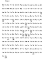
- the 14 Cys residues in the S protein are amino acid residues 48, 65, 69, 76, 90, 107, 121, 124, 137, 138, 139, 147, 149, and 221 from the N-terminus of the amino acid sequence of the S protein (underlined in Fig. 2).
- the HBsAg protein is a transmembrane protein with its PreS region at the N-terminus sticking out of the particle.
- the PreS region is contiguous to an S protein portion that extends through the particle membrane, reenters the membrane inside the particle, and comes out of the membrane outside of the particle and enters the membrane again with the C-terminus buried inside the membrane.
- the S protein exists inside and outside of the particle with three transmembrane sites. It is believed that the 14 Cys residues are scattered in these regions, with Cys residues 48, 65, and 69 inside the particles, Cys residues 76, 90, 107 in the second transmembrane region, Cys residues 121, 124, 137, 138, 139, 147 exposed outside of the particles, and Cys residues 149 and 221 in the third transmembrane region.
- any of the 14 Cys residues may be modified but those in the transmembrane sites and outside of the particles, near the C-terminus, are preferable. Specifically, substitution of Cys residues 76, 90, 137, 138, 139, 147, 149, and 221 are preferable, and substitution of two or more of these 8 Cys residues is more preferable. Further, substitution of all of the 8 Cys residues, or substitution of the 8 Cys residues except for Cys residue 137 or 138 is particularly preferable.
- the Cys residues may be replaced by any amino acid, but a hydrophobic amino acid is preferable because the amino acids in the transmembrane sites exist in the hydrophobic lipid bilayer membrane of the particle.
- Hydrophobic amino acids include alanine, glycine, isoleucine, leucine, methionine, phenylalanine, proline, tryptophane, tyrosine, and valine, among which substitution by an alanine residue is preferable.
- substitution by a hydrophilic amino acid is preferable.
- Hydrophilic amino acids include arginine, asparagine, aspartic acid, glutamic acid, glutamine, histidine, lysine, serine, and threonine, among which substitution by a serine residue is preferable.
- the Cys residues can be modified by a common site-specific mutation introducing method.
- a mutant protein may be prepared by introducing point mutation in the base sequence using PCR, or a site-specific mutation inducing method (Hashimoto-Gotoh, Gene 152, 271-275 (1995), elsewhere) may be used.
- a method described in Cell Technology, separate volume, New Cell Technology Experiment Protocol, Shujunsha , 241-248 (1993), or a method using a commercially available kit for example, Quickchange Site-Directed Mutagenesis Kit, Stratagene may be used as well.
- a method of introducing mutation using PCR is particularly preferable. Specifically, the method prepares a primer into which mutation has been introduced, and the primer is hybridized with a base sequence coding for a Cys residue to be replaced. The hybridized fragments are then amplified by PCR. In this manner, the method amplifies a gene that encodes an L protein in which the Cys residue is replaced with a different amino acid. Expression of the gene for example in eukaryotes produces hollow nanoparticles in which the Cys residue has been replaced.
- the receptor on the surface of the resulting particles may be modified to any bio-recognizing molecule. This enables hollow nanoparticles of the present invention to very specifically deliver and transfer substances to any cell or tissue, including the hepatocytes.
- the particle-forming protein is not just limited to the hepatitis B virus surface-antigen protein as long as it can form particles.
- natural proteins derived from animal cells, plant cells, viruses, or fungi may be used.
- synthetic proteins may be used as well.
- a particle-forming protein with suppressed antigenic action may be used.
- such a protein may be hepatitis B virus surface-antigen protein modified to suppress its antigenic action, or other types of modified proteins (hepatitis B virus surface-antigen protein modified by genetic engineering), as disclosed in International Publication WO01/64930.
- the hepatitis B virus surface-antigen protein or modified hepatitis B virus surface-antigen protein may be joined to other proteins such as a growth factor or antibody.
- the bio-recognizing molecule incorporated in the particle-forming protein may be, for example, cell function regulatory molecules such as a growth factor or cytokine; molecules, such as a receptor, cell surface-antigen, or tissue specific antigen, for recognizing cells or tissues; molecules derived from viruses or micro organisms; antibodies; sugar chains; and lipids. Specific examples include a cancer-specific antibody for the EGF receptor or IL-2 receptor, and EGF. Receptors displayed on the HBV are another example. These molecules are suitably selected according to the type of target cell or tissue. As the term is used herein, the "bio-recognizing molecule” refers to molecules that recognize specific cells. (In other words, molecules that render the hollow nanoparticles the ability to recognize specific cells.)
- hollow nanoparticles of the present invention are useful in specifically transporting a cell transfer substance to a specific cell.
- hollow nanoparticles of the present invention may be particles of hepatitis B virus surface-antigen protein, and may be administered through intravenous injection as a drug containing a cell transfer substance inside the hollow nanoparticles.
- the particles circulate through the body and reach the hepatocytes by the action of the hepatocyte specific receptors displayed on the particle surface, thereby infecting the host.
- the cell transfer substance is then transported into the hepatocytes, thereby specifically transferring the cell transfer substance into the liver tissue.
- the cell transfer substance encapsulated in the hollow protein nanoparticles is not particularly limited.
- the cell transfer substance may be genes such as DNA or RNA; natural or synthetic proteins; oligonucleotides; peptides; medicaments; or natural or synthetic compounds.
- cell transfer substances may be incorporated into the hollow nanoparticles by various methods commonly used in chemical or molecular biological experimental techniques. Some of the preferred examples include an electroporation method, ultrasonic method, simple diffusion method, and a method using charged lipids.
- the particle-forming protein may be fused with the cell transfer substance to form the particles.
- the particle-forming protein is fused with the cell transfer substance in the manner described below.
- a plasmid is prepared that has incorporated a gene encoding a hepatitis B virus surface-antigen protein. Downstream of the gene, the plasmid also includes a gene encoding a protein drug. The plasmid is used to produce particles in a eukaryotic cell, thereby producing a drug in which the protein drug is fused with the hepatitis B virus surface-antigen protein forming the particles.
- the drug may be administered through oral administration, intramuscular administration, intraperitoneal administration, subcutaneous administration, or other administration routes.
- the drug of the present invention allows a substance to be specifically transported into cells or tissues in vivo or in vitro .
- Specific transport of a substance into a specific cell or specific tissue with the use of the drug may be used as a treatment method of various diseases, or one of the steps in the procedure of the treatment method.
- HBsAg refers to hepatitis B virus surface antigen.
- HBsAg is an envelope protein of HBV, and includes an S protein consisting of 226 amino acids, as shown in Fig. 2.
- M protein includes the entire sequence of the S protein with additional 55 amino acids (pre-S2 peptide) at the N-terminus.
- L protein contains the entire sequence of the M protein with additional 108 or 119 amino acids (pre-S1 peptide) at the N-terminus.
- the pre-S regions (pre-S1, pre-S2) of the HBsAg L protein have important roles in the binding of HBV to the hepatocytes.
- the Pre-S1 region has a direct binding site for the hepatocytes, and the pre-S2 region has a polymeric albumin receptor that binds to the hepatocytes via polymeric albumin in the blood.
- HBsAg in the eukaryotic cell causes the protein to accumulate as a membrane protein on the membrane surface of the endoplasmic reticulum.
- the L protein molecules of HBsAg agglomerate and are released as particles into the ER lumen, carrying the ER membrane with them as they develop.
- a gene fragment encoding a HBsAg L protein incorporated in pGLDL IIP39-RcT was inserted in an animal cell expression plasmid pTB1455, downstream of the SR ⁇ ' promoter.
- a pBO442 plasmid was obtained.
- the base sequence of the HBsAg L protein is represented by SEQ ID NO: 1
- the amino acid sequence is represented by SEQ ID NO: 2.
- the pBO442 plasmid was used to transform Escherichia coli K12 strain (DH 5 ⁇ or XL-1 Blue) for methylating DNA, and methylated pBO442 plasmid was purified.
- a base sequence coding for a Cys residue was modified to a base sequence coding for an Ala residue or Ser residue. This was achieved by introducing mutation by a site-specific mutation introducing method using PCR, using the pBO442 plasmid as a template. Referring to Fig. 3, the following specifically describes a preparation method of a HBsAg L protein gene plasmid in which Cys residue 48 is replaced by a Ser residue, for example.
- the pBO442 plasmid as a template plasmid has an SR ⁇ ' promoter. Downstream of the SR ⁇ ' promoter is a coding region for the HBsAg L protein. A region of the plasmid including a codon (tgt) coding for Cys residue 48 of the HBsAg L protein was hybridized with a mutated synthetic oligonucleotide (primer). Using the plasmid as a template, the strand was extended by PCR. Here, the mutated primer is designed to cause a mismatch with the codon for the Cys residue 48 of the template (tgt for the template, aga for the primer). This allows for amplification of the plasmid in which a codon (tgt) for Cys residue 48 is replaced with a Ser codon (tct).
- a codon (tgt) for Cys residue 48 is replaced with a Ser codon (tct).
- Table 1 shows base sequences of mismatch primers prepared in the manner described above, wherein the primers in the mismatch primer set (rows 1 through 14) are for substituting the 14 Cys residues, respectively, and the primers in the mismatch primer set (rows 15 through 17) are for substituting two or three adjacent Cys residues.
- the substitution position indicates the position of substituted Cys residue from the N-terminus of the amino acid sequence in the S protein.
- Table 1 also shows the size and sequence of the corresponding primer (a pair of sense and anti-sense strands), and annealing temperatures in the PCR reaction.
- the substitution position is denoted, for example, by the notation "C/48/S", which means that Cys residue 48 has been replaced with a Ser residue.
- C/76/A means that Cys residue 76 has been replaced with an Ala residue.
- the base sequences of the 17 pairs of 34 primers are respectively represented by SEQ ID NOs: 3 through 36 in the order of appearance in Table 1.
- SEQ ID NOs: 1 and 2 represent base sequences of the primer set for substituting Cys residue 48.
- the base sequences of the primer sets for substituting Cys residues 65, 69, 76, 90, 107, 121, 124, 137, 138, 139, 147, 149, and 221 are represented by SEQ ID NOs: 3 and 4, 5 and 6, 7 and 8, 9 and 10, 11 and 12, 13 and 14, 15 and 16, 17 and 18, 19 and 20, 21 and 22, 23 and 24, 25 and 26, and 27 and 28, respectively.
- SEQ ID NOs: 29 and 30 represent base sequences of the primer set for substituting Cys residues 147 and 149.
- SEQ ID NOs: 31 and 32 represent base sequences of the primer set for substituting Cys residues 138 and 139.
- the base sequences of the primer set for substituting Cys residues 137, 138, and 139 are represented by SEQ ID NOs: 33 and 34.
- the PCR was carried out in a 50 ⁇ l solution containing 50 nmol template DNA, 15 pmol synthetic DNA primer (each mutated primer), 40 nmol dATP, 40 nmol dCTP, 40 nmol dGTP, 40 nmol dTTP, 2.5 unit Pfu turbo DNA polymerase, 10 mM KCl, 10 mM (NH 4 ) 2 SO 4 , 20 mM Tris-Cl (pH 8.75), 2 mM MgSO 4 , 0.1% Triton® X-100, and 100 ⁇ g/ml BSA. After 30-second heating at 95°C, the reaction was carried out in 18 cycles at 95°C for 30 seconds and at 68°C for 20 minutes with 1-minute annealing in between. Different annealing temperatures were used for the different primers, as shown in Table 1.
- the restriction enzyme DpnI (10,000 unit/ml) for digesting methylated DNA was added.
- the mixture was allowed to react for 1 hour at 37°C.
- the template plasmid DNA with unsubstituted Cys residue is obtained by transforming E. coli that methylates DNA. Accordingly, the template DNA is methylated.
- DNA with substituted Cys residue is not methylated. Therefore, the restriction enzyme DpnI only digests the template DNA.
- the remaining DNA i.e., the L protein DNA with substituted Cys residue was used to transform E. coli XL-1 Blue, and plasmid DNA was extracted from the resulting colonies. From the restriction enzyme map and the base sequence, it was confirmed that the resulting plasmid DNA was successfully mutated.
- the L protein plasmid with substituted Cys residue was used as a template, and the PCR reaction was repeated using another mismatch primer. In this manner, an L protein gene plasmid with increased numbers of substituted Cys residues was prepared.
- a total of 27 plasmids with 1 to 9 modified Cys residues were prepared.
- Table 2 shows these 27 plasmids (numbered 1 to 27), their names (pBO***) and substitution positions in the expressed proteins.
- Fig. 4 schematically illustrates the primary structure of the HBsAg L proteins. The diagram also shows positions of cysteine in the S protein (indicated by arrows), and substitution positions of amino acid (S (serine) or A (alanine)) in the S protein encoded by the gene in each plasmid. Plasmid Plasmid Name Substitution Position No. pBO No.
- DMEM Dulbecco-modified eagle medium
- FBS fetal bovine serum
- the solution was moved to an electroporation cuvette (4 mm across electrodes), and gene transfer was carried out with an electroporator (Bio-Rad) at 950 ⁇ F and 0.3 kV.
- the cells in the cuvette were moved to a 60 mm dish (product of Falcon), and were cultured in 6 M1 of DMEM containing 5% FBS. The incubation was made for 14 to 15 hours at 37°C in the presence of 5% CO 2 . By changing the medium to 6 M1 serum-free medium CHO-SFM II (Invitrogen), the cells were further incubated for 4 days, and the medium was collected.
- Fig. 5 shows the result of detection for the expression of 25 mutant HBsAg that were respectively mutated by the plasmids 1 through 25. Antigenicity was measured in relative values with respect to that of the wild-type held at 100. The result of detection for the culture medium in which no plasmid was used and no particles were formed by the transformation was a negative control. Note that, for the experiments that were carried out three or more times, the standard deviation is indicated by bars.
- HBsAg L proteins were prepared in which 2 to 9 Cys residues had been replaced (rows 15 to 25). All of these HBsAg L proteins had antigenicity that compared to or excelled that of the wild-type.
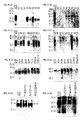
- the molecular weight of mutant particles and the presence or absence of dimerized particles were examined by sodium dodecyl sulfate-polyacrylamide gel electrophoresis (SDS-PAGE) and Western blotting.
- the sediment was separated in half and subjected to SDS-PAGE under reduced condition and non-reduced condition, respectively.
- SDS-PAGE was performed after treating the sediment in mercaptoethanol at 95°C for 5 minutes. The process prevented formation of disulfide bonds between Cys residues, allowing the proteins to be detected without causing polymerization.
- SDS-PAGE was performed without heat-treating the sediment in mercaptoethanol. Accordingly, proteins were detected in polymerized form.
- mutant HBsAg particles showed bands, near 53 kDa range under reduced condition, and near 100 kDa range under non-reduced condition. This confirmed that the mutant HBsAg had a molecular weight of 53 kDa, and that the mutant HBsAg existed as dimmers when in particle form. It is believed that the presence of the HBsAg dimmers is related to the ability of the protein to form particles.
- a gene fragment encoding the HBsAg L protein incorporated in the pGLDL II P39-RcT was replaced with the mutant HBsAg gene prepared in Example 1, so as to prepare a mutant HBsAg gene plasmid for expression in yeasts.
- the mutant HBsAg gene plasmid was expressed in yeasts (particles were prepared) according to the method disclosed in International Publication WO01/64930.
- mutant HBsAg particles are used to transfer the GFP gene into the human hepatic cancer cell HepG2.
- the human hepatic cancer cells HepG2 were inoculated on a 3.5 cm glass-bottom Petri dish with 1 ⁇ 10 5 cells/well. The cells were incubated overnight using D-MEM containing 10% fetal bovine serum. The incubation was carried out at 37°C in the presence of 5% CO 2 until the exponential growth stage was reached.
- the mutant HBsAg particles were mixed with a green fluorescent protein expression plasmid (GFP expression plasmid pTB701-hGFP), and the GFP expression plasmid was encapsulated by carrying out electroporation under the conditions of 110 V and 950 ⁇ F, using a 4 mm-pitch cuvette.
- the HBsAg particles containing the GFP expression plasmid were mixed with a HepG2 culture medium, and were incubated for 4 days in D-MEM at 37°C in the presence of 5% CO 2 .
- GFP expression in the HepG2 was observed with a confocal laser fluorescence microscope (Fig. 7(a)).
- a sample was also prepared for the wild-type HBsAg particles. According to the method described above, substantially the same number of HepG2 cells were cultured in a Petri dish, and particles containing an equal amount of GFP gene plasmid were mixed with an equal amount of the HepG2 culture medium. The sample was incubated for 4 days in D-MEM at 37°C in the presence of 5% CO 2 . GFP expression in the HepG2 was observed with a confocal laser fluorescence microscope (Fig. 7(b)).
- Fig. 7 It can be seen from Fig. 7 that more cells fluoresced when the mutant HBsAg was used for the gene transfer. That is, by mutating the protein forming the HBsAg particles, the efficiency of gene transfer to the cell was improved.
- the HBsAg particles with the substituted Cys residue can be used to transfer genes specifically to the human hepatocytes with improved efficiency.
- mutant HBsAg gene Gene transfer to the HepG2 cells was carried out using a mutant HBsAg gene with 7 to 8 cysteine substitutions.
- mutant HBsAg genes three kinds of plasmids prepared from the wild-type HBsAg genes were used: Plasmid pBO539 with Cys residues 76, 90, 149, 221 substituted to Ala residues, and Cys residues 138, 139, and 147 substituted to Ser residues; plasmid pBO552 with Cys residues 76, 90, 149, 221 substituted to Ala residues, and Cys residues 137, 139, 147 substituted to Ser residues; and plasmid pBO540 with Cys residues 76, 90, 149, 221 substituted to Ala residues, and Cys residues 137, 138, 139, 147 substituted to Ser residues (Table 3).
- the plasmids pBO539, pBO552, and pBO540 were used to prepare HBsAg particles BNP-Lm7a, BNP-Lm7b, and BNP-Lm8, respectively. Plasmid Expressed Particles Substitution Position pBO539 BNP-Lm7a C/76,90,149,221/A C/138,139,147/S pBO552 BNP-Lm7b C/76,90,149,221/A C/137,139,147/S pB0540 BNP-Lm8 C/76,90,149,221/A C/137,138,139,147/S
- mutant HBsAg particle expression vector DNA was transferred into COS7 cells by an electroporation method. After culturing the cells for 4 days, the supernatant was collected, concentrated by centrifugal filtration, and the particles were collected. The following specifically describes the procedure used in the experiment.
- COS7 cells were cultured in a 5% FCS-MEM medium to obtain 80% confluent cells.
- the cells were treated with trypsin after 6 to 8 hours and were collected and counted with a hemocytometer. After counting, the cells were fractionated and centrifuged to obtain 4 ⁇ 10 6 cells in each 1.5 ml tube. The centrifugation was carried out for 30 seconds at 12000 rpm. After centrifugation, the supernatant was suctioned and removed, and the cells were suspended in 300 ⁇ l of electroporation solution (Epmedium: RPMI-1640 + 10 mM glucose + 0.1 mM DDT).
- the medium in each 6 cm Petri dish was changed to 3 ml of CHO-S-SFM II (GIBCO). The cells were further incubated for 4 days on the new medium. After 4 days, the cells and supernatant were separately collected, and were stored at -20°C and 4°C, respectively. For each plasmid DNA sample, electroporation was carried out 4 times.
- the supernatant of the cultured cells was used for the IMx analysis as described below.
- the IMxHBsAg assay system (Dinabot) is a fully automated immunoassay system that detects HBs antigens in the supernatant, using EIA employing a sandwich method.
- the antigenicity so detected in the supernatant is represented by RATE value.
- the antigenicity was compared with the antigenicity of a standard HBsAg (human serum-derived HBsAg particles) provided in the system, so as to convert the concentration of the expressed particles to a HBsAg equivalent.
- the measured sample was 90 ⁇ l of supernatant mixed with 90 ⁇ l of 1% fetal bovine serum-containing Dulbecco phosphate buffer saline solution (diluent).
- the particle concentration and the level of particle expression for the Cys-substituted particles are low in the IMxHBsAg assay system, requiring the particles to be concentrated for actual use. As such, the particles were concentrated by ultra centrifugal filtration concentration, as described below.
- COS7 cells were cultured in a 5% FCS-MEM medium to obtain 80% confluent cells.
- the cells were treated with trypsin after 6 to 8 hour incubation on a new medium, and were collected and counted with a hemocytometer. After counting, the cells were fractionated and centrifuged to obtain 4 ⁇ 10 6 cells in each 1.5 ml tube. The centrifugation was carried out for 30 seconds at 12000 rpm. After centrifugation, the supernatant was suctioned and removed, and the cells were suspended in 300 ⁇ l of electroporation solution (Ep medium: RPMI-1640 + 10 mM glucose + 0.1 mM DDT).
- HepG2 cells in a 6 cm Petri dish were treated with trypsin, and collected cells were counted with a hemocytometer. After confirming the cell count, the cells were fractionated on an 8-well chamber slide, with each well containing 9000 cells. The cells were incubated overnight at 37°C.
- mutant HBsAg particles GFP-BNP-Lm8
- wild-type HBsAg particles that have incorporated GFP expression vector DNA.
- FuGene 6 FuGene 6
- a mixture of the HepG2 cells and an electroporation buffer containing neither the particles nor vector, or a mixture of the HepG2 cells and only the GFP expression vector DNA was prepared. After 3 days, cell fluorescence was observed with a confocal microscope. Note that, the HBsAg particles enveloping the GFP expression vector DNA contained the GFP expression vector in various proportions.
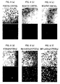
- Fig. 8 represents micrographs in confocal microscopy, showing transmission images in the upper column, and fluorescent images in the lower column.
- (a) represents a positive control
- (b) represents a negative control using only the electroporation buffer without particles or vector
- (c) represents a negative control using only GFP expression vector DNA
- (d) represents a sample containing the wild-type HBsAg particles (HBsAg particles (69 ng)) + GFP expression vector DNA (200 ng)
- (e) represents a sample containing GFP-BNP-Lm8 particles (BNP-Lm8 (12 ng)) + GFP expression vector (200 ng)
- (f) represents a sample containing GFP-BNP-Lm8 particles (BNP-Lm8 (24 ng)) + GFP expression vector (200 ng).
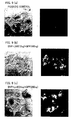
- Fig. 9(a) through Fig. 9(c) are enlarged views of Fig. 8, magnifying Fig. 8(a), Fig. 8(e), and Fig. (f), respectively.
- the cells fluoresce green far more strongly in the sample containing the GFP-BNP-Lm8 particles than in the sample containing the wild-type particles enveloping the GFP expression vector.
- the sample containing the GFP-BNP-Lm8 particles showed fluorescence even when the amount of particles added was as small as 10 ng.
- Fig. 10 (a) represents a positive control (containing a compound material of GFP expression vector DNA (200 ng) and FuGene 6 (0.5 ⁇ l)), (b) represents a negative control (containing only GFP expression vector DNA (200 ng)), (c) represents a sample containing the wild-type HBsAg particles incorporating the GFP expression vector (wild-type HBsAg (WT) (6.4 ng) + GFP expression vector (200 ng)), immediately after the collection (two photographs on the left) and one week after the collection (two photographs on the right), and (d) represents a sample containing the BNP-Lm8 particles incorporating the GFP expression vector (BNP-Lm8 (6.4 ng) + GFP expression vector (200 ng)), immediately after the collection (two photographs on the left) and one week after the collection (two photographs on the right).
- the cells can exhibit green fluorescence of the GFP expression vector even when the GFP expression vector is incorporated in the BNP-Lm8 particles that had been stored for one week at 4°C. That is, the BNP-Lm8 had desirable preservability.
- FIG. 11 A sample obtained from the HepG2 cells by carrying out an experiment according to the foregoing procedure (Fig. 12) was compared with a sample obtained by using not the HepG2 cells but human colon cancer derived cells WirDr (Fig. 12).
- Fig. 11 and Fig. 12 the photographs on the left are phase-contrast images, and the photographs on the right are fluorescent images. Further, in Fig. 11 and Fig.
- (a) represents a positive control (containing a compound material of GFP expression vector DNA (200 ng) and FuGene 6 (0.5 ⁇ l))
- (b) represents a negative control (containing only the GFP expression vector DNA)
- (c) represents wild-type HBsAg particles containing the GFP expression vector (wild-type HBsAg (WT) (14 ng) + GFP expression vector (200 ng))
- (d) represents a sample using the BNP-Lm8 particles containing the GFP expression vector (BNP-Lm8 (5 ng) + GFP expression vector (200 ng)).
- the result therefore showed that the human hepatocyte-specific gene transfer was also possible with the BNP-Lm8 particles.
- (a) represents a positive control (containing a compound material of GFP expression vector DNA (200 ng) and FuGene 6 (0.5 ⁇ l))
- (b) represents a negative control (containing only GFP expression vector DNA)
- (c) represents a sample including wild-type HBsAg particle containing the GFP expression vector DNA (wild-type HBsAg (WT) (3.2 ng) + GFP expression vector DNA (200 ng))
- (d) represents a sample including wild-type HBsAg particles containing the GFP expression vector DNA (wild-type HBsAg (WT) (6.4 ng) + GFP expression vector DNA (200 ng))
- (e) represents a sample including BNP-Lm8 particles containing the GFP expression vector DNA (BNP-Lm8 (3.2 ng) + GFP expression vector DNA (200 ng))
- (f) represents a sample including BNP-Lm7b particles containing the GFP expression vector DNA (BNP-Lm8 (6.4
- the HBsAg particles with the substituted Cys residues, particularly the BNP-Lm8, showed stronger fluorescence than the wild-type HBsAg particles, indicating that these particles can sufficiently transfer DNA to cells such as the human hepatocytes and other animal cells, or that these particles can efficiently envelope various substances. Further, the specificity of these particles to the hepatocytes was well preserved.
- the BNP-Lm7b with the substitution of 7 Cys residues, and the BNP-Lm8 both had stronger fluorescence than the wild-type particles, but the latter showed stronger fluorescence than the former. That is, with the substitution of all of Cys residues considered to be unnecessary in the particle formation, the BNP-Lm8 had higher transfer efficiency of DNA into cells compared with the wild-type particles.
- the present invention provides hollow nanoparticles that can deliver substances to specific cells.
- the hollow nanoparticles have a stable particle structure, allowing substances to be transferred into cells with improved efficiency.
- a drug prepared according to the invention can be used by a convenient method of intravenous injection to selectively and efficiently deliver disease-treating substances to specific cells or tissues.
- the invention is a great leap forward from conventional gene therapy in that it does not require any surgical operation, and that the risk of side effect is greatly reduced.
- the drug is therefore usable in clinical applications in its present form.
Landscapes
- Health & Medical Sciences (AREA)
- Chemical & Material Sciences (AREA)
- Life Sciences & Earth Sciences (AREA)
- Engineering & Computer Science (AREA)
- General Health & Medical Sciences (AREA)
- Genetics & Genomics (AREA)
- Bioinformatics & Cheminformatics (AREA)
- Medicinal Chemistry (AREA)
- Organic Chemistry (AREA)
- Pharmacology & Pharmacy (AREA)
- Animal Behavior & Ethology (AREA)
- Public Health (AREA)
- Veterinary Medicine (AREA)
- Epidemiology (AREA)
- Biotechnology (AREA)
- Molecular Biology (AREA)
- Biomedical Technology (AREA)
- Biochemistry (AREA)
- Physics & Mathematics (AREA)
- Biophysics (AREA)
- General Engineering & Computer Science (AREA)
- Zoology (AREA)
- Wood Science & Technology (AREA)
- Optics & Photonics (AREA)
- Proteomics, Peptides & Aminoacids (AREA)
- Chemical Kinetics & Catalysis (AREA)
- Nuclear Medicine, Radiotherapy & Molecular Imaging (AREA)
- General Chemical & Material Sciences (AREA)
- Gastroenterology & Hepatology (AREA)
- Nanotechnology (AREA)
- Plant Pathology (AREA)
- Virology (AREA)
- Microbiology (AREA)
- Inorganic Chemistry (AREA)
- Peptides Or Proteins (AREA)
- Micro-Organisms Or Cultivation Processes Thereof (AREA)
- Medicinal Preparation (AREA)
- Medicines That Contain Protein Lipid Enzymes And Other Medicines (AREA)
- Medicines Containing Material From Animals Or Micro-Organisms (AREA)
Abstract
The invention provides hollow nanoparticles of a protein
with the ability to recognize specific cells such as the
hepatocytes and to form particles (for example, hepatitis B virus
surface-antigen protein), wherein the protein has a cysteine
residue substituted to a different amino acid. The hollow
nanoparticles have a stable particle structure and can be used
to efficiently transfer substances to specific target cells or
tissues. The invention also provides a drug using the hollow
nanoparticles.
Description
The present invention relates to hollow nanoparticles for
encapsulating a substance to be transferred into a cell for
treating a disease. Specifically, the invention relates to a drug
that allows the disease-treating substance encapsulated in
particles to be specifically transferred into a specific cell or
tissue.
In the field of medicine, there has been active research on
drugs that directly and effectively act on the affected area
without causing serious side effects. One area of active research
is a method known as a drug delivery system (DDS), in which
active ingredients of drugs or other substances are specifically
delivered to a target cell or tissue, where they can exhibit their
effects.
A gene transfer method is one known example of a method
of transferring a protein drug to a target cell or tissue. In the
gene transfer method, an expression vector that has
incorporated a gene for encoding the protein is transferred into
a target cell by an electroporation method or other techniques.
Inside the cell, the gene is expressed into the protein drug.
However, the conventional gene transfer methods are not
sufficient to specifically transfer the protein drug to a target cell
or tissue.
Under these circumstances, the inventors of the present
invention have previously proposed a method of specifically and
safely delivering and transferring various substances (including
genes, proteins, compounds) into a target cell or tissue, using
hollow nanoparticles of a protein that has the ability to form
particles and has incorporated a bio-recognizing molecule, as
disclosed in International Publication with International
Publication No. WO01/64930 (published on September 7, 2001)
(hereinafter referred to as "International Publication
WO01/64930"). However, the publication does not fully discuss
how the method can be used to efficiently transfer these
substances to a target cell or tissue.
The present invention was made in view of the foregoing
problems, and an object of the invention is to provide hollow
protein nanoparticles for specifically and efficiently transferring
a substance into a target cell or tissue. The present invention
also provides a drug containing hollow protein nanoparticles
encapsulating a substance to be transferred into a cell.
The inventors of the present invention accomplished the
present invention by finding that a modified cysteine residue in
the particle protein greatly improves uptake of a particle
substance by a specific cell.
That is, the present invention discloses hollow
nanoparticles of a particle-forming protein with the ability to
recognize specific cells (for example, hepatocytes), wherein the
protein has a modified cysteine residue.
An example of such a "particle-forming protein" is a
hepatitis B virus surface-antigen protein. In eukaryotic cells,
the protein is expressed as a membrane protein on the
endoplasmic reticulum and accumulates thereon before it is
released as particles. Hollow nanoparticles of the present
invention may be produced by transforming a eukaryotic cell
(for example, animal cells including mammal cells, or yeast
cells) with a vector including a gene coding for a
particle-forming protein, and then expressing the gene in the
eukaryotic cell.
When the hepatitis B virus surface-antigen protein is used
to form particles, the particles recognize the hepatocytes and
deliver a substance contained in the particles specifically to the
hepatocytes. Thus, with particles containing a substance (gene,
etc.) for treating a liver disease, an effective therapeutic drug
can be provided that effectively and specifically acts on the
hepatocytes.
For example, the hepatitis B virus surface-antigen protein
may be modified to lack its infectivity to the hepatocytes and
display a growth factor or an antibody. Particles of such a
modified protein can deliver a substance contained in the
particles to specific cells other than the hepatocytes. For
example, by displaying an antibody that specifically recognizes
a certain cancer cell, the particles can recognize the cancer cell
and deliver a substance contained in the particles specifically to
the cancer cell.
The hepatitis B virus surface-antigen protein contains S
protein (described later) that includes a total of 14 cysteine
(Cys) residues associated with the oxidation-reduction of the
protein. The Cys residues are believed to control the particle
structure they form. However, the cysteine residues have a
problem of instability. During the purification and/or
preservation of the particles, the cysteine residues form excess
disulfide bonds, causing the protein to polymerize by the
randomly formed intermolecular and intramolecular disulfide
crosslinkage. Such polymerization can be prevented by
modifying some of the Cys residues that do not play important
role in the control of the protein structure. (Alternatively,
polymerization can be prevented by modifying other Cys
residues that have essentially no influence on the ability to form
particles or recognize cells.) This imparts stability to the
particles and thereby improves the efficiency by which
substances are encapsulated in the hollow nanoparticles. As a
result, the substances can be efficiently transferred into a cell.
Specifically, it is preferable that substitution takes place
at Cys residues 76, 90, 139, 147, 149, 221 and at least one of
Cys residues 137 and 138 from the N-terminus of the amino
acid sequence of the S protein in the hepatitis B virus
surface-antigen protein.
It is preferable that the Cys residues be modified by
substitution by other amino acids. Alternatively, the Cys
residues may be modified by deletion. Being a transmembrane
protein, the hepatitis B virus surface-antigen protein exists
both inside and outside of the particles, across the lipid bilayer
membrane. Among the Cys residues in the protein, it is
preferable that those believed to exist inside the lipid bilayer
membrane be replaced with hydrophobic amino acids (for
example, alanine (Ala)), and that those believed to exist inside
and outside of the particles be replaced with hydrophilic amino
acids (for example, serine (Ser)).
The Cys residues can be modified in this manner to
prepare modified hollow nanoparticles. For example, a gene
encoding a particle-forming protein may be mutated and
incorporated into a vector to transform a eukaryotic cell in
which the mutated gene is expressed.
The present invention discloses a drug that contains a
therapeutic substance in its hallow nanoparticles. The drug can
be used by a convenient method of intravenous injection to
effectively treat specific diseased cells or tissues. The drug is a
great leap forward from conventional disease treatment methods
in that it does not require large dose or any surgical operation
in disease treatment including gene therapy, and that the risk
of side effect is greatly reduced. The drug is therefore usable in
clinical applications in its present form.
The present invention also discloses a treatment method
for treating diseases through administration of the drug
disclosed in the present invention.
Hollow nanoparticles of the present invention contain
modified cysteine residues in the protein forming the particles.
The particle-forming protein may incorporate a bio-recognizing
molecule (i.e., a molecule recognizing a specific cell) that
enables a substance to be specifically delivered to a target cell
or tissue. Examples of such particle-forming proteins include
sub viral particles obtained from various viruses. A specific
example is a hepatitis B virus (HBV) surface-antigen protein. (In
the following, the hepatitis B virus surface-antigen protein may
be denoted by "HBsAg.")
The protein particles of such a particle-forming protein
may be obtained through the protein expression in eukaryotic
cells. Specifically, in the eukaryotic cell, the particle-forming
protein is expressed on the endoplasmic reticulum as a
membrane protein and accumulates thereon before it is released
as particles. The eukaryotic cell may be obtained from yeasts, or
animals including mammals.
As will be described later in Examples, the inventors of
the present invention have reported that the expression of HBV
surface-antigen L protein in recombinant yeast cells produces
ellipsoidal hollow particles with a minor axis of 20 nm and a
major axis of 150 nm, with a large number of L proteins
embedded in the yeast-derived lipid bilayer membrane (J. Biol.
Chem., Vol. 267, No. 3, 1953-1961, 1992). The particles contain
no HBV genome and lack the viral function. Therefore, the
particles are very safe to the human body. Further, because the
particles on the particle surface display hepatocyte-specific
receptors that make the HBV highly infectious to the
hepatocytes, the particles are highly effective as a carrier for
delivering substances specifically to the hepatocytes.
The method of producing protein particles using
recombinant yeasts is therefore suitable in efficiently producing
the particles from a soluble protein in the yeast.
The HBsAg contains S protein (described later) includes a
total of 14 cysteine (Cys) residues associated with the
oxidation-reduction of the protein. The Cys residues are
believed to control the particle structure they form. However,
the cysteine residues have a problem of instability. During the
purification and/or preservation of the particles, the cysteine
residues form excess disulfide bonds, causing the protein to
polymerize. The particles can be stabilized to improve their
preservation property by modifying some of the Cys residues
that do not play important role in the control of the protein
structure. Further, with the Cys residues modified in this
manner, substances can be more easily encapsulated in the
hollow nanoparticles and thereby more efficiently transferred
into the cell.
The site of modified Cys residue is not particularly limited
as long as the functionality of the HBsAg particles is maintained.
The HBsAg L protein includes PreS1, PreS2, and S protein,
wherein the PreS1 and PreS2 serve as hepatocytes recognition
sites. It is believed that modification of amino acid in the S
protein has only a small effect on the functionality of the HBsAg
particles. It is therefore possible, by modifying some of the 14
cysteine (Cys) residues in the S protein (represented by "C" in
the schematic diagram of Fig. 1), to break the disulfide bond
without losing the ability of the HBsAg particles to recognize
hepatocytes or form particles.
The 14 Cys residues in the S protein are amino acid
residues 48, 65, 69, 76, 90, 107, 121, 124, 137, 138, 139, 147,
149, and 221 from the N-terminus of the amino acid sequence
of the S protein (underlined in Fig. 2). As illustrated in the
schematic representation in Fig. 1, the HBsAg protein is a
transmembrane protein with its PreS region at the N-terminus
sticking out of the particle. The PreS region is contiguous to an
S protein portion that extends through the particle membrane,
reenters the membrane inside the particle, and comes out of the
membrane outside of the particle and enters the membrane
again with the C-terminus buried inside the membrane. That is,
the S protein exists inside and outside of the particle with three
transmembrane sites. It is believed that the 14 Cys residues are
scattered in these regions, with Cys residues 48, 65, and 69
inside the particles, Cys residues 76, 90, 107 in the second
transmembrane region, Cys residues 121, 124, 137, 138, 139,
147 exposed outside of the particles, and Cys residues 149 and
221 in the third transmembrane region.
Any of the 14 Cys residues may be modified but those in
the transmembrane sites and outside of the particles, near the
C-terminus, are preferable. Specifically, substitution of Cys
residues 76, 90, 137, 138, 139, 147, 149, and 221 are
preferable, and substitution of two or more of these 8 Cys
residues is more preferable. Further, substitution of all of the 8
Cys residues, or substitution of the 8 Cys residues except for
Cys residue 137 or 138 is particularly preferable.
The Cys residues may be replaced by any amino acid, but
a hydrophobic amino acid is preferable because the amino acids
in the transmembrane sites exist in the hydrophobic lipid
bilayer membrane of the particle. Hydrophobic amino acids
include alanine, glycine, isoleucine, leucine, methionine,
phenylalanine, proline, tryptophane, tyrosine, and valine,
among which substitution by an alanine residue is preferable.
On the other hand, for the Cys residues believed to exist outside
and inside the particles, substitution by a hydrophilic amino
acid is preferable. Hydrophilic amino acids include arginine,
asparagine, aspartic acid, glutamic acid, glutamine, histidine,
lysine, serine, and threonine, among which substitution by a
serine residue is preferable.
The Cys residues can be modified by a common
site-specific mutation introducing method. For example, a
mutant protein may be prepared by introducing point mutation
in the base sequence using PCR, or a site-specific mutation
inducing method (Hashimoto-Gotoh, Gene 152, 271-275 (1995),
elsewhere) may be used. Further, a method described in Cell
Technology, separate volume, New Cell Technology Experiment
Protocol, Shujunsha, 241-248 (1993), or a method using a
commercially available kit (for example, Quickchange
Site-Directed Mutagenesis Kit, Stratagene) may be used as well.
Among these methods, a method of introducing mutation
using PCR is particularly preferable. Specifically, the method
prepares a primer into which mutation has been introduced,
and the primer is hybridized with a base sequence coding for a
Cys residue to be replaced. The hybridized fragments are then
amplified by PCR. In this manner, the method amplifies a gene
that encodes an L protein in which the Cys residue is replaced
with a different amino acid. Expression of the gene for example
in eukaryotes produces hollow nanoparticles in which the Cys
residue has been replaced.
The receptor on the surface of the resulting particles may
be modified to any bio-recognizing molecule. This enables
hollow nanoparticles of the present invention to very specifically
deliver and transfer substances to any cell or tissue, including
the hepatocytes.
The particle-forming protein is not just limited to the
hepatitis B virus surface-antigen protein as long as it can form
particles. For example, natural proteins derived from animal
cells, plant cells, viruses, or fungi may be used. Various types of
synthetic proteins may be used as well. Further, when there is a
possibility that, for example, virus-derived antigen proteins may
trigger antibody reaction in a target organism, a
particle-forming protein with suppressed antigenic action may
be used. For example, such a protein may be hepatitis B virus
surface-antigen protein modified to suppress its antigenic
action, or other types of modified proteins (hepatitis B virus
surface-antigen protein modified by genetic engineering), as
disclosed in International Publication WO01/64930. Further,
for the particle-forming protein, the hepatitis B virus
surface-antigen protein or modified hepatitis B virus
surface-antigen protein may be joined to other proteins such as
a growth factor or antibody.
The bio-recognizing molecule incorporated in the
particle-forming protein (may be contained in the
particle-forming protein itself, or fused with the particle-forming
protein by being ligated either directly or indirectly) may be, for
example, cell function regulatory molecules such as a growth
factor or cytokine; molecules, such as a receptor, cell
surface-antigen, or tissue specific antigen, for recognizing cells
or tissues; molecules derived from viruses or micro organisms;
antibodies; sugar chains; and lipids. Specific examples include
a cancer-specific antibody for the EGF receptor or IL-2 receptor,
and EGF. Receptors displayed on the HBV are another example.
These molecules are suitably selected according to the type of
target cell or tissue. As the term is used herein, the
"bio-recognizing molecule" refers to molecules that recognize
specific cells. (In other words, molecules that render the hollow
nanoparticles the ability to recognize specific cells.)
The hollow nanoparticles of the present invention so
prepared are useful in specifically transporting a cell transfer
substance to a specific cell. For example, hollow nanoparticles
of the present invention may be particles of hepatitis B virus
surface-antigen protein, and may be administered through
intravenous injection as a drug containing a cell transfer
substance inside the hollow nanoparticles. The particles
circulate through the body and reach the hepatocytes by the
action of the hepatocyte specific receptors displayed on the
particle surface, thereby infecting the host. The cell transfer
substance is then transported into the hepatocytes, thereby
specifically transferring the cell transfer substance into the liver
tissue.
The cell transfer substance encapsulated in the hollow
protein nanoparticles is not particularly limited. For example,
the cell transfer substance may be genes such as DNA or RNA;
natural or synthetic proteins; oligonucleotides; peptides;
medicaments; or natural or synthetic compounds.
These cell transfer substances may be incorporated into
the hollow nanoparticles by various methods commonly used in
chemical or molecular biological experimental techniques. Some
of the preferred examples include an electroporation method,
ultrasonic method, simple diffusion method, and a method
using charged lipids. When the cell transfer substance is a
protein, the particle-forming protein may be fused with the cell
transfer substance to form the particles. When applying the
method, the particle-forming protein is fused with the cell
transfer substance in the manner described below. For example,
a plasmid is prepared that has incorporated a gene encoding a
hepatitis B virus surface-antigen protein. Downstream of the
gene, the plasmid also includes a gene encoding a protein drug.
The plasmid is used to produce particles in a eukaryotic cell,
thereby producing a drug in which the protein drug is fused
with the hepatitis B virus surface-antigen protein forming the
particles.
Other than intravenous injection, the drug may be
administered through oral administration, intramuscular
administration, intraperitoneal administration, subcutaneous
administration, or other administration routes.
The drug of the present invention allows a substance to be
specifically transported into cells or tissues in vivo or in vitro.
Specific transport of a substance into a specific cell or specific
tissue with the use of the drug may be used as a treatment
method of various diseases, or one of the steps in the procedure
of the treatment method.
In the following, the present invention will be described in
more detail by way of Examples with reference to the attached
drawings. It should be appreciated that the present invention is
not limited in any way by the following Examples, and various
modifications to details of the invention are possible.
In the following, HBsAg refers to hepatitis B virus surface
antigen. HBsAg is an envelope protein of HBV, and includes an
S protein consisting of 226 amino acids, as shown in Fig. 2. M
protein includes the entire sequence of the S protein with
additional 55 amino acids (pre-S2 peptide) at the N-terminus. L
protein contains the entire sequence of the M protein with
additional 108 or 119 amino acids (pre-S1 peptide) at the
N-terminus.
The pre-S regions (pre-S1, pre-S2) of the HBsAg L protein
have important roles in the binding of HBV to the hepatocytes.
The Pre-S1 region has a direct binding site for the hepatocytes,
and the pre-S2 region has a polymeric albumin receptor that
binds to the hepatocytes via polymeric albumin in the blood.
Expression of HBsAg in the eukaryotic cell causes the
protein to accumulate as a membrane protein on the membrane
surface of the endoplasmic reticulum. The L protein molecules
of HBsAg agglomerate and are released as particles into the ER
lumen, carrying the ER membrane with them as they develop.
The Examples below used HBsAg L protein.
According to the method described in J. Biotechnol., Vol.
33:, No. 2, 157-174, 1994 reported by the inventors of the
present invention, a gene fragment encoding a HBsAg L protein
incorporated in pGLDL IIP39-RcT was inserted in an animal cell
expression plasmid pTB1455, downstream of the SRα' promoter.
As a result, a pBO442 plasmid was obtained. The base
sequence of the HBsAg L protein is represented by SEQ ID NO:
1, and the amino acid sequence is represented by SEQ ID NO: 2.
The pBO442 plasmid was used to transform Escherichia coli
K12 strain (DH 5α or XL-1 Blue) for methylating DNA, and
methylated pBO442 plasmid was purified.
In a region of the pBO442 plasmid encoding the HBsAg L
protein, a base sequence coding for a Cys residue was modified
to a base sequence coding for an Ala residue or Ser residue.
This was achieved by introducing mutation by a site-specific
mutation introducing method using PCR, using the pBO442
plasmid as a template. Referring to Fig. 3, the following
specifically describes a preparation method of a HBsAg L
protein gene plasmid in which Cys residue 48 is replaced by a
Ser residue, for example.
As shown in Fig. 3, the pBO442 plasmid as a template
plasmid has an SRα' promoter. Downstream of the SRα'
promoter is a coding region for the HBsAg L protein. A region of
the plasmid including a codon (tgt) coding for Cys residue 48 of
the HBsAg L protein was hybridized with a mutated synthetic
oligonucleotide (primer). Using the plasmid as a template, the
strand was extended by PCR. Here, the mutated primer is
designed to cause a mismatch with the codon for the Cys
residue 48 of the template (tgt for the template, aga for the
primer). This allows for amplification of the plasmid in which a
codon (tgt) for Cys residue 48 is replaced with a Ser codon (tct).
Table 1 shows base sequences of mismatch primers
prepared in the manner described above, wherein the primers in
the mismatch primer set (rows 1 through 14) are for
substituting the 14 Cys residues, respectively, and the primers
in the mismatch primer set (rows 15 through 17) are for
substituting two or three adjacent Cys residues. The
substitution position indicates the position of substituted Cys
residue from the N-terminus of the amino acid sequence in the
S protein. Table 1 also shows the size and sequence of the
corresponding primer (a pair of sense and anti-sense strands),
and annealing temperatures in the PCR reaction. The
substitution position is denoted, for example, by the notation
"C/48/S", which means that Cys residue 48 has been replaced
with a Ser residue. Similarly, "C/76/A" means that Cys residue
76 has been replaced with an Ala residue.

The base sequences of the 17 pairs of 34 primers are
respectively represented by SEQ ID NOs: 3 through 36 in the
order of appearance in Table 1. Specifically, SEQ ID NOs: 1 and
2 represent base sequences of the primer set for substituting
Cys residue 48. Similarly, the base sequences of the primer sets
for substituting Cys residues 65, 69, 76, 90, 107, 121, 124, 137,
138, 139, 147, 149, and 221 are represented by SEQ ID NOs: 3
and 4, 5 and 6, 7 and 8, 9 and 10, 11 and 12, 13 and 14, 15
and 16, 17 and 18, 19 and 20, 21 and 22, 23 and 24, 25 and 26,
and 27 and 28, respectively. Further, SEQ ID NOs: 29 and 30
represent base sequences of the primer set for substituting Cys
residues 147 and 149. Similarly, SEQ ID NOs: 31 and 32
represent base sequences of the primer set for substituting Cys
residues 138 and 139. The base sequences of the primer set for
substituting Cys residues 137, 138, and 139 are represented by
SEQ ID NOs: 33 and 34.
The PCR was carried out in a 50 µl solution containing 50
nmol template DNA, 15 pmol synthetic DNA primer (each
mutated primer), 40 nmol dATP, 40 nmol dCTP, 40 nmol dGTP,
40 nmol dTTP, 2.5 unit Pfu turbo DNA polymerase, 10 mM KCl,
10 mM (NH4)2SO4, 20 mM Tris-Cl (pH 8.75), 2 mM MgSO4, 0.1%
Triton® X-100, and 100 µg/ml BSA. After 30-second heating at
95°C, the reaction was carried out in 18 cycles at 95°C for 30
seconds and at 68°C for 20 minutes with 1-minute annealing in
between. Different annealing temperatures were used for the
different primers, as shown in Table 1.
After the reaction, 1 µl of restriction enzyme DpnI (10,000
unit/ml) for digesting methylated DNA was added. The mixture
was allowed to react for 1 hour at 37°C. As described above, the
template plasmid DNA with unsubstituted Cys residue is
obtained by transforming E. coli that methylates DNA.
Accordingly, the template DNA is methylated. On the other hand,
DNA with substituted Cys residue is not methylated. Therefore,
the restriction enzyme DpnI only digests the template DNA. The
remaining DNA, i.e., the L protein DNA with substituted Cys
residue was used to transform E. coli XL-1 Blue, and plasmid
DNA was extracted from the resulting colonies. From the
restriction enzyme map and the base sequence, it was
confirmed that the resulting plasmid DNA was successfully
mutated.
The L protein plasmid with substituted Cys residue was
used as a template, and the PCR reaction was repeated using
another mismatch primer. In this manner, an L protein gene
plasmid with increased numbers of substituted Cys residues
was prepared.
According to these procedures, a total of 27 plasmids with
1 to 9 modified Cys residues were prepared. Table 2 shows
these 27 plasmids (numbered 1 to 27), their names (pBO***)
and substitution positions in the expressed proteins. Fig. 4
schematically illustrates the primary structure of the HBsAg L
proteins. The diagram also shows positions of cysteine in the S
protein (indicated by arrows), and substitution positions of
amino acid (S (serine) or A (alanine)) in the S protein encoded by
the gene in each plasmid.
| Plasmid | Plasmid Name | Substitution Position |
| No. | pBO No. | |
| 1 | 454 | C/48/S |
| 2 | 497 | C/65/S |
| 3 | 498 | C/69/S |
| 4 | 468 | C/76/A |
| 5 | 455 | C/90/A |
| 6 | 456 | C/107/A |
| 7 | 460 | C/121/S |
| 8 | 461 | C/124/S |
| 9 | 465 | C/137/S |
| 10 | 466 | C/138/S |
| 11 | 467 | C/139/S |
| 12 | 499 | C/147/S |
| 13 | 469 | C/149/A |
| 14 | 470 | C/221/A |
| 15 | 511 | C/76,90/A |
| 16 | 514 | C/90/A,C/139/S |
| 17 | 518 | C/90/A, C/147/S |
| 18 | 513 | C/90,149/A |
| 19 | 512 | C/90,221/A |
| 20 | 519 | C/76,90,221/A |
| 21 | 520 | C/76,90,149,221/A |
| 22 | 528 | C/76,90,149,221/A, C/139/S |
| 23 | 529 | C/76,90,149,221/A, C/147/S |
| 24 | 533 | C/76,90,149,221/A, C/139,147/S |
| 25 | 539 | C/76,90,149,221/A, C/138,139,147/S |
| 26 | 540 | C/76,90,149,221/A, C/137,138,139,147/S |
| 27 | 541 | C/76,90,149,221/A, C/107,137,138,139,147/S |
Cell line COS7 derived from the monkey kidney was
cultured in a Dulbecco-modified eagle medium (DMEM)
containing 5% fetal bovine serum (FBS). The incubation was
made at 37°C and in the presence of 5% CO2. For each sheet of
T-75 flask (product of Falcon), 4 × 10⁶ cells were obtained.
Meanwhile, 18.5 mg of glucose was added to 10 M1 of RPMI1640
medium. To 1 M1 of the solution was added 2 µl of 50 mM
dithiothreitol (DTT). In 0.3 M1 of the solution, 4 × 10⁶ COS7
cells and 5 µg of the plasmid DNA obtained in Example 1 were
suspended. The solution was moved to an electroporation
cuvette (4 mm across electrodes), and gene transfer was carried
out with an electroporator (Bio-Rad) at 950 µF and 0.3 kV. The
cells in the cuvette were moved to a 60 mm dish (product of
Falcon), and were cultured in 6 M1 of DMEM containing 5% FBS.
The incubation was made for 14 to 15 hours at 37°C in the
presence of 5% CO2. By changing the medium to 6 M1
serum-free medium CHO-SFM II (Invitrogen), the cells were
further incubated for 4 days, and the medium was collected.
To 90 µl of collected medium was added 90 µl of Dulbecco
phosphate buffer (PBS) containing 1% FBS, and antigenicity
was detected with the IMX HBsAg assay system (Dinabot). The
presence of antigenicity was regarded as the formation of HBsAg
particles. Therefore, the presence of HBsAg particles in the
culture medium was confirmed when antigenicity was detected.
Fig. 5 shows the result of detection for the expression of 25
mutant HBsAg that were respectively mutated by the plasmids 1
through 25. Antigenicity was measured in relative values with
respect to that of the wild-type held at 100. The result of
detection for the culture medium in which no plasmid was used
and no particles were formed by the transformation was a
negative control. Note that, for the experiments that were
carried out three or more times, the standard deviation is
indicated by bars.
The result of single amino acid substitution for the 14 Cys
residues (rows 1 to 14) showed that those with the substitution
of Cys residues in the transmembrane regions had desirable
antigenicity. In particular, those with the substitution of Cys
residues 76, 90, 149, and 221 had antigenicity that compared
to or excelled that of the wild-type. Further, those with the
substitution of Cys residues outside of the particles and near
the C-terminus maintained desirable antigenicity. In particular,
those with the substitution of Cys residues 139 and 147 had
antigenicity that compared to or excelled that of the wild-type.
By combining these Cys residue substitution positions that
incur particularly desirable antigenicity, HBsAg L proteins were
prepared in which 2 to 9 Cys residues had been replaced (rows
15 to 25). All of these HBsAg L proteins had antigenicity that
compared to or excelled that of the wild-type.
The molecular weight of mutant particles and the
presence or absence of dimerized particles were examined by
sodium dodecyl sulfate-polyacrylamide gel electrophoresis
(SDS-PAGE) and Western blotting.
Specifically, to 180 µl of collected medium was added 60
µl of anti-HBs mouse monoclonal antibody-immobilized
microparticles of the IMX HBsAg assay system. The mixture was
allowed to stand overnight at 4°C with the tube slowly rotated.
The particles in the medium were sedimented with the
microparticles for 3 minutes, using a desktop high-speed
centrifuge. The resulting sediments were suspended in 0.5 M1
TBST (10mM Tris-HCl buffer, pH 7.5, 150 mM NaCl, 0.1
Tween20). The washing was carried out 5 times by repeating the
procedure of centrifugation and sediment collection.
The sediment was separated in half and subjected to
SDS-PAGE under reduced condition and non-reduced condition,
respectively. Under reduced condition, SDS-PAGE was
performed after treating the sediment in mercaptoethanol at
95°C for 5 minutes. The process prevented formation of
disulfide bonds between Cys residues, allowing the proteins to
be detected without causing polymerization. On the other hand,
under non-reduced condition, SDS-PAGE was performed
without heat-treating the sediment in mercaptoethanol.
Accordingly, proteins were detected in polymerized form.
Western blotting was carried out on the electrophorased gel,
using an anti-HBs goat antibody-biotin conjugation of the IMX
HBsAg assay system as the first antibody, and an
alkaliphosphatase-labeled anti-biotin rabbit antibody as the
secondary antibody. (Fig. 6(a) through Fig. 6(h)). Fig. 6(a)
through Fig. 6(d) show results under reduced condition, and Fig.
6(e) through Fig. 6(h) show results under non-reduced condition.
Numbers on the lanes indicate substitution positions of Cys
residues. As can be seen from the results, all mutant HBsAg
particles showed bands, near 53 kDa range under reduced
condition, and near 100 kDa range under non-reduced
condition. This confirmed that the mutant HBsAg had a
molecular weight of 53 kDa, and that the mutant HBsAg existed
as dimmers when in particle form. It is believed that the
presence of the HBsAg dimmers is related to the ability of the
protein to form particles.
A gene fragment encoding the HBsAg L protein
incorporated in the pGLDL II P39-RcT was replaced with the
mutant HBsAg gene prepared in Example 1, so as to prepare a
mutant HBsAg gene plasmid for expression in yeasts. The
mutant HBsAg gene plasmid was expressed in yeasts (particles
were prepared) according to the method disclosed in
International Publication WO01/64930.
The following describes how the mutant HBsAg particles
are used to transfer the GFP gene into the human hepatic
cancer cell HepG2.
First, the human hepatic cancer cells HepG2 were
inoculated on a 3.5 cm glass-bottom Petri dish with 1 × 10⁵
cells/well. The cells were incubated overnight using D-MEM
containing 10% fetal bovine serum. The incubation was carried
out at 37°C in the presence of 5% CO2 until the exponential
growth stage was reached. The mutant HBsAg particles were
mixed with a green fluorescent protein expression plasmid (GFP
expression plasmid pTB701-hGFP), and the GFP expression
plasmid was encapsulated by carrying out electroporation under
the conditions of 110 V and 950 µF, using a 4 mm-pitch cuvette.
The HBsAg particles containing the GFP expression plasmid
were mixed with a HepG2 culture medium, and were incubated
for 4 days in D-MEM at 37°C in the presence of 5% CO2. GFP
expression in the HepG2 was observed with a confocal laser
fluorescence microscope (Fig. 7(a)). As a comparative example, a
sample was also prepared for the wild-type HBsAg particles.
According to the method described above, substantially the
same number of HepG2 cells were cultured in a Petri dish, and
particles containing an equal amount of GFP gene plasmid were
mixed with an equal amount of the HepG2 culture medium. The
sample was incubated for 4 days in D-MEM at 37°C in the
presence of 5% CO2. GFP expression in the HepG2 was observed
with a confocal laser fluorescence microscope (Fig. 7(b)).
It can be seen from Fig. 7 that more cells fluoresced when
the mutant HBsAg was used for the gene transfer. That is, by
mutating the protein forming the HBsAg particles, the efficiency
of gene transfer to the cell was improved.
The experiment showed that, on the cultured cell level,
the HBsAg particles with the substituted Cys residue can be
used to transfer genes specifically to the human hepatocytes
with improved efficiency.
Gene transfer to the HepG2 cells was carried out using a
mutant HBsAg gene with 7 to 8 cysteine substitutions. As the
mutant HBsAg genes, three kinds of plasmids prepared from the
wild-type HBsAg genes were used: Plasmid pBO539 with Cys
residues 76, 90, 149, 221 substituted to Ala residues, and Cys
residues 138, 139, and 147 substituted to Ser residues;
plasmid pBO552 with Cys residues 76, 90, 149, 221 substituted
to Ala residues, and Cys residues 137, 139, 147 substituted to
Ser residues; and plasmid pBO540 with Cys residues 76, 90,
149, 221 substituted to Ala residues, and Cys residues 137,
138, 139, 147 substituted to Ser residues (Table 3). The
plasmids pBO539, pBO552, and pBO540 were used to prepare
HBsAg particles BNP-Lm7a, BNP-Lm7b, and BNP-Lm8,
respectively.
| Plasmid | Expressed Particles | Substitution Position |
| pBO539 | BNP-Lm7a | C/76,90,149,221/A C/138,139,147/S |
| pBO552 | BNP-Lm7b | C/76,90,149,221/A C/137,139,147/S |
| pB0540 | BNP-Lm8 | C/76,90,149,221/A C/137,138,139,147/S |
In order to obtain mutant HBsAg particles, mutant HBsAg
particle expression vector DNA was transferred into COS7 cells
by an electroporation method. After culturing the cells for 4
days, the supernatant was collected, concentrated by
centrifugal filtration, and the particles were collected. The
following specifically describes the procedure used in the
experiment.
For each sample, COS7 cells were cultured in a 5%
FCS-MEM medium to obtain 80% confluent cells. On a new
medium, the cells were treated with trypsin after 6 to 8 hours
and were collected and counted with a hemocytometer. After
counting, the cells were fractionated and centrifuged to obtain 4
× 106 cells in each 1.5 ml tube. The centrifugation was carried
out for 30 seconds at 12000 rpm. After centrifugation, the
supernatant was suctioned and removed, and the cells were
suspended in 300 µl of electroporation solution (Epmedium:
RPMI-1640 + 10 mM glucose + 0.1 mM DDT).
To the suspended solution of cells was added 5 µg of the
plasmid DNA pBO539, pBO552, or pBO540 shown in Table 3.
The solution was then moved to a cuvette (4 mm gap) that had
been pre-cooled on ice. Immediately after a voltage was applied
under 0.3 KV and 950 µF (electroporation), the sample was
ice-cooled. Thereafter, the cells in the cuvette were
re-suspended in an 8 ml medium, and were inoculated in two 6
cm Petri dishes in equal amounts.
After culturing the cells at 37°C for 14 to 15 hours, the
medium in each 6 cm Petri dish was changed to 3 ml of
CHO-S-SFM II (GIBCO). The cells were further incubated for 4
days on the new medium. After 4 days, the cells and
supernatant were separately collected, and were stored at -20°C
and 4°C, respectively. For each plasmid DNA sample,
electroporation was carried out 4 times.
The supernatant of the cultured cells was used for the IMx
analysis as described below.
The IMxHBsAg assay system (Dinabot) is a fully
automated immunoassay system that detects HBs antigens in
the supernatant, using EIA employing a sandwich method. The
antigenicity so detected in the supernatant is represented by
RATE value. As required, the antigenicity was compared with
the antigenicity of a standard HBsAg (human serum-derived
HBsAg particles) provided in the system, so as to convert the
concentration of the expressed particles to a HBsAg equivalent.
The measured sample was 90 µl of supernatant mixed with 90
µl of 1% fetal bovine serum-containing Dulbecco phosphate
buffer saline solution (diluent). The measurement was carried
out always with a positive control (reference HBsAg) and a
negative control (only diluent), and a HBsAg equivalent was
calculated based on a calibration curve. A comparison was also
made with the supernatant of the COS7 cells that have
incorporated the wild-type HBsAg gene vector (pB0411). The
results are shown in Table 4 below.
| Sample | RATE | Particle Concentration (ng/ml) |
| Positive Control | 201.1 | - |
| Nagative Control | 3.6 | - |
| Wild-Type HBsAg Particles | 94.7 | 132.9 |
| BNP-Lm7a | 15.7 | 17.7 |
| BNP-Lm7b | 15.4 | 17.2 |
| BNP-Lm8 | 10.6 | 10.2 |
It can be seen from the result that the standard HBsAg
equivalents (RATE values) of the particles are higher than that
of the negative control, yielding values of 15.7, 15.4, and 10.6
for the BNP-Lm7a, BNP-Lm7b, and BNP-Lm8, respectively.
Therefore, it can be said that all samples had mutant HBsAg
proteins secreted and expressed in the supernatant.
The particle concentration and the level of particle
expression for the Cys-substituted particles are low in the
IMxHBsAg assay system, requiring the particles to be
concentrated for actual use. As such, the particles were
concentrated by ultra centrifugal filtration concentration, as
described below.
Sixteen ml of the supernatant was transferred to a
centrifugal filtration concentration device (Viva spin, 1,000,000
MWCO, Sartorius), and concentrated to a liquid amount of 800
µl at 4°C and 3500 rpm for 45 minutes. Ninety µl out of the 800
µl liquid was used to measure a HBsAg equivalent by IMx. From
the amount of antigen calculated from the calibration curve, the
yield was determined to be around 40%.
COS7 cells were cultured in a 5% FCS-MEM medium to
obtain 80% confluent cells. The cells were treated with trypsin
after 6 to 8 hour incubation on a new medium, and were
collected and counted with a hemocytometer. After counting,
the cells were fractionated and centrifuged to obtain 4 × 10⁶
cells in each 1.5 ml tube. The centrifugation was carried out for
30 seconds at 12000 rpm. After centrifugation, the supernatant
was suctioned and removed, and the cells were suspended in
300 µl of electroporation solution (Ep medium: RPMI-1640 + 10
mM glucose + 0.1 mM DDT).
To the suspension solution of the cells were added 5 µg of
wild-type L-particle (particles of HBsAg L protein) expression
plasmid DNA (pB0441) or cysteine-modified L-particle
expression plasmid DNA, and 1 µg of wild-type S-particle
(particles of HBsAg S protein) expression plasmid DNA (pB603)
or cysteine-modified S-particle expression plasmid DNA. The
mixture was transferred to a cuvette (4 mm gap). Immediately
after a voltage was applied under 0.3 KV and 950 µF
(electroporation), the sample was ice-cooled. Thereafter, the
cells in the cuvette were re-suspended in an 8 ml medium, and
were inoculated in two 6 cm Petri dishes in equal amounts.
After culturing the cells at 37°C for 14 to 15 hours, the
medium in each 6 cm Petri dish was changed to 3 ml of
CHO-S-SFM II (GIBCO). The cells were further incubated for 4
days on the new medium. After 4 days, the cells and
supernatant were separately collected, and were stored at -20°C
and 4°C, respectively. With a IMx HBsAg assay system, a
particle concentration (HBsAg equivalent) of the supernatant
was measured.
| Sample | RATE |
| Positive Control | 255.0 |
| Nagative Control | 6.2 |
| Wild-Type HBsAg Particles | 363.9 |
| BNP-Lm7b | 58 |
| BNP-Lm8 | 47.6 |
It can be seen from the result that the HBsAg equivalents
(RATE values) of the mutant HBsAg particles are considerably
higher than those obtained in Experiment (2), yielding values of
58.0 and 47.6 for the BNP-Lm7b and BNP-Lm8, respectively.
Five hundred ng of GFP expression vector DNA was added
to the concentrated mutant HBsAg particles (BNP-Lm8)
obtained in (3) above, and the total amount was adjusted to 500
µl. In a 4-mm gap cuvette for electroporation, the sample was
electrophorased at 50 V and 750 µF and allowed to stand for 5
minutes at room temperature. As a result, mutant HBsAg
particles were prepared that has incorporated GFP expression
vector DNA.
HepG2 cells in a 6 cm Petri dish were treated with trypsin,
and collected cells were counted with a hemocytometer. After
confirming the cell count, the cells were fractionated on an
8-well chamber slide, with each well containing 9000 cells. The
cells were incubated overnight at 37°C. To the HepG2 cells were
added mutant HBsAg particles (GFP-BNP-Lm8) that have
incorporated the GFP expression vector obtained in (5), and
wild-type HBsAg particles that have incorporated GFP
expression vector DNA. As a positive control, a mixture of the
HepG2 cells and a compound material containing the GFP
expression vector DNA and FuGene 6 (Roche) was prepared. As
a negative control, a mixture of the HepG2 cells and an
electroporation buffer containing neither the particles nor vector,
or a mixture of the HepG2 cells and only the GFP expression
vector DNA was prepared. After 3 days, cell fluorescence was
observed with a confocal microscope. Note that, the HBsAg
particles enveloping the GFP expression vector DNA contained
the GFP expression vector in various proportions.
Fig. 8 represents micrographs in confocal microscopy,
showing transmission images in the upper column, and
fluorescent images in the lower column. In Fig. 8, (a) represents
a positive control, (b) represents a negative control using only
the electroporation buffer without particles or vector, (c)
represents a negative control using only GFP expression vector
DNA, (d) represents a sample containing the wild-type HBsAg
particles (HBsAg particles (69 ng)) + GFP expression vector DNA
(200 ng), (e) represents a sample containing GFP-BNP-Lm8
particles (BNP-Lm8 (12 ng)) + GFP expression vector (200 ng)),
and (f) represents a sample containing GFP-BNP-Lm8 particles
(BNP-Lm8 (24 ng)) + GFP expression vector (200 ng).
Fig. 9(a) through Fig. 9(c) are enlarged views of Fig. 8,
magnifying Fig. 8(a), Fig. 8(e), and Fig. (f), respectively.
It can be seen that the cells fluoresce green far more
strongly in the sample containing the GFP-BNP-Lm8 particles
than in the sample containing the wild-type particles enveloping
the GFP expression vector. In fact, the sample containing the
GFP-BNP-Lm8 particles showed fluorescence even when the
amount of particles added was as small as 10 ng. By repeating
the experiment, the DNA transfer efficiency of the BNP-Lm8
particles into the cells was confirmed to be reproducible.
In order to examine preservability of newly collected
particles in terms of their ability to transfer genes, particles
immediately after the collection were stored for one week at 4°C.
After the storage, GFP expression vector DNA was transferred,
and the particles were added to a HepG2 cell medium. After 3
days, fluorescence was observed with a confocal microscope.
The result is shown in Fig. 10. For each sample, the photograph
on the left is a transmission image, and the photograph on the
right is a fluorescent image.
In Fig. 10, (a) represents a positive control (containing a
compound material of GFP expression vector DNA (200 ng) and
FuGene 6 (0.5 µl)), (b) represents a negative control (containing
only GFP expression vector DNA (200 ng)), (c) represents a
sample containing the wild-type HBsAg particles incorporating
the GFP expression vector (wild-type HBsAg (WT) (6.4 ng) + GFP
expression vector (200 ng)), immediately after the collection (two
photographs on the left) and one week after the collection (two
photographs on the right), and (d) represents a sample
containing the BNP-Lm8 particles incorporating the GFP
expression vector (BNP-Lm8 (6.4 ng) + GFP expression vector
(200 ng)), immediately after the collection (two photographs on
the left) and one week after the collection (two photographs on
the right).
It can be seen that the cells can exhibit green fluorescence
of the GFP expression vector even when the GFP expression
vector is incorporated in the BNP-Lm8 particles that had been
stored for one week at 4°C. That is, the BNP-Lm8 had desirable
preservability.
A sample obtained from the HepG2 cells by carrying out
an experiment according to the foregoing procedure (Fig. 11)
was compared with a sample obtained by using not the HepG2
cells but human colon cancer derived cells WirDr (Fig. 12). In
Fig. 11 and Fig. 12, the photographs on the left are
phase-contrast images, and the photographs on the right are
fluorescent images. Further, in Fig. 11 and Fig. 12, (a)
represents a positive control (containing a compound material
of GFP expression vector DNA (200 ng) and FuGene 6 (0.5 µl)),
(b) represents a negative control (containing only the GFP
expression vector DNA), (c) represents wild-type HBsAg particles
containing the GFP expression vector (wild-type HBsAg (WT) (14
ng) + GFP expression vector (200 ng)), and (d) represents a
sample using the BNP-Lm8 particles containing the GFP
expression vector (BNP-Lm8 (5 ng) + GFP expression vector (200
ng)).
It can be seen from this that the HepG2 cells including
wild-type HBsAg particles containing the GFP expression vector,
and the HepG2 cells including BNP-Lm8 particles containing
the GFP expression vector both incorporated the GFP, while the
GFP was not incorporated in the human colon cancer derived
cells WirDr. The result therefore showed that the human
hepatocyte-specific gene transfer was also possible with the
BNP-Lm8 particles.
In order to examine transfer efficiency of GFP into cells
using GFP-BNP-Lm8 and GFP-BNP-Lm7b, a sample obtained by
carrying out the foregoing experiment with the GFP-BNP-Lm8
was compared with a sample obtained by carrying out the
foregoing experiment with the GFP-BNP-Lm7b (Fig. 13). In Fig.
13, (a) represents a positive control (containing a compound
material of GFP expression vector DNA (200 ng) and FuGene 6
(0.5 µl)), (b) represents a negative control (containing only GFP
expression vector DNA), (c) represents a sample including
wild-type HBsAg particle containing the GFP expression vector
DNA (wild-type HBsAg (WT) (3.2 ng) + GFP expression vector
DNA (200 ng)), (d) represents a sample including wild-type
HBsAg particles containing the GFP expression vector DNA
(wild-type HBsAg (WT) (6.4 ng) + GFP expression vector DNA
(200 ng)), (e) represents a sample including BNP-Lm8 particles
containing the GFP expression vector DNA (BNP-Lm8 (3.2 ng) +
GFP expression vector DNA (200 ng)), (f) represents a sample
including BNP-Lm7b particles containing the GFP expression
vector DNA (BNP-Lm8 (6.4 ng) + GFP expression vector DNA
(200 ng)), (g) represents a sample including BNP-Lm7b particles
containing the GFP expression vector DNA (BNP-Lm8 (3.2 ng) +
GFP expression vector (200 ng)), and (h) represents a sample
including BNP-Lm8 particles containing the GFP expression
vector DNA (BNP-Lm8 (6.4 ng) + GFP expression vector (200
ng)).
It can be seen from this that the cell fluorescence, which
was strong with the BNP-Lm7b particles containing the GFP
expression vector, was even stronger with the BNP-LM8
particles containing the GFP expression vector.
In sum, the HBsAg particles with the substituted Cys
residues, particularly the BNP-Lm8, showed stronger
fluorescence than the wild-type HBsAg particles, indicating that
these particles can sufficiently transfer DNA to cells such as the
human hepatocytes and other animal cells, or that these
particles can efficiently envelope various substances. Further,
the specificity of these particles to the hepatocytes was well
preserved. The BNP-Lm7b with the substitution of 7 Cys
residues, and the BNP-Lm8 both had stronger fluorescence than
the wild-type particles, but the latter showed stronger
fluorescence than the former. That is, with the substitution of
all of Cys residues considered to be unnecessary in the particle
formation, the BNP-Lm8 had higher transfer efficiency of DNA
into cells compared with the wild-type particles.
As described above, the present invention provides hollow
nanoparticles that can deliver substances to specific cells. The
hollow nanoparticles have a stable particle structure, allowing
substances to be transferred into cells with improved efficiency.
A drug prepared according to the invention can be used by
a convenient method of intravenous injection to selectively and
efficiently deliver disease-treating substances to specific cells or
tissues. The invention is a great leap forward from conventional
gene therapy in that it does not require any surgical operation,
and that the risk of side effect is greatly reduced. The drug is
therefore usable in clinical applications in its present form.


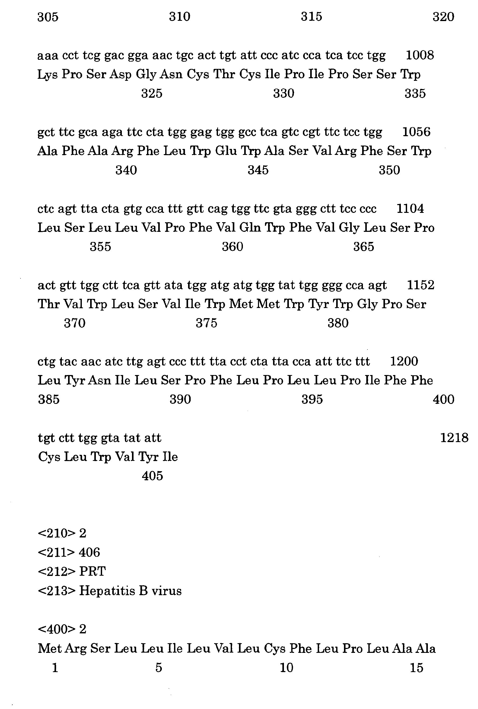


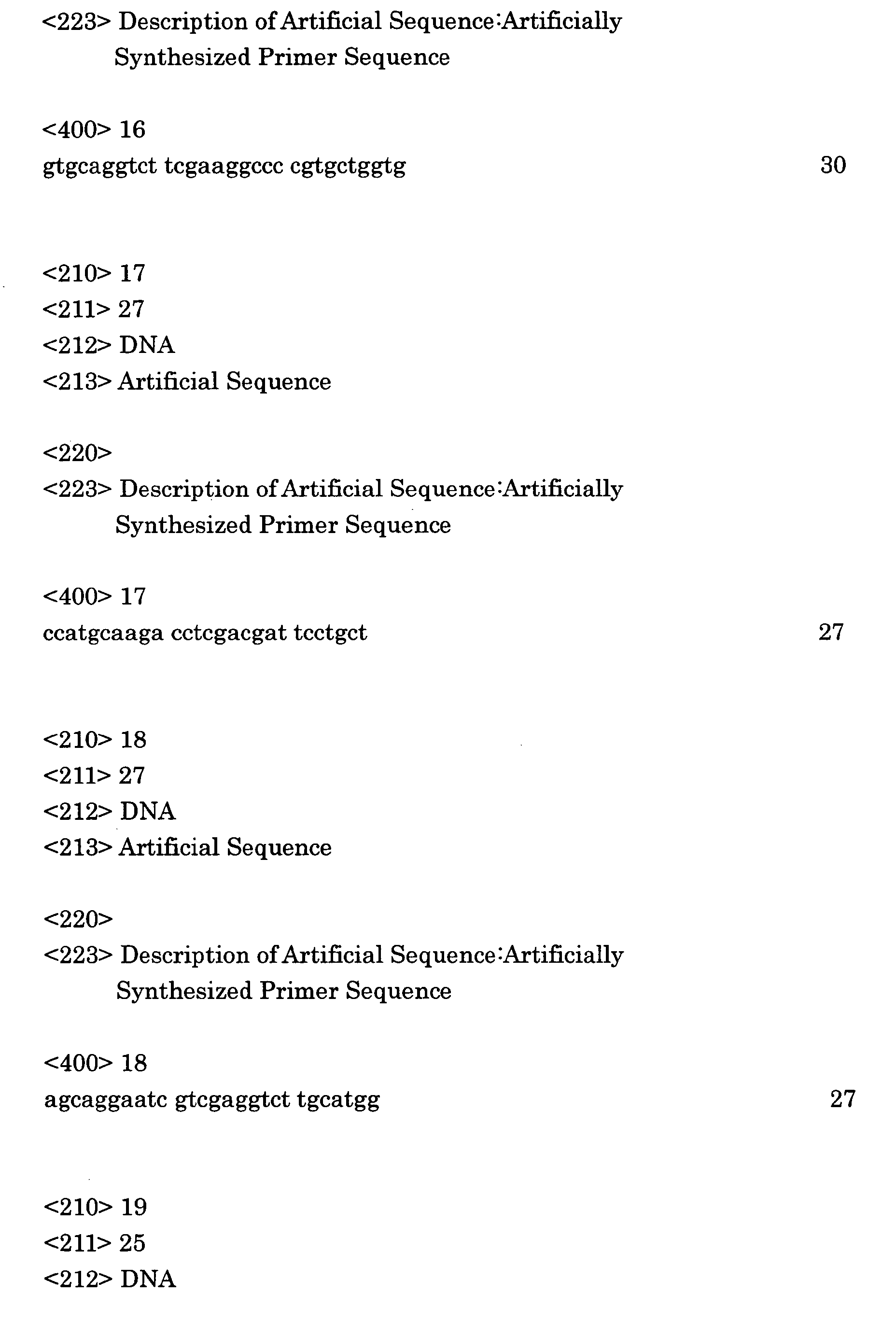
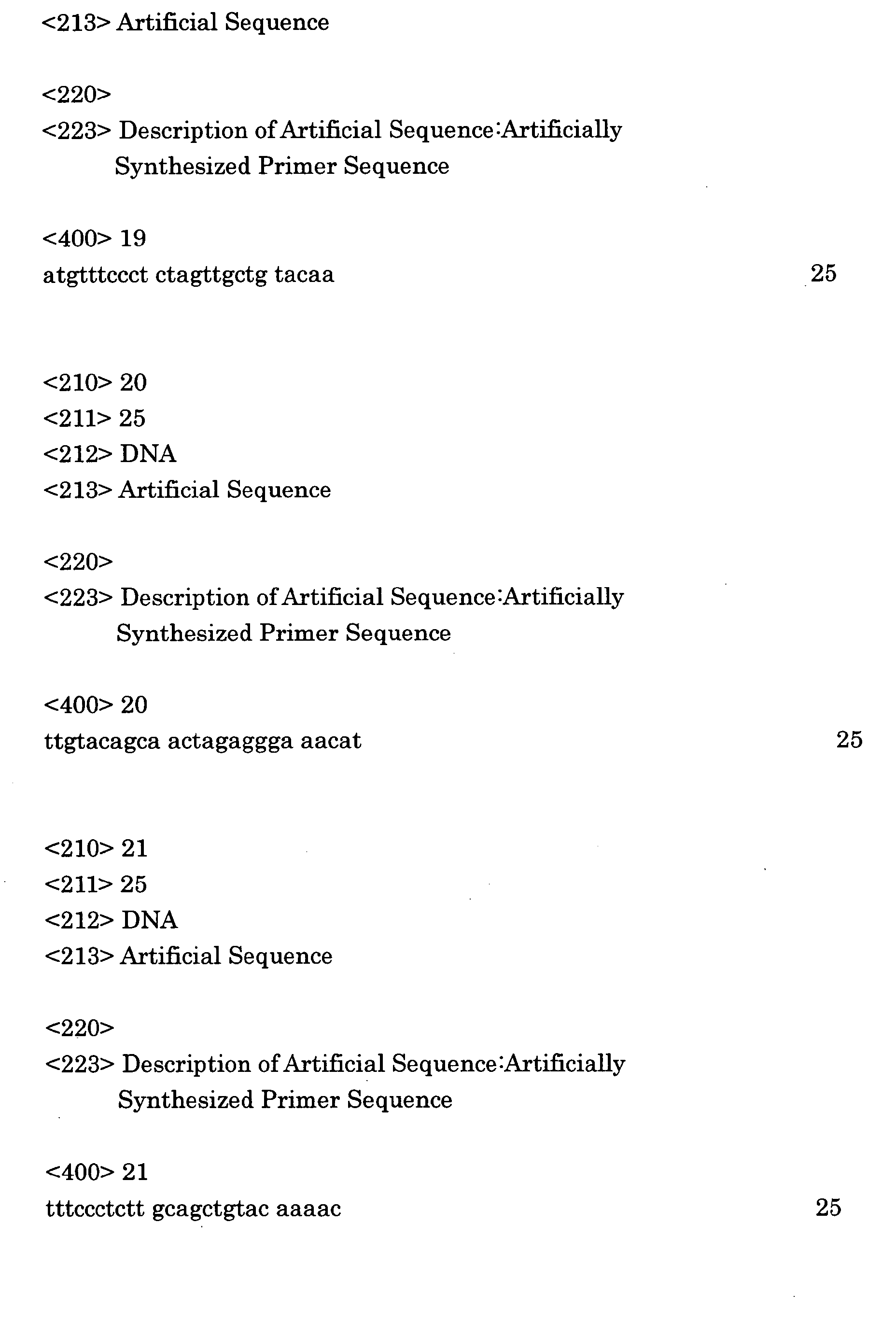


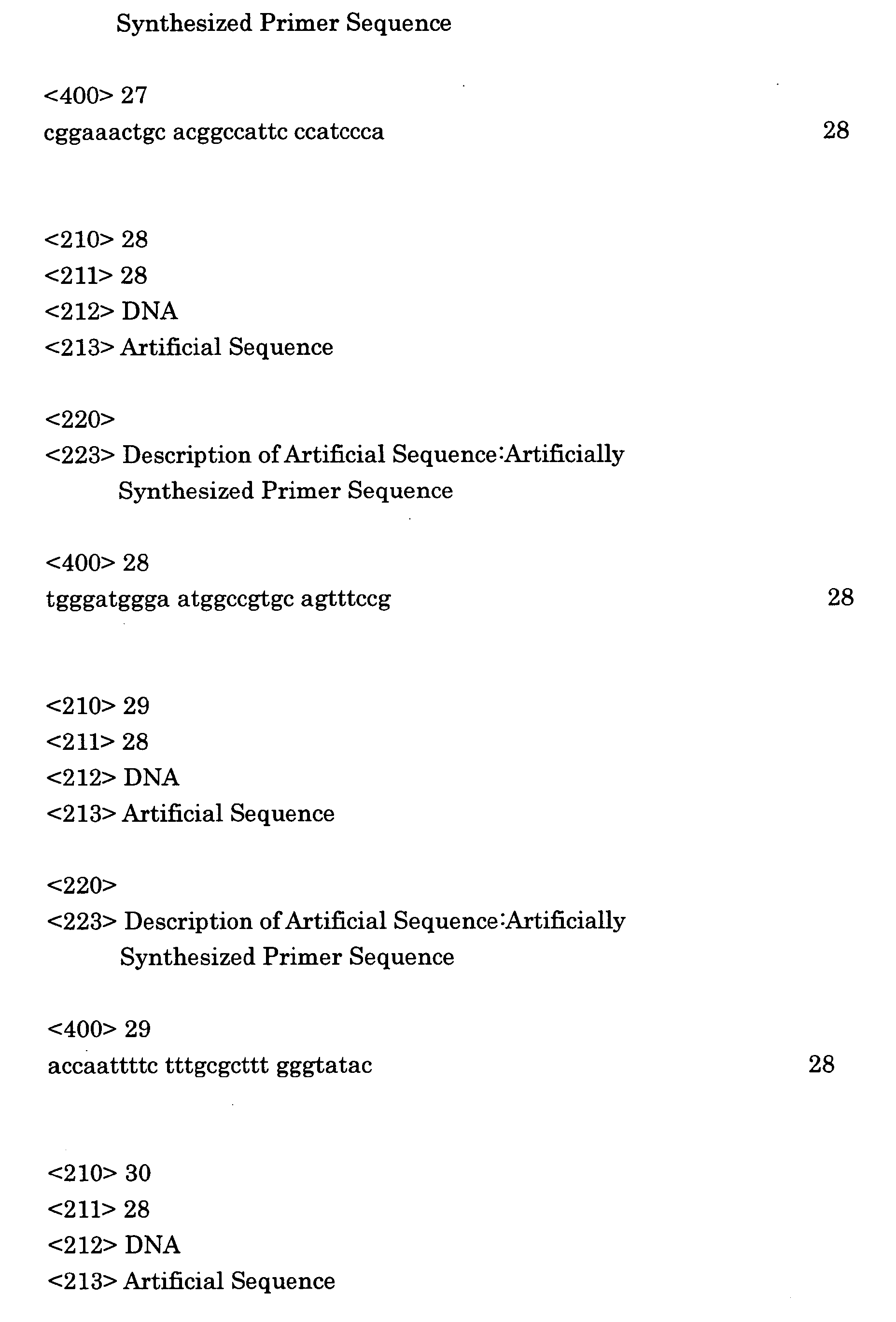
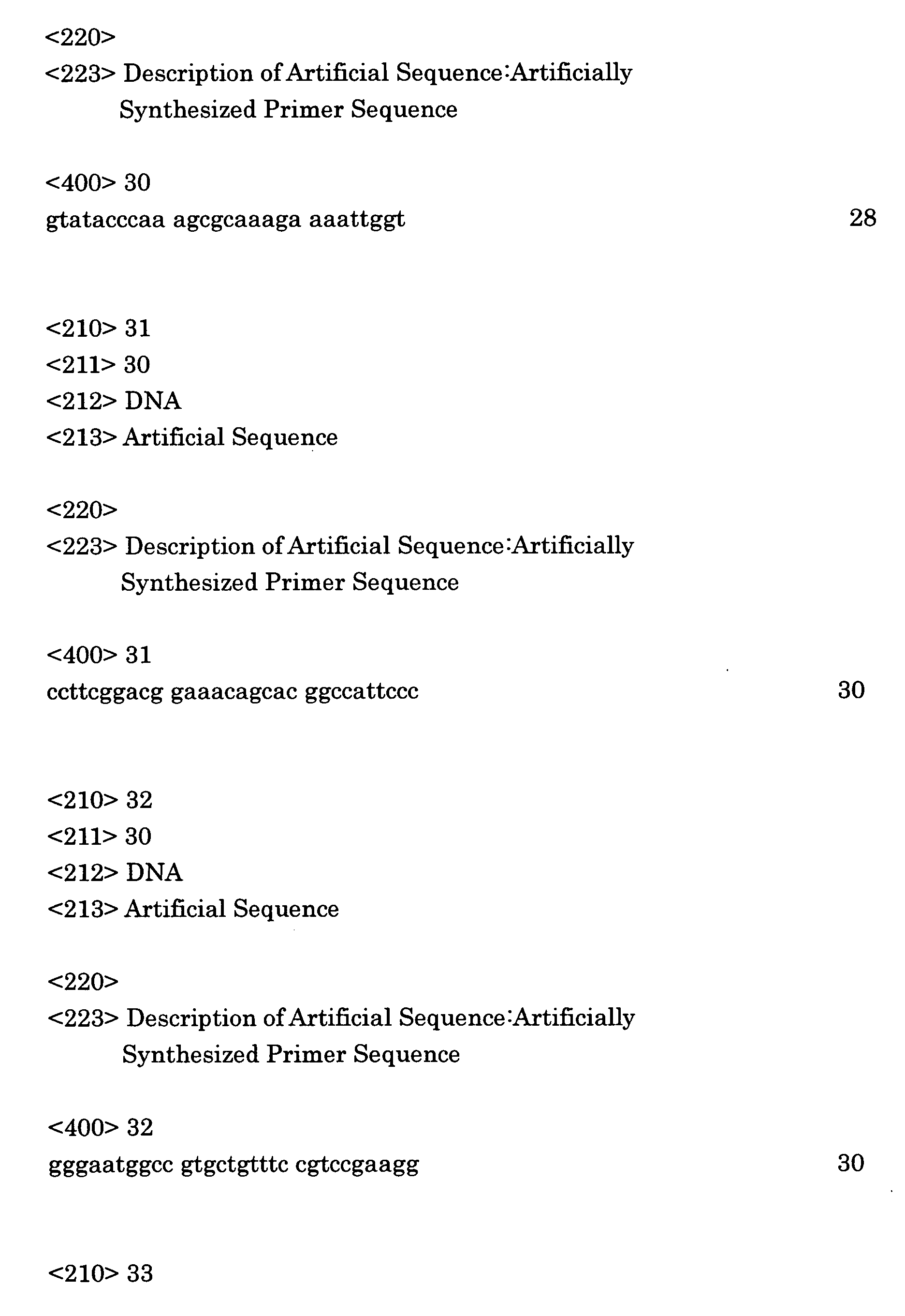


Claims (11)
- Hollow nanoparticles formed of a particle-forming protein with an ability to recognize a specific cell, wherein the protein contains at least one modified cysteine residue.
- Hollow nanoparticles as set forth in claim 1, wherein the protein comprises a hepatitis B virus surface-antigen protein.
- Hollow nanoparticles as set forth in claim 1 or 2, wherein at least one cysteine residue present in a transmembrane region is replaced with a hydrophobic amino acid, and/or at least one cysteine residue present outside or inside the particles is replaced with a hydrophilic amino acid.
- Hollow nanoparticles as set forth in claim 3, wherein at least one cysteine residue present in a transmembrane region is replaced with an alanine residue, and/or at least one cysteine residue present inside or outside the particles is replaced with a serine residue.
- Hollow nanoparticles as set forth in any one of claims 2 through 4, wherein the hepatitis B virus surface-antigen protein contains S protein with an amino acid sequence whose cysteine residues 76, 90, 139, 147, 149, 221 and at least one of cysteine residues 137 and 138 of the amino acid sequence from its N-terminus have been replaced.
- Hollow nanoparticles as set forth in any one of claims 1 through 5, wherein the cysteine residues are modified by mutating a gene that encodes the particle-forming protein and by expressing the mutated gene.
- Hollow nanoparticles as set forth in claim 6, which are obtained by transforming a eukaryotic cell with a vector including the mutated gene, and by expressing the mutated gene in the eukaryotic cell.
- Hollow nanoparticles as set forth in claim 7, wherein the eukaryotic cell is an animal cell or a yeast cell.
- A drug which comprises hollow nanoparticles of any one of claims 1 through 8 in which a substance to be transferred into a cell is encapsulated.
- A drug as set forth in claim 9, wherein the substance to be transferred into a cell comprises a gene.
- A therapeutic method using a drug of claim 9 or 10.
Applications Claiming Priority (5)
| Application Number | Priority Date | Filing Date | Title |
|---|---|---|---|
| JP2002191386 | 2002-06-28 | ||
| JP2002191386 | 2002-06-28 | ||
| PCT/JP2003/008244 WO2004002459A1 (en) | 2002-06-28 | 2003-06-27 | Hollow nanoparticle having modified cysteine residue and drug with the use thereof |
| JP2003183863A JP2004081210A (en) | 2002-06-28 | 2003-06-27 | Hollow nanoparticles composed of protein with modified cysteine residue and drugs using the same |
| JP2003183863 | 2003-06-27 |
Publications (2)
| Publication Number | Publication Date |
|---|---|
| EP1541135A1 true EP1541135A1 (en) | 2005-06-15 |
| EP1541135A4 EP1541135A4 (en) | 2005-09-28 |
Family
ID=30002325
Family Applications (1)
| Application Number | Title | Priority Date | Filing Date |
|---|---|---|---|
| EP03761834A Withdrawn EP1541135A4 (en) | 2002-06-28 | 2003-06-27 | HOLLOW NANOPARTICLE COMPRISING A MODIFIED CYSTEINE RESIDUE AND MEDICAMENT IN WHICH IT IS USED |
Country Status (5)
| Country | Link |
|---|---|
| US (1) | US20060141042A1 (en) |
| EP (1) | EP1541135A4 (en) |
| JP (1) | JP2004081210A (en) |
| KR (1) | KR100632073B1 (en) |
| WO (1) | WO2004002459A1 (en) |
Cited By (1)
| Publication number | Priority date | Publication date | Assignee | Title |
|---|---|---|---|---|
| US10898568B2 (en) | 2015-09-30 | 2021-01-26 | Boehringer Ingelheim Vetmedica Gmbh | Modular antigen transportation molecules and uses thereof in animals |
Families Citing this family (8)
| Publication number | Priority date | Publication date | Assignee | Title |
|---|---|---|---|---|
| WO2009106999A2 (en) * | 2008-02-28 | 2009-09-03 | Deutsches Krebsforschungszentrum, Stiftung Des Öffentlichen Rechts | Hollow nanoparticles and uses thereof |
| CN107252489A (en) | 2009-04-13 | 2017-10-17 | 法国健康和医学研究院 | HPV particles and application thereof |
| WO2013119877A1 (en) | 2012-02-07 | 2013-08-15 | Aura Biosciences, Inc. | Virion-derived nanospheres for selective delivery of therapeutic and diagnostic agents to cancer cells |
| WO2015042325A1 (en) | 2013-09-18 | 2015-03-26 | Aura Biosciences, Inc. | Virus-like particle conjugates for diagnosis and treatment of tumors |
| US9709713B1 (en) | 2014-06-18 | 2017-07-18 | Peter C. Chen | High quality telescope mirrors made from polymer matrix composite materials and method |
| KR20180095511A (en) | 2015-10-30 | 2018-08-27 | 더 유나이티드 스테이츠 오브 어메리카, 애즈 리프리젠티드 바이 더 세크러테리, 디파트먼트 오브 헬쓰 앤드 휴먼 서비씨즈 | Targeted Cancer Therapy |
| CN105713917A (en) * | 2016-02-20 | 2016-06-29 | 深圳市圣必智科技开发有限公司 | Preparation method of anti-hepatitis B surface antigen-antibody based on green fluorescent protein luminescence structural domain labeling |
| AU2018251788B2 (en) | 2017-04-12 | 2025-04-10 | Aura Biosciences, Inc. | Targeted combination therapy |
Family Cites Families (4)
| Publication number | Priority date | Publication date | Assignee | Title |
|---|---|---|---|---|
| FR2581394B1 (en) * | 1985-05-02 | 1988-08-05 | Grp Genie Genetique | PARTICLES HAVING IMMUNOGENIC PROPERTIES OF THE HBS ANTIGEN AND CARRYING A FOREIGN ANTIGENIC SITE TO THE EPITOPES CARRIED BY THE HBS ANTIGEN, ANIMAL VECTORS AND CELLS FOR THE PRODUCTION OF SUCH PARTICLES AND COMPOSITIONS CONTAINING SUCH PARTICLES FOR THE PRODUCTION |
| US5595739A (en) * | 1993-05-07 | 1997-01-21 | Abbott Laboratories | Hepatitis B virus mutants, reagents and methods for detection |
| EP0919568A1 (en) * | 1997-12-01 | 1999-06-02 | Sorin Diagnostics S.r.l. | Escape mutant of the surface antigen of hepatitis B virus |
| JP4085231B2 (en) * | 2000-02-28 | 2008-05-14 | 株式会社ビークル | Protein hollow nanoparticles, substance carrier using the same, and method for introducing substance into cells |
-
2003
- 2003-06-27 WO PCT/JP2003/008244 patent/WO2004002459A1/en not_active Ceased
- 2003-06-27 US US10/519,559 patent/US20060141042A1/en not_active Abandoned
- 2003-06-27 EP EP03761834A patent/EP1541135A4/en not_active Withdrawn
- 2003-06-27 KR KR1020047021415A patent/KR100632073B1/en not_active Expired - Fee Related
- 2003-06-27 JP JP2003183863A patent/JP2004081210A/en not_active Withdrawn
Cited By (1)
| Publication number | Priority date | Publication date | Assignee | Title |
|---|---|---|---|---|
| US10898568B2 (en) | 2015-09-30 | 2021-01-26 | Boehringer Ingelheim Vetmedica Gmbh | Modular antigen transportation molecules and uses thereof in animals |
Also Published As
| Publication number | Publication date |
|---|---|
| KR20050024420A (en) | 2005-03-10 |
| US20060141042A1 (en) | 2006-06-29 |
| EP1541135A4 (en) | 2005-09-28 |
| KR100632073B1 (en) | 2006-10-04 |
| WO2004002459A1 (en) | 2004-01-08 |
| JP2004081210A (en) | 2004-03-18 |
Similar Documents
| Publication | Publication Date | Title |
|---|---|---|
| US7597905B2 (en) | Hollow protein nano-particles and method of introducing substances into cells | |
| EP0038765B1 (en) | Vaccine against viral hepatitis b, method and transformed eucaryotic cells for the preparation of this vaccine | |
| CN116547000A (en) | Modified red blood cells and their use in the delivery of active agents | |
| EP1491210B1 (en) | Therapeutic drug using antibody-presenting hollow protein nanoparticles and hollow protein nanoparticles | |
| US20060141042A1 (en) | Hollow nanoparticle having modified cysteine residue and drug with the use thereof | |
| US20090011036A1 (en) | Drug containing hollow protein nanoparticles of particle-forming protein, fused with disease-treating target-cell-substance | |
| CN114903987A (en) | A kind of mRNA medicine for treating chronic hepatitis B virus infection and its preparation method and application | |
| KR100674325B1 (en) | Protein hollow nanoparticles and drugs using the same | |
| US6479464B1 (en) | Compositions and methods for highly efficient transfection | |
| EP1007549B1 (en) | Compositions and methods for highly efficient transfection | |
| CN120248078B (en) | A modified polypeptide based on ARC protein and its application as a nucleic acid delivery vector | |
| US20030100496A1 (en) | Compositions and methods for highly efficient transfection | |
| CN120732814B (en) | A lipid nanoparticle and its application | |
| NL2040094B1 (en) | Ferritin vaccine carrier, preparation method thereof, and application | |
| EP1609482A1 (en) | Drug for treating hemophilia and method of treating hemophilia using the same | |
| JP2003286198A (en) | Therapeutic agent using protein hollow nanoparticles that display growth factors etc. | |
| WO2012157945A2 (en) | Method for expressing a target protein at the surface of a vacuole, and drug delivery carrier | |
| WO2005049824A1 (en) | Hollow nanoparticle presenting biological structure-recognition sites, method of producing the same and utilization of the same |
Legal Events
| Date | Code | Title | Description |
|---|---|---|---|
| PUAI | Public reference made under article 153(3) epc to a published international application that has entered the european phase |
Free format text: ORIGINAL CODE: 0009012 |
|
| 17P | Request for examination filed |
Effective date: 20050122 |
|
| AK | Designated contracting states |
Kind code of ref document: A1 Designated state(s): AT BE BG CH CY CZ DE DK EE ES FI FR GB GR HU IE IT LI LU MC NL PT RO SE SI SK TR |
|
| A4 | Supplementary search report drawn up and despatched |
Effective date: 20050816 |
|
| RIC1 | Information provided on ipc code assigned before grant |
Ipc: 7A 61K 9/51 A |
|
| STAA | Information on the status of an ep patent application or granted ep patent |
Free format text: STATUS: THE APPLICATION HAS BEEN WITHDRAWN |
|
| 18W | Application withdrawn |
Effective date: 20080929 |