EP1536245A1 - NMR spectroscopy of proteins involving selective excitation by ESR - Google Patents
NMR spectroscopy of proteins involving selective excitation by ESR Download PDFInfo
- Publication number
- EP1536245A1 EP1536245A1 EP04027680A EP04027680A EP1536245A1 EP 1536245 A1 EP1536245 A1 EP 1536245A1 EP 04027680 A EP04027680 A EP 04027680A EP 04027680 A EP04027680 A EP 04027680A EP 1536245 A1 EP1536245 A1 EP 1536245A1
- Authority
- EP
- European Patent Office
- Prior art keywords
- electromagnetic wave
- sample
- protein
- spectroscopy analysis
- analysis according
- Prior art date
- Legal status (The legal status is an assumption and is not a legal conclusion. Google has not performed a legal analysis and makes no representation as to the accuracy of the status listed.)
- Granted
Links
- 238000005481 NMR spectroscopy Methods 0.000 title claims abstract description 83
- 102000004169 proteins and genes Human genes 0.000 title claims description 97
- 108090000623 proteins and genes Proteins 0.000 title claims description 97
- 230000005284 excitation Effects 0.000 title claims 2
- 230000008859 change Effects 0.000 claims abstract description 67
- 230000027455 binding Effects 0.000 claims abstract description 63
- 238000009739 binding Methods 0.000 claims abstract description 62
- 238000000034 method Methods 0.000 claims abstract description 37
- 235000018102 proteins Nutrition 0.000 claims description 66
- 239000003446 ligand Substances 0.000 claims description 44
- 238000004611 spectroscopical analysis Methods 0.000 claims description 31
- 238000005259 measurement Methods 0.000 claims description 30
- 238000004458 analytical method Methods 0.000 claims description 24
- CKLJMWTZIZZHCS-REOHCLBHSA-N aspartic acid group Chemical group N[C@@H](CC(=O)O)C(=O)O CKLJMWTZIZZHCS-REOHCLBHSA-N 0.000 claims description 20
- 238000001948 isotopic labelling Methods 0.000 claims description 14
- 238000001228 spectrum Methods 0.000 claims description 14
- 238000010276 construction Methods 0.000 claims description 7
- 125000004435 hydrogen atom Chemical class [H]* 0.000 claims description 6
- 238000000655 nuclear magnetic resonance spectrum Methods 0.000 claims description 5
- 125000000539 amino acid group Chemical group 0.000 claims description 4
- 238000001514 detection method Methods 0.000 claims description 4
- 229910052739 hydrogen Inorganic materials 0.000 claims description 4
- 239000001257 hydrogen Substances 0.000 claims description 4
- 125000000291 glutamic acid group Chemical group N[C@@H](CCC(O)=O)C(=O)* 0.000 claims description 2
- IJGRMHOSHXDMSA-UHFFFAOYSA-N Atomic nitrogen Chemical compound N#N IJGRMHOSHXDMSA-UHFFFAOYSA-N 0.000 claims 2
- OKTJSMMVPCPJKN-UHFFFAOYSA-N Carbon Chemical compound [C] OKTJSMMVPCPJKN-UHFFFAOYSA-N 0.000 claims 1
- 229910052799 carbon Inorganic materials 0.000 claims 1
- 229910052757 nitrogen Inorganic materials 0.000 claims 1
- 239000000523 sample Substances 0.000 abstract description 121
- 239000000126 substance Substances 0.000 abstract description 30
- 239000007864 aqueous solution Substances 0.000 abstract description 3
- 230000003993 interaction Effects 0.000 description 20
- 235000003704 aspartic acid Nutrition 0.000 description 19
- OQFSQFPPLPISGP-UHFFFAOYSA-N beta-carboxyaspartic acid Natural products OC(=O)C(N)C(C(O)=O)C(O)=O OQFSQFPPLPISGP-UHFFFAOYSA-N 0.000 description 19
- WHUUTDBJXJRKMK-UHFFFAOYSA-N Glutamic acid Natural products OC(=O)C(N)CCC(O)=O WHUUTDBJXJRKMK-UHFFFAOYSA-N 0.000 description 16
- WHUUTDBJXJRKMK-VKHMYHEASA-N L-glutamic acid Chemical compound OC(=O)[C@@H](N)CCC(O)=O WHUUTDBJXJRKMK-VKHMYHEASA-N 0.000 description 16
- 238000010586 diagram Methods 0.000 description 16
- 235000013922 glutamic acid Nutrition 0.000 description 16
- 239000004220 glutamic acid Substances 0.000 description 16
- 125000002915 carbonyl group Chemical group [*:2]C([*:1])=O 0.000 description 11
- 230000003068 static effect Effects 0.000 description 9
- 238000010494 dissociation reaction Methods 0.000 description 8
- 125000004429 atom Chemical group 0.000 description 7
- 230000005593 dissociations Effects 0.000 description 7
- 230000008569 process Effects 0.000 description 7
- 230000005540 biological transmission Effects 0.000 description 4
- 230000006916 protein interaction Effects 0.000 description 4
- 108091005804 Peptidases Proteins 0.000 description 3
- 239000004365 Protease Substances 0.000 description 3
- 102100037486 Reverse transcriptase/ribonuclease H Human genes 0.000 description 3
- XUIMIQQOPSSXEZ-UHFFFAOYSA-N Silicon Chemical compound [Si] XUIMIQQOPSSXEZ-UHFFFAOYSA-N 0.000 description 3
- 238000005411 Van der Waals force Methods 0.000 description 3
- 238000010521 absorption reaction Methods 0.000 description 3
- 238000000862 absorption spectrum Methods 0.000 description 3
- 230000003213 activating effect Effects 0.000 description 3
- 238000004925 denaturation Methods 0.000 description 3
- 230000036425 denaturation Effects 0.000 description 3
- 238000003929 heteronuclear multiple quantum coherence Methods 0.000 description 3
- 229910052710 silicon Inorganic materials 0.000 description 3
- 239000010703 silicon Substances 0.000 description 3
- 102000004190 Enzymes Human genes 0.000 description 2
- 108090000790 Enzymes Proteins 0.000 description 2
- 102000003886 Glycoproteins Human genes 0.000 description 2
- 108090000288 Glycoproteins Proteins 0.000 description 2
- 241000713772 Human immunodeficiency virus 1 Species 0.000 description 2
- 235000001014 amino acid Nutrition 0.000 description 2
- 150000001413 amino acids Chemical class 0.000 description 2
- 238000013459 approach Methods 0.000 description 2
- 239000013078 crystal Substances 0.000 description 2
- 239000003814 drug Substances 0.000 description 2
- 238000007876 drug discovery Methods 0.000 description 2
- 238000002474 experimental method Methods 0.000 description 2
- 238000010230 functional analysis Methods 0.000 description 2
- 238000013537 high throughput screening Methods 0.000 description 2
- 238000009413 insulation Methods 0.000 description 2
- 230000033001 locomotion Effects 0.000 description 2
- 238000001208 nuclear magnetic resonance pulse sequence Methods 0.000 description 2
- 108020004707 nucleic acids Proteins 0.000 description 2
- 102000039446 nucleic acids Human genes 0.000 description 2
- 150000007523 nucleic acids Chemical class 0.000 description 2
- 230000010355 oscillation Effects 0.000 description 2
- 230000002093 peripheral effect Effects 0.000 description 2
- 238000012545 processing Methods 0.000 description 2
- 238000001243 protein synthesis Methods 0.000 description 2
- 230000004044 response Effects 0.000 description 2
- 238000012916 structural analysis Methods 0.000 description 2
- 230000007704 transition Effects 0.000 description 2
- 230000014616 translation Effects 0.000 description 2
- UGFAIRIUMAVXCW-UHFFFAOYSA-N Carbon monoxide Chemical compound [O+]#[C-] UGFAIRIUMAVXCW-UHFFFAOYSA-N 0.000 description 1
- UFHFLCQGNIYNRP-UHFFFAOYSA-N Hydrogen Chemical compound [H][H] UFHFLCQGNIYNRP-UHFFFAOYSA-N 0.000 description 1
- 102100034343 Integrase Human genes 0.000 description 1
- 102100030856 Myoglobin Human genes 0.000 description 1
- 108010062374 Myoglobin Proteins 0.000 description 1
- 108010092799 RNA-directed DNA polymerase Proteins 0.000 description 1
- FFBHFFJDDLITSX-UHFFFAOYSA-N benzyl N-[2-hydroxy-4-(3-oxomorpholin-4-yl)phenyl]carbamate Chemical compound OC1=C(NC(=O)OCC2=CC=CC=C2)C=CC(=C1)N1CCOCC1=O FFBHFFJDDLITSX-UHFFFAOYSA-N 0.000 description 1
- 229910002091 carbon monoxide Inorganic materials 0.000 description 1
- 230000007850 degeneration Effects 0.000 description 1
- 238000013461 design Methods 0.000 description 1
- 230000001066 destructive effect Effects 0.000 description 1
- 238000011161 development Methods 0.000 description 1
- 238000003745 diagnosis Methods 0.000 description 1
- 201000010099 disease Diseases 0.000 description 1
- 208000037265 diseases, disorders, signs and symptoms Diseases 0.000 description 1
- 208000018459 dissociative disease Diseases 0.000 description 1
- 230000000694 effects Effects 0.000 description 1
- 238000005421 electrostatic potential Methods 0.000 description 1
- 230000001747 exhibiting effect Effects 0.000 description 1
- 150000003278 haem Chemical class 0.000 description 1
- 230000007246 mechanism Effects 0.000 description 1
- 125000004433 nitrogen atom Chemical group N* 0.000 description 1
- 230000001590 oxidative effect Effects 0.000 description 1
- 125000004430 oxygen atom Chemical group O* 0.000 description 1
- 238000002360 preparation method Methods 0.000 description 1
- 125000002924 primary amino group Chemical group [H]N([H])* 0.000 description 1
- 230000004850 protein–protein interaction Effects 0.000 description 1
- 230000035945 sensitivity Effects 0.000 description 1
- 239000000243 solution Substances 0.000 description 1
- 241000894007 species Species 0.000 description 1
- 230000008685 targeting Effects 0.000 description 1
Images





Classifications
-
- G—PHYSICS
- G01—MEASURING; TESTING
- G01R—MEASURING ELECTRIC VARIABLES; MEASURING MAGNETIC VARIABLES
- G01R33/00—Arrangements or instruments for measuring magnetic variables
- G01R33/20—Arrangements or instruments for measuring magnetic variables involving magnetic resonance
- G01R33/44—Arrangements or instruments for measuring magnetic variables involving magnetic resonance using nuclear magnetic resonance [NMR]
- G01R33/46—NMR spectroscopy
-
- G—PHYSICS
- G01—MEASURING; TESTING
- G01R—MEASURING ELECTRIC VARIABLES; MEASURING MAGNETIC VARIABLES
- G01R33/00—Arrangements or instruments for measuring magnetic variables
- G01R33/20—Arrangements or instruments for measuring magnetic variables involving magnetic resonance
- G01R33/60—Arrangements or instruments for measuring magnetic variables involving magnetic resonance using electron paramagnetic resonance
Definitions
- the present invention relates to judgment of the function and binding strength of molecules. More specifically, the present invention relates to a method and apparatus for judging the interaction of a protein-ligand complex and a protein-protein complex.
- SAR-by-NMR is a method which detects the interaction between target protein which has been subject to 15 N isotope labeling and ligand from the change in chemical shift to advance molecular design of ligand with stronger interaction
- Patent Document 1 WO 97/18469
- Non-Patent Document 1 Yoshiki Yamaguchi and Kazuo Shimada, "A new approach to a study on novel drug discovery using nuclear magnetic resonance", protein, nucleic acid, enzyme, 45 (2000) 895.
- the Affinity-NMR is a detection method which uses the change in relaxation time of a signal produced from the change in translational motion when ligand of low molecular weight is bound to target protein.
- the Affinity-NMR does not require a large amount of isotope labeled protein, measurement is relatively inexpensive. It can detect only the binding strength between protein and ligand. The position of an active region or a binding region and the structure of target protein cannot be found. It is difficult to obtain structural information as a guide for developing more excellent ligand.
- a protein-protein (glycoprotein) complex and huge protein are measured by combining selective isotope labeling performing protein synthesis using a medium in which specified amino acid has been subject to isotope labeling with nuclear magnetic resonance (see Non-Patent Document 2: J. Wigelt, M. Wikstrom, J. Schultz, M. J. P. van Dongen, Combinational Chemistry & High Throughput Screening, 5 (2002) 623). Only a resonant signal from the atom on the specified amino acid residue of a sample which has been subject to the selective isotope labeling is observed to make it possible to relegate the residue of protein of high molecular weight.
- ESR Error-senor-senor
- a technique for selectively supplying energy to the specified residue contributing to the binding of target protein in a mixed sample to control the state for changing the binding portion of the protein and a technique for measuring the presence or absence of binding and the process of interaction are important.
- An object of the present invention is to provide a method and apparatus which can control and detect the interaction of protein in a mixed sample andmeasure the process of interaction.
- a unit providing a uniform static magnetic field to a sample, an oscillator applying an electromagnetic wave in a THz field onto the sample, and a probe and an observing unit which can detect a nuclear magnetic resonance signal.
- FIGS. 1, 2 and 3 are diagrams schematically showing the arrangement of the components of a nuclear magnetic resonance (hereinafter, abbreviated as an NMR) preferable for embodying the present invention.
- FIGS. 1 and 2 are examples in which magnets for static magnetic field 6 divided into two, a sample 4 , and a probe coil for nuclear magnetic resonance 5 are arranged between the magnet for static magnetic fields 6.
- FIG. 3 is an example in which the sample 4 is arranged in a bore 7 of the magnet 6.
- the numeral 1 denotes a THz electromagnetic wave oscillator
- the numeral 2 denotes a THz electromagnetic wave detector
- the numeral 3 denotes a sample tube
- the numeral 4 denotes a sample
- the numeral 7 denotes a bore of the magnet 6
- the numeral 8 denotes an extension line of the probe coil for nuclear magnetic resonance 5
- the numeral 9 denotes a guide for introducing the sample tube 3.
- the THz electromagnetic wave oscillator 1 and the THz electromagnetic wave detector 2 have extension lines, which are omitted in the drawing.
- the probe coil for nuclear magnetic resonance 5 is a solenoid coil in FIGS. 1 and 2 and a saddle coil in FIG. 3.
- the THz electromagnetic wave oscillator 1 and the THz electromagnetic wave detector 2 are provided. In FIG. 2, only the THz electromagnetic wave oscillator 1 is provided.
- an electric signal is sent to the THz oscillator 1 to apply an electromagnetic wave onto the sample 4, a nuclear magnetic resonance signal of the sample is detected by the probe coil for nuclear magnetic resonance 5, and the binding change in target protein is observed.
- the probe coil for nuclear magnetic resonance 5 may be a transmission coil of an electromagnetic wave for nuclear magnetic resonance.
- the applied electromagnetic wave from the THz oscillator 1 is transmitted through the probe coil for nuclear magnetic resonance 5 and the sample tube 3 to reach the sample 4.
- the maximum sensitivity region of the probe coil 5 of the NMR and the THz electromagnetic wave reaching region are almost the same.
- the peripheral portion of the probe coil for nuclear magnetic resonance 5 of the mechanism part for holding the probe coil for nuclear magnetic resonance 5, the sample tube 3, and the guide 9 is provided with a protection construction for guard.
- the portion corresponding to the region of the protection construction is provided with THz electromagnetic wave transmission windows.
- FIGS. 4A and 4B are diagrams showing an example of the protection construction in which FIG. 4A is a cross-sectional view and FIG. 4B is a perspective view.
- FIGS. 4A and 4B are examples when the probe coil 5 is used at room temperature.
- the numeral 10 denotes a shield part of the protection construction.
- the numerals 13 and 13' denote THz electromagnetic wave incident and outgoing windows.
- the numeral 5 denotes a probe coil.
- the numerals 12 and 12' denote connection holes of the guide 9 for introducing the sample tube 3.
- the probe coils 5 are connected in series, in parallel, or in serial parallel and are extended to the outside by the extension lines 8 (FIGS. 1 to 3) , which is omitted in the drawing.
- the probe coil 5 may be of a solenoid type as shown in the drawing or may be of a saddle type as shown in FIG. 3.
- the guide 9 for introducing the sample tube 3 is coupled to the connection holes 12 and 12'.
- the sample tube 3 is introduced via the guide 9 and the connection hole 12 into the probe coil 5.
- the shield part 10 is provided with the THz electromagnetic wave incident and outgoing windows 13 and 13'. As shown in FIGS. 1 and 3, when the THz electromagnetic wave oscillator 1 and the THz electromagnetic wave detector 2 are provided, the incident and outgoing windows 13 and 13' are provided. As shown in FIG. 2, when only the THz electromagnetic wave oscillator 1 is provided, only the incident window 13 may be provided.
- the probe When the probe coil 5 is used at room temperature, the probe need not be sealed for insulation.
- the windows 13 and 13' for the THz electromagnetic wave transmission may be opened in part of the shield part 10.
- the window 13' which can observe a transmitted THz electromagnetic wave is provided on the opposite side of the THz electromagnetic wave incident window 13 so that NMR measurement and THz spectroscopy are performed at the same time. Unless there is any special condition, it is desirable that the incident windows 13 and 13' be only vacuum or air.
- the inside of the shield part 10 is at a very low temperature, not shown.
- the windows 13 and 13' for transmitting THz electromagnetic wave must be closed for insulation.
- the THz electromagnetic wave must be transmitted.
- the windows 13 and 13' shown in FIGS. 4A and 4B may be closed by a silicon crystal thin plate.
- a silicon crystal thin plate For instance, as understood from data described in the document such as American Institute of Physics Handbook, 3rd Edition ed. D. E. Gray (McGraw-Hill, 1972), it is optimum for the silicon crystal (silicon thin plate) to transmit the electromagnetic wave in the THz field.
- the THz field is a boundary field between an electromagnetic wave and light and is a frequency bandwidth in which large amounts of resonance and absorption of phonon of a sample exist.
- a sample When a sample is placed in a uniform static magnetic field and has unpaired spin, it has an energy level corresponding to the orientation of unpaired spin by the Zeeman effect.
- an electromagnetic wave having a resonance frequency of the difference of between energy levels When incident, transition occurs between the levels of unpaired spin.
- the frequency Under a high magnetic field in which the frequency reaches the THz field, the energy dissipates to the phonon mode existing near the resonance frequency.
- the dissipating energy passes through the process of the lattice system to be thermal energy in the residue near the unpaired spin. The energy is supplied only to the specified portion of the sample.
- the electronic state in the specified residue is changed by the binding or dissociation to/from ligand.
- energy supply to the specified residue is eliminated (or occurs).
- the dissipation (or emergence) of the signal change in chemical shift by NMR measurement can detect and observe interaction.
- the interaction and binding of the specified amino residue having a group with high binding activity such as carbonyl in the side chain are selectively changed to make it possible to observe the binding of protein of high molecular weight to ligand and the interaction between proteins by a nuclear magnetic resonance signal.
- a wavelength variable THz oscillator is used, resonance oscillation of the entire protein and resonance in the specified structure are provided to make it possible to observe its molecular structure and chemical change by nuclear magnetic resonance.
- the energy of a THz electromagnetic wave applied is low and 0.4 kJ/mol when the frequency is 1 THz.
- the problem of the denaturation of protein which must be considered in an experiment such as Ramann spectroscopy using light above infrared rays can be reduced.
- Protein included in the sample 4 has about 20 kinds of amino acid residues which are bound by peptide bond. Some of the residues having carbonyl in the portion called a side chain not contributing to the peptide bond exist. Depending on an aqueous solution condition, the side chain carbonyl has a doublet state in which a proton is ionized and one unpaired spin is provided near an oxygen atom.
- FIG. 5 shows the structural formula of the carbonyl in which a proton is ionized. As indicated by the arrows, when the entire sample is placed under a strong uniform static magnetic field, the unpaired electron spin of the side chain carbonyl is arranged in parallel with the magnetic field.
- the energy provided to the specified portion of protein is moved to the atom therenear so that structural change occurs along with it.
- the energy provided to the specified electron spin is above activating energy of the binding, the ligand-protein binding or protein-protein binding can be changed.
- protease of HIV-1 virus is given.
- the protein has a function of creating protease itself, reverse transcriptase, and integrarze from forerunner body protein of the HIV-1 virus.
- the portion exhibiting protease revitalization is known to be aspartic acid in the 25th position of the protein. It is known that the aspartic acid has carbonyl in the side chain and the carbonyl has unpaired spin and is bound to a molecule of obstruction medicine via hydrogen bond.
- a magnetic field corresponding to a resonance frequency of about 1 GHz of a proton is about 21 T (tesla).
- the resonance frequency of a pulse electromagnetic wave necessary for flipping unpaired electron spin is determined by the equation (1) showing the energy level of the Zeeman splitting.
- ⁇ g ⁇ H h
- g a g value of an electron
- ⁇ a Bohr magneton (9.274 ⁇ 10 -24 JT -1 )
- H is an external magnetic field strength.
- the resonance frequency of the unpaired spin corresponding to the external magnetic field strength of 21 T is about 500 GHz which is close to the THz field.
- the 500 GHz corresponds to 1.09 cal/mol.
- the unpaired spin brings about resonance.
- the energy of the electromagnetic wave is relaxed near the unpaired spin.
- An energy of about 20K is provided to the residue portion, which corresponds to about 1/100 of the van der Waals force in the aqueous solution.
- the energy provided by one application is weak and is about 1/100 of the van der Waals force.
- Very low is the possibility of the problem of the denaturation of protein concerned in the spectroscopy technique using an electromagnetic wave having a wavelength below infrared rays such as Ramann spectroscopy which has been widely used for the structural and functional analysis.
- the electromagnetic wave application is combined with the nuclear magnetic resonance measurement to make it possible to observe the state change of various samples.
- the state change includes not only change brought about in the entire protein but also selective change focusing on the specified residue. The observation of the change in a protein structure brought about by electromagnetic wave application will be described below in detail.
- FIG. 6 shows a target protein sample 103 obtained by subjecting protein 101 having aspartic acid or glutamic acid in the active center to isotope labeling 102 of aspartic acid or glutamic acid.
- the binding of nuclear spins to be observed is determined.
- the 1 H- 15 N-HMQC spectrum is used.
- an NOESY spectrum is used.
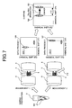
- FIG. 7 shows the procedure for observing the structural change in the protein by THz electromagnetic wave application.
- FIG. 8 is a schematic diagram which estimates the structural change (change amount of length between hydrogens) of the sample protein from the measured result obtained by the THz electromagnetic wave application described in FIG. 7. It is estimated that the length between hydrogens of the isotope labeling 102 of the sample protein is changed as indicated by the arrow according to the measured result shown in the upper stage. The magnitude corresponds to the ⁇ C and ⁇ S. It is found that information on the structural change near the active center of the protein sample caused by the THz-ray application can be obtained by the method for multiple spectroscopy analysis of the present invention. The information is combined with information on hydrogen whose relegation is known to make it possible to analyze the structural change and the change in the chemical characteristic such as electrostatic potential.
- FIG. 9 shows a target protein sample 105 obtained by binding, to ligand 104, sample protein obtained by subjecting the protein 101 having aspartic acid or glutamic acid in the active center to the isotope labeling 102 of aspartic acid or glutamic acid.
- sample preparation is performed so as to perform selective isotope labeling to the sample protein and binding the ligand 104 thereto.
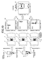
- FIG. 10 shows the procedure for observing the protein-ligand interaction.
- the energy provided by the THz electromagnetic wave 110 is supplied from the unpaired spin to aspartic acid or glutamic acid residue.
- the energy can be controlled by changing the pulse width of the THz electromagnetic wave and the number of times of pulse.
- the energy supplied to the binding portion to the ligand is smaller than the activating energy.
- the binding to the ligand is not dissociated and no change in the NMR spectrum occurs.
- the number of times of pulse and the pulse width are controlled to increase the energy supplied to the sample. In the state that the energy equivalent to the activating energy of binding can be supplied to the binding region, the change in NMR spectrum starts appearing at the peak 125.
- the peak 120 in the binding region in the sample 103 of the target protein as a reference obtained in measurement 1 is compared with the peak 125 obtained by allowing the THz electromagnetic wave 110 to be incident upon the sample 105 including the ligand on the same coordinate.
- Chemical shift change amount ⁇ C causedby application of the THz electromagnetic wave in the sample shown in FIG. 9 is obtained.
- FIG. 11 is a schematic diagram which estimates the binding strength between the sample protein and the ligand from the measured result by the THz electromagnetic wave application described in FIG. 10.
- the application of the THz electromagnetic wave necessary for changing the sample protein and the ligand from the binding state to the state sufficient for cutting the binding is determined.
- the peak 125 in FIG. 10 is almost the same as the peak 124.
- the peak 125 is close to the peak 120.
- the chemical shift change amount ⁇ C is closely related to the binding strength between the binding region and the ligand.
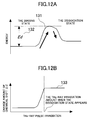
- FIG. 12A shows an energy curve 131 of the protein-ligand complex for explaining the behavior of the curve 133.
- Ed dissociation energy
- the ligand cannot be dissociated from the protein.
- the change amount of chemical shift is small or is not changed.
- the ligand 104 is away from the target protein 101 to increase the change amount of chemical shift.
- the THz electromagnetic wave application when the chemical shift is increased corresponds to the dissociation energy 132 of the ligand.
- the measurement of the change amount of chemical shift is performed to various ligands.
- the magnitude of the dissociation energy relative to the ligands can be evaluated.
- the energy is selectively supplied to the active center residue by electromagnetic wave application under a magnetic field to change the interaction, binding in the active center or the molecular structure for measurement by nuclear magnetic resonance.
- the binding strength and the structural change in the active center can be observed.
- a preprocess specifying the frequency of the applied THz electromagnetic wave must be performed.
- the frequency of the THz electromagnetic wave is variable.
- the THz electromagnetic wave detector 2 is used to determine the applying frequency while measuring the amount of transmission of the THz electromagnetic wave.
- the frequency of the electromagnetic wave absorbed in a magnetic field can be set to an unknown sample protein.
- the preprocess is advanced as follows.
- the NMR measuring apparatus is provided with open space and a probe construction which can allow the THz electromagnetic wave to be incident. Selective observation of the specified region by multiple spectroscopy measurement using the THz electromagnetic wave is found to be effective for observing protein-ligand binding and protein-protein interaction.
- sample including target protein and ligand, 110...THz electromagnetic wave, 120...peak signal of binding region observed by sample of target protein, 121...peak signal of binding region observed by allowing THz electromagnetic wave to be incident in sample of target protein, 124...peak signal of binding region observed by sample obtained by combining target protein with ligand, 125...peak signal of binding region observed by allowing THz electromagnetic wave to be incident by sample obtained by combining target protein with ligand, 131...energy curve of protein-ligand complex, 132...dissociation energy between ligand and protein, and 133...THz electromagnetic wave application-chemical shift change amount curve
Landscapes
- Physics & Mathematics (AREA)
- Spectroscopy & Molecular Physics (AREA)
- High Energy & Nuclear Physics (AREA)
- Condensed Matter Physics & Semiconductors (AREA)
- General Physics & Mathematics (AREA)
- Investigating Or Analysing Biological Materials (AREA)
- Investigating Or Analysing Materials By Optical Means (AREA)
Abstract
Description
[Patent Document 1] WO 97/18469
Claims (20)
- Amethod for multiple spectroscopy analysis comprising the steps of: applying a magnetic field to a sample; applying an electromagnetic wave in pulse form or in continuous wave form onto said sample; and detecting nuclear magnetic resonance of the sample, wherein a binding change in a specified portion of said sample is observed by a change in the nuclear magnetic resonance signal of said sample.
- The method for multiple spectroscopy analysis according to claim 1, wherein said application step applies the electromagnetic wave in pulse form or in continuous wave form corresponding to a resonance frequency in a magnetic field of unpaired spin existing in a residue side chain included in protein in the sample.
- The method for multiple spectroscopy analysis according to claim 1, wherein said application step is performed before, during, or after performing a nuclear spin excitation step of detecting nuclear magnetic resonance measurement.
- The method for multiple spectroscopy analysis according to claim 3, wherein the electromagnetic wave in pulse form or in continuous wave form is applied one or more times at predetermined time intervals from a predetermined time by a user.
- The method for multiple spectroscopy analysis according to claim 1, wherein said electromagnetic wave in pulse form or in continuous wave form has a frequency in a THz field.
- The method for multiple spectroscopy analysis according to claim 1, wherein in the step of detecting a nuclear magnetic resonance signal of the sample, the electromagnetic wave in pulse form or in continuous wave form is applied onto the sample.
- The method for multiple spectroscopy analysis according to claim 1, wherein any one of protein, a protein-ligand complex, and a protein-protein complex is targeted as said sample so that the specified binding change in the samples is observed by comparing spectra obtained by the nuclear magnetic resonance detection step.
- The method for multiple spectroscopy analysis according to claim 1, wherein in said step of observing the binding change in the specified portion in the sample, the user controls an application condition of the electromagnetic wave in pulse form or in continuous wave form based on a change in a nuclear magnetic resonance spectrum of said sample.
- The method for multiple spectroscopy analysis according to claim 1, wherein any one of an aspartic acid residue, a glutamic acid residue, and a natural amino acid residue other than these included in the protein existing in the sample, or an amino acid residue of an arbitrary combination of these is subject to isotope labeling in any one of hydrogen, carbon and nitrogen, or an arbitrary combination of these.
- The method for multiple spectroscopy analysis according to claim 1, wherein in the step of applying an electromagnetic wave in pulse form or in continuous wave form onto the sample, a frequency of said electromagnetic wave is continuously or intermittently modulated at a predetermined frequency or over a predetermined frequency range.
- An apparatus for multiple spectroscopy analysis comprising: means for applying a magnetic field to a sample; means for applying an electromagnetic wave in pulse form or in continuous wave form onto said sample; and observing and comparing means for detecting a nuclear magnetic resonance signal of said sample, wherein a binding change in a specified portion of said sample is observed by the nuclear magnetic resonance signal observing and comparing means.
- The apparatus for multiple spectroscopy analysis according to claim 11, wherein the electromagnetic wave in pulse form or in continuous wave form has a frequency in a THz field.
- The apparatus for multiple spectroscopy analysis according to claim 11, wherein the electromagnetic wave applied by said electromagnetic wave application means is an electromagnetic wave in pulse form or in continuous wave form having a resonance frequency corresponding to an applied magnetic field in unpaired spin existing in a residue side chain of protein existing in the sample.
- The apparatus for multiple spectroscopy analysis according to claim 11, wherein the electromagnetic wave applied by said electromagnetic wave application means has a function of continuously applying the electromagnetic wave onto the sample, and the nuclear magnetic resonance measuring means by the electromagnetic wave are operated at the application of the electromagnetic wave.
- The apparatus for multiple spectroscopy analysis according to claim 11, wherein said electromagnetic wave application means are operated before, during, or after performing nuclear magnetic resonance measurement.
- The apparatus for multiple spectroscopy analysis according to claim 11, further comprising an electromagnetic wave application unit having a function of setting and executing a scheme applying said electromagnetic wave in pulse form or in continuous wave form one or more times at predetermined time intervals from a predetermined time by a user.
- The apparatus for multiple spectroscopy analysis according to claim 16, wherein said electromagnetic wave application means apply an electromagnetic wave in pulse form or in continuous wave form whose frequency is continuously or intermittently modulated at a predetermined frequency or over a predetermined frequency range.
- The apparatus for multiple spectroscopy analysis according to claim 11, wherein a region positioned between the electromagnetic wave application unit and the sample in the unit detecting a nuclear magnetic resonance signal transmits the electromagnetic wave.
- The apparatus for multiple spectroscopy analysis according to claim 11, wherein a unit observing the binding change in the specified portion of the sample has a function of recording a plurality of nuclear magnetic resonance observed data pieces in which the application condition of the electromagnetic wave in pulse form or in continuous wave form is different, and a function of performing data operation such as addition and subtraction of the observed data pieces.
- The apparatus for multiple spectroscopy analysis according to claim 11, wherein a magnetic field application unit has a construction having open space which can provide an electromagnetic wave waveguide between an electromagnetic wave oscillator and the sample.
Applications Claiming Priority (2)
| Application Number | Priority Date | Filing Date | Title |
|---|---|---|---|
| JP2003395167 | 2003-11-26 | ||
| JP2003395167A JP4192078B2 (en) | 2003-11-26 | 2003-11-26 | Nuclear magnetic resonance method |
Publications (2)
| Publication Number | Publication Date |
|---|---|
| EP1536245A1 true EP1536245A1 (en) | 2005-06-01 |
| EP1536245B1 EP1536245B1 (en) | 2007-07-11 |
Family
ID=34463793
Family Applications (1)
| Application Number | Title | Priority Date | Filing Date |
|---|---|---|---|
| EP04027680A Expired - Lifetime EP1536245B1 (en) | 2003-11-26 | 2004-11-22 | NMR spectroscopy of proteins involving selective excitation by ESR |
Country Status (4)
| Country | Link |
|---|---|
| US (1) | US7202664B2 (en) |
| EP (1) | EP1536245B1 (en) |
| JP (1) | JP4192078B2 (en) |
| DE (1) | DE602004007455T2 (en) |
Families Citing this family (4)
| Publication number | Priority date | Publication date | Assignee | Title |
|---|---|---|---|---|
| JP4402026B2 (en) * | 2005-08-30 | 2010-01-20 | キヤノン株式会社 | Sensing device |
| JP2007298453A (en) | 2006-05-01 | 2007-11-15 | Canon Inc | Sample analysis method and apparatus using nuclear magnetic resonance with terahertz wave irradiation |
| WO2014018548A2 (en) * | 2012-07-23 | 2014-01-30 | University Of Washington Through Its Center For Commercialization | Method and system for concentrating magnetization of nuclear spins |
| CN115963141B (en) * | 2022-12-28 | 2025-11-28 | 苏州纽迈分析仪器股份有限公司 | Nuclear magnetic resonance and near infrared based combined equipment and method thereof |
Family Cites Families (9)
| Publication number | Priority date | Publication date | Assignee | Title |
|---|---|---|---|---|
| GB8819753D0 (en) * | 1988-08-19 | 1988-09-21 | Nycomed As | Apparatus |
| EP0726444B1 (en) * | 1995-02-10 | 2001-10-31 | Bruker Analytik Gmbh | Magnetic resonance method and apparatus for detecting an atomic structure of a sample along a surface thereof |
| US5698401A (en) * | 1995-11-14 | 1997-12-16 | Abbott Laboratories | Use of nuclear magnetic resonance to identify ligands to target biomolecules |
| AU711092B2 (en) | 1995-11-14 | 1999-10-07 | Abbvie Inc. | Use of nuclear magnetic resonance to design ligands to target biomolecules |
| US5891643A (en) * | 1995-11-14 | 1999-04-06 | Abbott Laboratories | Use of nuclear magnetic resonance to design ligands to target biomolecules |
| US6428956B1 (en) * | 1998-03-02 | 2002-08-06 | Isis Pharmaceuticals, Inc. | Mass spectrometric methods for biomolecular screening |
| US6348793B1 (en) * | 2000-11-06 | 2002-02-19 | Ge Medical Systems Global Technology, Company, Llc | System architecture for medical imaging systems |
| US20040038216A1 (en) * | 2002-08-23 | 2004-02-26 | Hajduk Philip J. | Method for the structural determination of ligands bound to macromolecular targets by nuclear magnetic resonance |
| US20040082075A1 (en) * | 2002-10-25 | 2004-04-29 | Robert Powers | Multi-step NMR for the identification and evaluation of biomolecule-compound interactions |
-
2003
- 2003-11-26 JP JP2003395167A patent/JP4192078B2/en not_active Expired - Fee Related
-
2004
- 2004-11-22 DE DE602004007455T patent/DE602004007455T2/en not_active Expired - Lifetime
- 2004-11-22 EP EP04027680A patent/EP1536245B1/en not_active Expired - Lifetime
- 2004-11-26 US US10/996,426 patent/US7202664B2/en not_active Expired - Fee Related
Non-Patent Citations (6)
| Title |
|---|
| DE JONG EAM ET AL: "Mapping of ligand binding sites on macromolecules by means of spin-labeled ligands and 2D difference spectroscopy", JOURNAL OF MAGNETIC RESONANCE, vol. 80, 1988, pages 197 - 213, XP009045055 * |
| DEROSE VJ, HOFFMAN BM: "Protein structure and mechanism studied by electron nuclear double resonance spectroscopy", METHODS IN ENZYMOLOGY, vol. 246, 1995, pages 554 - 589, XP009044992 * |
| FIELDING L: "NMR methods for the determination of protein-ligand dissociation constants", CURRENT TOPICS IN MEDICINAL CHEMISTRY, vol. 3, 2003, pages 39 - 53, XP009045029 * |
| IMPROTA S ET AL: "Probing protein structure by solvent perturbation of NMR spectra", EUR. J. BIOCHEM., vol. 227, 1995, pages 78 - 86, XP002320845 * |
| JAHNKE W: "Spin labels as a tool to identify and characterize protein - ligand interactions by NMR spectroscopy", CHEMBIOCHEM, vol. 3, 2002, pages 167 - 173, XP002320844 * |
| MEYER B, PETERS T: "NMR spectroscopy techniques for screening and identifying ligand binding to protein receptors", ANGEW. CHEM. INT. ED., vol. 42, no. 8, 24 February 2003 (2003-02-24), pages 864 - 890, XP002320843 * |
Also Published As
| Publication number | Publication date |
|---|---|
| DE602004007455T2 (en) | 2008-03-13 |
| US7202664B2 (en) | 2007-04-10 |
| JP4192078B2 (en) | 2008-12-03 |
| DE602004007455D1 (en) | 2007-08-23 |
| EP1536245B1 (en) | 2007-07-11 |
| JP2005156345A (en) | 2005-06-16 |
| US20050110495A1 (en) | 2005-05-26 |
Similar Documents
| Publication | Publication Date | Title |
|---|---|---|
| EP0277745B1 (en) | Methods and apparatus for detecting certain compounds | |
| AU699894B2 (en) | Detection and sorting of materials | |
| Shin et al. | Room-temperature operation of a radiofrequency diamond magnetometer near the shot-noise limit | |
| US7924006B2 (en) | Method and apparatus for analyzing sample utilizing nuclear magnetic resonance under terahertz-wave irradiation | |
| WO2010120783A1 (en) | Detection of j-coupling using atomic magnetometer | |
| Grosse et al. | Field cycling by fast NMR probe transfer: Design and application in field-dependent CIDNP experiments | |
| JP4599490B2 (en) | Method and configuration of NMR spectroscopy | |
| Kanorsky et al. | Millihertz magnetic resonance spectroscopy of Cs atoms in body-centered-cubic He 4 | |
| US3792346A (en) | Proton-enhanced nuclear induction spectroscopy | |
| Rudakov | Some aspects of the effective detection of ammonium nitrate-based explosives by pulsed NQR method | |
| Steber et al. | Structural studies of biomolecules in the gas phase by chirped-pulse Fourier transform microwave spectroscopy | |
| US7202664B2 (en) | Method and apparatus for multiple spectroscopy analysis by using nuclear magnetic resonance | |
| Rudakov et al. | Modifications of the steady-state free-precession sequence for the detection of pure nuclear quadrupole resonance | |
| Eaton et al. | Saturation recovery EPR | |
| Blank | A new approach to distance measurements between two spin labels in the> 10 nm range | |
| Skalla et al. | Optical magnetic resonance imaging of Rb vapor in low magnetic fields | |
| Heise et al. | Two-dimensional high-resolution NMR spectra in matched B0 and B1 field gradients | |
| US5528146A (en) | Method and apparatus for detecting electron spin transitions in zero field | |
| Harrison et al. | The Zeeman Effect on Lines in the (1, 0) Band of the F 4Δ-X 4Δ Transition of the FeH Radical | |
| Chen et al. | Single-shot spatially-localized NQR using field-dependent relaxation rates | |
| Lurie et al. | Continuous-wave NMR imaging of solids | |
| US12313713B2 (en) | Methods and systems for acquiring magnetic resonance data from a sample | |
| Sweger et al. | The effect of spin polarization in DEER spectroscopy | |
| Melnikov et al. | Application of pulsed heating in time-resolved EPR spectroscopy for longitudinal relaxation measurements | |
| Maciejewski | Basics of NMR Spectroscopy |
Legal Events
| Date | Code | Title | Description |
|---|---|---|---|
| PUAI | Public reference made under article 153(3) epc to a published international application that has entered the european phase |
Free format text: ORIGINAL CODE: 0009012 |
|
| AK | Designated contracting states |
Kind code of ref document: A1 Designated state(s): AT BE BG CH CY CZ DE DK EE ES FI FR GB GR HU IE IS IT LI LU MC NL PL PT RO SE SI SK TR |
|
| AX | Request for extension of the european patent |
Extension state: AL HR LT LV MK YU |
|
| 17P | Request for examination filed |
Effective date: 20051125 |
|
| AKX | Designation fees paid |
Designated state(s): DE |
|
| GRAP | Despatch of communication of intention to grant a patent |
Free format text: ORIGINAL CODE: EPIDOSNIGR1 |
|
| GRAS | Grant fee paid |
Free format text: ORIGINAL CODE: EPIDOSNIGR3 |
|
| GRAA | (expected) grant |
Free format text: ORIGINAL CODE: 0009210 |
|
| AK | Designated contracting states |
Kind code of ref document: B1 Designated state(s): DE |
|
| REF | Corresponds to: |
Ref document number: 602004007455 Country of ref document: DE Date of ref document: 20070823 Kind code of ref document: P |
|
| PLBE | No opposition filed within time limit |
Free format text: ORIGINAL CODE: 0009261 |
|
| STAA | Information on the status of an ep patent application or granted ep patent |
Free format text: STATUS: NO OPPOSITION FILED WITHIN TIME LIMIT |
|
| 26N | No opposition filed |
Effective date: 20080414 |
|
| PGFP | Annual fee paid to national office [announced via postgrant information from national office to epo] |
Ref country code: DE Payment date: 20141118 Year of fee payment: 11 |
|
| REG | Reference to a national code |
Ref country code: DE Ref legal event code: R119 Ref document number: 602004007455 Country of ref document: DE |
|
| PG25 | Lapsed in a contracting state [announced via postgrant information from national office to epo] |
Ref country code: DE Free format text: LAPSE BECAUSE OF NON-PAYMENT OF DUE FEES Effective date: 20160601 |