EP1533613A1 - Soluble transferrin receptor - Google Patents
Soluble transferrin receptor Download PDFInfo
- Publication number
- EP1533613A1 EP1533613A1 EP04025210A EP04025210A EP1533613A1 EP 1533613 A1 EP1533613 A1 EP 1533613A1 EP 04025210 A EP04025210 A EP 04025210A EP 04025210 A EP04025210 A EP 04025210A EP 1533613 A1 EP1533613 A1 EP 1533613A1
- Authority
- EP
- European Patent Office
- Prior art keywords
- plasma
- concentration
- markers
- stfr
- patients
- Prior art date
- Legal status (The legal status is an assumption and is not a legal conclusion. Google has not performed a legal analysis and makes no representation as to the accuracy of the status listed.)
- Withdrawn
Links
Images
Classifications
-
- G—PHYSICS
- G01—MEASURING; TESTING
- G01N—INVESTIGATING OR ANALYSING MATERIALS BY DETERMINING THEIR CHEMICAL OR PHYSICAL PROPERTIES
- G01N33/00—Investigating or analysing materials by specific methods not covered by groups G01N1/00 - G01N31/00
- G01N33/48—Biological material, e.g. blood, urine; Haemocytometers
- G01N33/50—Chemical analysis of biological material, e.g. blood, urine; Testing involving biospecific ligand binding methods; Immunological testing
- G01N33/68—Chemical analysis of biological material, e.g. blood, urine; Testing involving biospecific ligand binding methods; Immunological testing involving proteins, peptides or amino acids
- G01N33/6893—Chemical analysis of biological material, e.g. blood, urine; Testing involving biospecific ligand binding methods; Immunological testing involving proteins, peptides or amino acids related to diseases not provided for elsewhere
-
- G—PHYSICS
- G01—MEASURING; TESTING
- G01N—INVESTIGATING OR ANALYSING MATERIALS BY DETERMINING THEIR CHEMICAL OR PHYSICAL PROPERTIES
- G01N2333/00—Assays involving biological materials from specific organisms or of a specific nature
- G01N2333/435—Assays involving biological materials from specific organisms or of a specific nature from animals; from humans
- G01N2333/79—Transferrins, e.g. lactoferrins, ovotransferrins
-
- G—PHYSICS
- G01—MEASURING; TESTING
- G01N—INVESTIGATING OR ANALYSING MATERIALS BY DETERMINING THEIR CHEMICAL OR PHYSICAL PROPERTIES
- G01N2800/00—Detection or diagnosis of diseases
- G01N2800/04—Endocrine or metabolic disorders
- G01N2800/042—Disorders of carbohydrate metabolism, e.g. diabetes, glucose metabolism
-
- G—PHYSICS
- G01—MEASURING; TESTING
- G01N—INVESTIGATING OR ANALYSING MATERIALS BY DETERMINING THEIR CHEMICAL OR PHYSICAL PROPERTIES
- G01N2800/00—Detection or diagnosis of diseases
- G01N2800/32—Cardiovascular disorders
- G01N2800/324—Coronary artery diseases, e.g. angina pectoris, myocardial infarction
Definitions
- the invention concerns a method for detecting coronary syndromes, in particular, coronary artery disease (CAD), using risk markers.
- CAD coronary artery disease
- a number of markers for example, troponin T, C-reactive protein (CRP) as well as brain natriuretic peptide (BNP), are known for diagnosing coronary diseases such as NSTEMI and acute coronary syndrome. Elevation of the concentration of one of these markers is associated with an increase in the likelihood of ischemic events including death. Further, it has already been found that CRP and troponin I or troponin T are two independent markers for risk stratification of patients suffering from acute coronary syndrome.
- this object is achieved by using sTfR (soluble transferrin receptor) or/and frataxin or/and sTfR/log ferritin (ferritin index) as a risk marker for coronary syndromes or/and diabetes mellitus.
- the invention thus, relates to the use of sTfR for a novel purpose as well as to the use of the novel marker frataxin for diagnosing or prognosing coronary diseases or/and the risk of diabetes mellitus.
- sTfR or/and frataxin are preferably used as markers and more preferably sTfR and frataxin and ferritin index.
- a BNP peptide a CRP peptide, a troponin peptide, hepcidin or fragments thereof, in particular, hs-CRP, hepcidin, NT-proBNP or/and troponin T.
- the above-mentioned compounds can be used as cardiac biomarkers and are strong predictors of risk among patients for acute coronary syndromes (ACS) or/and diabetes mellitus.
- ACS acute coronary syndromes
- inreased levels of the aforementioned compounds are associated with higher rates of death and recurrent ischemic events.
- biomarkers whereby at least one marker selected from the groups: 1) soluble transferrin receptor (sTfR) and/or frataxin and/or ferritin index, 2) CRP and/or hepcidin, 3) a BNP peptide as well as 4) a troponin peptide or fragments of those markers are used.
- sTfR soluble transferrin receptor
- CRP CRP
- hepcidin CRP and/or hepcidin
- BNP peptide a troponin peptide or fragments of those markers
- Elevation in sTfR is a marker for functional iron deficiency in chronic diseases. Functional iron deficiency thereby is characterized by sTfR values greater 4 mg/L. It has now been found that it is also an early prognostic marker of coronary syndromes, in particular, coronary syndromes in chronic infection and inflammation processes.
- sTfR is the earliest marker of anemia of chronic disease (ACD) and has now been recognized as an early risk marker for coronary artery disease (CAD).
- sTfR is a transmembrane protein. sTfR binds diferric transferrin, thereby delivering iron to the cytosol. In the case of an increased cellular demand for iron the sTfR expression is increased to facilitate iron uptake.
- Cytosolic iron content is regulated by the enzyme aconitase, an iron-sulfur protein.
- aconitase binds to iron-responsive element-binding protein (IRE-BP), leading to an iron uptake.
- IRE-BP iron-responsive element-binding protein
- the iron uptake again is downregulated by the protein frataxin.
- mitochondrial iron accumulation is triggered by the lack or decrease of frataxin.
- Mitochondrial iron overload causes a damage to mitochondrial functions through the iron-sulfur (Fe-S) cluster-containing subunits of the respiratory complex.
- Fe-S iron-sulfur
- An sTfR therefore, can be used as a marker of mitochondrial dysfunction.
- a damage to mitochondrial functions has the following consequences:
- sTfR has now been found to be useful also as a marker in coronary diseases, in particular, in coronary diseases on cardiac muscle cells.
- Mitochondrial defects would preferentially be seen on tissues that generate energy by respiratory oxidation.
- Cardiac myocytes derive most of their ATP from the oxidation of free fatty acids. Therefore, decreased ATP generation leads to increased H 2 O 2 production in cardiac muscle (so-called iron-induced oxidative stress). Therefore, mainly cardiac myocytes are victims of deficits in mitochondrial ATP production.
- cellular defects as in myocardial infarction and chronic inflammation will have an impact on the elevated concentration of sTfR.
- sTfR can be used as a biochemical marker for risk stratification among patients suffering from cellular defects as occurring in mycoardial infarction and chronic inflammation.
- concentration of sTfR correlates with the degree of cellular damage in the patient's tissue.
- sTfR as an independent risk marker in coronary syndromes or/and diabetes mellitus allows to determine particular diseases in patients and, thus, to determine effective therapies, e.g. effective Epo therapy.
- rH-Epo protects the myocardion from ischemia reperfusion injury and promotes beneficial remodelling.
- the therapeutic role of recombinant human Epo (rH-Epo) in the treatment of myocardial ischemia and infarction can be explained by its role in the regulation of the functional iron deficiency, its role as a tissue-protective cytokine and its role in the regulation of deficits in mitochondrial ATP production.
- amounts of > 2.5 mg/l, more preferred of > 3 mg/l and, in particular, > 4 mg/l of sTfR are considered as an indication of coronary syndromes and/or a risk of coronary syndromes and/or of diabetes mellitus and/or the risk of diabetes mellitus.
- the determination of sTfR is a sensitive tool for the assessment of functional iron deficiency in different patient groups.
- sTfR values were significantly higher in patients compared to healthy controls.
- the assessment of sTfR allows to stratify coronary risks, in particular, among patients with chronic diseases more effectively than by established biochemical markers (heart diseases, diabetes, renal failure, rheumatoid arthritis).
- the assessment of sTfR, optionally in combination with the ferritin index further allows to stratify the risk of diabetes mellitus.
- frataxin is used as a risk marker for coronary syndromes and/or diabetes mellitus. It was found that frataxin is one of the earliest markers of functional iron deficiency and a risk marker for coronary artery disease (CAD).
- CAD coronary artery disease
- sTfR/log ferritin also designated ferritin index
- ferritin index is the ratio of soluble transferrin receptor concentration to ferritin concentration.
- Values of sTfR > 4 mg/l and of ferritin > 100 ⁇ g/l reflect a functional iron deficit.
- Characteristic for latent iron deficit are values of ferritin ⁇ 100 ⁇ g/l while sTfR usually exceeds > 4 mg/l, without an increased risk of coronary syndromes. Thus, knowledge of the ferritin value is decisive for ferritin index evaluation.
- values of the ferritin index of ⁇ 2 for CRP > 5 mg or ⁇ 3.2 for CRP ⁇ 5 mg are considered as an indication of coronary syndromes and a risk of coronary syndromes or/and of diabetes mellitus or/and a risk of diabetes mellitus.
- Latent iron deficit Risk of coronary syndrome (functional iron deficit) sTfR > 4 mg/l > 4 mg/l ferritin ⁇ 100 ⁇ g/l > 100 ⁇ g/l sTfR [mg/l]/log ferritin > [ ⁇ g/l] with acute phase (CRP > 5 mg/l) 2 ⁇ 2 sTfR [mg/l]/log ferritin > [ ⁇ g/l] without acute phase (CRP ⁇ 5 mg/l) 3.2 ⁇ 3.2
- the invention also relates to the use of sTfR or/and frataxin or/and ferritin index as a risk marker in the manufacture of an agent to detect and/or predict coronary syndromes or/and diabetes mellitus as well as to the in vitro use of sTfR or/and frataxin or/and ferritin index as a risk marker for coronary syndromes or/and diabetes mellitus.
- One or both or all three of the mentioned markers are preferably combined with established cardiac biomarkers.
- CRP in particular, hs-CRP and/or hepcidin have been used primarily as a marker of systemic chronic inflammation. It is now appreciated, however, that inflammation also plays a central role in arteriosclerosis and its complications. Thus, CRP and/or hepcidin may not only reflect the degree of underlying inflammation predisposing to arteriosclerosis but also play a direct role in promoting plaque rupture and thrombosis. Preferably, amounts of > 2 mg/l (referring to blood), more preferred > 3 mg/l and most preferred > 3.5 mg/l of CRP, in particular, of hs-CRP are considered as an indication of coronary syndromes and/or a risk of coronary syndromes.
- BNP peptides include BNP-32, a 32-amino acid neurohormone, preproBNP (108 amino acids), NT-proBNP (76 amino acids) as well as fragments thereof. NT-proBNP is preferred.
- NT-proBNP being part of the neurohormonal axis, is elevated in the setting of left ventricular overload. Changes in NT-proBNP concentration can be used to evaluate the success of treatment in patients with left ventricular dysfunction. NT-proBNP levels have been shown to be elevated in acute coronary syndrome, even in the absence of infarction. As ischemia may lead to a transient decrease both of systolic function and of compliance, evaluation in NT-proBNP may reflect not only the underlying impairment in left ventricular function but also the severity of the acute ischemic insult.
- amounts of > 100 pg/ml (referring to blood), more preferred > 125 pg/ml and most preferred > 150 pg/ml of a BNP peptide, in particular, of BNP, preproBNP or NT-proBNP, most preferred of NTproBNP are considered as an indication of coronary syndromes and/or a risk of coronary syndromes.
- Troponin peptides include troponin I and troponin T as well as fragments thereof.
- Troponin T is a sensitive and specific marker of myocardial necrosis.
- a level of > 0.05 ⁇ g/l (referring to blood), in particular, of > 0.1 ⁇ g/l, preferably of > 0.2 ⁇ g/l is considered as an indication of coronary syndromes and/or a risk of coronary syndromes.
- markers may be measured such as ischemia-modified albumin (IMA) which is a marker for mycoardial ischemia.
- IMA can be used in early evaluation of acute coronary syndromes (ACS) prior to heart attack in patients having chest pain suggestive of cardiac origin.
- ACS acute coronary syndromes
- Myoglobin and CK/CK-MB are markers for the degree of necrosis in heart muscle damage after myocardial infarction.
- the preferred combination of markers according to the invention allows novel cardiac risk stratification in coronary syndromes and, in particular, arrangement of patient groups in different disease categories in a simple manner. Further, therapy can be proposed in a simple manner due to the information obtained by the markers.
- the markers of the invention and, in particular, the preferred combinations of markers also allow for a novel risk stratification of diabetes mellitus.
- the markers sTfR or/and frataxin or/and ferritin index used according to the invention are also preferably used in combination with one or more markers of the following groups (i) to (iv). They are preferably used together with a marker of group (iv), which are non-specific markers of myocardial injury.
- Markers of this type are characteristic, for example, for diseases associated with inflammation such as stable angina or hypertension.
- markers associated with inflammation and acute phase respond include C-reactive protein, interleukin-1 ⁇ , interleukin-1 receptor antagonist, interleukin-6, monocyte chemotactic protein-1, soluble intercellular adhesion molecule-1, soluble vascular cell adhesion molecule-1, tumor necrosis factor ⁇ (TNF ⁇ ), caspase-3 and hemoglobin ⁇ 2, whereby TNF ⁇ , IL-1 or/and IL-6 are preferably used markers according to the invention.
- Activation of the inflammatory response may be manifested in early stages of ACS. Therefore, measurement of the circulating concentrations of non-specific markers for inflammation and acute phase reactants can be used to identify individuals with ACS as well as individuals at risk for developing ACS.
- C-reactive protein is a (CRP) is a homopentameric Ca 2+ -binding acute phase protein with 21 kDa subunits that is involved in host defense. CRP preferentially binds to phosphorylcholine, a common constituent of microbial membranes.
- Phosphorylcholine is also found in mammalian cell membranes, but it is not present in a form that is reactive with CRP.
- CRP can interact with DNA and histones, and it has been suggested that CRP is a scavenger of nuclear material released from damaged cells into the circulation (Robey, F.A. et al., J. Biol. Chem. 259:7311-7316, 1984).
- CRP synthesis is induced by II-6, and indirectly by IL-1, since IL-1 can trigger the synthesis of IL-6 by Kupffer cells in the hepatic sinusoids.
- the normal plasma concentration of CRP is ⁇ 3 ⁇ g/ml (30 nM) in 90% of the healthy population, and ⁇ 10 ⁇ g/ml (100 nM) in 99% of healthy individuals.
- Plasma CRP concentrations can be measured by rate nephelometry or ELISA.
- the plasma concentration of CRP is significantly elevated in patients with AMI and unstable angina, but not stable angina (Biasucci, L.M. et al., Circulation 94:874-877, 1996; Biasucci, L.M.
- CRP may also be elevated in the plasma of individuals with variant or resolving unstable angina, but mixed results have been reported (Benamer, H. et al., Am. J. Cardiol. 82:845-850, 1980; Caligiuri, G.
- CRP may not be useful in predicting the outcome of patients with AMI or unstable angina (Curzen, N.P. et al., Heart 80:23-27, 1998; Rebuzzi, A.G. et al., Am. J. Cardiol. 82:715-719, 1998; Oltrona, L. et al., Am. J. Cardiol. 80:1002-1006, 1997).
- the concentration of CRP will be elevated in the plasma from individuals with any condition that may elicit an acute phase response, such as infection, surgery, trauma and stroke.
- CRP is a secreted protein that is released into the bloodstream soon after synthesis.
- CRP synthesis is upregulated by IL-6, and the plasma CRP concentration is significantly elevated within 6 hours of stimulation (Biasucci, L.M. et al., Am. J. Cardiol. 77:85-87, 1996).
- the plasma CRP concentration peaks approximately 50 hours after stimulation, and begins to decrease with a half-life of approximately 19 hours in the bloodstream (Biasucci, L.M. et al., Am. J. Cardiol. 77:85-87, 1996).
- Other investigations have confirmed that the plasma CRPconcentration in individuals with unstable angina (Biasucci, L.M. et al., Circulation 94:874-877, 1996).
- CRP The plasma concentration of CRP can approach 100 ⁇ g/ml (1 ⁇ M) in individuals with ACS (Biasucci, L.M. et al., Circulation 94:874-877, 1996; Liuzzo, G. et al., Circulation 94:2373-2380, 1996).
- CRP is a specific marker of the acute phase response. Elevations of CRP have been identified in the plasma of individuals with AMI and unstable angina, most likely as a result of activation of the acute phase response associated with atherosclerotic plaque rupture or cardiac tissue injury.
- CRP is a highly nonspecific marker for ACS, and elevations of the CRP concentration in plasma may occur from unrelated conditions involving activation of the immune system. Despite its high degree of non-specificity for ACS, CRP may be useful in the identification of unstable angina and AMI when used with another marker that is specific for cardiac tissue injury.
- Interleukin-1 ⁇ is a 17 kDa secreted proinflammatory cytokine that is involved in the acute phase response and is a pathogenic mediator of many diseases.
- IL-1 ⁇ is normally produced by macrophages and epithelial cells. IL-1 ⁇ is also released from cells undergoing apoptosis.
- the normal serum concentration of IL-1 ⁇ is ⁇ 30 pg/ml (1.8,pM). There have been no conclusive investigations into potential elevations of the plasma concentration. of IL-1 ⁇ in individuals with ACS, possibly due to sensitivity limitations of the assay or clearance of IL-1 ⁇ from the bloodstream soon after ACS onset.
- IL-1 ⁇ would be elevated earlier than other acute phase proteins such as CRP in unstable angina and AMI, since IL-1 ⁇ is an early participant in the acute phase response. Furthermore, IL-1 ⁇ is released from cells undergoing apoptosis, which may be activated in the early stages of ischemia. In this regard, elevation of the plasma IL-1 ⁇ concentration associated with ACS requires further investigation using a high-sensitivity assay. Elevations of the plasma IL-1 ⁇ concentration are associated with activation of the acute phase response in proinflammatory conditions such as trauma and infection. IL-1 ⁇ has a biphasic physiological half-life of 5 minutes followed by 4 hours (Kudo, S. et al., Cancer Res. 50:5751-5755,1990).
- IL-1 ⁇ is released into the extracellular milieu upon activation of the inflammatory response or apoptosis. It is possible that IL-1 ⁇ is elevated for only a short time after AMI and unstable angina episodes, and most blood samples taken on admission from patients with ACS are outside the window of IL-1 ⁇ elevation following insult.
- Interleukin-1 receptor antagonist is a 17 kDa member of the IL-1 family predominantly expressed in hepatocytes, epithelial cells, monocytes, macrophages, and neutrophils.
- IL-1ra has both intracellular and extracellular forms produced through alternative splicing.
- IL-1ra is thought to participate in the regulation of physiological IL-1 activity.
- IL-1ra has no IL-1-like physiological activity, but is able to bind the IL-1 receptor on T-cells and fibroblasts with an affinity similar to that of IL- 1 ⁇ , blocking the binding of IL-1 ⁇ and IL-1 ⁇ and inhibiting their bioactivity (Stockman, B.J.
- IL-1ra is normally present in higher concentrations than IL-1 in plasma, and it has been suggested that IL-1ra levels are a better correlate of disease severity than EL- 1 (Biasucci, L.M. et al., Circulation 99:2079-2084, 1999). Furthermore, there is evidence that IL-1ra is an acute phase protein (Gabay, C. et al., J Clin. Invest.
- the normal plasma concentration of IL-1ra is ⁇ 200 pg/ml (12 pM).
- the plasma concentration of IL-1ra is elevated in patients with AMI and unstable angina that proceeded to AMI, death, or refractory angina (Biasucci, L.M. et al., Circulation 99:2079-2084, 1999; Latini, R. et al., J Cardiovasc. Pharmacol. 23:1-6, 1994). Furthermore, IL-1ra was significantly elevated in severe AMI as compared to uncomplicated AMI (Latini, R. et al., J Cardiovasc. Pharmacol. 23:1-6, 1994).
- IL-1ra may be a useful marker of ACS severity in unstable angina and AMI. Elevations in the plasma concentration of IL-1ra are associated with any condition that involves activation of the inflammatory or acute phase response, including infection, trauma, and arthritis. IL-1ra is released into the bloodstream in pro-inflammatory conditions, and it may also be released as a participant in the acute phase response. The major sources of clearance of IL-1ra from the bloodstream appear to be kidney and liver (Kim,.D.C. et al., J Pharm. Sci. 84:575-580, 1995).
- IL-1ra concentrations were elevated in the plasma of individuals with unstable angina within 24 hours of onset, and these elevations may even be evident within 2 hours of onset (Biasucci, L.M. et al., Circulation 99:2079-2084, 1999).
- the plasma concentration of IL-1ra was higher 48 hours after onset than levels at admission, while the concentration decreased in patients with uneventful progression (Biasucci, L.M. et al., Circulation 99:2079-2084, 1999).
- the plasma concentration of IL-1ra associated with unstable angina can approach 1.4 ng/ml (80 pM).
- IL-1ra may be a useful marker of ACS severity.
- IL-1ra may be a useful indirect marker of IL-1 activity, which elicits the production of IL-6.
- IL-1ra may be useful not only in grading the severity of unstable angina and AMI, but also in the identification of the early stages of the acute phase response, before IL-6 concentrations are significantly elevated.
- Interleukin-6 is a 20 kDa secreted protein that is a hematopoietin family proinflammatory cytokine.
- LL-6 is an acute-phase reactant and stimulates the synthesis of a variety of proteins, including adhesion molecules. Its major finiction is to mediate the acute phase production of hepatic proteins, and its synthesis is induced by the cytokine IL-1.
- IL-6 is normally produced by macrophages and T lymphocytes.
- the normal serum concentration of IL-6 is ⁇ 3 pg/ml (0. 15 pM).
- the plasma concentration of IL-6 is elevated in patients with AMI and unstable angina, to a greater degree in AMI (Biasucci, L.M. et al., Circulation 94:874-877, 1996; Manten,
- Tumor necrosis factor ⁇ is a 17 kDa secreted proinflammatory cytokine that is involved in the acute phase response and is a pathogenic mediator of many diseases.
- TNF ⁇ is normally produced by macrophages and natural killer cells.
- the normal serum concentration of TNF ⁇ is ⁇ 40 pg/ml (2 pM).
- the plasma concentration of TNF ⁇ is elevated in patients with AMI, and is marginally elevated in patients with unstable angina (Li, D. et al., Am. Heart J 137:1145-1152, 1999; Squadrito, F. et al., Inflamm. Res. 45:14-19, 1996; Latini, R. et al., J Cardiovasc. Pharmacol.
- TNF ⁇ has a halflife of approximately 1 hour in the bloodstream, indicating that it may be removed from the circulation soon after symptom onset.
- TNF ⁇ was elevated 4 hours after the onset of chest pain, and gradually declined to normal levels within 48 hours of onset (Li, D. et al., Am. Heart J 137:1145-1152, 1999).
- the concentration of TNF ⁇ in the plasma of AMI patients exceeded 300 pg/ml (15 pM) (Squadrito, F. et aL, inflamm. Res. 45:14-19, 1996).
- Soluble intercellular adhesion molecule also called CD54, is a 85-110 kDa cell surface-bound immunoglobulin-like integrin ligand that facilitates binding of leukocytes to antigen-presenting cells and endothelial cells during leukocyte recruitment and migration.
- sICAM- 1 is normally produced by vascular endothelium, hematopoietic stem cells and non-hematopoietic stem cells, which can be found in intestine and epidermis.
- sICAM-1 can be released from the cell surface during cell death or as a result of proteolytic activity.
- the normal plasma concentration of sICAM-1 is approximately 250 ng/ml (2.9 nM).
- sICAM-1 The plasma concentration of sICAM-1 is significantly elevated in patients with AMI and unstable angina, but not stable angina (Pellegatta, F. et al., J Cardiovasc. Pharmacol. 30:455-460, 1997; Miwa, K. et al., Cardiovasc. Res. 36:37-44, 1997; Ghaisas, N.K. et al., Am. J CardioL 80:617-619, 1997; Ogawa, H. et al., Am. J Cardiol. 83:38-42,1999). Furthermore, ICAM-1 is expressed in atherosclerotic lesions and in areas predisposed to lesion formation, so it may be released into the bloodstream upon plaque rupture (Eyama, K.
- Elevations of the plasma concentration of sICAM-1 are associated with ischemic stroke, hjead trauma, atherosolerosis, cancer, preeclampsia, multiple sclerosis, cystic fibrosis, and other nonspecific inflammatory states (Kim, I.S., J Neurol. Sci. 137:69-78, 1996; Laskowitz, D.T. et al., J Stroke Cerebrovasc. Dis. 7:234-241, 1998).
- the plasma concentration of sICAM-1 is elevated during the acute stage of AMI and unstable angina.
- the elevation of plasma sICAM-1 reaches its peak within 9-12 hours of AMI onset, and returns to normal levels within 24 hours (Pellegatta, F. et al., J Cardiovasc. Pharmacol. 30:455-460, 1997).
- the plasma concentration of sICAM can approach 700 ng/ml (8 nM), in patients with ATMI (Pellgatta, F. et al., J. Cardiovasc. Pharmacol. 30:455-460,1997).
- sICAM-1 is elevated in the plasma of individuals with AMI and unstable angina, but it is not specific for these diseases.
- ICAM-1 is present in atherosclerotic plaques, and may be released into the bloodstream upon plaque rupture.
- sICAM may be useful not only as a marker of inflammation, but also plaque rupture associated with ACS.
- Vascular cell adhesion molecule also called CD 106, is a 100- 110 kDa cell surface-bound immunoglobulin-like integrin ligand that facilitates binding of B lymphocytes and developing T lymphocytes to antigen-presenting cells during lymphocyte recruitment.
- VCAM is normally produced by endothelial cells, which line blood and lymph vessels, the heart, and other body cavities. VCAM-1 can be released from the cell surface during cell death or as a result of proteolytic activity.
- the normal serum concentration of sVCAM is approximately 650 ng/ml (6.5 nM).
- the plasma concentration of sVCAM-1 is marginally elevated in patients with AMI, unstable angina, and stable angina (Mulvihill, N. et al., Am.
- sVCAM-1 is expressed in atherosclerotic lesions and its plasma concentration may correlate with the extent of atherosclerosis (liyama, K. et al., Circ. Res. 85:199-207, 1999; Peter, K. et al., Arterioscler. Thromb. Vasc. Biol. 17:505-512, 1997). Elevations in the plasma concentration of sVCAM-1 are associated with ischemic stroke, cancer, diabetes, preeclampsia, vascular injury, and other nonspecific inflammatory states (Bitsch, A.
- Monocyte chemotactic protein-1 (MCP-1) is a 10 kDa chemotactic factor that attracts monocytes and basophils, but not neutrophils or eosiniphils. MCP-1 is normally found inequilibrium between a monomeric and homodimeric form, and it is normally produced in and secreted by monocytes and vascular endothelial cells (Yoshimura, T. et al., FEBSLett. 244:487-493, 1989; Li, Y.S. et al., Mol. Cell. Biochem. 126:61-68, 1993).
- MCP-1 has been implicated in the pathogenesis of a variety of diseases that involve monocyte infiltration, including psoriasis, rheumatoid arthritis, and atherosclerosis.
- the normal concentration of MCP-1 in plasma is ⁇ 0. 1 ng/ml.
- the plasma concentration of MCP-1 is elevated in patients with AMI, and may be elevated in the plasma of patients with unstable angina, but no elevations are associated with stable angina (Soejima, H. et al., J. Am. Coll. Cardiol. 34:983-988, 11- 999 - Nishiyama, K. et al., Jpn. Circ. J 62:710-712, 1998; Matsumori, A. et al., J.
- MCP-1 also may be involved in the recruitment of monocytes into the arterial wall during atheroselerosis. Elevations of the serum concentration of MCP-1 are associated with various conditions associated with inflammation, including alcoholic liver disease, interstitial lung disease, sepsis, and systemic lupus erythematosus (Fisher, N.C. et al., Gut 45:416-420, 1999; Suga, M. et al., Eur. Respir, J. 14:376-382) 1999; Bossink, AM. et al., Blood 86:3841-3847, 1995; Kaneko, H. et al. J Rheumatol.
- MCP-1 is released into the bloodstream upon activation of monocytes and endothelial cells.
- concentration of MCP- 1 in plasma form patients with AMI has been reported to approach 1 ng/ml (100 pM), and can remain elevated for one month (Soejima, H. et al., J. Am. Coll. Cardiol. 34:983-988, 1999).
- the kinetics of MCP-1 release into and clearance from the bloodstream in the context of ACS are currently unknown.
- MCP-1 is a specific marker of the presence of a pro-inflammatory condition that involves monocyte migration.
- MCP-1 is not specific for ACS, but it concentration is reportedly elevated in the plasma of patients with AMI.
- MCP-1 concentrations may not be elevated in the plasma of patients with unstable angina or stable angina, which suggests that MCP-1 may be useful in discriminating AMI from unstable and stable angina.
- Caspase-3 also called C-PP-32, YAMA, and apopain, is an interleukin-1 ⁇ converting enzyme (ICE)-like intracellular cysteine proteinase that is activated during cellular apoptosis.
- Caspase-3 is present as an inactive 32 kDa precursor that is proteolytically activated during apoptosis induction into a heterodimer of 20 kDa and 11 kDa subunits (Femandes-Alnemri, T. et al., J. Biol. Chem. 269:30761-30764, 1994).
- caspase-3 as a marker of cardiac cell death is presently unknown, since there have been no published reports finding caspase-3 in the peripheral blood of patients with AMI. Interestingly, ischemia-induced apoptosis may have characteristics that distinguish it from other forms of apoptosis, but the induction of caspase-3 is common to all apoptotic pathways. Caspase-3 may not prove to be more useful than other cytosolic markers of cardiac cell death, since all of these markers are released following a loss of plasma membrane integrity.
- Hemoglobin is an oxygen-carrying iron-containing globular protein found in erythrocytes. It is a heterodimer of two globin subunits. a 2 ⁇ 2 is referred to as fetal Hb, ⁇ 2 ⁇ 2 is called adult HbA, and ⁇ 2 ⁇ 2 is called adult HbA 2 . 90-95% of hemoglobi is HbA, and the ⁇ 2 globin chain is found in all Hb types, even sickle cell hemoglobin. Hb is responsible for carrying oxygen to cells throughout the body. Hb ⁇ 2 is not normally detected in serum.
- hPDGS Human lipocalin-type prostaglandin D synthase
- ⁇ -trace Human lipocalin-type prostaglandin D synthase
- the upper limit of hPDGS concentrations in apparently healthy individuals is reported to be approximately 420 ng/ml (Patent No. EP0999447A1). Elevations of hPDGS have been identified in blood from patients with unstable angina and cerebral infarction (Patent No. EP0999447A1).
- hPDGS appears to be a useful marker of ischemic episodes, and concentrations of hPDGS were found to decrease over time in a patient with angina pectoris following percutaneous transluminal coronary angioplasty (PTCA), suggesting that the hPGDS concentration decreases as ischemia is resolved (Patent No. EP0999447A1).
- Markers of group (iii) are non-specific markers for myocardial injury related to atherosclerotic plaque rupture. Markers of this type are indicative, in particular, for diseases associated with plaque rupture such as artherosclerosis or unstable angina. These, too, are very early markers, whereby the appearance of markers related to artherosclerotic plaque rupture may precede specific markers of myocardial injury when ACS is due to atherosclerotic plaque rupture.
- markers of atherosclerotic plaque rupture include human neutrophil elastase, inducible nitric oxide synthase, lysophosphatidic acid, malondialdehyde-modified low density lipoprotein and various members of the matrix metalloproteinase (MMP) family, including MMP-1, MMP-2, MMP-3 and MMP-9. According to the invention MMP is preferably used as a further marker.
- MMP matrix metalloproteinase
- HNE Human neutrophil elastase
- HNE release also can be measured through the specific detection of fibrinopeptide Bb30-43, a specific HINE-derived fibrinopeptide, in plasma.
- Plasma HNE is elevated in patients with coronary stenosis, and its elevation is greater in patients with complex plaques than those with simple plaques (Kosar, F. et al., Angiology 49:193-201, 1998- Amaro, A. et al., Eur. Heart J. 16:615-622, 1995).
- Plasma HNE is not significantly elevated in patients with stable angina, but is elevated in patients with unstable angina and AMI, as determined by measuring fibrinopeptide Bb30-43 with concentrations in unstable angina being 2.5-fold higher than those associated with AMI (Dinerman, J.L. et al., J Am. Coll. Cardiol. 15:1559-1563, 1990; Mehta, J. et al., Circulation 79:549-556, 1989).
- Serum HNE is elevated in cardiac surgery, exercise-induced muscle damage, giant cell arteritis, acute respiratory distress syndrome, appendicitis, pancreatitis, sepsis, smoking-associated emphysema, and cystic fibrosis (Genereau, T.
- HNE may also be released during blood coagulation (Plow, E.F. and Plescia, J., Thromb, Haemost. 59:360-363, 1988; Plow, E.F., J Clin. Invest. 69:564-572, 1982). Serum elevations of HNE could also be associated with any non-specific infection or inflammatory state that involves neutrophil recruitment and activation. It is most likely released upon plaque rupture, since activated neutrophils are present in atherosclerotlie plaques. HNE is presumably cleared by the liver after it has formed a complex with ⁇ 1 -PI.
- Inducible nitric oxide synthase is a 130 kDa cytosolic protein in epithelial cells macrophages whose expression is regulated by cytokines, including interferon- ⁇ , interleukin-1 ⁇ , interleukin-6, and tumor necrosis factor ⁇ , and lipopolysaccharide.
- iNOS catalyzes the synthesis of nitric oxide (NO) from L-arginine, and its induction results in a sustained high-output production of NO, which has antimicrobial activity and is a mediator of a variety of physiological and inflammatory events.
- NO production by iNOS is approximately 100 fold more than the amount produced by constitutively-expressed NOS (Depre, C.
- iNOS is expressed in coronary atherosclerotic plaque, and it may interfere with plaque stability through the production of peroxynitrate, which is a product of NO and superoxide and enhances platelet adhesion and aggregation (Depre, C. et al., Cardiovasc. Res. 41:465-472, 1999). iNOS expression during cardiac ischemia may not be elevated, suggesting that iNOS maybe useful in the differentiation of angina from AMI (Hammerman, S.I. et al., Am. J Physiol.
- Elevations in the plasma iNOS concentration may be associated with cirrhosis, iron-deficiency anemia, or any other condition that results in macrophage activation, including bacterial infection (Jimenez, W. et al., Hepatology 30:670-676, 1999; Ni, Z. et al., Kidney Int. 52:195-201, 1997).
- NOS may be released into the bloodstream as a result of atheroselerotic plaque rupture, and the presence of increased amounts of iNOS in the bloodstream may not only indicate that plaque rupture has occurred, but also that an ideal environment has been created to promote platelet adhesion.
- iNOS is not specific for atherosclerotic plaque rupture, and its expression can be induced during non-specific inflammatory conditions.
- Lysophosphatidic acid is a lysophospholipid intermediate formed in the synthesis of phosphoglycerides and triacylglycerols. It is formed by the acylation of glycerol-3 phosphate by acyl-coenzyme A and during mild oxidation of low-density lipoprotein (LDL).
- LPA low-density lipoprotein
- LPA is a lipid second messanger with vasoactive properties, and it can function as a platelet activator.
- LPA is a component of atherosclerotic lesions, particularly in the core, which is most prone to rupture (Siess, W., Proc. Natl. Acad. Sci. U. S. A. 96, 6931-6936, 1999).
- the normal plasma LPA concentration is 540 nM.
- Serum LPA is elevated in renal failure and in ovarian cancer and other gynecologic cancers (Sasagawa, T. et al., J. Nutr. Sci. Vitaminol. (Tokyo) 44:809-818,1998; Xu, Y. et al., JAMA 280:719-723, 1998).
- LPA is most likely released as a direct result of plaque rupture.
- the plasma LPA concentration can exceed 60 ⁇ M in patients with gynecologic cancers (Xu, Y. et al., JAMA 280:719-723, 1998).
- Serum LPA may be a useful marker of atherosclerotic plaque rupture, which may allow the discrimination of unstable angina from stable angina. However, LPA may not be as specific as other markers of plaque rupture.
- MDA-modified LDL Malondialdehyde-modified low-density lipoprotein
- oxidized LDL because MDA modifications of LDL occur m the absence of lipid peroxidation (Holvoet, P., Acta Cardiol. 53:253-260, 1998).
- the normal plasma concentration of MDA-modified LDL is less than 4 ⁇ g/ml ( ⁇ 10 ⁇ M).
- Plasma concentrations of oxidized LDL are elevated in stable angina, unstable angina, and AMI, indicating that it may be a marker of atheroselerosis (Holvoet, P, Acta Cardiol. 53:253-260, 1998; Holvoet, P. et al., Circulation 98:14871494, 1998).
- Plasma MDA-modified LDL is not elevated in stable angina, but is significantly elevated in unstable angina and AMI (Holvoet, P., Acta Cardiol. 53:253260, 1998; Holvoet, P. et al., Circulation 98:1487-1494, 1998; Holvoet, P. et al., JAMA 281.1718-1721, 1999).
- Plasma MDA-modified LDL is elevated in individuals with beta-thallasemia and in renal transplant patients (Livrea, M.A. et al., Blood 92:39363942, 1998; Ghanem, H. et al., Kidney Int. 49:488-493, 1996; van den Dorpel, M.A. et al., Transpl. Int. 9 Suppl. 1:S54-S57, 1996). Furthermore, serum MDA-modified LDL may be elevated during hypoxia (Balagopalakrishna, C. et al., Adv. Exp. Med. Biol. 411:337-345, 1997).
- the plasma concentration of MDA-modified LDL is elevated within 6-8 hours from the onset of chest pain.
- Plasma concentrations of MDA-modified LDL can approach 20 ⁇ g/ml ( ⁇ 50 ⁇ M) in patients with AMI, and 15 ⁇ g/ml ( ⁇ 40 ⁇ M) in patients with unstable angina (Holvoet, P. et al., Circulation 98: 1487-1494, 1998).
- Plasma MDA-modified LDL has a half-life of less than 5 minutes in mice (Ling, W. et al., J Clin. Invest. 100:244-252, 1997).
- MDA-modified LDL appears to be a specific marker of atherosclerotic plaque rupture in acute coronary symptoms. II is unclear.
- MDA-modified LDL may be useful in discriminating unstable angina and AMI from stable angina, but it alone can not distinguish AMI from unstable angina. In this regard, MDA-modified LDL would be most useful as part of a panel of markers, particularly with another marker that can distinguish AMI from unstable angina.
- Matrix metalloproteinase-1 also called collagenase-1, is a 41/44 kDa zinc- and calcium-binding proteinase that cleaves primarily type I collagen, but can also cleave collagen types II, III, VII and X.
- the active 41/44 kDa enzyme can undergo autolysis to the still active 22/27 kDa form.
- MMP-1 is synthesized by a variety of cells, including smooth muscle cells, mast cells, macrophage-derived foam cells, T lymphocytes, and endothelial cells (Johnson, J. L. et al., Arterioscler. Thromb. Vasc. Biol. 18:1707- 1715, 1998).
- MMP- 1 is involved in extracellular matrix remodeling, which can occur following injury or during intervascular cell migration.
- MMP-1 can be found in the bloodstream either in a free form or in complex with TIMP- 1, its natural inhibitor.
- MMP- 1 is normally found at a concentration of ⁇ 25 ng/ml in plasma.
- MMP-1 is found in the shoulder region of atheroselerotic plaques, which is the region most prone to rupture, and may be involved in atherosclerotic plaque destabilization (Johnson, J.L. et al., Arterioscler. Thromb. Vasc. Biol. 18: 1707-1715, 1998).
- Serum MMP-1 has been implicated in the pathogenesis of myocardial reperfusion injury (Shibata, M. et al., Angiology 50:573582. 1999).
- Serum MMP-1 may be elevated inflammatory conditions that induce mast cell degranulation.
- Serum MMP-1 concentrations are elevated in patients with arthritis and systemic lupus erythematosus (Keyszer, G. et al., Z Rheumatol 57:392-398, 1998; Keyszer, G. J Rheumatol. 26:251-258, 1999).
- Serum MMP-1 also is elevated in patients with prostate cancer, and the degree of elevation corresponds to the metastatic potential of the tumor (Baker, T. et al., Br.
- MMP-1 The serum concentration of MMP-1 may also be elevated in patients with other types of cancer. Serum MMP-1 is decreased in patients with hemochromatosis and also in patients with chronic viral hepatitis, where the concentration is inversely relaled to the severity (George, D.K. et aL, Gut 42:715-720, 1998; Murawaki, Y. et al., J Gastroenterol. Hepatol. 14:138-145, 1999). MMP-1 is released during mast cell degranulation, and is presumably released during atherosclerotic plaque rupture.
- MMP-1 concentrations are lower in heparinized plasma than in EDTA plasma or serum, and diluted samples give higher concentration values in an ELISA assay than undiluted samples, presumably due to reduction of the inihibitory effects of protein MMP inhibitors or matrix components (Lein, M. et al., Clin. Biochem. 30:491-496, 1997).
- Serum MMP-1 was decreased in the first four days following AMI, and increased thereafter, reaching peak levels 2 weeks after the onset of AMI (George, DX. et al., Gut 42:715-720, 1998).
- Matrix metalloproteinase-2 also called gelatinase A, is a 66 kDa zinc- and calcium-binding proteinase that is synthesized as an inactive 72 kD a precursor.
- Mature MMP-3 cleaves type 1 gelatin and collagen of types IV, V, VII, and X.
- MMP-2 is synthesized by a variety of cells, including vascular smooth muscle cells, mast cells, macrophage-derived foam cells, T lymphocytes, and endothelial cells (Johnson, J.L. et al., Arterioscler. Thromb. Vasc. Biol. 18:1707 ⁇ 1715, 1998).
- MMP-2 is usually found in plasma in complex with TIMP-2, its physiological regulator (Murawaki, Y. et al., J. Hepatol. 30:1090-1098, 1999).
- the normal plasma concentration of MMP-2 is ⁇ ⁇ 550 ng/ml (8 nM).
- MMP-2 expression is elevated in vascular smooth muscle cells within atheroselerotic lesions, and it may be released into the bloodstream in cases of plaque instability (Kai, H. et al., J. Am. Coll. Gardiol. 32:368-372. 1998).
- MMP-2 has been implicated as a contributor to plaque instability and rupture (Shah, P.K. et al., Circulation 92:1565-1569, 1995).
- Serum MMP-2 concentrations were elevated inpatients with stable angina, unstable angina, and AMI, with elevations being significantly greater in unstable angina and AMI than in stable angina (Kai, H. et al., J. Am. Coll. Cardiol. 32:368-372, 1998). There was no change in the serum MMP-2 concentration in individuals with stable angina following a treadmill exercise test (Kai, H. et al., J. Am. Coll. Cardiol. 32:368-372,1998).
- Serum and plasma MMP-2 is elevated in patients with gastric cancer, hepatocellular carcinoma, liver cirrhosis, urothelial carcinoma, rheumatoid arthritis, and lung cancer (Murawaki, Y, et al., J. Hepatol. 30:1090-1098, 1999; Endo, K, et al., Anticancer Res. 17:2253-2258, 1997; Gohji, K. et al., Cancer 78:2379-2387, 1996; Gruber, B.L. et al., Clin. Immunol. Immunopathol. 78:161-171, 1996; Garbisa, S. et al., Cancer Res. 52:4548-4549, 1992).
- MMP-2 may also be translated from the platelet cytosol to the extracellular space during platelet aggregation (Sawicki, G. et al., Thromb. Haemost. 80:836-839, 1998). MMP-2 was elevated on admission in the serum of individuals with unstable angina and AMI, with maximum levels approaching 1.5 ⁇ g/ml (25 nM) (Kai, H. et al., J.
- Matrix metalloproteinase-3 also called stromelysin-1, is a 45 kDa zinc- and calcium-binding proteinase that is synthesized as an inactive 60 kDa precursor.
- Mature MMP-3 cleaves proteoglycan, fibrinectin, laminin, and type IV collagen, but not type I collagen.
- MMP-3 is synthesized by a variety of cells, including smooth muscle cells, mast cells, macrophage-derived foam cells, T lymphocytes, and endothelial cells (Johnson, J.L. et al., Arterioscler. Thromb. Vasc. Biol. 18: 1707-1715, 1998).
- MMP-3 is involved in extracellular matrix remodeling, which can occur following injury or during intervascular cell migration.
- MMP-3 is normally found at a concentration of ⁇ 125 ng/ml in plasma.
- the serum MMP-3 concentration also has been shown to increase with age, and the concentration in males is approximately 2 times higher in males than in females (Manicourt, D.H. et al., Arthritis Rheum. 37:1774-1783, 1994). There have been no conclusive published investigations into changes in the serum or plasma concentration of MMP-3 associated with ACS.
- MMP-3 is found in the shoulder region of atherosclerotic plaques, which is the region most prone to rupture, and may be involved in atheroselerotic plaque destabilization (Johnson, J. L. et al., Arterioscler. Thromb. Vasc. Biol. 18: 1707-1715, 1998). Therefore, MMP-3 concentration maybe elevated as a result of atherosclerotic plaque rupture in unstable angina. Serum MMP-3 may be elevated inflammatory conditions that induce mast cell degranulation. Serum MMP-3 concentrations are elevated in patients with arthritis and systemic lupus erythematosus (Zucker, S. et al. J. Rheumatol. 26:78-80, 1999; Keyszer, G.
- Serum MMP-3 also is elevated in patients with prostate and urothelial cancer, and also glomerulonephritis (Lein, M. et al., Urologe A 37:377-3 81, 1998; Gohji, K. et al., Cancer 78:2379-2387,1996; Akiyama, K. et al., Res. Commun. Mol.
- the serum concentration of MMP-3 may also be elevated in patients with other types of cancer. Serum MMP-3 js decreased in patients with hemochromatosis (George, D.K. et al., Gut 42:715-720, 1998).
- Matrix metalloproteinase-9 also called gelatinase B, is an 84 kDa zinc- and calcium-binding proteinase that is synthesized as an inactive 92 kDa precursor.
- Mature MMP-9 cleaves gelatin types 1 and V, and collagen types 1V and V.
- MMP-9 exists as a monomer, a homodimer, and a heterodimer with a 25 kDa ⁇ 2 -microglobulin-related protein (Triebel, S. et al., FEBS Lett. 314:3 86-3 8 8, 1992).
- MMP-9 is synthesized by a variety of cell types, most notably by neutrophils.
- MMP-9 The normal plasma concentration of MMP-9 is ⁇ 35 ng/ml (400 pM). MMP-9 expression is elevated in vascular smooth muscle cells within atheroselerotic lesions, and it may be released into the bloodstream in cases of plaque instability (Kai, H. et al., J. Am. Coll. Cardiol. 32:368-372, 1998). Furthermore, MMP-9 may have a pathogenic role in the development of ACS (Brown, D.L. et al., Circulation 91:2125-2131, 1995). Plasma MMP-9 concentrations are significantly elevated in patients with unstable angina and AMI, but not stable angina (Kai, H. et al., J. Am. Coll. Cardiol. 32:3 68-372, 1998).
- the elevations in patients with AMI may also indicate that those individuals were suffering from unstable angina. Elevations in the plasma concentration of MMP-9 may also be greater in unstable angina than in AMI, but these differences may not be statistically significant. There was no significant change in plasma MMP-9 levels after a treadmill exercise test in patients with stable angina (Kai, H. et al., J. Am. Coll. Cardiol. 32:368-372, 1998). Plasma MMP-9 is elevated in individuals with rheumatoid arthritis, septic shock, giant cell arteritis and various carcinomas (Graber, B.L. et al., Clin. Immunol. Immunopathol. 78:161-171, 1996; Nakamura, T. et al., Am.
- MMP-9 concentration may be elevated in stroke and cerebral hemorrhage (Mun-Bryce, S. and Rosenberg, G.A., J. Cereb. Blood Flow Metab, 18: 1163-1172, 1998; Romanic, A.M. et al., Stroke 29:1020-1030, 1998; Rosenberg, G.A., J. Neurotrauma 12:833-842, 1995).
- MMPP-9 was elevated on admission in the serum of individuals with unstable angina and AMI, with maximum levels approaching 150 ng/ml (1.7 nM) (Kai, H. et al, J. Am. Coll. Cardiol. 32:368-372, 1998).
- the serum MMP-9 concentration was highest on admission in patients unstable angina, and the concentration decreased gradually after treatment, approaching baseline more than 1 week after onset (Kai, H., et al., J. Am. Coll. Cardiol. 32:368-372, 1998).
- markers are plasmin, ⁇ -thromboglobulin ( ⁇ -TG), PF4, FPA, PDGF, prothrombin fragment 1+2, P-selection, thrombin, D-dimer, von Willebrand factor, TF and coagulation cascade.
- the first mechanism involves the activation of platelets to facilitate adherence to the site of vessel injury.
- the activated platelets then aggregate to form a platelet plaque that reduces or temporarily stops blood loss.
- the process of platelet aggregation, plaque formation and tissue repair are all accelerated and enhanced by numerous factors secreted by activated platelets.
- Platelet aggregation and plaque formation is mediated by the formation of fibrinogen bridge between activated platelets.
- the coagulation cascade results in the generation of fibrin from fibrinogen and the formation of an insoluble fibrin clot that strengthens the platelet plaque.
- the markers of group (ii) are coagulation factors which are indicative, in particular, of conditions associated with platelet activation, e.g. atherosclerosis and unstable angina.
- Plasmin is a 78 kDa serine proteinase that proteolytically digests crosslinked fibrin, resulting in clot dissolution.
- the 70 kDa serine proteinase inhibitor ⁇ 2-antiplasmin ( ⁇ 2AP) regulates plasmin activity by forming a covalent 1:1 stoichiometric complex with plasmin.
- the resulting ⁇ 150 kDa plasmin- ⁇ 2AP complex (PAP), also called plasmin inhibitory complex (PIC) is formed immediately after ⁇ 2AP comes in contact with plasmin that is activated during fibrinolysis.
- the normal serum concentration of PAP is ⁇ 1 ⁇ g/ml (6.9 nM).
- Elevations in the serum concentration of PAP can be attributed to the activation of fibrinolysis. Elevations in the serum concentration of PAP may be associated with clot presence, or any condition that causes or is a result of fibrinolysis activation. These conditions can include atherosclerosis, disseminated intravascular coagulation, AMI, surgery, trauma, unstable angina, stroke, and thrombotic thrombocytopenic purpura. PAP is formed immediately following proteolytic activation of plasmin. PAP is a specific marker for fibrinolysis activation and the presence of a recent or continual hypercoagulable state. It is not specific for ACS and can be elevated in many other disease states.
- ⁇ -thromboglobulin is a 36 kDa platelet ⁇ granule component that is released upon platelet activation.
- the normal plasma concentration of ⁇ TG is ⁇ 40 ng,/ml (1.1 nM).
- Plasma levels of ⁇ -TG appear to be elevated in patients with unstable angina and AMI, but not stable angina (De Caterina, R. et al., Eur. Heart J. 9:913-922, 1988; Bazzan, M. et al., Cardiologia 34, 217-220, 1989). Plasma ⁇ -TG elevations also seem to be correlated with episodes of ischemia in patients with unstable angina (Sobel, M. et al., Circulation 63:300-306, 1981).
- Elevations in the plasma concentration of ⁇ TG may be associated with clot presence, or any condition that causes platelet activation. These conditions can include atheroselerosis, disseminated intravascular coagulation, surgery, trauma, and thrombotic thrombocytopenic purpura, and stroke (Landi, G. et al., Neurology 37:1667-1671, 1987).
- ⁇ TG is released into the circulation immediately after platelet activation and aggregation. It has a biphasic half-life of 10 minutes, followed by an extended 1 hour half-life in plasma (Switalska, H.I. et al., J. Lab. Clin. Med. 106:690-700, 1985).
- Plasma ⁇ TG concentration is reportedly elevated dring unstable angina and AMI, but these studies may not be completely reliable. Special precautions must be taken to avoid platelet activation during the blood sampling process. Platelet activation is common during regular blood sampling, and could lead to artificial elevations of plasma ⁇ TG concentration. In addition, the amount of ⁇ TG released into the bloodstream is dependent on the platelet count of the individual, which can be quite variable. Plasma concentrations of ⁇ TG associated with ACS can approach 70 ng/ml (2 nM), but this value may be influenced by platelet activation during the sampling procedure.
- Platelet factor 4 is a 40 kDa platelet ⁇ granule component that is released upon platelet activation. PF4 is a marker of platelet activation and has the ability to bind and neutralize heparin.
- the normal plasma concentration of PF4 is ⁇ 7 ng/ml (175 pM).
- the plasma concentration of PF4 appears to be elevated in patients with AM1 and unstable angina, but not stable angina (Gallino, A. et al., Am. Heart J. 112:285-290, 1986; Sakata, K. et al., Jpn. Circ. J. 60:277-284, 1996; Bazzan, M. et al., Cardiologia 34:217-220, 1989).
- Plasma PF4 elevations also seem to be correlated with episodes of ischemia in patients with unstable angina (Sobel, M. et al., Circulation 63:300-306, 1981). Elevations in the plasma concentration of PF4 may be associated with clot presence, or any condition that causes platelet activation. These conditions can include atherosclerosis, disseminated intravascular coagulation, surgery, trauma, thrombotic thrombocytopenic purpura, and acute stroke (Carter, AM et al., Arterioscler. Thromb. Vasc. Biol. 18: 1124-1131, 1998). PF4 is released into the circulation immediately after platelet activation and aggregation.
- Plasma PF4 concentration is reportedly elevated during unstable angina and AMI, but these studies may not be completely reliable. Special precautions must be taken to avoid platelet activation during the blood sampling process. Platelet activation is common during regular blood sampling, and could lead to artificial elevations of plasma PF4 concentration. In addition, the amount of PF4 released into the bloodstream is dependent on the platelet count of the individual, which can be quite variable. Plasma concentrations of PF4 associated with disease can exceed 100 ng/ml (2.5 nM), but it is likely that this value may be influenced by platelet activation during the sampling procedure.
- Fibrinopeptide A is a 16 amino acid, 1. 5 kDa peptide that is liberated from amino terminus of fibrinogen by the action of thrombin. Fibrinogen is synthesized and secreted by the liver.
- the normal plasma concentration of FPA is ⁇ 5 ng/ml (3.3 nM).
- the plasma FPA concentration is elevated in patients vrith AMI, unstable angina, and variant angina, but not stable angina (Gensini, G.F. et al., Thromb.Res. 50:517-525, 1988; Gallino, A. et al., Am. Heart J. 112:285-290, 1986: Sakata, K. et al., Jpn. Circ.
- plasma FPA may indicate the severity of angina (Gensini, G.F. et al., Thromb. Res. 50:517-525, 1988).
- Elevations in the plasma concentration of FPA are associated with any condition that involves activation of the coagulation pathway, including stroke, surgery, cancer, disseminated intravascular coagulation, nephrosis, and thrombotic thrombocytopenic purpura.
- FPA is released into the circulation following thrombin activation and cleavage of fibrinogen. Because FPA is a small polypeptide, it is likely cleared from the bloodstream rapidly.
- FPA has been demonstrated to be elevated for more than one month following clot formation, and maximum plasma FPA concentrations can exceed 40 ng/ml in active angina (Gensini, G.F. et al., Thromb. Res. 50:517-525, 1988; Tohgi, H. et al., Stroke 21:16631667, 1990).
- Platelet-derived growth factor is a 28 kDa secreted homo- or heterodimeric protein composed of the homologous subunits A and/or B (Mahadevan, D. et al., J. Biol. Chem. 270:27595-27600, 1995).
- PDGF is a potent mitogen for mesenchymal cells, and has been implicated In the pathogenesis of atherosclerosis.
- PDGF is released by aggregating platelets and monocytes near sites of vascular injury.
- the normal plasma concentration of PDGF is ⁇ 0.4 ng/ml (15. pM). Plasma PDGF concentrations are higher in individuals with AMI and unstable angina than in healthy controls or individuals with stable angina (Ogawa, H.
- Plasma PDGF is elevated in individuals with brain tumors, breast cancer, and hypertension (Kurimoto, M. et al., Acta Neurochir. (Wien) 137:182-187, 1995; Seymour, L. et al., Breast Cancer Res. Treat. 26:247-252, 1993; Rossi, E.
- Plasma PDGF may also be elevated in any proinflammatory condition or any condition that causes platelet activation including surgery, trauma, disseminated intravascular coagulation, and thrombotic thrombocytopenic purpura.
- PDGF is released from the secretory granules of platelets and monocytes upon activation.
- PDGF has a biphasic half-life of approximately 5 minutes and 1 hour in animals (Cohen, A.M et al., J. Surg. Res. 49:447-452, 1990; Bowen-Pope, D.F. et al., Blood 64:458-469, 1984).
- PDGF may be a sensitive and specific marker of platelet activation. In addition, it may be a sensitive marker of vascular injury, and the accompanying monocyte and platelet activation.
- Prothrombin fragment 1+2 is a 32 kDa polypeptide that is liberated from the amino teminus of thrombin during thrombin activation.
- the normal plasma concentration of F1+2 is ⁇ 32 ng/ml (1 nM).
- Reports from investigations of plasma F1+2 concentration elevations that are associated with ACS are conflicting.
- the plasma concentration of F1+2 is reportedly elevated in patients with AMI and unstable angina, but not stable angina, but the changes were not robust (Merlini, P.A. et al., Circulation 90:61-68, 1994).
- Other reports have indicated that there is no significant change in the plasma F1+2 concentration in cardiovascular disease (Biasucci, L.M.
- the concentration of F1+2 in plasma can be elevated during any condition associated with coagulation activation, including stroke, surgery, trauma, thrombotic thrombocytopenic purpura, and disseminated intravascular coagulation.
- F1+2 is released into the bloodstream immediately upon thrombin activation.
- F1+2 has a halflife of approximately 90 minutes in plasma, and it has been suggested that this long half-life may mask bursts of thrombin formation (Biasucci, LM. et al., Circulation 93:2121-2127, 1996).
- P-selectin also called granule membrane protein-140, GMP-140, PADGEM, and CD-62P, is a ⁇ 140 kDa adhesion molecule expressed in platelets and endothelial cells. P-selectin is stored in the alpha granules of platelets and in the Weibel-Palade bodies of endothelial cells. Upon activation, P-selectin is rapidly translocated to the surface of endothelial cells and platelets to facilitate the "rolling" cell surface interaction with neutrophils and monocytes. Membrane-bound and soluble forms of P-selectin have been identified.
- Soluble P-selectin may be produced by shedding of membrane-bound P-selectin either by proteolysis of the extracellular Pselectin molecule, or by proteolysis of components of the intracellular cytoskeleton in close proximity to the surface-bound P-selectin molecule (Fox, J.E., Blood Coagul. Fibrinolysis 5:291-304, 1994). Additionally, soluble P-selectin may be translated from mRNA that does not encode the N-terminal transmembrane domain (Dunlop, L.C. et. al., J. Exp. Med. 175:1147-1150, 1992; Johnston, G.I. et al., J. Biol. Chem.
- Activated platelets can shed membrane-bound P-selectin and remain in the circulation, and the shedding of P-selectin can elevate the plasma P-selectin concentration by approximately 70 ng/ml (Michelson, A.D. et al., Proc. Natl. Acad. Sci. U. S. A. 93:11877-11882, 1996). Soluble P-selectin may also adopt a different conformation than membrane-bound P-selectin. Soluble P-selectin has a monomeric rod-like structure with a globular domain at one end, and the membrane-bound molecule forms rosette structures with the globular domain facing outward (Ushiyama, S.
- Soluble P-selectin may play an important role in regulating inflammation and thrombosis by blocking interactions between leukocytes and activated platelets and endothelial cells (Gamble, J.R. et al., Science 249:414-417, 1990).
- the normal plasma concentration of soluble P-selectin is ⁇ 200 ng/ml.
- Blood is normally collected using citrate as an anticoagulant, but some studies have used EDTA plasma with additives such as prostaglandin E to prevent platelet activation. EDTA may be a suitable anticoagulant that will yield results comparable to those obtained using citrate.
- the plasma concentration of soluble P-selectin may not be affected by potential platelet activation during the sampling procedure.
- the plasma soluble P-selectin concentration was significantly elevated in patients with AMI and unstable angina, but not stable angina, even following an exercise stress test (Ikeda, H. et al., Circulation 92:1693-1696, 1995; Tomoda, H. and Aoki, N., Angiology 49:8 07-813, 1998; Hollander, J.E. et al., J. Am. Coll. Cardiol. 34:95-105, 1999; Kaikita, K. et al., Circulation 92:1726-1730, 1995; Ikeda, H. et al., Coron. Artery Dis.
- the sensitivity and specificity of membrane-bound P-selectin versus soluble P-selectin for AMI is 71 % versus 76% and 32% versus 45% (Hollander, J.E. et al., J. Am. Coll. Cardiol.,34:95-105,1999).
- the sensitivity and specificity of mcinbrane-bound P-selectin versus soluble P-selectin for unstable angina + AMI is 71 % versus 79% and 30% versus 35% (Hollander, J.E. et al., J. Am. Coll. Cardiol. 34:95-105, 1999).
- P-selectin expression is greater in coronary atherectomy specimens from individuals with unstable angina than stable angina (Tenaglia, A.N. et al., Am. J. Cardiol. 79:742-747, 1997). Furthermore, plasma soluble P-selectin may be elevated to a greater degree in patients with AMI than in patients with unstable angina. Plasma soluble and membrane-bound P-selectin also is elevated in individuals with non-insulin dependent diabetes mellitus and congestive heart failure (Nomura, S. et al., Thromb. Haemost. 80:388-392, 1998; O'Connor, C.M. et al., Am. J. Cardiol.
- Soluble P-selectin concentration is elevated in the plasma of individuals with idiopathic thrombocytopenic purpura, rheumatoid arthritis, hypercholesterogruia, acute stroke, atherosclerosis, hypertension, acute lung injury, connective tissue disease, thrombotic thrombocytopenic purpura, hemolytic uremic syndrome, disseminated intravascular coagulation, and chronic renal failure (Katayama, M. et al., Br. J. Haematol. 84:702-710, 1993; Haznedaroglu, I.C. et al., Acta Haematol.
- any condition that involves platelet activation can potentially be a source of plasma elevations in P-selectin.
- P-selectin is rapidly presented on the cell surface following platelet of endothelial cell activation. Soluble P-selectin that has been translated from an alternative mRNA lacking a transmembrane domain is also released into the extracellular space following this activation.
- Soluble P-selectin can also be formed by proteolysis involving membrane-bound P-selectin, either directly or indirectly.
- Plasma soluble P-selectin is elevated on admission in patients with AMI treated with tPA or coronary angioplasty, with a peak elevation occurring 4 hours after onset (Shimomura, H. et al., Am. J. Cardiol. 81:3197-400,1998).
- Plasma soluble P-selectin was elevated less than one hour following an anginal attack in patients with unstable angina, and the concentration decreased with time, approaching baseline more than 5 hours after attack onset (Ikeda, H. et al., Circulation 92:1693-1696Y 1995).
- P-selectin may be a sensitive and specific marker of platelet and endothelial cell activation, conditions that support thrombus formation and inflammation. It is not, however, a specific marker of ACS. When used with another marker that is specific for cardiac tissue injury, P-selectin may be useful in the discrimination of unstable angina and AMI from stable angina.
- soluble Pselectin may be elevated to a greater degree in AMI than in unstable angina.
- P-selectin normally exists in two forms, mernbrane-bound and soluble.
- a soluble form of P-selectin is produced by platelets and endothelial cells, and by shedding of membrane-bound P-selectin, potentially through a proteolytic mechanism.
- Soluble P-selectin may prove to be the most useful currently identified marker of platelet activation, since its plasma concentration may not be as influenced by the blood sampling procedure as other markers of platelet activation, such as PF4 and ⁇ -TG.
- Thrombin is a 37 kDa serine proteinase that proteolytically cleaves fibrinogen to form fibrin, which is ultimately integrated into a crosslinked network during clot formation.
- Antithrombin III (ATIII) is a 65 kDa serine proteinase inhibitor that is a physiological regulator of thrombin, factor XIa, factor Xlla, and factor IXa proteolytic activity. The inhibitory activity of ATIII is dependent upon the binding of heparin. Heparin enhances the inhibitory activity of ATIII by 2-3 orders of magnitude, resulting in almost instantaneous inactivation of proteinases inhibited by ATIII.
- ATIII inhibits its target proteinases through the formation of a covalent 1:1 stoichiometric complex.
- the normal plasma. concentration of the approximately 100 kDa thrombin-ATIII complex (TAT) is ⁇ 5 ng/ml (50 pM).
- TAT concentration is elevated in patients with AMI and unstable angina, especially during spontaneous ischemic episodes (Biasucci, L.M. et al., Am. J. Cardiol. 77:85-87, 1996; Kienast, S. et al., Thromb. Haemost. 70:550-553, 1993).
- TAT maybe elevated in the plasma of individuals with stable angina (Manten, A. et al., Cardiovasc. Res.
- TAT has a half-life of approximately 5 minutes in the bloodstream (Biasucci, L.M. et al., Am. J. Cardiol. 77:85-87, 1996). TAT concentration is elevated in, exhibits a sharp drop after 15 minutes, and returns to baseline less than 1 hour following coagulation activation.
- the plasma concentration of TAT can approach 50 ng/ml in ACS (Biasucci, L.M. et al., Circulation 93:212 1 -2127, 1996).
- TAT is a specific marker of coagulation activation, specifically, thrombin activation. TAT may be useful as a marker of coagulation activation on a diagnostic panel with other markers that are specific for plaque rupture and/or cardiac tissue injury.
- D-dimer is a crosslinked fibrin degradation product with an approximate molecular mass of 200 kDa.
- the normal plasma concentration of D-dimer is ⁇ 150 ng/ml (750 pM).
- the plasma concentration of D-dimer is elevated in patients with AMI and unstable angina, but not stable angina (Hofmneister, H.M. et al., Circulation 91:2520-2527, 1995; Bayes-Genis, A. et al., Thromb. Haemost. 81:865-868, 1999; Gurfinkel, E. et al., Br. Heart J. 71:151-155, 1994; Kruskal, J.B. et al., N. Engl. J. Med.
- the plasma concentration of D-dimer also will be elevated during any condition associated with coagulation and fibrinolysis activation, including stroke, surgery, atherosclerosis, trauma, and thrombotic thrombocytopenic purpura. D-dimer is released into the bloodstream immediately following proteolytic clot dissolution by plasmin. Plasma D-dimer concentrations are elevated soon after ACS onset (within 6 hours), and will remain elevated in proportion to the degree of hypercoagulability of the individual. In this regard, further investigation is needed to determine the kinetics of D-dimer removal form the bloodstream following ACS.
- the plasma concentration of D-dimer can exceed 2 ⁇ g/ml in patients with unstable angina (Gurfinkel, E. et al., Br. Heart J 71:151-155, 1994).
- Plasma D-dimer is a specific marker of fibrinolysis and indicates the presence of a prothrombotic state associated with AMI and unstable angina.
- D-dimer is not specific for ACS, and plasma elevations of D-dimer may be associated with various risk factors for ACS. However, when used as a member of a panel that contains markers specific for cardiac injury, D-dimer may allow that discrimination of unstable angina and AMI from stable angina. This differentiation may allow physicians to more effectively treat patients presenting with, acute chest pain.
- von Willebrand factor is a plasma protein produced by platelets, megakaryocytes, and endothelial cells composed of 220 kDa monomers that associate to form a series of high molecular weight multimers. These multimers normally range in molecular weight from 600-20,000 kDa.
- vWF participates in the coagulation process by stabilizing circulating coagulation factor VIII and by mediating platelet adhesion to exposed subendothelium, as well as to other platelets.
- the A1 domain of vWF binds to the platelet glycoprotein Ib-IX-V complex and non-fibrillar collagen type VI, and the A3 domain binds fibrillar collagen types I and III (Emsley, J.
- vWF vWF
- Other domains present in the vWF molecule include the integrin binding domain, which mediates platelet-platelet interactions, the the protease cleavage domain, which appears to be relevant to the pathogenesis of type 11A von Willebrand disease.
- the interaction of vWF with platelets is tightly regulated to avoid interactions between vWF and platelets in normal physiologic conditions.
- vWF normally exists in a globular state, and it undergoes a conformation transition to an extended chain structure under conditions of high sheer stress, commonly found at sites of vascular injury. This conformational change exposes intramolecular domains of the molecule and allows vWF to interact with platelets.
- vWF may cause vWF release from endothelial cells, making a larger number of vWF molecules available for interactions with platelets.
- the conformational change in vWF can be induced in vitro by the addition of non-physiological modulators like ristocetin and botrocetin (Miyata, S. et al., J. Biol. Chem. 271:9046-9053, 1996).
- ristocetin and botrocetin ristocetin and botrocetin
- vWF may not be required for its interaction with the subendothelial matrix (Sixma, J.J. and de Groot, P.G., Mayo Clin. Proc. 66:628-633, 1991). This suggests that vWF may bind to the exposed subendothelial matrix at sites of vascular injury, undergo a conformational change because of the high localized shear stress, and rapidly bind circulating platelets, which will be integrated into the newly formed thrombus. Measurement of the total amount of YWF would allow one who is skilled in the art to identify changes in total vWF concentration associated with stroke or cardiovascular disease. This measurement could be performed through the measurement of various forms of the vWF molecule.
- Measurement of the A1 domain would allow the measurement of active vWF in the circulation, indicating that a procoagulant state exists because the A1 domain is accessible for platelet binding.
- an assay that specifically measures vWF molecules with both the exposed A1 domain and either the integrin binding domain or the A3 domain would also allow for the identification of active vWF that would be available for mediating platelet-platelet interactions or mediate crosslinking of platelets to vascular subendothelium, respectively.
- vWF forms when used in an assay that employs antibodies specific for the protease cleavage domain may allow assays to be used to determine the circulating concentration of various vWF forms in any individual, regardless of the presence of von Willebrand disease.
- the normal plasma concentration of vWF is 5-10 ⁇ g/ml, or 60-110% activity, as measured by platelet aggregation.
- the measurement of specific forms of vWF may be of importance in any type of vascular disease, including stroke and cardiovascular disease.
- the plasma vWF concentration is reportedly elevated in individuals with AMI and unstable angina, but not stable angina (Goto, S. et al., Circulation 99:608-613, 1999; Tousoulis, D.
- vWF concentrations may be a predictor of adverse clinical outcome in patients with unstable angina (Montalescot, G. et al., Circulation 98:294-299).
- vWF concentrations also have been demonstrated to be elevated in patients with stroke and subarachnoid hemorrhage, and also appear to be useful in assessing risk of mortality following stroke (Blann, A. et al., Blood Coagul.
- vWF Fibrinolysis 10:277-284,1999; Hirashima, Y. et al.. Neurochem Res. 22:1249-1255, 1997; Catto, A.J. et al., Thromb. Hemost. 77:1104-1108, 1997).
- the plasma concentration of vWF may be elevated in conjunction with any event that is associated with endothelial cell damage or platelet activation.
- vWF is present at high concentration in the bloodstream, and it is released from platelets and endothelial cells upon activation. vWF would likely have the greatest utility as a marker of platelet activation or, specifically, conditions that favor platelet activation and adhesion to sites of vascular injury.
- vWF conformation of vWF is also known to be altered by high shear stress, as would be associated with a partially stenosed blood vessel. As the blood flows past a stenosed vessel, it is subjected to shear stress considerably higher than what it encounters in the circulation of an undiseased individual. Another aspect of this invention measures the forms of vWF that arise from shear stress and the correlation of the forms to the presence of ACS.
- Tissue factor is a 45 kDa cell surface protein expressed in brain, kidney, and heart, and in a transcriptionally regulated manner on perivascular cells and monocytes. TF forms a complex with factor VIIa in the presence of Ca 2+ ions, and it is physiologically active when it is membrane bound. This complex proteolytically cleaves factor X to form factor Xa. It is normally sequestered from the bloodstream. Tissue factor can be detected in the bloodstream in a soluble form, bound to factor VIIa, or in a complex with factor VIIa, and tissue factor pathway inhibitor that can also include factor Xa. TF also is expressed on the surface of macrophages, which are commonly found in atheroselerotic plaques.
- the normal serum concentration of TF is ⁇ 0.2 ng/ml (4.5 pM).
- the plasma TF concentration is elevated in patients with ischemic heart disease (Falciani, M. et al., Thromb. Haemost. 79:495-499, 1998).
- TF is elevated in patients with unstable angina and AMI, but not in patients with stable anclina (Falciani, M. et al., Thromb. Haemost. 79:495-499, 1998; Suefuji, H. et al., Am. Heart J. 134:253-259, 1997; Misumi, K. et al., Am. J. Cardiol. 81:22-26,1998).
- TF expression on macrophages and TF activity in atherosclerotic plaques is more common in unstable angina than stable angina (Soejima, H. et al., Circulation 99:2908-2913, 1999; Kaikita, K. et al., Arterioscler. Thromb. Vasc, Biol. 17:2232-2237, 1997; Ardissino, D. et al., Lancet 349:769-771, 1997).
- the differences in plasma TF concentration in stable versus unstable angina may not be of statistical significance. Elevations in the serum concentration of TF are associated with any condition that causes or is a result of coagulation activation through the extrinsic pathway.
- TF is released immediately when vascular injury is coupled with extravascular cell injury.
- TF levels in ischemic heart disease patients can exceed 800 pg/ml within 2 days of onset (Falciani, M. et al., Thromb. Haemost.
- TF levels were decreased in the chronic phase of AMI, as compared with the chronic phase (Suefuji, H. et al., Am. Heart J. 134:253 -259, 1997).
- TF is a specific marker for activation of the extrinsic coagulation pathway and the presence of a general hypercoagulable state. It may be a sensitive marker of vascular injury resulting from plaque rupture and could have some benefit as a member of a panel. It is not specific for ACS, can be elevated in many disease states, and may even be artificially elevated by the blood sampling procedure. However, it may be possible to use TF as a marker to rule out patients for thrombolytic therapy.
- tissue-type plasminogen activator tPA
- thrombolytic therapy results in an activation of fibrinolysis, and the patient is unable to maintain blood clots.
- tPA tissue-type plasminogen activator
- the coagulation cascade can be activated through either the extrinsic or intrinsic pathways. These enzymatic pathways share one final common pathway.
- the first step of the common pathway involves the proteolytic cleavage of prothrombin by the factor Xa/factor Va prothrombinase complex to yield active thrombin.
- Thrombin is a serine proteinase that proteolytically cleaves fibrinogen. Thrombin first removes fibrinopeptide A from fibrinogen, yielding desAA fibrin monomer, which can form complexes with all other fibrinogen-derived proteins, including fibrin degradation products, fibrinogen degradation products, desAA fibrin, and fibrinogen.
- the desAA fibrin monomer is generically referred to as soluble fibrin, as it is the first product of fibrinogen cleavage, but it is not yet crosslinked via factor XIIIa into an insoluble fibrin clot.
- DesAA fibrin monomer also can undergo further proteolytic cleavage by thrombin to remove fibrinopeptide B, yielding desAABB fibrin monomer.
- This monomer can polymerize with other desAABB fibrin monomers to form soluble desAABB fibrin polymer, also referred to as soluble fibrin or thrombus precursor protein (TpPTM).
- TpPTM is the immediate precursor to insoluble fibrin, which forms a "mesh-like" structure to provide structural rigidity to the newly formed thrombus.
- TpPTM in plasma is a direct measurement of active clot formation.
- the normal plasma concentration of TpPTM is ⁇ 6 ng/ml (Laurino, J.P. et al., Ann. Clin. Lab. Sci. 27:338-345, 1997).
- American Biogenetic Sciences has developed an assay for TpPTM (US Patent Nos. 5453359 and 5843690) and states that its TpPTM assay can assist in the early diagnosis of AMI, the ruling out of AMI in chest pain patients, and the identification of patients with unstable angina that will progress to AMI.
- Other studies have confirmed that TpPTM is elevated in patients with AMI, most often within 6 hours of onset (Laurino, J.P.
- TpPTM The plasma concentration of TpPTM is also elevated in patients with unstable angina, but these elevations may be indicative of the severity of angina and the eventual progression to AMI (Laurino, J.P. et al., Ann. Clin. Lab. Sci. 27:338-345, 1997).
- TpPTM concentration of TpPTM in plasma will theoretically be elevated during any condition that causes or is a result of coagulation activation, including disseminated intravascular coagulation, deep venous thrombosis, congestive heart failure, surgery, cancer, gastroenteritis, and cocaine overdose (Laurino, J.P. et al., Ann. Clin. Lab. Sci. 27:338-345, 1997).
- TpPTM is released into the bloodstream immediately following thrombin activation. TpPTM likely has a short half-life in the bloodstream because it will be rapidly converted to insoluble fibrin at the site of clot formation.
- Plasma TpPTM concentrations peak within 3 hours of AMI onset, returning to normal after 12 hours from onset.
- TpPTM The plasma concentration of TpPTM can exceed 30 ng/ml in CVD (Laurino, J.P. et al., Ann. Clin. Lab. Sci. 27:338-345, 1997).
- TpPTM is a sensitive and specific marker of coagulation activation. It has been demonstrated that TpPTM is useful in the diagnosis of AMI, but only when it is used in conjunction with a specific marker of cardiac tissue injury. TpPTM is not a specific marker of ACS, and its concentration will be elevated in numerous disease states that involve coagulation activation, including conditions that are considered risk factors for the development of ACS. TpPTM may also be useful in determining the severity of unstable angina. American Biogenetic Sciences, Inc.
- TpPTM may be the best available marker for coagulation activation.
- markers of group (i) are preferably used according to the invention, which are acute markers as well as specific markers for myocardial injury. Markers of this type are associated with acute coronary disease (ACS) and indicate, for example, myocardial injury and acute myocardial infarction (AMI).
- ACS acute coronary disease
- AMI acute myocardial infarction
- markers examples include anexin V, also called lipocortin V, endonexin II, calphobindin I, calcium binding protein 33, placental anticoagulant protein I, thromboplastin inhibitor, vascular anticoagulant- ⁇ , anchorin CII, B-type natriuretic peptide (BNP), also called brain-type natriuretic peptide, enolase, TnT, Tnl, fTnT, CK, GP, H-FABP, PG AM as well as S-100.
- BNP B-type natriuretic peptide
- enolase TnT, Tnl, fTnT, CK, GP, H-FABP, PG AM as well as S-100.
- Annexin V also called lipocortin V, endonexin II, calphobindin I, calcium binding protein 33, placental anticoagulant protein I, thromboplastin inhibitor, vascular anticoagulant-a, and anchorin CII, is a 33 kDa calcium-binding protein that is an indirect inhibitor and regulator of tissue factor.
- Annexin V is composed of four homologous repeats with a consensus sequence common to all annexin family members, binds calcium and phosphatidyl serine, and is expressed in a wide variety of tissues, including heart, skeletal muscle, liver, and endothelial cells (Giambanco, I. et al., J. Histochem. Cytochem.
- the normal plasma concentration of annexin V is ⁇ 2 ng/ml (Kaneko, N. et al., Clin. Chim. Acta 251:65-80, 1996).
- the plasma concentration of annexin V is elevated in individuals with AMI (Kaneko, N. et al., Clin. Chim. Acta 1-51:65-80, 1996). Due to its wide tissue distribution, elevation of the plasma concentration of annexin V may be associated with any condition involving noncardiac tissue injury.
- B-type natriuretie peptide also called brain-type natriuretic peptide is a 32 amino acid, 4 kDa peptide that is involved in the natriuresis system to regulate blood pressure and fluid balance (Bonow, R.O., Circulation 93:1946-1950, 1996).
- pre pro BNP The precursor to BNP is synthesized as a 108-amino acid molecule, referred to as "pre pro BNP” that is proteolytically processed into a 76-amino acid N-terminal peptide (amino acids 1-76), referred to as “NT pro BNP” and the 32-amino acid mature hormone, referred to as BNP or BNP 32 (amino acids 77-108). It has been suggested that each of these species - NT pro-BNP, BNP-32, and the pre pro BNP - can circulate in human plasma (Tateyama et al., Biochem. Biophys. Res. Commun. 185: 760-7 (1992); Hunt et al., Biochem. Biophys. Res.
- pre pro BNP and NT pro BNP and peptides which are derived from BNP, pre pro BNP and NT pro BNP and which are present in the blood as a result of proteolyses of BNP, NT pro BN-P and pre pro BNP, are collectively described as markers related to or associated with BNP.
- proteolytic degradation of BNP and of peptides related to BNP have also been described in the literature and these proteolytic fragments are also encompassed it the term "BNP related peptides”.
- BNP and BNP-related peptides are predominantly found in the secretory granules of the cardiac ventricles, and are released from the heart in response to both ventricular volume expansion and pressure overload (Wilkins, M. et al., Lancet 349:1307-1310, 1997). Elevations of BNP are associated with raised atrial and pulmonary wedge pressures, reduced ventricular systolic and diastolic function, left ventricular hypertrophy, and myocardial infarction (Sagnella, G.A, Clinical Science 95:519-529, 1998). Furthermore, there are numerous reports of elevated BNP concentration associated with congestive heart failure and renal failure.
- BNP and BNP-related peptides are likely not specific for ACS, they may be sensitive markers of ACS because they may indicate not only cellular damage due to ischemia, but also a perturbation of the natriuretic system associated with ACS.
- the term "BNP" as used herein refers to the mature 32-amino acid BNP molecule itself. As the skilled artisan will recognize, however, other markers related to BNP may also serve as diagnostic or prognostic indicators in patients with ACS. For example, BNP is synthesized as a 108-amino acid pre pro-BNP molecule that is proteolytically processed into a 76-amino acid "NT pro BNP" and the 32-ammio acid BNP molecule.
- NT pro-BNP Because of its relationship to BNP, the concentration of NT pro-BNP molecule can also provide diagnostic or prognostic information in patients.
- the phrase "marker related to BNP or BNP related peptide" refers to any polypeptide that originates from the pre pro-BNP molecule, other than the 32-amino acid BNP molecule itself.
- a marker related to or associated with BNP includes the NT pro-BNP molecule, the pro domain, a fragment of BNP that is smaller than the entire 32-amino acid sequence, a fragment of pre pro-BN-P other than BNP, and a fragment of the pro domain.
- proteases which can proteolyze BNP and BNP related molecules and that these proteolyzed molecules (peptides) are also considered to be "BNP related" and are additionally subjects of this invention.
- Enolase is a 78 kDa homo- or heterodimeric cytosolic protein produced from ⁇ , ⁇ , and ⁇ subunits. Enolase catalyzes the interconversion of 2-phosphoglycerate and phosphoenolpyruvate in the glycolytic pathway. Enolase is present as ⁇ , ⁇ , ⁇ , ⁇ , and ⁇ isoforms. The ⁇ subunit is found in most tissues, the ⁇ subunit is found in cardiac and skeletal muscle, and the ⁇ subunit is found primarily in neuronal and neuroendocrine tissues. ⁇ -enolase is composed of ⁇ and ⁇ enolase, and is specific for muscle.
- the normal plasma concentration of ⁇ -enolase is ⁇ 10 ng/ml (120 pM).
- talolase is elevated in the serum of individuals with AMI, but not in individuals with angina (Nomura, M. et al., Br. Heart J. 58:29-33, 1987; Herraez-Dominguez, M.V. et al., Clin. Chim. Acta 64:307-315, 1975). Further investigations into possible changes in plasma ⁇ -enolase concentration associated with unstable and stable angina need to be performed.
- the plasma concentration of ⁇ -enolase is elevated during heart surgery, muscular dystrophy, and skeletal muscle injury (Usui, A. et aL, Cardiovasc. Res.
- ⁇ -enolase is released into the bloodstream immediately following cardiac or skeletal muscle injury.
- the plasma ⁇ -enolase concentration was elevated to more than 150 ng/ml in the perioperative stage of cardiac surgery, and remained elevated for 1 week.
- Serum ⁇ -enolase concentrations peaked approximately 12-14 hours after the onset of chest pain and AMI and approached baseline after 1 week had elapsed from onset, with maximum levels approaching 1 ⁇ g/ml (Kato, K. et al., Clin. Chim. Acia 131:75-85, 1983; Nomura, M. et al., Br. Heart J. 58:29-33, 1987).
- Troponin I is a 25 kDa inhibitory element of the troponin complex, found in all striated muscle tissue. Tnl binds to actin in the absence of Ca 2+ , inhibiting the ATPase activity of actomyosin.
- a Tnl isoform, that is found in cardiac tissue (cTnl) is 40% divergent from skeletal muscle Tnl, allowing both isoforms to be immunologically distinguished.
- the normal plasma concentration of cTnl is ⁇ 0.1 ng/ml (4 pM). The plasma cTnl concentration is elevated in patients with AMI.
- cTnl may be useful in determining the severity of unstable angina because the extent of myocardial ischemia is directly proportional to unstable angina severity.
- the plasma cTnl concentration may be elevated in conjunction with cardiac trauma, congestive heart failure, and cardiac surgery, non-ischem:ic dilated cardiomyopathy, muscular disorders, CNS disorders, HIV infection, chronic renal failure, sepsis, lung disease, and endocrine disorders (Khan, I.A. et al., Am. J. Emerg. Med. 17:225-229, 1999). This apparent nonspecificity may be related to the quality and specificity of the antibodies used in the immunoassay.
- cTnl is released into the bloodstream following cardiac cell death.
- the plasma concentration of cTnl in patients with AMI is significantly elevated 4-6 hours after onset, peaks between 12-16 hours, and can remain elevated for one week.
- the release kinetics of cTnl associated with unstable angina may be similar.
- the measurement of specific forms of cardiac troponin, including free cardiac troponin I and complexes of cardiac troponin I with troponin C and/or T may provide the user with the ability to identify various stages of ACS. Free and complexed cardiac-troponin T may be used in a manner analogous to that described above for cardiac troponin I.
- Cardiac troponin T complex may be useful either alone or when expressed as a ratio with total cardiac troponin I to provide information related to the presence of progressing myocardial damage. Ongoing ischemia may result in the release of the cardiac troponin TIC complex, indicating that higher ratios of cardiac troponin TIC:total cardiac troponin I may be indicative of continual damage caused by unresolved ischemia.
- Creatine kinase is a 85 kDa cytosolic enzyme that catalyzes the reversiible formation ADP and phosphocreatine from ATP and creatine.
- CK is a homo- or heterodimer composed of M and B chains.
- CK-MB is the isoform that is most specific for cardiac tissue, but it is also present in skeletal muscle and other tissues.
- the normal plasma concentration of CK-MB is ⁇ 5 ng/ml.
- the plasma CK-MB concentration is significantly elevated in patients with AMI. Plasma CK-MB is not elevated in patients with stable angina, and investigation into plasma CK-MB concentration elevations in patients with unstable angina have yielded mixed results (Thygesen, K. et al., Eur.
- Elevations of the plasma CK-MB concentration are associated with skeletal muscle injury and renal disease.
- CK-MB is released into the bloodstream following cardiac cell death.
- the plasma concentration of CK-MB in patients with AMI is significantly elevated 4-6 hours after onset, peaks between 12-24 hours, and returns to baseline after 3 days.
- the release kinetics of CK-MB associated with unstable angina may be similar.
- Glycogen phosphorylase is a 188 kDa intracellular allosteric enzyme that catalyzes the removal of glucose (liberated as glucose-1-phosphate) from the nonreducing ends of glycogen in the presence of inorganic phosphate during glycogenolysis.
- GP is present as a homodimer, which associates with another homodimer to form a tetrameric enzymatically active phosphorylase A.
- GP-BB is normally associated with the sarcoplasmic reticulum glycogenolysis complex, and this association is dependent upon the metabolic state of the myocardium (Mair, J., Clin. Chim. Acta 272:79-86, 1998). At the onset of hypoxia, glycogen is broken down, and GP-BB is converted from a bound form to a free cytoplasmic form (Krause, E.G. et al.. Mol. Cell Biochem. 160-161:289-295, 1996). The normal plasma GP-BB concentration is ⁇ 7 ng/ml (36 pM).
- GP-BB concentration is significantly elevated in patients with AMI and unstable angina with transient ST-T elevations, but not stable angina (Mair, J. et al., Br. Heart J. 72:125-127, 1994, Mair, J., Clin. Chim. Acta 272:79-86, 1998; Rabitzsch, G. et al., Clin. Chem. 41:966-978, 1995; Rabitzsch, G. et al., Lancet 341:1032-1033, 1993). Furthermore, GP-BB also can be used to detect perioperative AMI and myocardial ischemia in patients undergoing coronary artery bypass surgery (Rabitzsch, G. et al., Biomed. Biochim.
- GP-BB has been demonstrated to be a more sensitive marker of unstable angina and AMI early after onset than CK-MB, cardiac tropopnin T, and myoglobin (Rabitzsch, G. et al., Clin. Chem. 41:966-978, 1995). Because it is also found in the brain, the plasma GP-BB concentration also may be elevated during ischemic cerebral injury. GP-BB is released into the bloodstream under ischernic conditions that also involve an increase in the permeability of the cell membrane, usually a result of cellular necrosis.
- GP-BB is significantly elevated within 4 hours of chest pain onset in individuals with unstable angina and transient ST-T ECG alterations, and is significantly elevated while myoglobin, CK-MB, and cardiac troponin T are still within normal levels (Mair, J. et al., Br. Heart J. 72:125-127, 1994). Furthermore, GP-BB can be significantly elevated 1-2 hours after chest pain onset in patients with AMI (Rabitzsch, G. et al., Lancet 341:1032-1033, 1993). The plasma GP-BB concentration in patients with unstable angina and AMI can exceed 50 ng/ml (250 pM) (Mair, J. et al., Br. Heart J.
- GP-BB appears to be a very sensitive marker of myocardial ischemia, with specificity similar to that of CK-BB. GP-BB plasma concentrations are elevated within the first 4 hours after AIMI onset, which suggests that it may be a very useful early marker of myocardial damage.
- GP-BB is not only a more specific marker of cardiac tissue damage, but also ischemia, since it is released to an unbound form during cardiac ischemia and would not normally be released upon traumatic injury. This is best illustrated by the usefulness of GP-BB in detecting myocardial ischemia during cardiac surgery. GP-BB may be a very useful marker of early myocardial ischemia during AMI and severe unstable angina.
- Heart-type fatty acid binding protein is a cytosolic 15 kDa lipid-binding protein involved in lipid metabolism.
- Heart-type FABP antigen is found not only in heart tissue, but also in kidney, skeletal muscle, aorta, adrenals, placenta, and brain (Veerkamp, J.H. and Maatman, R.G., Prog. Lipid Res. 34:17-52, 1995; Yoshimoto, K. et al., Heart Vessels 10:304-309, 1995).
- heart-type FABP mRNA can be found in testes, ovary, lung, mammary gland, and stomach (Veerkamp, J. H. and Maatman, R. G, Prog.
- the normal plasma concentration of FABP is ⁇ 6 ng/ml (400 pM).
- the plasma H-FABP concentration is elevated in patients with AMI and unstable angina (Ishii, J. et al., Clin. Chem. 43:1372-1378, 1997; Tsuji, R. et al., Int. J. Cardiol. 41:209-217, 1993).
- H-FABP may be useful in estimating infarct size in patients with AMI (Glatz, J. F. et al., Br. Heart J. 71:135-140 1994).
- Myocardial tissue as a source of H-FABP can be confirmed by determinmig the ratio of myoglobin/FABP (grams/grams), A ratio of approximately 5 indicates that FABP is of myocardial origin, while a higher ratio indicates skeletal muscle sources (Van Nieuwenhoven, F.A. et al., Circulation 92:28482854- 1995). Because of the presence of H-FABP in skeletal muscle, kidney and brain, elevations in the plasma H-FABP concentration may be associated with skeletal muscle injury, renal disease, or stroke. H-FABP is released into the bloodstrearn following cardiac tissue necrosis.
- the plasma H-FABP concentration can be significantly elevated 1-2 hours after the onset of chest pain, earlier than CK-MB and myoglobin (Tsuji, R. et al., Int. J. Cardiol. 41:209-217, 1993; Van Nieuwenhoven, F.A. et al., Circulation 92:2848-2854, 1995; Tanaka, T. et al., Clin, Biochem. 24:195-201, 1991). Additionally, H-FABP is rapidly cleared from the bloodstream, and plasma concentrations return to baseline after 24 hours after AMI onset (Glatz, S.P. et al., Br. Heart J. 71:13 5-140, 1994; Tanaka, T. et al., Clin. Biochem. 24:195-201, 1991).
- Phosphoglyceric acid mutase is a 57 kDa homo- or heterodimenic intracellular glycolytic enzyme composed of 29 kDa M or B subunits that catalyzes the interconversion of 3-phosphoglycerate to 2-phosphoglycerate in the presence of magnesium.
- Cardiac tissue contains isozymes MM, MB, and BB
- skeletal muscle contains primarily PGAM-MM
- most other tissues contain PGAM-BB (Durany, N. and Carreras, J., Comp. Biochem. Physiol. B. Biochem. Mol. Biol. 114:217-223, 1996).
- PGAM-MB is the most specific isozyme for cardiac tissue.
- PGAM is elevated in the plasma of patients with AMI but further studies need to be performed to determine changes in the plasma PGAM concentration associated with AMI, unstable angina and stable angina (Mair, J., Crit. Rev. Clin. Lab. Sci. 34:1-66, 1997). Plasma PGAM-MB concentration elevations may be associated with unrelated myocardial or possibly skeletal tissue damage. PGAM-MB is most likely released into the circulation following cellular. necrosis. PGAM has a half-life of less than 2 hours in the bloodstream of rats (Grisolia, S. et al., Physiol. Chem. Phys. 8:37-52, 1976).
- S-100 is a 21 kDa homo- or heterodimenic cytosolic, Ca2 2+ -binding protein produced from ⁇ and ⁇ subunits. It is thought to participate in the activation of cellular processes along the Ca 2+ -dependent signal transduction pathway (Bonfrer, J.M. et al., Br. J. Cancer 77:2210-2214, 1998).
- S-100ao ( ⁇ isoform) is found in striated muscles, heart and kidney
- S-100a ( ⁇ isoform) is found in glial cells, but not in Schwann cells
- S -100b ( ⁇ isoform) is found in high concentrations in glial cells and Schwann cells, where it is a major cytosolic component (Kato, K. and Kimura, S., Biochim. Biophys. Acta 842:146-150, 1985; Hasegawa, S. et al., Eur. Urol. 1993).
- the normal serum concentration of S-100ao is ⁇ 0.25 ng/ml (12 pM), and its concentration may be influenced by age and sex, with higher concentrations in males and older individuals (Kikuchi, T. et al., Hinyokika Kiyo 36:1117-1123, 1990; Morita, T. et al., Nippon Hinyokika Gakkai Zasshi 81:1162-1167, 1990; Usui, A. et al., Clin. Chem. 36:639-641, 1990).
- the serum concentration of S-100ao is elevated in patients with AMI, but not in patients with angina pectoris with suspected AMI (Usui, A. et al.,Clin. Chem.
- Serum S-100ao is elevated in the serum of patients with renal cell carcinoma, bladder tumor, renal failure, and prostate cancer, as well as in patients undergoing open heart surgery (Hasegawa, S. et al., Eur. Urol. 24:393-396, 1993; Kikuchi, T. et aL, Hinyokika Kiyo 36:1117-1123, 1990; Morita, T. et al., Nippon Hinyokika Gakkai Zasshi 81: 11621167, 1990; Usui, A. et al., Clin. Chem.
- S-100ao is a cytosolic protein that will be released into the extracellular space following cell death.
- the serum concentration of S-100ao is significantly elevated on admission in patients with AMI, increases to peak levels 8 hours after admission, decreases and returns to baseline one week later (Usui, A. et al., Clin. Chem. 36:639-641, 1990).
- S-100ao appears to be significantly elevated earlier after AMI onset than CK-MB (Usui, A. et al., Clin. Chem. 36:639-641, 1990).
- the maximum serum S-100ao concentration can exceed 100 ng/ml.
- S-100ao maybe rapidly cleared from the bloodstream by the kidney, as suggested by the rapid decrease of the serum S-100ao concentration of heart surgery patients following reperfusion and its increased urine concentration, but further investigation is needed to determine the kinetics of S-100ao release into and clearance from the bloodstream in the context of ACS (Usui, A. et al., Clin. Chem. 35:1942-1944, 1989).
- S-100ao is found in high concentration in cardiac tissue and appears to be a sensitive marker of cardiac injury.
- Major sources of non-specificity of this marker for ACS include skeletal muscle and renal tissue injury.
- S-100ao may be significantly elevated soon after AMI onset, and it may allow for the discrimination of AMI from unstable angina.
- S-100ao may allow physicians to distinguish AMI from unstable angina.
- sTfR or/and frataxin or/and ferritin index is preferably used together with at least one of the markers given in groups (i), (ii), (iii) and (iv).
- at least one marker of each of groups (i) to (iv) is used.
- the markers given in group (iv) above are replaced according to the invention by hepcidin, sTfR and frataxin.
- the markers of group (iii) are replaced by sTfR and frataxin.
- Groups 2, 3 and 4 according to the invention relate to markers which provide information at different stages of a disease.
- Group 1, for example, refers to the stage of chronic inflammation, group 2 the stage of instable angina and groups 3 and 4 ACS and AMI, respectively.
- Epo-iron therapy is the suitable procedure in the case of high sTfR and high hepcidin values and low frataxin values in an early stage. If the stage is unknown, suitable therapy can be chosen by determining group 3 and group 4 markers. For example, if group 3 and group 4 markers are normal, Epo-iron therapy is promising. In the case of suspected ACS and AMI, respectively, a determination of groups 3 and 4 is especially favorable for reliable diagnosis.
- the coagulation markers given in group (ii) above can be assigned to group 2.
- sTfR can be used as independent risk markers in different patient groups.
- Suitable patient subgroups are healthy seniors, heart patients as well as diabetes patients.
- the above-mentioned risk markers permit an effective therapy and the determination of effective therapies for individual patient groups, respectively.
- an effective Epo therapy can be indicated at an early stage and be controlled by the above markers.
- the simultaneous assessment of the above-mentioned markers as proposed by the invention provides additional prognostic information at different stages of coronary syndromes or/and diabetes mellitus.
- further criteria are considered when electing the patients.
- coronary angiography as well as patients with one stenosis > 30% are inclusion criteria.
- Exclusion criteria are patients with a surgery or PTCA or oral anticoagulation within the previous 4 weeks.
- Patients with a trauma or resuscitation or thrombosis within the previous 12 weeks are preferably excluded as well.
- Soluble transferrin receptor as novel cardiovascular risk factor
- sTfR soluble transferrin receptor
- soluble transferrin receptor As a novel risk marker for coronary artery disease.
- the patients with CAD showed significantly higher values of soluble transferrin receptor (sTfR) than the controls.
- sTfR soluble transferrin receptor
- the sTfR was the strongest independent predictor of CAD.
- sTfR chronic diseases such as rheumatoid arthritis, renal insufficiency, CAD or diabetes had significantly higher values of sTfR.
- This example shows baseline levels of soluble transferrin receptor for cardiovascular risk prediction obtained from patients with documented coronary artery disease. Further, the results presented allow to place sTfR into perspective of classical risk predictors.
Landscapes
- Life Sciences & Earth Sciences (AREA)
- Health & Medical Sciences (AREA)
- Engineering & Computer Science (AREA)
- Molecular Biology (AREA)
- Chemical & Material Sciences (AREA)
- Biomedical Technology (AREA)
- Urology & Nephrology (AREA)
- Hematology (AREA)
- Immunology (AREA)
- Biotechnology (AREA)
- Analytical Chemistry (AREA)
- Cell Biology (AREA)
- Proteomics, Peptides & Aminoacids (AREA)
- Food Science & Technology (AREA)
- Medicinal Chemistry (AREA)
- Physics & Mathematics (AREA)
- Microbiology (AREA)
- Biochemistry (AREA)
- General Health & Medical Sciences (AREA)
- General Physics & Mathematics (AREA)
- Pathology (AREA)
- Investigating Or Analysing Biological Materials (AREA)
- Medicines That Contain Protein Lipid Enzymes And Other Medicines (AREA)
Abstract
Description
- destabilization of iron-sulfur clusters of the mitochondrial respiratory chain
- a deficit of mitochondrial ATP production
- secretion of frataxin by the mitochondrion
- loss of aconitase activity in the cytosol reflecting a decrease of cytosolic iron content, leading to an increase in TfR and sTfR on the cell surface.
| Latent iron deficit | Risk of coronary syndrome (functional iron deficit) | |
| sTfR | > 4 mg/l | > 4 mg/l |
| ferritin | < 100 µg/l | > 100 µg/l |
| sTfR [mg/l]/log ferritin > [µg/l] with acute phase (CRP > 5 mg/l) | 2 | < 2 |
| sTfR [mg/l]/log ferritin > [µg/l] without acute phase (CRP < 5 mg/l) | 3.2 | < 3.2 |
Free and complexed cardiac-troponin T may be used in a manner analogous to that described above for cardiac troponin I. Cardiac troponin T complex may be useful either alone or when expressed as a ratio with total cardiac troponin I to provide information related to the presence of progressing myocardial damage. Ongoing ischemia may result in the release of the cardiac troponin TIC complex, indicating that higher ratios of cardiac troponin TIC:total cardiac troponin I may be indicative of continual damage caused by unresolved ischemia.


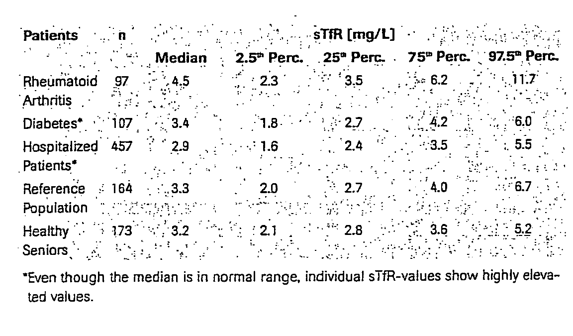
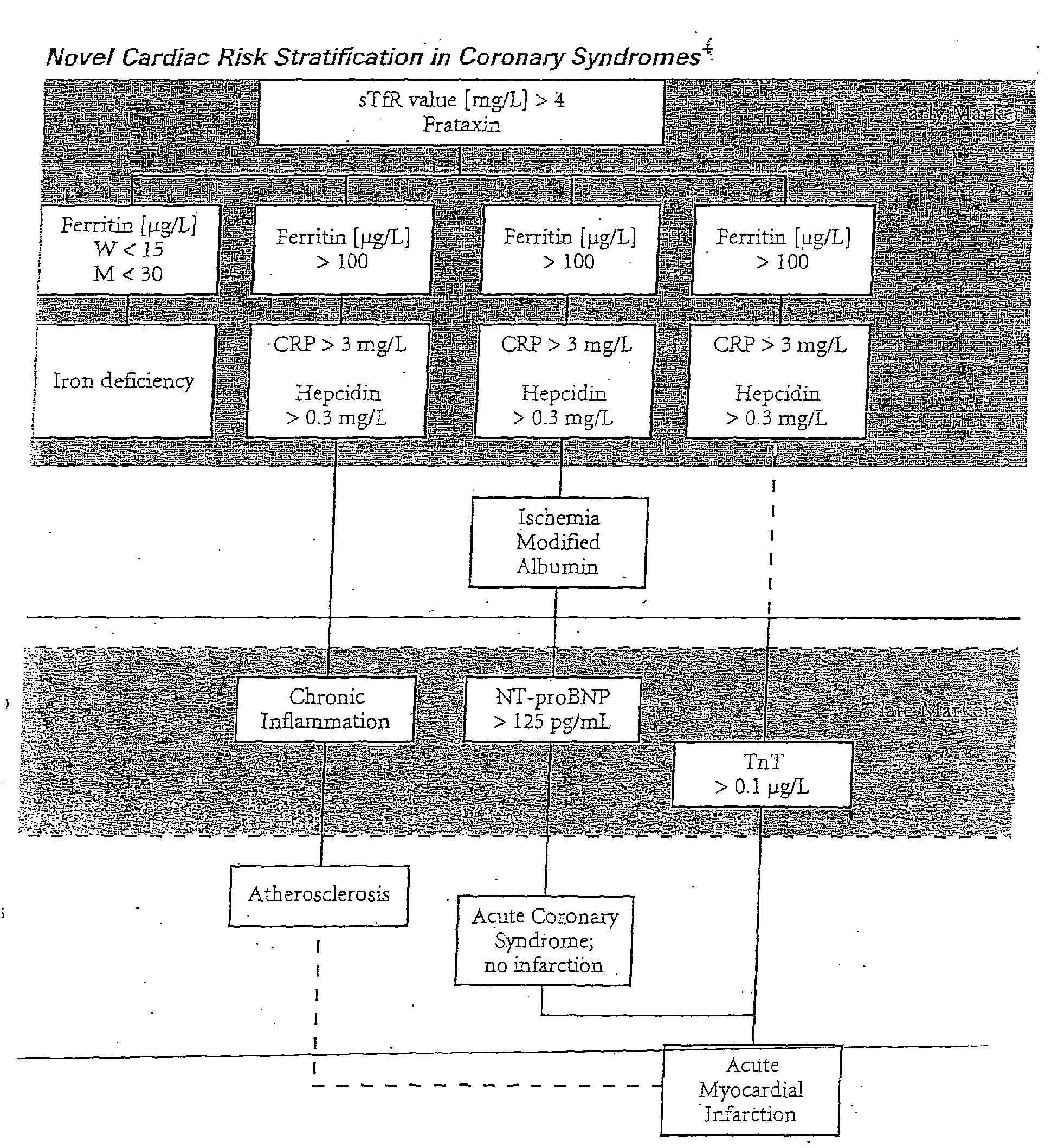
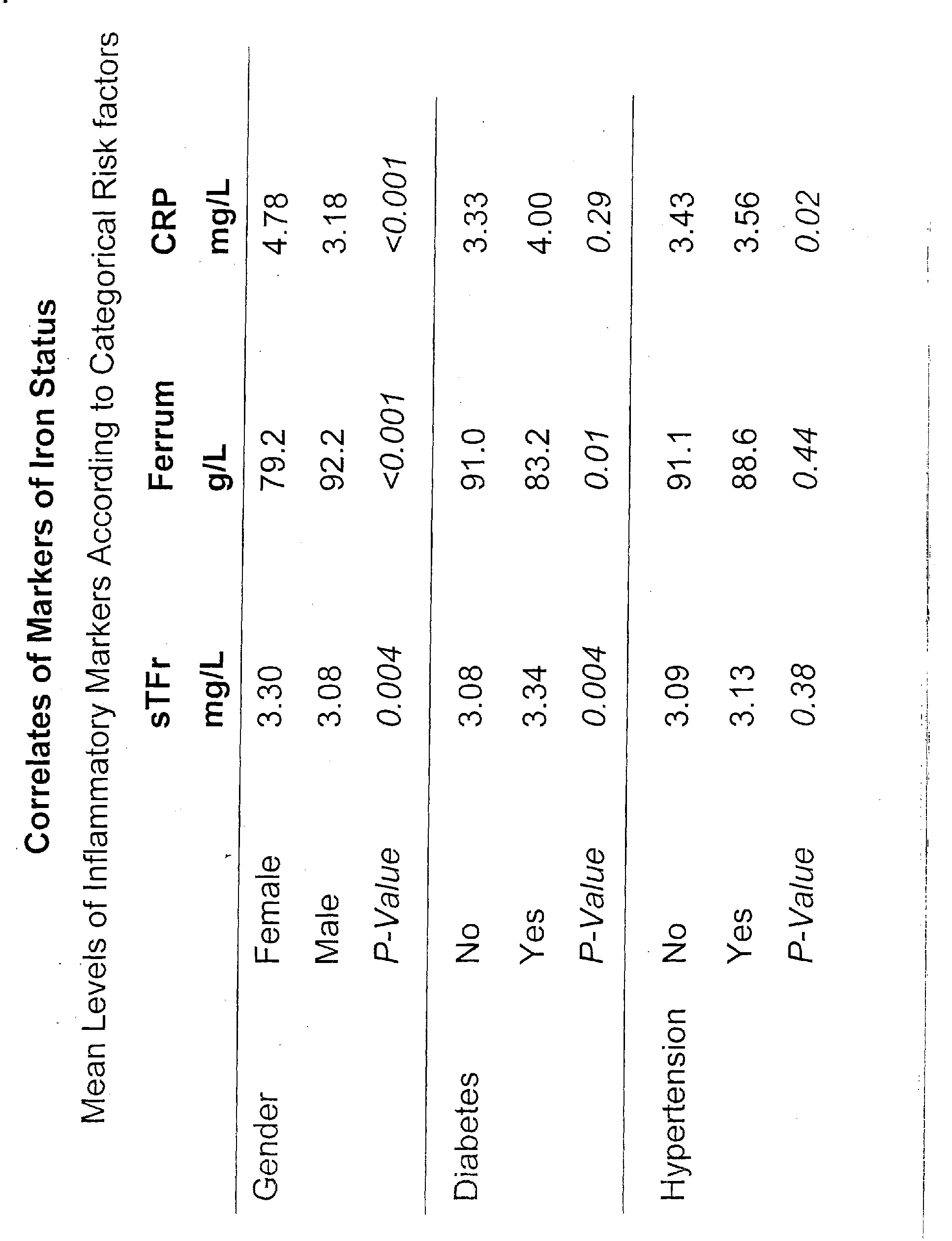


Claims (8)
- Use of sTfR (soluble transferrin receptor) or/and frataxin or/and sTfR/log ferritin as a risk marker for coronary syndromes or/and diabetes mellitus.
- Use according to claim 1, as a risk marker for coronary artery disease (CAD).
- Use of at least three compounds selected from sTfR, frataxin, CRP, hs-CRP, hepcidin, BNP, preproBNP, NT-proBNP, troponin I or/and troponin T as risk markers for coronary syndromes or/and diabetes mellitus.
- Use of at least one compound of each of the following groups:or fragments thereof1) sTfR, frataxin, sTfR/log ferritin2) CRP, in particular hs-CRP, hepcidin,3) BNP, preproBNP, NT-proBNP, and4) troponin T, troponin I
for providing prognostic information regarding coronary syndromes or/and diabetes mellitus. - Use according to any of claims 1 to 4, wherein additionally one or more of the markers ofis/are determined.(i) acute markers and/or specific markers of myocardial injury,(ii) non-specific markers of myocardial injury related to coagulation(iii) non-specific markers of myocardial injury related to artherosclerotic plaque rupture, or/and(iv) non-specific markers of myocardial injury
- Use according to claim 5, wherein
group (i) markers are selected from anexin V, also called lipocortin V, endonexin II, calphobindin I, calcium binding protein 33, placental anticoagulant protein I, thromboplastin inhibitor, vascular anticoagulant-α, anchorin CII, B-type natriuretic peptide (BNP), also called brain-type natriuretic peptide, enolase, TnT, Tnl, fTnT, CK, GP, H-FABP, PG AM as well as S-100,
group (ii) markers are selected from plasmin, β-thromboglobulin (β-TG), PF4, FPA, PDGF, prothrombin fragment 1+2, P-selection, thrombin, D-dimer, von Willebrand factor, TF and coagulation cascade,
group (iii) markers are selected from human neutrophil elastase, inducible nitric oxide synthase, lysophosphatidic acid, malondialdehyde-modified low density lipoprotein and members of the matrix metalloproteinase (MMP) family, including MMP-1, MMP-2, MMP-3 and MMP-9, and/or
group (iv) markers are selected from C-reactive protein, interleukin-1β, interleukin-1 receptor antagonist, interleukin-6, monocyte chemotactic protein-1, soluble intercellular adhesion molecule-1, soluble vascular cell adhesion molecule-1, tumor necrosis factor α (TNFα), caspase-3 and hemoglobin α2. - Reagent kit for the diagnosis of coronary syndromes or/and diabetes mellitus containing means to determine at least one compound of each of the following groups:1) sTfR, frataxin, sTfR/log ferritin2) CRP, in particular, hs-CRP, hepcidin,3) BNP, preproBNP, NT-proBNP, and4) troponin T, troponin I.
- Reagent kit for the diagnosis of coronary syndromes containing means to determine at least sTfR and/or frataxin and/or sTfR/log ferritin and at least one marker of group (i), (ii), III) and/or (iv) as defined in claim 5 or 6.
Priority Applications (1)
| Application Number | Priority Date | Filing Date | Title |
|---|---|---|---|
| EP04025210A EP1533613A1 (en) | 2003-10-22 | 2004-10-22 | Soluble transferrin receptor |
Applications Claiming Priority (5)
| Application Number | Priority Date | Filing Date | Title |
|---|---|---|---|
| EP03023980 | 2003-10-22 | ||
| EP03023980 | 2003-10-22 | ||
| EP04010822 | 2004-05-06 | ||
| EP04010822 | 2004-05-06 | ||
| EP04025210A EP1533613A1 (en) | 2003-10-22 | 2004-10-22 | Soluble transferrin receptor |
Publications (1)
| Publication Number | Publication Date |
|---|---|
| EP1533613A1 true EP1533613A1 (en) | 2005-05-25 |
Family
ID=34436703
Family Applications (1)
| Application Number | Title | Priority Date | Filing Date |
|---|---|---|---|
| EP04025210A Withdrawn EP1533613A1 (en) | 2003-10-22 | 2004-10-22 | Soluble transferrin receptor |
Country Status (4)
| Country | Link |
|---|---|
| US (1) | US20050136455A1 (en) |
| EP (1) | EP1533613A1 (en) |
| JP (1) | JP2005128020A (en) |
| CA (1) | CA2485722A1 (en) |
Cited By (1)
| Publication number | Priority date | Publication date | Assignee | Title |
|---|---|---|---|---|
| US8440712B2 (en) | 2005-08-15 | 2013-05-14 | Arrow International Limited | Crystalline sodium atorvastatin |
Families Citing this family (15)
| Publication number | Priority date | Publication date | Assignee | Title |
|---|---|---|---|---|
| US7645739B2 (en) * | 2001-02-21 | 2010-01-12 | Alavita Pharmaceuticals, Inc. | Modified annexin compositions and methods of using same |
| US7635680B2 (en) * | 2001-02-21 | 2009-12-22 | Alavita Pharmaceuticals, Inc. | Attenuation of reperfusion injury |
| US20050222030A1 (en) * | 2001-02-21 | 2005-10-06 | Anthony Allison | Modified annexin proteins and methods for preventing thrombosis |
| US20090291086A1 (en) * | 2001-02-21 | 2009-11-26 | Alavita Pharmaceuticals, Inc. | Compositions and Methods for Treating Cerebral Thrombosis and Global Cerebral Ischemia |
| US7635676B2 (en) * | 2001-02-21 | 2009-12-22 | Alavita Pharmaccuticals, Inc. | Modified annexin proteins and methods for their use in organ transplantation |
| EP1814563A4 (en) * | 2004-07-26 | 2008-04-16 | Bayer Healthcare Llc | PLATELET ACTIVATION MARKERS AS PREDICTORS OF DISEASE AND RESPONSE TO TREATMENT AND FOR MONITORING THERAPEUTIC EVOLUTION |
| EP1904846A4 (en) * | 2005-06-29 | 2009-03-18 | Rules Based Medicine Inc | METHODS AND KITS FOR THE DIAGNOSIS OF ACUTE CORONARY SYNDROME |
| GB0600916D0 (en) * | 2006-01-17 | 2006-02-22 | Perkinelmer Instr Las Inc | detecting and predicting pre-eclampsia |
| PL2084543T3 (en) * | 2006-10-26 | 2018-04-30 | B.R.A.H.M.S Gmbh | Risk stratification for acute coronary syndrome by means of fragments/partial peptides of provasopressin, especially copeptin or neurophysin ii |
| US8187827B2 (en) * | 2007-05-31 | 2012-05-29 | Naohisa Tomosugi | Diagnostic methods for acute ischemic disease using activated hepcidin as an indicator |
| ATE533061T1 (en) | 2007-08-03 | 2011-11-15 | Brahms Gmbh | METHOD FOR DIAGNOSING BACTERIAL INFECTIONS |
| EP2020603A1 (en) * | 2007-08-03 | 2009-02-04 | BRAHMS Aktiengesellschaft | Method for risk stratification in stable coronary artery disease |
| AU2009207923A1 (en) * | 2008-01-23 | 2009-07-30 | Rigshospitalet | Classification of individuals suffering from cardiovascular diseases according to survival prognoses as found by measuring the levels of biomarker YKL-40 |
| CN102203607A (en) * | 2008-10-31 | 2011-09-28 | B.R.A.H.M.S有限公司 | Methods and assays for classifying foods and/or beverages and/or diets and/or nutritional therapies and/or drugs according to their effect on the cardiovascular system |
| EP2478371B1 (en) * | 2009-09-17 | 2016-05-04 | Roche Diagnostics GmbH | Multimarker panel for left ventricular hypertrophy |
Citations (1)
| Publication number | Priority date | Publication date | Assignee | Title |
|---|---|---|---|---|
| DE19820201A1 (en) * | 1998-05-06 | 1999-11-11 | Wilhelm Krone | Diagnostic test for cardiovascular risk factors and their complications |
-
2004
- 2004-10-21 CA CA002485722A patent/CA2485722A1/en not_active Abandoned
- 2004-10-22 US US10/971,870 patent/US20050136455A1/en not_active Abandoned
- 2004-10-22 JP JP2004308314A patent/JP2005128020A/en active Pending
- 2004-10-22 EP EP04025210A patent/EP1533613A1/en not_active Withdrawn
Patent Citations (1)
| Publication number | Priority date | Publication date | Assignee | Title |
|---|---|---|---|---|
| DE19820201A1 (en) * | 1998-05-06 | 1999-11-11 | Wilhelm Krone | Diagnostic test for cardiovascular risk factors and their complications |
Non-Patent Citations (9)
| Title |
|---|
| BRAUN SIEGMUND ET AL: "Value of serum ferritin and soluble transferrin receptor for prediction of coronary artery disease and its clinical presentations.", ATHEROSCLEROSIS. MAY 2004, vol. 174, no. 1, May 2004 (2004-05-01), pages 105 - 110, XP002322614, ISSN: 0021-9150 * |
| JOSE MANUEL FERNANDEZ-REAL ET AL.: "Blood Letting in High-Ferritin Type 2 Diabetes", DIABETES CARE, vol. 25, no. 12, December 2002 (2002-12-01), pages 2249 - 2255, XP002322613 * |
| LEHMANN P ET AL: "Biologic markers in the differential diagnosis of anemias", CLINICAL CHEMISTRY, vol. 48, no. 6 Supplement, June 2002 (2002-06-01), & 54TH ANNUAL MEETING OF THE AMERICAN ASSOCIATION FOR CLINICAL CHEMISTRY (AACC); ORLANDO, FLORIDA, USA; JULY 28-AUGUST 01, 2002, pages A33 - A34, XP001205953, ISSN: 0009-9147 * |
| LEHMANN P ET AL: "Is soluble Transferrin Receptor a prognostic cardiovascular risk factor?", BLOOD, vol. 102, no. 11, 16 November 2003 (2003-11-16), & 45TH ANNUAL MEETING OF THE AMERICAN SOCIETY OF HEMATOLOGY; SAN DIEGO, CA, USA; DECEMBER 06-09, 2003, pages 3b, XP009045467, ISSN: 0006-4971 * |
| LEHMANN P ET AL: "Prediction of iron overload in diabetes diseases", CLINICAL CHEMISTRY, vol. 48, no. 6 Supplement, June 2002 (2002-06-01), & 54TH ANNUAL MEETING OF THE AMERICAN ASSOCIATION FOR CLINICAL CHEMISTRY (AACC); ORLANDO, FLORIDA, USA; JULY 28-AUGUST 01, 2002, pages A34, XP001205952, ISSN: 0009-9147 * |
| LEHMANN P ET AL: "S-transferrin-receptor and ferritin as markers in anemias of chronic inflammatory processes in tumor patients", ONKOLOGIE, vol. 23, no. Sonderheft 7, October 2000 (2000-10-01), & ANNUAL MEETING OF THE GERMAN AND AUSTRIAN SOCIETY FOR HEMATOLOGY AND ONCOLOGY; GRAZ, AUSTRIA; OCTOBER 21-25, 2000, pages 193, XP009045470, ISSN: 0378-584X * |
| LEHMANN P ET AL: "S-transferrin-receptor, ferritin and CRP as markers in differential diagnosis of anemias of chronic inflammatory processes", CLINICAL CHEMISTRY, vol. 47, no. S6, June 2001 (2001-06-01), & 53RD ANNUAL MEETING OF THE AACC/CSCC; CHICAGO, ILLINOIS, USA; JULY 29-AUGUST 02, 2001, pages A28 - A29, XP009045473, ISSN: 0009-9147 * |
| LEHMANN, P.,;KOLBE-BUSCH, S.; LOTZ, J.; BALDAUF, A.; VOLKMAMM, M.; ROWDDIGER, R.: "S-Transferrin Receptor, Ferritin and CRP as Markers in Differential Diagnosis of Anemias of Chronic Inflammatory Processes", CLINICAL CHEMISTRY AND LABORATORY MEDICINE, WALTER DE GRUYTER UND CO, DE, vol. 39, no. SPEC SUPPL, May 2001 (2001-05-01), pages S338, XP008035982, ISSN: 1434-6621 * |
| VALK DE B ET AL: "IRON, ATHEROSCLEROSIS, AND ISCHEMIC HEART DISEASE", ARCHIVES OF INTERNAL MEDICINE, AMERICAN MEDICAL ASSOCIATION, CHICAGO, IL, US, vol. 159, no. 14, 26 July 1999 (1999-07-26), pages 1542 - 1548, XP009026912, ISSN: 0003-9926 * |
Cited By (1)
| Publication number | Priority date | Publication date | Assignee | Title |
|---|---|---|---|---|
| US8440712B2 (en) | 2005-08-15 | 2013-05-14 | Arrow International Limited | Crystalline sodium atorvastatin |
Also Published As
| Publication number | Publication date |
|---|---|
| US20050136455A1 (en) | 2005-06-23 |
| JP2005128020A (en) | 2005-05-19 |
| CA2485722A1 (en) | 2005-04-22 |
Similar Documents
| Publication | Publication Date | Title |
|---|---|---|
| US7358055B2 (en) | Diagnostic markers of acute coronary syndrome and methods of use thereof | |
| US7713705B2 (en) | Markers for differential diagnosis and methods of use thereof | |
| US20040253637A1 (en) | Markers for differential diagnosis and methods of use thereof | |
| US20050164238A1 (en) | Methods and compositions for the diagnosis of sepsis | |
| US20050148029A1 (en) | Methods and compositions for determining treatment regimens in systemic inflammatory response syndromes | |
| AU2003302340B8 (en) | Markers for differential diagnosis and methods of use thereof | |
| JP4828600B2 (en) | Use of NT-proANP and NT-proBNP for diagnosis of heart disease | |
| US20040126767A1 (en) | Method and system for disease detection using marker combinations | |
| US20040121350A1 (en) | System and method for identifying a panel of indicators | |
| US20040203083A1 (en) | Use of thrombus precursor protein and monocyte chemoattractant protein as diagnostic and prognostic indicators in vascular diseases | |
| US20040209307A1 (en) | Diagnostic markers of stroke and cerebral injury and methods of use thereof | |
| US20050136455A1 (en) | Soluble transferrin receptor | |
| JP2006030183A (en) | Multiple marker panel based on P1GF for type 1 and type 2 diabetes | |
| CA2511649A1 (en) | Multimarker panel for diabetes type 1 and 2 | |
| WO2011054829A1 (en) | NT-pro ANP AND SFlt-1 FOR THE DIFFERENTIATION BETWEEN CIRCULATORY AND ISCHEMIC EVENTS | |
| US20050014198A1 (en) | Assays and kits for detecting and monitoring heart disease | |
| US20080280376A1 (en) | Method of Evaluation of the Relative Risk of Developing Atherosclerosis in Patients |
Legal Events
| Date | Code | Title | Description |
|---|---|---|---|
| PUAI | Public reference made under article 153(3) epc to a published international application that has entered the european phase |
Free format text: ORIGINAL CODE: 0009012 |
|
| PUAF | Information related to the publication of a search report (a3 document) modified or deleted |
Free format text: ORIGINAL CODE: 0009199SEPU |
|
| AK | Designated contracting states |
Kind code of ref document: A1 Designated state(s): AT BE BG CH CY CZ DE DK EE ES FI FR GB GR HU IE IT LI LU MC NL PL PT RO SE SI SK TR |
|
| AX | Request for extension of the european patent |
Extension state: AL HR LT LV MK |
|
| D17D | Deferred search report published (deleted) | ||
| 17P | Request for examination filed |
Effective date: 20050510 |
|
| 17Q | First examination report despatched |
Effective date: 20050704 |
|
| RIN1 | Information on inventor provided before grant (corrected) |
Inventor name: DR. SIEGMUND BRAUN Inventor name: DR. STEFAN BLANKENBERG Inventor name: PROF. DR. KARL J. LACKNER Inventor name: ROEDDIGER, RALF Inventor name: LEHMANN, PAUL |
|
| 17Q | First examination report despatched |
Effective date: 20050704 |
|
| STAA | Information on the status of an ep patent application or granted ep patent |
Free format text: STATUS: THE APPLICATION IS DEEMED TO BE WITHDRAWN |
|
| 18D | Application deemed to be withdrawn |
Effective date: 20070508 |