EP1533374A1 - Novel peptides having camp producing activity - Google Patents
Novel peptides having camp producing activity Download PDFInfo
- Publication number
- EP1533374A1 EP1533374A1 EP03730658A EP03730658A EP1533374A1 EP 1533374 A1 EP1533374 A1 EP 1533374A1 EP 03730658 A EP03730658 A EP 03730658A EP 03730658 A EP03730658 A EP 03730658A EP 1533374 A1 EP1533374 A1 EP 1533374A1
- Authority
- EP
- European Patent Office
- Prior art keywords
- crsp
- seq
- peptide
- amino acid
- shows
- Prior art date
- Legal status (The legal status is an assumption and is not a legal conclusion. Google has not performed a legal analysis and makes no representation as to the accuracy of the status listed.)
- Withdrawn
Links
Images










Classifications
-
- C—CHEMISTRY; METALLURGY
- C07—ORGANIC CHEMISTRY
- C07K—PEPTIDES
- C07K14/00—Peptides having more than 20 amino acids; Gastrins; Somatostatins; Melanotropins; Derivatives thereof
- C07K14/435—Peptides having more than 20 amino acids; Gastrins; Somatostatins; Melanotropins; Derivatives thereof from animals; from humans
- C07K14/46—Peptides having more than 20 amino acids; Gastrins; Somatostatins; Melanotropins; Derivatives thereof from animals; from humans from vertebrates
- C07K14/47—Peptides having more than 20 amino acids; Gastrins; Somatostatins; Melanotropins; Derivatives thereof from animals; from humans from vertebrates from mammals
-
- A—HUMAN NECESSITIES
- A61—MEDICAL OR VETERINARY SCIENCE; HYGIENE
- A61P—SPECIFIC THERAPEUTIC ACTIVITY OF CHEMICAL COMPOUNDS OR MEDICINAL PREPARATIONS
- A61P19/00—Drugs for skeletal disorders
- A61P19/08—Drugs for skeletal disorders for bone diseases, e.g. rachitism, Paget's disease
- A61P19/10—Drugs for skeletal disorders for bone diseases, e.g. rachitism, Paget's disease for osteoporosis
-
- A—HUMAN NECESSITIES
- A61—MEDICAL OR VETERINARY SCIENCE; HYGIENE
- A61P—SPECIFIC THERAPEUTIC ACTIVITY OF CHEMICAL COMPOUNDS OR MEDICINAL PREPARATIONS
- A61P25/00—Drugs for disorders of the nervous system
- A61P25/04—Centrally acting analgesics, e.g. opioids
-
- A—HUMAN NECESSITIES
- A61—MEDICAL OR VETERINARY SCIENCE; HYGIENE
- A61P—SPECIFIC THERAPEUTIC ACTIVITY OF CHEMICAL COMPOUNDS OR MEDICINAL PREPARATIONS
- A61P3/00—Drugs for disorders of the metabolism
- A61P3/04—Anorexiants; Antiobesity agents
-
- A—HUMAN NECESSITIES
- A61—MEDICAL OR VETERINARY SCIENCE; HYGIENE
- A61P—SPECIFIC THERAPEUTIC ACTIVITY OF CHEMICAL COMPOUNDS OR MEDICINAL PREPARATIONS
- A61P35/00—Antineoplastic agents
-
- A—HUMAN NECESSITIES
- A61—MEDICAL OR VETERINARY SCIENCE; HYGIENE
- A61P—SPECIFIC THERAPEUTIC ACTIVITY OF CHEMICAL COMPOUNDS OR MEDICINAL PREPARATIONS
- A61P43/00—Drugs for specific purposes, not provided for in groups A61P1/00-A61P41/00
-
- A—HUMAN NECESSITIES
- A61—MEDICAL OR VETERINARY SCIENCE; HYGIENE
- A61P—SPECIFIC THERAPEUTIC ACTIVITY OF CHEMICAL COMPOUNDS OR MEDICINAL PREPARATIONS
- A61P7/00—Drugs for disorders of the blood or the extracellular fluid
- A61P7/10—Antioedematous agents; Diuretics
-
- A—HUMAN NECESSITIES
- A61—MEDICAL OR VETERINARY SCIENCE; HYGIENE
- A61P—SPECIFIC THERAPEUTIC ACTIVITY OF CHEMICAL COMPOUNDS OR MEDICINAL PREPARATIONS
- A61P9/00—Drugs for disorders of the cardiovascular system
- A61P9/10—Drugs for disorders of the cardiovascular system for treating ischaemic or atherosclerotic diseases, e.g. antianginal drugs, coronary vasodilators, drugs for myocardial infarction, retinopathy, cerebrovascula insufficiency, renal arteriosclerosis
-
- A—HUMAN NECESSITIES
- A61—MEDICAL OR VETERINARY SCIENCE; HYGIENE
- A61P—SPECIFIC THERAPEUTIC ACTIVITY OF CHEMICAL COMPOUNDS OR MEDICINAL PREPARATIONS
- A61P9/00—Drugs for disorders of the cardiovascular system
- A61P9/12—Antihypertensives
-
- C—CHEMISTRY; METALLURGY
- C07—ORGANIC CHEMISTRY
- C07K—PEPTIDES
- C07K14/00—Peptides having more than 20 amino acids; Gastrins; Somatostatins; Melanotropins; Derivatives thereof
- C07K14/435—Peptides having more than 20 amino acids; Gastrins; Somatostatins; Melanotropins; Derivatives thereof from animals; from humans
- C07K14/575—Hormones
-
- A—HUMAN NECESSITIES
- A61—MEDICAL OR VETERINARY SCIENCE; HYGIENE
- A61K—PREPARATIONS FOR MEDICAL, DENTAL OR TOILETRY PURPOSES
- A61K38/00—Medicinal preparations containing peptides
Definitions
- the present invention relates to novel and useful peptides, genes encoding these peptides and pharmaceutical compositions comprising the same. More particularly, the present invention relates to peptides having the properties of (1) to (10); (1) being expressed in central nervous system, (2) strongly acting on calcitonin receptors, (3) promoting a cAMP productivity of cells, (4) incorporating sodium ion in a concentration-dependent manner, (5) suppressing the uptake of calcium ion, and (6) suppressing cell proliferation; genes encoding these peptides; and pharmaceutical compositions comprising the same.
- Calcitonin a known peptide, is a peptide hormone comprising 32 amino acids. In mammals, it is secreted from C cell of thyroid gland and has a function of lowering the calcium level in the blood. It has been utilized as therapeutic agents for such as hypercalcemia and metabolic bone diseases. Calcitonin has been recognized as being present in peripheral systems such as thyroid gland but not as being present in central nervous system. Contrary to the above, calcitonin receptors are present also in central nervous system. Such being the case, there has been a demand for revealing the significance of existence as well as the functions of calcitonin receptors in central nervous system.
- a calcitonin gene-related peptide is a peptide comprising 37 amino acids produced in such a manner that mRNA is derived from the same gene as calcitonin by a mechanism of alternative splicing.
- the calcitonin gene-related peptide has also the same function as calcitonin i.e. reducing the calcium concentration in blood. Moreover, it activates calcium ion channels and increases the numbers of acetylcholine receptors, suggesting the possibility of being a neurotransmitter. It has been reported that the CGRP is localized mostly to centripetal sensory nerve and central nerve (Goodman, et al., Life Sci. 38:2169-2178 (1986); Poyner, D. R.
- CGRP receptors also have already been known to be located in cerebral, cardiovascular system, endothelial and smooth muscle cells.
- the present invention is targeting to provide novel and useful peptides which being expressed in central nervous system and act on calcitonin receptors, genes encoding these peptides and pharmaceutical compositions containing the same.
- the present inventors have been searching novel physiologically active peptides by using the productivity of cAMP from central nervous system as an index and consequently isolated and purified novel peptides believed to act on cells via calcitonin receptors.
- the sequence of such a peptide structure has been confirmed to be novel from the database search results (Genbank, swissprot, DDBJ).
- the present inventors named the novel peptides as calcitonin receptor-stimulating peptides (CRSP) as they act on calcitonin receptors.
- CRSP calcitonin receptor-stimulating peptides
- the present invention relates to a peptide which has at least the following amino acid sequence , or an amino acid sequence where a part of amino acids are deleted, substituted or added, and has the following characteristics: (1) being expressed in central nervous system, (2) strongly acting on calcitonin receptors and (3) promoting a cAMP productivity of cells, more preferably, (4) incorporating sodiumion in a concentration-depending manner, (5) suppressing the uptake of calcium ion, and (6) suppressing cell proliferation.
- the present invention relates to the above described peptide having an amino acid sequence set forth in SEQ ID NO: 1, SEQ NO: 2, SEQ ID NO: 6, SEQ ID NO: 9, SEQ ID NO: 12, SEQ ID NO: 16 or SEQ ID NO: 19 of the Sequence Listing, or having an amino acid sequence where a part of amino acids are deleted, substituted or added, and having 75% or more homology to the said amino acid sequence.
- the peptides of the present invention comprise peptides having homology to a calcitonin gene-related peptide (CGRP), therefore, they may be expected to have the physiological activity similar to that of CGRP in addition to the effects of CRSP. Accordingly, in addition to the above (1) to (6), the peptides of the invention may also be expected to have the following characteristics similar to those of CGRP: (7) strongly acting on calcitonin-gene related peptide receptors, (8) inducing relaxation of blood vessels, (9) promoting diuresis, and (10) altering the proliferation potency of vascular endothelial cells, vascular smooth muscles and fibroblasts.
- CGRP calcitonin gene-related peptide
- the present invention further relates to the genes having base sequences encoding the above described peptides of the invention.
- the present invention relates to the above described genes having a base sequence set forth in SEQ ID NO: 3, SEQ ID NO: 7, SEQ ID NO: 10, SEQ ID NO: 13, SEQ ID NO: 17 or SEQ ID NO: 20 of the Sequence Listing.
- the present invention still further relates to a pharmaceutical composition comprising at least one of the above described peptides of the invention and a pharmaceutically acceptable carrier.
- the present invention relates to the above described pharmaceutical composition where the pharmaceutical composition is a preventive/treating agent for osteoporosis, a preventive/treating agent for cancer, diuretic agent, an appetite suppressant, an analgesic agent, an antihypertensive agent or a drug for prevention of restenosis after PTCA (percutaneous transluminal coronary angioplasty).
- PTCA percutaneous transluminal coronary angioplasty
- CRSP CRSP
- CRSP-2 CRSP-2
- CRSP-3 pCRSP-3
- CT-2 CT-2
- pCGRP pig calcitonin gene-related peptide
- pCT pig calcitonin
- pAM pig adrenomedullin
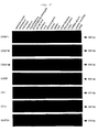
- Fig. 24 shows the result of a high-sensitivity quantitative determination, by RT-PCR, of expressed amount of gene of CRSP, CRSP-2, CRSP-3, CT-2, CT and CGRP.
- the vertical axis in the drawing shows various genes while the horizontal axis shows tissues of pigs where expressed amount of gene was measured.
- GAPDH glycosylase dehydrogenase
- the present inventors have purified two kinds of novel physiologically active peptides from an extract of porcine brain using a cAMP production of renal epithelial cells as an index.
- amino acid sequence of the resulting peptides is analyzed, it has been found that they are a peptide (hereinafter, it will be referred to as CRSP) comprising the following 38 amino acids and a peptide (hereinafter, it will be referred to as CRSP-G) comprising the following 39 amino acids where glycine is bonded to a C-terminal of the above described peptide.
- a synthetic primer was prepared on the basis of the amino acid sequence of the resulting peptide and amplified by a PCR using pig gene as a template whereupon an aimed gene was cloned.
- the primer used was at the N-terminal side and was at the C-terminal side.
- a base sequence of the resulting gene is shown in SEQ ID NO: 3 of Sequence Listing and an amino acid sequence coded thereby is shown as follows. (In the formula, the underlined 81st to 118th ones are CRSP.)
- amino acid sequence is shown in SEQ ID NO: 4 of Sequence Listing.
- the peptide of the present invention was prepared from a library of bovine and canine thyroid cDNA.
- amino acid sequence of the peptide of the present invention of bovine is shown in SEQ ID NO: 6 of Sequence Listing, a nucleotide sequence of the gene thereof is shown in SEQ ID NO: 7 of Sequence Listing and an amino acid sequence of a peptide on the basis of the gene is shown in SEQ ID NO: 8.
- amino acid sequence of the peptide of the present invention of canine is shown in SEQ ID NO: 9 of Sequence Listing
- a nucleotide sequence of the gene thereof is shown in SEQ ID NO: 10 of Sequence Listing
- an amino acid sequence of a peptide on the basis of the gene is shown in SEQ ID NO: 11.
- CT-2 gene coding for a peptide having a homology to pig calcitonin (CT) was identified.
- a peptide which is a product of the gene was named CT-2.
- SEQ ID NO: 12 An amino acid sequence of CRSP-2 is shown in SEQ ID NO: 12 of Sequence Listing, a nucleotide sequence of cDNA thereof is shown in SEQ ID NO: 13 and an amino acid sequence of a peptide on the basis of the cDNA (a precursor peptide) is shown in SEQ ID NO: 14.
- SEQ ID NO: 14 An amino acid sequence of CRSP-2 gene is shown in SEQ ID NO: 15.
- SEQ ID NO: 16 of Sequence Listing An amino acid sequence of CRSP-3 is shown in SEQ ID NO: 16 of Sequence Listing, a nucleotide sequence of cDNA thereof is shown in SEQ ID NO: 17 and an amino acid sequence of a peptide on the basis of the cDNA (a precursor peptide) is shown in SEQ ID NO: 18.
- CT-2 An amino acid sequence of CT-2 is shown in SEQ ID NO: 19 of Sequence Listing, a nucleotide sequence of cDNA thereof is shown in SEQ ID NO: 20 and an amino acid sequence of a peptide on the basis of the cDNA (a precursor peptide) is shown in SEQ ID NO: 21.
- SEQ ID NO: 22 A nucleotide sequence of CRSP-3 gene and CT-2 gene is shown in SEQ ID NO: 22.
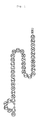
- a structure of pig CRSP is schematically shown as an example of the peptides of the present invention.
- the second Cys and the seventh Cys are in an -S-S- bond mode.
- Fig. 2 shows comparison of amino acid sequences of pig CRSP (pCRSP) of the peptide of the present invention, pig calcitonin gene-related peptide (pCGRP-I), human calcitonin gene-related peptide I (hCGRP-I), human calcitonin gene-related peptide II (hCGRP-II), human amylin (hAmylin), pig calcitonin (pCT) and human adrenomedullin (hAM).
- pCGRP-I pig calcitonin gene-related peptide
- hCGRP-I human calcitonin gene-related peptide I
- hCGRP-II human calcitonin gene-related peptide II
- human amylin hAmylin
- pig calcitonin pCT
- adrenomedullin hAM
- the CRSP of the present invention has a homology in terms of amino acid sequence to pig calcitonin gene-related peptide (pCGRP), human CGRP-I and human CGRP-II to an extent of 71.1%, 63.2% and 71.1%, respectively.
- Fig. 3 The result of Northern blotting for investigating the expression site of CRSP is shown in Fig. 3 by a photograph as a drawing. An arrow in Fig. 3 shows the position of CRSP mRNA. Further, content of peptides in each tissue was measured by means of radioimmunoassay using antibody against CRSP. Result of the measurement is shown in the following Table 1.
- CRSP of the present invention has been detected in central nervous system such as midbrain and hypothalamus, and in peripheral system such as thyroid gland. Moderate expression was detected in pons/medulla oblongata. Expression, though only a little, was detected even in cerebrum and pituitaries. On the other hand, the concentration of CRSP in tissues was most abundant in posterior pituitary and thyroid gland, and also high in midbrain, hypothalamus and anterior pituitary.
- CRSP dissolved in DMEM was added to a medium for LLC-PK 1 cells and amount of cAMP secreted therefrom was quantified by means of radioimmunoassay using a cAMP-specific antibody.
- the result is shown in Fig. 4.
- the vertical axis in Fig. 4 shows the production amount of cAMP (pmol/10 5 cells/30 minutes) while the horizontal axis thereof shows a reverse logarithm (-log (peptide concentration(M))) of concentration of each peptide.
- CRSP of the present invention an open circle ( ⁇ ) shows pig calcitonin gene-related peptide (pig CGRP) and an open rhombus ( ⁇ ) shows pig calcitonin (pig CT).
- ⁇ pig calcitonin gene-related peptide
- ⁇ pig calcitonin
- CRSP of the present invention very strongly increases (ED 50 being about 1.5 nM) an intracellular adenylyl cyclase activity of LLC-PK 1 cells derived from pig renal epithelial cells in a concentration-dependent manner. The activity was stronger than pig calcitonin (ED 50 being about 8. 7 nM) and pig CGRP (ED 50 being about 62 nM) to an extent of about 6-fold and about 40-fold, respectively.
- CRSP of the present invention was investigated in function of promoting incorporation of sodium ion and calcium ion.
- a culture solution of LLC-PK 1 cells was substituted with a Hanks' choline chloride solution, CRSP and sodium chloride having a predetermined radioactivity ( 22 Na) were added thereto so as to make the final concentration 0 M, 10 -8 M, 10 -7 M and 10 -6 M each, the cells were washed after 10 minutes so that 22 Na contained in the solution outside the cells was completely removed and radioactivity incorporated into the cells was measured by a gamma-counter.
- a culture solution of LLC-PK 1 cells was substituted with a Hanks' choline chloride solution, sodium chloride having a predetermined radioactivity ( 22 Na) as well as CRSP and an amyloride derivative (5-(N-ethyl-N-isopropyl)-amiloride) (EIPA) which is an Na/H co-transporter inhibitor were added thereto so as to make the final concentration 0 M CRSP + 0 M EIPA, 10 -6 M CRSP + 0 M EIPA, 10 -6 M CRSP + 10 -8 M EIPA, 10 -6 M CRSP + 10 -7 M EIPA, 10 -6 M CRSP + 10 -6 M EIPA and 0 M CRSP + 10 -6 M EIPA each. Cells were washed after 10 minutes so that 22 Na contained in the outer solution of the cells was completely removed and radioactivity incorporated into the cells was measured by a gamma-counter.
- EIPA amyloride derivative
- Fig. 5 incorporad amounts (cpm) of each ion
- Fig. 6 shows incorporated amounts (cpm) of each ion
- left end of the horizontal axis shows control (no peptide added) and to the right thereof shows CRSP for each concentration.
- Right side of the control in the horizontal axis in Fig. 6 shows the case of a sole administration of CRSP
- the right side thereof shows each concentration of co-administration of CRSP with EIPA
- the right end shows the case of a sole administration of EIPA.
- sCT in Fig. 7 shows salmon calcitonin.
- the marks *, ** and *** show that there was a significant different when p ⁇ 0.05, p ⁇ 0.01 and p ⁇ 0.001, respectively.
- Fig. 8 shows incorporated amount of 125 I-DU ( ⁇ 100 cpm/well) while the horizontal axis shows a reverse logarithm (-log (peptide concentration (M))) of each peptide.
- a closed circle (•) in Fig. 8 shows CRSP of the present invention, an open circle ( ⁇ ) shows pig calcitonin (pig CT) and an open rhombus ( ⁇ ) shows salmon calcitonin (salmon CT).
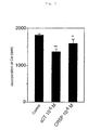
- each solution of CRSP in DMEM in various concentrations was added to LLC- PK 1 cells of 10,000 cells/well, incubation was conducted, the cells on the incubation dish was taken out by a trypsin-EDTA solution and cell numbers were counted using a counting plate.
- the result is shown in Fig. 9.
- the vertical axis in Fig. 9 shows cell numbers ( ⁇ 1,000 cells/well), while the left end of the horizontal axis shows control (no CRSP added) and to the right thereof shows CRSP of each concentration.
- the mark * shows that there was a significant difference when p ⁇ 0.05.
- CRSP has an action of suppressing the growth of LLC-PK 1 cells.
- Pig calcitonin receptor was introduced into a mammalian expression vector pcDNA 3.1, expressed in rhesus monkey renal cells (COS-7) and production amount of cAMP by stimulation of CRSP was measured by radioimmunoassay.
- CTR was expressed in opossum renal cells (OK cells) and production amount of cAMP by stimulation of CRSP was measured by radioimmunoassay while incorporation of sodium ion was measured by measuring the incorporation of sodium chloride having radioactivity ( 22 Na) by a gamma-counter. The results are shown in Fig.
- Fig. 10 changes in cAMP productivity by CRSP stimulation in cells (COS-7) where calcitonin receptor was expressed in a genetic engineering manner
- Fig. 11 changes in cAMP productivity in opossum renal epithelial cells where pig calcitonin receptor (CTR) was expressed
- Fig. 12 changes in incorporation of sodium ion in opossum renal epithelial cells where pig calcitonin receptor (CTR) was expressed).
- the vertical axis in Fig. 10 shows a production amount of cAMP (pmol/well/30 minutes) while the horizontal axis shows a reverse logarithm (-log (ligand concentration (M))) of concentration of each peptide (ligand).
- a closed circle (•) of Fig. 10 shows CRSP of the present invention and an open rhombus ( ⁇ ) shows pig calcitonin (pCT).
- the vertical axis in Fig. 11 shows production amount of cAMP (fmol/well/hour) while the horizontal axis shows a reverse logarithm (-log (CRSP concentration (M))) of concentration of CRSP.
- FIG. 11 shows the case of opossum renal epithelial cells into which pig calcitonin receptor (CTR) is introduced and a closed square ( ⁇ ) shows the case of opossum renal epithelial cells (OK cells).
- the vertical axis in Fig. 12 shows the ratio of incorporated amounts of sodium ion (where the control being 100) while the horizontal axis shows a reverse logarithm (-log (CRSP concentration (M))) of CRSP concentration.
- a closed circle (•) of Fig. 12 shows the case where opossum renal epithelial cells into which pig calcitonin receptor (CTR) is introduced and a closed square ( ⁇ ) shows the case of opossum renal epithelial cells (OK cells).
- a mark *** in Fig. 12 shows that there was a significant difference when p ⁇ 0.001.
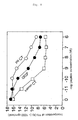
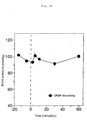
- CRSP corresponding to an amount of 16 nmol/kg dissolved in a physiological saline solution was administered at a time within a short period from jugular vein of rat under an anesthetized condition and calcium concentration in the plasma and blood pressure were measured.
- the results are shown in Fig. 13 (changes in calcium concentration in blood by administration of CRSP) and Fig. 14 (changes in blood pressure by administration of CRSP).
- the vertical axis in Fig. 13 shows a calcium concentration in blood while the horizontal axis shows time (minutes).
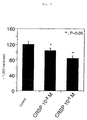
- the vertical axis in Fig. 14 shows blood pressure (mmHg) while the horizontal axis shows time (minutes).
- a mark ** in Fig. 13 shows that there was a significant difference when p ⁇ 0.01.
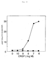
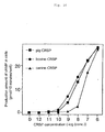
- Pig calcitonin receptor was introduced into mammalian expression vector pcDNA 3.1 and then expressed in rhesus monkey renal cells (COS-7) whereupon production amount of cAMP by CRSP stimulation was measured by radioimmunoassay. The result is shown in Fig. 15.
- cAMP productivity of CRSP using LLC-PK 1 cells was investigated.
- the result is shown in Fig. 16.
- the vertical axis in Fig. 16 shows production amount of cAMP (pmol/well/10 minutes) while the horizontal axis shows the concentration of CRSP added thereto.
- CT-2 With regard to CRSP-2 and CRSP-3, the expression was observed in the central nervous system, thyroid gland and ovary. On the contrary, with regard to CT-2, its band was not amplified in any tissue whereby no expression was observed. It is presumed that CT-2 is expressed only in other areas than the tissues shown in Fig. 24.
- the peptides of the present invention stimulate calcitonin receptor in a stronger manner than calcitonin whereupon adenylyl cyclase is activated by that.
- the above shows that the peptide of the present invention is a genuine endogenous ligand for calcitonin receptor.
- the peptides of the present invention are believed to act on the calcitonin receptor in peripheral tissues and elicit the following pharmaceutical effects.
- Calcitonin promotes incorporation of calcium into bone via calcitonin receptor which is a receptor of calcitonin. It is concluded that, like calcitonin, the peptides of the present invention act on calcitonin receptor and promote the incorporation of calcium into bone. Actually, as shown in Fig. 13, a decrease in calcium concentration in plasma was detected in rats by administration of the peptide. It is concluded that the result supports the promotion of incorporation of calcium into bones. By utilizing such a function, the peptides are applicable as a drug for recovery of bone density, reduced due to osteoporosis, to a healthy state and can be used as preventive and treating agents for osteoporosis.
- the present peptides have an activity of lowering the calcium concentration in plasma whereupon they are applicable as drugs for lowering the calcium concentration inplasma of hypercalcemia as a result of alkalosis, etc. to a normal level and can be used as treating and preventive agents for hypercalcemia.
- the peptides of the present invention activate the action of an amyloride-sensitive Na/H co-transporter of renal epithelial cells. That is the activity opposed to amyloride which is a potassium-retaining diuretic agent, whereby they can be used as an antidiuretic agent.
- the peptides of the present invention strongly suppress the growth of renal epithelial cells via a calcitonin receptor. It has been reported that calcitonin strongly suppresses the growth of adenocarcinoma cells, etc. via a calcitonin receptor ( Cancer Res. 1985, 45, 4890-4894 and others). Accordingly, it is expected that the peptides of the present invention will similarly suppress the growth of cancer cells via a calcitonin receptor in a stronger manner than calcitonin and they are able to be used as treating and preventive agents for cancer.
- the peptides of the present invention act on calcitonin receptor in the central nervous system and elicit the following pharmaceutical effects.
- calcitonin acts for suppressing the appetite in the central nervous system ( Science 1979, 206, 850-852; The Bone 1992, 6, 69-74; etc.).
- the peptides of the present invention which can activate a calcitonin receptor more strongly than calcitonin, have a great possibility to elicit a strong appetite-suppressing action.
- the peptides of the present invention by means of administration, can be used as a treating and preventive agent for obesity and diseases caused thereby (such as hypertension and hyperlipemia).
- calcitonin preparations show analgesic action to pain of bone due to metastasis of cancer, osteoporosis, etc., to pain by migraine and pancreatitis, etc. ( Am. J. Med. Sci. 1997, 313, 13-16; etc.).
- the peptides of the present invention which activate the calcitonin receptor morestrongly than calcitonin, have a strong analgesic action same as calcitonin and they are effective as analgesics to those pains.
- the peptides of the present invention have characteristics of: (1) being expressed in central nervous systems, (2) strongly acting on calcitonin receptor, (3) promoting a cAMP productivity of cells, and relate to the peptides which have at least the following amino acid sequence or an amino acid sequence where a part of the amino acids are deleted, substituted or added.
- the peptides of the present invention include amide derivatives, ester derivatives and derivatives of functional group of side chain of each amino acid such as amino group of lysine and hydroxyl group of serine provided that the above described amino acid sequence is comprised. More preferable peptides of the present invention further have the characteristics of: (4) incorporating sodium ion concentration-dependently, (5) suppressing the incorporation of calcium ion, (6) suppressing the cell proliferation.
- the peptides of the present invention are not limited to the above described amino acid sequence so far as they have the above described characteristics (1) to (3) or, preferably, (1) to (6), and they may be peptides where a part of the amino acids in the amino acid sequence are deleted, substituted and/or added.
- the amino acid sequence as such, though not limited to, it preferably has 50% or more homology, more preferably may have 75% or more, 80% or more, still more preferably may as well have 85% or more homology.
- the preferable peptides of the present invention include, those which comprising an amino acid sequence having 50% or more, preferably 60% or more homology to the amino acid sequence of calcitonin or calcitonin gene-related peptides.
- CRSPs of the present invention derived from a pig have been described, however, the peptides of the present invention are not limited to those derived from pig but comprise the peptides derived from mammals such as human beings, rats, dogs and cows and fish such as salmon, etc.
- the peptides of the present invention can be separated or purified from natural products by a manner such as extraction, or can be synthesized by way of common peptide synthesis methods. It is further possible to produce the peptide by means of a gene recombination technique where the gene of the present invention is incorporated into a suitable vector, and prokaryotic cells or eukaryotic cells are used as hosts.
- the pharmaceutical composition of the present invention comprises the peptide of the present invention and a pharmaceutically acceptable carrier.
- the pharmaceutical composition of the present invention can be administered either orally or parenterally, and preferably it is administered parenterally by such as intravenous injection and intramuscular injection.
- the pharmaceutical composition of the present invention can be made into preparations such as a preparation for injection, a freeze-dried preparation and a preparation for rectal administration. It also can be made into preparations for external application such as adhesive device, ointment and plaster. It can further be made into preparations such as sublingual tablets. Formulation can be carried out by means of general methods for peptide preparation.
- the pharmaceutical composition of the present invention can be used in combination with other effective ingredients other than the peptide of the invention.
- the dose of the pharmaceutical composition of the present invention can be administered usually within a range from 1 ⁇ g/kg to 1,000 mg/kg of body weight, preferably from 0.1 mg/kg to 500 mg/kg of body weight per day in terms of the amount of the peptides of the present invention, however, may be varied depending on nature of the patient and condition of the disease. Since the peptide of the present invention acts more on calcitonin receptor than calcitonin does, it may be applied to the diseases in the same manner as in the case of the conventional calcitonin preparations.
- the peptide of the invention further includes a peptide having identity with a calcitonin gene-related peptide (CGRP), it can be utilized not only for the above described object but a physiological activity thereof similar to CGRP can also be utilized.
- CGRP calcitonin gene-related peptide
- CGRP The known physiological activity of CGRP include such as acting strongly on a CGRP receptor and promoting the cAMP production in cells. It also has been reported that CGRP induces relaxation of blood vessel ( Regul. Pept. 1986, 15, 1-23; etc.), promotes diuresis ( Proc. Soc. Exp. Biol. Med. 1998, 188, 316-322; etc.) and alters the proliferation potency of vascular endothelial cells and cells such as vascular smooth muscle and fibroblast ( Proc. Natl. Acad. Sci. USA 1990, 87, 3299-3303; Regul. Pept. 2001, 101, 169-178; etc.).
- peptides of the present invention particularly those having a high homology to CGRP, it is expected to have function of (1) being expressed in central nervous system, (2) strongly acting on a calcitonin receptor, (3) promoting a cAMP productivity of cells, (4) incorporating sodium ion concentration-dependently, (5) suppressing the incorporation of calcium ion, and (6) suppressing the cell proliferation.
- the pharmaceutical composition containing the peptide of the present invention can be utilized as preparations in use for the following purposes in addition to the above described usages. Namely, it can be used for such as a hypotensive agent where the function of blood vessel relaxation is directly utilized, a hypotensive agent where the function of blood vessel relaxation via central and peripheral nervous systems is utilized, a diuretic, and preventives for restenosis after PTCA (percutaneous transluminal coronary angioplasty) where the function of suppressing the proliferation of smooth muscle of blood vessel is utilized.
- PTCA percutaneous transluminal coronary angioplasty
- Pig brain (20 kg) was boiled for 10 minutes in water of a 4-fold amount, cooled and homogenized by addition of acetic acid as to make its final concentration 1M and the insoluble matters were removed by centrifugation to prepare a pig brain extract.
- This was desalted and concentrated by means of ultrafiltration (Pericon Cassette PLAC #000-05, manufactured by Millipore) and acetone was gradually added to the extract after desalting under cooling with ice so as to make the final concentration 66%.
- the precipitate was removed by centrifugal separation, acetone was removed from the supernatant liquid using an evaporator and the residue was adsorbed with a reversed phase column (LC-SORB SPW-C-ODS; Chemco; 1.5 l).
- the freeze-dried specimen was weighed, dissolved in 1 M acetic acid, adsorbed with an open column filled with a cation-exchange resin (SP-Sephadex, Pharmacia, 3 ⁇ 28 cm) and fractionated into a fraction (SP-I) which just passed therethrough (SP-I), a fraction (SP-II) eluted with 2 M pyridine (pH8.0) and a fraction (SP-III) eluted with 2 M pyridine-acetic acid (pH 5.0) to prepare an SP-III strongly ionic fraction.
- the fraction was freeze-dried, dissolved in 1 M acetic acid and fractionated into fractions corresponding to molecular weights by means of gel filtration (Sephadex G-50, Pharmacia, 7.5 ⁇ 145 cm, 1 M acetic acid, flow rate: 100 ml/hour) whereupon the part corresponding to molecular weights of from 1 kDa to 5 kDa was collected.
- gel filtration Sephadex G-50, Pharmacia, 7.5 ⁇ 145 cm, 1 M acetic acid, flow rate: 100 ml/hour
- CM52 ion-exchange chromatography
- LLC-PK 1 epidermal cells derived from kidney of pig
- DMEM Dulbecco's modified Eagle medium
- BSA bovine serum albumin
- the method therefor is as follows.
- a peptide (about 50 pmol) was prepared. Molecular weight of this peptide was 4,099 and a theoretical isoelectric point was calculated to be 12.81. In addition, it had such a hydrophobic property that was able to be eluted with acetonitrile of about 32% concentration in the presence of 0.1% of trifluoroacetic acid in a C 18 column (4.6 ⁇ 250 mm, 218TP54) of Vydac as shown above.
- amino acid sequence was determined by an Edman method using an automatic amino acid sequencer. Firstly, 5 pmol of the pure specimen was analyzed by the amino acid sequencer from its N-terminal and the following was determined.
- the present peptide has a homology to a calcitonin gene-related peptide and has the following sequence.
- a probe was prepared by formation of a synthetic primer using of the N-terminal side and of the C-terminal side on the basis of the amino acid sequence of Example 2 and by amplification using a PCR where pig gene was used as a template.
- a complementary DNA ⁇ phage library was prepared from pig hypothalamus mRNA (3 ⁇ g) using a Time Saver cDNA preparing kit of Pharmacia and ⁇ ZAPII of Stratagene.
- a method for the screening was as follows. Thus, Escherichia coli was infected to ⁇ phage (having about 100,000 independent clones), mixed with 0.7% agarose containing an LB medium, sown on an incubation dish laid with a 1.5% agar medium containing an LB medium and incubated for about 8 hours at 37°C.
- the plaque formed thereby was transcribed to a Nylon filter, made into alkaline (by treating for 2 minutes with a 0.5 M sodium hydroxide and 1.5 M sodium chloride solution; for 2 minutes with a 0.5 M Tris-hydrochloride (pH 7.5) and 1.5 M sodium chloride solution; and for 5 minutes with a 45 mM sodium citrate (pH 7.0) and 450 mM sodium chloride solution) and then the filter was dried at 80°C for 2 hours.
- the dried filter was dipped for at 37°C 2 hours in a pre-hybridization solution (50% of formamide, 0.09 M of sodium citrate (pH 7.0), 0.9 M of sodium chloride, 0.5% of bovine serum albumin, 0.5% of Ficoll, 0.5% of polyvinylpyrrolidone and 0.5% of sodium laurylsulfate), a probe labeled with 32 P was added thereto and the mixture was subj ected to hybridization at 42°c for 16 hours.
- a pre-hybridization solution 50% of formamide, 0.09 M of sodium citrate (pH 7.0), 0.9 M of sodium chloride, 0.5% of bovine serum albumin, 0.5% of Ficoll, 0.5% of polyvinylpyrrolidone and 0.5% of sodium laurylsulfate
- the isolated positive ⁇ phage clone was converted to a plasmid pBluescript which is a vector suitable for a DNA sequencing using Helper Phage R408 of Stratagene and a nucleotide sequence was determined by a Sanger method.
- the determined base sequence is shown in SEQ ID NO: 3 of the Sequence Listing while its amino acid sequence is shown in SEQ ID NO: 4.
- the resulting CRSP has a homology in terms of the amino acid sequence topig calcitonin (CT) gene-related peptide (CGRP), human CGRP-I and human CGRP-II to an extent of 71.1%, 63.2% and 71.1%, respectively (refer to Fig. 2).
- CT amino acid sequence topig calcitonin
- RNA for expressed amount of gene was carried out by means of extracting with acidic guanidine, phenol and chloroform.
- About 1 g of pig tissue was homogenized with 5 ml of a denaturing solution (4 M of guanidine thiocyanate, 25 mM of sodium citrate (pH 7.0), 0.1 M of 2-mercaptoethanol and 0.5% of sodium sarcosinate), well stirred with a 1/10 amount of 2 M sodium acetate, an equivalent amount of water-saturated phenol and a 1/5 amount of chloroform and the mixture was subjected to a centrifugal separation.
- a denaturing solution 4 M of guanidine thiocyanate, 25 mM of sodium citrate (pH 7.0), 0.1 M of 2-mercaptoethanol and 0.5% of sodium sarcosinate
- RNA precipitate
- 30 ⁇ g of this RNA were subjected to an electrophoresis using a formaldehyde-agarose modified gel (1% of agarose, 2.2 M of formaldehyde, 20 mM of Mops (pH 7), 8 mM of sodium acetate and 1 mM of EDTA), the gel after the electrophoresis was well washed with water to remove formaldehyde and then treated with 0.05 M of sodium hydroxide for 15 minutes and a solution of 0.3 M of sodium citrate (pH 7) and 3 M of sodium chloride for 45minutes.
- a formaldehyde-agarose modified gel 1% of agarose, 2.2 M of formaldehyde, 20 mM of Mops (pH 7), 8 mM of sodium acetate and 1 mM of EDTA
- RNA contained in the gel was transferred to a Nylon filter by a capillary blot method and the filter to which RNA was transcribed was dried at 80°C. After that, the filter was dipped in a ULTRAhyb hybridization solution of Ambion at 37°C for 2 hours, a probe labeled with 32 P was added where CRSP was encoded (a base pair between 346th to 488th ones on cDNA) and hybridization was conducted at 42°C for 16 hours.
- the filter was washed with a washing solution (with a solution of 30 mM of sodium citrate (pH 7.0), 300 mM of sodium chloride and 0.1% of sodium laurylsulfate at room temperature for 5 minutes for two times and with a solution of 1.5 mM of sodium citrate (pH 7.0), 15 mM of sodium chloride and 0.1% of sodium laurylsulfate at 65°C for 1 hour for two times) and then exposed to an X-ray film whereupon RNA was quantified.
- a washing solution with a solution of 30 mM of sodium citrate (pH 7.0), 300 mM of sodium chloride and 0.1% of sodium laurylsulfate at room temperature for 5 minutes for two times and with a solution of 1.5 mM of sodium citrate (pH 7.0), 15 mM of sodium chloride and 0.1% of sodium laurylsulfate at 65°C for 1 hour for two times
- Amount of peptide in each tissue was measured by radioimmunoassay utilizing an antibody prepared to the synthetic peptide.
- the tissue (about 1 g) was boiled with water in a 10-fold amount and cooled with ice, acetic acid was added so as to make it 1 M, homogenization was conducted and insoluble matter were removed by centrifugal separation to prepare an extract.
- the extract was freeze-dried and dissolved in a buffer (50 mM of sodium phosphate (pH 7.4), 80 mM of sodium chloride, 25 mM of EDTA, 0.5% of Triton X-100, 0.5% of bovine serum albumin and 0.05% of sodium azide) and each 100 ⁇ l thereof were taken out to a test tube.
- a buffer 50 mM of sodium phosphate (pH 7.4), 80 mM of sodium chloride, 25 mM of EDTA, 0.5% of Triton X-100, 0.5% of bovine serum albumin and 0.05% of sodium azi
- LLC-PK 1 cells were incubated in a culture medium (a Dulbecco-modified Eagle medium (DMEM) containing 10% of fetal calf serum (FCS)), CRSP dissolved in DMEM containing 0.05% of bovine serum albumin was substituted for a culture medium for the cells after two days, incubation was conducted for 30 minutes, the medium was recovered and amount of the secreted cAMP was quantified by radioimmunoassay using a cAMP-specific antibody.
- DMEM Dulbecco-modified Eagle medium
- FCS fetal calf serum
- Example 7 (Action for incorporation of sodium ion and calcium ion in the presence of CRSP in LLC-PK 1 cells)
- LLC-PK 1 cells were incubated for two days in a DMEM (containing 10% of FCS) on a six-well incubation dish. Measurement of incorporation of sodium ion was conducted as follows.
- An incubation solution for LLC-PK 1 cells was substituted with a Hanks' choline chloride solution, sodium chloride ( 22 Na) having a predetermined amount of radioactivity and CRSP were added thereto so as to make the final concentration 0 M, 10 -8 M, 10 -7 M and 10 -6 M, respectively, the cells were washed after 10 minutes to completely remove 22 Na contained in the solution outside the cells and radioactivity incorporated into the cells was measured by a gamma-counter.
- an incubated solution for LLC-PK 1 cells was substituted with a Hanks' choline chloride solution, sodium chloride ( 22 Na) havingapredetermined radioactivity and each of CRSP and an amiloride derivative (5-(N-ethyl-N-isopropyl)-amiloride) (EIPA) which is an Na/H co-transporter inhibitor were added so as to make the final concentration 0 M of CRSP + 0 M of EIPA, 10 -6 M of CRSP + 0 M of EIPA, 10 -6 M of CRSP + 10 -8 M of EIPA, 10 -6 M of CRSP + 10 -7 M of EIPA, 10 -6 M of CRSP + 10 -6 M of EIPA and 0 M of CRSP + 10 -6 M of EIPA, the cells were washed after 10 minutes to completely remove 22 Na contained in the solution outside the cells and radioactivity incorporated into the cells was measured by a gamma-counter.
- EIPA amiloride derivative
- LLC-PK 1 cells were incubated for two days in a DMEM (containing 10% of FCS) on a 24-well collagen plate, once washed with a DMEM and substituted with a DMEM (containing 10% of FCS) containing 0 and 10 -12 to 10 -6 M of CRSP followed by incubating. After 2 hours, a DMEM (containing 0.1% of BSA) containing 125 I-labeled bromodeoxyuridine was added to each well and, after 5 hours, radioactivity incorporated into the nuclei was measured by a gamma-counter whereupon the amount of DNA synthesized as a result of cell proliferation was measured.
- DMEM containing 10% of FCS
- LLC-PK 1 cells of 10,000 cells/well were sown on a 24-well collagen plate and incubated with a DMEM (containing 10% of FCS) for 24 hours. Each well was once washed with a DMEM, the medium was substituted with a DMEM (containing 10% of FCS) containing CRSP of concentrations of 0 M, 10 -8 M and 10 -6 M and incubation was conducted. After 24 hours, the cells on the incubation dish were peeled off with a trypsin-EDTA solution and cell numbers were counted by a counting plate.
- Pig calcitonin receptor was introduced into a mammalian expression vector pcDNA 3.1 and expressed in rhesus monkey renal cells (COS-7) and production amount of cAMP by CRSP stimulation was measured by radioimmunoassay.
- CTR was expressed in opossum renal cells (OK cells) and production amount of cAMP by CRSP stimulation was measured by radioimmunoassay while incorporation of sodium ion was measured by incorporation of radioactive sodium chloride ( 22 Na) using a gamma-counter.
- CRSP corresponding to the amount of 16 nmol/kg dissolved in a physiological saline solution was administered at a time within short period from a carotid artery of rat under an anesthetized condition.
- Pig brain (20 kg) was boiled for 10 minutes in water of a 4-fold amount, cooled and homogenized by addition of acetic acid to as to make its final concentration 1M and the insoluble matters were removed by centrifugation to prepare a pig brain extract.
- This was desalted and concentrated by means of ultrafiltration (Pericon Cassette PLAC #000-05, manufactured by Millipore) and acetone was gradually added to the extract after desalting under cooling with ice so as to make the final concentration 66%.
- the precipitate was removed by centrifugal separation, acetone was removed from the supernatant liquid using an evaporator and the residue was adsorbed with a reversed phase column (LC-SORB SPW-C-ODS; Chemco; 1.5 l).
- the freeze-dried specimen was weighed, dissolved in 1 M acetic acid, adsorbed with an open column filled with a cation-exchange resin (SP-Sephadex, Pharmacia, 3 ⁇ 28 cm) and fractionated into a fraction (SP-I) which just passed therethrough (SP-I), a fraction (SP-II) eluted with 2 M pyridine (pH 8.0) and a fraction (SP-III) eluted with 2M pyridine-acetic acid (pH5.0) to prepare an SP-III strongly ionic fraction.
- the fraction was freeze-dried, dissolved in 1 M acetic acid and fractionated into fractions corresponding to molecular weights by means of gel filtration (Sephadex G-50, Pharmacia, 7.5 ⁇ 145 cm, 1 M acetic acid, flow rate: 100 ml/hour) whereupon the part corresponding to molecular weights of from 1 kDa to 5 kDa was collected.
- gel filtration Sephadex G-50, Pharmacia, 7.5 ⁇ 145 cm, 1 M acetic acid, flow rate: 100 ml/hour
- CM52 ion-exchange chromatography
- LLC-PK 1 epidermal cells derived from kidney of pig
- DMEM Dulbecco's modified Eagle medium
- the method therefor is as follows.
- a rise in the cAMP production caused by CRSP-Gly was observed in a fraction eluted by about 0. 27 M of ammonium formate (pH 6.5).
- Example 12 (Action for promotion of cAMP production by CRSP-Gly)
- Pig calcitonin receptor was introduced into a mammalian expression vector pcDNA 3.1, then expressed in rhesus monkey renal cells (COS-7) and production amount of cAMP by CRSP or CRSP-Gly stimulation was measured by radioimmunoassay.
- RNA was extracted from canine and bovine thyroid gland by a method using acidic guanidine, phenol and chloroform. Purification of mRNA was conducted using an Oligotex-dT30 mRNA purifying kit of Takara Shuzo. Complementary DNA ⁇ phage library was prepared from canine and bovine thyroid gland mRNA (3 ⁇ g) using a Time Saver cDNA preparation kit of Pharmacia and a ⁇ ZAPII of Stratagene. With regard to a probe, a pig calcitonin receptor-stimulating peptide cDNA having a full length of 700 bp was used. A method for the screening was as follows.
- Escherichia coli was infected to ⁇ phage (having about 300,000 independent clones), mixed with 0.7% agarose containing an LB medium, sown on an incubation dish laid with a 1.5% agar medium containing an LB medium and incubated for about 8 hours at 37°C. After it was cooled in a refrigerator for about 2 hours, the plaque formed thereby was transferred to a Nylon filter, made into alkaline (by treating for 2 minutes with a 0.
- the filter was dipped at 37°C for 2 hours in a pre-hybridization solution (20% of formamide, 0.09 M of sodium citrate (pH 7.0), 0.9 M of sodium chloride, 0.5% of bovine serum albumin, 0.5% of Ficoll, 0.5% of polyvinylpyrrolidone and 0.5% of sodium laurylsulfate), a probe labeled with 32 P was added thereto and the mixture was subjected to hybridization at 42°c for 16 hours.
- a pre-hybridization solution (20% of formamide, 0.09 M of sodium citrate (pH 7.0), 0.9 M of sodium chloride, 0.5% of bovine serum albumin, 0.5% of Ficoll, 0.5% of polyvinylpyrrolidone and 0.5% of sodium laurylsulfate
- the isolated positive ⁇ phage clone was converted to a plasmid pBluescript which is a vector suitable for a DNA sequencing using Helper Phage R408 of Stratagene and a nucleotide sequence was determined by a Sanger method.
- a nucleotide sequence of cDNA coding for the determined bovine CRSP is shown in SEQ ID NO: 7 of the Sequence Listing which will be given later and an amino acid sequence of a precursor peptide of the coded bovine CRSP is shown in SEQ ID NO: 8.
- An amino acid sequence of bovine CRSP is shown in SEQ ID NO: 6.
- a nucleotide sequence of cDNA coding for the determined canine CRSP is shown in SEQ ID NO: 10 of the Sequence Listing which will be given later and an amino acid sequence of a precursor peptide of the coded canine CRSP is shown in SEQ ID NO: 11.
- An amino acid sequence of canine CRSP is shown in SEQ ID NO: 9.
- Example 14 Quantification of production amount of cAMP by stimulation with bovine CRSP or canine CRSP
- LLC-PK 1 cells were incubated in a culture medium (a Dulbecco-modified Eagle medium (DMEM) containing 10% of fetal bovine serum). After two days, the cells were washed twice with a DMEM containing 0.05% of bovine serum albumin, the medium in the incubation dish was substituted with a DMEM containing 0. 5 mM of 3-isobutyl-1-methylxanthine and 0.05% of bovine serum albumin (DMEM/BSA/IBMX) and incubation was conducted at 37°C for 30 minutes.
- DMEM Dulbecco-modified Eagle medium
- the medium was substituted with a DMEM/BSA/IBMX in which CRSP was dissolved so as to make it 0 M and 10 -12 to 10 -6 M and incubation was conducted at 37°C for 10 minutes. After the incubation, the medium in the incubating dish was removed, ethanol was added thereto and the incubating dish was once frozen in a freezer for the cell lysis. Then ethanol was transferred to a test tube and the specimen was evaporated to dryness using a centrifugal evaporator.
- Pig genomic library was purchased from Clontech. With regard to a probe, a precursor cDNA having a full length of 700 bp of a pig calcitonin receptor-stimulating peptide was used. Method for screening was that Escherichia coli was infected to ⁇ phage (it has about 1,000,000 independent clones and about 300,000 thereof were used) into which pig genome was inserted, mixed with 0.7% agarose containing an LB medium, sown on an incubation dish where 1.5 % agar medium containing an LB medium was spread and incubated at 37°C for about 8 hours.
- the resulting plaque was transferred to a Nylon filter and subjected to an alkaline modification (being treated with a 0.5 M of sodium hydroxide and 1.5 M of sodium chloride solution for 2 minutes, with a 0.5 M Tris-hydrochloride (pH 7.5) and 1. 5 M sodium chloride solution for 2 minutes and with a 45 mM of sodium citrate (pH 7.0) and 450 mM of sodium chloride solution for 5 minutes) and the filter was treated at 80°C for 2 hours.
- an alkaline modification being treated with a 0.5 M of sodium hydroxide and 1.5 M of sodium chloride solution for 2 minutes, with a 0.5 M Tris-hydrochloride (pH 7.5) and 1. 5 M sodium chloride solution for 2 minutes and with a 45 mM of sodium citrate (pH 7.0) and 450 mM of sodium chloride solution for 5 minutes
- the filter was dipped in a pre-hybridization solution (20% of formamide, 0.09 M of sodium citrate (pH 7.0), 0.9 M of sodium chloride, 0.5% of bovine fetal albumin, 0.5% of Ficoll, 0.5% of polyvinylpyrrolidone and 0.5% of sodium laurylsulfate) at 37°C for 2 hour, a probe labeled with 32 P was added thereto and hybridization was conducted at 42°C for 16 hours.
- a pre-hybridization solution (20% of formamide, 0.09 M of sodium citrate (pH 7.0), 0.9 M of sodium chloride, 0.5% of bovine fetal albumin, 0.5% of Ficoll, 0.5% of polyvinylpyrrolidone and 0.5% of sodium laurylsulfate
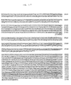
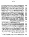
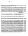
- the isolated positive ⁇ phage clone was cleaved using various restriction enzymes, analysis was conducted by Southern blotting method to prepare a restriction enzyme map and DNA containing the positive clone was sub-cloned to pBluescript using an appropriate restriction enzyme. A nucleotide sequence was determined by a Sanger method. As a result of a screening, 25 positive clones were able to be prepared. As a result of analysis using restriction enzyme and determination of a nucleotide sequence, it was found that ten clones coded for gene of CRSP (Fig. 17). It was also found that 9 of the remaining 15 clones coded for other gene (Fig. 18 and Fig. 19) and six thereof coded for still other gene.
- the nine clones had sequences having a homology to CRSP.
- the genes were named CRSP-2.
- Other six clones had sequences having a homology to CRSP and CT.
- Those genes were named CRSP-3 and CT-2.
- Fig. 17 to Fig. 19 the underlined parts show exons.
- a nucleotide sequence of CRSP gene is shown in SEQ ID NO: 5 of the Sequence Listing which will be shown later.
- a CRSP-2 gene is shown in SEQ ID NO: 15 of the Sequence Listing.
- a nucleotide sequence of DNA fragment containing CRSP-3 gene and CT-2 gene is shown in SEQ ID NO: 23 of the Sequence Listing.
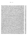
- Fig. 20 to Fig. 22 cDNA sequences which were presumed using the gene sequences of CRSP and CT/CGRP as references are shown in Fig. 20 to Fig. 22 (Fig. 20: CRSP-2; Fig. 21: CRSP-3; and Fig. 22: CT-2).
- the parts enclosed with solid lines are mature peptides
- the gray parts are glycine which is presumed to be converted to amide
- the oblique parts are signal peptides
- glutamine at the N-terminal side of the mature substance is presumed to be converted to pyroglutamic acid.
- Comparison of each CRSP with CGRP, AM, CT-2 and CT amino acids is Fig. 23.
- a nucleotide sequence of cDNA of CRSP-2 is shown in SEQ ID NO: 13 of the Sequence Listing which will be given later, that of cDNA of CRSP-3 is shown in SEQ ID NO: 17 and that of cDNA of CT-2 is shown in SEQ ID NO: 21.
- a cDNA library there was used a ⁇ phage library into which pig hypothalamus DNA was inserted being prepared in conducting a cDNA cloning of CRSP.
- a probe a pig calcitonin receptor-stimulatingpeptide having a full length of 700 bp was used.
- Method for screening was that Escherichia coli was infected to ⁇ phage into which pig hypothalamus DNA was inserted, mixed with 0.7% agarose containing an LB medium, sown on an incubation dish where 1.5 % agar medium containing an LB medium was spread and incubated at 37°C for about 8 hours.
- the resulting plaque was transferred to a Nylon filter and subjected to an alkaline denaturation (being treated with a 0.5 M of sodium hydroxide and 1.5 M of sodium chloride solution for 2 minutes, with a 0.5 M Tris-hydrochloride (pH 7.5) and 1.5 M sodium chloride solution for 2 minutes and with a 45 mM of sodium citrate (pH 7.0) and 450 mM of sodium chloride solution for 5 minutes) and the filter was treated at 80°C for 2 hours.
- an alkaline denaturation being treated with a 0.5 M of sodium hydroxide and 1.5 M of sodium chloride solution for 2 minutes, with a 0.5 M Tris-hydrochloride (pH 7.5) and 1.5 M sodium chloride solution for 2 minutes and with a 45 mM of sodium citrate (pH 7.0) and 450 mM of sodium chloride solution for 5 minutes
- an alkaline denaturation being treated with a 0.5 M of sodium hydroxide and 1.5 M of sodium chloride solution for 2 minutes,
- the filter was dipped in a pre-hybridization solution (20% of formamide, 0.09 M of sodium citrate (pH 7.0), 0.9 M of sodium chloride, 0.5% of bovine fetal albumin, 0.5% of Ficoll, 0.5% of polyvinylpyrrolidone and 0.5% of sodium laurylsulfate) at 37°C for 2 hour, a probe labeled with 32 P was added thereto and hybridization was conducted at 42°C for 16 hours.
- a pre-hybridization solution (20% of formamide, 0.09 M of sodium citrate (pH 7.0), 0.9 M of sodium chloride, 0.5% of bovine fetal albumin, 0.5% of Ficoll, 0.5% of polyvinylpyrrolidone and 0.5% of sodium laurylsulfate
- the isolated positive ⁇ phase clone was converted to a plasmid pBluescript which is a vector suitable for DNA sequence using a Helper Phage R408 of Stratagene and a nucleotide sequence was determined by a Sanger method. As a result thereof, 13 positive clones were able to be prepared. All of six among the above were clones containing almost the full length of the CRSP precursor cDNA while all of other six were clones containing almost the full length of the CRSP-2 precursor cDNA (Fig. 20). A remaining one was a short clone coding for a 3'-untranslated region of CRSP-3.
- PCR where pig hypothalamus cDNA (corresponding to 20 ng of mRNA) was a template was carried out using primers (CRSP-3: GCCCAGCTTACGTCTCCTTT and TCAGGTAACTGCAATGATTT; CT-2: AGCAGCTTTGATTCTGCCAC and ACCTCCTCTCTGATATTCCA) and Pyrobest DNA polymerase of Takara Shuzo for 30 cycles each comprising at 94°C for 15 seconds, at 55°C for 15 seconds and at 72°C for 1 minute.
- primers CRSP-3: GCCCAGCTTACGTCTCCTTT and TCAGGTAACTGCAATGATTT
- CT-2 AGCAGCTTTGATTCTGCCAC and ACCTCCTCTCTGATATTCCA
- the amplified DNA was subjected to an agarose gel electrophoresis, bands stained with ethidium bromide were recovered from agarose gel and subcloned into pBluescript II of Stratagene and a nucleotide sequence was determined by a Sanger method.
- Example 18 High-sensitivity quantification of expressed amount of gene of CRSP, CRSP-2, CRSP-3, CT-2, CT, CGRP and GAPDH by RT-PCR
- a template cDNA was prepared using total RNA (4 ⁇ g) of each tissue of pig, oligo dT primer and a Revertra Ace reverse transcriptase kit of Toyobo and 1/40 thereof was used for RT-PCR.
- the PCR for amplification of each genetic cDNA was conducted using rTaq polymerase of Toyobo in 30 cycles each comprising at 94°C for 15 seconds, at 60°C for 15 seconds and at 72°C for 1 minute and, with regard to the primer, the following sequences were used.
- the amplified DNA was subjected to electrophoresis using 3% agarose gel and analyzed using Fuji Film FLA 2000. As a result, expression was observed in the central nervous system, thyroid gland and ovary for CRSP-2 and CRSP-3. On the other hand, no band was amplified in any tissue and no expression was observed for CT-2 (Fig. 24). It is believed that CT-2 was expressed limitedly in the areas other than the tissues shown in Fig. 24.
- the present invention provides novel and useful peptides which are expressed in central nerves and strongly act on calcitonin receptor.
- the peptides of the present invention have a high homology to calcitonin gene-related peptides and have stronger action than calcitonin. They are also abundantly expressed in central nervous system and are very useful peptides as novel calcitonin-like peptides for osteoporosis and as analgesic agent, etc.
Landscapes
- Health & Medical Sciences (AREA)
- Chemical & Material Sciences (AREA)
- Life Sciences & Earth Sciences (AREA)
- Organic Chemistry (AREA)
- Medicinal Chemistry (AREA)
- General Health & Medical Sciences (AREA)
- Chemical Kinetics & Catalysis (AREA)
- General Chemical & Material Sciences (AREA)
- Public Health (AREA)
- Nuclear Medicine, Radiotherapy & Molecular Imaging (AREA)
- Veterinary Medicine (AREA)
- Pharmacology & Pharmacy (AREA)
- Bioinformatics & Cheminformatics (AREA)
- Animal Behavior & Ethology (AREA)
- Engineering & Computer Science (AREA)
- Toxicology (AREA)
- Zoology (AREA)
- Proteomics, Peptides & Aminoacids (AREA)
- Molecular Biology (AREA)
- Genetics & Genomics (AREA)
- Biophysics (AREA)
- Biochemistry (AREA)
- Gastroenterology & Hepatology (AREA)
- Physical Education & Sports Medicine (AREA)
- Hematology (AREA)
- Endocrinology (AREA)
- Diabetes (AREA)
- Orthopedic Medicine & Surgery (AREA)
- Heart & Thoracic Surgery (AREA)
- Rheumatology (AREA)
- Cardiology (AREA)
- Obesity (AREA)
- Biomedical Technology (AREA)
- Child & Adolescent Psychology (AREA)
- Pain & Pain Management (AREA)
- Neurosurgery (AREA)
- Vascular Medicine (AREA)
- Urology & Nephrology (AREA)
- Neurology (AREA)
- Medicines That Contain Protein Lipid Enzymes And Other Medicines (AREA)
Abstract
It is intended to provide novel and useful proteins which
are expressed in the central nerve system and act on a calcitonin
receptor, genes thereof and medicinal compositions containing
the same. Peptides having at least the following amino acid
sequence:



Description
The present invention relates to novel and useful peptides,
genes encoding these peptides and pharmaceutical compositions
comprising the same. More particularly, the present invention
relates to peptides having the properties of (1) to (10); (1)
being expressed in central nervous system, (2) strongly acting
on calcitonin receptors, (3) promoting a cAMP productivity of
cells, (4) incorporating sodium ion in a
concentration-dependent manner, (5) suppressing the uptake of
calcium ion, and (6) suppressing cell proliferation; genes
encoding these peptides; and pharmaceutical compositions
comprising the same.
Calcitonin, a known peptide, is a peptide hormone
comprising 32 amino acids. In mammals, it is secreted from
C cell of thyroid gland and has a function of lowering the calcium
level in the blood. It has been utilized as therapeutic agents
for such as hypercalcemia and metabolic bone diseases.
Calcitonin has been recognized as being present in peripheral
systems such as thyroid gland but not as being present in central
nervous system. Contrary to the above, calcitonin receptors
are present also in central nervous system. Such being the
case, there has been a demand for revealing the significance
of existence as well as the functions of calcitonin receptors
in central nervous system.
A calcitonin gene-related peptide (CGRP) is a peptide
comprising 37 amino acids produced in such a manner that mRNA
is derived from the same gene as calcitonin by a mechanism of
alternative splicing. The calcitonin gene-related peptide
(CGRP) has also the same function as calcitonin i.e. reducing
the calcium concentration in blood. Moreover, it activates
calcium ion channels and increases the numbers of acetylcholine
receptors, suggesting the possibility of being a
neurotransmitter. It has been reported that the CGRP is
localized mostly to centripetal sensory nerve and central nerve
(Goodman, et al., Life Sci. 38:2169-2178 (1986); Poyner, D.
R. Pharmac. Ther. 56:23-51 (1992)), and mainly has a role to
activate adenylyl cyclase. A wide variety of CGRP functions
including dilating peripheral and cerebral blood vessel (Brain,
et al., Nature 313:54-568 (1985)), increasing heart rate
(Sigrist, et al., Endocrinology 119:381-389 (1986)), adjusting
calcium metabolism (Grunditz, et al., Endocrinology
119:2313-2324 (1986)), leading to less intestinal movement
(Fargeas, et al., Peptides 6:1167-1171 (1985)), regulating
glucose metabolism (lowering insulin secretion and insulin
sensitivity) (Hermansen, et al., Reg. Peptides 27:149-157
(1990)), suppressing the appetite (Molina, et al., Diabetes
39:260-265 (1990)) and suppressing growth hormone release
(Tannenbaum, et al., Endocrinology, 116: 2685-2687 (1985)) have
been reported.
CGRP receptors also have already been known to be located
in cerebral, cardiovascular system, endothelial and smooth
muscle cells.
There also have been reported the inventions relating
to the synthetic calcitonin receptor-binding compound
characterized by having a molecular weight of less than about
10,000 daltons and forming a reagent covalently linked to a
radioactive metal chelating agent, where the reagent has a
binding affinity to calcitonin receptor higher than the binding
affinity of radioiodated natural calcitonin to the calcitonin
receptor (Published Japanese translation of PCT application
No. 2001/523,257); the calcitonin receptor activating
substance comprising a pyridine derivatives (Unexamined Patent
Application Publication No. 2001/294,574); the polynucleotide
and polypeptide of human calcitonin gene-related peptide
receptor component factor (CGRP-RCF) (Unexamined Patent
Application Publication No. 10/201,482), etc.
Those peptides not having been confirmed as acting
directly on calcitonin receptors of central nervous system,
there has been a demand for a peptide which acts directly and
more strongly on calcitonin receptors in central nervous system.
The present invention is targeting to provide novel and
useful peptides which being expressed in central nervous system
and act on calcitonin receptors, genes encoding these peptides
and pharmaceutical compositions containing the same.
The present inventors have been searching novel
physiologically active peptides by using the productivity of
cAMP from central nervous system as an index and consequently
isolated and purified novel peptides believed to act on cells
via calcitonin receptors. The sequence of such a peptide
structure has been confirmed to be novel from the database search
results (Genbank, swissprot, DDBJ). The present inventors
named the novel peptides as calcitonin receptor-stimulating
peptides (CRSP) as they act on calcitonin receptors.
Accordingly, the present invention relates to a peptide
which has at least the following amino acid sequence



 , or an amino acid sequence where a part of amino acids are deleted,
substituted or added, and has the following characteristics:
(1) being expressed in central nervous system, (2) strongly
acting on calcitonin receptors and (3) promoting a cAMP
productivity of cells, more preferably, (4) incorporating
sodiumion in a concentration-depending manner, (5) suppressing
the uptake of calcium ion, and (6) suppressing cell
proliferation.
, or an amino acid sequence where a part of amino acids are deleted,
substituted or added, and has the following characteristics:
(1) being expressed in central nervous system, (2) strongly
acting on calcitonin receptors and (3) promoting a cAMP
productivity of cells, more preferably, (4) incorporating
sodiumion in a concentration-depending manner, (5) suppressing
the uptake of calcium ion, and (6) suppressing cell
proliferation.
To be more specific, the present invention relates to
the above described peptide having an amino acid sequence set
forth in SEQ ID NO: 1, SEQ NO: 2, SEQ ID NO: 6, SEQ ID NO: 9,
SEQ ID NO: 12, SEQ ID NO: 16 or SEQ ID NO: 19 of the Sequence
Listing, or having an amino acid sequence where a part of amino
acids are deleted, substituted or added, and having 75% or more
homology to the said amino acid sequence.
In another embodiment, the peptides of the present
invention comprise peptides having homology to a calcitonin
gene-related peptide (CGRP), therefore, they may be expected
to have the physiological activity similar to that of CGRP in
addition to the effects of CRSP. Accordingly, in addition to
the above (1) to (6), the peptides of the invention may also
be expected to have the following characteristics similar to
those of CGRP: (7) strongly acting on calcitonin-gene related
peptide receptors, (8) inducing relaxation of blood vessels,
(9) promoting diuresis, and (10) altering the proliferation
potency of vascular endothelial cells, vascular smooth muscles
and fibroblasts.
The present invention further relates to the genes having
base sequences encoding the above described peptides of the
invention. To be more specific, the present invention relates
to the above described genes having a base sequence set forth
in SEQ ID NO: 3, SEQ ID NO: 7, SEQ ID NO: 10, SEQ ID NO: 13,
SEQ ID NO: 17 or SEQ ID NO: 20 of the Sequence Listing.
The present invention still further relates to a
pharmaceutical composition comprising at least one of the above
described peptides of the invention and a pharmaceutically
acceptable carrier. Particularly, the present invention
relates to the above described pharmaceutical composition where
the pharmaceutical composition is a preventive/treating agent
for osteoporosis, a preventive/treating agent for cancer,
diuretic agent, an appetite suppressant, an analgesic agent,
an antihypertensive agent or a drug for prevention of restenosis
after PTCA (percutaneous transluminal coronary angioplasty).
There is shown comparison of amino acid sequences of CRSP
(pCRSP), CRSP-2 (pCRSP-2), CRSP-3 (pCRSP-3) and CT-2 (pCT-2)
which are peptides of the present invention with amino acid
sequences of pig calcitonin gene-related peptide (pCGRP), pig
calcitonin (pCT) and pig adrenomedullin (pAM).
Fig. 24 shows the result of a high-sensitivity
quantitative determination, by RT-PCR, of expressed amount of
gene of CRSP, CRSP-2, CRSP-3, CT-2, CT and CGRP. The vertical
axis in the drawing shows various genes while the horizontal
axis shows tissues of pigs where expressed amount of gene was
measured. In order to normalize total amount of RNA, expressed
amount of GAPDH (glyceraldehydes-3-phosphate dehydrogenase)
was also measured.
The present inventors have purified two kinds of novel
physiologically active peptides from an extract of porcine brain
using a cAMP production of renal epithelial cells as an index.
When the amino acid sequence of the resulting peptides is
analyzed, it has been found that they are a peptide (hereinafter,
it will be referred to as CRSP) comprising the following 38
amino acids



 and a peptide (hereinafter, it will be referred to as CRSP-G)
comprising the following 39 amino acids where glycine is bonded
to a C-terminal of the above described peptide.
and a peptide (hereinafter, it will be referred to as CRSP-G)
comprising the following 39 amino acids where glycine is bonded
to a C-terminal of the above described peptide.




When the amino acid sequences of those peptides were
searched by databases (Genbank, swissprot, DDBJ), both have
been found to be confirmed to have novel amino acid sequences.
Those amino acid sequences are shown in SEQ NO: 1 and
SEQ ID NO: 2 of the Sequence Listing, respectively.
A synthetic primer was prepared on the basis of the amino
acid sequence of the resulting peptide and amplified by a PCR
using pig gene as a template whereupon an aimed gene was cloned.
On the basis of the amino acid sequence, the primer used
was
 at the N-terminal side and was
 at the C-terminal side.
A base sequence of the resulting gene is shown in SEQ
ID NO: 3 of Sequence Listing and an amino acid sequence coded
thereby is shown as follows.










 (In the formula, the underlined 81st to 118th ones are CRSP.)
The amino acid sequence is shown in SEQ ID NO: 4 of Sequence
Listing.
Using pig CRSP cDNA as a probe, the peptide of the present
invention was prepared from a library of bovine and canine
thyroid cDNA.
An amino acid sequence of bovine CRSP represented by
one-letter codes for amino acids is as follows.


When it is represented by three-letter codes for amino
acids, it is as follows.




An amino acid sequence of canine CRSP represented by
one-letter codes for amino acids is as follows.


When it is represented by three-letter codes for amino
acids, it is as follows.




Each of those amino acid sequences, nucleotide sequences
and total amino acid sequences coded by gene is shown in Sequence
Listing.
An amino acid sequence of the peptide of the present
invention of bovine is shown in SEQ ID NO: 6 of Sequence Listing,
a nucleotide sequence of the gene thereof is shown in SEQ ID
NO: 7 of Sequence Listing and an amino acid sequence of a peptide
on the basis of the gene is shown in SEQ ID NO: 8.
An amino acid sequence of the peptide of the present
invention of canine is shown in SEQ ID NO: 9 of Sequence Listing,
a nucleotide sequence of the gene thereof is shown in SEQ ID
NO: 10 of Sequence Listing and an amino acid sequence of a peptide
on the basis of the gene is shown in SEQ ID NO: 11.
Using the full length of coding region of pig CRSP cDNA
as a probe, gene coding for a peptide having a homology to CRSP
was prepared from a pig genomic library. Peptides which are
products of the gene are named CRSP-2 and CRSP-3, respectively.
Similarly, gene coding for a peptide having a homology
to pig calcitonin (CT) was identified. A peptide which is a
product of the gene was named CT-2.
An amino acid sequence of CRSP-2 as represented by
three-letter codes for amino acids is as follows.




An amino acid sequence of CRSP-3 as represented by
three-letter codes for amino acids is as follows.




An amino acid sequence of CT-2 as represented by
three-letter codes for amino acids is as follows.




An amino acid sequence of CRSP-2 is shown in SEQ ID NO:
12 of Sequence Listing, a nucleotide sequence of cDNA thereof
is shown in SEQ ID NO: 13 and an amino acid sequence of a peptide
on the basis of the cDNA (a precursor peptide) is shown in SEQ
ID NO: 14. A nucleotide sequence of CRSP-2 gene is shown in
SEQ ID NO: 15.
An amino acid sequence of CRSP-3 is shown in SEQ ID NO:
16 of Sequence Listing, a nucleotide sequence of cDNA thereof
is shown in SEQ ID NO: 17 and an amino acid sequence of a peptide
on the basis of the cDNA (a precursor peptide) is shown in SEQ
ID NO: 18.
An amino acid sequence of CT-2 is shown in SEQ ID NO:
19 of Sequence Listing, a nucleotide sequence of cDNA thereof
is shown in SEQ ID NO: 20 and an amino acid sequence of a peptide
on the basis of the cDNA (a precursor peptide) is shown in SEQ
ID NO: 21. A nucleotide sequence of CRSP-3 gene and CT-2 gene
is shown in SEQ ID NO: 22.
In Fig. 1, a structure of pig CRSP is schematically shown
as an example of the peptides of the present invention. In
the case of the peptide of this example, the second Cys and
the seventh Cys are in an -S-S- bond mode.
Fig. 2 shows comparison of amino acid sequences of pig
CRSP (pCRSP) of the peptide of the present invention, pig
calcitonin gene-related peptide (pCGRP-I), human calcitonin
gene-related peptide I (hCGRP-I), human calcitonin
gene-related peptide II (hCGRP-II), human amylin (hAmylin),
pig calcitonin (pCT) and human adrenomedullin (hAM).
The CRSP of the present invention has a homology in terms
of amino acid sequence to pig calcitonin gene-related peptide
(pCGRP), human CGRP-I and human CGRP-II to an extent of 71.1%,
63.2% and 71.1%, respectively.
Comparison of amino acids for CRSP, CRGP, AM, CT-2 and
CT with regard to peptides derived from pig is shown in Fig.
23.
The result of Northern blotting for investigating the
expression site of CRSP is shown in Fig. 3 by a photograph as
a drawing. An arrow in Fig. 3 shows the position of CRSP mRNA.
Further, content of peptides in each tissue was measured by
means of radioimmunoassay using antibody against CRSP. Result
of the measurement is shown in the following Table 1.
| Concentration of CRSP in Tissues as Measured by Radioimmunoassay | |
| Tissues | Immunoactivity of CRSP (pmol/g tissue) |
| Cerebral cortex | 0.29 ± 0.04 |
| Cerebellum | 0.18 ± 0.02 |
| Midbrain | 7.5 ± 0.9 |
| Hippocampus | 0.78 ± 0.16 |
| Caudatum | 1.3 ± 0.1 |
| Thalamus | 3.5 ± 0.4 |
| Hypothalamus | 9.9 ± 1.2 |
| Pons/Medulla oblongata | 2.2 ± 0.3 |
| Spinal cord | 0.52 ± 0.06 |
| Olfactory bulb | 0.74 ± 0.22 |
| | 14 ± 2 |
| Posterior pituitary | 96 ± 15 |
| Lung | 0.11 ± 0.00 |
| Adrenal gland | 0.42 ± 0.05 |
| Kidney/Cortex | 0.12 ± 0.01 |
| Kidney/Medulla | 0.088 ± 0.039 |
| Liver | 0.13 ± 0.02 |
| Spleen | 0.11 ± 0.01 |
| Stomach | 0.29 ± 0.00 |
| Small intestine | 0.072 ± 0.018 |
| Pancreas | 0.066 ± 0.010 |
| Thyroid gland | 68 ± 39 |
| Ovary | 0.18 ± 0.09 |
| Cardiac atrium | 0.20 ± 0.04 |
| Cardiac ventricle | 0.21 ± 0.09 |
| Aorta | 0.33 ± 0.19 |
| Each value represents mean ± standard error (n = 3) |
As a result, high levels of gene expression regarding
CRSP of the present invention have been detected in central
nervous system such as midbrain and hypothalamus, and in
peripheral system such as thyroid gland. Moderate expression
was detected in pons/medulla oblongata. Expression, though
only a little, was detected even in cerebrum and pituitaries.
On the other hand, the concentration of CRSP in tissues was
most abundant in posterior pituitary and thyroid gland, and
also high in midbrain, hypothalamus and anterior pituitary.
Biological activity of CRSP was then investigated.
Firstly, cAMP productivity of CRSP was investigated using
LLC-PK1 cells.
CRSP dissolved in DMEM was added to a medium for LLC-PK1
cells and amount of cAMP secreted therefrom was quantified by
means of radioimmunoassay using a cAMP-specific antibody. The
result is shown in Fig. 4. The vertical axis in Fig. 4 shows
the production amount of cAMP (pmol/105 cells/30 minutes) while
the horizontal axis thereof shows a reverse logarithm (-log
(peptide concentration(M))) of concentration of each peptide.
A closed circle (•) of Fig. 4 shows CRSP of the present invention,
an open circle (○) shows pig calcitonin gene-related peptide
(pig CGRP) and an open rhombus (⋄) shows pig calcitonin (pig
CT). As a result, CRSP of the present invention very strongly
increases (ED50 being about 1.5 nM) an intracellular adenylyl
cyclase activity of LLC-PK1 cells derived from pig renal
epithelial cells in a concentration-dependent manner. The
activity was stronger than pig calcitonin (ED50 being about 8. 7
nM) and pig CGRP (ED50 being about 62 nM) to an extent of about
6-fold and about 40-fold, respectively.
Then, CRSP of the present invention was investigated in
function of promoting incorporation of sodium ion and calcium
ion.
A culture solution of LLC-PK1 cells was substituted with
a Hanks' choline chloride solution, CRSP and sodium chloride
having a predetermined radioactivity (22Na) were added thereto
so as to make the final concentration 0 M, 10-8 M, 10-7 M and
10-6 M each, the cells were washed after 10 minutes so that 22Na
contained in the solution outside the cells was completely
removed and radioactivity incorporated into the cells was
measured by a gamma-counter. Similarly, a culture solution
of LLC-PK1 cells was substituted with a Hanks' choline chloride
solution, sodium chloride having a predetermined radioactivity
(22Na) as well as CRSP and an amyloride derivative
(5-(N-ethyl-N-isopropyl)-amiloride) (EIPA) which is an Na/H
co-transporter inhibitor were added thereto so as to make the
final concentration 0 M CRSP + 0 M EIPA, 10-6 M CRSP + 0 M EIPA,
10-6 M CRSP + 10-8 M EIPA, 10-6 M CRSP + 10-7 M EIPA, 10-6 M CRSP
+ 10-6 M EIPA and 0 M CRSP + 10-6 M EIPA each. Cells were washed
after 10 minutes so that 22Na contained in the outer solution
of the cells was completely removed and radioactivity
incorporated into the cells was measured by a gamma-counter.
The results are shown in Fig. 5 (incorporation of sodium
ion in the absence of EIPA), Fig. 6 (incorporation of sodium
ion in the presence of EIPA) and Fig. 7 (incorporation of calcium
ion). The vertical axes in Fig. 5, Fig. 6 and Fig. 7 show
incorporated amounts (cpm) of each ion, while the left end of
the horizontal axis shows control (no peptide added) and to
the right thereof shows CRSP for each concentration. Right
side of the control in the horizontal axis in Fig. 6 shows the
case of a sole administration of CRSP, the right side thereof
shows each concentration of co-administration of CRSP with EIPA
and the right end shows the case of a sole administration of
EIPA. sCT in Fig. 7 shows salmon calcitonin. In Fig. 5, Fig.
6 and Fig. 7, the marks *, ** and *** show that there was a
significant different when p < 0.05, p < 0.01 and p < 0.001,
respectively.
As a result, it was found that CRSP activated an
amyloride-sensitive Na/H co-transporter on LLC-PK1 cells
whereby incorporation of sodium into cells was promoted and
incorporation of calcium into cells was suppressed.
A suppressive action of CRSP for cell proliferation was
then investigated.
A solution of each peptide in DMEM in each concentration
was added to LLC-PK1 cells, then DMEM (containing 0.1% BSA)
which contained bromodeoxyuridine labeled with 125I (125I-DU)
was added to each well and, after 5 hours, radioactivity
incorporated into the nuclei was measured by a gamma-counter
to measure the DNA amount synthesized as a result of cell
proliferation. The result is shown in Fig. 8. The vertical
axis in Fig. 8 shows incorporated amount of 125I-DU (× 100
cpm/well) while the horizontal axis shows a reverse logarithm
(-log (peptide concentration (M))) of each peptide. A closed
circle (•) in Fig. 8 shows CRSP of the present invention, an
open circle (○) shows pig calcitonin (pig CT) and an open rhombus
(⋄) shows salmon calcitonin (salmon CT).
With regard to the number of proliferated cells, each
solution of CRSP in DMEM in various concentrations was added
to LLC- PK1 cells of 10,000 cells/well, incubation was conducted,
the cells on the incubation dish was taken out by a trypsin-EDTA
solution and cell numbers were counted using a counting plate.
The result is shown in Fig. 9. The vertical axis in Fig. 9
shows cell numbers (× 1,000 cells/well), while the left end
of the horizontal axis shows control (no CRSP added) and to
the right thereof shows CRSP of each concentration. The mark
* shows that there was a significant difference when p < 0.05.
As a result thereof, it was found that CRSP has an action
of suppressing the growth of LLC-PK1 cells.
Action of CRSP of the present invention on calcitonin
receptor was investigated.
Pig calcitonin receptor (CTR) was introduced into a
mammalian expression vector pcDNA 3.1, expressed in rhesus
monkey renal cells (COS-7) and production amount of cAMP by
stimulation of CRSP was measured by radioimmunoassay.
Similarly, CTR was expressed in opossum renal cells (OK cells)
and production amount of cAMP by stimulation of CRSP was measured
by radioimmunoassay while incorporation of sodium ion was
measured by measuring the incorporation of sodium chloride
having radioactivity (22Na) by a gamma-counter. The results
are shown in Fig. 10 (changes in cAMP productivity by CRSP
stimulation in cells (COS-7) where calcitonin receptor was
expressed in a genetic engineering manner), Fig. 11 (changes
in cAMP productivity in opossum renal epithelial cells where
pig calcitonin receptor (CTR) was expressed) and Fig. 12 (changes
in incorporation of sodium ion in opossum renal epithelial cells
where pig calcitonin receptor (CTR) was expressed).
The vertical axis in Fig. 10 shows a production amount
of cAMP (pmol/well/30 minutes) while the horizontal axis shows
a reverse logarithm (-log (ligand concentration (M))) of
concentration of each peptide (ligand). A closed circle (•)
of Fig. 10 shows CRSP of the present invention and an open rhombus
(⋄) shows pig calcitonin (pCT). The vertical axis in Fig. 11
shows production amount of cAMP (fmol/well/hour) while the
horizontal axis shows a reverse logarithm (-log (CRSP
concentration (M))) of concentration of CRSP. A closed circle
(•) of Fig. 11 shows the case of opossum renal epithelial cells
into which pig calcitonin receptor (CTR) is introduced and a
closed square (▪) shows the case of opossum renal epithelial
cells (OK cells). The vertical axis in Fig. 12 shows the ratio
of incorporated amounts of sodium ion (where the control being
100) while the horizontal axis shows a reverse logarithm (-log
(CRSP concentration (M))) of CRSP concentration. A closed
circle (•) of Fig. 12 shows the case where opossum renal
epithelial cells into which pig calcitonin receptor (CTR) is
introduced and a closed square (▪) shows the case of opossum
renal epithelial cells (OK cells). A mark *** in Fig. 12 shows
that there was a significant difference when p < 0.001.
As a result, it was noted that, when CTR was expressed
in COS-7 and stimulated by CRSP, intracellular adenylyl cyclase
activity increases (ED50 being about 0.2 nM). The activity was
stronger than pig calcitonin (ED50 being about 71 nM) to an extent
of about 350-fold. Such an action of CRSP was stronger than
pig calcitonin to an extent of about 350-fold and was strongest
among the known physiologically active peptides. It was
similarly noted that, when CTR was expressed in opossum renal
epithelial cells (OK cells) and stimulation was conducted by
CRSP, intracellular adenylyl cyclase activity increases and,
at the same time, an amyloride-sensitive Na/H co-transporter
was activated whereupon incorporation of sodium into cells was
promoted.
Changes in blood pressure and ion concentration in plasma
in rats by CRSP were also investigated.
CRSP corresponding to an amount of 16 nmol/kg dissolved
in a physiological saline solution was administered at a time
within a short period from jugular vein of rat under an
anesthetized condition and calcium concentration in the plasma
and blood pressure were measured. The results are shown in
Fig. 13 (changes in calcium concentration in blood by
administration of CRSP) and Fig. 14 (changes in blood pressure
by administration of CRSP). The vertical axis in Fig. 13 shows
a calcium concentration in blood while the horizontal axis shows
time (minutes). The vertical axis in Fig. 14 shows blood
pressure (mmHg) while the horizontal axis shows time (minutes).
A mark ** in Fig. 13 shows that there was a significant difference
when p < 0.01.
As a result, it was noted that, when CRSP was administered,
calcium concentration in plasma transiently lowered while there
was no apparent change in blood pressure by that.
Then, the difference in cAMP production-promoting
activities by CRSP and CRSP-Gly was investigated.
Pig calcitonin receptor (CTR) was introduced into
mammalian expression vector pcDNA 3.1 and then expressed in
rhesus monkey renal cells (COS-7) whereupon production amount
of cAMP by CRSP stimulation was measured by radioimmunoassay.
The result is shown in Fig. 15.
As a result, it was noted that CRSP and CRSP-Gly have
nearly the same cAMP productivity.
Further, with regard to the peptides of the present
invention identified in cow and dog (bovine CRSP and canine
CRSP), cAMP productivity of CRSP using LLC-PK1 cells by the
same manner as above was investigated. The result is shown
in Fig. 16. The vertical axis in Fig. 16 shows production amount
of cAMP (pmol/well/10 minutes) while the horizontal axis shows
the concentration of CRSP added thereto.
As a result, it was found that even bovine and canine
CRSPs achieved nearly the same result as in the case of pig
CRSP.
Expressed amounts of gene by various peptides of the
present invention, CRSP, CRSP-2, CRSP-3, CT-2, CT, CGRP and
GAPDH were measured by a high-sensitivity quantitative
determination using an RT-PCR. The result is shown in Fig.
24.
With regard to CRSP-2 and CRSP-3, the expression was
observed in the central nervous system, thyroid gland and ovary.
On the contrary, with regard to CT-2, its band was not amplified
in any tissue whereby no expression was observed. It is presumed
that CT-2 is expressed only in other areas than the tissues
shown in Fig. 24.
As described above, it is noted that the peptides of the
present invention stimulate calcitonin receptor in a stronger
manner than calcitonin whereupon adenylyl cyclase is activated
by that. The above shows that the peptide of the present
invention is a genuine endogenous ligand for calcitonin
receptor.
Accordingly, the peptides of the present invention are
believed to act on the calcitonin receptor in peripheral tissues
and elicit the following pharmaceutical effects.
Calcitonin promotes incorporation of calcium into bone
via calcitonin receptor which is a receptor of calcitonin. It
is concluded that, like calcitonin, the peptides of the present
invention act on calcitonin receptor and promote the
incorporation of calcium into bone. Actually, as shown in Fig.
13, a decrease in calcium concentration in plasma was detected
in rats by administration of the peptide. It is concluded that
the result supports the promotion of incorporation of calcium
into bones. By utilizing such a function, the peptides are
applicable as a drug for recovery of bone density, reduced due
to osteoporosis, to a healthy state and can be used as preventive
and treating agents for osteoporosis.
In addition, as shown in Fig. 13, the present peptides
have an activity of lowering the calcium concentration in plasma
whereupon they are applicable as drugs for lowering the calcium
concentration inplasma of hypercalcemia as a result of alkalosis,
etc. to a normal level and can be used as treating and preventive
agents for hypercalcemia.
The peptides of the present invention activate the action
of an amyloride-sensitive Na/H co-transporter of renal
epithelial cells. That is the activity opposed to amyloride
which is a potassium-retaining diuretic agent, whereby they
can be used as an antidiuretic agent.
As shown in Fig. 8 and Fig. 9, the peptides of the present
invention strongly suppress the growth of renal epithelial cells
via a calcitonin receptor. It has been reported that calcitonin
strongly suppresses the growth of adenocarcinoma cells, etc.
via a calcitonin receptor (Cancer Res. 1985, 45, 4890-4894 and
others). Accordingly, it is expected that the peptides of the
present invention will similarly suppress the growth of cancer
cells via a calcitonin receptor in a stronger manner than
calcitonin and they are able to be used as treating and preventive
agents for cancer.
It was detected the large number of the peptides of the
present invention in the central nervous system. With regard
to calcitonin already known in the prior art as an agonist,
its expression in the central nervous system is not detected
while calcitonin receptor is expressed in the central nervous
system and such a fact shows that the peptide of the present
invention is a genuine endogenous ligand in the central nervous
system. It has been reported that, when calcitonin is
administered into a cerebral ventricle, suppression of appetite
and analgesic action are resulted. Since calcitonin is not
expressed in the brain, it is believed that the present peptides
carry such an action in the brain.
Therefore, it is believed that the peptides of the present
invention act on calcitonin receptor in the central nervous
system and elicit the following pharmaceutical effects.
It has been reported that calcitonin acts for suppressing
the appetite in the central nervous system (Science 1979, 206,
850-852; The Bone 1992, 6, 69-74; etc.). The peptides of the
present invention, which can activate a calcitonin receptor
more strongly than calcitonin, have a great possibility to elicit
a strong appetite-suppressing action. The peptides of the
present invention, by means of administration, can be used as
a treating and preventive agent for obesity and diseases caused
thereby (such as hypertension and hyperlipemia).
It has been further reported that calcitonin preparations
show analgesic action to pain of bone due to metastasis of cancer,
osteoporosis, etc., to pain by migraine and pancreatitis, etc.
(Am. J. Med. Sci. 1997, 313, 13-16; etc.). There is a high
possibility that the peptides of the present invention, which
activate the calcitonin receptor morestrongly than calcitonin,
have a strong analgesic action same as calcitonin and they are
effective as analgesics to those pains.
The peptides of the present invention have
characteristics of: (1) being expressed in central nervous
systems, (2) strongly acting on calcitonin receptor, (3)
promoting a cAMP productivity of cells, and relate to the
peptides which have at least the following amino acid sequence



 or an amino acid sequence where a part of the amino acids are
deleted, substituted or added. The peptides of the present
invention include amide derivatives, ester derivatives and
derivatives of functional group of side chain of each amino
acid such as amino group of lysine and hydroxyl group of serine
provided that the above described amino acid sequence is
comprised. More preferable peptides of the present invention
further have the characteristics of: (4) incorporating sodium
ion concentration-dependently, (5) suppressing the
incorporation of calcium ion, (6) suppressing the cell
proliferation.
or an amino acid sequence where a part of the amino acids are
deleted, substituted or added. The peptides of the present
invention include amide derivatives, ester derivatives and
derivatives of functional group of side chain of each amino
acid such as amino group of lysine and hydroxyl group of serine
provided that the above described amino acid sequence is
comprised. More preferable peptides of the present invention
further have the characteristics of: (4) incorporating sodium
ion concentration-dependently, (5) suppressing the
incorporation of calcium ion, (6) suppressing the cell
proliferation.
The peptides of the present invention are not limited
to the above described amino acid sequence so far as they have
the above described characteristics (1) to (3) or, preferably,
(1) to (6), and they may be peptides where a part of the amino
acids in the amino acid sequence are deleted, substituted and/or
added. With regard to the amino acid sequence as such, though
not limited to, it preferably has 50% or more homology, more
preferably may have 75% or more, 80% or more, still more
preferably may as well have 85% or more homology. Furthermore,
the preferable peptides of the present invention include, those
which comprising an amino acid sequence having 50% or more,
preferably 60% or more homology to the amino acid sequence of
calcitonin or calcitonin gene-related peptides.
In the above example, CRSPs of the present invention
derived from a pig have been described, however, the peptides
of the present invention are not limited to those derived from
pig but comprise the peptides derived from mammals such as human
beings, rats, dogs and cows and fish such as salmon, etc.
The peptides of the present invention can be separated
or purified from natural products by a manner such as extraction,
or can be synthesized by way of common peptide synthesis methods.
It is further possible to produce the peptide by means of a
gene recombination technique where the gene of the present
invention is incorporated into a suitable vector, and
prokaryotic cells or eukaryotic cells are used as hosts.
The pharmaceutical composition of the present invention
comprises the peptide of the present invention and a
pharmaceutically acceptable carrier. The pharmaceutical
composition of the present invention can be administered either
orally or parenterally, and preferably it is administered
parenterally by such as intravenous injection and intramuscular
injection.
The pharmaceutical composition of the present invention
can be made into preparations such as a preparation for injection,
a freeze-dried preparation and a preparation for rectal
administration. It also can be made into preparations for
external application such as adhesive device, ointment and
plaster. It can further be made into preparations such as
sublingual tablets. Formulation can be carried out by means
of general methods for peptide preparation.
The pharmaceutical composition of the present invention
can be used in combination with other effective ingredients
other than the peptide of the invention. With regard to the
dose of the pharmaceutical composition of the present invention,
it can be administered usually within a range from 1 µg/kg to
1,000 mg/kg of body weight, preferably from 0.1 mg/kg to 500
mg/kg of body weight per day in terms of the amount of the peptides
of the present invention, however, may be varied depending on
nature of the patient and condition of the disease. Since the
peptide of the present invention acts more on calcitonin receptor
than calcitonin does, it may be applied to the diseases in the
same manner as in the case of the conventional calcitonin
preparations.
Since the peptide of the invention further includes a
peptide having identity with a calcitonin gene-related peptide
(CGRP), it can be utilized not only for the above described
object but a physiological activity thereof similar to CGRP
can also be utilized.
The known physiological activity of CGRP include such
as acting strongly on a CGRP receptor and promoting the cAMP
production in cells. It also has been reported that CGRP induces
relaxation of blood vessel (Regul. Pept. 1986, 15, 1-23; etc.),
promotes diuresis (Proc. Soc. Exp. Biol. Med. 1998, 188, 316-322;
etc.) and alters the proliferation potency of vascular
endothelial cells and cells such as vascular smooth muscle and
fibroblast (Proc. Natl. Acad. Sci. USA 1990, 87, 3299-3303;
Regul. Pept. 2001, 101, 169-178; etc.).
Accordingly, with regard to some of the peptides of the
present invention, particularly those having a high homology
to CGRP, it is expected to have function of (1) being expressed
in central nervous system, (2) strongly acting on a calcitonin
receptor, (3) promoting a cAMP productivity of cells, (4)
incorporating sodium ion concentration-dependently, (5)
suppressing the incorporation of calcium ion, and (6)
suppressing the cell proliferation. In addition to the above,
it is also expected to have the similar functions to those of
CGRP such as (7) strongly acting on calcitonin-related peptide
receptor, (8) inducing relaxation of bloodvessel, (9) promoting
diuresis, and (10) altering the proliferation potency of
vascular endothelial cells, vascular smooth muscle and
fibroblast, although all the peptides of the present invention
do not always have those functions of (7) to (10).
According to the functions that CGRP have, the
pharmaceutical composition containing the peptide of the
present invention can be utilized as preparations in use for
the following purposes in addition to the above described usages.
Namely, it can be used for such as a hypotensive agent where
the function of blood vessel relaxation is directly utilized,
a hypotensive agent where the function of blood vessel relaxation
via central and peripheral nervous systems is utilized, a
diuretic, and preventives for restenosis after PTCA
(percutaneous transluminal coronary angioplasty) where the
function of suppressing the proliferation of smooth muscle of
blood vessel is utilized.
Meanwhile, all the contents disclosed in the
specification of Japanese Patent Application No. 2002/162, 797
will be incorporated herein.
The present invention will now be more specifically
illustrated by way of the following Examples although the present
invention is never limited by those Examples.
Pig brain (20 kg) was boiled for 10 minutes in water of
a 4-fold amount, cooled and homogenized by addition of acetic
acid as to make its final concentration 1M and the insoluble
matters were removed by centrifugation to prepare a pig brain
extract. This was desalted and concentrated by means of
ultrafiltration (Pericon Cassette PLAC #000-05, manufactured
by Millipore) and acetone was gradually added to the extract
after desalting under cooling with ice so as to make the final
concentration 66%. The precipitate was removed by centrifugal
separation, acetone was removed from the supernatant liquid
using an evaporator and the residue was adsorbed with a reversed
phase column (LC-SORB SPW-C-ODS; Chemco; 1.5 l). After the
column was washed with a 3-fold amount of 0.5 M acetic acid,
the peptide fraction was eluted with a 3-fold amount of eluting
buffer (water : acetonitrile : 10% trifluoroacetic acid =
40:60:1). Acetonitrile was removed from the eluate using an
evaporator followed by subjecting to a freeze-drying.
The freeze-dried specimen was weighed, dissolved in 1
M acetic acid, adsorbed with an open column filled with a
cation-exchange resin (SP-Sephadex, Pharmacia, 3 × 28 cm) and
fractionated into a fraction (SP-I) which just passed
therethrough (SP-I), a fraction (SP-II) eluted with 2 M pyridine
(pH8.0) and a fraction (SP-III) eluted with 2 M pyridine-acetic
acid (pH 5.0) to prepare an SP-III strongly ionic fraction.
The fraction was freeze-dried, dissolved in 1 M acetic acid
and fractionated into fractions corresponding to molecular
weights by means of gel filtration (Sephadex G-50, Pharmacia,
7.5 × 145 cm, 1 M acetic acid, flow rate: 100 ml/hour) whereupon
the part corresponding to molecular weights of from 1 kDa to
5 kDa was collected. It was again subjected to gel filtration
(Sephadex G-25, Pharmacia, 7.5 × 145 cm, 1 M acetic acid, flow
rate: 100 ml/hour) to separate into fractions corresponding
to molecular weights and the part corresponding to molecular
weights of from 2 kDa to 4 kDa was collected and freeze-dried.
The freeze-dried specimen was subjected to a
concentration-gradient elution by ammonium formate of from 10
mM to 0.5 M using ion-exchange chromatography (CM52, Whatman,
2.4 × 45 cm; solution A: 10 mM ammonium formate (pH 6.5) :
acetonitrile = 9:1; solution B: 1 M ammonium formate (pH 6.5) :
acetonitrile = 9:1; flow rate: 35 ml/hour).
Then a 1/1,000 amount of each fraction was freeze-dried,
LLC-PK1 (epithelial cells derived from kidney of pig) was
stimulated by a medium where the freeze-dried specimen was
dissolved in a Dulbecco's modified Eagle medium (DMEM,
containing 0.05% of bovine serum albumin (BSA)), incubation
was conducted at 37°C for 1 hour and amount of cAMP secreted
to the medium was quantified by radioimmunoassay.
The method therefor is as follows. Thus, an equivalent
amount of a mixed solution of dioxane-triethylamine (dioxane :
triethylamine = 4:1) dissolved in succinic acid anhydride so
as to make 4% was added to 100 µl of a medium containing an
unknown amount of cAMP and a medium containing a known amount
of cAMP, the mixture was allowed to stand at room temperature
for 30 minutes so that cAMP was succinylated and evaporated
to dryness using a centrifugal evaporator, the residue was
dissolved in 1 ml of buffer (50 mM sodium acetate (pH 6.2),
1 mM EDTA, 0.025% of sodium azide, 0.5% serum albumin and 0.01%
Triton X-100), each 100 µl thereof was taken out to a test tube
and a radio-labeled succinylated cAMP and antibody were added
to each 50 µl followed by being allowed to stand at 4°C for
48 hours. After that, 100 µl of 1% γ-globulin and 500 µl of
25% polyethylene glycol were added thereto, the mixture was
well mixed and centrifuged and radioactivity of the precipitate
was measured by a gamma-counter and compared with radioactivity
of the standard curve prepared by cAMP of the known concentration
whereupon the amount of cAMP was quantified.
The fraction where a rise of cAMP amount was observed
(about 0.3 M of ammonium formate (pH 6.5)) was further subjected
to a concentration-gradient elution with ammonium formate using
a cation-exchange HPLC (TSK-gel CM-2SW, Tosoh, 7.8 × 300 mm;
solution A: 10 mM ammonium formate (pH 3.8) : acetonitrile =
9:1; solution B: 1 M ammonium formate (pH 3.8) : acetonitrile
= 9:1; flow rate: 2 ml/min). Again, a 1/1,000 amount of each
fraction was taken out, its activity was measured, the fraction
where activity was observed (about 0.36 M of ammonium formate
(pH 3.8)) was further subjected to a concentration-gradient
elution by acetonitrile using a reversed phase HPLC (C18 218TP54,
Vydac, 4.6 × 250 mm; solution A: water : acetonitrile : 10%
trifluoroacetic acid = 90:10:1; solution B: water :
acetonitrile : 10% trifluoroacetic acid = 40:60:1); flow rate:
1 ml/min) and a fraction having activity (about 32% of
acetonitrile) was collected. This was again separated by a
reversed phase HPLC (diphenyl 219TP5215, Vydac, 2.1 × 150 mm;
solution A: water : acetonitrile : 10% trifluoroacetic acid
= 90:10:1; solution B: water : acetonitrile : 10% trifluoroacetic
acid = 40:60:1; flow rate: 0.2 ml/min) whereupon a final pure
specimen (about 29% of acetonitrile) was prepared.
A peptide (about 50 pmol) was prepared. Molecular weight
of this peptide was 4,099 and a theoretical isoelectric point
was calculated to be 12.81. In addition, it had such a
hydrophobic property that was able to be eluted with acetonitrile
of about 32% concentration in the presence of 0.1% of
trifluoroacetic acid in a C18 column (4.6 × 250 mm, 218TP54)
of Vydac as shown above.
An amino acid sequence was determined by an Edman method
using an automatic amino acid sequencer. Firstly, 5 pmol of
the pure specimen was analyzed by the amino acid sequencer from
its N-terminal and the following was determined.
Ser-Xaa-Asn-Thr-Ala-Thr-Xaa-Met-Thr-His-Arg-Leu-Val-Gly-Leu-Leu-Ser-Arg-Ser-Gly-Ser-Met-Val
After that, 10 pmol of the pure specimen was subjected
to a trypsin degradation and subjected to a
concentration-gradient elution by acetonitrile using a
reversed phase HPLC (C18 218TP5215, Vydac, 2.1 × 150 mm; solution
A: water : acetonitrile : 10% trifluoroacetic acid = 90:10:1;
solution B: water : acetonitrile : 10% trifluoroacetic acid
= 40:60:1; flow rate: 0.2 ml/min). The resulting peaks were
analyzed by the amino acid sequencer whereupon the following
six sequences were able to be obtained.






As a result of comparison of those sequences with the
databases, it was concluded that the present peptide has a
homology to a calcitonin gene-related peptide and has the
following sequence.

In the meanwhile, its molecular weight was measured using
a mass spectrometer and the result was 4,130.6 ± 0.7 Da. This
was different from 4,042 Da presumed from the amino acid sequence
to an extent of about 89 Da but the difference is presumed to
be due to oxidation of two methionines (2 × 16 Da) and the presence
of glycine (57 Da) at C-terminal which was not able to be measured
by the amino acid sequencer. Finally, from the next Example
3, it was found to be as follows which was identical with those
presumptions.


 (A disulfide bond is formed between the second Cys and
the seventh Cys.)
(A disulfide bond is formed between the second Cys and
the seventh Cys.)
An amino acid sequence of the resulting CRSP is shown
in SEQ ID NO: 1 of Sequence Listing while an amino acid sequence
of CRSP-Gly is shown in SEQ ID NO: 2.
A probe was prepared by formation of a synthetic primer
using
 of the N-terminal side and
 of the C-terminal side on the basis of the amino acid sequence
of Example 2 and by amplification using a PCR where pig gene
was used as a template.
of the C-terminal side on the basis of the amino acid sequence
of Example 2 and by amplification using a PCR where pig gene
was used as a template.
A complementary DNA λ phage library was prepared from
pig hypothalamus mRNA (3 µg) using a Time Saver cDNA preparing
kit of Pharmacia and λ ZAPII of Stratagene. A method for the
screening was as follows. Thus, Escherichia coli was infected
to λ phage (having about 100,000 independent clones), mixed
with 0.7% agarose containing an LB medium, sown on an incubation
dish laid with a 1.5% agar medium containing an LB medium and
incubated for about 8 hours at 37°C. After it was cooled in
a refrigerator for about 2 hours, the plaque formed thereby
was transcribed to a Nylon filter, made into alkaline (by
treating for 2 minutes with a 0.5 M sodium hydroxide and 1.5
M sodium chloride solution; for 2 minutes with a 0.5 M
Tris-hydrochloride (pH 7.5) and 1.5 M sodium chloride solution;
and for 5 minutes with a 45 mM sodium citrate (pH 7.0) and 450
mM sodium chloride solution) and then the filter was dried at
80°C for 2 hours. After that, the dried filter was dipped for
at 37°C 2 hours in a pre-hybridization solution (50% of formamide,
0.09 M of sodium citrate (pH 7.0), 0.9 M of sodium chloride,
0.5% of bovine serum albumin, 0.5% of Ficoll, 0.5% of
polyvinylpyrrolidone and 0.5% of sodium laurylsulfate), a probe
labeled with 32P was added thereto and the mixture was subj ected
to hybridization at 42°c for 16 hours. After the filter was
washed with washing solutions (with a solution of 30 mM of sodium
citrate (pH 7.0), 300 mM of sodium chloride and 0.1% of sodium
laurylsulfate at room temperature for 5 minutes for two times
and with a solution of 3 mM of sodium citrate (pH 7.0), 30 mM
of sodium chloride and 0.1% of sodium laurylsulfate at 65°C
for 1 hour for two times), it was exposed to X-ray film, the
area sensitized with radioactive ray was compared with the plaque
on the culture dish and positive clone was isolated from the
plaque.
The isolated positive λ phage clone was converted to a
plasmid pBluescript which is a vector suitable for a DNA
sequencing using Helper Phage R408 of Stratagene and a nucleotide
sequence was determined by a Sanger method.
The determined base sequence is shown in SEQ ID NO: 3
of the Sequence Listing while its amino acid sequence is shown
in SEQ ID NO: 4.
The resulting CRSP has a homology in terms of the amino
acid sequence topig calcitonin (CT) gene-related peptide (CGRP),
human CGRP-I and human CGRP-II to an extent of 71.1%, 63.2%
and 71.1%, respectively (refer to Fig. 2).
Preparation of RNA for expressed amount of gene was carried
out by means of extracting with acidic guanidine, phenol and
chloroform. About 1 g of pig tissue was homogenized with 5
ml of a denaturing solution (4 M of guanidine thiocyanate, 25
mM of sodium citrate (pH 7.0), 0.1 M of 2-mercaptoethanol and
0.5% of sodium sarcosinate), well stirred with a 1/10 amount
of 2 M sodium acetate, an equivalent amount of water-saturated
phenol and a 1/5 amount of chloroform and the mixture was
subjected to a centrifugal separation. After that, an aqueous
layer was separated, the same amount of 2-propanol was added
thereto and the mixture was well stirred, allowed to stand at
-20°C for 1 hour and centrifuged to recover the precipitate
(RNA). 30 µg of this RNA were subjected to an electrophoresis
using a formaldehyde-agarose modified gel (1% of agarose, 2.2
M of formaldehyde, 20 mM of Mops (pH 7), 8 mM of sodium acetate
and 1 mM of EDTA), the gel after the electrophoresis was well
washed with water to remove formaldehyde and then treated with
0.05 M of sodium hydroxide for 15 minutes and a solution of
0.3 M of sodium citrate (pH 7) and 3 M of sodium chloride for
45minutes. Afterthat, RNA contained in the gel was transferred
to a Nylon filter by a capillary blot method and the filter
to which RNA was transcribed was dried at 80°C. After that,
the filter was dipped in a ULTRAhyb hybridization solution of
Ambion at 37°C for 2 hours, a probe labeled with 32P was added
where CRSP was encoded (a base pair between 346th to 488th ones
on cDNA) and hybridization was conducted at 42°C for 16 hours.
After the hybridization, the filter was washed with a washing
solution (with a solution of 30 mM of sodium citrate (pH 7.0),
300 mM of sodium chloride and 0.1% of sodium laurylsulfate at
room temperature for 5 minutes for two times and with a solution
of 1.5 mM of sodium citrate (pH 7.0), 15 mM of sodium chloride
and 0.1% of sodium laurylsulfate at 65°C for 1 hour for two
times) and then exposed to an X-ray film whereupon RNA was
quantified.
The result is shown in Fig. 3.
Amount of peptide in each tissue was measured by
radioimmunoassay utilizing an antibody prepared to the
synthetic peptide. The tissue (about 1 g) was boiled with water
in a 10-fold amount and cooled with ice, acetic acid was added
so as to make it 1 M, homogenization was conducted and insoluble
matter were removed by centrifugal separation to prepare an
extract. The extract was freeze-dried and dissolved in a buffer
(50 mM of sodium phosphate (pH 7.4), 80 mM of sodium chloride,
25 mM of EDTA, 0.5% of Triton X-100, 0.5% of bovine serum albumin
and 0.05% of sodium azide) and each 100 µl thereof were taken
out to a test tube. To a buffer containing CRSP as such and
to a buffer in which a known concentration of CRSP were added
the same amount of radiolabeled CRSP and antibody, the mixture
was stirred and allowed to stand at 4°C for 48 hours, 100 µl
of γ-globulin and 500 µl of 23% polyethylene glycol were added
thereto, the mixture was well mixed and centrifuged and
radioactivity of the precipitate was compared with
radioactivity of a standard solution prepared from CRSP of known
concentration whereupon the amount of CRSP was quantified.
The result is shown in the above Table 1.
LLC-PK1 cells were incubated in a culture medium (a
Dulbecco-modified Eagle medium (DMEM) containing 10% of fetal
calf serum (FCS)), CRSP dissolved in DMEM containing 0.05% of
bovine serum albumin was substituted for a culture medium for
the cells after two days, incubation was conducted for 30 minutes,
the medium was recovered and amount of the secreted cAMP was
quantified by radioimmunoassay using a cAMP-specific antibody.
The result is shown in Fig. 4.
LLC-PK1 cells were incubated for two days in a DMEM
(containing 10% of FCS) on a six-well incubation dish.
Measurement of incorporation of sodium ion was conducted as
follows.
An incubation solution for LLC-PK1 cells was substituted
with a Hanks' choline chloride solution, sodium chloride (22Na)
having a predetermined amount of radioactivity and CRSP were
added thereto so as to make the final concentration 0 M, 10-8
M, 10-7 M and 10-6 M, respectively, the cells were washed after
10 minutes to completely remove 22Na contained in the solution
outside the cells and radioactivity incorporated into the cells
was measured by a gamma-counter. Similarly, an incubated
solution for LLC-PK1 cells was substituted with a Hanks' choline
chloride solution, sodium chloride (22Na) havingapredetermined
radioactivity and each of CRSP and an amiloride derivative
(5-(N-ethyl-N-isopropyl)-amiloride) (EIPA) which is an Na/H
co-transporter inhibitor were added so as to make the final
concentration 0 M of CRSP + 0 M of EIPA, 10-6 M of CRSP + 0 M
of EIPA, 10-6 M of CRSP + 10-8 M of EIPA, 10-6 M of CRSP + 10-7
M of EIPA, 10-6 M of CRSP + 10-6 M of EIPA and 0 M of CRSP +
10-6 M of EIPA, the cells were washed after 10 minutes to completely
remove 22Na contained in the solution outside the cells and
radioactivity incorporated into the cells was measured by a
gamma-counter.
The result in the absence of EIPA is shown in Fig. 5 while
the result in the presence of EIPA is shown in Fig. 6.
Incorporation of calcium ion was conducted as follows.
Thus, LLC-PK1 cells were washed with a Hanks' solution
(calcium-free Hanks' solution) where concentration of calcium
chloride was lowered to 0 mM. Calcium chloride (45Ca) was added
to a calcium-free Hanks' solution on the cells together with
CRSP, the cells were washed after 10 minutes to completely wash
out 45Ca contained in the solution outside the cells and
radioactivity incorporated into the cells was measured by a
liquid scintillation counter.
The result is shown in Fig. 7.
LLC-PK1 cells were incubated for two days in a DMEM
(containing 10% of FCS) on a 24-well collagen plate, once washed
with a DMEM and substituted with a DMEM (containing 10% of FCS)
containing 0 and 10-12 to 10-6 M of CRSP followed by incubating.
After 2 hours, a DMEM (containing 0.1% of BSA) containing
125I-labeled bromodeoxyuridine was added to each well and, after
5 hours, radioactivity incorporated into the nuclei was measured
by a gamma-counter whereupon the amount of DNA synthesized as
a result of cell proliferation was measured.
The result is shown in Fig. 8.
Similarly, LLC-PK1 cells of 10,000 cells/well were sown
on a 24-well collagen plate and incubated with a DMEM (containing
10% of FCS) for 24 hours. Each well was once washed with a
DMEM, the medium was substituted with a DMEM (containing 10%
of FCS) containing CRSP of concentrations of 0 M, 10-8 M and
10-6 M and incubation was conducted. After 24 hours, the cells
on the incubation dish were peeled off with a trypsin-EDTA
solution and cell numbers were counted by a counting plate.
The result is shown in Fig. 9.
Pig calcitonin receptor (CTR) was introduced into a
mammalian expression vector pcDNA 3.1 and expressed in rhesus
monkey renal cells (COS-7) and production amount of cAMP by
CRSP stimulation was measured by radioimmunoassay.
The result is shown in Fig. 10.
Similarly, CTR was expressed in opossum renal cells (OK
cells) and production amount of cAMP by CRSP stimulation was
measured by radioimmunoassay while incorporation of sodium ion
was measured by incorporation of radioactive sodium chloride
(22Na) using a gamma-counter.
The results are shown in Fig. 11 and Fig. 12, respectively.
CRSP corresponding to the amount of 16 nmol/kg dissolved
in a physiological saline solution was administered at a time
within short period from a carotid artery of rat under an
anesthetized condition.
The results are shown in Fig. 13 and Fig. 14.
Pig brain (20 kg) was boiled for 10 minutes in water of
a 4-fold amount, cooled and homogenized by addition of acetic
acid to as to make its final concentration 1M and the insoluble
matters were removed by centrifugation to prepare a pig brain
extract. This was desalted and concentrated by means of
ultrafiltration (Pericon Cassette PLAC #000-05, manufactured
by Millipore) and acetone was gradually added to the extract
after desalting under cooling with ice so as to make the final
concentration 66%. The precipitate was removed by centrifugal
separation, acetone was removed from the supernatant liquid
using an evaporator and the residue was adsorbed with a reversed
phase column (LC-SORB SPW-C-ODS; Chemco; 1.5 l). After the
column was washed with a 3-fold amount of 0.5 M acetic acid,
the peptide fraction was eluted with a 3-fold amount of eluting
buffer (water : acetonitrile : 10% trifluoroacetic acid =
40:60:1). Acetonitrile was removed from the eluate using an
evaporator followed by subjecting to a freeze-drying. The
freeze-dried specimen was weighed, dissolved in 1 M acetic acid,
adsorbed with an open column filled with a cation-exchange resin
(SP-Sephadex, Pharmacia, 3 × 28 cm) and fractionated into a
fraction (SP-I) which just passed therethrough (SP-I), a
fraction (SP-II) eluted with 2 M pyridine (pH 8.0) and a fraction
(SP-III) eluted with 2M pyridine-acetic acid (pH5.0) to prepare
an SP-III strongly ionic fraction. The fraction was
freeze-dried, dissolved in 1 M acetic acid and fractionated
into fractions corresponding to molecular weights by means of
gel filtration (Sephadex G-50, Pharmacia, 7.5 × 145 cm, 1 M
acetic acid, flow rate: 100 ml/hour) whereupon the part
corresponding to molecular weights of from 1 kDa to 5 kDa was
collected. It was again subjected to gel filtration (Sephadex
G-25, Pharmacia, 7.5 × 145 cm, 1 M acetic acid, flow rate: 100
ml/hour) to separate into fractions corresponding to molecular
weights and the part corresponding to molecular weights of from
2 kDa to 4 kDa was collected and freeze-dried.
The freeze-dried specimen was subjected to a
concentration-gradient elution by ammonium formate of from 10
mM to 0.5 M using ion-exchange chromatography (CM52, Whatman,
2.4 × 45 cm; solution A: 10 mM ammonium formate (pH 6.5) :
acetonitrile = 9:1; solution B: 1 M ammonium formate (pH 6.5) :
acetonitrile = 9:1; flow rate: 35 ml/hour).
Then a 1/1,000 amount of each fraction was freeze-dried,
LLC-PK1 (epithelial cells derived from kidney of pig) was
stimulated by a medium where the freeze-dried specimen was
dissolved in a Dulbecco's modified Eagle medium (DMEM,
containing 0.05% of bovine serum albumin), incubation was
conducted at 37°C for 1 hour and amount of cAMP secreted to
the medium was quantified by radioimmunoassay.
The method therefor is as follows. Thus, an equivalent
amount of a mixed solution of dioxane-triethylamine (dioxane :
triethylamine = 4:1) dissolved in succinic acid anhydride so
as to make 4% was added to 100 µl of a medium containing an
unknown amount of cAMP and a medium containing a known amount
of cAMP, the mixture was allowed to stand at room temperature
for 30 minutes so that cAMP was succinylated and evaporated
to dryness using a centrifugal evaporator, the residue was
dissolved in 1 ml of buffer (50 mM of sodium acetate (pH 6.2),
1 mM of EDTA, 0.025% of sodium azide, 0.5% of serum albumin
and 0.01% of Triton X-100), each 100 µl thereof was taken out
to a test tube and a radiolabeled succinylated cAMP and antibody
were added to each 50 µl followed by being allowed to stand
at 4°C for 48 hours. After that, 100 µl of 1% γ-globulin and
500 µl of 25% polyethylene glycol were added thereto, the mixture
was well mixed and centrifuged and radioactivity of the
precipitate was measured by a gamma-counter and compared with
radioactivity of the standard curve prepared by cAMP of the
known concentration whereupon the amount of cAMP was quantified.
A rise in the cAMP production caused by CRSP-Gly was
observed in a fraction eluted by about 0. 27 M of ammonium formate
(pH 6.5). A concentration-gradient elution with ammonium
formate was further conducted using a cation-exchange HPLC
(TSK-gel CM-2SW, Tosoh, 7.8 × 300 mm; solution A: 10 mM ammonium
formate (pH 3.8) : acetonitrile = 9:1; solution B: 1 M ammonium
formate (pH 3.8) : acetonitrile = 9:1; flow rate: 2 ml/min).
Again, a 1/1,000 amount of each fraction was taken out, its
activity was measured, the fraction where activity was observed
(being eluted with about 0.36 M of ammonium formate (pH 3.8))
was further subjected to a concentration-gradient elution by
acetonitrile using a reversed phase HPLC (C18 218TP54, Vydac,
4.6 × 250 mm; A solution: water : acetonitrile : 10%
trifluoroacetic acid = 90:10:1; B solution: water :
acetonitrile : 10% trifluoroacetic acid = 40:60:1; flow rate:
1 ml/min) and a fraction having activity (about 32% of
acetonitrile) was collected. This was again separated by a
reversed phase HPLC (diphenyl 219TP5215, Vydac, 2. 1 × 150 mm;
solution A: water : acetonitrile : 10% trifluoroacetic acid
= 90:10:1; solution B: water : acetonitrile : 10% trifluoroacetic
acid = 40:60:1; flow rate: 0.2 ml/min) whereupon a final pure
specimen (being eluted with about 29% of acetonitrile) was
prepared.
In the actual purifying step, about 50 pmol of peptide
were prepared. Molecular weight of this peptide was 4, 157 Da
and a theoretical isoelectric point was calculated to be 11.41.
In addition, it had such a hydrophobic property that it was
able to be eluted with acetonitrile of about 32% concentration
in the presence of 0.1% of trifluoroacetic acid in a C18 column
(4.6 × 250 mm, 218TP54) of Vydac as shown above.
Pig calcitonin receptor (CTR) was introduced into a
mammalian expression vector pcDNA 3.1, then expressed in rhesus
monkey renal cells (COS-7) and production amount of cAMP by
CRSP or CRSP-Gly stimulation was measured by radioimmunoassay.
The result is shown in Fig. 15.
RNA was extracted from canine and bovine thyroid gland
by a method using acidic guanidine, phenol and chloroform.
Purification of mRNA was conducted using an Oligotex-dT30 mRNA
purifying kit of Takara Shuzo. Complementary DNA λ phage
library was prepared from canine and bovine thyroid gland mRNA
(3 µg) using a Time Saver cDNA preparation kit of Pharmacia
and a λ ZAPII of Stratagene. With regard to a probe, a pig
calcitonin receptor-stimulating peptide cDNA having a full
length of 700 bp was used. A method for the screening was as
follows. Thus, Escherichia coli was infected to λ phage (having
about 300,000 independent clones), mixed with 0.7% agarose
containing an LB medium, sown on an incubation dish laid with
a 1.5% agar medium containing an LB medium and incubated for
about 8 hours at 37°C. After it was cooled in a refrigerator
for about 2 hours, the plaque formed thereby was transferred
to a Nylon filter, made into alkaline (by treating for 2 minutes
with a 0. 5 M sodium hydroxide and 1.5 M sodium chloride solution;
for 2 minutes with a 0.5 M Tris-hydrochloride (pH 7.5) and 1.5
M sodium chloride solution; and for 5 minutes with a 45 mM sodium
citrate (pH 7.0) and 450 mM sodium chloride solution) and then
the filter was treated at 80°C for 2 hours. After that, the
filter was dipped at 37°C for 2 hours in a pre-hybridization
solution (20% of formamide, 0.09 M of sodium citrate (pH 7.0),
0.9 M of sodium chloride, 0.5% of bovine serum albumin, 0.5%
of Ficoll, 0.5% of polyvinylpyrrolidone and 0.5% of sodium
laurylsulfate), a probe labeled with 32P was added thereto and
the mixture was subjected to hybridization at 42°c for 16 hours.
After the filter was washed with washing solutions (with a
solution of 30 mM of sodium citrate (pH 7.0), 300 mM of sodium
chloride and 0.1% of sodium laurylsulfate at room temperature
for 5 minutes for two times and with a solution of 15 mM of
sodium citrate (pH 7.0), 150 mM of sodium chloride and 0.1%
of sodium laurylsulfate at 60°C for 1 hour for two times), it
was exposed to X-ray film, the area sensitized with radioactive
ray was compared with the plaque on the culture dish and positive
clone was isolated from the plaque. The isolated positive λ
phage clone was converted to a plasmid pBluescript which is
a vector suitable for a DNA sequencing using Helper Phage R408
of Stratagene and a nucleotide sequence was determined by a
Sanger method.
A nucleotide sequence of cDNA coding for the determined
bovine CRSP is shown in SEQ ID NO: 7 of the Sequence Listing
which will be given later and an amino acid sequence of a precursor
peptide of the coded bovine CRSP is shown in SEQ ID NO: 8. An
amino acid sequence of bovine CRSP is shown in SEQ ID NO: 6.
A nucleotide sequence of cDNA coding for the determined
canine CRSP is shown in SEQ ID NO: 10 of the Sequence Listing
which will be given later and an amino acid sequence of a precursor
peptide of the coded canine CRSP is shown in SEQ ID NO: 11.
An amino acid sequence of canine CRSP is shown in SEQ ID NO:
9.
LLC-PK1 cells were incubated in a culture medium (a
Dulbecco-modified Eagle medium (DMEM) containing 10% of fetal
bovine serum). After two days, the cells were washed twice
with a DMEM containing 0.05% of bovine serum albumin, the medium
in the incubation dish was substituted with a DMEM containing
0. 5 mM of 3-isobutyl-1-methylxanthine and 0.05% of bovine serum
albumin (DMEM/BSA/IBMX) and incubation was conducted at 37°C
for 30 minutes. After the incubation, the medium was
substituted with a DMEM/BSA/IBMX in which CRSP was dissolved
so as to make it 0 M and 10-12 to 10-6 M and incubation was conducted
at 37°C for 10 minutes. After the incubation, the medium in
the incubating dish was removed, ethanol was added thereto and
the incubating dish was once frozen in a freezer for the cell
lysis. Then ethanol was transferred to a test tube and the
specimen was evaporated to dryness using a centrifugal
evaporator. The dried specimen was dissolved in a DMEM, each
100 µl thereof was taken out, the same amount of
dioxane-triethylamine mixture (dioxane : triethylamine = 4:1)
in which succinic acid anhydride was dissolved so as to make
it 4% was added thereto and the mixture was allowed to stand
at room temperature for 30 minutes so that cAMP was succinylated.
It was again evaporated to dryness using a centrifugal evaporator,
dissolved in 1 ml of buffer (50 mM of sodium acetate (pH 6.2),
1 mM of EDTA, 0.025% of sodium azide, 0.5% of serum albumin
and 0.01% of Triton X-100), each 100 µl thereof was taken out
into a test tube, 50 µl of a radiolabeled succinylated cAMP
and 50 µl of antibody were added and the mixture was allowed
to stand at 4°C for 48 hours. After that, 100 µl of 1% γ-globulin
and 500 µl of 25% polyethylene glycol were added, the mixture
was well mixed and centrifuged and radioactivity of the
precipitate was measured using a gamma-counter and compared
with radioactivity of the standard curve prepared by cAMP of
known concentrations whereupon the amount of cAMP was
quantified.
The result is shown in Fig. 16.
Pig genomic library was purchased from Clontech. With
regard to a probe, a precursor cDNA having a full length of
700 bp of a pig calcitonin receptor-stimulating peptide was
used. Method for screening was that Escherichia coli was
infected to λ phage (it has about 1,000,000 independent clones
and about 300,000 thereof were used) into which pig genome was
inserted, mixed with 0.7% agarose containing an LB medium, sown
on an incubation dish where 1.5 % agar medium containing an
LB medium was spread and incubated at 37°C for about 8 hours.
After cooling in a refrigerator for about 2 hours, the resulting
plaque was transferred to a Nylon filter and subjected to an
alkaline modification (being treated with a 0.5 M of sodium
hydroxide and 1.5 M of sodium chloride solution for 2 minutes,
with a 0.5 M Tris-hydrochloride (pH 7.5) and 1. 5 M sodium chloride
solution for 2 minutes and with a 45 mM of sodium citrate (pH
7.0) and 450 mM of sodium chloride solution for 5 minutes) and
the filter was treated at 80°C for 2 hours. Then, the filter
was dipped in a pre-hybridization solution (20% of formamide,
0.09 M of sodium citrate (pH 7.0), 0.9 M of sodium chloride,
0.5% of bovine fetal albumin, 0.5% of Ficoll, 0.5% of
polyvinylpyrrolidone and 0.5% of sodium laurylsulfate) at 37°C
for 2 hour, a probe labeled with 32P was added thereto and
hybridization was conducted at 42°C for 16 hours. After the
filter was washed with washing solutions (with a 30 mM of sodium
citrate (pH 7.0), 300 mM of sodium chloride and 0.1% of sodium
laurylsulfate solution at room temperature for 5 minutes for
two times and with a 7.5 mM of sodium citrate (pH 7.0), 75 mM
of sodium chloride and 0.1% of sodium laurylsulfate solution
at 55°C for 1 hour for two times), it was exposed to an X-ray
film, the area which was sensitized with radioactive ray was
compared with the plaque on the incubation dish and a positive
clone was isolated from the plaque. The isolated positive λ
phage clone was cleaved using various restriction enzymes,
analysis was conducted by Southern blotting method to prepare
a restriction enzyme map and DNA containing the positive clone
was sub-cloned to pBluescript using an appropriate restriction
enzyme. A nucleotide sequence was determined by a Sanger method.
As a result of a screening, 25 positive clones were able to
be prepared. As a result of analysis using restriction enzyme
and determination of a nucleotide sequence, it was found that
ten clones coded for gene of CRSP (Fig. 17). It was also found
that 9 of the remaining 15 clones coded for other gene (Fig.
18 and Fig. 19) and six thereof coded for still other gene.
The nine clones had sequences having a homology to CRSP. The
genes were named CRSP-2. Other six clones had sequences having
a homology to CRSP and CT. Those genes were named CRSP-3 and
CT-2. In Fig. 17 to Fig. 19, the underlined parts show exons.
A nucleotide sequence of CRSP gene is shown in SEQ ID
NO: 5 of the Sequence Listing which will be shown later. A
CRSP-2 gene is shown in SEQ ID NO: 15 of the Sequence Listing.
A nucleotide sequence of DNA fragment containing CRSP-3 gene
and CT-2 gene is shown in SEQ ID NO: 23 of the Sequence Listing.
cDNA sequences which were presumed using the gene
sequences of CRSP and CT/CGRP as references are shown in Fig.
20 to Fig. 22 (Fig. 20: CRSP-2; Fig. 21: CRSP-3; and Fig. 22:
CT-2). In Fig. 20 to Fig. 22, the parts enclosed with solid
lines are mature peptides, the gray parts are glycine which
is presumed to be converted to amide and the oblique parts are
signal peptides and, in Fig. 22, glutamine at the N-terminal
side of the mature substance is presumed to be converted to
pyroglutamic acid. Comparison of each CRSP with CGRP, AM, CT-2
and CT amino acids is Fig. 23.
A nucleotide sequence of cDNA of CRSP-2 is shown in SEQ
ID NO: 13 of the Sequence Listing which will be given later,
that of cDNA of CRSP-3 is shown in SEQ ID NO: 17 and that of
cDNA of CT-2 is shown in SEQ ID NO: 21.
Mature amino acid sequences of CRSP-2, CRSP-3 and CT-2
are presumed to be as follows.

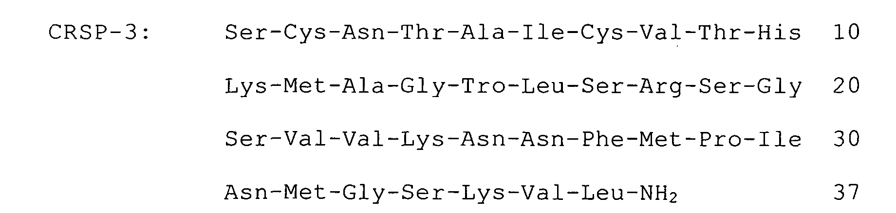


An amino acid sequence of CRSP-2 set forth in SEQ ID NO:
12 of the Sequence Listing, that of CRSP-3 is shown in SEQ ID
NO: 16 and that of CT-2 is shown in SEQ ID NO: 19.
With regard to a cDNA library, there was used a λ phage
library into which pig hypothalamus DNA was inserted being
prepared in conducting a cDNA cloning of CRSP. With regard
to a probe, a pig calcitonin receptor-stimulatingpeptide having
a full length of 700 bp was used. Method for screening was
that Escherichia coli was infected to λ phage into which pig
hypothalamus DNA was inserted, mixed with 0.7% agarose
containing an LB medium, sown on an incubation dish where 1.5 %
agar medium containing an LB medium was spread and incubated
at 37°C for about 8 hours. After cooling in a refrigerator for
about 2 hours, the resulting plaque was transferred to a Nylon
filter and subjected to an alkaline denaturation (being treated
with a 0.5 M of sodium hydroxide and 1.5 M of sodium chloride
solution for 2 minutes, with a 0.5 M Tris-hydrochloride (pH
7.5) and 1.5 M sodium chloride solution for 2 minutes and with
a 45 mM of sodium citrate (pH 7.0) and 450 mM of sodium chloride
solution for 5 minutes) and the filter was treated at 80°C for
2 hours. Then, the filter was dipped in a pre-hybridization
solution (20% of formamide, 0.09 M of sodium citrate (pH 7.0),
0.9 M of sodium chloride, 0.5% of bovine fetal albumin, 0.5%
of Ficoll, 0.5% of polyvinylpyrrolidone and 0.5% of sodium
laurylsulfate) at 37°C for 2 hour, a probe labeled with 32P was
added thereto and hybridization was conducted at 42°C for 16
hours. After the filter was washed with washing solutions (with
a 30 mM of sodium citrate (pH 7.0), 300 mM of sodium chloride
and 0.1% of sodium laurylsulfate solution at room temperature
for 5 minutes for two times and with a 15 mM of sodium citrate
(pH 7.0), 150 mM of sodium chloride and 0.1% of sodium
laurylsulfate solution at 60°C for 1 hour for two times), it
was exposed to an X-ray film, the area which was sensitized
with radioactive ray was compared with the plaque on the
incubation dish and a positive clone was isolated from the plaque.
The isolated positive λ phase clone was converted to a plasmid
pBluescript which is a vector suitable for DNA sequence using
a Helper Phage R408 of Stratagene and a nucleotide sequence
was determined by a Sanger method. As a result thereof, 13
positive clones were able to be prepared. All of six among
the above were clones containing almost the full length of the
CRSP precursor cDNA while all of other six were clones containing
almost the full length of the CRSP-2 precursor cDNA (Fig. 20).
A remaining one was a short clone coding for a 3'-untranslated
region of CRSP-3.
A PCR where pig hypothalamus cDNA (corresponding to 20
ng of mRNA) was a template was carried out using primers (CRSP-3:
GCCCAGCTTACGTCTCCTTT and TCAGGTAACTGCAATGATTT; CT-2:
AGCAGCTTTGATTCTGCCAC and ACCTCCTCTCTGATATTCCA) and Pyrobest
DNA polymerase of Takara Shuzo for 30 cycles each comprising
at 94°C for 15 seconds, at 55°C for 15 seconds and at 72°C for
1 minute. The amplified DNA was subjected to an agarose gel
electrophoresis, bands stained with ethidium bromide were
recovered from agarose gel and subcloned into pBluescript II
of Stratagene and a nucleotide sequence was determined by a
Sanger method.
Analysis of a nucleotide sequence of the amplified DNA
was conducted using a primer of CRSP-3 and, as a result, it
was found to code for CRSP-3 (Fig. 5). On the other hand, in
the PCR using a primer of CT-2, no amplification of DNA was
observed.
A template cDNA was prepared using total RNA (4 µg) of
each tissue of pig, oligo dT primer and a Revertra Ace reverse
transcriptase kit of Toyobo and 1/40 thereof was used for RT-PCR.
The PCR for amplification of each genetic cDNA was conducted
using rTaq polymerase of Toyobo in 30 cycles each comprising
at 94°C for 15 seconds, at 60°C for 15 seconds and at 72°C for
1 minute and, with regard to the primer, the following sequences
were used.







The amplified DNA was subjected to electrophoresis using
3% agarose gel and analyzed using Fuji Film FLA 2000. As a
result, expression was observed in the central nervous system,
thyroid gland and ovary for CRSP-2 and CRSP-3. On the other
hand, no band was amplified in any tissue and no expression
was observed for CT-2 (Fig. 24). It is believed that CT-2 was
expressed limitedly in the areas other than the tissues shown
in Fig. 24.
The present invention provides novel and useful peptides
which are expressed in central nerves and strongly act on
calcitonin receptor. The peptides of the present invention
have a high homology to calcitonin gene-related peptides and
have stronger action than calcitonin. They are also abundantly
expressed in central nervous system and are very useful peptides
as novel calcitonin-like peptides for osteoporosis and as
analgesic agent, etc.

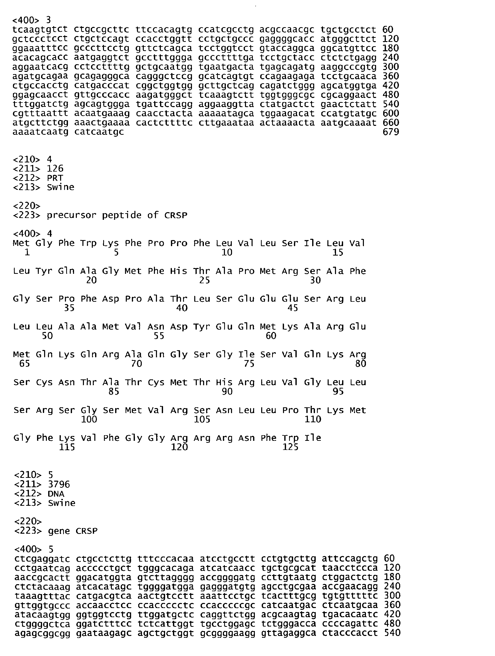
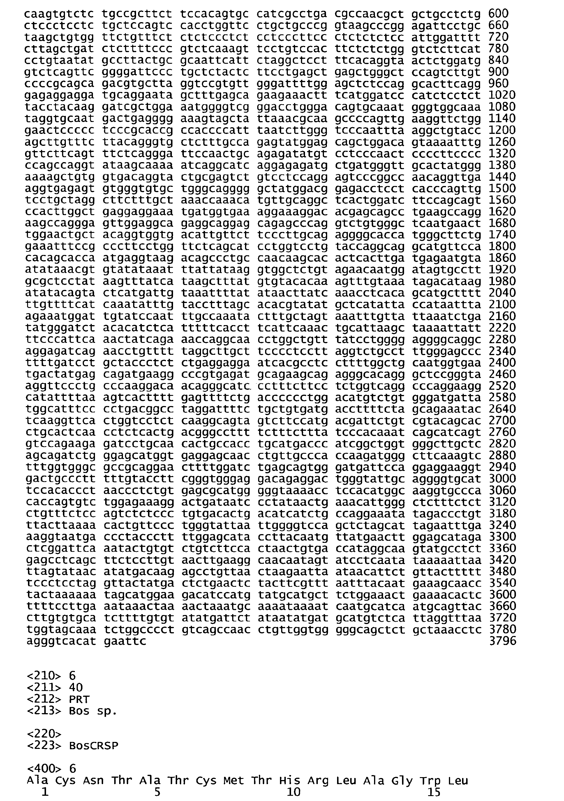


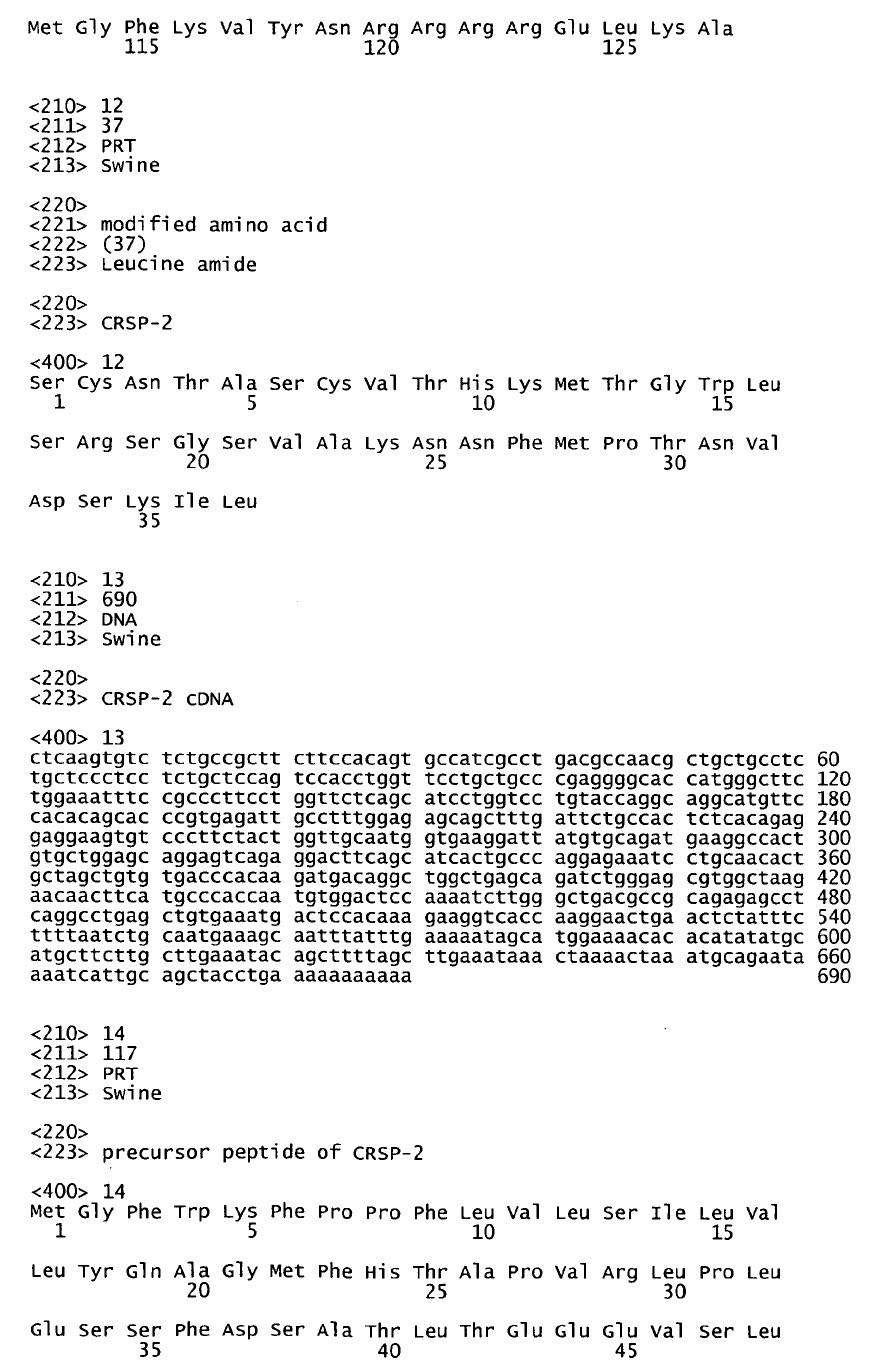
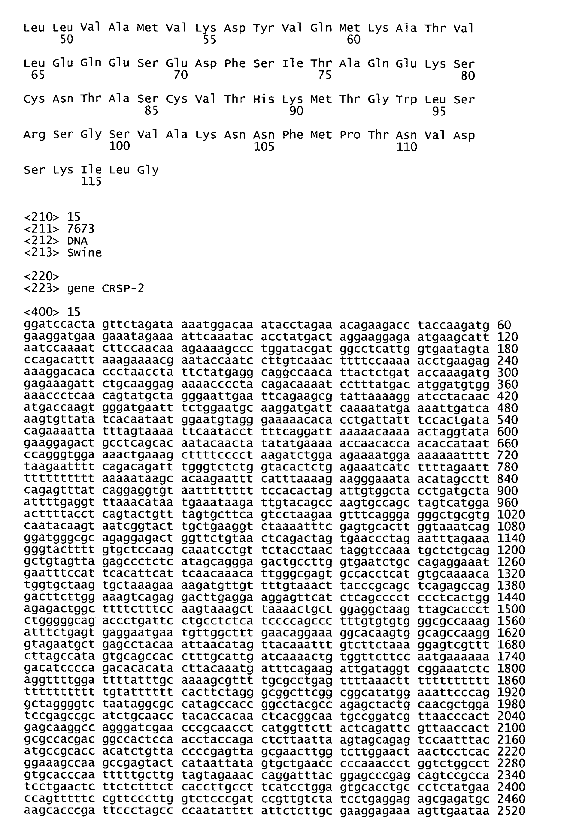
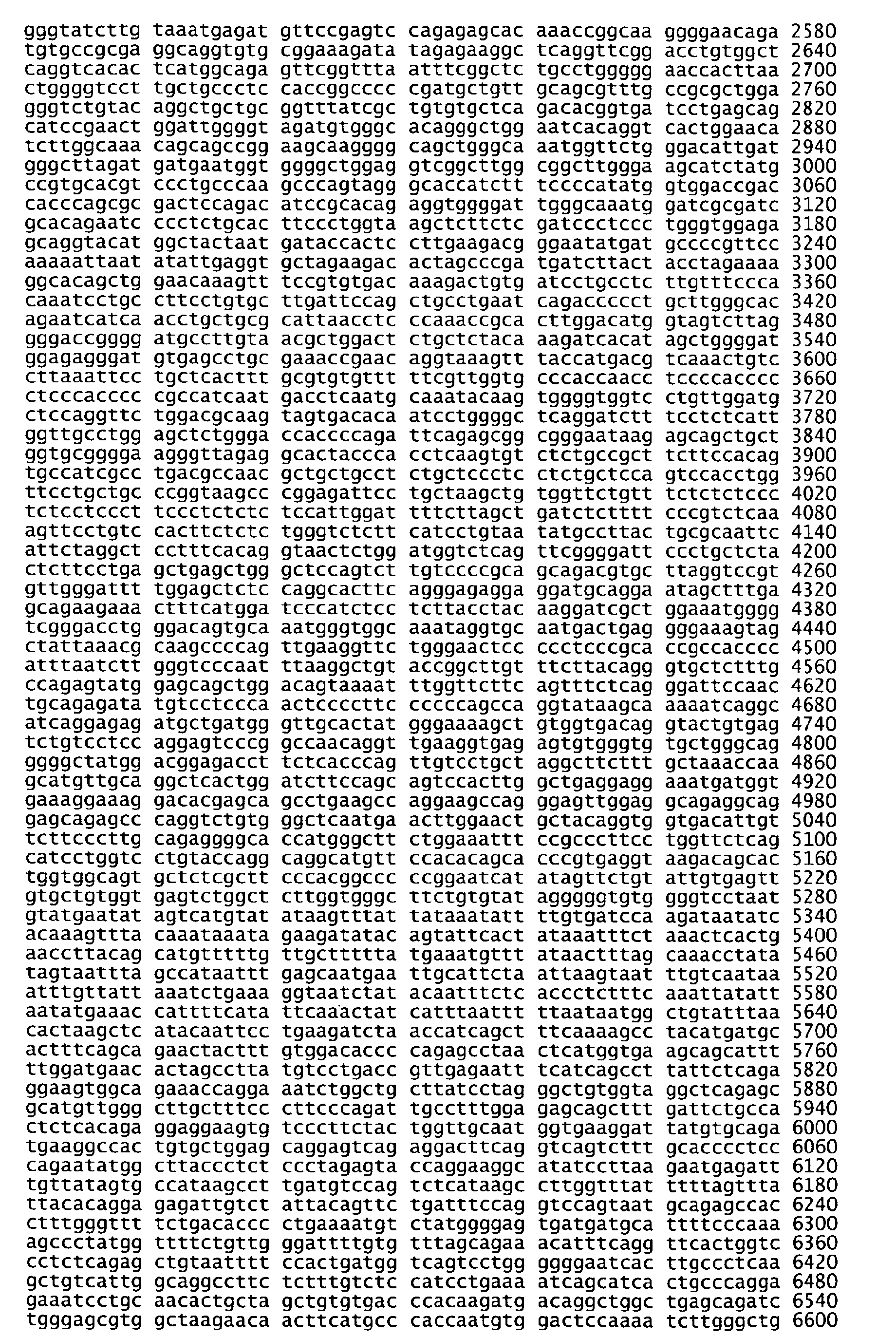
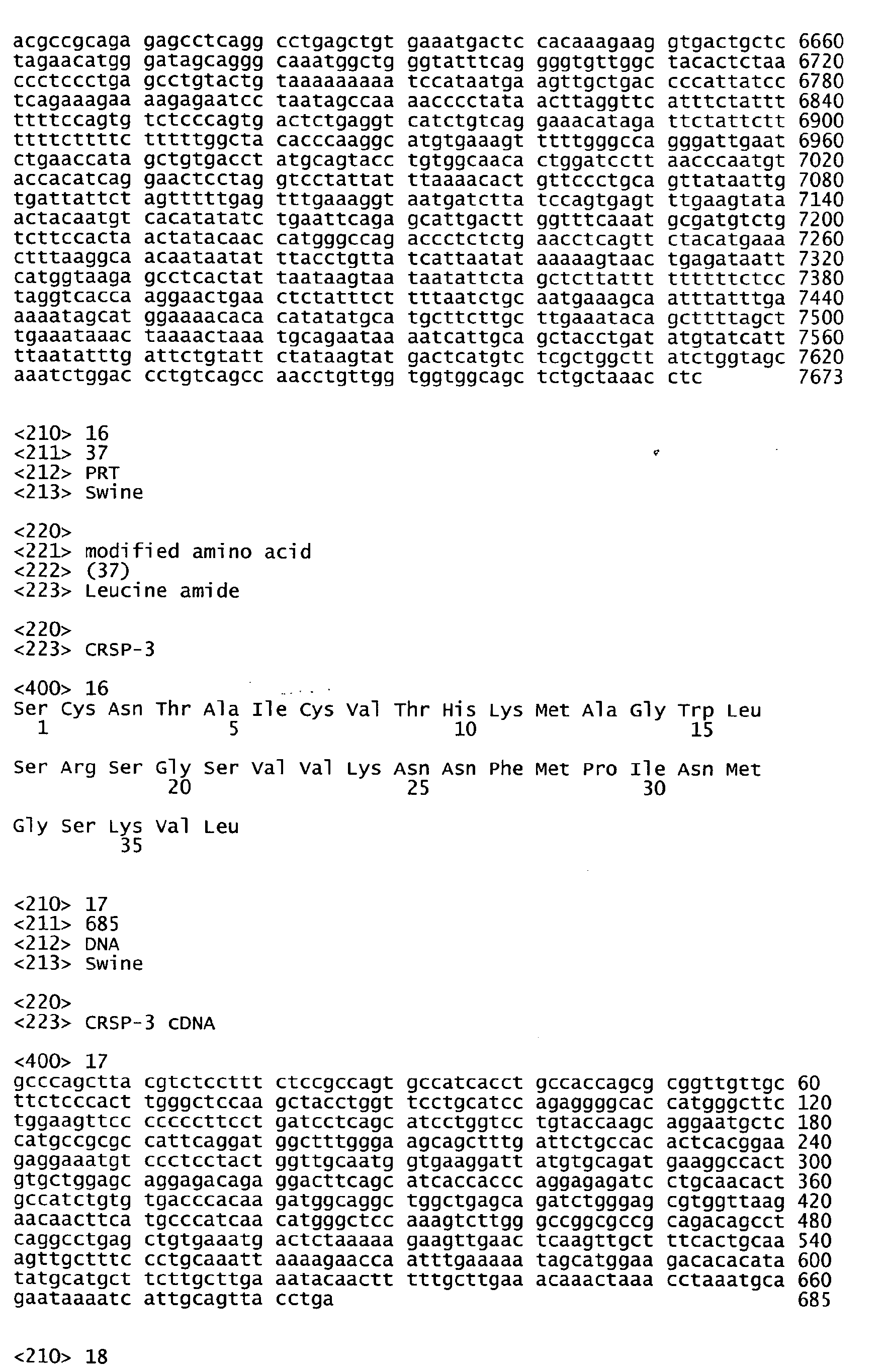

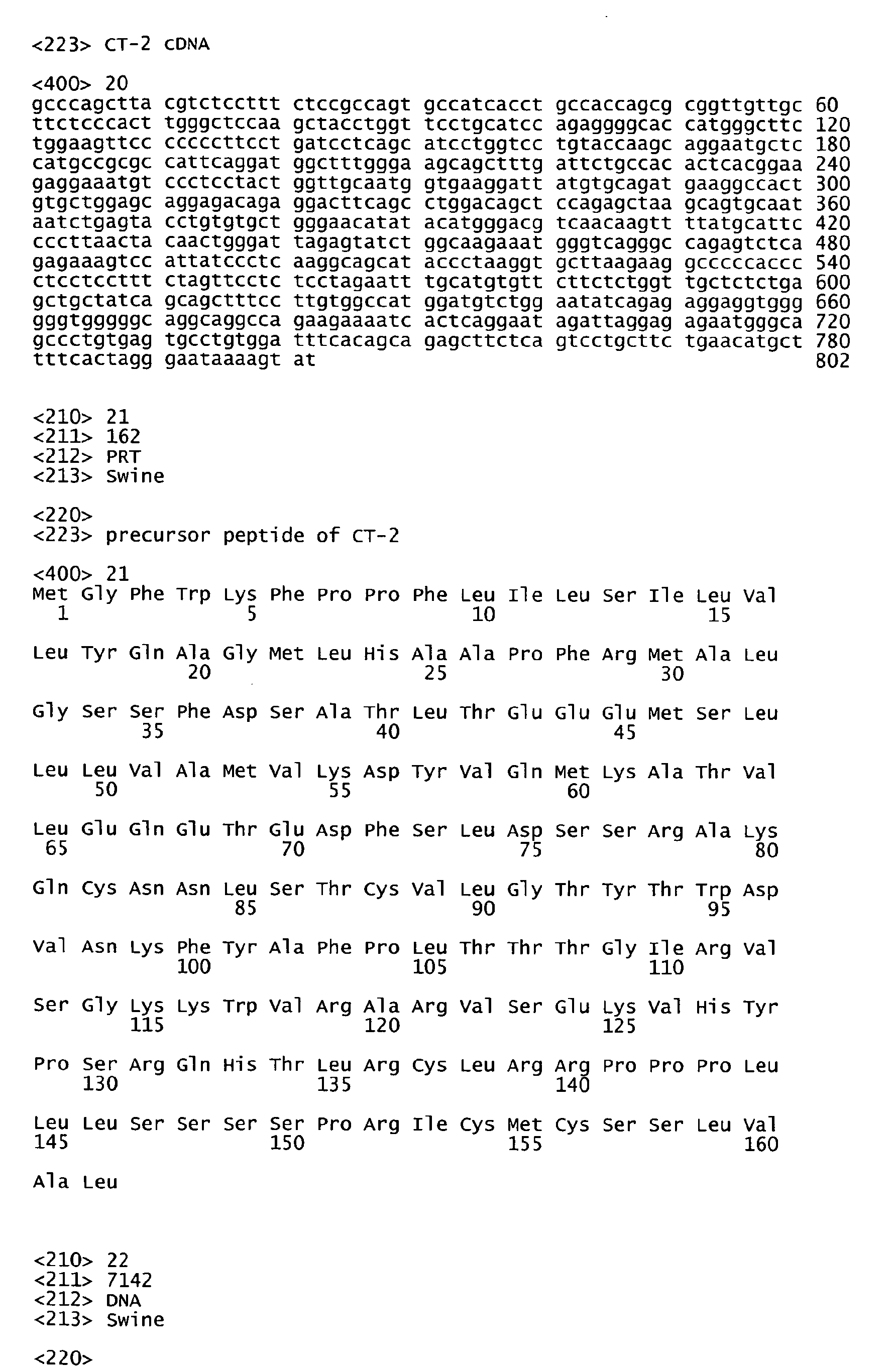

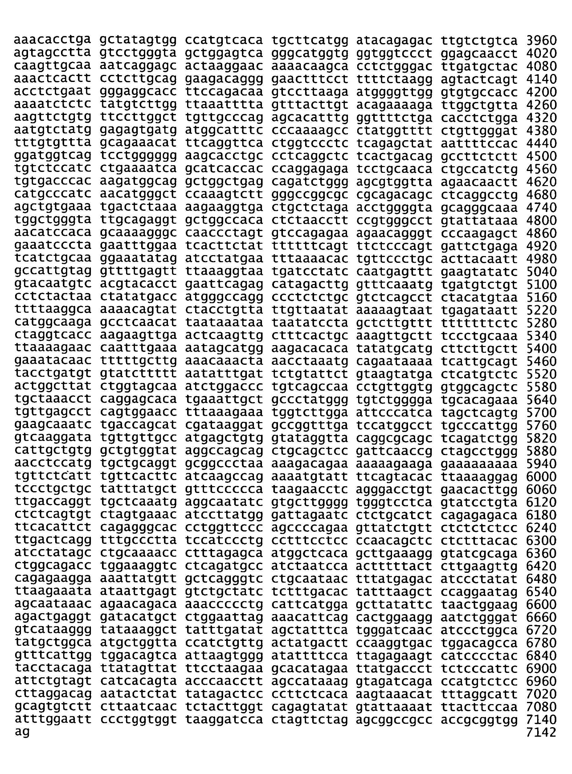
Claims (9)
- A peptide which has at least the following amino acid sequence


 or an amino acid sequence where a part of the amino acids are deleted, substituted or added and has properties of (1) being expressed in central nervous system, (2) strongly acting on calcitonin receptors and (3) promoting the cAMP productivity of cells.
- The peptide according to claim 1, wherein the peptide further has (4) a property of incorporation of sodium ion concentration-dependently, (5) a property of suppressing the incorporation of calcium ion and (6) a property of suppressing the cell proliferation.
- The peptide according to claim 1 or 2, wherein the peptide has at least an amino acid sequence shown in SEQ ID NO: 1, SEQ ID NO: 2, SEQ ID NO: 6, SEQ ID NO: 9, SEQ IS NO: 12, SEQ ID NO: 16 or SEQ ID NO: 19 of the Sequence Listing or an amino acid sequence where a part of amino acids are deleted, substituted or added.
- The peptide according to any of claims 1 to 3, wherein the peptide is a peptide derived from mammals.
- A gene which encodes the peptides according to any of claims 1 to 4.
- The gene according to claim 5, wherein the gene has a nucleotide sequence shown in SEQ ID NO: 3, SEQ ID NO: 7, SEQ ID NO: 10, SEQ ID NO: 13, SEQ IS NO: 17 or SEQ ID NO: 20 of the Sequence Listing.
- A pharmaceutical composition comprising the peptide according to any of claims 1 to 4 and a pharmaceutically acceptable carrier.
- The pharmaceutical composition according to claim 7, wherein the pharmaceutical composition is a preventive/treating agent for osteoporosis, a preventive/treating agent for cancer, a diuretic agent, an appetite suppressing agent or an analgesic agent.
- The pharmaceutical composition according to claim 7, wherein the pharmaceutical composition is a hypotensive agent or a drug which prevents restenosis after PTCA (percutaneous transluminal coronary angioplasty).
Applications Claiming Priority (3)
| Application Number | Priority Date | Filing Date | Title |
|---|---|---|---|
| JP2002162797A JP2004008027A (en) | 2002-06-04 | 2002-06-04 | Novel peptide having cAMP production activity |
| JP2002162797 | 2002-06-04 | ||
| PCT/JP2003/006641 WO2003102180A1 (en) | 2002-06-04 | 2003-05-28 | NOVEL PEPTIDES HAVING cAMP PRODUCING ACTIVITY |
Publications (2)
| Publication Number | Publication Date |
|---|---|
| EP1533374A1 true EP1533374A1 (en) | 2005-05-25 |
| EP1533374A4 EP1533374A4 (en) | 2007-08-08 |
Family
ID=29706616
Family Applications (1)
| Application Number | Title | Priority Date | Filing Date |
|---|---|---|---|
| EP03730658A Withdrawn EP1533374A4 (en) | 2002-06-04 | 2003-05-28 | NEW PEPTIDES WITH CAMP PRODUCTION ACTIVITY |
Country Status (5)
| Country | Link |
|---|---|
| US (1) | US20050256302A1 (en) |
| EP (1) | EP1533374A4 (en) |
| JP (1) | JP2004008027A (en) |
| CN (1) | CN1697878A (en) |
| WO (1) | WO2003102180A1 (en) |
Families Citing this family (7)
| Publication number | Priority date | Publication date | Assignee | Title |
|---|---|---|---|---|
| EP1743940B1 (en) | 2004-04-02 | 2012-01-18 | Japan Science and Technology Agency | Cardioinhibitory/ antihypertensive novel endogenous physiologically active peptide |
| US8168592B2 (en) | 2005-10-21 | 2012-05-01 | Amgen Inc. | CGRP peptide antagonists and conjugates |
| WO2008060568A2 (en) * | 2006-11-15 | 2008-05-22 | Neurogen Corporation | Amide-substituted aryl piperidines |
| US8865762B2 (en) * | 2009-07-17 | 2014-10-21 | Korea Research Institute Of Bioscience And Biotechnology | Method for the treatment of bone diseases comprising colforsin daropate |
| GB2490974B (en) * | 2011-09-08 | 2014-10-29 | Nordic Semiconductor Asa | Radio communication system |
| JP6709024B2 (en) * | 2015-07-03 | 2020-06-10 | 国立研究開発法人医薬基盤・健康・栄養研究所 | Peptide or protein fractionation method |
| CN112979748B (en) * | 2021-04-16 | 2022-05-06 | 吉林大学 | An active peptide with the effect of preventing chronic alcoholic liver injury |
Family Cites Families (4)
| Publication number | Priority date | Publication date | Assignee | Title |
|---|---|---|---|---|
| EP0672419A1 (en) * | 1991-12-12 | 1995-09-20 | Asahi Kasei Kogyo Kabushiki Kaisha | Blood pressure regulator |
| US6268474B1 (en) * | 1998-04-30 | 2001-07-31 | Creighton University | Peptide antagonists of CGRP-receptor superfamily and methods of use |
| AU765753B2 (en) * | 1999-05-17 | 2003-09-25 | Conjuchem Biotechnologies Inc. | Protection of endogenous therapeutic peptides from peptidase activity through conjugation to blood components |
| GB0109438D0 (en) * | 2001-04-17 | 2001-06-06 | Isis Innovation | Peptides |
-
2002
- 2002-06-04 JP JP2002162797A patent/JP2004008027A/en active Pending
-
2003
- 2003-05-28 EP EP03730658A patent/EP1533374A4/en not_active Withdrawn
- 2003-05-28 US US10/516,768 patent/US20050256302A1/en not_active Abandoned
- 2003-05-28 WO PCT/JP2003/006641 patent/WO2003102180A1/en not_active Ceased
- 2003-05-28 CN CNA038187493A patent/CN1697878A/en active Pending
Also Published As
| Publication number | Publication date |
|---|---|
| WO2003102180A1 (en) | 2003-12-11 |
| JP2004008027A (en) | 2004-01-15 |
| CN1697878A (en) | 2005-11-16 |
| EP1533374A4 (en) | 2007-08-08 |
| US20050256302A1 (en) | 2005-11-17 |
Similar Documents
| Publication | Publication Date | Title |
|---|---|---|
| JP2774769B2 (en) | Adrenomedullin | |
| EP0478797B1 (en) | Novel physiologically active peptide originating in hog | |
| O'Carroll et al. | Characterization of cloned human somatostatin receptor SSTR5. | |
| Katafuchi et al. | Calcitonin receptor-stimulating peptide, a new member of the calcitonin gene-related peptide family: its isolation from porcine brain, structure, tissue distribution, and biological activity | |
| US5200509A (en) | Human somatomedin carrier protein subunits and process for producing them; recombinant DNA molecules, hosts, processes and human somatomedin carrier protein-like polypeptides | |
| EP2152297B1 (en) | Unacylated ghrelin as therapeutic agent in the treatment of metabolic disorders | |
| Kinoshita et al. | Urine and plasma levels of uroguanylin and its molecular forms in renal diseases | |
| EP0451194B1 (en) | Recombinant dna molecules, hosts, processes and human somatomedin carrier protein-like polypeptides | |
| JPWO1991016342A1 (en) | Novel biologically active peptides derived from pigs | |
| JP2001252087A (en) | Acid labile subunit (ALS) of the insulin-like growth factor (IGF) binding protein complex | |
| EP0790305B1 (en) | Mutant human growth hormones and their uses | |
| EA001201B1 (en) | Antisecretory factor peptides regulating pathological permeability changes | |
| US20050256302A1 (en) | Novel peptides having camp producing activity | |
| JP2001517445A (en) | Mutants of thyroid stimulating hormone and methods based thereon | |
| EP0568622A1 (en) | CORTICOTROPIN RELEASING FACTOR BINDING PROTEIN (CRF-bp) | |
| EP0546081A1 (en) | New insulin-like growth factor binding protein (igfbp-4) | |
| CA2357677A1 (en) | Insulin-synthesis genes | |
| Kelly | Growth hormone and prolactin | |
| EP0815228A2 (en) | Cellubrevin homologs | |
| Mayer et al. | Potency of natural and synthetic canine gastrin-releasing decapeptide on canine antral muscle | |
| Inagami et al. | Structure and physiological actions of rat atrial natriuretic factor. | |
| JPH11186A (en) | Member of frzb family, frazzled | |
| EP0386979A2 (en) | DNA encoding ovine-active, growth-hormone-receptor-binding, lactogenic protein | |
| WO1996019501A1 (en) | Modulators of bone cell function and uses thereof | |
| HK1000826B (en) | Recombinant dna molecules, hosts, processes and human somatomedin carrier protein-like polypeptides |
Legal Events
| Date | Code | Title | Description |
|---|---|---|---|
| PUAI | Public reference made under article 153(3) epc to a published international application that has entered the european phase |
Free format text: ORIGINAL CODE: 0009012 |
|
| 17P | Request for examination filed |
Effective date: 20050103 |
|
| AK | Designated contracting states |
Kind code of ref document: A1 Designated state(s): AT BE BG CH CY CZ DE DK EE ES FI FR GB GR HU IE IT LI LU MC NL PT RO SE SI SK TR |
|
| A4 | Supplementary search report drawn up and despatched |
Effective date: 20070711 |
|
| STAA | Information on the status of an ep patent application or granted ep patent |
Free format text: STATUS: THE APPLICATION IS DEEMED TO BE WITHDRAWN |
|
| 18D | Application deemed to be withdrawn |
Effective date: 20071010 |