TECHNICAL FIELD OF THE INVENTION
The present invention relates to methods and compositions for the prediction of therapy outcome
(e.g. tumor response to therapy), diagnosis, prognosis, prevention and treatment of neoplastic
diseases. Cancer cells display a specific pattern of gene expression related to their morphological
type, state of progression, acquirement of genomic alterations, point mutations in critical genes
such as gatekeepers and tumor suppressors or due to the dependency of external signals such as
growth factors, hormones or other secondary messengers.
The invention discloses genes which show an altered expression in a particular neoplastic tissue
compared to the corresponding healthy tissue or to other neoplastic lesions unresponsive to a given
chemotherapy. They are useful as diagnostic markers and could be also regarded as therapeutically
targets. Methods are disclosed for predicting, diagnosing and prognosing as well as preventing and
treating neoplastic disease. The genes disclosed in this invention have been identified in breast
cancers but are predictable of outcome to a certain therapy regimen and therefor they are also
relevant for other types of cancers in tissues other than breast.
BACKGROUND OF THE INVENTION AND PRIOR ART
Cancer is the second leading cause of death in the United States after cardiovascular disease. One
in three Americans will develop cancer in his or her lifetime, and one of every four Americans will
die of cancer. More specifically breast cancer claims the lives of approximately 40,000 women and
is diagnosed in approximately 200,000 women annually in the United States alone. Cancer are
classified based on different parameters, such as tumor size, invasion status, involvement of lymph
notes, metastasis, histolopathology, imunohistochemical markers, and molecular markers (WHO.
International Classification of diseases (1); Sabin and Wittekind, 1997 (2)). With the recent
advances in gene chip technology, researchers are increasingly focusing on the categorization of
tumors based on the distinct expression of marker genes Sorlie et al., 2001 (3): van 't Veer et al.,
2002 (4).
Chemotherapy remains a mainstay in therapeutic regimens offered to patients with breast cancer,
particularly those who have cancer that has metastasized from its site of origin (Perez, 1999, (5)).
There are several chemo-therapeutic agents that have demonstrated activity in the treatment of
breast cancer and research is continuously in an attempt to determine optimal drugs and regimens.
However, different patients tend to respond differently to the same therapeutic regimen. Currently,
the individuals response to certain therapy can only be assessed statistically, based on data of
former clinical studies. There are still a great number of patients who will not benefit from a
systemic chemotherapy. Especially, breast cancers are very heterogeneous in their aggressiveness
and treatment response. They contain different genetic mutations and variations affecting growths
characteristic and sensitivity to several drugs. Identification of each tumor's molecular fingerprint,
then, could help to segregate patients who have particularly aggressive tumors or who need to be
treated with specific beneficial therapies. As research involving genetics and associated responses
to treatment matures, standard practice will undoubtedly become more individualized, enabling
physicians to provide specific treatment regimens matched with a tumor's genetic profiles to
ensure optimal outcomes.
SUMMARY OF THE INVENTION
The present invention relates to the identification of 185 human genes being differentially
expressed in neoplastic tissue resulting in an altered clinical behavior of a neoplastic lesion. The
differential expression of these 185 genes is not limited to a specific neoplastic lesion in a certain
tissue of the human body.
In preferred embodiments of this invention the neoplastic lesion, of which these 185 genes are
altered in their expression is a cancer of the human breast. This cancer is not limited to females
and may also be diagnosed and analyzed in males.
The invention relates to various methods, reagents and kits for diagnosing, staging, prognosis,
monitoring and therapy of breast cancer. "Breast cancer" as used herein includes carcinomas, (e.g.,
carcinoma in situ, invasive carcinoma, metastatic carcinoma) and pre-malignant conditions,
neomorphic changes independent of their histological origin (e.g. ductal, lobular, medullary, mixed
origin). The compositions, methods, and kits of the present invention comprise comparing the level
of mRNA expression of a single or plurality (e.g. 2, 5, 10, or 50 or more) of genes (hereinafter
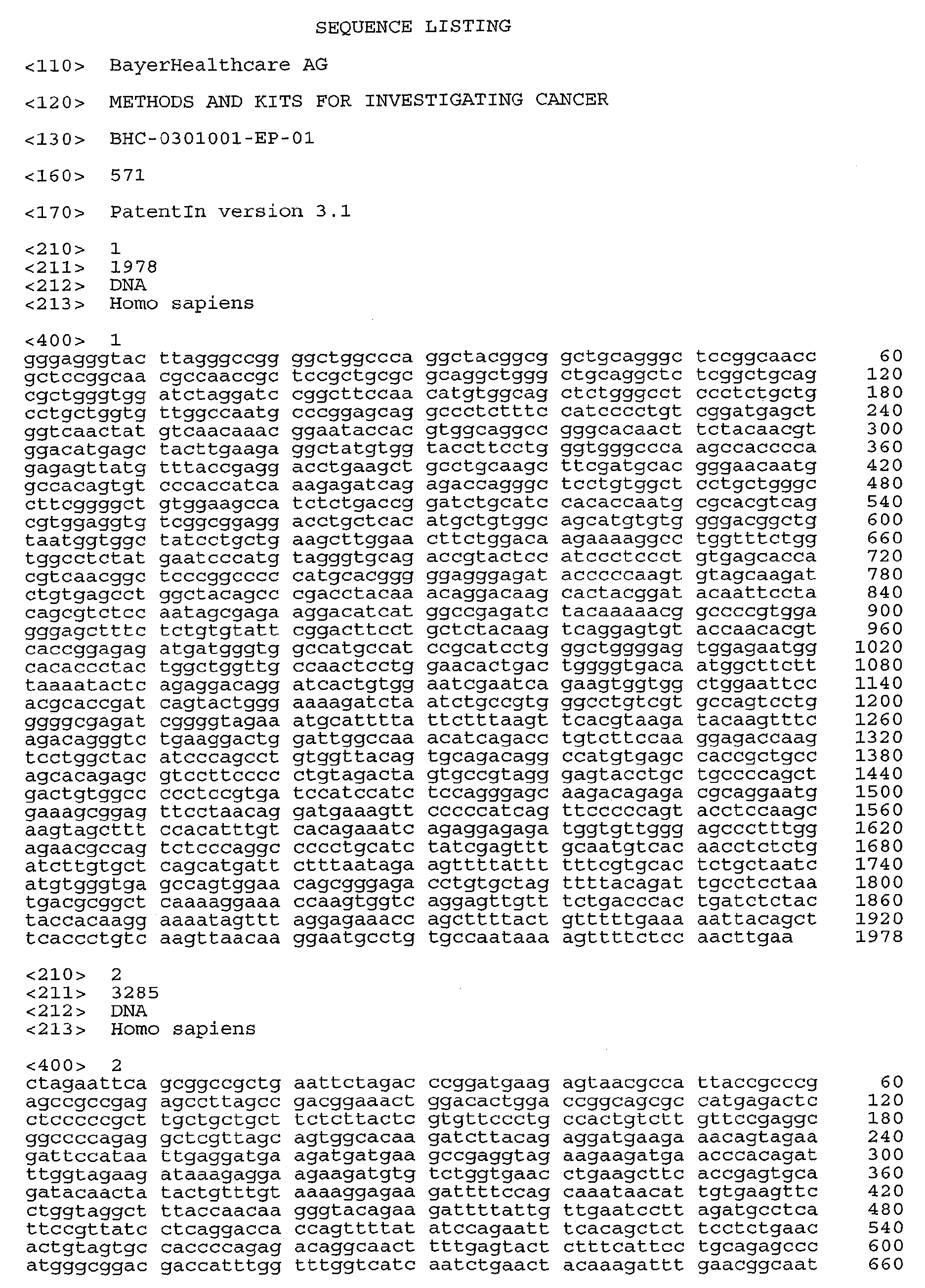
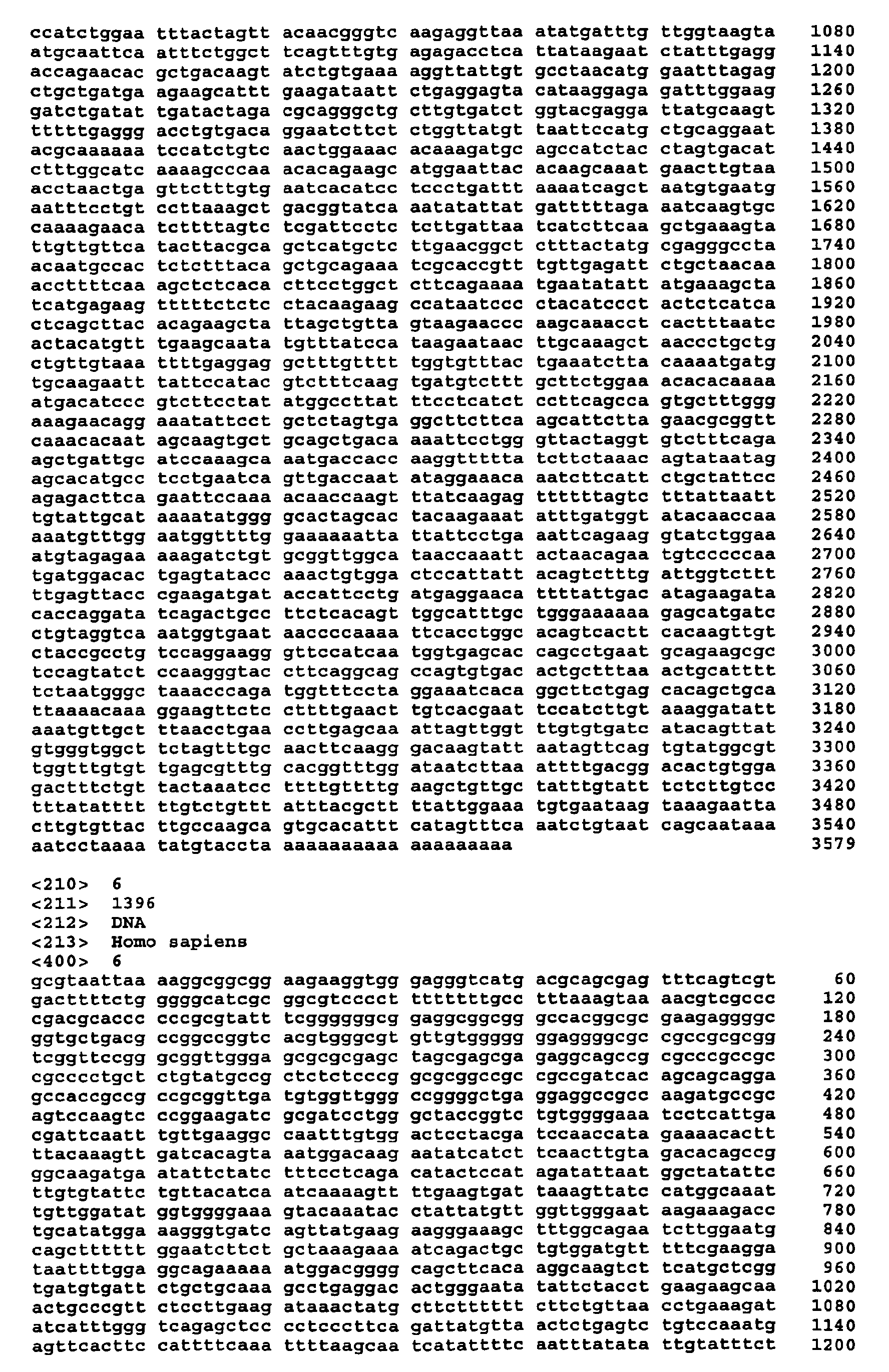
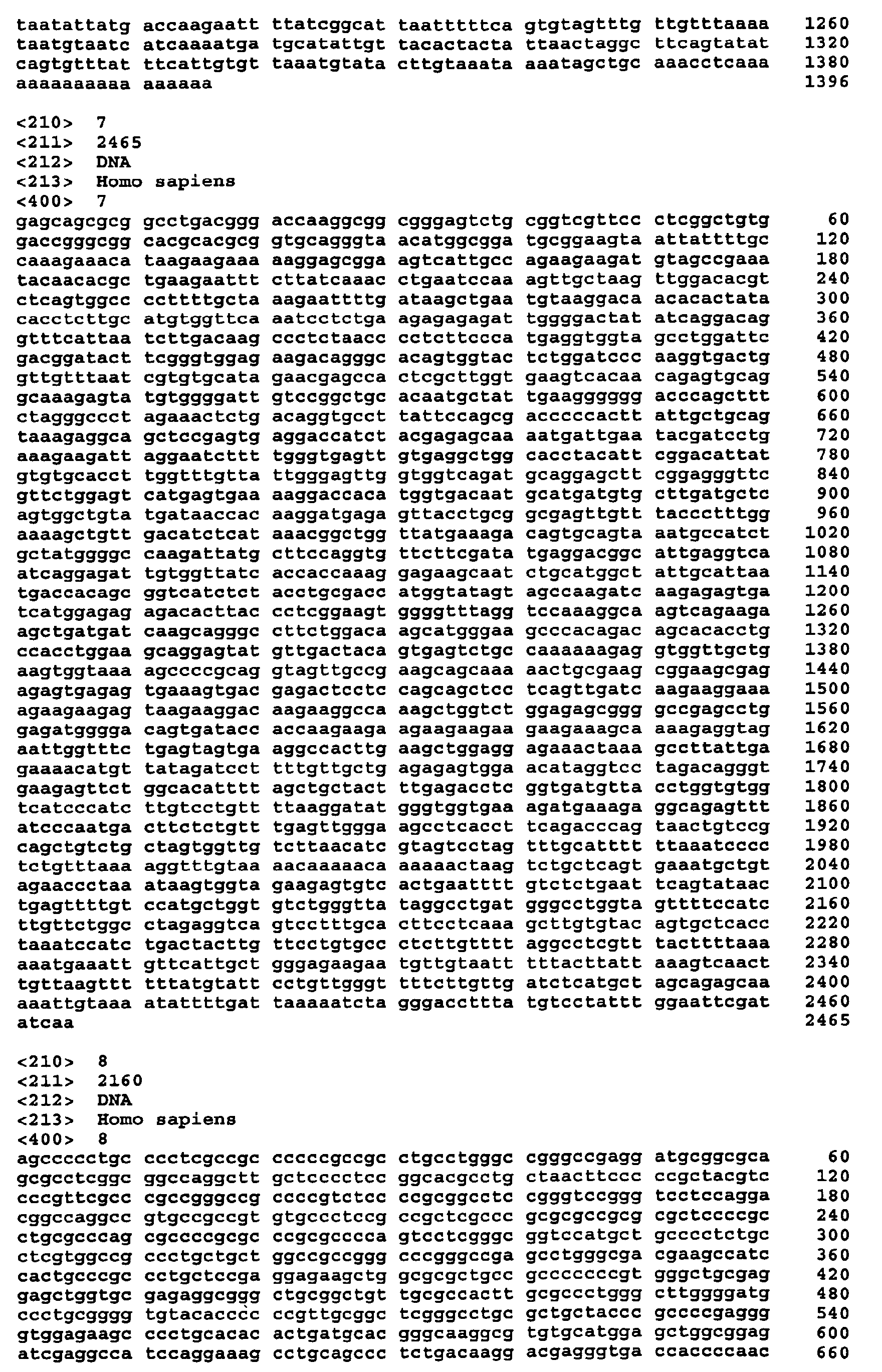
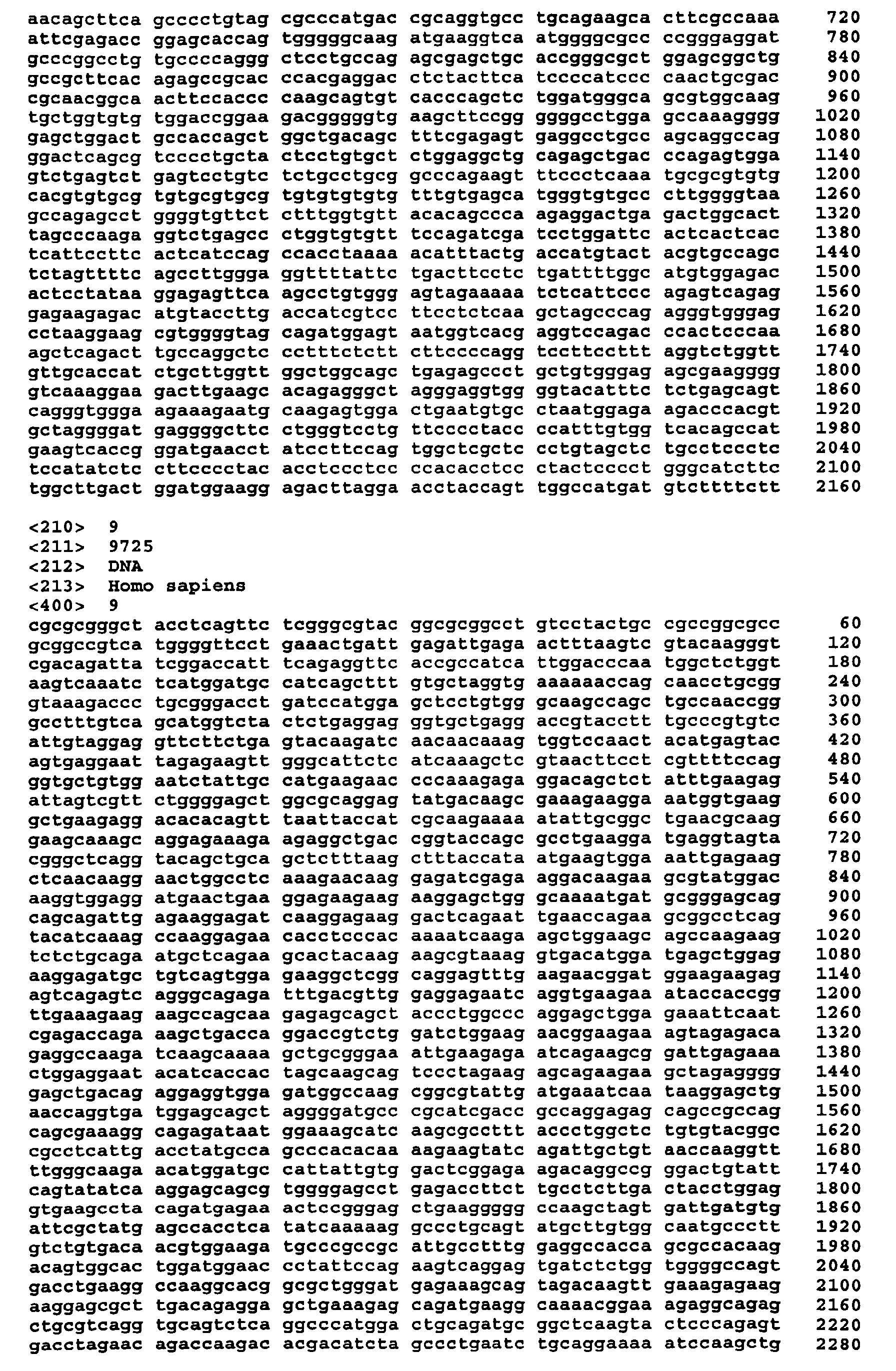
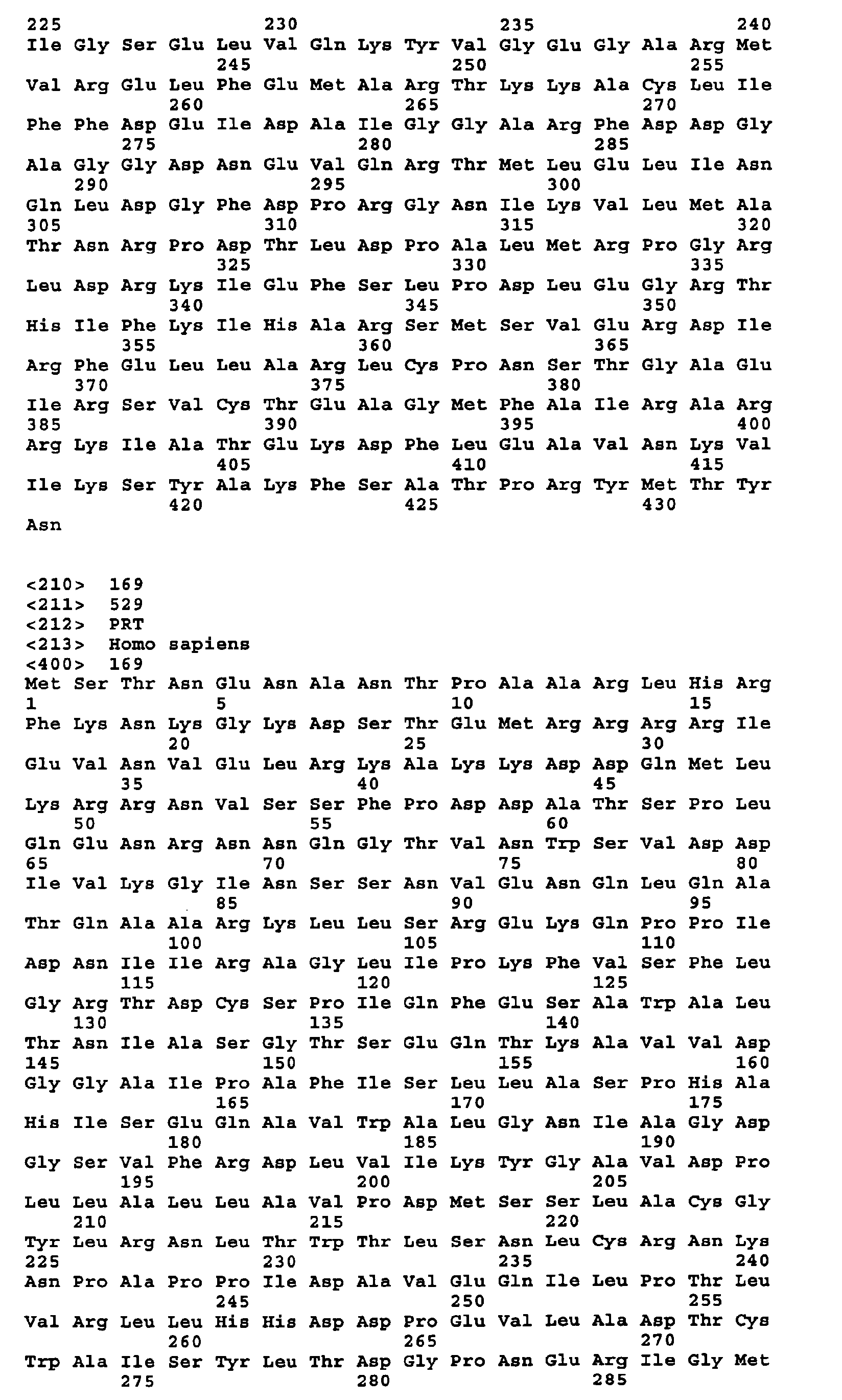
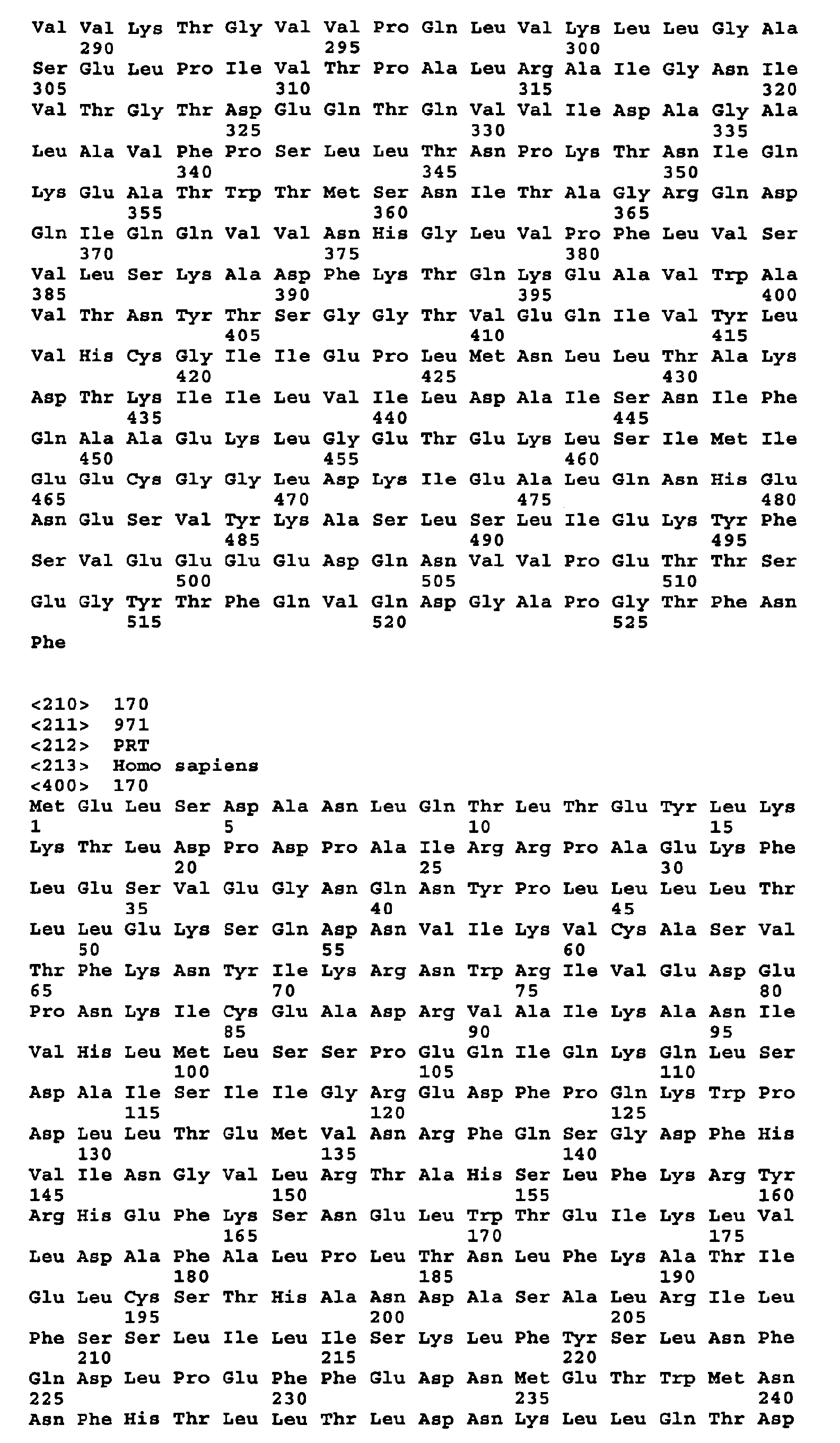
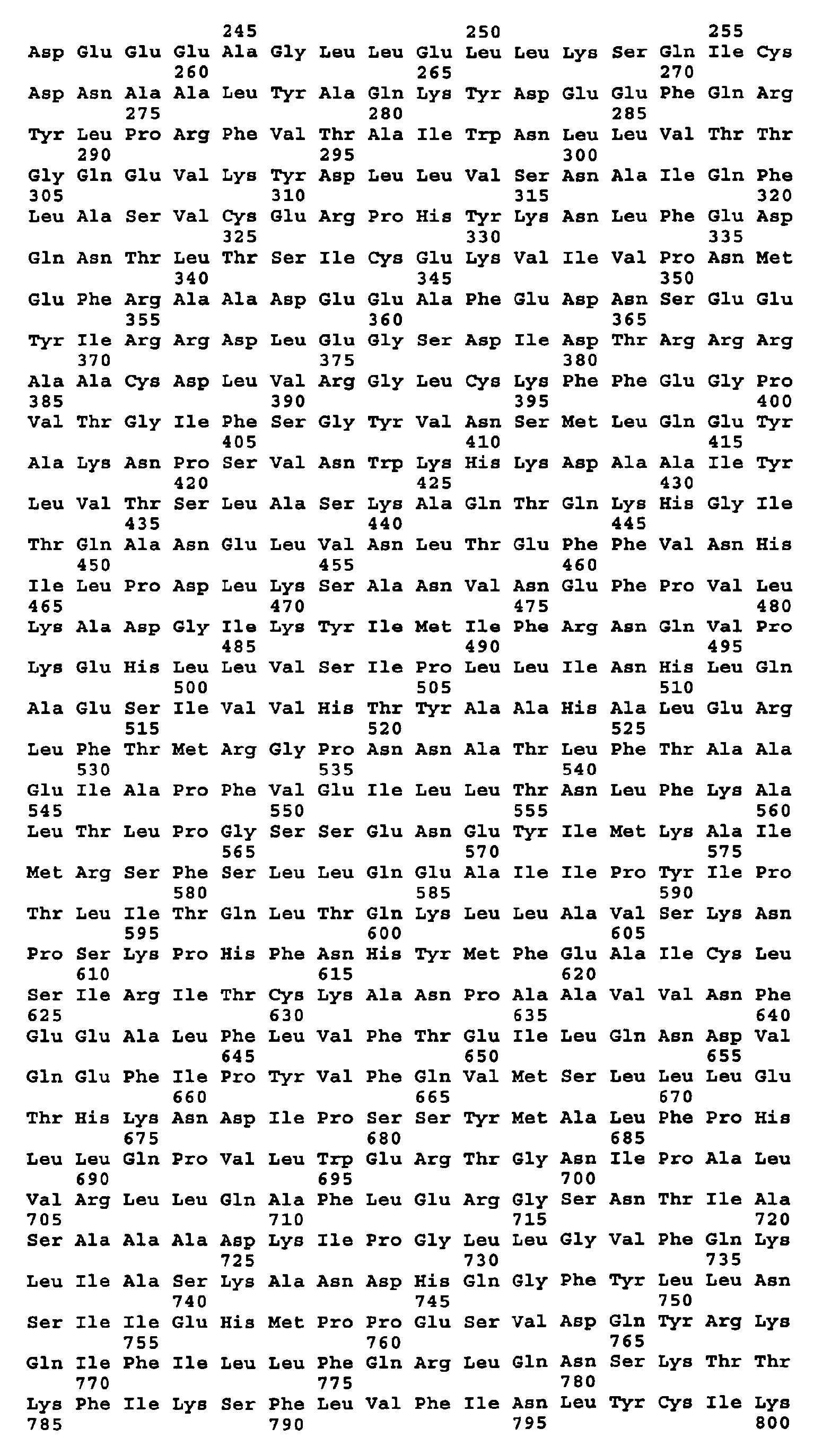
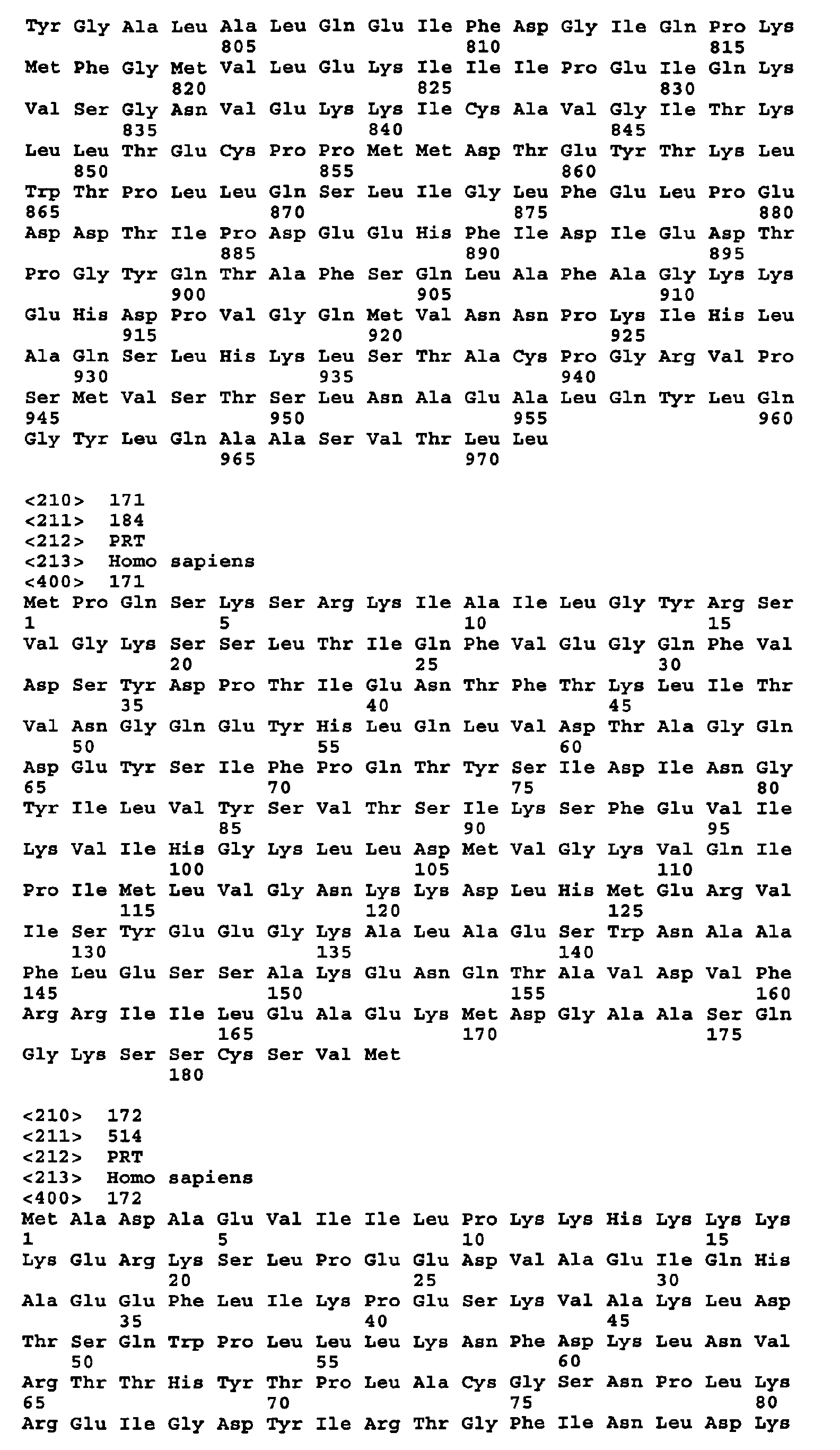
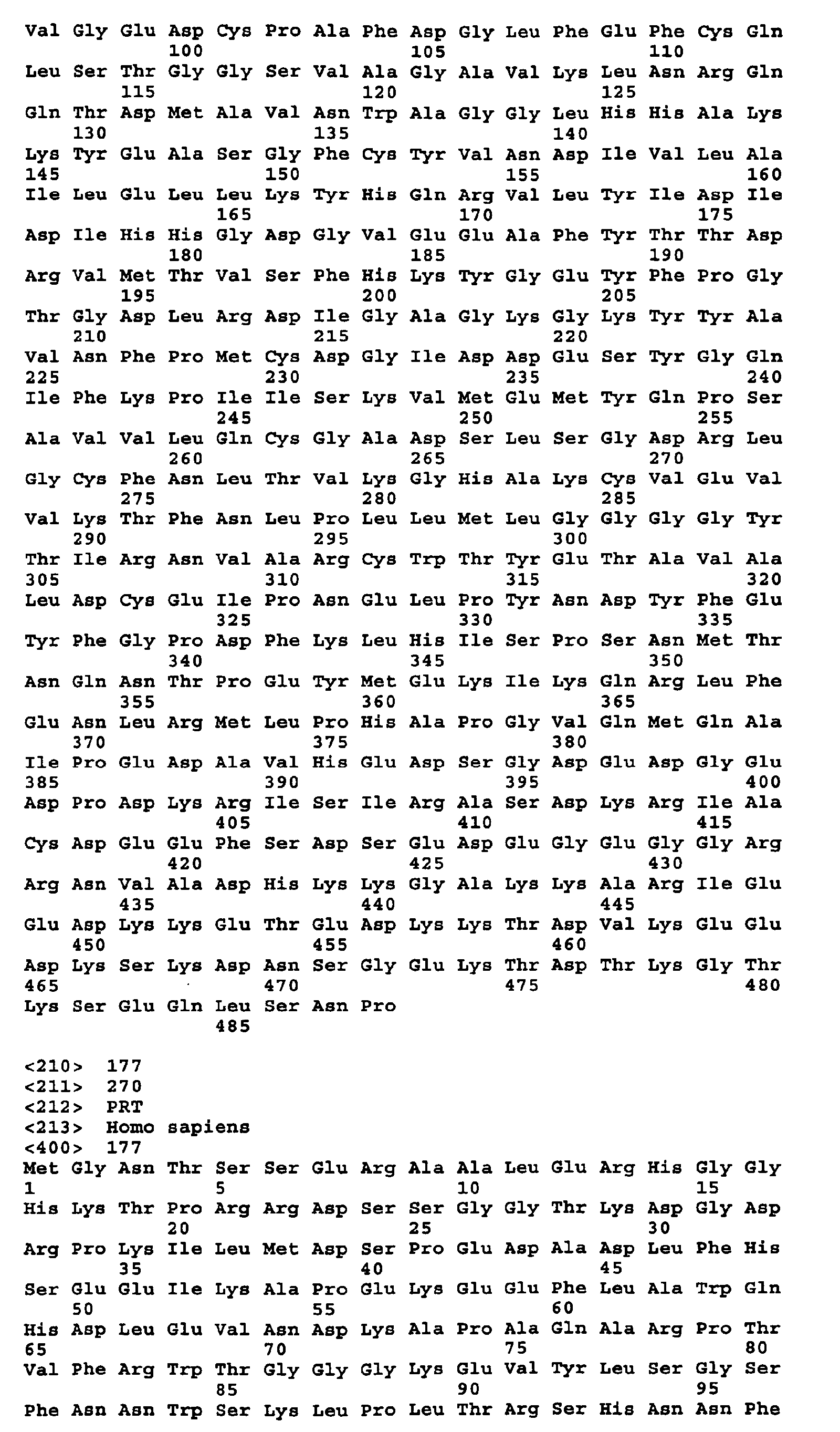
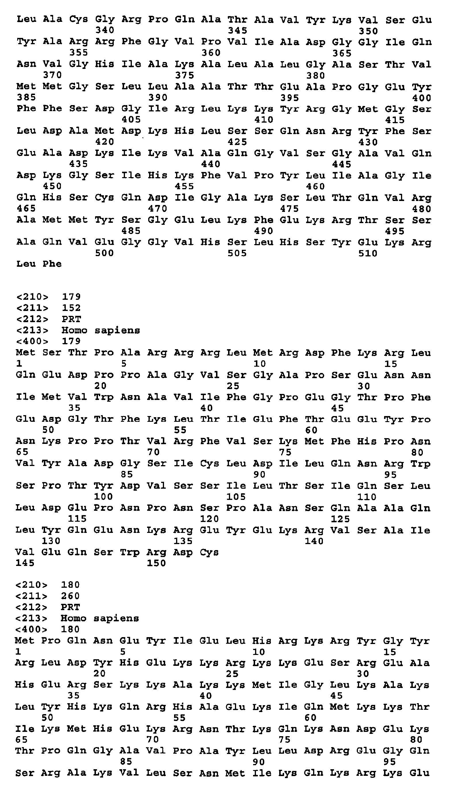
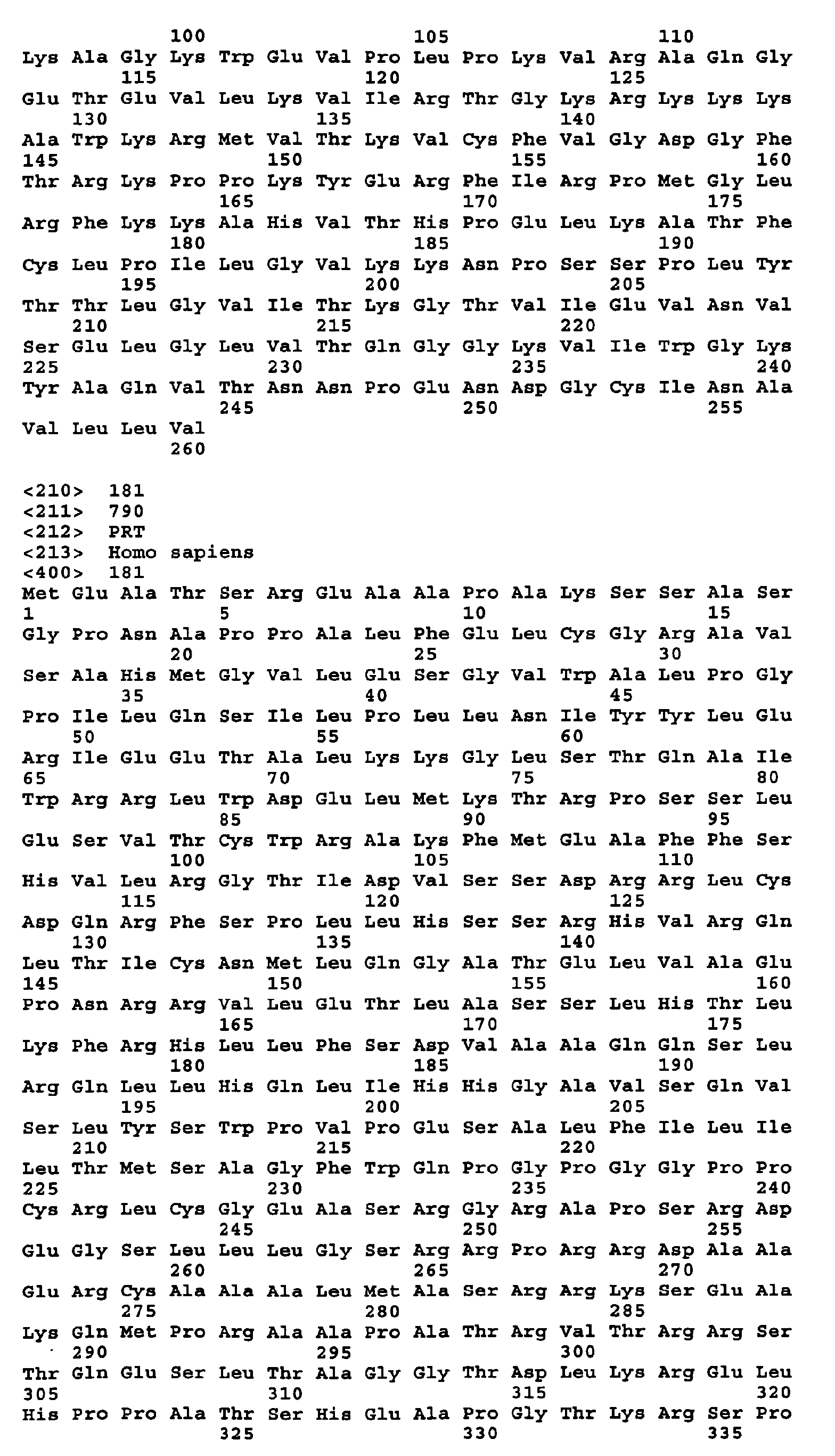
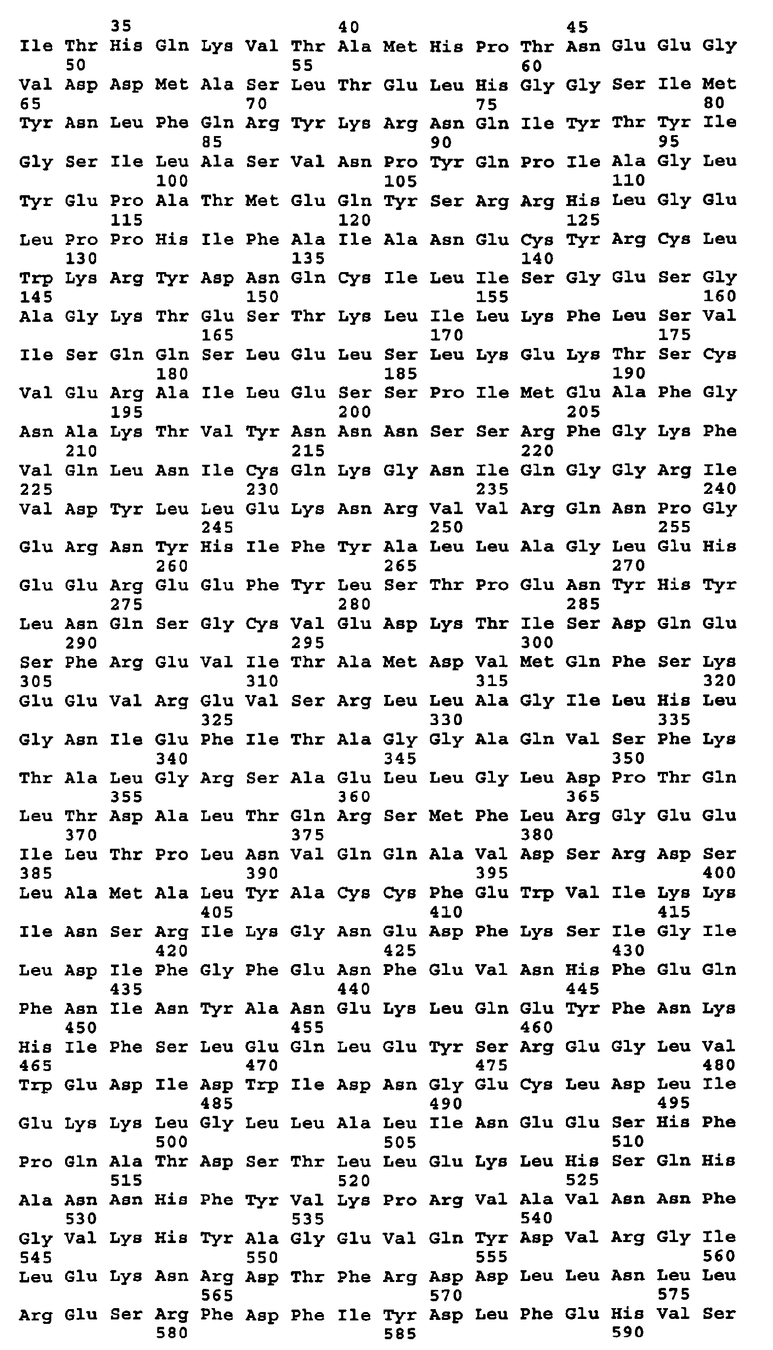
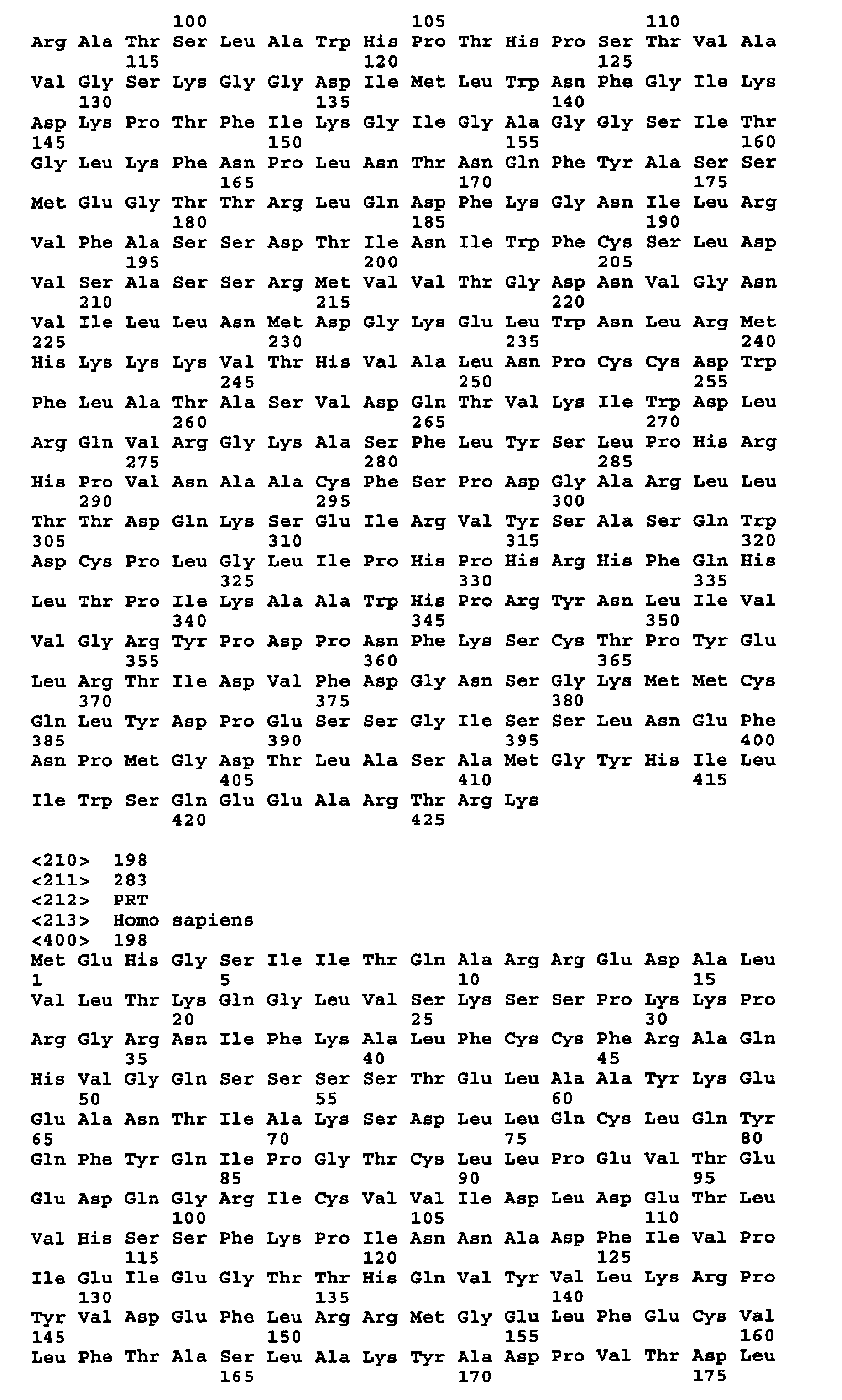
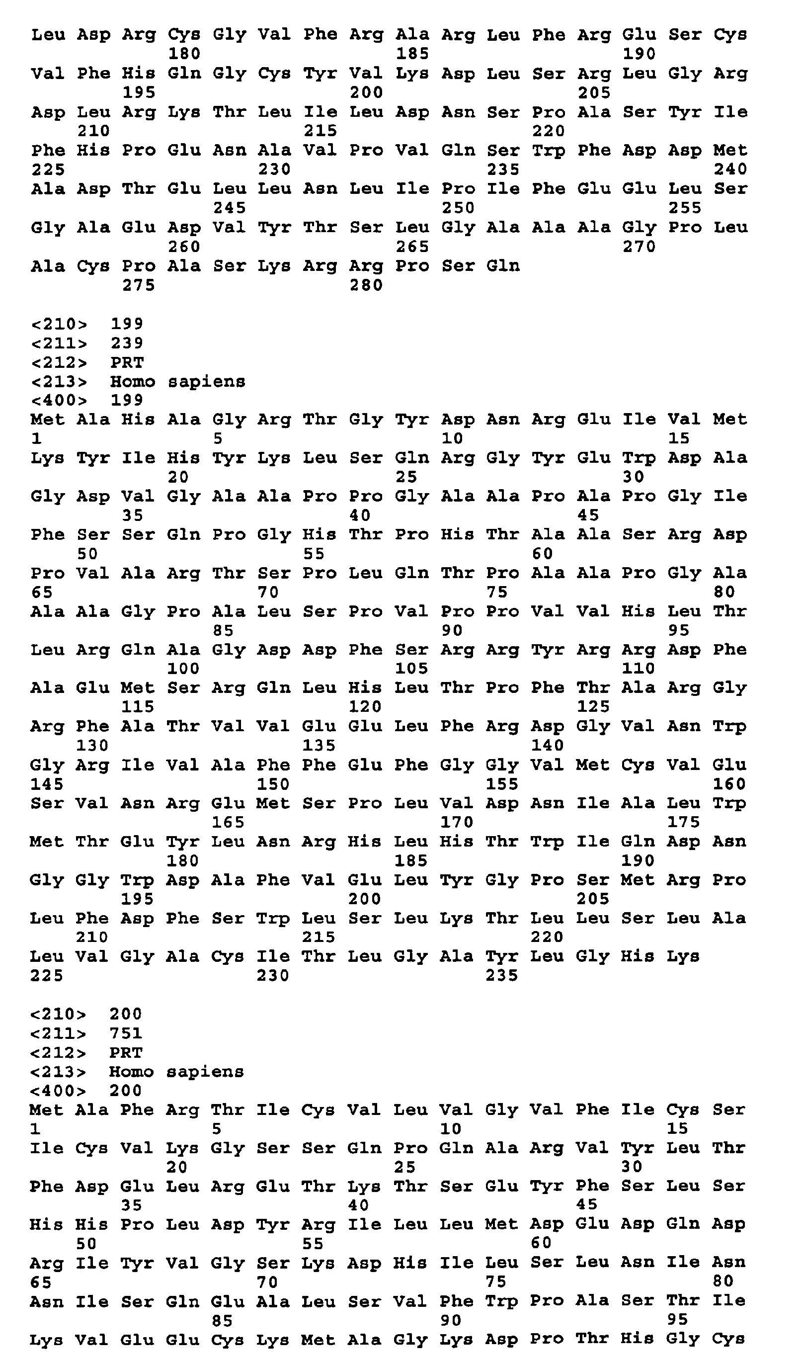
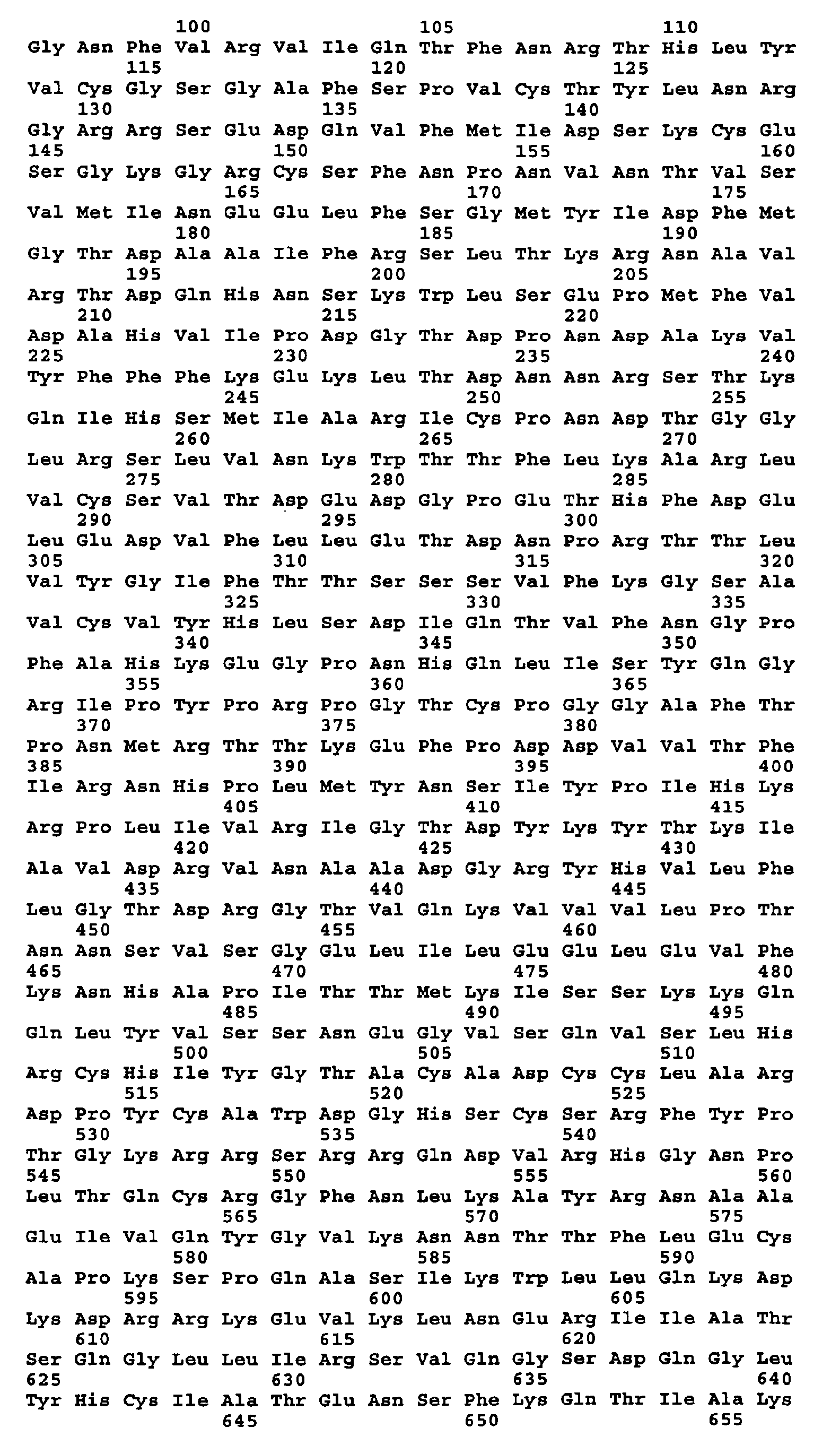
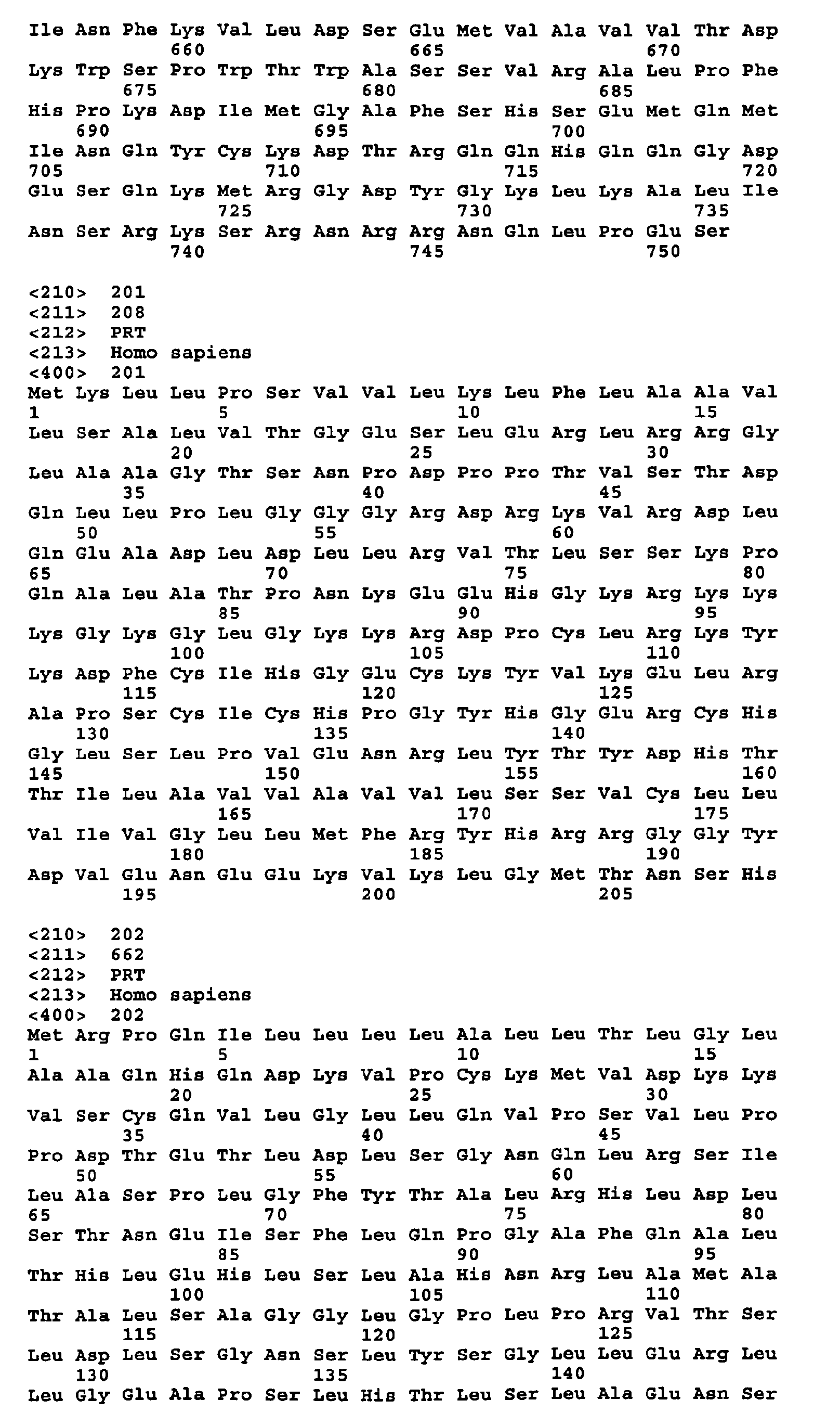
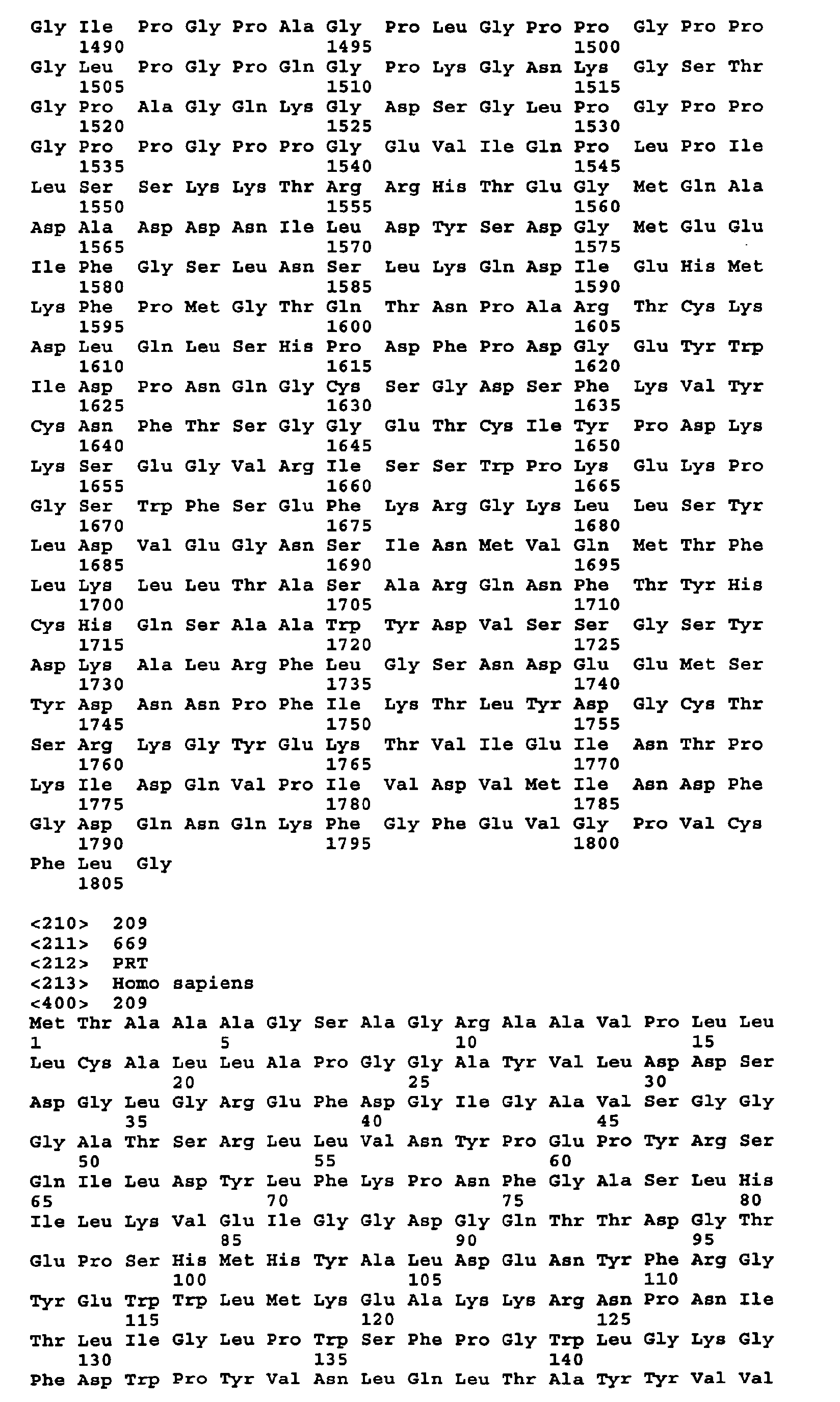
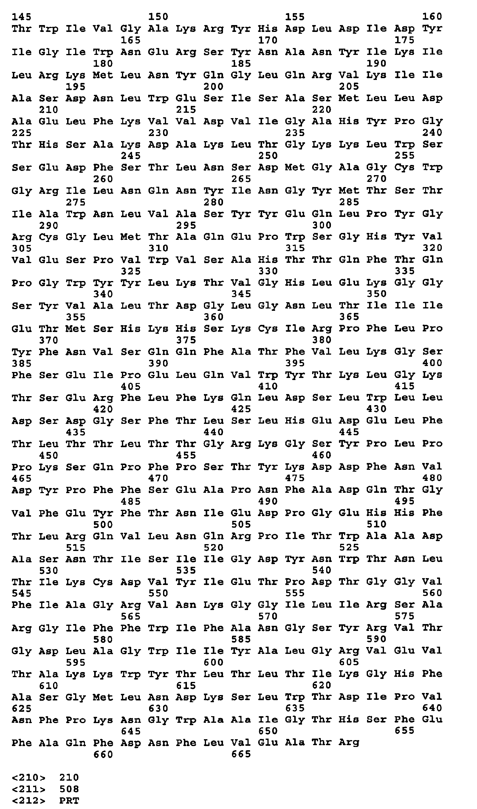
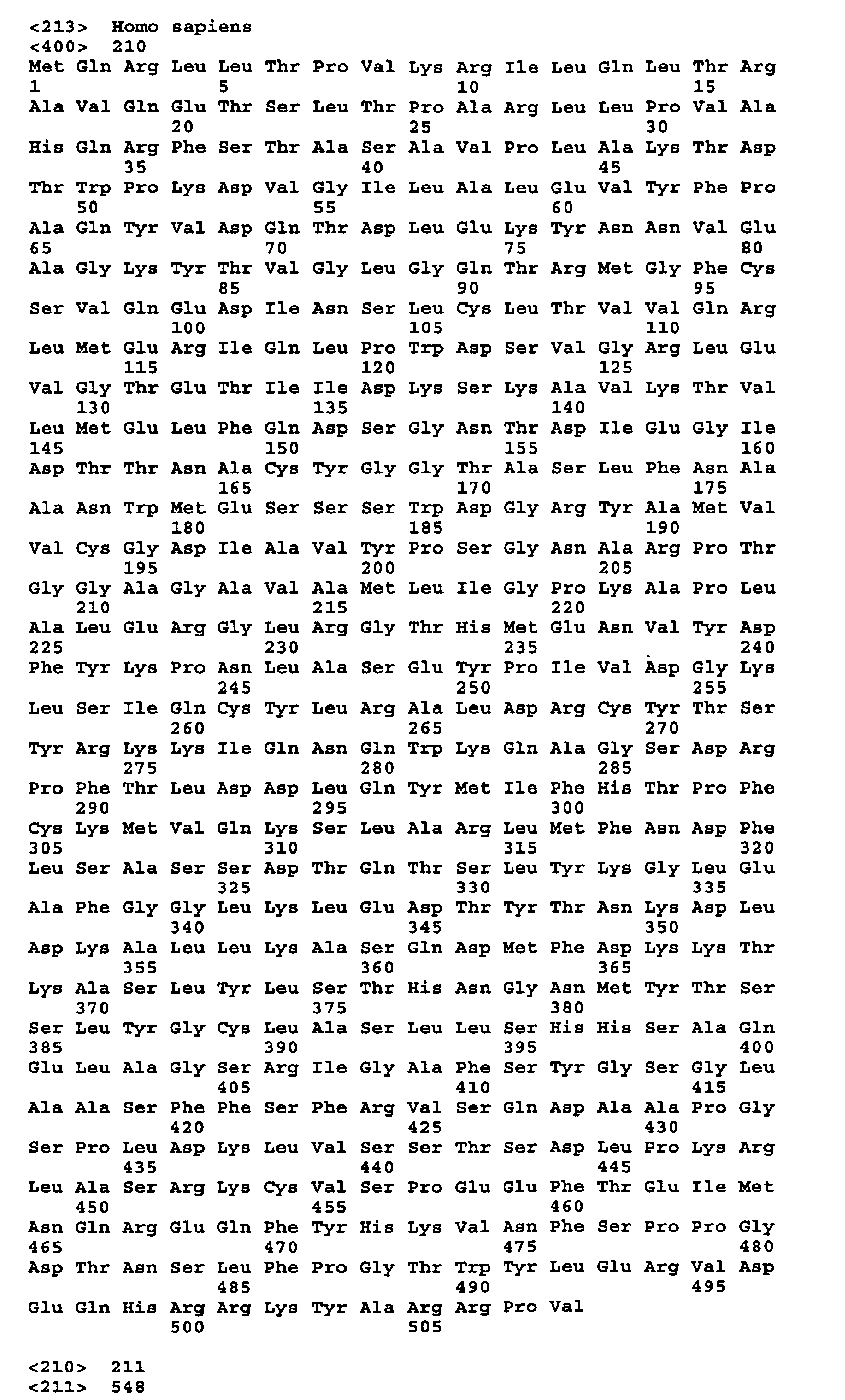
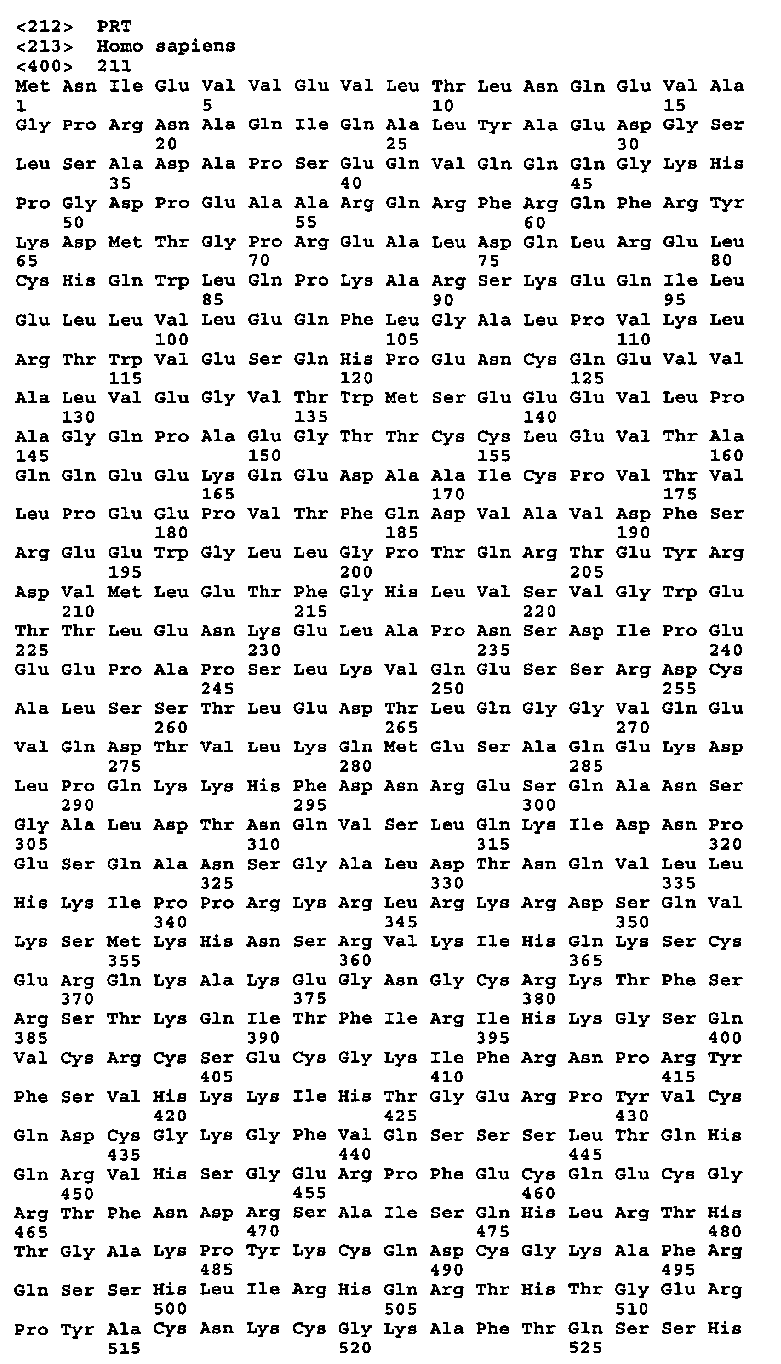
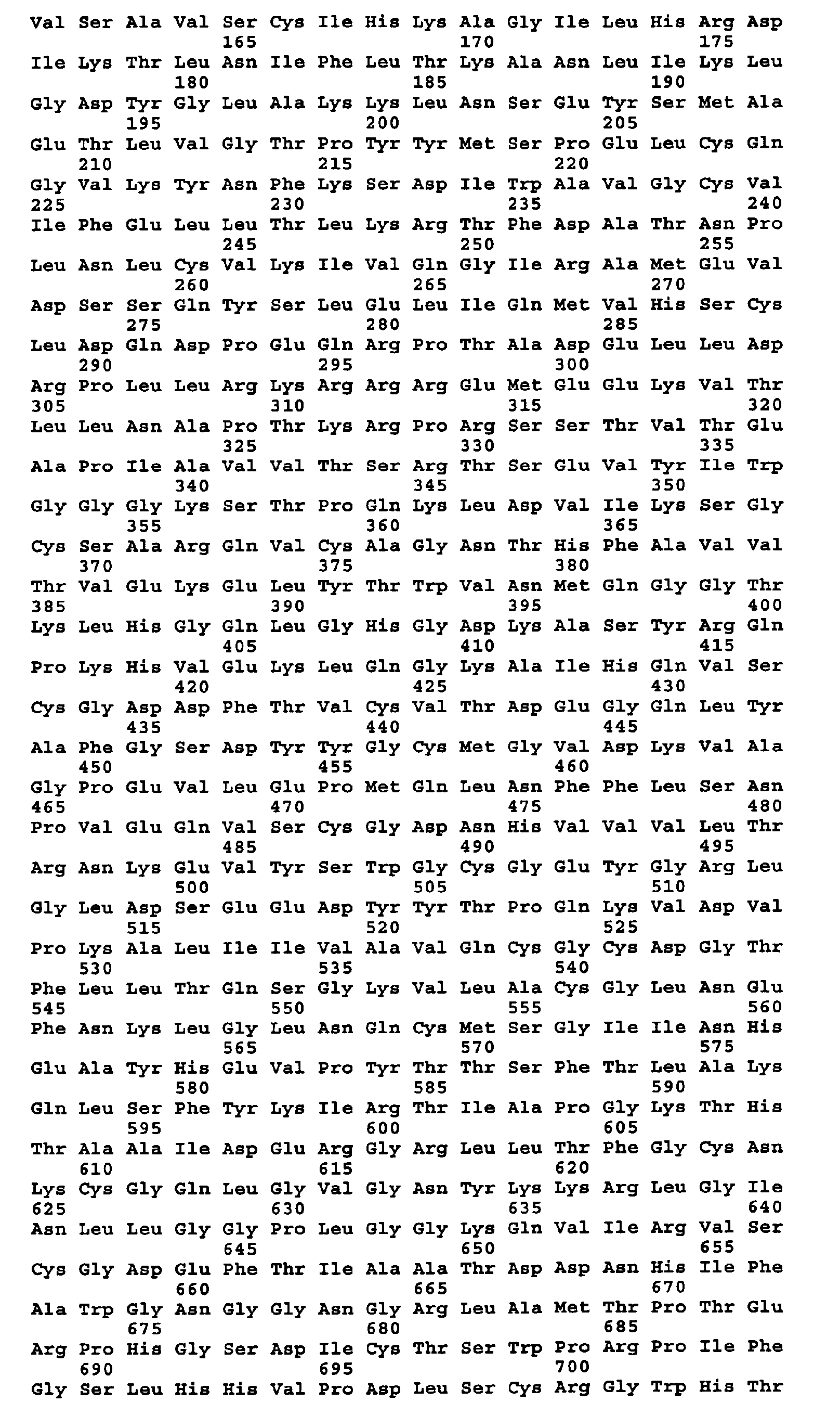
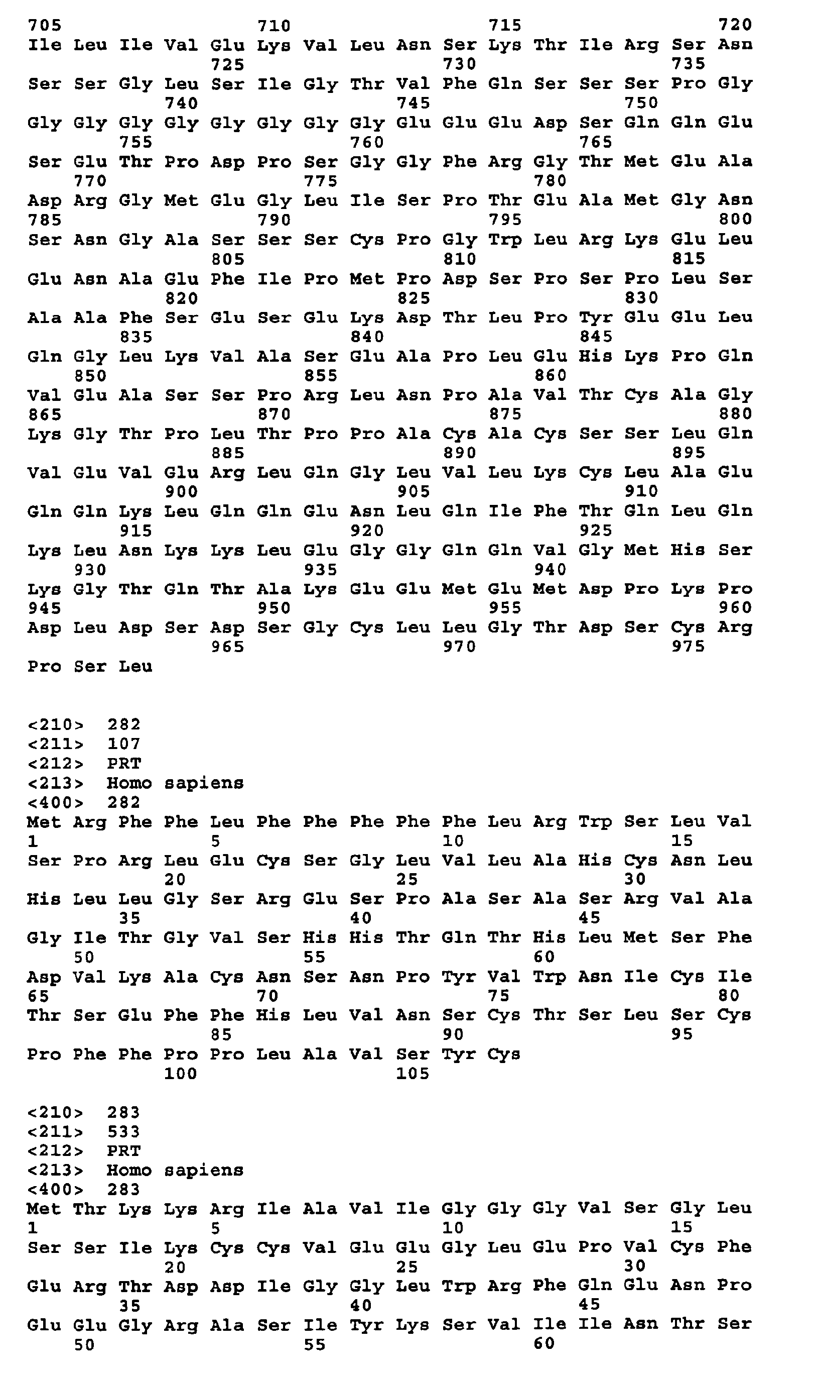
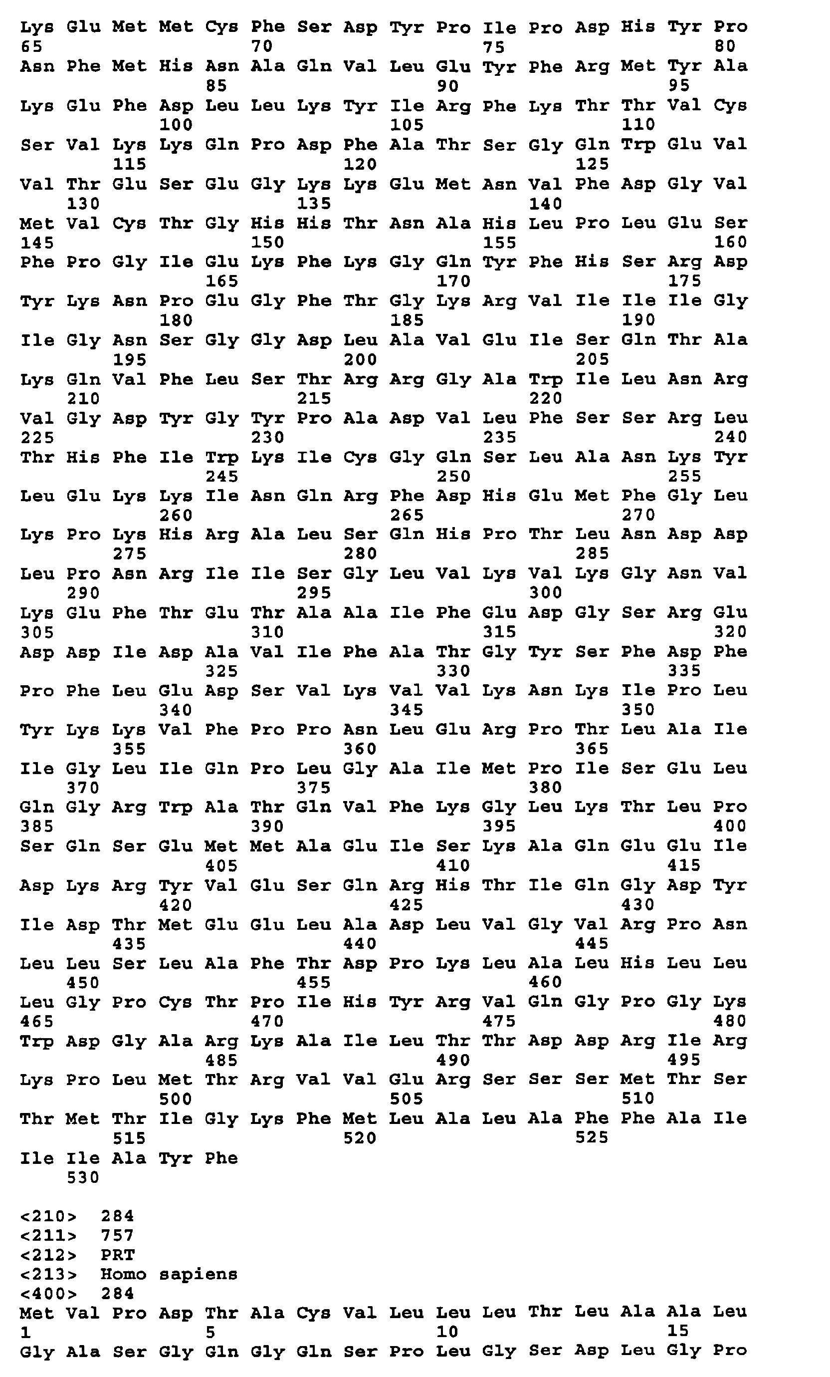
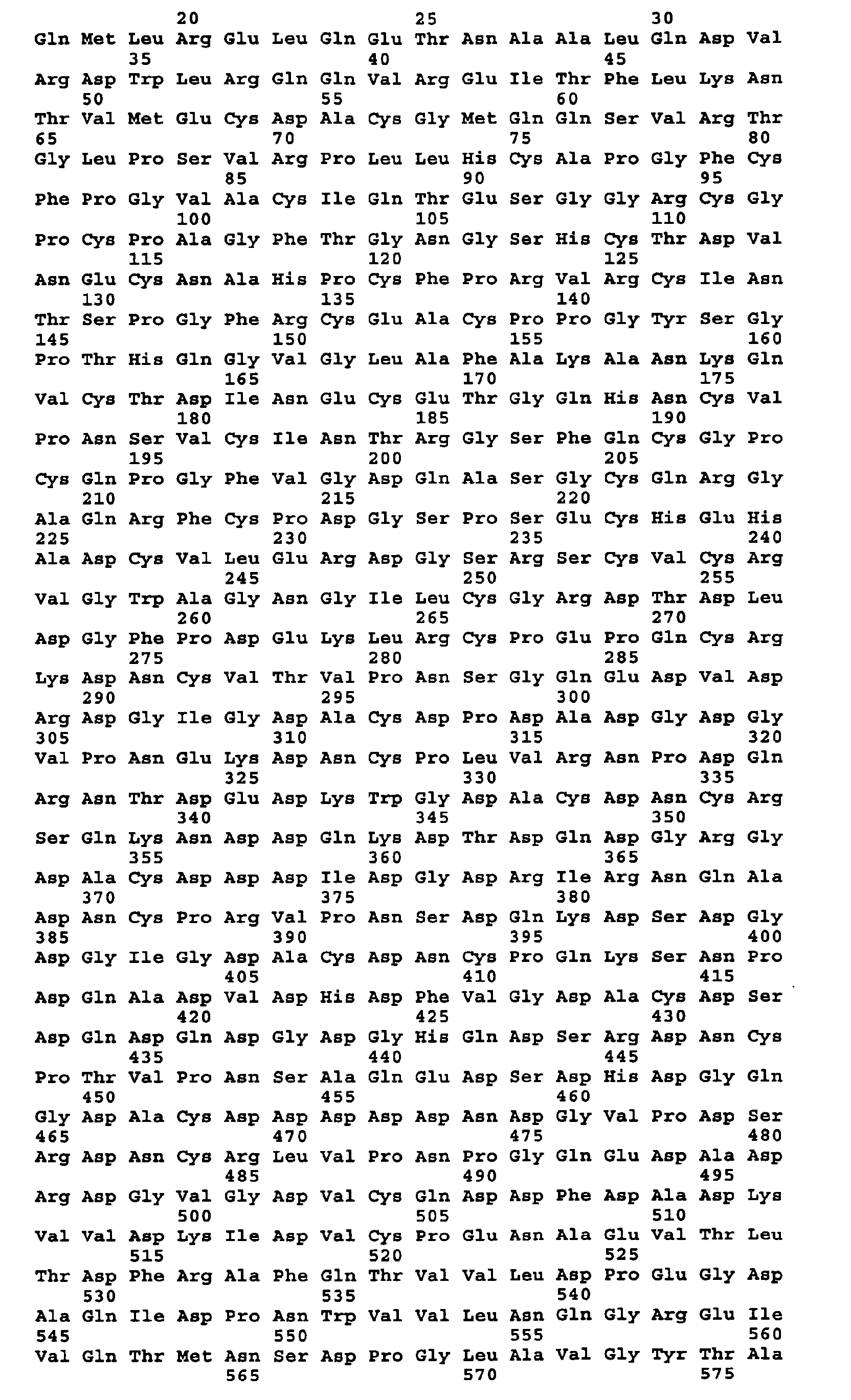
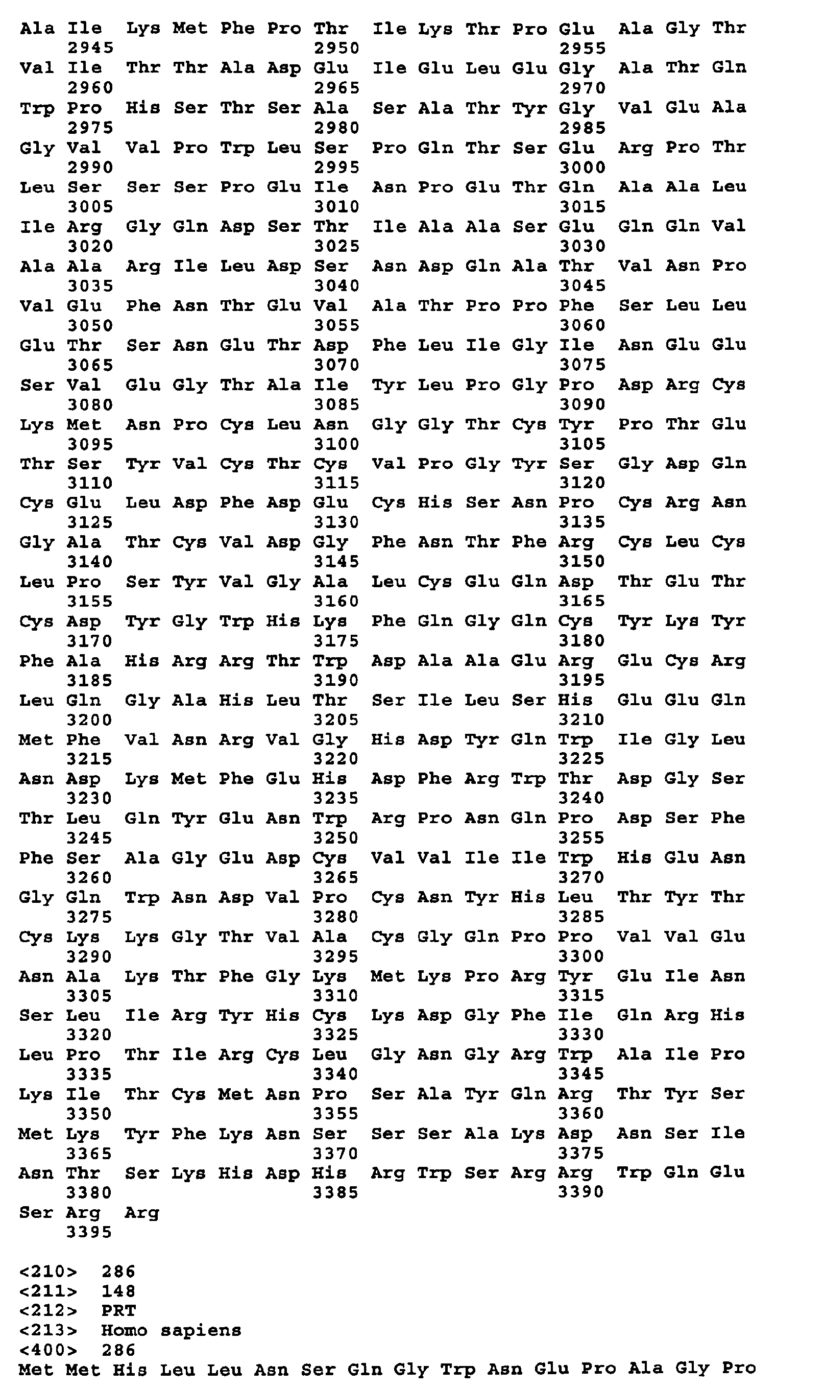
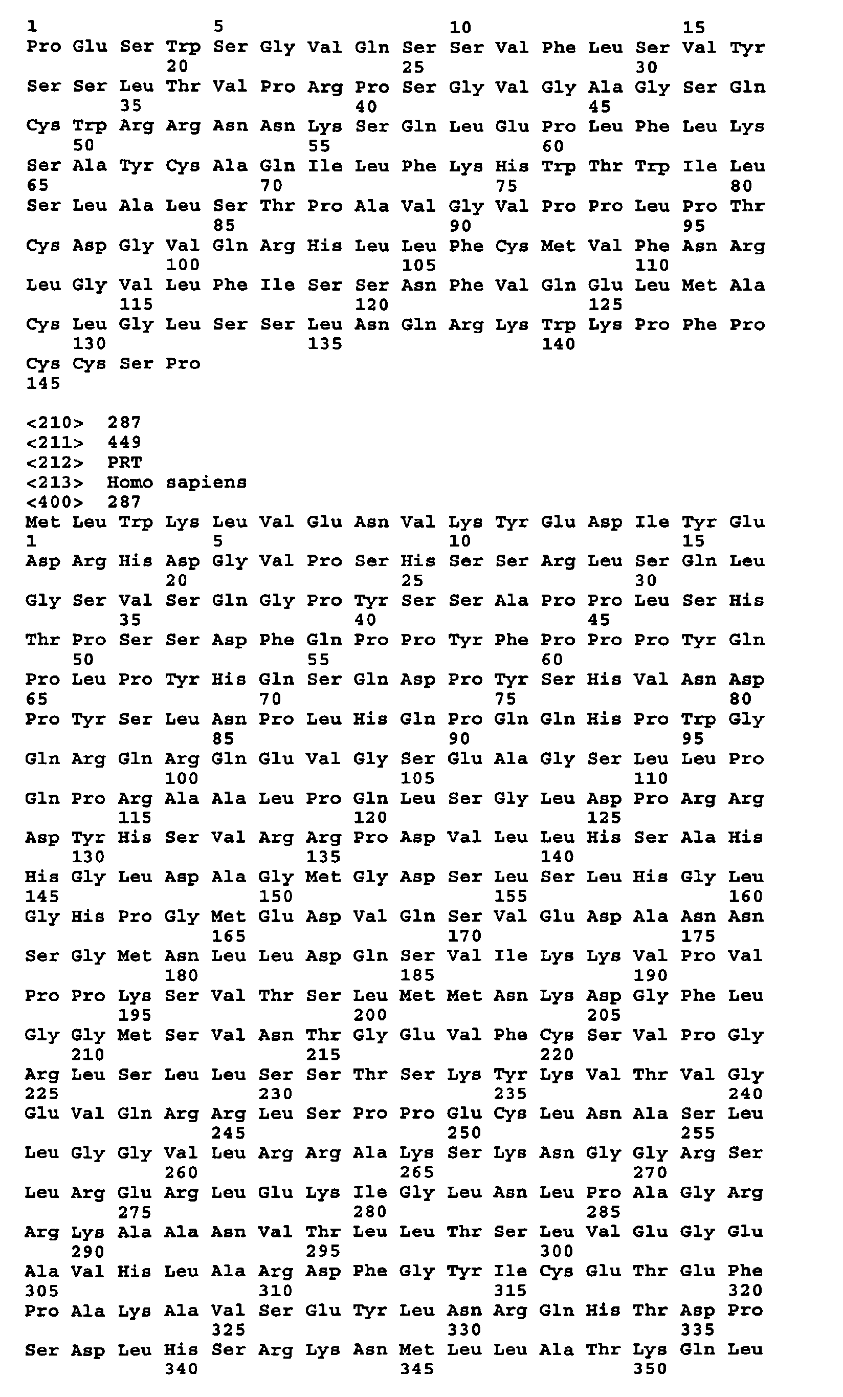
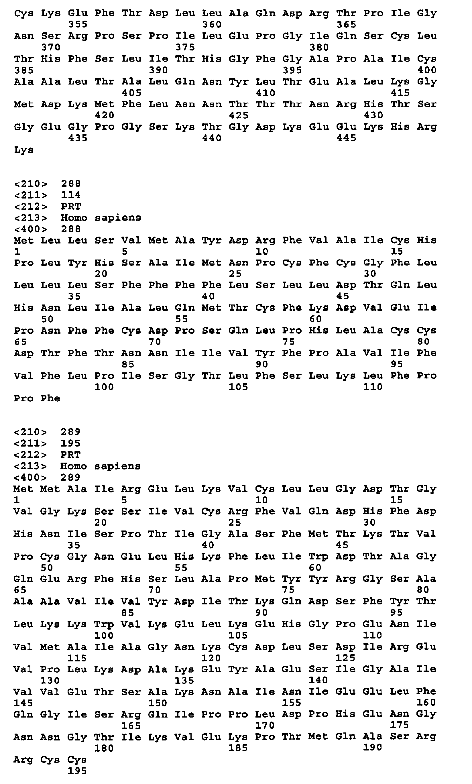
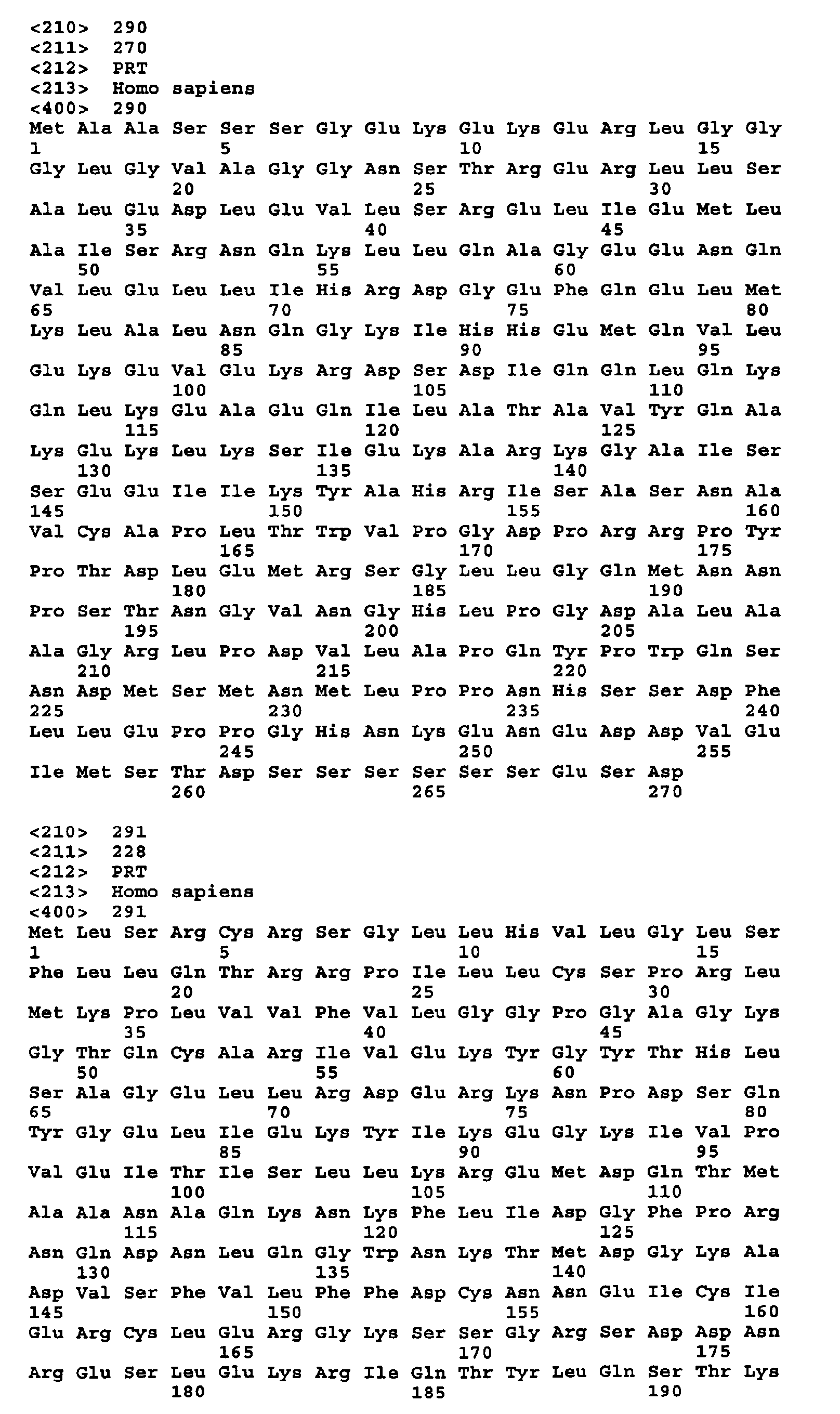
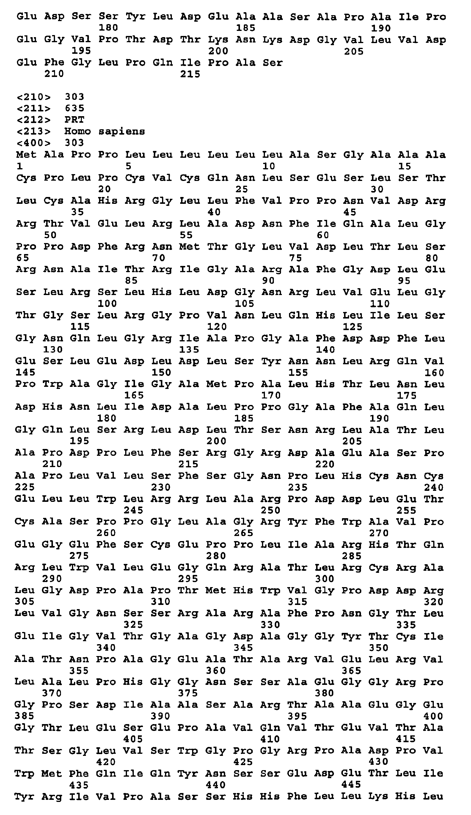
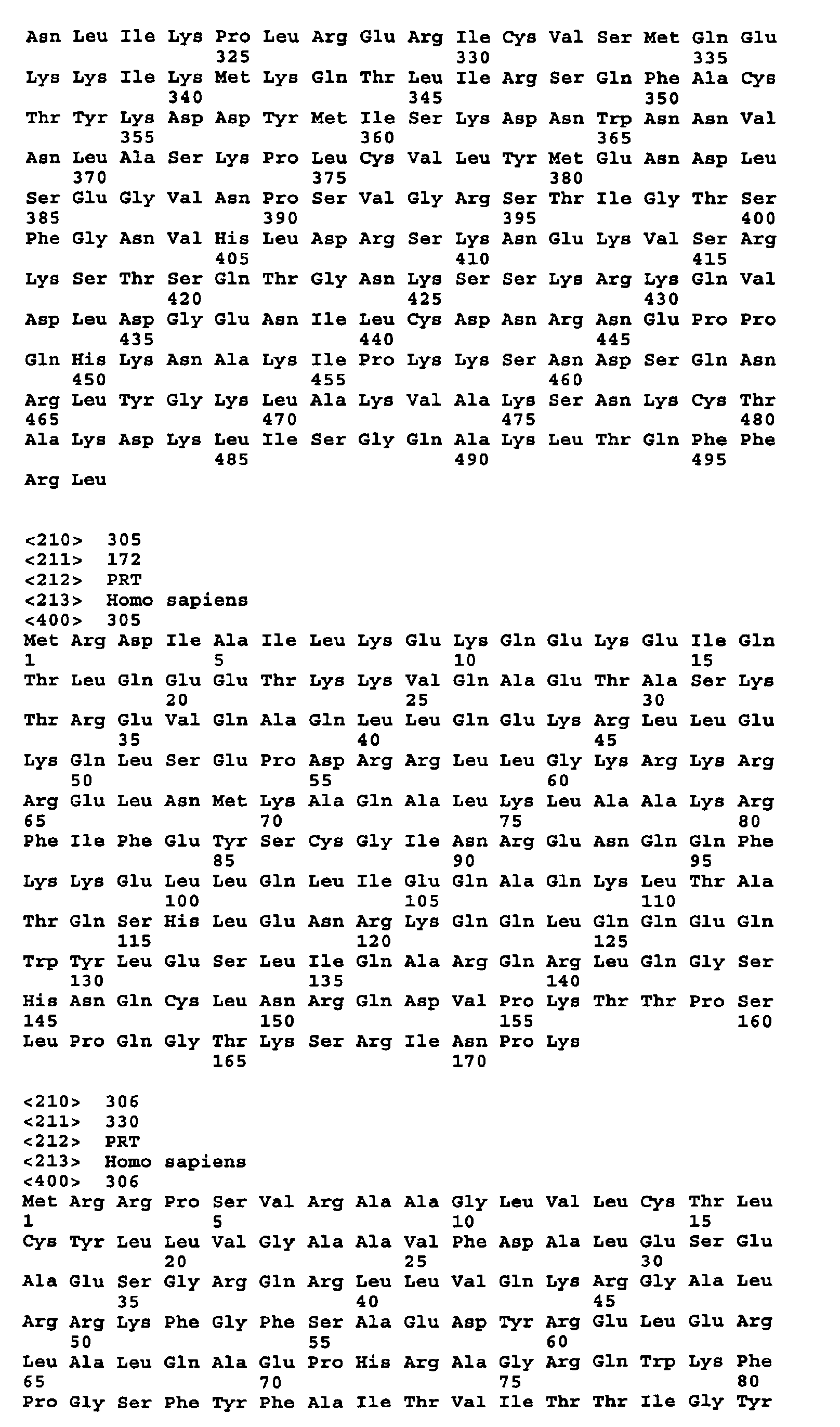
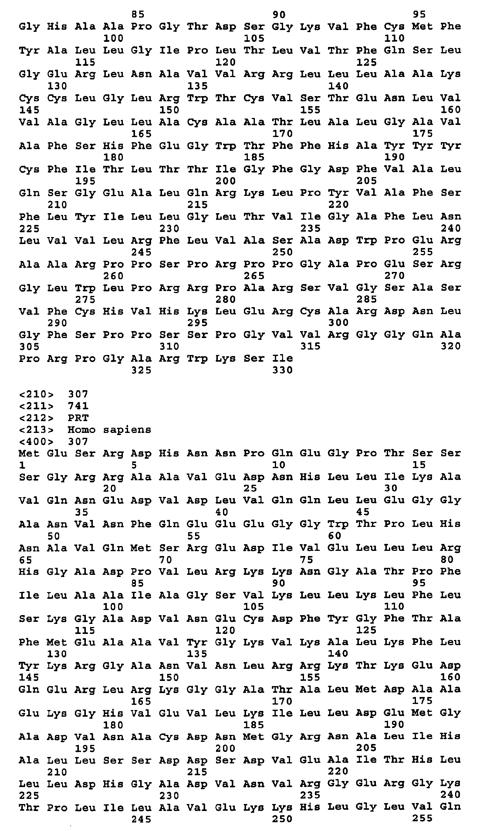
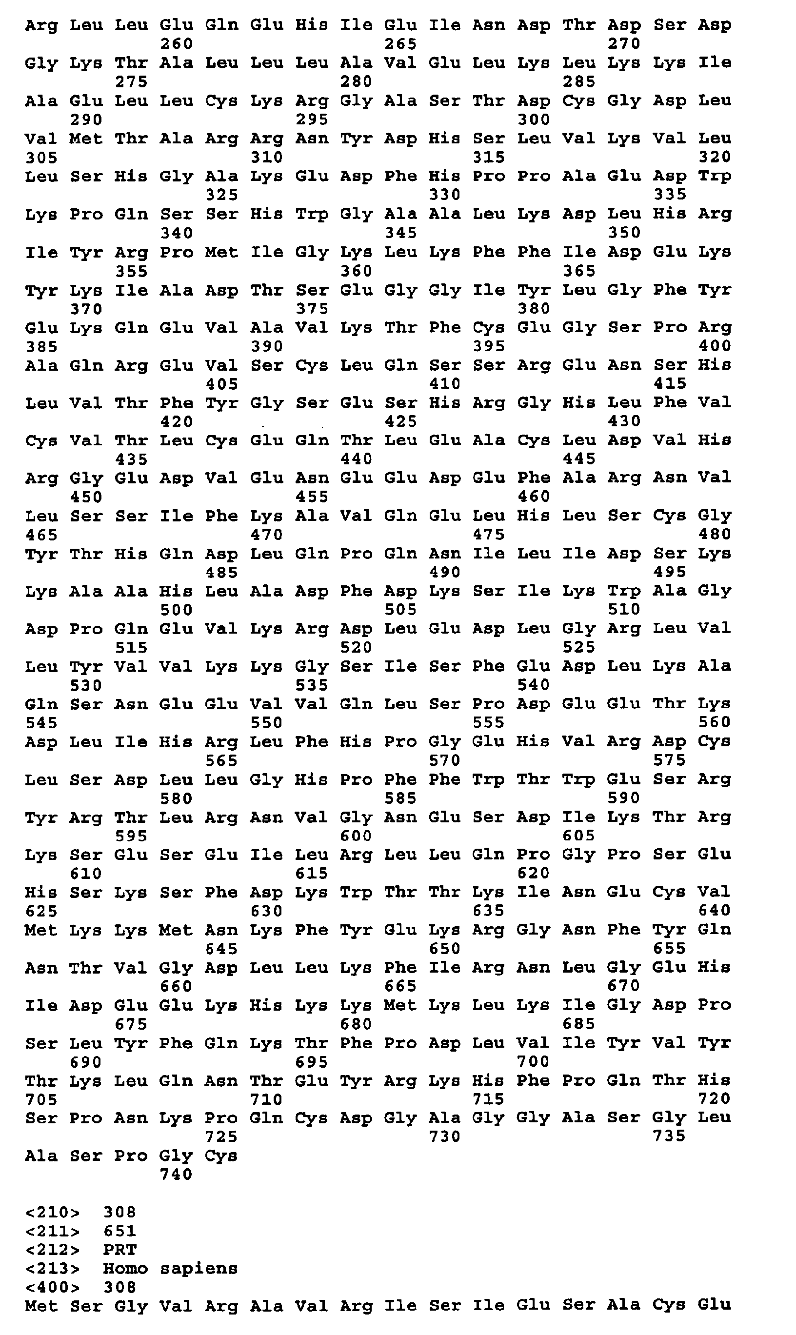
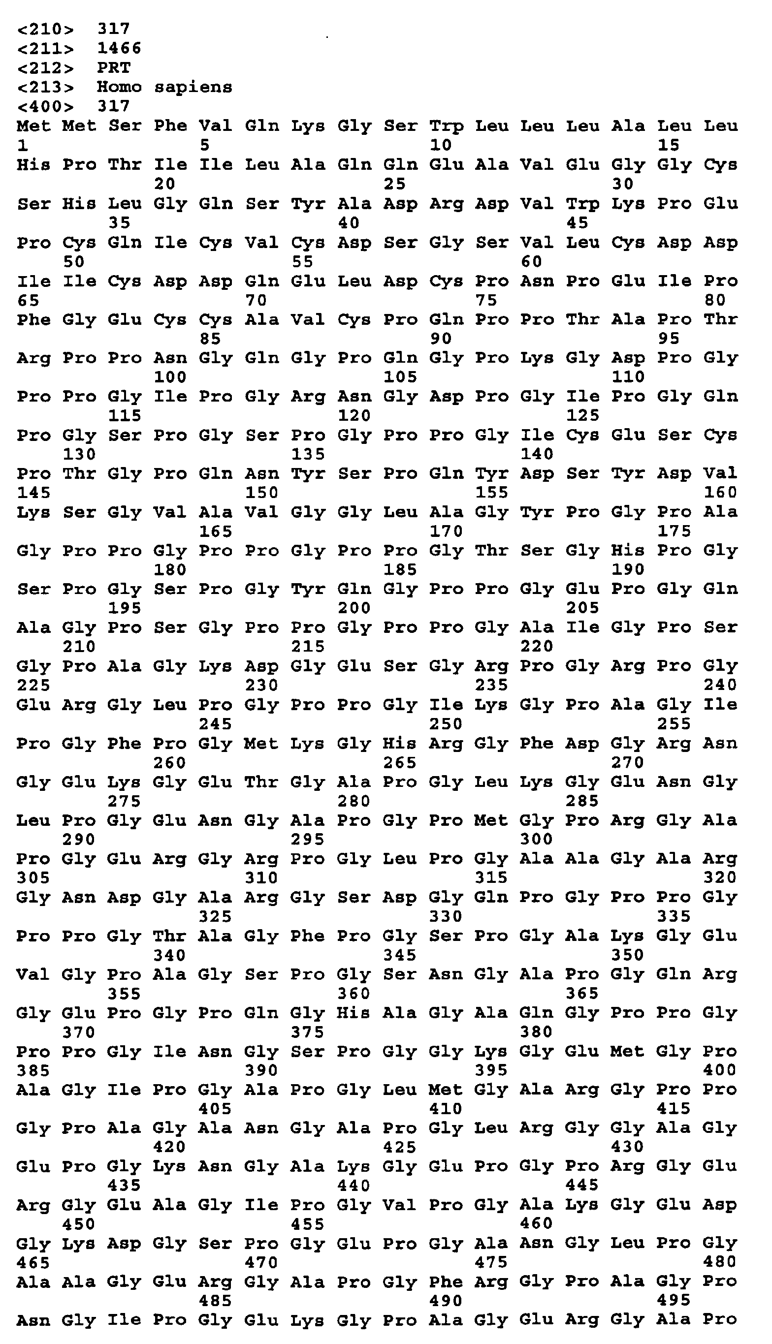
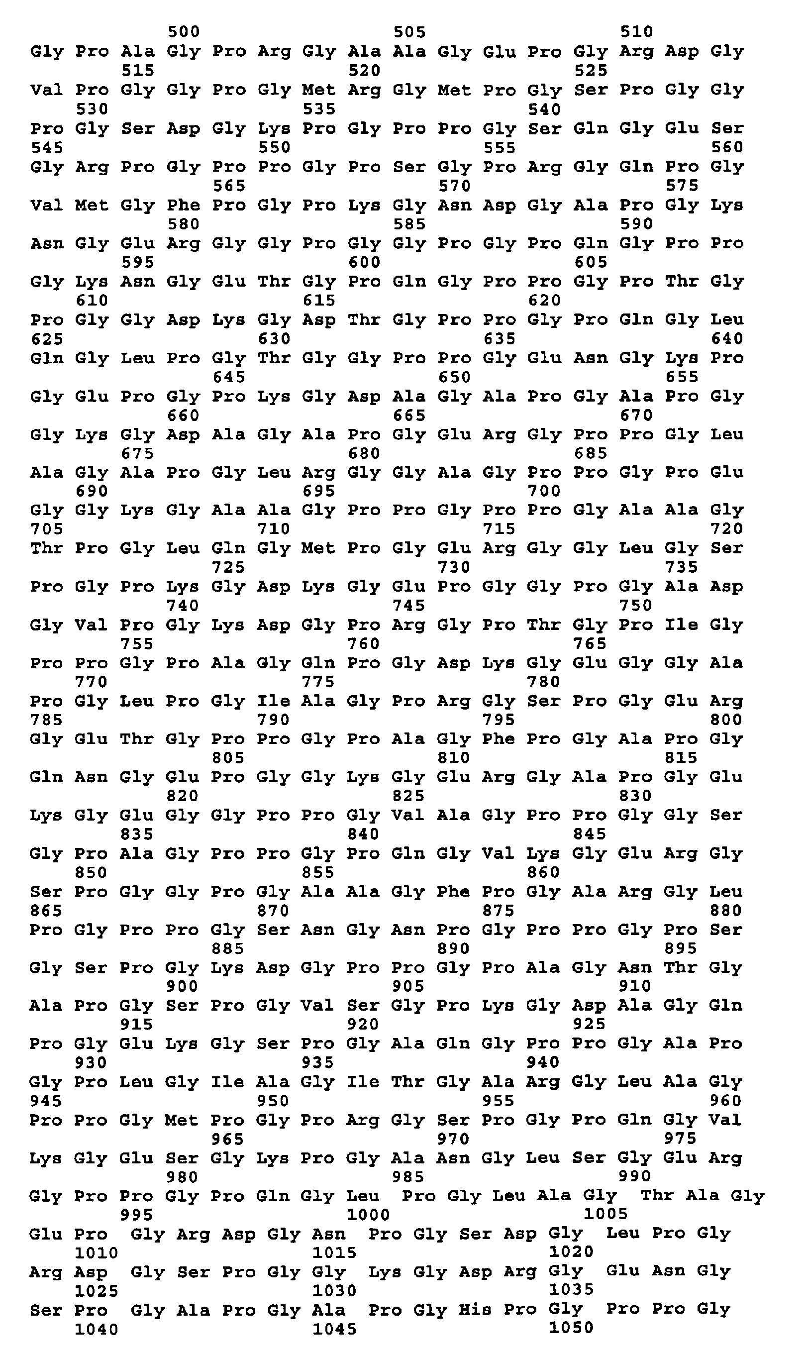
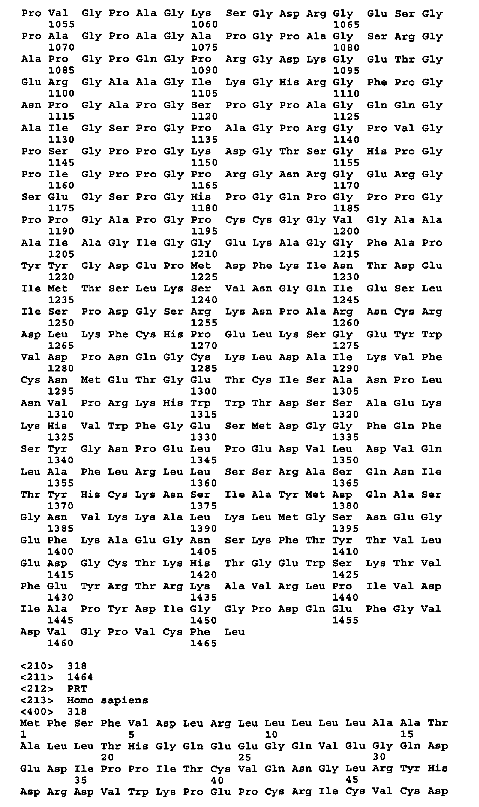
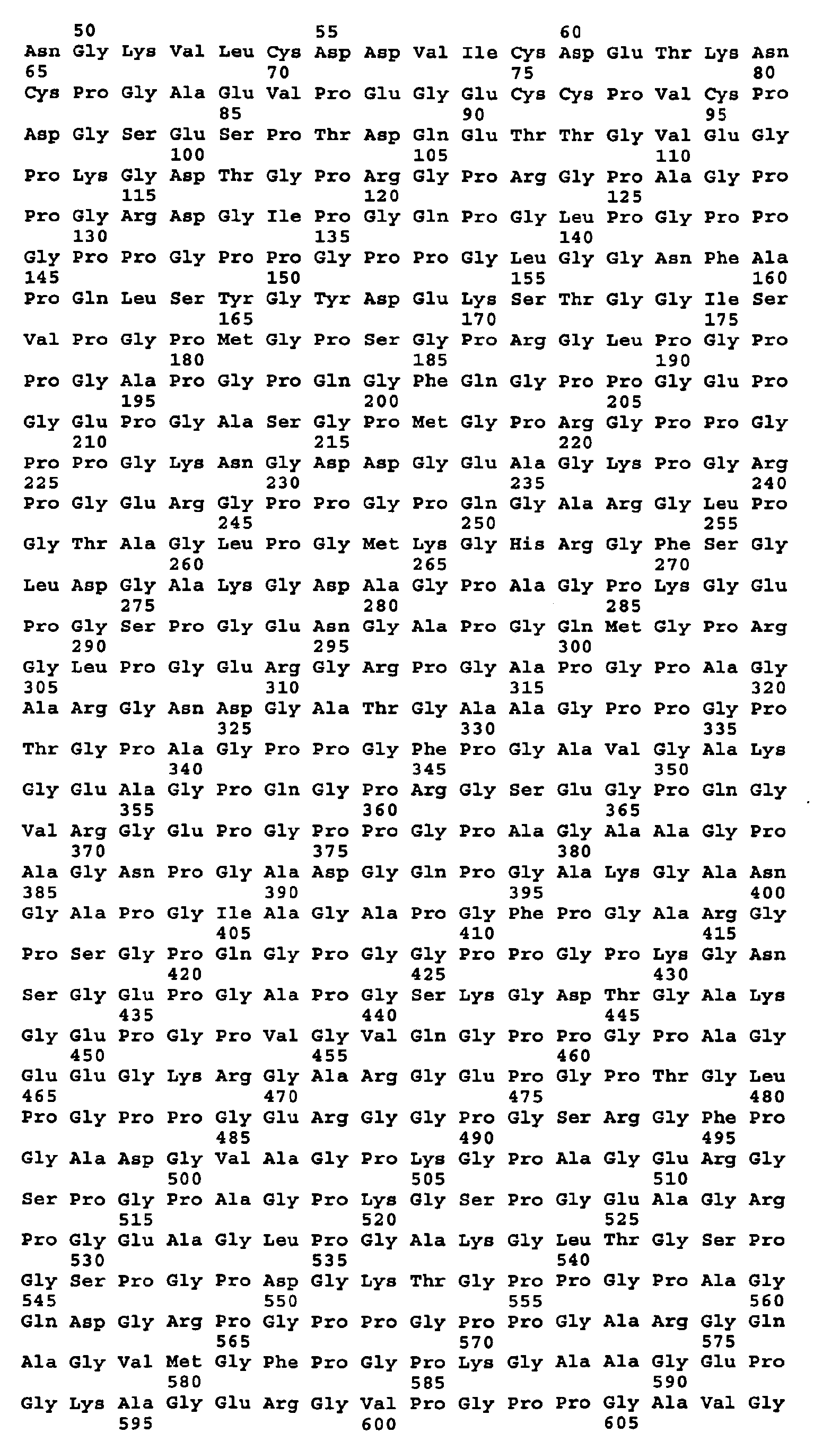
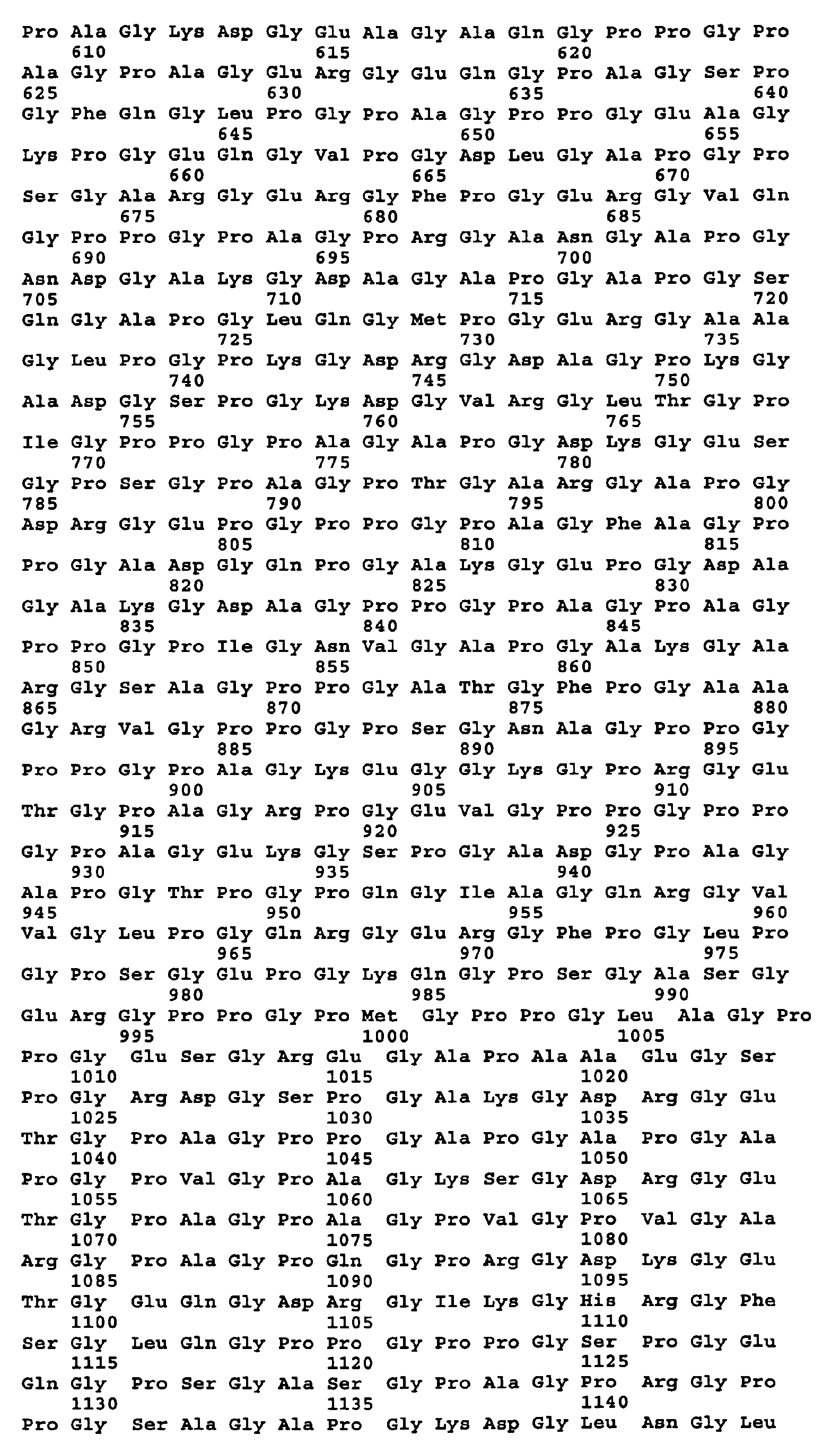
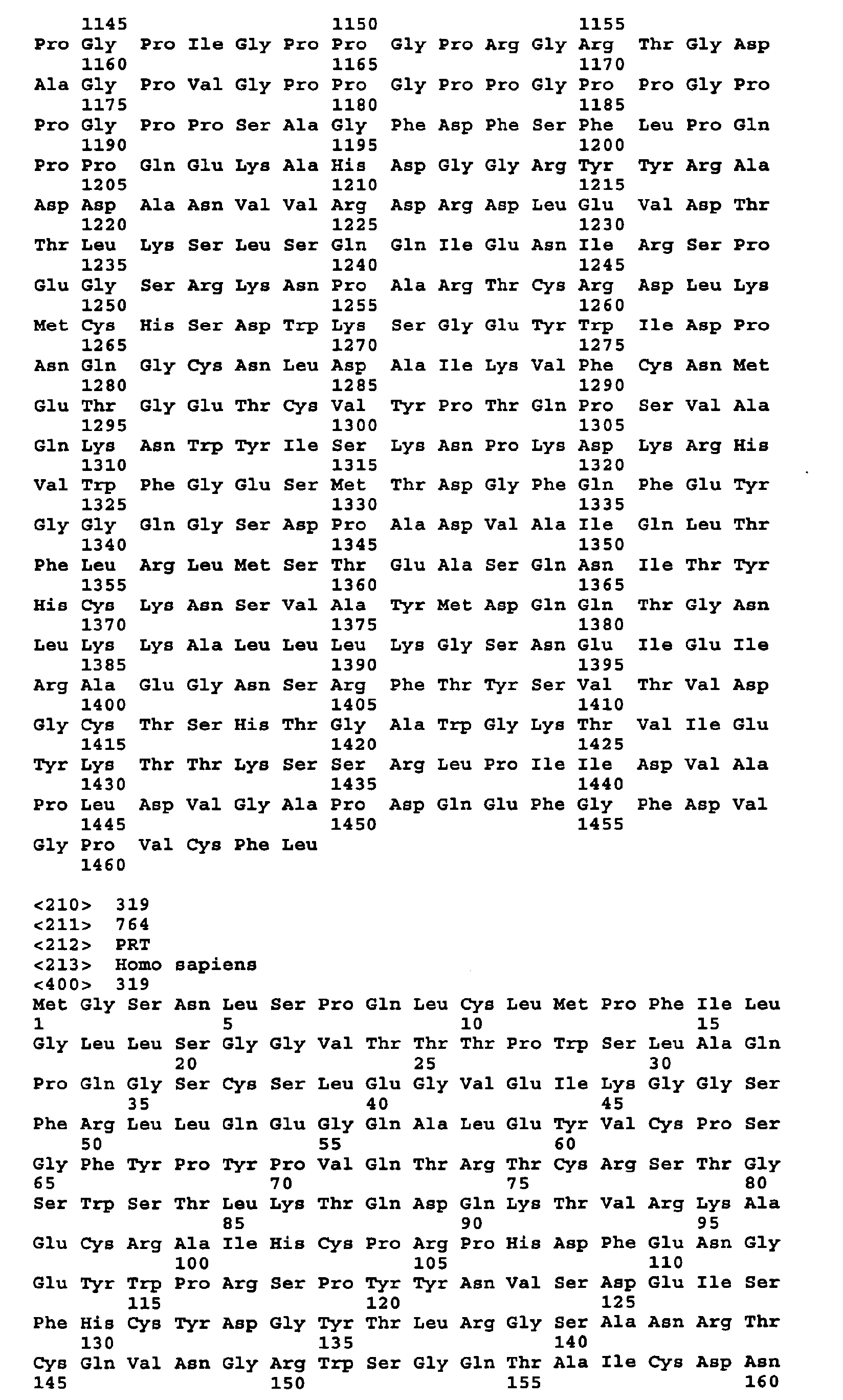
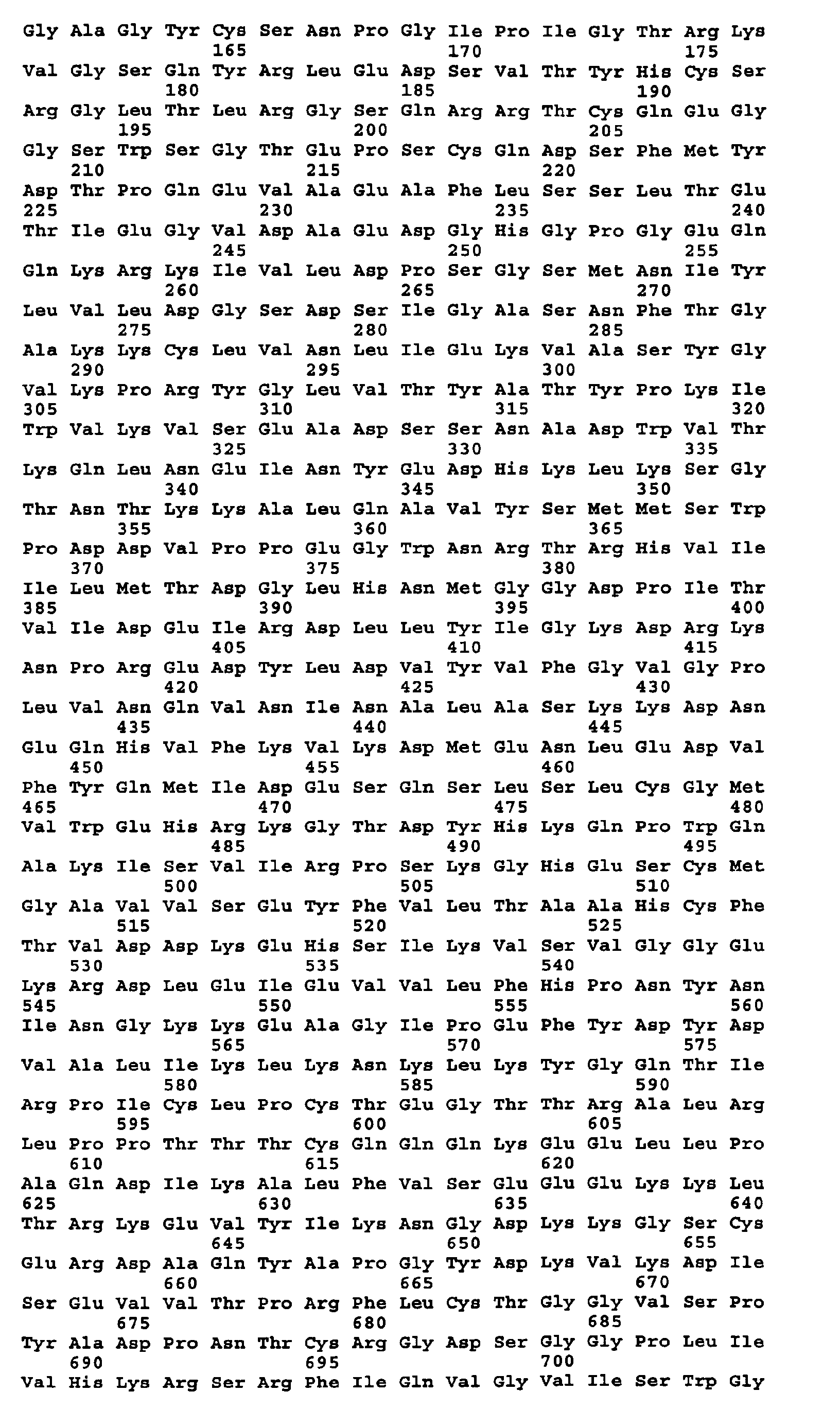
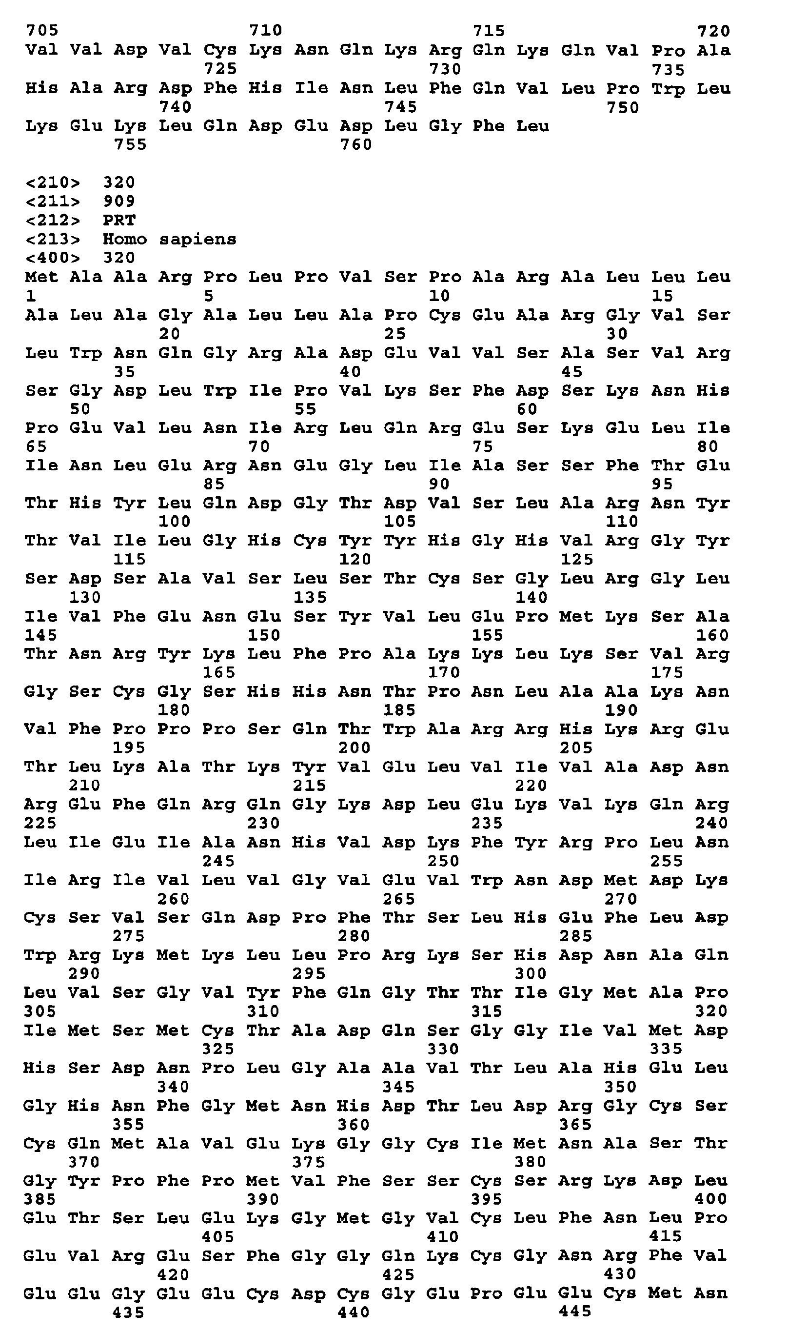
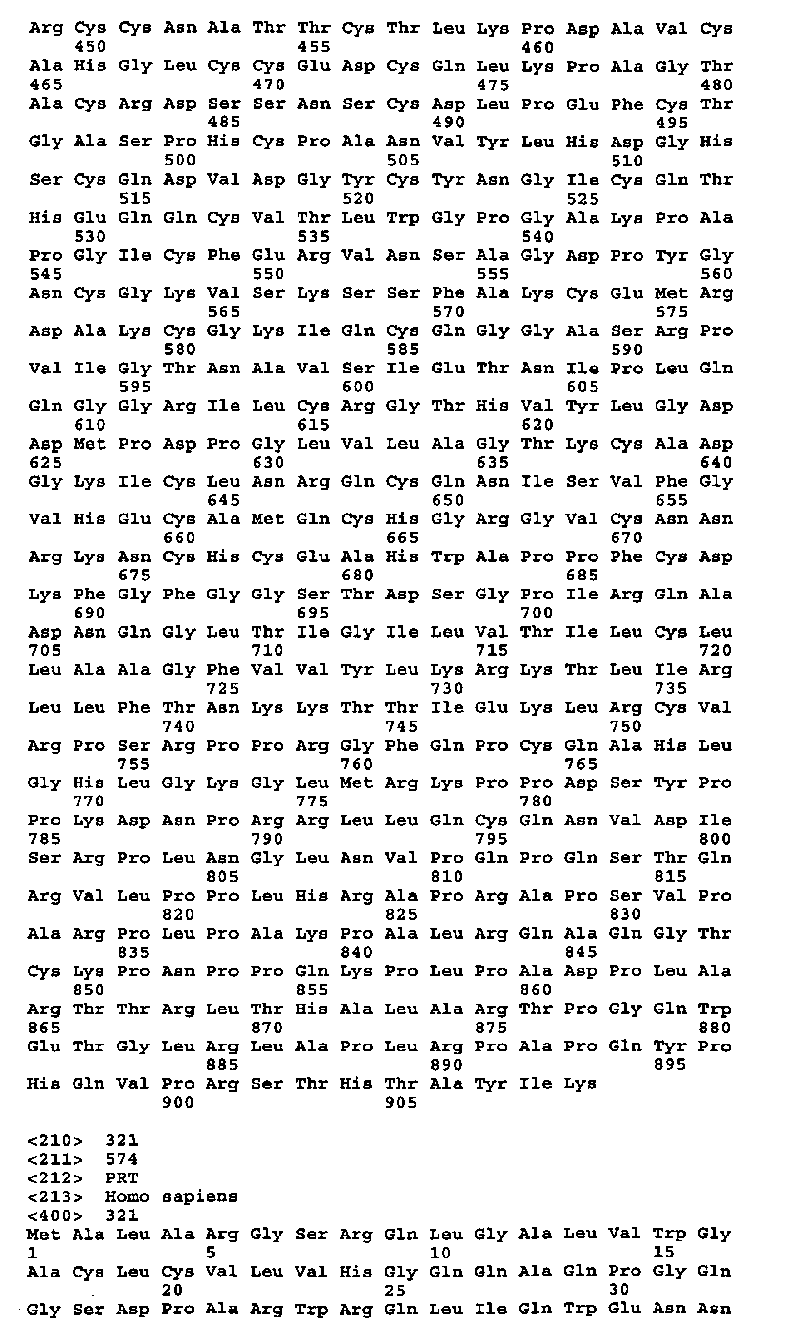
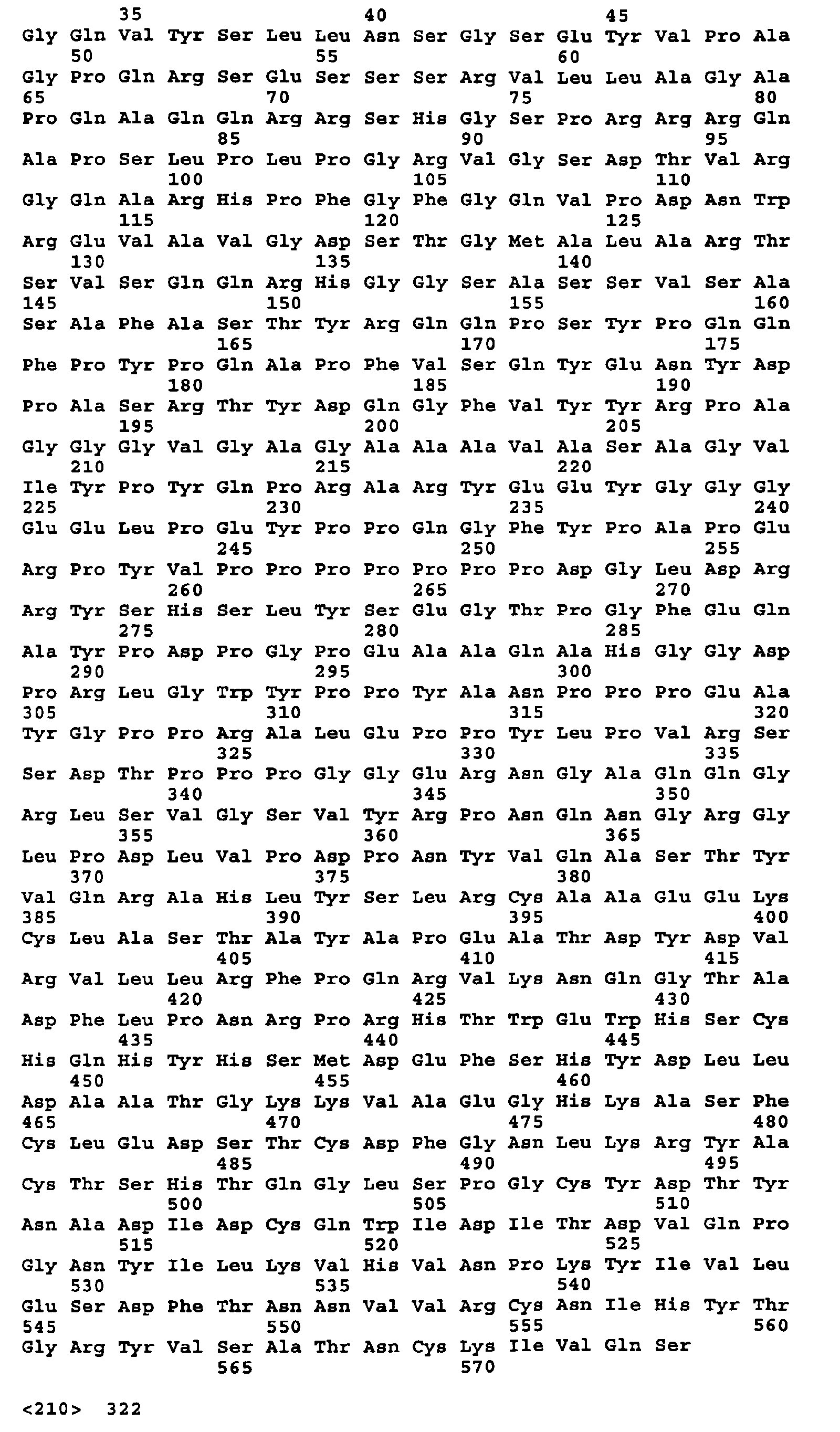
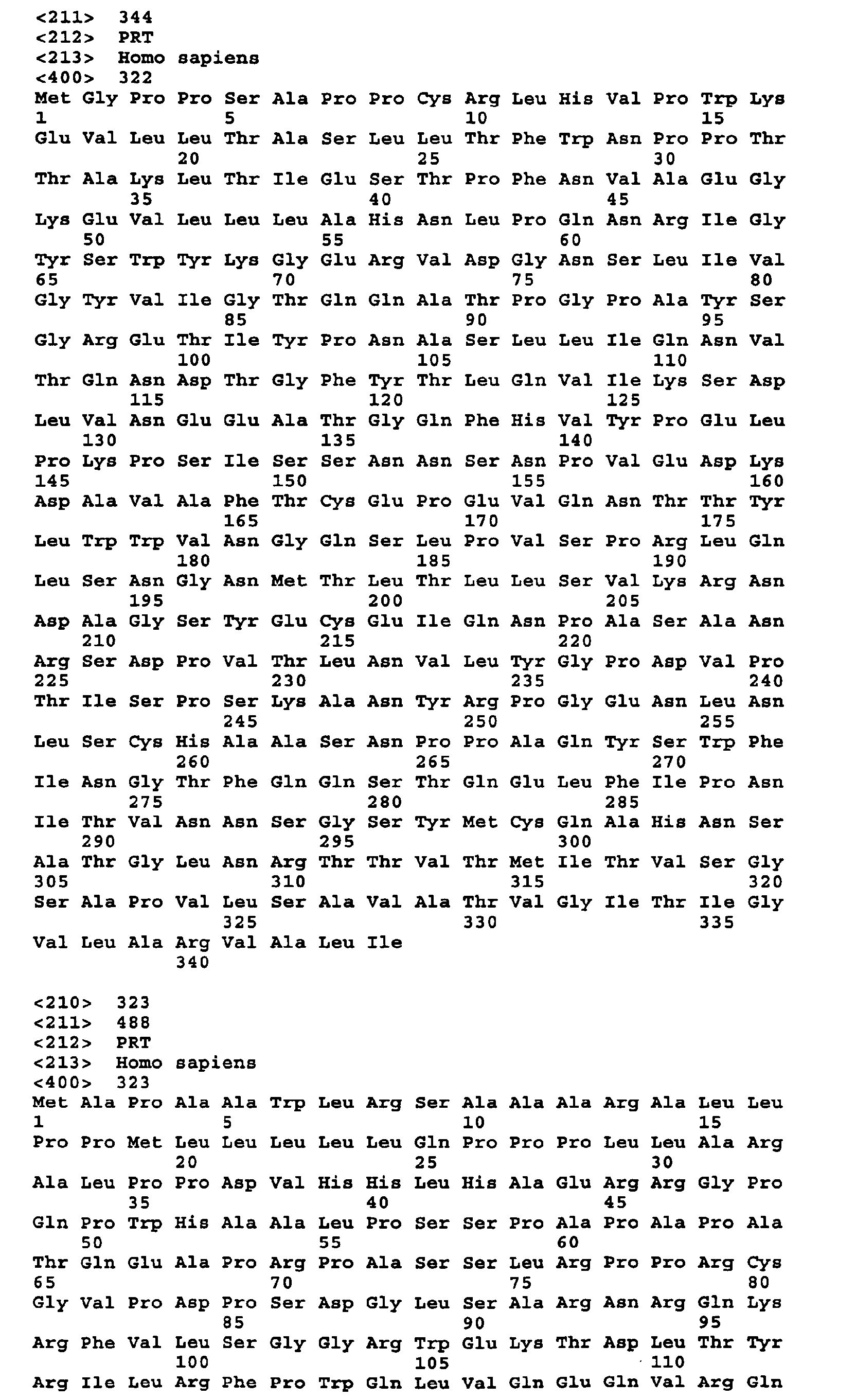
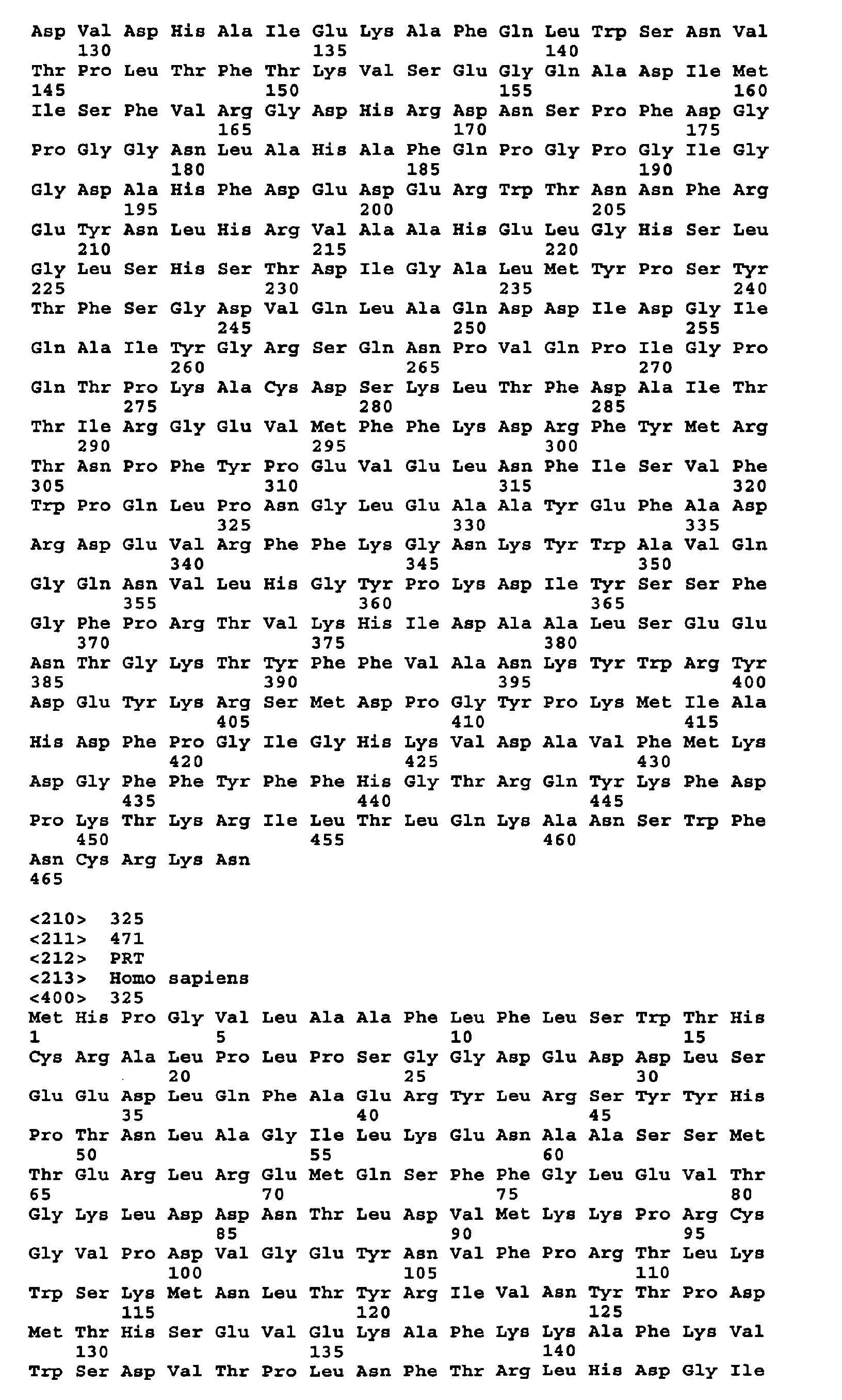
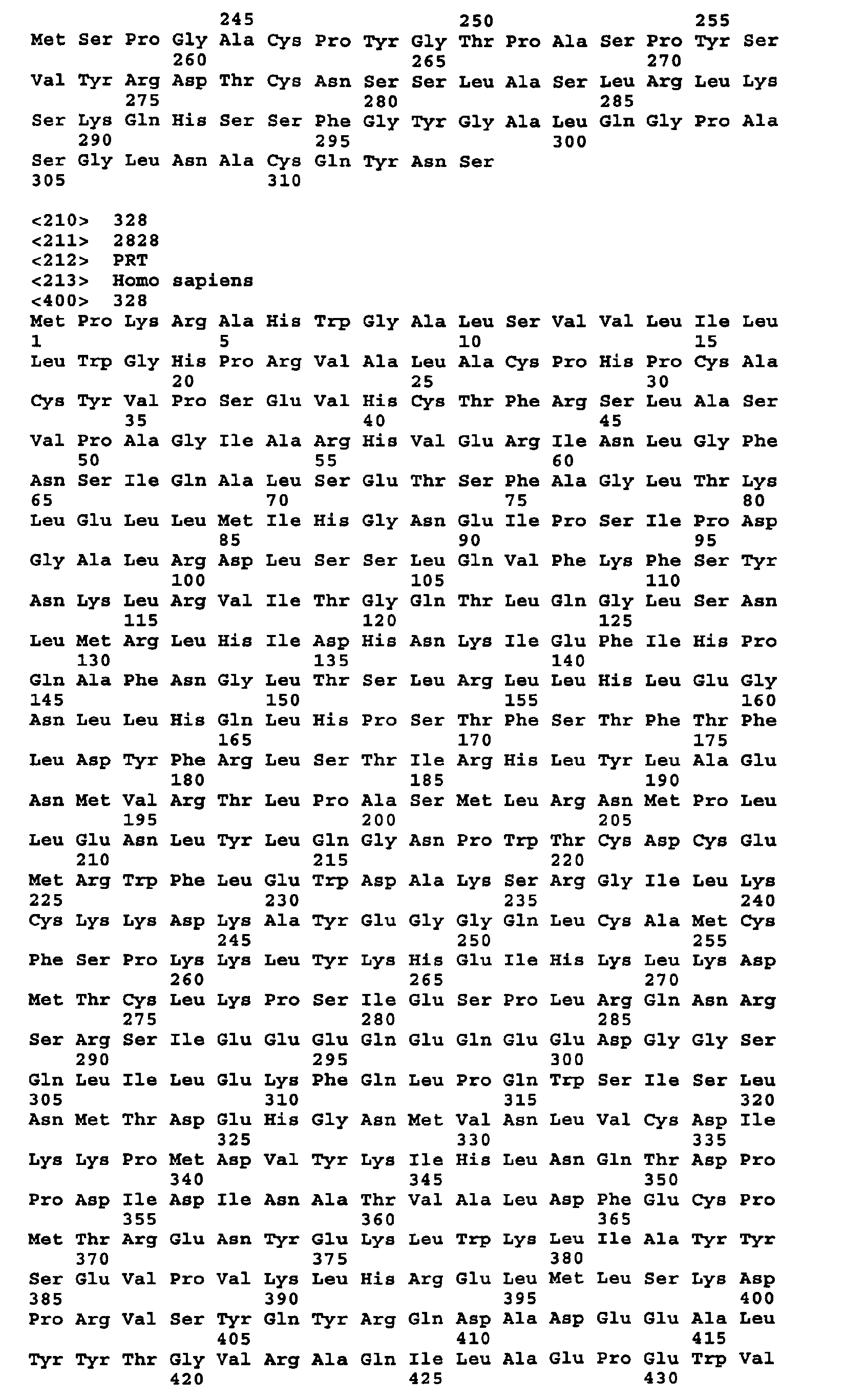
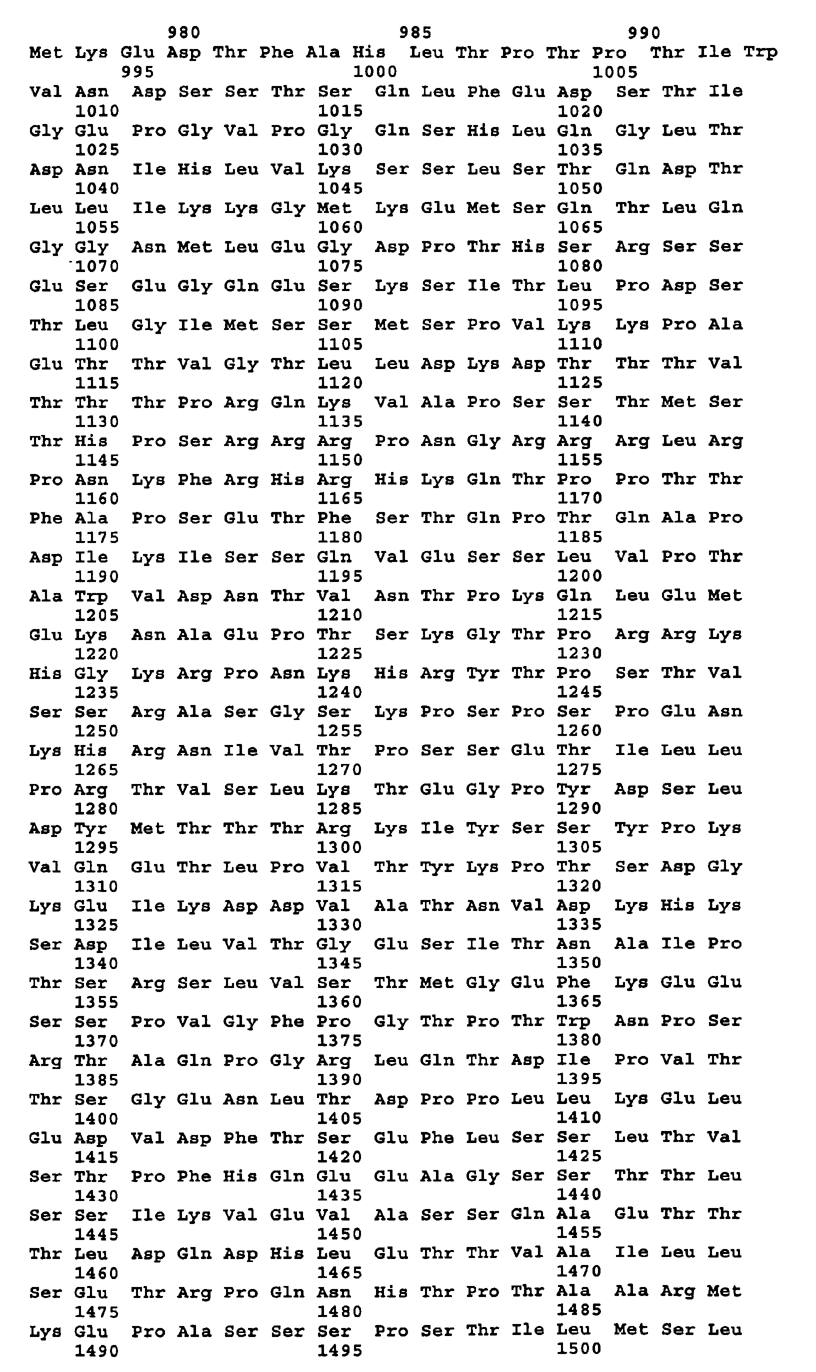
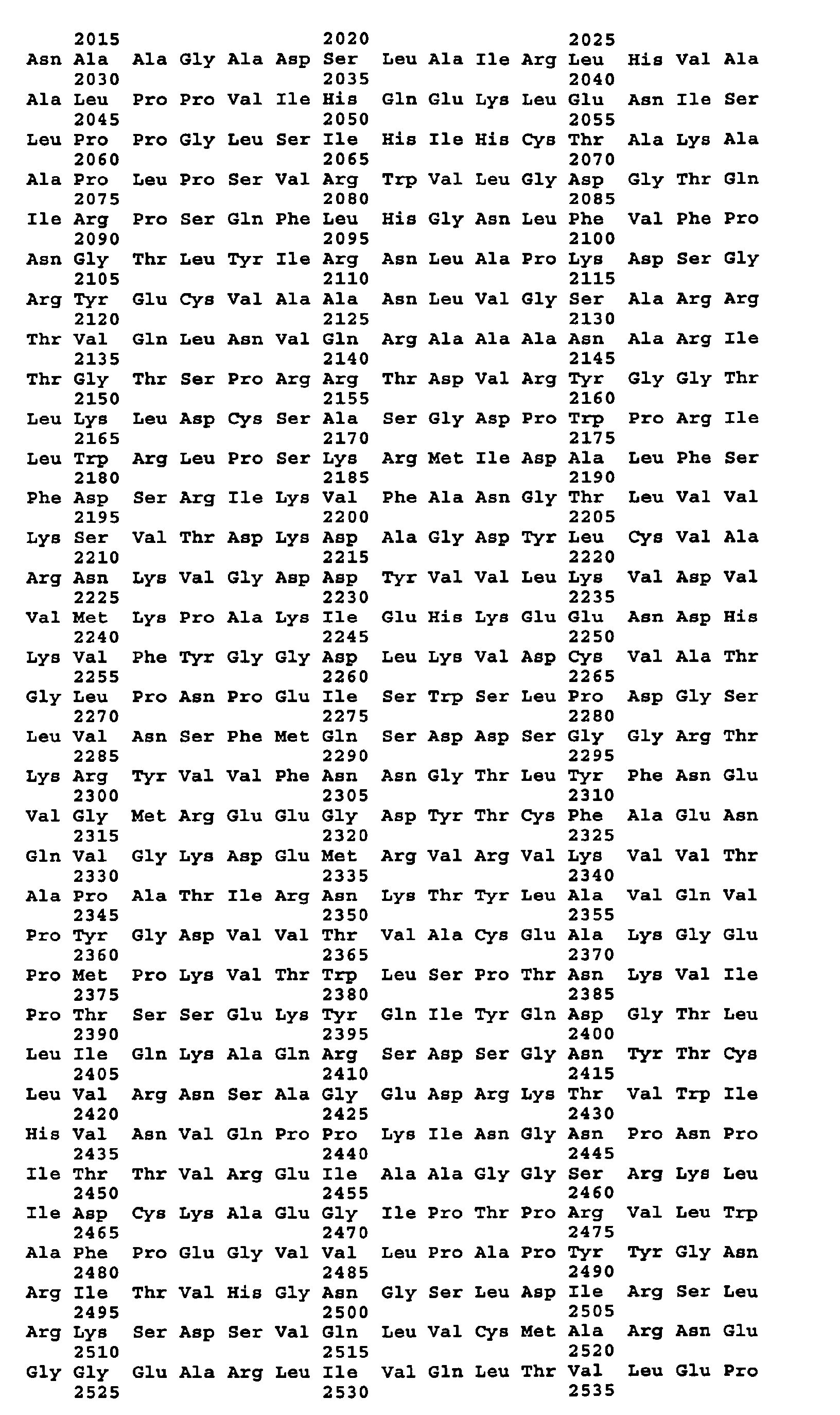
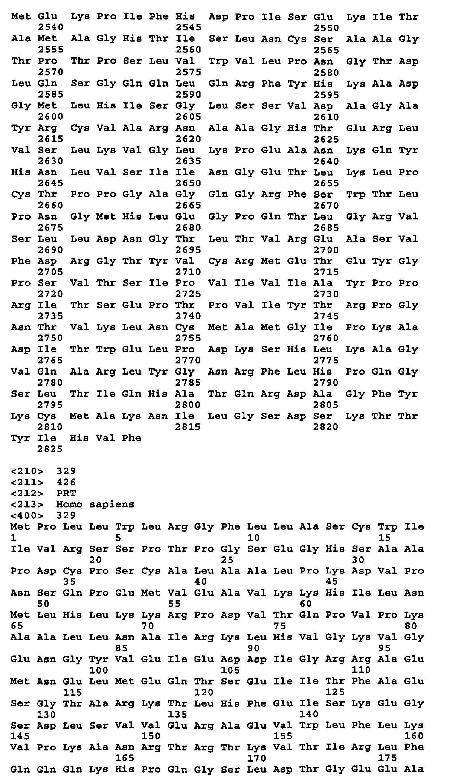
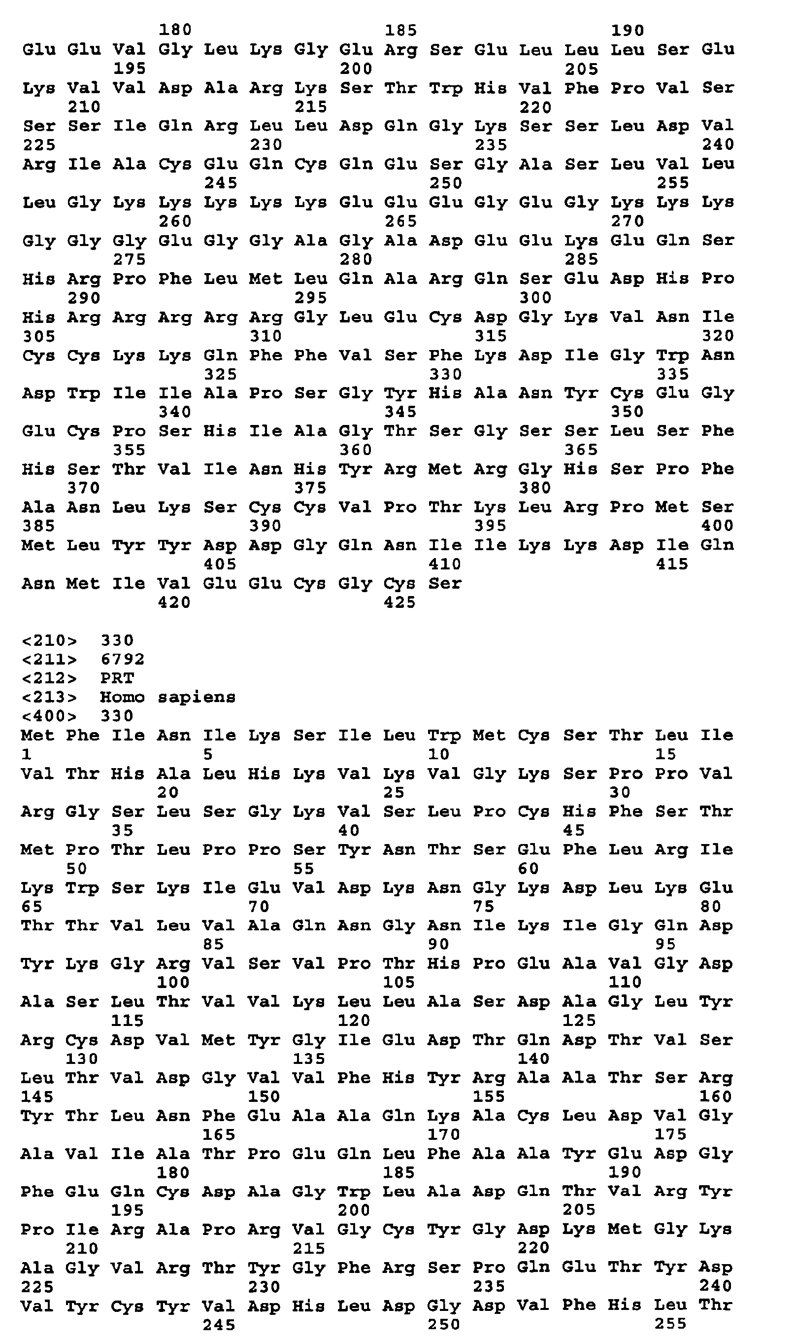
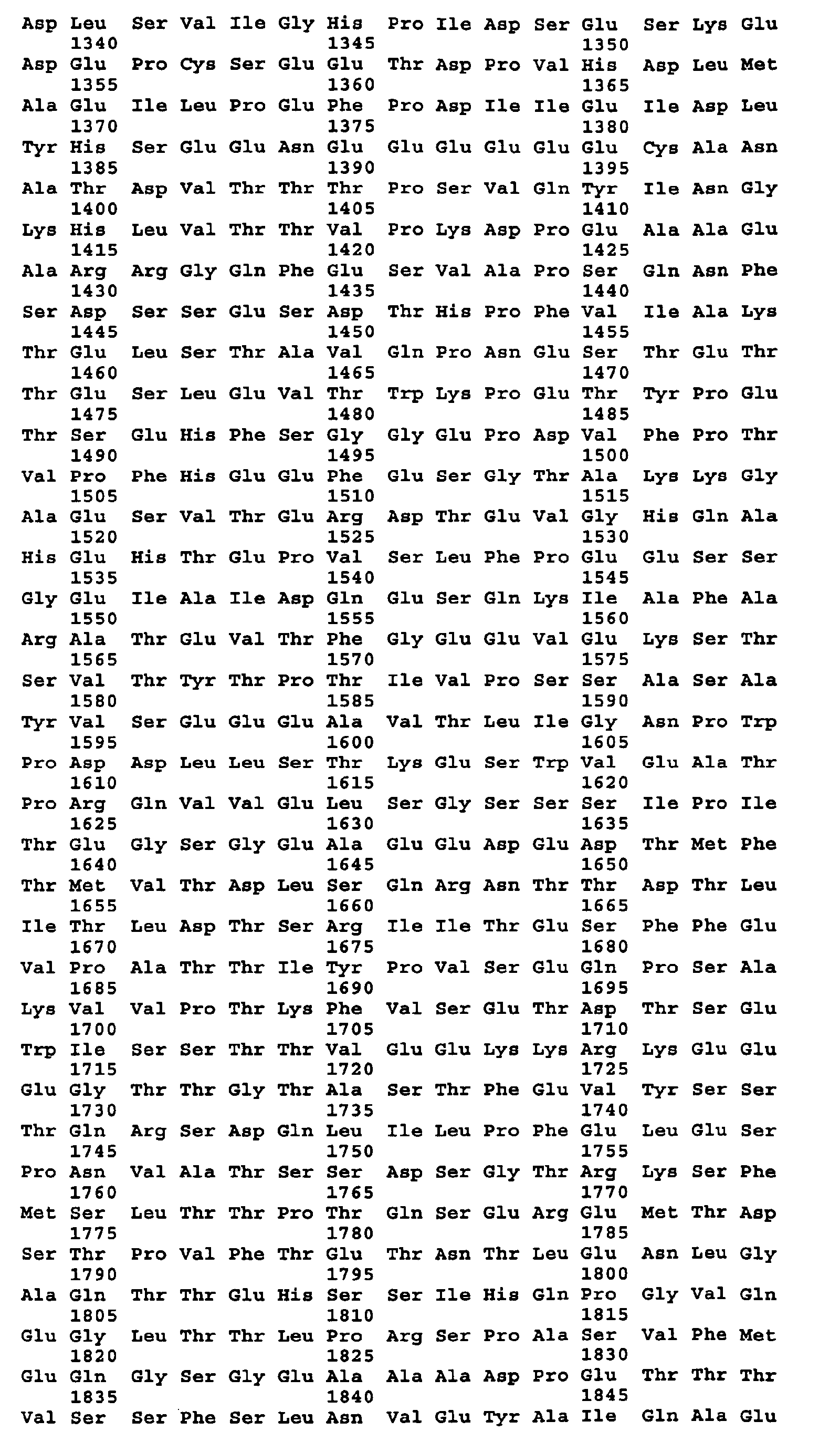
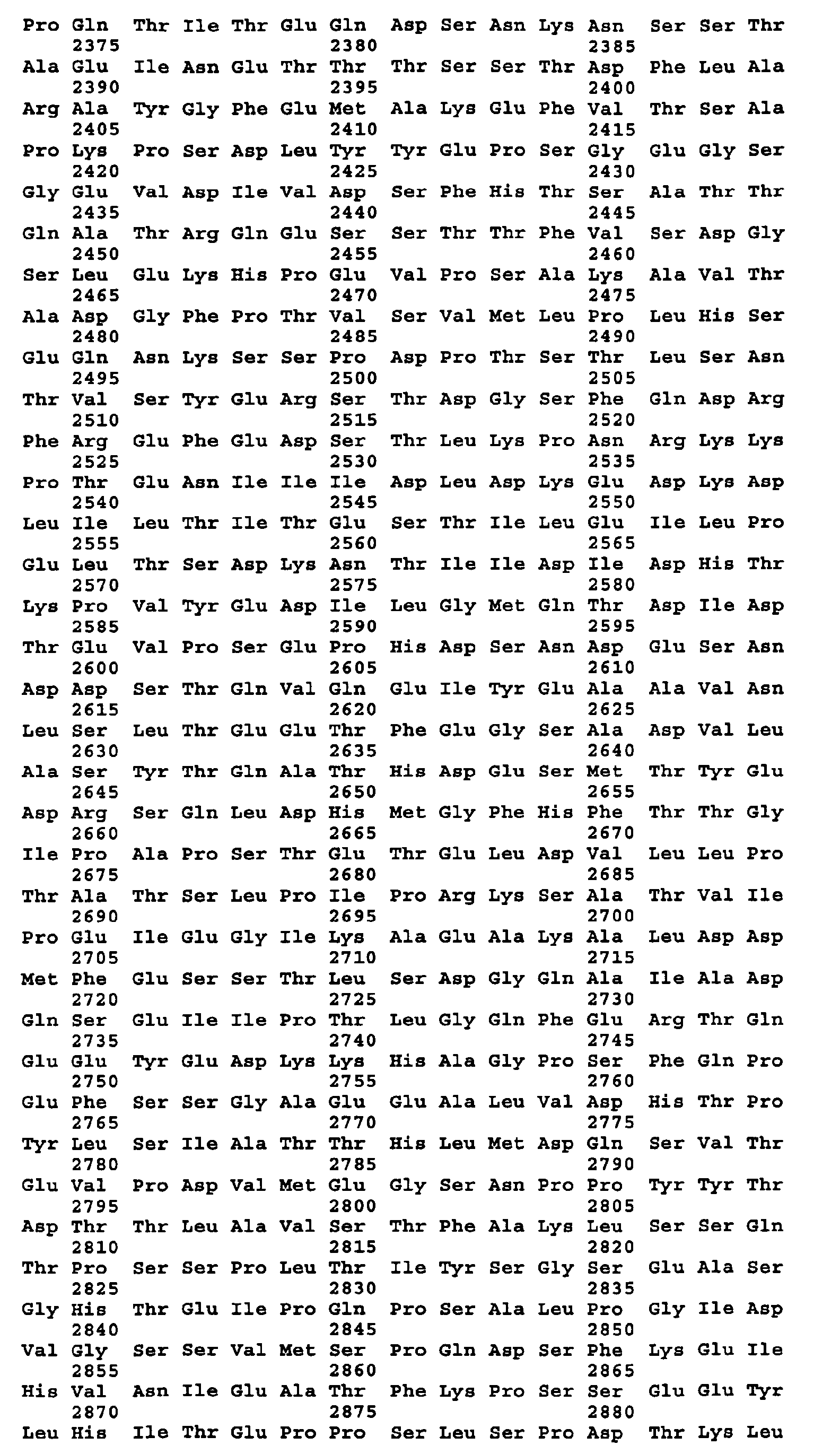
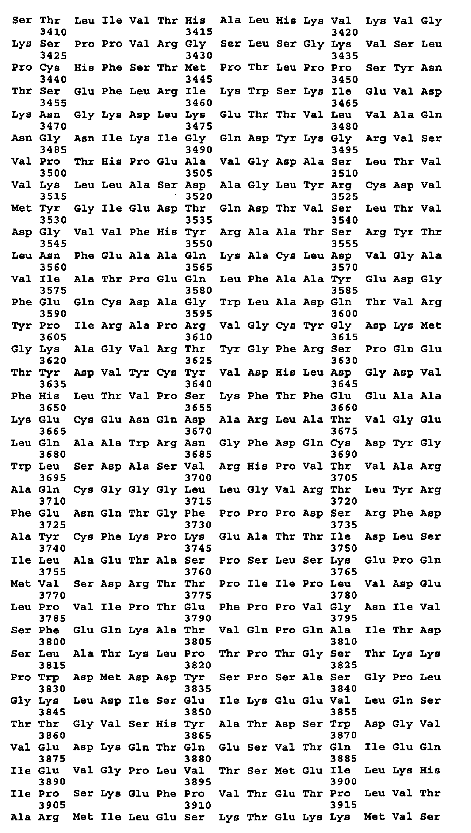
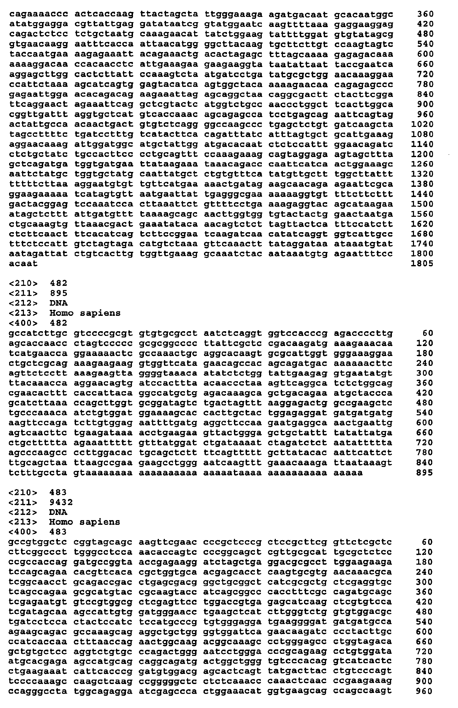
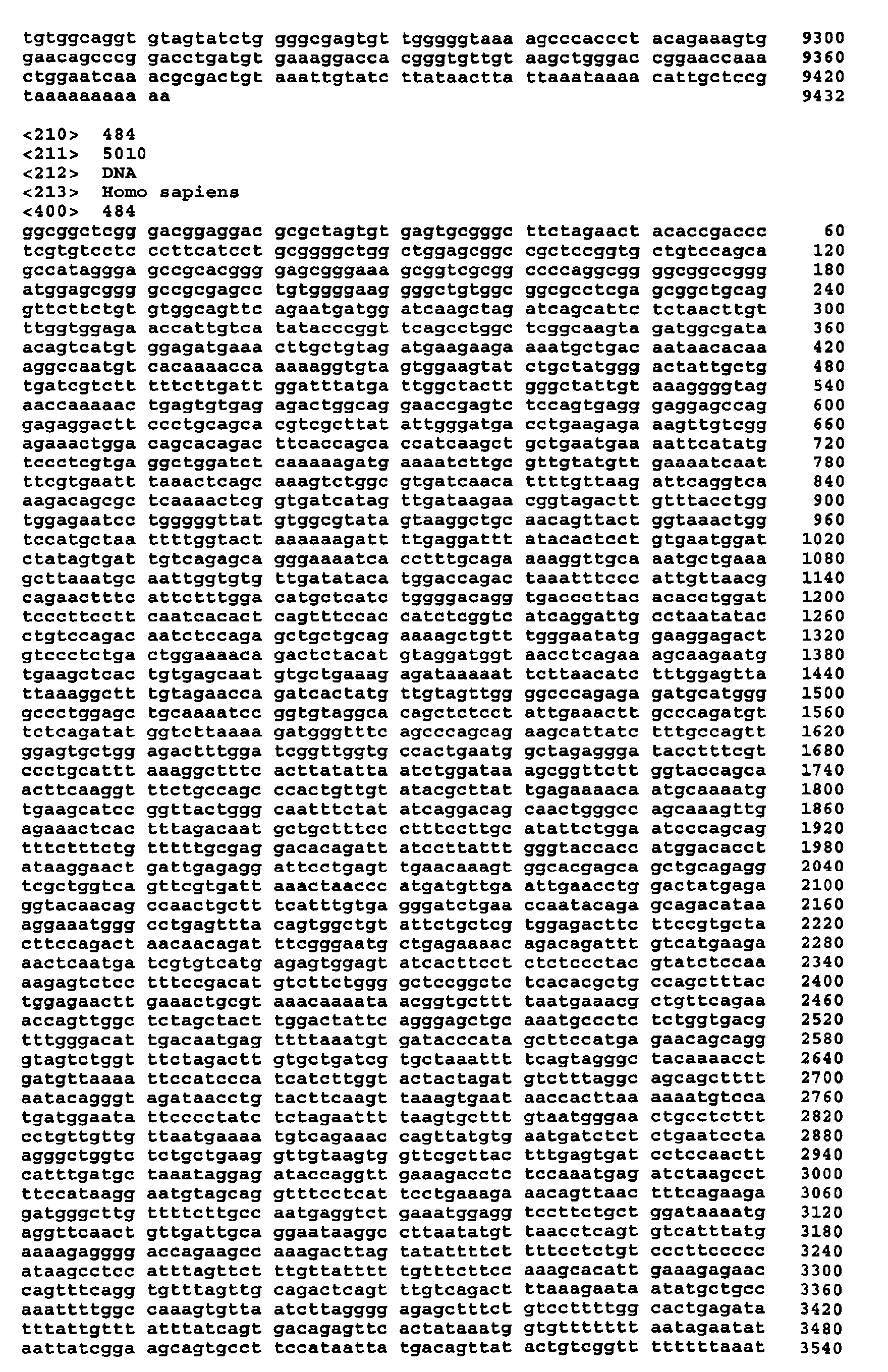
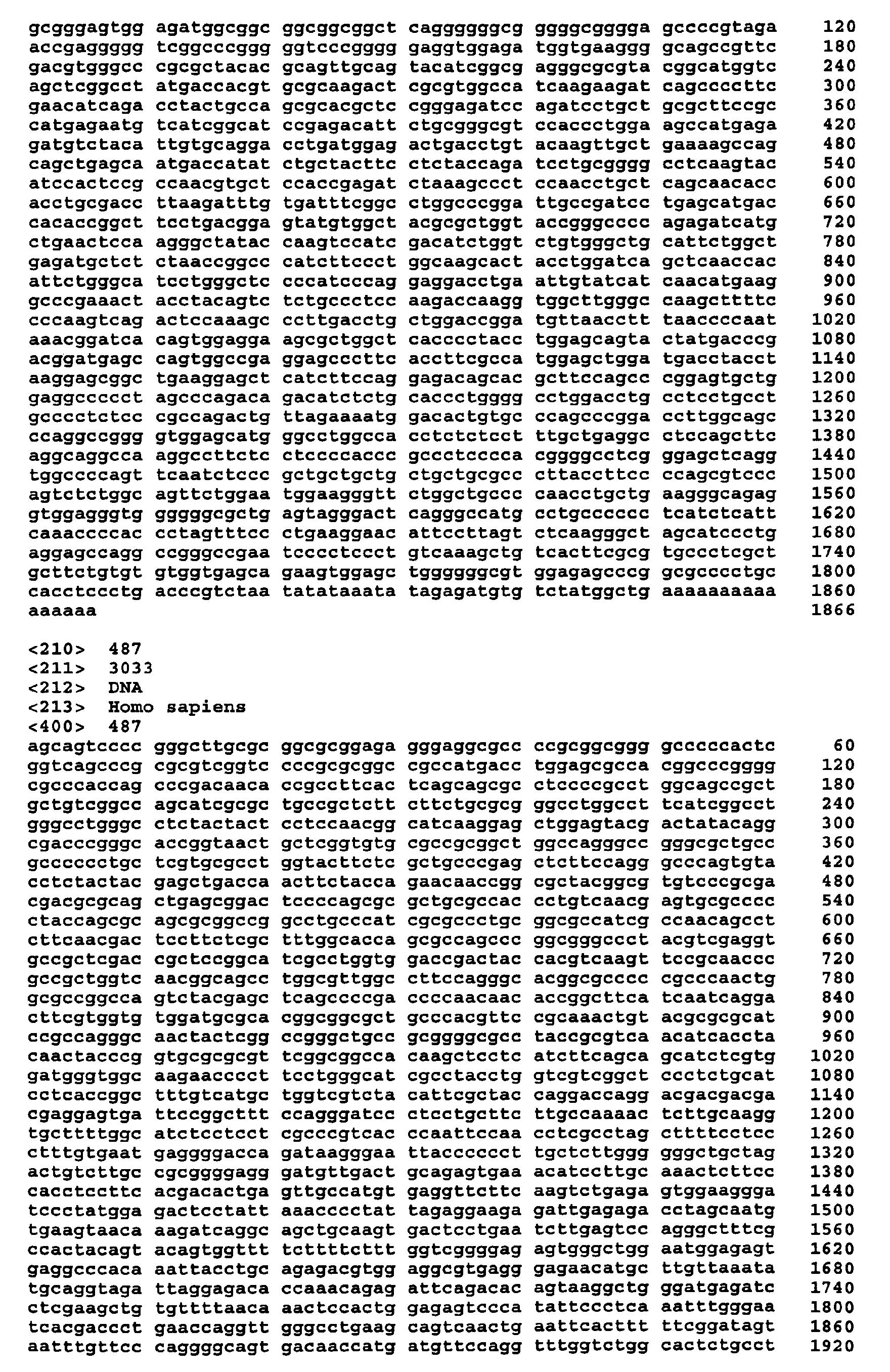
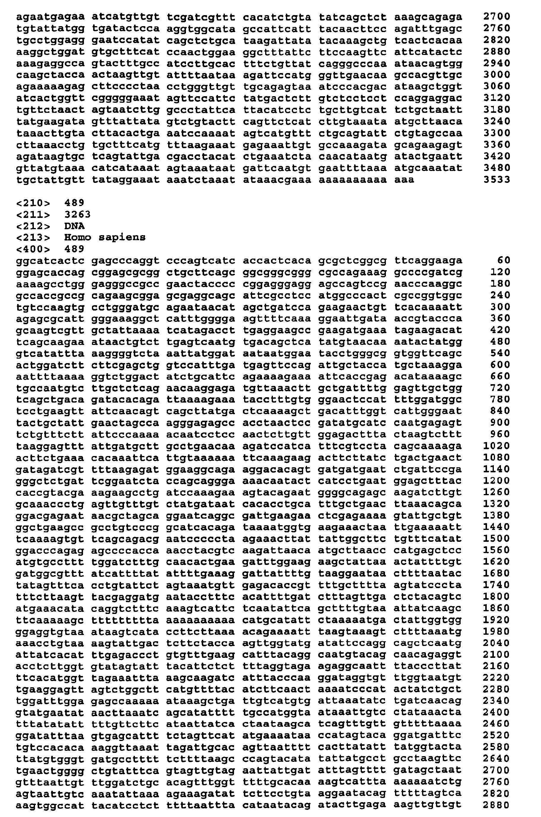
"marker genes", listed in Table 1a and 1b, SEQ ID NO:1 to 165 and 472 to 491, the respective
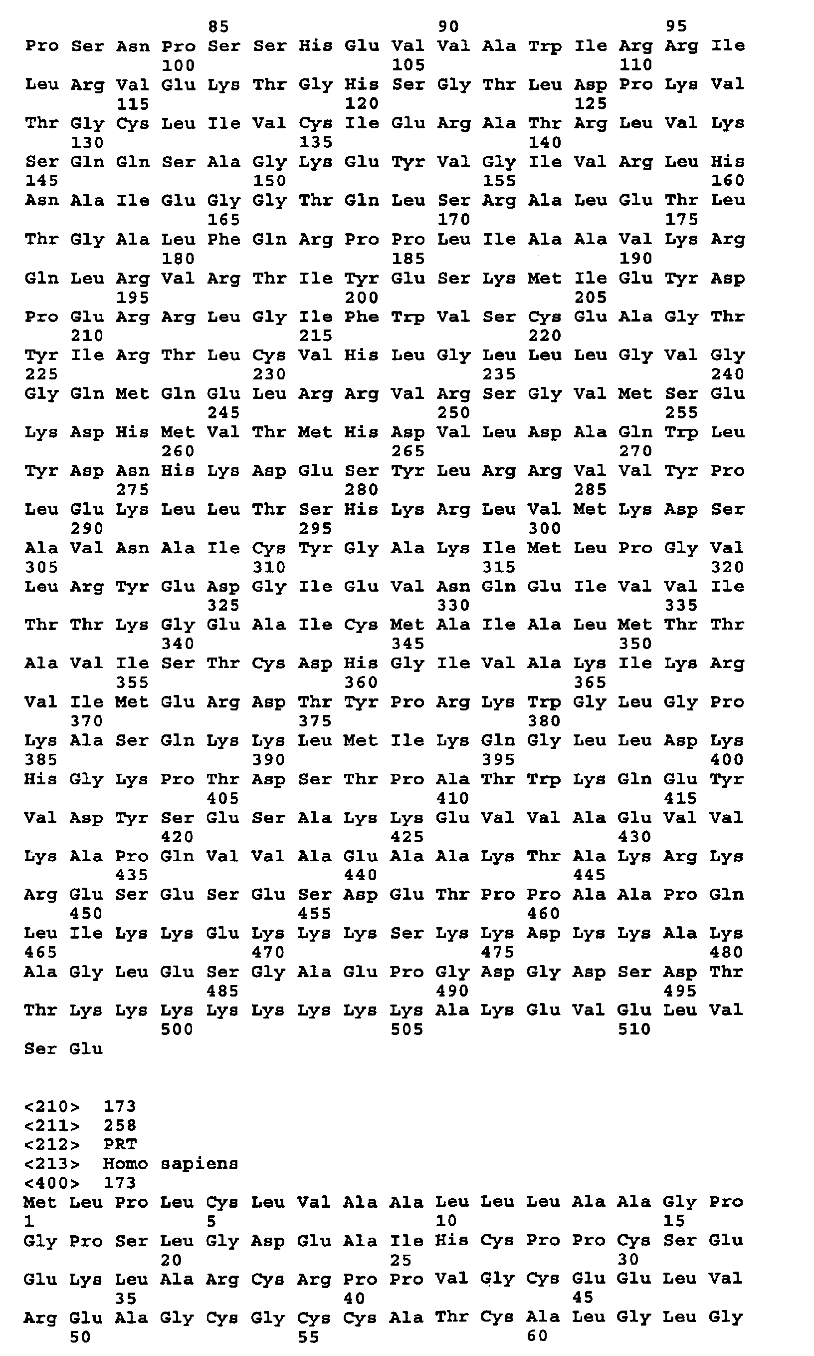
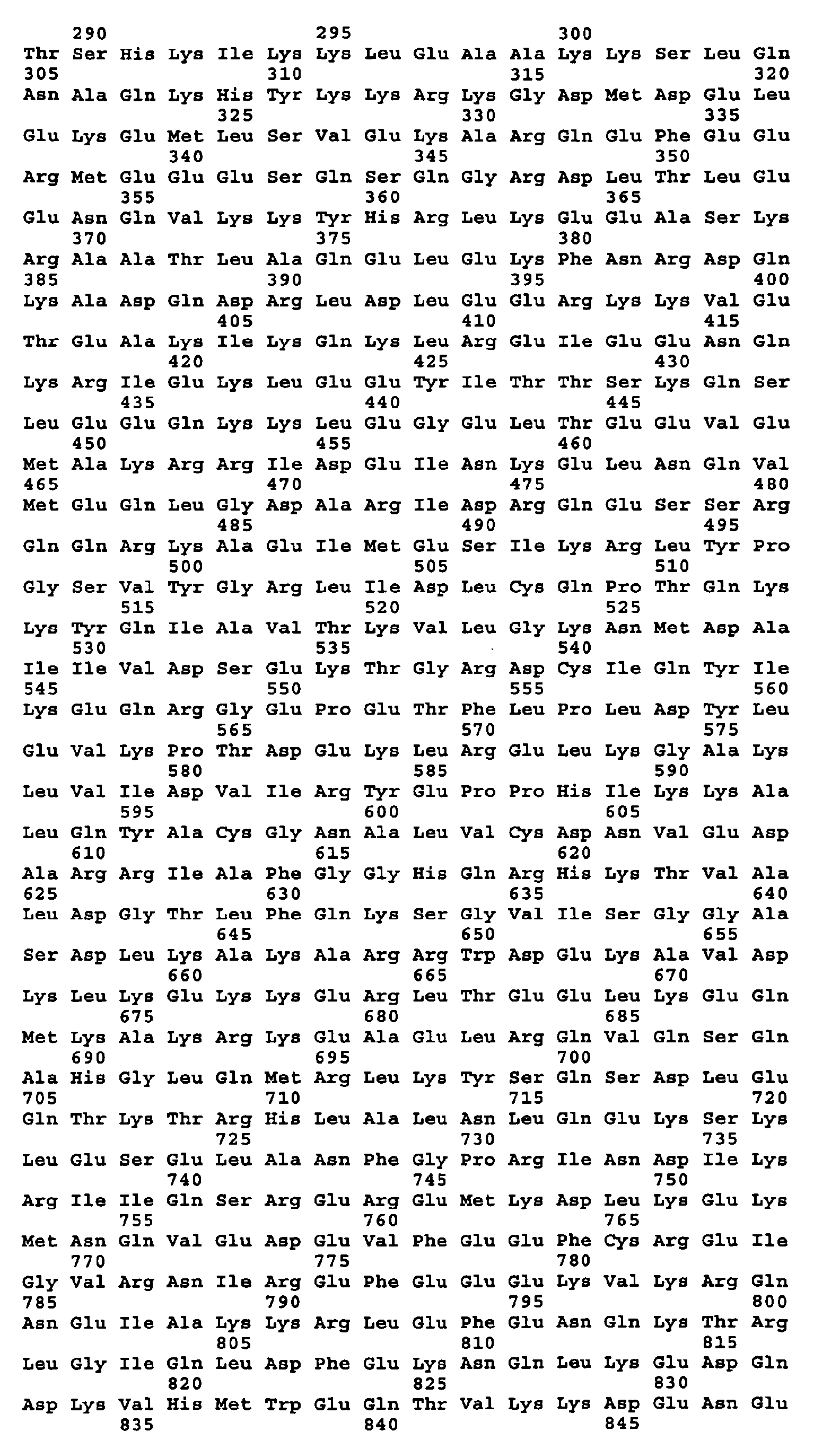
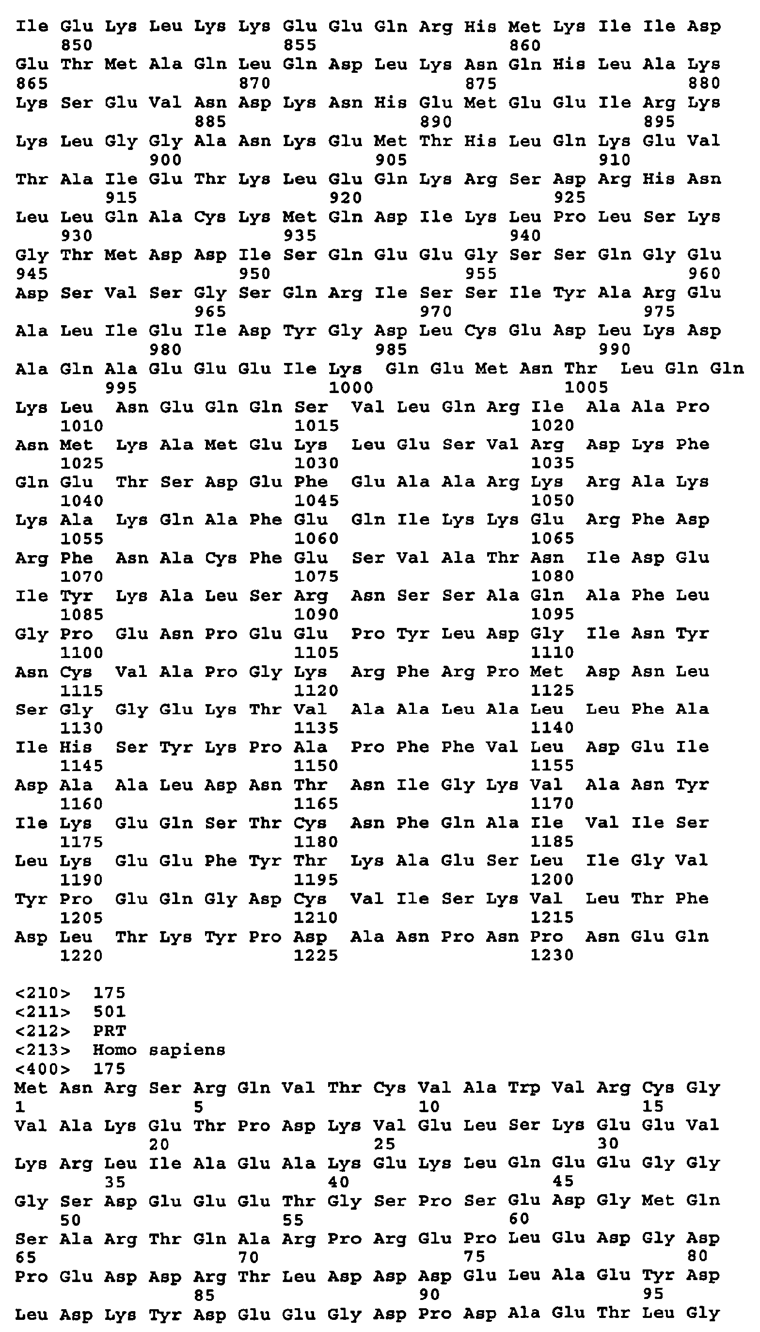
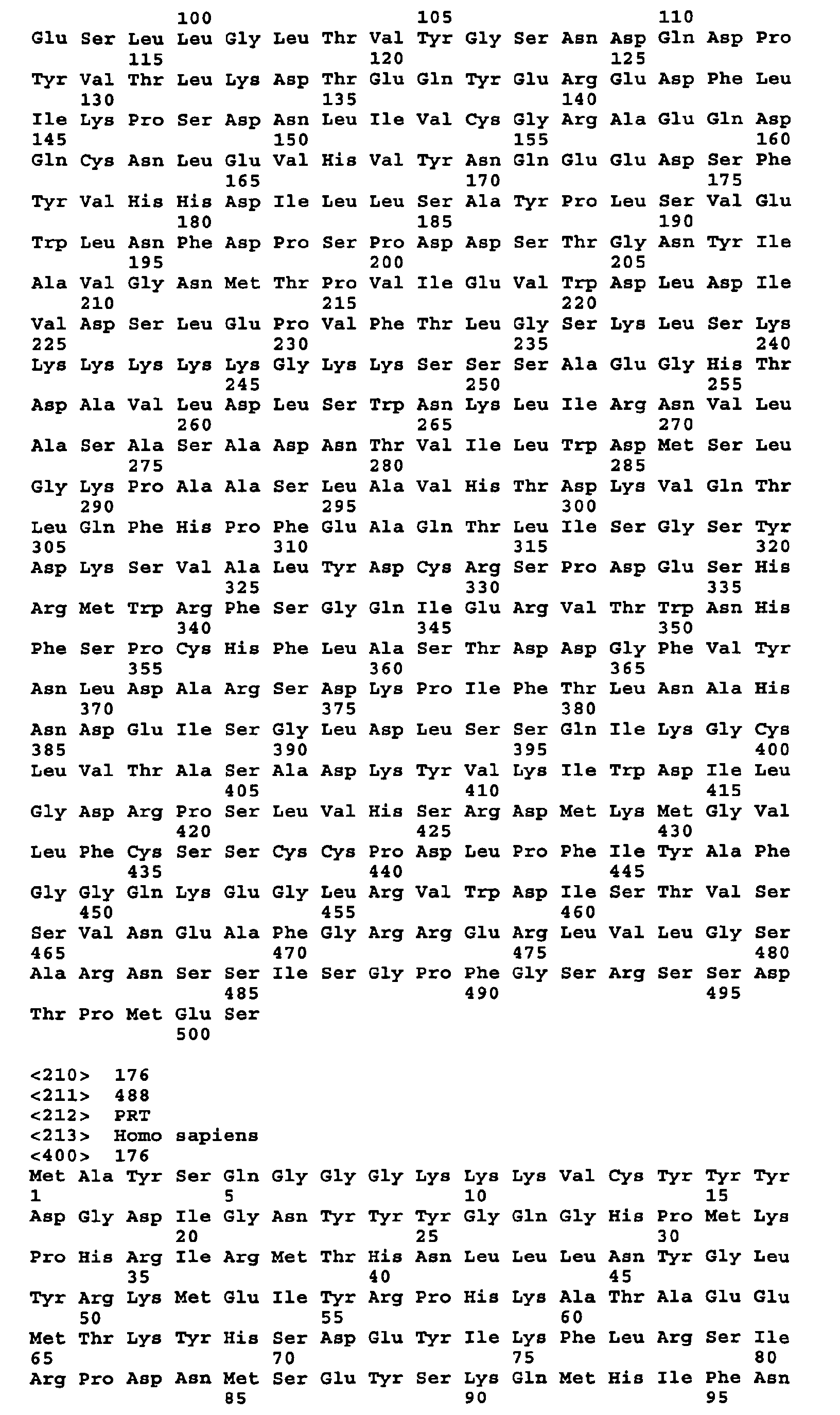
polypeptide sequences coded by them are numerated SEQ ID NO:166 to 330 and 492 to 511, see
also Table 1a and 1b) in a patient sample, and the average level of expression of the marker
gene(s) in a sample from a control subject (e.g., a human subject without breast cancer). A
preferred sub-set of marker genes representing a specific test composition or kit is listed in Table
2.
The invention relates further to various compositions, methods, reagents and kits, for prediction of
clinically measurable tumor therapy response to a given breast cancer therapy. The compositions,
methods, and kits of the present invention comprise comparing the level of mRNA expression of a
single or plurality (e.g. 2, 5, 10, or 50 or more) of breast cancer marker genes in an unclassified
patient sample, and the average level of expression of the marker gene(s) in a sample cohort
comprising patient responding in different intensity to an administered breast cancer therapy. In
preferred embodiments of this invention the specific expression of the marker genes can be utilized
for discrimination of responders and non-responders to an anthracycline based (e.g.
polychemotherapies with epirubicin or doxorubicin) chemo-therapeutic intervention.
In further preferred embodiments, the control level of mRNA expression is the average level of
expression of the marker gene(s) in samples from several (e.g., 2, 3, 4, 5, 8, 10, 12, 15, 20, 30 or
50) control subjects. These control subjects may either be not affected by breast cancer or be
identified and classified by their clinical response prior to the determination of their individual
expression profile.
As elaborated below, a significant change in the level of expression of one or more of the marker
genes (set of marker genes) in the patient sample relative to the control level provides significant
information regarding the patient's breast cancer status and responsiveness to chemotherapy. In the
compositions, methods, and kits of the present invention the marker genes listed in Table 1a and
1b may also be used in combination with well known breast cancer marker genes (e.g. CEA,
mammaglobin, or CA 15-3)
According to the invention, the marker gene(s) and marker gene sets are selected such that the
positive predictive value of the compositions, methods, and kits of the invention is at least about
10%, preferably about 25%, more preferably about 50% and most preferably about 90%. Also
preferred for use in the compositions, methods, and kits of the invention are marker gene(s) and
sets that are differentially expressed, as compared to normal breast cells, by at least the minimal
mean differential expression factor presented in Table 3, in at least about 20%, more preferably
about 50% and most preferably about 75% of any of the following conditions: stage 0 breast
cancer patients, stage I breast cancer patients, stage II breast cancer patients, stage III breast cancer
patients, stage IV breast cancer patients, grade I breast cancer patients, grade II breast cancer
patients, grade III breast cancer patients, malignant breast cancer patients, patients with primary
carcinomas of the breast, and all other types of cancers, malignancies and transformations
associated with the breast.
The detection of marker gene expression is not limited to the detection within a primary, secondary
or metastatic lesion of breast cancer patients, and may also be detected in lymphnodes affected by
breast cancer cells or minimal residual disease cells either locally deposited (e.g. bone marrow,
liver, kidney) or freely floating throughout the patients body.
In one embodiment of the compositions, methods, reagents and kits of the present invention, the
sample to be analyzed is tissue material from neoplastic lesion taken by aspiration or punctuation,
excision or by any other surgical method leading to biopsy or resected cellular material. In one
embodiment of the compositions, methods, and kits of the present invention, the sample comprises
cells obtained from the patient. The cells may be found in a breast cell "smear" collected, for
example, by a nipple aspiration, ductal lavarge, fine needle biopsy or from provoked or
spontaneous nipple discharge. In another embodiment, the sample is a body fluid. Such fluids
include, for example, blood fluids, lymph, ascitic fluids, gynecological fluids, or urine but not
limited to these fluids.
In accordance with the compositions, methods, and kits of the present invention the determination
of gene expression is not limited to any specific method or to the detection of mRNA. The
presence and/or level of expression of the marker gene in a sample can be assessed, for example,
by measuring and/or quantifying of:
In one aspect, the present invention provides a composition, method, and kit of assessing whether a
patient is afflicted with breast cancer (e.g., new detection or "screening", detection of recurrence,
reflex testing, especially in patients having an enhanced risk of developing breast cancer (e.g.,
patients having a familial history of breast cancer and patients identified as having a mutant oncogene).
For this purpose the composition, method, and kit comprises comparing:
A significant increase as well as decrease in the level of expression of the selected marker genes
(e.g. 2, 5, 10, or 50 or more) in the patient sample relative to each marker gene's normal level of
expression is an indication that the patient is afflicted with breast cancer.
The composition, method, and kit of the present invention is also useful for prognosing the
progression or the outcome of the malignant neoplasia. For this purpose the composition, method,
and kit comprises comparing
The composition, method, and kit of the present invention is particularly useful for identifying
patients who will respond to a certain chemotherapy. For this purpose the composition, method,
and kit comprises comparing
In another aspect, the invention provides a composition, method, and kit of assessing the efficacy
of a therapy for inhibiting breast cancer in a patient. This composition, method, and kit comprises
comparing:
It will be appreciated that in this composition, method, and kit the "therapy" may be any therapy
for treating breast cancer including, but not limited to, chemotherapy, anti-hormonal therapy,
directed antibody therapy, radiation therapy and surgical removal of tissue, e.g., a breast tumor.
Thus, the compositions, methods, and kits of the invention may be used to evaluate a patient
before, during and after therapy, for example, to evaluate the reduction in tumor burden.
In a further aspect, the present invention provides a composition, method, and kit for monitoring
the progression of breast cancer in a patient. This composition, method, and kit comprising:
In another aspect, the invention provides a composition, method, and kit for
in vitro selection of a
therapy regime (e.g. the kind of chemotherapeutical argents) for inhibiting breast cancer in a
patient. This composition, method, and kit comprises the steps of:
in each of the aliquots; and d) selecting one of the test compositions which induces a lower level of expression of genes
from SEQ ID 11, 17, 22, 25, 31, 36, 48, 49, 57, 83, 107, 108, 112, and 159 and/or a higher
level of expression of genes from SEQ ID 24, 47, 54, 58, 59, 60, 67, 79, 80, 88, 114, 118,
135, and 141 in the aliquot containing that test composition, relative to the level of
expression of each marker gene in the aliquots containing the other test compositions.
The invention further provides a composition, method, and kit of assessing the carcinogenic
potential of a certain biological or chemical compound. This composition, method, and kit
comprises the steps of:
The invention further provides a composition, method, and kit of treating a patient afflicted with
breast cancer. This composition, method, and kit comprises providing to cells of the patient an
antisense oligonucleotide complementary to a polynucleotide sequence of a marker gene listed
within Table 1a and 1b
The invention additionally provides a composition, method, and kit of inhibiting breast cancer
cells in a patient at risk for developing breast cancer. This composition, method, and kit comprises
inhibiting expression of a marker gene listed in Table 1a and 1b.
In yet another embodiment the invention provides compositions, methods, and kits of screening for
agents which regulate the activity of a polypeptide comprising a polypeptide selected from SEQ ID
NO: 166 to 330 and 492 to 511. A test compound is contacted with the particular polypeptide.
Binding of the test compound to the polypeptide is detected. A test compound which binds to the
polypeptide is thereby identified as a potential therapeutic agent for the treatment of malignant
neoplasia and more particularly breast cancer.
In even another embodiment the invention provides another composition, method, and kit of
screening for agents which regulate the activity of a polypeptide comprising a polypeptide selected
from SEQ ID NO: 166 to 330 and 492 to 511. A test compound is contacted with the particular
polypeptide. A biological activity mediated by the polypeptide is detected. A test compound which
decreases the biological activity is thereby identified as a potential therapeutic agent for decreasing
the activity of the particular polypeptide in malignant neoplasia and especially in breast cancer A
test compound which increases the biological activity is thereby identified as a potential therapeutic
agent for increasing the activity of the particular polypeptide in malignant neoplasia and
especially in breast cancer
The invention thus provides polypeptides selected from one of the polypeptides with SEQ ID NO:
166 to 330 and 492 to 511 which can be used to identify compounds which may act, for example,
as regulators or modulators such as agonists and antagonists, partial agonists, inverse agonists,
activators, co-activators and inhibitors of the polypeptide comprising a polypeptide selected from
SEQ ID NO: 166 to 330 and 492 to 511 Accordingly, the invention provides reagents and
compositions, methods, and kits for regulating a polypeptide comprising a polypeptide selected
from SEQ ID NO: 166 to 330 and 492 to 511 in malignant neoplasia and more particularly breast
cancer. The regulation can be an up- or down regulation. Reagents that modulate the expression,
stability or amount of a polynucleotide listed in Table 1a and 1b (SEQ ID NO: 1 to 165 and 472 to
491 or the activity of the polypeptide comprising a polypeptide selected from SEQ ID NO: 166 to
330 and 492 to 511 can be a protein, a peptide, a peptidomimetic, a nucleic acid, a nucleic acid
analogue (e.g. peptide nucleic acid, locked nucleic acid) or a small molecule. Compositions,
methods, and kits that modulate the expression, stability or amount of a polynucleotide comprising
a polynucleotide selected from SEQ ID NO: 1 to 165 and 472 to 491 (listed in Table 1a and 1b) or
the activity of the polypeptide comprising a polypeptide selected from SEQ ID NO: 166 to 330 and
492 to 511 (Table 1) can be gene replacement therapies, antisense, ribozyme and triplex nucleic
acid approaches.
The invention further provides a composition, method, and kit of making an isolated hybridoma
which produces an antibody useful for assessing whether a patient is afflicted with breast cancer.
The composition, method, and kit comprises isolating a protein encoded by a marker gene listed
within Table 1a and 1b or a polypeptide fragment of the protein, immunizing a mammal using the
isolated protein or polypeptide fragment, isolating splenocytes from the immunized mammal,
fusing the isolated splenocytes with an immortalized cell line to form hybridomas, and screening
individual hybridomas for production of an antibody which specifically binds with the protein or
polypeptide fragment to isolate the hybridoma. The invention also includes an antibody produced
by this method. Such antibodies specifically bind to a full-length or partial polypeptide comprising
a polypeptide selected from SEQ ID NO: 166 to 330 and 492 to 511 (listed in Table 1a and 1b) for
use in prediction, prevention, diagnosis, prognosis and treatment of malignant neoplasia and breast
cancer in particular.
Yet another embodiment of the invention is the use of a reagent which specifically binds to a
polynucleotide comprising a polynucleotide selected from SEQ ID NO: 1 to 165 and 472 to 491 or
to a polypeptide comprising a polypeptide selected from SEQ ID NO: 166 to 330 and 492 to 511
(listed in Table 1a and 1b) in the preparation of a medicament for the treatment of malignant
neoplasia and breast cancer in particular.
Still another embodiment is the use of a reagent that modulates the activity or stability of a
polypeptide comprising a polypeptide selected from SEQ ID NO: 166 to 330 and 492 to 511
(Table 1a and 1b) or the expression, amount or stability of a polynucleotide comprising a
polynucleotide selected from SEQ ID NO: 1 to 165 and 472 to 491 (Table 1a and 1b) in the
preparation of a medicament for the treatment of malignant neoplasia and breast cancer in
particular.
Still another embodiment of the invention is a pharmaceutical composition which includes a
reagent which specifically binds to a polynucleotide comprising a polynucleotide selected from
SEQ ID NO: 1 to 165 (Tablel) or a polypeptide comprising a polypeptide selected from SEQ ID
NO: 166 to 300, and a pharmaceutically acceptable carrier.
A further embodiment of the invention is a pharmaceutical composition comprising a polynucleotide
including a sequence which hybridizes under stringent conditions to a polynucleotide
comprising a polynucleotide selected from SEQ ID NO: 1 to 165 and 472 to 491 and encoding a
polypeptide exhibiting the same biological function as given for the respective polynucleotide in
Table 1a and 1b or 4, or encoding a polypeptide comprising a polypeptide selected from SEQ ID
NO: 166 to 330 and 492 to 511. Pharmaceutical compositions, useful in the present invention may
further include fusion proteins comprising a polypeptide comprising a polynucleotide selected
from SEQ ID NO: 1 to 165 and 472 to 491, or a fragment thereof, antibodies, or antibody
fragments
The invention also provides various kits. Such kit comprises reagents for assessing expression of a
single or a plurality of genes selected from the marker genes listed in Table 1a and 1b or selected
from the sub-set of genes listed in Table 2.
In one aspect, the invention provides a kit for assessing whether a patient is afflicted with breast
cancer.
In another aspect, the invention provides a kit for assessing the suitability of each of a plurality of
compounds for inhibiting a breast cancer in a patient. The kit comprises reagents for assessing
expression of a marker gene listed within Table 1a and 1b, or reagents for assessing the expression
of each marker gene of a marker gene set listed in Table 2. The kit may also comprise a plurality of
compounds.
In an additional aspect, the invention provides a kit for assessing the presence of breast cancer
cells. This kit comprises an antibody, wherein the antibody binds specifically with a protein
encoded by a marker gene listed within Table 1a and 1b or polypeptide fragment of the protein.
The kit may also comprise a plurality of antibodies, wherein the plurality binds specifically with
the protein encoded by each marker gene of a marker gene set listed in Table 2.
In yet another aspect, the invention provides a kit for assessing the presence of breast cancer cells,
wherein the kit comprises a nucleic acid probe. The probe hybridizes specifically with a RNA
transcript of a marker gene listed within Table 1a and 1b or cDNA of the transcript. The kit may
also comprise a plurality of probes, wherein each of the probes hybridizes specifically with a RNA
transcript of one of the marker genes of a marker gene set listed in Table 2.
It will be appreciated that the compositions, methods, and kits of the present invention may also
include known cancer marker genes including known breast cancer marker genes. It will further be
appreciated that the compositions, methods, and kits may be used to identify cancers other than
breast cancer.
DETAILED DESCRIPTION OF THE INVENTION
DEFINITIONS
"Differential expression", or "expression" as used herein, refers to both quantitative as well as
qualitative differences in the genes' expression patterns depending on differential development,
different genetic background of tumor cells and/or reaction to the tissue environment of the tumor.
Differentially expressed genes may represent "marker genes," and/or "target genes". The
expression pattern of a differentially expressed gene disclosed herein may be utilized as part of a
prognostic or diagnostic breast cancer evaluation., Alternatively, a differentially expressed gene
disclosed herein may be used in methods for identifying reagents and compounds and uses of these
reagents and compounds for the treatment of breast cancer as well as methods of treatment. The
differential regulation of the gene is not limited to a specific cancer cell type or clone, but rather
displays the interplay of cancer cells, muscle cells, stromal cells, connective tissue cells, other
epithelial cells, endothelial cells and blood vessesl as well as cells of the immune system (e.g.
lymphocytes, macrophages, killer cells).
"Biological activity" or "bioactivity" or "activity" or "biological function", which are used
interchangeably, herein mean an effector or antigenic function that is directly or indirectly
performed by a polypeptide (whether in its native or denatured conformation), or by any fragment
thereof in vivo or in vitro. Biological activities include but are not limited to binding to
polypeptides, binding to other proteins or molecules, enzymatic activity, signal transduction,
activity as a DNA binding protein, as a transcription regulator, ability to bind damaged DNA, etc.
A bioactivity can be modulated by directly affecting the subject polypeptide. Alternatively, a
bioactivity can be altered by modulating the level of the polypeptide, such as by modulating
expression of the corresponding gene.
The term "marker" or "biomarker" refers a biological molecule, e.g., a nucleic acid, peptide,
hormone, etc., whose presence or concentration can be detected and correlated with a known
condition, such as a disease state.
The term "marker gene," as used herein, refers to a differentially expressed gene which expression
pattern may be utilized as part of predictive, prognostic or diagnostic process in malignant
neoplasia or breast cancer evaluation, or which, alternatively, may be used in methods for
identifying compounds useful for the treatment or prevention of malignant neoplasia and breast
cancer in particular. A marker gene may also have the characteristics of a target gene.
"Target gene", as used herein, refers to a differentially expressed gene involved in breast cancer in
a manner by which modulation of the level of target gene expression or of target gene product
activity may act to ameliorate symptoms of malignant neoplasia and breast cancer in particular. A
target gene may also have the characteristics of a marker gene.
The term "neoplastic lesion" or " neoplastic disease" or "neoplasia" refers to a cancerous tissue
this includes carcinomas, (e.g., carcinoma in situ, invasive carcinoma, metastatic carcinoma) and
pre-malignant conditions, neomorphic changes independent of their histological origin (e.g. ductal,
lobular, medullary, mixed origin). The term "cancer" is not limited to any stage, grade,
histomorphological feature, invasiveness, agressivity or malignancie of an affected tissue or cell
aggregation. In particular stage 0 breast cancer, stage I breast cancer, stage II breast cancer, stage
III breast cancer, stage IV breast cancer, grade I breast cancer, grade II breast cancer, grade III
breast cancer, malignant breast cancer, primary carcinomas of the breast, and all other types of
cancers, malignancies and transformations associated with the breast are included. The terms
"neoplastic lesion" or " neoplastic disease" or "neoplasia" or "cancer" are not limited to any tissue
or cell type they also include primary, secondary or metastatic lesion of cancer patients, and also
comprises lymphnodes affected by cancer cells or minimal residual disease cells either locally
deposited (e.g. bone marrow, liver, kidney) or freely floating throughout the patients body.
The term "biological sample", as used herein, refers to a sample obtained from an organism or
from components (e.g., cells) of an organism. The sample may be of any biological tissue or fluid.
Frequently the sample will be a "clinical sample" which is a sample derived from a patient. Such
samples include, but are not limited to, sputum, blood, blood cells (e.g., white cells), tissue or fine
needle biopsy samples, cell-containing bodyfluids, free floating nucleic acids, urine, peritoneal
fluid, and pleural fluid, or cells therefrom. Biological samples may also include sections of tissues
such as frozen or fixed sections taken for histological purposes. A biological sample to be analyzed
is tissue material from neoplastic lesion taken by aspiration or punctuation, excision or by any
other surgical method leading to biopsy or resected cellular material. Such biological sample may
comprises cells obtained from a patient. The cells may be found in a breast cell "smear" collected,
for example, by a nipple aspiration, ductal lavarge, fine needle biopsy or from provoked or
spontaneous nipple discharge. In another embodiment, the sample is a body fluid. Such fluids
include, for example, blood fluids, lymph, ascitic fluids, gynecological fluids, or urine but not
limited to these fluids.
The term "therapy modality", "therapy mode", "regimen" or "chemo regimen" as well as "therapy
regime" refers to a timely sequential or simultaneous administration of anti tumor, and/or immune
stimulating, and/or blood cell proliferative agents, and/or radation therapy, and/or hyperthermia,
and/or hypothermia for cancer therapy. The administration of these can be performed in an
adjuvant and/or neoadjuvant mode. The composition of such "protocol" may vary in dose of the
single agent, timeframe of application and frequency of administration within a defined therapy
window. Currently various combinations of various drugs and/or physical methods, and various
schedules are under investigation.
By "array" or "matrix" is meant an arrangement of addressable locations or "addresses" on a
device. The locations can be arranged in two dimensional arrays, three dimensional arrays, or other
matrix formats. The number of locations can range from several to at least hundreds of thousands.
Most importantly, each location represents a totally independent reaction site. Arrays include but
are not limited to nucleic acid arrays, protein arrays and antibody arrays. A "nucleic acid array"
refers to an array containing nucleic acid probes, such as oligonucleotides, polynucleotides or
larger portions of genes. The nucleic acid on the array is preferably single stranded. Arrays
wherein the probes are oligonucleotides are referred to as "oligonucleotide arrays" or
"oligonucleotide chips." A "microarray," herein also refers to a "biochip" or "biological chip", an
array of regions having a density of discrete regions of at least about 100/cm2, and preferably at
least about 1000/cm2. The regions in a microarray have typical dimensions, e.g., diameters, in the
range of between about 10-250 µm, and are separated from other regions in the array by about the
same distance. A "protein array" refers to an array containing polypeptide probes or protein probes
which can be in native form or denatured. An "antibody array" refers to an array containing
antibodies which include but are not limited to monoclonal antibodies (e.g. from a mouse),
chimeric antibodies, humanized antibodies or phage antibodies and single chain antibodies as well
as fragments from antibodies.
The term "agonist", as used herein, is meant to refer to an agent that mimics or upregulates (e.g.,
potentiates or supplements) the bioactivity of a protein. An agonist can be a wild-type protein or
derivative thereof having at least one bioactivity of the wild-type protein. An agonist can also be a
compound that upregulates expression of a gene or which increases at least one bioactivity of a
protein. An agonist can also be a compound which increases the interaction of a polypeptide with
another molecule, e.g., a target peptide or nucleic acid.
The term "antagonist" as used herein is meant to refer to an agent that downregulates (e.g.,
suppresses or inhibits) at least one bioactivity of a protein. An antagonist can be a compound
which inhibits or decreases the interaction between a protein and another molecule, e.g., a target
peptide, a ligand or an enzyme substrate. An antagonist can also be a compound that
downregulates expression of a gene or which reduces the amount of expressed protein present.
"Small molecule" as used herein, is meant to refer to a composition, which has a molecular weight
of less than about 5 kD and most preferably less than about 4 kD. Small molecules can be nucleic
acids, peptides, polypeptides, peptidomimetics, carbohydrates, lipids or other organic (carbon-containing)
or inorganic molecules. Many pharmaceutical companies have extensive libraries of
chemical and/or biological mixtures, often fungal, bacterial, or algal extracts, which can be
screened with any of the assays of the invention to identify compounds that modulate a bioactivity.
The terms "modulated" or "modulation" or "regulated" or "regulation" and "differentially
regulated" as used herein refer to both upregulation (i.e., activation or stimulation (e.g., by
agonizing or potentiating) and down regulation [i.e., inhibition or suppression (e.g., by
antagonizing, decreasing or inhibiting)].
"Transcriptional regulatory unit" refers to DNA sequences, such as initiation signals, enhancers,
and promoters, which induce or control transcription of protein coding sequences with which they
are operably linked. In preferred embodiments, transcription of one of the genes is under the
control of a promoter sequence (or other transcriptional regulatory sequence) which controls the
expression of the recombinant gene in a cell-type in which expression is intended. It will also be
understood that the recombinant gene can be under the control of transcriptional regulatory
sequences which are the same or which are different from those sequences which control
transcription of the naturally occurring forms of the polypeptide.
The term "derivative" refers to the chemical modification of a polypeptide sequence, or a
polynucleotide sequence. Chemical modifications of a polynucleotide sequence can include, for
example, replacement of hydrogen by an alkyl, acyl, or amino group. A derivative polynucleotide
encodes a polypeptide which retains at least one biological or immunological function of the
natural molecule. A derivative polypeptide is one modified by glycosylation, pegylation, or any
similar process that retains at least one biological or immunological function of the polypeptide
from which it was derived.
The term "nucleotide analog" refers to oligomers or polymers being at least in one feature different
from naturally occurring nucleotides, oligonucleotides or polynucleotides, but exhibiting
functional features of the respective naturally occurring nucleotides (e.g. base paring,
hybridization, coding information) and that can be used for said compositions. The nucleotide
analogs can consist of non-naturally occurring bases or polymer backbones, examples of which are
LNAs, PNAs and Morpholinos. The nucleotide analog has at least one molecule different from its
naturally occurring counterpart or equivalent.
"BREAST CANCER GENES" or "BREAST CANCER GENE" as used herein refers to the
polynucleotides of SEQ ID NO:1 to 165 and 472 to 491 (listed in Table 1a and 1b), as well as
derivatives, fragments, analogs and homologues thereof, the polypeptides encoded thereby, (SEQ
ID NO:166 to 330 and 492 to 511, see Table 1) as well as derivatives, fragments, analogs and
homologues thereof and the corresponding genomic transcription units which can be derived or
identified with standard techniques well known in the art using the information disclosed in Tables
1 to 5. The Genename, Reference Sequence, unique Gene-identifier, and the Locuslink ID numbers
of the polynucleotide sequences of the SEQ ID NO: 1 to 65 and the polypeptides of the SEQ ID
NO: 166 to 330 and 492 to 511 are shown in Table 1a and 1b, the gene description, gene function
and subcellelar localization is given in Tables 4a and 4b.
The term "chromosomal region" as used herein refers to a consecutive DNA stretch on a
chromosome which can be defined by cytogenetic or other genetic markers such as e.g. restriction
length polymorphisms (RFLPs), single nucleotide polymorphisms (SNPs), expressed sequence tags
(ESTs), sequence tagged sites (STSs), microsatellites, variable number of tandem repeats (VNTRs)
and genes. Typically a chromosomal region consists of up to 2 Megabases (MB), up to 4 MB, up
to 6 MB, up to 8 MB, up to 10 MB, up to 20 MB or even more MB.
The term "kit" as used herein refers to any manufacture (e.g. a diagnostic or research product)
comprising at least one reagent, e.g. a probe, for specifically detecting the expression of at least
one marker gene disclosed in the invention, in particular of those genes listed in Table 2, whereas
the manufacture is being sold, distributed, and/or promoted as a unit for performing the methods of
the present invention. The genes, primer and probes listed in Table 2 and 5 or any combination of
at least two of them, regard as one single test for the purposes, methods and disclosures of this
invention. Also reagents (e.g. immunoassays) to detect the presence, the stability, activity,
complexity of the respective marker gene products comprising polypeptides selected from SEQ ID
NO:166 to 330 and 492 to 511 regard as components of the kit. In addition, any combination of
nucleic acid and protein detection as disclosed in the invention are regard as a kit.
The present invention provides polynucleotide sequences and proteins encoded thereby, as well as
probes derived from the polynucleotide sequences, antibodies directed to the encoded proteins, and
predictive, preventive, diagnostic, prognostic and therapeutic uses for individuals which are at risk
for or which have malignant neoplasia and breast cancer in particular. The sequences disclosure
herein have been found to be differentially expressed in samples from breast cancer.
The present invention is based on the identification of 185 genes that are differentially regulated
(up- or down regulated) in tumor biopsies of patients with clinical evidence of breast cancer.. The
characterization of the co-expression of some of these genes provides newly identified roles in
breast cancer. The gene names, the database accession numbers (Genename, Reference Sequence,
unique Gene-identifier, and the Locuslink ID numbers) as well as the putative or known functions
of the encoded proteins and their subcellular localization are given in Tables 1 to 4a and 4b. The
primer sequences used for the gene amplification and hybridization probes are shown in Table 5.
The present invention relates to:
Determination of an expression level can comprise a quantitatification of the expression level
and/or a purely qualitative determination of the expression level.A "pattern of expression levels" of a single gene is to be understood as the expression level of said
gene as determined by suitable methods.Nucleic acid molecules, referred to with a specific SEQ ID NO, within the meaning of the
invention, are to be understood as comprising also variants of said nucleic acid molecules, which
can be derived from the original nucleic acid molecules by deletion, insertion or transposition of
nucleotides, provided said variants still have an 80, 90, 95, or 99% sequence identity towards the
original sequence. Preferrably the variants still have the same biological activity and/or function as
have the original molecules.It is obvious to the person skilled in the art that a reference to a nucleotide sequence is meant to
comprise the reference to the associated protein sequence which is coded by said nucleotide
sequence."% identity" of a first sequence towards a second sequence, within the meaning of the invention,
means the % identity which is calculated as follows: First the optimal global alignment between
the two sequences is determined with the CLUSTALW algorithm [Thomson JD, Higgins DG,
Gibson TJ. 1994. ClustalW: Improving the sensitivity of progressive multiple sequence alignment
through sequence weighting, positions-specific gap penalties and weight matrix choice. Nucleic
Acids Res., 22: 4673-4680], Version 1.8, applying the following command line syntax: ./clustalw -
infile=./infile.txt -output= -outorder=aligned -pwmatrix=gonnet -pwdnamatrix=clustalw
-pwgapopen=10.0 -pwgapext=0.1 -matrix=gonnet -gapopen=10.0 -gapext=0.05 -gapdist=8
-hgapresidues=GPSNDQERK -maxdiv=40. Implementations of the CLUSTAL W algorithm are
readily available at numerous sites on the internet, including, e.g., http://www.ebi.ac.uk.
Thereafter, the number of matches in the alignment is determined by counting the number of
identical nucleotides (or amino acid residues) in aligned positions. Finally, the total number of
matches is divided by the number of nucleotides (or amino acid residues) of the longer of the two
sequences, and multiplied by 100 to yield the % identity of the first sequence towards the second
sequence.
3. A method of count 1 or 2, wherein said method comprises multiple determinations of a
pattern of expression levels, at different points in time, thereby allowing to monitor the
development of said neoplastic disease in said subject. 4. A method of count 1, wherein said method comprises an estimation of the likelihood of
success of a given mode of treatment for said neoplastic disease in said subject. 5. A method of count 1, wherein said method comprises an assessment of whether the subject
is expected to respond or whether the subject is expected not to a given mode of treatment
for said neoplastic disease.
The terms "to respond" or "not to respond" are to be understood in a qualitative and/or in a
quantitative fashion. "To respond" and "not to respond" is to be assessed with regard to a suitable
reference responses, such as, e.g., responses shown by "responders" and "not-responders" to a
certain mode of treatment or modality of treatment. 6. A method of count 4 or 5, wherein a predictive algorithm is used.
Predictive algorithms, which are well known to a person skilled in the art of data analysis, are to be
understood as being any kind of predictive algorithm known in the art. Preferred examples of such
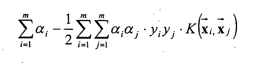
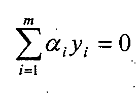
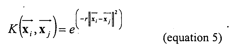
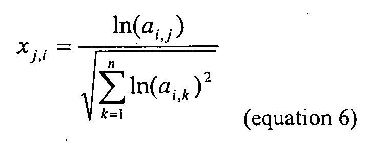
algorithms are, e.g., the SVM algorithm disclosed in Example 4. 7. A method of count 6, wherein the predictive algorithm is a Support Vector Machine.
Support Vector Machines are algorithms, well known to the person skilled in the art of data
analysis. A Support Vector Machine algorithm is disclosed in Example 4. 8. A method of any of counts 4 to 7, wherein said given mode of treatment
(i) acts on cell proliferation, and/or (ii) acts on cell survival, and/or (iii) acts on cell motility, and/or (iv) is an anthracycline based mode of treatment, and/or (v) comprises administration of epirubicin and/or cyclophoshamid. 9. A method of treatment for a subject afflicted with a neoplastic disease, comprising
(i) identifying a promising mode of treatment with the method of count 4 or 5, (ii) treating said neoplastic disease in said patient by the mode of treatment identified
in step (i). 10. A method of screening for subjects afflicted with a neoplastic disease, wherein the method
of count 1 or 2 is applied to a plurality of subjects. 11. A method of screening for substances and/or therapy modalities having curative effect on a
neoplastic disease comprising
(i) obtaining a biological sample from a subject afflicted with said neoplastic disease, (ii) assessing, from said biological sample, using the method of count 4 or 5, whether
said subject is expected to respond to a given mode of treatment for said neoplastic
disease, (iii) if said subject is expected to respond to said given mode of treatment, incubating
said biological sample with said substance under said therapy modalities, (iv) observing changes in said biological sample triggered by said test substance under
said therapy modalities, (v) selecting or rejecting said test substance and/or said therapy modalities, based on
the observation of changes in said biological sample under (iv).
Selecting specific biological samples of, e.g., good responders to a given threapy can help to
identify novel substances and/or therapy modalities for the treatment of said specific neoplastic
disease.
12. A method of screening for compounds having curative effect on a neoplastic disease
comprising
(i) incubating biological samples or extracts of these with a test substance, (ii) determining the pattern of expression levels of at least 1, 2, 3, 5, 10, 15, 20, 30, or
47 marker genes, comprised in a group of marker genes consisting of SEQ ID
NO:1 to 17, 19 to 33, 35 to 50, 52 to 64, 66 to 85, 88 to 91, and 93 to 165 and 472
to 491 in said biological sample, (iii) comparing the pattern of expression levels determined in (ii) with one or several
reference pattem(s), (iv) selecting or rejecting said test substance, based on the comparison performed
under (iii). 13. A method of any of counts 1 to 12 wherein said marker genes are comprised in a group of
marker genes listed in Table 2.
Marker genes listed in Table 2 are shown to be particularly informative with respect to assessing
the propability of success of a certain mode of treatment for a given neoplastic disease. Marker
genes of Table 2 are preferred marker genes, according to the invention. 14. A method of any of counts 1 to 13, wherein the expression level is determined
(i) with a hybridization based method, or (ii) with a hybridization based method utilizing arrayed probes, or (iii) with a hybridization based method utilizing individually labeled probes, or (iv) by real time real time PCR, or (v) by assessing the expression of polypeptides, proteins or derivatives thereof, or (vi) by assessing the amount of polypeptides, proteins or derivatives thereof. 15. A method of any of counts 1 to 14, wherein the neoplastic disease is breast cancer.
The methods of the invention are preferably performed ex vivo. More preferably, methods of the
invention are performed ex vivo on samples that are already available or can be obtained without
intervention of a physician or other medically trained personnel. 16. A kit comprising at least 6, 8, 10, 15, 20, 30, or 47 primer pairs and probes suitable for
marker genes comprised in a group of marker genes consisting of
(i) SEQ IID NO:1 to SEQ ID NO: 165, or (ii) the marker genes listed in Table 2. 17. A kit comprising at least 6, 8, 10, 15, 20, 30, or 47 sets of individually labeled probes, each
having a sequence comprised in a group of sequences consisting of SEQ ID NO:331 to
SEQ ID NO:471. 18. A kit comprising at least 6, 8, 10, 15, 20, 30, or 47 sets of arrayed probes, each having a
sequence comprised in a group of sequences consisting of SEQ ID NO:331 to SEQ ID
NO:471.
Biological relevance of the genes which are part of the invention
Some of the genes listed in Table 1a and 1b represent biological, cellular processes and are
characterized by similar regulation of genes. By the way of illustration but limited to the following
examples a few characteristic genes from Table 1 are described in later by greater detail:
MAD2L1
The initiation of chromosome segregation at anaphase is linked by the spindle assembly
checkpoint to the completion of chromosome-microtubule attachment during metaphase. To
determine the function of the Mad2 protein during normal cell division, knock out experiments in
mice were performed. These cells were unable to arrest in response to spindle disruption. At
embryonic day 6.5, the cells of the epiblast began rapid cell division, and the absence of a
checkpoint resulted in widespread chromosome missegregation and apoptosis. In contrast, the
postmitotic trophoblast giant cells survived without Mad2. Thus, the spindle assembly checkpoint
is required for accurate chromosome segregation in mitotic mouse cells and for embryonic
viability, even in the absence of spindle damage.
Meiosis I nondisjunction in spindle checkpoint mutants could be prevented by delaying the onset
of anaphase. In a recombinant-defective mutant, the checkpoint delayed the biochemical events of
anaphase I, suggesting that chromosomes that are attached to microtubules but are not under
tension can activate the spindle checkpoint. Spindle checkpoint mutants reduced the accuracy of
chromosome segregation in meiosis I much more than that in meiosis II, suggesting that checkpoint
defects may contribute to Down syndrome and possibly to the "chaotic" polyploidy observed in
cancer.
IGFBP4
Seven structurally distinct insulin-like growth factor binding proteins have been isolated and their
cDNAs cloned: IGFBP1, IGFBP2, IGFBP3, IGFBP4, IGFBP5, IGFBP6, and IGFBP7. The
proteins display strong sequence homologies, suggesting that they are encoded by a closely related
family of genes. The IGFBPs contain 3 structurally distinct domains each comprising
approximately one-third of the molecule. The N-terminal domain 1 and the C-terminal domain 3 of
the 6 human IGFBPs show moderate to high levels of sequence identity including 12 and 6
invariant cysteine residues in domains 1 and 3, respectively (IGFBP6 contains 10 cysteine residues
in domain 1), and are thought to be the IGF binding domains. Domain 2 is defined primarily by a
lack of sequence identity among the 6 IGFBPs and by a lack of cysteine residues, though it does
contain 2 cysteines in IGFBP4. Domain 3 is homologous to the thyroglobulin type I repeat unit.
Studies suggested that the primary effect of the proteins is the attenuation of IGF activity and
suggested that they contribute to the control of IGF-mediated cell growth and metabolism
DDB2
In human cells, efficient global genomic repair of DNA damage induced by ultraviolet radiation
requires the p53 tumor suppressor. The p48 gene is required for expression of an ultraviolet
radiation-damaged DNA-binding activity and is disrupted by mutations in the subset of xeroderma
pigmentosum group E cells that lack this activity, DDB-negative XPE. p48 mRNA levels are
strongly depend on basal p53 expression and increase further after DNA damage in a p53-dependent
manner. Furthermore, like p53 -/- cells, xeroderma pigmentosum group E cells are
deficient in global genomic repair. These results identified p48 as a link between p53 and the
nucleotide excision-repair apparatus.
UV-damaged DNA-binding activity (UV-DDB) is deficient in cell lines and primary tissues from
rodents. Transfection of p48 conferred UV-DDB to hamster cells and enhanced removal of
cyclobutane pyrimidine dimers (CPDs) from genomic DNA and from the nontranscribed strand of
an expressed gene. Expression of p48 suppressed UV-induced mutations arising from the
nontranscribed strand but had no effect on cellular UV sensitivity. The results defined the role of
p48 in DNA repair, demonstrated the importance of CPDs in mutagenesis, and suggested how
rodent models can be improved to better reflect cancer susceptibility in humans.
HSPA2
Several heat-shock protein genes are located in the major histocompatibility complex on
chromosome 6, e.g., HSPA1. However HSPA2 is located on 14q22-q24 . isolated The clone for
HSPA2 is characterized by a single open reading frame of 1,917 basepairs that encodes a 639-amino
acid protein with a predicted molecular weight of 70,030 Da. Analysis of the sequence
indicated that HSPA2 is the human homolog of the murine Hsp70-2 gene with 91.7% identity in
the nucleotide coding sequence and 98.2% in the corresponding amino acid sequence. HSPA2 has
less amino acid homology to the other members of the human HSP70 gene family. HSPA2 is
constitutively expressed in most tissues, with very high levels in testis and skeletal muscle. HSPA2
is expressed abundantly in muscle, heart, esophagus, and brain, and to a lesser extent in testis. A
female homozygous knockout mice for Hsp70-2 undergo normal meiosis and is fertile. In contrast,
homozygous male knockout mice lacked postmeiotic spermatids and mature sperm and were
infertile. Hsp70-2 is normally associated with synaptonemal complexes in the nuclei of meiotic
spermatocytes. In the male knockouts, these structures were abnormal by late prophase. One can
observe also a large increase in spermatocyte apoptosis.
Polynucleotides
A "BREAST CANCER GENE" polynucleotide can be single- or double-stranded and comprises a
coding sequence or the complement of a coding sequence for a "BREAST CANCER GENE"
polypeptide. Degenerate nucleotide sequences encoding human "BREAST CANCER GENE"
polypeptides, as well as homologous nucleotide sequences which are at least about 50, 55, 60, 65,
70, preferably about 75, 90, 96, or 98% identical to the nucleotide sequences of SEQ ID NO: 1 to
165 and 472 to 491 also are "BREAST CANCER GENE" polynucleotides. Percent sequence
identity between the sequences of two polynucleotides is determined using computer programs
such as ALIGN which employ the FASTA algorithm, using an affine gap search with a gap open
penalty of -12 and a gap extension penalty of -2. Complementary DNA (cDNA) molecules, species
homologues, and variants of "BREAST CANCER GENE" polynucleotides which encode
biologically active "BREAST CANCER GENE" polypeptides also are "BREAST CANCER
GENE" polynucleotides.
Preparation of Polynucleotides
A naturally occurring "BREAST CANCER GENE" polynucleotide can be isolated free of other
cellular components such as membrane components, proteins, and lipids. Polynucleotides can be
made by a cell and isolated using standard nucleic acid purification techniques, or synthesized
using an amplification technique, such as the polymerase chain reaction (PCR), or by using an
automatic synthesizer. Methods for isolating polynucleotides are routine and are known in the art.
Any such technique for obtaining a polynucleotide can be used to obtain isolated "BREAST
CANCER GENE" polynucleotides. For example, restriction enzymes and probes can be used to
isolate polynucleotide fragments which comprises "BREAST CANCER GENE" nucleotide
sequences. Isolated polynucleotides are in preparations which are free or at least 70, 80, or 90%
free of other molecules.
"BREAST CANCER GENE" cDNA molecules can be made with standard molecular biology
techniques, using "BREAST CANCER GENE" mRNA as a template. Any RNA isolation
technique which does not select against the isolation of mRNA may be utilized for the purification
of such RNA samples. See, for example, Sambrook et al., 1989, (6); and Ausubel, F. M. et al.,
1989, (7), both of which are incorporated herein by reference in their entirety. Additionally, large
numbers of tissue samples may readily be processed using techniques well known to those of skill
in the art, such as, for example, the single-step RNA isolation process of Chomczynski, P. (1989,
U.S. Pat. No. 4,843,155), which is incorporated herein by reference in its entirety.
"BREAST CANCER GENE" cDNA molecules can thereafter be replicated using molecular
biology techniques known in the art and disclosed in manuals such as Sambrook et al., 1989, (6) .
An amplification technique, such as PCR, can be used to obtain additional copies of
polynucleotides of the invention, using either human genomic DNA or cDNA as a template.
Alternatively, synthetic chemistry techniques can be used to synthesizes "BREAST CANCER
GENE" polynucleotides. The degeneracy of the genetic code allows alternate nucleotide sequences
to be synthesized which will encode a "BREAST CANCER GENE" polypeptide or a biologically
active variant thereof.
Identification of differential expression
Transcripts within the collected RNA samples which represent RNA produced by differentially
expressed genes may be identified by utilizing a variety of methods which are e11 known to those
of skill in the art. For example, differential screening [Tedder, T. F. et al., 1988, (8)], subtractive
hybridization [Hedrick, S. M. et al., 1984, (9); Lee, S. W. et al., 1984, (10)], and, preferably,
differential display (Liang, P., and Pardee, A. B., 1993, U.S. Pat. No. 5,262,311, which is
incorporated herein by reference in its entirety), may be utilized to identify polynucleotide
sequences derived from genes that are differentially expressed.
Differential screening involves the duplicate screening of a cDNA library in which one copy of the
library is screened with a total cell cDNA probe corresponding to the mRNA population of one
cell type while a duplicate copy of the cDNA library is screened with a total cDNA probe
corresponding to the mRNA population of a second cell type. For example, one cDNA probe may
correspond to a total cell cDNA probe of a cell type derived from a control subject, while the
second cDNA probe may correspond to a total cell cDNA probe of the same cell type derived from
an experimental subject. Those clones which hybridize to one probe but not to the other potentially
represent clones derived from genes differentially expressed in the cell type of interest in control
versus experimental subjects.
Subtractive hybridization techniques generally involve the isolation of mRNA taken from two
different sources, e.g., control and experimental tissue, the hybridization of the mRNA or single-stranded
cDNA reverse-transcribed from the isolated mRNA, and the removal of all hybridized,
and therefore double-stranded, sequences. The remaining non-hybridized, single-stranded cDNAs,
potentially represent clones derived from genes that are differentially expressed in the two mRNA
sources. Such single-stranded cDNAs are then used as the starting material for the construction of
a library comprising clones derived from differentially expressed genes.
The differential display technique describes a procedure, utilizing the well known polymerase
chain reaction (PCR; the experimental embodiment set forth in Mullis, K. B., 1987, U.S. Pat. No.
4,683,202) which allows for the identification of sequences derived from genes which are
differentially expressed. First, isolated RNA is reverse-transcribed into single-stranded cDNA,
utilizing standard techniques which are well known to those of skill in the art. Primers for the
reverse transcriptase reaction may include, but are not limited to, oligo dT-containing primers,
preferably of the reverse primer type of oligonucleotide described below. Next, this technique uses
pairs of PCR primers, as described below, which allow for the amplification of clones representing
a random subset of the RNA transcripts present within any given cell. Utilizing different pairs of
primers allows each of the mRNA transcripts present in a cell to be amplified. Among such
amplified transcripts may be identified those which have been produced from differentially
expressed genes.
The reverse oligonucleotide primer of the primer pairs may contain an oligo dT stretch of
nucleotides, preferably eleven nucleotides long, at its 5' end, which hybridizes to the poly(A) tail
of mRNA or to the complement of a cDNA reverse transcribed from an mRNA poly(A) tail.
Second, in order to increase the specificity of the reverse primer, the primer may contain one or
more, preferably two, additional nucleotides at its 3' end. Because, statistically, only a subset of the
mRNA derived sequences present in the sample of interest will hybridize to such primers, the
additional nucleotides allow the primers to amplify only a subset of the mRNA derived sequences
present in the sample of interest. This is preferred in that it allows more accurate and complete
visualization and characterization of each of the bands representing amplified sequences.
The forward primer may contain a nucleotide sequence expected, statistically, to have the ability to
hybridize to cDNA sequences derived from the tissues of interest. The nucleotide sequence may be
an arbitrary one, and the length of the forward oligonucleotide primer may range from about 9 to
about 13 nucleotides, with about 10 nucleotides being preferred. Arbitrary primer sequences cause
the lengths of the amplified partial cDNAs produced to be variable, thus allowing different clones
to be separated by using standard denaturing sequencing gel electrophoresis. PCR reaction
conditions should be chosen which optimize amplified product yield and specificity, and,
additionally, produce amplified products of lengths which may be resolved utilizing standard gel
electrophoresis techniques. Such reaction conditions are well known to those of skill in the art, and
important reaction parameters include, for example, length and nucleotide sequence of
oligonucleotide primers as discussed above, and annealing and elongation step temperatures and
reaction times. The pattern of clones resulting from the reverse transcription and amplification of
the mRNA of two different cell types is displayed via sequencing gel electrophoresis and
compared. Differences in the two banding patterns indicate potentially differentially expressed
genes.
When screening for full-length cDNAs, it is preferable to use libraries that have been size-selected
to include larger cDNAs. Randomly-primed libraries are preferable, in that they will contain more
sequences which contain the 5' regions of genes. Use of a randomly primed library may be
especially preferable for situations in which an oligo d(T) library does not yield a full-length
cDNA. Genomic libraries can be useful for extension of sequence into 5' nontranscribed regulatory
regions.
Commercially available capillary electrophoresis systems can be used to analyze the size or
confirm the nucleotide sequence of PCR or sequencing products. For example, capillary
sequencing can employ flowable polymers for electrophoretic separation, four different fluorescent
dyes (one for each nucleotide) which are laser activated, and detection of the emitted wavelengths
by a charge coupled device camera. Output/light intensity can be converted to electrical signal
using appropriate software (e.g. GENOTYPER and Sequence NAVIGATOR, Perkin Elmer; ABI),
and the entire process from loading of samples to computer analysis and electronic data display
can be computer controlled. Capillary electrophoresis is especially preferable for the sequencing of
small pieces of DNA which might be present in limited amounts in a particular sample.
Once potentially differentially expressed gene sequences have been identified via bulk techniques
such as, for example, those described above, the differential expression of such putatively
differentially expressed genes should be corroborated. Corroboration may be accomplished via, for
example, such well known techniques as Northern analysis and/or RT-PCR. Upon corroboration,
the differentially expressed genes may be further characterized, and may be identified as target
and/or marker genes, as discussed, below.
Also, amplified sequences of differentially expressed genes obtained through, for example,
differential display may be used to isolate full length clones of the corresponding gene. The full
length coding portion of the gene may readily be isolated, without undue experimentation, by
molecular biological techniques well known in the art. For example, the isolated differentially
expressed amplified fragment may be labeled and used to screen a cDNA library. Alternatively, the
labeled fragment may be used to screen a genomic library.
An analysis of the tissue distribution of the mRNA produced by the identified genes may be
conducted, utilizing standard techniques well known to those of skill in the art. Such techniques
may include, for example, Northern analyses and RT-PCR. Such analyses provide information as
to whether the identified genes are expressed in tissues expected to contribute to breast cancer.
Such analyses may also provide quantitative information regarding steady state mRNA regulation,
yielding data concerning which of the identified genes exhibits a high level of regulation in,
preferably, tissues which may be expected to contribute to breast cancer.
Such analyses may also be performed on an isolated cell population of a particular cell type
derived from a given tissue. Additionally, standard in situ hybridization techniques may be utilized
to provide information regarding which cells within a given tissue express the identified gene.
Such analyses may provide information regarding the biological function of an identified gene
relative to breast cancer in instances wherein only a subset of the cells within the tissue is thought
to be relevant to breast cancer.
Extending Polynucleotides
In one embodiment of such a procedure for the identification and cloning of full length gene
sequences, RNA may be isolated, following standard procedures, from an appropriate tissue or
cellular source. A reverse transcription reaction may then be performed on the RNA using an
oligonucleotide primer complimentary to the mRNA that corresponds to the amplified fragment,
for the priming of first strand synthesis. Because the primer is anti-parallel to the mRNA,
extension will proceed toward the 5' end of the mRNA. The resulting RNA hybrid may then be
"tailed" with guanines using a standard terminal transferase reaction, the hybrid may be digested
with RNase H, and second strand synthesis may then be primed with a poly-C primer. Using the
two primers, the 5' portion of the gene is amplified using PCR. Sequences obtained may then be
isolated and recombined with previously isolated sequences to generate a full-length cDNA of the
differentially expressed genes of the invention. For a review of cloning strategies and recombinant
DNA techniques, see e.g., Sambrook et al., (6); and Ausubel et al., (7).
Various PCR-based methods can be used to extend the polynucleotide sequences disclosed herein
to detect upstream sequences such as promoters and regulatory elements. For example, restriction
site PCR uses universal primers to retrieve unknown sequence adjacent to a known locus [Sarkar,
1993, (11)]. Genomic DNA is first amplified in the presence of a primer to a linker sequence and a
primer specific to the known region. The amplified sequences are then subjected to a second round
of PCR with the same linker primer and another specific primer internal to the first one. Products
of each round of PCR are transcribed with an appropriate RNA polymerase and sequenced using
reverse transcriptase.
Inverse PCR also can be used to amplify or extend sequences using divergent primers based on a
known region [Triglia et al., 1988, (12)]. Primers can be designed using commercially available
software, such as OLIGO 4.06 Primer Analysis software (National Biosciences Inc., Plymouth,
Minn.), to be e.g. 2230 nucleotides in length, to have a GC content of 50% or more, and to anneal
to the target sequence at temperatures about 68-72°C. The method uses several restriction enzymes
to generate a suitable fragment in the known region of a gene. The fragment is then circularized by
intramolecular ligation and used as a PCR template.
Another method which can be used is capture PCR, which involves PCR amplification of DNA
fragments adjacent to a known sequence in human and yeast artificial chromosome DNA
[Lagerstrom et al., 1991, (13))]. In this method, multiple restriction enzyme digestions and
ligations also can be used to place an engineered double-stranded sequence into an unknown
fragment of the DNA molecule before performing PCR.
Additionally, PCR, nested primers, and PROMOTERFINDER libraries (CLONTECH, Palo Alto,
Calif.) can be used to walk genomic DNA (CLONTECH, Palo Alto, Calif.). This process avoids
the need to screen libraries and is useful in finding intron/exon junctions.
The sequences of the identified genes may be used, utilizing standard techniques, to place the
genes onto genetic maps, e.g., mouse [Copeland & Jenkins, 1991, (14)] and human genetic maps
[Cohen, et al., 1993 ,(15)]. Such mapping information may yield information regarding the genes'
importance to human disease by, for example, identifying genes which map near genetic regions to
which known genetic breast cancer tendencies map.
Identification of Polynucleotide Variants and Homologues or splice Variants
Variants and homologues of the "BREAST CANCER GENE" polynucleotides described above
also are "BREAST CANCER GENE" polynucleotides. Typically, homologous "BREAST
CANCER GENE" polynucleotide sequences can be identified by hybridization of candidate
polynucleotides to known "BREAST CANCER GENE" polynucleotides under stringent
conditions, as is known in the art. For example, using the following wash conditions: 2X SSC (0.3
M NaCl, 0.03 M sodium citrate, pH 7.0), 0.1% SDS, room temperature twice, 30 minutes each;
then 2X SSC, 0.1% SDS, 50 EC once, 30 minutes; then 2X SSC, room temperature twice, 10
minutes each homologous sequences can be identified which contain at most about 25-30%
basepair mismatches. More preferably, homologous polynucleotide strands contain 15-25%
basepair mismatches, even more preferably 5-15% basepair mismatches.
Species homologues of the "BREAST CANCER GENE" polynucleotides disclosed herein also can
be identified by making suitable probes or primers and screening cDNA expression libraries from
other species, such as mice, monkeys, or yeast. Human variants of "BREAST CANCER GENE"
polynucleotides can be identified, for example, by screening human cDNA expression libraries. It
is well known that the Tm of a double-stranded DNA decreases by 1-1.5°C with every 1% decrease
in homology [Bonner et al., 1973, (16)]. Variants of human "BREAST CANCER GENE"
polynucleotides or "BREAST CANCER GENE" polynucleotides of other species can therefore be
identified by hybridizing a putative homologous "BREAST CANCER GENE" polynucleotide with
a polynucleotide having a nucleotide sequence of one of the sequences of the SEQ ID NO: 1 to 165
and 472 to 491 or the complement thereof to form a test hybrid. The melting temperature of the
test hybrid is compared with the melting temperature of a hybrid comprising polynucleotides
having perfectly complementary nucleotide sequences, and the number or percent of basepair
mismatches within the test hybrid is calculated.
Nucleotide sequences which hybridize to "BREAST CANCER GENE" polynucleotides or their
complements following stringent hybridization and/or wash conditions also are "BREAST
CANCER GENE" polynucleotides. Stringent wash conditions are well known and understood in
the art and are disclosed, for example, in Sambrook et al., (6). Typically, for stringent
hybridization conditions a combination of temperature and salt concentration should be chosen
that is approximately 12to20°C below the calculated Tm of the hybrid under study. The Tm of a
hybrid between a "BREAST CANCER GENE" polynucleotide having a nucleotide sequence of
one of the sequences of the SEQ ID NO: 1 to 165 and 472 to 491 or the complement thereof and a
polynucleotide sequence which is at least about 50, preferably about 75, 90, 96, or 98% identical
to one of those nucleotide sequences can be calculated, for example, using the equation below
[Bolton and McCarthy, 1962, (17):
Tm = 81.5°C - 16.6(log10[Na+]) + 0.41(%G + C) - 0.63(%formamide) - 600/l),
where l = the length of the hybrid in basepairs.
Stringent wash conditions include, for example, 4X SSC at 65°C, or 50% formamide, 4X SSC at
28°C, or 0.5X SSC, 0.1% SDS at 65°C. Highly stringent wash conditions include, for example,
0.2X SSC at 65°C.
The biological function of the identified genes may be more directly assessed by utilizing relevant
in vivo and in vitro systems. In vivo systems may include, but are not limited to, animal systems
which naturally exhibit breast cancer predisposition, or ones which have been engineered to
exhibit such symptoms, including but not limited to oncogene overexpression (e.g. HER2/neu, ras,
raf, or EGFR) malignant neoplasia mouse.
Splice variants derived from the same genomic region, encoded by the same pre mRNA can be
identified by hybridization conditions described above for homology search. The specific
characteristics of variant proteins encoded by splice variants of the same pre transcript may differ
and can also be assayed as disclosed. A "BREAST CANCER GENE" polynucleotide having a
nucleotide sequence of one of the sequences of the SEQ ID NO: 1 to 165 and 472 to 491 or the
complement thereof may therefor differ in parts of the entire sequence. The prediction of splicing
events and the identification of the utilized acceptor and donor sites within the pre mRNA can be
computed (e.g. Software Package GRAIL or GenomeSCAN) and verified by PCR method by those
with skill in the art.
Antisense oligonucleotides
Antisense oligonucleotides are nucleotide sequences which are complementary to a specific DNA
or RNA sequence. Once introduced into a cell, the complementary nucleotides combine with
natural sequences produced by the cell to form complexes and block either transcription or
translation. Preferably, an antisense oligonucleotide is at least 6 nucleotides in length, but can be at
least 7, 8, 10, 12, 15, 20, 25, 30, 35, 40, 45, or 50 or more nucleotides long. Longer sequences also
can be used. Antisense oligonucleotide molecules can be provided in a DNA construct and
introduced into a cell as described above to alter the level of "BREAST CANCER GENE" gene
products in the cell.
Antisense oligonucleotides can be deoxyribonucleotides, ribonucleotides, peptide nucleic acids
(PNAs; described in U.S. Pat. No. 5,714,331), locked nucleic acids (LNAs; described in WO
99/12826), or a combination of them. Oligonucleotides can be synthesized manually or by an
automated synthesizer, by covalently linking the 5' end of one nucleotide with the 3' end of another
nucleotide with non-phosphodiester internucleotide linkages such alkylphosphonates,
phosphorothioates, phosphorodithioates, alkylphosphonothioates, alkylphosphonates, phosphor-amidates,
phosphate esters, carbamates, acetamidate, carboxymethyl esters, carbonates, and
phosphate triesters[Brown, 1994, (55); Sonveaux, 1994, (56) and Uhlmann et al., 1990, (57)].
Modifications of "BREAST CANCER GENE" expression can be obtained by designing antisense
oligonucleotides which will form duplexes to the control, 5', or regulatory regions of the
"BREAST CANCER GENE". Oligonucleotides derived from the transcription initiation site, e.g.,
between positions 10 and +10 from the start site, are preferred. Similarly, inhibition can be
achieved using "triple helix" base-pairing methodology. Triple helix pairing is useful because it
causes inhibition of the ability of the double helix to open sufficiently for the binding of
polymerases, transcription factors, or chaperons. Therapeutic advances using triplex DNA have
been described in the literature [Gee et al., 1994, (58)]. An antisense oligonucleotide also can be
designed to block translation of mRNA by preventing the transcript from binding to ribosomes.
Precise complementarity is not required for successful complex formation between an antisense
oligonucleotide and the complementary sequence of a "BREAST CANCER GENE" polynucleotide.
Antisense oligonucleotides which comprise, for example, 2, 3, 4, or 5 or more stretches
of contiguous nucleotides which are precisely complementary to a "BREAST CANCER GENE"
polynucleotide, each separated by a stretch of contiguous nucleotides which are not
complementary to adjacent "BREAST CANCER GENE" nucleotides, can provide sufficient
targeting specificity for "BREAST CANCER GENE" mRNA. Preferably, each stretch of
complementary contiguous nucleotides is at least 4, 5, 6, 7, or 8 or more nucleotides in length.
Non-complementary intervening sequences are preferably 1, 2, 3, or 4 nucleotides in length. One
skilled in the art can easily use the calculated melting point of an antisense-sense pair to determine
the degree of mismatching which will be tolerated between a particular antisense oligonucleotide
and a particular "BREAST CANCER GENE" polynucleotide sequence.
Antisense oligonucleotides can be modified without affecting their ability to hybridize to a
"BREAST CANCER GENE" polynucleotide. These modifications can be internal or. at one or
both ends of the antisense molecule. For example, internucleoside phosphate linkages can be
modified by adding cholesteryl or diamine moieties with varying numbers of carbon residues
between the amino groups and terminal ribose. Modified bases and/or sugars, such as arabinose
instead of ribose, or a 3', 5' substituted oligonucleotide in which the 3' hydroxyl group or the 5'
phosphate group are substituted, also can be employed in a modified antisense oligonucleotide.
These modified oligonucleotides can be prepared by methods well known in the art[ Agrawal et
al., 1992, (59); Uhlmann et al., 1987, (57) and Uhlmann et al., 2000 (60)].
Ribozymes
Ribozymes are RNA molecules with catalytic activity [Cech, 1987, (61); Cech, 1990, (62) and
Couture & Stinchcomb, 1996, (63)]. Ribozymes can be used to inhibit gene function by cleaving
an RNA sequence, as is known in the art (e.g., Haseloff et al., U.S. Patent 5,641,673). The
mechanism of ribozyme action involves sequence-specific hybridization of the ribozyme molecule
to complementary target RNA, followed by endonucleolytic cleavage. Examples include
engineered hammerhead motif ribozyme molecules that can specifically and efficiently catalyze
endonucleolytic cleavage of specific nucleotide sequences.
The transcribed sequence of a "BREAST CANCER GENE" can be used to generate ribozymes
which will specifically bind to mRNA transcribed from a "BREAST CANCER GENE" genomic
locus. Methods of designing and constructing ribozymes which can cleave other RNA molecules in
trans in a highly sequence specific manner have been developed and described in the art [Haseloff
et al., 1988, (64)]. For example, the cleavage activity of ribozymes can be targeted to specific
RNAs by engineering a discrete "hybridization" region into the ribozyme. The hybridization region
contains a sequence complementary to the target RNA and thus specifically hybridizes with the
target [see, for example, Gerlach et al., EP 0 321201].
Specific ribozyme cleavage sites within a "BREAST CANCER GENE" RNA target can be
identified by scanning the target molecule for ribozyme cleavage sites which include the following
sequences: GUA, GUU, and GUC. Once identified, short RNA sequences of between 15 and 20
ribonucleotides corresponding to the region of the target RNA containing the cleavage site can be
evaluated for secondary structural features which may render the target inoperable. Suitability of
candidate "BREAST CANCER GENE" RNA targets also can be evaluated by testing accessibility
to hybridization with complementary oligonucleotides using ribonuclease protection assays.
Longer complementary sequences can be used to increase the affinity of the hybridization
sequence for the target. The hybridizing and cleavage regions of the ribozyme can be integrally
related such that upon hybridizing to the target RNA through the complementary regions, the
catalytic region of the ribozyme can cleave the target.
Ribozymes can be introduced into cells as part of a DNA construct. Mechanical methods, such as
microinjection, liposome-mediated transfection, electroporation, or calcium phosphate precipitation,
can be used to introduce a ribozyme-containing DNA construct into cells in which it is
desired to decrease "BREAST CANCER GENE" expression. Alternatively, if it is desired that the
cells stably retain the DNA construct, the construct can be supplied on a plasmid and maintained
as a separate element or integrated into the genome of the cells, as is known in the art. A ribozyme-encoding
DNA construct can include transcriptional regulatory elements, such as a promoter
element, an enhancer or UAS element, and a transcriptional terminator signal, for controlling
transcription of ribozymes in the cells.
As taught in Haseloff et al., U.S Pat. No. 5,641,673, ribozymes can be engineered so that ribozyme
expression will occur in response to factors which induce expression of a target gene. Ribozymes
also can be engineered to provide an additional level of regulation, so that destruction of mRNA
occurs only when both a ribozyme and a target gene are induced in the cells.
Polypeptides
"BREAST CANCER GENE" polypeptides according to the invention comprise an polypeptide
selected from SEQ ID NO: 166 to 330 and 492 to 511 or encoded by any of the polynucleotide
sequences of the SEQ ID NO: 1 to 165 and 472 to 491 or derivatives, fragments, analogues and
homologues thereof. A BREAST CANCER GENE" polypeptide of the invention therefore can be
a portion, a full-length, or a fusion protein comprising all or a portion of a "BREAST CANCER
GENE" polypeptide.
Protein Purification
"BREAST CANCER GENE" polypeptides can be purified from any cell which expresses the
responding protein, including host cells which have been transfected with "BREAST CANCER
GENE" expression constructs.. A purified "BREAST CANCER GENE" polypeptide is separated
from other compounds which are normally associate with the "BREAST CANCER GENE"
polypeptide in the cell, such as certain proteins, carbohydrates, or lipids, using methods well-known
in the art. Such methods include, but are not limited to, size exclusion chromatography,
ammonium sulfate fractionation, ion exchange chromatography, affinity chromatography, and
preparative gel electrophoresis. A preparation of purified "BREAST CANCER GENE" polypeptides
is at least 80% pure; preferably, the preparations are 90%, 95%, or 99% pure. Purity of
the preparations can be assessed by any means known in the art, such as SDS-polyacrylamide gel
electrophoresis.
Obtaining Polypeptides
"BREAST CANCER GENE" polypeptides can be obtained, for example, by purification from
human cells, by expression of "BREAST CANCER GENE" polynucleotides, or by direct chemical
synthesis.
Biologically Active Variants
"BREAST CANCER GENE" polypeptide variants which are biologically active, i.e., retain an
"BREAST CANCER GENE" activity, can be also regarded as "BREAST CANCER GENE"
polypeptides. Preferably, naturally or non-naturally occurring "BREAST CANCER GENE"
polypeptide variants have amino acid sequences which are at least about 60, 65, or 70, preferably
about 75, 80, 85, 90, 92, 94, 96, or 98% identical to any of the amino acid sequences of the
polypeptides of SEQ ID NO: 166 to 330 and 492 to 511 or the polypeptides encoded by any of the
polynucleotides of SEQ ID NO: 1 to 165 and 472 to 491 or a fragment thereof. Percent identity
between a putative "BREAST CANCER GENE" polypeptide variant and of the polypeptides of
SEQ ID NO: 166 to 330 and 492 to 511 polypeptides encoded by any of the polynucleotides of
SEQ ID NO: 1 to 165 and 472 to 491 or a fragment thereof is determined by conventional
methods. [See, for example, Altschul et al., 1986, (19) and Henikoff & Henikoff, 1992, (20)].
Briefly, two amino acid sequences are aligned to optimize the alignment scores using a gap
opening penalty of 10, a gap extension penalty of 1, and the "BLOSUM62" scoring matrix of
Henikoff & Henikoff, 1992 (20).
Those skilled in the art appreciate that there are many established algorithms available to align two
amino acid sequences. The "FASTA" similarity search algorithm of Pearson & Lipman is a
suitable protein alignment method for examining the level of identity shared by an amino acid
sequence disclosed herein and the amino acid sequence of a putative variant [Pearson & Lipman,
1988, (21), and Pearson, 1990, (22)]. Briefly, FASTA first characterizes sequence similarity by
identifying regions shared by the query sequence (e.g., SEQ ID NO: 1 to 165 and 472 to 491) and a
test sequence that have either the highest density of identities (if the ktup variable is 1) or pairs of
identities (if ktup=2), without considering conservative amino acid substitutions, insertions, or
deletions. The ten regions with the highest density of identities are then rescored by comparing the
similarity of all paired amino acids using an amino acid substitution matrix, and the ends of the
regions are "trimmed" to include only those residues that contribute to the highest score. If there
are several regions with scores greater than the "cutoff" value (calculated by a predetermined
formula based upon the length of the sequence the ktup value), then the trimmed initial regions are
examined to determine whether the regions can be joined to form an approximate alignment with
gaps. Finally, the highest scoring regions of the two amino acid sequences are aligned using a
modification of the Needleman-Wunsch-Sellers algorithm [Needleman & Wunsch, 1970, (23), and
Sellers, 1974, (24)], which allows for amino acid insertions and deletions. Preferred parameters for
FASTA analysis are: ktup=1, gap opening penalty=10, gap extension penalty=1, and substitution
matrix=BLOSUM62. These parameters can be introduced into a FASTA program by modifying
the scoring matrix file ("SMATRIX"), as explained in Appendix 2 of Pearson, (22).
FASTA can also be used to determine the sequence identity of nucleic acid molecules using a ratio
as disclosed above. For nucleotide sequence comparisons, the ktup value can range between one to
six, preferably from three to six, most preferably three, with other parameters set as default.
Variations in percent identity can be due, for example, to amino acid substitutions, insertions, or
deletions. Amino acid substitutions are defined as one for one amino acid replacements. They are
conservative in nature when the substituted amino acid has similar structural and/or chemical
properties. Examples of conservative replacements are substitution of a leucine with an isoleucine
or valine, an aspartate with a glutamate, or a threonine with a serine.
Amino acid insertions or deletions are changes to or within an amino acid sequence. They typically
fall in the range of about 1 to 5 amino acids. Guidance in determining which amino acid residues
can be substituted, inserted, or deleted without abolishing biological or immunological activity of a
"BREAST CANCER GENE" polypeptide can be found using computer programs well known in
the art, such as DNASTAR software. Whether an amino acid change results in a biologically active
"BREAST CANCER GENE" polypeptide can readily be determined by assaying for "BREAST
CANCER GENE" activity, as described for example, in the specific Examples, below. Larger
insertions or deletions can also be caused by alternative splicing. Protein domains can be inserted
or deleted without altering the main activity of the protein.
Fusion Proteins
Fusion proteins are useful for generating antibodies against "BREAST CANCER GENE"
polypeptide amino acid sequences and for use in various assay systems. For example, fusion
proteins can be used to identify proteins which interact with portions of a "BREAST CANCER
GENE" polypeptide. Protein affinity chromatography or library-based assays for protein-protein
interactions, such as the yeast two-hybrid or phage display systems, can be used for this purpose.
Such methods are well known in the art and also can be used as drug screens.
A "BREAST CANCER GENE" polypeptide fusion protein comprises two polypeptide segments
fused together by means of a peptide bond. The first polypeptide segment comprises at least 25,
50, 75, 100, 150, 200, 300, 400, 500, 600, 700 or 750 contiguous amino acids of an amino acid
sequence encoded by any polynucleotide sequences of the SEQ ID NO: 1 to 165 and 472 to 491 or
of a biologically active variant, such as those described above. The first polypeptide segment also
can comprise full-length "BREAST CANCER GENE".
The second polypeptide segment can be a full-length protein or a protein fragment. Proteins
commonly used in fusion protein construction include β-galactosidase, β-glucuronidase, green
fluorescent protein (GFP), autofluorescent proteins, including blue fluorescent protein (BFP),
glutathione-S-transferase (GST), luciferase, horseradish peroxidase (HRP), and chloramphenicol
acetyltransferase (CAT). Additionally, epitope tags are used in fusion protein constructions,
including histidine (His) tags, FLAG tags, influenza hemagglutinin (HA) tags, Myc tags, VSV-G
tags, and thioredoxin (Trx) tags. Other fusion constructions can include maltose binding protein
(MBP), S- tag, Lex a DNA binding domain (DBD) fusions, GAL4 DNA binding domain fusions,
and herpes simplex virus (HSV) BP16 protein fusions. A fusion protein also can be engineered to
contain a cleavage site located between the "BREAST CANCER GENE" polypeptide-encoding
sequence and the heterologous protein sequence, so that the "BREAST CANCER GENE"
polypeptide can be cleaved and purified away from the heterologous moiety.
A fusion protein can be synthesized chemically, as is known in the art. Preferably, a fusion protein
is produced by covalently linking two polypeptide segments or by standard procedures in the art of
molecular biology. Recombinant DNA methods can be used to prepare fusion proteins, for
example, by making a DNA construct which comprises coding sequences selected from any of the
polynucleotide sequences of the SEQ ID NO: 1 to 165 and 472 to 491 in proper reading frame with
nucleotides encoding the second polypeptide segment and expressing the DNA construct in a host
cell, as is known in the art. Many kits for constructing fusion proteins are available from
companies such as Promega Corporation (Madison, WI), Stratagene (La Jolla, CA), CLONTECH
(Mountain View, CA), Santa Cruz Biotechnology (Santa Cruz, CA), MBL International
Corporation (MIC; Watertown, MA), and Quantum Biotechnologies (Montreal, Canada; 1-888-DNA-KITS).
Identification of Species Homologues
Species homologues of human a "BREAST CANCER GENE" polypeptide can be obtained using
"BREAST CANCER GENE" polynucleotides (described below) to make suitable probes or
primers for screening cDNA expression libraries from other species, such as mice, monkeys, or
yeast, identifying cDNAs which encode homologues of a "BREAST CANCER GENE"
polypeptide, and expressing the cDNAs as is known in the art.
Expression of Polynucleotides
To express a "BREAST CANCER GENE" polynucleotide, the polynucleotide can be inserted into
an expression vector which contains the necessary elements for the transcription and translation of
the inserted coding sequence. Methods which are well known to those skilled in the art can be used
to construct expression vectors containing sequences encoding "BREAST CANCER GENE"
polypeptides and appropriate transcriptional and translational control elements. These methods
include in vitro recombinant DNA techniques, synthetic techniques, and in vivo genetic
recombination. Such techniques are described, for example, in Sambrook et al., (6) and in Ausubel
et al., (7).
A variety of expression vector/host systems can be utilized to contain and express sequences
encoding a "BREAST CANCER GENE" polypeptide. These include, but are not limited to,
microorganisms, such as bacteria transformed with recombinant bacteriophage, plasmid, or cosmid
DNA expression vectors; yeast transformed with yeast expression vectors, insect cell systems
infected with virus expression vectors (e.g., baculovirus), plant cell systems transformed with virus
expression vectors (e.g., cauliflower mosaic virus, CaMV; tobacco mosaic virus, TMV) or with
bacterial expression vectors (e.g., Ti or pBR322 plasmids), or animal cell systems.
The control elements or regulatory sequences are those regions of the vector enhancers, promoters,
5' and 3' untranslated regions which interact with host cellular proteins to carry out transcription
and translation. Such elements can vary in their strength and specificity. Depending on the vector
system and host utilized, any number of suitable transcription and translation elements, including
constitutive and inducible promoters, can be used. For example, when cloning in bacterial systems,
inducible promoters such as the hybrid lacZ promoter of the BLUESCRIPT phagemid (Stratagene,
LaJolla, Calif.) or pSPORT1 plasmid (Life Technologies) and the like can be used. The
baculovirus polyhedrin promoter can be used in insect cells. Promoters or enhancers derived from
the genomes of plant cells (e.g., heat shock, RUBISCO, and storage protein genes) or from plant
viruses (e.g., viral promoters or leader sequences) can be cloned into the vector. In mammalian cell
systems, promoters from mammalian genes or from mammalian viruses are preferable. If it is
necessary to generate a cell line that contains multiple copies of a nucleotide sequence encoding a
"BREAST CANCER GENE" polypeptide, vectors based on SV40 or EBV can be used with an
appropriate selectable marker.
Bacterial and Yeast Expression Systems
In bacterial systems, a number of expression vectors can be selected depending upon the use
intended for the "BREAST CANCER GENE" polypeptide. For example, when a large quantity of
the "BREAST CANCER GENE" polypeptide is needed for the induction of antibodies, vectors
which direct high level expression of fusion proteins that are readily purified can be used. Such
vectors include, but are not limited to, multifunctional E. coli cloning and expression vectors such
as BLUESCRIPT (Stratagene). In a BLUESCRIPT vector, a sequence encoding the "BREAST
CANCER GENE" polypeptide can be ligated into the vector in frame with sequences for the
amino terminal Met and the subsequent 7 residues of β-galactosidase so that a hybrid protein is
produced. pIN vectors [Van Heeke & Schuster, (113) ] or pGEX vectors (Promega, Madison,
Wis.) also can be used to express foreign polypeptides as fusion proteins with glutathione S-transferase
(GST). In general, such fusion proteins are soluble and can easily be purified from
lysed cells by adsorption to glutathione agarose beads followed by elution in the presence of free
glutathione. Proteins made in such systems can be designed to include heparin, thrombin, or factor
Xa protease cleavage sites so that the cloned polypeptide of interest can be released from the GST
moiety at will.
In the yeast Saccharomyces cerevisiae, a number of vectors containing constitutive or inducible
promoters such as alpha factor, alcohol oxidase, and PGH can be used. For reviews, see Ausubel et
al., (7) and Grant et al., (114).
Plant and Insect Expression Systems
If plant expression vectors are used, the expression of sequences encoding "BREAST CANCER
GENE" polypeptides can be driven by any of a number of promoters. For example, viral promoters
such as the 35S and 19S promoters of CaMV can be used alone or in combination with the omega
leader sequence from TMV [Takamatsu, 1987, (25)]. Alternatively, plant promoters such as the
small subunit of RUBISCO or heat shock promoters can be used [Coruzzi et al., 1984, (26);
Broglie et al., 1984, (27); Winter et al., 1991, (28)]. These constructs can be introduced into plant
cells by direct DNA transformation or by pathogen-mediated transfection. Such techniques are
described in a number of generally available reviews.
An insect system also can be used to express a "BREAST CANCER GENE" polypeptide. For
example, in one such system Autographa californica nuclear polyhedrosis virus (AcNPV) is used
as a vector to express foreign genes in Spodoptera frugiperda cells or in Trichoplusia larvae.
Sequences encoding "BREAST CANCER GENE" polypeptides can be cloned into a nonessential
region of the virus, such as the polyhedrin gene, and placed under control of the polyhedrin
promoter. Successful insertion of "BREAST CANCER GENE" polypeptides will render the
polyhedrin gene inactive and produce recombinant virus lacking coat protein. The recombinant
viruses can then be used to infect S. frugiperda cells or Trichoplusia larvae in which "BREAST
CANCER GENE" polypeptides can be expressed [Engelhard et al., 1994, (29)].
Mammalian Expression Systems
A number of viral-based expression systems can be used to express "BREAST CANCER GENE"
polypeptides in mammalian host cells. For example, if an adenovirus is used as an expression
vector, sequences encoding "BREAST CANCER GENE" polypeptides can be ligated into an
adenovirus transcription/translation complex comprising the late promoter and tripartite leader
sequence. Insertion in a nonessential E1 or E3 region of the viral genome can be used to obtain a
viable virus which is capable of expressing a "BREAST CANCER GENE" polypeptide in infected
host cells [Logan & Shenk, 1984, (30)]. If desired, transcription enhancers, such as the Rous
sarcoma virus (RSV) enhancer, can be used to increase expression in mammalian host cells.
Human artificial chromosomes (HACs) also can be used to deliver larger fragments of DNA than
can be contained and expressed in a plasmid. HACs of 6M to 10M are constructed and delivered to
cells via conventional delivery methods (e.g., liposomes, polycationic amino polymers, or
vesicles).
Specific initiation signals also can be used to achieve more efficient translation of sequences
encoding "BREAST CANCER GENE" polypeptides. Such signals include the ATG initiation
codon and adjacent sequences. In cases where sequences encoding a "BREAST CANCER GENE"
polypeptide, its initiation codon, and upstream sequences are inserted into the appropriate
expression vector, no additional transcriptional or translational control signals may be needed.
However, in cases where only coding sequence, or a fragment thereof, is inserted, exogenous
translational control signals (including the ATG initiation codon) should be provided. The
initiation codon should be in the correct reading frame to ensure translation of the entire insert.
Exogenous translational elements and initiation codons can be of various origins, both natural and
synthetic. The efficiency of expression can be enhanced by the inclusion of enhancers which are
appropriate for the particular cell system which is used [Scharf et al., 1994, (31)].
Host Cells
A host cell strain can be chosen for its ability to modulate the expression of the inserted sequences
or to process the expressed "BREAST CANCER GENE" polypeptide in the desired fashion. Such
modifications of the polypeptide include, but are not limited to, acetylation, carboxylation,
glycosylation, phosphorylation, lipidation, and acylation. Posttranslational processing which
cleaves a "prepro" form of the polypeptide also can be used to facilitate correct insertion, folding
and/or function. Different host cells which have specific cellular machinery and characteristic
mechanisms for Post-translational activities (e.g., CHO, HeLa, MDCK, HEK293, and WI38), are
available from the American Type Culture Collection (ATCC; 10801 University Boulevard,
Manassas, VA 20110-2209) and can be chosen to ensure the correct modification and processing
of the foreign protein.
Stable expression is preferred for long-term, high-yield production of recombinant proteins. For
example, cell lines which stably express "BREAST CANCER GENE" polypeptides can be
transformed using expression vectors which can contain viral origins of replication and/or
endogenous expression elements and a selectable marker gene on the same or on a separate vector.
Following the introduction of the vector, cells can be allowed to grow for 12 days in an enriched
medium before they are switched to a selective medium. The purpose of the selectable marker is to
confer resistance to selection, and its presence allows growth and recovery of cells which
successfully express the introduced "BREAST CANCER GENE" sequences. Resistant clones of
stably transformed cells can be proliferated using tissue culture techniques appropriate to the cell
type [Freshney et al., 1986, (32).
Any number of selection systems can be used to recover transformed cell lines. These include, but
are not limited to, the herpes simplex virus thymidine kinase (Wigler et al., 1977, (33)] and
adenine phosphoribosyltransferase [Lowy et al., 1980, (34)] genes which can be employed in tk- or
aprt- cells, respectively. Also, antimetabolite, antibiotic, or herbicide resistance can be used as the
basis for selection. For example, dhfr confers resistance to methotrexate [Wigler et al., 1980, (35)],
npt confers resistance to the aminoglycosides, neomycin and G418 [Colbere-Garapin et al., 1981,
(36)], and als and pat confer resistance to chlorsulfuron and phosphinotricin acetyltransferase,
respectively. Additional selectable genes have been described. For example, trpB allows cells to
utilize indole in place of tryptophan, or hisD, which allows cells to utilize histinol in place of
histidine [Hartman & Mulligan, 1988, (37)]. Visible markers such as anthocyanins, β-glucuronidase
and its substrate GUS, and luciferase and its substrate luciferin, can be used to
identify transformants and to quantify the amount of transient or stable protein expression
attributable to a specific vector system [Rhodes et al., 1995, (38)].
Detecting Expression and gene product
Although the presence of marker gene expression suggests that the "BREAST CANCER GENE"
polynucleotide is also present, its presence and expression may need to be confirmed. For example,
if a sequence encoding a "BREAST CANCER GENE" polypeptide is inserted within a marker
gene sequence, transformed cells containing sequences which encode a "BREAST CANCER
GENE" polypeptide can be identified by the absence of marker gene function. Alternatively, a
marker gene can be placed in tandem with a sequence encoding a "BREAST CANCER GENE"
polypeptide under the control of a single promoter. Expression of the marker gene in response to
induction or selection usually indicates expression of the "BREAST CANCER GENE"
polynucleotide.
Alternatively, host cells which contain a "BREAST CANCER GENE" polynucleotide and which
express a "BREAST CANCER GENE" polypeptide can be identified by a variety of procedures
known to those of skill in the art. These procedures include, but are not limited to, DNA-DNA or
DNA-RNA hybridization and protein bioassay or immunoassay techniques which include
membrane, solution, or chip-based technologies for the detection and/or quantification of
polynucleotide or protein. For example, the presence of a polynucleotide sequence encoding a
"BREAST CANCER GENE" polypeptide can be detected by DNA-DNA or DNA-RNA
hybridization or amplification using probes or fragments or fragments of polynucleotides encoding
a "BREAST CANCER GENE" polypeptide. Nucleic acid amplification-based assays involve the
use of oligonucleotides selected from sequences encoding a "BREAST CANCER GENE"
polypeptide to detect transformants which contain a "BREAST CANCER GENE" polynucleotide.
A variety of protocols for detecting and measuring the expression of a "BREAST CANCER
GENE" polypeptide, using either polyclonal or monoclonal antibodies specific for the polypeptide,
are known in the art. Examples include enzyme-linked immunosorbent assay (ELISA),
radioimmunoassay (RIA), and fluorescence activated cell sorting (FACS). A two-site, monoclonal-based
immunoassay using monoclonal antibodies reactive to two non-interfering epitopes on a
"BREAST CANCER GENE" polypeptide can be used, or a competitive binding assay can be
employed. These and other assays are described in Hampton et al., (39) and Maddox et al., 40).
A wide variety of labels and conjugation techniques are known by those skilled in the art and can
be used in various nucleic acid and amino acid assays. Means for producing labeled hybridization
or PCR probes for detecting sequences related to polynucleotides encoding "BREAST CANCER.
GENE" polypeptides include oligo labeling, nick translation, end-labeling, or PCR amplification
using a labeled nucleotide. Alternatively, sequences encoding a "BREAST CANCER GENE"
polypeptide can be cloned into a vector for the production of an mRNA probe. Such vectors are
known in the art, are commercially available, and can be used to synthesize RNA probes in vitro
by addition of labeled nucleotides and an appropriate RNA polymerase such as T7, T3, or SP6.
These procedures can be conducted using a variety of commercially available kits (Amersham
Pharmacia Biotech, Promega, and US Biochemical). Suitable reporter molecules or labels which
can be used for ease of detection include radionuclides, enzymes, and fluorescent,
chemiluminescent, or chromogenic agents, as well as substrates, cofactors, inhibitors, magnetic
particles, and the like.
Expression and Purification of Polypeptides
Host cells transformed with nucleotide sequences encoding a "BREAST CANCER GENE"
polypeptide can be cultured under conditions suitable for the expression and recovery of the
protein from cell culture. The polypeptide produced by a transformed cell can be secreted or stored
intracellular depending on the sequence and/or the vector used. As will be understood by those of
skill in the art, expression vectors containing polynucleotides which encode "BREAST CANCER
GENE" polypeptides can be designed to contain signal sequences which direct secretion of soluble
"BREAST CANCER GENE" polypeptides through a prokaryotic or eukaryotic cell membrane or
which direct the membrane insertion of membrane-bound "BREAST CANCER GENE"
polypeptide.
As discussed above, other constructions can be used to join a sequence encoding a "BREAST
CANCER GENE" polypeptide to a nucleotide sequence encoding a polypeptide domain which will
facilitate purification of soluble proteins. Such purification facilitating domains include, but are
not limited to, metal chelating peptides such as histidine-tryptophan modules that allow
purification on immobilized metals, protein A domains that allow purification on immobilized
immunoglobulin, and the domain utilized in the FLAGS extension/affinity purification system
(Immunex Corp., Seattle, Wash.). Inclusion of cleavable linker sequences such as those specific
for Factor Xa or enterokinase (Invitrogen, San Diego, CA) between the purification domain and
the "BREAST CANCER GENE" polypeptide also can be used to facilitate purification. One such
expression vector provides for expression of a fusion protein containing a "BREAST CANCER
GENE" polypeptide and 6 histidine residues preceding a thioredoxin or an enterokinase cleavage
site. The histidine residues facilitate purification by IMAC (immobilized metal ion affinity
chromatography [Porath et al., 1992, (41)], while the enterokinase cleavage site provides a means
for purifying the "BREAST CANCER GENE" polypeptide from the fusion protein. Vectors which
contain fusion proteins are disclosed in Kroll et al., (42).
Chemical Synthesis
Sequences encoding a "BREAST CANCER GENE" polypeptide can be synthesized, in whole or
in part, using chemical methods well known in the art (see Caruthers et al., (43) and Horn et al.,
(44). Alternatively, a "BREAST CANCER GENE" polypeptide itself can be produced using
chemical methods to synthesize its amino acid sequence, such as by direct peptide synthesis using
solid-phase techniques [Merrifield, 1963, (45) and Roberge et al., 1995, (46)]. Protein synthesis
can be performed using manual techniques or by automation. Automated synthesis can be
achieved, for example, using Applied Biosystems 431A Peptide Synthesizer (Perkin Elmer).
Optionally, fragments of "BREAST CANCER GENE" polypeptides can be separately synthesized
and combined using chemical methods to produce a full-length molecule.
The newly synthesized peptide can be substantially purified by preparative high performance
liquid chromatography [Creighton, 1983, (47)]. The composition of a synthetic "BREAST
CANCER GENE" polypeptide can be confirmed by amino acid analysis or sequencing (e.g., the
Edman degradation procedure; see Creighton, (47). Additionally, any portion of the amino acid
sequence of the "BREAST CANCER GENE" polypeptide can be altered during direct synthesis
and/or combined using chemical methods with sequences from other proteins to produce a variant
polypeptide or a fusion protein.
Production of Altered Polypeptides
As will be understood by those of skill in the art, it may be advantageous to produce "BREAST
CANCER GENE" polypeptide-encoding nucleotide sequences possessing non-natural occurring
codons. For example, codons preferred by a particular prokaryotic or eukaryotic host can be
selected to increase the rate of protein expression or to produce an RNA transcript having
desirable properties, such as a half-life which is longer than that of a transcript generated from the
naturally occurring sequence.
The nucleotide sequences disclosed herein can be engineered using methods generally known in
the art to alter "BREAST CANCER GENE" polypeptide-encoding sequences for a variety of
reasons, including but not limited to, alterations which modify the cloning, processing, and/or
expression of the polypeptide or mRNA product. DNA shuffling by random fragmentation and
PCR re-assembly of gene fragments and synthetic oligonucleotides can be used to engineer the
nucleotide sequences. For example, site-directed mutagenesis can be used to insert new restriction
sites, alter glycosylation patterns, change codon preference, produce splice variants, introduce
mutations, and so forth.
Predictive, Diagnostic and Prognostic Assays
The present invention provides compositions, methods, and kits for determining whether a subject
is at risk for developing malignant neoplasia and breast cancer in particular by detecting the
disclosed biomarkers, i.e., the disclosed polynucleotide markers comprising any of the
polynucleotides sequences of the SEQ ID NO 1 to 165 and 472 to 491 and/or the polypeptide
markers encoded thereby or polypeptide markers comprising any of the polypeptide sequences of
the SEQ ID NO: 166 to 330 and 492 to 511 for malignant neoplasia and breast cancer in particular.
In clinical applications, biological samples can be screened for the presence and/or absence of the
biomarkers identified herein. Such samples are for example needle biopsy cores, surgical resection
samples, or body fluids like serum, thin needle nipple aspirates and urine. For example, these
methods include obtaining a biopsy, which is optionally fractionated by cryostat sectioning to
enrich diseases cells to about 80% of the total cell population. In certain embodiments,
polynucleotides extracted from these samples may be amplified using techniques well known in
the art. The expression levels of selected markers detected would be compared with statistically
valid groups of diseased and healthy samples.
In one embodiment the compositions, methods, and kits comprises determining whether a subject
has an abnormal mRNA and/or protein level of the disclosed markers, such as by Northern blot
analysis, reverse transcription-polymerase chain reaction (RT-PCR), in situ hybridization,
immunoprecipitation, Western blot hybridization, or immunohistochemistry. According to the
method, cells are obtained from a subject and the levels of the disclosed biomarkers, protein or
mRNA level, is determined and compared to the level of these markers in a healthy subject. An
abnormal level of the biomarker polypeptide or mRNA levels is likely to be indicative of
malignant neoplasia such as breast cancer.
In another embodiment the compositions, methods, and kits comprises determining whether a
subject has an abnormal DNA content of said genes or said genomic loci, such as by Southern blot
analysis, dot blot analysis, Fluorescence or Colorimetric In Situ Hybridization, Comparative
Genomic Hybridization or quantitative PCR. In general these assays comprise the usage of probes
from representative genomic regions. The probes contain at least parts of said genomic regions or
sequences complementary or analogous to said regions. In particular intra- or intergenic regions of
said genes or genomic regions. The probes can consist of nucleotide sequences or sequences of
analogous functions (e.g. PNAs, Morpholino oligomers) being able to bind to target regions by
hybridization. In general genomic regions being altered in said patient samples are compared with
unaffected control samples (normal tissue from the same or different patients, surrounding
unaffected tissue, peripheral blood) or with genomic regions of the same sample that don't have
said alterations and can therefore serve as internal controls. In a preferred embodiment regions
located on the same chromosome are used. Alternatively, gonosomal regions and /or regions with
defined varying amount in the sample are used. In one favored embodiment the DNA content,
structure, composition or modification is compared that lie within distinct genomic regions.
Especially favored are methods that detect the DNA content of said samples, where the amount of
target regions are altered by amplification and or deletions. In another embodiment the target
regions are analyzed for the presence of polymorphisms (e.g. Single Nucleotide Polymorphisms or
mutations) that affect or predispose the cells in said samples with regard to clinical aspects, being
of diagnostic, prognostic or therapeutic value. Preferably, the identification of sequence variations
is used to define haplotypes that result in characteristic behavior of said samples with said clinical
aspects.
In one embodiment, the compositions, methods, and kits for the prediction, diagnosis or prognosis
of malignant neoplasia and breast cancer in particular are done by the detection of:
in a biological sample comprising the following steps: hybridizing any polynucleotide or
analogous oligomer specified in (a) to (d) to a polynucleotide material of a biological sample,
thereby forming a hybridization complex; and detecting said hybridization complex.
In another embodiment the method for the prediction, diagnosis or prognosis of malignant
neoplasia is done as just described but, wherein before hybridization, the polynucleotide material
of the biological sample is amplified.
In another embodiment the method for the diagnosis or prognosis of malignant neoplasia and
breast cancer in particular is done by the detection of:
1. DNA array technology
In one embodiment, the present Invention also provides a method wherein polynucleotide probes
are immobilized an a DNA chip in an organized array. Oligonucleotides can be bound to a solid
Support by a variety of processes, including lithography. For example a chip can hold up to
410.000 oligonucleotides (GeneChip, Affymetrix). The present invention provides significant
advantages over the available tests for malignant neoplasia, such as breast cancer, because it
increases the reliability of the test by providing an array of polynucleotide markers an a single
chip.
The method includes obtaining a biologocal sample which can be a biopsy of an affected person,
which is optionally fractionated by cryostat sectioning to enrich diseased cells to about 80% of the
total cell population and the use of body fluids such as serum or urine, serum or cell containing
liquids (e.g. derived from fine needle aspirates). The DNA or RNA is then extracted, amplified,
and analyzed with a DNA chip to determine the presence of absence of the marker polynucleotide
sequences. In one embodiment, the polynucleotide probes are spotted onto a substrate in a
two-dimensional matrix or array. samples of polynucleotides can be labeled and then hybridized to
the probes. Double-stranded polynucleotides, comprising the labeled sample polynucleotides
bound to probe polynucleotides, can be detected once the unbound portion of the sample is washed
away.
The probe polynucleotides can be spotted on substrates including glass, nitrocellulose, etc. The
probes can be bound to the substrate by either covalent bonds or by non-specific interactions, such
as hydrophobic interactions. The sample polynucleotides can be labeled using radioactive labels,
fluorophores, chromophores, etc. Techniques for constructing arrays and methods of using these
arrays are described in EP0 799 897; WO 97/29212; WO 97/27317; EP 0 785 280; WO 97/02357;
U.S. Pat. No. 5,593,839; U.S. Pat. No. 5,578,832; EP 0 728 520; U.S. Pat. No. 5,599,695; EP 0 721
016; U.S. Pat. No. 5,556,752; WO 95/22058; and U.S. Pat. No. 5,631,734. Further, arrays can be
used to examine differential expression of genes and can be used to determine gene function. For
example, arrays of the instant polynucleotide sequences can be used to determine if any of the
polynucleotide sequences are differentially expressed between normal cells and diseased cells, for
example. High expression of a particular message in a diseased sample, which is not observed in a
corresponding normal sample, can indicate a breast cancer specific protein.
Accordingly, in one aspect, the invention provides probes and primers that are specific to the
polynucleotide sequences of SEQ ID NO: 1 to 165 and 472 to 491.
In one embodiment, the composition, method, and kit comprise using a polynucleotide probe to
determine the presence of malignant or breast cancer cells in particular in a tissue from a patient.
Specifically, the method comprises:
wherein a statistically significant difference in the amount of hybridization with the RNA of the
first tissue sample as compared to the amount of hybridization with the RNA of the second tissue
sample is indicative of malignant neoplasia and breast cancer in particular in the first tissue
sample.
2. Data analysis methods
Comparison of the expression levels of one or more "BREAST CANCER GENES" with reference
expression levels, e.g., expression levels in diseased cells of breast cancer or in normal counterpart
cells, is preferably conducted using computer systems. In one embodiment, expression levels are
obtained in two cells and these two sets of expression levels are introduced into a computer system
for comparison. In a preferred embodiment, one set of expression levels is entered into a computer
system for comparison with values that are already present in the computer system, or in computer-readable
form that is then entered into the computer system.
In one embodiment, the invention provides a computer readable form of the gene expression
profile data of the invention, or of values corresponding to the level of expression of at least one
"BREAST CANCER GENE" in a diseased cell. The values can be mRNA expression levels
obtained from experiments, e.g., microarray analysis. The values can also be mRNA levels
normalised relative to a reference gene whose expression is constant in numerous cells under
numerous conditions, e.g., GAPDH. In other embodiments, the values in the computer are ratios
of, or differences between, normalized or non-normalized mRNA levels in different samples.
The gene expression profile data can be in the form of a table, such as an Excel table. The data can
be alone, or it can be part of a larger database, e.g., comprising other expression profiles. For
example, the expression profile data of the invention can be part of a public database. The
computer readable form can be in a computer. In another embodiment, the invention provides a
computer displaying the gene expression profile data.
In one embodiment, the invention provides a method for determining the similarity between the
level of expression of one or more "BREAST CANCER GENES" in a first cell, e.g., a cell of a
subject, and that in a second cell, comprising obtaining the level of expression of one or more
"BREAST CANCER GENES" in a first cell and entering these values into a computer comprising
a database including records comprising values corresponding to levels of expression of one or
more "BREAST CANCER GENES" in a second cell, and processor instructions, e.g., a user
interface, capable of receiving a selection of one or more values for comparison purposes with data
that is stored in the computer. The computer may further comprise a means for converting the
comparison data into a diagram or chart or other type of output.
In another embodiment, values representing expression levels of "BREAST CANCER GENES"
are entered into a computer system, comprising one or more databases with reference expression
levels obtained from more than one cell. For example, the computer comprises expression data of
diseased and normal cells. Instructions are provided to the computer, and the computer is capable
of comparing the data entered with the data in the computer to determine whether the data entered
is more similar to that of a normal cell or of a diseased cell.
In another embodiment, the computer comprises values of expression levels in cells of subjects at
different stages of breast cancer, and the computer is capable of comparing expression data entered
into the computer with the data stored, and produce results indicating to which of the expression
profiles in the computer, the one entered is most similar, such as to determine the stage of breast
cancer in the subject.
In yet another embodiment, the reference expression profiles in the computer are expression
profiles from cells of breast cancer of one or more subjects, which cells are treated in vivo or in
vitro with a drug used for therapy of breast cancer. Upon entering of expression data of a cell of a
subject treated in vitro or in vivo with the drug, the computer is instructed to compare the data
entered to the data in the computer, and to provide results indicating whether the expression data
input into the computer are more similar to those of a cell of a subject that is responsive to the drug
or more similar to those of a cell of a subject that is not responsive to the drug. Thus, the results
indicate whether the subject is likely to respond to the treatment with the drug or unlikely to
respond to it.
In one embodiment, the invention provides a system that comprises a means for receiving gene
expression data for one or a plurality of genes; a means for comparing the gene expression data
from each of said one or plurality of genes to a common reference frame; and a means for
presenting the results of the comparison. This system may further comprise a means for clustering
the data.
In another embodiment, the invention provides a computer program for analyzing gene expression
data comprising (i) a computer code that receives as input gene expression data for a plurality of
genes and (ii) a computer code that compares said gene expression data from each of said plurality
of genes to a common reference frame.
The invention also provides a machine-readable or computer-readable medium including program
instructions for performing the following steps: (i) comparing a plurality of values corresponding
to expression levels of one or more genes characteristic of breast cancer in a query cell with a
database including records comprising reference expression or expression profile data of one or
more reference cells and an annotation of the type of cell; and (ii) indicating to which cell the
query cell is most similar based on similarities of expression profiles. The reference cells can be
cells from subjects at different stages of breast cancer. The reference cells can also be cells from
subjects responding or not responding to a particular drug treatment and optionally incubated in
vitro or in vivo with the drug.
The reference cells may also be cells from subjects responding or not responding to several
different treatments, and the computer system indicates a preferred treatment for the subject.
Accordingly, the invention provides a method for selecting a therapy for a patient having breast
cancer, the method comprising: (i) providing the level of expression of one or more genes
characteristic of breast cancer in a diseased cell of the patient; (ii) providing a plurality of
reference profiles, each associated with a therapy, wherein the subject expression profile and each
reference profile has a plurality of values, each value representing the level of expression of a gene
characteristic of breast cancer; and (iii) selecting the reference profile most similar to the subject
expression profile, to thereby select a therapy for said patient. In a preferred embodiment step (iii)
is performed by a computer. The most similar reference profile may be selected by weighing a
comparison value of the plurality using a weight value associated with the corresponding
expression data.
The relative abundance of an mRNA in two biological samples can be scored as a perturbation and
its magnitude determined (i.e., the abundance is different in the two sources of mRNA tested), or
as not perturbed (i.e., the relative abundance is the same). In various embodiments, a difference
between the two sources of RNA of at least a factor of about 25% (RNA from one source is 25%
more abundant in one source than the other source), more usually about 50%, even more often by a
factor of about 2 (twice as abundant), 3 (three times as abundant) or 5 (five times as abundant) is
scored as a perturbation. Perturbations can be used by a computer for calculating and expression
comparisons.
Preferably, in addition to identifying a perturbation as positive or negative, it is advantageous to
determine the magnitude of the perturbation. This can be carried out, as noted above, by
calculating the ratio of the emission of the two fluorophores used for differential labeling, or by
analogous methods that will be readily apparent to those of skill in the art.
The computer readable medium may further comprise a pointer to a descriptor of a stage of breast
cancer or to a treatment for breast cancer.
In operation, the means for receiving gene expression data, the means for comparing the gene
expression data, the means for presenting, the means for normalizing, and the means for clustering
within the context of the systems of the present invention can involve a programmed computer
with the respective functionalities described herein, implemented in hardware or hardware and
software; a logic circuit or other component of a programmed computer that performs the
operations specifically identified herein, dictated by a computer program; or a computer memory
encoded with executable instructions representing a computer program that can cause a computer
to function in the particular fashion described herein.
Those skilled in the art will understand that the systems and methods of the present invention may
be applied to a variety of systems, including IBM-compatible personal computers running MS-DOS
or Microsoft Windows.
The computer may have internal components linked to external components. The internal
components may include a processor element interconnected with a main memory. The computer
system can be an Intel Pentium®-based processor of 200 MHz or greater clock rate and with 32
MB or more of main memory. The external component may comprise a mass storage, which can be
one or more hard disks (which are typically packaged together with the processor and memory).
Such hard disks are typically of 1 GB or greater storage capacity. Other external components
include a user interface device, which can be. a monitor, together with an inputing device, which
can be a "mouse", or other graphic input devices, and/or a keyboard. A printing device can also be
attached to the computer.
Typically, the computer system is also linked to a network link, which can be part of an Ethernet
link to other local computer systems, remote computer systems, or wide area communication
networks, such as the Internet. This network link allows the computer system to share data and
processing tasks with other computer systems.
Loaded into memory during operation of this system are several software components, which are
both standard in the art and special to the instant invention. These software components
collectively cause the computer system to function according to the methods of this invention.
These software components are typically stored on a mass storage. A software component
represents the operating system, which is responsible for managing the computer system and its
network interconnections. This operating system can be, for example, of the Microsoft Windows'
family, such as Windows 95, Windows 98, or Windows NT. A software component represents
common languages and functions conveniently present on this system to assist programs
implementing the methods specific to this invention. Many high or low level computer languages
can be used to program the analytic methods of this invention. Instructions can be interpreted
during run-time or compiled. Preferred languages include C/C++, and JAVA® . Most preferably,
the methods of this invention are programmed in mathematical software packages which allow
symbolic entry of equations and high-level specification of processing, including algorithms to be
used, thereby freeing a user of the need to procedurally program individual equations or
algorithms. Such packages include Matlab from Mathworks (Natick, Mass.), Mathematica from
Wolfram Research (Champaign, I11.), or S-Plus from Math Soft (Cambridge, Mass.). Accordingly,
a software component represents the analytic methods of this invention as programmed in a
procedural language or symbolic package. In a preferred embodiment, the computer system also
contains a database comprising values representing levels of expression of one or more genes
characteristic of breast cancer. The database may contain one or more expression profiles of genes
characteristic of breast cancer in different cells.
In an exemplary implementation, to practice the methods of the present invention, a user first loads
expression profile data into the computer system. These data can be directly entered by the user
from a monitor and keyboard, or from other computer systems linked by a network connection, or
on removable storage media such as a CD-ROM or floppy disk or through the network. Next the
user causes execution of expression profile analysis software which performs the steps of
comparing and, e.g., clustering co-varying genes into groups of genes.
In another exemplary implementation, expression profiles are compared using a method described
in U.S. Patent No. 6,203,987. A user first loads expression profile data into the computer system.
Geneset profile definitions are loaded into the memory from the storage media or from a remote
computer, preferably from a dynamic geneset database system, through the network. Next the user
causes execution of projection software which performs the steps of converting expression profile
to projected expression profiles. The projected expression profiles are then displayed.
In yet another exemplary implementation, a user first leads a projected profile into the memory.
The user then causes the loading of a reference profile into the memory. Next, the user causes the
execution of comparison software which performs the steps of objectively comparing the profiles.
3. Detection of variant polynucleotide sequence
In yet another embodiment, the invention provides methods for determining whether a subject is at
risk for developing a disease, such as a predisposition to develop malignant neoplasia, for example
breast cancer, associated with an aberrant activity of any one of the polypeptides encoded by any
of the polynucleotides of the SEQ ID NO: 1 to 165 and 472 to 491, wherein the aberrant activity of
the polypeptide is characterized by detecting the presence or absence of a genetic lesion
characterized by at least one of these:
To illustrate, such genetic lesions can be detected by ascertaining the existence of at least one of
these:
The present invention provides assay techniques for detecting mutations in the encoding
polynucleotide sequence. These methods include, but are not limited to, methods involving
sequence analysis, Southern blot hybridization, restriction enzyme site mapping, and methods
involving detection of absence of nucleotide pairing between the polynucleotide to be analyzed
and a probe.
Specific diseases or disorders, e.g., genetic diseases or disorders, are associated with specific
allelic variants of polymorphic regions of certain genes, which do not necessarily encode a mutated
protein. Thus, the presence of a specific allelic variant of a polymorphic region of a gene in a
subject can render the subject susceptible to developing a specific disease or disorder.
Polymorphic regions in genes, can be identified, by determining the nucleotide sequence of genes
in populations of individuals. If a polymorphic region is identified, then the link with a specific
disease can be determined by studying specific populations of individuals, e.g. individuals which
developed a specific disease, such as breast cancer. A polymorphic region can be located in any
region of a gene, e.g., exons, in coding or non coding regions of exons, introns, and promoter
region.
In an exemplary embodiment, there is provided a polynucleotide composition comprising a
polynucleotide probe including a region of nucleotide sequence which is capable of hybridising to
a sense or antisense sequence of a gene or naturally occurring mutants thereof, or 5' or 3' flanking
sequences or intronic sequences naturally associated with the subject genes or naturally occurring
mutants thereof. The polynucleotide of a cell is rendered accessible for hybridization, the probe is
contacted with the polynucleotide of the sample, and the hybridization of the probe to the sample
polynucleotide is detected. Such techniques can be used to detect lesions or allelic variants at
either the genomic or mRNA level, including deletions, substitutions, etc., as well as to determine
mRNA transcript levels.
A preferred detection method is allele specific hybridization using probes overlapping the mutation
or polymorphic site and having about 5, 10, 20, 25, or 30 nucleotides around the mutation or
polymorphic region. In a preferred embodiment of the invention, several probes capable of
hybridising specifically to allelic variants are attached to a solid phase support, e.g., a "chip".
Mutation detection analysis using these chips comprising oligonucleotides, also termed "DNA
probe arrays" is described e.g., in Cronin et al. (48). In one embodiment, a chip comprises all the
allelic variants of at least one polymorphic region of a gene. The solid phase support is then
contacted with a test polynucleotide and hybridization to the specific probes is detected.
Accordingly, the identity of numerous allelic variants of one or more genes can be identified in a
simple hybridization experiment.
In certain embodiments, detection of the lesion comprises utilizing the probe/primer in a
polymerase chain reaction (PCR) (see, e.g. U.S. Patent Nos. 4,683,195 and 4,683,202), such as
anchor PCR or RACE PCR, or, alternatively, in a ligase chain reaction (LCR) [Landegran et al.,
1988, (49) and Nakazawa et al., 1994 (50)], the latter of which can be particularly useful for
detecting point mutations in the gene; Abravaya et al., 1995, ,(51)]. In a merely illustrative
embodiment, the method includes the steps of (i) collecting a sample of cells from a patient, (ii)
isolating polynucleotide (e.g., genomic, mRNA or both) from the cells of the sample, (iii)
contacting the polynucleotide sample with one or more primers which specifically hybridize to a
polynucleotide sequence under conditions such that hybridization and amplification of the
polynucleotide (if present) occurs, and (iv) detecting the presence or absence of an amplification
product, or detecting the size of the amplification product and comparing the length to a control
sample. It is anticipated that PCR and/or LCR may be desirable to use as a preliminary
amplification step in conjunction with any of the techniques used for detecting mutations described
herein.
Alternative amplification methods include: self sustained sequence replication [Guatelli, J.C. et al.,
1990, (52)], transcriptional amplification system [Kwoh, D.Y. et al., 1989, (53)], Q-Beta replicase
[Lizardi, P.M. et al., 1988, (54)], or any other polynucleotide amplification method, followed by
the detection of the amplified molecules using techniques well known to those of skill in the art.
These detection schemes are especially useful for the detection of polynucleotide molecules if such
molecules are present in very low numbers.
In a preferred embodiment of the subject assay, mutations in, or allelic variants, of a gene from a
sample cell are identified by alterations in restriction enzyme cleavage patterns. For example,
sample and control DNA is isolated, amplified (optionally), digested with one or more restriction
endonucleases, and fragment length sizes are determined by gel electrophoresis. Moreover; the use
of sequence specific ribozymes (see, for example, U.S. Patent No. 5,498,531) can be used to score
for the presence of specific mutations by development or loss of a ribozyme cleavage site.
4. In situ hybridization
In one aspect, the method comprises in situ hybridization with a probe derived from a given marker
polynucleotide, which sequence is selected from any of the polynucleotide sequences of the SEQ
ID NO: 1 to 165 and 472 to 491 or a sequence complementary thereto. The method comprises
contacting the labeled hybridization probe with a sample of a given type of tissue from a patient
potentially having malignant neoplasia and breast cancer in particular as well as normal tissue
from a person with no malignant neoplasia, and determining whether the probe labels tissue of the
patient to a degree significantly different (e.g., by at least a factor of two, or at least a factor of
five, or at least a factor of twenty, or at least a factor of fifty) than the degree to which normal
tissue is labelled.
Polypeptide detection
The subject invention further provides a method of determining whether a cell sample obtained
from a subject possesses an abnormal amount of marker polypeptide which comprises (a)
obtaining a cell sample from the subject, (b) quantitatively determining the amount of the marker
polypeptide in the sample so obtained, and (c) comparing the amount of the marker polypeptide so
determined with a known standard, so as to thereby determine whether the cell sample obtained
from the subject possesses an abnormal amount of the marker polypeptide. Such marker
polypeptides may be detected by immunohistochemical assays, dot-blot assays, ELISA and the
like.
Antibodies
Any type of antibody known in the art can be generated to bind specifically to an epitope of a
"BREAST CANCER GENE" polypeptide. An antibody as used herein includes intact immunoglobulin
molecules, as well as fragments thereof, such as Fab, F(ab)2, and Fv, which are capable of
binding an epitope of a "BREAST CANCER GENE" polypeptide. Typically, at least 6, 8, 10, or
12 contiguous amino acids are required to form an epitope. However, epitopes which involve non-contiguous
amino acids may require more, e.g., at least 15, 25, or 50 amino acids.
An antibody which specifically binds to an epitope of a "BREAST CANCER GENE" polypeptide
can be used therapeutically, as well as in immunochemical assays, such as Western blots, ELISAs,
radioimmunoassays, immunohistochemical assays, immunoprecipitations, or other immunochemical
assays known in the art. Various immunoassays can be used to identify antibodies having
the desired specificity. Numerous protocols for competitive binding or immunoradiometric assays
are well known in the art. Such immunoassays typically involve the measurement of complex
formation between an immunogen and an antibody which specifically binds to the immunogen.
Typically, an antibody which specifically binds to a "BREAST CANCER GENE" polypeptide
provides a detection signal at least 5-, 10-, or 20-fold higher than a detection signal provided with
other proteins when used in an immunochemical assay. Preferably, antibodies which specifically
bind to "BREAST CANCER GENE" polypeptides do not detect other proteins in immunochemical
assays and can immunoprecipitate a "BREAST CANCER GENE" polypeptide from solution.
"BREAST CANCER GENE" polypeptides can be used to immunize a mammal, such as a mouse,
rat, rabbit, guinea pig, monkey, or human, to produce polyclonal antibodies. If desired, a
"BREAST CANCER GENE" polypeptide can be conjugated to carrier protein, such as bovine
serum albumin, thyroglobulin, and keyhole limpet hemocyanin. Depending on the host species,
various adjuvants can be used to increase the immunological response. Such adjuvants include, but
are not limited to, Freund's adjuvant, mineral gels (e.g., aluminum hydroxide), and surface active
substances (e.g. lysolecithin, pluronic polyols, polyanions, peptides, oil emulsions, keyhole limpet
hemocyanin, and dinitrophenol). Among adjuvants used in humans, BCG (bacilli Calmette-Guerin)
and Corynebacterium parvum are especially useful.
Monoclonal antibodies which specifically bind to a "BREAST CANCER GENE" polypeptide can
be prepared using any technique which provides for the production of antibody molecules by
continuous cell lines in culture. These techniques include, but are not limited to, the hybridoma
technique, the human B cell hybridoma technique, and the EBV hybridoma technique [Kohler et
al., 1985, (65); Kozbor et al., 1985, (66); Cote et al., 1983, (67) and Cole et al., 1984, (68)].
In addition, techniques developed for the production of chimeric antibodies, the splicing of mouse
antibody genes to human antibody genes to obtain a molecule with appropriate antigen specificity
and biological activity, can be used [Morrison et al., 1984, (69); Neuberger et al., 1984, (70);
Takeda et al., 1985, (71)]. Monoclonal and other antibodies also can be humanized to prevent a
patient from mounting an immune response against the antibody when it is used therapeutically.
Such antibodies may be sufficiently similar in sequence to human antibodies to be used directly in
therapy or may require alteration of a few key residues. Sequence differences between rodent
antibodies and human sequences can be minimized by replacing residues which differ from those
in the human sequences by site directed mutagenesis of individual residues or by grating of entire
complementarity determining regions. Alternatively, humanized antibodies can be produced using
recombinant methods, as described in GB2188638B. Antibodies which specifically bind to a
"BREAST CANCER GENE" polypeptide can contain antigen binding sites which are either
partially or fully humanized, as disclosed in U.S. Patent 5,565,332.
Alternatively, techniques described for the production of single chain antibodies can be adapted
using methods known in the art to produce single chain antibodies which specifically bind to
"BREAST CANCER GENE" polypeptides. Antibodies with related specificity, but of distinct
idiotypic composition, can be generated by chain shuffling from random combinatorial
immunoglobulin libraries [Burton, 1991, (72)].
Single-chain antibodies also can be constructed using a DNA amplification method, such as PCR,
using hybridoma cDNA as a template [Thirion et al., 1996, (73)]. Single-chain antibodies can be
mono- or bispecific, and can be bivalent or tetravalent. Construction of tetravalent, bispecific
single-chain antibodies is taught, for example, in Coloma & Morrison, (74). Construction of
bivalent, bispecific single-chain antibodies is taught in Mallender & Voss, (75).
A nucleotide sequence encoding a single-chain antibody can be constructed using manual or
automated nucleotide synthesis, cloned into an expression construct using standard recombinant
DNA methods, and introduced into a cell to express the coding sequence, as described below.
Alternatively, single-chain antibodies can be produced directly using, for example, filamentous
phage technology [Verhaar et al., 1995, (76); Nicholls et al., 1993, (77)].
Antibodies which specifically bind to "BREAST CANCER GENE" polypeptides also can be
produced by inducing in vivo production in the lymphocyte population or by screening
immunoglobulin libraries or panels of highly specific binding reagents as disclosed in the literature
[Orlandi et al., 1989, (789) and Winter et al., 1991, (79)].
Other types of antibodies can be constructed and used therapeutically in methods of the invention.
For example, chimeric antibodies can be constructed as disclosed in WO 93/03151. Binding
proteins which are derived from immunoglobulins and which are multivalent and multispecific,
such as the antibodies described in WO 94/13804, also can be prepared.
Antibodies according to the invention can be purified by methods well known in the art. For
example, antibodies can be affinity purified by passage over a column to which a "BREAST
CANCER GENE" polypeptide is bound. The bound antibodies can then be eluted from the column
using a buffer with a high salt concentration.
Immunoassays are commonly used to quantify the levels of proteins in cell samples, and many
other immunoassay techniques are known in the art. The invention is not limited to a particular
assay procedure, and therefore is intended to include both homogeneous and heterogeneous
procedures. Exemplary immunoassays which can be conducted according to the invention include
fluorescence polarisation immunoassay (FPIA), fluorescence immunoassay (FIA), enzyme
immunoassay (EIA), nephelometric inhibition immunoassay (NIA), enzyme linked immunosorbent
assay (ELISA), and radioimmunoassay (RIA). An indicator moiety, or label group, can be attached
to the subject antibodies and is selected so as to meet the needs of various uses of the method
which are often dictated by the availability of assay equipment and compatible immunoassay
procedures. General techniques to be used in performing the various immunoassays noted above
are known to those of ordinary skill in the art.
Other methods to quantify the level of a particular protein, or a protein fragment, or modified
protein in a particular sample are based on flow-cytometric methods. Flow cytometry allows the
identification of proteins on the cell surface as well as of intracellular proteins using fluorochrome
labeled, protein specific antibodies or non-labeled antibodies in combination with fluorochrome
labeled secondary antibodies. General techniques to be used in performing flow cytometric assays
noted above are known to those of ordinary skill in the art. A special method based on the same
principles is the microsphere-based flow cytometric. Microsphere beads are labeled with precise
quantities of fluorescent dye and particular antibodies. Such techniques are provided by Luminex
Inc. WO 97/14028. In another embodiment the level of a particular protein or a protein fragment,
or modified protein in a particular sample may be determined by 2D gel-electrophoresis and/or
mass spectrometry. Determination of protein nature, sequence, molecular mass as well charge can
be achieved in one detection step. Mass spectrometry can be performed with methods known to
those with skills in the art as MALDI, TOF, or combinations of these
In another embodiment, the level of the encoded product, i.e., the product encoded by any of the
polynucleotide sequences of the SEQ ID NO: 1 to 165 and 472 to 491 or a sequence
complementary thereto, in a biological fluid (e.g., blood or urine) of a patient may be determined
as a way of monitoring the level of expression of the marker polynucleotide sequence in cells of
that patient. Such a method would include the steps of obtaining a sample of a biological fluid
from the patient, contacting the sample (or proteins from the sample) with an antibody specific for
a encoded marker polypeptide, and determining the amount of immune complex formation by the
antibody, with the amount of immune complex formation being indicative of the level of the
marker encoded product in the sample. This determination is particularly instructive when
compared to the amount of immune complex formation by the same antibody in a control sample
taken from a normal individual or in one or more samples previously or subsequently obtained
from the same person.
In another embodiment, the method can be used to determine the amount of marker polypeptide
present in a cell, which in turn can be correlated with progression of the disorder, e.g., plaque
formation. The level of the marker polypeptide can be used predictively to evaluate whether a
sample of cells contains cells which are, or are predisposed towards becoming, plaque associated
cells. The observation of marker polypeptide level can be utilized in decisions regarding, e.g., the
use of more stringent therapies.
As set out above, one aspect of the present invention relates to diagnostic assays for determining,
in the context of cells isolated from a patient, if the level of a marker polypeptide is significantly
reduced in the sample cells. The term "significantly reduced" refers to a cell phenotype wherein
the cell possesses a reduced cellular amount of the marker polypeptide relative to a normal cell of
similar tissue origin. For example, a cell may have less than about 50%, 25%, 10%, or 5% of the
marker polypeptide that a normal control cell. In particular, the assay evaluates the level of marker
polypeptide in the test cells, and, preferably, compares the measured level with marker polypeptide
detected in at least one control cell, e.g., a normal cell and/or a transformed cell of known
phenotype.
Of particular importance to the subject invention is the ability to quantify the level of marker
polypeptide as determined by the number of cells associated with a normal or abnormal marker
polypeptide level. The number of cells with a particular marker polypeptide phenotype may then
be correlated with patient prognosis. In one embodiment of the invention, the marker polypeptide
phenotype of the lesion is determined as a percentage of cells in a biopsy which are found to have
abnormally high/low levels of the marker polypeptide. Such expression may be detected by
immunohistochemical assays, dot-blot assays, ELISA and the like.
Immunohistochemistry
Where tissue samples are employed, immunohistochemical staining may be used to determine the
number of cells having the marker polypeptide phenotype. For such staining, a multiblock of tissue
is taken from the biopsy or other tissue sample and subjected to proteolytic hydrolysis, employing
such agents as protease K or pepsin. In certain embodiments, it may be desirable to isolate a
nuclear fraction from the sample cells and detect the level of the marker polypeptide in the nuclear
fraction.
The tissues samples are fixed by treatment with a reagent such as formalin, glutaraldehyde,
methanol, or the like. The samples are then incubated with an antibody, preferably a monoclonal
antibody, with binding specificity for the marker polypeptides. This antibody may be conjugated to
a Label for subsequent detection of binding. samples are incubated for a time Sufficient for
formation of the immunocomplexes. Binding of the antibody is then detected by virtue of a Label
conjugated to this antibody. Where the antibody is unlabelled, a second labeled antibody may be
employed, e.g., which is specific for the isotype of the anti-marker polypeptide antibody. Examples
of labels which may be employed include radionuclides, fluorescence, chemoluminescence, and
enzymes.
Where enzymes are employed, the Substrate for the enzyme may be added to the samples to
provide a colored or fluorescent product. Examples of suitable enzymes for use in conjugates
include horseradish peroxidase, alkaline phosphatase, malate dehydrogenase and the like. Where
not commercially available, such antibody-enzyme conjugates are readily produced by techniques
known to those skilled in the art.
In one embodiment, the assay is performed as a dot blot assay. The dot blot assay finds particular
application where tissue samples are employed as it allows determination of the average amount of
the marker polypeptide associated with a Single cell by correlating the amount of marker
polypeptide in a cell-free extract produced from a predetermined number of cells.
In yet another embodiment, the invention contemplates using a panel of antibodies which are
generated against the marker polypeptides of this invention, which polypeptides are encoded by
any of the polynucleotide sequences of the SEQ ID NO: 1 to 165 and 472 to 491. Such a panel of
antibodies may be used as a reliable diagnostic probe for breast cancer. The assay of the present
invention comprises contacting a biopsy sample containing cells, e.g., macrophages, with a panel
of antibodies to one or more of the encoded products to determine the presence or absence of the
marker polypeptides.
The diagnostic methods of the subject invention may also be employed as follow-up to treatment,
e.g., quantification of the level of marker polypeptides may be indicative of the effectiveness of
current or previously employed therapies for malignant neoplasia and breast cancer in particular as
well as the effect of these therapies upon patient prognosis.
The diagnostic assays described above can be adapted to be used as prognostic assays, as well.
Such an application takes advantage of the sensitivity of the assays of the Invention to events
which take place at characteristic stages in the progression of plaque generation in case of
malignant neoplasia. For example, a given marker gene may be up- or down-regulated at a very
early stage, perhaps before the cell is developing into a foam cell, while another marker gene may
be characteristically up or down regulated only at a much later stage. Such a method could involve
the steps of contacting the mRNA of a test cell with a polynucleotide probe derived from a given
marker polynucleotide which is expressed at different characteristic levels in breast cancer tissue
cells at different stages of malignant neoplasia progression, and determining the approximate
amount of hybridization of the probe to the mRNA of the cell, such amount being an indication of
the level of expression of the gene in the cell, and thus an indication of the stage of disease
progression of the cell; alternatively, the assay can be carried out with an antibody specific for the
gene product of the given marker polynucleotide, contacted with the proteins of the test cell. A
battery of such tests will disclose not only the existence of a certain neoplastic lesion, but also will
allow the clinician to select the mode of treatment most appropriate for the disease, and to predict
the likelihood of success of that treatment.
The methods of the invention can also be used to follow the clinical course of a given breast
cancer predisposition. For example, the assay of the Invention can be applied to a blood sample
from a patient; following treatment of the patient for BREAST CANCER, another blood sample is
taken and the test repeated. Successful treatment will result in removal of demonstrate differential
expression, characteristic of the breast cancer tissue cells, perhaps approaching or even surpassing
normal levels.
Polypeptide activity
In one embodiment the present invention provides a method for screening potentially therapeutic
agents which modulate the activity of one or more "BREAST CANCER GENE" polypeptides,
such that if the activity of the polypeptide is increased as a result of the upregulation of the
"BREAST CANCER GENE" in a subject having or at risk for malignant neoplasia and breast
cancer in particular, the therapeutic substance will decrease the activity of the polypeptide relative
to the activity of the some polypeptide in a subject not having or not at risk for malignant neoplasia
or breast cancer in particular but not treated with the therapeutic agent. Likewise, if the activity of
the polypeptide as a result of the downregulation of the "BREAST CANCER GENE" is decreased
in a subject having or at risk for malignant neoplasia or breast cancer in particular, the therapeutic
agent will increase the activity of the polypeptide relative to the activity of the same polypeptide in
a subject not having or not at risk for malignant neoplasia or breast cancer in particular, but not
treated with the therapeutic agent.
The activity of the "BREAST CANCER GENE" polypeptides indicated in Table 2 or 3 may be
measured by any means known to those of skill in the art, and which are particular for the type of
activity performed by the particular polypeptide. Examples of specific assays which may be used
to measure the activity of particular polynucleotides are shown below.
a) G protein coupled receptors
In one embodiment, the "BREAST CANCER GENE" polynucleotide may encode a G protein
coupled receptor. In one embodiment, the present invention provides a method of screening
potential modulators (inhibitors or activators) of the G protein coupled receptor by measuring
changes in the activity of the receptor in the presence of a candidate modulator.
1) Gi-coupled receptors
Cells (such as CHO cells or primary cells) are stably transfected with the relevant receptor and
with an inducible CRE-luciferase construct. Cells are grown in 50% Dulbecco's modified Eagle
medium / 50% F12 (DMEM/F12) supplemented with 10% FBS, at 37°C in a humidified
atmosphere with 10% CO2 and are routinely split at a ratio of 1:10 every 2 or 3 days. Test cultures
are seeded into 384 - well plates at an appropriate density (e.g. 2000 cells / well in 35 µl cell
culture medium) in DMEM/F12 with FBS, and are grown for 48 hours (range: ~ 24 - 60 hours,
depending on cell line). Growth medium is then exchanged against serum free medium (SFM; e.g.
Ultra-CHO), containing 0,1% BSA. Test compounds dissolved in DMSO are diluted in SFM and
transferred to the test cultures (maximal final concentration 10 µmolar), followed by addition of
forskolin (~ 1 µmolar, final conc.) in SFM + 0,1% BSA 10 minutes later. In case of antagonist
screening both, an appropriate concentration of agonist, and forskolin are added. The plates are
incubated at 37°C in 10% CO2 for 3 hours. Then the supernatant is removed, cells are lysed with
lysis reagent (25 mmolar phosphate-buffer, pH 7,8, containing 2 mmolar DDT, 10% glycerol and
3% Triton X100). The luciferase reaction is started by addition of substrate-buffer (e.g. luciferase
assay reagent, Promega) and luminescence is immediately determined (e.g. Berthold luminometer
or Hamamatzu camera system).
2) Gs-coupled receptors
Cells (such as CHO cells or primary cells) are stably transfected with the relevant receptor and
with an inducible CRE-luciferase construct. Cells are grown in 50% Dulbecco's modified Eagle
medium / 50% F12 (DMEM/F12) supplemented with 10% FBS, at 37°C in a humidified
atmosphere with 10% CO2 and are routinely split at a ratio of 1:10 every 2 or 3 days. Test cultures
are seeded into 384 - well plates at an appropriate density (e.g. 1000 or 2000 cells / well in 35 µl
cell culture medium) in DMEM/F12 with FBS, and are grown for 48 hours (range: ~ 24 - 60 hours,
depending on cell line). The assay is started by addition of test-compounds in serum free medium
(SFM; e.g. Ultra-CHO) containing 0,1% BSA: Test compounds are dissolved in DMSO, diluted in
SFM and transferred to the test cultures (maximal final concentration 10 µmolar, DMSO conc. <
0,6 %). In case of antagonist screening an appropriate concentration of agonist is added 5 - 10
minutes later. The plates are incubated at 37°C in 10% CO2 for 3 hours. Then the cells are lysed
with 10 µl lysis reagent per well (25 mmolar phosphate-buffer, pH 7,8 , containing 2 mmolar DDT,
10% glycerol and 3% Triton X100) and the luciferase reaction is started by addition of 20 µl
substrate-buffer per well (e.g. luciferase assay reagent, Promega). Measurement of luminescence is
started immediately (e.g. Berthold luminometer or Hamamatzu camera system).
3) Gg-coupled receptors
Cells (such as CHO cells or primary cells) are stably transfected with the relevant receptor. Cells
expressing functional receptor protein are grown in 50% Dulbecco's modified Eagle medium /
50% F12 (DMEM/F12) supplemented with 10% FBS, at. 37°C in a humidified atmosphere with
5% CO2 and are routinely split at a cell line dependent ratio every 3 or 4 days. Test cultures are
seeded into 384 - well plates at an appropriate density (e.g. 2000 cells / well in 35 µl cell culture
medium) in DMEM/F12 with FBS, and are grown for 48 hours (range: ~ 24 - 60 hours, depending
on cell line). Growth medium is then exchanged against physiological salt solution (e.g. Tyrode
solution). Test compounds dissolved in DMSO are diluted in Tyrode solution containing 0.1%
BSA and transferred to the test cultures (maximal final concentration 10 µmolar). After addition of
the receptor specific agonist the resulting Gq-mediated intracellular calcium increase is measured
using appropriate read-out systems (e.g. calcium-sensitive dyes).
b) Ion channels
Ion channels are integral membrane proteins involved in electrical signaling, transmembrane signal
transduction, and electrolyte and solute transport. By forming macromolecular pores through the
membrane lipid bilayer, ion channels account for the flow of specific ion species driven by the
electrochemical potential gradient for the permeating ion. At the single molecule level, individual
channels undergo conformational transitions ("gating") between the 'open' (ion conducting) and
'closed' (non conducting) state. Typical single channel openings last for a few milliseconds and
result in elementary transmembrane currents in the range of 10-9 - 10-12 Ampere. Channel gating is
controlled by various chemical and/or biophysical parameters, such as neurotransmitters and
intracellular second messengers ('ligand-gated' channels) or membrane potential ('voltage-gated'
channels). Ion channels are functionally characterized by their ion selectivity, gating properties,
and regulation by hormones and pharmacological agents. Because of their central role in signaling
and transport processes, ion channels present ideal targets for pharmacological therapeutics in
various pathophysiological settings.
In one embodiment, the "BREAST CANCER GENE" may encode an ion channel. In one
embodiment, the present invention provides a method of screening potential activators or
inhibitors of channels activity of the "BREAST CANCER GENE" polypeptide. Screening for
compounds interaction with ion channels to either inhibit or promote their activity can be based on
(1.) binding and (2.) functional assays in living cells[ Hille (112)].
c) DNA-binding proteins and transcription factors
In one embodiment, the "BREAST CANCER GENE" may encode a DNA-binding protein or a
transcription factor. The activity of such a DNA-binding protein or a transcription factor may be
measured, for example, by a promoter assay which measures the ability of the DNA-binding
protein or the transcription factor to initiate transcription of a test sequence linked to a particular
promoter. In one embodiment, the present invention provides a method of screening test
compounds for its ability to modulate the activity of such a DNA-binding protein or a transcription
factor by measuring the changes in the expression of a test gene which is regulated by a promoter
which is responsive to the transcription factor.
Promotor assays
A promoter assay was set up with a human hepatocellular carcinoma cell HepG2 that was stably
transfected with a luciferase gene under the control of a gene of interest (e.g. thyroid hormone)
regulated promoter. The vector 2xIROluc, which was used for transfection, carries a thyroid
hormone responsive element (TRE) of two 12 bp inverted palindromes separated by an 8 bp spacer
in front of a tk minimal promoter and the luciferase gene. Test cultures were seeded in 96 well
plates in serum - free Eagle's Minimal Essential Medium supplemented with glutamine, tricine,
sodium pyruvate, non - essential amino acids, insulin, selen, transferrin, and were cultivated in a
humidified atmosphere at 10 % CO2 at 37°C. After 48 hours of incubation serial dilutions of test
compounds or reference compounds (L-T3, L-T4 e.g.) and co-stimulator if appropriate (final
concentration 1 nM) were added to the cell cultures and incubation was continued for the optimal
time (e.g. another 4-72 hours). The cells were then lysed by addition of buffer containing Triton
X100 and luciferin and the luminescence of luciferase induced by T3 or other compounds was
measured in a luminometer. For each concentration of a test compound replicates of 4 were tested.
EC50 - values for each test compound were calculated by use of the Graph Pad Prism Scientific
software.
Screening Methods
The invention provides assays for screening test compounds which bind to or modulate the activity
of a "BREAST CANCER GENE" polypeptide or a "BREAST CANCER GENE" polynucleotide.
A test compound preferably binds to a "BREAST CANCER GENE" polypeptide or polynucleotide.
More preferably, a test compound decreases or increases "BREAST CANCER GENE"
activity by at least about 10, preferably about 50, more preferably about 75, 90, or 100% relative to
the absence of the test compound.
Test Compounds
Test compounds can be pharmacological agents already known in the art or can be compounds
previously unknown to have any pharmacological activity. The compounds can be naturally
occurring or designed in the laboratory. They can be isolated from microorganisms, animals, or
plants, and can be produced recombinant, or synthesised by chemical methods known in the art. If
desired, test compounds can be obtained using any of the numerous combinatorial library methods
known in the art, including but not limited to, biological libraries, spatially addressable parallel
solid phase or solution phase libraries, synthetic library methods requiring deconvolution, the one-bead
one-compound library method, and synthetic library methods using affinity chromatography
selection. The biological library approach is limited to polypeptide libraries, while the other four
approaches are applicable to polypeptide, non-peptide oligomer, or small molecule libraries of
compounds. [For review see Lam, 1997, (80)].
Methods for the synthesis of molecular libraries are well known in the art [see, for example,
DeWitt et al., 1993, (81); Erb et al., 1994, (82); Zuckermann et al., 1994, (83); Cho et al., 1993,
(84); Carell et al., 1994; (85) and Gallop et al., 1994, (86). Libraries of compounds can be
presented in solution [see, e.g., Houghten, 1992, (87)], or on beads [Lam, 1991, (88)], DNA-chips
[Fodor, 1993, (89)], bacteria or spores (Ladner, U.S. Patent 5,223,409), plasmids [Cull et al., 1992,
(901)], or phage [Scott & Smith, 1990, (91); Devlin, 1990, (92); Cwirla et al., 1990, (93); Felici,
1991, (94)].
High Throughput Screening
Test compounds can be screened for the ability to bind to "BREAST CANCER GENE"
polypeptides or polynucleotides or to affect "BREAST CANCER GENE" activity or "BREAST
CANCER GENE" expression using high throughput screening. Using high throughput screening,
many discrete compounds can be tested in parallel so that large numbers of test compounds can be
quickly screened. The most widely established techniques utilize 96-well, 384-well or 1536-well
microtiter plates. The wells of the microtiter plates typically require assay volumes that range from
5 to 500 µl. In addition to the plates, many instruments, materials, pipettors, robotics, plate
washers, and plate readers are commercially available to fit the microwell formats.
Alternatively, free format assays, or assays that have no physical barrier between samples, can be
used. For example, an assay using pigment cells (melanocytes) in a simple homogeneous assay for
combinatorial peptide libraries is described by Jayawickreme et al., (95). The cells are placed
under agarose in culture dishes, then beads that carry combinatorial compounds are placed on the
surface of the agarose. The combinatorial compounds are partially released the compounds from
the beads. Active compounds can be visualised as dark pigment areas because, as the compounds
diffuse locally into the gel matrix, the active compounds cause the cells to change colors.
Another example of a free format assay is described by Chelsky, (96). Chelsky placed a simple
homogenous enzyme assay for carbonic anhydrase inside an agarose gel such that the enzyme in
the gel would cause a color change throughout the gel. Thereafter, beads carrying combinatorial
compounds via a photolinker were placed inside the gel and the compounds were partially released
by UV light. Compounds that inhibited the enzyme were observed as local zones of inhibition
having less color change.
In another example, combinatorial libraries were screened for compounds that had cytotoxic
effects on cancer cells growing in agar [Salmon et al., 1996, (97)].
Another high throughput screening method is described in Beutel et al., U.S. Patent 5,976,813. In
this method, test samples are placed in a porous matrix. One or more assay components are then
placed within, on top of, or at the bottom of a matrix such as a gel, a plastic sheet, a filter, or other
form of easily manipulated solid support. When samples are introduced to the porous matrix they
diffuse sufficiently slowly, such that the assays can be performed without the test samples running
together.
Binding Assays
For binding assays, the test compound is preferably a small molecule which binds to and occupies,
for example, the ATP/GTP binding site of the enzyme or the active site of a "BREAST CANCER
GENE" polypeptide, such that normal biological activity is prevented. Examples of such small
molecules include, but are not limited to, small peptides or peptide-like molecules.
In binding assays, either the test compound or a "BREAST CANCER GENE" polypeptide can
comprise a detectable label, such as a fluorescent, radioisotopic, chemiluminescent, or enzymatic
label, such as horseradish peroxidase, alkaline phosphatase, or luciferase. Detection of a test
compound which is bound to a "BREAST CANCER GENE" polypeptide can then be
accomplished, for example, by direct counting of radioemmission, by scintillation counting, or by
determining conversion of an appropriate substrate to a detectable product.
Alternatively, binding of a test compound to a "BREAST CANCER GENE" polypeptide can be
determined without labeling either of the interactants. For example, a microphysiometer can be
used to detect binding of a test compound with a "BREAST CANCER GENE" polypeptide. A
microphysiometer (e.g., CytosensorJ) is an analytical instrument that measures the rate at which a
cell acidifies its environment using a light-addressable potentiometric sensor (LAPS). Changes in
this acidification rate can be used as an indicator of the interaction between a test compound and a
"BREAST CANCER GENE" polypeptide [McConnell et al., 1992, (98)].
Determining the ability of a test compound to bind to a "BREAST CANCER GENE" polypeptide
also can be accomplished using a technology such as real-time Bimolecular Interaction Analysis
(BIA) [Sjolander & Urbaniczky, 1991, (99), and Szabo et al., 1995, (100)]. BIA is a technology for
studying biospecific interactions in real time, without labeling any of the interactants (e.g.,
BIAcore™). Changes in the optical phenomenon surface plasmon resonance (SPR) can be used as
an indication of real-time reactions between biological molecules.
In yet another aspect of the invention, a "BREAST CANCER GENE" polypeptide can be used as a
"bait protein" in a two-hybrid assay or three-hybrid assay [see, e.g., U.S. Patent 5,283,317; Zervos
et al., 1993, (101); Madura et al., 1993, (102); Bartel et al., 1993, ( 1034); Iwabuchi et al., 1993,
(104) and Brent WO 94/10300], to identify other proteins which bind to or interact with the
"BREAST CANCER GENE" polypeptide and modulate its activity.
The two-hybrid system is based on the .modular nature of most transcription factors, which consist
of separable DNA-binding and activation domains. Briefly, the assay utilizes two different DNA
constructs. For example, in one construct, polynucleotide encoding a "BREAST CANCER GENE"
polypeptide can be fused to a polynucleotide encoding the DNA binding domain of a known
transcription factor (e.g., GAL4). In the other construct a DNA sequence that encodes an
unidentified protein ("prey" or "sample") can be fused to a polynucleotide that codes for the
activation domain of the known transcription factor. If the "bait" and the "prey" proteins are able
to interact in vivo to form an protein- dependent complex, the DNA-binding and activation
domains of the transcription factor are brought into close proximity. This proximity allows
transcription of a reporter gene (e.g., LacZ), which is operably linked to a transcriptional
regulatory site responsive to the transcription factor. Expression of the reporter gene can be
detected, and cell colonies containing the functional transcription factor can be isolated and used
to obtain the DNA sequence encoding the protein which interacts with the "BREAST CANCER
GENE" polypeptide.
It may be desirable to immobilize either a "BREAST CANCER GENE" polypeptide (or
polynucleotide) or the test compound to facilitate separation of bound from unbound forms of one
or both of the interactants, as well as to accommodate automation of the assay. Thus, either a
"BREAST CANCER GENE" polypeptide (or polynucleotide) or the test compound can be bound
to a solid support. Suitable solid supports include, but are not limited to, glass or plastic slides,
tissue culture plates, microtiter wells, tubes, silicon chips, or particles such as beads (including, but
not limited to, latex, polystyrene, or glass beads). Any method known in the art can be used to
attach a "BREAST CANCER GENE" polypeptide (or polynucleotide) or test compound to a solid
support, including use of covalent and non-covalent linkages, passive absorption, or pairs of
binding moieties attached respectively to the polypeptide (or polynucleotide) or test compound and
the solid support. Test compounds are preferably bound to the solid support in an array, so that the
location of individual test compounds can be tracked. Binding of a test compound to a "BREAST
CANCER GENE" polypeptide (or polynucleotide) can be accomplished in any vessel suitable for
containing the reactants. Examples of such vessels include microtiter plates, test tubes, and
microcentrifuge tubes.
In one embodiment, a "BREAST CANCER GENE" polypeptide is a fusion protein comprising a
domain that allows the "BREAST CANCER GENE" polypeptide to be bound to a solid support.
For example, glutathione S-transferase fusion proteins can be adsorbed onto glutathione sepharose
beads (Sigma Chemical, St. Louis, Mo.) or glutathione derivatized microtiter plates, which are
then combined with the test compound or the test compound and the nonadsorbed "BREAST
CANCER GENE" polypeptide; the mixture is then incubated under conditions conducive to
complex formation (e.g., at physiological conditions for salt and pH). Following incubation, the
beads or microtiter plate wells are washed to remove any unbound components. Binding of the
interactants can be determined either directly or indirectly, as described above. Alternatively, the
complexes can be dissociated from the solid support before binding is determined.
Other techniques for immobilising proteins or polynucleotides on a solid support also can be used
in the screening assays of the invention. For example, either a "BREAST CANCER GENE"
polypeptide (or polynucleotide) or a test compound can be immobilized utilizing conjugation of
biotin and streptavidin. Biotinylated "BREAST CANCER GENE" polypeptides (or
polynucleotides) or test compounds can be prepared from biotin NHS (N-hydroxysuccinimide)
using techniques well known in the art (e.g., biotinylation kit, Pierce Chemicals, Rockford, III.)
and immobilized in the wells of streptavidin-coated 96 well plates (Pierce Chemical).
Alternatively, antibodies which specifically bind to a "BREAST CANCER GENE" polypeptide,
polynucleotide, or a test compound, but which do not interfere with a desired binding site, such as
the ATP/GTP binding site or the active site of the "BREAST CANCER GENE" polypeptide, can
be derivatised to the wells of the plate. Unbound target or protein can be trapped in the wells by
antibody conjugation.
Methods for detecting such complexes, in addition to those described above for the GST-immobilized
complexes, include immunodetection of complexes using antibodies which
specifically bind to a "BREAST CANCER GENE" polypeptide or test compound, enzyme-linked
assays which rely on detecting an activity of a "BREAST CANCER GENE" polypeptide, and SDS
gel electrophoresis under non-reducing conditions.
Screening for test compounds which bind to a "BREAST CANCER GENE" polypeptide or
polynucleotide also can be carried out in an intact cell. Any cell which comprises a "BREAST
CANCER GENE" polypeptide or polynucleotide can be used in a cell-based assay system. A
"BREAST CANCER GENE" polynucleotide can be naturally occurring in the cell or can be
introduced using techniques such as those described above. Binding of the test compound to a
"BREAST CANCER GENE" polypeptide or polynucleotide is determined as described above.
Modulation of Gene Expression
In another embodiment, test compounds which increase or decrease "BREAST CANCER GENE"
expression are identified. A "BREAST CANCER GENE" polynucleotide is contacted with a test
compound in an approriate expression test system as described below or in a cell system, and the
expression of an RNA or polypeptide product of the "BREAST CANCER GENE" polynucleotide
is determined. The level of expression of appropriate mRNA or polypeptide in the presence of the
test compound is compared to the level of expression of mRNA or polypeptide in the absence of
the test compound. The test compound can then be identified as a modulator of expression based
on this comparison. For example, when expression of mRNA or polypeptide is greater in the
presence of the test compound than in its absence, the test compound is identified as a stimulator
or enhancer of the mRNA or polypeptide expression. Alternatively, when expression of the mRNA
or polypeptide is less in the presence of the test compound than in its absence, the test compound
is identified as an inhibitor of the mRNA or polypeptide expression.
The level of "BREAST CANCER GENE" mRNA or polypeptide expression in the cells can be
determined by methods well known in the art for detecting mRNA or polypeptide. Either
qualitative or quantitative methods can be used. The presence of polypeptide products of a
"BREAST CANCER GENE" polynucleotide can be determined, for example, using a variety of
techniques known in the art, including immunochemical methods such as radioimmunoassay,
Western blotting, and immunohistochemistry. Alternatively, polypeptide synthesis can be
determined in vivo, in a cell culture, or in an in vitro translation system by detecting incorporation
of labeled amino acids into a "BREAST CANCER GENE" polypeptide.
Such screening can be carried out either in a cell-free assay system or in an intact cell. Any cell
which expresses a "BREAST CANCER GENE" polynucleotide can be used in a cell-based assay
system. A "BREAST CANCER GENE" polynucleotide can be naturally occurring in the cell or
can be introduced using techniques such as those described above. Either a primary culture or an
established cell line, such as CHO or human embryonic kidney 293 cells, can be used.
Therapeutic Indications and Methods
Therapies for treatment of breast cancer primarily relied upon effective chemotherapeutic drugs
for intervention on the cell proliferation, cell growth or angiogenesis. The advent of genomics-driven
molecular target identification has opened up the possibility of identifying new breast
cancer-specific targets for therapeutic intervention that will provide safer, more effective
treatments for malignant neoplasia patients and breast cancer patients in particular. Thus, newly
discovered breast cancer-associated genes and their products can be used as tools to develop
innovative therapies. The identification of the Her2/neu receptor kinase presents exciting new
opportunities for treatment of a certain subset of tumor patients as described before. Genes playing
important roles in any of the physiological processes outlined above can be characterized as breast
cancer targets. Genes or gene fragments identified through genomics can readily be expressed in
one or more heterologous expression systems to produce functional recombinant proteins. These
proteins are characterized in vitro for their biochemical properties and then used as tools in high-throughput
molecular screening programs to identify chemical modulators of their biochemical
activities. Modulators of target gene expression or protein activity can be identified in this manner
and subsequently tested in cellular and in vivo disease models for therapeutic activity.
Optimization of lead compounds with iterative testing in biological. models and detailed
pharmacokinetic and toxicological analyses form the basis for drug development and subsequent
testing in humans.
This invention further pertains to the use of novel agents identified by the screening assays
described above. Accordingly, it is within the scope of this invention to use a test compound
identified as described herein in an appropriate animal model. For example, an agent identified as
described herein (e.g., a modulating agent, an antisense polynucleotide molecule, a specific
antibody, ribozyme, or a human "BREAST CANCER GENE" polypeptide binding molecule) can
be used in an animal model to determine the efficacy, toxicity, or side effects of treatment with
such an agent. Alternatively, an agent identified as described herein, can be used in an animal
model to determine the mechanism of action of such an agent. Furthermore, this invention pertains
to uses of novel agents identified by the above described screening assays for treatments as
described herein.
A reagent which affects human "BREAST CANCER GENE" activity can be administered to a
human cell, either in vitro or in vivo, to reduce or increase human "BREAST CANCER GENE"
activity. The reagent preferably binds to an expression product of a human "BREAST CANCER
GENE". If the expression product is a protein, the reagent is preferably an antibody. For treatment
of human cells ex vivo, an antibody can be added to a preparation of stem cells which have been
removed from the body. The cells can then be replaced in the same or another human body, with or
without clonal propagation, as is known in the art.
In one embodiment, the reagent is delivered using a liposome. Preferably, the liposome is stable in
the animal into which it has been administered for at least about 30 minutes, more preferably for at
least about 1 hour, and even more preferably for at least about 24 hours. A liposome comprises a
lipid composition that is capable of targeting a reagent, particularly a polynucleotide, to a
particular site in an animal, such as a human. Preferably, the lipid composition of the liposome is
capable of targeting to a specific organ of an animal, such as the lung, liver, spleen, heart brain,
lymph nodes, and skin.
A liposome useful in the present invention comprises a lipid composition that is capable of fusing
with the plasma membrane of the targeted cell to deliver its contents to the cell. Preferably, the
transfection efficiency of a liposome is about 0.5 µg of DNA per 16 nmol of liposome delivered to
about 106 cells, more preferably about 1.0 µg of DNA per 16 nmol of liposome delivered to about
106 cells, and even more preferably about 2.0 µg of DNA per 16 nmol of liposome delivered to
about 106 cells. Preferably, a liposome is between about 100 and 500 nm, more preferably between
about 150 and 450 nm, and even more preferably between about 200 and 400 nm in diameter.
Suitable liposomes for use in the present invention include those liposomes usually used in, for
example, gene delivery methods known to those of skill in the art. More preferred liposomes
include liposomes having a polycationic lipid composition and/or liposomes having a cholesterol
backbone conjugated to polyethylene glycol. Optionally, a liposome comprises a compound
capable of targeting the liposome to a particular cell type, such as a cell-specific ligand exposed on
the outer surface of the liposome.
Complexing a liposome with a reagent such as an antisense oligonucleotide or ribozyme can be
achieved using methods which are standard in the art (see, for example, U.S. Patent 5,705,151).
Preferably, from about 0.1 µg to about 10 µg of polynucleotide is combined with about 8 nmol of
liposomes, more preferably from about 0.5 µg to about 5 µg of polynucleotides are combined with
about 8 nmol liposomes, and even more preferably about 1.0 µg of polynucleotides is combined
with about 8 nmol liposomes.
In another embodiment, antibodies can be delivered to specific tissues in vivo using receptor-mediated
targeted delivery. Receptor-mediated DNA delivery techniques are taught in, for
example, Findeis et al., 1993, (105); Chiou et al., 1994, (106); Wu & Wu, 1988, (107); Wu et al.,
1994, (108); Zenke et al., 1990, (109); Wu et al., 1991, (110).
Determination of a Therapeutically Effective Dose
The determination of a therapeutically effective dose is well within the capability of those skilled
in the art. A therapeutically effective dose refers to that amount of active ingredient which
increases or decreases human "BREAST CANCER GENE" activity relative to the human
"BREAST CANCER GENE" activity which occurs in the absence of the therapeutically effective
dose.
For any compound, the therapeutically effective dose can be estimated initially either in cell
culture assays or in animal models, usually mice, rabbits, dogs, or pigs. The animal model also can
be used to determine the appropriate concentration range and route of administration. Such
information can then be used to determine useful doses and routes for administration in humans.
Therapeutic efficacy and toxicity, e.g., ED50 (the dose therapeutically effective in 50% of the
population) and LD50 (the dose lethal to 50% of the population), can be determined by standard
pharmaceutical procedures in cell cultures or experimental animals. The dose ratio of toxic to
therapeutic effects is the therapeutic index, and it can be expressed as the ratio, LD50/ED50.
Pharmaceutical compositions which exhibit large therapeutic indices are preferred. The data
obtained from cell culture assays and animal studies is used in formulating a range of dosage for
human use. The dosage contained in such compositions is preferably within a range of circulating
concentrations that include the ED50 with little or no toxicity. The dosage varies within this range
depending upon the dosage form employed, sensitivity of the patient, and the route of
administration.
The exact dosage will be determined by the practitioner, in light of factors related to the subject
that requires treatment. Dosage and administration are adjusted to provide sufficient levels of the
active ingredient or to maintain the desired effect. Factors which can be taken into account include
the severity of the disease state, general health of the subject, age, weight, and gender of the
subject, diet, time and frequency of administration, drug combination(s), reaction sensitivities, and
tolerance/response to therapy. Long-acting pharmaceutical compositions can be administered every
3 to 4 days, every week, or once every two weeks depending on the half-life and clearance rate of
the particular formulation.
Normal dosage amounts can vary from 0.1 to 100,000 micrograms, up to a total dose of about 1 g,
depending upon the route of administration. Guidance as to particular dosages and methods of
delivery is provided in the literature and generally available to practitioners in the art. Those
skilled in the art will employ different formulations for nucleotides than for proteins or their
inhibitors. Similarly, delivery of polynucleotides or polypeptides will be specific to particular
cells, conditions, locations, etc.
If the reagent is a single-chain antibody, polynucleotides encoding the antibody can be constructed
and introduced into a cell either ex vivo or in vivo using well-established techniques including, but
not limited to, transferrin-polycation-mediated DNA transfer, transfection with naked or
encapsulated nucleic acids, liposome-mediated cellular fusion, intracellular transportation of
DNA-coated latex beads, protoplast fusion, viral infection, electroporation, a gene gun, and
DEAE- or calcium phosphate-mediated transfection.
Effective in vivo dosages of an antibody are in the range of about 5 µg to about 50 µg/kg, about 50
µg to about 5 mg/kg, about 100 µg to about 500 µg/kg of patient body weight, and about 200 to
about 250 µg/kg of patient body weight. For administration of polynucleotides encoding single-chain
antibodies, effective in vivo dosages are in the range of about 100 ng to about 200 ng, 500 ng
to about 50 mg, about 1 µg to about 2 mg, about 5 µg to about 500 µg, and about 20 µg to about
100 µg of DNA.
If the expression product is mRNA, the reagent is preferably an antisense oligonucleotide or a
ribozyme. Polynucleotides which express antisense oligonucleotides or ribozymes can be
introduced into cells by a variety of methods, as described above.
Preferably, a reagent reduces expression of a "BREAST CANCER GENE" gene or the activity of
a "BREAST CANCER GENE" polypeptide by at least about 10, preferably about 50, more
preferably about 75, 90, or 100% relative to the absence of the reagent. The effectiveness of the
mechanism chosen to decrease the level of expression of a "BREAST CANCER GENE" gene or
the activity of a "BREAST CANCER GENE" polypeptide can be assessed using methods well
known in the art, such as hybridization of nucleotide probes to "BREAST CANCER GENE"-specific
mRNA, quantitative RT-PCR, immunologic detection of a "BREAST CANCER GENE"
polypeptide, or measurement of "BREAST CANCER GENE" activity.
In any of the embodiments described above, any of the pharmaceutical compositions of the
invention can be administered in combination with other appropriate therapeutic agents. Selection
of the appropriate agents for use in combination therapy can be made by one of ordinary skill in
the art, according to conventional pharmaceutical principles. The combination of therapeutic
agents can act synergistically to effect the treatment or prevention of the various disorders
described above. Using this approach, one may be able to achieve therapeutic efficacy with lower
dosages of each agent, thus reducing the potential for adverse side effects.
Any of the therapeutic methods described above can be applied to any subject in need of such
therapy, including, for example, birds and mammals such as dogs, cats, cows, pigs, sheep, goats,
horses, rabbits, monkeys, and most preferably, humans.
All patents and patent applications cited in this disclosure are expressly incorporated herein by
reference. The above disclosure generally describes the present invention. A more complete
understanding can be obtained by reference to the following specific examples which are provided
for purposes of illustration only and are not intended to limit the scope of the invention.
Pharmaceutical Compositions
The invention also provides pharmaceutical compositions which can be administered to a patient
to achieve a therapeutic effect. Pharmaceutical compositions of the invention can comprise, for
example, a "BREAST CANCER GENE" polypeptide, "BREAST CANCER GENE" polynucleotide,
ribozymes or antisense oligonucleotides, antibodies which specifically bind to a "BREAST
CANCER GENE" polypeptide, or mimetics, agonists, antagonists, or inhibitors of a "BREAST
CANCER GENE" polypeptide activity. The compositions can be administered alone or in
combination with at least one other agent, such as stabilizing compound, which can be
administered in any sterile, biocompatible pharmaceutical carrier, including, but not limited to,
saline, buffered saline, dextrose, and water. The compositions can be administered to a patient
alone, or in combination with other agents, drugs or hormones.
In addition to the active ingredients, these pharmaceutical compositions can contain suitable
pharmaceutically acceptable carriers comprising excipients and auxiliaries which facilitate
processing of the active compounds into preparations which can be used pharmaceutically.
Pharmaceutical compositions of the invention can be administered by any number of routes
including, but not limited to, oral, intravenous, intramuscular, intraarterial, intramedullary,
intrathecal, intraventricular, transdermal, subcutaneous, intraperitoneal, intranasal, parenteral,
topical, sublingual, or rectal means. Pharmaceutical compositions for oral administration can be
formulated using pharmaceutically acceptable carriers well known in the art in dosages suitable for
oral administration. Such carriers enable the pharmaceutical compositions to be formulated as
tablets, pills, dragees, capsules, liquids, gels, syrups, slurries, suspensions, and the like, for
ingestion by the patient.
Pharmaceutical preparations for oral use can be obtained through combination of active
compounds with solid excipient, optionally grinding a resulting mixture, and processing the
mixture of granules, after adding suitable auxiliaries, if desired, to obtain tablets or dragee cores.
suitable excipients are carbohydrate or protein fillers, such as sugars, including lactose, sucrose,
mannitol, or sorbitol; starch from corn, wheat, rice, potato, or other plants; cellulose, such as
methyl cellulose, hydroxypropylmethylcellulose, or sodium carboxymethylcellulose; gums including
arabic and tragacanth; and proteins such as gelatin and collagen. If desired, disintergrating
or solubilizing agents can be added, such as the cross-linked polyvinyl pyrrolidone, agar, alginic
acid, or a salt thereof, such as sodium alginate.
Dragee cores can be used in conjunction with suitable coatings, such as concentrated sugar
solutions, which also can contain gum arabic, talc, polyvinylpyrrolidone, carbopol gel, polyethylene
glycol, and/or titanium dioxide, lacquer solutions, and suitable organic solvents or solvent
mixtures. Dyestuffs or pigments can be added to the tablets or dragee coatings for product
identification or to characterize the quantity of active compound, i.e., dosage.
Pharmaceutical preparations which can be used orally include push-fit capsules made of gelatin, as
well as soft, sealed capsules made of gelatin and a coating, such as glycerol or sorbitol. Push-fit
capsules can contain active ingredients mixed with a filler or binders, such as lactose or starches,
lubricants, such as talc or magnesium stearate, and, optionally, stabilizers. In soft capsules, the
active compounds can be dissolved or suspended in suitable liquids, such as fatty oils, liquid, or
liquid polyethylene glycol with or without stabilizers.
Pharmaceutical formulations suitable for parenteral administration can be formulated in aqueous
solutions, preferably in physiologically compatible buffers such as Hanks' solution, Ringer's
solution, or physiologically buffered saline. Aqueous injection suspensions can contain substances
which increase the viscosity of the suspension, such as sodium carboxymethyl cellulose, sorbitol,
or dextran. Additionally, suspensions of the active compounds can be prepared as appropriate oily
injection suspensions. Suitable lipophilic solvents or vehicles include fatty oils such as sesame oil,
or synthetic fatty acid esters, such as ethyl oleate or triglycerides, or liposomes. Non-lipid polycationic
amino polymers also can be used for delivery. Optionally, the suspension also can contain
suitable stabilizers or agents which increase the solubility of the compounds to allow for the
preparation of highly concentrated solutions. For topical or nasal administration, penetrants
appropriate to the particular barrier to be permeated are used in the formulation. Such penetrants
are generally known in the art.
The pharmaceutical compositions of the present invention can be manufactured in a manner that is
known in the art, e.g., by means of conventional mixing, dissolving, granulating, dragee making,
levigating, emulsifying, encapsulating, entrapping, or lyophilizing processes. The pharmaceutical
composition can be provided as a salt and can be formed with many acids, including but not
limited to, hydrochloric, sulfuric, acetic, lactic, tartaric, malic, succinic, etc. Salts tend to be more
soluble in aqueous or other protonic solvents than are the corresponding free base forms. In other
cases, the preferred preparation can be a lyophilized powder which can contain any or all of the
following: 150 mM histidine, 0.1%2% sucrose, and 27% mannitol, at a pH range of 4.5 to 5.5, that
is combined with buffer prior to use.
Further details on techniques for formulation and administration can be found in the latest edition
of REMINGTON'S PHARMACEUTICAL SCIENCES (111). After pharmaceutical compositions have
been prepared, they can be placed in an appropriate container and labeled for treatment of an
indicated condition. Such labeling would include amount, frequency, and method of
administration.
One strategy for identifying genes that are involved in breast cancer is to detect genes that are
expressed differentially under conditions associated with the disease versus non-disease or in the
context of therapy response conditions. The sub-sections below describe a number of experimental
systems which can be used to detect such differentially expressed genes. In general, these
experimental systems include at least one experimental condition in which subjects or samples are
treated in a manner associated with breast cancer, in addition to at least one experimental control
condition lacking such disease associated treatment or does not respond to such treatment.
Differentially expressed genes are detected, as described below, by comparing the pattern of gene
expression between the experimental and control conditions.
Once a particular gene has been identified through the use of one such experiment, its expression
pattern may be further characterized by studying its expression in a different experiment and the
findings may be validated by an independent technique. Such use of multiple experiments may be
useful in distinguishing the roles and relative importance of particular genes in breast cancer and
the treatment thereof. A combined approach, comparing gene expression pattern in cells derived
from breast cancer patients to those of in vitro cell culture models can give substantial hints on the
pathways involved in development and/or progression of breast cancer. It can also elucidate the
role of such genes in the development of resistance or insensitivity to certain therapeutic agents
(e.g. chemotherapeutic drugs).
Among the experiments which may be utilized for the identification of differentially expressed
genes involved in malignant neoplasia and breast cancer in paticular, are experiments designed to
analyze those genes which are involved in signal transduction. Such experiments may serve to
identify genes involved in the proliferation of cells.
Below are methods described for the identification of genes which are involved in breast cancer.
Such represent genes which are differentially expressed in breast cancer conditions relative to their
expression in normal, or non-breast cancer conditions or upon experimental manipulation based on
clinical observations. Such differentially expressed genes represent "target" and/or "marker" genes.
Methods for the further characterization of such differentially expressed genes, and for their
identification as target and/or marker genes, are presented below.
Alternatively, a differentially expressed gene may have its expression modulated, i.e.,
quantitatively increased or decreased, in normal versus breast cancer states, or under control versus
experimental conditions. The degree to which expression differs in normal versus breast cancer or
control versus experimental states need only be large enough to be visualized via standard
characterization techniques, such as, for example, the differential display technique described
below. Other such standard characterization techniques by which expression differences may be
visualized include but are not limited to quantitative RT-PCR and Northern analyses, which are
well known to those of skill in the art.
In Addition to the experiments described above the following describes algorithms and statistical
analyses which can be utilized for data evaluation and for the classification as well as response
prediction for a sofar not classsified biological sample in the context of control samples. Predictive
algorithms and equations described below have already shown their power to subdivide individual
cancers.
EXAMPLE 1
Expression profiling utilizing quantitative kinetic RT-PCR
For a detailed analysis of gene expression by quantitative PCR methods, one will utilize primers
flanking the genomic region of interest and a fluorescent labeled probe hybridizing in-between.
Using the PRISM 7700 Sequence Detection System of PE Applied Biosystems (Perkin Elmer,
Foster City, CA, USA) with the technique of a fluorogenic probe, consisting of an oligonucleotide
labeled with both a fluorescent reporter dye and a quencher dye, one can perform such a
expression measurement. Amplification of the probe-specific product causes cleavage of the probe,
generating an increase in reporter fluorescence. Primers and probes were selected using the Primer
Express software and localized mostly in the 3' region of the coding sequence or in the 3'
untranslated region (see Table 5 for primer- and probe- sequences). All primer pairs were checked
for specificity by conventional PCR reactions and gel electrophoresis. To standardize the amount
of sample RNA, GAPDH was selected as a reference, since it was not differentially regulated in
the samples analyzed. To performe such an expression analysis of genes within a biological
samples the respective primer/probes are prepared by mixing 25 µl of the 100 µM stock solution
"Upper Primer", 25 µl of the 100 µM stock solution "Lower Primer" with 12,5 µl of the 100 µM
stock solution TaqMan-probe (FAM/Tamra) and adjusted to 500 µl with aqua dest ( Primer/probe-mix).
For each reaction 1,25 µl cDNA of the patient samples were mixed with 8,75 µl nuclease-free
water and added to one well of a 96 Well-Optical Reaction Plate (Applied Biosystems Part
No. 4306737). 1,5 µl of the Primer/Probe-mix described above, 12,5µl Taq Man Universal-PCR-mix
(2x) (Applied Biosystems Part No. 4318157) and 1 µl Water are then added. The 96 well
plates are closed with 8 Caps/Strips (Applied Biosystems Part Number 4323032) and centrifuged
for 3 minutes. Measurements of the PCR reaction are done according to the instructions of the
manufacturer with a TaqMan 7900 HT from Applied Biosystems (No. 20114) under appropriate
conditions (2 min. 50°C, 10 min. 95°C, 0.15min. 95°C, 1 min. 60°C; 40 cycles). Prior to the
maesurement of so far unclassified biological samples control expreiments will e.g. cell lines,
healthy control samples, samples of defined therapy response could be used for standardization of
the experimental conditions.
TaqMan validation experiments were performed showing that the efficiencies of the target and the
control amplifications are approximately equal which is a prerequisite for the relative
quantification of gene expression by the comparative ΔΔCT method, known to those with skills in
the art. Herefor the SoftwareSDS 2.0 from Applied Biosystems can be used according to the
respective instructions. CT-values are then further analyzed with appropriate software (Microsoft
Excel™) of statistical software packages (SAS).
As well as the technology described above, provided by Perkin Elmer, one may use other
technique implementations like Lightcycler™ from Roche Inc. or iCycler from Stratagene
Inc.capable of real time detection of an RT-PCR reaction.
EXAMPLE 2
Expression profiling utilizing DNA microarrays
Expression profiling can bee carried out using the Affymetrix Array Technology. By hybridization
of mRNA to such a DNA-array or DNA-Chip, it is possible to identify the expression value of
each transcripts due to signal intensity at certain position of the array. Usually these DNA-arrays
are produced by spotting of cDNA, oligonucleotides or subcloned DNA fragments. In case of
Affymetrix technology app. 400.000 individual oligonucleotide sequences were synthesized on the
surface of a silicon wafer at distinct positions. The minimal length of oligomers is 12 nucleotides,
preferable 25 nucleotides or full length of the questioned transcript. Expression profiling may also
be carried out by hybridization to nylon or nitro-cellulose membrane bound DNA or
oligonucleotides. Detection of signals derived from hybridization may be obtained by either
colorimetric, fluorescent, electrochemical, electronic, optic or by radioactive readout. Detailed
description of array construction have been mentioned above and in other patents cited. To
determine the quantitative and qualitative changes in the chromosomal region to analyze, RNA
from tumor tissue which is suspected to contain such genomic alterations has to be compared to
RNA extracted from benign tissue (e.g. epithelial breast tissue, or micro dissected ductal tissue) on
the basis of expression profiles for the whole transcriptome. With minor modifications, the sample
preparation protocol followed the Affymetrix GeneChip Expression Analysis Manual (Santa Clara,
CA). Total RNA extraction and isolation from tumor or benign tissues, biopsies, cell isolates or
cell containing body fluids can be performed by using TRIzol (Life Technologies, Rockville, MD)
and Oligotex mRNA Midi kit (Qiagen, Hilden, Germany), and an ethanol precipitation step should
be carried out to bring the concentration to 1 mg/ml. Using 5-10 mg of mRNA to create double
stranded-cDNA by the Superscript system (Life Technologies). First strand cDNA synthesis was
primed with a T7-(dT24) oligonucleotide. The cDNA can be extracted with phenol/chloroform and
precipitated with ethanol to a final concentration of 1mg /ml. From the generated cDNA, cRNA
can be synthesized using Enzo's (Enzo Diagnostics Inc., Farmingdale, NY) in vitro Transcription
Kit. Within the same step the cRNA can be labeled with biotin nucleotides Bio-11-CTP and Bio-16-UTP
(Enzo Diagnostics Inc., Farmingdale, NY). After labeling and cleanup (Qiagen, Hilden
(Germany) the cRNA then should be fragmented in an appropriated fragmentation buffer (e.g., 40
mM Tris-Acetate, pH 8.1, 100 mM KOAc, 30 mM MgOAc, for 35 minutes at 94 °C). As per the
Affymetrix protocol, fragmented cRNA should be hybridized on the HG_U133 arrays A and B,
comprising app. 40.000 probed transcripts each, for 24 hours at 60 rpm in a 45 °C hybridization
oven. After Hybridization step the chip surfaces have to be washed and stained with streptavidin
phycoerythrin (SAPE; Molecular Probes, Eugene, OR) in Affymetrix fluidics stations. To amplify
staining, a second labeling step can be introduced, which is recommended but not compulsive.
Here one should add SAPE solution twice with an antistreptavidin biotinylated antibody.
Hybridization to the probe arrays may be detected by fluorometric scanning (Hewlett Packard
Gene Array Scanner; Hewlett Packard Corporation, Palo Alto, CA).
After hybridization and scanning, the microarray images can be analyzed for quality control,
looking for major chip defects or abnormalities in hybridization signal. Therefor either Affymetrix
GeneChip MAS 5.0 Software or other microarray image analysis software can be utilized. Primary
data analysis should be carried out by software provided by the manufacturer..
In case of the genes analyses in one embodiment of this invention the primary data have been
analyzed by further bioinformatic tools and additional filter criteria. The bioinformatic analysis is
described in detail below.
EXAMPLE 3
Data analysis from expression profiling experiments
According to Affymetrix measurement technique (Affymetrix GeneChip Expression Analysis
Manual, Santa Clara, CA) a single gene expression measurement on one chip yields the average
difference value and the absolute call. Each chip contains 16-20 oligonucleotide probe pairs per
gene or cDNA clone. These probe pairs include perfectly matched sets and mismatched sets, both
of which are necessary for the calculation of the average difference, or expression value, a measure
of the intensity difference for each probe pair, calculated by subtracting the intensity of the
mismatch from the intensity of the perfect match. This takes into consideration variability in
hybridization among probe pairs and other hybridization artifacts that could affect the fluorescence
intensities. The average difference is a numeric value supposed to represent the expression value
of that gene. The absolute call can take the values 'A' (absent), 'M' (marginal), or 'P' (present)
and denotes the quality of a single hybridization. We used both the quantitative information given
by the average difference and the qualitative information given by the absolute call to identify the
genes which are differentially expressed in biological samples from individuals with breast cancer
versus biological, samples from the normal population. With other algorithms than the Affymetrix
one we have obtained different numerical values representing the same expression values and
expression differences upon comparison.
The differential expression E in one of the breast cancer groups compared to the normal population
is calculated as follows. Given n average difference values d
1, d
2, ..., d
n in the breast cancer
population and m average difference values c
1, c
2, ..., c
m in the population of normal individuals, it
is computed by the equation:
If dj<50 or ci<50 for one or more values of i and j, these particular values ci and/or dj are set to an
"artificial" expression value of 50. These particular computation of E allows for a correct
comparison to TaqMan results.
A gene is called up-regulated in breast cancer versus normal if E ≥ minimal change factor given in
Table 3 and if the number of absolute calls equal to 'P' in the breast cancer population is greater
than n/2. The minimal fold change factors in Table 3 are given for those patient populations
responding to a given chemotherapy (CR), non responding to a administered chemotherapy (NC)
or those tissues without any pathological signs of a tumor (NB). Fold changes greater than 1 refers
to an increase in gene expression in the first names tissue sample compared to the second. This
regulation factors are mean values and may differ individually, here the combined profiles of all
185 genes listed in Table 1a and 1b in a cluster analysis or a principle component analysis will
indicate the classification group for such sample.
According to the above, a gene is called down-regulated in breast cancer versus normal if E≤
minimal change factor given in Table 3 and if the number of absolute calls equal to 'P' in the
breast cancer population is greater than n/2. Values smaller than 1 describe an decreased
expression of the given gene.
The minimal fold change factors given in Table 3 indicate also the relative up- and downregulation
of those gene indicative of tumor presence. These genes do show in the comparison of
any tumor tissue to the normal healthy counterpart (NT) the highest increase or decrease factors
(e.g. SEQ ID: 43, 55, 65, or 162)
The final list of differentially regulated genes consists of all up-regulated and all down-regulated
genes in biological samples from individuals with breast cancer versus biological samples from the
normal population or of an individual response pattern. Those genes on this list which are
interesting for a diagnostic or pharmaceutical application were finally validated by quantitative
real time RT-PCR (see Example 1). If a good correlation between the expression values/behavior
of a transcript could be observed with both techniques, such a gene is listed in Tables 1 to 5.
EXAMPLE 4
Analysis of differential gene expression patterns using support vector machines
Support vector machines (SVM) are well suited for two-class or multi-class pattern recognition
(Weston and Watkins, 1999 (115); Vapnik, 1995 (116); Vapnik, 1998 (117); Burges, 1998 (118).
For the two-class classification problem, (e.g. tumor tissue vs. non tumor tissue, or therapy
response vs. non response) assume that we have a set of samples, i.e., a series of input vectors
x i ∈ R d (i = 1, 2, ..., m)
with corresponding labels
yi ∈ {+ 1,-1} (i = 1, 2, ...,m).
Here, +1 and -1 indicate the two classes. To classify gene expression patterns of marker genes
from Table 1a and 1b or 2 for describing the current tumor status or probable response to a
therapeutic agent, the input vector dimension is equal to the number of different oligonucleotide
types present on the oligonucleotide array or a subset hereof, and each input vector unit stands for
the hybridization value of one specific oligonucleotide type.
The goal is to construct a binary classifier or derive a decision function from the available samples
which has a small probability of misclassifying a future sample.
An SVM implements the following idea: it maps the input vectors
x i ∈ R d
into a high-dimensional feature space
Φ(x) ∈ H
and constructs an Optimal Separating Hyperplane (OSH), which maximizes the margin, the
distance between the hyperplane and the nearest data points of each class in the space H. By
choosing OSH from among the many that can separate the positive from the negative examples in
the feature space, SVMs are avoiding the risk of overfitting.
Different mappings construct different SVMs. The mapping
Φ : R d ↦ H
is performed by a kernel function
K( x i , x j )
which defines an inner product in the space H.
The decision function implemented by SVM can be written as (Burges, 1998 (118):
where the coefficients α
i are obtained by solving the following convex Quadratic Programming
(QP) problem:
Maximize
subject to
0 ≤ α i ≤ C
and
The regularity parameter C (equation 3) controls the trade off between margin and
misclassification error. The x j are called Support Vectors only if the corresponding α i > 0.
Two of the kernel functions used in the current example:
where the first one (equation 4) is called the polynomial kernel function of degree
d which will
eventually revert to the linear function when
d = 1, the latter (equation 5) is called the Radial Basic
Function (RBF) kernel.
For a given data set, only the kernel function and the regularity parameter C must be selected to
specify one SVM. An SVM has many attractive features. For instance, the solution of the QP
problem is globally optimised while with neural networks the gradient based training algorithms
only guarantee finding a local minima. In addition, SVM can handle large feature spaces, can
effectively avoid overfitting (see above) by controlling the margin, can automatically identify a
small subset made up of informative points, i.e., the Support Vectors, etc.
The classification of biological sample and thereby the identification of an neoplastic lesion as
well as the response of such lesion to therapeutic agents based on gene expression data is a multi-class
classification problem. The class number k is equal to the number tumor subcalsses (e.g.
histological features, TNM stage, grade, hormonal status) and is equal to response subgroupe to a
certain therapeutic agent (e.g. pathologicaly confirmed complete remission, good remission, partial
remission, or no remission, as well as progressive disease) which shall be predicted, i.e., which are
present in the training data set. Due to the limited number of different classes in the present sample
set, we decided to handle the multi-class classification by reducing the multi-classification to a
series of binary classifications. For a k-class classification, k SVMs are constructed. The ith SVM
will be trained with all of the samples in the ith class with positive labels and all other samples
with negative labels. Finally an unknown sample is classified into the class that corresponds to the
SVM with the highest output value. This method is used to construct a prediction/classification
system for gene expression patterns of differentially expressed marker genes as given in Table 1a
and 1b and 2.
Each data point generated by a microarray hybridization experiment or by real time RT-PCR (cf.
example 1 and 2) corresponds to and is determined by the number of mRNA copies present in the
analysed sample, i.e., from an experiment with
n oligonucleotide types on a polynucleotide array, a
series of
n expression-level values is obtained. These
n values are typically stored in a metrics file
which is the result of the analysis of a "cel file" by the Affymetrix® Microarray Suite or software
described above. The data from a series of
m metrics files (representing
m expression analyses) are
taken to build an expression matrix, in which each of the
m rows consists of an
n-element
expression vector for a single experiment. In order to normalise the expression values of the
m
experiments, we define x
i,j to be the sum of the logarithms of the expression level
αi,j for gene
j
(whose mRNA hybridizes with the oligonucleotide type
j' present on the microarray, or gives a
valid ΔΔC
T intesity), normalized so that the expression vector
x i has the Euclidean length 1:
Initial analyses are carried out using a set of 20000-element expression vectors for 150
experiments as described in example 1 and 2 (100 experiments in the training set and 50 in the test
set).
Using the knowledge that the 150 experiments represent three different response classes and two
different tumor states as well as the information of tumor and non-tumor tissue, we trained the
SVMs described above with the training set to recognize those response classes and disease states.
The test set was used to assess the prediction accuracy. Here we have preformed crossvalidations
utilizing the "leave one out" method and for more stringent testing a four to five fold validation
(leave 25% out) with n iterations (n>100).
In such crossvalidations and classification experiments the predictive power of a subset of marker
genes chosen from Table 1a and 1b (e.g. SEQ ID: 27, 38, 55, 81, 97, 98) has been tested. The
average cross validation error rate was 8.333 % with affinity levels as follows:
| Tissue sample | True response | Predicted CR | Predicted NC |
| Sample_1 | CR | 0.9141 | -0.9141 |
| Sample_2 | CR | 1.281 | -1.281 |
| Sample_3 | CR | 1.149 | -1.149 |
| Sample_4 | CR | 0.3987 | -0.3987 |
| Sample_5 | CR | 0.2182 | -0.2182 |
| Sample_6 | CR | 0.7127 | -0.7127 |
| Sample_7 | NC | -1.124 | 1.124 |
| Sample_8 | NC | -1.492 | 1.492 |
| Sample_9 | NC | -1.896 | 1.896 |
| Sample_10 | NC | 0.475 | -0.475 |
| Sample_11 | NC | -1.962 | 1.962 |
| Sample_12 | NC | -0.7557 | 0.7557 |
The misclassification of one sample can be compensated by addition of more marker genes from
Table 1a and 1b. These data show the minimal number of marker genes that could be combined for
a predictive assay or kit.
EXAMPLE 5
In order to optimize prediction of non responding tumor samples one may use this class from the
trainings cohort and run multiple statistical tests, suitable for group comparison such as t-test or
Wilcoxon. As listed in Table 6 one can identify such genes with a differential expression in the
non responding tumor tissue and a significance level (p-value) below 0.05. In Table 6 20 genes are
selected fulfilling the criterion of low p-value and high expressional fold change between the two
classes.
One may combine the gene list selected as most preffered given in Table 2 with those genes from
Table 1b and performe classification expriments for any sofar unclassified sample and predict
response to chemotherapy.
While as those algorithms described in Example 4 can be implemented in a certain kernel to
classify samples according to their specific gene expression into two classes another approach can
be taken to predict class membership by implementation of a k-NN classification. The method of
k-Nearest Neighbors (k-NN), proposed by T. M. Cover and P. E. Hart, an important approach to
nonparametric classification, is quite easy and efficient. Partly because of its perfect mathematical
theory, NN method develops into several variations. As we know, if we have infinitely many
sample points, then the density estimates converge to the actual density function. The classifier
becomes the Bayesian classifier if the large-scale sample is provided. But in practice, given a small
sample, the Bayesian classifier usually fails in the estimation of the Bayes error especially in a
high-dimensional space, which is called the disaster of dimension. Therefore, the method of k-NN
has a great pity that the sample space must be large enough.
In k-nearest-neighbor classification, the training data set is used to classify each member of a
"target" data set. The structure of the data is that there is a classification (categorical) variable of
interest (e.g. "responder" (CR) or "non-responder" (NC)), and a number of additional predictor
variables (gene expression values). Generally speaking, the algorithm is as follows:
Of course the computing time goes up as k goes up, but the advantage is that higher values of k
provide smoothing that.reduces vulnerability to noise in the training data. In practical applications,
typically, k is in units or tens rather than in hundreds or thousands.
The "nearest neighbors" are determined if given the considered the vector and the distance
measurement. Given a training set of expression values for a certain number of samples
T = {(x1, y1), (x2, y2), ··· , (x m, ym)}, to determine the class of the input vector x.
The most special case is the k-NN method, while k= 1, which just searches the one nearest
neighbor:
j = argmin //x - xi//
then, (x, yj) is the solution.
For estimation on the error rate of this classification the following considerations could be made:
A training set T = {(x1, y1), (x2, y2), ··· , (x m, ym)} is called (k, d%)-stable if the error rate of k-NN
method is d%, where d% is the empirical error rate from independent experiments. If the
clustering of data are quite distinct (the class distance is the crucial standard of classification), then
the k must be small. The key idea is we prefer the least k in the case that d% is bigger the threshold
value.
The k-NN method gathers the nearest k neighbors and let them vote - the class of most neighbors
wins. Theoretically, the more neighbors we consider, the smaller error rate it takes place. The
general case is a little more complex. But by imagination, it is true to be the more
k the lower upper bound asymptotic to PBayes(e) if N is fixed.
One can use such algorithm to classify and cross validate a given cohort of samples based on the
genes presented by this invention in Tables 1a and 1b. Most preferably the classification shall be
performed based on the expression levels of the genes presented in Table 1b in combination with
the genes from Table 2. With k = 3 and > 100 iteration one can get classifications as depicted
below for a cross-validation experiment with the three classes "normal breast tissue" (not affected
by cancer), non responding tumor (NC), and responding tumor (CR). Affinities ranging from -1 to
1 for a given class.
| Tissue sample | True response | Predicted normal breast | Predicted-NC | Predicted-CR Remarks |
| "normal" tissue | | 1 | -0.5 | -0.5 |
| Sample_1 | CR | -0.4994 | -0.5 | 0.9994 |
| Sample_2 | CR | -0.4988 | -0.5 | 0.9988 |
| Sample_3 | CR | -0.4988 | -0.5 | 0.9988 |
| Sample_4 | CR | -0.5 | -0.5 | 1 |
| Sample_5 | CR | -0.4988 | -0.5 | 0.9988 |
| Sample_6 | CR | -0.5 | -0.5 | 1 |
| Sample_7 | CR | -0.5 | -0.4988 | 0.9988 |
| Sample_8 | CR | -0.4883 | -0.4649 | 0.9532 |
| Sample_9 | NC | -0.497 | 0.997 | -0.5 |
| Sample_10 | NC | -0.4969 | 0.9969 | -0.5 |
| Sample_11 | NC | -0.4975 | 0.9975 | -0.5 |
| Sample_12 | NC | -0.4982 | 0.9982 | -0.5 |
| Sample_13 | NC | 1 | -0.5 | -0.5 low tumor % |
| Sample_14 | NC | -0.5 | -0.4988 | 0.9988 false |
| Sample_15 | NC | -0.4976 | 0.9976 | -0.5 |
| Sample_16 | NC | -0.4976 | 0.9976 | -0.5 |
The misclassification of one sample can be compensated by addition of more marker genes from
Table 1a These data show the minimal number of marker genes that could be combined for a
predictive assay or kit.
EXAMPLE 6
In order to get the most accurate prediction for response to chemotherapy based on the expression
levels of genes listed in Tables 1a and Table 1b. One can implement a step wise classification
model identifying first those individuals (tumor tissues) with the highes affinity (e.g. by k-NN
classification) to the class of responding tumors (CR). If an sofar unclassified tumor sample did
not belong to the class of CR on may performe a second classification step for this sample unsing
the expression levels of the genes from Table 1a (e.g. SEQ ID Nos: 2, 8, 9, 21, 24, 35, 53, 54, 57,
64, 80, 87, 89, 95, 97, 118 and 146 ) which will give in a k-NN classification a better separation of
the non responding tumors from those which will respond partially. For this second classification
step only the predefined classes NC and PR should be utilized.
References
Patents cited
| U.S. Pat. No. 4,843,155 |
Chomczynski, P. |
| U.S. Pat. No. 5,262,31 |
Liang, P., and Pardee, A. B., 1993 |
| U.S. Pat. No. 4,683,202 |
Mullis, K. B., 1987 |
| U.S. Pat. No. 5,593,839 |
| U.S. Pat. No. 5,578,832 |
| U.S. Pat. No. 5,556,752 |
| U.S. Pat. No. 5,631,734. |
| U.S. Pat. No. 5,599,695 |
| U.S. Pat. No. 4,683,195 |
| U.S. Pat. No. 5,498,531 |
| U.S. Pat. No. 5,714,331 |
| U.S. Pat. No. 5,641,673 |
Haseloff et al., |
| U.S. Pat. No. 5,223,409 |
Lander, E., |
| U.S. Pat. No. 5,976,813 |
Beutel et al. |
| U.S. Pat. No. 5,283,317 |
| U.S. Pat No. 6,203,987 |
| WO 97/29212 |
| WO 97/27317 |
| WO 95/22058 |
| WO 99/12826 |
| WO 97/02357 |
| WO 94/13804 |
| WO 94/10300 |
| EP 0 785 280 |
| EP 0 799 897 |
| EP 0 728 520 |
| EP 0 721 016 |
| EP 0 321 201 |
| GB2188638B |
Other references cited
(1) Publications cited:WHO. International Classification of Diseases, 10th edition (ICD-10).
WHO
(2) Sabin, L.H., Wittekind, C. (eds): TNM Classification of Malignant Tumors. Wiley,
New York, 1997
(3) Sorlie et al., Proc Natl Acad Sci U S A. 2001 Sep 11;98(19):10869-74 (3);
(4) van't Veer et al., Nature. 2002 Jan 31;415(6871):53b-6. (4).
(5) Perez, E.A.: Current Managment of Metastatic Breast Cancer. Semin. Oncol., 1999; 26
(Suppl.12): 1-10
(6) Sambrook et al., MOLECULAR CLONING: A LABORATORY MANUAL, 2d ed., 1989
(7) Ausubel et al., CURRENT PROTOCOLS IN MOLECULAR BIOLOGY, John Wiley & Sons,
New York, N.Y., 1989.
(8) Tedder, T. F. et al., Proc. Natl. Acad. Sci. U.S.A. 85:208-212, 1988
(9) Hedrick, S. M. et al., Nature 308:149-153, 1984
(10) Lee, S. W. et al., Proc. Natl. Acad. Sci. U.S.A. 88:4225, 1984
(11) Sarkar, PCR Methods Applic. 2, 318-322, 1993
(12) Triglia et al., Nucleic Acids Res. 16, 81-86, 1988
(13) Lagerstrom et al., PCR Methods Applic. 1, 111-119, 1991
(14) Copeland & Jenkins, Trends in Genetics 7: 113-118, 1991
(15) Cohen, et al., Nature 366: 698-701, 1993
(16) Bonner et al., J. Mol. Biol. 81, 123 1973
(17) Bolton and McCarthy, Proc. Natl. Acad. Sci. U.S.A. 48, 1390 1962
(19) Altschul et al., Bull. Math. Bio. 48:603, 1986,
(20) Henikoff & Henikoff, Proc. Natl. Acad. Sci. USA 89:10915, 1992
(21) Pearson & Lipman, Proc. Nat'1 Acad. Sci. USA 85:2444, 1988
(22) Pearson et al., Meth. Enzymol. 183:63, 1990
(23) Needleman & Wunsch, J. Mol. Biol.48:444,1970
(24) Sellers, SIAM J. Appl. Math.Xno:787, 1974
(25) Takamatsu, EMBO J. 6, 307-311, 1987
(26) Coruzzi et al., EMBO J. 3, 1671-1680, 1984
(27) Broglie et al., Science 224, 838-843, 1984
(28) Winter et al., Results Probl. Cell Differ. 17, 85-105, 1991
(29) Engelhard et al., Proc. Nat. Acad. Sci. 91, 3224-3227, 1994
(30) Logan & Shenk, Proc. Natl. Acad. Sci. 81, 3655-3659, 1984
(31) Scharf et al., Results Probl. Cell Differ. 20, 125-162, 1994
(32) Freshney R.I., ed., ANIMAL CELL CULTURE, 1986
(33) Wigler et al., Cell 11, 223-232, 1977
(34) Lowy et al., Cell 22, 817-823, 1980
(35) Wigler et al., Proc. Natl. Acad. Sci. 77, 3567-3570, 1980
(36) Colbere-Garapin et al., J. Mol. Biol. 150, 114, 1981
(37) Hartman & Mulligan, Proc. Natl. Acad. Sci. 85, 8047-8051, 1988
(38) Rhodes et al., Methods Mol. Biol. 55, 121-131, 1995
(39) Hampton et al., SEROLOGICAL METHODS: A LABORATORY MANUAL, APS Press, St. Paul,
Minn., 1990
(40) Maddox et al.; J. Exp. Med. 158, 1211-1216, 1983
(41) Porath et al., Prot. Exp. Purif. 3, Xno3-281, 1992
(42) Kroll et al., DNA Cell Biol. 12, 441-453, 1993
(43) Caruthers et al., Nucl. Acids Res. Symp. Ser. 215-223, 1980
(44) Horn et al. Nucl. Acids Res. Symp. Ser. 225-232; 1980
(45) Merrifield, J. Am. Chem. Soc. 85, 2149-2154, 1963
(46) Roberge et al., Science Xno9, 202-204, 1995
(47) Creighton, PROTEINS: STRUCTURES AND MOLECULAR PRINCIPLES, WH and Co.,
New York, N.Y., 1983
(48) Cronin et al., Human Mutation 7:244, 1996
(49) Landegran et al., Science 241:1077-1080, 1988
(50) Nakazawa et al., PNAS 91:360-364, 1994
(51) Abravaya et al., Nuc Acid Res 23:675-682, 1995
(52) Guatelli, J.C. et al., Proc. Natl. Acad. Sci. USA 87:1874-1878, 1990
(53) Kwoh, D.Y. et al., Proc. Natl. Acad. Sci. USA 86:1173 -1177, 1989
(54) Lizardi, P.M. et al., Bio/Technology 6:1197, 1988
(55) Brown, Meth. Mol. Biol. 20, 18, 1994
(56) Sonveaux, Meth. Mol. Biol. Xno, 1-72, 1994
(57) Uhlmann et al., Chem. Rev. 90, 543-583, 1990
(58) Gee et al., in Huber & Carr, MOLECULAR AND IMMUNOLOGIC APPROACHES, Publishing
Co., Mt. Kisco, N.Y., 1994
(59) Agrawal et al., Trends Biotechnol. 10, 152-158, 1992
(60) Uhlmann et al., Tetrahedron. Lett. 215, 3539-3542, 1987
(61) Cech, Science 236, 1532-1539, 1987
(62) Cech, Ann. Rev. Biochem. 59, 543-568, 1990
(63) Couture & Stinchcomb, Trends Genet. 12, 510-515, 1996
(64) Haseloff et al. Nature 334, 585-591, 1988
(65) Kohler et al., Nature 256, 495-497, 1985
(66) Kozbor et al., J. Immunol. Methods 81, 3142, 1985
(67) Cote et al., Proc. Natl. Acad. Sci. 80, 20Xno-2030, 1983.
(68) Cole et al., Mol. Cell Biol. 62, 109-120, 1984
(69) Morrison et al., Proc. Natl. Acad. Sci. 81, 6851-6855, 1984
(70) Neuberger et al., Nature 312, 604-608, 1984
(71) Takeda et al., Nature 314, 452-454, 1985
(72) Burton, Proc. Natl. Acad. Sci. 88, 11120-11123, 1991
(73) Thirion et al., Eur. J. Cancer Prev. 5, 507-11, 1996
(74) Coloma & Morrison, Nat. Biotechnol. 15, 159-63, 1997
(75) Mallender & Voss, J. Biol. Chem. Xno9, 199-206, 1994
(76) Verhaar et al., Int. J. Cancer 61, 497-501, 1995
(77) Nicholls et al., J. Immunol. Meth. 165, 81-91, 1993
(78) Orlandi et al., Proc. Natl. Acad. Sci. 86, 3833-3837, 1989
(79) Winter et al., Nature 349, 293-299, 1991
(80) Lam, Anticancer Drug Des. 12, 145, 1997
(81) DeWitt et al., Proc. Natl. Acad. Sci. U.S.A. 90, 6909, 1993
(82) Erb et al. Proc. Natl. Acad. Sci. U.S.A. 91, 11422, 1994
(83) Zuckermann et al., J. Med. Chem. 37, Xno78, 1994
(84) Cho et al., Science Xno1, 1303, 1993
(85) Carell et al., Angew. Chem. Int. Ed. Engl. 33, 2059 & 2061, 1994
(86) Gallop et al., J. Med. Chem. 37, 1233, 1994
(87) Houghten, BioTechniques 13, 412-421, 1992
(88) Lam, Nature 354, 8284, 1991
(89) Fodor, Nature 364, 555-556, 1993
(90) Cull et al., Proc. Natl. Acad. Sci. U.S.A. 89, 1865-1869, 1992
(91) Scott & Smith, Science 249, 386-390, 1990
(92) Devlin, Science 249, 404-406, 1990
(93) Cwirla et al., Proc. Natl. Acad. Sci. 97, 6378-6382, 1990
(94) Felici, J. Mol. Biol. 222, 301-310, 1991
(95) Jayawickreme et al., Proc. Natl. Acad. Sci. U.S.A. 19, 1614-1618, 1994
(96) Chelsky, Strategies for Screening Combinatorial Libraries 1995
(97) Salmon et al., Molecular Diversity 2,57-63, 1996
(98) McConnell et al., Science 257, 1906-1912, 1992
(99) Sjolander & Urbaniczky, Anal. Chem. 63, 2338-2345, 1991
(100) Szabo et al., Curr. Opin. Struct. Biol. 5, 699-705, 1995
(101) Zervos et al., Cell 72, 223-232, 1993
(102) Madura et al., J. Biol. Chem. Xno8, 12046-12054, 1993
(103) Bartel et al., BioTechniques 14, 920-924, 1993
(104) Iwabuchi et al., Oncogene 8, 1693-1696, 1993
(105) Findeis et al. Trends in Biotechnol. 11, 202-205, 1993
(106) Chiou et al., GENE THERAPEUTICS: METHODS AND APPLICATIONS OF DIRECT GENE
TRANSFER J.A. Wolff, ed., 1994
(107) Wu & Wu, J. Biol. Chem. Xno3, 621-24, 1988
(108) Wu et al., J. Biol. Chem. Xno9, 542-46, 1994
(109) Zenke et al., Proc. Natl. Acad. Sci. U.S.A. 87, 3655-59, 1990
(110) Wu et al., J. Biol. Chem. Xno6, 338-42, 1991
(111) REMINGTON'S PHARMACEUTICAL SCIENCES Maack Publishing Co., Easton, Pa.
(112) Hille, Excitable Membranes, Sunderland, MA, Sinauer Associates, Inc.
(113) Van Heeke & Schuster, J. Biol. Chem. 264, 5503-5509, 1989
(114) Grant et al., Methods Enzymol. 153, 516-544, 1987
(115) Weston and Watkins, Proceedings of the Seventh European Symposium On Artificial
Neural Networks, 1999
(116) Vapnik, The Nature of Statistical Learning Theory, 1995, Springer, New York
(117) Vapnik, Statistical Learning Theory, 1998, Wiley, New York
(118) Burges, Data Mining and Knowledge Discovery, 2(2):955-974, 1998
|
List of 165 genes which are differentially expressed in responders compared to non-responders or normal healthy tissue. Reference is given to the SEQ ID NOs of the sequence listing.
|
| SEQ ID NO: (DNA Sequence) |
SEQ ID NO: Gene_Symbol (Protein Sequence) |
Ref. Sequences [A] |
Gene_ID |
Locus_Link_ID |
| 1 |
166 CTSB |
NM_001908 |
4503138 |
1508 |
| 2 |
167 SSR1 |
NM_003144 |
14781630 |
6745 |
| 3 |
168 STX8 |
NM_002803 |
4506208 |
5701 |
| 4 |
169 KPNA2 |
NM_002266 |
4504896 |
3838 |
| 5 |
170 CSE1L |
NM_001316 |
18591914 |
1434 |
| 6 |
171 RHEB2 |
NM_005614 |
18600748 |
6009 |
| 7 |
172 DKC1 |
NM_001363 |
15011921 |
1736 |
| 8 |
1731GFBP4 |
NM_001552 |
10835020 |
3487 |
| 9 |
174 SMC1L1 |
NM_006306 |
- |
8243 |
| 10 |
175 PWP1 |
NM_007062 |
5902033 |
11137 |
| 11 |
176 HDAC2 |
NM_001527 |
4557640 |
3066 |
| 12 |
177 PRKAB1 |
NM_006253 |
18602783 |
5564 |
| 13 |
178 IMPDH2 |
NM_000884 |
4504688 |
3615 |
| 14 |
179 UBE2A |
NM_003336 |
4507768 |
7319 |
| 15 |
180 YR-29 |
NM_014886 |
7662676 |
10412 |
| 16 |
181 MUF1 |
NM_006369 |
5453747 |
10489 |
| 17 |
182 MYO10 |
NM_012334 |
11037056 |
4651 |
| 18 |
183 EGFR |
NM_005228 |
4885198 |
1956 |
| 19 |
184 IFRD1 |
NM_001550 |
4504606 |
3475 |
| 20 |
185 CD2BP2 |
NM_006110 |
5174408 |
10421 |
| 21 |
186 ARL3 |
NM_004311 |
4757773 |
403 |
| 22 |
187 CCNB2 |
NM_004701 |
10938017 |
9133 |
| 23 |
188 FMOD |
NM_002023 |
18548671 |
2331 |
| 24 |
189 SLC7A8 |
NM_012244 |
14751202 |
23428 |
| 25 |
190 E2-EPF |
NM_014501 |
7657045 |
27338 |
| 26 |
191 AGT |
NM_000029 |
4557286 |
183 |
| 27 |
192 FHL2 |
NM_001450 |
4503722 |
2274 |
| 28 |
193 LDLC |
NM_007357 |
6678675 |
22796 |
| 29 |
194 MGC16824 |
NM_020314 |
10092674 |
57020 |
| 30 |
195 UGDH |
NM_003359 |
4507812 |
7358 |
| 31 |
196 MAD2L1 |
NM_002358 |
6466452 |
4085 |
| 32 |
197 DDB2 |
NM_000107 |
4557514 |
1643 |
| 33 |
198 OS4 . |
NM_005730 |
5031964 |
10106 |
| 34 |
199 BCL2 |
NM_000633 |
13646672 |
596 |
| 35 |
200 SEMA3C |
NM_006379 |
5454047 |
10512 |
| 36 |
201 DTR |
NM_001945 |
4503412 |
1839 |
| 37 |
202 GARP |
NM_005512 |
5031706 |
2615 |
| 38 |
203 ACK1 |
NM_005781 |
8922074 |
10188 |
| 39. |
204 EDG2 |
NM_001401 |
16950637 |
1902 |
| 40 |
205 RARRES3 |
NM_004585 |
8051633 |
5920 |
| 41 |
206 CCNH |
NM_001239 |
17738313 |
902 |
| 42 |
207 PREP |
NM_002726 |
4506042 |
5550 |
| 43 |
208 COL11A1 |
NM_001854 |
18548530 |
1301 |
| 44 |
209 GALC |
NM_000153 |
4557612 |
2581 |
| 45 |
210 HMGCS2 |
NM_005518 |
5031750 |
3158 |
| 46 |
211 ZNF274 |
NM_016324 |
7706506 |
10782 |
| 47 |
212 TFF1 |
NM_003225 |
4507450 |
7031 |
| 48 |
213 RAD51 |
NM_002875 |
4506388 |
5888 |
| 49 |
214 ASNS |
NM_001673 |
4502258 |
440 |
| 50 |
215 PCMT1 |
NM_005389 |
4885538 |
5110 |
| 51 |
216 ESR1 |
NM_000125 |
4503602 |
2099 |
| 52 |
217 ACAT1 |
NM_000019 |
4557236 |
38 |
| 53 |
218 XPA |
NM_000380 |
4507936 |
7507 |
| 54 |
219 LAF4 |
NM_002285 |
4504938 |
3899 |
| 55 |
220 COL10A1 |
NM_000493 |
18105031 |
1300 |
| 56 |
221 KlAA1041 |
NM_014947 |
15299048 |
22887 |
| 57 |
222 PLA2G7 |
NM_005084 |
4826883 |
7941 |
| 58 |
223 GRP |
NM_002091 |
4504158 |
2922 |
| 59 |
224 CYP2B6 |
NM_000767 |
14550410 |
1555 |
| 60 |
225 CHAD |
NM_001267 |
4502798 |
1101 |
| 61 |
226 GALNT10 |
NM_017540 |
9055207 |
55568 |
| 62 |
227 GADD45B |
NM_015675 |
9945331 |
4616 |
| 63 |
228 WBSCR20 |
NM_017528 |
8923713 |
114049 |
| 64 |
229 BTBD2 |
NM_017797 |
8923361 |
55643 |
| 65 |
230 PGR |
NM_000926 |
4505766 |
5241 |
| 66 |
231 TBPL1 |
NM_004865 |
4759233 |
9519 |
| 67 |
232 C4B |
NM_000592 |
14577918 |
721 |
| 68 |
233 CCNG1 |
NM_004060 |
- |
900 |
| 69 |
234 PDHB |
NM_000925 |
4505686 |
5162 |
| 70 |
235 HNRPDL |
NM_005463 |
14110410 |
9987 |
| 71 |
236 TAF11 |
NM_005643 |
5032150 |
6882 |
| 72 |
237 AMACR |
NM_014324 |
14725899 |
23600 |
| 73 |
238 EMD |
NM_000117 |
4557552 |
2010 |
| 74 |
239 NR2F1 |
NM_005654 |
5032172 |
7025 |
| 75 |
240 HSF2 |
NM_004506 |
6806888 |
3298 |
| 76 |
241 SPG4 |
NM_014946 |
- |
6683 |
| 77 |
242 TRIP11 |
NM_004239 |
10863904 |
9321 |
| 78 |
243 OCLN |
NM_002538 |
9257230 |
4950 |
| 79 |
244 CACNA1D |
NM_000720 |
- |
776 |
| 80 |
245 CYP2B7 |
NR_001278 |
14550410 |
1556 |
| 81 |
246 FHL1 |
NM_001449 |
4503720 |
2273 |
| 82 |
247 MSX2 |
NM_002449 |
18560141 |
4488 |
| 83 |
248 PAI-RBP1 |
NM_015640 |
7661625 |
26135 |
| 84 |
249 CLDN14 |
NM_012130 |
18593128 |
23562 |
| 85 |
250 ITPK1 |
NM_014216 |
18583687 |
3705 |
| 86 |
251 ERBB2 |
NM_004448 |
4758297 |
2064 |
| 87 |
252 TP53 |
NM_000546 |
8400737 |
7157 |
| 88 |
253 HSPA2 |
NM_021979 |
13676856 |
3306 |
| 89 |
254 LIG1 |
NM_015541 |
18554950 |
26018 |
| 90 |
255 GSS |
NM_000178 |
4504168 |
2937 |
| 91 |
256 PRO1843 |
NM_018507 |
8924082 |
55378 |
| 92 |
257 MKI67 |
NM_002417 |
4505188 |
4288 |
| 93 |
258 BIK |
NM_001197 |
7262371 |
638 |
| 94 |
259 KIAA0225 |
D86978 |
18566873 |
23165 |
| 95 |
260 TNRC15 |
AB014542 |
18550089 |
26058 |
| 96 |
261 SFRS5 |
NM_006925 |
5902077 |
6430 |
| 97 |
262 RPL17 |
NM_000985 |
14591906 |
6139 |
| 98 |
263 GNG12 |
NM_018841 |
- |
55970 |
| 99 |
264 LAP1B |
NM_015602 |
17488747 |
26092 |
| 100 |
265 LOC253782 |
AL080192 |
- |
253782 |
| 101 |
266 COL5A1 |
NM_000093 |
18571690 |
1289 |
| 102 |
267 CXCL13 |
NM_006419 |
5453576 |
10563 |
| 103 |
268 TTS-2.2 |
AF055000 |
3231586CB1 |
57104 |
| 104 |
269 KlAA0056 |
D29954 |
18578675 |
23310 |
| 105 |
270 FLJ22642 |
AI700633 |
- |
- |
| 106 |
271 LOC113146 |
W28438 |
15300131 |
113146 |
| 107 |
272 GPR126 |
NM_020455 |
18562351 |
57211 |
| 108 |
273 PMSCL1 |
NM_005033 |
4826921 |
5393 |
| 109 |
274 KIAA0418 |
NM_014631 |
7662103 |
- |
| 110 |
275 SULF1 |
NM_015170 |
18571189 |
23213 |
| 111 |
276 KIAA0673 |
NM_015102 |
14720169 |
261734 |
| 112 |
277 FLJ10803 |
NM_018224 |
- |
55744 |
| 113 |
278 DKFZp586M0723 |
AL050227 |
- |
- |
| 114 |
279 C4A |
NM_007293 |
14577920 |
720 |
| 115 |
280 ZAP3 |
L40403 |
18597333 |
56252 |
| 116 |
281 NEK9 |
NM_033116 |
14916458 |
91754 |
| 117 |
282 FLJ13125 |
AK023187 |
14726621 |
- |
| 118 |
283 FMO5 |
NM_001461 |
4503760 |
2330 |
| 119 |
284 COMP |
NM_000095 |
4557482 |
1311 |
| 120 |
285 CSPG2 |
NM_004385 |
4758081 |
1462 |
| 121 |
286 LOC151996 |
AA418080 |
18554956 |
- |
| 122 |
287 TFAP2B |
NM_003221 |
4507442 |
7021 |
| 123 |
288 OR7E38P |
AF065854 |
18544324 |
10821 |
| 124 |
289 RAB31 |
NM_006868 |
5803130 |
11031 |
| 125 |
290 HSPC126 |
NM_014166 |
14759175 |
29079 |
| 126 |
291 UMP-CMPK |
NM_016308 |
7706496 |
51727 |
| 127 |
292 FLJ22195 |
NM_022758 |
12232426 |
64771 |
| 128 |
293 DCTN4 |
NM_016221 |
14733974 |
51164 |
| 129 |
294 FLJ20273 |
NM_019027 |
9506670 |
54502 |
| 130 |
295 KIF4A |
NM_012310 |
14765683 |
24137 |
| 131 |
296 THTP |
NM_024328 |
13236576 |
79178 |
| 132 |
297 PLSCR4 |
NM_020353 |
9966818 |
57088 |
| 133 |
298 FLJ11323 |
NM_018390 |
8922994 |
55344 |
| 134 |
299 MGC11242 |
NM_024320 |
13236560 |
79170 |
| 135 |
300 CEGP1 |
NM_020974 |
10190747 |
57758 |
| 136 |
301 SRR |
NM_021947 |
8922495 |
63826 |
| 137 |
302 HSPC177 |
NM_015961 |
7705488 |
51510 |
| 138 |
303 MGC3103 |
NM_024036 |
13128987 |
78999 |
| 139 |
304 FLJ20641 |
NM_017915 |
8923595 |
55010 |
| 140 |
305 FLJ13646 |
NM_024584 |
13375767 |
79635 |
| 141 |
306 KCNK15 |
NM_022358 |
16507967 |
60598 |
| 142 |
307 RNASEL |
NM_021133 |
10863928 |
6041 |
| 143 |
308 CRSP6 |
NM_004268 |
18577903 |
9440 |
| 144 |
309 COL5A2 |
NM_000393 |
16554580 |
1290 |
| 145 |
310 LOC51218 |
NM_016417 |
9994192 |
51218 |
| 146 |
311 APBB2 |
NM_173075 |
18557629 |
323 |
| 147 |
312 yy15c12.s1 |
N31716 |
- |
- |
| 148 |
313 AD037 |
NM_032023 |
14042936 |
83937 |
| 149 |
314 FLJ20477 |
AA203365 |
8923441 |
- |
| 150 |
315 MARKL1 |
NM_031417 |
13899224 |
57787 |
| 151 |
316 LUM |
NM_002345 |
4505046 |
4060 |
| 152 |
317 COL3A1 |
NM_000090 |
15149480 |
1281 |
| 153 |
318 COL1A1 |
NM_000088 |
18587373 |
1277 |
| 154 |
319 BF |
NM_001710 |
14550403 |
629 |
| 155 |
320 ADAM12 |
NM_003474 |
13259517 |
8038 |
| 156 |
321 LOXL1 |
NM_005576 |
5031882 |
4016 |
| 157 |
322 CEACAM6 |
NM_002483 |
4505340 |
4680 |
| 158 |
323 MMP11 |
NM_005940 |
13027795 |
4320 |
| 159 |
324 MMP1 |
NM_002421 |
13027798 |
4312 |
| 160 |
325 MMP13 |
NM_002427 |
13027796 |
4322 |
| 161 |
326 SERPINH1 |
NM_001235 |
4757923 |
872 |
| 162 |
327 PITX1 |
NM_002653 |
4505824 |
5307 |
| 163 |
328 RAD52 |
NM_015419 |
18390318 |
25878 |
| 164 |
329 INHBA |
NM_002192 |
4504698 |
3624 |
| 165 |
330 CSPG2 |
NM_004385 |
4758081 |
1462 |
|
List of 20 genes which are differentially expressed in non-responding tumors compared to tumors with at least a minor therapy assosiated regression or normal healthy tissue. Reference is given to the SEQ ID NOs of the sequence listing.
|
| SEQ ID NO: (DNA Sequence) |
SEQ ID NO: Gene_Symbol (Protein Sequence) |
Ref. Sequences [A] |
UniGene_ID |
Locus_Link_ID |
| 472 |
492 PRG1 |
NM_002727 |
1908 |
5552 |
| 473 |
493 GBP1 |
NM_002053 |
62661 |
2633 |
| 474 |
494 ALEX2 |
NM_014782 |
48924 |
9823 |
| 475 |
495 CD53 |
NM_000560 |
82212 |
963 |
| 476 |
496 VCAM1 |
NM_001078 |
109225 |
7412 |
| 477 |
497 MAPT |
NM_005910 |
101174 |
4137 |
| 478 |
498 EGR2 |
NM_000399 |
1395 |
1959 |
| 479 |
499 TDO2 |
NM_005651 |
183671 |
6999 |
| 480 |
500 ADAMDEC1 |
NM_014479 |
145296 |
27299 |
| 481 |
501 TFEC |
NM_012252 |
113274 |
22797 |
| 482 |
502 BTF3 |
NM_001207 |
101025 |
689 |
| 483 |
503 FLNB |
NM_001457 |
81008 |
2317 |
| 484 |
504 TFRC |
NM_003234 |
77356 |
7037 |
| 485 |
505 EIF4B |
NM_001417 |
93379 |
1975 |
| 486 |
506 MAPK3 |
- |
861 |
5595 |
| 487 |
507 LOC161291 |
- |
85335 |
161291 |
| 488 |
508 SLC1A1 |
NM_004170 |
91139 |
6505 |
| 489 |
509 MST4 |
NM_016542 |
23643 |
51765 |
| 490 |
510 BLAME |
NM_014036 |
20450 |
56833 |
| 491 |
511 NME7 |
NM_013330 |
274479 |
29922 |
|
List of 47 preferred genes which differentially expressed in responders compared to non responders or normal healthy tissue. Listed genes are preferred genes, e.g., for use in the assessment whether or not a subject is expected to respond or not to respond to a given mode of treatment.
|
| SEQ ID NO: (DNA Sequence) |
SEQ ID NO: Gene Symbol (Protein Sequence) |
Ref. Sequences [A] |
Gene_ID |
Locus_Link_ID |
| 4 |
169 KPNA2 |
NM_002266 |
4504896 |
3838 |
| 5 |
170 CSE1L |
NM_001316 |
18591914 |
1434 |
| 6 |
171 RHEB2 |
NM_005614 |
18600748 |
6009 |
| 7 |
172 DKC1 |
NM_001363 |
15011921 |
1736 |
| 8 |
173 IGFBP4 |
NM_001552 |
10835020 |
3487 |
| 11 |
176 HDAC2 |
NM_001527 |
4557640 |
3066 |
| 12 |
177 PRKAB1 |
NM_006253 |
18602783 |
5564 |
| 13 |
178 IMPDH2 |
NM_000884 |
4504688 |
3615 |
| 15 |
180 YR-29 |
NM_014886 |
7662676 |
10412 |
| 22 |
187 CCNB2 |
NM_004701 |
10938017 |
9133 |
| 23 |
188 FMOD |
NM_002023 |
18548671 |
2331 |
| 24 |
189 SLC7A8 |
NM_012244 |
14751202 |
23428 |
| 25 |
190 E2-EPF |
NM_014501 |
7657045 |
27338 |
| 26 |
191 AGT |
NM_000029 |
4557286 |
183 |
| 27 |
192 FHL2 |
NM_001450 |
4503722 |
2274 |
| 29 |
194 MGC16824 |
NM_020314 |
10092674 |
57020 |
| 31 |
196 MAD2L1 |
NM_002358 |
6466452 |
4085 |
| 32 |
197 DDB2 |
NM_000107 |
4557514 |
1643 |
| 40 |
205 RARRES3 |
NM_004585 |
8051633 |
5920 |
| 43 |
208 COL11A1 |
NM_001854 |
18548530 |
1301 |
| 50 |
215 PCMT1 |
NM_005389 |
4885538 |
5110 |
| 51 |
216 ESR1 |
NM_000125 |
4503602 |
2099 |
| 55 |
220 COL10A1 |
NM_000493 |
18105031 |
1300 |
| 58 |
223 GRP |
NM_002091 |
4504158 |
2922 |
| 61 |
226 GALNT10 |
NM_017540 |
9055207 |
55568 |
| 65 |
230 PGR |
NM_000926 |
4505766 |
5241 |
| 68 |
233 CCNG1 |
NM_004060 |
- |
900 |
| 69 |
234 PDHB |
NM_000925 |
4505686 |
5162 |
| 74 |
239 NR2F1 |
NM_005654 |
5032172 |
7025 |
| 81 |
246 FHL1 |
NM_001449 |
4503720 |
2273 |
| 82 |
247 MSX2 |
NM_002449 |
18560141 |
4488 |
| 83 |
248 PAI-RBP1 |
NM_015640 |
7661625 |
26135 |
| 92 |
257 MKI67 |
NM_002417 |
4505188 |
4288 |
| 98 |
263 GNG12 |
NM_018841 |
- |
55970 |
| 100 |
265 LOC253782 |
AL080192 |
- |
253782 |
| 101 |
266 COL5A1 |
NM_000093 |
18571690 |
1289. |
| 104 |
269 KIAA0056 |
D29954 |
18578675 |
23310 |
| 105 |
270 FLJ22642 |
A1700633 |
- |
- |
| 106 |
271 LOC113146 |
W28438 |
15300131 |
113146 |
| 108 |
273 PMSCL1 |
NM_005033 |
4826921 |
5393 |
| 113 |
278 DKFZp586M 0723 |
AL050227 |
- |
- |
| 124 |
289 RAB31 |
NM-006868 |
5803130 |
11031 |
| 128 |
293 DCTN4 |
NM_016221 |
14733974 |
51164 |
| 132 |
297 PLSCR4 |
NM_020353 |
9966818 |
57088 |
| 129 |
294 FLJ20273 |
NM_019027 |
9506670 |
54502 |
| 133 |
298 FLJ11323 |
NM_018390 |
8922994 |
55344 |
| 138 |
303 MGC3103 |
NM_024036 |
13128987 |
78999 |
|
Relative expression of 165 genes in complete responders as compared to non- responders and normal tissue. (CR - complete responder to therapy; NC - no change in tumor state; NT - normal healthy tissue)
|
| SEQ ID NO: (DNA Sequence) |
SEQ ID NO: Gene_Symbol (Protein Sequence) |
CR_vs._NC |
CR_vs_NT |
NC_vs_NT |
| 1 |
166 CTSB |
1.69033759 |
2.53990608 |
1.50260284 |
| 2 |
167 SSR1 |
1.69676002 |
1.56735024 |
0.92373125 |
| 3 |
168 STX8 |
1.42795315 |
1.65931125 |
1.16202079 |
| 4 |
169 KPNA2 |
2.10809096 |
2.08540708 |
0.98923961 |
| 5 |
170 CSE1L |
2.00249838 |
2.79008752 |
1.39330326 |
| 6 |
171 RHEB2 |
1.84519193 |
1.60184035 |
0.86811584 |
| 7 |
172 DKC1 |
2.25597289 |
2.3855889 |
1.0574546 |
| 8 |
173 IGFBP4 |
0.27862606 |
0.38691248 |
1.38864428 |
| 9 |
174 SMC1L1 |
1.69816116 |
1.71849631 |
1.01197481 |
| 10 |
175 PWP1 |
0.64477544 |
0.59496475 |
0.92274723 |
| 11 |
176 HDAC2 |
3.14799689 |
2.11008385 |
0.67029413 |
| 12 |
177 PRKAB1 |
0.52384682 |
0.56333165 |
1.07537477 |
| 13 |
1781MPDH2 |
0.43342682 |
0.53415121 |
1.23239078 |
| 14 |
179 UBE2A |
1.56667644 |
1.8748269 |
1.19669056 |
| 15 |
180 YR-29 |
0.51635771 |
0.3928245 |
0.7607604 |
| 16 |
181 MUF1 |
1.48621121 |
1.67042393 |
1.12394787 |
| 17 |
182 MY010 |
2.64854259 |
1.9657171 |
0.74218822 |
| 18 |
183 EGFR |
1.84523855 |
0.3988927 |
0.21617406 |
| 19 |
184 IFRD1 |
2.34518159 |
0.67841153 |
0.28927889 |
| 20 |
185 CD2BP2 |
0.40973605 |
0.74398402 |
1.81576414 |
| 21 |
186 ARL3 |
0.46877208 |
0.81409499 |
1.73665419 |
| 22 |
187 CCNB2 |
2.94729142 |
5.81162556 |
1.97185304 |
| 23 |
188 FMOD |
0.33346407 |
0.24429053 |
0.73258426 |
| 24 |
189 SLC7A8 |
0.23327957 |
0.68038164 |
2.91659333 |
| 25 |
190 E2-EPF |
2.50218494 |
4.49667635 |
1.79709992 |
| 26 |
191 AGT |
0.38629467 |
0.52277847 |
1.35331525 |
| 27 |
192 FHL2 |
0.31699809 |
0.39190285 |
1.23629407 |
| 28 |
193 LDLC |
0.56234146 |
0.88888889 |
1.58069244 |
| 29 |
194 MGC16824 |
0.51520913 |
0.67362665 |
1.30748198 |
| 30 |
195 UGDH |
0.4487715 |
0.59229116 |
1.31980566 |
| 31 |
196 MAD2L1 |
4.48217081 |
6.89647789 |
1.53864683 |
| 32 |
197 DDB2 |
0.37904516 |
0.3243275 |
0.85564341 |
| 33 |
198 OS4 |
0.64290847 |
0.50896135 |
0.79165444 |
| 34 |
199 BCL2 |
0.37660415 |
0.26111358 |
0.69333698 |
| 35 |
200 SEMA3C |
0.5199821 |
0.48877024 |
0.93997512 |
| 36 |
201 DTR |
7.22480411 |
0.4189956 |
0.05799404 |
| 37 |
202 GARP |
0.47456604 |
0.3525155 |
0.74281654 |
| 38 |
203 ACK1 |
0.52564876 |
0.49278642 |
0.93748232 |
| 39 |
204 EDG2 |
0.71655585 |
0.46969319 |
0.6554872 |
| 40 |
205 RARRES3 |
0.24142196 |
1.41881212 |
5.87689745 |
| 41 |
206 CCNH |
0.55809994 |
0.42039831 |
0.75326706 |
| 42 |
207 PREP |
1.84855753 |
1.63361667 |
0.88372509 |
| 43 |
208 COL11A1 |
0.6377322 |
30.5047541 |
47.8331723 |
| 44 |
209 GALC |
0.50650838 |
0.63980608 |
1.26316978 |
| 45 |
210 HMGCS2 |
0.04797018 |
0.03074921 |
0.64100686 |
| 46 |
211 ZNF274 |
1.70500973 |
0.86640362 |
0.50815172 |
| 47 |
212 TFF1 |
0.0321807 |
0.2064045 |
6.41392222 |
| 48 |
213 RAD51 |
3.1036169 |
2.89007176 |
0.93119475 |
| 49 |
214 ASNS |
3.60284107 |
2.12910917 |
0.59095284 |
| 50 |
215 PCMT1 |
2.46691568 |
1.76150989 |
0.71405355 |
| 51 |
216 ESR1 |
0.12287491 |
0.2490413 |
2.02678727 |
| 52 |
217 ACAT1 |
0.51017664 |
0.39593742 |
0.7760791 |
| 53 |
218 XPA |
0.51539825 |
0.52117332 |
1.01120505 |
| 54 |
219 LAF4 |
0.23519327 |
0.35275966 |
1.49987143 |
| 55 |
220 COL10A1 |
0.38555774 |
9.32859382 |
24.1950629 |
| 56 |
221 KIAA1041 |
1.44589009 |
1.01679685 |
0.70323246 |
| 57 |
222 PLA2G7 |
4.23491725 |
4.95203213 |
1.16933386 |
| 58 |
223 GRP |
0.12594309 |
0.25636115 |
2.03553163 |
| 59 |
224 CYP2B6 |
0.01213194 |
0.12755005 |
10.513574 |
| 60 |
225 CHAD |
0.02707726 |
0.17583189 |
6.49371152 |
| 61 |
226 GALNT10 |
0.32020561 |
0.93356021 |
2.91550231 |
| 62 |
227 GADD45B |
0.51944741 |
0.22157381 |
0.42655678 |
| 63 |
228 WBSCR20 |
1.61337697 |
2.19652173 |
1.36144358 |
| 64 |
229 BTBD2 |
0.59662324 |
1.02610179 |
1.71984885 |
| 65 |
230 PGR |
0.06700908 |
0.12481888 |
1.86271582 |
| 66 |
231 TBPL1 |
1.71529386 |
1.53220024 |
0.89325816 |
| 67 |
232 C4B |
0.12173232 |
0.37926849 |
3.11559395 |
| 68 |
233 CCNG1 |
0.46882525 |
0.37588048 |
0.80174965 |
| 69 |
234 PDHB |
0.48347992 |
0.82135629 |
1.69884261 |
| 70 |
235 HNRPDL |
0.62657647 |
0.54249869 |
0.86581401 |
| 71 |
236 TAF11 |
1.83477376 |
1.42164687 |
0.77483497 |
| 72 |
237 AMACR |
0.61312794 |
0.84739097 |
1.38207854 |
| 73 |
238 EMD |
1.6831552 |
1.40144514 |
0.83262978 |
| 74 |
239 NR2F1 |
0.2644964 |
0.09725355 |
0.36769327 |
| 75 |
240 HSF2 |
1.72328808 |
1.03289666 |
0.5993755 |
| 76 |
241 SPG4 |
2.02820496 |
1.22197745 |
0.60249209 |
| 77 |
242 TRIP11 |
0.63637488 |
0.86619209 |
1.36113495 |
| 78 |
243 OCLN |
0.47955471 |
0.70987061 |
1.48027033 |
| 79 |
244 CACNA1D |
0.16768932 |
0.44304396 |
2.64205236 |
| 80 |
245 CYP2B7 |
0.01399196 |
0.13737489 |
9.81812983 |
| 81 |
246 FHL1 |
0.30932043 |
0.03099618 |
0.10020734 |
| 82 |
247 MSX2 |
0.26991798 |
0.51082405 |
1.89251586 |
| 83 |
248 PAI-RBP1 |
2.81808253 |
1.95566986 |
0.69397182 |
| 84 |
249 CLDN14 |
0.34578658 |
0.30319698 |
0.87683272 |
| 85 |
250 ITPK1 |
0.59689657 |
0.52128465 |
0.87332492 |
| 86 |
251 ERBB2 |
1.86323083 |
7.16756759 |
3.84684897 |
| 87 |
252 TP53 |
0.51575976 |
1.18684511 |
2.30115879 |
| 88 |
253 HSPA2 |
0.09735986 |
0.34190488 |
3.51176445 |
| 89 |
254 LIG1 |
0.3244685 |
0.36453228 |
1.12347509 |
| 90 |
255 GSS |
0.58258632 |
0.84095907 |
1.44349265 |
| 91 |
256 PRO1843 |
0.57531505 |
0.51177072 |
0.88954864 |
| 92 |
257 MKI67 |
2.0943328 |
2.19410145 |
1.04763744 |
| 93 |
258 BIK |
0.50587875 |
1.55537704 |
3.0746044 |
| 94 |
259 KIAA0225 |
2.13074615 |
2.13861404 |
1.00369255 |
| 95 |
260 TNRC15 |
0.63566173 |
0.69130642 |
1.0875382 |
| 96 |
261 SFRS5 |
0.55670226 |
0.25236203 |
0.45331597 |
| 97 |
262 RPL17 |
0.67408803 |
0.65848911 |
0.97685923 |
| 98 |
263 GNG12 |
0.39809519 |
0.35596632 |
0.89417388 |
| 99 |
264 LAP1B |
0.59182478 |
0.87189088 |
1.47322468 |
| 100 |
265 LOC253782 |
0.33656287 |
1.0069827 |
2.99196016 |
| 101 |
266 COL5A1 |
0.48612506 |
1.91919073 |
3.94793618 |
| 102 |
267 CXCL13 |
1.09334867 |
2.55193586 |
2.33405493 |
| 103 |
268 TTS-2.2 |
0.52779839 |
0.24321886 |
0.46081774 |
| 104 |
269 KIAA0056 |
2.15880901 |
2.32531026 |
1.07712643 |
| 105 |
270 FLJ22642 |
0.50735263 |
0.47592636 |
0.93805833 |
| 106 |
271 LOC113146 |
0.4322237 |
0.20955508 |
0.48483016 |
| 107 |
272 GPR126 |
2.97045989 |
1.28374752 |
0.4321713 |
| 108 |
273 PMSCL1 |
3.85379762 |
5.25959238 |
1.36478168 |
| 109 |
274 KIAA0418 |
0.63562548 |
0.58234822 |
0.91618138 |
| 110 |
275 SULF1 |
1.05390365 |
3.85641652 |
3.65917372 |
| 111 |
276 KIAA0673 |
0.57391504 |
0.57797443 |
1.00707314 |
| 112 |
277 FLJ10803 |
2.8794926 |
0.80518888 |
0.27962874 |
| 113 |
278 DKFZp586M0723 |
0.13647343 |
0.11662161 |
0.85453708 |
| 114 |
279 C4A |
0.17445163 |
0.36240753 |
2.07740986 |
| 115 |
280 ZAP3 |
0.60561667 |
0.54605096 |
0.90164454 |
| 116 |
281 NEK9 |
0.42385526 |
0.71295236 |
1.6820656 |
| 117 |
282 FLJ13125 |
1.7458421 |
1.35110145 |
0.77389671 |
| 118 |
283 FMO5 |
0.08559415 |
0.30218827 |
3.53047791 |
| 119 |
284 COMP |
0.2912537 |
4.73047702 |
16.2417748 |
| 120 |
285 CSPG2 |
0.59090269 |
1.88790387 |
3.19494885 |
| 121 |
286 LOC151996 |
0.41338598 |
2.34521857 |
5.67319337 |
| 122 |
287 TFAP2B |
0.43320817 |
1.34577659 |
3.10653554 |
| 123 |
288 OR7E38P |
2.4721374 |
2.04397969 |
0.82680667 |
| 124 |
289 RAB31 |
0.40394741 |
2.19420728 |
5.43191319 |
| 125 |
290 HSPC126 |
1.62954666 |
1.26787014 |
0.77805083 |
| 126 |
291 UMP-CMPK |
1.92778452 |
1.24300347 |
0.64478341 |
| 127 |
292 FLJ22195 |
1.43061659 |
1.51916101 |
1.06189249 |
| 128 |
293 DCTN4 |
0.50788607 |
0.54260141 |
1.06835262 |
| 129 |
294 FLJ20273 |
0.38803157 |
0.89334309 |
2.30224333 |
| 130 |
295 KIF4A |
2.22685745 |
3.35533346 |
1.50675718 |
| 131 |
296 THTP |
0.58831486 |
0.8535722 |
1.45087649 |
| 132 |
297 PLSCR4 |
0.3444877 |
0.14809284 |
0.42989295 |
| 133 |
298 FLJ11323 |
2.11180669 |
1.12860006 |
0.53442394 |
| 134 |
299 MGC11242 |
0.39970231 |
0.96317642 |
2.40973447 |
| 135 |
300 CEGP1 |
0.06321053 |
0.22757341 |
3.6002451 |
| 136 |
301 SRR |
0.43030252 |
0.50748029 |
1.17935701 |
| 137 |
302 HSPC177 |
0.54280584 |
0.75044087 |
1.38252174 |
| 138 |
303 MGC3103 |
2.49147139 |
2.67377209 |
1.0731699 |
| 139 |
304 FLJ20641 |
2.19559981 |
2.13795703 |
0.97374623 |
| 140 |
305 FLJ13646 |
0.50690215 |
0.68417519 |
1.34971847 |
| 141 |
306 KCNK15 |
0.08400027 |
0.30393847 |
3.6183034 |
| 142 |
307 RNASEL |
0.43951061 |
0.48409168 |
1.10143344 |
| 143 |
308 CRSP6 |
1.57038515 |
1.63575579 |
1.04162714 |
| 144 |
309 COL5A2 |
0.44650047 |
1.59810403 |
3.57917657 |
| 145 |
310 LOC51218 |
0.59078156 |
1.08711676 |
1.84013321 |
| 146 |
311 APBB2 |
0.34810181 |
0.3281072 |
0.94256105 |
| 147 |
312 yy15c12.s1 |
1.37222353 |
1.42335867 |
1.03726444 |
| 148 |
313 AD037 |
2.09401866 |
1.44748322 |
0.69124657 |
| 149 |
314 FLJ20477 |
0.52024352 |
0.42892996 |
0.82447919 |
| 150 |
315 MARKL1 |
1.86975496 |
1.64523021 |
0.87991755 |
| 151 |
316 LUM |
0.81501967 |
1.26269875 |
1.54928623 |
| 152 |
317 COL3A1 |
0.60780953 |
1.3093042 |
2.15413568 |
| 153 |
318 COL1A1 |
0.55118736 |
1.72152105 |
3.1232956 |
| 154 |
319 BF |
0.23831298 |
1.7123556 |
7.18532235 |
| 155 |
320 ADAM 12 |
0.53384591 |
0.70372001 |
1.31820811 |
| 156 |
321 LOXL1 |
0.48175564 |
1.99702419 |
4.14530526 |
| 157 |
322 CEACAM6 |
0.57151883 |
7.72858988 |
13.5228963 |
| 158 |
323 MMP11 |
0.75362281 |
6.87206597 |
9.11870749 |
| 159 |
324 MMP1 |
26.1407301 |
117.806871 |
4.50664042 |
| 160 |
325 MMP13 |
0.24808412 |
2.09572957 |
8.4476569 |
| 161 |
326 SERPINH1 |
1.28483815 |
2.27223116 |
1.76849603 |
| 162 |
327 PITX1 |
1.54911156 |
16.9745142 |
10.9575802 |
| 163 |
328 RAD52 |
0.66443667 |
1.71706792 |
2.58424617 |
| 164 |
329 INHBA |
0.72936034 |
4.21043511 |
5.77277773 |
| 165 |
330 CSPG2 |
0.77410378 |
1.86511138 |
2.40938157 |
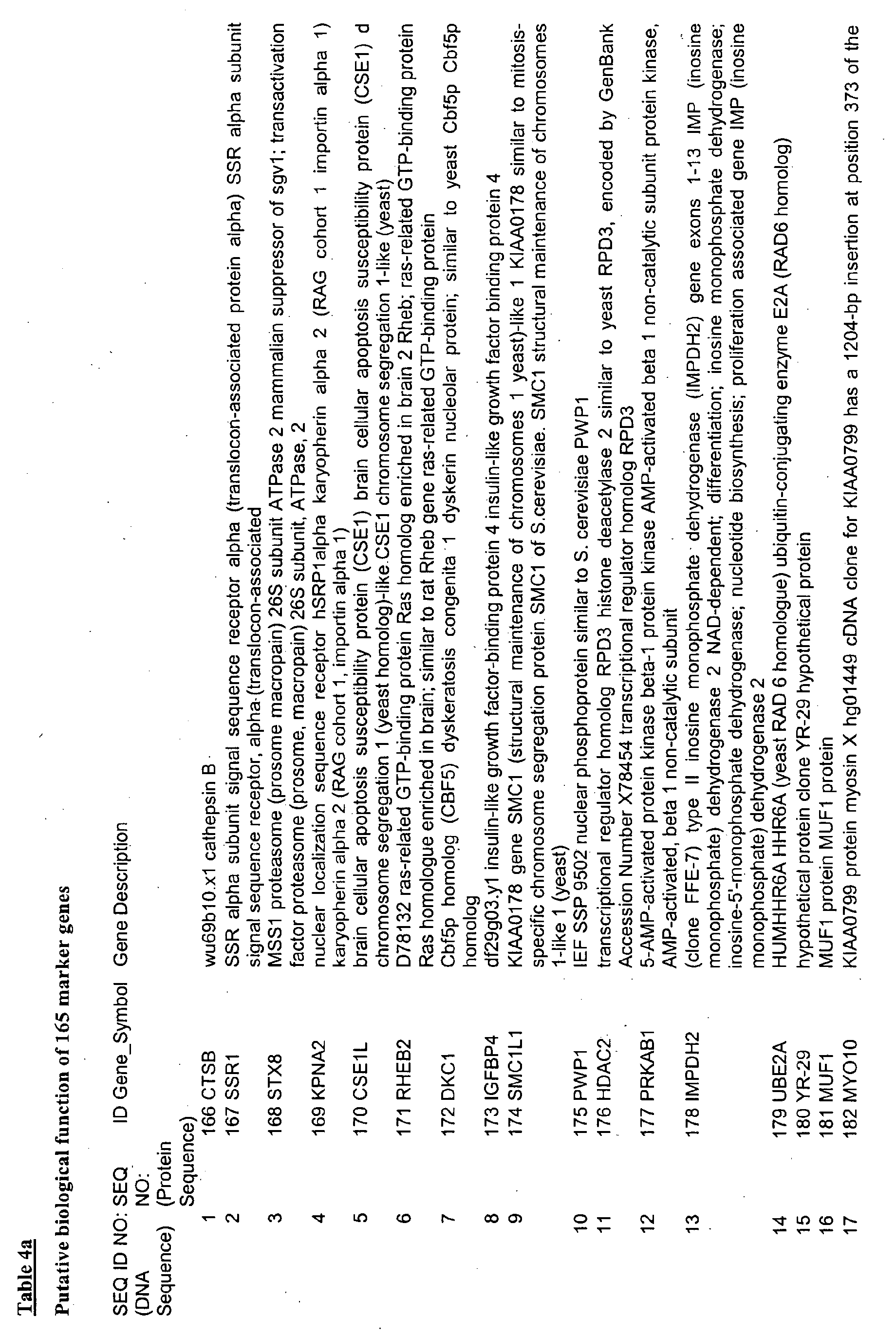

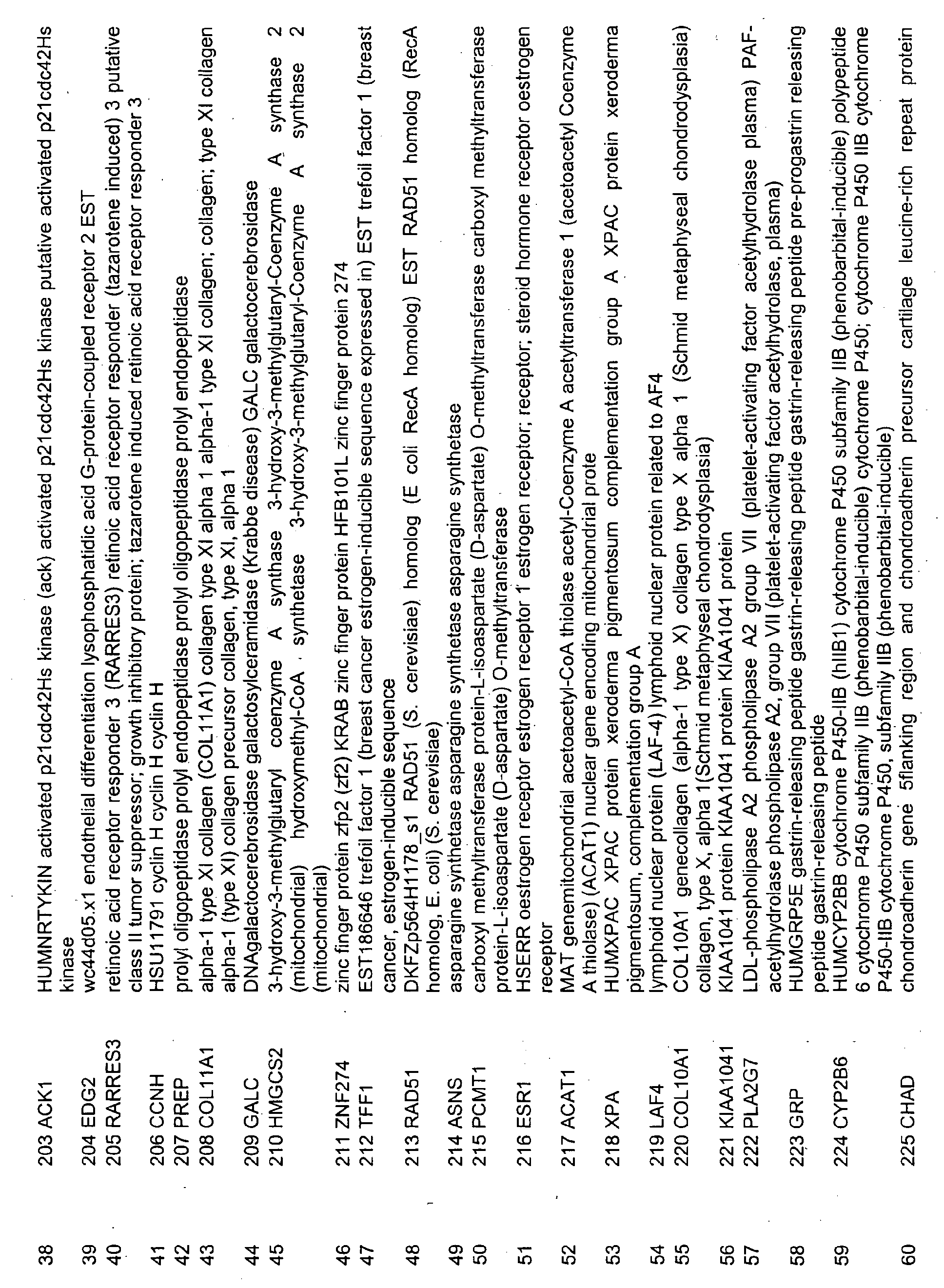

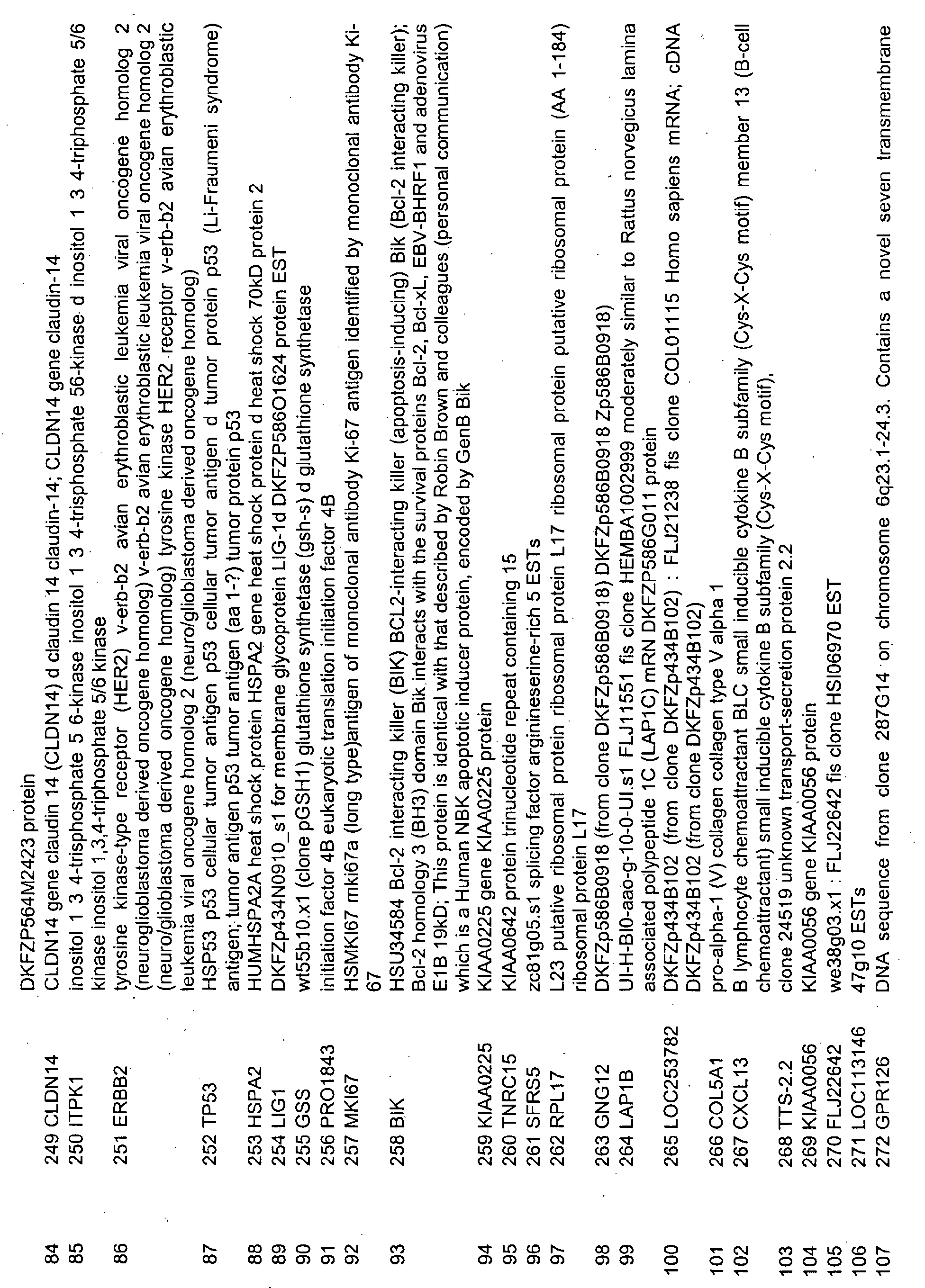

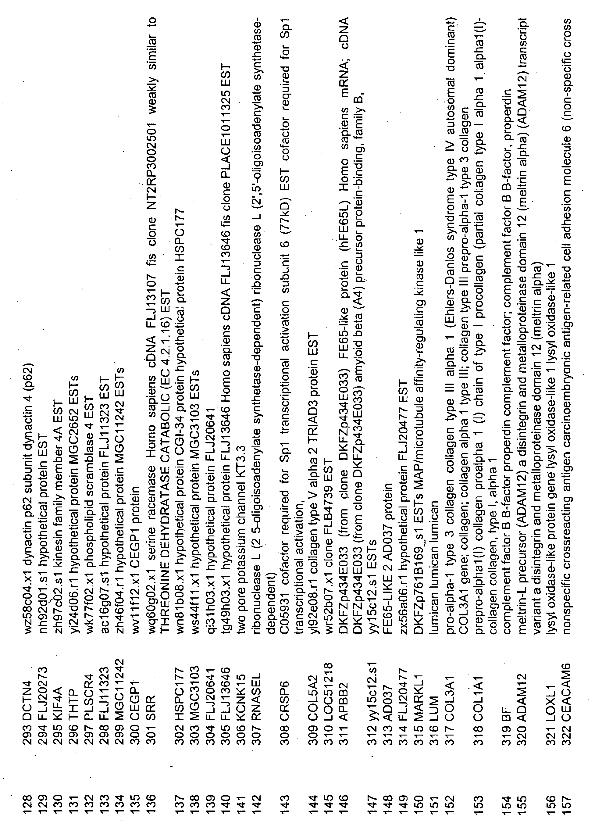

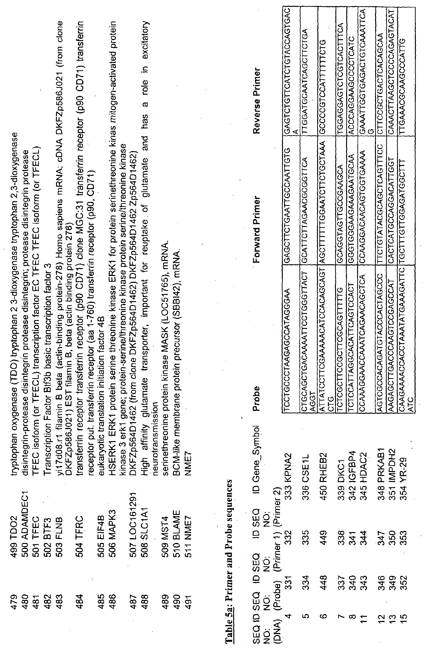
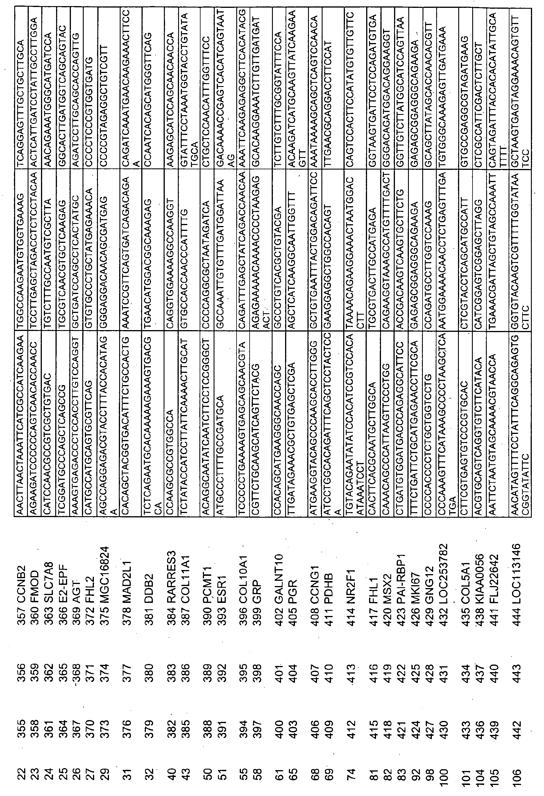
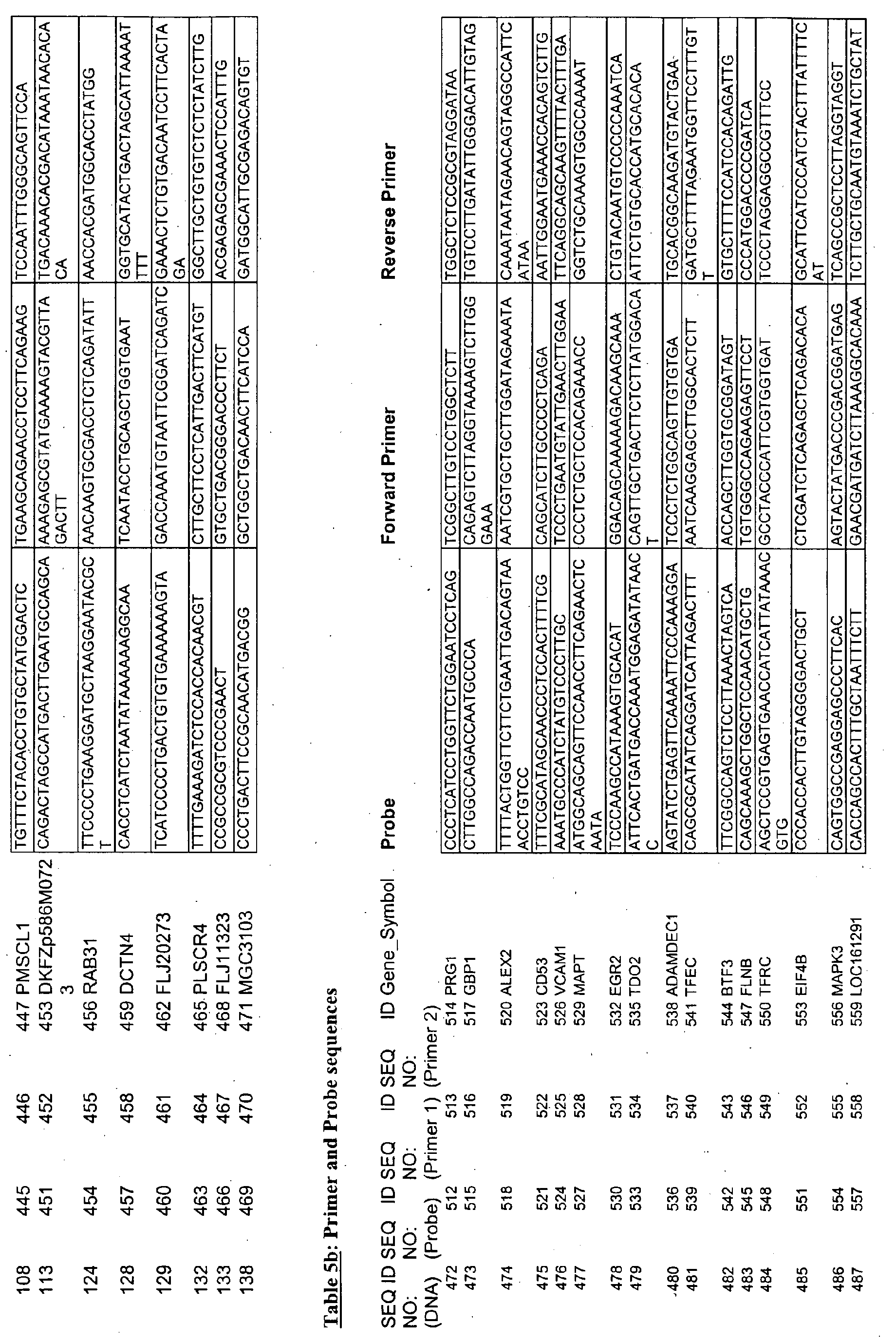

|
Statistical relevance of 20 genes differentially in non-responders (NC) as compared to responding tumors. (CR - complete responder to therapy)
|
| SEQ ID NO: (DNA Sequence) |
SEQ ID NO: Gene_Symbol, (Protein Sequence) |
T-Test p-value |
Welch-Test p-value |
Wilcoxon p-value |
| 472 |
492 PRG1 |
0.0002116 |
0.0002631 |
0.0003108 |
| 473 |
493 GBP1 |
0.0020070 |
0.0023060 |
0.0029530 |
| 474 |
494 ALEX2 |
0.0003502 |
0.0012570 |
0.0001554 |
| 475 |
495 CD53 |
0.0019770 |
0.0039540 |
0.0018650 |
| 476 |
496 VCAM1 |
0.0010630 |
0.0010690 |
0.0018650 |
| 477 |
497 MAPT |
0.0005838 |
0.0007540 |
0.0001554 |
| 478 |
498 EGR2 |
0.0008870 |
0.0009158 |
0.0006216 |
| 479 |
499 TDO2 |
0.0084350 |
0.0105000 |
0.0018650 |
| 480 |
500 ADAMDEC1 |
0.0018700 |
0.0021870 |
0.0029530 |
| 481 |
501 TFEC |
0.0085550 |
0.0155500 |
0.0010880 |
| 482 |
502 BTF3 |
0.0001140 |
0.0001471 |
0.0003108 |
| 483 |
503 FLNB |
0.0006050 |
0.0007720 |
0.0018650 |
| 484 |
504 TFRC |
0.0005408 |
0.0010110 |
0.0010880 |
| 485 |
505 EIF4B |
0.0013130 |
0.0013330 |
0.0006216 |
| 486 |
506 MAPK3 |
0.0001388 |
0.0003527 |
0.0006216 |
| 487 |
507 LOC161291 |
0.0015790 |
0.0031610 |
0.0006216 |
| 488 |
508 SLC1A1 |
0.0000179 |
0.0000389 |
0.0001554 |
| 489 |
509 MST4 |
0.0000888 |
0.0000904 |
0.0001554 |
| 490 |
510 BLAME |
0.0048620 |
0.0081110 |
0.0029530 |
| 491 |
511 NME7 |
0.0020950 |
0.0021980 |
0.0006216 |