EP1521771B1 - STREPTOCOCCUS PNEUMONIAE PBP2x MINI-PROTEIN AND USES THEREOF. - Google Patents
STREPTOCOCCUS PNEUMONIAE PBP2x MINI-PROTEIN AND USES THEREOF. Download PDFInfo
- Publication number
- EP1521771B1 EP1521771B1 EP03764076A EP03764076A EP1521771B1 EP 1521771 B1 EP1521771 B1 EP 1521771B1 EP 03764076 A EP03764076 A EP 03764076A EP 03764076 A EP03764076 A EP 03764076A EP 1521771 B1 EP1521771 B1 EP 1521771B1
- Authority
- EP
- European Patent Office
- Prior art keywords
- pbp2x
- mini
- protein
- amino acids
- positions
- Prior art date
- Legal status (The legal status is an assumption and is not a legal conclusion. Google has not performed a legal analysis and makes no representation as to the accuracy of the status listed.)
- Expired - Lifetime
Links
- 101001072513 Streptococcus pneumoniae serotype 4 (strain ATCC BAA-334 / TIGR4) Penicillin-binding protein 2x Proteins 0.000 title claims abstract description 68
- 241000193998 Streptococcus pneumoniae Species 0.000 title claims abstract description 13
- 229940031000 streptococcus pneumoniae Drugs 0.000 title claims abstract description 13
- 150000003952 β-lactams Chemical class 0.000 claims abstract description 42
- 150000001413 amino acids Chemical class 0.000 claims abstract description 41
- 108090000623 proteins and genes Proteins 0.000 claims abstract description 40
- 102000004169 proteins and genes Human genes 0.000 claims abstract description 38
- 239000012634 fragment Substances 0.000 claims abstract description 22
- 239000003242 anti bacterial agent Substances 0.000 claims abstract description 20
- 229940088710 antibiotic agent Drugs 0.000 claims abstract description 20
- 102000007079 Peptide Fragments Human genes 0.000 claims abstract description 9
- 108010033276 Peptide Fragments Proteins 0.000 claims abstract description 9
- 235000018102 proteins Nutrition 0.000 claims description 39
- 235000001014 amino acid Nutrition 0.000 claims description 32
- 238000000034 method Methods 0.000 claims description 18
- 239000013078 crystal Substances 0.000 claims description 17
- 239000013598 vector Substances 0.000 claims description 15
- 230000000694 effects Effects 0.000 claims description 14
- 238000012216 screening Methods 0.000 claims description 13
- 238000012360 testing method Methods 0.000 claims description 13
- 239000003446 ligand Substances 0.000 claims description 10
- 230000005764 inhibitory process Effects 0.000 claims description 9
- 150000007523 nucleic acids Chemical class 0.000 claims description 9
- 230000002401 inhibitory effect Effects 0.000 claims description 8
- 210000004027 cell Anatomy 0.000 claims description 7
- 108020004707 nucleic acids Proteins 0.000 claims description 7
- 102000039446 nucleic acids Human genes 0.000 claims description 7
- 238000006467 substitution reaction Methods 0.000 claims description 7
- DHMQDGOQFOQNFH-UHFFFAOYSA-N Glycine Chemical compound NCC(O)=O DHMQDGOQFOQNFH-UHFFFAOYSA-N 0.000 claims description 6
- MTCFGRXMJLQNBG-UHFFFAOYSA-N Serine Natural products OCC(N)C(O)=O MTCFGRXMJLQNBG-UHFFFAOYSA-N 0.000 claims description 5
- 239000013543 active substance Substances 0.000 claims description 4
- 239000000126 substance Substances 0.000 claims description 3
- 239000004471 Glycine Substances 0.000 claims description 2
- QNAYBMKLOCPYGJ-REOHCLBHSA-N L-alanine Chemical compound C[C@H](N)C(O)=O QNAYBMKLOCPYGJ-REOHCLBHSA-N 0.000 claims description 2
- RJFAYQIBOAGBLC-BYPYZUCNSA-N Selenium-L-methionine Chemical group C[Se]CC[C@H](N)C(O)=O RJFAYQIBOAGBLC-BYPYZUCNSA-N 0.000 claims description 2
- AYFVYJQAPQTCCC-UHFFFAOYSA-N Threonine Natural products CC(O)C(N)C(O)=O AYFVYJQAPQTCCC-UHFFFAOYSA-N 0.000 claims description 2
- 239000004473 Threonine Substances 0.000 claims description 2
- 235000004279 alanine Nutrition 0.000 claims description 2
- 230000000692 anti-sense effect Effects 0.000 claims description 2
- 125000001360 methionine group Chemical group N[C@@H](CCSC)C(=O)* 0.000 claims description 2
- 210000001236 prokaryotic cell Anatomy 0.000 claims description 2
- 102000007056 Recombinant Fusion Proteins Human genes 0.000 abstract description 2
- 108010008281 Recombinant Fusion Proteins Proteins 0.000 abstract description 2
- 108700020474 Penicillin-Binding Proteins Proteins 0.000 description 17
- 238000012217 deletion Methods 0.000 description 14
- 230000037430 deletion Effects 0.000 description 14
- 108091034117 Oligonucleotide Proteins 0.000 description 13
- 108020004414 DNA Proteins 0.000 description 10
- 239000000872 buffer Substances 0.000 description 9
- 230000002255 enzymatic effect Effects 0.000 description 9
- 150000007970 thio esters Chemical class 0.000 description 9
- 108090000279 Peptidyltransferases Proteins 0.000 description 8
- FAPWRFPIFSIZLT-UHFFFAOYSA-M Sodium chloride Chemical compound [Na+].[Cl-] FAPWRFPIFSIZLT-UHFFFAOYSA-M 0.000 description 8
- 239000003112 inhibitor Substances 0.000 description 8
- 238000004458 analytical method Methods 0.000 description 7
- 230000001580 bacterial effect Effects 0.000 description 7
- 239000002773 nucleotide Substances 0.000 description 7
- 125000003729 nucleotide group Chemical group 0.000 description 7
- 230000003115 biocidal effect Effects 0.000 description 6
- 108090000765 processed proteins & peptides Proteins 0.000 description 6
- 229930182555 Penicillin Natural products 0.000 description 5
- 230000004075 alteration Effects 0.000 description 5
- 238000007796 conventional method Methods 0.000 description 5
- 238000002447 crystallographic data Methods 0.000 description 5
- 230000007423 decrease Effects 0.000 description 5
- 230000003301 hydrolyzing effect Effects 0.000 description 5
- 238000003780 insertion Methods 0.000 description 5
- 230000037431 insertion Effects 0.000 description 5
- 239000000463 material Substances 0.000 description 5
- 238000000159 protein binding assay Methods 0.000 description 5
- JGSARLDLIJGVTE-MBNYWOFBSA-N Penicillin G Chemical compound N([C@H]1[C@H]2SC([C@@H](N2C1=O)C(O)=O)(C)C)C(=O)CC1=CC=CC=C1 JGSARLDLIJGVTE-MBNYWOFBSA-N 0.000 description 4
- 238000003556 assay Methods 0.000 description 4
- 238000002425 crystallisation Methods 0.000 description 4
- 230000008025 crystallization Effects 0.000 description 4
- 230000001086 cytosolic effect Effects 0.000 description 4
- 238000002330 electrospray ionisation mass spectrometry Methods 0.000 description 4
- 238000010828 elution Methods 0.000 description 4
- 239000013604 expression vector Substances 0.000 description 4
- 230000007062 hydrolysis Effects 0.000 description 4
- 238000006460 hydrolysis reaction Methods 0.000 description 4
- 238000004519 manufacturing process Methods 0.000 description 4
- 229940049954 penicillin Drugs 0.000 description 4
- 239000011780 sodium chloride Substances 0.000 description 4
- 239000000758 substrate Substances 0.000 description 4
- KCXVZYZYPLLWCC-UHFFFAOYSA-N EDTA Chemical compound OC(=O)CN(CC(O)=O)CCN(CC(O)=O)CC(O)=O KCXVZYZYPLLWCC-UHFFFAOYSA-N 0.000 description 3
- LFQSCWFLJHTTHZ-UHFFFAOYSA-N Ethanol Chemical compound CCO LFQSCWFLJHTTHZ-UHFFFAOYSA-N 0.000 description 3
- LYCAIKOWRPUZTN-UHFFFAOYSA-N Ethylene glycol Chemical compound OCCO LYCAIKOWRPUZTN-UHFFFAOYSA-N 0.000 description 3
- 101710139363 Mini-ribonuclease 3 Proteins 0.000 description 3
- MSFSPUZXLOGKHJ-UHFFFAOYSA-N Muraminsaeure Natural products OC(=O)C(C)OC1C(N)C(O)OC(CO)C1O MSFSPUZXLOGKHJ-UHFFFAOYSA-N 0.000 description 3
- 108010013639 Peptidoglycan Proteins 0.000 description 3
- JLCPHMBAVCMARE-UHFFFAOYSA-N [3-[[3-[[3-[[3-[[3-[[3-[[3-[[3-[[3-[[3-[[3-[[5-(2-amino-6-oxo-1H-purin-9-yl)-3-[[3-[[3-[[3-[[3-[[3-[[5-(2-amino-6-oxo-1H-purin-9-yl)-3-[[5-(2-amino-6-oxo-1H-purin-9-yl)-3-hydroxyoxolan-2-yl]methoxy-hydroxyphosphoryl]oxyoxolan-2-yl]methoxy-hydroxyphosphoryl]oxy-5-(5-methyl-2,4-dioxopyrimidin-1-yl)oxolan-2-yl]methoxy-hydroxyphosphoryl]oxy-5-(6-aminopurin-9-yl)oxolan-2-yl]methoxy-hydroxyphosphoryl]oxy-5-(6-aminopurin-9-yl)oxolan-2-yl]methoxy-hydroxyphosphoryl]oxy-5-(6-aminopurin-9-yl)oxolan-2-yl]methoxy-hydroxyphosphoryl]oxy-5-(6-aminopurin-9-yl)oxolan-2-yl]methoxy-hydroxyphosphoryl]oxyoxolan-2-yl]methoxy-hydroxyphosphoryl]oxy-5-(5-methyl-2,4-dioxopyrimidin-1-yl)oxolan-2-yl]methoxy-hydroxyphosphoryl]oxy-5-(4-amino-2-oxopyrimidin-1-yl)oxolan-2-yl]methoxy-hydroxyphosphoryl]oxy-5-(5-methyl-2,4-dioxopyrimidin-1-yl)oxolan-2-yl]methoxy-hydroxyphosphoryl]oxy-5-(5-methyl-2,4-dioxopyrimidin-1-yl)oxolan-2-yl]methoxy-hydroxyphosphoryl]oxy-5-(6-aminopurin-9-yl)oxolan-2-yl]methoxy-hydroxyphosphoryl]oxy-5-(6-aminopurin-9-yl)oxolan-2-yl]methoxy-hydroxyphosphoryl]oxy-5-(4-amino-2-oxopyrimidin-1-yl)oxolan-2-yl]methoxy-hydroxyphosphoryl]oxy-5-(4-amino-2-oxopyrimidin-1-yl)oxolan-2-yl]methoxy-hydroxyphosphoryl]oxy-5-(4-amino-2-oxopyrimidin-1-yl)oxolan-2-yl]methoxy-hydroxyphosphoryl]oxy-5-(6-aminopurin-9-yl)oxolan-2-yl]methoxy-hydroxyphosphoryl]oxy-5-(4-amino-2-oxopyrimidin-1-yl)oxolan-2-yl]methyl [5-(6-aminopurin-9-yl)-2-(hydroxymethyl)oxolan-3-yl] hydrogen phosphate Polymers Cc1cn(C2CC(OP(O)(=O)OCC3OC(CC3OP(O)(=O)OCC3OC(CC3O)n3cnc4c3nc(N)[nH]c4=O)n3cnc4c3nc(N)[nH]c4=O)C(COP(O)(=O)OC3CC(OC3COP(O)(=O)OC3CC(OC3COP(O)(=O)OC3CC(OC3COP(O)(=O)OC3CC(OC3COP(O)(=O)OC3CC(OC3COP(O)(=O)OC3CC(OC3COP(O)(=O)OC3CC(OC3COP(O)(=O)OC3CC(OC3COP(O)(=O)OC3CC(OC3COP(O)(=O)OC3CC(OC3COP(O)(=O)OC3CC(OC3COP(O)(=O)OC3CC(OC3COP(O)(=O)OC3CC(OC3COP(O)(=O)OC3CC(OC3COP(O)(=O)OC3CC(OC3COP(O)(=O)OC3CC(OC3COP(O)(=O)OC3CC(OC3CO)n3cnc4c(N)ncnc34)n3ccc(N)nc3=O)n3cnc4c(N)ncnc34)n3ccc(N)nc3=O)n3ccc(N)nc3=O)n3ccc(N)nc3=O)n3cnc4c(N)ncnc34)n3cnc4c(N)ncnc34)n3cc(C)c(=O)[nH]c3=O)n3cc(C)c(=O)[nH]c3=O)n3ccc(N)nc3=O)n3cc(C)c(=O)[nH]c3=O)n3cnc4c3nc(N)[nH]c4=O)n3cnc4c(N)ncnc34)n3cnc4c(N)ncnc34)n3cnc4c(N)ncnc34)n3cnc4c(N)ncnc34)O2)c(=O)[nH]c1=O JLCPHMBAVCMARE-UHFFFAOYSA-N 0.000 description 3
- 230000010933 acylation Effects 0.000 description 3
- 238000005917 acylation reaction Methods 0.000 description 3
- 230000008033 biological extinction Effects 0.000 description 3
- GPRBEKHLDVQUJE-VINNURBNSA-N cefotaxime Chemical compound N([C@@H]1C(N2C(=C(COC(C)=O)CS[C@@H]21)C(O)=O)=O)C(=O)/C(=N/OC)C1=CSC(N)=N1 GPRBEKHLDVQUJE-VINNURBNSA-N 0.000 description 3
- 229960004261 cefotaxime Drugs 0.000 description 3
- 239000013612 plasmid Substances 0.000 description 3
- QKNYBSVHEMOAJP-UHFFFAOYSA-N 2-amino-2-(hydroxymethyl)propane-1,3-diol;hydron;chloride Chemical compound Cl.OCC(N)(CO)CO QKNYBSVHEMOAJP-UHFFFAOYSA-N 0.000 description 2
- IJGRMHOSHXDMSA-UHFFFAOYSA-N Atomic nitrogen Chemical compound N#N IJGRMHOSHXDMSA-UHFFFAOYSA-N 0.000 description 2
- 229930186147 Cephalosporin Natural products 0.000 description 2
- SOEGEPHNZOISMT-BYPYZUCNSA-N Gly-Ser-Gly Chemical compound NCC(=O)N[C@@H](CO)C(=O)NCC(O)=O SOEGEPHNZOISMT-BYPYZUCNSA-N 0.000 description 2
- DGAQECJNVWCQMB-PUAWFVPOSA-M Ilexoside XXIX Chemical compound C[C@@H]1CC[C@@]2(CC[C@@]3(C(=CC[C@H]4[C@]3(CC[C@@H]5[C@@]4(CC[C@@H](C5(C)C)OS(=O)(=O)[O-])C)C)[C@@H]2[C@]1(C)O)C)C(=O)O[C@H]6[C@@H]([C@H]([C@@H]([C@H](O6)CO)O)O)O.[Na+] DGAQECJNVWCQMB-PUAWFVPOSA-M 0.000 description 2
- 108091028043 Nucleic acid sequence Proteins 0.000 description 2
- 238000012408 PCR amplification Methods 0.000 description 2
- 229920002565 Polyethylene Glycol 400 Polymers 0.000 description 2
- 239000012614 Q-Sepharose Substances 0.000 description 2
- 239000012505 Superdex™ Substances 0.000 description 2
- BFNBIHQBYMNNAN-UHFFFAOYSA-N ammonium sulfate Chemical compound N.N.OS(O)(=O)=O BFNBIHQBYMNNAN-UHFFFAOYSA-N 0.000 description 2
- 229910052921 ammonium sulfate Inorganic materials 0.000 description 2
- 235000011130 ammonium sulphate Nutrition 0.000 description 2
- 239000001166 ammonium sulphate Substances 0.000 description 2
- 230000015572 biosynthetic process Effects 0.000 description 2
- 210000004899 c-terminal region Anatomy 0.000 description 2
- 229940124587 cephalosporin Drugs 0.000 description 2
- 150000001780 cephalosporins Chemical class 0.000 description 2
- 230000001186 cumulative effect Effects 0.000 description 2
- 238000002050 diffraction method Methods 0.000 description 2
- 238000009510 drug design Methods 0.000 description 2
- LIWAQLJGPBVORC-UHFFFAOYSA-N ethylmethylamine Chemical compound CCNC LIWAQLJGPBVORC-UHFFFAOYSA-N 0.000 description 2
- 230000005284 excitation Effects 0.000 description 2
- 238000005227 gel permeation chromatography Methods 0.000 description 2
- XKUKSGPZAADMRA-UHFFFAOYSA-N glycyl-glycyl-glycine Chemical compound NCC(=O)NCC(=O)NCC(O)=O XKUKSGPZAADMRA-UHFFFAOYSA-N 0.000 description 2
- 108010067216 glycyl-glycyl-glycine Proteins 0.000 description 2
- YMAWOPBAYDPSLA-UHFFFAOYSA-N glycylglycine Chemical compound [NH3+]CC(=O)NCC([O-])=O YMAWOPBAYDPSLA-UHFFFAOYSA-N 0.000 description 2
- 238000011534 incubation Methods 0.000 description 2
- 238000004949 mass spectrometry Methods 0.000 description 2
- 239000011159 matrix material Substances 0.000 description 2
- 238000005259 measurement Methods 0.000 description 2
- 238000002703 mutagenesis Methods 0.000 description 2
- 231100000350 mutagenesis Toxicity 0.000 description 2
- 230000035772 mutation Effects 0.000 description 2
- 239000008363 phosphate buffer Substances 0.000 description 2
- 238000002264 polyacrylamide gel electrophoresis Methods 0.000 description 2
- 238000002360 preparation method Methods 0.000 description 2
- 102000004196 processed proteins & peptides Human genes 0.000 description 2
- 239000000047 product Substances 0.000 description 2
- 238000000746 purification Methods 0.000 description 2
- 238000012163 sequencing technique Methods 0.000 description 2
- 238000002741 site-directed mutagenesis Methods 0.000 description 2
- 229910052708 sodium Inorganic materials 0.000 description 2
- 239000011734 sodium Substances 0.000 description 2
- RDZTWEVXRGYCFV-UHFFFAOYSA-M sodium 2-[4-(2-hydroxyethyl)piperazin-1-yl]ethanesulfonate Chemical compound [Na+].OCCN1CCN(CCS([O-])(=O)=O)CC1 RDZTWEVXRGYCFV-UHFFFAOYSA-M 0.000 description 2
- 238000007619 statistical method Methods 0.000 description 2
- 230000004083 survival effect Effects 0.000 description 2
- 238000003786 synthesis reaction Methods 0.000 description 2
- LHNIIDJCEODSHA-OQRUQETBSA-N (6r,7r)-3-[(e)-2-(2,4-dinitrophenyl)ethenyl]-8-oxo-7-[(2-thiophen-2-ylacetyl)amino]-5-thia-1-azabicyclo[4.2.0]oct-2-ene-2-carboxylic acid Chemical compound N([C@H]1[C@H]2SCC(=C(N2C1=O)C(=O)O)\C=C\C=1C(=CC(=CC=1)[N+]([O-])=O)[N+]([O-])=O)C(=O)CC1=CC=CS1 LHNIIDJCEODSHA-OQRUQETBSA-N 0.000 description 1
- JKMHFZQWWAIEOD-UHFFFAOYSA-N 2-[4-(2-hydroxyethyl)piperazin-1-yl]ethanesulfonic acid Chemical compound OCC[NH+]1CCN(CCS([O-])(=O)=O)CC1 JKMHFZQWWAIEOD-UHFFFAOYSA-N 0.000 description 1
- ORILYTVJVMAKLC-UHFFFAOYSA-N Adamantane Natural products C1C(C2)CC3CC1CC2C3 ORILYTVJVMAKLC-UHFFFAOYSA-N 0.000 description 1
- 241000894006 Bacteria Species 0.000 description 1
- 108020004256 Beta-lactamase Proteins 0.000 description 1
- 108010006303 Carboxypeptidases Proteins 0.000 description 1
- 102000005367 Carboxypeptidases Human genes 0.000 description 1
- 102100025698 Cytosolic carboxypeptidase 4 Human genes 0.000 description 1
- DEFJQIDDEAULHB-QWWZWVQMSA-N D-alanyl-D-alanine Chemical compound C[C@@H]([NH3+])C(=O)N[C@H](C)C([O-])=O DEFJQIDDEAULHB-QWWZWVQMSA-N 0.000 description 1
- 108090000790 Enzymes Proteins 0.000 description 1
- 102000004190 Enzymes Human genes 0.000 description 1
- 241000588724 Escherichia coli Species 0.000 description 1
- 241001198387 Escherichia coli BL21(DE3) Species 0.000 description 1
- 102000051366 Glycosyltransferases Human genes 0.000 description 1
- 108700023372 Glycosyltransferases Proteins 0.000 description 1
- 101000932590 Homo sapiens Cytosolic carboxypeptidase 4 Proteins 0.000 description 1
- CKLJMWTZIZZHCS-REOHCLBHSA-N L-aspartic acid Chemical compound OC(=O)[C@@H](N)CC(O)=O CKLJMWTZIZZHCS-REOHCLBHSA-N 0.000 description 1
- 108010052285 Membrane Proteins Proteins 0.000 description 1
- 108010047290 Multifunctional Enzymes Proteins 0.000 description 1
- 102000006833 Multifunctional Enzymes Human genes 0.000 description 1
- 101001033003 Mus musculus Granzyme F Proteins 0.000 description 1
- DEFJQIDDEAULHB-UHFFFAOYSA-N N-D-alanyl-D-alanine Natural products CC(N)C(=O)NC(C)C(O)=O DEFJQIDDEAULHB-UHFFFAOYSA-N 0.000 description 1
- 238000005481 NMR spectroscopy Methods 0.000 description 1
- 241001028048 Nicola Species 0.000 description 1
- 241000283973 Oryctolagus cuniculus Species 0.000 description 1
- 108020004511 Recombinant DNA Proteins 0.000 description 1
- 241000194017 Streptococcus Species 0.000 description 1
- 241001134658 Streptococcus mitis Species 0.000 description 1
- 241000194025 Streptococcus oralis Species 0.000 description 1
- 238000002441 X-ray diffraction Methods 0.000 description 1
- 230000009471 action Effects 0.000 description 1
- 108010056243 alanylalanine Proteins 0.000 description 1
- 125000000539 amino acid group Chemical group 0.000 description 1
- 230000003321 amplification Effects 0.000 description 1
- 230000002924 anti-infective effect Effects 0.000 description 1
- 238000013459 approach Methods 0.000 description 1
- 235000003704 aspartic acid Nutrition 0.000 description 1
- 230000008901 benefit Effects 0.000 description 1
- OQFSQFPPLPISGP-UHFFFAOYSA-N beta-carboxyaspartic acid Natural products OC(=O)C(N)C(C(O)=O)C(O)=O OQFSQFPPLPISGP-UHFFFAOYSA-N 0.000 description 1
- 102000006635 beta-lactamase Human genes 0.000 description 1
- JFPVXVDWJQMJEE-IZRZKJBUSA-N cefuroxime Chemical compound N([C@@H]1C(N2C(=C(COC(N)=O)CS[C@@H]21)C(O)=O)=O)C(=O)\C(=N/OC)C1=CC=CO1 JFPVXVDWJQMJEE-IZRZKJBUSA-N 0.000 description 1
- 229960001668 cefuroxime Drugs 0.000 description 1
- 238000005119 centrifugation Methods 0.000 description 1
- 239000013043 chemical agent Substances 0.000 description 1
- 239000003795 chemical substances by application Substances 0.000 description 1
- 238000004587 chromatography analysis Methods 0.000 description 1
- 238000010367 cloning Methods 0.000 description 1
- 150000001875 compounds Chemical class 0.000 description 1
- 238000010276 construction Methods 0.000 description 1
- 239000011548 crystallization buffer Substances 0.000 description 1
- 238000004925 denaturation Methods 0.000 description 1
- 230000036425 denaturation Effects 0.000 description 1
- 238000001514 detection method Methods 0.000 description 1
- 229940042399 direct acting antivirals protease inhibitors Drugs 0.000 description 1
- 238000002825 functional assay Methods 0.000 description 1
- 238000010353 genetic engineering Methods 0.000 description 1
- 238000013537 high throughput screening Methods 0.000 description 1
- 238000002744 homologous recombination Methods 0.000 description 1
- 230000006801 homologous recombination Effects 0.000 description 1
- 238000009396 hybridization Methods 0.000 description 1
- 230000002209 hydrophobic effect Effects 0.000 description 1
- 230000003100 immobilizing effect Effects 0.000 description 1
- 230000003993 interaction Effects 0.000 description 1
- 238000005342 ion exchange Methods 0.000 description 1
- 239000007788 liquid Substances 0.000 description 1
- 239000006166 lysate Substances 0.000 description 1
- 230000007246 mechanism Effects 0.000 description 1
- 239000012528 membrane Substances 0.000 description 1
- 239000000203 mixture Substances 0.000 description 1
- 230000004048 modification Effects 0.000 description 1
- 238000012986 modification Methods 0.000 description 1
- 230000009456 molecular mechanism Effects 0.000 description 1
- 238000000302 molecular modelling Methods 0.000 description 1
- 229910052757 nitrogen Inorganic materials 0.000 description 1
- 238000003199 nucleic acid amplification method Methods 0.000 description 1
- 238000006384 oligomerization reaction Methods 0.000 description 1
- 230000003287 optical effect Effects 0.000 description 1
- 244000052769 pathogen Species 0.000 description 1
- 230000037361 pathway Effects 0.000 description 1
- 101150001513 pbpX gene Proteins 0.000 description 1
- 239000008188 pellet Substances 0.000 description 1
- 150000002960 penicillins Chemical class 0.000 description 1
- 239000000137 peptide hydrolase inhibitor Substances 0.000 description 1
- 230000035699 permeability Effects 0.000 description 1
- 230000001376 precipitating effect Effects 0.000 description 1
- 230000008569 process Effects 0.000 description 1
- ZLIBICFPKPWGIZ-UHFFFAOYSA-N pyrimethanil Chemical compound CC1=CC(C)=NC(NC=2C=CC=CC=2)=N1 ZLIBICFPKPWGIZ-UHFFFAOYSA-N 0.000 description 1
- 230000001105 regulatory effect Effects 0.000 description 1
- 238000004153 renaturation Methods 0.000 description 1
- 230000000241 respiratory effect Effects 0.000 description 1
- 238000003757 reverse transcription PCR Methods 0.000 description 1
- 239000000523 sample Substances 0.000 description 1
- 125000003607 serino group Chemical group [H]N([H])[C@]([H])(C(=O)[*])C(O[H])([H])[H] 0.000 description 1
- 238000002415 sodium dodecyl sulfate polyacrylamide gel electrophoresis Methods 0.000 description 1
- 238000000527 sonication Methods 0.000 description 1
- 241000894007 species Species 0.000 description 1
- 239000006228 supernatant Substances 0.000 description 1
- 230000005469 synchrotron radiation Effects 0.000 description 1
- 230000009897 systematic effect Effects 0.000 description 1
- 238000002560 therapeutic procedure Methods 0.000 description 1
- 238000013518 transcription Methods 0.000 description 1
- 230000035897 transcription Effects 0.000 description 1
- 238000013519 translation Methods 0.000 description 1
- 238000001262 western blot Methods 0.000 description 1
Images
Classifications
-
- C—CHEMISTRY; METALLURGY
- C07—ORGANIC CHEMISTRY
- C07K—PEPTIDES
- C07K14/00—Peptides having more than 20 amino acids; Gastrins; Somatostatins; Melanotropins; Derivatives thereof
- C07K14/195—Peptides having more than 20 amino acids; Gastrins; Somatostatins; Melanotropins; Derivatives thereof from bacteria
- C07K14/315—Peptides having more than 20 amino acids; Gastrins; Somatostatins; Melanotropins; Derivatives thereof from bacteria from Streptococcus (G), e.g. Enterococci
- C07K14/3156—Peptides having more than 20 amino acids; Gastrins; Somatostatins; Melanotropins; Derivatives thereof from bacteria from Streptococcus (G), e.g. Enterococci from Streptococcus pneumoniae (Pneumococcus)
-
- C—CHEMISTRY; METALLURGY
- C12—BIOCHEMISTRY; BEER; SPIRITS; WINE; VINEGAR; MICROBIOLOGY; ENZYMOLOGY; MUTATION OR GENETIC ENGINEERING
- C12N—MICROORGANISMS OR ENZYMES; COMPOSITIONS THEREOF; PROPAGATING, PRESERVING, OR MAINTAINING MICROORGANISMS; MUTATION OR GENETIC ENGINEERING; CULTURE MEDIA
- C12N9/00—Enzymes; Proenzymes; Compositions thereof; Processes for preparing, activating, inhibiting, separating or purifying enzymes
- C12N9/10—Transferases (2.)
- C12N9/1025—Acyltransferases (2.3)
- C12N9/104—Aminoacyltransferases (2.3.2)
-
- G—PHYSICS
- G01—MEASURING; TESTING
- G01N—INVESTIGATING OR ANALYSING MATERIALS BY DETERMINING THEIR CHEMICAL OR PHYSICAL PROPERTIES
- G01N2333/00—Assays involving biological materials from specific organisms or of a specific nature
- G01N2333/195—Assays involving biological materials from specific organisms or of a specific nature from bacteria
- G01N2333/315—Assays involving biological materials from specific organisms or of a specific nature from bacteria from Streptococcus (G), e.g. Enterococci
-
- G—PHYSICS
- G01—MEASURING; TESTING
- G01N—INVESTIGATING OR ANALYSING MATERIALS BY DETERMINING THEIR CHEMICAL OR PHYSICAL PROPERTIES
- G01N2415/00—Assays, e.g. immunoassays or enzyme assays, involving penicillins or cephalosporins
-
- G—PHYSICS
- G01—MEASURING; TESTING
- G01N—INVESTIGATING OR ANALYSING MATERIALS BY DETERMINING THEIR CHEMICAL OR PHYSICAL PROPERTIES
- G01N2500/00—Screening for compounds of potential therapeutic value
Definitions
- the present invention relates to a modified recombinant protein derived from Streptococcus pneumoniae PBP2x, referred to as mini-PBP2x, and also to uses thereof for selecting and identifying antibiotics which are active on ⁇ -lactam-resistant strains of S. pneumoniae.
- PBPs Bacticillin Binding Proteins
- membrane proteins which catalyse essential steps of the synthesis of bacterial wall peptidoglycan.
- Each bacterial species has several PBPs, the molecular weight of which varies between 30 kDa and 100 kDa.
- the high molecular weight PBPs comprise a short cytoplasmic domain, a single transmembrane domain and a large periplasmic domain, and are divided up into class A (having both transpeptidase activity and glyco-syltransferase activity (bridging)) and into class B (having an N-terminal domain of unknown function and a domain where the transpeptidase activity is located), which are readily identifiable by virtue of units of amino acids which are conserved for each class of PBP.
- the low molecular weight PBPs essentially possess carboxypeptidase activity. Biochemical studies indicate that only the high molecular weight PBPs are multifunctional enzymes essential to bacterial survival; on the other hand, the low molecular weight PBPs are not essential and regulate only the degree of bridging of the bacterial wall.
- ⁇ -lactams are based on structural analogy between the ring of ⁇ -lactams and the D-alanyl-D-alanine of the C-terminal end of the peptides of the peptidoglycan; ⁇ -lactams are pseudosubstrates for the transpeptidase, capable of acylating the serine residue of the active site of this transpeptidase, which is then deacylated very slowly, thus disturbing peptidoglycan synthesis.
- the mechanisms of ⁇ -lactam resistance in Gram + bacteria comprise essentially: production of ⁇ -lactamases which hydrolyse the ring of ⁇ -lactams before they reach their target (PBPs), alteration of membrane permeability, and modification of PBPs.
- Streptococcus pneumoniae has developed ⁇ -lactam resistance by modifying its PBPs; the combination of point mutations in the PBPs genes and of events of homologous recombination of these genes with those of related Streptococcus strains ( S. mitis, S. oralis ) results in the production of PBPs which have a low affinity for ⁇ -lactams.
- the PBP2x protein which is essential for the survival of S. pneumoniae and constitutes the primary factor of resistance to ⁇ -lactams ( Hakenbeck et al., J. Bacteriol., 1998, 180, 1831-1840 ), represents the major target for identifying novel antibiotics which are active on ⁇ -lactam-resistant strains of S. pneumoniae .
- PBP2x is a 750 amino acid protein comprising a cytoplasmic region (positions 1 to 18), a transmembrane region (positions 19 to 48), a non penicillin-binding domain (or n-PB, positions 49 - 265), a penicillin-binding domain/transpeptidase domain (positions 266 to 615) and a C-terminal domain (616 - 750).
- PBP2x * The 3-dimensional structure of PBP2x has been determined from a PBP2x comprising a deletion of the cytoplasmic and transmembrane regions, referred to as PBP2x * ; this structure has been determined with a resolution of, respectively, 3.5, 2.4 and 3.2 ⁇ , for a ⁇ -lactam-sensitive strain of S. pneumoniae (strain R6: Pares et al., Nature Struct. Biol., 1996, 3, 284 - 289 ; Gordon et al., J. Mol. Biol., 2000, 299, 477 - 485 ) and for a resistant clinical isolate ( Dessen et al., J. Biol. Chem., 2001, 276, 45106 - 45112 ; accession numbers in the database PROTEIN DATA BANK ( http://www.rcsb.org/ ), respectively 1PMD, 1QME and 1K25).
- the inventors have investigated, choosing, as a model, PBP2x of a penicillin-sensitive strain of S. pneumoniae (strain R6), the 3-dimensional structure of which is known, whether it is possible to obtain PBP2xs which are more crystallizable in a reproducible manner and which have a better diffraction power.
- the approach envisaged can be applied to any PBP2x derived from a ⁇ -lactam-sensitive or -resistant strain of S. pneumoniae .
- This mini-protein represents a tool which is perfectly suited to selecting and identifying novel antibiotics which are active on ⁇ -lactam-resistant strains of S. pneumoniae.
- a subject of the present invention is a protein derived from a Streptococcus pneumoniae PBP2x, characterized in that it consists of a concatenation of the fragments corresponding respectively to the amino acids located between positions 74 to 90, 186 to 199, 218 to 228 and 257 - 750, with reference to the sequence of the PBP2x protein of the strain R6 (SWISSPROT P14677 or GENBANK 18266817), each one of said fragments being preceded by a peptide fragment of 1 to 7 amino acids.
- the protein according to the invention is hereinafter referred to as PBP2x mini-protein or mini-PBP2x; it therefore comprises the deletion of amino acids located respectively between positions 1 to 73, 91 to 185, 200 to 217 and 229 to 256, as defined above, and the insertion, in place of said deletions, of a peptide fragment of 1 to 7 amino acids.
- said peptide fragment of 1 to 7 amino acids comprises amino acids of the sequence of said Streptococcus pneumoniae PBP2x protein corresponding to those located between positions -1 to -7, relative to the residues of positions 74, 186, 218 and 257, and/or between positions +1 to +7, relative to the residues of positions 90, 199 and 228, as defined above.
- said peptide fragment of 1 to 7 amino acids comprises amino acids in which the volume of the side chain is small, such as alanine (A), serine (S), glycine (G) or threonine (T).
- said S. pneumoniae PBP2x protein is defined by the following characteristics: • it is encoded by the gene referred to as pbpX , corresponding to that located in the genome of the S. pneumoniae strain R6, between positions 2263 and 4515 of the locus having the NCBI accession number AE008411 or the GENBANK accession number 15457852; it comprises the following amino acid units (one-letter code), specific to the class B PBPs: M1: RGXhX(D/S)RSGXXXA M2: (R/K)XX P XG M3: (G/Y)h E XXXDXXL M4: hXX(S/T)hDXXX Q M5: T(G/S)EhhXXXXSPXh(D/N) M6: hEP(A/G) S XX K M7: hXX S XNh M8: K ( T /S) G , in
- sequence identity is assessed as a function of the percentage of amino acids residues which are identical, when the two sequences are aligned, so as to obtain the maximum correspondence between them.
- a protein which has an amino acid sequence having at least X% identity with a reference sequence is defined, in the present invention, as a protein whose sequence can include up to 100-X alterations per 100 amino acids of the reference sequence, while at the same time conserving the functional properties of said reference protein.
- alteration includes deletions, substitutions or insertions, which are consecutive or dispersed, of amino acids in the reference sequence.
- the similarity of a sequence relative to a reference sequence is assessed as a function of the percentage of amino acid residues which are identical or which differ by conservative substitutions, when the two sequences are aligned so as to obtain the maximum correspondence between them.
- conservative substitution is intended to mean the substitution of an amino acid with another which has similar chemical properties (size, charge or polarity), which generally does not modify the functional properties of the protein.
- a protein which has an amino acid sequence having at least X% similarity with a reference sequence is defined, in the present invention, as a protein whose sequence can include up to 100-X nonconservative alterations per 100 amino acids of the reference sequence.
- nonconservative alterations includes the deletions, nonconservative substitutions or insertions, which are consecutive or dispersed, of amino acids in the reference sequence.
- the present invention encompasses the mini-PBP2xs derived from a PBP2x of any ⁇ -lactam-sensitive or -resistant strain of S. pneumoniae , in particular of ⁇ -lactam-resistant clinical isolates.
- ⁇ -lactam-resistant strain C 506 which is described in the article in the names of Laible et al. (Mol. Microbiol., 1989, 3, 1337-1348 ).
- mini-PBP2x protein it is derived from a ⁇ -lactam-resistant strain of Streptococcus pneumoniae.
- mini-PBP2x protein consists of the concatenation of the fragments, as defined above, of PBP2x of the ⁇ -lactam-sensitive Streptococcus pneumoniae strain R6 (SWISSPROT P14677) and it has the sequence SEQ ID No. 1.
- mini-PBP2x protein comprises a substitution of at least one methionine residue with a selenomethionine residue.
- mini-PBP2x protein it is associated with a ligand, in particular in the form of a mini-PBP2x/ligand complex.
- said ligand consists of an organic molecule, in particular a protein such as an antibody, or an inorganic molecule; said ligand is in particular a substrate, such as a pseudo-substrate, capable of binding to said mini-PBP2x protein via its active site and of inhibiting the activity of said mini-PBP2x.
- mini-PBP2x protein it is in the form of a crystal.
- said crystal consists of a mini-PBP2x in free form or associated with a ligand as defined above.
- the conditions for crystallization of the mini-PBP2x are determined by the suspended drop technique.
- crystals of mini-PBP2x are obtained under the following conditions: mini-PBP2x (12 mg/ml), 100 mM sodium Hepes, pH 7.5, 2% V/V PEG 400, 2M ammonium sulphate, at a temperature of 8°C.
- mini-PBP2xs consisting of fragments of PBP2x from ⁇ -lactam-resistant strains of S. pneumoniae are of use for screening and identifying novel antibiotics; the mini-PBP2xs consisting of fragments of PBP2x from sensitive strains of S. pneumoniae , in particular the mini-PBP2x of sequence SEQ ID No.1, are of use as a control, in screening and identifying antibiotics.
- the subject of the present invention is also an isolated nucleic acid molecule, characterized in that it has a sequence encoding a mini-PBP2x as defined above, which are sense or antisense.
- nucleic acid molecules according to the invention are obtained by conventional methods, known in themselves, according to standard protocols such as those described in Current Protocols in Molecular Biology (Frederick M. AUSUBEL, 2000, Wiley and son Inc, Library of Congress, USA ) .
- the sequences encoding PBP2x can be obtained by amplification of a nucleic acid sequence by PCR or RT-PCR, or else by screening genomic DNA libraries by hybridization with a homologous probe. For example, they are amplified by PCR using a suitable pair of primers, such as the pair of sequences SEQ ID Nos. 2-3.
- the derived nucleic acid molecules, encoding a mini-PBP2x are obtained by the conventional methods, making it possible to introduce mutations into a nucleic acid sequence, known in themselves, according to the abovementioned standard protocols.
- the sequence encoding the mini-PBP2x can be obtained by site-directed mutagenesis according to the method of Kunkel et al. (P.N.A.S., 1985, 82, 488-492 ), using the primers SEQ ID Nos. 4 to 7, and then PCR amplification using the primers SEQ ID Nos. 8 and 9, as defined above.
- a subject of the present invention is also a recombinant vector, characterized in that it comprises a nucleic acid molecule encoding a mini-PBP2x as defined above.
- said recombinant vector is an expression vector in which said nucleic acid molecule or one of its fragments is placed under the control of suitable elements for regulating transcription and translation.
- said vector may comprise sequences (tags) fused in-frame with the 5' and/or 3' end of said insert, of use for immobilizing and/or detecting and/or purifying the protein expressed from said vector.
- said expression vector is a prokaryotic vector.
- vectors are constructed and introduced into host cells by the conventional methods of recombinant DNA and genetic engineering, which are known in themselves.
- a subject of the present invention is also cells transformed with a recombinant vector as defined above.
- said cells are prokaryotic cells.
- the recombinant vectors and the transformed cells as defined above are of particular use for producing mini-PBP2x and the derived peptides, as defined above.
- a subject of the present invention is also the use of a mini-PBP2x as defined above, for screening and identifying antibiotics.
- said screening is carried out by a method comprising at least the following steps:
- the binding of said test molecule with the mini-PBP2x can be measured using conventional binding assays which make it possible to detect molecules capable of binding covalently at the active serine (S 337 , with reference to the sequence of PBP2x of the strain R6), in particular using a ligand pre-labelled with a chromophore or with a fluorophore, such as a cephalosporin coupled to a chromophore (nitrocefin), or else by measuring the decrease in intrinsic fluorescence of said mini-PBP2x, as described in the article in the names of Jamin et al., Biochem. J., 1993, 292, 735-741 .
- the inhibition of the enzymatic activity of the mini-PBP2x can be determined either by measuring the inhibition of hydrolysis of the thiol ester substrates, or by measuring the efficiency of acylation of the active serine, using conventional techniques as described in the article in the names of Zhao et al., J. Bacteriol., 1997, 179, 4901-4908 .
- said identification is carried out by a method comprising at least the following steps:
- a subject of the invention is also a kit for implementing the methods as defined above, characterized in that it includes at least one protein as defined above.
- the mini-PBP2x according to the invention which can be produced in large amounts in a soluble functional form which is readily crystallizable, for all the PBP2xs, has the following advantages:
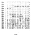
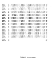
- - Figure 1 illustrates the amino acid sequence of a mini-PBP2x (SEQ ID No. 1), derived from the PBP2x of the ⁇ -lactam-sensitive S. pneumoniae strain R6 (SWISSPROT P14677); the amino acids of PBP2x which have been deleted in the mini-PBP2x are replaced with a dash and those of the peptide fragments which have been inserted are represented in italics; the units specific to the class B PBPs are underlined.
- SEQ ID No. 1 illustrates the amino acid sequence of a mini-PBP2x (SEQ ID No. 1), derived from the PBP2x of the ⁇ -lactam-sensitive S. pneumoniae strain R6 (SWISSPROT P14677); the amino acids of PBP2x which have been deleted in the mini-PBP2x are replaced with a dash and those of the peptide fragments which have been inserted are represented in italics; the units specific to the class B PBPs are underlined.
- a vector for expression of a mini-PBP2x was constructed from the plasmid pGEX-S-PBP2x*-f1 ( Mouz et al., J. Biol. Chem., 1999, 274, 19175-19180 ) containing the sequence encoding the PBP2x* of the ⁇ -lactam-sensitive S. pneumoniae strain R6 (fragment 49-750 of the PBP2x of the GENBANK amino acid sequence P14677, corresponding to the GENBANK nucleotide sequence X16367).
- deletions of the amino acids located between positions 49 to 73, 94 to 183, 200 to 217 and 230 to 256 and the insertion, in place of said deletions, respectively of the linking fragments Gly-Ser-Gly, Gly-Gly, Gly and Gly-Gly-Gly was carried out by site-directed mutagenesis according to the method of Kunkel et al. (P.N.A.S., 1985, 82, 488-492 ), using the protocols as described in Mouz et al. (J. Biol. Chem., 1999, 274, 19175-19180 ).
- the phagemide called pGEX-S-PBP2x*-f1 is converted to single strand and then used as a matrix for the mutagenesis steps (deletions and insertions).
- the mutagenesis steps were carried out in two stages, the first with the oligonucleotides Mini 1 (SEQ ID No. 4) and Mini 3 (SEQ ID No. 6) and the second with the oligonucleotides Mini 2 (SEQ ID No. 5) and Mini 4 (SEQ ID No. 7):
- the plasmid thus obtained was used as a matrix to PCR amplify the DNA fragment corresponding to the mini-PBP2x and to introduce, at its ends, the NdeI and XhoI restriction sites, for cloning into the commercial vector pET30b (NOVAGEN).
- the oligonucleotides used for the PCR amplification are as follows:
- the peptide sequence deduced from the nucleotide sequence obtained by automatic sequencing has the sequence SEQ ID No. 1 ( Figure 1 ), corresponding to that expected for a mini-PBP2x.
- the mini-PBP2x consists of the succession of fragments corresponding respectively to the amino acids located between positions 74 to 93, 184 to 199, 218 to 229 and 257-750 of PBP2x (SWISSPROT accession number P14677), each fragment being preceded, respectively, by the linking fragment GSG, GG, G and GGG.
- the mini-PBP2x is produced in E. coli , using the expression vector described in Example 1.1, and it is then purified by chromatography, successively on a Q-Sepharose, Resource Q and Superdex 200 column.
- the product obtained is analysed by polyacrylamide gel electrophoresis (SDS-PAGE) and by mass spectrometry (Electrospray ionization-mass spectrometry, ESI-MS).
- the 50 ml of bacterial lysate are centrifuged at 40 000 g, at +4°C, for 20 min, and the supernatant obtained is loaded onto a 20 ml ion exchange column (Q-Sepharose, AMERSHAM-PHARMACIA), pre-equilibrated with buffer A.
- a linear sodium gradient is produced with buffer B (20 mM Tris HCl, pH 8.0, 300 mM NaCl, 1 mM EDTA).
- the mini-PBP2x protein is eluted from. 38% buffer B.
- the eluted fractions are analysed by Western blotting using rabbit polyclonal antibodies directed against PBP2x*, and are then pooled together.
- the pool of fractions containing the mini-PBP2x is diluted 10-fold in buffer A without NaCl, and is then loaded onto a 6 ml Resource Q column (AMERSHAM-PHARMACIA) pre-equilibrated with buffer A.
- a linear sodium gradient is produced with the buffer.
- the mini-PBP2x protein is eluted from 55% of buffer B.
- the fractions corresponding to the elution peak are analysed by SDS-15% PAGE.
- the purest fractions are pooled together and the pool obtained is concentrated to a volume of 2 to 4 ml, and then loaded onto a gel chromatography column (Superdex 200, 16/60, AMERSHAM-PHARMACIA), pre-equilibrated in a 10 mM Hepes buffer, pH 7.5, 100 mM NaCl, 1 mM EDTA.
- the mini-PBP2x is eluted in a symmetrical peak at an elution volume corresponding to an apparent mass of 60 kDa.
- mini-PBP2x protein thus obtained has a degree of purity greater than 95%.
- the final yield of purified mini-PBP2x protein is 10 to 15 mg/litre of culture, depending on the preparations.
- the purification by gel chromatography shows the presence of a symmetrical elution peak which reflects the homogeneity of the protein in terms of oligomerization.
- the elution volume corresponds to a monomeric protein.
- the mini-PBP2x protein exhibits excellent solubility since it can be concentrated to 15 mg/ml without precipitating.
- the analysis by mass spectrometry shows the presence of a homogeneous protein which has a molecular mass of 59 465 Da corresponding to that calculated from the amino acid sequence.
- the molecular weight was determined by ESI-MS (electrospray ionization-mass spectrometry).
- the theoretical isoelectric point was determined from the amino acid composition of the mini-PBP2x.
- the molar extinction coefficient was determined experimentally by measuring inhibition of the mini-PBP2x by cefotaxime, or theoretically from the amino acid sequence.
- transpeptidase activity (hydrolytic activity) of the mini-PBP2x, defined by the value kcat/Km, was determined in the presence of two PBP2x substrate analogues [N-benzoyl-D-alanylmercaptoacetic thiol ester (S2d) and carboxymethylbenzoylaminothioacetate thiol ester (S2a)], synthesized according to the protocol described in Adam et al. (Biochem. J., 1990, 270, 525-529 ).
- the physicochemical characteristics of the mini-PBP2x are given in Table II below. TABLE II : Physicochemical parameters of the mini-PBP2x Parameter Mini-PBP2x Molecular weight (daltons) 59 465 ⁇ 5 Theoretical isoelectric point 4.5 Extinction coefficient at 280 nm (mol -1 cm -1 L -1 ) 52 830 ⁇ 50
- Table III Compared kinetic parameters of mini-PBP2x and of PBP2x* Parameters 1 Mini-PBP2x PBP2x* 2 PBP2x* 3 k 2 /K (cefotaxime) 146 100 ⁇ 9 000 162 000 ⁇ 400 209 000 + 1 800 kcat/Km (S2a) 76 ⁇ 3 610 ⁇ 150 139 ⁇ 8 kcat/Km (S2d) 2 890 ⁇ 400 5 000 ⁇ 1 400 2 500 ⁇ 200 1 Expressed as M -1 ⁇ s -1 , 2 Jamin et al., Biochem. J., 1993, 292, 735-741 , 3 Mouz et al., P.N.A.S., 1998, 95, 13403-13406
- the mini-PBP2x is purified as described in Example 1.
- the conditions for crystallization of the mini-PBP-2x, obtained by the suspended drop method, are as follows: mini-PBP2x (12 mg/ml), 100 mM sodium Hepes, pH 7.5, 2% V/V PEG 400, 2M ammonium sulphate, at a temperature of 8°C.
- the crystals appear after a few weeks; they exhibit a hexagonal bipyramidal form with a length of the order of 150 ⁇ m.
- the diffraction data was recorded at the ESRF ( European Synchrotron Radiation Facility , Grenoble, France). More precisely, the crystals were immersed in the crystallization buffer as described in Example 3, supplemented with ethylene glycol (10% V/V) before being rapidly cooled in liquid nitrogen. The diffraction data was recorded on the ID14-EH2 beam line and processed using the MOSFLM and SCALA programs of the CCP4 suite ( Acta Crystallograph. Sect. D, 1994, 54, 905-921 ).
- the results of a complete diffraction set collected at the ESRF are given in Table IV below, by comparison with the data previously obtained with soluble PBP2x* ( Dessen et al., J. Biol. Chem., 2001, 48, 45106-45112 ; Gordon et al., J. Mol. Biol., 2000, 299, 477-485 ).
- Table IV shows that the diffraction data from mini-PBP2x amount to 51 815 unique reflexions with an Rsym of 10% (30% in the final layer of diffraction: 2.64 to 2.5 ⁇ ), with a degree of completion of 99.9%.
- Table V A finer analysis of the data obtained with the mini-PBP2x, as a function of the resolution, is given in Table V below; the most significant data are indicated in bold.
- Table V Statistical analysis of the diffraction data for mini-PBP2x as a function of resolution Dmin -1 RfaC 2 Rfull 3 Rcum 4 Av-I 5 ⁇ 6 I/ ⁇ Nmea 7 Nref 8 7.91 0.069 0.070 0.069 39 616 5 195.0 7.6 4 938 1 556 5.59 0.085 0.080 0.077 18 912 2 986.4 6.3 10 812 3 027 4.56 0.092 0.075 0.085 26 060 4 275.6 6.1 14 633 3 889 3.95 0.087 0.056 0.086 26 842 4 120.7 6.5 18 118 4 617 3.54 0.084 0.063 0.085 16 584 2 210.7 7.5 20 977 5 210 3.23 0.094 0.067 0.086 9 999 1 434.4 7.0 23 488
- the screening of inhibitors of a mini-PBP2x is carried out using binding assays or using assays for inhibition of the enzymatic activity (hydrolytic activity) of the mini-PBP2x, in the presence of the test molecule.
- the mini-PBP2x prepared as described in Example 1, is distributed into the wells of a plate (2 to 500 ⁇ l of mini-PBP2x in 10 mM phosphate buffer, pH 7), and the test molecules are then added and the plates are incubated at a temperature of between 5°C and 40°C for a period of between 30 s and 1 h.
- the variation in fluorescence of the mini-PBP2x, in the presence or absence of test molecule is measured in a wavelength window of between 305 and 360 nm, after excitation at a wavelength of 280 nm; said measurement is made either continuously or after various incubation times of between 30 s and 1 h.
- the molecules capable of binding at the active site of the mini-PBP2x, corresponding to those for which a decrease in the intrinsic fluorescence of the mini-PBP2x is observed, are selected.
- the hydrolysis of pseudosubstrates of thioester type [N-benzoyl-D-alanylmercaptoacetic thiol ester (S2d) and carboxymethylbenzoylaminothioacetate thiol ester (S2a)] is measured with the mini-PBP2x, alone or in the presence of test molecule, according to the protocols as described in Zhao et al., mentioned above.
- the molecules capable of inhibiting the hydrolytic activity of the mini-PBP2x are selected.
- Table V Statistical analysis of the diffraction data for mini-PBP2x as a function of resolution Dmin -1 Rfac 2 Rfull 3 Rcum 4 Av-I 5 ⁇ 6 I/ ⁇ Nmea 7 Nref 8 7.91 0.069 0.070 0.069 39 616 5 195.0 7.6 4 938 1 556 5.59 0.085 0.080 0.077 18 912 2 986.4 6.3 10 812 3 027 4.56 0.092 0.075 0.085 26 060 4 275.6 6.1 14 633 3 889 3.95 0.087 0.056 0.086 26 842 4 120.7 6.5 18 118 4 617 3.54 0.084 0.063 0.085 16 584 2 210.7 7.5 20 977 5 210 3.23 0.094 0.067 0.086 9 999 1 434.4 7.0 23 488 5 776 2.99 0.125 0.105 0.089 5 454 996.0 5.5 25 470 6 242 2.80 0.165 0.136 0.092 3 227 771.4 4.2 27 3
- the screening of inhibitors of a mini-PBP2x is carried out using binding assays or using assays for inhibition of the enzymatic activity (hydrolytic activity) of the mini-PBP2x, in the presence of the test molecule.
- the mini-PBP2x prepared as described in Example 1, is distributed into the wells of a plate (2 to 500 ⁇ l of mini-PBP2x in 10 mM phosphate buffer, pH 7), and the test molecules are then added and the plates are incubated at a temperature of between 5°C and 40°C for a period of between 30 s and 1 h.
- the variation in fluorescence of the mini-PBP2x, in the presence or absence of test molecule is measured in a wavelength window of between 305 and 360 nm, after excitation at a wavelength of 280 nm; said measurement is made either continuously or after various incubation times of between 30 s and 1 h.
- the molecules capable of binding at the active site of the mini-PBP2x, corresponding to those for which a decrease in the intrinsic fluorescence of the mini-PBP2x is observed, are selected.
- the hydrolysis of pseudosubstrates of thioester type [N-benzoyl-D-alanylmercaptoacetic thiol ester (S2d) and carboxymethylbenzoylaminothioacetate thiol ester (S2a)] is measured with the mini-PBP2x, alone or in the presence of test molecule, according to the protocols as described in Zhao et al., mentioned above.
- the molecules capable of inhibiting the hydrolytic activity of the mini-PBP2x are selected.
Landscapes
- Health & Medical Sciences (AREA)
- Chemical & Material Sciences (AREA)
- Organic Chemistry (AREA)
- Life Sciences & Earth Sciences (AREA)
- Genetics & Genomics (AREA)
- Bioinformatics & Cheminformatics (AREA)
- Engineering & Computer Science (AREA)
- General Health & Medical Sciences (AREA)
- Biochemistry (AREA)
- Medicinal Chemistry (AREA)
- Molecular Biology (AREA)
- Zoology (AREA)
- Wood Science & Technology (AREA)
- Biotechnology (AREA)
- General Engineering & Computer Science (AREA)
- Biomedical Technology (AREA)
- Biophysics (AREA)
- Microbiology (AREA)
- Gastroenterology & Hepatology (AREA)
- Proteomics, Peptides & Aminoacids (AREA)
- Pulmonology (AREA)
- Peptides Or Proteins (AREA)
- Micro-Organisms Or Cultivation Processes Thereof (AREA)
- Medicines Containing Antibodies Or Antigens For Use As Internal Diagnostic Agents (AREA)
- Measuring Or Testing Involving Enzymes Or Micro-Organisms (AREA)
- Medicines That Contain Protein Lipid Enzymes And Other Medicines (AREA)
- Enzymes And Modification Thereof (AREA)
- Investigating Or Analysing Biological Materials (AREA)
Abstract
Description
- The present invention relates to a modified recombinant protein derived from Streptococcus pneumoniae PBP2x, referred to as mini-PBP2x, and also to uses thereof for selecting and identifying antibiotics which are active on β-lactam-resistant strains of S. pneumoniae.
- Resistance to antibiotics represents a major problem in anti-infection therapy. For several years, the appearance of an increasing number of bacterial strains very resistant to the compounds of the β-lactam family (penicillins and cephalosporins, etc.), which represent the antibiotics most commonly used throughout the world for more than 60 years, has been observed; currently, 21% of clinical isolates of Streptococcus pneumoniae, one of the major pathogens of the upper respiratory pathways, are very resistant to β-lactams (minimum inhibitory concentration (MIC) > 2 µg/ml; Doern et al., Antimicrob. Agents Chemother, 2001, 45, 1721-1729).
- The target for β-lactams is PBPs (Penicillin Binding Proteins), membrane proteins which catalyse essential steps of the synthesis of bacterial wall peptidoglycan. Each bacterial species has several PBPs, the molecular weight of which varies between 30 kDa and 100 kDa.
- The high molecular weight PBPs comprise a short cytoplasmic domain, a single transmembrane domain and a large periplasmic domain, and are divided up into class A (having both transpeptidase activity and glyco-syltransferase activity (bridging)) and into class B (having an N-terminal domain of unknown function and a domain where the transpeptidase activity is located), which are readily identifiable by virtue of units of amino acids which are conserved for each class of PBP.
- The low molecular weight PBPs essentially possess carboxypeptidase activity. Biochemical studies indicate that only the high molecular weight PBPs are multifunctional enzymes essential to bacterial survival; on the other hand, the low molecular weight PBPs are not essential and regulate only the degree of bridging of the bacterial wall.
- The method of action of β-lactams is based on structural analogy between the ring of β-lactams and the D-alanyl-D-alanine of the C-terminal end of the peptides of the peptidoglycan; β-lactams are pseudosubstrates for the transpeptidase, capable of acylating the serine residue of the active site of this transpeptidase, which is then deacylated very slowly, thus disturbing peptidoglycan synthesis.
- The mechanisms of β-lactam resistance in Gram+ bacteria comprise essentially: production of β-lactamases which hydrolyse the ring of β-lactams before they reach their target (PBPs), alteration of membrane permeability, and modification of PBPs.
- Streptococcus pneumoniae has developed β-lactam resistance by modifying its PBPs; the combination of point mutations in the PBPs genes and of events of homologous recombination of these genes with those of related Streptococcus strains (S. mitis, S. oralis) results in the production of PBPs which have a low affinity for β-lactams.
- Among the S. pneumoniae PBPs, the PBP2x protein, which is essential for the survival of S. pneumoniae and constitutes the primary factor of resistance to β-lactams (Hakenbeck et al., J. Bacteriol., 1998, 180, 1831-1840), represents the major target for identifying novel antibiotics which are active on β-lactam-resistant strains of S. pneumoniae.
- PBP2x is a 750 amino acid protein comprising a cytoplasmic region (
positions 1 to 18), a transmembrane region (positions 19 to 48), a non penicillin-binding domain (or n-PB, positions 49 - 265), a penicillin-binding domain/transpeptidase domain (positions 266 to 615) and a C-terminal domain (616 - 750). - Structure-function analysis of PBP2x has made it possible to specify the molecular mechanisms of the resistance of PBP2x to β-lactams (Mouz et al., P.N.A.S., 1998, 95, 13403 - 13406; J. Biol. Chem., 1999, 274, 19175-19180).
- The 3-dimensional structure of PBP2x has been determined from a PBP2x comprising a deletion of the cytoplasmic and transmembrane regions, referred to as PBP2x*; this structure has been determined with a resolution of, respectively, 3.5, 2.4 and 3.2 Å, for a β-lactam-sensitive strain of S. pneumoniae (strain R6: Pares et al., Nature Struct. Biol., 1996, 3, 284 - 289; Gordon et al., J. Mol. Biol., 2000, 299, 477 - 485) and for a resistant clinical isolate (Dessen et al., J. Biol. Chem., 2001, 276, 45106 - 45112; accession numbers in the database PROTEIN DATA BANK (http://www.rcsb.org/), respectively 1PMD, 1QME and 1K25).
- The three-dimensional structure of a complex between a PBP2x* of a sensitive strain (strain R6) and a β-lactam (cefuroxime) has also been determined with a resolution of 2.8 Å (accession number 1QMF in the database PROTEIN DATA BANK and Gordon et al., J. Mol. Biol., 2000, 299, 477-485).
- However, the data currently available have not made it possible to identify novel antibiotic molecules which are active on β-lactam-resistant strains of S. pneumoniae, in particular due to the difficulty in obtaining crystals having a good diffraction power (2.5 Å) for native PBP2x (wild-type, sensitive to β-lactams), the PBP2x-inhibitor complexes and the β-lactam-resistant variants of PBP2x.
- Specifically, analysis of PBP2x crystals which diffract at 2.4 Å (1QME, Gordon et al., J. Mol. Biol., 2000, 299, 477 - 485) has shown that they contain a form of PBP2x which is partially proteolysed at the peptide bond between the residues of positions 182 and 183, obtained under non-reproducible experimental conditions, resulting from exposure of the PBP2x protein at 25°C for several months, during the crystallization process.
- The inventors have investigated, choosing, as a model, PBP2x of a penicillin-sensitive strain of S. pneumoniae (strain R6), the 3-dimensional structure of which is known, whether it is possible to obtain PBP2xs which are more crystallizable in a reproducible manner and which have a better diffraction power.
- They have thus noted that additional deletions in the region corresponding to the n-PB domain (positions 50 to 265 of PBP2x) which do not modify the enzymatic properties of PBP2x (binding to β-lactams and - transpeptidase activity) make it possible to readily obtain crystals which have a better quality of diffraction.
- The approach envisaged can be applied to any PBP2x derived from a β-lactam-sensitive or -resistant strain of S. pneumoniae.
- This mini-protein represents a tool which is perfectly suited to selecting and identifying novel antibiotics which are active on β-lactam-resistant strains of S. pneumoniae.
- Consequently, a subject of the present invention is a protein derived from a Streptococcus pneumoniae PBP2x, characterized in that it consists of a concatenation of the fragments corresponding respectively to the amino acids located between positions 74 to 90, 186 to 199, 218 to 228 and 257 - 750, with reference to the sequence of the PBP2x protein of the strain R6 (SWISSPROT P14677 or GENBANK 18266817), each one of said fragments being preceded by a peptide fragment of 1 to 7 amino acids.
- The protein according to the invention is hereinafter referred to as PBP2x mini-protein or mini-PBP2x; it therefore comprises the deletion of amino acids located respectively between
positions 1 to 73, 91 to 185, 200 to 217 and 229 to 256, as defined above, and the insertion, in place of said deletions, of a peptide fragment of 1 to 7 amino acids. - According to an advantageous embodiment of said mini-PBP2x protein, said peptide fragment of 1 to 7 amino acids comprises amino acids of the sequence of said Streptococcus pneumoniae PBP2x protein corresponding to those located between positions -1 to -7, relative to the residues of positions 74, 186, 218 and 257, and/or between positions +1 to +7, relative to the residues of
positions 90, 199 and 228, as defined above. - According to another advantageous embodiment of said mini-PBP2x protein, said peptide fragment of 1 to 7 amino acids comprises amino acids in which the volume of the side chain is small, such as alanine (A), serine (S), glycine (G) or threonine (T).
- For the purpose of the present invention, said S. pneumoniae PBP2x protein is defined by the following characteristics:
• it is encoded by the gene referred to as pbpX, corresponding to that located in the genome of the S. pneumoniae strain R6, between positions 2263 and 4515 of the locus having the NCBI accession number AE008411 or the GENBANK accession number 15457852;
it comprises the following amino acid units (one-letter code), specific to the class B PBPs:M1: RGXhX(D/S)RSGXXXA M2: (R/K)XXPXG M3: (G/Y)hEXXXDXXL M4: hXX(S/T)hDXXXQ M5: T(G/S)EhhXXXXSPXh(D/N) M6: hEP(A/G)SXXK M7: hXXSXNh M8: K(T/S)G,
its sequence exhibits, over its entirety, at least 30% identity, preferably at least 50% identity, or at least 85% similarity, with the sequence of the strain R6 (SWISSPROT P14677). - The identity of a sequence relative to a reference sequence is assessed as a function of the percentage of amino acids residues which are identical, when the two sequences are aligned, so as to obtain the maximum correspondence between them.
- A protein which has an amino acid sequence having at least X% identity with a reference sequence is defined, in the present invention, as a protein whose sequence can include up to 100-X alterations per 100 amino acids of the reference sequence, while at the same time conserving the functional properties of said reference protein. For the purpose of the present invention, the term "alteration" includes deletions, substitutions or insertions, which are consecutive or dispersed, of amino acids in the reference sequence.
- The similarity of a sequence relative to a reference sequence is assessed as a function of the percentage of amino acid residues which are identical or which differ by conservative substitutions, when the two sequences are aligned so as to obtain the maximum correspondence between them. For the purpose of the present invention, the term "conservative substitution" is intended to mean the substitution of an amino acid with another which has similar chemical properties (size, charge or polarity), which generally does not modify the functional properties of the protein.
- A protein which has an amino acid sequence having at least X% similarity with a reference sequence is defined, in the present invention, as a protein whose sequence can include up to 100-X nonconservative alterations per 100 amino acids of the reference sequence. For the purpose of the present invention, the term "nonconservative alterations" includes the deletions, nonconservative substitutions or insertions, which are consecutive or dispersed, of amino acids in the reference sequence.
- The present invention encompasses the mini-PBP2xs derived from a PBP2x of any β-lactam-sensitive or -resistant strain of S. pneumoniae, in particular of β-lactam-resistant clinical isolates. By way of nonlimiting example, mention may be made of the β-lactam-resistant strain C 506, which is described in the article in the names of Laible et al. (Mol. Microbiol., 1989, 3, 1337-1348).
- According to an advantageous embodiment of said mini-PBP2x protein, it is derived from a β-lactam-resistant strain of Streptococcus pneumoniae.
- According to another advantageous embodiment of said mini-PBP2x protein, it consists of the concatenation of the fragments, as defined above, of PBP2x of the β-lactam-sensitive Streptococcus pneumoniae strain R6 (SWISSPROT P14677) and it has the sequence SEQ ID No. 1.
- According to another advantageous embodiment of said mini-PBP2x protein, it comprises a substitution of at least one methionine residue with a selenomethionine residue.
- According to another advantageous embodiment of said mini-PBP2x protein, it is associated with a ligand, in particular in the form of a mini-PBP2x/ligand complex.
- In accordance with the invention, said ligand consists of an organic molecule, in particular a protein such as an antibody, or an inorganic molecule; said ligand is in particular a substrate, such as a pseudo-substrate, capable of binding to said mini-PBP2x protein via its active site and of inhibiting the activity of said mini-PBP2x.
- According to yet another advantageous embodiment of said mini-PBP2x protein, it is in the form of a crystal.
- In accordance with the invention, said crystal consists of a mini-PBP2x in free form or associated with a ligand as defined above.
- In accordance with the invention, the conditions for crystallization of the mini-PBP2x are determined by the suspended drop technique. For example, crystals of mini-PBP2x are obtained under the following conditions: mini-PBP2x (12 mg/ml), 100 mM sodium Hepes, pH 7.5, 2% V/V PEG 400, 2M ammonium sulphate, at a temperature of 8°C.
- The mini-PBP2xs consisting of fragments of PBP2x from β-lactam-resistant strains of S. pneumoniae are of use for screening and identifying novel antibiotics; the mini-PBP2xs consisting of fragments of PBP2x from sensitive strains of S. pneumoniae, in particular the mini-PBP2x of sequence SEQ ID No.1, are of use as a control, in screening and identifying antibiotics.
- The subject of the present invention is also an isolated nucleic acid molecule, characterized in that it has a sequence encoding a mini-PBP2x as defined above, which are sense or antisense.
- The nucleic acid molecules according to the invention are obtained by conventional methods, known in themselves, according to standard protocols such as those described in Current Protocols in Molecular Biology (Frederick M. AUSUBEL, 2000, Wiley and son Inc, Library of Congress, USA ).
- The sequences encoding PBP2x can be obtained by amplification of a nucleic acid sequence by PCR or RT-PCR, or else by screening genomic DNA libraries by hybridization with a homologous probe. For example, they are amplified by PCR using a suitable pair of primers, such as the pair of sequences SEQ ID Nos. 2-3.
- The derived nucleic acid molecules, encoding a mini-PBP2x, are obtained by the conventional methods, making it possible to introduce mutations into a nucleic acid sequence, known in themselves, according to the abovementioned standard protocols. For example, the sequence encoding the mini-PBP2x can be obtained by site-directed mutagenesis according to the method of Kunkel et al. (P.N.A.S., 1985, 82, 488-492), using the primers SEQ ID Nos. 4 to 7, and then PCR amplification using the primers SEQ ID Nos. 8 and 9, as defined above.
- A subject of the present invention is also a recombinant vector, characterized in that it comprises a nucleic acid molecule encoding a mini-PBP2x as defined above.
- Preferably, said recombinant vector is an expression vector in which said nucleic acid molecule or one of its fragments is placed under the control of suitable elements for regulating transcription and translation. In addition, said vector may comprise sequences (tags) fused in-frame with the 5' and/or 3' end of said insert, of use for immobilizing and/or detecting and/or purifying the protein expressed from said vector.
- Preferably, said expression vector is a prokaryotic vector.
- These vectors are constructed and introduced into host cells by the conventional methods of recombinant DNA and genetic engineering, which are known in themselves.
- A subject of the present invention is also cells transformed with a recombinant vector as defined above.
- According to an advantageous embodiment of the invention, said cells are prokaryotic cells.
- The recombinant vectors and the transformed cells as defined above are of particular use for producing mini-PBP2x and the derived peptides, as defined above.
- A subject of the present invention is also the use of a mini-PBP2x as defined above, for screening and identifying antibiotics.
- According to an advantageous embodiment of the invention, said screening is carried out by a method comprising at least the following steps:
- a1) bringing a mini-PBP2x as defined above into contact with a test substance,
- b1) detecting, by any suitable means, the binding of said test molecule with the mini-PBP2x and/or the inhibition of the activity of said mini-PBP2x resulting from this binding, and
- c1) selecting and identifying the active substances capable of binding to the mini-PBP2x and/or of inhibiting the activity of said mini-PBP2x, which can be used as antibiotics.
- The binding of said test molecule with the mini-PBP2x can be measured using conventional binding assays which make it possible to detect molecules capable of binding covalently at the active serine (S337, with reference to the sequence of PBP2x of the strain R6), in particular using a ligand pre-labelled with a chromophore or with a fluorophore, such as a cephalosporin coupled to a chromophore (nitrocefin), or else by measuring the decrease in intrinsic fluorescence of said mini-PBP2x, as described in the article in the names of Jamin et al., Biochem. J., 1993, 292, 735-741.
- The inhibition of the enzymatic activity of the mini-PBP2x can be determined either by measuring the inhibition of hydrolysis of the thiol ester substrates, or by measuring the efficiency of acylation of the active serine, using conventional techniques as described in the article in the names of Zhao et al., J. Bacteriol., 1997, 179, 4901-4908.
- According to another advantageous embodiment of the invention, said identification is carried out by a method comprising at least the following steps:
- a2) preparing crystals from a mini-PBP2x as defined above,
- b2) determining the three-dimensional structure of said mini-PBP2x from the crystal obtained in a2), and
- c2) identifying active substances capable of binding to the mini-PBP2x and/or of inhibiting the activity of said mini-PBP2x, which can be used as antibiotics.
- In accordance with the invention:
- the crystal is prepared from a free mini-PBP2x derived from a PBP2x from a β-lactam-sensitive or -resistant strain of S. pneumoniae, or else from mini-PBP2x/ligand complexes, by the suspended drop technique;
- the three-dimensional structure of the mini-PBP2x is determined by conventional techniques known in themselves, such as nuclear magnetic resonance and X-ray diffraction;
- the PBP2x inhibitors are identified by modelling of the structure of the free mini-PBP2x or of the mini-PBP2x/ligand complexes.
- Advantageously:
- the mini-PBP2x according to step a2) is subjected to a prior treatment comprising a step of denaturation by temperature or by a chemical agent, in the presence or absence of ligands, followed by a step of renaturation under suitable conditions;
- the mini-PBP2x according to step a1) or a2) consists of the concatenation of the fragments, as defined above, of a PBP2x from a β-lactam-resistant strain of S. pneumoniae; and
- the detection carried out in step b1) or the determination and identification carried out respectively in steps b2) and c2) are carried out by comparison with a mini-PBP2x consisting of the concatenation of the fragments, as defined above, of a PBP2x from a β-lactam-sensitive strain of S. pneumoniae.
- A subject of the invention is also a kit for implementing the methods as defined above, characterized in that it includes at least one protein as defined above.
- The mini-PBP2x according to the invention, which can be produced in large amounts in a soluble functional form which is readily crystallizable, for all the PBP2xs, has the following advantages:
- it is suitable for systematic screening of novel antibiotic molecules using functional assays (high throughput screening),
- it is suitable for the structure-function study of the β-lactam-resistant variants of PBP2x and for the rational design of novel antibiotic molecules (molecular modelling or drug design).
- Besides the arrangements above, the invention also comprises other arrangements, which will emerge from the following description, which refers to examples of use of the mini-PBP2x which is the subject of the present invention and also to Table I which summarizes the applicant's sequences and to the attached drawing, in which:
-Figure 1 illustrates the amino acid sequence of a mini-PBP2x (SEQ ID No. 1), derived from the PBP2x of the β-lactam-sensitive S. pneumoniae strain R6 (SWISSPROT P14677); the amino acids of PBP2x which have been deleted in the mini-PBP2x are replaced with a dash and those of the peptide fragments which have been inserted are represented in italics; the units specific to the class B PBPs are underlined.Table I: Sequence listing Identification number Sequence SEQ ID No. 1 Mini-PBP2x derived from PBP2x of the strain R6 (SWISSPROT P14677) SEQ ID No. 2 Primer 5'ICNter SEQ ID No. 3 Primer 3'ICCter SEQ ID No. 4 Oligonucleotide mini 1 SEQ ID No. 5 Oligonucleotide mini 2 SEQ ID No. 6 Oligonucleotide mini 3 SEQ ID No. 7 Oligonucleotide mini 4 SEQ ID No. 8 Oligonucleotide mini2x NdeI SEQ ID No. 9 Oligonucleotide mini2x XhoI - It should be clearly understood, however, that these examples are given only by way of illustration of the subject of the invention, of which they in no way constitute a limitation.
- A vector for expression of a mini-PBP2x was constructed from the plasmid pGEX-S-PBP2x*-f1 (Mouz et al., J. Biol. Chem., 1999, 274, 19175-19180) containing the sequence encoding the PBP2x* of the β-lactam-sensitive S. pneumoniae strain R6 (fragment 49-750 of the PBP2x of the GENBANK amino acid sequence P14677, corresponding to the GENBANK nucleotide sequence X16367).
- The deletions of the amino acids located between positions 49 to 73, 94 to 183, 200 to 217 and 230 to 256 and the insertion, in place of said deletions, respectively of the linking fragments Gly-Ser-Gly, Gly-Gly, Gly and Gly-Gly-Gly, was carried out by site-directed mutagenesis according to the method of Kunkel et al. (P.N.A.S., 1985, 82, 488-492), using the protocols as described in Mouz et al. (J. Biol. Chem., 1999, 274, 19175-19180).
- More precisely, the phagemide called pGEX-S-PBP2x*-f1 is converted to single strand and then used as a matrix for the mutagenesis steps (deletions and insertions). The mutagenesis steps were carried out in two stages, the first with the oligonucleotides Mini 1 (SEQ ID No. 4) and Mini 3 (SEQ ID No. 6) and the second with the oligonucleotides Mini 2 (SEQ ID No. 5) and Mini 4 (SEQ ID No. 7):
- - Mini 1:
5'-CATAAATAGTCCCACGTTTGGCCCCGGATCCACGCGGAACCAG-3', this oligonucleotide makes it possible to delete the nucleotide sequence corresponding to the amino acids located between positions 49 and 73 of PBP2x and to insert in place of said deletion a linking fragment corresponding to a peptide Gly-Ser-Gly. - - Mini 2:
5'-GTTTGGGTAACTACGATTGGGACCTCCAGAGGTTGCATCCTCAGCAATCGG-3', this oligonucleotide makes it possible to delete the nucleotide sequence corresponding to the amino acids located between positions 94 and 183 of PBP2x and to insert in place of said deletion a linking fragment corresponding to a peptide Gly-Gly. - - Mini 3:
5'-GTTCAAGGAACTCTCCATTCCACCGCCGATAAAACTAGAAGCAAATTG-3', this oligonucleotide makes it possible to delete the nucleotide sequence corresponding to the amino acids located between positions 200 and 217 of PBP2x and to insert in place of said deletion a linking fragment corresponding to a peptide Gly. - - Mini 4: 5'-TGTATAAACATCCTTACCGTCCCCACCTCCCCCTGCAAGAATACTGTTC-3', this oligonucleotide makes it possible to delete the nucleotide sequence corresponding to the amino acids located between positions 230 and 256 of PBP2x and to insert in place of said deletion a linking fragment corresponding to a peptide Gly-Gly-Gly.
- The plasmid thus obtained, named pGEX-S-mini-PBP2x-f1, was used as a matrix to PCR amplify the DNA fragment corresponding to the mini-PBP2x and to introduce, at its ends, the NdeI and XhoI restriction sites, for cloning into the commercial vector pET30b (NOVAGEN). The oligonucleotides used for the PCR amplification are as follows:
- oligo Mini2x NdeI (SEQ ID No. 8):
5'-CCGCATATGGCCAAACGTGGGACTATTTAT-3' - oligo Mini2x XhoI (SEQ ID No. 9):
5'-GGCTCGAGTTAGTCTCCTAAAGTTAATGTAAT-3' - The nucleotide sequence of the mini-PBP2x, in the expression vector thus obtained, named pET30b-mini-PBP2x, was confirmed by automatic sequencing.
- The peptide sequence deduced from the nucleotide sequence obtained by automatic sequencing has the sequence SEQ ID No. 1 (
Figure 1 ), corresponding to that expected for a mini-PBP2x. - The mini-PBP2x consists of the succession of fragments corresponding respectively to the amino acids located between positions 74 to 93, 184 to 199, 218 to 229 and 257-750 of PBP2x (SWISSPROT accession number P14677), each fragment being preceded, respectively, by the linking fragment GSG, GG, G and GGG.
- The mini-PBP2x is produced in E. coli, using the expression vector described in Example 1.1, and it is then purified by chromatography, successively on a Q-Sepharose, Resource Q and Superdex 200 column. The product obtained is analysed by polyacrylamide gel electrophoresis (SDS-PAGE) and by mass spectrometry (Electrospray ionization-mass spectrometry, ESI-MS).
- More precisely, 2 litres of culture of the E. coli strain BL21 (DE 3), transformed with the plasmid pET30b-mini-PBP2x, are induced at an optical density of 1 (600 nm), for 15 h at 16°C. After centrifugation, the bacterial pellet is washed with 500 ml of buffer A (20 mM Tris HCl, pH 8.0, 20 mM NaCl, 1 mM EDTA) containing protease inhibitors, and is then resuspended in 50 ml of the same buffer.
- After sonication (6 min on an ice/ethanol bed), the 50 ml of bacterial lysate are centrifuged at 40 000 g, at +4°C, for 20 min, and the supernatant obtained is loaded onto a 20 ml ion exchange column (Q-Sepharose, AMERSHAM-PHARMACIA), pre-equilibrated with buffer A. A linear sodium gradient is produced with buffer B (20 mM Tris HCl, pH 8.0, 300 mM NaCl, 1 mM EDTA). The mini-PBP2x protein is eluted from. 38% buffer B. The eluted fractions are analysed by Western blotting using rabbit polyclonal antibodies directed against PBP2x*, and are then pooled together. The pool of fractions containing the mini-PBP2x is diluted 10-fold in buffer A without NaCl, and is then loaded onto a 6 ml Resource Q column (AMERSHAM-PHARMACIA) pre-equilibrated with buffer A. A linear sodium gradient is produced with the buffer. The mini-PBP2x protein is eluted from 55% of buffer B. The fractions corresponding to the elution peak are analysed by SDS-15% PAGE. The purest fractions are pooled together and the pool obtained is concentrated to a volume of 2 to 4 ml, and then loaded onto a gel chromatography column (Superdex 200, 16/60, AMERSHAM-PHARMACIA), pre-equilibrated in a 10 mM Hepes buffer, pH 7.5, 100 mM NaCl, 1 mM EDTA. The mini-PBP2x is eluted in a symmetrical peak at an elution volume corresponding to an apparent mass of 60 kDa.
- The preparation of mini-PBP2x protein thus obtained has a degree of purity greater than 95%. The final yield of purified mini-PBP2x protein is 10 to 15 mg/litre of culture, depending on the preparations.
- The purification by gel chromatography shows the presence of a symmetrical elution peak which reflects the homogeneity of the protein in terms of oligomerization. The elution volume corresponds to a monomeric protein.
- The mini-PBP2x protein exhibits excellent solubility since it can be concentrated to 15 mg/ml without precipitating.
- The analysis by mass spectrometry shows the presence of a homogeneous protein which has a molecular mass of 59 465 Da corresponding to that calculated from the amino acid sequence.
- The physicochemical and enzymatic properties of the purified mini-PBP2x obtained in Example 1 were measured in a similar way to those of the variants of PBP2x, as described in Mouz et al. (P.N.A.S., 1998, 95, 13403-13406).
- More precisely:
- The molecular weight was determined by ESI-MS (electrospray ionization-mass spectrometry).
- The theoretical isoelectric point was determined from the amino acid composition of the mini-PBP2x.
- The molar extinction coefficient was determined experimentally by measuring inhibition of the mini-PBP2x by cefotaxime, or theoretically from the amino acid sequence.
- The efficiency of acylation of the mini-PBP2x by β-lactams, which is defined by the value k2/K where K = k-1/k according to equation 1 (Eql), was determined, in the presence of cefotaxime (3rd generation antibiotic); the interaction of the PBPs (enzyme E) with the β-lactams (I) is represented by the equation Eq1, in which EI, EI* and P represent, respectively, the Michaëlis-Menten complex, the acyl-enzyme covalent complex and the product (degraded β-lactam):

- The transpeptidase activity (hydrolytic activity) of the mini-PBP2x, defined by the value kcat/Km, was determined in the presence of two PBP2x substrate analogues [N-benzoyl-D-alanylmercaptoacetic thiol ester (S2d) and carboxymethylbenzoylaminothioacetate thiol ester (S2a)], synthesized according to the protocol described in Adam et al. (Biochem. J., 1990, 270, 525-529).
- The physicochemical characteristics of the mini-PBP2x are given in Table II below.
TABLE II: Physicochemical parameters of the mini-PBP2x Parameter Mini-PBP2x Molecular weight (daltons) 59 465 ± 5 Theoretical isoelectric point 4.5 Extinction coefficient at 280 nm (mol-1 cm-1 L-1) 52 830 ± 50 - The kinetic parameters for the mini-PBP2x are given in Table III below, by comparison with those of PBP2x* (Jamin et al., Biochem. J., 1993, 292, 735-741; Mouz et al., P.N.A.S., 1998, 95, 13403-13406).
Table III: Compared kinetic parameters of mini-PBP2x and of PBP2x* Parameters1 Mini-PBP2x PBP2x*2 PBP2x*3 k2/K (cefotaxime) 146 100 ± 9 000 162 000 ± 400 209 000 + 1 800 kcat/Km (S2a) 76 ± 3 610 ± 150 139 ± 8 kcat/Km (S2d) 2 890 ± 400 5 000 ± 1 400 2 500 ± 200 1Expressed as M-1·s-1, 2 Jamin et al., Biochem. J., 1993, 292, 735-741, 3 Mouz et al., P.N.A.S., 1998, 95, 13403-13406 - Analysis of the kinetic parameters of the variants of PBP2x shows that, by comparison to PBP2x*, from which only the cytoplasmic and transmembrane domains of PBP2x are deleted (
residues 1 to 48), the mini-PBP2x, which has additional deletions in the n-PB domain (non penicillin-binding domain), has enzymatic properties (binding to β-lactams and transpeptidase activity) equivalent to those of PBP2x*. - The mini-PBP2x is purified as described in Example 1. The conditions for crystallization of the mini-PBP-2x, obtained by the suspended drop method, are as follows: mini-PBP2x (12 mg/ml), 100 mM sodium Hepes, pH 7.5, 2% V/V PEG 400, 2M ammonium sulphate, at a temperature of 8°C.
- The crystals appear after a few weeks; they exhibit a hexagonal bipyramidal form with a length of the order of 150 µm.
- The diffraction data was recorded at the ESRF (European Synchrotron Radiation Facility, Grenoble, France). More precisely, the crystals were immersed in the crystallization buffer as described in Example 3, supplemented with ethylene glycol (10% V/V) before being rapidly cooled in liquid nitrogen. The diffraction data was recorded on the ID14-EH2 beam line and processed using the MOSFLM and SCALA programs of the CCP4 suite (Acta Crystallograph. Sect. D, 1994, 54, 905-921).
- The crystals which diffract up to 2.5 Å exhibit a hexagonal elemental cell (a = b = 136.4 Å, c = 142.8 Å) and belong to the space group P61/P65, with two molecules in the asymmetric unit. The results of a complete diffraction set collected at the ESRF are given in Table IV below, by comparison with the data previously obtained with soluble PBP2x* (Dessen et al., J. Biol. Chem., 2001, 48, 45106-45112; Gordon et al., J. Mol. Biol., 2000, 299, 477-485).
Table IV: Compared crystallography data for mini-PBP2x and for PBP2x* Crystallography data Mini-PBP2x PBP2x* (Dessen et al.) PBP2x* (Gordon et al.) Dimensions (Å) a = b = 136.4, c = 142.8 a = b = 146.56, c = 132.61 a = b = 129.9, c = 139.86 Space group P61/P65 P32 P41212 Minimum resolution (Å) 2.5 3.2 2.4 Number of unique reflexions 51 815 52 413 42 234 Rsym (%) 10 (29.6)* 11.1 (36.7)* 6.3 (36.8)* I/σI 2.3 10.2 (3.1)* Redundancy 4.0 3.2 7 Completion (%) 99.9 96.1 (97.8)* 99.7 (93.5)* *Corresponding value in the final layer of diffraction. - Table IV shows that the diffraction data from mini-PBP2x amount to 51 815 unique reflexions with an Rsym of 10% (30% in the final layer of diffraction: 2.64 to 2.5 Å), with a degree of completion of 99.9%.
- A finer analysis of the data obtained with the mini-PBP2x, as a function of the resolution, is given in Table V below; the most significant data are indicated in bold.
Table V: Statistical analysis of the diffraction data for mini-PBP2x as a function of resolution Dmin-1 RfaC2 Rfull3 Rcum4 Av-I5 σ 6 I/σ Nmea7 Nref8 7.91 0.069 0.070 0.069 39 616 5 195.0 7.6 4 938 1 556 5.59 0.085 0.080 0.077 18 912 2 986.4 6.3 10 812 3 027 4.56 0.092 0.075 0.085 26 060 4 275.6 6.1 14 633 3 889 3.95 0.087 0.056 0.086 26 842 4 120.7 6.5 18 118 4 617 3.54 0.084 0.063 0.085 16 584 2 210.7 7.5 20 977 5 210 3.23 0.094 0.067 0.086 9 999 1 434.4 7.0 23 488 5 776 2.99 0.125 0.105 0.089 5 454 996.0 5.5 25 470 6 242 2.80 0.165 0.136 0.092 3 227 771.4 4.2 27 395 6 710 2.64 0.223 0.179 0.096 2 006 648.9 3.1 29 137 7 145 2.5 0.296 0.220 0.100 1 378 594.1 2.3 30 891 7 580 1: minimum resolution (Å), 2: Confidence factor in the range,
3: Confidence factor for full reflexions on a film,
4: Cumulative confidence factor, 5: Mean of intensities <I>,
6: Standard deviation of intensities, 7: Number of reflexions measured, 8: Number of unique reflexions in the resolution range. - The data given in Tables IV and V above show that the mini-PBP2x makes it possible to obtain crystals with a good diffraction power (2.5 Å).
- The screening of inhibitors of a mini-PBP2x, which can be used as antibiotics, is carried out using binding assays or using assays for inhibition of the enzymatic activity (hydrolytic activity) of the mini-PBP2x, in the presence of the test molecule.
- It has been shown that the intrinsic fluorescence of PBP2x decreases during the binding, at the active site, of inhibitors such as β-lactams (Jamin et al., Biochem. J. 292, 735-741). Consequently, mini-PBP2x inhibitors which can be used as antibiotics are screened in a similar way.
- More precisely, the mini-PBP2x, prepared as described in Example 1, is distributed into the wells of a plate (2 to 500 µl of mini-PBP2x in 10 mM phosphate buffer, pH 7), and the test molecules are then added and the plates are incubated at a temperature of between 5°C and 40°C for a period of between 30 s and 1 h. The variation in fluorescence of the mini-PBP2x, in the presence or absence of test molecule, is measured in a wavelength window of between 305 and 360 nm, after excitation at a wavelength of 280 nm; said measurement is made either continuously or after various incubation times of between 30 s and 1 h. The molecules capable of binding at the active site of the mini-PBP2x, corresponding to those for which a decrease in the intrinsic fluorescence of the mini-PBP2x is observed, are selected.
- The hydrolysis of pseudosubstrates of thioester type [N-benzoyl-D-alanylmercaptoacetic thiol ester (S2d) and carboxymethylbenzoylaminothioacetate thiol ester (S2a)] is measured with the mini-PBP2x, alone or in the presence of test molecule, according to the protocols as described in Zhao et al., mentioned above. The molecules capable of inhibiting the hydrolytic activity of the mini-PBP2x are selected.
Table V: Statistical analysis of the diffraction data for mini-PBP2x as a function of resolution Dmin-1 Rfac2 Rfull3 Rcum4 Av-I5 σ 6 I/σ Nmea7 Nref8 7.91 0.069 0.070 0.069 39 616 5 195.0 7.6 4 938 1 556 5.59 0.085 0.080 0.077 18 912 2 986.4 6.3 10 812 3 027 4.56 0.092 0.075 0.085 26 060 4 275.6 6.1 14 633 3 889 3.95 0.087 0.056 0.086 26 842 4 120.7 6.5 18 118 4 617 3.54 0.084 0.063 0.085 16 584 2 210.7 7.5 20 977 5 210 3.23 0.094 0.067 0.086 9 999 1 434.4 7.0 23 488 5 776 2.99 0.125 0.105 0.089 5 454 996.0 5.5 25 470 6 242 2.80 0.165 0.136 0.092 3 227 771.4 4.2 27 395 6 710 2.64 0.223 0.179 0.096 2 006 648.9 3.1 29 137 7 145 2.5 0.296 0.220 0.100 1 378 594.1 2.3 30 891 7 580 1: minimum resolution (Å), 2: Confidence factor in the range,
3: Confidence factor for full reflexions on a film,
4: Cumulative confidence factor, 5: Mean of intensities <I>,
6: Standard deviation of intensities, 7: Number of reflexions measured, 8: Number of unique reflexions in the resolution range. - The data given in Tables IV and V above show that the mini-PBP2x makes it possible to obtain crystals with a good diffraction power (2.5 Å).
- The screening of inhibitors of a mini-PBP2x, which can be used as antibiotics, is carried out using binding assays or using assays for inhibition of the enzymatic activity (hydrolytic activity) of the mini-PBP2x, in the presence of the test molecule.
- It has been shown that the intrinsic fluorescence of PBP2x decreases during the binding, at the active site, of inhibitors such as β-lactams (Jamin et al., Biochem. J. 292, 735-741). Consequently, mini-PBP2x inhibitors which can be used as antibiotics are screened in a similar way.
- More precisely, the mini-PBP2x, prepared as described in Example 1, is distributed into the wells of a plate (2 to 500 µl of mini-PBP2x in 10 mM phosphate buffer, pH 7), and the test molecules are then added and the plates are incubated at a temperature of between 5°C and 40°C for a period of between 30 s and 1 h. The variation in fluorescence of the mini-PBP2x, in the presence or absence of test molecule, is measured in a wavelength window of between 305 and 360 nm, after excitation at a wavelength of 280 nm; said measurement is made either continuously or after various incubation times of between 30 s and 1 h. The molecules capable of binding at the active site of the mini-PBP2x, corresponding to those for which a decrease in the intrinsic fluorescence of the mini-PBP2x is observed, are selected.
- The hydrolysis of pseudosubstrates of thioester type [N-benzoyl-D-alanylmercaptoacetic thiol ester (S2d) and carboxymethylbenzoylaminothioacetate thiol ester (S2a)] is measured with the mini-PBP2x, alone or in the presence of test molecule, according to the protocols as described in Zhao et al., mentioned above. The molecules capable of inhibiting the hydrolytic activity of the mini-PBP2x are selected.
-
- <110> COMMISSARIAT A L'ENERGIE ATOMIQUE
CENTRE NATIONAL DE LA RECHERCHE SCIENTIFIQUE
DIDEBERG Otto
VERNET Thierry
MOUZ Nicolas - <120> STREPTOCOCCUS PNEUMONIAE PBP2x MINI-PROTEIN AND USES THEREOF.
- <130> F263FR79s
- <140>
<141> - <160> 9
- <170> PatentIn Ver. 2.1
- <210> 1
<211> 551
<212> PRT
<213> Streptococcus pneumoniae - <400> 1


- <210> 2
<211> 46
<212> DNA
<213> Artificial sequence - <220>
<223> Description of the artificial sequence:primer - <400> 2
gtcgacttag tctcctaaag ttaatttaat ttttttaatg tttttg 46 - <210> 3
<211> 21
<212> DNA
<213> Artificial sequence - <220>
<223> Description of the artificial sequence:primer - <400> 3
ggatccggga caggcactcgc 21 - <210> 4
<211> 43
<212> DNA
<213> Artificial sequence - <220>
<223> Description of the artificial sequence:primer - <400> 4
cataaatagt cccacgtttg gccccggatc cacgcggaaccag 43 - <210> 5
<211> 51
<212> DNA
<213> Artificial sequence - <220>
<223> Description of the artificial sequence:primer - <400> 5
gtttgggtaa ctacgattgg gacctccaga ggttgcatcc tcagcaatcg g 51 - <210> 6
<211> 48
<212> DNA
<213> Artificial sequence - <220>
<223> Description of the artificial sequence:primer - <400> 6
gttcaaggaa ctctccattc caccgccgat aaaactagaa gcaaattg 48 - <210> 7
<211> 49
<212> DNA
<213> Artificial sequence - <220>
<223> Description of the artificial sequence:primer - <400> 7
tgtataaaca tccttaccgt ccccacctcc ccctgcaaga atactgttc 49 - <210> 8
<211> 30
<212> DNA
<213> Artificial sequence - <220>
<223> Description of the artificial sequence:primer - <400> 8
ccgcatatgg ccaaacgtgg gactatttat 30 - <210> 9
<211> 32
<212> DNA
<213> Artificial sequence - <220>
<223> Description of the artificial séquence:primer - <400> 9
ggctcgagtt agtctcctaa agttaatgta at 32
Claims (17)
- Protein, named mini-PBP2X, derived from a Streptococcus pneumoniae PBP2x, characterized in that it consists of a concatenation of the fragments corresponding respectively to the amino acids located between positions 74 to 90, 186 to 199, 218 to 228 and 257-750, with reference to the sequence of the PBP2x protein of the strain R6 (SWISSPROT P14677 or GENBANK 18266817), each one of said fragments being preceded by a peptide fragment of 1 to 7 amino acids.
- Protein according to Claim 1, characterized in that said peptide fragment comprises amino acids of said Streptococcus pneumoniae PBP2x protein located between positions -1 to -7, relative to the residues at positions 74, 186, 218 and 257, and/or between positions +1 to +7, relative to the residues at positions 90, 199 and 228, as defined in Claim 1.
- Protein according to Claim 1 or Claim 2, characterized in that said peptide fragment comprises amino acids chosen from alanine (A), serine (S), glycine (G) and threonine (T).
- Protein according to any one of Claims 1 to 3, characterized in that it is derived from a β-lactam-resistant strain of S. pneumoniae.
- Protein according to any one of Claims 1 to 3, characterized in that it has the sequence SEQ ID No. 1.
- Protein according to any one of Claims 1 to 5, characterized in that it comprises a substitution of at least one methionine residue with a selenomethionine residue.
- Protein according to any one of Claims 1 to 6, characterized in that it is associated with a ligand.
- Protein according to any one of Claims 1 to 7, characterized in that it is in the form of a crystal.
- Isolated nucleic acid molecule, characterized in that it has a sequence encoding the mini-PBP2x according to any one of Claims 1 to 6, which are sense or antisense.
- Recombinant vector, characterized in that it comprises the nucleic acid molecule according to Claim 9.
- Recombinant vector according to Claim 10, characterized in that it consists of a prokaryotic vector.
- Cells transformed with a recombinant vector according to either one of Claims 10 and 11.
- Cells according to Claim 12, characterized in that they are prokaryotic cells.
- Use of a mini-PBP2x according to any one of Claims 1 to 8, for screening antibiotics.
- Method for screening antibiotics, characterized in that it comprises at least the following steps:a1)bringing a mini-PBP2x according to any one of Claims 1 to 7 into contact with a test substance,b1)detecting, by any suitable means, the binding of said test molecule with the mini-PBP2x and/or the inhibition of the activity of said mini-PBP2x resulting from this binding, andc1)selecting and identifying the active substances capable of binding to the mini-PBP2x and/or of inhibiting the activity of said mini-PBP2x, which can be used as antibiotics.
- Method for identifying antibiotics, characterized in that it comprises at least the following steps:a2)preparing crystals from a mini-PBP2x according to any one of Claims 1 to 7,b2)determining the three-dimensional structure of said mini-PBP2x from the crystal obtained in a2), andc2)identifying active substances capable of binding to the mini-PBP2x and/or of inhibiting the activity of said mini-PBP2x, which can be used as antibiotics.
- Screening kit for implementing the method according to Claim 15 or Claim 16, characterized in that it includes at least one protein, according to any one of Claims 1 to 8.
Applications Claiming Priority (3)
| Application Number | Priority Date | Filing Date | Title |
|---|---|---|---|
| FR0208724A FR2842199B1 (en) | 2002-07-11 | 2002-07-11 | PBP2X MINI PROTEIN FROM STREPTOCOCCUS PNEUMONIAE AND ITS APPLICATIONS |
| FR0208724 | 2002-07-11 | ||
| PCT/IB2003/003397 WO2004007541A1 (en) | 2002-07-11 | 2003-07-11 | STREPTOCOCCUS PNEUMONIAE PBP2x MINI-PROTEIN AND USES THEREOF. |
Publications (2)
| Publication Number | Publication Date |
|---|---|
| EP1521771A1 EP1521771A1 (en) | 2005-04-13 |
| EP1521771B1 true EP1521771B1 (en) | 2008-09-24 |
Family
ID=29763736
Family Applications (1)
| Application Number | Title | Priority Date | Filing Date |
|---|---|---|---|
| EP03764076A Expired - Lifetime EP1521771B1 (en) | 2002-07-11 | 2003-07-11 | STREPTOCOCCUS PNEUMONIAE PBP2x MINI-PROTEIN AND USES THEREOF. |
Country Status (9)
| Country | Link |
|---|---|
| US (1) | US20060003330A1 (en) |
| EP (1) | EP1521771B1 (en) |
| JP (1) | JP2006512896A (en) |
| AT (1) | ATE409194T1 (en) |
| AU (1) | AU2003249452A1 (en) |
| CA (1) | CA2492691A1 (en) |
| DE (1) | DE60323753D1 (en) |
| FR (1) | FR2842199B1 (en) |
| WO (1) | WO2004007541A1 (en) |
Family Cites Families (2)
| Publication number | Priority date | Publication date | Assignee | Title |
|---|---|---|---|---|
| DE19717346C2 (en) * | 1997-04-24 | 1999-05-20 | Max Planck Gesellschaft | DNA probes, methods and kit for identifying antibiotic resistant bacterial strains |
| US6800744B1 (en) * | 1997-07-02 | 2004-10-05 | Genome Therapeutics Corporation | Nucleic acid and amino acid sequences relating to Streptococcus pneumoniae for diagnostics and therapeutics |
-
2002
- 2002-07-11 FR FR0208724A patent/FR2842199B1/en not_active Expired - Fee Related
-
2003
- 2003-07-11 EP EP03764076A patent/EP1521771B1/en not_active Expired - Lifetime
- 2003-07-11 JP JP2004521031A patent/JP2006512896A/en active Pending
- 2003-07-11 AT AT03764076T patent/ATE409194T1/en not_active IP Right Cessation
- 2003-07-11 AU AU2003249452A patent/AU2003249452A1/en not_active Abandoned
- 2003-07-11 CA CA002492691A patent/CA2492691A1/en not_active Abandoned
- 2003-07-11 DE DE60323753T patent/DE60323753D1/en not_active Expired - Fee Related
- 2003-07-11 US US10/520,655 patent/US20060003330A1/en not_active Abandoned
- 2003-07-11 WO PCT/IB2003/003397 patent/WO2004007541A1/en not_active Ceased
Also Published As
| Publication number | Publication date |
|---|---|
| CA2492691A1 (en) | 2004-01-22 |
| DE60323753D1 (en) | 2008-11-06 |
| WO2004007541A1 (en) | 2004-01-22 |
| FR2842199B1 (en) | 2006-02-03 |
| JP2006512896A (en) | 2006-04-20 |
| AU2003249452A1 (en) | 2004-02-02 |
| FR2842199A1 (en) | 2004-01-16 |
| EP1521771A1 (en) | 2005-04-13 |
| ATE409194T1 (en) | 2008-10-15 |
| US20060003330A1 (en) | 2006-01-05 |
| AU2003249452A8 (en) | 2004-02-02 |
Similar Documents
| Publication | Publication Date | Title |
|---|---|---|
| ES2627334T3 (en) | Endoglucosidase from streptococcus pyogenes and methods to use it | |
| Bouadloun et al. | Codon‐specific missense errors in vivo. | |
| Raquet et al. | TEM β-lactamase mutants hydrolysing third-generation cephalosporins: a kinetic and molecular modelling analysis | |
| Lim et al. | The crystal structure of an Fe-superoxide dismutase from the hyperthermophile Aquifex pyrophilus at 1.9 Å resolution: structural basis for thermostability | |
| Ryu et al. | Pivotal role of amino acid at position 138 in the allosteric hinge reorientation of cAMP receptor protein. | |
| Moser et al. | Crystal structure of the phosphatidylinositol-specific phospholipase C from the human pathogen Listeria monocytogenes | |
| Qi et al. | Structure-function relationship of bacterial prolipoprotein diacylglyceryl transferase: functionally significant conserved regions | |
| CN101426911B (en) | Modified creatinine amide hydrolase having improved affinity for substrate, and reagent composition for determination of creatinine | |
| Saavedra-Alanis et al. | Rat liver mitochondrial processing peptidase. Both alpha-and beta-subunits are required for activity. | |
| Kwon et al. | Multiple disordered loops function in corepressor-induced dimerization of the biotin repressor | |
| Kiparisov et al. | Structural and functional analysis of 5S rRNA in Saccharomyces cerevisiae | |
| Zhao et al. | Expression, characterization, and crystallization of a member of the novel phospholipase D family of phosphodiesterases | |
| Takeshita et al. | A small chimerically bifunctional monomeric protein: Tapes japonica lysozyme | |
| Celis et al. | Correlation between genetic and translational maps of gene 23 in bacteriophage T4 | |
| Ding et al. | Parallel cloning, expression, purification and crystallization of human proteins for structural genomics | |
| KR20260038285A (en) | Method for preparing L-amino acid ligase and its application in dipeptide synthesis | |
| WO2000040707A1 (en) | Modified forms of hepatitis c virus ns3 protease | |
| US6428971B1 (en) | Teichoic acid enzymes and assays | |
| US20150094229A1 (en) | Methods for the screening of antibacterial substances | |
| Yamamoto et al. | Alteration of the carbohydrate-binding specificity of the Bauhinia purpurea lectin through the preparation of a chimeric lectin | |
| Dandanell et al. | Escherichia coli purine nucleoside phosphorylase II, the product of the xapA gene | |
| US20060003330A1 (en) | Streptococcus pneumoniae pbp2x mini-protein and uses thereof | |
| JP2007039398A (en) | Antibiotic screening method using the formation of toxin-antitoxin complex as an index | |
| Bal et al. | Characterization of peptidyl-tRNA hydrolase encoded by open reading frame Rv1014c of Mycobacterium tuberculosis H37Rv. | |
| WO1997049819A1 (en) | Novel amino-terminal deblocking enzyme |
Legal Events
| Date | Code | Title | Description |
|---|---|---|---|
| PUAI | Public reference made under article 153(3) epc to a published international application that has entered the european phase |
Free format text: ORIGINAL CODE: 0009012 |
|
| 17P | Request for examination filed |
Effective date: 20050208 |
|
| AK | Designated contracting states |
Kind code of ref document: A1 Designated state(s): AT BE BG CH CY CZ DE DK EE ES FI FR GB GR HU IE IT LI LU MC NL PT RO SE SI SK TR |
|
| AX | Request for extension of the european patent |
Extension state: AL LT LV MK |
|
| DAX | Request for extension of the european patent (deleted) | ||
| RIN1 | Information on inventor provided before grant (corrected) |
Inventor name: VERNET, THIERRY Inventor name: DIDEBERG, OTTO Inventor name: MOUZ, NICOLAS |
|
| 17Q | First examination report despatched |
Effective date: 20070724 |
|
| GRAP | Despatch of communication of intention to grant a patent |
Free format text: ORIGINAL CODE: EPIDOSNIGR1 |
|
| GRAS | Grant fee paid |
Free format text: ORIGINAL CODE: EPIDOSNIGR3 |
|
| GRAA | (expected) grant |
Free format text: ORIGINAL CODE: 0009210 |
|
| AK | Designated contracting states |
Kind code of ref document: B1 Designated state(s): AT BE BG CH CY CZ DE DK EE ES FI FR GB GR HU IE IT LI LU MC NL PT RO SE SI SK TR |
|
| REG | Reference to a national code |
Ref country code: GB Ref legal event code: FG4D |
|
| REG | Reference to a national code |
Ref country code: CH Ref legal event code: EP |
|
| REG | Reference to a national code |
Ref country code: IE Ref legal event code: FG4D |
|
| REF | Corresponds to: |
Ref document number: 60323753 Country of ref document: DE Date of ref document: 20081106 Kind code of ref document: P |
|
| RAP2 | Party data changed (patent owner data changed or rights of a patent transferred) |
Owner name: CENTRE NATIONAL DE LA RECHERCHE SCIENTIFIQUE (CNRS Owner name: COMMISSARIAT A L'ENERGIE ATOMIQUE |
|
| PG25 | Lapsed in a contracting state [announced via postgrant information from national office to epo] |
Ref country code: FI Free format text: LAPSE BECAUSE OF FAILURE TO SUBMIT A TRANSLATION OF THE DESCRIPTION OR TO PAY THE FEE WITHIN THE PRESCRIBED TIME-LIMIT Effective date: 20080924 Ref country code: AT Free format text: LAPSE BECAUSE OF FAILURE TO SUBMIT A TRANSLATION OF THE DESCRIPTION OR TO PAY THE FEE WITHIN THE PRESCRIBED TIME-LIMIT Effective date: 20080924 Ref country code: SI Free format text: LAPSE BECAUSE OF FAILURE TO SUBMIT A TRANSLATION OF THE DESCRIPTION OR TO PAY THE FEE WITHIN THE PRESCRIBED TIME-LIMIT Effective date: 20080924 |
|
| NLT2 | Nl: modifications (of names), taken from the european patent patent bulletin |
Owner name: COMMISSARIAT A L'ENERGIE ATOMIQUE EN CENTRE NATION Effective date: 20081224 |
|
| NLV1 | Nl: lapsed or annulled due to failure to fulfill the requirements of art. 29p and 29m of the patents act | ||
| PG25 | Lapsed in a contracting state [announced via postgrant information from national office to epo] |
Ref country code: BE Free format text: LAPSE BECAUSE OF FAILURE TO SUBMIT A TRANSLATION OF THE DESCRIPTION OR TO PAY THE FEE WITHIN THE PRESCRIBED TIME-LIMIT Effective date: 20080924 |
|
| PG25 | Lapsed in a contracting state [announced via postgrant information from national office to epo] |
Ref country code: ES Free format text: LAPSE BECAUSE OF FAILURE TO SUBMIT A TRANSLATION OF THE DESCRIPTION OR TO PAY THE FEE WITHIN THE PRESCRIBED TIME-LIMIT Effective date: 20090104 Ref country code: BG Free format text: LAPSE BECAUSE OF FAILURE TO SUBMIT A TRANSLATION OF THE DESCRIPTION OR TO PAY THE FEE WITHIN THE PRESCRIBED TIME-LIMIT Effective date: 20081224 |
|
| PG25 | Lapsed in a contracting state [announced via postgrant information from national office to epo] |
Ref country code: PT Free format text: LAPSE BECAUSE OF FAILURE TO SUBMIT A TRANSLATION OF THE DESCRIPTION OR TO PAY THE FEE WITHIN THE PRESCRIBED TIME-LIMIT Effective date: 20090224 Ref country code: NL Free format text: LAPSE BECAUSE OF FAILURE TO SUBMIT A TRANSLATION OF THE DESCRIPTION OR TO PAY THE FEE WITHIN THE PRESCRIBED TIME-LIMIT Effective date: 20080924 Ref country code: RO Free format text: LAPSE BECAUSE OF FAILURE TO SUBMIT A TRANSLATION OF THE DESCRIPTION OR TO PAY THE FEE WITHIN THE PRESCRIBED TIME-LIMIT Effective date: 20080924 Ref country code: CZ Free format text: LAPSE BECAUSE OF FAILURE TO SUBMIT A TRANSLATION OF THE DESCRIPTION OR TO PAY THE FEE WITHIN THE PRESCRIBED TIME-LIMIT Effective date: 20080924 Ref country code: SK Free format text: LAPSE BECAUSE OF FAILURE TO SUBMIT A TRANSLATION OF THE DESCRIPTION OR TO PAY THE FEE WITHIN THE PRESCRIBED TIME-LIMIT Effective date: 20080924 |
|
| PG25 | Lapsed in a contracting state [announced via postgrant information from national office to epo] |
Ref country code: EE Free format text: LAPSE BECAUSE OF FAILURE TO SUBMIT A TRANSLATION OF THE DESCRIPTION OR TO PAY THE FEE WITHIN THE PRESCRIBED TIME-LIMIT Effective date: 20080924 Ref country code: DK Free format text: LAPSE BECAUSE OF FAILURE TO SUBMIT A TRANSLATION OF THE DESCRIPTION OR TO PAY THE FEE WITHIN THE PRESCRIBED TIME-LIMIT Effective date: 20080924 |
|
| PLBE | No opposition filed within time limit |
Free format text: ORIGINAL CODE: 0009261 |
|
| STAA | Information on the status of an ep patent application or granted ep patent |
Free format text: STATUS: NO OPPOSITION FILED WITHIN TIME LIMIT |
|
| PG25 | Lapsed in a contracting state [announced via postgrant information from national office to epo] |
Ref country code: IT Free format text: LAPSE BECAUSE OF FAILURE TO SUBMIT A TRANSLATION OF THE DESCRIPTION OR TO PAY THE FEE WITHIN THE PRESCRIBED TIME-LIMIT Effective date: 20080924 |
|
| 26N | No opposition filed |
Effective date: 20090625 |
|
| PG25 | Lapsed in a contracting state [announced via postgrant information from national office to epo] |
Ref country code: SE Free format text: LAPSE BECAUSE OF FAILURE TO SUBMIT A TRANSLATION OF THE DESCRIPTION OR TO PAY THE FEE WITHIN THE PRESCRIBED TIME-LIMIT Effective date: 20081224 |
|
| PG25 | Lapsed in a contracting state [announced via postgrant information from national office to epo] |
Ref country code: MC Free format text: LAPSE BECAUSE OF NON-PAYMENT OF DUE FEES Effective date: 20090731 |
|
| REG | Reference to a national code |
Ref country code: CH Ref legal event code: PL |
|
| GBPC | Gb: european patent ceased through non-payment of renewal fee |
Effective date: 20090711 |
|
| REG | Reference to a national code |
Ref country code: FR Ref legal event code: ST Effective date: 20100331 |
|
| REG | Reference to a national code |
Ref country code: IE Ref legal event code: MM4A |
|
| PG25 | Lapsed in a contracting state [announced via postgrant information from national office to epo] |
Ref country code: FR Free format text: LAPSE BECAUSE OF NON-PAYMENT OF DUE FEES Effective date: 20090731 Ref country code: CH Free format text: LAPSE BECAUSE OF NON-PAYMENT OF DUE FEES Effective date: 20090731 Ref country code: LI Free format text: LAPSE BECAUSE OF NON-PAYMENT OF DUE FEES Effective date: 20090731 |
|
| PG25 | Lapsed in a contracting state [announced via postgrant information from national office to epo] |
Ref country code: GB Free format text: LAPSE BECAUSE OF NON-PAYMENT OF DUE FEES Effective date: 20090711 |
|
| PG25 | Lapsed in a contracting state [announced via postgrant information from national office to epo] |
Ref country code: DE Free format text: LAPSE BECAUSE OF NON-PAYMENT OF DUE FEES Effective date: 20100202 |
|
| PG25 | Lapsed in a contracting state [announced via postgrant information from national office to epo] |
Ref country code: IE Free format text: LAPSE BECAUSE OF NON-PAYMENT OF DUE FEES Effective date: 20090711 |
|
| PG25 | Lapsed in a contracting state [announced via postgrant information from national office to epo] |
Ref country code: GR Free format text: LAPSE BECAUSE OF FAILURE TO SUBMIT A TRANSLATION OF THE DESCRIPTION OR TO PAY THE FEE WITHIN THE PRESCRIBED TIME-LIMIT Effective date: 20081225 |
|
| PG25 | Lapsed in a contracting state [announced via postgrant information from national office to epo] |
Ref country code: LU Free format text: LAPSE BECAUSE OF NON-PAYMENT OF DUE FEES Effective date: 20090711 |
|
| PG25 | Lapsed in a contracting state [announced via postgrant information from national office to epo] |
Ref country code: HU Free format text: LAPSE BECAUSE OF FAILURE TO SUBMIT A TRANSLATION OF THE DESCRIPTION OR TO PAY THE FEE WITHIN THE PRESCRIBED TIME-LIMIT Effective date: 20090325 |
|
| PG25 | Lapsed in a contracting state [announced via postgrant information from national office to epo] |
Ref country code: TR Free format text: LAPSE BECAUSE OF FAILURE TO SUBMIT A TRANSLATION OF THE DESCRIPTION OR TO PAY THE FEE WITHIN THE PRESCRIBED TIME-LIMIT Effective date: 20080924 |
|
| PG25 | Lapsed in a contracting state [announced via postgrant information from national office to epo] |
Ref country code: CY Free format text: LAPSE BECAUSE OF FAILURE TO SUBMIT A TRANSLATION OF THE DESCRIPTION OR TO PAY THE FEE WITHIN THE PRESCRIBED TIME-LIMIT Effective date: 20080924 |