EP1516625A1 - Uses of neuronal channels-forming pannexins for therapy and diagnosis in mammals - Google Patents
Uses of neuronal channels-forming pannexins for therapy and diagnosis in mammals Download PDFInfo
- Publication number
- EP1516625A1 EP1516625A1 EP03292281A EP03292281A EP1516625A1 EP 1516625 A1 EP1516625 A1 EP 1516625A1 EP 03292281 A EP03292281 A EP 03292281A EP 03292281 A EP03292281 A EP 03292281A EP 1516625 A1 EP1516625 A1 EP 1516625A1
- Authority
- EP
- European Patent Office
- Prior art keywords
- pannexin
- compound
- channel
- mammal
- neurological disorder
- Prior art date
- Legal status (The legal status is an assumption and is not a legal conclusion. Google has not performed a legal analysis and makes no representation as to the accuracy of the status listed.)
- Withdrawn
Links
- 241000124008 Mammalia Species 0.000 title claims abstract description 27
- 230000001537 neural effect Effects 0.000 title claims abstract description 11
- 238000002560 therapeutic procedure Methods 0.000 title description 2
- 238000003745 diagnosis Methods 0.000 title 1
- 150000001875 compounds Chemical class 0.000 claims abstract description 45
- 208000012902 Nervous system disease Diseases 0.000 claims abstract description 24
- 230000000971 hippocampal effect Effects 0.000 claims abstract description 23
- 210000002763 pyramidal cell Anatomy 0.000 claims abstract description 23
- 238000000034 method Methods 0.000 claims abstract description 20
- 238000000338 in vitro Methods 0.000 claims abstract description 17
- 238000010171 animal model Methods 0.000 claims abstract description 12
- 238000004519 manufacturing process Methods 0.000 claims abstract description 12
- 239000003814 drug Substances 0.000 claims abstract description 7
- 229940079593 drug Drugs 0.000 claims abstract description 7
- 238000001727 in vivo Methods 0.000 claims abstract description 5
- 108050004251 Pannexin Proteins 0.000 claims description 58
- 102000015982 Pannexin Human genes 0.000 claims description 51
- 102000010970 Connexin Human genes 0.000 claims description 18
- 108050001175 Connexin Proteins 0.000 claims description 18
- 230000035772 mutation Effects 0.000 claims description 11
- 230000006735 deficit Effects 0.000 claims description 7
- 239000012634 fragment Substances 0.000 claims description 7
- 239000000556 agonist Substances 0.000 claims description 5
- 239000005557 antagonist Substances 0.000 claims description 5
- 230000008786 sensory perception of smell Effects 0.000 claims description 5
- 208000026139 Memory disease Diseases 0.000 claims description 4
- 241001465754 Metazoa Species 0.000 claims description 4
- 208000027520 Somatoform disease Diseases 0.000 claims description 4
- 206010015037 epilepsy Diseases 0.000 claims description 4
- 208000027753 pain disease Diseases 0.000 claims description 4
- 201000000980 schizophrenia Diseases 0.000 claims description 4
- 230000004304 visual acuity Effects 0.000 claims description 4
- 230000008403 visual deficit Effects 0.000 claims description 4
- 238000012163 sequencing technique Methods 0.000 claims description 3
- 208000025966 Neurological disease Diseases 0.000 abstract description 7
- 101100350972 Rattus norvegicus Panx1 gene Proteins 0.000 description 68
- 101100350975 Rattus norvegicus Panx2 gene Proteins 0.000 description 51
- 210000000287 oocyte Anatomy 0.000 description 30
- 210000004027 cell Anatomy 0.000 description 26
- 108090000765 processed proteins & peptides Proteins 0.000 description 25
- 108091032973 (ribonucleotides)n+m Proteins 0.000 description 20
- 108090000623 proteins and genes Proteins 0.000 description 20
- 230000014509 gene expression Effects 0.000 description 18
- 235000018102 proteins Nutrition 0.000 description 16
- 102000004169 proteins and genes Human genes 0.000 description 16
- 238000002474 experimental method Methods 0.000 description 14
- 102000004196 processed proteins & peptides Human genes 0.000 description 11
- 101000589399 Homo sapiens Pannexin-3 Proteins 0.000 description 10
- 210000004556 brain Anatomy 0.000 description 10
- 108700026244 Open Reading Frames Proteins 0.000 description 9
- 102100032364 Pannexin-3 Human genes 0.000 description 9
- 230000000692 anti-sense effect Effects 0.000 description 9
- 210000001320 hippocampus Anatomy 0.000 description 9
- 102200145358 rs104894397 Human genes 0.000 description 9
- 241000700159 Rattus Species 0.000 description 8
- 238000009826 distribution Methods 0.000 description 8
- 239000012528 membrane Substances 0.000 description 8
- 102000040650 (ribonucleotides)n+m Human genes 0.000 description 7
- 241000269370 Xenopus <genus> Species 0.000 description 7
- 108020004999 messenger RNA Proteins 0.000 description 7
- 108091034117 Oligonucleotide Proteins 0.000 description 6
- 235000001014 amino acid Nutrition 0.000 description 6
- 210000001638 cerebellum Anatomy 0.000 description 6
- 230000008878 coupling Effects 0.000 description 6
- 238000010168 coupling process Methods 0.000 description 6
- 238000005859 coupling reaction Methods 0.000 description 6
- 210000004242 electrical synapse Anatomy 0.000 description 6
- 210000003976 gap junction Anatomy 0.000 description 6
- 238000012986 modification Methods 0.000 description 6
- 230000004048 modification Effects 0.000 description 6
- 239000000523 sample Substances 0.000 description 6
- 239000000126 substance Substances 0.000 description 6
- 241000699666 Mus <mouse, genus> Species 0.000 description 5
- 150000001413 amino acids Chemical class 0.000 description 5
- 238000004458 analytical method Methods 0.000 description 5
- 230000004071 biological effect Effects 0.000 description 5
- 230000000694 effects Effects 0.000 description 5
- 238000007901 in situ hybridization Methods 0.000 description 5
- 210000002569 neuron Anatomy 0.000 description 5
- 210000004885 white matter Anatomy 0.000 description 5
- 102100025623 Gap junction delta-2 protein Human genes 0.000 description 4
- 101000954092 Homo sapiens Gap junction beta-2 protein Proteins 0.000 description 4
- 108010015417 connexin 36 Proteins 0.000 description 4
- 102000048085 human GJB2 Human genes 0.000 description 4
- 230000003993 interaction Effects 0.000 description 4
- 210000000956 olfactory bulb Anatomy 0.000 description 4
- 239000000816 peptidomimetic Substances 0.000 description 4
- 238000000636 Northern blotting Methods 0.000 description 3
- 230000001413 cellular effect Effects 0.000 description 3
- 239000002299 complementary DNA Substances 0.000 description 3
- 108010015404 connexin 38 Proteins 0.000 description 3
- 230000007423 decrease Effects 0.000 description 3
- 208000037265 diseases, disorders, signs and symptoms Diseases 0.000 description 3
- 230000006870 function Effects 0.000 description 3
- 230000005714 functional activity Effects 0.000 description 3
- 238000002347 injection Methods 0.000 description 3
- 239000007924 injection Substances 0.000 description 3
- 210000001153 interneuron Anatomy 0.000 description 3
- 210000000056 organ Anatomy 0.000 description 3
- 230000008520 organization Effects 0.000 description 3
- 230000010355 oscillation Effects 0.000 description 3
- 230000010287 polarization Effects 0.000 description 3
- 230000002285 radioactive effect Effects 0.000 description 3
- 210000000278 spinal cord Anatomy 0.000 description 3
- 210000001519 tissue Anatomy 0.000 description 3
- KDCGOANMDULRCW-UHFFFAOYSA-N 7H-purine Chemical compound N1=CNC2=NC=NC2=C1 KDCGOANMDULRCW-UHFFFAOYSA-N 0.000 description 2
- 108091026890 Coding region Proteins 0.000 description 2
- 108020004635 Complementary DNA Proteins 0.000 description 2
- XFXPMWWXUTWYJX-UHFFFAOYSA-N Cyanide Chemical compound N#[C-] XFXPMWWXUTWYJX-UHFFFAOYSA-N 0.000 description 2
- 101000856659 Mus musculus Gap junction delta-2 protein Proteins 0.000 description 2
- 241001282736 Oriens Species 0.000 description 2
- 241000251539 Vertebrata <Metazoa> Species 0.000 description 2
- JLCPHMBAVCMARE-UHFFFAOYSA-N [3-[[3-[[3-[[3-[[3-[[3-[[3-[[3-[[3-[[3-[[3-[[5-(2-amino-6-oxo-1H-purin-9-yl)-3-[[3-[[3-[[3-[[3-[[3-[[5-(2-amino-6-oxo-1H-purin-9-yl)-3-[[5-(2-amino-6-oxo-1H-purin-9-yl)-3-hydroxyoxolan-2-yl]methoxy-hydroxyphosphoryl]oxyoxolan-2-yl]methoxy-hydroxyphosphoryl]oxy-5-(5-methyl-2,4-dioxopyrimidin-1-yl)oxolan-2-yl]methoxy-hydroxyphosphoryl]oxy-5-(6-aminopurin-9-yl)oxolan-2-yl]methoxy-hydroxyphosphoryl]oxy-5-(6-aminopurin-9-yl)oxolan-2-yl]methoxy-hydroxyphosphoryl]oxy-5-(6-aminopurin-9-yl)oxolan-2-yl]methoxy-hydroxyphosphoryl]oxy-5-(6-aminopurin-9-yl)oxolan-2-yl]methoxy-hydroxyphosphoryl]oxyoxolan-2-yl]methoxy-hydroxyphosphoryl]oxy-5-(5-methyl-2,4-dioxopyrimidin-1-yl)oxolan-2-yl]methoxy-hydroxyphosphoryl]oxy-5-(4-amino-2-oxopyrimidin-1-yl)oxolan-2-yl]methoxy-hydroxyphosphoryl]oxy-5-(5-methyl-2,4-dioxopyrimidin-1-yl)oxolan-2-yl]methoxy-hydroxyphosphoryl]oxy-5-(5-methyl-2,4-dioxopyrimidin-1-yl)oxolan-2-yl]methoxy-hydroxyphosphoryl]oxy-5-(6-aminopurin-9-yl)oxolan-2-yl]methoxy-hydroxyphosphoryl]oxy-5-(6-aminopurin-9-yl)oxolan-2-yl]methoxy-hydroxyphosphoryl]oxy-5-(4-amino-2-oxopyrimidin-1-yl)oxolan-2-yl]methoxy-hydroxyphosphoryl]oxy-5-(4-amino-2-oxopyrimidin-1-yl)oxolan-2-yl]methoxy-hydroxyphosphoryl]oxy-5-(4-amino-2-oxopyrimidin-1-yl)oxolan-2-yl]methoxy-hydroxyphosphoryl]oxy-5-(6-aminopurin-9-yl)oxolan-2-yl]methoxy-hydroxyphosphoryl]oxy-5-(4-amino-2-oxopyrimidin-1-yl)oxolan-2-yl]methyl [5-(6-aminopurin-9-yl)-2-(hydroxymethyl)oxolan-3-yl] hydrogen phosphate Polymers Cc1cn(C2CC(OP(O)(=O)OCC3OC(CC3OP(O)(=O)OCC3OC(CC3O)n3cnc4c3nc(N)[nH]c4=O)n3cnc4c3nc(N)[nH]c4=O)C(COP(O)(=O)OC3CC(OC3COP(O)(=O)OC3CC(OC3COP(O)(=O)OC3CC(OC3COP(O)(=O)OC3CC(OC3COP(O)(=O)OC3CC(OC3COP(O)(=O)OC3CC(OC3COP(O)(=O)OC3CC(OC3COP(O)(=O)OC3CC(OC3COP(O)(=O)OC3CC(OC3COP(O)(=O)OC3CC(OC3COP(O)(=O)OC3CC(OC3COP(O)(=O)OC3CC(OC3COP(O)(=O)OC3CC(OC3COP(O)(=O)OC3CC(OC3COP(O)(=O)OC3CC(OC3COP(O)(=O)OC3CC(OC3COP(O)(=O)OC3CC(OC3CO)n3cnc4c(N)ncnc34)n3ccc(N)nc3=O)n3cnc4c(N)ncnc34)n3ccc(N)nc3=O)n3ccc(N)nc3=O)n3ccc(N)nc3=O)n3cnc4c(N)ncnc34)n3cnc4c(N)ncnc34)n3cc(C)c(=O)[nH]c3=O)n3cc(C)c(=O)[nH]c3=O)n3ccc(N)nc3=O)n3cc(C)c(=O)[nH]c3=O)n3cnc4c3nc(N)[nH]c4=O)n3cnc4c(N)ncnc34)n3cnc4c(N)ncnc34)n3cnc4c(N)ncnc34)n3cnc4c(N)ncnc34)O2)c(=O)[nH]c1=O JLCPHMBAVCMARE-UHFFFAOYSA-N 0.000 description 2
- 125000003275 alpha amino acid group Chemical group 0.000 description 2
- 239000000074 antisense oligonucleotide Substances 0.000 description 2
- 238000012230 antisense oligonucleotides Methods 0.000 description 2
- 230000015572 biosynthetic process Effects 0.000 description 2
- OBZHEBDUNPOCJG-SZTGPWMUSA-N carbenoxolone Chemical compound C([C@H]1C2=CC(=O)[C@@H]34)[C@](C)(C(O)=O)CC[C@@]1(C)CC[C@@]2(C)[C@]4(C)CC[C@H]1[C@@]3(C)CC[C@@H](OC(=O)CCC(O)=O)C1(C)C OBZHEBDUNPOCJG-SZTGPWMUSA-N 0.000 description 2
- 229960000530 carbenoxolone Drugs 0.000 description 2
- 238000004891 communication Methods 0.000 description 2
- 230000003247 decreasing effect Effects 0.000 description 2
- 230000001419 dependent effect Effects 0.000 description 2
- 238000011161 development Methods 0.000 description 2
- 230000018109 developmental process Effects 0.000 description 2
- 208000035475 disorder Diseases 0.000 description 2
- 210000001508 eye Anatomy 0.000 description 2
- 210000004565 granule cell Anatomy 0.000 description 2
- 210000003734 kidney Anatomy 0.000 description 2
- 238000002372 labelling Methods 0.000 description 2
- 210000004185 liver Anatomy 0.000 description 2
- 230000007257 malfunction Effects 0.000 description 2
- 210000000478 neocortex Anatomy 0.000 description 2
- 239000002773 nucleotide Substances 0.000 description 2
- 125000003729 nucleotide group Chemical group 0.000 description 2
- 230000008447 perception Effects 0.000 description 2
- 108091033319 polynucleotide Proteins 0.000 description 2
- 102000040430 polynucleotide Human genes 0.000 description 2
- 239000002157 polynucleotide Substances 0.000 description 2
- 229920001184 polypeptide Polymers 0.000 description 2
- 210000001176 projection neuron Anatomy 0.000 description 2
- 210000002307 prostate Anatomy 0.000 description 2
- 230000009467 reduction Effects 0.000 description 2
- 230000002829 reductive effect Effects 0.000 description 2
- 238000007363 ring formation reaction Methods 0.000 description 2
- 238000012216 screening Methods 0.000 description 2
- 238000006467 substitution reaction Methods 0.000 description 2
- 238000012360 testing method Methods 0.000 description 2
- 210000001685 thyroid gland Anatomy 0.000 description 2
- 102000007469 Actins Human genes 0.000 description 1
- 108010085238 Actins Proteins 0.000 description 1
- 108020000948 Antisense Oligonucleotides Proteins 0.000 description 1
- 108020005544 Antisense RNA Proteins 0.000 description 1
- 241000238017 Astacoidea Species 0.000 description 1
- 108020004705 Codon Proteins 0.000 description 1
- BWGNESOTFCXPMA-UHFFFAOYSA-N Dihydrogen disulfide Chemical compound SS BWGNESOTFCXPMA-UHFFFAOYSA-N 0.000 description 1
- 108700024394 Exon Proteins 0.000 description 1
- 101000941893 Felis catus Leucine-rich repeat and calponin homology domain-containing protein 1 Proteins 0.000 description 1
- 241000238631 Hexapoda Species 0.000 description 1
- 241000282412 Homo Species 0.000 description 1
- 101000856663 Homo sapiens Gap junction delta-3 protein Proteins 0.000 description 1
- 101001092197 Homo sapiens RNA binding protein fox-1 homolog 3 Proteins 0.000 description 1
- 108700005084 Multigene Family Proteins 0.000 description 1
- 241000699670 Mus sp. Species 0.000 description 1
- CZPWVGJYEJSRLH-UHFFFAOYSA-N Pyrimidine Chemical compound C1=CN=CN=C1 CZPWVGJYEJSRLH-UHFFFAOYSA-N 0.000 description 1
- 102100035530 RNA binding protein fox-1 homolog 3 Human genes 0.000 description 1
- 241000283984 Rodentia Species 0.000 description 1
- 238000002679 ablation Methods 0.000 description 1
- 230000009471 action Effects 0.000 description 1
- 210000004100 adrenal gland Anatomy 0.000 description 1
- 125000000539 amino acid group Chemical group 0.000 description 1
- 230000006399 behavior Effects 0.000 description 1
- 238000012742 biochemical analysis Methods 0.000 description 1
- 230000031018 biological processes and functions Effects 0.000 description 1
- 230000005540 biological transmission Effects 0.000 description 1
- 210000002779 brain fornix Anatomy 0.000 description 1
- 125000003178 carboxy group Chemical group [H]OC(*)=O 0.000 description 1
- 210000003169 central nervous system Anatomy 0.000 description 1
- 230000008859 change Effects 0.000 description 1
- 230000009920 chelation Effects 0.000 description 1
- 210000000349 chromosome Anatomy 0.000 description 1
- 230000004186 co-expression Effects 0.000 description 1
- 230000019771 cognition Effects 0.000 description 1
- 230000003920 cognitive function Effects 0.000 description 1
- 230000000295 complement effect Effects 0.000 description 1
- 239000003184 complementary RNA Substances 0.000 description 1
- 238000005094 computer simulation Methods 0.000 description 1
- 239000000470 constituent Substances 0.000 description 1
- 210000000877 corpus callosum Anatomy 0.000 description 1
- 125000004122 cyclic group Chemical group 0.000 description 1
- 125000000151 cysteine group Chemical group N[C@@H](CS)C(=O)* 0.000 description 1
- 230000001086 cytosolic effect Effects 0.000 description 1
- SUYVUBYJARFZHO-RRKCRQDMSA-N dATP Chemical compound C1=NC=2C(N)=NC=NC=2N1[C@H]1C[C@H](O)[C@@H](COP(O)(=O)OP(O)(=O)OP(O)(O)=O)O1 SUYVUBYJARFZHO-RRKCRQDMSA-N 0.000 description 1
- SUYVUBYJARFZHO-UHFFFAOYSA-N dATP Natural products C1=NC=2C(N)=NC=NC=2N1C1CC(O)C(COP(O)(=O)OP(O)(=O)OP(O)(O)=O)O1 SUYVUBYJARFZHO-UHFFFAOYSA-N 0.000 description 1
- RGWHQCVHVJXOKC-SHYZEUOFSA-J dCTP(4-) Chemical compound O=C1N=C(N)C=CN1[C@@H]1O[C@H](COP([O-])(=O)OP([O-])(=O)OP([O-])([O-])=O)[C@@H](O)C1 RGWHQCVHVJXOKC-SHYZEUOFSA-J 0.000 description 1
- 238000007405 data analysis Methods 0.000 description 1
- 210000001787 dendrite Anatomy 0.000 description 1
- 230000002999 depolarising effect Effects 0.000 description 1
- 238000013461 design Methods 0.000 description 1
- 235000014113 dietary fatty acids Nutrition 0.000 description 1
- 201000010099 disease Diseases 0.000 description 1
- 230000009977 dual effect Effects 0.000 description 1
- 230000009881 electrostatic interaction Effects 0.000 description 1
- 230000007515 enzymatic degradation Effects 0.000 description 1
- 230000001747 exhibiting effect Effects 0.000 description 1
- 239000013604 expression vector Substances 0.000 description 1
- 230000004438 eyesight Effects 0.000 description 1
- 229930195729 fatty acid Natural products 0.000 description 1
- 239000000194 fatty acid Substances 0.000 description 1
- 150000004665 fatty acids Chemical class 0.000 description 1
- 238000010230 functional analysis Methods 0.000 description 1
- 230000003371 gabaergic effect Effects 0.000 description 1
- 238000010353 genetic engineering Methods 0.000 description 1
- 150000004676 glycans Chemical class 0.000 description 1
- 210000002216 heart Anatomy 0.000 description 1
- 239000005556 hormone Substances 0.000 description 1
- 229940088597 hormone Drugs 0.000 description 1
- 102000057301 human GJD3 Human genes 0.000 description 1
- 229910052739 hydrogen Inorganic materials 0.000 description 1
- 239000001257 hydrogen Substances 0.000 description 1
- 230000002209 hydrophobic effect Effects 0.000 description 1
- 230000001771 impaired effect Effects 0.000 description 1
- 230000006872 improvement Effects 0.000 description 1
- 238000011534 incubation Methods 0.000 description 1
- 230000002401 inhibitory effect Effects 0.000 description 1
- 238000007689 inspection Methods 0.000 description 1
- 230000035992 intercellular communication Effects 0.000 description 1
- 238000011835 investigation Methods 0.000 description 1
- 210000002429 large intestine Anatomy 0.000 description 1
- 230000002045 lasting effect Effects 0.000 description 1
- 210000004072 lung Anatomy 0.000 description 1
- 210000005171 mammalian brain Anatomy 0.000 description 1
- 238000013507 mapping Methods 0.000 description 1
- 238000005259 measurement Methods 0.000 description 1
- 230000007246 mechanism Effects 0.000 description 1
- 230000005055 memory storage Effects 0.000 description 1
- 238000001466 metabolic labeling Methods 0.000 description 1
- 239000000203 mixture Substances 0.000 description 1
- 238000010369 molecular cloning Methods 0.000 description 1
- 210000001577 neostriatum Anatomy 0.000 description 1
- 210000000653 nervous system Anatomy 0.000 description 1
- 102000039446 nucleic acids Human genes 0.000 description 1
- 108020004707 nucleic acids Proteins 0.000 description 1
- 150000007523 nucleic acids Chemical class 0.000 description 1
- 229920001282 polysaccharide Polymers 0.000 description 1
- 239000005017 polysaccharide Substances 0.000 description 1
- 239000013641 positive control Substances 0.000 description 1
- 238000002360 preparation method Methods 0.000 description 1
- 125000002924 primary amino group Chemical group [H]N([H])* 0.000 description 1
- 125000006239 protecting group Chemical group 0.000 description 1
- 238000000746 purification Methods 0.000 description 1
- 210000000449 purkinje cell Anatomy 0.000 description 1
- 108091016189 rat pannexin 3 Proteins 0.000 description 1
- 238000011084 recovery Methods 0.000 description 1
- 230000004044 response Effects 0.000 description 1
- 238000003757 reverse transcription PCR Methods 0.000 description 1
- 230000002441 reversible effect Effects 0.000 description 1
- 230000033764 rhythmic process Effects 0.000 description 1
- 210000002027 skeletal muscle Anatomy 0.000 description 1
- 210000000952 spleen Anatomy 0.000 description 1
- 238000010186 staining Methods 0.000 description 1
- 238000010561 standard procedure Methods 0.000 description 1
- 239000007858 starting material Substances 0.000 description 1
- 238000007619 statistical method Methods 0.000 description 1
- 150000003431 steroids Chemical class 0.000 description 1
- 210000002784 stomach Anatomy 0.000 description 1
- 239000000758 substrate Substances 0.000 description 1
- 210000000225 synapse Anatomy 0.000 description 1
- 230000001360 synchronised effect Effects 0.000 description 1
- 238000003786 synthesis reaction Methods 0.000 description 1
- 210000001550 testis Anatomy 0.000 description 1
- 210000001103 thalamus Anatomy 0.000 description 1
- 230000036962 time dependent Effects 0.000 description 1
- 238000013518 transcription Methods 0.000 description 1
- 230000035897 transcription Effects 0.000 description 1
- 230000009466 transformation Effects 0.000 description 1
- 238000011870 unpaired t-test Methods 0.000 description 1
- 210000003932 urinary bladder Anatomy 0.000 description 1
Images

Classifications
-
- A—HUMAN NECESSITIES
- A61—MEDICAL OR VETERINARY SCIENCE; HYGIENE
- A61K—PREPARATIONS FOR MEDICAL, DENTAL OR TOILETRY PURPOSES
- A61K38/00—Medicinal preparations containing peptides
- A61K38/16—Peptides having more than 20 amino acids; Gastrins; Somatostatins; Melanotropins; Derivatives thereof
- A61K38/17—Peptides having more than 20 amino acids; Gastrins; Somatostatins; Melanotropins; Derivatives thereof from animals; from humans
- A61K38/1703—Peptides having more than 20 amino acids; Gastrins; Somatostatins; Melanotropins; Derivatives thereof from animals; from humans from vertebrates
- A61K38/1709—Peptides having more than 20 amino acids; Gastrins; Somatostatins; Melanotropins; Derivatives thereof from animals; from humans from vertebrates from mammals
-
- A—HUMAN NECESSITIES
- A61—MEDICAL OR VETERINARY SCIENCE; HYGIENE
- A61P—SPECIFIC THERAPEUTIC ACTIVITY OF CHEMICAL COMPOUNDS OR MEDICINAL PREPARATIONS
- A61P25/00—Drugs for disorders of the nervous system
- A61P25/08—Antiepileptics; Anticonvulsants
-
- A—HUMAN NECESSITIES
- A61—MEDICAL OR VETERINARY SCIENCE; HYGIENE
- A61P—SPECIFIC THERAPEUTIC ACTIVITY OF CHEMICAL COMPOUNDS OR MEDICINAL PREPARATIONS
- A61P25/00—Drugs for disorders of the nervous system
- A61P25/18—Antipsychotics, i.e. neuroleptics; Drugs for mania or schizophrenia
-
- A—HUMAN NECESSITIES
- A61—MEDICAL OR VETERINARY SCIENCE; HYGIENE
- A61P—SPECIFIC THERAPEUTIC ACTIVITY OF CHEMICAL COMPOUNDS OR MEDICINAL PREPARATIONS
- A61P35/00—Antineoplastic agents
Definitions
- the present invention relates to the industrial developments in connection with the domain of neurobiology. More precisely, the invention concerns medical and pharmaceutical applications of particular mammalian neuronal proteins.
- the present invention is thus related to the use of neuronal channel-forming pannexins for the manufacture of drugs for preventing and/or treating neurological disorders involving hippocampal pyramidal cells in mammals.
- the invention concerns the use of pannexins for in vitro diagnosing such neurological disorders.
- the present invention is also directed to methods for selecting, in vitro or in vivo using an animal model, compounds useful for preventing and/or treating, in mammals, neurological disorders involving the hippocampal pyramidal cells, by modifying the channel-forming ability of pannexins.
- Gap junctions are collections of intercellular channels that, in vertebrates, are formed by connexins, a multi-gene family of which 20 members have been identified in humans (1). It is generally accepted that gap junctions between neurons represent the anatomical substrate of electrical synapses (reviewed in 2, 3). Although the incidence of electrical coupling relative to chemical synapses in the adult is relatively low, several studies have demonstrated that different types of interneurons of the hippocampus and neocortex communicate via electrical synapses in a cell-specific manner (4-11). Therefore, this additional form of intercellular communication appears to be more widespread than previously imagined and delineates independent networks of coupled cells. In this respect, electrical synapses have emerged as a common mechanism for synchronizing neuronal ensembles at different frequency bands, which have been proposed to underlie a variety of cognitive processes (e.g., perception, learning, and memory).
- cognitive processes e.g., perception, learning, and memory
- Cx36 connexin36
- Pannexins are known to share structural features with gap junction proteins of invertebrates (innexins) and vertebrates (connexins) (25).
- pannexins can form gap junction channels and, therefore, contribute to electrical communication in the nervous system of mammals. Indeed, it appears that pannexins form intercellular channels that allow communication between neurons, thus making a novel class of electrical synapses exhibiting some specific features compared to the channel-forming proteins characterized so far.
- pannexins in contrast to connexins, the other class of proteins forming intercellular channels in mammals, which are mainly targeted to dendrites and form inter-dendritic electrical synapses, pannexins may also be axonally targeted and form axo-axonal gap junctions.
- pannexins are interestingly the first proteins with gap junction-forming ability to be localized in the pyramidal cells of the hippocampus of mammals.
- pannexins are thought to underlie neurological disorders involving hippocampal pyramidal neurons.
- pannexins represent an advantageous tool for the development of drugs that will modify their channel forming properties.
- the first aspect of the present invention concerns the use of at least one neuronal channel-forming pannexin, or at least one biologically active fragment thereof, or at least one biologically active derivative thereof, for the manufacture of a drug for preventing and/or treating, in a mammal, a neurological disorder involving the hippocampal pyramidal cells.
- pannexins or biologically active fragments or derivatives thereof for pharmaceutical purposes, which means the use of pannexins or biologically active fragments or derivatives thereof, either directly (e.g., the use of pannexins themselves as drugs), or indirectly.
- indirect uses of pannexins are the use as screening tools for selecting compounds which may be used as drugs, or the use as starting materials for obtaining such compounds, via modification, transformation, etc..., of the pannexin structure.
- peptides are synonymous.
- a “peptide” is a molecule comprised of a linear array of amino acid residues connected to each other in the linear array by peptide bonds. Such linear array may optionally be cyclic, i.e., the ends of the linear peptide or the side chains of amino acids within the peptide may be joined, e.g., by a chemical bond.
- Such peptides according to the invention may include from about three to about 500 amino acids, and may further include secondary, tertiary or quaternary structures, as well as intermolecular associations with other peptides or other non-peptide molecules.
- Such intermolecular associations may be through, without limitation, covalent bonding (e.g., through disulfide linkages), or through chelation, electrostatic interactions, hydrophobic interactions, hydrogen bonding, ion-dipole interactions, dipole-dipole interactions, or any combination of the above.
- certain preferred peptides according to the invention comprise, consist essentially of, or consist of an allelic variant of pannexin.
- an "allelic variant” is a peptide having from one to two amino acid substitutions from a parent peptide, but retaining the biological activity of interest of the parent peptide.
- Retaining the biological activity of interest of the parent peptide means herein retaining the ability of pannexin to contribute to channel formation.
- Peptides according to the invention can be conveniently synthesized using art recognized techniques.
- Preferred peptidomimetics retain the biological activity of the parent peptide, as described above.
- a "peptidomimetic" is an organic molecule that mimics some properties of peptides, preferably their biological activity.
- Preferred peptidomimetics are obtained by structural modification of peptides according to the invention, preferably using unnatural amino acids, D amino acid instead of L aminoacid, conformational restraints, isosteric replacement, cyclization, or other modifications.
- Still other preferred modifications include those intended to enhance resistance to enzymatic degradation, improvement in the bioavailability, and more generally in the pharmacokinetic properties, compared to a parent pannexin peptide.
- the peptides used in the therapeutic method according to the present invention may also be obtained using genetic engineering methods.
- a method that allows a person skilled in the art to select in vitro , and optionally to purify a biologically active derivative that exhibits an agonist or an antagonist biological activity of a pannexin is described hereunder. According to this method, the selection of said biologically active derivative is performed via determining the changes induced by this candidate compound, such as the channel-forming ability involving a pannexin.
- a biologically active derivative of a pannexin may be a protein, a peptide, a hormone, an antibody or a synthetic compound.
- a definition of the term "compound” is given hereafter.
- mammal is an animal or a human.
- mammal it is meant herein a human.
- the terms "neurological disorder involving the hippocampal pyramidal cells” mean any “disorder” (or “disease” or “trouble”) related to malfunctions or impairments or disruptions of electrical coupling between hippocampal pyramidal neurons. Since this electrical coupling is responsible for synchronizing neurons, said malfunctions or impairments or disruptions affect the following biological processes: ultra-fast oscillations in the hippocampus, memory storage, higher cognitive functions (e.g., perception, learning, memory), olfaction, and vision.
- a "neurological disorder involving the hippocampal pyramidal cells” as referred to herein is preferably selected from epilepsy, schizophrenia, memory disorders, pain disorders, visual deficits, visual acuity, odor discrimination, and olfaction deficits.
- the second aspect of the present invention is directed to the use of at least one neuronal pannexin for in vitro diagnosing, in a mammal, a neurological disorder involving the hippocampal pyramidal cells.
- Such a use for in vitro diagnostic entails advantageously at least one of the following :
- said in vitro diagnostic comprises at least:
- the present invention relates to methods for in vitro selecting a compound useful for preventing and/or treating, in a mammal, a neurological disorder involving the hippocampal pyramidal cells.
- the selection is based upon the fact that said compound is capable of modifying the channel-forming ability involving a pannexin.
- modifying or modulating the channel-forming ability of a pannexin or “modifying or modulating the channel-forming ability involving a pannexin”, it is meant that said channel-forming ability is either induced (equivalents of "induced” being herein “increased”, “promoted”, “enhanced”, and “stimulated"), or inhibited (equivalents of "inhibited” being “reduced”, “decreased”, “suppressed”, and “blocked”).
- This may reflect, for instance, (i) an increase or decrease in expression or in activity of the pannexin polynucleotides or proteins; or (ii) a change in the amount of said polynucleotides or proteins, in the cellular distribution thereof, in the level of expression thereof, in the type of activity thereof.
- such a method comprises at least:
- step d) if P1 is significantly greater than P0, the selected compound is an agonist of said pannexin.
- « agonist » it is meant herein a compound capable of restoring or increasing the channel-forming ability and/or the amplitude and/or kinetics of the membrane currents involving a pannexin.
- step d) if P1 is significantly lower than P0, the selected compound is an antagonist of said pannexin.
- an “antagonist” is herein a compound capable of inhibiting or decreasing the channel-forming ability and/or the amplitude and/or kinetics of the membrane currents involving a pannexin.
- a “compound” herein refers to any type of molecule, biological or chemical, natural, recombinant or synthetic.
- a compound may be a nucleic acid (e.g., an antisense or sense oligonucleotide including an antisense RNA), a peptide, a fatty acid, an antibody, a polysaccharide, a steroid, a purine, a pyrimidine, an organic molecule, a chemical moiety, and the like.
- nucleic acid e.g., an antisense or sense oligonucleotide including an antisense RNA
- a peptide e.g., a fatty acid
- an antibody e.g., an antisense or sense oligonucleotide including an antisense RNA
- a peptide e.g., a fatty acid
- an antibody e.g., an antisense or sense oligonucleotide including an anti
- the method of the invention allows the selection of a compound of interest, based on its specific modulatory action on pannexins.
- a method for in vitro selecting a compound useful for preventing and/or treating, in a mammal, a neurological disorder involving the hippocampal pyramidal cells, said compound being capable of specifically modifying the channel-forming ability of a pannexin, without modifying the channel-forming ability of a connexin comprises at least:
- the selected compound is either an agonist or an antagonist as defined above.
- the method of selection advantageously further comprises purifying said selected compound.
- Purification may be performed using standard techniques that are well known by the person skilled in the art.
- the present invention is directed to an animal model for in vivo selecting a compound useful for preventing and/or treating, in a mammal, a neurological disorder involving the hippocampal pyramidal cells.
- the selection is based upon the fact that said compound is capable of modifying the channel-forming ability involving a pannexin in said animal model.
- such an animal model is constructed by introducing at least one mutation in a pannexin gene of an animal, said mutation being responsible for the lack of production of pannexin, or for the production of an inactive pannexin, in said animal model.
- cDNA clones were obtained by screening a rat hippocampal cDNA library (postnatal day 15) with [ ⁇ - 32 P] end labeled oligonucleotides complementary to nucleotides (nt) 181-225 and 316-360 of the mouse Px1 open reading frame (ORF), and to nt 199-243 and 334-378 of the human Px2 ORF (25).
- a probe for Px3 was generated by PCR using the oligonucleotide pair derived from nt 569-592 and 1059-1082 of the rat Px3 ORF, identified in the database.
- pannexin gene expression was investigated by reacting blots containing rat polyA+ RNA (Rat MTN Blot I and II, catalog #7764-1 and 7795-1, respectively; Clontech, Palo Alto, CA) with [ ⁇ - 32 P] dCTP labeled probes derived either from the entire ORF of Px1 and Px2, or with a fragment derived from the ⁇ -actin transcript (supplied along with the blots).
- Radioactive (26) and non-radioactive (27) in situ hybridization experiments were performed essentially as described previously.
- [ ⁇ - 35 S] dATP end labeled oligonucleotides corresponded to nt 181-225 and 334-378 of the mouse Px1 and human Px2 coding sequence, whereas the entire rat ORF was used to generate digoxygenin-labeled sense and antisense riboprobes.
- transcripts encoding Px1 and Px2 were determined by radioactive in situ hybridization in horizontal brain sections obtained from rats at postnatal day 15 (Fig. 2, A-B).
- pannexin The coding sequence of each pannexin was subcloned into the pBSxG expression vector (28).
- preparation of Xenopus oocytes, biochemical analysis and RNA injection were performed as described elsewhere (29). Metabolic labeling of oocytes indicated that all three pannexin RNAs directed the synthesis of specific polypeptide bands, whose electrophoretic mobility was similar to that of the in vitro translated constructs (not shown).
- cells were injected with a total volume of 40 nl of either an antisense oligonucleotide (3 ng/cell) to suppress the endogenous Xenopus Cx38 (30), or a mixture of antisense plus the specified RNA (20-80 ng/cell).
- pannexins The ability of pannexins to form hemi-channels was assessed in single oocytes 2-4 days after RNA injection, using a two-electrode voltage-clamp.
- Whole-cell membrane currents ( I m ) were measured from single oocytes co-injected with pannexin RNAs and an oligonucleotide antisense to Xenopus Cx38 (Fig. 3A). Cells were initially clamped at a membrane potential ( V m ) of -40 mV and depolarizing steps lasting 2 sec were applied in 10 mV increments up to +60 mV (bottom traces).
- V m membrane potential
- oocytes were co-injected with Px1 RNA (40-80 ng/cell) together with the specified amounts of RNAs encoding either Px2 or the W77R mutation of human Cx26, which is devoid of functional activity (31).
- Px1 RNA 40-80 ng/cell
- RNAs encoding either Px2 or the W77R mutation of human Cx26, which is devoid of functional activity (31).
- pannexins formed intercellular channels oocytes were stripped of the vitelline envelope 1-2 days after RNA injection, and manually paired in homotypic configuration (same construct in both oocytes) for 24-48 hr before measuring junctional conductance with a dual voltage clamp (Fig. 4). The setup, hardware and software used for electrophysiological measurements and data analysis were as previously described (29, 32).
- Both cells of a pair were initially clamped at -40 mV, and alternating pulses of ⁇ 10-20 mV were imposed to one cell (Fig. 4A).
- the current delivered to the cell clamped at -40 mV during the voltage pulse is equal in magnitude to the junctional current and can be divided by the voltage to yield the value of junctional conductance ( G j ).
- junctional currents ( I j ) were recorded from oocyte pairs in response to ( V j steps of opposite polarity (bottom traces) applied, from a holding potential of -40 mV, in 20 mV increments.
- Fig. 4A junctional currents ( I j ) were recorded from oocyte pairs in response to ( V j steps of opposite polarity (bottom traces) applied, from a holding potential of -40 mV, in 20 mV increments.
- the steady-state junctional conductance G jss was measured at the end of the V j step and normalized to the values recorded at ⁇ 20 mV; Px1 +Px2 (filled circles) and mCx36 (open squares). Data describing the G j / V j relationship were fit (smooth cyanide lines) to a Boltzmann equation, whose parameters were in agreement with those previously reported (43, 44).
- Results are shown as mean ⁇ SEM.
- An independent experiment is defined as a series of data obtained with oocytes isolated from one animal. Comparisons between two populations of data were made using the Student's unpaired t-test. P values of 0.01 or less were considered to be significant.
- the rat pannexin cDNAs were assigned the following accession numbers: AJ557015 (Px1); AJ557016 (Px2); AJ557017 (Px3).
- the human pannexins Px1, Px2, and Px3 were assigned accession numbers NP_056183, NP_443071, and NP_443191, respectively.
- Fig. 2, A and B illustrate a partially overlapping expression profile and indicate that the transcripts encoding Px1 and Px2 are abundant in the olfactory bulb, cortex, hippocampus, and cerebellum. No signal was detected in parallel competition experiments with an excess of unlabeled probe (not shown).
- oocytes were depolarized to +40 mV (top left traces) and +60 mV (top right traces) from a holding potential of -40 mV. Peak currents were reached with a significant delay following the imposition of the voltage step (32 and 68 msec at +40 mV and 62 and 96 msec at +60 mV, for Px1+W77R and Px1+Px2, respectively).
- the plot shows the relationship of V j to steady-state junctional conductance ( G jss ). Because of the much larger non-junctional currents that were present in Px1 homotypic pairs, reliable G jss / V j plots with the complete polarization paradigm were difficult to obtain. Although it has been reported that junctional currents measured in insect cells are sensitive to changes in membrane potential (35), the relative voltage insensitivity of pannexin intercellular channels with polarization of one cell is a strong indication that polarization of both cells is not likely to affect significantly junctional conductance.
Landscapes
- Health & Medical Sciences (AREA)
- Life Sciences & Earth Sciences (AREA)
- Public Health (AREA)
- Chemical & Material Sciences (AREA)
- Engineering & Computer Science (AREA)
- Medicinal Chemistry (AREA)
- Bioinformatics & Cheminformatics (AREA)
- Veterinary Medicine (AREA)
- Pharmacology & Pharmacy (AREA)
- Animal Behavior & Ethology (AREA)
- General Health & Medical Sciences (AREA)
- General Chemical & Material Sciences (AREA)
- Organic Chemistry (AREA)
- Nuclear Medicine, Radiotherapy & Molecular Imaging (AREA)
- Chemical Kinetics & Catalysis (AREA)
- Zoology (AREA)
- Gastroenterology & Hepatology (AREA)
- Marine Sciences & Fisheries (AREA)
- Immunology (AREA)
- Proteomics, Peptides & Aminoacids (AREA)
- Epidemiology (AREA)
- Biomedical Technology (AREA)
- Neurology (AREA)
- Neurosurgery (AREA)
- Psychiatry (AREA)
- Pain & Pain Management (AREA)
- Medicines That Contain Protein Lipid Enzymes And Other Medicines (AREA)
- Pharmaceuticals Containing Other Organic And Inorganic Compounds (AREA)
Abstract
Description
- The present invention relates to the industrial developments in connection with the domain of neurobiology. More precisely, the invention concerns medical and pharmaceutical applications of particular mammalian neuronal proteins.
- The present invention is thus related to the use of neuronal channel-forming pannexins for the manufacture of drugs for preventing and/or treating neurological disorders involving hippocampal pyramidal cells in mammals.
- Moreover, the invention concerns the use of pannexins for in vitro diagnosing such neurological disorders.
- The present invention is also directed to methods for selecting, in vitro or in vivo using an animal model, compounds useful for preventing and/or treating, in mammals, neurological disorders involving the hippocampal pyramidal cells, by modifying the channel-forming ability of pannexins.
- Gap junctions are collections of intercellular channels that, in vertebrates, are formed by connexins, a multi-gene family of which 20 members have been identified in humans (1). It is generally accepted that gap junctions between neurons represent the anatomical substrate of electrical synapses (reviewed in 2, 3). Although the incidence of electrical coupling relative to chemical synapses in the adult is relatively low, several studies have demonstrated that different types of interneurons of the hippocampus and neocortex communicate via electrical synapses in a cell-specific manner (4-11). Therefore, this additional form of intercellular communication appears to be more widespread than previously imagined and delineates independent networks of coupled cells. In this respect, electrical synapses have emerged as a common mechanism for synchronizing neuronal ensembles at different frequency bands, which have been proposed to underlie a variety of cognitive processes (e.g., perception, learning, and memory).
- More precisely, besides the undisputed role of chemical transmission in network oscillations, both computer simulations and electrophysiological recordings have recently emphasized a key role for electrical synapses in the generation of synchronous activity in the hippocampus and neocortex (6, 12-17). Accordingly, the identification of connexin36 (Cx36) as the main neuronal connexin expressed in several areas of the brain (18) suggested that it may be an important component of gap junctions involved in the synchronization of large-scale neuronal networks. This possibility has been directly tested in mice with a targeted ablation of Cx36, which exhibit impaired electrical coupling in several brain regions (15, 19-23). Loss of this gap junction protein abolishes electrical coupling between hippocampal interneurons and disrupts gamma frequency network oscillations in vitro and in vivo (15, 24). The specificity of this impairment was indicated by the finding that high frequency rhythms in hippocampal pyramidal cells are unaffected by the lack of Cx36 (15).
- These observations raise two possibilities: either a different connexin is specifically deployed throughout the pyramidal cell network or, alternatively, another class of molecules expressed in the mammalian brain forms electrical junctions between pyramidal cells. The latter hypothesis has received theoretical support from the discovery, in the database, of a novel family of genes encoding proteins for which the name "pannexins" has been proposed (25).
- Pannexins are known to share structural features with gap junction proteins of invertebrates (innexins) and vertebrates (connexins) (25).
- As shown for the first time in the context of the present invention, pannexins can form gap junction channels and, therefore, contribute to electrical communication in the nervous system of mammals. Indeed, it appears that pannexins form intercellular channels that allow communication between neurons, thus making a novel class of electrical synapses exhibiting some specific features compared to the channel-forming proteins characterized so far.
- On the one hand, in contrast to connexins, the other class of proteins forming intercellular channels in mammals, which are mainly targeted to dendrites and form inter-dendritic electrical synapses, pannexins may also be axonally targeted and form axo-axonal gap junctions.
- On the other hand, as shown herein, pannexins are interestingly the first proteins with gap junction-forming ability to be localized in the pyramidal cells of the hippocampus of mammals.
- On the basis of their cellular distribution in the brain (see the detailed description below), and their aforementioned specific features, pannexins are thought to underlie neurological disorders involving hippocampal pyramidal neurons.
- Therefore, thanks to their specific features, pannexins represent an advantageous tool for the development of drugs that will modify their channel forming properties.
- Thus, the first aspect of the present invention concerns the use of at least one neuronal channel-forming pannexin, or at least one biologically active fragment thereof, or at least one biologically active derivative thereof, for the manufacture of a drug for preventing and/or treating, in a mammal, a neurological disorder involving the hippocampal pyramidal cells.
- The "use" as mentioned above encompasses any use of pannexins or biologically active fragments or derivatives thereof, for pharmaceutical purposes, which means the use of pannexins or biologically active fragments or derivatives thereof, either directly (e.g., the use of pannexins themselves as drugs), or indirectly. Examples of indirect uses of pannexins are the use as screening tools for selecting compounds which may be used as drugs, or the use as starting materials for obtaining such compounds, via modification, transformation, etc..., of the pannexin structure.
- The terms and expressions « pannexins », « gap junction proteins », « channel-forming proteins », « gap junction pannexins », « channel-forming pannexins » refer to the same proteins and can be used interchangeably.
- For purposes of the invention, the terms "peptides", "proteins" and "polypeptides" are synonymous. A "peptide" is a molecule comprised of a linear array of amino acid residues connected to each other in the linear array by peptide bonds. Such linear array may optionally be cyclic, i.e., the ends of the linear peptide or the side chains of amino acids within the peptide may be joined, e.g., by a chemical bond. Such peptides according to the invention may include from about three to about 500 amino acids, and may further include secondary, tertiary or quaternary structures, as well as intermolecular associations with other peptides or other non-peptide molecules. Such intermolecular associations may be through, without limitation, covalent bonding (e.g., through disulfide linkages), or through chelation, electrostatic interactions, hydrophobic interactions, hydrogen bonding, ion-dipole interactions, dipole-dipole interactions, or any combination of the above.
- In addition, certain preferred peptides according to the invention comprise, consist essentially of, or consist of an allelic variant of pannexin. As used herein, an "allelic variant" is a peptide having from one to two amino acid substitutions from a parent peptide, but retaining the biological activity of interest of the parent peptide.
- "Retaining the biological activity of interest of the parent peptide" means herein retaining the ability of pannexin to contribute to channel formation.
- Peptides according to the invention can be conveniently synthesized using art recognized techniques.
- Preferred peptidomimetics retain the biological activity of the parent peptide, as described above. As used herein, a "peptidomimetic" is an organic molecule that mimics some properties of peptides, preferably their biological activity. Preferred peptidomimetics are obtained by structural modification of peptides according to the invention, preferably using unnatural amino acids, D amino acid instead of L aminoacid, conformational restraints, isosteric replacement, cyclization, or other modifications. Other preferred modifications include without limitation, those in which one or more amide bond is replaced by a non-amide bond, and/or one or more amino acid side chain is replaced by a different chemical moiety, or one of more of the N-terminus, the C-terminus or one or more side chain is protected by a protecting group, and/or double bonds and/or cyclization and/or stereospecificity is introduced into the amino chain to increase rigidity and/or binding affinity.
- Still other preferred modifications include those intended to enhance resistance to enzymatic degradation, improvement in the bioavailability, and more generally in the pharmacokinetic properties, compared to a parent pannexin peptide.
- All of these variations are well known in the art. Thus, given the peptide sequences of pannexin, those skilled in the art are enabled to design and produce peptidomimetics having binding characteristics similar to or superior to such peptides.
- The peptides used in the therapeutic method according to the present invention may also be obtained using genetic engineering methods.
- A person skilled in the art will refer to the general literature to determine which appropriate codons may be used to synthetize the desired peptide.
- A method that allows a person skilled in the art to select in vitro, and optionally to purify a biologically active derivative that exhibits an agonist or an antagonist biological activity of a pannexin is described hereunder. According to this method, the selection of said biologically active derivative is performed via determining the changes induced by this candidate compound, such as the channel-forming ability involving a pannexin.
- A biologically active derivative of a pannexin may be a protein, a peptide, a hormone, an antibody or a synthetic compound. A definition of the term "compound" is given hereafter.
- In the context of the present invention, a "mammal" is an animal or a human. Preferably, by "mammal", it is meant herein a human.
- As used herein, the terms "neurological disorder involving the hippocampal pyramidal cells" mean any "disorder" (or "disease" or "trouble") related to malfunctions or impairments or disruptions of electrical coupling between hippocampal pyramidal neurons. Since this electrical coupling is responsible for synchronizing neurons, said malfunctions or impairments or disruptions affect the following biological processes: ultra-fast oscillations in the hippocampus, memory storage, higher cognitive functions (e.g., perception, learning, memory), olfaction, and vision. Thus, a "neurological disorder involving the hippocampal pyramidal cells" as referred to herein is preferably selected from epilepsy, schizophrenia, memory disorders, pain disorders, visual deficits, visual acuity, odor discrimination, and olfaction deficits.
- The second aspect of the present invention is directed to the use of at least one neuronal pannexin for in vitro diagnosing, in a mammal, a neurological disorder involving the hippocampal pyramidal cells.
- Such a use for in vitro diagnostic entails advantageously at least one of the following :
- mapping of a specific disorder to the chromosome region where the pannexin gene is located;
- sequencing of said gene;
- identification of at least one mutation comprised therein;
- testing the functional effects of said mutation;
- establishing a genotype/phenotype correlation.
- In a particular embodiment, said in vitro diagnostic comprises at least:
- a) sequencing a pannexin gene in a mammal suspected to have a neurological disorder involving the hippocampal pyramidal cells; and
- b) identifying at least one mutation responsible for the lack of production of pannexin, or for the production of an inactive pannexin, in said mammal.
-
- According to a third aspect, the present invention relates to methods for in vitro selecting a compound useful for preventing and/or treating, in a mammal, a neurological disorder involving the hippocampal pyramidal cells. The selection is based upon the fact that said compound is capable of modifying the channel-forming ability involving a pannexin.
- By "modifying or modulating the channel-forming ability of a pannexin" or "modifying or modulating the channel-forming ability involving a pannexin", it is meant that said channel-forming ability is either induced (equivalents of "induced" being herein "increased", "promoted", "enhanced", and "stimulated"), or inhibited (equivalents of "inhibited" being "reduced", "decreased", "suppressed", and "blocked"). This may reflect, for instance, (i) an increase or decrease in expression or in activity of the pannexin polynucleotides or proteins; or (ii) a change in the amount of said polynucleotides or proteins, in the cellular distribution thereof, in the level of expression thereof, in the type of activity thereof.
- In a first embodiment, such a method comprises at least:
- a) measuring the channel-forming ability of a pannexin in the absence of any compound (P0);
- b) measuring the channel-forming ability of said pannexin in the presence of a compound (P1);
- c) comparing P0 and P1; and
- d) if P1 is significantly different from P0, selecting said compound.
-
- On the one hand, in step d), if P1 is significantly greater than P0, the selected compound is an agonist of said pannexin.
- By « agonist », it is meant herein a compound capable of restoring or increasing the channel-forming ability and/or the amplitude and/or kinetics of the membrane currents involving a pannexin.
- On the other hand, in step d), if P1 is significantly lower than P0, the selected compound is an antagonist of said pannexin.
- An "antagonist" is herein a compound capable of inhibiting or decreasing the channel-forming ability and/or the amplitude and/or kinetics of the membrane currents involving a pannexin.
- A "compound" herein refers to any type of molecule, biological or chemical, natural, recombinant or synthetic. For instance, such a compound may be a nucleic acid (e.g., an antisense or sense oligonucleotide including an antisense RNA), a peptide, a fatty acid, an antibody, a polysaccharide, a steroid, a purine, a pyrimidine, an organic molecule, a chemical moiety, and the like. Also encompassed by the term "compound" are fragments, derivatives, structural analogs or combinations of the above. In particular, the biologically active fragments or derivatives of pannexins, as defined above, are also encompassed by the term "compound".
- Methods for measuring channel-forming ability of proteins are well-characterized in the art. For instance, the skilled artisan, relying on the detailed description below, can perform the experimental procedure recited therein.
- In a second embodiment, the method of the invention allows the selection of a compound of interest, based on its specific modulatory action on pannexins. In this respect, such a method for in vitro selecting a compound useful for preventing and/or treating, in a mammal, a neurological disorder involving the hippocampal pyramidal cells, said compound being capable of specifically modifying the channel-forming ability of a pannexin, without modifying the channel-forming ability of a connexin, comprises at least:
- a) measuring the channel-forming ability of each of said pannexin (P0) and said connexin (C0) in the absence of said compound;
- b) measuring the channel-forming ability of each of said pannexin (P1) and said connexin (C1) in the presence of said compound;
- c) comparing P0 and P1, and C0 and C1; and
- d) if P1 is significantly different from P0, and if C1 is not significantly different from C0, selecting said compound.
-
- Yet in this embodiment, the selected compound is either an agonist or an antagonist as defined above.
- In both embodiments described above, the method of selection advantageously further comprises purifying said selected compound.
- Purification may be performed using standard techniques that are well known by the person skilled in the art.
- According to a fourth aspect, the present invention is directed to an animal model for in vivo selecting a compound useful for preventing and/or treating, in a mammal, a neurological disorder involving the hippocampal pyramidal cells.
- As indicated above, the selection is based upon the fact that said compound is capable of modifying the channel-forming ability involving a pannexin in said animal model.
- In a particular embodiment, such an animal model is constructed by introducing at least one mutation in a pannexin gene of an animal, said mutation being responsible for the lack of production of pannexin, or for the production of an inactive pannexin, in said animal model.
- The present invention is illustrated, while not being limited, by the following figures:
- Fig. 1. Gene organization and mRNA expression in rodents.
- (A) The loci of the three pannexins (Px) in the mouse genome, indicating their exon (numbered boxed regions) and intron structure, are displayed. Within each exon, nucleotides contributing to the presumed protein sequence for each pannexin are shaded.
- (B) Northern blot analysis performed on rat polyA+ RNA. Lanes 1-16: adrenal gland, bladder, eye, spinal cord, thyroid, stomach, prostate, large intestine, testis, kidney, skeletal muscle, liver, lung, spleen, brain, heart.
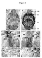
- Fig. 2. Expression of Px1 and Px2 mRNA in the brain.
- (A-B) Distribution of transcripts encoding Px1 and Px2.
OB: olfactory bulb; Cx: cortex; Hi: hippocampus; Cb: cerebellum.
Scale bar is 2.5 mm. - (C-F) Non-radioactive in situ hybridization.
(C) and (E): expression of Px1; (D) and (F): expression of Px2.
SP: stratum pyramidalis; SO: stratum oriens; SR: stratum radiatum; WM: white matter; EG: external granule cell layer; MC: molecular cell layer; GC: granule cell layer.
Scale bars are 50 µm (C-D) and 250 µm (E-F). - Fig. 3. Functional expression of pannexins in single Xenopus
oocytes.
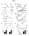
- (A) Whole-cell membrane currents (I m) from single oocytes.
For clarity, representative traces are shown only in 20 mV increments. - (B) Current-voltage relationships.
Current-voltage relationships were determined for oocytes injected with either antisense oligonucleotides, or Px1, Px2, and Px3 RNAs plus antisense. Results are shown as mean±SEM from at least 8 independent experiments. Antisense (n=45); Px1 (n=80); Px2 (n=46); Px3 (n=41). - (C-F) Functional interaction of Px1 and Px2 proteins.
Antisense-treated oocytes were co-injected with Px1 RNA together with equal amounts of RNAs encoding either Px2 or the W77R mutation of human Cx26, which is devoid of functional activity (31). - (C-D) Results are shown as mean±SEM from 4 independent experiments. Antisense (n=39); Px1+W77R (n=60); Px1+Px2 (n=67).
- (E) The lower panels show the mean±SEM from 3 independent experiments for Px1+W77R (n=45) and Px1+Px2 (n=50); *P<0.001.
- (F) The lower right panel illustrates the mean±SEM from 3 independent experiments, for Px1+W77R (n=44) and Px1+Px2 (n=41); *P<0.001.
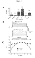
- Fig. 4. Functional expression of pannexins in paired oocytes.
- (A) Results are shown as the mean±SEM of the indicated number of oocyte pairs from 4-5 independent experiments. G j values recorded from oocytes expressing the neuronal mouse connexin36 (mCx36) are shown for comparison.
- (B) Px1 and Px1+Px2 intercellular channels.
- (C) Results are shown as the mean±SEM of 7-12 pairs (from 4 independent experiments) whose G j was 3.2±0.8 µS and 4.8±1.1 µS for mCx36 and Px1+Px2, respectively.
-
- The present invention will be better understood in the light of the following detailed description of experiments, including examples. Nevertheless, the skilled artisan will appreciate that this detailed description is not limitative and that various modifications, substitutions, omissions, and changes may be made without departing from the scope of the invention.
- cDNA clones were obtained by screening a rat hippocampal cDNA library (postnatal day 15) with [α-32P] end labeled oligonucleotides complementary to nucleotides (nt) 181-225 and 316-360 of the mouse Px1 open reading frame (ORF), and to nt 199-243 and 334-378 of the human Px2 ORF (25). A probe for Px3 was generated by PCR using the oligonucleotide pair derived from nt 569-592 and 1059-1082 of the rat Px3 ORF, identified in the database. The tissue distribution of pannexin gene expression was investigated by reacting blots containing rat polyA+ RNA (Rat MTN Blot I and II, catalog #7764-1 and 7795-1, respectively; Clontech, Palo Alto, CA) with [α-32P] dCTP labeled probes derived either from the entire ORF of Px1 and Px2, or with a fragment derived from the β-actin transcript (supplied along with the blots).
- For Northern blot analysis, the two filters were hybridized with probes for each of the three pannexins and exposed for 16 hrs at -70°C (Fig. 1 B).
- Radioactive (26) and non-radioactive (27) in situ hybridization experiments were performed essentially as described previously.
- [α-35S] dATP end labeled oligonucleotides corresponded to nt 181-225 and 334-378 of the mouse Px1 and human Px2 coding sequence, whereas the entire rat ORF was used to generate digoxygenin-labeled sense and antisense riboprobes.
- The distribution of transcripts encoding Px1 and Px2 was determined by radioactive in situ hybridization in horizontal brain sections obtained from rats at postnatal day 15 (Fig. 2, A-B).
- The coding sequence of each pannexin was subcloned into the pBSxG expression vector (28). In vitro transcription, preparation of Xenopus oocytes, biochemical analysis and RNA injection were performed as described elsewhere (29). Metabolic labeling of oocytes indicated that all three pannexin RNAs directed the synthesis of specific polypeptide bands, whose electrophoretic mobility was similar to that of the in vitro translated constructs (not shown).
For physiological analysis, cells were injected with a total volume of 40 nl of either an antisense oligonucleotide (3 ng/cell) to suppress the endogenous Xenopus Cx38 (30), or a mixture of antisense plus the specified RNA (20-80 ng/cell). The ability of pannexins to form hemi-channels was assessed in single oocytes 2-4 days after RNA injection, using a two-electrode voltage-clamp.
Whole-cell membrane currents (I m) were measured from single oocytes co-injected with pannexin RNAs and an oligonucleotide antisense to Xenopus Cx38 (Fig. 3A). Cells were initially clamped at a membrane potential (V m) of -40 mV and depolarizing steps lasting 2 sec were applied in 10 mV increments up to +60 mV (bottom traces).
In Fig. 3B, peak current values above holding currents (ΔI m) were calculated and plotted as a function of V m. Mean values from Px1-injected cells were significantly different (P<0.01) from those of control oocytes starting at a V m of -10 mV. For Px1 steady-state currents (open circles), values recorded for 20 msec at the end of the pulse were averaged and plotted as above. - To investigate whether Px1 and Px2 could functionally interact, oocytes were co-injected with Px1 RNA (40-80 ng/cell) together with the specified amounts of RNAs encoding either Px2 or the W77R mutation of human Cx26, which is devoid of functional activity (31). To analyze whether pannexins formed intercellular channels, oocytes were stripped of the vitelline envelope 1-2 days after RNA injection, and manually paired in homotypic configuration (same construct in both oocytes) for 24-48 hr before measuring junctional conductance with a dual voltage clamp (Fig. 4). The setup, hardware and software used for electrophysiological measurements and data analysis were as previously described (29, 32).
Both cells of a pair were initially clamped at -40 mV, and alternating pulses of ±10-20 mV were imposed to one cell (Fig. 4A). The current delivered to the cell clamped at -40 mV during the voltage pulse is equal in magnitude to the junctional current and can be divided by the voltage to yield the value of junctional conductance (G j).
In Fig. 4B, junctional currents (I j) were recorded from oocyte pairs in response to (V j steps of opposite polarity (bottom traces) applied, from a holding potential of -40 mV, in 20 mV increments.
In Fig. 4C, the steady-state junctional conductance G jss was measured at the end of the V j step and normalized to the values recorded at ±20 mV; Px1 +Px2 (filled circles) and mCx36 (open squares). Data describing the G j/V j relationship were fit (smooth cyanide lines) to a Boltzmann equation, whose parameters were in agreement with those previously reported (43, 44). - Results are shown as mean±SEM. An independent experiment is defined as a series of data obtained with oocytes isolated from one animal. Comparisons between two populations of data were made using the Student's unpaired t-test. P values of 0.01 or less were considered to be significant.
- The rat pannexin cDNAs were assigned the following accession numbers: AJ557015 (Px1); AJ557016 (Px2); AJ557017 (Px3).
- The human pannexins Px1, Px2, and Px3 were assigned accession numbers NP_056183, NP_443071, and NP_443191, respectively.
- Analysis of the cDNA sequences for Px1, Px2 and Px3 identified open reading frames (ORFs) encoding proteins with calculated molecular mass of 48,072, 73,270 and 44,976 daltons, respectively. The sequences of all three proteins predict, like for connexins, four transmembrane domains and cytoplasmic amino- and carboxy termini. A hallmark of gap junction-forming proteins is the presence of conserved, regularly spaced cysteine residues located on the two extracellular loops. Whereas the connexins contain three such residues, pannexins contain only two, thus resembling in this respect innexins, the invertebrate constituents of intercellular channels (33). A comparison of the cDNAs to the mouse genomic sequence (obtained from the Ensembl database; http://www.ensembl.org) resulted in the determination of the exon-intron structure of the three mouse pannexin genes (Fig. 1A). Considerable variability was found in the organization and length of the three gene loci, the protein coding regions could be assigned to five, three, and four exons respectively, for the Px1, Px2, and Px3 genes.
- Northern blots indicated that Px1 and Px2 transcripts were co-expressed in many tissues including eye, thyroid, prostate, kidney, liver and CNS (Fig. 1 B). Px1 probe hybridized to a 2.2-kb mRNA that was detectable in several organs including spinal cord and brain (Fig. 1 B). The 3.5-kb Px2 was most abundant in spinal cord and brain and was also present in other organs (Fig. 1 B). A less prominent 2.5-kb transcript was observed in some organs. By contrast, Px3 transcripts could only be detected in the skin, which was found, by RT-PCR, to be devoid of Px1 and Px2 mRNA (not shown).
In situ hybridization studies demonstrated widespread expression of both transcripts in many brain regions, including cortex, striatum, olfactory bulb, hippocampus, thalamus, cerebellum. Fig. 2, A and B, illustrate a partially overlapping expression profile and indicate that the transcripts encoding Px1 and Px2 are abundant in the olfactory bulb, cortex, hippocampus, and cerebellum. No signal was detected in parallel competition experiments with an excess of unlabeled probe (not shown). - Upon closer inspection at the cellular level, a differential distribution of Px1 and Px2 mRNA was apparent. In hippocampus, for example, both Px1 (Fig. 2C) and Px2 (Fig. 2D) were expressed in the pyramidal cell layer and in individual neurons (arrowheads) in the stratum oriens and stratum radiatum. Based on their location, the scattered neurons (NeuN positive, data not shown) can be inferred to be GABAergic interneurons. By contrast, in the cerebellum, Px1 expressing cells (Fig. 2E) were abundant in the white matter where Px2 expression was absent (Fig. 2F, asteriks; note, however, the high Px2 labeling in the Purkinje cell layer in Fig. 2F, arrows). The labeling of Px1 expressing cells in the white matter was not restricted to the cerebellum but was also observed in other white matter structures (e.g. corpus callosum, fimbria fornix), which, similarly, were also devoid of Px2 expression (not shown).
- No staining was obtained with sense probes (not shown).
- Large, voltage-activated outward currents were consistently induced when oocytes expressing Px1 were stepped to voltages greater than -20 mV (Fig. 3A-B). At large positive potentials, Px1 currents reached a peak within 30-60 msec of the imposition of the voltage step and then declined slowly, this rectification becoming more pronounced with increasing positive potentials. By contrast, neither Px2 nor Px3 induced membrane currents above those recorded in controls (Fig. 3A-B). Furthermore, incubation of oocytes for 10-30 min in carbenoxolone completely suppressed Px1 currents (peak amplitudes at +60 mV were 1189±170 and 255±47 nA for control medium and 30 µM carbenoxolone, respectively; n=4) and this effect was fully reversible (peak amplitude at +60 mV after a 30 min recovery period in control medium was 963±239 nA; n=4).
Since the in situ hybridization studies revealed co-expression of Px1 and Px2, our subsequent investigations entailed a more detailed functional analysis of these two proteins. To test whether they could form heteromeric channels, currents were recorded from oocytes co-expressing Px1 with Px2 (40-80 ng of RNA/cell) and were found to be significantly reduced with respect to those measured from cells that had been injected with the same amount of Px1 RNA (not shown). To exclude the possibility that this behavior was merely due to overloading of the synthetic machinery given the difference in the total amount of RNA injected, oocytes were injected with equal amounts of RNA for Px1 and the W77R mutation of human Cx26 (40-80 ng each/cell), which is devoid of functional activity (31). These experiments showed a reduction in current amplitude, suggesting that Px1 +Px2 form channels that are different from those composed of Px1 alone (Fig. 3C-D). As illustrated, ΔI m recorded from Px1 +Px2 expressing oocytes was significantly less (*P<0.001) than that measured from Px1+W77R cells.
Given that Px2 expression in the brain appears to be much stronger than Px1, we also tested whether Px2 could simply function as a dominant negative partner by co-injecting Px1 and Px2 RNAs at a 1:5 ratio (40:200 ng/cell for Px1:Px2, respectively). Current amplitudes recorded at +60 mV were similar, irrespective of whether Px1 and Px2 were injected at a ratio of 1:1 (591±40 nA; n=27) or of 1:5 (517±45 nA; n=20), further indicating that they both interact and form functionally heteromeric channels. Interestingly, a decrease in current amplitude was not observed when voltage-activated currents were measured from oocytes receiving RNAs for Px1 and Px3, which are not co-expressed in rat tissues (not shown). Moreover, following the imposition of a voltage step, Px1 +Px2 channels reached peak currents with a significant delay compared to Px1 expressing cells (Fig. 3E), which could result from slower opening or slower closing or both. In Fig. 3E, oocytes were depolarized to +40 mV (top left traces) and +60 mV (top right traces) from a holding potential of -40 mV. Peak currents were reached with a significant delay following the imposition of the voltage step (32 and 68 msec at +40 mV and 62 and 96 msec at +60 mV, for Px1+W77R and Px1+Px2, respectively).
Finally, as illustrated in Fig. 3F, analysis of the kinetics of channel closure at the more positive membrane potentials, revealed that currents recorded from Px1+Px2 expressing cells, presumably reflecting heteromeric hemi-channels, gated more slowly than those measured from oocytes injected with Px1+W77R RNA, presumably reflecting homomeric Px1 hemi-channels. Px2 slows the kinetics of voltage-dependent closure of Px1 hemi-channels. Cells were depolarized to +60 mV from a holding potential of -40 mV (top panels). The time-dependent decline in I m was well fit by a first order exponential decay function (lower left panel; cyanide line superposed to the re-scaled current traces shown above). - Px1 alone and in combination with Px2 induced the assembly of intercellular channels, whereas Px2 alone failed to do so (Fig. 4A). As illustrated, pairs of uninjected cells from the different batches of oocytes developed a variable level of junctional currents that exhibited the well-known voltage-dependent gating of endogenous Cx38 (42), whereas antisense controls showed negligible coupling, indicating that endogenous currents had been suppressed. It should be noted that intercellular channels were consistently detected only from batches of oocytes in which a robust junctional conductance was recorded with homotypic pairs expressing either mouse Cx36 (Fig. 4A) or human Cx26 wild-type (not shown), which served as positive controls. In this series of experiments, 20 out of 23 Px1 pairs and 36 out of 42 Px1+Px2 pairs were coupled. As shown in Fig. 4B, both Px1 and Px1+Px2 pairs displayed a remarkable insensitivity to transjunctional potentials of opposite polarities (Vj). Thus, with a driving force ≤ ±60 mV, junctional currents varied linearly with voltage (Fig. 4B) whereas, at higher transjunctional potentials, the conductance of Px1+Px2 channels displayed only a very modest reduction (∼15% ) of the initial values (Fig. 4C), similar to what reported for crayfish septate junctions (34) and human Cx31.9 (32). In Fig. 4C, the plot shows the relationship of V j to steady-state junctional conductance (G jss). Because of the much larger non-junctional currents that were present in Px1 homotypic pairs, reliable G jss/V j plots with the complete polarization paradigm were difficult to obtain.
Although it has been reported that junctional currents measured in insect cells are sensitive to changes in membrane potential (35), the relative voltage insensitivity of pannexin intercellular channels with polarization of one cell is a strong indication that polarization of both cells is not likely to affect significantly junctional conductance. -
- 1. Willecke, K., Eiberger, J., Degen, J., Eckardt, D., Romualdi, A., Guldenagel, M., Deutsch, U. & Sohl, G. (2002) Biol Chem 383, 725-37.
- 2. Bennett, M.V. (2000) Brain Res
Brain Res Rev 32, 16-28. - 3. Galarreta, M. & Hestrin, S. (2001)
Nat Rev Neurosci 2, 425-33. - 4. Katsumaru, H., Kosaka, T., Heizmann, C.W. & Hama, K. (1988) Exp Brain Res 72, 363-70.
- 5. Galarreta, M. & Hestrin, S. (1999) Nature 402, 72-5.
- 6. Gibson, J.R., Beierlein, M. & Connors, B.W. (1999) Nature 402, 75-9.
- 7. Beierlein, M., Gibson, J.R. & Connors, B.W. (2000)
Nat Neurosci 3, 904-10. - 8. Tamas, G., Buhl, E.H., Lorincz, A. & Somogyi, P. (2000)
Nat Neurosci 3, 366-71. - 9. Venance, L., Rozov, A., Blatow, M., Burnashev, N., Feldmeyer, D. & Monyer, H. (2000) Proc Natl Acad Sci USA 97, 10260-5.
- 10.Meyer, A.H., Katona, I., Blatow, M., Rozov, A. & Monyer, H. (2002) J Neurosci 22, 7055-64.
- 11.Blatow, M., Rozov, A., Katona, I., Hormuzdi, S.G., Meyer, A.H., Whittington, M. A., Caputi, A. & Monyer, H. (2003) Neuron 38, 805-17.
- 12.Ylinen, A., Bragin, A., Nadasdy, Z., Jando, G., Szabo, I., Sik, A. &
Buzsaki, G. (1995)
J Neurosci 15, 30-46. - 13.Draguhn, A., Traub, R.D., Schmitz, D. & Jefferys, J.G. (1998) Nature 394, 189-92.
- 14.Traub, R.D. & Bibbig, A. (2000)
J Neurosci 20, 2086-93. - 15.Hormuzdi, S.G., Pais, I., LeBeau, F.E., Towers, S.K., Rozov, A., Buhl, E.H., Whittington, M.A. & Monyer, H. (2001) Neuron 31, 487-95.
- 16.McBain, C.J. & Fisahn, A. (2001)
Nat Rev Neurosci 2, 11-23. - 17.Traub, R.D., Pais, I., Bibbig, A., LeBeau, F.E., Buhl, E.H., Hormuzdi,
S.G., Monyer, H. & Whittington, M.A. (2003) Proc Natl
Acad Sci USA 100, 1370-4. - 18.Condorelli, D.F., Belluardo, N., Trovato-Salinaro, A. & Mudo, G. (2000)
Brain Res
Brain Res Rev 32, 72-85. - 19.Deans, M.R., Gibson, J.R., Sellitto, C., Connors, B.W. & Paul, D.L. (2001) Neuron 31, 477-85.
- 20.Guldenagel, M., Ammermuller, J., Feigenspan, A., Teubner, B., Degen,
J., Sohl, G., Willecke, K. & Weiler, R. (2001)
J Neurosci 21, 6036-44. - 21.Deans, M.R., Volgyi, B., Goodenough, D.A., Bloomfield, S.A. & Paul, D.L. (2002) Neuron 36, 703-12.
- 22.Landisman, C.E., Long, M.A., Beierlein, M., Deans, M.R., Paul, D.L. & Connors, B.W. (2002) J Neurosci 22, 1002-9.
- 23.Long, M.A., Deans, M.R., Paul, D.L. & Connors, B.W. (2002) J Neurosci 22, 10898-905.
- 24.Buhl, D.L., Harris, K.D., Hormuzdi, S.G., Monyer, H. & Buzsaki, G. (2003) J Neurosci 23, 1013-8.
- 25.Panchin, Y., Kelmanson, I., Matz, M., Lukyanov, K., Usman, N. &
Lukyanov, S. (2000)
Curr Biol 10, R473-4. - 26.Wisden, W. & Morris, B.J. (2002) Int Rev Neurobiol 47, 3-59.
- 27.Ernsberger, U., Patzke, H. & Rohrer, H. (1997) Mech Dev 68, 115-26.
- 28.Groves, J.D. & Tanner, M.J. (1992) J Biol Chem 267, 22163-70.
- 29.White, T.W., Deans, M.R., O'Brien, J., Al-Ubaidi, M.R., Goodenough,
D.A., Ripps, H. & Bruzzone, R. (1999)
Eur J Neurosci 11, 1883-90. - 30.Barrio, L.C., Suchyna, T., Bargiello, T., Xu, L.X., Roginski, R.S., Bennett, M.V. & Nicholson, B.J. (1991) Proc Natl Acad Sci USA 88, 8410-4.
- 31.Bruzzone, R., Gomes, D., Denoyelle, E., Duval, N., Perea, J., Veronesi,
V., Weil, D., Petit, C., Gabellec, M-M., D'Andrea, P. & White, T.W.
(2001)
Cell Commun Adhes 8, 425-31. - 32.White, T.W., Srinivas, M., Ripps, H., Trovato-Salinaro, A., Condorelli, D.F. & Bruzzone, R. (2002) Am J Physiol Cell Physiol 283, C960-70.
- 33.Phelan, P. & Starich, T.A. (2001) Bioessays 23, 388-96.
- 34.Johnston, M.F. & Ramon, F. (1981) Biophys J 39, 115-7.
- 35.Verselis, V.K., Bennett, M.V. & Bargiello, T.A. (1991) Biophys J 59, 114-26.
- 36.Kelmanson, I.V., Shagin, D.A., Usman, N., Matz, M.V., Lukyanov, S.A.
& Panchin, Y.V. (2002)
Eur J Neurosci 16, 2475-6. - 37.Paul, D.L., Ebihara, L., Takemoto, L.J., Swenson, K.I. & Goodenough, D.A. (1991) J Cell Biol 115, 1077-89.
- 38.Stebbings, L.A., Todman, M.G., Phelan, P., Bacon, J.P. & Davies, J.A.
(2000)
Mol Biol Cell 11, 2459-70. - 39.Bruzzone, R., White, T.W. & Paul, D.L. (1996) EurJ Biochem 238, 1-27.
- 40.Schmitz, D., Schuchmann, S., Fisahn, A., Draguhn, A., Buhl, E.H., Petrasch-Parwez, E., Dermietzel, R., Heinemann, U. & Traub, R.D. (2001) Neuron 31, 831-40.
- 41.Traub, R.D., Bibbig, A., Piechotta, A., Draguhn, R. & Schmitz, D. (2001) J Neurophysiol 85, 1246-56.
- 42.Ebihara, L., Beyer, E.C., Swenson, K.I., Paul, D.L. & Goodenough, D.A. (1989) Science 243, 1194-5.
- 43.Srinivas, M., Rozental, R., Kojima, T., Dermietzel, R., Mehler, M., Condorelli, D.F., Kessler, J.A. & Spray, D.C. (1999) J Neurosci 19, 9848-55.
- 44.Al-Ubaidi, M.R., White, T.W., Ripps, H., Poras, I., Avner, P., Gomes, D. & Bruzzone, R. (2000) J Neurosci Res 59, 813-26.
- 45. Horwell et al., Bioorg. Med. Chem. 4: 1573 (1996).
- 46. Liskamp et al., Recl. Trav. Chim. Pays-Bas 1: 113 (1994).
- 47. Gante et al., Angew. Chem. Int. Ed. Engl. 33 : 1699 (1994).
- 48. Seebach et al., Helv. Chim. Acta 79 : 913 (1996).
-
Claims (16)
- Use of at least one neuronal channel-forming pannexin, or at least one biologically active fragment thereof, or at least one biologically active derivative thereof, for the manufacture of a drug for preventing and/or treating, in a mammal, a neurological disorder involving the hippocampal pyramidal cells.
- Use of at least one neuronal channel-forming pannexin for in vitro diagnosing, in a mammal, a neurological disorder involving the hippocampal pyramidal cells.
- The use according to claim 2, wherein said in vitro diagnostic comprises at least:a) sequencing a pannexin gene in a mammal suspected to have a neurological disorder involving the hippocampal pyramidal cells; andb) identifying at least one mutation responsible for the lack of production of pannexin or for the production of an inactive pannexin in said mammal.
- The use according to any of claims 1 to 3, wherein said mammal is a human.
- The use according to any of claims 1 to 4, wherein said neurological disorder is selected from epilepsy, schizophrenia, memory disorders, pain disorders, visual deficits, visual acuity, odor discrimination, and olfaction deficits.
- Method for in vitro selecting a compound useful for preventing and/or treating, in a mammal, a neurological disorder involving the hippocampal pyramidal cells, said compound being capable of modifying the channel-forming ability of a pannexin, wherein said method comprises at least:a) measuring the channel-forming ability of said pannexin in the absence of said compound (P0);b) measuring the channel-forming ability of said pannexin in the presence of said compound (P1);c) comparing P0 and P1; andd) if P1 is significantly different from P0, selecting said compound.
- Method for in vitro selecting a compound useful for preventing and/or treating, in a mammal, a neurological disorder involving the hippocampal pyramidal cells, said compound being capable of specifically modifying the channel-forming ability of a pannexin, without modifying the channel-forming ability of a connexin, wherein said method comprises at least:a) measuring the channel-forming ability of each of said pannexin (P0) and said connexin (C0) in the absence of said compound;b) measuring the channel-forming ability of each of said pannexin (P1) and said connexin (C1) in the presence of said compound;c) comparing P0 and P1, and C0 and C1; andd) if P1 is significantly different from P0, and if C1 is not significantly different from C0, selecting said compound.
- The method according to claim 6 or 7, wherein, in step d), if P1 is significantly greater than P0, said compound is an agonist of said pannexin.
- The method according to claim 6 or 7, wherein, in step d), if P1 is significantly lower than P0, said compound is an antagonist of said pannexin.
- The method according to any of claims 6 to 9, further comprising purifying said compound.
- The method according to any of claims 6 to 10, wherein said mammal is a human.
- The method according to any of claims 6 to 11, wherein said neurological disorder is selected from epilepsy, schizophrenia, memory disorders, pain disorders, visual deficits, visual acuity, odor discrimination, and olfaction deficits.
- An animal model for in vivo selecting a compound useful for preventing and/or treating, in a mammal, a neurological disorder involving the hippocampal pyramidal cells, wherein said compound is capable of modifying the channel-forming ability of a pannexin in said animal model.
- The animal model according to claim 13, wherein at least one mutation is introduced in a pannexin gene of an animal, said mutation being responsible for the lack of production of pannexin, or for the production of an inactive pannexin, in said animal model.
- The animal model according to claim 13 or 14, wherein said mammal is a human.
- The animal model according to any of claims 13 to 15, wherein said neurological disorder is selected from epilepsy, schizophrenia, memory disorders, pain disorders, visual deficits, visual acuity, odor discrimination, and olfaction deficits.
Priority Applications (3)
| Application Number | Priority Date | Filing Date | Title |
|---|---|---|---|
| EP03292281A EP1516625A1 (en) | 2003-09-16 | 2003-09-16 | Uses of neuronal channels-forming pannexins for therapy and diagnosis in mammals |
| US10/942,001 US20080132446A1 (en) | 2003-09-16 | 2004-09-16 | Uses of neuronal pannexins for therapy and diagnosis in mammals |
| PCT/IB2004/003232 WO2005026170A2 (en) | 2003-09-16 | 2004-09-16 | Uses of neuronal pannexins for therapy and diagnosis in mammals |
Applications Claiming Priority (1)
| Application Number | Priority Date | Filing Date | Title |
|---|---|---|---|
| EP03292281A EP1516625A1 (en) | 2003-09-16 | 2003-09-16 | Uses of neuronal channels-forming pannexins for therapy and diagnosis in mammals |
Publications (1)
| Publication Number | Publication Date |
|---|---|
| EP1516625A1 true EP1516625A1 (en) | 2005-03-23 |
Family
ID=34178639
Family Applications (1)
| Application Number | Title | Priority Date | Filing Date |
|---|---|---|---|
| EP03292281A Withdrawn EP1516625A1 (en) | 2003-09-16 | 2003-09-16 | Uses of neuronal channels-forming pannexins for therapy and diagnosis in mammals |
Country Status (3)
| Country | Link |
|---|---|
| US (1) | US20080132446A1 (en) |
| EP (1) | EP1516625A1 (en) |
| WO (1) | WO2005026170A2 (en) |
Cited By (1)
| Publication number | Priority date | Publication date | Assignee | Title |
|---|---|---|---|---|
| CN107519157A (en) * | 2008-09-10 | 2017-12-29 | 原子能和替代能源委员会 | Application of the anti-connection protein agent in terms of psychotropic agent molecule curative effect is adjusted |
Citations (2)
| Publication number | Priority date | Publication date | Assignee | Title |
|---|---|---|---|---|
| WO2003033016A1 (en) * | 2001-10-16 | 2003-04-24 | Jenny Phipps | Use of neurotrophic factors for treating neurodegenerative diseases and cancer |
| US20030180406A1 (en) * | 2002-03-21 | 2003-09-25 | Helmut Sies | Treatment of diseases involving defective gap junctional communication |
-
2003
- 2003-09-16 EP EP03292281A patent/EP1516625A1/en not_active Withdrawn
-
2004
- 2004-09-16 US US10/942,001 patent/US20080132446A1/en not_active Abandoned
- 2004-09-16 WO PCT/IB2004/003232 patent/WO2005026170A2/en not_active Ceased
Patent Citations (2)
| Publication number | Priority date | Publication date | Assignee | Title |
|---|---|---|---|---|
| WO2003033016A1 (en) * | 2001-10-16 | 2003-04-24 | Jenny Phipps | Use of neurotrophic factors for treating neurodegenerative diseases and cancer |
| US20030180406A1 (en) * | 2002-03-21 | 2003-09-25 | Helmut Sies | Treatment of diseases involving defective gap junctional communication |
Cited By (1)
| Publication number | Priority date | Publication date | Assignee | Title |
|---|---|---|---|---|
| CN107519157A (en) * | 2008-09-10 | 2017-12-29 | 原子能和替代能源委员会 | Application of the anti-connection protein agent in terms of psychotropic agent molecule curative effect is adjusted |
Also Published As
| Publication number | Publication date |
|---|---|
| US20080132446A1 (en) | 2008-06-05 |
| WO2005026170A2 (en) | 2005-03-24 |
| WO2005026170A3 (en) | 2005-06-02 |
Similar Documents
| Publication | Publication Date | Title |
|---|---|---|
| Daggett et al. | Molecular and functional characterization of recombinant human metabotropic glutamate receptor subtype 5 | |
| Sommer et al. | A glutamate receptor channel with high affinity for domoate and kainate. | |
| Sakai et al. | Cloning and functional expression of a novel degenerin‐like Na+ channel gene in mammals | |
| JP3534411B2 (en) | DNA encoding human calcium channel α-1E subunit | |
| EP0783577B1 (en) | Chimeric receptors and methods for identifying compounds active at metabotropic glutamate receptors and the use of such compounds in the treatment of neurological disorders and diseases | |
| EP1042468B1 (en) | Low-voltage activated calcium channel compositions and methods | |
| Mounsey et al. | Modulation of skeletal muscle sodium channels by human myotonin protein kinase. | |
| Nash et al. | Cloning and characterization of the opossum kidney cell D1 dopamine receptor: expression of identical D1A and D1B dopamine receptor mRNAs in opossum kidney and brain. | |
| EP2086992B1 (en) | Human salty taste receptor and methods of modulating salty taste perception | |
| US20030219834A1 (en) | Methods of modulating cold sensory perception | |
| White et al. | Virtual cloning, functional expression, and gating analysis of human connexin31. 9 | |
| US6534287B1 (en) | Human metabotropic glutamate receptor | |
| Hoptak-Solga et al. | Zebrafish short fin mutations in connexin43 lead to aberrant gap junctional intercellular communication | |
| Lü et al. | Cloning and functional expression of novel N-type Ca2+ channel variants | |
| US6287859B1 (en) | Identification, functional expression and chromosal localization of a sustained human proton-gated cation channel | |
| EP1780278A2 (en) | Mammalian calcium channels and related probes, cell lines and methods | |
| Daggett et al. | The human N‐methyl‐D‐aspartate receptor 2C subunit: genomic analysis, distribution in human brain, and functional expression | |
| CA2321193A1 (en) | Novel gabab receptor dna sequences | |
| US20020009772A1 (en) | Novel human calcium channels and related probes, cell lines and mehtods | |
| US20030211968A1 (en) | Neuromedin u receptor nmur2 and nucleotides encoding it | |
| EP1516625A1 (en) | Uses of neuronal channels-forming pannexins for therapy and diagnosis in mammals | |
| US20050048522A1 (en) | Novel guanosine triphosphate (GTP) binding protein-coupled receptor proteins | |
| Devignot et al. | A novel glycine receptor αZ1 subunit variant in the zebrafish brain | |
| KR100554551B1 (en) | Novel, ubiquitous potassium-channel proteins and genes for the same | |
| WO2001092468A2 (en) | Novel compositions for the expression of the human peptide histidine transporter 1 and methods of use thereof |
Legal Events
| Date | Code | Title | Description |
|---|---|---|---|
| PUAI | Public reference made under article 153(3) epc to a published international application that has entered the european phase |
Free format text: ORIGINAL CODE: 0009012 |
|
| AK | Designated contracting states |
Kind code of ref document: A1 Designated state(s): AT BE BG CH CY CZ DE DK EE ES FI FR GB GR HU IE IT LI LU MC NL PT RO SE SI SK TR |
|
| AX | Request for extension of the european patent |
Extension state: AL LT LV MK |
|
| 17P | Request for examination filed |
Effective date: 20050921 |
|
| AKX | Designation fees paid |
Designated state(s): AT BE BG CH CY CZ DE DK EE ES FI FR GB GR HU IE IT LI LU MC NL PT RO SE SI SK TR |
|
| 17Q | First examination report despatched |
Effective date: 20060112 |
|
| STAA | Information on the status of an ep patent application or granted ep patent |
Free format text: STATUS: THE APPLICATION IS DEEMED TO BE WITHDRAWN |
|
| 18D | Application deemed to be withdrawn |
Effective date: 20091114 |