Technical Field
The present invention relates to a method of isolating
a DNA encoding a peptide capable of acting as an agonist, an
antagonist or an inverse agonist for a receptor; to a method
of detecting or isolating the peptide; a cell line derived from
an endocrine cell used for these methods; to a process for
producing a peptide using the cell line derived from the
endocrine cell; to a B-cell line in which a receptor is expressed
and which is used for the above methods; a method of isolating
a DNA encoding a peptide which is capable of reacting with a
substance to be tested using the B-cell line; a method of
isolating a DNA encoding a peptide using the B-cell line; to
a constitutively activated mutant G-protein coupled receptor
obtained by the method of isolating a DNA; to a method of isolating
an agonist, an antagonist or an inverse agonist for a G-protein
coupled receptor using the B-cell line; to a method of isolating
an activator or an inhibitor for a peptide using the B-cell
line; and to a host-vector system using the B-cell line as a
host.
Background Art
It has been known that, in general, peptide hormones and
neurotransmitters are produced as precursors (preproforms) and
finally become active peptides as a result of action of plural
processing enzymes or modifying enzymes [Curr. Opin. Chem. Biol.,
2, 31 (1998)].
Specific processing enzymes and modifying enzymes are
expressed only in specific endocrine cells and, therefore,
active peptides are not usually produced even when a precursor
gene of an active peptide (such as vasopressin and glucagon)
produced in nerve cells and endocrine cells of hypothalamus,
pituitary gland or Langerhans islets of pancreas, for example,
is expressed in non-endocrine cells such as COS cells and CHO
cells which are frequently used in gene expression.
Cells having an inherent property of producing specific
active peptides, such as endocrine cells, express processing
enzymes or modifying enzymes for the active peptide and,
therefore, it is likely that, when a precursor gene of the peptide
is expressed using the cells as a host, the peptide is able
to be expressed in an active form. With regard to other active
peptide which is able to be subjected to normal processing or
modification by processing enzymes or modifying enzymes being
expressed in the cells, it is also likely that the active peptide
is able to be expressed by means of expression of a precursor
gene using the cells as a host.
With regard to a method for producing an active peptide
where a known active peptide precursor gene is transfected into
a cell line which is established from endocrine cells
(hereinafter, referred to as endocrine cell line) and expressed,
there have been known a method where the preproinsulin gene
or the preproamyrin gene is expressed by transfecting into RIN
cell established from cancer cells of Langerhans islets of rat
induced by irradiation of radioactive ray whereupon insulin
or amyrin is produced (US 6194176 and US 6110707), a method
where the preproinsulin gene is expressed by transfecting into
MIN6 cell line established from tumor β cell produced in
transgenic mouse whereupon C peptide is produced [Nat. Cell
Biol., 2, 805 (2000)], etc.
As mentioned above, there have been known several methods
for producing known active peptides using endocrine cell lines
but there has been no literature mentioning or suggesting that
an endocrine cell line is useful for screening novel active
peptides.
In addition, in order to efficiently find a peptide having
new activity and a gene encoding the peptide, there has been
demanded further improvement concerning endocrine cell lines
using as hosts for gene expression, endocrine cell lines using
as sources for genes and assay system for detecting activity
of peptides.
With regard to endocrine cell lines, there have been known
the following (1) cell lines derived from hypothalamus and (2)
those derived from Langerhans islets.
(1) Cell lines derived from hypothalamus
With regard to the cell lines derived from hypothalamus,
those of the following (i) to (iv) have been known.
With regard to the above cell lines, there have been known
GN, etc. GN cell expresses GnRH [Proc. Natl. Acad. Sci. USA,
88, 3402 (1991)].
When cell lines derived from hypothalamus expressing an
endogenous leptin receptor are obtained, it is possible to
analyze a biological function of leptin to each nerve cell but
any of the above-mentioned cell lines derived from hypothalamus
does not express a leptin receptor and, up to now, no cell line
derived from hypothalamus expressing an endogenous leptin
receptor has been known [Diabetes, 49, 1443 (2000); Neuron,
5, 1 (1990)].
Development of leptin has progressed as an anti-obesity
agent or antidiabetic agent but, as a result of resistance to
leptin, drugs in place of leptin have been demanded. It is
believed that clarification of action mechanism of leptin
results in finding of novel physiologically active substances
and identification of targets for the development of new drugs.
When cell lines derived from hypothalamus expressing an
endogenous leptin receptor are obtained, it is possible to screen
and evaluate the above-mentioned drugs using the cell lines.
There are not known hypothalamus derived cell lines that
express preproneuromedin U gene, RF amide-related peptide
(RFRP) preproprotein gene, preproorexin gene,
preproopiomelanocortin gene, preproneuropeptide Y gene,
preproneuropeptide FF gene, preprocorticotropin-releasing
hormone gene, preprothyrotropin-releasing hormone gene,
preproghrelin gene, prepromelanin concentration hormone gene,
cocaine- and amphetamine-regulated transcript (CART) gene,
type 2 neuromedin U receptor (NMU2R), RFRP receptor gene, type
4 melanocortin receptor (MC4R) gene, type 1 neuropeptide Y
receptor (NPY1R) gene, type 5 neuropeptide Y receptor (NPY5R)
gene, type 2 neuropeptide FF receptor (NPFF2) gene, type 1
corticotropin-releasing hormone receptor (CRHR-1) gene, type
2 corticotropin-releasing hormone receptor (CRHR-2) gene,
ghrelin receptor gene, type 1 melanin concentration hormone
receptor (MCHR1) gene, sulfonylurea receptor gene or
preproagouti-related peptide gene, in addition to leptin
receptor gene, or polypeptides encoded by those genes.
Hypothalamus has the most important controlling function
for maintaining the life and has been well noted as a target
site for drugs. Therefore, if hypothalamus derived cell lines
maintaining the function are able to be obtained, function of
each hypothalamus nerve cell is able to be studied in a cellular
level and in a molecular level. That is also very useful for
the development of drugs where the hypothalamus nerve cell is
a target. It has been known that endocrine cells existing in
hypothalamus produce various active peptides and, therefore,
it is believed that various cell lines derived from hypothalamus
are very useful sources for screening novel active peptides.
(2) Cell lines derived from Langerhans islets
Until now, cell lines of the following (i) to (vii) have
been known as cell lines derived from Langerhans islets.
All of the above-mentioned cell lines established from
cells of Langerhans islets are cancer cells or cells derived
from cancer and, therefore, it is possible that they have
different property from that of normal cells.
In screening novel physiologically active substances,
receptors, ligands or active peptides, it is desired to obtain
further various cell lines maintaining the property of normal
cells. Since various cells of Langerhans islets are believed
to control each other, it is possible to analyze the role of
each cell when various cell lines are obtained and are subjected
to co-incubation.
In addition, when various cell lines derived from
Langerhans islets having the same genetic background are able
to be obtained, that is useful for a comparative analysis of
differentiation stage and function of each cell line. Until
now, there has been no various immortalized cells derived from
Langerhans islets having the same genetic background.
It has been known that endocrine cells existing in
Langerhans islets produces various active peptides such as
glucagon, insulin, somatostatin and pancreatic polypeptides
and, therefore, the endocrine cells are believed to be very
useful sources for the screening new active peptides as well.
In order to efficiently check the activity of a peptide
which is produced from endocrine cell lines or endocrine cells
into which any gene is transfected, a simple and highly-sensitive
assay system by which activity of the peptide is able to be
detected is necessary. In order to efficiently find a peptide
having a novel activity or a gene encoding the peptide, various
assay systems which are highly sensitive and simple arenecessary.
It is also believed that, when various endocrine cell lines
are used as peptide sources or gene sources, a peptide having
a new activity or a gene encoding the peptide is able to be
efficiently found.
Many assay systems have been constructed already.
Examples of the assay systems having a good sensitivity include
a bioassay system, etc.
In the case of abioassay system, the detection sensitivity,
signal to noise ratio, multiplicity of use and simplicity depend
greatly on which cell is used. When a transformant is used
as the above cell, efficiency of the assay depends greatly on
whether the efficiency of gene transfection is good, whether
the expression of gene is high, whether induced expression of
gene is possible, whether induction rate of the induced
expression is high, etc. When animal cells are used, the assay
efficiency depend greatly on whether cultivation is possible
in a serum-free medium, whether they are non-adherent cells
(floating cells) having a good growth, etc.
As an example of assay systems which are able to be used
for screening active peptides (ligands), a bioassay system where
G-protein coupled receptor (hereinafter, abbreviated as GPCR)
is utilized has been known. By the GPCR bioassay system, there
have been isolated novel ligands, agonists or antagonists. GPCR
is coupled to a heterotrimeric G protein constituted from three
kinds of G proteins (guanine nucleotide-binding proteins), i.e.
Gα, Gβ and Gγ and, via activation of G protein, signal is
transduced into cells. Since GPCR has seven transmembrane
regions, it is also called a seven transmembrane receptor.
With regard to the GPCR bioassay system, there have been
known many methods such as a method where an increase in the
amount of intracellular Ca2+ is detected and a method where a
reporter system is used, etc.
GPCRs transduce different signals depending upon the
difference in subtypes of the coupled G protein. In the case
of a method using a reporter system, there are differences in
detectable signal, sensitivity, ratio of signal to noise ratio,
etc. depending upon a factor that what promoter is used for
the expression of the reporter gene.
With regard to a promoters, there have been utilized a
promoter having TPA-responding elements (TRE), an NFAT (nuclear
factor of activated T cells)-responding promoter, a promoter
having cAMP-responding elements (CRE), a promoter having
serum-responding elements (SRE), etc.
Steric structure of Gα coupled to GPCR has been clarified
already and it has been known that, when amino acids of C terminal
of Gα protein which are important for interaction with GPCR
are substituted with the corresponding amino acids of other
Gα protein, it is now able to be coupled to GPCR which is not
primarily coupled [Mol. Pharmacol., 57, 13 (2000)].
For example, a chimeric Gα protein (Gαq-i) where 5 amino
acids at C terminal of Gαq are substituted with those of Gαi
is able to be coupled to GPCRs which transduce signals by coupling
to Gαi, a chimeric Gα protein (Gαs-q) where 5 amino acids at
C terminal of Gαs are substituted with those of Gαq is able to
coupled to GPCRs which transduce signals by coupling to Gαq
and a chimeric Gα protein (Gαs-i) where 5 amino acids at C terminal
of Gαs are substituted with those of Gαi is able to be coupled
to GPCRs which transduce signals by coupling to Gαi.
With regard to GPCRs, there has been known a GPCR called
a constitutively activated GPCR which transduces signals even
when no ligand is present provided that it is excessively
expressed in cells. A signal which is transduced in the absence
of a ligand called a constitutive activity. In the
constitutively activated GPCRs, there are that which is present
in nature and a mutated GPCR which is constructed by introduction
of modification such as substitution, deletion, etc. of amino
acid(s) [Mol. Pharmacol., 57, 890 (2000); WO 98/46995]. In
some cases, the mutated GPCR increases the affinity with an
agonist and, therefore, it is useful for screening ligands [J.
Biol. Chem., 272, 1822 (1997)].
In a GPCR bioassay system, there have been utilized animal
cells such as CHO cells, COS-7 cells, 293 cells and HEK293 EBNA
cells, etc., melanophores of frog [Mol. Pharmacol., 57, 125
(2000)] or yeast [Trends Biotechnol., 15, 487 (1997) ; G Protein
Receptors, CRC Press, 49-68 (2000)], etc. as cells for the assay.
Although the above-mentioned GPCR bioassay systems are
good assay systems, there have been demanded further
improvements in detection sensitivity, signal to noise ratio,
multiplicity of use, simplicity/convenience, host-vector
system used, etc.
When an expression cloning is carried out using the
above-mentioned assay cells as a host, it is possible to
efficiently obtain a useful gene and, up to now, there have
been obtained genes of β chain of interferon γ receptor and
PACAP (pituitary adenylate cyclase-activating polypeptide)
receptor using expression of reporter gene as an index [Nature,
365, 170 (1993); Cell, 76, 803 (1994)]. However, with regard
to the expression cloning method, the same improvement as in
GPCR bioassay system has been demanded as well. Incidentally,
there has been neither description nor suggestion that
constitutively activated GPCR gene is able to be isolated by
an expression cloning method.
Disclosure of the Invention
Objects of the present invention are to provide the
following (1) to (5).
The present invention relates to the following (1) to
(106).
In the present specification, a peptide means a substance
where two or more amino acids are linked by peptide bond and
includes polypeptide and oligopeptide.
[I] Construction of an expression cloning system of an
active peptide precursor gene and an efficient production system
of an active peptide
An expression cloning system of an active peptide
precursor gene and an efficient production system of an active
peptide are able to be constructed as follows.
1. Preparing cell lines derived from endocrine cells
Processing from a precursor of an active peptide and
modif ication after the processing are different for each peptide
and are diverse. Known endocrine cell lines such as MIN6 are
able to be utilized as a host but, for such a purpose of
transfecting a precursor gene of an active peptide into a host
cell to produce the active peptide efficiently or of cloning
a precursor gene using activity of the expressed peptide as
an index, it is preferred that many kinds of endocrine cell
lines are obtained and a cell expressing a processing enzyme
or modifying enzyme suitable for the active peptide is selected
among them and is utilized as a host. Endocrine cell line is
able to be established by the following methods.
(1) Establishment of cell lines derived from endocrine
cells
Cell lines derived from endocrine cells of the present
invention is able to be established by isolating cells from
tissues containing endocrine cells derived from a non-human
transgenic animal transfected with an oncogene, followed by
culturing the cells.
Examples of the non-human transgenic animal include a
transgenic mouse, a transgenic rat, a transgenic pig and a
transgenic rabbit, etc.
With regard to an oncogene, an oncogene derived from a
virus such as SV40 and adenovirus can be exemplified and examples
include the large T antigen gene of SV40 and E1A and E1B of
adenovirus, etc. To be more specific, the large T antigen gene
derived from SV40tsA58 which is a temperature-sensitive mutant
and E1A 12S which is a hybrid of adenovirus 2 and adenovirus
5 , etc. can be exemplified. In order to establish a good cell
line, it is preferred to use the large Tantigen gene of SV40tsA58.
In order to express the oncogene in endocrine cells, it is
necessary that an appropriate promoter capable of working in
endocrine cells is present upstream of the coding region of
the oncogene product. With regard to a promoter, an endogenous
promoter of the oncogene per se derived from a virus, a promoter
of an animal gene expressed in endocrine cells, etc. can be
used. For example, the endogenous promoter of the large T
antigen gene of SV40tsA58, H-2Kb class I prompter, etc. can
be used.
A transgenic mouse transfected with the large T antigen
gene of SV40tsA58 can be prepared by the methods mentioned in
Japanese Published Unexamined Patent Application No.
292,958/1993, Jpn. J. Cancer Res., 82, 1344 (1991) and Proc.
Natl. Acad. Sci. USA, 88, 5096 (1991). A transgenic rat
transfected with the large T antigen gene of SV40tsA58 can be
prepared by a method mentioned in Japanese Published Unexamined
Patent Application 228,930/2000 or Exp. Anim., 48, 255 (1999).
To be more specific, a plasmid SV tsA 58 ori(-)-2 [Cytotechnology,
7, 165 (1991)], constructed by inserting the total genomic DNA
of SV40tsA58 cleaved to open the circle with a restriction enzyme
BamHI into aplasmidpBR322 , is cleavedwith a restriction enzyme
BamHI to remove the vector site whereupon a DNA (5,240 bp)
containing the large T antigen gene of SV40tsA58 is prepared.
The endogenous promoter of the large T antigen gene of SV40tsA58
is present in the DNA and, therefore, the large T antigen gene
of SV40tsA58 is expressed in somatic cell transfected with the
DNA. The above DNA containing the large T antigen gene of
SV40tsA58 is microinjected into a male pronucleus of a rat
fertilized egg in the pronuclear stage which is a totipotent
cell. The resulting egg is transplanted into an oviduct of
a foster rat to give offsprings, then offsprings having the
injected gene are selected and the individual rats into which
the gene is integrated in a stable manner are obtained whereupon
it is possible to efficiently produce a transgenic rat where
the large T antigen gene of SV40tsA58 is integrated into the
chromosome of cells of each tissue during the ontogenesis.
Known methods can be used as to a method for isolating
cells from the tissues containing endocrine cells derived from
a non-human transgenic animals transfected with an oncogene.
For example, cells derived from hypothalamus can be
isolated by an enzymatic method mentioned in J. Pharmacol.
Toxicol. Method, 36, 45-52 (1996), etc. and cells of Langerhans
islets can be isolated by a method mentioned in Diabetes, 46,
1755 (1997), etc.
Culturing the isolated cells can be carried out according
to conventional methods for culturing animal cells.
With regard to a medium for culturing the cells, a medium
used generally, such as RPMI 1640 medium [J. Am. Med. Assoc.,
199, 519 (1967)], Eagle's MEM [Science, 122, 501 (1952)],
Dulbecco's modified Eagle medium (DMEM) [Virology, 8, 396
(1959)], 199 medium [Proc. Soc. Exp. Biol. Med., 73, 1 (1950)],
a medium where fetal bovine serum or the like is added to those
media, etc can be used.
Cultivation is usually carried out for 1 to 7 day(s) under
the conditions of pH 6 to 8 at 30 to 40°C in the presence of
5% CO2, etc. In order to grow the cells derived from a transgenic
animal transfected with the large T antigen gene of
temperature-sensitive mutant SV40tsA58, it is preferred to
culture at the temperature by which activity of the
temperature-sensitive large T antigen mutant is not inhibited
(30 to 33°C or, preferably, 33°C).
During the cultivation, an antibiotic such as kanamycin
or penicillin can be added to the medium if necessary.
When the cells are subcultured for 2 to 3 months,
immortalized cells are able to be obtained.
According to the above-mentioned method, a large number
of cell lines derived from endocrine cells such as cell lines
derived fromhypothalamus and cell lines derived fromLangerhans
islets are able to be prepared.
Many different kinds of cells are present in endocrine
tissues such as hypothalamus and Langerhans islets etc., and
it is possible to obtain many kinds of immortalized cell lines
by the above method.
In the present invention, it has been found for the first
time that many kinds of immortalized cell lines are able to
be obtained abundantly in very efficient manner with good
reproducibility particularly from endocrine cells of
transgenic animals transfected with the large T antigen gene
of temperature-sensitive mutant SV40tsA58 [Exp. Anim., 48, 255
(1999)].
A mixture of the obtained cell lines is able to be frozen
and preserved according to a general cryopreservation method
for animal cells and, upon necessity, the cells are able to
be thawed to use for various analyses and experiments.
A mixture of the above-obtained cell lines is separated
into single clones according to a general method such as a colony
formation method [Endocrinology, 136, 4084 (1995)], and the
resulting clones can be isolated as each cell line.
Examples of the cell line which is obtained by the above
method include those obtained in the following Examples such
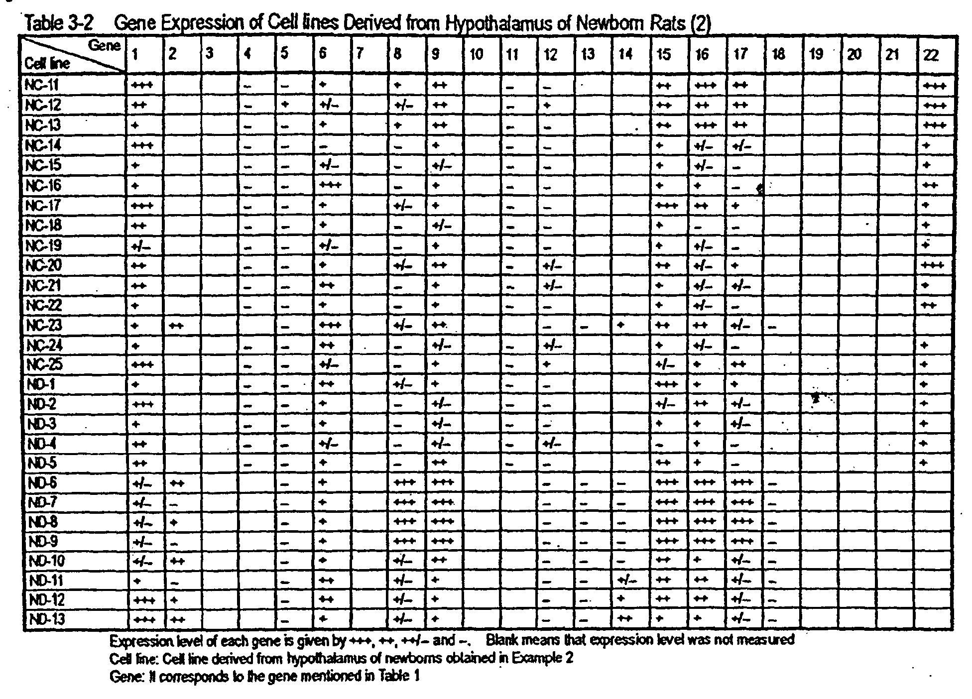
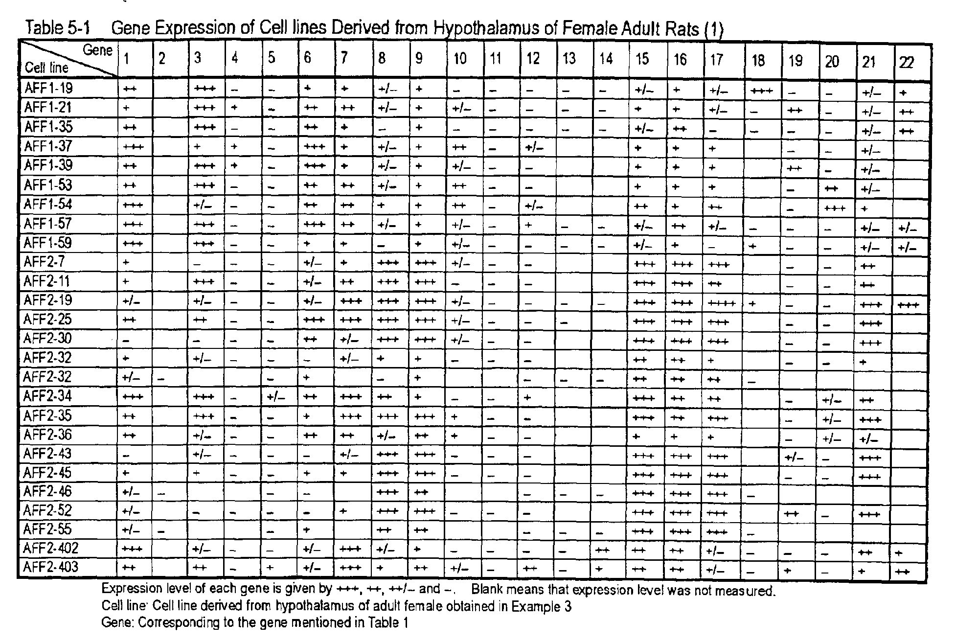
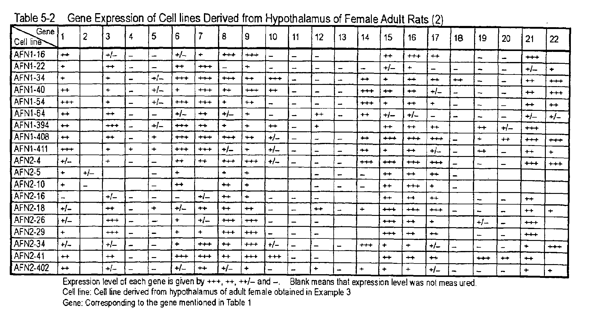
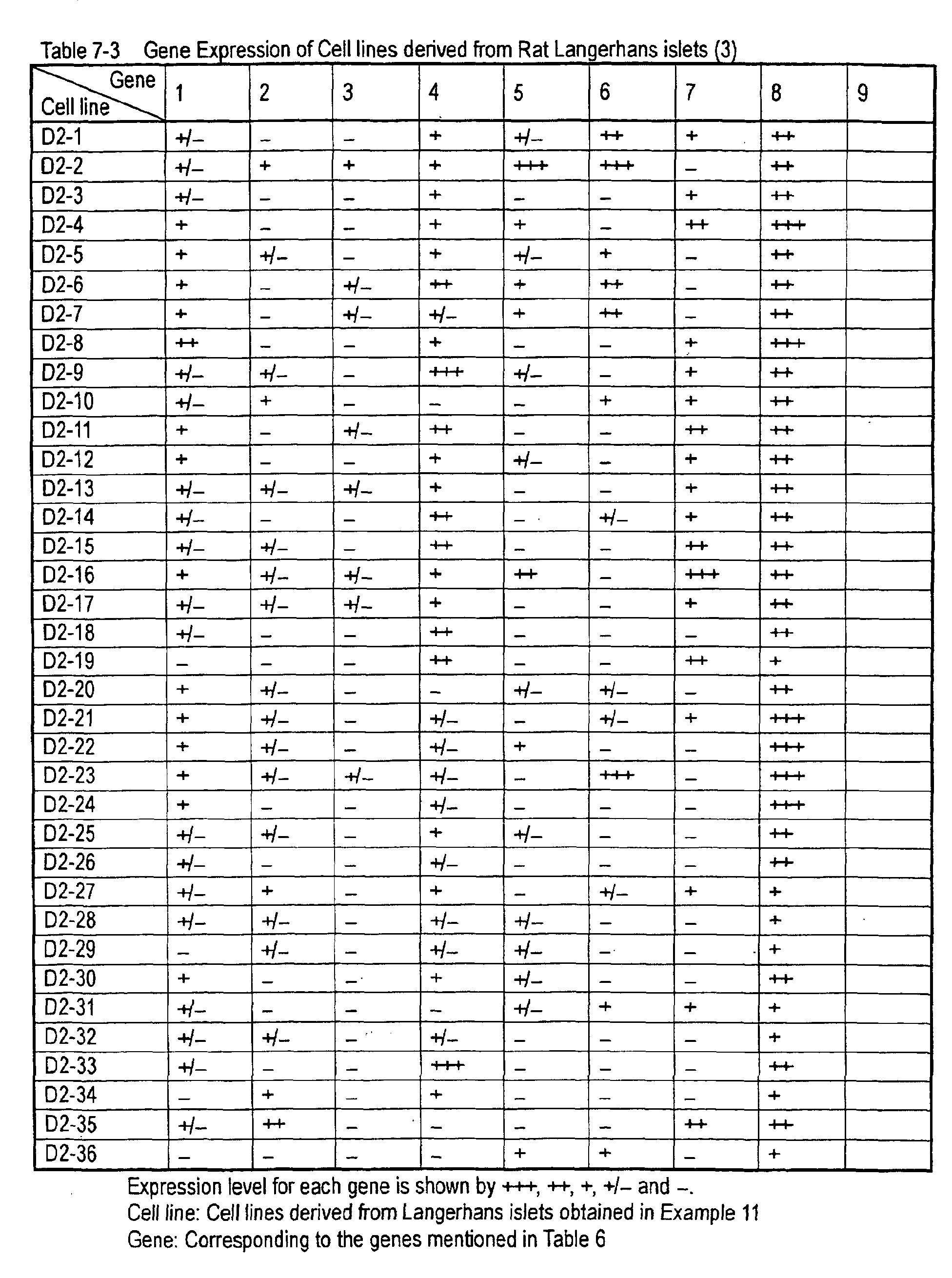
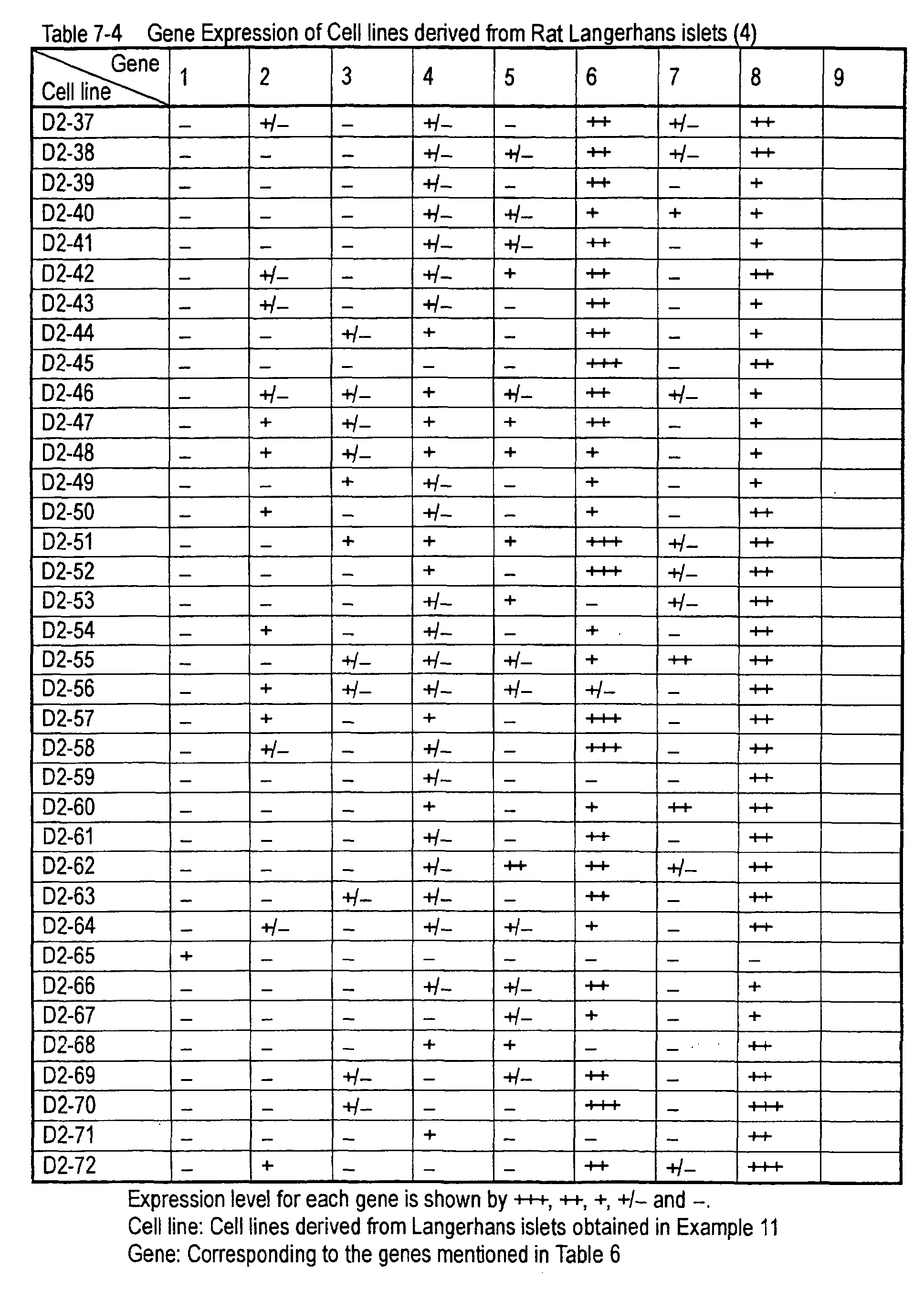
as 798 single clones (including the clones as shown in Tables
2 to Table 5-2) derived from hypothalamus and 261 single clones
(R-1 to R-45, M-1 to M-63, F-1 to F-9, D-1 to D-72 and D2-1
to D2-72) derived from Langerhans islets. Cell lines having
the identical property as the above cell lines are able to be
obtained by the above-mentioned method of the present invention
with good reproducibility.
(2) Selection of endocrine cell lines
With regard to many kinds of the immortalized cell lines
obtained in the above (1), it is possible to select endocrine
cells suitable for the expression of an aimed active peptide
by checking the expression of the genes or gene products of
various processing enzymes and modifying enzymes which have
been known to be expressed in endocrine cells.
With regard to cell lines derived from hypothalamus or
cell lines derived from Langerhans islets, for example, it is
possible to select endocrine cell lines suitable for the object
by checking the expression of the gene or the gene product of
prohormone convertase (P1/P3, PC2, furin, PC4, PACE4, PC5/PC6,
PC7/SPC7/LPC/PC8), carboxypeptidase E (CPE) or peptidylglycine
α-amidating monooxigenase (PAM), etc..
It is also possible to select endocrine cells suitable
for the expression of an aimed active peptide by checking the
expression of active peptides expressed in endocrine cells (such
as insulin, glucagon, somatostatin and pancreatic polypeptide,
etc.) from the above-mentioned cell lines.
It is further possible to select endocrine cell line
suitable for the expression of an active peptide by transfecting
an active peptide precursor gene to the above-mentioned cell
lines according to the method mentioned in the following 3.
and checking whether the active peptide is produced.
Examples of amethod for checking the expression of various
genes include RT-PCR and Northern blotting. Examples of a
method for checking the expression of various gene products
or the production of active peptides include a cell staining
using various antibodies and Western blotting.
The endocrine cell lines prepared as above are also very
useful as sources for peptides and for cDNAs for screening of
various active peptides.
(3) Characterization of immortalized cell lines
Characterization of many kinds of immortalized cell lines
prepared in the above (1) and endocrine cell lines selected
in the above (2) is able to be carried out by checking the
expression of gene which has been known to be expressed in cell
of the tissue wherefrom the cell line is derived and the gene
product.
For example, in cell lines derived from hypothalamus,
it is possible to conduct the characterization by checking the
expression of leptin receptor gene, preproopiomelanocortin
gene, CART gene, preproagouti-related peptide gene,
preproneuromedin U gene, RFRP preproprotein gene,
preprocorticotropin-releasing hormone gene, prepromelanin
concentration hormone gene, preproorexin gene, preproghrelin
gene, preprothyrotropin-releasing hormone gene,
preproneuropeptide FF gene, MC4R gene, NPY1R gene, NPY5R gene,
NMU2R gene, RFRP receptor gene, CRHR-1 gene, CRHR-2 gene, MCHR1
gene, ghrelin receptor gene, NPFF2 gene, etc.
In the cell lines derived from Langerhans islets, it is
possible to conduct the characterization by checking the
expression of preproinsulin gene, preproglucagon gene,
preprosomatostatin gene, prepro pancreatic polypeptide gene,
prohormone convertase 1 (PC1) gene, prohormone convertase 2
(PC2) gene, glucagon-likepeptide-1 (GLP-1) receptor gene, PDX1
(pancreatic-duodenal homeobox 1) gene, Pax4 gene, Pax6 gene,
neurogenin 3 gene, neuro D gene, Nkx 2.2 gene, Nkx 6.1 gene,
glucokinase gene, type 2 glucose transporter gene, etc. [Adv.
Pharmacol., 47, 255 (2000)].
Expression of various kinds of gene and expression of
various kinds of gene products are able to be carried out
according to the method mentioned in the following 2.
It is also possible to characterize the above-mentioned
cell lines by checking the reactivity of a ligand, an agonist
or an antagonist for a receptor which has been known to be
expressed in cell of the tissue wherefrom the cell line is derived.
Specific examples are as follows.
[Leptin receptor]
It is possible to check whether a functional leptin
receptor (Ob-Rb) is expressed in a cell by stimulating the cell
with leptin and then subjecting the cell to cell staining using
anti-STAT antibody (manufactured by NEB) or
anti-phosphorylated STAT antibody or to Western blotting or
by examining the change of the expression level of c-fos gene
or SOCS-3 gene. In addition, it is able to confirm whether
the leptin receptor is functional by checking the change of
the expression level of the reporter gene using STAT reporter.
[Ciliary neurotrophic factor (CNTF) receptor]
It is possible to check whether a functional CNTF receptor
is expressed in a cell by stimulating the cell with CNTF and
then subjecting the cell to a cell staining using anti-STAT
antibody or anti-phosphorylated STAT antibody or to Western
blotting.
[GPCR coupled to Gαq protein, Gα11 protein, Gα15 protein
or Gα16 protein]
When cell is stimulated by a ligand or an agonist and
then checked whether the intracellular Ca2+ level is increased,
it is possible to check whether the functional receptor is
expressed in the cell.
When a reporter gene whose expression increases by an
increase of Ca2+ level or by activation of protein kinase C is
previously transfected into the cell, it is possible to check
whether the functional receptor is expressed in the cell by
checking the expression of the reporter gene.
Examples of such a receptor in the case of cell lines
derived from hypothalamus include type 1 NMU receptor (NMU1R),
type 2 NMU receptor (NMU2R), ghrelin receptor, type 1 orexin
receptor (OX1R), type 2 orexin receptor (OX2R) and type 1
angiotensin II receptor, etc. and those in the case of cell
lines derived from Langerhans islets include P2Y1 receptor,
etc.
[GPCR coupled to Gαs protein]
When a cell is stimulated by a ligand or an agonist and
then checked whether intracellular cAMP level is increased,
it is possible to check whether the functional receptor is
expressed in the cell.
When a reporter gene whose expression increases as a result
of increase of the cAMP level is previously transfected into
the cell, it is also possible to check the increase of the
intracellular cAMP level by expression of the reporter gene.
Examples of such a receptor in the case of cell lines
derived from hypothalamus include type 4 melanocortin receptor
(MC4R), type 1 CRH receptor (CRHR-1), type 2 CRH receptor
(CRHR-2), galanin receptor, GLP-1 receptor and GLP-2 receptor ,
etc. and those in the case of cell lines derived from Langerhans
islets include GLP-1 receptor, etc.
[GPCR coupled to Gαi protein]
When cAMP levels in the cell stimulated by forskolin solely
and in the cell stimulated forskolin and a ligand (or an agonist)
are compared and a reduction in an increase of cAMP level in
the cell in the latter case is checked, it is possible to check
whether the functional receptor is expressed in the cell.
When a reporter gene whose expression increases as a result
of the increase in cAMP level is previously transfected to the
cell, it is also possible to check the increase of the
intracellular cAMP level by expression of the reporter gene.
Examples of such a receptor in the case of cell lines
derived from hypothalamus include type 1 NPY receptor (NPY 1R),
type 5 NPY receptor (NPY 5R), RFRP receptor and type 2 NPFF
receptor (NPFF 2R), etc. and those in the case of cell lines
derived from Langerhans islets include five types of
somatostatin receptors (type 1 to type 5), etc.
[Sulfonylurea receptor]
When cell is stimulated by sulfonylurea and then it is
checked whether the Ca2+ level in the cell increases, it is
possible to check whether the functional receptor is expressed
in the cell.
When a reporter gene whose expression increases as a result
of an increase in Ca2+ level is previously transfected to the
cell, it is also possible to check the increase of the
intracellular Ca2+ level by expression of the reporter gene.
[Insulin receptor]
When cell is stimulated by insulin and then
phosphorylation of insulin receptor or phosphorylation of
insulin receptor substrate 1 (IRS-1) or IRS-2 is checked, it
is possible to check whether the functional insulin receptor
is expressed in the cell.
[Glucose transporter]
When cell is stimulated by glucose at a high concentration
(15 to 30 mmol/L) and then it is checkedwhether Ca2+ concentration
in the cell increases, it is possible to check whether the
functional glucose transporter is expressed in the cell. It
is also possible to check that by measuring the membrane
potential of the cell.
In Langerhans islets, there is a cell which is activated
(for example, Ca2+ concentration in the cell increases) by the
reaction with glucose at a high concentration (15 to 30 mmol/L)
like in the case of β-cell whereupon synthesis and secretion
of insulin are promoted. Accordingly, with regard to the cell
lines derived from Langerhans islets, it is also possible to
characterize each cell line by checking the reactivity with
glucose.
It is further possible to conduct the characterization
by checking the expression of glucagon which is able to be
utilized as a marker for α cell, insulin which is able to be
utilized as a marker for β cell, somatostatin which is able
to be utilized as a marker for δ cell, pancreatic polypeptide
which is able to be utilized as a marker for γ cell and nestin
which is able to be utilized as a marker for multipotential
stem cell [Diabetes, 50, 521 (2001)].
(4) Control of differentiation characteristics of
immortalized cell line
When the immortalized cell line prepared in the above
1.(1) is cultured under different conditions, it is possible
to control the differentiation of the cell line. It is also
possible that, with regard to the cell line where differentiation
characteristics is changed, to select an endocrine cell line
or to characterize immortalized cell line using the method
mentioned in the above (2) or (3).
With regard to a method for culturing to control the
differentiation characteristics, the culturing methods
mentioned in the following (a) to (h) and those where such methods
are combined can be exemplified.
2. Isolation of an active peptide precursor gene
With regard to the active peptide precursor gene, any
gene can be used so far as it is a DNA encoding the active peptide
precursor. The DNA encoding the active peptide precursor can
be isolated as follows. On the basis of the nucleotide sequence
of cDNA of the active peptide precursor, a region of the cDNA
including the active peptide precursor coding region is
appropriately selected. A DNA comprising the sequence of 20
to 40 bases of the 5'-terminal nucleotide sequence of the
selected region at the 3'-terminal and a DNA comprising the
sequence complementary to 20 to 40 bases of the 3'-terminal
nucleotide sequence of the selected region at the 3' -terminal
are synthesized by a DNA synthesizer, respectively. cDNA is
prepared from tissues and cells in which the active peptide
is expressed. By way of PCR using the prepared cDNA as a template
and the two kinds of synthetic DNAs as primers, it is possible
to amplify and isolate the DNA encoding the active peptide
precursor. Preparation of the cDNA from the tissues and the
cells and PCR can be carried out by a method mentioned in
"Molecular Cloning: A Laboratory Manual" 3rd ed., Cold Spring
Harbor Laboratory Press (2001) (hereinafter, referred to as
"Molecular Cloning; 3rd ed.").
It is also possible to express and produce any peptide
by the method mentioned in the following 3. and 5. where the
active peptide coding region of the active peptide precursor
gene is substituted with a DNA encoding a peptide. It is further
possible to prepare a random peptide library using a mixture
of plural DNAs having random sequences as the DNA encoding a
peptide in the above method.
3. construction of an expression vector for an active
peptide precursor gene and transfection of the vector into a
host
A recombinant vector where an active peptide precursor
gene is inserted downstream of a promoter of an appropriate
expression vector is constructed and the resulting vector is
transfected into a host cell using the endocrine cell line
selected in the above 1. as the host cell for gene expression
whereupon an expression system of the active peptide precursor
gene can be constructed.
(1) Transfection of a gene using a plasmid vector
Examples of a component necessary for a plasmid vector
for the expression of the active peptide precursor gene include
a plasmid having an expression unit (a structure wherein a
restriction enzyme site into which a DNA encoding the aimed
active peptide precursor can be inserted and a polyadenylation
signal are linked to downstream of a promoter in this order)
for a foreign gene to be expressed.
With regard to the promoter, any promoter can be used
so far as it functions in animal cells and examples of the promoter
include a promoter of IE (immediate early) gene of human
cytomegalovirus (CMV), an early promoter of SV40, long terminal
repeat (LTR) promoter of Moloney murine leukemia virus, LTR
promoter of Rous sarcoma virus, a promoter of herpes simplex
virus (HSV) thymine kinase (TK) gene, retrovirus promoter, heat
shock promoter, SRα promoter and metallothionein promoter, etc.
It is also possible to use a promoter having a responsive element
for the specific transcription factor which will be mentioned
in 6. (2) later. It is further possible to use an enhancer of
IE gene of human CMV, etc. together with the above promoter.
With regard to promoter, it is also possible to use a promoter
which reacts with a specific transcription factor in animal
cells and functions.
In that case, it is necessary to utilize animal cells
which are ale to express the transcription factor or animal
cells into which expression plasmid of the transcription factor
is transfected so that the transcription factor is able to be
expressed.
With regard to such a promoter, an example includes a
promoter which responds the transcription factor Gal4p derived
from a yeast (Saccharomyces cerevisae), etc.
The promoter responding to Gal4p reacts not only with
Gal4p but also with chimeric protein of a DNA-binding domain
of Gal4p and a transcription activation domain of any
transcription factor.
Examples of the transcription activation domain of the
transcription factor include a transactivation domain of VP16
of herpes simplex virus [Nature, 335, 563 (1988)] and a ligand
binding domain of estrogen receptor [Cell., 54, 199 (1988) ; Proc.
Natl. Acad. Sci. USA, 90, 1657 (1993)]. When the ligand binding
domain of estrogen receptor is utilized, transcription is able
to be started by addition of estrogen to the cell.
With regard to a polyadenylation signal, any
polyadenylation signal can be utilized and, for example, a
polyadenylation signal of SV40 early gene, a polyadenylation
signal of rabbit β-globulin gene, a polyadenylation signal of
bovine growth hormone, etc. can be used.
In order to increase the copy numbers of expression plasmid
transfected to a host or in order to maintain the expression
plasmid transfected to a host in a state of a plasmid, a plasmid
vector having a replication origin necessary for replication
in animal cells is used. With regard to the replication origin,
the replication origin of SV40 and oriP, the replication origin
of Epstein-Barr virus, can be used. A plasmid vector having
the replication origin of SV40 increases its copy numbers in
a host in which SV40 large T antigen is expressed and, therefore,
it is advantageous for high-level expression of the target gene.
With regard to the immortalized cell line prepared in the above
1., in which the temperature-sensitive SV40 large T antigen
is expressed, the aimed gene is able to be highly expressed
using a plasmid vector having SV40 replication origin in the
cell line cultured under the condition where the large T antigen
functions. A vector comprising oriP is not integrated into
a chromosome of a host in which EBNA-1 (Epstein-Barr virus
nuclear antigen-1) of Epstein-Barr virus is expressed, but is
maintained outside the chromosome.
Whether the vector comprising oriP is stably maintained
outside the chromosome is dependent on the host. When B-cell
line in which EBNA-1 is expressed is used as a host, the vector
is usually stably maintained outside the chromosome. It has
been known that, even EBNA-1 is not expressed inherently in
a cell, there may be a case that the vector comprising oriP
is maintained in a state of a plasmid when foreign EBNA-1 gene
is transfected into the cell and expressed.
Further, in the plasmid vector, a drug-resistant gene
containing an endogenous promoter for Escherichia coli and a
replication origin necessary for replication in E. coli are
necessary for the preparation of the vector using E. coli.
Examples of a drug-resistant gene for E. coli include
ampicillin-resistant gene (β-lactamase gene),
tetracycline-resistant gene and kanamycin-resistant gene, etc.
derived from E. coli. Examples of a replication origin
necessary for replication in E. coli include the replication
origin of pBR322 and the replication origin of co1E1, etc.
In order to obtain a stable transformant, not for transient
expression, a plasmid having an expression unit of a
drug-resistant gene for animal cells (a structure wherein a
drug-resistant gene and a polyadenylation signal are linked
to downstream of a promoter in this order) is further preferred.
Examples of the drug-resistant gene for animal cells include
neomycin-resistant gene, hygromycin-resistant gene,
blasticidin-resistant gene, puromycin-resistant gene and
zeocin-resistant gene. With regard to the promoter and the
polyadenylation signal, those which are the same as those used
for the expression unit into which active peptide precursor
gene is inserted can be used.
Examples of the plasmid vector for expression include
pcDNAI/Amp, pcDNA3.1(+), pcDNA3.1(-), pcDNA3.1(+)Hygro,
pcDNA3.1 (-) Hygro (all of those are manufactured by Invitrogen) ,
pCDM8,pREP4, pAGE107, pAGE103, pAMo [J.Biol. Chem., 268, 22782
(1993); another name: pAMoPRC3Sc (Japanese Published
Unexamined Patent Application No. 336,963/1993)], pAMoA,
pAS3-3, pAMoh (Example 19), pAGal9-nd (refer to Example 18
mentioned later) and pAGal9-d (refer to Example 18 mentioned
later).
A DNA encoding the active peptide precursor is inserted
downstream of the promoter of the expression unit for a foreign
gene to construct a recombinant vector.
With regard to a method for transfecting the recombinant
vector, any method can be used so far as it is a method for
transfecting a DNA into animal cells and examples of the method
include electroporation method [Cytotechnology, 3, 133 (1990)],
calcium phosphate method (Japanese Published Unexamined Patent
Application No. 227,075/1990), a lipofection method [Proc. Natl.
Acad. Sci. USA, 84, 7413 (1987)] and a method mentioned in
Virology, 52, 456 (1973), etc.
It is also possible to use a commercially available reagent
for transfection of a DNA. Examples of the reagent for
transfection of a DNA include Lipofectamine plus (manufactured
by Gibco BRL), LipofectAMINE 2000 (manufactured by Gibco BRL),
LipofectAMINE (manufactured by Gibco BRL), Lipofectin
(manufactured by Gibco BRL), DMRIE-C (manufactured by Gibco
BRL), Superfect (manufactured by Qiagen), Effectene
(manufactured by Qiagen),TransFast (manufactured by Promega),
GeneJammer (manufactured by Strantagene), FuGENE (manufactured
by Roche), DuoFect(manufactured by Q-biogene) and Transfectram
(manufactured by BioSEPRA), etc.
In the gene transfection according to the above method,
it is preferred to use an expression vector having expression
level as high as possible for each endocrine cell line used
as a host. Therefore, it is preferred to attempt the
optimization of the expression vector, the promoter and the
method for gene transfection for each host used.
The optimum condition for gene transfection can be
determined by detecting the activity of the produced peptide
in a high sensitivity utilizing an assay method using a reporter
systemwhichwill be mentioned later in 6. The optimum condition
for gene transfection can be determined very efficiently by
utilizing a 96-well plate in the assay system and setting a
condition for gene transfection for each well.
(2) Gene transfection using a virus vector
With regard to a virus vector for the expression of the
aimed active peptide precursor gene, it is possible to use a
vector which is able to produce a recombinant virus in a packaging
cell and comprises a promoter suitable for the expression of
the aimed gene in a host cell line and examples of the vector
include MFG [Proc. Natl. Acad. Sci. USA, 92, 6733-6737 (1995)],
pBabePuro [Nucleic Acids Research, 18, 3587-3596 (1990)], LL-CG,
CL-CG, CS-CG, CLG [Journal of Virology, 72 , 8150-8157 (1998)]
and pAdex1 [Nucleic Acids Res., 23, 3816-3821 (1995)], etc.
In those virus vectors, at least one gene encoding a protein
necessary for packaging of the virus is deficient.
Examples of the protein necessary for the packaging in
the case of retrovirus vector include gag, pol and env derived
from mouse retrovirus, etc.; those in the case of lentivirus
vector include gag, pol, env, vpr, vpu, vif, tat, rev and nef
derived from HIV virus; those in the case of adenovirus vector
include E1A and E1B derived from adenovirus, etc.; and those
in the case of adeno-associated virus include proteins such
as Rep(p5, p19, p40) and Vp(Cap).
With regard to the promoter, the promoter mentioned in
the above (1) can be used.
The active peptide precursor gene is inserted downstream
of the promoter in the virus vector to construct a recombinant
virus vector.
The constructed recombinant virus vector plasmid is
transfected into a packaging cell suitable for the virus vector
plasmid.
With regard to the packaging cell, any packaging cell
can be used so far as it is a cell which is able to supplement
the protein necessary for packaging and encoding the above gene
deficient in the virus vector and, for example, HEK 293 cell
derived from human kidney, mouse fibroblast NIH 3T3, etc. in
which the gene is expressed can be used.
The above-mentioned recombinant virus vector is
transfected into the above-mentioned packaging cell to produce
a recombinant virus.
Examples of the method for the transfection of the above
virus vector into the above packaging cell include calcium
phosphate method (Japanese Published Unexamined Patent
Application No. 227,075/1990) and lipofection method [Proc.
Natl. Acad. Sci. USA, 84, 7413 (1987)].
The virus vector is able to be transfected into a host
by infecting a host cell with the produced recombinant virus.
It is preferred to attempt the optimization of the
expression vector, the promoter, and the method for gene
transfection, for each host used. The optimum condition for
gene transfection is determined by the same method as in the
above (1).
4. Expression cloning system of an active peptide
precursor gene
An expression cloning system of an active peptide
precursor gene is able to be constructed as follows.
(1) Preparation of a cDNA library
In an expression cloning system of an active peptide
precursor gene, a cDNA library is prepared by inserting cDNAs
isolated fromcells and tissues into an expression vector instead
of a specific active peptide precursor gene.
With regard to a source for a cDNA, any tissue and cell
of human being or animals can be used but, in order to screen
active peptides, it is preferred to use a cell having an ability
of expression of the active peptide or to use a tissue containing
the cell.
For example, total brain, various sites of brain
(hypothalamus, thalamus, pituitary gland, cerebellum,
hippocampus, striate body, substantia nigra, caudate nucleus,
amygdaloid body and callosum), adrenal gland, kidney, small
intestine, colon, heart, lymph node, spinal cord, trachea,
pancreas, bone marrow, liver, mammary gland, uterus, lung,
placenta, stomach, thyroid gland, skeletal muscle, pancreatic
Langerhans islets, α cell, β cell, δ cell, γ cell, etc. can
be used. The immortalized cell line prepared in the above 1.
can be used as well.
mRNA is isolated from the above tissues or cells,
double-stranded cDNA is synthesized from the mRNA and the cDNA
is inserted downstream of the promoter of the expression vector
mentioned in 3.(1) to construct a recombinant vector. The
recombinant vector is transfected into E. coli whereupon a cDNA
library is able to be prepared. An example of the cDNA library
prepared by such a method includes a cDNA library derived from
human hypothalamus prepared using the inducible expression
vector pAGal9-nd (refer to Example 23), etc. The cDNA library
is diluted to an appropriate concentration and cultured on an
agar medium whereupon each of the resulting colonies is able
to be isolated as clone (E. coli). In addition, when a plasmid
(a recombinant vector) is isolated from each clone (E. coli),
a cDNA clone (a plasmid) is able to be isolated. The plasmid
is able to be isolated by a general method mentioned in Molecular
Cloning, 3rd ed., etc. or using a kit such as QIAprep 96 Turbo
Miniprep Kit (manufactured by Qiagen), etc.
(2) Screening a cDNA encoding an active peptide precursor
The clones (E. coli) isolated from the cDNA library are
divided into pools each having 1 to 10,000 clone(s) or,
preferably, 10 to 100 clones and E. coli is cultured for each
pool to isolate plasmids (a mixture of cDNA clones). plasmids
(recombinant vectors) derived from each pool are transfected
into host cells by a method mentioned in 3. where the cells
prepared in 1. is used as host cells. The resulting
transformants are cultured by the method mentioned in 5. (1)
later and a protein encoded by each cDNA clone is expressed.
The transformant or the culture supernatant is used as a sample,
its activity is measured by the method mentioned in 6. later
and the pool where activity is detected is selected. The
selected pool is divided into smaller pools again and the same
operation is carried out. The step is repeated and, finally,
the above operation is carried out for each 1 clone whereupon
a cDNA encoding the aimed active peptide precursor is able to
be identified.
When a nucleotide sequence of a cDNA contained in each
cDNA clone is apparent, cDNAs having a high possibility of
encoding an active peptide precursor are selected and the above
operation is carried out whereupon a DNA having the aimed
activity is able to be screened efficiently. An example of
the cDNA having a high possibility of encoding an active peptide
precursor is a DNA encoding a peptide characterized in that
it has a signal sequence, that it consists of 80 to 250 amino
acids, that there is a sequence where two continuous basic amino
acids in a region other than a signal sequence and that it has
no membrane-binding domain. A cDNA having a high possibility
of encoding an active peptide precursor can be selected by
analyzing the above-mentioned characteristics for an amino acid
sequence which is able to be encoded by each cDNA. A signal
sequence and a transmembrane region are able to be predicted
from the amino acid sequence using a program such as PSORT [Trends
Biochem. Sciences, 24 , 34 (1999)] and SignalP [Signal P web
site (http://www.cbs.dtu.dk/services/SignalP/)], etc. or
using analytical software MacMolly 3.5 (Aroka), analytical
software SOSUI system ver 1.0/10 (Mitsui Joho Kaihatsu), etc.
5. Production of an active peptide
(1) Production of an active peptide using a transformant
An aimed active peptide is able to be produced from culture
medium or cell obtained by culturing the transformant obtained
in the above 3. or an extract from the cell.
Cultivation is able to be carried out according to the
culturing method of the above-mentioned 1. (1) or the culturing
method to control the differentiation characteristics of the
immortalized cell line of the above 1.(4).
(2) Production of an active peptide using an endocrine
cell line
Many kinds of endocrine cell lines established by the
method mentioned in the above 1. are cultured according to the
methods of above 1.(1) or 1.(4) whereupon an active peptide
specific for each cell line is able to be produced.
Examples of the known active peptide produced by a cell
line derived from hypothalamus include NPY, α-MSH, NPFF, CRH
and TRH, etc. and examples of the known active peptide produced
by a cell line derived from Langerhans islets include insulin,
glucagon, somatostatin, pancreatic polypeptide and ghrelin,
etc. It is considered that a novel active peptide is also
produced from many kinds of endocrine cell lines established
by the method mentioned in the above 1. and such a cell line
is very useful for screening a novel active peptide as well.
(3) Release of an active peptide by secretory stimulation
It has been known that, in endocrine cells, an active
peptide is accumulated in secretory granules and, when an
appropriate secretory stimulation is applied, it is at once
released outside the cells. Therefore, even in the production
of an active peptide using the cell line derived from an endocrine
cell according to the present invention, there are some cases
where it is preferred to apply an appropriate secretory
stimulation to the cell.
Examples of the secretory stimulant include
high-potassium (such as 50 to 100 mmol/L), high-glucose (such
as 25 mmol/L), tolbutamide (such as 100 µmol/L) and ATP (such
as 1 µmol/L to 10 mmol/L), etc.
Substances which are able to be used as secretory
stimulants can also be selected as follows.
A substance to be tested is added to the cell line derived
from an endocrine cell and a substance to be tested having an
activity of increasing the Ca2+ concentration in the cells is
selected. Measurement of Ca2+ concentration can be carried out
according to known methods using an instrument such as CAF-110
(manufactured by Nippon Bunko), FLIPR (manufactured by
Molecular Devices) and FDSS 6000 system (manufactured by
Hamamatsu Photonics), etc.
An active peptide precursor gene (such as vasopressin
precursor gene or CRH precursor gene) is transfected into an
endocrine cell line, cultivation is carried out for 2 to 4 days,
a substance to be tested is added thereto and the amount of
the active peptide (such as vasopressin or CRH) in the medium
is measured. The amount of the active peptide is compared with
that in the case where a substance to be tested is not added,
and a substance which increases the amount of the active peptide
by adding is selected as a secretory stimulant.
With regard to a secretory stimulant which is used
conclusively, it is important to select the one which does not
affect the detection of the activity. In the case of an assay
cell using a reporter system of the following 6., it is necessary
to select a stimulant which does not increase the reporter
activity by acting on the assay cell.
It is possible to prepare a peptide solution of a high
concentration by using as little medium as possible, adding
an appropriate secretory stimulant and releasing a peptide at
once. Such a peptide solution of a high concentration is
preferred for the above-mentioned assay system because the
activity is detected efficiently.
When an assay is carried out by layering the assay cells
of the following 6., concentration of peptide near the assay
cell is able to be enhanced by addition of an appropriate
secretory stimulant even if a medium in a small amount is not
used and, therefore, activity is able to be detected more
efficiently as comparedwith the case when no secretory stimulant
is used.
In some cases, accumulation of the produced active peptide
to secretory granules is able to be controlled depending upon
the culturing condition.
For example, when cultivation is carried out using a medium
containing high amount of glucose (25 mmol/L glucose) in the
production of insulin by MIN6 which is a β cell line derived
from a mouse, insulin is produced in a high amount but is secreted
into the medium constantly while, when cultivation is carried
out using a medium containing low amount of glucose (5.5 mmol/L
glucose), the produced amount is low but insulin is accumulated
in secretory granules.
Accordingly, a lot of active peptide is able to be released
at once when MIN6 in which active peptide precursor gene is
expressed is cultured in a high-glucose-containing medium [such
as DMEM (HG): DMEM containing 25 mmol/L glucose, 15% of fetal
bovine serum, 25 U/ml of penicillin and 25 µg/ml of streptomycin
(manufactured by Nissui Seiyaku)], then it is cultured in a
low-glucose-containing medium [such as DMEM (LG): DMEM
containing 5.5 mmol/L of glucose, 15% of fetal bovine serum,
25 U/ml of penicillin and 25 µg/ml of streptomycin (manufactured
byNissui Seiyaku)], the resulting active peptide is accumulated
in secretory granules and, after that, an appropriate secretory
stimulation is carried out.
(4) Modification of active peptide
An appropriate modification necessary for activity of
peptide is able to be applied when the endocrine cell line
prepared in 1. or, particularly, the endocrine cell line which
expresses gene of various kinds of processing enzymes and repair
enzymes mentioned in 1.(2) is used to produce a peptide.
Examples of modification of peptide as such are amidation of
amino acid at C terminal, addition of pyroglutamic acid to
N-terminal, addition of fatty acid and addition of sugar chain,
etc.
6. Measurement of activity of peptide
After an active peptide precursor gene is transfected
into and expressed in endocrine cell, activity of the peptide
is able to be measured using culture supernatant of the cell,
cell extract of the cell, membrane fraction of the cell or the
cell per se. With regard to a method for the measurement of
activity, any method which is able to measure the activity can
be used and it is preferred to use a method having a sensitivity
of as high as possible. Examples of themethod include a sandwich
ELISA using an antibody which binds to the active peptide and
a bioassay specific to the active peptide.
When an active peptide is a ligand for a receptor such
as GPCR, the active peptide is able to be detected simply and
conveniently with a high sensitivity and a high signal/noise
ratio by an assay system using a receptor.
Hereinafter, specific examples using a reporter gene in
an assay system utilizing a receptor will be shown. A cell
used for the assay (hereinafter, sometimes referred to as an
assay cell) has a reporter gene ligated downstream of a promoter
responding to the signal from the receptor such as GPCR and
is a cell having an expression unit of the receptor gene of
an active peptide. When the active peptide is bound to a receptor
on the assay cell, signal transduces from the receptor and
expression of the reporter gene is induced. Accordingly, when
a sample is contacted with the assay cell and the expression
level of the reporter gene is measured, it is possible to detect
the active peptide in the sample. The receptor for the active
peptide is GPCR which is a type of inducing an increase in Ca2+
in the cell, it is also possible to detect the active peptide
in the sample when the sample is contacted to the assay cell
and an increase in Ca2+ in the cell is measured. An assay cell
is able to be constructed by the following methods.
(1) Construction of a host-vector system for expression
of a DNA encoding the receptor of the active peptide
A host-vector system for expression of a DNA encoding
the receptor of the active peptide is able to be constructed
by a general methodmentioned, for example, in Molecular Cloning,
3rd ed. In a system where a B-cell line which is adapted for
serum-free culture and expresses EBNA-1 gene of Epstein-Barr
virus constructed in the present invention is used as a host
and a plasmid having a replication origin oriP of Epstein-Barr
virus is a vector, vector is stably present in a state of plasmid
outside the chromosome of host cell and has the following
properties (i) to (iii) which is preferred in view of
construction of assay cell.
(i) Efficiency of transfection of a vector into host cell
is high. (ii) Since host cell and the resulting assay cell
are non-adherent cells (suspended cells), manipulation of
culture is simple and easy and simplicity and multiplicity of
use of the assay are high. When a cell where the expression
of the peptide is to be checked is an adherent cell, it is easy
to assay by layering assay cells on the cells and, therefore,
detecting sensitivity of the assay system is able to be increased
in some assay methods. (iii) a host cell and the resulting
assay cell are able to be cultured in a serum-free medium. Since
various components are contained, there is no need of using
serum which can increase the background of the assay system
in an assay system and, therefore, detection sensitivity of
assay system, signal/noise ratio, multiplicity of use and
simplicity/convenience are high. The system constructed in
the present invention will be shown.
(a) Preparing a B-cell line which is adapted for serum-free
culture and in which EBNA-1 gene of Epstein-Barr virus is
expressed
With regard to B-cell line for expressing EBNA-1 gene
of Epstein-Barr virus, any B-cell line can be used so far as
it is a B-cell line in which EBNA-1 gene of Epstein-Barr virus
is expressed but, in view of safety, it is preferred to use
a cell line which does not produce Epstein-Barr virus. With
regard to such a cell, Namalwa cell (ATCC No: CRL-1432), Raji
cell (ATCC No: CCL-86) and Daudi cell (ATCC No: CCL-213) can
be used for example. More advantageously, Namalwa cell can
be used. When the above-mentioned cell line is adapted for
serum-free culture according to a known method [Cytotechnology,
1, 151 (1988); Dev. Biol. Stand., 99, 153 (1999); Pharmacol.
Ther., 53, 355 (1992); Cytotechnology, 5, 3 (1991); Adv. Biochem.
Eng. Biotechnol., 34, 95 (1987); Japanese Patent No. 1,653,986],
it is possible to prepare a B-cell line which is adapted for
serum-free culture and in which EBNA-1 gene of Epstein-Barr
virus is expressed. With regard to B-cell line in which EBNA-1
gene of Epstein-Barr virus is expressed and which is adapted
for serum-free culture, Namalwa cell adapted for serum-free
culture is used preferably and, more preferably, Namalwa KJM-1
cell [Cytotechnology, 1, 151 (1988)] prepared by adapting
Namalwa cell for serum-free culture.
With regard to a medium for culturing the B-cell line
which is adapted for serum-free culture and in which EBNA-1
gene of Epstein-Barr virus is expressed, amediumusedgenerally,
such as RPMI 1640 medium [J. Am. Med. Assoc., 199, 519 (1967)],
Eagle's MEM [Science, 122, 501 (1952)], DMEM [Virology, 8, 396
(1959)], 199 medium [Proc. Soc. Exp. Biol. Med., 73, 1 (1950)]
or media in which additives for serum-free culture are added
to those media [Cytotechnology, 1, 151 (1988); Dev. Biol. Stand.,
99, 153 (1999); Pharmcol. Threr., 53, 355 (1992); Cytotechnology,
5, 3 (1991); Adv. Biochem. Eng. Biotechnol., 34, 95 (1987)],
etc. can be used. Also, a commercially available medium for
serum-free culture can be used.
Cultivation is usually carried out for 1 to 7 day(s) under
the condition of pH 6 to 8, at 30 to 40°C, in the presence of
5% CO2, etc. During the cultivation, an antibiotic substance
such as kanamycin and penicillin can be added to the medium
according to need. In addition, when plasmid containing
drug-resistant gene is introduced into the cell as will be
mentioned later, cultivation can be carried out by addition
of the corresponding drug to the medium.
(b) Construction of a vector
An example of the expression vector into which a DNA
encoding a receptor of an active peptide is inserted includes
a vector which is able to express the DNA in a host animal cell
and has oriP of Epstein-Barr virus as a replication origin
necessary for replication in the animal cell. Examples of such
an expression vector include pAMo, pAMoA, pAMoh, etc. or that
where the oriP is introduced to the expression vector of the
above 3.(1), etc.
DNA encoding a receptor of active peptide is inserted
downstream of a promoter of expression unit for foreign gene
in an expression vector to construct a plasmid where the receptor
is expressed. DNA encoding the receptor is able to be isolated
as follows. On the basis of a nucleotide sequence of cDNA
encoding the receptor, a region of the cDNA including the
receptor coding region is appropriately selected. A DNA
comprising a sequence of 20 to 40 bases of the 5'-terminal
nucleotide sequence of the selected region at 3'-terminal and
a DNA comprising a sequence complementary to 20 to 40 bases
of the 3' -terminal nucleotide sequence of the selected region
at 3'-terminal are synthesized by a DNA synthesizer,
respectively. A cDNA is prepared from tissues and cells in
which the receptor is expressed. It is possible to amplify
and isolate the DNA encoding the receptor by means of a PCR
using the two kinds of the synthetic DNAs as primers and using
the prepared cDNA as a template. Preparation of cDNA from the
tissues or the cells and PCR are able to be carried out by a
method mentioned in Molecular Cloning, 3rd ed.
(c) Transfecting the expression plasmid into a host and
preparing a transformant
The expression plasmid is transfected into the host cell
prepared in 6.(1)(a) by the method mentioned in the above 3.(1).
A transformant can be prepared and cultured according to a known
method mentioned in Japanese Published Unexamined Patent
Application No. 227,075/1990 or Japanese Published Unexamined
Patent Application No. 257,891/1990.
(2) Construction of a host-vector system for inducible
expression of a DNA encoding a receptor for an active peptide
An inducible expression systemof a DNA encoding a receptor
for an active peptide is incorporated in a host-vector system
where a B-cell line which is adapted for serum-free culture
and in which EBNA-1 gene of Epstein-Barr virus is expressed
and which is prepared in the above 6. (1) is a host and plasmid
having oriP of Epstein-Barr virus is a vector whereupon a
host-vector system capable of an efficient inducible expression
of the DNA can be constructed. Although the cell, in which
a DNA encoding a receptor for foreign active peptide is expressed,
often shows poor growth, it is possible that the assay cell
is able to be well grown when expression of the DNA is suppressed
until being used for the assay. In case a receptor is GPCR,
when GPCR is transiently highly expressed using an inducible
expression system as shown in the following [II](2), it is now
possible to detect a constitutive activity of GPCR.
Inducible expression system is such a system that an
inducible expression vector having a structure of being ligated
a DNA downstream of a promoter responding to activated
transcription factor is transfected to a host where a
transcription factor which is activated by addition of specific
drug, etc. is expressed and the DNA is induced and expressed
by addition of drug, etc. With regard to the inducible
expression system, for example, a system where estrogen is used
[Cell, 54, 199 (1988): Proc. Natl. Acad. Sci. USA, 90, 1657
(1993)], a system where tetracycline-resistant operon is used
[Proc. Natl. Acad. Sci. USA, 89, 5547 (1992); Methods Enzymol.,
283, 159 (1997)], a system where insect hormone ecdysone is
used [Proc. Natl. Acad. Sci. USA, 93, 3346 (1996)], a system
where lactose operon is used [Cell, 48, 555 (1987)], etc. can
be used.
In a system where estrogen is used [Cell, 54, 199 (1988):
Proc. Natl. Acad. Sci. USA, 90, 1657 (1993)], a chimeric protein
(Gal4-ER) of a ligand binding domain of estrogen receptor and
a DNA binding domain of a transcript factor Gal4p derived from
yeast (Saccharomyces cerevisiae) is used as a transcription
factor to be expressed in the host while, as a promoter for
expression vector, Gal4p-responding promoter is used. In this
system, as a result of addition of estrogen, expression of any
gene under the promoter is able to be induced. Moreover, when
concentration of estrogen used is changed, expression level
of gene is able to be controlled.
In a system where tetracycline-resistant operon is used
[Proc. Natl. Acad. Sci. USA, 89, 5547 (1992); Science Methods
Enzymol., 283, 159 (1997)], tetracycline-controlled
transactivator (a chimeric protein of tetracycline repressor
of E. coli and a transcription activation region of VP16 of
HSV) or reverse tetracycline-controlled transactivactor (a
chimeric protein of a mutated tetracycline repressor which
activates the transcription upon binding to tetracycline and
a transcription activation region of VP16 of HSV) is used as
a transcription factor to be expressed in a host while, as a
promoter for expression vector, promoter having a
tetracycline-responding element is used. In this system, it
is possible to induce the expression of any DNA under the above
promoter by removal of tetracycline or doxycycline in case the
tetracycline-controlled transactivator is used while, in case
the reverse tetracycline-controlled transactivator is used,
it is possible to do that by addition of the drug. When
concentration of the existing drug is changed, expression level
of gene can be controlled.
In a system where lactose operon of E. coli is used, a
lactose repressor of E. coli is used as a transcription factor
to be expressed in a host while, as a promoter for vector, a
promoter having a lactose operator is used. In this system,
expression of any DNA under the promoter can be induced by
addition of isopropyl-β-D-thiogalactoside.
In a system using insect hormone, a ecdysone receptor
derived from vinegar fly and a heterodimer of retinoic acid
receptor RXR are used as transcription factors to be expressed
in a host while, as a promoter for vector, a promoter having
an ecdysone-responding element is used. In this system,
expression of any DNA under the promoter can be induced by
addition muristerone A or ponasterone A.
Inducible expression vector can be constructed, for
example, by the following method.
The above promoter having a responsive element
corresponding to a transcription factor to be expressed in a
host is introduced into a plasmid vector for expression mentioned
in 6.(1)(b).
The promoter can also be constructed by insertion of DNA
containing one or more responsive element (s) into promoter for
expression of exogenous gene mentioned, for example, in 3. (1).
Its examples include Gal4p-responsive promoter having five
Gal4p responsive elements (corresponding to transcription
factor Gal4-ER) (pAGalSd1 of Example 18, etc.), promoter having
a tetracycline-responsive element ligated to CMV promoter under
downstream side of tetracycline-responsive element (pTRE2 of
Clontech, etc.), promoter having a lactose operator ligated
to lactose operator at downstream side of LTR promoter of RSV
(pORSVI/MCS of Stratagene, etc.) and promoter having
ecdysone-responsive element ligated to heat shock promoter at
downstream side of five ecdysone-responsive elements (pEGSH
of Stratagene, pIND of Invitrogen, etc.).
With regard to responsive element and TATA element in
the promoter, it is possible to optimize sensitivity and
signal/noise ratio by investigating each element, number and
position. As will be mentioned later in 6.(3), a plasmid where
reporter is inducibly expressed where reporter gene is inserted
in downstream s ide of a promoter to be investigated, each plasmid
where reporter is inducibly expressed is transfected to a host
cell to obtain a transformant, a reporter is subjected to an
inducible expression by the transformant and a promoter where
expression level of reporter upon non-induction is low and ratio
of the expression level of reporter upon induction to that upon
non-induction is high is selected whereupon optimization of
promoter is able to be carried out.
Examples of the vector for inducible expression subjected
to optimization of promoter as such include pAGal9-d and
pAGal9-nd (both in Example 18), etc.
When DNA encoding a receptor of active peptide is inserted
at downstream side of promoter for inducible expression
mentioned above, it is possible to construct a plasmid which
is able to inducibly express the DNA encoding a receptor of
active peptide.
Host which expresses transcription factor for conducting
the inducible expression of the DNA is able to be constructed
as follows.
With regard to a vector for construction of expression
plasmid, any vector can be used so far as it is a vector which
is able to express the transcription factor in animal cells
and example of the vector includes the vector mentioned in 3. (1).
Expression plasmid of transcription factor constructed as such
is constructed according to a common method such as that in
Molecular Cloning, 3rd ed. Examples of the expression plasmid
of transcription factor as such include expression plasmid
pGERbsrR2 of Gal4-ER (Example 18), expression plasmid pTet-Off
of tetracycline-controlled transactivator (manufactured by
Clontech), expression plasmid pTet-On (manufactured by
Clontech) of reverse tetracycline-controlled transactivactor,
expression plasmid pCMVLacI (manufactured by Stratagene) of
lactose repressor, expression plasmid pERV3 (manufactured by
Stratagene) of retinoic acid receptor RXR and ecdysone receptor
and pVgRXR (manufactured by Invitrogen), etc.
With regard to a drug-resistant gene for animal cells
used in the above vector for expression of transcription factor,
it is preferred to use other drug-resistant gene than the
drug-resistant gene for animal cells used for the above vector
for inducible expression.
Expression plasmid of transcription factor is transfected
by the method mentioned in 6.(1) into a B-cell line which is
adapted for serum-free culture and in which EBNA-1 gene of
Epstein-Barr virus is expressed and which is prepared in 6. (1)
to obtain many transformants. Reporter inducible expression
plasmid where the reporter gene which will be mentioned later
in 6.(3) instead of DNA encoding a receptor of active peptide
is inserted into the above-mentioned vector for inducible
expression is prepared and induced into those transformants.
For each of the transformants, expression of reporter gene is
inducedbyadditionofdrug, etc. forinductionoftranscription,
expression levels of reporter gene upon induction and upon
non-induction are measured and the ratio of inducible expression
([expression amount of reporter upon induction]/[expression
amount of reporter upon non-induction]) is calculated. A
transformant having excellent property in the construction of
assay cell (transformant where expression level upon
non-induction is low and the ratio of inducible expression is
high) is selected as a host cell.
When no host cell having excellent property is obtained
even when many transformants are subjected to selection,
strength of promoter for the expression of transcription factor,
promoter sequence in the inducible expression plasmid, etc.
are checked and selection of a host cell can be carried out
according to the above-mentioned method once again.
Examples of a host cell having excellent property obtained
by the above-mentioned method include a cell line KJMGER8
(Example 18) where Gal4-ER expression plasmid pGERbsrR2 is
transfected into Namalwa KJM-1 cell and integrated into
chromosomal DNA, etc.
A host-vector systemusingKJMGER8 as a host and pAGal9-nd
or pAGal9-d as a vector for inducible expression is a very good
inducible expression system where there is no omission of gene
expression upon non-induction and, in addition, induction ratio
of gene expression is high.
(3) Construction of host into which reporter system is
integrated
(a) Construction of a reporter plasmid
Vector for expression into which reporter gene is inserted
is able to be constructed by the induction of promoter having
a responsive element corresponding to transcription factor into
plasmid vector used for the expression of active peptide
precursor gene mentioned in 3.(1). The promoter can also be
constructed, for example, by insertion of DNA containing one
or more responsive element (s) into promoter for expression of
exogenous gene mentioned in 3.(1).
With regard to promoter for expression of reporter gene
in the case of a reporter system corresponding to signal of
receptor, that having a responsive element corresponding to
signal of receptor is used. When a receptor is GPCR for example,
a responsive element corresponding to the kind of G protein
where GPCR bound to the active peptide to be measured is coupled
is used. To be more specific, in the case of GPCR coupled to
Gas, intracellular cAMP increases by binding to a ligand whereby
a promoter having cAMP-responsive element (CRE) can be used;
in the case of GPCR coupled to Gαi, intracellular cAMP decreases
or MAP kinase cascade is activated by binding to a ligand whereby
a promoter having CRE or serum responsive element (SRE) can
be used; and, in the case of GPCR coupled to Gαq, Gα11, Gα15
or Gα16, protein kinase C is activated or intracellular Ca2+
is increased by binding to a ligand whereby a promoter having
TPA responsive element (TRE) or NFAT (nuclearfactorof activated
T cells) responsive element can be used.
With regard to responsive element and TATA element in
the promoter, it is possible to optimize sensitivity and
signal/noise ratio by investigating each element, number and
position. To be more specific, each reporter plasmid having
promoter to be investigated is constructed, each reporter
plasmid is induced into a host cell to obtain a transformant
as will be mentioned in (b) later, the transformant is stimulated
to express a reporter and a promoter in which the expression
level of reporter upon non-stimulation is low and the ratio
of expression level of reporter upon stimulation to that upon
non-stimulation is high is selected.
With regard to a reporter gene, any reporter gene can
be utilized and examples of the reporter gene include
chloramphenicol acetyltransf erase gene, β-galactosidase gene,
β-lactamase gene, firefly luciferase gene, Renilla reniformis
luciferase gene and green fluorescent protein (GFP) gene [Mol.
Biotechnol., 13, 29 (1999); Anal. Chem., 70, 579A (1998); J.
Recept. Signal. Transduct. Res., 19, 395 (1999); J.Recept.Signal
Transduct.Res., 20, 189 (2000); Methods Mol. Biol., 130, 165
(2000)].
With regard to drug-resistant gene for animal cells, it
is preferred to use a drug-resistant gene for animal cells which
is different from the drug-resistant gene for animal cells [the
drug-resistant gene for animal cells used for expression plasmid
of DNA encoding a receptor of active peptide mentioned in 6. (1)
or (2) or that used for expression plasmid for transcription
factor mentioned in 6. (2)] being already transfected to a host
cell into which the present reporter plasmid is transfected.
In conducting an optimization of promoter of reporter gene
expression unit, it is preferred to use oriP of Epstein-Barr
virus where plasmid stably exists outside the chromosome of
a host cell as a replication origin necessary for replication
in animal cells.
Examples of the reporter plasmid constructed as such
include pACREpluc which expresses firefly luciferase under the
control of promoter having 16 CREs which is a responsive element
corresponding to signal of GPCR and pACRERluc which expresses
Renilla reniformis luciferase under the control of promoter
having 16 CREs.
(b) Incorporation of reporter system into host cell and
selection of excellent transformant
The reporter plasmid constructed by the method mentioned
in the above (a) is transfected to a host cell constructed by
the method mentioned in the above (1) or (2) to obtain many
stable transformants. A transformant having excellent
property in view of construction of assay cell (having good
response to stimulation and having low background) is selected
from those as follows and used as a host cell for the construction
of assay cell. With regard to a method for transfecting a
reporter plasmid, the method mentioned in the above 3. (1) can
be used. When the reporter plasmid contains oriP of
Epstein-Barr virus, the host cell is transfected after
disruption of the function of oriP or after removal of oriP
by cleaving with restriction enzyme whereby the plasmid is able
to be integrated into a chromosome of the host cell.
With regard to the transformant, expression level of
reporter gene upon induction by a drug, etc. for induction of
the transcription and that upon non-induction are compared and
a cell line having a good response to stimulation is selected.
For example, when a promoter having CRE in a host cell where
GPCR is expressed is used, the cell is stimulated by forskolin
whereby reporter gene is able to be expressed. When TRE is
used in the same host cell, a reporter gene is able to be expressed
by PMA (phorbol 12-myristate 13-acetate). In that case, a cell
line where expression level (background) of reporter gene
without stimulation by a substance promoting the transcription
from the responsive element is as low as possible is selected.
It is also possible that expression level of reporter gene upon
stimulation of the cell with an agonist or a ligand for GPCR
and that upon non-stimulation are compared and a cell line having
a good response to stimulation is selected. In that case, a
cell line where the expression level (background) of reporter
gene upon non-stimulation is as low as possible is selected.
For example, Namalwa KJM-1 and KJMGER8 endogenously express
adenosine receptor of a 2a type (A2a) coupled to Gαs and,
therefore, in the case of a transformant where a reporter plasmid
containing CRE as responsive element is introduced into such
a cell, it is possible to use 5' -N-ethylcarboxamide adenosine
(NECA) which is an agonist for A2a as a stimulant.
Expression level of reporter gene is able to be measured
using activity and amount of reporter polypeptide as an index
according to known methods [Mol. Biotechnol., 13, 29 (1999);
Anal. Chemi., 70 , 579A (1998); J. Recept. Signal Transduct.
Res., 19, 395 (1999); J. Recept. Signal Transduct. Res., 20,
189 (2000); Methods Mol. Biol., 130, 165 (2000)].
Examples of the transformant transfected with a reporter
system and having good response to stimulation and lowbackground
prepared as such include GBC7 which is KJMGER8 transfected with
pACREpluc which is a reporter plasmid for expression of a firefly
luciferase under the control of CRE and GBCR2 which is KJMGER8
transfected with pACRERluc which is a reporter plasmid for
expression of Renilla reniformis luciferase under the control
of CRE, etc.
(4) Construction of host into which Gα protein or a chimeric
Gα protein is incorporated
Since signal is different depending upon G protein coupled
to GPCR bound to an active peptide, it is usually necessary
to change an assay method when measurement of activity of various
peptides is carried out. Thus, for each coupling G protein,
it is necessary to construct suitable a reporter plasmid and
a host cell into which the reporter plasmid is introduced. In
that case, expression level of each Gα protein is different
depending upon the host cell used and, therefore, there are
some cases where sensitivity and signal/noise ratio of the
reporter system increase when Gα protein is expressed together.
Examples of Gα protein to be expressed include Gαs , Gαq, Gα1,
Gα11, Gα12, Gα13, Gα14, Gα15, Gα16, Gαo, Gαz, Gαt or Gαgust.
Incidentally it has been known that Gα15 or Gα16 flow a signal
which is same as that for Gαo coupled to many GPCRs. Therefore,
when Gα15 or Gα16 is expressed in a host cell, it is now possible
to detect many GPCR signals in a reporter system using TRE and
NFAT responsive element. In the case of GPCR coupled to Gαq,
Gα11, Gα15 or Gα16, although it is possible to detect the signal
from GPCR using an increase in an intracellular Ca2+ as an index,
there are some cases where an increase in an intracellular Ca2+
by stimulation with a ligand easily in the case of a host cell
where Gαq, Gα11, Gα15 or Gα16 is expressed.
On the other hand, when chimeric Gαs where five amino
acids at C terminal of Gαs are substituted with the corresponding
amino acids of other Gα (Gαq, Gαi, Gα11, Gα12, Gα13, Gα14, Gα15,
Gα16, Gαo, Gαz, Gαt or Gαgust) is expressed in a host cell, GPCR
inherently coupled to Gα other than Gαs couples to the chimeric
Gas and, like the signal mediated by Gαs, it is possible to detect
the signal from the GPCR by a reporter system using CRE.
Further, when chimeric Gαq where five amino acids at C
terminal of Gαq are substituted with the corresponding amino
acids of other Gα (Gαs, Gαi, Gα11, Gα12, Gα14, Gα15, Gα16, Gαo,
Gαz, Gαt or Gαgust) is expressed in a host cell, GPCR inherently
coupled to Gα other than Gαq couples to the chimeric Gαq and,
like the signal mediated by Gαq, it is possible to detect the
signal from the GPCR by a reporter system using NFAT responsive
element or TRE. In that case, it is also possible to detect
the signal from GPCR using an increase in an intracellular Ca2+
as an index.
Accordingly, for example, when both a chimeric Gα protein
where five amino acids at C terminal of Gαs are substituted
with the corresponding amino acids of Gαq and a chimeric Gα
protein where five amino acids at C terminal of Gαs are
substituted with the corresponding amino acids of Gαi are
expressed in a host cell which expresses reporter gene under
the control of CRE prepared in 6. (3), it is possible to prepare
a multipurpose host cell useful for construction of assay cell
where signal of not only GPCR coupled to Gαs but also GPCR coupled
to Gαi and GPCR coupled to Gαq are detectable.
Expression unit of a chimeric Gα protein gene is able
to be incorporated, by the methods mentioned in the following
(a) and (b), into a transformant (such as GBC7 or GBCR2) which
is to be a host for the expression of GPCR constructed by the
above-mentioned methods 6.(1) to (3).
(a) Construction of expression plasmid of a chimeric Gα
protein
DNA encoding a chimeric Gα protein is able to be constructed
according to a general method [Science, 249, 662 (1990); Nature,
363, 274 (1993); Mol. Pharmacol., 50, 885 (1996); FEBS lett.,
406, 165 (1997); Mol. Pharmacol., 57, 13 (2000)].
When the DNA encoding the chimeric Gα protein is inserted
into the downstream area of promoter of the expression vector
mentioned in the above 3. (1), expression plasmid of the chimeric
Gα protein can be constructed. It is preferred that the
drug-resistant gene for animal cells used for the chimeric Gα
protein expression plasmid is a drug-resistant gene for animal
cells which is different from the drug-resistant gene for animal
cells used for expression vector mentioned in 6. (1), inducible
expression vector mentioned in 6.(2), expression plasmid of
transcription factor mentioned in 6.(2) and reporter plasmid
mentioned in 6.(3).
For example, when a DNA encoding a chimeric Gα protein
(Gαs-q) where five amino acids at C terminal of Gαs4 which is
a subtype of Gαs are substituted with five amino acids at C
terminal of Gαq is incorporated in expression plasmid pAMoh,
it is possible to construct a Gαs-q expression plasmid pAMoh-Gs-q.
Further, when a DNA encoding a chimeric Gα protein (Gαs-i) where
five amino acids at C terminal of Gαs4 are substituted with five
amino acids at C terminal of Gαi is incorporated in expression
plasmid pAMoh, it is possible to construct a Gαs-i expression
plasmid pAMoh-Gs-i. Furthermore, it is possible to construct
a plasmid pAMopGs-qMoGs-i for co-expression of Gαs-q and Gαs-i
using pAMoh-Gs-q and pAMoh-Gs-i.
It is also possible to use virus which expresses the
chimeric Gα protein instead of plasmid.
(b) Incorporation of chimeric Gα protein gene expression
unit into host cell
It is possible to obtain a stable transformant when any
chimeric Gα protein expression plasmid constructed by the method
mentioned in the above (a) is transfected according to the method
mentioned in 3.(1) into a host cell constructed by the method
mentioned in the above 6.(1) to (3). When oriP of Epstein-Barr
virus is contained in the chimeric Gα protein expression plasmid,
it is possible to incorporate the chimeric Gα protein expression
plasmid into chromosome of a host cell when oriP is removed
or function of oriP is destructed by means of cleavage with
a restriction enzyme followed by introducing into a host cell.
In the case of a stable transformant obtained by
introduction of chimeric Gα protein expression plasmid into
a host cell into which the reporter system constructed by the
method mentioned in 6.(3) is incorporated, it is possible to
select a trans formant having good property (having good response
to stimulation and having little background) using expression
level of reporter gene which is and is not stimulated with an
agonist or a ligand for the GPCR as an index by the same manner
as in the method mentioned in the above 6.(3)(b).
In the case of a stable transformant obtained by
transfecting a chimeric Gα protein expression plasmid into a
host cell into which reporter plasmid constructed by the method
mentioned in 6.(1) or (2) is not , it is possible to select
a transformant having good property (having good response to
stimulation and having little background) by the same manner
as above by means of co-transfecting a reporter plasmid having
a responsive element corresponding to chimeric Gα protein and
a GPCR expression plasmid into each transformant.
Examples of the host cell constructed as above include
GBCC13 selected from the transformants where pAMopGs-qMoGs-i
is introduced into GBC7 and GBCRC6 selected from the
transformants where pAMopGs-qMoGs-i is transfected to GBCR2,
etc. When GBCC13 is used, it is now possible to detect the
signal from GPCR coupled to Gαs , Gαq and Gαi using activity of
firefly luciferase as an index. When GBCRC6 is used, it is
now possible to detect the signal from GPCR coupled to Gαs ,
Gαq and Gαi using activity of Renilla reniformis luciferase as
an index.
(5) Construction of assay system
(a) Assay system using reporter
Expression plasmid of receptor of active peptide is
prepared by the method mentioned in 6.(1)(b) or (2)(a), the
plasmid is introduced into a host cell constructed in 6.(3)
or (4) and the resulting stable transformant is able to be used
as an assay cell.
It is also possible that a stable transformant obtained
by co-transfecting an expression plasmid of the receptor for
the above active peptide and the reporter plasmid which is able
to be constructed by the method mentioned in the above 3(a)
into the host cell constructed in 6.(1) or (2) is used as an
assay cell.
It is furtherpossible that a stable transformant obtained
by a co-transfecting an expression plasmid of receptor of the
above active peptide, the above reporter plasmid and the
expression plasmid of a chimeric Gα protein which is able to
be constructed by the method mentioned in the above 6.(4) (a)
into the host cell constructed in 6.(1) or (2) is used as an
assaycell. It is furthermore possible to use a reporter plasmid
into which expression unit of a chimeric Gα protein is
incorporated instead of expression plasmid of the above chimeric
Gα protein and the above reporter plasmid.
It is still further possible that a stable transformant
obtained by co-transfecting an expression plasmid of receptor
for the above active peptide and an expression plasmid of the
above chimeric Gα protein into the host cell constructed in
6.(3) is used as an assay cell.
It is preferred that the host cell constructed in 6. (2)
to (4) is cultured in a medium to which an appropriate amount
of drug for the selection corresponding to the drug-resistant
gene owned by the vector which is incorporated into each cell
is added. Cultivation can be carried out using a method
mentioned in 6. (1) (a). For example, it is preferred to culture
in a medium to which blasticidin S (2.0 µg/ml) is added in the
case of KJMGER8, a medium to which blasticidin S (2.0 µg/ml)
and hygromycin B (300 µg/ml) are added in the case of GBC7 and
GBCR2 and a medium to which blasticidin S (2.0 µg/ml), hygromycin
B (300 µg/ml) and puromycin (2.0 µg/ml) are added in the case
of GBCC13 and GBCRC6.
A stable transformant is able to be prepared by culturing
in a medium to which an appropriate amount of drug for selection
corresponding to the drug-resistant gene contained in the
introduced plasmid is added. For example, when expression
vector using pAGal9-nd having G418-resistant gene is
transfected into a host cell GBCRC6, a medium containing 0.5
mg/ml of geneticin is used. It is preferred that a drug which
is preferably added to the medium for culturing the
above-mentioned host is added to the medium at the same time.
When inducible expression of receptor of active peptide
is carried out, a host cell expressing a transcription factor
necessary for the inducible expression is used as mentioned
in (2) and a vector for inducible expression having a responsive
element corresponding to the transcription factor is used. For
example, when a host cell where Gal4-ER is expressed for the
inducible expression is used as in the case of GBCC13 or GBCRC6,
inducible expression vector containing Gal4p-responding
element such as pAGal9-nd or pAGal9-d is used for the
construction of an expression plasmid of a receptor for an active
peptide.
In the case of expression by constitutive promoter as
in the case of expression plasmid mentioned in 6. (1) (b), it
is possible to express the receptor of active peptide on the
cell surface of the assay cell merely by culturing assay cell.
In the case of inducible expression, a drug which is necessary
for the inducible expression mentioned in 6.(2) is added and
the assay cell which is constructed above is cultured whereby
the receptor of active peptide is expressed on the cell surface
of the assay cell. For example, in the case of an assay cell
obtained by transfecting an inducible expression plasmid of
a receptor of an active peptide containing Gal4p responsive
element into a host cell wherein Gal4-ER is expressed, 0.1 to
1000 nmol/L or, preferably, 0.1 to 10 nnmol/L of 17-β estradiol
is added and cultivation is carried out for not shorter than
3 hours or, preferably, 6 to 48 hours whereupon receptor of
the active peptide is able to be expressed on the cell surface
of the assay cell.
After a sample where the activity is to be measured is
added to the assay cell in which receptor of active peptide
is expressed on the cell surface and cultivation is carried
out for 2 to 24 hours or, preferably 6 hours, expression level
of reporter gene, expression level of reporter polypeptide or
activity of reporter polypeptide is measured according to known
methods [Mol. Biotechnol., 13, 29 (1999); Anal. Chemi., 70,
579A (1998); J.Recept.Signal Transduct.Res., 19, 395 (1999) ;
J. Recept. Signal Transduct.Res., 20, 189 (2000); Methods Mol.
Biol., 130 , 165 (2000)]. As a control, the same assay is carried
out using active peptide samples of various concentrations,
a sample containing no such an active peptide, or a supernatant
liquid of cultured solution of cell line into which no active
peptide precursor gene is introduced, etc. When comparison
is conducted with the results of the controls, it is possible
to detect and measure the active peptide in the sample.
When an assay cell is constructed using a GPCR where it
is not apparent whether a peptide is a ligand or an orphan GPCR
instead of the known GPCR, a ligand peptide for the GPCR is
able to be identified. Further, when such an assay cell is
used and activity is measured in the case of expression cloning
of the active peptide precursor gene mentioned in the above
4., it is possible to screen a novel active peptide precursor
gene.
(b) Assay system using no reporter
An expression plasmid for receptor of active peptide is
prepared by the method mentioned in 6. (1) (b) or (2)(a), the
plasmid is transfected to the host cell constructed in 6. (1)
to (4) and the resulting stable transformant is able to be used
as an assay cell. The assay cell can be prepared and cultured
and a peptide receptor can be expressed on the cell surface
by the same manner as in the above (a). It is also possible
that the immortalized cell prepared in the above 1. is used
as an assay cell.
When signal in the assay cell obtained by binding of active
peptide to the receptor is measured, the peptide in the sample
is able to be detected. Signal in the cell is dependent on
the receptor and examples of the signal include release of
arachidonic acid, release of acetylcholine, increase in
intracellular Ca2+, production of intracellular cAMP, decrease
in intracellular cAMP, production of intracellular cGMP,
production of inositol phosphate, change in the cell membrane
potential, phosphorylation of intracellular protein (such as
CREB, STAT1, STAT2, STAT3, STAT4, STAT5a, STAT5b, STAT6, MAP
kinase, ATF-2, c-Jun, c-fos, Iκ-Bα, Iκ-Bβ, Smad1, Smad2, Smad3,
Smad5 and Smad8), activation of c-fos, change of intracellular
pH and cell growth which can be measured according to the known
methods mentioned in literatures [J. Biol. Chem., 271, 1857
(1996); Science, 268, 98 (1995); J. Pharmacol. Exp. Ther., 275,
1274 (1995); J. Biol. Chem., 272, 1822 (1997); J. Recept. Signal
Transduct. Res., 17, 57 (1997); Endocrinology, 138, 1400 (1997);
Endocrinology, 138, 1471 (1997); Nat. Biotechnol., 16, 1334
(1998); Biochem. Biophys. Res. Commun., 251, 471 (1998); Brit.
J. Pharmacol., 125, 1387 (1998); Trends Biotechnol., 15, 487
(1997); Anal. Biochem., 252, 115 (1997); Nature, 358, 325 (1992);
Nature, 393, 272 (1998); Cell, 92, 573 (1998); J. Biol. Chem.,
272, 27497 (1997); Trends Pharmacol. Sci., 18, 430 (1997); Trends
Pharmacol. Sci., 20, 370 (1999); WO 98/46995]. Further, when
gene where expression level changes or protein where expression
level or state changes upon binding of active peptide to receptor
is measured, it is possible to detect an active peptide binding
to the receptor.
When an assay cell is constructed using a GPCR where it
is not apparent whether a peptide is a ligand or an orphan GPCR
instead of the known GPCR, a ligand peptide for the GPCR is
able to be identified. Further, when such an assay cell is
used and activity is measured in the case of expression cloning
of the active peptide precursor gene mentioned in the above
4., it is possible to screen a novel active peptide precursor
gene.
[II] Utilization of host-vector system
The host-vector system constructed in the above [I] 6.
is able to be utilized not only for the construction of assay
system of receptor of active peptide but also for the followings.
1. Construction of an assay system for a GPCR and screening
of a ligand, an agonist or an antagonist for a GPCR using the
assay system
(a) An assay system using a reporter
An expression plasmid of any GPCR is constructed in the
same manner as the method mentioned in [I] 6.(1)(b) or [I]
6.(2)(a), an assay cell is prepared in the same manner as in
[I] 6.(5)(a) and the GPCR is expressed on a cell surface. The
cell is incubated after addition of any substance such as peptide
or compound as a substance to be tested, expression level of
reporter gene is measured, comparison is conducted with the
control (in case where incubation is conducted only by addition
of a solvent used for dissolving the substance) and a substance
where expression level of reporter gene increases is selected
whereupon a ligand or an agonist for the GPCR is able to be
isolated.
With regard to the substance to be tested, an experiment
of addition of the same substance to be tested is conducted
using an assay cell to which control vector into which no GPCR
gene is inserted is transfected and it is able to be confirmed
that, in case expression level of reporter gene is not increased,
the substance does not act on signal transduction of the GPCR
or on the transcription of the reporter gene but is a ligand
or an agonist capable of acting on GPCR. When the same experiment
is carried out using an assay cell in which other GPCR is expressed
concerning the ligand or agonist, a GPCR specificity of the
ligand or the agonist is able to be evaluated.
After incubation is carried out by addition of a ligand
or an agonist of a GPCR and any substance as a substance to
be tested to an assay cell inwhich a GPCR is expressed, expression
level of the reporter gene is measured and a substance where
expression level of the reporter gene decreases as compared
with the control (the case where only a ligand or an agonist
is added) is selected whereupon it is possible to isolate an
antagonist for the GPCR.
With regard to the substance, the same experiment is
carried out using an assay cell where other GPCR is expressed
and an agonist to the GPCR and it is able to be confirmed that,
in case the expression level of reporter gene is not decreased,
the substance does not act on the signal transduction and on
the transcription of reporter gene but is an antagonist merely
acting on a specific GPCR.
(b) An assay system using no reporter
It is possible to screen a ligand, an agonist or an
antagonist for a GPCR by measuring a signal using a method for
the measurement of the signal in the assay cell mentioned in
[I] 6. (5) (b) instead of measuring the expression of the reporter
gene in the above (a),.
2. Evaluation of constitutive activity of a GPCR
When a GPCR gene is highly expressed transiently in an
assay system of GPCR using an inducible expression system,
constitutive activity of the GPCR is able to be measured.
A stable transformant which is obtained by transfecting
the inducible expression plasmid of a GPCR prepared according
to the method mentioned in [I] 6.(2)(a) to a host cell which
expresses a transcription factor necessary for inducible
expression of GPCR and constructed by the method mentioned in
[1] 6.(3) or [I] 6.(4) wherein reporter system is incorporated
is used as a cell for the evaluation.
The GPCR of the transformant is subjected to an inducible
expression by the same manner as in [I] 6. (5) and expression
level of reporter gene is compared with the case where no GPCR
is subjected to an inducible expression. When expression level
of reporter gene increases in case GPCR is subjected to an
inducible expression, it can be judged that the GPCR has a
constitutive activity. As such, it is now possible to select
the GPCR showing a constitutive activity (a constitutively
activated GPCR).
3. Screening of inverse agonist or agonist using a
constitutively activated GPCR
By the same manner as in 2., an assay cell which is able
to inducibly express any constitutively activated GPCR is
constructed. After any substance such as peptide or compound
is added as a substance to be tested to the cell, the GPCR is
subjected to an inducible expression, expression level of
reporter gene is measured and a substance where expression level
of reporter gene decreases as compared with the control (where
only a solvent in which the substance is dissolved added) is
selected whereupon it is possible to isolate an inverse agonist
of the constitutively activated GPCR.
When a substance where expression level of reporter gene
increases as compared with the control is selected, an agonist
of the constitutively activated GPCR is able to be isolated.
With regard to the substance, when an experiment of
addition of the same substance to be tested is carried out using
an assay cell to which a control vector is transfected where
no GPCR gene is inserted thereinto and when the expression level
of reporter gene does not change, it is able to be confirmed
that the substance to be tested does not act on the signal
transduction of GPCR and on the transcription of reporter gene
but is inverse agonist or agonist acting on GPCR. When the
same experiment is carried out using an assay cell in which
another constitutively activated GPCR is expressed concerning
the inverse agonist or the agonist, it is nowpossible to evaluate
the specificity of the inverse agonist or the agonist to GPCR.
In addition, when incubation is carried out by addition
of an agonist for the constitutively activated GPCR and any
substance in the same manner as in [II] 1. , expression level
of reporter gene is measured and a substance where expression
level of reporter gene decreases as compared with the control
(in case only agonist is added) is selected and further when
only the substance is added without addition of the agonist,
it is now possible that a substance where expression level of
reporter gene does not decrease as compared with the case where
neither agonist nor the substance is added is selected as
antagonist (neutral antagonist) for the GPCR. When the same
experiment is carried out using an assay cell to which another
constitutively activated GPCR is transfected concerning the
antagonist, it is possible to evaluate the specificity of the
antagonist to GPCR.
4. Expression cloning
cDNA library is prepared by insertion of cDNA isolated
from cells or tissues into an expression vector mentioned in
[I] 6.(1)(b) or [I] 6.(2)(a).
With regard to a source for cDNA, any tissue or cell of
human being or animals can be used. For example, various kinds
of tissues, cells or cell lines derived from human being and
animals can be used.
mRNA is isolated from those cells, double-stranded cDNA
is synthesized from the mRNA, the cDNA is inserted into a
downstream side of promoter of vector for the expression
mentioned in [I] 6.(1)(b) or [I] 6.(2)(a) to prepare a
recombinant vector and the resulting recombinant vector is
transfected to E. coli to prepare a cDNA library. Preparation
of the cDNA library as mentioned above can be carried out
according to publicly known methods such as those mentioned
in Molecular Cloning, 3rd ed. and Japanese Published Unexamined
Patent Application No. 336,963/1993. The resulting cDNA
library (E. coli to which the recombinant vector is transfected)
is diluted to an appropriate concentration and cultured on an
agar medium and each of the resulting colonies is able to be
isolated as clone. It is also possible that plasmid
(recombinant vector) is isolated as each cDNA clone from each
clone (E. coli). The plasmid is able to be isolated by a common
method mentioned, for example, in Molecular Cloning, 3rd ed.
or using a kit such as QIAprep 96 Turbo Miniprep Kit (manufactured
by Qiagen).
The cDNA library is divided into pools each comprising
1 to 10,000 clone(s) (E. coli) or, preferably, 10 to 100 clones
and E. coli is cultured for each pool to isolate a plasmid (cDNA
clone mixture). The resulting recombinant vector derived from
each pool in transfected to a host cell into which the reporter
system prepared in [I] 6. (3) (b) or [I] 6. (4) (b) is incorporated
by the method mentioned in [I] 3. (1) to give a stable transformant
by the method mentioned in [I] 6.(5).
The resulting stable transformant is cultured by the
method mentioned in [I] 6.(5) to express each cDNA clone.
Expression level of the reporter gene of the transformant is
measured and, at the same time, expression level of the reporter
gene of the case where no cDNA is expressed (in the case of
an inducible expression, the casewhere expression is not induced
while, in the case of constitutive expression, a host cell into
which empty vector is transfected) is also measured. Both are
compared and a pool where expression level of reporter gene
when cDNA is expressed is selected. The selected pool is divided
into smaller pools again and the same operation is carried out.
Such a step is repeated and, finally, the above operation is
carried out for each 1 clone whereupon it is possible to isolate
cDNA encoding a molecule (such as a constitutively activated
GPCR, a transcription factor, a signal transduction molecule
or an enzyme, etc.) having an activity of increasing the
transcription from responsive element controlling the
expression of reporter gene in the host cell.
Further, after cDNA for each pool is expressed in the
transformant by the same manner as above, any substance is added
to the transformant followed by culturing the transformants
for 2 to 12 hours (preferably, for 6 hours), then expression
level of reporter gene is measured and, at the same time,
expression level of reporter gene is measured in case the
transformant is cultured without addition any substance as well.
Both are compared and a pool where expression level of reporter
gene when any substance is added is selected. The selected
pool is divided again into smaller pools and the same operation
is carried out. The step is repeated and, finally, the above
operation is carried out for each 1 clone whereupon it is possible
to isolate a cDNA encoding a peptide (such as GPCR where any
substance is ligand or agonist, transcription factor which
activates or inactivates any substancewhen it is present, signal
transduction molecule which activates or inactivates any
substance when it is present or enzyme which activates or
inactivates any substance when it is present) where
transcription from responsive element controlling the
expression of reporter gene is increased by reacting with any
substance.
It is also possible to conduct an expression cloning
without dividing into pools as follows.
From the total cDNA library prepared by the above method,
plasmid is prepared by a common method or using a kit such as
Plasmid Maxi Kit (manufactured by Qiagen). The plasmid is
introduced into a host cell which is able to express
cell surface molecule or a molecule detectable in a state of
living cell such as GFP as a reporter whereupon a stable
transformant is prepared. After cDNA is expressed by the same
manner as above, cell wherein expression of the detectable
molecule as such is high is isolated. Alternatively, after
any substance is added to the cell where cDNA is expressed
followed by incubating, cell where expression of the molecule
is high is isolated. When the molecule is a cell surfacemolecule,
the cell is stained with an antibody which recognizes the
molecule and a cell where a binding ability to antibody is high
is collected using a fluorescence activated cell sorter
(hereinafter, abbreviated as FACS). When the molecule is GFP,
a cell where expression level of GFP is high is collected using
an FACS. After the collected cells are incubated, the same
operation is carried out whereupon it is possible to concentrate
the cells in which expression of the molecule increases upon
expression of cDNA or the cells in which expression of the
molecule increases upon addition of any substance. From the
cells, introduced plasmid is able to be recovered by a known
method such as a Hart method [Mol. Cell. Biol., 8, 2837 (1988)].
The plasmid is introduced into a host cell again and the same
operation is carried out whereupon it is able to be confirmed
that whether expression of the molecule increases when cDNA
in the plasmid is expressed or whether expression of the molecule
increases when any substance is added to the cell in which the
cDNA is expressed.
With regard to a nucleotide sequence of the cDNA isolated
by the above method, the DNA fragment per se or that after being
cleaved by an appropriate restriction enzyme or the like is
incorporated into vector by a common method mentioned, for
example, in Molecular Cloning, 3rd ed. whereupon the nucleotide
sequence is able to be decided by a commonly used nucleotide
sequence analyzing method such as a dideoxy method by Sanger,
et al. [Proc. Natl. Acad. Sci. USA, 74, 5463 (1977)] or using
a commercially available kit (Dye Terminator Cycle Sequencing
FS Ready Reaction Kit, dRhodamine Terminator Cycle Sequencing
FS Ready Reaction Kit or BigDye Terminator Cycle Sequencing
FS Ready Kit; manufactured by Applied Biosystems) and a DNA
sequencer such as ABI PRISM 377 (manufactured by Applied
Biosystems). From the nucleotide sequence, it is possible to
decide an amino acid sequence of peptide encoded by the resulting
cDNA.
Expression plasmid of peptide prepared as such is prepared
according to a method mentioned in [II] 1. or [II] 3. to construct
an assay cell. When the peptide has an action of activating
the transcription either constitutively or by being activated,
any substance is added followed by incubating and expression
level of reporter gene is measured whereupon it is possible
to select and isolate an inhibitor or an activator for the
peptide.
5. Construction of a constitutively activated mutant GPCR
Into a DNA encoding GPCR which is not a constitutively
activated type is introduced a site-specific mutation or a random
mutation according to a method mentioned in literatures
[Molecular Cloning, ed. 3; Current Protocols in Molecular
Biology, John Wiley & Sons, (1987-2001) (hereinafter, referred
to as Current Protocols in Molecular Biology); Nucleic Acids
Res., 10, 6487 (1982); Proc. Natl. Acad. Sci. USA, 79, 6409
(1982); Gene, 34, 315 (1985); Nucleic Acids Res., 13, 4431
(1985); Proc. Natl. Acad. Sci. USA, 82, 48 (1985); Methods Mol.
Biol., 57, (1996); BioTechniques, 25, 958 (1998); BioTechniques,
25, 958 (1998); BioTechniques, 24, 428 (1998); Proc. Natl. Acad.
Sci. USA, 93, 9670 (1996)].
DNA encoding a mutated GPCR is inserted into a downstream
side of promoter of vector for inducible expression mentioned
in [I] 6. (2) (a) to prepare a library of many mutated DNAs. The
mutated DNA library is divided into pools by the same method
as mentioned in [II] 4. and DNA in each pool is introduced into
a host cell into which expression unit of transcription factor
necessary for inducible expression and reporter system
mentioned in [I] 6.(3) or [I] 6.(4) and a stable transformant
is prepared. Expressions of reporter gene when expression of
DNA is and is not induced are compared in accordance with the
method mentioned in [II]2 and a pool where expression level
of reporter gene increases when the expression is induced is
selected. The selected pool is more finely divided into pools
and the same operation is carried out. The step is repeated
and, finally, the above operation is carried out for each 1
clone whereupon it is possible to isolate a DNA encoding a
constitutively activated mutant GPCR. It is also possible to
conduct a selection using an FACS by the method mentioned in
[II]4.
Depending upon the site of mutation and upon the type
of amino acid to be mutated, strength of constitutive activity
of GPCR encoded by the mutated DNA and reactivity to a ligand,
an agonist or an antagonist change. To screen an inverse agonist
and agonist, it is preferred that specificity to a ligand, an
agonist and an antagonist is not different from that of a wild
type GPCR and a mutant GPCR as such is able to be isolated by
a randommutagenesis into a site where there is a high possibility
of not changing the specificity to the ligand, agonist and
antagonist as compared with the wild type GPCR [for example,
from the second half of the third transmembrane region to the
first half of the second transmembrane region, each of amino
acids corresponding to (Asp or Glu)-Arg Tyr sequence
(hereinafter, referred to as D/ERY motive) existing in the above
region and preserved by many GPCRs and from the second half
of the third intracellular domain to the first half of the sixth
transmembrane region] on the basis of the findings up to now
[J. Biol. Chem., 271, 1857 (1996); Science, 268, 98 (1995);
J. Pharmacol. Exp. Ther., 275, 1274 (1995); J. Biol. Chem.,
272, 1822 (1997); J. Recept. Signal Transduct. Res., 17, 57
(1997); Mol. Neurobiol., 17, 109 (1998); J. Biol. Chem., 274,
18574 (1999); Proc. Natl. Acad. Sci. USA, 97, 7615 (2000); Mol.
Pharmacol., 57, 890 (2000); WO 98/46995]. It is also possible
to isolate by a random mutagenesis into the 20th or the 22nd
amino acid residue in N-terminal counting (proline residue as
the 0th one) from proline residue preserved by many G-protein
coupled receptor existing in the sixth transmembrane region
(in the case of G-protein coupled receptor where no proline
residue is preserved, from an amino acid residue corresponding
to the position of the proline residue). It is also possible
that mutation is randomly introduced throughout the whole
regions of a DNA encoding GPCR, then a constitutively activated
GPCR is selected therefrom and evaluation is conducted whether
the selected one is an aimed mutant whereupon it is possible
to select a mutant having strong constitutive activity being
suitable for exploring an inverse agonist in high sensitivity
and in high signal/noise ratio.
When site and type (substitution, deletion or addition
of amino acid(s)) of mutation introduced in a constitutively
activated mutant GPCR are determined, it is possible to obtain
information concerning the correlation between structure and
activity of a GPCR. A constitutively activated mutant GPCR
is often a cause for diseases and, therefore, it is possible
to check the relation to the disease on the basis of the
information.
With regard to molecule (such as transcription factor,
signal transduction molecule or enzyme) having an activity of
increasing the transcription from the used transcription
factor-responsive element (such as CRE and NKκ responsive
element) either directly or indirectly, it is also possible
to isolate a constitutively activated mutant by introducing
random mutations by the same manner as above.
It is further possible to isolate a dominant negative
mutant and a mutant which no longer shows a constitutive activity
by introducing random mutations to the above molecule.
[III] Utilization of immortalized cell line derived from
hypothalamus and pancreatic Langerhans islets
1. Method for screening or evaluation of drug,
physiologically active substance and receptor using the cell
line of the present invention
In accordance with the method of the present invention,
it is now possible to obtain many kinds of cell lines keeping
the character of endocrine cells such as hypothalamic cells
and Langerhans islets cells, etc. and, therefore, when such
cell lines are used, it is now for the first time possible to
efficiently screen or evaluate the substances (peptide,
compound, drug, etc.) acting on various endocrine cells such
as hypothalamic cells and Langerhans islets cells, etc.
In addition, when the cell line of the present invention
is used, specific endocrine cells can be increased in large
quantities and, therefore, it is now possible to isolate
physiologically active substances and receptors expressing in
specific endocrine cells.
Hereinafter, specific examples using cell lines derived
from hypothalamus or Langerhans islets will be given in (a)
and (b).
(a) When any substance (peptide, compound, drug, etc.)
is contacted with a mixture of many kinds of cell lines derived
from hypothalamus or each cell line derived from hypothalamus
or a mixture of many kinds of cell lines derived from Langerhans
islets or each cell line derived from Langerhans islets prepared
in the above [I] 1. (1) and reactivity of the cell line(s) to
any substance is checked, it is now possible to screen or evaluate
the substance acting on the cell line(s).
With regard to a method for culturing the cell, a method
mentioned in the above [I] 1.(1) or [I] 1.(4) can be used.
Measurement of reactivity of the cell with any substance
is able to be carried out according to a method, for example,
where reaction of assay cell is measured mentioned in the above
[I] 6.(5)(b).
With regard to a drug to be contacted with the cell, any
drug can be used and examples of the drug include a drug showing
a pharmaceutical effect by acting on hypothalamus (such as
anti-obesity drug, remedy for cachexia, anti-allergic drug,
anti-immune drug, anti-inflammatory drug, remedy for tumor,
remedy for virus and psychoneurotic drug), drug showing
pharmaceutical effect acting on Langerhans islets (such as
antidiabetic drug, remedy for cachexia, anti-immune drug,
anti-inflammatory drug, remedy for tumor, remedy for virus and
anti-obesity drug), drug where adverse action in hypothalamus
or Langerhans islets is a problem and drug where molecule (such
as various receptor) expressed in hypothalamic cell or
Langerhans islets cell is a target.
With regard to a peptide to be contacted to cell, any
peptide can be used and, for example, peptide where receptor
thereof is unknown and secretory protein, membrane protein and
peptide where function thereof is unknown can be used. Instead
of such a peptide, it is also possible to use a cell which expresses
the peptide.
With regard to a compound to be contacted to the cell,
any compound can be used and, for example, substances which
have been known to be present in organism of animals can be
used.
When a substance acting on the cell line is found, it
is possible to isolate a molecule (receptor, enzyme,
transcription factor, etc.) interacting on the substance from
the cell line reacting with the substance. With regard to a
method for isolating the molecule interacting on the substance,
for example, a purifying method where affinity to the substance
is utilized, or an expression cloning method can be used.
(b) It is possible to screen physiologically active
substances using culture medium or cell after culturing the
cell line of the present invention under various conditions
or extract of the cell. With regard to a method for culturing,
the method mentioned in the above [I] 1.(1) or [I] 1.(4) can
be used.
For example, each of the above-mentioned culture solution
or cell of the above cell line and extract of the cell is contacted
to cell in which any protein (such as receptor, enzyme,
transcription factor, etc.) is expressed and cell in which such
a protein is not expressed and it is checked whether cell in
which any protein (receptor, enzyme, transcription factor,
etc.) is expressed reacts strongly whereupon it is now possible
to screen a physiologically active substance acting on any
protein.
Measurement of reactivity of the cell can, for example,
be carried out according to the method for measuring the reaction
of assay cell mentioned in the above [I] 6.(5)(b).
When activity is detected in culture solution, cell or
cell extract, a physiologically active substance is isolated
by a common method [Spectrometric Identification of Organic
Compounds, 6th Edition, John Wiley & Sons (1997)] and its
structure (total or partial structure) can be determined. When
a physiologically active substance is a peptide, the
corresponding gene can be isolated on the basis of the structure
(total or partial structure) determined using a common method
[Structure Analysis of Protein for Gene Cloning, Tokyo Kagaku
Dojin, (1993)]. When a physiologically active substance is
a peptide, a gene encoding the active substance is isolated
by means of expression cloning using a gene library or a cDNA
library prepared using the cell showing the activity as a source
whereby structure of the active substance can be clarified.
2. Isolation of useful gene and useful peptide expressed
in hypothalamic cell or Langerhans islets cell
Many kinds of cells are present in hypothalamus and
Langerhans islets and each cell is believed to play a specific
function important for the maintenance of homeostasis of living
body. Such a function is achieved by way of action of various
receptors and various physiologically active substances
expressed by hypothalamic cells or Langerhans islets cells.
It is believed that gene and peptide specifically expressed
in specific hypothalamic cells or specific Langerhans islets
cells participate in the cell-specific function. Particularly,
secretory peptide specifically expressed in specific
hypothalamic cells or specific Langerhans islets cells has a
high possibility of functioning as a physiologically active
substance. It is also believed that gene where expression
varies upon stimulation of hypothalamic cells or Langerhans
islets cells has a high possibility of encoding receptor and
physiologically active substance which are important for
controlling the function of hypothalamic cells or Langerhans
islets cells.
Therefore, when gene or peptide specifically expressed
in specific hypothalamic cells or specific Langerhans islets
cells is screened, it is believed that peptide (various kinds
of receptors, various kinds of physiologically active substance,
various kinds of transcription factors, etc.) important for
expression of function of the cells or gene coding therefor
is able to be isolated. When a cell line keeping the character
of hypothalamic cells or Langerhans islets cells obtained by
the present invention is used, it is possible to increase one
kind of hypothalamic cell or Langerhans islets cells in large
quantities and, therefore, it is now possible to isolate a gene
and a peptide which are specifically expressed in specific
hypothalamic cells or specific Langerhans islets cells. In
the present invention, it has been succeeded in preparing many
kinds of cell lines keeping the character of hypothalamic cells
or Langerhans islets cells and, therefore, it is now possible
to screen and isolate a gene and a peptide specifically expressed
in various hypothalamic cells or Langerhans islets cells, a
gene and a peptide where expression changes upon stimulation
of specific hypothalamic cells or specific Langerhans islets
cells or secretory protein, etc. expressed in specific
hypothalamic cells or Langerhans islets cells. Hereinafter,
specific examples will be shown in (a), (b), (c) and (d). It
is also possible to isolate the corresponding gene of human
being and other mammals such as mouse utilizing the homeostasis
of the rat hypothalamic cells or rat Langerhans islets cells
by the methods of (a) to (d).
(a) Isolation of a gene encoding a peptide having a
secretory signal sequence using a signal sequence trap method
A peptide ligand and a receptor existing on cell surface
usually have a signal sequence and, therefore, when a signal
sequence trap method [Science, 261, 600 (1993); Nat. Biotechnol.,
17, 487 (1999)] is carried out using a cell line of the present
invention as a source, it is possible to efficiently isolate
a ligand and a receptor expressed in the cell line.
It has been known that, in neuropeptide and hormone or
receptors thereof, expression level of gene thereof changes
by various stimulations. Therefore, when a signal sequence
trap method is carried out using the cell line of the present
invention being subjected to various stimulations as a source,
it is possible to more efficiently isolate a ligand and a receptor
expressed in the cell line.
With regard to a method for culturing or stimulating the
cell line, the methods mentioned in the above [I]1.(1) and [I]
1.(4) can be used.
cDNA used in a signal sequence trap method is a partial
fragment and, therefore, a full-length cDNA is isolated by a
hybridization using a probe specific to the sequence of the
cDNA. When a nucleotide sequence of the full-length cDNA is
determined, a peptide encoded by the cDNA can be identified.
When the amino acid sequence of the peptide is subjected
to homology search, motive search or hydrophobicity search,
it is possible to estimate the function of the peptide. For
example, as a result of the above analysis, it can be estimated
whether the peptide functions as a receptor, ligand, transporter,
enzyme, etc. When it is checked whether it has a secretory
signal sequence or transmembrane domain, it is also possible
to confirm whether the peptide is a secretory protein or a
membrane protein.
When DNA encoding the peptide is expressed in an
appropriate cell, it is possible to produce the peptide and
to experimentally check the activity of the peptide. With
regard to the method for the expression, a method similar to
that mentioned in the following 3. can be exemplified.
(b) Isolation of a gene specifically expressed in specific
cell line
With regard to a method for the isolation, a subtraction
method [Proc. Natl. Acad. Sci. USA, 85, 5738 (1988)] where a
gene in which expression is different in two different samples
is isolated and a method by means of a representational
difference analysis [Nucleic Acids Res., 22, 5640 (1994)] can
be exemplified. It is also possible that, using a DNA chip
method [Tampakushitsu Kakusan Koso, 45, 1841 (2000); Nippon
Rinsho, 57, 465 (1999)], etc., gene expressed in each cell line
is analyzed and compared with gene expressed in other cell lines
whereby gene which is specifically expressed in each cell line
is identified.
Hereinafter, an example using a subtraction method will
be given.
A cDNA library prepared from any cell line of the present
invention is subjected to subtraction using mRNA isolated from
a cell other than the cell line (for example, a cell line derived
from the same tissue (hypothalamus or Langerhans islets) but
showing different character or cell or cell line derived from
other tissues). After a differential cDNA library where the
cell line-specific gene is concentrated is prepared, a
nucleotide sequence of inserted cDNA in the differential cDNA
library is randomly decided from 5' side. Primer which is
specific to the decided sequence is prepared and a PCR is carried
out using the primer whereupon it is possible to check the
expression level of gene having the sequence in two kinds of
cells used for the differentiation. As a result, gene which
is specifically expressed in the cell line is able to be
identified.
When the whole nucleotide sequence of cDNA corresponding
to the specifically expressed gene is determined, a peptide
encoded by the cDNA is able to be identified. When cDNA in
the differential library is a partial fragment, a full-length
cDNA is able to be isolated by a hybridization using a probe
which is specific to the sequence of the cDNA. When a nucleotide
sequence of the full-length cDNA is decided, a peptide encoded
by the cDNA is able to be identified.
When the amino acid sequence of the peptide is subjected
to homology search, motive search or hydrophobicity search,
function of the peptide can be estimated. For example, as a
result of the above analysis, it can be estimated whether the
peptide functions as a receptor, a ligand, a transcription factor,
a transporter, an enzyme, etc. When it is checked whether it
has a secretory signal sequence or transmembrane domain, it
is also possible to determine whether the peptide is a secretory
protein or a membrane protein.
When DNA encoding the peptide is expressed in an
appropriate cell, it is possible to produce the peptide and
to experimentally check the activity of the peptide.
When the above signal sequence trap method is carried
out using the differentiated cDNA, a ligand and a receptor which
are specifically expressed in a cell line of the present
invention is able to be efficiently isolated. When a library
for a signal sequence trap using a vector which is able to be
subtracted is prepared, it is possible to efficiently carry
out the subtraction and the signal sequence trap.
(c) Efficient isolation of a gene encoding a ligand and
a receptor
It has been known that, with regard to neuropeptide,
hormone or receptor thereof, expression level of gene thereof
changes by various kinds of stimulations. Therefore, with
regard to any cell line of the present invention forwhich various
kinds of stimulations are conducted, when genes where expression
level changes in case various stimulations are and are not
conducted are compared, it is now possible to more efficiently
isolate a gene encoding a ligand and a receptor. As to a specific
method therefor, a subtraction method mentioned in the above
(b), a representational difference analysis method or a DNA
chip method can be used. With regard to a method for culturing
and that for stimulation, the methods mentioned in the above
[I] 1.(1) and [I] 1.(4) can be used.
When the above-mentioned signal sequence trap method is
carried out using a differentiated cDNA isolated by a subtraction
method, it is possible to efficiently isolate a ligand and a
receptor whose expression level changes when any cell line of
the present invention is stimulated. When a library for a signal
sequence trap is prepared using a vector where subtraction is
possible, it is possible to efficiently conduct subtraction
and signal sequence trap.
(d) Identification and isolation of a peptide
specifically expressed in a specific cell line or a peptide
whose expression level changes upon stimulation of a specific
cell line
When analysis is carried out using a commonly-known
proteomics means [Proteome Research: New Frontiers in
Functional Genomics, Springer (1997); Proteome and Protein
Analysis, Springer (2000)], it is possible to identify a peptide
which is specifically expressed in a specific cell line or a
peptide whose expression level changes upon stimulation of a
specific cell line. Commercially available instruments can
be used as well.
For example, after any cell line of the present invention
is cultured under various conditions, a peptide existing in
culture medium or cell extract is analyzed. When the same
analysis is carried out for a peptide existing in culture medium
or extract of other cell line and results of both analyses are
compared, apeptidewhich is specifically expressed in a specific
cell line is able to be identified. A secretory peptide which
is specifically expressed in a cell line derived from
hypothalamus is expected to act as neuropeptide or hormone.
Therefore, it is considered to be possible that, when a peptide
specifically existing in a cultured mediumof a cell line derived
from hypothalamus is identified, it is possible to isolate
neuropeptide and hormone.
It has been also known that expression level and secreted
amount vary in neuropeptide and hormone by various kinds of
stimulations. Therefore, when any hypothalamic cell line is
cultured under various conditions and a secretory peptide whose
expression level changes depending upon the cultivation
condition is identified, it is believed to be possible that
neuropeptide and hormone can be efficiently isolated.
With regard to a method for culturing or for stimulation,
the methods mentioned in the above [1] 1. (1) and [I] 1. (4) can
be used.
A partial amino acid sequence of the peptide isolated
above is able to be decided using a known method. It is also
possible to isolate the corresponding gene on the basis of the
amino acid sequence using a known method.
3. Method for the production of a peptide expressed in
hypothalamic cells or Langerhans islets cells
When a gene encoding a peptide isolated by the method
mentioned in [I] 4. or [III] 2. or gene corresponding to other
mammals such as human being and mouse isolated by utilizing
a homology to the gene is expressed in an appropriate host cell,
a peptide encoded by the gene is able to be manufactured. With
regard to a method therefor, the method mentioned, for example,
in Molecular Cloning, 3rd ed., Current Protocols in Molecular
Biology can be used.
Thus, when a recombinant vector where gene encoding a
peptide expressed by the cell line of the present invention
is inserted into a downstream of a promoter of an appropriate
expression vector is constructed and then the vector is
transfected to a host cell, it is possible that a transformant
expressing the peptide is obtained and that the transformant
is cultured to produce the peptide.
With regard to a host cell, any cell can be used so far
as it is able to express the aimed gene such as prokaryotic
cell, yeast, animal cell, insect cell and animal cell. It is
also possible to use individual animal and individual plant.
With regard to an expression vector, that where autonomous
replication is possible in the host cell or incorporation into
chromosome is possible and that which contains promoter at the
position suitable for the transcription of gene to be expressed
(preliminarily called as gene A) can be used.
When prokaryote such as bacterium is used as a host cell,
it is preferred that the expression vector of gene A is able
to be autonomously replicable in prokaryote and also that it
is constituted from promoter, ribosome-binding sequence, gene
A and transcription termination sequence. Gene which controls
a promoter can be also contained therein.
Examples of the expression vector include pLEX
(manufactured by Invitrogen), pRSET (manufactured by
Invitrogen), pGEMEX-1 (manufactured by Promega), pQE-30
(manufactured by Qiagen), pKYP 10 (Japanese Published
Unexamined Patent Application No. 110,600/1983), pKYP 200
[Agric.Biol.Chem., 48, 669 (1984)], pLSA1 [Agric.Biol. Chem.,
53, 277 (1989)], pGEL1 [Proc. Natl. Acad. Sci., USA, 82 , 4306
(1985)], pCAL-n (manufactured by Stratagene), pTrs30 (FERM
BP-5407), pTrs32 (FERM BP-5408), pGHA2 (FERM BP-400), pGKA2
(FERM B-6798), pTerm2 (Japanese Published Unexamined Patent
Application No. 22979/1991, US 5,342,775), pKK233-2
(manufactured by Amersham Biosciences), pGEX (manufactured by
Amersham Biosciences), pET3-a (manufactured by Novagene),
pSupex, pUB110, pTP5, pC194, pMAL-c2X (manufactured by New
England Biolabs), etc.
With regard to a promoter, any promoter can be used so
far as it is able to initiate a transcription in a host cell
such as Escherichia coli. examples of the promoter include
a promoter derived from E. coli, phage, etc. such as trp promoter
(Ptrp), lac promoter (Plac), PL promoter and PR promoter, SP01
promoter, SP02 promoter and penP promoter, etc. It is also
possible to use an artificially designed and modified promoter,
etc. such as (Ptrp × 2) which is a promoter where two Ptrp are
connected in series, tac promoter, lacT7 promoter and let I
promoter.
With regard to a ribosome-binding sequence, it is
preferred to use a plasmid where a distance between
Shine-Dalgarno sequence and initiation codon is appropriately
adjusted (such as 6 to 18 bases).
Although a transcription termination sequence is not
always necessary for the expression of gene A, it is preferred
to place a transcription termination sequence immediately
beneath a structural gene.
Examples of a host cell include microorganisms belonging
to genus Escherichia, genus Serratia, genus Bacillus, genus
Brevibacterium, genus Corynebacterium, genus Microbacterium,
genus Pseudomonas, etc. such as Escherichia coli XL1-Blue,
Escherichia coli XL2-Blue Escherichia coli DH1, Escherichia
coli MC1000, Escherichia coli KY3276, Escherichia coli W1485,
Escherichia coli JM109, Escherichia coli HB101, Escherichia
coli No. 49, Escherichia coli W3110, Escherichia coli NY49,
Escherichia coli BL21 (DE3), Escherichia coli BL21 (DE3) pLysS,
Escherichia coli HMS174 (DE3), Escherichia coli HMS 174 (DE3)
pLysS, Serratia ficaria, Serratia fonticola, Serratia
liquefaciens, Serratiamarcescens , Bacillus subtilis, Bacillus
amyloliquefaciens, Brevibacterium ammoniagenes,
Brevibacterium immariophilum ATCC 14068, Brevibacterium
saccharolyticum ATCC 14066, Corynebacterium glutamicum ATCC
13032, Corynebacterium glutamicum ATCC 14067, Corynebacterium
glutamicum ATCC 13869, Corynebacterium acetoacidophilum ATCC
13870, Microbacterium ammoniaphilum ATCC 15354, Pseudomonas
sp. D-0110, etc.
With regard to a method for transfecting a recombinant
vector, any method can be used so far as it is a method for
transfecting a DNA into the above host cell and examples of
the method include an electroporation method [Nucleic Acids
Res., 16, 6127 (1988)], a method using calcium ion [Proc. Natl.
Acad. Sci. USA, 69, 2110 (1972)] a protoplast method (Japanese
Published Unexamined Patent Application No. 248,394/1988) and
a method mentioned in Gene, 17, 107 (1982) and Mol. Gen. Genet.,
168, 111 (1979).
When a yeast strain is used as a host cell, examples of
the expression vector include YEp13 (ATCC 37115), YEp24 (ATCC
37051), YCp50 (ATCC 37419), pHS19, pHS15, etc. With regard
to a promoter, any promoter can be used so far as it is able
to initiate a transcription in a yeast strain and examples of
the promoter include a promoter such as PH05 promoter, PGK
promoter, GAP promoter, ADH promoter, gal 1 promoter, gal 10
promoter, heat shock protein promoter, MF α1 promoter, CUP 1
promoter, etc.
With regard to a host cell, yeast strain belonging to
genus Saccharomyces, genus Schizosacchromyces, genus
Kluyveromyces, genus Trichosporon, genus Schwanniomyces are
exemplified and, to be more specific, Saccharomyces cerevisiae,
Schizosaccharomyces pombe, Kluyveromyces lactis, Trichosporon
pullulans, Schwanniomyces alluvis, etc. can be exemplified.
With regard to a method for tansfecting a recombinant
vector, any method can be used so far as it is a method for
tansfecting a DNA into yeast and examples of the method include
an electroporationmethod [Methods. Enzymol., 194, 182 (1990)],
a spheroplast method [Proc. Natl. Acad. Sci. USA, 84, 1929
(1978)], a lithium acetate method [J. Bacteriol., 153, 163
(1983) and a method mentioned in Proc. Natl. Acad. Sci. USA,
75, 1929 (1978).
A transformant wherein an animal cell is used as a host
cell is able to be obtained by the method mentioned in [I] 3.
Examples of a host cell include a mouse myeloma cell,
a rat myeloma cell, a mouse hybridoma, CHO cell which is a Chinese
hamster cell, BHK cell, a renal cell of African green monkey,
Namalwa cell or Namalwa KJM-1 cell which is a human cell, a
human fetal renal cell, ahuman leukemia cell, HBT 5637 (Japanese
Published Unexamined Patent Application No. 000,299/1988) and
a human colorectal cancer cell line, etc.
Examples of a mouse myeloma include SP2/0, NS0, etc.;
examples of a rat myeloma cell include YB2/0, etc.; examples
of a human fetal renal cell include HEK293, 293, etc.; examples
of a human leukemia cell include BALL-1, etc.; examples of a
renal cell of African green monkey include COS-1, COS-7, etc.;
and examples of a human colorectal cancer cell line include
HCT-15, etc.
As mentioned in the above [I] 3., the immortalized cell
line of the present invention can be used as well.
When insect cell is used as a host cell, it is able to
express a peptide by a method mentioned, for example, in
Baculovirus Expression Vectors: A Laboratory Manual, W. H.
Freeman and Company, New York (1992), Molecular Biology, 3rd
ed., Current Protocols in Molecular Biology, etc.
Thus, arecombinant gene transfervector andabaculovirus
are co-transfected into an insect cell to obtain a recombinant
virus on culture supernatant of the insect cell and insect cells
are further infected with the resulting recombinant virus
whereupon a peptide is able to be expressed.
Examples of the gene transfer vector used in the above
method include pVL1392 (manufactured by Pharmingen), pVL1393
(manufactured by Pharmingen) and pBlueBac4. 5 (manufactured by
Invitrogen), etc.
With regard to the baculovirus, Autographa californica
nuclear polyhedrosis virus which is a virus infecting an insect
belonging to Noctuidae family, etc. can be used for example.
With regard to an insect cell, an ovarian cell of Spodoptera
frugiperda, an ovarian cell of Trichoplusiani, a cultured cell
derived from ovary of silkworm, etc. Examples of the ovarian
cell of Spodoptera frugiperda include Sf9 and sf21 [Baculovirus
Expression Vectors: A Laboratory Manual, W. H. Freeman and
Company, New York (1992)], etc.; examples of the ovarian cell
of Trichoplusia ni include High Five (manufactured by
Invitrogen), etc.; and examples of the cultured cell derived
from ovary of silkworm include Bambyx mari N4, etc.
Examples of a method for the co-transfection of the
above-mentioned recombinant gene transfer vector and the
above-mentioned baculovirus into insect cell to obtain a
recombinant virus include a calcium phosphate method (Japanese
Published Unexamined Patent Application No. 227,025/1990), a
lipofectionmethod [Proc. Natl. Acad. Sci. USA, 84, 7413 (1987)],
etc.
It is also possible to transfect a DNA to an insect cell
using the same method as a method for transfecting a DNA into
animal cell and examples of the method include an electroporation
method [Cytotechnology, 3, 133 (1990)], a calcium phosphate
method (Japanese Published Unexamined Patent Application No.
227,075/1990), a lipofection method [Proc. Natl. Acad. Sci.
USA, 84, 7413 (1987)], etc.
When plant cell or plant individual is used as a host,
it is possible to produce a peptide expressed by the cell line
of the present invention according to a known method [Soshiki
Baiyo, 20 (1994); Soshiki Baiyo, 21 (1995); Trends in
Biotechnology, 15, 45 (1997)].
With regard to a promoter used for gene expression, any
promoter can be used so far as it is able to initiate a
transcription in plant cell and examples of the promoter include
35S promoter of cauliflower mosaic virus, rice actin 1 promoter,
etc. It is also possible to improve the efficiency of gene
expression by insertion of intron 1 of corn alcohol dehydrogenase
gene, etc. between a promoter and a gene to be expressed.
Examples of a host cell include plant cells or the like,
such as cells of potato, tobacco, corn, rice, rape, soybean,
tomato, wheat, barley, rye, alfalfa, flax, etc. With regard
to a method for transfecting a recombinant vector, any method
can be used so far as it is a method for transfecting a DNA
into a plant cell and examples of the method include
Agrobacterium (Japanese Published Unexamined Patent
Application No. 140,885/1984, No. 070,080/1985; WO 94/00977),
an electroporation method [Cytotechnology, 3, 133 (1990);
Japanese Published Unexamined Patent Application No.
251,887/1985], a method using a particle gun (Japanese Patent
No. 2,606,856, No. 2,517,813), etc.
Cell and organ of plant to which gene is transfected can
be cultured in large quantities using a jar fermenter. It is
also possible to prepare a plant individual (transgenic plant)
transfected with a gene by means of re-differentiation of plant
cell transfected with the gene.
It is possible to produce a peptide where the cell lines
of thepresent invention is expressed using an animal individual.
For example, in accordance with a known method [Am. J. Clin.
Nutr., 63, 639S (1996); Am. J. Clin. Nutr., 63, 627S (1996);
Bio/Technology, 9, 830 (1991)], the peptide is able to be
produced in an animal to which a gene encoding the peptide is
transfected.
With regard to a promoter, any promoter can be used so
far as it is able to initiate a transcription in animals and,
for example, a promoter which is specific to mammary gland cell
such as α-casein promoter, β-casein promoter, β-lactoglobulin
promoter and whey acidic protein promoter can be advantageously
used.
When a transformant derived from a microorganism, an
animal cell or a plant cell having recombinant vector where
a gene encoding the peptide expressed by the cell line of the
present invention is inserted is cultured according to the
conventional culturing method, the peptide is produced and
accumulated and the peptide is then recovered from the culture
to produce the peptide.
When a transformant is an animal individual or a plant
individual, it is bred or cultivated according to the
conventional method to produce and accumulate the peptide and
the peptide is then recovered from the animal individual or
the plant individual to produce the peptide.
In the case of animal individual, for example, a non-human
transgenic animal comprising a gene encoding the peptide is
bred to produce and accumulate the peptide in the animal and
the peptide is then recovered from the animal whereupon the
peptide is able to be produced. Examples of the place for
production and accumulation in the animal include milk, egg,
etc. of the animal.
In the case of plant individual, for example, a transgenic
plant comprising a gene encoding the peptide is cultivated to
produce and accumulate the peptide in the plant and the peptide
is then recovered from the plant whereupon the peptide is able
to be produced.
When the transformant for producing the peptide is
prokaryote such as E. coli or eukaryotic microbe such as yeast,
a medium in which such a living organism is cultured can be
either natural or synthetic medium so far as it is a medium
containing carbon source, nitrogen source, inorganic salt, etc.
which are able to be assimilated by the living organism and
are able to efficiently conduct cultivation of the transformant.
With regard to a carbon source, any carbon source can
be used so far as it is able to be assimilated by the transformant
and there can be used carbohydrate such as glucose, fructose,
sucrose, molasses containing them, starch or hydrolyze starch;
organic acid such as acetic acid and propionic acid; and alcohol
such as ethanol and propanol.
With regard to a nitrogen source, there can be used ammonia,
ammonium salt of various inorganic and organic acids such as
ammonium chloride, ammonium sulfate, ammonium acetate and
ammonium phosphate, other nitrogen-containing compounds,
peptone, meat extract, yeast extract, corn steep liquor,
hydrolyzed casein, soybean cake, hydrolyzed soybean cake,
various fermented microbe cells and digested product thereof,
etc.
With regard to an inorganic salt, there can be used
potassium primary phosphate, potassium secondary phosphate,
magnesium phosphate, magnesium sulfate, sodium chloride,
ferrous sulfate, manganese sulfate, copper sulfate, calcium
carbonate, etc.
Cultivation is carried out under an aerobic condition
such as shake culture or deep aeration stirring culture. With
regard to cultivation temperature, 15 to 40°C is preferred and
cultivation time is usually 16 to 96 hours. During cultivation,
pH is kept at 3.0 to 9.0. Adjustment of pH is carried out using
inorganic or organic acid, alkaline solution, urea, calcium
carbonate, ammonia, etc.
During the cultivation, an antibiotic substance such as
ampicillin or tetracycline can be added to a medium if necessary.
When microorganisms transfectedwith an expression vector
using inducible promoter as a promoter are cultured, an inducer
can be added to a medium if necessary. For example, when
microorganisms transfected with an expression vector using lac
promoter is cultured and when microorganisms transfected with
an expression vector using trp promoter is cultured,
isopropyl-β-D-thiogalactoside or the like and indoleacrylic
acid or the like can be added to the medium respectively.
When a transformant for the manufacture of the peptide
is animal cell, a commonly-used RPMI 1640 medium [J. Am. Med.
Assocn., 199, 519 (1967)], Eagle's MEM medium [Science, 122,
501 (1952)], DMEM medium [Virology, 8, 396 (1959)], 199 medium
[Proc. Soc. Exp. Biol. Med., 73, 1 (1950)] or the above-mentioned
medium to which FCS or the like is added can be used as a medium
for culturing the cell.
Culturing is carried out usually for 1 to 7 day(s) under
the condition of pH 6 to 8, at 30 to 40°C, in the presence of
5% CO2, etc.
During the cultivation, an antibiotic substance such as
kanamycin and penicillin can be added to the medium if necessary.
With regard to a medium for culturing a transformant
prepared using insect cell as a host cell, commonly-used TNM-FH
medium (manufactured by Pharmingen), Sf-900 II SFM medium
(manufactured by Gibco), ExCell 400 and ExCell 405 (both
manufactured by JRH Biosciences), Grace' s insect medium [Nature,
195, 788 (1962)], etc. can be used.
It is preferred that pH is 6 to 7 and cultivation
temperature is 25 to 30°C and, usually, cultivation time is
1 to 5 day(s).
During the cultivation, an antibiotic substance such as
gentamycin can be added to a medium if necessary.
With regard to a method for the expression of gene, it
is also possible to express a partial peptide in addition to
express a full-length peptide.
The above-mentioned full-length peptide or partial
peptide is also able to be expressed as a secretory protein
or a fused protein according to a method mentioned, for example,
in Molecular Cloning, 3rd ed. , in addition to be solely expressed
in a cell. Examples of the protein to be fused include
β-galactosidase, protein A, IgG-binding domain of protein A,
chloramphenicol acetyltransferase, polyarginine,
polyglutamic acid, protein G, maltose binding protein,
glutathione S-transferase, polyhistidine chain (His-tag), S
peptide, DNA-binding protein domain, Tac antigen, thioredoxin,
green fluorescent protein, FLAG peptide and epitope of any
antibody [Jikken Igaku, 13, 469 (1995)].
With regard to a method for the production of peptide
(including partial peptide) expressed by the cell line of the
present invention, there is a method to produce in a host cell,
a method to secrete outside the host cell or a method to produce
on an outer membrane of host cell and, by changing the host
cell used or the structure of the peptide to be produced, the
method therefor is able to be selected.
When the peptide (including partial peptide) is produced
in a host cell or on an outer membrane of a host cell, it is
possible to positively secrete the peptide outside the host
cell by a method by Paulson, et al. [J. Biol. Chem., 264, 17619
(1989)], a method by Row, et al. [Proc. Natl. Acad. Sci., USA,
86, 8227 (1989); Genes Develop., 4, 1288 (1990)] or a method
in accordance with that mentioned in Japanese Published
Unexamined Patent Application No. 336,963/1993, WO 94/23021,
etc.
Thus, when expression is carried out in such a form that
signal peptide is added before the peptide (including partial
peptide) by means of genetic recombination, an aimed peptide
is able to be positively secreted outside the host cell. It
is also possible that tag for purification and detection is
added between the signal peptide and the peptide or at the C
terminal of the peptide.
Examples of the tag for purification and detection include
β-galactosidase, protein A, IgG-binding domain of protein A,
chloramphenicol acetyltransferase, polyarginine,
polyglutamic acid, protein G, maltose binding protein,
glutathione S-transferase, polyhistidine chain (His-tag), S
peptide, DNA-binding protein domain, Tac antigen, thioredoxin,
green fluorescent protein, FLAG peptide and epitope of any
antibody [Jikken Igaku, 13, 469 (1995)], etc.
It is also possible that production amount is increased
utilizing a gene amplification system using a dihydrofolate
reductase gene, etc. according to a method mentioned in Japanese
Published Unexamined Patent Application No. 227,075/1990.
In order to isolate and purify the peptide from the culture
of transformant for producing the peptide (including partial
peptide), a common isolation/purification method for enzymes
can be used. For example, When the peptide is accumulated in
a cell of the transformant in a dissolved state, the culture
is centrifuged to recover the cells in the culture and the cells
are washed and then crushed by sonicator, French press,
Manton-Gaulin homogenizer, Dynomill, etc. to give a cell-free
extract.
The cell-free extract is centrifuged and the resulting
supernatant liquid is subjected to a means such as extraction
with solvent, removal of salt by salting-out using ammonium
sulfate or the like, precipitating method using an organic
solvent, anion-exchange chromatography using resin such as
diethylaminoethyl(DEAE)-Sepharose or Diaion HPA-75
(manufactured by Mitsubishi Chemical), cation-exchange
chromatography using resin such as S-Sepharose FF (manufactured
by Pharmacia), hydrophobic chromatography using resin such as
butyl Sepharose and phenyl Sepharose, gel filtration using a
molecular sieve, affinity chromatography method,
chromatofocusing method, electrophoretic method such as
isoelectric electrophoresis, etc. to give a purified specimen.
When the peptide (including partial peptide) is expressed
by formation of insoluble matter in cells, the cells are
recovered in the same manner, crushed and centrifuged, the
peptide is recovered from the resulting precipitated fraction
by a common method and the insoluble matter of the peptide is
made soluble by a peptide denaturing agent. The solubilized
liquid is diluted or dialyzed to such an extent that no peptide
denaturing agent is contained therein or that the concentration
of the peptide denaturing agent no longer denatures the peptide
and the peptide is reconstructed to a normal steric structure
and then subjected to the same isolating/purifying method as
above to give a purified specimen.
When the peptide is secreted outside the cells, the culture
is treated by means of centrifugal separation or the like to
give a soluble fraction. It is possible to prepare a purified
specimen of the peptide from the soluble fraction by the same
means as in the isolating/purifying method from the above
supernatant liquid of the cell-free extract.
It is also possible that the peptide (including partial
peptide) is produced as a fused protein with other protein and
is purified utilizing affinity chromatography using a substance
having affinity to the fused protein [Jikken Igaku, 13, 469
(1995)]. For example, the peptide is produced as a fused protein
with protein A according to a method of Row, et al. [Proc. Natl.
Acad. Sci., USA, 86 , 8227 (1989); Genes Develop., 4, 1288 (1990)]
or a method in accordance with that mentioned in Japanese
Published Unexamined Patent Application No. 336,963/1993, WO
94/23021 and is able to be purified by affinity chromatography
using immunoglobulin G. It is also possible that the peptide
is produced as a fused protein with FLAG peptide and is purified
by affinity chromatography using anti-FLAG antibody [Proc. Natl.
Acad. Sci., USA, 86, 8227 (1989)]; Genes Develop., 4, 1288
(1990)].
It is further possible to purify by means of affinity
chromatography using an antibody to the peptide per se.
It is furthermore possible to produce the peptide using
an in vitro transcription/translation system in accordance with
a known method [J. Biomol. NMR, 6, 129 (1995); Science, 242,
1162 (1988); J. Biochem., 110, 166 (1991)].
The peptide is also able to be manufactured by a chemical
synthetic method such as a Fmoc method
(fluorenylmethyoxycarbonyl method) and a tBoc method
(tert-butyloxycarbonyl method). It is further possible to
chemically synthesize it utilizing a peptide synthesizer such
as that manufactured by Advanced ChemTech, Applied Biosystems,
Amersham Biosciences, Shimadzu, etc.
Structure analysis of the purified peptide is able to
be carried out by a method which is commonly used in protein
chemistry such as a method mentioned in "Protein Structure
Analysis for Gene Cloning" (by Hisashi Hirano, published by
Tokyo Kagaku Dojin, 1993).
4. Method for preparing antibody recognizing a peptide
expressed in hypothalamic cells or Langerhans islets cells
It is possible to prepare an antibody recognizing a peptide
expressed in hypothalamic cells or Langerhans islets cells using
a peptide prepared by a method mentioned in [I] 5. or [III]
3. as an antigen. It is also possible to use the synthetic
peptide as an antigen. It is further possible that a cell line
of the present invention or a cell membrane fraction prepared
from the cell line is used as an antigen to prepare an antibody
which specifically recognizes a surface antigen of the cell
line.
Method for the production of polyclonal antibody and
monoclonal antibody will be shown in the following (i) and (ii).
(i) Production of polyclonal antibody
Purified specimen of a full-length or a partial fragment
of the peptide prepared by the above mentioned method 3. or
a peptide (partial peptide) having a partial amino acid sequence
of the above peptide is used as an antigen and administered
to animal whereupon a polyclonal antibody is able to be produced.
It is also possible to use the cell line of the present invention
or a cell membrane fraction prepared form the cell line as an
antigen.
With regard to animals for the administration, rabbit,
goat, rat of 3 to 20 weeks age, mouse, hamster, etc. can be
used. Dose of the antigen is preferred to be 50 to 100 µg per
animal. When a partial peptide is used, it is preferred that
a product where the partial peptide is subjected to a covalent
bond to a carrier protein such as keyhole limpet hemocyanin
or bovine thyroglobulin is used as an antigen. Apartial peptide
used as an antigen is able to be synthesized by a peptide
synthesizer. When the cell line of the present invention is
used as an antigen, 3 to 5 × 10⁵ cells are administered to one
animal. When a cell membrane fraction prepared from the cell
line is used as an antigen, 1 to 10 mg is administered to one
animal.
Administration of the antigen is carried out for 3 to
10 times every one to two week (s) after the first administration.
After each administration, blood is collected from venous plexus
of fundus on the 3rd to the 7th day and it is confirmed that
the serum reacts with the antigen used for immunization by an
enzyme-linked immunosorbent assay (ELISA) ["Enzyme Immunoassay
Method" published by Igaku Shoin, 1976; Antibodies: ALaboratory
Manual, Cold Spring Harbor Laboratory (1988)], etc.
Serum is prepared from non-human mammal where a serum
shows a sufficient antibody value to the antigen used for
immunization and the serum is isolated and purified whereupon
a polyclonal antibody is able to be prepared.
Examples of a method for isolation and purification
include treatments by means of centrifugal separation,
salting-out using 40 to 50% saturated ammonium sulfate,
precipitation with caprylic acid [Antibodies: A Laboratory
Manual, Cold Spring Harbor Laboratory (1988)] or a method where
chromatography using DEAE-Sepharose column, anion-exchange
column, protein A or G-column or gel filtration column, etc.
either solely or jointly.
(ii) Preparation of monoclonal antibody
(a) Preparation of antibody-producing cells
Mouse or rat where a serum thereof shows a sufficient
antibody value to a peptide used in (i) for immunization where
the cell line of the present invention is expressed or a partial
fragment or a partial peptide of the peptide is used as a source
for supplying antibody-producing cells.
An antigen substance is subjected to a final
administration to mouse or rat showing the antibody value and,
on the 3rd to 7th day thereafter, spleen is excised. The spleen
is finely cut in an MEM medium (manufactured by Nissui Seiyaku),
unwound using a forceps and centrifuged at 1,200 rpm for 5 minutes
and the supernatant liquid is discarded. Spleen cells of the
resulting precipitation fraction are treated with a
Tris-ammonium chloride buffer (pH 7.65) for 1 to 2 minute(s)
to remove red blood corpuscles and, after that, washed with
an MEM medium for three times and the resulting spleen cells
are used as antibody-producing cells.
(b) Preparation of myeloma cells
With regard to myeloma cells, established cells prepared
from mouse or rat are used.
For example, 8-azaguanine-resistant mouse (derived from
BALB/b) myeloma cell line P3-X6 3Ag8-U1 [Curr. Top. Microbiol.
Immunol., 81, 1 (1978); Eur. J. Immunol., 6, 511 (1976)],
SP2/0-Ag14 [Nature, 276, 269 (1978)], P3-X63-Ag8653 [J.
Immunol., 123, 1548 (1979)], P3-X63-Ag8 [Nature, 256, 495
(1975)], etc. can be used.
Those cell lines are subcultured using a 8-azaguanine
medium [to a medium (hereinafter, referred to a normal medium)
where 1.5 mmol/L glutamine, 50 µmol/L 2-mercaptoethanol, 10
µg/mL gentamycin and 10% fetal bovine serum (FCS) are added
are added to an RPMI 1640 medium is further added 15 µg/mL
8-azaguanine] although, before 3 to 4 days of cell fusion, they
are cultured in a normal medium and, in fusion, not less than
2 × 10⁷ cells are used.
(c) Production of hybridoma
The antibody-producing cells prepared in (a) and the
myeloma cells prepared in (b) are well washed with an MEM medium
or a PBS (1.83 g of disodium phosphate, 0.21 g of monosodium
phosphate, 7.65 g of salt and 1 liter of distilled water; pH
7.2), mixed so as to make cell numbers of (antibody-producing
cells): (myeloma cell) = 5 to 10:1 and centrifuged at 1,200 rpm
for 5 minutes and a supernatant liquid is discarded therefrom.
Cell group of the resulting precipitated fraction is well
unwound, then 0.2 to 1 mL of a solution prepared by mixing 2
g of polyethylene glycol 1000 (PEG-1000), 2 ml of MEM and 0.7
ml of dimethyl sulfoxide (DMSO) is added to the cell group (per
108 antibody-producing cells) with stirring at 37°C and 1 to
2 mL of an MEM is further added thereto for several times every
1 to 2 minute(s). After the addition, an MEM is added so as
to make the total volume 50 mL.
The solution prepared as such is centrifuged at 900 rpm
for 5 minutes and the supernatant liquid is discarded. Cells
in the resulting precipitated fraction are gently unwound and
gently suspended in 100 mL of HAT medium [a medium where
hypoxanthine (0.1 mmol/L), thymidine (15 µmol/L) and
aminopterin (0.4 µmol/L) are added to a normal medium] by means
of sucking and spouting using a measuring pipette.
The suspension is placed on a 96-well culture plate in
an amount of 100 µL/well and cultured in a 5% CO2 incubator
at 37°C for 7 to 14 days. After culturing, a part of the culture
supernatant is subjected to an ELISA mentioned, for example,
in Antibodies: A Laboratory Manual, Cold Spring Harbor
Laboratory, Chapter 14 (1988) to select a hybridoma which
specifically reacts with the peptide expressed by the cell line
of the present invention.
With regard to specific examples of ELISA, the following
methods can be exemplified. Peptide where the cell line of
the present invention is expressed in antigen upon immunization,
a partial fragment of the peptide or partial peptide is coated
on an appropriate plate, made to react with culture supernatant
of cultured hybridoma or purified antibodywhich will be prepared
later in (d) as a primary antibody, then made to react with
anti-rat or anti-mouse immunoglobulin antibody labeled with
enzyme such as peroxidase as a secondary antibody and subjected
to a coloration reaction corresponding to a labeled enzyme and
aproductwhichspecificallyreactswiththepeptide, the partial
fragment of the peptide or the partial peptide coated on the
plate is selected as a hybridoma which produces a monoclonal
antibody specifically recognizing the peptide expressed by the
cell line of the present invention.
Cloning is repeated twice by a limiting dilution method
using the hybridoma [the first cloning uses an HT medium (an
HT medium wherefrom aminopterin is removed) and the second one
uses a normal medium] and that where a stable and strong antibody
value is noted is selected as a hybridoma strain which produces
an antibody specifically recognizing the peptide expressed by
the cell line of the present invention.
(d) Preparation of monoclonal antibody
Mouse or nude mouse of 8 to 10 weeks age treated with
pristane [0.5 ml of 2,6,10,14-tetramethylpentadecane
(pristane) is intraperitoneally administered and raised for
two weeks] is intraperitoneally injected with 5 to 20 × 106
monoclonal antibody-producing hybridoma cells (per mouse)
prepared in (c) which specifically recognizes the peptide
expressed by the cell line of the present invention. Within
10 to 21 days, the hybridoma becomes an ascites tumor. Ascites
is collected from the mouse having the ascites tumor and
centrifuged at 3,000 rpm for 5 minutes to remove a solid. From
the resulting supernatant liquid, a monoclonal antibody is able
to be purified and prepared by the same method as that used
for polyclonal one.
Decision of subclass of the antibody is carried out using
a mouse monoclonal antibody typing kit or a rat monoclonal
antibody typing kit. Protein amount is calculated by a Lawry
method or from absorbance at 280 nm.
5. Method for the utilization of antibody recognizing
a peptide expressed in hypothalamic cells or Langerhans islets
cells
When cells or tissues are stained using the antibody
prepared in the above 4., it is possible to identify the cell
which expresses a peptide expressed in hypothalamic cells or
Langerhans islets cells. In addition, after the cell is stained
using the antibody prepared in the above 4., an FACS is used
whereupon it is possible to isolate the cell which expresses
a peptide expressed in hypothalamic cells or Langerhans islets
cells.
Thus, when the antibody prepared in the above [III] 4.
is used, it is now possible to check the distribution of specific
hypothalamic cells or specific Langerhans islets cells or to
isolate specific hypothalamic cells or specific Langerhans
islets cells. It is also possible that, after nerve stem cells,
somatic stem cells or ES cells are cultured under various culture
conditions, cells which are differentiated in specific
hypothalamic cells or specific Langerhans islets cells are
detected and isolated using the antibody.
With regard to a method for immunological detection and
quantification of antigen using antibody, there are a
fluorescent antibody method, an enzyme immunoassay (EIA), a
radio-labeled immunoassay (RIA), an immunohistochemical
staining method, an immunocytochemical staining method, a
western blotting method, a dot blotting method, an
immunoprecipitation method, a sandwich ELISA [Manual for
Experiments of Single Clone Antibody (Kondansha Scientific)
(1987); Lectures on Chemical Experiments, No. 5, Second Series,
Method for Studies of Immunobiochemistry (Tokyo Kagaku Dojin)
(1986)], etc.
A fluorescent antibody method is a method where microbe,
animal cell or insect cell or tissue in which the peptide
expressed by the cell line of the present invention is expressed
inside or outside of the cells is made to react with an antibody
which specifically recognizes the peptide, then further made
to react with an antibody which reacts with the above antibody
recognized by a fluorescent substance such as fluoresceine
isothiocyanate (FITC) (such as anti-mouse IgG antibody when
the peptide or an antibody which specifically recognizes the
peptide is a mouse IgG class) and then the fluorescent dye is
measured by a flow cytometer.
An EIA is a method where, with a sample to be measured
containing the peptide expressed by the cell line of the present
invention, an antibody which specifically recognizes the
peptide is made to react and then an antibody which reacts with
the antibody labeled with enzyme such as peroxidase (such as
anti-mouse IgG antibody when the peptide or an antibody
specifically recognizes the peptide is a mouse IgG class) is
made to further react therewith and a coloration reaction using
enzyme is carried out whereupon the colored amount is measured
by a spectrophotometer.
An RIA is a method where, with a sample to be measured
containing the peptide expressed by the cell line of the present
invention, an antibody which specifically recognizes the
peptide is made to react and then an antibody which reacts with
the antibody labeled with radioisotope (such as anti-mouse IgG
antibody when an antibody specifically recognizes the peptide
is a mouse IgG class) is made to further react therewith and
the amount of radioactivity is measured by a scintillation
counter or the like.
An immunohistochemical staining method and an
immunocytochemical staining method are the methods where, with
microbe, animal cells or insect cells or tissues where the
peptide expressed by the cell line of the present invention
is expressed inside or outside of the cells, an antibody which
specifically recognizes the peptide is made to react, then an
antibody which reacts with the antibody being labeled with a
fluorescent substance such as FITC, an enzyme such as peroxidase,
biotin, etc. (such as anti-mouse IgG antibody when the antibody
which specifically recognizes the peptide is a mouse IgG class)
or a fragment thereof is made to react therewith and an
observation under a microscope is carried out.
A western blotting is a method where an extract of microbe,
animal cells or insect cells or tissues in which the peptide
expressed by the cell line of the present invention is expressed
inside or outside of the cell is fractionated by an
SDS-polyacrylamide gel electrophoresis [Antibodies: A
Laboratory Manual, Cold Spring Harbor Laboratory (1988)], the
gel is blotted on a polyvinylidene fluoride (PVDF) membrane
or a nitrocellulose membrane, an antibody which specifically
recognizes the peptide is made to react with the membrane, then
an antibody which reacts with the antibody labeled with a
fluorescent substance such as FITC, an enzyme such as peroxidase,
biotin or the like (such as anti-mouse IgG antibody when an
antibody which specifically recognizes the peptide is a mouse
IgG class) or a fragment thereof is made to react therewith
and a reaction corresponding to the labeled substance is carried
out.
A dot blotting method is a method where an extract of
microbe, animal cells or insect cells or tissues in which the
peptide expressed by the cell line of the present invention
is expressed inside or outside of the cells is blotted on a
nitrocellulose membrane, an antibody which specifically
recognizes the peptide is made to react with the membrane, then
an antibody which reacts with the antibody labeled with a
fluorescent substance such as FITC, an enzyme such as peroxidase,
biotin or the like (such as anti-mouse IgG antibody when an
antibody which specifically recognizes the peptide is a mouse
IgG class) or a fragment thereof is made to react therewith
and a reaction corresponding to the labeled substance is carried
out.
An immunoprecipitation method is a methodwhere an extract
of microbe, animal cells or insect cells or tissues in which
the peptide expressed by the cell line of the present invention
is expressed inside or outside of the cells is made to react
with an antibody which specifically recognizes the peptide,
then a carrier having a specific binding ability to
immunoglobulin such as protein G Sepharose is added and an
antigen-antibody complex is precipitated.
A sandwich ELISA is a method where, with a plate in which
an antibody specifically recognizing the peptide expressed by
the cell line of the present invention is adsorbed therewith,
an extract of microbe, animal cells or insect cells or tissues
expressing the peptide inside or outside of the cells is made
to react and, after that, an antibody which specifically
recognizes the peptide labeled with an enzyme such as peroxidase
(where the site for recognizing the antigen is different from
the above-mentioned antigen) is react therewith followed by
subj ecting to a reaction corresponding to the labeled substance.
Brief Description of Drawings
Fig. 1 shows an immunostaining of SV40 large T antigen
of immortalized cells derived from hypothalamus. With regard
to a mixture of immortalized hypothalamic cells derived from
fetal transgenic rat cultured at 33°C, (a) shows the result
of immunostaining of SV40 large T antigen and (b) shows the
result of nucleus staining thereof.
Fig. 2 shows the result of immunostaining of SV40 large
Tantigen of a mixture of immortalized hypothalamic cells derived
from fetal transgenic rat. (a) shows the case when cultured
at 33°C and (b) shows the case when cultured at 39°C.
Fig. 3 shows the morphology of an immortalized
hypothalamic cell line AMN2-100 when it was cultured in a medium
containing 10% of serum at the temperature which was changed.
(a) shows the case when cultured at 33°C and (b) shows the case
when cultured at 39°C.
Fig. 4 shows the morphology of immortalized hypothalamic
cell line AMF2-20 when it was cultured at 33°C in the medium
which was changed. (a) shows the case when cultured in a medium
containing 10% of serum, (b) shows the case when cultured in
a DMEM/F-12 (N2) medium and (c) shows the case when cultured
in a DMEM/F-12 (-FCS) medium.
Fig. 5 shows the morphology of immortalized hypothalamic
cell line AMF2-20 when it was cultured wherein the medium and
the temperature were changed. (a) shows the case when cultured
in a DMEM/F-12 (FCS) medium at 33°C for 8 days and (b) shows
the case when cultured in a DMEM/F-12 (FCS) medium at 37°C for
5 days and, after that, the medium was changed to a DMEM/F-12
(-FCS) medium and further culturing at 37°C for 3 days.
Fig. 6 shows the result when a mixture of immortalized
hypothalamic cells derived from adult females of transgenic
rat was subjected to an immunostaining of a leptin receptor.
Fig. 7. (a) shows the result where immunostaining of NPY
was carried out for hypothalamic cell line AMN2-25. (b) shows
a negative control where no anti-NPY antibody was added.
Fig. 8 shows the result of an immunostaining of STAT 3
for immortalized hypothalamic cell line NC-12. (a) shows the
case where no stimulation with leptin was conducted and (b)
shows the case where stimulation with leptin was conducted.
Fig. 9 shows the changes in intracellular calcium
concentration by stimulation of cell line derived from
immortalized hypothalamus with sulfonylurea stimulation. (a)
shows the case of a mixture of immortalized cells derived from
hypothalamus of female imago of a transgenic rat and (b) shows
the case where an immortalized hypothalamic cell line AMN2-25
was used. Abscissa is time and, at the time shown by an arrow,
HBSS buffer or sulfonylurea was added to the cells. Ordinate
shows calcium concentration in terms of fluorescence intensity
ratio in two excited wavelengths.
Fig. 10 shows the result where immortalized cell line
F-8 derived from Langerhans islets was subjected to an
immunostaining and a nuclear staining with PC2. (a) and (b)
show immunostaining with PC2 while (c) and (d) show nuclear
staining therewith. (a) and (c) shows the cases where
cultivation was conducted in an RPMI 1640 (05918) medium while
(b) and (d) show the cases where the cells cultured in an RPMI
1640 (05918) were cultured for two weeks after substituting
the medium with a DMEM (D-5796) medium.
Fig. 11 shows Renilla reniformis luciferase activity upon
stimulation with vasopressin of a stable transformant where
pAGa 19-V2 was transfected to a host cell line GBCR 2. +Estrogen
means the case when stimulation with 17β-estradiol was conducted
whereupon inducible expression of V2 gene was carried out while
- Estrogen means the case when no stimulation with 17β-estradiol
was conducted. Each bar at the right side thereof shows activity
when stimulation with vasopressin was conducted while each bar
at the left side thereof shows activity when no stimulation
with vasopressin was conducted. Ordinate means an activity
ratio where the activity in case stimulation with vasopressin
was not conducted was defined as 1.
Fig. 12 shows Renilla reniformis luciferase activity when
a stable transformant where pAGal9-V2 was transfected to a host
cell line GBCRC 6 was subjected to an inducible expression of
V2 gene expression with 17β-estradiol and then stimulation with
various concentrations of vasopressin was conducted. Abscissa
shows concentration of vasopressin [log(mol/L)] while ordinate
shows activity ratio where the activity in case stimulation
with vasopressin was not conducted was defined as 1.
Fig. 13 shows Renilla reniformis luciferase activity when
a stable transformant where pAGal9-CRHR1 was transfected to
a host cell line GBCRC 6 was subjected to an inducible expression
of CRHR1 gene expression with 17β-estradiol and then stimulation
with various concentrations of corticotropin-releasing hormone
was conducted. Abscissa shows concentration of
corticotropin-releasing hormone [log(mol/L)] while ordinate
shows activity ratio where the activity in case stimulation
with corticotropin-releasing hormone was not conducted was
defined as 1.
Fig. 14 shows Renilla reniformis luciferase activity when
a stable transformant where pAGal9-AT1 was transfected to a
host cell line GBCRC 6 was subjected to an inducible expression
of AT1 gene expression with 17β-estradiol and then stimulation
with various concentrations of angiotensin II was conducted.
Abscissa shows concentration of angiotensin II [log(mol/L)]
while ordinate shows activity ratio where the activity in case
stimulation with angiotensin II was not conducted was defined
as 1.
Fig. 15 shows Renilla reniformis luciferase activity when
a stable transformant where pAGal9-B1 was transfected to a host
cell line GBCRC 6 was subjected to an inducible expression of
B1 gene expression with 17β-estradiol and then stimulation with
various concentrations of Des-Arg9-bradykinin was conducted.
Abscissa shows concentration of Des-Arg9-bradykinin
[log(mol/L)] while ordinate shows activity ratio where the
activity in case stimulation with Des-Arg9-bradykinin was not
conducted was defined as 1.
Fig. 16 shows Renilla reniformis luciferase activity when
a stable transformant where pAGal9-sst5 was transfected to a
host cell line GBCRC 6 was subjected to an inducible expression
of sst5 gene expression with 17β-estradiol and then stimulation
with various concentrations of somatostatin was conducted.
Abscissa shows concentration of somatostatin [log(mol/L)]
while ordinate shows activity ratio where the activity in case
stimulation with somatostatin was not conducted was defined
as 1.
Fig. 17 shows Renilla reniformis luciferase activity when
a stable transformant where pAGal9-MC1R was transfected to a
host cell line GBCR 2 was subjected to an inducible expression
of MC1R gene expression with 17β-estradiol and then stimulation
with various concentrations of PAMP or PAMP-12 was conducted.
A graph on the left side shows the case when stimulated with
PAMP while a graphon the right side shows the case when stimulated
with PAMP-12. Abscissa of the left graph shows concentration
of PAMP [log(mol/L)] and that of the right graph shows
concentration of PAMP-12 [log(mol/L)] while each ordinate shows
activity ratio where the activity in case stimulation with PAMP
or PAMP-12 was not conducted was defined as 1.
Best Mode for Carrying Out the Invention
[Example 1] Establishment of cell lines derived from
hypothalamus of fetal rats
A domain including hypothalamus was excised from fetal
transgenic rats (14-day-old, 17-day-old and 21-day-old fetal
rats; 10 to 15 rats for each) transfectedwith the large T antigen
gene of a temperature-sensitive mutant SV40tsA58, which was
prepared by a method mentioned in Exp. Anim., 48, 255 (1999).
Excision of the domain including hypothalamus was carried out
according to a known method [Endocrinology, 137, 5651 (1996)].
The transgenic rats were purchased from YS New Technology
Institute, Inc..
Separation and recovery of the cells derived from
hypothalamus were carried out as follows using an enzymatic
method. The excised hypothalamus was washed with Leibovitz' s
L-15 medium (manufactured by Gibco), cut into small sizes using
scissors and centrifuged at 800 rpm for 5 minutes. After the
centrifugal separation, the supernatant was removed, 5 ml of
0.25% trypsin solution (manufactured by Gibco) and 50 µl of
10 µg/ml DNase I (manufactured by Boehringer Mannheim) were
added and the mixture was subjected to an enzymatic treatment
at 37°C for 10 minutes with stirring. After confirming the
progress of the enzymatic treatment under an inverted microscope,
2 ml of fetal bovine serum cooled at 4°C were added and the
mixture was well suspended. The suspension was centrifuged
at 800 rpm for 5 minutes and the cells were recovered.
The cells were suspended in 10 ml of DMEM/F-12 (FCS) medium
[a mixed medium comprising same amounts of DMEM and Ham' s F-12
medium containing 10% of fetal bovine serum, 50 U/ml of
penicillin and 50 µg/ml of streptomycin (manufactured by
Gibco)].
The cell suspension was plated on a 100-mm dish
(manufactured by Becton-Dickinson) coated with poly-lysine and
cultured at 33°C under the condition of 5% of carbon dioxide
gas concentration and 100% humidity. When the cells grew to
become confluent, the cells were sub-cultured to another 100-mm
dish coated with poly-lysine so as to make the cell density
to the cultured surface about 70%. The cultivation was
continued for about three months to obtain a mixture of
immortalized cell lines derived from hypothalamus. The
resulting mixture of immortalized cell lines derived from
hypothalamus was dispensed into 21 tubes at about 1 × 10⁶ cells
for each and then cryopreserved by a known method.
A colony-forming method was used for single cloning of
the cells. On a 100-mm dish coated with poly-lysine were plated
100 or 500 cells and cultured until colonies were formed. After
the cultivation, each colony was peeled off by the top of a
chip under microscopic observation and, at the same time, they
were sucked into the chip to recover the cells. The recovered
cells derived from each colony were plated on a 24-well plate
coated with poly-lysine and cultured. When the cells grew to
become confluent, they were cultured by a successive scaling-up
to a 6-well plate and 100-mm dish (both being coated with
poly-lysine). As such, four single-cloned cell lines were
finally obtained. Each of the cell lines was named EA-1, EA-2,
EA-4 and EA-8, respectively. EA-1 is a cell line derived from
14-day-old fetus, EA-2 and EA-4 are cell lines derived from
21-day-old fetuses and EA-8 is a cell line derived from
17-day-old fetus. Each of the resulting 4 cell lines was
cryopreserved using a known method. A doubling time of the
resulting cell lines was 36 to 72 hours. Even after one year
or more from the establishment, no significant change was noted
in the growing property of the cell lines.
[Example 2] Establishment of cell lines derived from
hypothalamus of newborn rats
Hypothalamus was excised by the same manner as in Example
1 from newborn transgenic rats (13 newborn ones on the 0th day
after birth) transfected with the large T antigen gene of
temperature-sensitive mutant Sv40tsA58 and the resulting cells
were cultured for about 3 months to obtain a mixture of
immortalized cell lines derived from hypothalamus. The
resulting mixture of immortalized cell lines derived from
hypothalamus was dispensed into 20 tubes at about 1 × 10⁶ cells
for each and then cryopreserved by a known method.
According to the method mentioned in Example 1, 141
single-cloned cell lines were obtained from the above
cryopreserved cell lines. The resulting 141 cell lines were
also cryopreserved by the same manner as above. A doubling
time for the resulting cell lines was 36 to 72 hours. Even
after one year or more from the establishment, no significant
change was noted in the growing property of the cell lines.
[Example 3] Establishment of cell lines derived from
hypothalamus of adult rats
Adult (eight-week-old after birth: one male and two
females) transgenic rats transfected with the large T antigen
gene of temperature-sensitive mutant SV40tsA58 were killed by
cervical dislocation and, after that, brain was excised. The
brain was divided into two equal parts along a midline and
hypothalamus was excised with a careful attention so that no
meninges was contained therein. Excision of the hypothalamus
was carried out as follows.
After the rat was decapitated, skin and muscle were
carefully removed from the head so that the skull was exposed.
The skull was excised with scissors along the midline from the
decapitated part and then excised with scissors in a right angle
to the first cutting angle to remove the upper part of the skull
whereupon the brain was exposed. The optic chiasma was removed
from the base of the brain by excising with scissors carefully
and the brain was taken out from the skull. The taken-out brain
was placed in such a manner that the venter was upside and
pituitary gland in the central area of the base of the brain
was identified by referring to the drawings on page 52 of "Color
Atlas - Guidebook for Anatomy of Rats" (published by Hirokawa
Shoten). Using the pituitary gland as an index, hypothalamic
domain was excised by referring to the drawings in "The Rat
Brain in Stereotaxic Coordinate" Second Edition, Academic Press
(1986).
By the same manner as in Example 1, the cells derived
from the excised hypothalamus of male or female rats were
separately cultured for about 3 months to obtain a mixture of
immortalized hypothalamus-derived cell lines. Each of the
resulting mixture of immortalized cell lines derived from
hypothalamus of adult male or female rats was dispensed into
11 tubes at about 1 × 10⁶ cells for each and then cryopreserved
by a known method.
From the mixture of immortalized cell lines derived from
hypothalamus of the adult male were obtained 373 single-cloned
cell lines according to the method mentioned in Example 1. Each
of the resulting 373 cell lines was cryopreserved by a known
method. A doubling time of the resulting cell lines was 36
to 72 hours. Even when after one year or more from the
establishment, no significant change was noted in the growing
property of the cell lines.
Similarly were prepared 280 single-cloned cell lines from
a mixture of immortalized cell lines derived from hypothalamus
of adult females. Each of the resulting 280 cell lines was
cryopreserved by a known method. A doubling time of the resulting
cell lines was 36 to 72 hours. Even when after one year or
more from the establishment, no significant change was noted
in the growing property of the cell lines.
[Example 4] Analysis of expression of SV40 large T
antigen in cell lines derived from hypothalamus
With regard to a mixture of immortalized cell lines derived
from hypothalamus of fetuses, newborns, adult male and adult
female of the rats obtained in Examples 1 to 3, expression of
SV40 large T antigen was investigated at the protein level using
an immunostaining method.
Each of the above-mentioned mixtures of immortalized cell
lines was cultured according to the method mentioned in Example
1, the medium was removed, 4% paraformaldehyde solution was
added and the reaction was carried out at 4°C for 10 minutes
so that the cells were immobilized. The immobilized cells were
treated with a 0.3% Triton X-100 solution, then antibody to
SV40 large T antigen diluted to an extent of 10-fold
(manufactured by Oncogene) was added thereto and a reaction
was carried out at 37°C for 60 minutes. After the reaction,
the cells were washed with PBS for one time, then a FITC-labeled
anti-mouse IgG (manufactured by KPL) diluted to an extent of
40-fold was added and a reaction was carried out at room
temperature for 60 minutes. After the reaction, the cells were
washed with PBS for one time, 1 µg/ml of a Hoechst 33342 solution
(manufactured by Calbiochem) was added thereto and a reaction
was carried out at room temperature for 5 minutes to stain the
nuclei.
The nucleus-stained cells were washed with PBS for one
time, dried, mounted with a solution containing a discoloration
inhibitor [PBS containing 90% of glycerol and 2.5% of
1,4-diazabicyclo-[2,2,2]-octane (DABCO); hereinafter,
referred to as "a solution of discoloration inhibitor"] and
observed under a fluorescence microscope. As shown in Fig.
1, a positive staining showing the expression of SV40 large
T antigen was confirmed in most of the cells.
From the above-mentioned results, it is believed that
the immortalized cell lines derived fromhypothalamus of fetuses,
newborns, adult male and adult female of rats obtained in
Examples 1 to 3 were immortalized due to expression of SV40
large T antigen.
Further, when staining was conducted similarly under the
condition of culturing cells at 39°C, signals were decreased
apparently as compared with the case of condition of culturing
cells at 33°C. The result was shown in Fig. 2. From the result,
it is believed that a temperature-sensitive SV40 large T antigen
was expressed in the obtained group of cells.
[Example 5] Study of method for culturing cell lines
derived from hypothalamus
Immortalized cell lines derived from hypothalamus of
fetuses, newborns, adult male and adult females of rats obtained
in Examples 1 to 3 were used to study influence of culturing
method on differentiation characteristics of cells using the
morphology of the cells as an index.
(1) Study of cultivation temperature
Each of the above-mentioned immortalized cell lines
(single clones and a mixture) was cultured according to the
method mentioned in Example 1. After the cultivation, about
2 × 10⁴ cells were plated on a 35-mm dish coated with poly-lysine
and cultured at 33°C, 37°C or 39°C for 5 days and the morphology
was observed under a microscope.
As a result, morphology of the cells changed when cultured
at 37°C or 39°C whereupon cells showing the morphology
characteristic to nerve cells, oligodendrocytes or astrocytes
appeared. The result shows that, when cultivation was carried
out at 37°C or 39°C, it is possible to differentiate the
above-mentioned immortalized cell line derived from
hypothalamus. Result of the experiment using immortalized cell
line (AMN2-100) derived from hypothalamus of adult male obtained
in Example 3 was shown in Fig. 3.
(2) Study of a medium
Each of the above-mentioned immortalized cell lines
(single clones andmixtures) was cultured according to the method
mentioned in Example 1. After the cultivation, about 2 × 10⁴
cells were plated on a 35-mm dish coated with poly-lysine and
cultured for 5 days in a DMEM/F-12 (FCS) medium, a DMEM/F-12
(N2) medium [a medium to which 1% of N-2 supplement (manufactured
by Gibco) was added instead of 10% of fetal calf serum of the
DMEM/F-12 (FCS) medium] or a DMEM/F-12 (FCS) medium to which
no serum component was added [hereinafter, referred to as a
DMEM/F-12 (-FCS) medium] and the morphology was observed under
a microscope.
As a result, when the cells were cultured in a DMEM/F-12
(N2) medium or a DMEM/F-12 (-FCS) medium, morphology of the
cell changed and cells showing the morphology characteristic
to nerve cells, oligodendrocytes or astrocytes appeared. The
result shows that, when cultivation was carried out in a medium
containing no serum such as a DMEM/F-12 (N2) mediumor a DMEM/F-12
(-FCS), it is possible to differentiate the above-mentioned
immortalized cell line derived from hypothalamus. Result of
the experiment using immortalized cell line (AMF2-20) derived
from hypothalamus of adult male obtained in Example 3 is shown
in Fig. 4.
(3) Study of cultivation temperature and medium
Each of the above-mentioned immortalized cell lines
(single clones and mixtures) was cultured according to the method
mentioned in Example 1. After the cultivation, about 2 × 10⁴
cells were plated on a 35-mm dish coated with poly-lysine and
cultured under the following two conditions and the morphology
was observed under a microscope. The condition 1 is a condition
where the cells were cultured at 33°C for 8 days while the
condition 2 is a condition where the cells were cultured at
37°C for 5 days, then the medium was exchanged to a DMEM/F-12
(-FCS) medium and cultivation was carried out for 3 days more.
As a result, under the condition 2, morphology of the
cell changed and cells showing the morphology characteristic
to nerve cells, oligodendrocytes or astrocytes appeared. The
result shows that, when cultivation was carried out under the
condition 2, it is possible to differentiate the above-mentioned
immortalized cell line derived from hypothalamus.
[Example 6] Analysis of expression of various genes in
cell lines derived from hypothalamus
With regard to the cell lines obtained in Examples 1 to
3, expression profile of various genes expressed in hypothalamic
cells as shown in Table 1 was analyzed according to the following
method.
Four cell lines derived from fetal rats obtained in Example
1, 57 cell lines derived from newborn rats obtained in Example
2, 49 cell lines derived frommale adult rats obtained in Example
3 and 46 cell lines derived from female adult rats obtained
in Example 3 were cultured according to the method mentioned
in Example 1. After the cultivation, about 1 × 10⁵ cells were
plated on a 100-mm dish coated with poly-lysine and cultured
for 5 days by shifting the cultivation temperature from 33°C
to 37°C.
After the cultivation, total RNA was prepared from each
cell line using an RNeasy Mini Kit (manufactured by Qiagen).
Five µg of the total RNA were used and a single-stranded cDNA
was synthesized by Superscript First-Strand Synthesis System
for RT-PCR (manufactured by Gibco). The single-stranded cDNA
was diluted with water to an extent of 50-fold and used as a
template for the PCR.
To the single-stranded cDNA (5 µl) were added a
gene-specific primer (each 20 pmol) as shown in Table 1, 1.6
µl of 2.5 mmol/L dNTP mixed solution, 1 µl of DMSO, 0.1 µl of
5 units/µl Recombinant Ex Taq DNA Polymerase (manufactured by
Takara Shuzo) and 2 µl of 10 × reaction buffer (manufactured
by Takara Shuzo) and then sterilized water was added thereto
to make the
total volume 20 µl. The resulting solution was
used and subj ected to a PCR under the following condition. After
heating at 95°C for 5 minutes , a reaction in 25 to 35 cycles
at 94°C for 1 minute, at 55, 60, 65 or 67°C (reaction temperature
for each gene amplification is shown in Table 1) for 1 minute
and at 72°C for 1 minute was conducted using a thermal cycler
DNA Engine (manufactured by MJ Research).
| No. | Gene | Primer Primer (SEQ ID NO) | Reaction Reaction Temp (°C) |
| 1 | Leptin receptor (Ob-Rb) | 1, 2 | 55 |
| 2 | Preproneuromedin U | 3, 4 | 65 |
| 3 | RFRP preproprotein | 5, 6 | 65 |
| 4 | Preproorexin | 7, 8 | 65 |
| 5 | Preproopiomelanocortin | 9, 10 | 65 |
| 6 | Preproneuropeptide Y | 11, 12 | 60 |
| 7 | Preproneuropeptide FF | 13, 14 | 65 |
| 8 | Preprocorticotropin-releasing hormone | 15, 16 | 65 |
| 9 | Preprothyrotropin-releasing hormone | 17, 18 | 65 |
| 10 | Preproghrelin | 19, 20 | 65 |
| 11 | Prepromelanin-concentrating hormone | 21, 22 | 60 |
| 12 | CART | 23, 24 | 60 |
| 13 | Type 2 neuromedin U receptor | 25, 26 | 65 |
| 14 | RFRP receptor | 27, 28 | 65 |
| 15 | Type 4 melanocortin receptor (MC4R) | 29, 30 | 60 |
| 16 | Type 1 neuropeptide Y receptor (NPY1R) | 31, 32 | 60 |
| 17 | Type 5 neuropeptide Y receptor (NYP5R) | 33, 34 | 60 |
| 18 | Type 2 neuropeptide FF receptor (NPFF2) | 35, 36 | 55 |
| 19 | Type 1 corticotropin-releasing hormone receptor (CRHR-1) | 37, 38 | 60 |
| 20 | Type 2 corticotropin-releasing hormone receptor (CRHR-2) | 39, 40 | 60 |
| 21 | Ghrelin receptor | 41, 42 | 67 |
| 22 | Type 1 melanin-concentrating hormone receptor (MCHR1) | 43, 44 | 65 |
After a part (8 µl) of the reaction solution was subjected
to an agarose gel electrophoresis, the gel was stained with
SYBR Green I nucleic acid stain (Molecular Probes). Pattern
of amplified DNA fragment was analyzed by FluorImager SI
(manufactured by Molecular Dynamics) to measure the amount of
amplified DNA fragment.
Quantitative determination of transcription product of
each gene was carried out by means of a semi-quantitative PCR
according to a conventional method [PCR Protocols, Academic
Press(1990)]. A transcript of rat glyceraldehydes 3-phosphate
dehydrogenase (hereinafter, abbreviated as G3PDH) was
quantified at the same time whereby it is confirmed that the
amounts of mRNA among cells and conversion efficiency from mRNA
to single-stranded cDNA by reverse transcriptase among the
samples were not different significantly. Quantitative
determination of the G3PDH transcript was carried out by a
quantitative PCR according to a common method [Proc. Natl. Acad.
Sci. USA, 87, 2725 (1990); J. Biol. Chem., 269, 14730 (1994);
Japanese Published Unexamined Patent Application No.
181,759/1994].
Results of analysis are shown from Table 2 to Table 5-2.
It was found that various cell lines where the expression
patterns of genes of the above (1) to (22) were different were
obtained.
Leptin receptor (Ob-Rb) gene expressed in arcuate nucleus,
paraventricular nucleus, lateral field, ventromedial nucleus
and dorsomedial nucleus of hypothalamus was expressed in many
cell lines. It has been believed that each of Ob-Rb-expressing
cells existing in arcuate nucleus, paraventricular nucleus,
lateral field, ventromedial nucleus and dorsomedial nucleus
has different function and, since expression of the above
mentioned genes of (1) to (22) differ depending upon cell lines,
many different Ob-Rb gene-expressing cell lines were able to
be obtained.
It is possible to presume what kind of cell the
above-mentioned O6-Rb-expressing cell line is and from what
nucleus it is derived using expression of gene which is
characteristic to the nucleus as an index. With regard to cell
line which does not express Ob-Rb, it is also possible to presume
from what nucleus it is derived in a similar manner.
AMN2-25 expresses Ob-Rb gene, preproneuropeptide Y gene,
preproghrelin gene, MC4R gene, NPY1R gene, NPY5R gene, ghrelin
receptor gene, etc. and, therefore, that is presumed to be a
NPY-producing cell derived from arcuate nucleus.
NC-12 expresses Ob-Rb gene, preproopiomelanocortin gene,
CART gene, MC4R gene, NPY1R gene, NPYSR gene, etc. and, therefore,
that is presumed to be an α-MSH-producing cell derived from
arcuate nucleus.
AFN2-4, AFF2-19 and AFF2-25 abundantly express
preprocorticotropin-releasing hormone gene and
preprothyrotropin-releasing hormone gene and also express MC4R
gene, NPY1R gene, NPY5R gene, etc. and, therefore, they are
presumed to be cells derived from paraventricular nucleus.
AFF2-25 expressed Ob-Rb gene too.
Cell lines such as AFF1-21, AFF1-37, AFF1-39, AFN1-411,
AMF1-48 and AMN2-72 which express preproorexin gene, cell line
(AMF2-14) which expresses melanin-aggregating hormone gene or
cell lines (AFF1-19 and AMF1-44) which express NPFF2 gene are
presumed to be cells derived from lateral field.
It is presumed that, in cell lines which express CRHR-1
gene or CRHR-2 gene, there are presumed to be a cell derived
from ventromedial nucleus or dorsomedial nucleus.
[Example 7] Analysis of expression of various gene
products in cell line derived from hypothalamus
(1) Analysis of expression of Ob-Rb
With regard to the immortalized cell line derived from
hypothalamus wherein expression of Ob-Rb gene was confirmed
in Example 6, expression of the gene product was analyzed by
the following immunostaining method.
The immortalized cell line (mixture and single clones
NC-6, NC-12 and AMN2-25) derived from hypothalamus wherein
expression of Ob-Rb gene was confirmed in Example 6 was cultured
according to the method mentioned in Example 1. After the
cultivation, about 1 × 10³ cells were plated on 8 chambers coated
with CELL-TAK (manufactured by Collaborative Biomedical
Products), cultivation temperature was shifted from 33°C to
37°C and cultivation was carried out for 5 days.
After the cultivation, the medium was removed, 4%
paraformaldehyde solution was added and the reaction was carried
out at 4°C for 10 minutes to immobilize the cells. The
immobilized cells were treated with a 0.3% Triton-X solution,
an antibody against Ob-Rb (manufactured by Santa Cruz) diluted
to an extent of 20-fold was added and the reaction was carried
out at 37°C for 60 minutes. As a control, normal goat serum
was used in place of the antibody against Ob-Rb. After the
reaction, washing was conducted with PBS for one time, Alexa
488-labeled anti-goat IgG (manufactured by Molecular Probes)
was added and the reaction was carried out at room temperature
for 60 minutes. After the reaction, washing was conducted with
PBS for one time, 1 µg/ml of Hoechst 33342 solution (manufactured
by Calbiochem) was added and the reaction was carried out at
room temperature for 5 minutes to stain the nuclei. After the
staining, washing was conducted with PBS for one time, drying
was done, mounting with a solution of a discoloration inhibitor
was conducted and an observation under a f luorescence microscope
was carried out.
As a result, a positive staining showing the expression
of Ob-Rb was confirmed in any of mixture and single clones NC-6,
NC-12 and AMN2-25. The result of immortalized cell line
(mixture) derived from hypothalamus of adult female is shown
in Fig. 5.
(2) Analysis of expression of neuropeptide Y
Immortalized cell line (mixture or single clone) derived
from hypothalamus where expression of preproneuropeptide Y
(preproNPY) gene was confirmed in Example 6 was also analyzed
in a similar manner using cyanine 3-labeled anti-goat IgG
(manufactured by Jackson) diluted to an extent of 200-fold and
an antibody against NPY (manufactured by Chemicon
International) diluted to an extent of 200-fold in place of
an antibody against Ob-Rb.
In the immortalized cell line derived from hypothalamus
where expression of preproNPY gene was confirmed, positive
staining showing the expression of NPY was confirmed. Result
of immortalized cell line AMN2-25 derived from hypothalamus
of adult male is shown in Fig. 6. It is able to be confirmed
by the same immunostaining method that the gene where expression
is confirmed in Example 6 is also expressed at the protein level.
[Example 8] Investigation of leptin reactivity in
hypothalamus-derived cell lines expressing leptin receptor
(1) Analysis by immunostaining using an anti-STAT
antibody
In a cell linewhere expression of a leptin receptor (Ob-Rb)
protein is confirmed by the method mentioned in Example 7, it
was analyzed by the following method whether a functional Ob-Rb
is expressed. The following method is a method where the fact
that a transcription factor STAT3 is phosphorylated upon
stimulation by leptin and is transferred from cytoplasm to
nucleus is utilized.
NC-6, NC-12 and AMN2-25 expressing leptin receptor
(Ob-Rb) protein are cultured according to the method mentioned
in Example 1. After the cultivation, about 1 × 10³ cells were
plated on 8 chambers coated with CELL-TAK and cultured for 3
days where cultivation temperature was shifted from 33°C to
37°C. After the cultivation, the medium was removed and
cultivation was carried out at 37°C for 17 hours after exchanging
to a DMEM/F-12 (-FCS) medium. After the cultivation, exchange
to a DMEM/F-12 (-FCS) medium was conducted. After 2 hours from
the exchange of the medium, the cells were stimulated with 100
nmol/L of human leptin (manufactured by R&D Systems) at 37°C
for 15 minutes.
After the stimulation, immunostaining was carried out
using an anti-STAT antibody (manufactured by NEB) and a cyanine
3-labeled anti-rabbit IgG (secondary antibody) diluted to an
extent of 200-fold. The immunostaining was carried out
according to the manual of Phospho Plus Stat 3 (Tyr 705) Antibody
Kit (NEB). Nuclear staining was also carried out by addition
of a 1 µg/ml Hoechst 33342 solution (manufactured by Calbiochem)
and by reacting at room temperature for 5 minutes. After the
staining, drying was conducted, mounting with a discoloration
inhibitor solution was done and an observation under a
fluorescence microscope was carried out.
With regard to NC-6, NC-12 and AMN2-25 expressing Ob-Rb
protein, cytoplasm was stained under the condition where there
was no stimulation with leptin while, when stimulation with
leptin was conducted, the area inside the nucleus was stained.
Such a result show that NC-6, NC-12 and AMN2-25 express a
functional OB-Rb. Result for the cell line NC-12 is shown in
Fig. 7.
(2) Analysis of change in expression of c-fos gene and
SOCS-3 gene
Expression of c-fos gene and SOCS-3 gene, which has been
known to be increased in hypothalamic cells by leptin stimulation,
was analyzed by the following method utilizing a PCR whereby
the leptin reactivity of the cell line, which was confirmed
to be expressing a functional OB-Rb in the above (1), was
confirmed from another view.
NC-6, NC-12 and AMN2-25 were cultured according to the
method mentioned in Example 1. After the cultivation, about
2 × 10⁴ cells were plated on a 35-mm dish coated with poly-lysine
and cultured out for 5 days by shifting the cultivation
temperature from 33°C to 37°C. After the cultivation, the medium
was removed and cultivation was carried out at 37°C for 17 hours
by exchanging to a DMEM/F-12 (-FCS) medium containing no fetal
calf serum. After the cultivation, exchange to a DMEM/F-12
(-FCS) medium was done.
After 2 hours from the exchange of the medium, 100 nmol/L
of human leptin (manufactured by R&D Systems) was added. After
incubating at 37°C for 0 minute, 30 minutes or 1 hour, total
RNA was collected from each cell line using an RNeasy Mini Kit
(manufactured by Qiagen). Single-stranded cDNA was
synthesized from the total RNA according to the method mentioned
in Example 6 and a PCR was carried out using the cDNA as a template.
With regard to primers specific to c-fos gene, synthetic DNAs
having the nucleotide sequences of SEQ ID NOS: 45 and 46 were
used while, with regard to primers specific to SOCS-3 gene,
synthetic DNAs having the nucleotide sequences of SEQ ID NOS:
47 and 48 were used. Expression of G3PDH was investigated as
well.
Although there was no change in expression level of G3PDH
gene in any of NC-6, NC-12 and AMN2-25, it was shown that the
expression was increased by stimulation with leptin in the case
of c-fos gene and SOCS-3 gene. Such a result also shows that
NC-6, NC-12 and AMN2-25 express a functional OB-Rb. The result
further shows that, by screening of genes which expression is
changed by leptin stimulation of the cell line where a functional
OB-Rb expression was confirmed by the above method, it is
possible to screen leptin-response genes.
[Example 9] Construction of peptide expression system
using a cell line derived from hypothalamus as a host
(1) Construction of an expression plasmid of
preprovasopressin gene
Preprovasopressin gene was obtained by a PCR using a
single-stranded cDNA prepared from mRNA (1 µg; manufactured
by Clontech) derived from human thymus as a template and two
kinds of synthetic DNAs having sequences mentioned in SEQ ID
NO: 49 and SEQ ID NO: 50 as primers specific to preprovasopressin
gene. To each of the above primers, HindIII site and NotI site
are introduced respectively. After the above PCR-amplified
fragment was cleaved by HindIII and NotI, a HindIII-NotI fragment
wasobtained. After plasmid pCDM8 (manufactured by Invitrogen)
was cleaved by HindIII and NotI, a HindIII-NotI fragment was
obtained. pCDM8-VP was constructed by ligating the
above-mentioned HindIII-NotI fragment derived from the above
PCR amplification fragment and the HindIII-NotI fragment
derived from pCDM8.
After pCDM8-VP was cleaved with HindIII and NotI, a
HindIII-NotI fragment containing preprovasopressin gene was
obtained. The HindIII-NotI fragment was inserted between
HindIII-NotI of expression plasmid pcDNAI/Amp (manufactured
by Invitrogen) whereupon pcDNAI/Amp-VP was constructed.
In addition, the HindIII-NotI fragment was inserted
between HindIII-NotI of expression plasmid pcDNA3.1(+)
(manufactured by Invitrogen) whereupon pcDNA3.1-VP was
constructed.
(2) Construction of an expression plasmid of
preprocorticotropin-releasing hormone gene
Preprocorticotropin-releasing hormone gene was obtained
by a PCR using a single-stranded cDNA prepared from mRNA (1
µg; manufactured by Clontech) derived from human hypothalamus
as a template and two kinds of synthetic DNAs having sequences
mentioned in SEQ ID NO: 51 and SEQ ID NO: 52 as primers specific
to preprocorticotropin-releasing hormone gene. To each of the
above primers, HindIII site and NotI site are introduced
respectively. After the above PCR-amplified fragment was
cleaved by HindIII and NotI, a HindIII-NotI fragmentwas obtained.
After plasmid pCDM8 was cleaved by HindIII and NotI, a
HindIII-NotI fragment was obtained. pCDM8-CRF was constructed
by ligating the above-mentioned HindIII-NotI fragment derived
from the above PCR amplification fragment and the HindIII-NotI
fragment derived from pCDM8.
After pCDM8-CRF was cleaved with HindIII and NotI, a
HindIII-NotI fragment comprising
preprocorticotropin-releasing hormone gene was obtained. The
HindIII-NotI fragment was inserted between HindIII-NotI of
expression plasmid pcDNAI/Amp (manufactured by Invitrogen)
whereupon pcDNAI/Amp-CRF was constructed.
In addition, the HindIII-NotI fragment was inserted
between HindIII-NotI of expression plasmid pcDNA3.1(+)
(manufactured by Invitrogen) whereupon pcDNA3.1-CRF was
constructed.
(3) Expression of a peptide using a single clone AFF2-34
as a host
The immortalized cell line (single clone AFF2-34) derived
from hypothalamus of adult male rat obtained in Example 3 was
cultured according to the method mentioned in Example 1. After
the cultivation, about 1 × 10⁴ cells were plated on one well
of a 96-well dish coated with poly-lysine and cultured at 33°C
for one day.
After the cultivation, preprovasopressin expression
plasmid (pCDM8-VP), preprocorticotropin-releasing hormone
expression plasmid (pCDM8-CRF) or control plasmid (pCDM8) was
transfected into AFF2-34 using Lipofectamine plus
(manufactured by Gibco; comprising a Lipofectamine reagent and
a plus reagent). The gene transfection was carried out
according to a direction attached to the lipofactamine plus
using 0.5 µg of plasmid, 0.5 µl of the Lipofectamine reagent
and 1 µl of the plus reagent. AFF2-34 cell line transfected
with the gene was cultured in 100 µl of a DMEM/F-12 (FCS) medium
at 33°C for one day. After the cultivation, the medium was
exchanged to 100 µl of a DMEM/F-12 (N2) medium and cultivation
was further carried out at 33°C for 2 days.
After the cultivation, the culture supernatant was
collected and a vasopressin activity and a
corticotropin-releasing hormone activity in the supernatant
were measured. The vasopressin activity was measured using
assay cells constructed in (1) of the following Example 21.
The corticotropin-releasing hormone activity was measured
using assay cells constructed in (2) of Example 21.
In the culture supernatant derived from the cells into
which preprovasopressin-expression plasmid was transfected,
a vasopressin activity was detected while, in the culture
supernatant derived from the cells to which
preprocorticotropin-releasing hormone-expression plasmid was
transfected, a corticotropin-releasing hormone activity was
detected. In the culture supernatant derived from the cells
to which control plasmidwas transfected, any of those activities
was not detected.
From the above result, it has now been apparent that an
active peptide is able to be produced when a precursor gene
of active peptide is expressed using an immortalized cell line
(single clone AFF2-34) derived from hypothalamus as a host.
(4) Expression of a peptide using a single clone AFF2-404
as a host
The same experiment as in the above (3) was carried out
using an immortalized cell line (single clone AFF2-404) derived
from hypothalamus of male adult rat obtained in Example 3. With
regard to the transfection of the gene to AFF2-404 however,
that was carried out using 0.2 µg of plasmid and 0.5 µl of
lipofectAMINE 2000 (manufactured by Gibco).
It has now been apparent that, even when AFF-2-404 is
used as a host, an active peptide is able to be produced by
expressing a precursor gene of active peptide as same as in
the above.
(5) Method for the selection of cell lines suitable for
the production of active peptides
When immortalized cell lines (a mixture) derived from
hypothalamus of male adult rat obtained in Example 3 were used
and subjected to making into single clone again, 708 single
clones were newly obtained. According to the method of the
above (3), vasopressin activity of the cell line where
preprovasopressin-expression plasmid (pcDNA3.1-VP) was
transfected into single clone was measured.
Six kinds of single clones (AF2-C3, AF2-C11, AF2-E2,
AF2-E11, AF2-F7 and AF2-G5) where activity of vasopressin was
high were obtained as good cell lines.
(6) Construction of an expression cloning systemof active
peptide precursor genes
The single clones AF2-E2 and AF2-G5 obtained in the above
(5) were cultured according to the method mentioned in Example
1. After the cultivation, about 2 × 10
4 cells each were plated
on each well of a 96-well dish coated with poly-lysine and
cultured at 33°C for one day. After the cultivation, each of
the plasmids of the following (i) to (iii) was transfected using
a Lipofectamine plus (manufactured by Gibco). Transfection
of the gene was carried out according to the direction attached
to Lipofectamine plus using 0.2 µg of plasmid, 0.5 µl of a
lipofectamine reagent and 1 µl of a plus reagent.
After the transfection, each cell was cultured in 100
µl of a DMEM/F-12 (FCS) medium at 33°C for one day. After the
cultivation, the medium was exchanged to 100 µl of a DMEM/F-12
(N2) medium and cultivation was carried out at 33°C for 2 days
more. Assay cells constructed in (1) of Example 21 were layered
on the above-cultured cells and, after that, 1 mmol/L of ATP
was added. After 6 hours from the addition, coelenterazine
h (Molecular Probes) (final concentration: 250 nmol/L) was added
and activity of a reporter (luciferase of Renilla reniformis)
was measured using a VIM camera (Argus-50/2D luminometer/MP:
manufactured by Hamamatsu Photonics).
When preprovasopressin expression plasmid pcDNA3.1-VP
(not diluted) was transfected, activity was detected in each
cell while, when only vector [pcDNA3.1(+)] was transfected,
no activity was detected.
In each cell, activity was detected even when the
expression plasmid was diluted to an extent of one-tenth using
avector. Since activity is able to be detected even when diluted
to an extent of one-tenth, it is now apparent that, when a suitable
hypothalamic cell line is selected and used as a host, at least
ten kinds of genes or libraries are transfected to a host cell
at the same time whereupon the aimed activity is able to be
measured.
In addition, when a gene encoding a random peptide is
expressed using various cell lines suitable for the production
of active peptides as hosts, various peptides are able to be
efficiently produced. For example, when the region of the gene
encoding an active peptide of any active peptide precursor gene
is substituted with a gene encoding a random peptide, it is
possible to produce a gene which expresses a random peptide.
[Example 10] Identification of cell lines derived from
hypothalamus reacting with sulfonylurea
(1) Detection of cell lines reacting with sulfonylurea
using immortalized cell lines (a mixture) derived from
hypothalamus
The immortalized cell lines (a mixture) derived from
hypothalamus of female adult rat obtained in Example 3 were
cultured by the method mentioned in Example 1. After the
cultivation, 5 µmol/L of Fura-2AM was added to the cells and
cultivation was carried out at 37°C for 1 hour. The resulting
cells were washed with an HBSS buffer (manufactured by Gibco)
twice and the cells were floated. Sulfonylurea (Tolbutamide;
manufactured by Alexis) was added to about 1 × 10⁶ cell to make
its final concentration 0.5 mmol/L and a rise in Ca2+
concentration in the cells was checked. Measurement of the
Ca2+ concentration in the cells was conducted using a CAF-110
instrument (manufactured by Nippon Bunko).
As a result, it was found that cell lines which reacted
with sulfonylurea were present in the above-mentioned
immortalized cell lines (a mixture). The result is shown in
Fig. 8 (a).
(2) Identification of a cell line reacting with
sulfonylurea using an immortalized cell line (single clone)
derived from hypothalamus
As same as in the above (1), reactivity of an immortalized
cell line (single clone AMN2-25) derived from hypothalamus of
male adult rat obtained in Example 3 with sulfonylurea was tested.
As shown in Fig. 8 (b), it was found that AMN2-25 also reacted
with sulfonylurea whereupon the Ca2+ concentration in the cell
increased. The result shows that the single clone AMN2-25
expresses a sulfonylurea receptor.
It is possible by the above method to identify or select
an immortalized cell line (single clone) derived from
hypothalamus reacting with sulfonylurea. The above result also
shows that a substance reacting with cell line derived from
hypothalamus is able to be screened when the immortalized cell
lines (a mixture or single clone) derived from hypothalamus
of fetus, newborn, male adult or female adult of rat obtained
in Examples 1 to 3 is contacted to any substances (such as proteins,
peptides, compounds, drugs, cells, culture supernatant of cells
or cell extracts) to search whether a cell line derived from
hypothalamus reacting with such a substance is present.
[Example 11] Establishment of cell lines derived from
Langerhans islets of pancreas
Pancreas was excised according to a known method
[Ryozaburo Takagi, Kazuyuki Hamaguchi and Junko Ono: "New
Lectures on Biochemical Experiments", volume 18, Cell Culturing
Technique, pages 154-159] from a transgenic rat (purchased from
YS New Technology Institute, Inc.) transfected with the large
T antigen gene of a temperature-sensitive mutant SV40tsA58,
which was prepared by the method mentioned in Exp. Anim. 48,
255 (1999). Thus, a Hanks' solution containing 0.7 mg/ml of
collagenase (manufactured by Wako Pure Chemical) was infused
into pancreas of rat and the pancreas was excised.
The pancreas was placed in a 50-ml tube, shaken for 30
minutes in a thermostat bath of 37°C, 8 ml of a Hanks' solution
[137 mmol/L of NaCl, 5.4 mmol/L of KCl, 0.3 mmol/L of Na2HPO4,
0.4 mmol/L of KH2PO4, 2.8 mmol/L of glucose, 0.8 mmol/L of MgSO4,
1.3 mmol/L of CaCl2, 10 mmol/L of HEPES/NaOH (pH 7.4) and 5
mmol/L of sodiumpyruvate] containing ice-cold 0.5% bovine serum
albumin [manufactured by Sigma: hereinafter, abbreviated to
as BSA] was added thereto and the mixture was vigorously shaken
to loosen the pancreatic tissues.
The pancreatic tissues were centrifuged for 2 minutes
at 1,200 rpm using a centrifugal separator (himacSCT-5BA)
manufactured by Hitachi to prepare a precipitate. Ice-cold
Hanks' solution (40 ml) was added to the precipitate to suspend
and the suspension was centrifuged at 1,200 rpm for 2 minutes
to prepare a precipitate. This operation was carried out until
there was no turbidity in the supernatant after centrifugal
separation.
A Hanks' solution (40 ml) was added to the resulting
precipitate to suspend and the suspension was filtered using
a tea strainer made of metal (manufactured by Tiger Crown).
Ice-cold Hanks' solution (20 ml) was added to the resulting
filtrate and the mixture was centrifuged at 1,200 rpm for 2
minutes to collect a precipitate. This operation was repeated
for two times more.
To the resulting precipitate was added 10 ml of Histopaque
1077 (manufactured by Sigma) to suspend and each 5 ml thereof
was placed in a 15-ml tube (Falcon). A Hanks' solution (5 ml)
of room temperature was gently layered on each of the dispensed
suspension using a Pasteur pipette and centrifuged for 15 minutes
under the condition of room temperature and 2,300 rpm.
Langerhans islets gathering at the boundary was collected
using a Pasteur pipette, 10 ml of a Hanks' solution containing
0.5% BSA was added and the mixture was centrifuged for 2 minutes
at 1,200 rpm to collect a precipitate. A Hanks' solution (10
ml) containing 0.5% BSA was added to the precipitate and the
mixture was centrifuged at 1,200 rpm for 2 minutes to collect
a precipitate (Langerhans islets). The operation was carried
out for one time more.
The resulting Langerhans islets was suspended in 10 ml
of a Hanks' solution containing ice-cold 0.5% BSA and
centrifuged at 800 rpm for 30 seconds to give a precipitate.
The precipitate was suspended in 10 ml of a balanced salt solution
not containing Ca2+ and Mg2+ [8 g/l of NaCl, 0.3 g/l of KCl,
0.05 g of NaH2PO4.H2O, 0.025 g/l of KH2PO4, 1 g/l of NaHCO3 and
2 g/l of glucose: hereinafter, abbreviated as a CMF solution]
and the mixture was centrifuged at 800 rpm for 1 minute to give
a precipitate.
To the precipitate was added 10 ml of a CMF solution
containing 0.02% EDTA and, after the mixture was slowly stirred
at room temperature for 5 minutes, it was centrifuged at 1,200
rpm for 1 minute to give a precipitate. The precipitate was
suspended in 10 ml of a CMF solution and centrifuged at 1,200
rpm for 1 minute to give a precipitate. The precipitate was
suspended in a CMF solution (10 ml) containing 3.3 mg/ml dispase
(manufactured by Sanko Junyaku), slowly stirred at room
temperature for 15 minutes and centrifuged at 1,200 rpm for
1 minute to give a precipitate (cells derived from Langerhans
islets).
The resulting cells derived from Langerhans islets were
washed with 10 ml of a CMF solution for three times, divided
into two and cultured in a 100-mm dish (manufactured by
Becton-Dickinson) coated with poly-lysine under the conditions
of 33°C, 5% of carbon dioxide gas concentration and 100% of
humidity using two kinds of media (each 10 ml) , i.e. (1) a
DMEM medium to which 10% of fetal bovine serum, 50 U/ml of
penicillin and 50 µg/ml of streptomycin were added (manufactured
by Sigma; product No. D-5796) [hereinafter, referred to as DMEM
(D-5796) medium; the medium contains 25 mmol/L of glucose] or
(2) an RPMI1640 medium to which 10% of fetal bovine serum, 50
U/ml of penicillin and 50 µg/ml of streptomycin were added
(manufactured by Nissui Seiyaku: product No. 05918)
[hereinafter, referred to as RPMI1640 (05918) medium].
At the stage where the cells grew and saturated, they
were subcultured in a 100-mm dish coated with poly-lysine using
each medium so as to make the cell density to the culture surface
about 70%. Cultivation was continued for about 3 months in
each medium to give a mixture of immortalized cell lines derived
from Langerhans islets. The resulting mixture of immortalized
cell lines derived from Langerhans islets was dispensed into
five tubes so that each tube contained about 5 × 10⁶ cells and
then cryopreserved by a known method.
The cryopreserved cells were separated into single clones
by the following colony formation method. Thus, 100 or 500
cells were plated on a 100-mm dish (manufactured by
Becton-Dickinson) coated with poly-lysine and cultured using
each medium until colonies were formed. After the cultivation,
each colony was peeled off using the top of a chip under the
microscopic observation, at the same time, it was sucked into
a chip to recover the cells. The recovered cells derived from
each colony were plated on a 24-well plate coated with
poly-lysine and cultured in each medium. At the stage where
the cells grew and saturated, they were successively scaled
up to a 6-well plate and a 100-mm dish (both being coated with
poly-lysine).
As a result of the above colony formation method, 117
and 144 cell lines made into single clones were obtained using
an RPMI1640 (05918) medium and a DMEM (D-5796) medium,
respectively. The cell lines obtained using the RPMI1640
(05918) medium were named R-1 to R-45, M-1 to M-63 and F-1 to
F-9 while those obtained using the DMEM (D-5796) medium were
named D-1 to D-72 and D2-1 to D2-72. Each of the resulting
cell lines was cryopreserved by known methods. Doubling time
of the resulting cell lines was 36 to 72 hours. Even after
one year or more from the establishment, there was no significant
change in the growing property of the cell lines.
[Example 12] Analysis of expression of various genes
in cell lines derived from pancreatic Langerhans islets
As for immortalized cell lines (a mixture or single clone)
derived from Langerhans islets obtained in Example 11,
expression of various kinds of genes expressed in Langerhans
islets, which are shown in Table 6, was analyzed by the following
method.
The cell lines obtained in Example 11 were cultured
according to the method mentioned in Example 11. After the
cultivation, about 1 × 10⁵ cells were plated on a 100-mm dish
coated with poly-lysine and cultured for 5 days at the
cultivation temperature of 33°C.
After the cultivation, total RNA was prepared from each
cell line using an RNeasy Mini Kit (manufactured by Qiagen).
The total RNA (5 µg) was used and single-stranded cDNA was
synthesized using a Superscript First-Strand Synthesis System
for RT-PCR (manufactured by Gibco). The single-stranded cDNA
was diluted with water to an extent of 50-fold and used as a
template for a PCR.
To the single-stranded cDNA (5 µl) were added the
gene-specific primers shown in Table 6 (20 pmol each), 1.6 µl
of 2.5 mmol/L dNTP mixed solution, 1 µl of DMSO, 0.1 µl of 5
units/µl of Recombinant Ex Taq DNA Polymerase (manufactured
by Takara Shuzo) and 2 µl of 10 × reaction buffer (manufactured
by Takara Shuzo) and then sterilized water was added thereto
to make the total volume 20 µl. The solution prepared as such
was used and subjected to a PCR under the following condition.
Using a thermal cycler DNA Engine (manufactured by MJ Research),
after a thermal treatment at 95°C for 5 minutes, a reaction
comprising at 94°C for 1 minute, at 60, 65 or 67°C (reaction
temperature for each gene amplification is shown in Table 6)
for 1 minute and at 72°C for 1 minute was carried out for 25
to 35 cycles.
A part (8 µl) of the reaction solution was subjected to
an agarose gel electrophoresis and the gel was stained with
SYBR Green I nucleic acid stain (Molecular Probes). The pattern
of the amplified DNA fragment was analyzed by a Fluor Image
SI (manufactured by Molecular Dynamics) whereupon the amount
of the amplified DNA fragment was measured.
Quantitative determination of the transcript of each gene
was carried out by a semi-quantitative PCR method according
to a common method [PCR Protocols, Academic Press (1990)]. The
transcript of rat glyceraldehyde-3-phosphate dehydrogenase
(hereinafter, abbreviated as G3PDH) was quantified at the same
time whereupon it was confirmed that there was no significant
difference in the mRNA level among the cells and also in
conversion efficiency from mRNA to single-stranded cDNA by a
reverse transcriptase among the samples. Quantitative
determination of the transcript of G3PDH was carried out by
a quantitative PCR method according to a common method [
Proc.
Natl. Acad. Sci. USA, 87, 2725 (1990) ;
J. Biol. Chem., 269,
14730 (1994); Japanese Published Unexamined Patent Application
No. 181,759/1994].
| No. | Gene | Primer (SEQ ID NO) | Reaction Temp (°C) |
| 1 | Preproinsulin | 53, 54 | 60 |
| 2 | Preproglucagon | 55, 56 | 60 |
| 3 | Preprosomatostatin | 57, 58 | 65 |
| 4 | Prepropancreatic polypeptide | 59, 60 | 60 |
| 5 | PC 1 | 61, 62 | 65 |
| 6 | PC 2 | 63, 64 | 65 |
| 7 | GLP-1 receptor (GLP-1R) | 65, 66 | 67 |
| 8 | PDX1 | 67, 68 | 65 |
| 9 | Neuro D | 75, 76 | 60 |
| 10 | Pax 4 | 69, 70 | 65 |
| 11 | Pax 6 | 71, 72 | 65 |
| 12 | Neurogenin 3 | 73, 74 | 65 |
| 13 | Nkx 2.2 | 77, 78 | 65 |
| 14 | Nkx 6.1 | 79, 80 | 65 |
| 15 | Glucokinase | 81, 82 | 65 |
| 16 | Type 2 glucose transporter | 83, 84 | 65 |
| 17 | beta-Cellulin | 85, 86 | 65 |
Among the analyzed results, expression levels of each
gene for preproinsulin (referred to as Ins in the tables),
preproglucagon (referred to as Glu in the tables),
preprosomatostatin (referred to as SM in the tables),
prepropancreatic polypeptide (referred to as PP in the tables) ,
PC1, PC2 , GLP-1 receptors (GLP-1R), PDX1 and neuro D in each
of cell lines of D-1 to D-72, D2-1 to D2-72 and R-1 to R-45
were shown as +++, ++, +, +/- and - in Table 7-1 to Table 7-6.
It was demonstrated that various cell lines with different
expression pattern of the above (1) to (17) genes were obtained.
A mixture (about 1 × 10³ cells) of immortalized cell lines
mentioned in Example 11 obtained using an RPMI 1640 (05918)
medium was plated on an 8-chamber Lab-Tek chamber slide
(manufactured by Nalge Nunc International) coated with CELL-TAK
and cultured at 33°C for 2 days in an RPMI 1640 (05918) medium.
After the cultivation, the medium was removed, a 4%
paraformaldehyde solution was added and reaction was carried
out at 4°C for 10 minutes to immobilize the cells. After the
immobilized cells were treated with a 0.3% Triton X-100 solution,
PBS containing 1% of fetal bovine serum was added and the mixture
was allowed to stand at roomtemperature for 30 minutes to conduct
a blocking. After the blocking, an anti-insulin antibody
(manufactured by Sigma) diluted to an extent of 1,000-fold was
added and reaction was carried out at 37°C for 60 minutes. After
the reaction, it was washed with PBS containing 1% of fetal
bovine serum for three times, Alexa 488-labeled anti-goat IgG
(manufactured by Molecular Probes) diluted to an extent of
200-fold was added and reaction was carried out at room
temperature for 60 minutes. After the reaction, it was washed
with PBS for two times, 1 µg/ml Hoechst 33342 solution
(manufactured by Calbiochem) was added and reaction was carried
out at room temperature for 5 minutes to stain the nuclei.
After staining, it was washed with PBS for two times,
dried, mounted with a solution of a discoloration inhibitor
and observed under a fluorescence microscope. As a result,
it was found that cells expressing insulin were present.
Presence of insulin-expressing cells was also confirmed
by the following method for each single clone of immortalized
cell lines mentioned in Example 11 obtained using an RPMI 1640
(05918) medium.
Each single clone (about 1 × 10⁴ cells) mentioned in Example
11 obtained using an RPMI 1640 (05918) medium was plated on
one well of a 96-well plate and cultured using an RPMI 1640
(05918) medium at 33°C for 2 days. After the cultivation,
immunostaining using an anti-insulin antibody and staining of
the nucleus using Hoechst 33342 were carried out by the same
method as above. As a result, it was found that single clones
M-8, M-9 and M-15 express insulin.
(2) Analysis of expression of processing enzyme PC1 and
preparing PC1-expressing cells
Each single clone (about 1 × 10⁴ cells) of the immortalized
cell lines mentioned in Example 11 obtained using an RPMI 1640
(05918) medium was plated on a well of a 96-well plate and cultured
at 33°C for 2 days using an RPMI 1640 (05918) medium.
After the cultivation, immunostaining using an anti-PC1
antibody and staining of nucleus usingHoechst 33342 were carried
out according to the above-mentioned method (1). With regard
to the antibody, an anti-PC1 antibody (manufactured by Chemicon
International) diluted to an extent of 50-fold was used. As
a result, it was found that single clones M-9 and M-33 express
PC1.
(3) Analysis of expression of processing enzyme PC2 and
preparing PC2-expressing cells
Each single clone (about 1 × 10⁴ cells) of the immortalized
cell lines mentioned in Example 11 obtained using an RPMI 1640
medium was plated on a well of a 96-well plate and cultured
at 33°C for 2 days using an RPMI medium.
After the cultivation, immunostaining using an anti-PC1
antibody and staining of nucleus using Hoechst 33342 were carried
out according to the above-mentioned method (1). With regard
to the antibody, an anti-PC2 antibody (manufactured by Chemicon
International) diluted to an extent of 50-fold was used. As
a result, it was found that many cells such as single clones
M-9, M-15, F-1 to F-9, etc. express PC2.
[Example 14] Identification of cell line derived from
Langerhans islets reacting with sulfonylurea
About 5 × 10⁶ cells of single clones (M-6, M-9, M-15, M-19,
M-20, M-33, M-43, F-1 and F-9) of immortalized cell lines
mentioned in Example 11 obtained using an RPMI 1640 (05918)
medium were plated on a 60-mm dish and cultured at 33°C for
2 days using an RPMI 1640 (05918) medium.
After the cultivation, 5 µmol/L of Fura-2AM was added
to those cells and cultivation was carried out at 37°C for 1
hour. After the cultivation, it was washed with an HBSS buffer
(manufactured by Gibco) for two times and the cells were floated.
Sulfonylurea (Tolbutamide, manufactured by Alexis) was added
to about 1 × 10⁶ of the cells so as to make the final concentration
0.5 mmol/L whereupon an increase in Ca2+ concentration in the
cells was checked. Measurement of the Ca2+ concentration in
the cells was carried out using a CAF-100 instrument
(manufactured by Nippon Bunko).
As a result, it was found that the Ca2+ concentration in
the cells increased in response to sulfonylurea in the all single
clones checked. The result shows that the all single clones
checked express sulfonylurea receptor.
The above result also shows that substances reacting with
a cell line derived fromLangerhans islets are able to be screened
by contacting a immortalized cell line (a mixture or single
clone) derived from rat Langerhans islets obtained in Example
11 with any substance (a protein, a peptide, a compound, an
drug, a cell, a culture supernatant of cells, a cell extract
or the like), and checking whether a cell line derived from
Langerhans islets reacting with the substance is present.
[Example 15] Investigation of culturing method for cell
lines derived from pancreatic Langerhans islets
(1) Investigation of medium
Single clones (18 clones) confirmed to express PC2 in
(3) of Example 13 were cultured in an RPMI 1640 (05918) medium
at 33°C and, after that, about 1 × 10⁴ cells were plated on a
well of a 96-well plate and cultured at 33°C for 2 weeks in
RPMI 1640 (05918) medium or DMEM (D-5796). After the
cultivation, expression levels of PC1 and PC2 were investigated
by the methods mentioned in (2) and (3) of Example 13.
As a result, it was found that, in many cell lines, growth
became slow and expression of PC1 and PC2 increased when cultured
in DMEM (D-5796). The result shows that, when the cells cultured
in RPMI 1640 (05918) medium are cultured for 2 weeks by
substituting with a DMEM (D-5796), differentiation
characteristic of the above cell lines derived from Langerhans
islets is able to be controlled. Fig. 9 shows the result obtained
by the use of the cell line F-8.
(2) Investigation of cultivation temperature
The same single clones as in the above (1) were cultured
at 33°C in RPMI 1640 (05918) medium and about 1 × 10⁴ cells were
plated on a well of a 96-well plate and cultured at 33°C or
37°C for 2 weeks in RPMI 1640 (05981) medium or DMEM (D-5796).
After the cultivation, expression levels of PC1 and PC2 were
investigated using the methods mentioned in (2) and (3) of
Example 13.
As a result, it was clarified that, when a single clone
F-1 was cultured at 37°C in DMEM (D-5796), expression of PC1
further increased as compared with the cultivation at 33°C in
the same medium. The result shows that, when a cell cultured
at 33°C is cultured at 37°C for 2 weeks, there may be the case
where the differentiation character of the above cell line
derived from Langerhans islets is able to be controlled.
[Example 16] Construction of an active peptide-expressing
system where a cell line derived from Langerhans islets is used
as a host.
The immortalized cell lines derived from rat Langerhans
obtained in Example 11 (single clones F-4, F-5 and F-8) were
cultured at 33°C in RPMI 1640 (05918) medium and, after that,
2 to 5 × 10⁴ cells were plated on a well of a 96-well plate
and cultured at 33°C for 1 day.
After the cultivation, preprovasopressin expression
plasmid (pcDNA3.1-VP) prepared in (1) of Example 9,
preprocorticotropin-releasing hormone expression plasmid
(pcDNA3.1-CRF) prepared in (2) of Example 9 or control plasmid
[pcDNA3.1(+)] was transfected into the cell using lipofectamine
plus (manufactured by Gibco). The gene transfection was carried
out using 0.2 µg of plasmid, 0.5 µl of the Lipofectamine reagent
and 1 µl of the plus reagent according to the direction attached
to the Lipofectamine plus.
The cell line into which the gene was transfected was
cultured at 33°C for one day in 100 µl of RPMI 1640 (05918)
medium. After the cultivation, the medium was exchanged to
100 µl of RPMI 1640 (05918) medium containing no serum and
cultivation was carried out at 33°C for 2 days more. After the
cultivation, the culture supernatant was collected and
vasopressin activity and corticotropin-releasing hormone
activity were measured in the culture supernatant. The
vasopressin activity was measured using assay cells constructed
in (1) of Example 21 which will be mentioned later. The
corticotropin-releasing hormone activity was measured using
assay cells constructed in (2) of Example 21.
In the culture supernatant derived from cells to which
preprovasopressin expression plasmid was transfected, a
vasopressin activity was detected while, in the culture
supernatant derived from cells to which
preprocorticotropin-releasing hormone expression plasmid was
transfected, a corticotropin-releasing hormone activity was
detected. In the culture supernatant derived from cells into
which control plasmid was transfected, none of those activities
was detected. In the above, the same result was obtained even
when a DMEM (D-5796) medium was used in place of the RPMI 1640
(05918) medium. From the above results, it was found that,
when a precursor gene of active peptide is expressed using
immortalized cell lines (single clones F-4, F-5 and F-8) derived
from Langerhans islets as hosts, active peptide is able to be
produced.
[Example 17] Construction of an expression cloning
system of an active peptide using a cell line derived from
pancreatic Langerhans islets as a host
(1) Construction of an expression cloning systemof active
peptide precursor genes
According to the method mentioned in Example 16, each
of the following plasmids (i) to (iii) was transfected into
an immortalized cell line (single clone F-8) derived from
Langerhans islets.
After the transfection, each cell was cultured at 33°C
for 2 days in 100 µl of DMEM (LG). After the cultivation, the
medium was exchanged to 100 µl of DMEM (LG) containing no serum
and cultivation was carried out at 33°C for 2 days more.
The assay cells constructed in (2) of Example 21 which
will be mentioned later were layered on the resulting cells
and then 25 mmol/L of glucose and 100 µmol/L of sulfonylurea
(Tolbutamide) were added thereto. After 6 hours from the
addition, coelenterazine h (Molecular Probes) (final
concentration: 250 nmol/L) was added and activity of reporter
(Renilla reniformis luciferase) was measured using a VIM camera
(Argus-50/2D luminometer/MP; manufactured by Hamamatsu
Photonics). The result was that, in all of the diluted samples,
activity was detected while, when only expression vector
[pcDNA3.1(+)] was transfected, no activity was detected.
Activity is able to be detected even when dilution is
conducted to an extent of 1/10 and 1/100 and, therefore, it
is now apparent that, when an appropriate cell line derived
from pancreatic Langerhans islets is selected and used as a
host, many (such as 10 to 100 cDNAs) were transfected to host
cells at the same time and then culture supernatant of the cells,
cell extract of the cells, membrane fraction of the cells or
the cells per se is/are used, the aimed activity is able to
be measured.
(2) Expression cloning of active peptide precursor genes
A cDNA library (Escherichia coli) was constructed
according to the method mentioned in Example 23 using an
appropriate expression vector [pcDNA3.1(+) (manufactured by
Invitrogen), pAGal9-nd (Example 18 which will be mentioned
later) or pAGal9-d (Example 18 which will be mentioned later)].
The cDNA library was cultured after dividing into pools
each comprising 1 to 100 clone(s) and then plasmids were
recovered from each pool. The plasmids were transfected to
an appropriate cell line derived from Langerhans islets using
a 96-well plate for expression.
After 3 days, assay cells which were able to be prepared
according to the method mentioned in Example 21 were layered
and, after 6 hours, a reporter activity was measured. When
pools each comprising 100 clones are used, it is possible to
conduct a screening for 9,600 clones using one 96-well plate.
When the pool where the activity is detected is divided into
smaller pools and subjected to the same operation, it is finally
possible to isolate an aimed active peptide precursor gene.
When a cDNA library which was constructed using pAGal9-nd
or pAGal9-d as a vector is used, it is necessary to transfect
a plasmid [such as PCR2.1/Gal4-VP16 (manufactured by
Invitrogen), pAMo-Gal4VP16 (refer to the following) or
pcDNA3-Gal4VP16 (refer to the following)] for the expression
of a chimeric protein [Nature, 335, 563 (1988)] consisting of
DNAbinding domain of Gal4p and transactivation domain of herpes
simplex virus VP16 together with the cDNA library to the host
cell at the same time.
Construction of pAMo-Gal4VP16 and pcDNA3-Gal4VP16 is
mentioned as follows.
pMC1 [J.Mol.Biol., 180, 1 (1984)] was cleaved with BstBI,
subjected to a Klenow treatment and further cleaved with AccI
to give a BstBI (blunt end) -AccI fragment comprising 3' -terminal
region of the VP16 gene. pCMVGal4 [EMBO J., 8, 2337 (1989)]
was cleaved with HindIII and ClaI to give a HindIII-ClaI fragment
comprising 5' -terminal sequence of the Gal4 gene. pAMo-nd was
cleaved with NotI, subjected to a Klenow treatment and further
cleaved with HindIII to give a NotI (blunt end)-HindIII fragment
comprising the ampicillin-resistant gene. The BstBI (blunt
end)-AccI fragment derived from pMC1, the HindIII-ClaI fragment
derived frompCMVGal4 and the NotI (blunt end)-HindIII fragment
derived from pAMo-nd were ligated to construct a Gal4-VP16
chimeric gene expression plasmid pAMo-Gal4VP16.
pAMo-Gal4VP16 was cleaved with HindI II and KpnI to give
a HindIII-KpnI fragment containing the Gal4-VP16 chimeric gene.
pcDNA3.1+ (manufactured by Invitrogen) was cleaved with HindIII
and KpnI to give a HindIII-KpnI fragment comprising the
ampicillin-resistant gene. HindIII-KpnI fragment derived
from pAMo-Gal4VP16 and HindIII-KpnI fragment derived from
pcDNA3.1+ were ligated to construct pcDNA3-Gal4VP16.
Genes encoding random peptides are expressed using
various cell lines suitable for the production of active peptides
as hosts whereupon many kinds of peptides are able to be produced
efficiently. For example, a gene part encoding an active
peptide of any active peptide precursor gene is substituted
with a gene encoding a random peptide whereupon gene which
expresses the random peptide is able to be prepared.
[Example 18] Construction of a novel host-vector system
(1)
(1) Construction of Gal4-ER expression plasmid pGERbsrR2
pSV2bsr (manufactured by Kaken Seiyaku) was cleaved with
PvuII and EcoRI and subjected to a Klenow treatment to prepare
aPvuII (blunt end) -EcoRI (blunt end) fragment of 2.6 kb. ERαAF2
in pM containing the Gal4-ER chimeric gene [Cell, 54, 199 (1988);
Proc. Natl. Acad. Sci., USA, 90, 1657 (1993)] (apportioned from
Dr. Shigeaki Kato, University of Tokyo) was cleaved with AatII
and NdeI and subjected to a Klenow treatment to prepare an AatII
(blunt end)-NdeI (blunt end) fragment. The above mentioned
PvuII (blunt end)-EcoRI (blunt end) fragment derived from
pSV2bsr and the AatII (blunt end)-NdeI (blunt end) fragment
derived from ERαAF2 in pM were ligated to construct a plasmid
pGERbsrR2. pGERbsrR2 is able to express a chimeric protein
(Gal4-ER) consisting of a DNA binding domain of transcription
factor Gal4p derived from a yeast (Saccharomyces cerevisiae)
and a ligand binding domain of estrogen receptor.
(2) Construction of an inducible expression plasmid of
firefly luciferase
pcDNA3 (Invitrogen) was cleaved with XhoI and subjected
to a Klenow treatment to prepare a XhoI (blunt end) fragment.
The fragment were ligated to construct pcDNA3 where cleaved
sites by XhoI disappeared.
pcDNA3 where the cleaved site with XhoI disappeared was
cleaved with KpnI and subjected to a Klenow treatment to prepare
a KpnI (blunt end) fragment. The fragment were ligated to
construct pcDNA3 where cleaved sites with XhoI and KpnI
disappeared. The plasmid was cleaved with BglII and subjected
to a Klenow treatment to a prepare BglII (blunt end) fragment.
pAMoERC3Sc (Japanese Published Unexamined Patent Application
No. 336,963/1993) was cleaved with XhoI and NsiI and subjected
to a Klenow treatment to obtain a XhoI (blunt end)-NsiI (blunt
end) fragment of 2.2 kb having an oriP sequence. The
above-mentioned BglII (blunt end) fragment derived from pcDNA3
where XhoI-cleaved site and KpnI-cleaved site disappeared and
the XhoI (blunt end)-NsiI (blunt end) fragment derived from
pAMoERC3Sc were ligated to construct a plasmid pcDNA3-oriP.
pcDNA3-oriP was cleaved with XhoI and HindIII to obtain
a XhoI-HindIII fragment. pSE0luc2 (WO 98/14474) was cleaved
with XhoI and NcoI and subjected to a Klenow treatment to obtain
a XhoI (blunt end)-NcoI (blunt end) fragment comprising the
ampicillin-resistant gene. The fragments were ligated to
construct a plasmid pASd1-luc1.
After pASd1-luc1 was cleaved with XhoI and HindIII, a
XhoI-HindIII fragment of 0.11 kb were obtained. The
above-mentioned XhoI-HindIII fragment derived from pcDNA3-oriP
and the XhoI-HindIII fragment derived from pASd1-luc1 were
ligated to construct a plasmid pcDNA3-oriP-Sd1.
pcDNA3-oriP-Sd1 was cleaved with XhoI and Asp 718 to obtain
a XhoI-Asp718 fragment. Four kinds of DNAs having nucleotide
sequences represented by SEQ ID NOS: 87, 88, 89 and
90,respectively were synthesized. When the synthetic DNAs were
mixed and annealed, a double-stranded DNA having
polyadenylation signal was constructed. Each of the synthetic
DNAs was phosphorylated using T4 polynucleotide kinase, mixed
and annealed to give a double-stranded DNA. When the
double-stranded DNA was ligated to the XhoI-Asp718 fragment
derived frompcDNA3-oriP-Sd1, a plasmid pcDNA3-oriP-Sd1-pA was
constructed.
pcDNA3-oriP-Sdl-pA was cleaved with XhoI and subjected
to a Klenow treatment to obtain a XhoI (blunt end) fragment.
pFR-luc (manufactured by Stratagene) was cleaved with HindIII
and BamHI and subjected to a Klenow treatment to obtain a HindIII
(blunt end)-BamHI (blunt end) fragment of 0.14 kb. The
above-mentioned XhoI (blunt end) fragment derived from
pcDNA3-oriP-Sd1-pA and the HindIII-BamHI fragment derived from
pFR-luc were ligated to obtain a plasmid pAGalSd1. The pAGalSd1
comprises a promoter having a sequence where Gal4p-responsive
elements (UASG) are repeated for 5 times.
pAGalSd1 was cleaved with EcoRI and subjected to a Klenow
treatment to obtain a EcoRI (blunt end) fragment. pSE0luc2
(WO 98/14474) was cleaved with HindIII and SacI and subjected
to a Klenow treatment to prepare a HindIII (blunt end)-SacI
(blunt end) fragment of 1. 7 kb comprising the firefly luciferase
gene. The above-mentioned HindIII (blunt end)-SacI (blunt end)
fragments derived from pSE01uc2 and the EcoRI (blunt end) derived
from0 pAGalSd1 were ligated to construct a plasmid pAGalSd1-luc.
Among the two HindIII sites existing in pAGalSd1-luc,
only a HindIII site far from the firefly luciferase gene was
made disappeared by a Klenow treatment to construct
pAGalSd4-luc.
pAGalSd4-luc was cleaved with Asp718 and subjected to
a partial digestion with StuI to obtain a Asp718-StuI fragment
of 9.5 kb derived from pAGalSd4-luc. The DNA fragments were
subjected to a Klenow treatment and self-ligated to construct
a plasmid pAGal9-luc.
(3) Construction of inducible expression vectors pAGal9-d
and pAGal9-nd
Expression plasmid pAGal9-luc havingoriP of Epstein-Barr
viruswas cleaved with HindIII and SacI to prepare a HindIII-SacI
fragment of 6.9 kb containing oriP. pAMo-d (Japanese Published
Unexamined Patent Application No. 211,885/2001) was cleaved
with HindIII and SacI to prepare a HindIII-SacI fragment
comprising the tetracycline-resistant gene (TcR). The
above-mentioned HindIII-SacI fragment derived from pAGal9-luc
and the HindIII-SacI fragment derived from pAMo-d were ligated
to construct a plasmid pAGal9-d where the firefly luciferase
gene in pAGal9-luc was substituted with a stuffer sequence of
pAMo-d.
pAGal9-luc was cleaved with HindIII and SacI to prepare
a HindIII-SacI fragment of 6.9 kb. pAMo-nd (Japanese Published
Unexamined Patent Application No. 211,885/2001) was cleaved
with HindIII and SacI to prepare a HindIII-SacI fragment
comprising the tetracycline-resistant gene. The
above-mentioned HindIII-SacI fragment derived from pAGal9-luc
and the HindIII-SacI fragment derived from pAMo-nd were ligated
to construct a plasmid pAGal9-nd where the firefly luciferase
gene in pAGal9-luc was substituted with a stuffer sequence of
pAMo-nd.
(4) Preparation of a cell line KJMGER8 where Gal4-ER
expression plasmid pGERbsrR2 was integrated in chromosomal DNA
of Namalwa KJM-1 cells
Gal4-ER chimeric transcription factor expression plasmid
pGERbsrR2 was dissolved in a TE buffer [10 mmol/L Tris-HCl (pH
8.0) and 1 mmol/L of ethylenediamine tetraacetate] so as to
make 1 µg/µL and, after that, the plasmid, 4 µg for 6 × 10⁶
cells, was transfected to Namalwa KJM-1 cells [Cytotechnology,
1, 151 (1988)] by an electroporation method [Cytotechnology,
3, 133 (1990)] to prepare transformed cells. Namalwa KJM-1
cell is a B-cell line adapted for serum-free culture, which
expresses the EBNA-1 gene.
The transformant was suspended in 8ml of an RPMI 1640-ITPSG
medium [a medium where a 1/40 amount of 7.5% NaHCO3, 3% 200
mmol/L of L-glutamine solution (manufactured by Invitrogen),
0.5% penicillin-streptomycin solution (manufactured by
Invitrogen comprising 5,000 units/ml of penicillin and 5,000
µg/ml of streptomycin), 10 mmol/L of
N-2-hydroxyethylpiperazine-N'-2-hydroxypropane-3-sulfonic
acid(HEPES), 3 µg/ml insulin, 5 µg/ml transferrin, 5 mmol/L
sodiumpyruvate, 125 nmol/L sodium selenite and 1mg/ml galactose
were added to an RPMI 1640 medium (manufactured by Nissui
Seiyaku)] and cultured at 37°C in a CO2 incubator for 24 hours.
After the cultivation, blasticidin S (KK-400:
manufactured by Kaken Seiyaku) was added so as to make 2.0 µg/ml,
dispensed in a 96-well plate (500 to 2,000 cells/well) and
cultivation was carried out to obtain many stable transformants
(single clones) where pGERbsrR2 was integrated in chromosomal
DNA. Each transformant was subcultured in RPMI 1640-ITPSG
medium containing 2.0 µg/ml of blasticidin S.
By the method as mentioned below, an excellent stable
transformant KJMGER8 cell having high induction ratio and low
background upon non-inducing stage was selected from the
above-mentioned stable transformants.
An inducible expression plasmid pAGal5d1-luc of firefly
luciferase was transfected to each transformant by an
electroporation method and cultured for 2 days. After the
cultivation, 17β-estradiol (E8875: manufactured by Sigma)
(final concentration 10 nmol/L) was added and, after the
cultivation for 24 hours more, the firefly luciferase activity
was measured. For the measurement of the activity, a
luminometer-LB 953 (manufactured by Berthold) was used, 100
µl of a buffer for dissolving the cells [1% Triton X-100, 100
mmol/L KH2PO4 (pH 7.8) and 1 mmol/L dithiothreitol] was
automatically injected into the above culture solution, then
300 µl of a substrate solution [25 mmol/L glycylglycine (pH
7.8), 15 mmol/L MgSO4, 5 mmol/L ATP and 0.33 mmol/L luciferin]
was automatically injected and the amounts of emission of light
during 10 seconds was measured for adopting as a luciferase
activity. For comparison, luciferase activity under the
condition where no 17β-estradiol was added was also measured.
Luciferase activity under the condition where
17β-estradiol was added and luciferase activity under the
condition where no 17β-estradiol was added were compared,
induction ratio for gene expression was calculated, and KJMGER8
cell was selected as a clone with high induction ratio and low
luciferase activity in the condition without addition of
17β-estradiol.
(5) Inducible expression of the firefly luciferase gene
using KJMGER8 a host
Inducible expression plasmid pAGal9-luc of firefly
luciferase or control plasmid (pAGal9-nd) was transfected to
KJMGER8 using 4 µg to 1.6 × 10⁶ cells by an electroporation
method to prepare transformed cells (KJMGER8/pAGal9-luc and
KJMGER8/pAGal9-nd).
The transformed cells were suspended in 8 ml of RPMI
1640-ITPSG medium and cultured in a CO2 incubator at 37°C for
24 hours. After the cultivation, blasticidin S (0.2 µg/ml)
and genetic in (manufactured by Gibco) (0.5 mg/ml) were added
and cultivation was carried out for 14 days more to obtain a
stable transformant. The stable transformant was subcultured
in RPMI 1640-ITPSG medium containing blasticidin S (0.2 µg/ml)
and geneticin (manufactured by Gibco) (0.5 mg/ml).
17β-Estradiol (E8875: manufactured by Sigma) (final
concentration 10 nmol/L) was added to the stable transformant
and cultured for 24 hours and, after that, the luciferase
activity was measured by the same manner as above. For
comparison, the luciferase activity under the condition where
no 17β-estradiol was added was also measured.
The luciferase activity under the condition where
17β-estradiol was added and the luciferase activity under the
condition where no 17β-estradiol was addedwere compared whereby
induction ratio for gene expression was calculated. As a result,
induction ratio of gene expression in KJMGER8/pAGal9-luc was
about 10,000-fold. Luciferase activity in KJMGER8/pAGal9-luc
under the condition where no 17β-estradiol was added was very
low (about 60 RLU/10 seconds). The result shows that, under
the condition where no 17β-estradiol was added, the luciferase
gene was rarely expressed.
As mentioned above, in a system where pAGal9-luc was used
as an inducible expression plasmid and KJMGER8 as a host,
luciferase activity at the non-induction state was very low
and that at the induction state was very high. Structure of
pAGal9-luc is such a structure that the firefly luciferase gene
is inserted as a reporter gene for inducible expression in
pAGal9-nd or pAGal9-d. Accordingly, a system where KJMGER8
is used as a host and pAGal9-nd or pAGal-d is used as an inducible
expression vector has been found to be a very good inducible
expression system where gene expression level in the
non-induction state is very low and, in addition, induction
ratio for gene expression is high.
[Example 19] Development of a novel host-vector system
(2)
(1) Construction of a reporter plasmid pACREpluc where
firefly luciferase is a reporter
pACREpluc which is a reporter plasmid capable of
expressing a firefly luciferase gene under the control of
cAMP-responding element (CRE) was constructed by the following
method. pACREplus has oriP of Epstein Barr virus and the
hygromycin-resistant gene.
pAMo [J. Biol. Chem., 268, 22782 (1993); another name:
pAMoPRC3Sc (Japanese Published Unexamined Patent Application
No. 336,963/1993)] was partially digested with ClaI to obtain
a DNA fragment where one site was cleaved. The DNA fragment
were partially digested by MluI to obtain a ClaI-MluI fragment
of 9.5 kb. pAGE248 [J. Biol. Chem., 269, 14730 (1994)] was
cleaved with ClaI and MluI to obtain a ClaI-MluI fragment of
1.5 kb comprising the hygromycin-resistant gene. The ClaI-MluI
fragment derived from pAMo and the ClaI-MluI fragment derived
from pAGE248 were ligated to construct a plasmid pAMoh.
pAMoh was cleaved with XhoI and HindIII to obtain a XhoI-HindIII
fragment comprising the hygromycin-resistant gene.
pAGal9-luc was cleaved with SalI and HindIII to obtain a SalI-HindIII
fragment comprising oriP and Gal4UAS. The SalI- HindIII
fragment derived from pAGal9-luc and the above-mentioned
XhoI-HindIII fragment derived from pAMoh were ligated to
construct a plasmid pAGal9h.
pBluescriptII KS+ (manufactured by Stratagene) was
cleaved with SalI and XhoI and subjected to a dephosphorylation
using phosphatase (Alkaline Phosphatase E. coli C75;
manufactured by Takara Shuzo) to obtain a SalI-XhoI fragment
comprising the ampicillin-resistant gene. As a result of
annealing of synthetic oligonucleotides having nucleotide
sequences of SEQ ID NOS: 91 and 92, respectively, a
double-stranded DNA containing two CRE sequences was prepared.
The double-stranded DNA was ligated to the above SalI-XhoI
fragment derived from pBluescriptII KS+ to construct a plasmid
pBS-CREI comprising two CRE sequences. The pBS-CREI is a
plasmid where the double-stranded DNA is inserted in such a
direction that cleaved site with SalI and cleaved site with
XhoI are regenerated and has each one of the above cleaved sites.
pBS-CREI was cleaved with ScaI and XhoI to prepare a
ScaI-XhoI fragment comprising ori of a phage f1. pBS-CREI was
cleaved with ScaI and SalI to prepare a ScaI-SalI fragment
comprising Co1E1 ori. The ScaI-XhoI fragment and the ScaI-SalI
fragment derived from pBS-CREI were ligated to construct
pBS-CREII comprising 4 CRE sequences.
pBS-CREII was cleaved with ScaI and XhoI to obtain a
ScaI-XhoI fragment comprising ori of a phage f1. pBS-CREII
was cleaved with ScaI and SalI to obtain a ScaI-SalI fragment
comprising Co1E1 ori. The ScaI-XhoI fragment and the ScaI-SalI
fragment derived from pBS-CREII were ligated to construct
pBS-CREIV comprising 8 CRE sequences.
pBS-CREIV was cleaved with ScaI and XhoI to obtain a
ScaI-XhoI fragment comprising ori of a phage f1. pBS-CREIV
was cleaved with ScaI and SalI to obtain a ScaI-SalI fragment
comprising ColEl ori. The ScaI-XhoI fragment and the ScaI-SalI
fragment derived from pBS-CREIV were ligated to construct
pBS-CREVIII comprising 16 CRE sequences.
pBS-CREVIII was cleaved with XhoI, subjected to a Klenow
treatment, and further cleaved with HindIII to obtain a
HindIII-XhoI (blunt end) fragment comprising 16 CREs. pAGalSd1
was cleaved with MluI and HindIII to obtain a MluI-HindIII
fragment of 1.4 kb. pAGal19h was cleaved with XbaI, subjected
to a Klenow treatment, and further cleaved with MluI to give
a XbaI (blunt end)-MluI fragment. The HindIII-XhoI (blunt end)
fragment derived from pBS-CREVIII, the MluI-HindIII fragment
derived from pAGalSd1 and the XbaI (blunt end) -MluI fragment
derived from pAGal19h were ligated to prepare a plasmid pACREh.
pAGal9-luc was cleaved with XhoI and NotI to obtain a
XhoI-NotI fragment comprising the firefly luciferase gene.
pACREh was cleaved with XhoI and NotI to obtain a XhoI-NotI
fragment comprising CRE sequences. The XhoI-NotI fragment
derived from pAGal9-luc and the XhoI-NotI fragment derived from
pACREh were ligated to construct a plasmid pACREluc.
pACREluc was cleaved with HindIII, subjected to a Klenow
treatment, and further cleaved with XhoI to obtain a HindIII
(blunt end) -XhoI fragment comprising CRE and a HindIII (blunt
end) -XhoI fragment comprising the firefly luciferase gene,
respectively. The above-mentioned two HindIII (blunt
end)-XhoI fragments derived from pACREluc were ligated to
construct a plasmid pACRElucH in which HindIII site in upstream
of CRE sequence in pACREluc disappeared.
pGL3-Enhancer vector (manufactured by Promega) was
cleaved with HindIII and HpaI to obtain a HindIII-HpaI fragment
comprising the luc+ gene (an improved firefly luciferase gene).
pACRElucH was cleaved with NotI, subjected to a Klenow treatment,
and further cleavedwith HindIII to obtain a HindIII-NotI (blunt
end) fragment containing CRE. The HindIII-HpaI fragment
derived from pGL3-Enhancer vector and the HindIII-NotI (blunt
end) fragment derived from pACRElucH were ligated to construct
a plasmid pACREpluc.
(2) Preparing Namalwa KJM-1 cells into which a reporter
plasmid pACREpluc was transfected
A reporter plasmid pACREpluc was dissolved in a TE buffer
so as to make 1 µg/µl and the plasmid was transfected to Namalwa
KJM-1 cells by an electroporation method [Cytotechnology, 3,
133 (1990)] using 4 µg to 6 × 10⁶ cells to give transformants.
The transformants were suspended in 8 ml of RPMI 1640-ITPSG
medium and cultured in a CO2 incubator at 37°C for 24 hours.
After the cultivation, hygromycin B (300 µg/ml) was added and
cultivation was carried out for 14 days more to obtain stable
transformants. The transformants were subcultured in RPMI
1640-ITPSG medium containing hygromycin B (300 µg/ml).
In order to confirm whether gene expression via CRE took
place, the transformants were stimulated under the following
condition using 5'-N-ethylcarboxamide adenosine (NECA:
manufactured by Sigma) which is an agonist of type 2a adenosine
receptor (A2a), forskolin (manufactured by Sigma) which is an
activator for adenylate cyclase or A23187 (manufactured by
Research Biochemicals International) which is a calcium
ionophore.
Thus, the above stable transformants were dispensed in
an amount of 1 × 10⁵ cells per well of a 48-well plate, then
NECA (final concentration 100 nmol/L), forskolin (final
concentration 100 µmol/L) or A23187 (final concentration 10
µmol/L) was added and stimulation was conducted by incubating
in a CO2 incubator for 5 hours.
After stimulation, firefly luciferase activity was
measured using the method mentioned in (4) of Example 18.
NECA and forskolin stimulation result in about 7-fold
and about 10-fold increase of firefly luciferase activity,
respectively. On the other hand, A23187 stimulation rarely
changed firefly luciferase activity.
From the above result, it is apparent that, in Namalwa
KJM-1 cells to which pACREpluc was transfected, the luciferase
gene was expressed by stimulation which promotes the
transcription from CRE.
Thus, when Namalwa KJM-1 cells to which pACREpluc was
transfected are used, it is possible to screen a substance having
activity for promoting the transcription from CRE.
When Namalwa KJM-1 cells to which pACREpluc was
transfected were stimulated with NECA and firefly luciferase
activity was measured after 6, 24, 48, 72 and 96 hours, the
activity was highest after 6 hours and, after 24 hours, the
activity was decreased to an extent of nearly one half.
Accordingly, when the cells are stimulated by any substance,
it is believed to be preferred to measure the activity after
6 hours from the stimulation.
(3) Preparation of stable transformants in which a
reporter plasmid pACREpluc was integrated into chromosomal DNA
of KJMGER8
pACREpluc cleaved with HpaI, SpeI or BalI which is a
restriction enzyme existing in one place in oriP of pACREpluc
was dissolved in a TE buffer so as to make 1 µg/µl and, after
that, it was transfected in an amount of 4 µg to 6 × 10⁶ cells
to KJMGER8 constructed in Example 18 by an electroporation method
[Cytotechnology, 3, 133 (1990)] to prepare transformants.
The transformants were suspended in 8 ml of RPMI 1640-ITPSG
medium and cultured in a CO2 incubator at 37°C for 24 hours.
After the cultivation, blasticidin S (2.0 µg/ml) and hygromycin
B (300 µg/ml) were added and cultivation was carried out for
7 days more. After the number of living cells were confirmed,
they were diluted in RPMI 1640-ITPSG medium containing
blasticidin S (2.0 µg/ml), hygromycin B (300 µg/ml) and 1 ×
105 cells/ml of KJMGER8 so as to make 150 cells/ml and cultured
after dispensing in a 96-well plate (30 cells in average per
well) and wells where one colony per well was actually formed
were selected to obtain 420 stable transformants (single clones).
Each transformant was subcultured using an RPMI 1640-ITPSG
medium containing blasticidin S (2.0 µg/ml) and hygromycin B
(300 µg/ml).
(4) Selection of stable transformants (single clones)
having good properties
Each of the clones prepared in the above (3) was plated
on a 96-well plate (1 to 2 × 10⁴ cells/well), NECA (final
concentration 100 nmol/L) was added thereto and the mixture
was incubated in a CO2 incubator for 6 hours. After the
incubation, firefly luciferase activity in each well was
measured using the method mentioned in (4) of Example 18. With
regard to a device for the measurement, Micro Lumat LB96P
(manufactured by Berthold) was used. Seventeen clones having
high activity were selected.
The 17 clones were used, stimulation with NECA was applied
by the same manner as in the above (2), comparison with the
case where no stimulation with NECA was conducted and 8 clones
where luciferase activity rose to an extent of 60-fold or more
were selected. Such 8 clones were named GBC1 to GBC8
respectively.
To the 8 clones was transfected pAGal9-GPR12 [a plasmid
for inducible expression of GPR12 which is a constitutively
activated G-protein coupled receptor: refer to Example 22 which
will be mentioned later] by the above electroporation method
using 4 µg to 6 × 10⁶ cells to prepare transformants.
The transformants were suspended in 8 ml of RPMI 1640-ITPSG
medium and cultured in a CO2 incubator at 37°C for 24 hours.
After the cultivation, blasticidin S (2.0 µg/ml), hygromycin
B (300µg/ml) andgeneticin (500 µg/ml) were added andcultivation
was carried out for 14 days more to obtain stable transformants.
The transformants were subcultured in an RPMI 1640-ITPSG medium
containing blasticidin S (2. 0 µg/ml), hygromycin B (300 µg/ml)
and geneticin (500 µg/ml).
To each transformant was added 17β-estradiol (final
concentration 10 nmol/L), incubation was carried out for 24
hours and luciferase activity was measured by the same method
as in (4) of Example 18. For comparison, luciferase activity
under the condition where no 17β-estradiol was added was also
measured.
Three clones (GBC5, GBC6 and GBC7) where rising rate of
luciferase activity upon addition of 17β-estradiol was high
were selected. The rising rates in GBC5, GBC6 and GBC7 were
56-, 193- and 364-fold, respectively.
To the 3 clones was transfected pAGal9-V2 [a plasmid for
inducible expression of type 2 vasopressin receptor (V2): refer
to (1) of Example 21 which will be mentioned later] by the same
method as above to prepare a stable transformant, then the
transfected gene (V2 gene) was subjected to an inducible
expression by means of stimulation with 17β-estradiol and
luciferase activity upon vasopressin stimulation was checked.
At the same time, activity where no stimulation with
17β-estradiol was conducted and activity where no stimulation
with vasopressin was conducted were checked.
As a result thereof, GBC7 which reacted with vasopressin
onlywhen stimulatedwith 17β-estradiol to give a high luciferase
activity (599-fold) was selected as a good cell line.
(5) Construction of a reporter plasmid pACRERluc where
Renilla reniformis luciferase is a reporter
pRL-SV40 vector (manufactured by Promega) was cleaved
with XbaI, subjected to a Klenow treatment, and further cleaved
with HindIII to obtain a HindIII-XbaI (blunt end) fragment
comprising the Renilla reniformis luciferase gene. pACRElucH
constructed in the above (1) was cleaved with NotI, subjected
to a Klenow treatment, and further cleaved with HindIII to obtain
a HindIII-NotI (blunt end) fragment comprising CRE. The
HindIII-XbaI (blunt end) fragment derived from pRL-SV40 vector
and the HindIII-NotI (blunt end) fragment derived from pACRElucH
were ligated to construct a plasmid pACRERluc.
(6) Preparation of stable transformants where a reporter
plasmid pACRERluc is integrated in chromosomal DNA of KJMGER8
pACRERluc was transfected into KJMGER8 according to the
method mentioned in the above (3) to prepare 96 stable
transformants (single clones). Each transformant was
subcultured in an RPMI 1640-ITPSG medium containing blasticidin
S (2.0 µg/ml) and hygromycin B (300 µg/ml).
(7) Selection of stable transformants (single clones)
having good properties
Clones having good properties were selected from the
clones prepared in the above (6) according to the method
mentioned in the above (4) except that activity of Renilla
reniformis luciferase was measured instead of measurement of
the activity of firefly luciferase.
Each clone obtained in the above (6) was plated on a 96-well
plate (1 to 2 × 10⁴ cells/well), NECA (final concentration 100
nmol/L) was added thereto and incubation was carried out in
a CO2 incubator for 6 hours. After that, coelenterazine h
(Molecular Probes) (final concentration 250 nmol/L) was added
to measure the activity of Renilla reniformis luciferase. For
the measurement of the activity, a Wallac 1420 ARVOsxMultilable
Counter (manufactured by Wallac Berthold Japan) was used. Nine
clones having high activity were selected. The 9 clones were
named GBCR1 to GBCR9, respectively.
With regard to the 9 clones, stimulation with NECA was
conducted and, as compared with the case no stimulation with
NECA was done, 2 clones where the activity of Renilla reniformis
luciferase increased to an extent of not less than 18-fold were
selected. The 2 clones were named GBCR1 and GBCR2,
respectively.
pAGal9-GPR12 was transfected to the 2 clones to prepare
stable transformants. The transformants were stimulated for
24 hours with 17β-estradiol and, after that, the activity of
Renilla reniformis luciferase was measured. For comparison,
the activity of Renilla reniformis luciferase under the
condition where no 17β-estradiol was added was also measured.
One clone (GBCR2) where a rising rate of Renilla reniformis
luciferase activity was high when 17β-estradiol was added was
selected as a good clone. The rising rate was 364-fold.
pAGal9-V2 was transfected into GBCR2 to prepare a stable
transformant. The transformant was stimulated with
17β-estradiol and, after the transfected V2 gene was subjected
to inducible expression, it was stimulated with vasopressin
and Renilla reniformis luciferase activity after the
stimulation was measured. At the same time, activity where
no stimulation with 17β-estradiol was conducted and activity
where no stimulation with vasopressinwas conducted were checked.
The result is shown in Fig. 10.
When GBCR2 was used as a host cell, it reacted with
vasopressin only when stimulated with 17β-estradiol, showing
high Renilla reniformis luciferase activity (139-fold) as
compared with the case where GBCR1 was used, and it was found
that GBCR2 is better in this system.
[Example 20] Development of a novel host-vector system
(3)
(1) Construction of a Gαs4 expression plasmid pAMoh-Gs4
A single-stranded cDNA was prepared from total RNA (5
µg) derived fromNamalwa KJM-1 according to the method mentioned
in Example 6. To the above-mentioned single-stranded cDNA (10
µl) were added Gαs4 gene-specific primers (each 20 pmol), 4 µl
of 2.5 nmol/l dNTP mixed solution, 2.5 µl of DMSO, 0.25 µl of
5 units/µl Pyrobest DNA Polymerase (manufactured by Takara
Shuzo) and 5 µl of 10 × reaction buffer (manufactured by Takara
Shuzo) and then sterilized water was added thereto to make the
total volume 50 µl. Synthetic DNAs having the sequences
mentioned in SEQ ID NOS: 93 and 94 were used as Gαs4 gene-specific
primers. To those primers were introduced a HindIII site and
a Asp718 site, respectively. Using a thermal cycler DNA Engine
(manufactured by MJ Research), after the treatment at 95°C for
5 minutes, a PCR was carried out under the condition of 30 cycles
of reactions each comprising 94°C for 30 seconds, 65°C for 1
minute and 72°C for 2 minutes.
DNA fragments amplified by a PCR were recovered by means
of an agarose gel electrophoretic method. The amplified
fragment was cleaved by HindIII and Asp718 to obtain a
HindIII-Asp718 fragment. A plasmid pAMoh was cleaved with
HindIII and Asp718, and a HindIII-Asp718 fragment was obtained.
The HindIII-Asp718 fragment derived from the PCR fragment and
the HindIII-Asp718 fragment derived from pAMoh were ligated
to construct a Gαs4 expression plasmid pAMoh-Gs4.
(2) Construction of a plasmid pAMoh-Gs-q
pAMoh-Gs4 constructed in the above (1) was cleaved with
XbaI and Asp718 to obtain a XbaI-Asp718 fragment comprising
oriP. In addition, after pAMoh-Gs4 was cleaved with XbaI, it
was partially digested with SphI to obtain a XbaI-SphI fragment
encoding Gαs4 with C-terminal deletion. Synthetic DNAs having
nucleotide sequences represented by SEQ ID NOS: 95 and 96,
respectively, were phosphorylated with T4 polynucleotide
kinase and then annealed to obtain a double-stranded DNA
containing a domain encoding C-terminal five amino acids of
Gαq. The double-stranded DNA, the XbaI-Asp718 fragment and the
XbaI-SphI fragment derived from pAMoh-Gs4 were ligated to
construct pAMoh-Gs-q.
(3) Construction of a plasmid pAMoh-Gs-i
Synthetic DNAs having nucleotide sequences represented
by SEQ ID NOS: 97 and 98, respectively, were phosphorylated
with T4 polynucleotide kinase and annealed to obtain a
double-stranded DNA comprising a domain encoding C-terminal
five amino acids of Gαi. The double-stranded DNA, the
XbaI -Asp 718 fragment derived from pAMoh-Gs4 and the XbaI-SphI
fragment derived from pAMoh-Gs4 prepared in the above (2) were
ligated to construct pAMoh-Gs-i.
(4) Construction of a plasmid pAMopGs-qMoGs-i
pACREluc was cleaved with ClaI, subjected to a Klenow
treatment, and further cleaved with CpoI to obtain a CpoI-ClaI
(blunt end) fragment comprising oriP and CRE-firefly luciferase
gene. pAMoh-Gs-i was cleaved with BssHII and CpoI to prepare
a BssHII-CpoI fragment encoding Gαs-i. pAGE248 [J.Biol. Chem.,
269, 14730 (1994)] was cleaved with XhoI, subjected to a Klenow
treatment, and further cleaved with BssHII to obtain a XhoI
(blunt end)-BssHII fragment comprising a part of LTR promoter
[hereinafter, sometimes abbreviated as Mo promoter] sequence
of Moloney murine leukemia virus. The CpoI-ClaI (blunt end)
fragment derived from pACREluc, the BssHII-CpoI fragment
derived frompAMoh-Gs-i and the XhoI (blunt end)-BssHII fragment
derived from pAGE248 were ligated to construct a plasmid
pACRElucMoGs-i.
pAMoh-Gs-q was cleaved with BssHII and CpoI to obtain
a BssHII-CpoI fragment encoding Gαs-q. The BssHII-CpoI fragment
derived from pAMoh-Gs-i and the already-prepared CpoI-ClaI
(blunt end) fragment derived from pACREluc and the XhoI (blunt
end)-BssHII fragment derived from pAGE248 were ligated to
construct a plasmid pACRElucMoGs-q.
pACRElucMoGs-i was cleaved with NaeI and BssHII to obtain
a NaeI-BssHII fragment comprising Mo promoter while it was
cleaved with CpoI and BssHII to obtain a CpoI-BssHII fragment
encoding Gαs-i. pACRElucMoGs-q was cleaved with NheI and CpoI
to obtain a NheI-CpoI fragment comprising CRE-firefly
luciferase gene while it was cleaved with NheI and NaeI to obtain
a NheI-NaeI fragment encoding Gαs-q. Four fragments comprising
the NaeI-BssHII fragment and the CpoI-BssHII fragment derived
from pACRElucMoGs-i and the NheI-CpoI fragment and the NheI-NaeI
fragment derived from pACRElucMoGs-q were ligated to construct
a plasmid pACRElucMoGs-qMoGs-i.
pACRElucMoGs-qMoGs-i plasmid was digested with SalI and
NotI and subjected to a Klenow treatment to obtain a SalI (blunt
end)-NotI (blunt end) fragment. The fragment were subjected
to a self-ligation to construct a plasmid pAMohGs-qMoGs-i where
a CRE reporter unit is removed from pACRElucMoGs-qMoGs-i.
pAMohGs-qMoGs-i was cleaved with CpoI, subjected to a
Klenow treatment, and further cleaved with AseI to obtain a
CpoI (blunt end)-AseI fragment not comprising the
hygromycin-resistant gene. pPUR (manufactured by Clontech;
GenBank Accession No. U07648) was cleaved with BamHI, subjected
to a Klenow treatment, and further cleaved with AseI to obtain
a BamHI (blunt end)-AseI fragment comprising the
puromycin-resistant gene. The BamHI (blunt end)-AseI fragment
derived from pPUR and the already-prepared CpoI (blunt end)-AseI
fragment derived from pAMohGs-qMoGs-i were ligated to construct
pAMopGs-qMoGs-i where the hygromycin-resistant gene in
pAMohGs-qMoGs-i was substituted with the puromycin-resistant
gene.
(5) Preparation of stable transformants where a plasmid
pAMopGs-qMoGs-i is integrated in chromosomal DNA of GBC7
After pAMopGs-qMoGs-i was cleaved with SpeI which is a
restriction enzyme existing in one place in oriP in
pAMopGs-qMoGs-i was dissolved in a TE buffer so as to make 1
µg/µl, it was transfected in an amount of 4 µg per 6 × 10⁶ cells
into GBC7 constructed in (4) of Example 19 according to an
electroporation method [Cytotechnology, 3, 133 (1990)] to give
transformants.
The transformants were suspended in 8 ml of RPMI 1640-ITPSG
medium and cultured in a CO2 incubator at 37°C for 24 hours.
After the cultivation, blasticidin S (2.0 µg/ml), hygromycin
B (300 µg/ml) and puromycin (manufactured by Sigma) (0.2 µg/ml)
were added thereto and cultivation was carried out for 7 days
more. After the cultivation, the number of living cells of
the transformant were determined, the transformants are diluted
in an RPMI 1640-ITPSGmedium containing blasticidin S (2.0µg/ml),
hygromycin B (300 µg/ml), puromycin (0.2 µg/ml) and KJMGER8
(1 × 10⁶ cells/ml) to make the transformant to be 150 cells/ml,
dispensed to a 96-well plate (30 cells/well) and cultured to
prepare 93 stable transformants (single clones). Each
transformant was subcultured in an RPMI 1640-ITPSG medium
containing blasticidin S (2.0 µg/ml), hygromycin B (300 µg/ml)
and puromycin (0.2 µg/ml).
(6) Selection of stable transformants (single clones)
having good properties
The 93 clones prepared in the above (5) were plated on
a 96-well plate (1 to 2 × 10⁴ cells/well) and pAGal9-AT1 [refer
to (3) of the following Example 21] was transfected into each
clone using a LipofectAMINE 2000 (manufactured by Gibco) to
prepare transformants. Specific method for the gene
transfection was carried out in accordance with the directions
for LipofectAMINE 2000 and, for each well, 0.3 µg of plasmid
and 0.8 µl of LipofectAMINE 2000 were used.
After the transformants were cultured for one day,
hygromycin B (300 µg/ml), blasticidin S (2.0 µg/ml), puromycin
(0.2 µg/ml) and geneticin (0.5 mg/ml) were added thereto and
cultivation was carried out at 37°C for 7 days. To each well
was added 17β-estradiol (final concentration 10 nmol/L) and
cultivation was carried out for one day more. To each well
was added angiotensin II (manufactured by Peptide Laboratories)
(final concentration 100 nmol/L) and, after incubating for 6
hours, firefly luciferase activity was measured using the method
of (4) of Example 19. At the same time, activity where no
stimulation with angiotensin II was done was checked. As a
result thereof, 10 clones showing high firefly luciferase
activity in response to angiotensin II were selected.
pAGal9-AT1 was transfected into the 10 clones by the
above-mentioned electroporation method using 4 µg per 6 × 10⁶
cells to prepare transformants.
The transformants were suspended in 8 ml of an RPMI
1640-ITPSG medium and cultured in a CO2 incubator at 37°C for
24 hours. After the cultivation, blasticidin S (2.0 µg/ml),
hygromycin B (300 µg/ml), puromycin (0.2 µg/ml) and geneticin
(500 µg/ml) were added and cultivation was carried out for 14
days more to prepare stable transformants. The transformants
were subcultured in an RPMI 1640-ITPSG medium containing
blasticidin S (2.0 µg/ml), hygromycin B (300 µg/ml), puromycin
(0.2 µg/ml) and geneticin (500 µg/ml).
To each transformant was added 17β-estradiol (final
concentration 10 nmol/L) and incubation was carried out for
24 hours and, after that, angiotensin II (final concentration
100 nmol/L) was added, incubation was carried out for 6 hours
more and firefly luciferase activity was measured using the
above-mentioned method. At the same time, activity where no
stimulation with 17β-estradiol was done and activity where no
stimulation with angiotensin II was done were measured. GBCC13
which showed a high firefly luciferase activity (85-fold) by
reacting with angiotensin II only upon being stimulated with
17β-estradiol was selected as a good cell line.
(7) Preparation of stable transformants where a plasmid
pAMopGs-qMoGs-i is integrated in chromosomal DNA of GBCR2
pAMopGs-qMoGs-i was transfected to GBCR2 using the method
mentioned in the above (5) to prepare 94 stable transformants
(single clones). Each transformant was subcultured in an RPMI
1640-ITPSG medium containing blasticidin S (2.0 µg/ml),
hygromycin B (300 µg/ml) and puromycin (0.2 µg/ml).
(8) Selection of stable transformants (single clones)
having good properties
Selection of clones having good properties from the clones
prepared in the above (7) was carried out. The method according
to that mentioned in the above (6) was conducted except that
activity of Renilla reniformis luciferase was measured instead
of measurement of firefly luciferase activity. Activity of
Renilla reniformis luciferase was measured according to the
method mentioned in (7) of Example 19.
pAGal9-AT1 was transfected to each of the clones prepared
in the above (7) to prepare transformants. Each transformant
was treated with 17β-estradiol and stimulated with angiotensin
II to measure a Renilla reniformis luciferase activity.
Simultaneously, an activity in the case without angiotensin
II stimulation was measured. Thirteen clones showing high
Renilla reniformis luciferase activity in response to
angiotensin II were selected.
pAGal9-AT1 was transfected to the 13 clones by an
electroporation method to prepare transformants. Each
transformant was treated with 17β-estradiol and stimulated with
angiotensin II to measure a Renilla reniformis luciferase
activity. At the same time, activity where no stimulation with
17β-estradiol was conducted and activity where no stimulation
with angiotensin II was conducted were measured. GBCRC6 which
showed a high Renilla reniformis luciferase activity (132-fold)
in response to angiotensin II only when stimulated with
17β-estradiol was selected as a good cell line.
[Example 21] Construction and utilization of assay cells
for the detection of signals from any GPCR
(1) Construction and utilization of assay cells for the
detection of signals from type 2 vasopressin receptor (V2)
A single-stranded cDNA prepared from mRNA (1 µg;
manufactured by Clontech) derived from human kidney was used
as a template while, as V2 gene-specific primers, synthetic
DNAs having the sequences mentioned by SEQ ID NOS: 99 and 100,
respectively, were used whereupon the V2 gene was prepared by
a PCR. A HindIII site and a Asp718 site are introduced to the
V2 gene-specific primers, respectively.
The resulting V2 gene amplified fragment was cleaved with
HindIII and Asp718 to prepare a HindIII-Asp718 fragment. The
plasmid pAGal9-d was cleaved with HindIII and Asp718 to prepare
a HindIII-Asp718 fragment. The above HindIII-Asp718 fragment
derived from the V2 gene amplified fragment and the
HindIII-Asp718 fragment derived from pAGal9-d were ligated to construct a V2 inducible expression plasmid pAGal9-V2.
pAGal9-V2 was dissolved in a TE buffer so as to make 1
µg/µl and, after that, it was transfected to GBCRC6 by an
electroporation method [Cytotechology, 3, 133 (1990)] using
4 µg per 1.6 × 10⁶ cells to give transformants.
The transformants were suspended in 8 ml of an RPMI
1640-ITPSG medium and cultured in a CO2 incubator at 37°C for
24 hours. After the cultivation, hygromycin B (0.3 mg/ml),
blasticidin S (0.2 µg/ml), puromycin (0.2 µg/ml) and geneticin
(0.5 mg/ml) were added and cultivation was carried out for 14
days more to prepare stable transformants. The transformants
were subcultured in an RPMI 1640-ITPSG medium containing
hygromycin B (0.3 mg/ml), blasticidin S (0.2 µg/ml), puromycin
(0.2 µg/ml) and geneticin (0.5 mg/ml).
After simulation with 17β-estradiol (final
concentration: 10 nmol/L) for 24 hours, vasopressin
(Arg8-Vasopressin; manufactured by Peptide Institute) in
various concentrations was added and, after 6 hours, activity
of Renilla reniformis luciferase was measured. The result is
shown in Fig. 11.
It is now apparent that, when the present assay system
is used, signals from V2 coupled to Gαs are able to be detected
in a high sensitivity and in a high signal/noise ratio. It
is possible to screen agonist or antagonist to V2 using the
present assay system.
(2) Construction and utilization of assay cells for the
detection of signals from type 1 corticotropin-releasing
hormone receptor (CRHR-1)
A single-stranded cDNA prepared from mRNA (1 µg;
manufactured by Clontech) derived from human hypothalamus was
used as a template while, as primers specific to the CRHR-1
gene, synthetic DNAs having sequences mentioned by SEQ ID NOS:
101 and 102, respectively, were used whereupon the CRHR-1 gene
was prepared by PCR. To the CRHR-1 gene-specific primers, a
HindIII site and a NotI site are introduced, respectively.
The resulting CRHR-1 gene amplified fragment was cleaved
with HindIII and NotI to obtain a HindIII-NotI fragment. A
plasmid pAGal9-d was cleaved with HindIII and NotI to obtain
a HindIII-NotI fragment. The above-mentioned HindIII-NotI
fragment derived from the CRHR-1 gene amplified fragment and
the HindIII-NotI fragment derived from pAGal9-d were ligated
to construct pAGal9-CRHR1 which is an inducible expression
plasmid of CRHR-1.
pAGal9-CRHR1 was transfected to GBCRC6 according to the
methodmentioned in the above (1) to prepare stable transformants.
After the transformants were stimulated with 17β-estradiol
(final concentration 10 nmol/L) for 24 hours, human
corticotropin-releasing hormone (manufactured by Peptide
Laboratories) of various concentrations was added thereto and,
after 6 hours, activity of Renilla reniformis luciferase was
measured. The result is shown in Fig. 12.
It has been found that, when the present assay system
is used, signals from CRHR-1 coupled to Gαs are able to be detected
in a high sensitivity and a high signal/noise ratio. Agonist
or antagonist of CRHR-1 is able to be screened using the present
assay system.
(3) Construction and utilization of assay cells for the
detection of signals from type I angiotensin II receptor (AT1)
A plasmid pAR1.8 [Nature, 351, 230 (1991)] was cleaved
with HindIII and NotI to obtain a HindIII-NotI fragment
comprising the bovine AT1 gene. pAGal9-luc was cleaved with
HindIII and NotI to obtain a HindIII-NotI fragment not comprising
the firefly luciferase gene. The HindIII-NotI fragment derived
from pAR1.8 and the HindIII-NotI fragment derived from
pAGal9-luc were ligated to construct pAGal9-AT1.
pAGal9-AT1 was transfected to GBCRC6 according to the
methodmentioned in the above (1) to prepare stable transformants.
The transformants were stimulated with 17β-estradiol (final
concentration 10 nmol/L) for 24 hours, human angiotensin II
(manufactured by Bachem) in various concentrations was added
and, after 6 hours, activity of Renilla reniformis luciferase
was measured. The result is shown in Fig. 13.
It is apparent that, when the present assay system is
used, signals from AT1 coupled to Gαq or Gαi are able to be detected
in a high sensitivity and a high signal/noise ratio. Agonist
or antagonist of AT1 is able to be screened using the present
assay system.
(4) Construction and utilization of assay cells for the
detection of signals from type 1 bradykinin receptor (B1)
Human chromosomal DNA (40 ng; manufactured by Clontech)
was used as a template while, as the B1 gene-specific primers,
synthetic DNAs having sequences mentioned in SEQ ID NOS: 103
and 104, respectively, were used to prepare the B1 gene by PCR.
To the B1 gene-specific primers, a HindIII site and a Asp718
site were introduced, respectively.
The resulting B1 gene amplified fragment was cleaved with
HindIII and Asp718 to obtain a HindIII-Asp718 fragment. The
plasmid pAGal9-d was cleaved with HindIII and Asp718I to obtain
a HindIII-Asp718 fragment. The HindIII-Asp718 fragment
derived from the above B1 gene amplified fragment and the
HindIII-Asp718 fragment derived from pAGal9-d were ligated to
construct pAGal9-B1 which is an inducible expression plasmid
of B1.
pAGal9-B1 was transfected to GBCRC6 according to the
methodmentioned in the above (1) toprepare stable transformants.
After the transformants were stimulated with 17β-estradiol
(final concentration 10 nmol/L) for 24 hours,
Des-Arg9-bradykinin (manufactured by Peptide Laboratories) in
various concentrations was added and, after 6 hours, activity
of Renilla reniformis luciferase was measured. The result is
shown in Fig. 14.
It is apparent that, when the present assay system is
used, signals from B1 coupled to Gαq are able to be detected
in a high sensitivity and a high signal/noise ratio. Agonist
or antagonist of B1 is able to be screened using the present
assay system.
(5) Construction and utilization of assay cells for the
detection of signals from type 5 somatostatin receptor (sst5)
A plasmid pAGal9-sst5 [refer to (2) of Example 23 which
will be mentioned later] was transfected to GBCRC6 according
to the method mentioned in the above (1) to prepare stable
transformants. The transformants were stimulated with
17β-estradiol (final concentration 10 nmol/L) for 24 hours,
human somatostatin (manufactured by Peptide Laboratories) in
various concentrations was added and, after 6 hours, activity
of Renilla reniformis luciferase was measured. The result is
shown in Fig. 15.
It is apparent that, when the present assay system is
used, signals from sst5 coupled to Gαi are able to be detected
in a high sensitivity and a high signal/noise ratio. Agonist
or antagonist of sst5 is able to be screened using the present
assay system.
By the above-mentioned method, good assay cells for the
detection of signals from any GPCR coupled to Gαs , Gαq or Gαi
were able to be constructed. It is possible to screen agonist
or antagonist to a specific GPCR using the cells.
[Example 22] Identification and utilization of a
constitutively activated GPCR using a novel host-vector system.
(1) Construction of inducible expression plasmids of
various GPCRs
Various kinds of GPCR genes were prepared by PCR by using
single-stranded cDNAs (5 µl) prepared from mRNA (manufactured
by Clontech) derived from various human organs or human
chromosomal DNA (100 ng; manufactured by Clontech) as templates
and by using synthetic DNA having the sequence of SEQ ID NO:
mentioned in the following Table 8-1 and Table 8-2 as various
GPCR gene-specific primers. With regard to an enzyme, TaKaRa
ExTaq (manufactured by Takara Shuzo), Pyrobest DNA Polymerase
(manufactured by Takara Shuzo), KOD DNA Polymerase
(manufactured by Toyobo), PfuTurbo DNA Polymerase
(manufactured by Stratagene), Herculase Enhanced DNA
Polymerase (manufactured by Stratagene) or Platinum Pfx DNA
Polymerase (manufactured by Gibco) was used. With regard to
a buffer for conducting the PCR, a buffer of 10-fold
concentration added to the using enzyme was used. In the PCR,
a thermal cycler DNA Engine (manufactured by MJ Research) was
used and, after treating at 95°C for 5 minutes, a reaction
comprising at 94°C for 1 minute, at annealing temperature (55,
60, 62 or 65°C) for 1 minute and at 72°C for 1 minute was carried
out for 25 to 35 cycles.
Various kinds of amplified GPCR gene fragments were
cleaved with restriction enzymes which cleave the sequence
designed on primers used for cloning of each of them (such as
HindIII-NotI). Fragments containing a GPCR gene were recovered
by an agarose electrophoresis. The cleaved fragments were
inserted into restriction enzyme site (such as HindIII-NotI)
corresponding to the plasmid pAGal9-nd to construct an inducible
expression plasmid. Sequence of the DNA fragment inserted in
the plasmid was determined according to the method mentioned
in (2) of Example 23 which will be mentioned later to confirm
that the aimed GPCR was encoded.
With regard to OGR1, a mutant (called OGR1S221N) where
the 221st serine was substituted with asparagine as a result
of a PCR error was also isolated. An amino acid sequence of
OGR1S221N is shown in SEQ ID NO: 187. A plasmid for expression
of the mutant is called pAGal9-OGR1S221N.
(2) Identification of a constitutively activated GPCR
Inducible expression plasmids for various GPCRs
constructed in (1) were used and the constitutive activity of
the GPCRs was measured by any of the following methods 1 to
5.
In the following Table 8-1 and Table 8-2, GPCR name,
sequence information source for the GPCR gene (accession number
of GenBank or International Laid-Open Number), PCR condition,
name of GPCR inducible expression plasmid constructed, and
constitutive activity value were mentioned only for GPCRs which
were found to be constitutively activated GPCRs. The
constitutive activity was expressed in an induction ratio of
a reporter activity in the case of stimulation with
17β-estradiol.
[Method 1]
Each of the GPCR inducible expression plasmids
constructed hereinabove and pAGal9-nd (control plasmid) was
transfected to GBCRC6 according to the method mentioned in (1)
of Example 21 to prepare stable transformants. The cells were
dispensed in a 96-well plate (about 1 × 10⁴ cells/well) and
incubated at 37°C for 1 day and, after that, 17β-estradiol (final
concentration 10 nmol/L) was added to each well followed by
incubating for 1 day more.
After the incubation, coelenterazineh (Molecular Proves)
(final concentration 250 nmol/L) was added to measure the
activity of Renilla reniformis luciferase. For the activity
measurement, a Wallac 1420 ARVOsx Multilabel Counter
(manufactured by Wallac Berthold Japan) or a VIM camera
(Argus-50/2D Luminometer/MP; manufactured by Hamamatsu
Photonics) was used. In addition, the same experiment was
carried out without addition of 17β-estradiol and the activity
of Renilla reniformis luciferase was measured. The value of
the activity where 17β-estradiol was added was divided by the
activity where no 17β-estradiol was added is named induction
ratio.
[Method 2]
Each of the GPCR inducible expression plasmids
constructed hereinabove and pAGal9-nd (control plasmid) was
transfected to GBCC13 according to the method mentioned in (1)
of Example 21 to give stable transformants. The cells were
dispensed in a 96-well plate (about 1 × 10⁴ cells/well) and
incubated at 37°C for 1 day, then 17β-estradiol (final
concentration 10 nmol/L) was added to each well and incubation
was carried out for 1 day more.
After the incubation, Steady Glo Luciferase Assay System
(manufactured by Promega) was added and firefly luciferase
activity was measured. For the measurement of activity, Auto
Lumat LB953 (manufactured by Berthold) or Top Count (Packard)
was used. In addition, the same experiment was carried out
without addition of 17β-estradiol and activity of firefly
luciferase was measured. The value of the activity where
17β-estradiol was added was divided by the activity where no
17β-estradiol was added is named induction ratio.
[Method 3]
This is a measuring method for constitutive activity of
GPCR which is the same as Method 1 except that GBCR2 was used
as a host and no puromycin was added in the incubation of the
transformants.
[Method 4]
This is a measuring method for constitutive activity of
GPCR which is the same as Method 2 except that GBC7 was used
as a host and no puromycin was added in the incubation of the
transformants.
[Method 5]
An inducible expression plasmid constructed hereinabove
or pAGal9-nd (control plasmid) was transfected to KJMGER8
according to the method mentioned in (1) of Example 21 together
with the reporter plasmid pACREpluc constructed in (1) of Example
19 to give stable transformants.
With regard to a medium for culturing the transformants,
a 1640-ITPSG medium to which blasticidin S (0.2 µg/ml),
hygromycin B (0.3 mg/ml) and geneticin (0.5 mg/ml) were added
was used. The cultured transformants were dispensed in a
96-well plate (about 1 × 10⁴ cells/well) and cultured at 37°C
for 1 day and, after that, 17β-estradiol (final concentration
10 nmol/L) was added to each well and cultivation was conducted
for 1 day more.
After the cultivation, firefly luciferase activity was
measured. The same experiment was conducted without addition
of 17β-estradiol and firefly luciferase activity was measured.
The value of the activity where 17β-estradiol was added was
divided by the activity where no 17β-estradiol was added is
named induction ratio.
In each method, the induction ratio of the cells
transfected with the control plasmid was about 1. It is judged
that the higher the induction ratio of the cells to which a
GPCR expression plasmid was transfected, the stronger the
constitutive activity of the GPCR.
| GPCR (Information Source for Sequence) | Annealing Temp. | Enzyme | Template (Primer) | Expression Plasmid | Constitutive Activity (Measuring Method) |
| GPR 3 (U 18550) | 60°C | KOD | Brain corpus callosum (SEQ ID NOS: 105, 106) | pAGal9-GPR3 | 6.5-fold (Method 5) |
| GPR 4 (U 21051) | 60°C | PfuTurbo | Lung (SEQ ID NOS: 107, 108) | pAGal9-GPR4 | 40-fold (Method 1) |
| GPR 6 (U 18549) | 60°C | ExTaq | Brain (SEQ ID NOS: 109, 110) | pAGal9-GPR6 | 63-fold (Method 1) |
| GPR 12 (U 18548) | 60°C | KOD | Brain corpus callosum (SEQ ID NOS: 111, 112) | pAGal9-GPR12 | 188-fold (Method 3) |
| GPR 25 (U 91939) | 65°C | ExTaq | Human genome (SEQ ID NOS: 113, 114) | pAGal9-GPR25 | 4.8-fold (Method 2) |
| GPR 26 (U 208288) | 55°C | Platinum Pfx | Brain (SEQ ID NOS: 115, 116) | pAGal9-GPR26 | 26-fold (Method 5) |
| GPR 30 (AF 027956) | 65°C | Pyrobest | Human genome (SEQ ID NOS: 117, 118) | pAGal9-GPR30 | 27-fold (Method 1) |
| GPR 35 (NM_005301) | 60°C | PfuTurbo | Human genome (SEQ ID NOS: 119, 120) | pAGal9-GPR35 | 25-fold (Method 1) |
| GPR 52 (AF 096784) | 60°C | PfuTurbo | Caudate nucleus (SEQ ID NOS: 121, 122) | pAGal9-GPR52 | 40-fold (Method 4) |
| GPR 61 (AF 317652) | 65°C | KOD | Human genome (SEQ ID NOS: 123, 124) | pAGal9-GPR61 | 12-fold (Method 1) |
| GPR 62 (AF 317653) | 60°C | Platinum Pfx | Human genome (SEQ ID NOS: 125, 126) | pAGal9-GPR62 | 12-fold (Method 1) |
| GPCR (Information Source for Sequence ) | Annealing Temp. | Enzyme | Template (Primer) | Expression Plasmid | Constitutive Activity (Measuring Method) |
| GPR 88 (XM_010608) | 62°C | Platinum Pfx | Human genome (SEQ ID NOS: 127, 128) | pAGal9-GPR 88 | 15-fold (Method 1) |
| GPR 92 (AJ 272207) | 50°C | Herculase | Human genome (SEQ ID NOS: 129, 130) | pAGal9-GPR 92 | 11-fold (Method 1) |
| GPCR 25 (U 95218) | 60°C | Pfu-Turbo | Spleen (SEQ ID NOS: 131, 132) | pAGal9-GPCR 25 | 20-fold (Method 1) |
| RE 2 (AF 091890) | 60°C | Platinum Pfx | Brain (SEQ ID NOS: 133, 134) | pAGal9-RE 2 | 12-fold (Method 1) |
| hRUP4 (WO00/31258) | 50°C | Pyrobest | Kidney (SEQ ID NOS: 135, 136) | pAGal9-hRUP4 | 2.2-fold (Method 1) |
| G2A (NM 013345) | 65°C | Pyrobest | Spleen (SEQ ID NOS: 137, 138) | pAGal9-G2A | 20-fold (Method 1) |
| OGR1 (U 48405) | 60°C | Pfu-Turbo | Brain (SEQ ID NOS: 139, 140) | pAGal9-OGR1 | 3.6-fold (Method 1) |
| OGR1S221N (U 48405) | 60°C | Pfu-Turbo | Brain (SEQ ID NOS: 139, 140) | pAGal9-OGR1S221N | 12.7-fold (Method 1) |
| P2Y12 (XM_010410) | 50°C | Platinum Pfx | Lymph node (SEQ ID NOS: 141, 142) | pAGal9-P2Y12 | 6.0-fold (Method 1) |
| IGS-1 (WO01/091841) | 60°C | KOD | Human genome (SEQ ID NOS: 143, 144) | pAGal9-IGS1 | 85-fold (Method 1) |
| HG52 (AX 147830) | 65°C | Pyrobest | Human genome (SEQ ID NOS: 145, 146) | pAGal9-HG52 | 43-fold (Method 1) |
| RUP12 (AX 148168) | 50°C | Pyrobest | Human genome (SEQ ID NOS: 147, 148) | pAGal9-RUP12 | 7-fold (Method 1) |
As shown above, a constitutive activity of GPCR is able
to be measured by any of methods 1 to 5 and it is apparent that
the GPCRs prepared here are constitutively activated GPCRs.
Although OGR1 itself is a constitutively activated GPCR, the
constitutive activity of the mutant OGR1S221N further increased
as compared with the constitutive activity of OGR1 itself. The
221st Serine in OGR1 is an amino acid which is the 22nd residue
to N-terminal as counted from a proline residue highly conserved
in the sixth transmembrane region. When it is applied to the
crystal structure of rhodopsin which has been clarif ied recently
[Science, 289, 739 (2000)], the amino acid at this site is
presumed to be an important residue in higher-order structure
and activation of GPCR as a whole. With regard to other GPCRs
in addition to OGR1, it is also believed that, when the amino
acid residue is substituted with other amino acid, a
constitutively activated mutant or a mutant where the
constitutive activity is enhanced is able to be prepared.
When any substance is added before addition of
17β-estradiol to each of the above-mentioned transformants,
it is possible to screen a substance (such as inverse agonist
or agonist) which inhibits or increases the constitutive
activity of each GPCR.
[Example 23] Expression cloning of a GPCR gene using
a novel host-vector system
(1) Preparation of a cDNA library derived from human
hypothalamus
A kit manufactured by Gibco (superscript Choice System
for cDNA Synthesis) was used and double-stranded cDNAs were
synthesized from 5.0 µg of poly(A)+RNA derived from human
hypothalamus (manufactured by Clontech) using oligo dT as a
primer. After adding phosphorylated a SfiI linker (Japanese
Published Unexamined Patent Application No. 336963/1993) to
both ends of the double-stranded cDNAs, they were subjected
to an agarose gel electrophoresis to recover DNA fragments with
not less than 1.0 kb.
pAGal9-nd (24 µg) constructed in (3) of Example 18 was
dissolved in 590 µl of a buffer (hereinafter, abbreviated as
Y-50 buffer) comprising 10 mmol/L Tris-HCl (pH 7.5), 6 mmol/L
MgCl2, 50 mmol/L NaCl and 6 mmol/L 2-mercaptoethanol and, after
that, 80 units of SfiI (manufacturedbyTakara Shuzo; with regard
to a restriction enzyme, that which is manufactured by Takara
Shuzo was used unless otherwise mentioned) was added thereto
followed by digestion at 37°C for 16 hours. After the reaction,
about 7.0-kb DNA fragments were recovered from the reaction
solution by means of an agarose gel electrophoresis.
The double-stranded DNAs (not less than 1.0 kb) to which
SfiI linkers were added were dissolved in 250 µl of T4 ligase
buffer, then 2 µg of DNA fragments of about 7.0 kb derived from
pAGal9-nd and 2000 units of T4DNA ligase were added to the
dissolved solution and a ligation reaction was carried out at
16°C for 16 hours. After the reaction, 5 µg of transfer RNA
(tRNA) was added to the reaction solution and, after
precipitating with ethanol, it was dissolved in 20 µl of a TE
buffer.
Escherichia coli MC 1061 ("Molecular Cloning", Third
Edition) was transformed by an electroporation method [Nucleic
Acids Res., 16, 6127 (1988)] using the above dissolved solution
to construct a cDNA library (E. coli). From the cDNA library,
about 3 × 10⁷ transformants (clones) having resistance to
ampicillin were obtained.
The cDNA library (E. coli) was divided into pools each
comprising about 100 clones and incubated in a 96-well plate
(Multiplate 96FII for incubation of cells; manufactured by
Sumitomo Bakelite). A part of the above was taken out, glycerol
(final concentration 25%) was added thereto and the mixture
was cryopreserved at -80°C.
From the residual culture solution was prepared plasmids
using a QIAprep 96 Turbo Miniprep Kit (manufactured by Qiagen).
The plasmids were dissolved in a TE buffer so as to make about
0.1 µg/µl.
(2) Expression cloning of a cDNA encoding a receptor
reacting with cortistatin
The plasmids derived from about 100 clones prepared in
the above (1) were transfected to GBCRC6 prepared in Example
20 using lipofectAMINE 2000 (manufactured by Gibco), which was
done for each pool to the cell. A specific method for the gene
transfection was conducted in accordance with the directions
for use of LipofectAMINE 2000, and 0.3 µg of plasmid and 0.8
µl of LipofectAMINE 2000 were used per well.
After incubating for one day, each 1/20 amount of the
cells were dispensed to a new 96-well plate. The medium was
exchanged to an RPMI 1640-ITPSG medium containing hygromycin
B (0.3 mg/ml), blasticidin S (0.2 µg/ml), puromycin (0.2 µg/ml)
and geneticin (0.5 mg/ml) and incubation was carried out at
37°C for 5 days to prepare stable transformants.
To each well was added 17β-estradiol (final concentration
10 nmol/L) and incubation was carried out for one day more.
To each well was added cortistatin (manufactured by Peptide
Laboratories) (final concentration 100 nmol/L), incubation was
carried out for 6 hours, then coelenterazineh (Molecular Probes)
(final concentration 250 nmol/L) was added thereto and activity
of Renilla reniformis luciferase was measured. For the activity
measurement, a Wallac 1420 ARVOsx multilabel counter
(manufactured by Wallac Berthold Japan) or a VIM camera
(Argus-50/2D luminometer/MP; manufactured by Hamamatsu
Photonics) was used.
A pool (100 clones of E. coli) corresponding to a well
showing high activity was divided for single clone and incubated
in a 96-well plate, a part thereof was taken out, glycerol (final
concentration 25%) was added thereto and the mixture was
cryopreserved at -80°C. APlasmidwaspreparedfromtheresidual
culture liquid using a QIAprep 96 Turbo Miniprep Kit
(manufactured by Qiagen). The plasmid was dissolved in a TE
buffer so as to make about 0.1 µg/µl.
The same assay as above was conducted using the plasmids
whereupon a'plasmid giving the reactivity to cortistatin was
identified.
Sequences at 5'-side and 3'-side of the cDNA were
determined using primers (synthetic DNA having sequences
represented by SEQ ID NO: 149 and NO: 150) which are specific
to the sequences in pAGal9-nd. Synthetic DNAs specific to
the determined sequence were prepared and used as primers and
further nucleotide sequence was determined. Such an operation
was repeated whereby the entire nucleotide sequence of the cDNA
was determined. A DNA sequencer 377 of Perkin-Elmer and a
reaction kit (ABI Prism™ BigDye™ Terminator Cycle Sequencing
Ready Reaction Kit; Applied Biosystems) were used for the
determination of the nucleotide sequences. As a result, it
was found that the cDNA contained in the plasmid encodes a type
5 somatostatin receptor (sst5). The plasmid was named
pAGal9-sst5.
As mentioned above, it was found that, when the present
system is used, a gene having an influence on reactivity to
any substance such as cortistatin is able to be obtained. Using
cortistatin and GBCRC6 to which pAGal9-sst5 is transfected,
it is also possible to screen a substance which inhibits the
activation of sst5 by cortistatin.
(3) Expression cloning of a cDNA encoding a receptor
reacting with proadrenomedullin N-20 terminal peptide (PAMP)
PAMP is a peptide comprising 20 amino acids and excised
from a precursor protein of adrenomedullin and its receptor
has not been clarified yet [Frontiers in Neuroendocrinology,
19, 100 (1998)]. Therefore, expression cloning of PAMP receptor
gene was attempted.
Screening of a gene affecting on the reactivity to human
PAMP-12 was carried out according to the method of the above
(2) using human PAMP-12 (human PAMP [9-20]; manufactured by
Peptide Laboratories) (final concentration 100 nmol/L) instead
of cortistatin (final concentration 100 nmol/L).
The result was that plural plasmids giving the reactivity
to PAMP-12 were isolated. When the nucleotide sequences of
cDNAs contained in those plasmids were determined, it was found
that all of them encode melanocortin receptor type 1 (MC1R).
One of the plasmids was named pAGal9-MC1R. The nucleotide
sequence was determined by the method mentioned in the above
(2).
According to the method mentioned in (1) of Example 21,
pAGal9-MC1R was transfected to GBCR2 to prepare stable
transformants. The transformants were stimulated with
17β-estradiol (final concentration 10 nmol/L) for 24 hours,
then human PAMP-12 or human PAMP (human PAMP [1-20]; manufactured
by Peptide Laboratories) in various concentrations was added
and, after 6 hours, the activity of Renilla reniformis luciferase
was measured. The result is shown in Fig. 16.
It was found that MC1R reacts with human PAMP-12 or human
PAMP in a very low concentration. Accordingly, it is believed
that PAMP-12 or PAMP acts as a ligand for MC1R in vivo.
On the basis of the present invention, it has been firstly
found that PAMP-12 or PAMP acts as a ligand for MC1R. The present
system is also useful for screening of a substance (such as
antagonist) which inhibits the activation of MC1R receptor by
PAMP-12 or PAMP.
(4) Expression cloning of cDNAs encoding a constitutively
activated GPCR (type 2 metabotropic glutamate receptor; mGlu2),
a signal transduction molecule (Gαs1) or transcription factors
(DP2 and ID4)
GBCRC6 was suspended in an RPMI 1640-ITPSG medium,
dispensed in a 96-well plate (about 1 × 10⁴/well) and incubated
at 37°C for one day. After the incubation, the plasmids derived
from each pool (about 100 clones) prepared in the above (1)
were transfected to the cell for each pool to prepare stable
transformants according to the method mentioned in the above
(2).
17β-Estradiol (final concentration 10 nmol/L) was added
to each well and incubated for one day more and coelenterazine
h (final concentration 250 nmol/L) was added to measure the
activity of Renilla reniformis luciferase. The activity
measurement was conducted according to the method mentioned
in the above (2).
A pool (E. coli of 100 clones) corresponding to a well
showing a high activity was divided into single clone and
incubated in a 96-well plate (Multiplate 96FII for cell
incubation; manufactured by Sumitomo Bakelite), a part thereof
was taken out, glycerol (final concentration 25%) was added
thereto and the mixture was cryopreserved at -80°C. A plasmid
was prepared from the residual culture liquid using a QIAprep
96 Turbo Miniprep Kit (manufactured by Qiagen). The plasmid
was dissolved in a TE buffer so as to make about 1 µg/µl.
The same assay as above was carried out using the plasmid
to isolate 4 kinds of plasmids giving high activity. Nucleotide
sequences of cDNAs contained in those plasmids were determined
according to the method mentioned in the above (2) and the result
was that each of those plasmids contained the gene encoding
type 2 metabotoropic glutamate receptor (mGlu2), signal
transduction molecule Gαs1, transcription factor DP2, or
transcription factor ID4. Each of the plasmids was named
pAGal9-mGlu2, pAGal9-Gαs1, pAGal9-dP2 and pAGal9-ID4,
respectively.
It has been proved that constitutively activated GPCRs
(such as mGlu2), signal transduction molecules (such as Gαs1)
and transcription factors (such as DP2 and ID4) are able to
be isolated using the present system. The result shows that,
when the present system is used, it is possible to isolated
molecules which are able to control, either directly or
indirectly, gene transcription associated with CRE, in addition
to the constitutively activated GPCRs.
It is noted that, when other reporter system is used
depending upon the purpose, genes of various molecules which
are able to control the activity of the reporter are able to
be obtained. For example, when a reporter system for the
detection of activation of NFκB is used, genes of various
molecules (such as receptors, secreted proteins, signal
transduction molecules and transcription factors)
participating in activation of NFκB are able to be isolated
by the present method.
According to the method mentioned in Example 22, it is
possible to screen a substance which inhibits or activates the
activity of the above-obtained peptides (such as receptors
showing constitutive activity, transcription factors, signal
transduction molecules and enzymes). Thus, any substance is
added to a transformant where pAGal9-mGlu2 is transfected to
GBCRC6 or GBCC13 and, after that, 17β-estradiol is added thereto
whereupon it is possible to screen a substance which inhibits
or increases the constitutive activity of mGlu2 (such as inverse
agonist or agonist).
When any substance is added to a transformant where
pAGal9-Gαs1 is transfected to GBCRC6 or GBCC13 and, after that,
17β-estradiol is added, it is possible to screen a substance
which inhibits or increases signal transduction in which Gαs1
is participated.
When any substance is added to a transformant where
pAGal9-dP2 is transfected to GBCRC6 or GBCC13 and, after that,
17β-estradiol is added, it is possible to screen a substance
which inhibits or increases transcription from CRE by way of
DP2.
Further, when any substance is added to a transformant
where pAGal9-ID4 is transfected to GBCRC6 or GBCC13 and, after
that, 17β-estradiol is added, it is possible to screen a substance
which inhibits or increases transcription from CRE by way of
ID4.
As such, a reporter system using a B-cell line which
expresses the EBNA-1 gene and is adapted for serum-free culture
is a system with very high applicability and sensitivity, and
is a system being very useful for expression cloning, screening
of substance, etc.
[Example 24] Construction of a constitutively activated
mutant GPCR using a novel host-vector system
When a library of mutated GPCRs was used instead of a
cDNA library in the expression cloning mentioned in Example
23, a constitutively activated mutant GPCR is able to be
efficientlyconstructed. It is alsopossible that each specific
mutant GPCR is evaluated according to the method mentioned in
Example 22 whereby a constitutively activated mutant GPCR is
able to be constructed.
(1) Construction of pAGal9-PGMO334 which is an inducible
expression plasmid of PGMO334
pAMo-PGMO334 (Japanese Published Unexamined Patent
Application No. 211,885/2001) which is an expression plasmid
of PGMO334, which is not a constitutively activated GPCR, was
cleavedwith HindIII and NotI to prepare a HindIIT-NotI fragment
comprising PGMO334 cDNA. pAGal9-nd was cleaved with HindIII
and NotI to prepare a HindIII-NotI fragment of about 1.4 kb.
The HindIII-NotI fragment derived from pAMo-PGMO334 and the
HindIII-NotI fragment derived from pAGal9-nd were ligated to
construct pAGal9-PGMO334 which is an inducible expression
plasmid of PGMO334.
(2) Construction of a mutated GPCR library
Many mutants where random mutations were introduced to
the area ranging from the second half of the third transmembrane
region to the first half of the second intracellular domain
of PGMO334 polypeptide or to the area ranging from the second
half of the third intracellular domain to the first half of
the sixth transmembrane region thereof were constructed as
follows utilizing PCRs.
Synthetic DNAs having nucleotide sequences mentioned in
SEQ ID NOS: 151 to 160 were prepared to introduce randommutations
to the area ranging from the second half of the third
transmembrane region to the first half of the second
intracellular domain of PGMO334 polypeptide (from the 132nd
amino acid to the 141st amino acid of PGMO334 polypeptide).
Synthetic DNAs having nucleotide sequences mentioned in
SEQ ID NOS: 161 to 17 0 were prepared to introduce randommutations
to the area ranging from the second half of the third
intracellular domain to the first half of the sixth transmembrane
region of PGMO334 polypeptide (from the 256th amino acid to
the 265th amino acid of PGMO334 polypeptide).
Synthetic DNAs were prepared having nucleotide sequences
mentioned in SEQ ID NOS: 171 and 172 as primers corresponding
to the vector parts of 5'-side and 3'-side of PGMO334 cDNA in
pAGal9-PGMO334.
pAGal9-PGMO334 (1 ng), any one of synthetic DNAs having
nucleotide sequences mentioned in SEQ ID NOS: 151 to 160 (20
pmol), a synthetic DNA having a nucleotide sequence mentioned
in SEQ ID NO: 172 (20 pmol), 1.6 µl of a 2.5 mmol/l dNTP mixed
solution, 1 µl of DMSO, 0.1 µl of a 5 units/µl of Pyrobest DNA
Polymerase (manufactured by Takara Shuzo) and 2 µl of 10 ×
reaction buffer were added and then sterilized water was added
thereto whereupon the total volume was made 20 µl. PCR was
carried out under the following condition using the prepared
solution. Thus, using a thermal cycler DNA Engine (manufactured
by MJ Research), treatment at 95°C for 5 minutes was conducted
and a reaction comprising at 94°C for 1 minute, at 50°C for 30
seconds and at 72°C for 1 minute was carried out for 25 cycles.
From the reaction solution, amplified DNA fragments
(mutated DNA1) were recovered by means of an agarose gel
electrophoresis. Each of the DNA fragments is a DNA encoding
a polypeptide (a polypeptide of the C-terminal side) to which
a random mutation is introduced into any one of the 132nd to
the 141st amino acids of PGMO334 polypeptide.
Similarly, a PCR was carried out using pAGal9-PGMO334
as a template and any one of synthetic DNAs having nucleotide
sequences mentioned in SEQ ID NOS: 161 to 170 and a synthetic
DNA having a nucleotide sequence mentioned in SEQ ID NO: 171
as primers to prepare a DNA (mutated DNA2) encoding a polypeptide
(a polypeptide of the N-terminal side) to which a random mutation
was introduced to any one of the amino acids from 256th one
to 265th one of PGMO334 polypeptide.
A 1/25 amount of the prepared mutated DNA1 and a 1/25
amount of the prepared mutated DNA2, 1.6 µl of a 2.5 mmol/l
of dNTP mixed solution, 1 µl of DMSO, 0.1 µl of a 5 units/µl
of Pyrobest DNA Polymerase (manufactured by Takara Shuzo) and
2 µl of 10 × reaction buffer were added and then sterilized
water was added thereto so as to make the total volume 20 µl.
The solution prepared as such was used and subjected to a PCR
under the following condition. Thus, using a thermal cycler
DNA Engine (manufactured by MJ Research), treatment at 98°C
for 5 minutes was conducted and a reaction comprising at 98°C
for 15 seconds, at 50°C for 20 seconds and at 72°C for 30 seconds
was carried out for 6 cycles.
After addition of a synthetic DNA (20 pmol) having a
nucleotide sequence mentioned in SEQ ID NO: 171 and a synthetic
DNA (20 pmol) having a nucleotide sequence mentioned in SEQ
ID NO: 172 to the reaction solution, using a thermal cycler
DNA Engine (manufactured by MJ Research), treatment at 95°C
for 2 minutes was conducted and, after that, a reaction
comprising at 95°C for 15 seconds, at 60°C for 20 seconds and
at 72°C for 1 minute was carried out for 23 cycles.
From the reaction solution, amplified DNA fragments
(mutated DNA3) were recovered by means of an agarose gel
electrophoresis. The mutated DNA3 was cleaved with HindIII
and NotI and inserted between HindIII-NotI of the plasmid
pAGal9-nd to construct a mutated PGMO334 library which is able
to inducibly express the PGMO334 mutants. Construction of the
library was carried out according to the method mentioned in
(1) of Example 22. Thus, Escherichia coli MC 1061 (Molecular
Cloning, third edition) was used as a host to prepare about
1 × 10⁴ transformants having resistance to ampicillin whereupon
a mutated PGMO334 library (E. coli) was constructed.
The mutated PGMO334 library (E. coli) was divided into
pools each comprising 10 clones and incubated in a 96-well plate
(Multiplate 96FII for incubation of cells; manufactured by
Sumitomo Bakelite), a part of it was taken out, glycerol (final
concentration 25%) was added thereto and the mixture was
cryopreserved at -80°C. Plasmids were prepared from the
residual culture liquid using a QIAprep 96 Turbo Miniprep Kit
(manufactured by Qiagen). The plasmids were dissolved in a
TE buffer so as to make about 0.1 µg/µl.
(3) Preparation of a constitutively activated mutant GPCR
According to the method mentioned in (2) of Example 23,
each of the plasmids derived from 10 clones of the mutated GPCR
library prepared in the above (2) was transfected to GBCRC 6
to obtain stable transformants. To each well was added
17β-estradiol (final concentration 10 nmol/L), incubation was
carried out for one day more, coelenterazine h (final
concentration 250 nmol/L) was added thereto and activity of
Renilla reniformis luciferase was measured. The activity
measurement was conducted by the method mentioned in (2) of
Example 23.
A pool (E. coli of 10 clones) corresponding to a well
showing a high activity was divided into a single clone and
incubated in a 96-well plate (Multiplate 96FII for cell
incubation; manufactured by Sumitomo Bakelite), a part thereof
was taken out, glycerol (final concentration 25%) was added
thereto and the mixture was cryopreserved at -80°C. A plasmid
was prepared from the residual culture liquid using a QIAprep
96 Turbo Miniprep Kit (manufactured by Qiagen). The plasmid
was dissolved in a TE buffer so as to make about 0.1 µg/µl.
The same assay as above was carried out using the plasmid
to identify a plasmid giving high activity. After the
identification, the nucleotide sequence of cDNA contained in
the plasmid was determined according to the method mentioned
in (2) of Example 23.
As a result of the above screening, the following 4 kinds
of constitutively activated mutants were isolated.
In addition, with regard to molecules (such as
transcription factors, signal transduction molecules or
enzymes) having an activity of increasing the transcription
from the used transcription factor-responsive element (such
as CRE and NFκB-responsive element) either directly or
indirectly, it is also possible to isolate a constitutively
activated mutant by introducing random mutations by the same
manner as above.
It is further possible to isolate a mutant showing no
constitutive activity any more or a dominant-negative mutant
by introduction of random mutations into the above molecule.
(4) Construction of other constitutively activated
mutants
In the intracellular side of the third transmembrane
region of GPCR, a highly conserved D/ERY motif is present. It
has been suggested that aspartic acid or glutamic acid residue
in the motif plays an important role in an interaction with
other transmembrane region [Science, 289, 739 (2000); EMBO J.,
12 , 1693 (1993)]. It has been also known that, in some of GPCR
(such as V2 vasopressin receptor), a constitutively activated
mutant is able to be prepared by substituting the aspartic acid
or glutamic acid residue with other amino acid [FEBS Letter,
441, 470 (1998)]. Therefore, with regard to many orphan GPCRs,
Construction of DNA encoding a mutant where the aspartic acid
or glutamic acid residue was converted to alanine as follow
was conducted and constitutive activity of the mutant was
evaluated.
The aimed mutagenesis to a GPCR gene was carried out as
follows using QuickChange Site-Directed Mutagenesis Kit
(manufactured by Stratagene) and according to a protocol of
the kit.
In the construction of mutants of orphan GPCRs as shown
in Table 9, complementary two primers of about 35 bases shown
in Table 9 containing the aimed mutation and having the same
sequence as surrounding area of the mutation introduction site
were synthesized. PCR was carried out using the primers where
a GPCR expression plasmid was used as a template. DpnI (a
restriction enzyme which selectively cleaves methylated DNA)
was added to the reaction solution and the template plasmid
was selectively cleaved. The DNA fragment prepared as such
contains vector part and a gene to which the aimed mutation
is introduced. The resulting DNA fragment was transformed to
Escherichia coli to give a plasmid containing the gene to which
aimed mutation was introduced.
Constitutive activity of the mutant GPCR encoded by the
gene contained in the resulting plasmid was measured by the
method 1 mentioned in (2) of Example 22.
Mutants of orphan GPCRs where potentiation of
constitutive activity was found as a result thereof are shown
in Table 9.
| GPCR Mutant (original GPCR) | Mutated Position | Mutation | Template Plasmid (SEQ ID NO of primer) | Expression Plasmid | Constitutive Activity |
| OGR1S221N/D118A (OGR1S221N) | 118 | Asp → Ala | pAgal9-OGR1S221N | pAGal19-OGR1S221N/D118A | 56-fold |
| RE2D124A (RE2) | 124 | Asp → Ala | pAgal9-RE2 (175, 176) | pAGal9-RE2D124A | 32-fold |
| GPR35D113A (GPR35) | 113 | Asp → Ala | pAgal9-GPR35 (177, 178) | pAGal9-GPR35D113A | 97-fold |
| GPCR25D111A (GPCR25) | 111 | Asp → Ala | pAgal9-GPCR25 (179, 180) | pAGal9-GPCR25D111A | 35-fold |
| Mutated position: The position from N-terminal of polypeptide |
| Asp → Ala: Substitution of aspartic acid with alanine |
As shown in Table 8-1 or Table 8-2, although OGR1S221N,
RE2, GPR35 and GPCR25 are inherently constitutively activated
GPCRs, it was clarified that their constitutive activity further
increases by introducing above mutations. On the other hand,
it was also clarified that such a mutation did not always
construct a constitutively activated GPCR. Amino acid
sequences of OGR1S221N/D118A, RE2D124A, GPR35D113A and
GPCR25FD111A are shown in SEQ ID NOS: 189, 190, 191 and 192,
respectively. Like OGR1S221N/D118A, it is also believed that
a mutant OGR1D118 (amino acid sequence thereof is shown in SEQ
ID NO: 188) where the 118th aspartic acid of OGR1 was substituted
with alanine similarly shows a constitutive activity.
[Example 25] Screening of ligands or agonists of orphan
GPCRs using a novel host-vector system
(1) Construction of pAGal9-GPR43 which is an inducible
expression plasmid of GPR43
According to the method mentioned in (1) of Example 20,
the GPR43 gene was prepared by PCR and the gene was inserted
in an expression vector pAGal9-d to construct pAGal9-GPR43 which
is an inducible expression plasmid of GPR43. With regard to
a template, human chromosomal DNA (100 ng; manufactured by
Clontech) was used. With regard to the GPR43 gene-specific
primers, synthetic DNA having sequences mentioned in SEQ ID
NOS: 181 and 182, respectively, were used. A HindIII site and
a NotI site were introduced to the above primers, respectively.
PCR-amplified fragment was cleaved with HindIII and NotI
to prepare a HindIII-NotI fragment. A plasmid pAGal9-d was
cleaved with HindIII and NotI to prepare a HindIII-NotI fragment.
The HindIII-NotI fragment derived from PCR fragment and the
HindIII-NotI fragment derived from pAGal9-d were ligated to
construct pAGal9-GPR43 which was an inducible expression
plasmid of GPR43.
(2) Screening ligands or agonists of GPR43
According to the method mentioned in (1) of Example 21,
pAGal9-GPR43 was transfected to GBCRC6 to prepare stable
transformants. After the transformants were stimulated with
17β-estradiol (final concentration 10 nmol/L) for 24 hours,
various substances of 100 nmol/L to 100 µmol/L were added thereto
and, after 6 hours, activity of Renilla reniformis luciferase
was measured.
As a result, it was found that 100 µmol/L of acetic acid
(CH3COOH), propionic acid (CH3CH2COOH) and acetate (CH3COONa)
acted as agonists for GPR43 (induction ratio of the reporter
was 4-fold, 5-fold and 9-fold, respectively). Accordingly,
it is possible to screen an antagonist for GPR43 using acetic
acid, propionic acid, acetate or propionate as an agonist.
Since acetic acid and acetate did not act on assay cells to
which other GPCRs were transfected, they were found to have
a GPR43-specific agonist activity. As shown in (4) which will
be given later, propionic acid also showed an agonist activity
to GPR41.
(3) Construction of pAGal9-GPR41 which is an inducible
expression plasmid of GPR41
According to the method mentioned in (1) of Example 20,
the GPR41 gene was prepared by PCR and the gene was inserted
in an expression vector pAGal9-d to construct pAGal9-GPR41 which
is an inducible expression plasmid of GPR41. With regard to
a template, human chromosomal DNA (100 ng; manufactured by
Clontech) was used. With regard to the GPR41 gene-specific
primers, synthetic DNAs having sequences mentioned in SEQ ID
NOS: 183 and 184, respectively, were used. To the above primers,
a HindIII site and a NotI site were introduced, respectively.
A PCR-amplified fragment was cleaved with HindIII and
NotI to prepare a HindIII-NotI fragment. A plasmid pAGal9-d
was cleaved with HindIII and NotI to prepare a HindIII-NotI
fragment. The HindIII-NotI fragment derived from PCR fragment
and the HindIII-NotI fragment derived frompAGal9-dwere ligated
to construct pAGal9-GPR41 which was an inducible expression
plasmid of GPR41.
(4) Screening of ligands or agonists of GPR41
According to the method mentioned in (1) of Example 21,
pAGal9-GPR41 was transfected to GBCRC6 to prepare stable
transformants. After the transformants were stimulated with
17β-estradiol (final concentration 10 nmol/L) for 24 hours,
various substances of 100 nmol/L to 1000 nmol/L were added
thereto and, after 6 hours, activity of Renilla reniformis
luciferase was measured.
As a result, it was found that 100 µmol/L of propionic
acid (CH3CH2COOH) and cyclopropanecarboxylic acid acted as
agonists for GPR41 (induction ratio of the reporters was 21-fold
and 22-fold, respectively). Accordingly, it is possible to
screen an antagonist for GPR41 using propionic acid or
cyclopropanecarboxylic acid as an agonist. Since
cyclopropanecarboxylic acid did not act on assay cells to which
other GPCRs were transfected, that was found to have a
GPR41-specific agonist activity. Acetic acid and acetate which
showed an agonist activity to GPR43 did not show an agonist
activity to GPR41.
(5) Construction of pAGal9-G10d which is an inducible
expression plasmid of G10d
According to the method mentioned in (1) of Example 20,
the G10d gene was prepared by PCR and the gene was inserted
in an expression vector pAGal9-d to construct pAGal9-G10d which
is an inducible expression plasmid of G10d. With regard to
a template, single-stranded DNAs derived from human spleen were
used. With regard to the G10d gene-specific primers, synthetic
DNAs having sequences mentioned in SEQ ID NOS: 185 and 186 were
used. To the above primers, a HindIII site and a NotI site
were introduced, respectively.
A PCR-amplified fragment was cleaved with HindIII and
NotI to prepare a HindIII-NotI fragment. A plasmid pAGal9-d
was cleaved with HindIII and NotI to prepare a HindIII-NotI
fragment. The HindIII-NotI fragment derived from the PCR
fragment and the HindIII-NotI fragment derived from pAGal9-d
were ligated to construct pAGal9-G10d which was an inducible
expression plasmid of G10d.
(6) Screening of ligands or agonists of G10d
According to the method mentioned in (1) of Example 21,
pAGal9-G10d was transfected to GBCRC6 to prepare stable
transformants. After the transformants were stimulated with
17β-estradiol (final concentration 10 nmol/L) for 24 hours,
various substances of 100 nmol/L or 1000 nmol/L were added
thereto and, after 6 hours, activity of Renilla reniformis
luciferase was measured. As a result, it was found that 100
µmol/L of α-melanocyte-stimulating hormone (α-MSH) and 100
nmol/L of adrenocorticotropic hormone (ACTH) acted as agonists
for G10d (induction ratio of the reporters was 10-fold and
11-fold, respectively). α-MSH or ACTH did not act on GBCRC6
to which control vector (pAGal9-d) was transfected. It is
possible to screen an antagonist for G10d using α-MSH or ACTH
as an agonist.
[Example 26] Construction of a constitutively activated
mutant of GPR43
By the same method as mentioned in Example 24, a
constitutively activated mutant of GPR43 was constructed.
(1) Construction of a mutated GPR43 library
By the same method as mentioned in (2) of Example 24,
there was constructed a mutated GRP43 library in which random
mutations were introduced to the area ranging from the second
half of the third transmembrane region to the first half of
the second intracellular domain of GPR43 polypeptide (from the
103rd amino acid to the 112th amino acid of GPR43 polypeptide),
or to the area ranging from the second half of the third
intracellular domain to the first half of the sixth transmembrane
region thereof (from the 213th amino acid to the 222nd amino
acid of GPR43 polypeptide). With regard to a template of the
PCR for the introduction of randommutations, pAGal9-GPR43 which
is a GPR43 inducible expression plasmid constructed in (1) of
Example 25 was used.
(2) Preparation of a constitutively activated mutant of
GPR43
By the same method as mentioned in (3) of Example 24,
a constitutively activated mutant of GPR43 was selected using
the mutated GPR43 library constructed in the above (1). As
a result of evaluation of a mutant (called GPR43R217P), where
the 217th arginine of GPR43 polypeptide was substituted with
proline by the method mentioned in (2) of Example 22, utilizing
GBCRC6 as a host, induction ratio of about 2-fold was noted
whereby it was confirmed to be a constitutively activated GPCR.
An amino acid sequence of GPR43R217P is shown in SEQ ID NO:
197.
(3) Construction of a mutant where further mutation was
introduced to GPR43R217P polypeptide
By the same method for the introduction of a random mutation
as mentioned in (2) of Example 24, there was constructed a DNA
encoding a mutant where the 106th glutamic acid residue in a
D/ERY motif of GPR43R217P polypeptide was substituted with other
amino acids. With regard to a template for the mutagenesis,
pAGal9-GPR43R217P which is an inducible expression plasmid of
GPR43R217P isolated in the above (2) was used.
Constitutive activity of the mutants encoded by DNAs
constructed above was evaluated by the method mentioned in (2)
of Example 22 (utilizing GBCRC6 as a host) whereby a mutant
in which a constitutive activity further increased was selected.
As a result, it was found that a mutant (called GPR43R217PE106D)
where the 106th glutamic acid of GPR43R217P polypeptide was
substituted with aspartic acid showed induction ratio of about
17-fold and, as compared with GPR43R217P polypeptide, it was
found that the constitutive activity further increased. An
amino acid sequence of GPR43R217PE106D is shown in SEQ ID NO:
198.
A DNA encoding GPR43R217PE106D is also able to be
constructed by a kit for mutagenesis (Quick Change Site-Directed
Mutagenesis Kit manufactured by Stratagene) using pAGal9-GPR43
which is an inducible expression plasmid of GPR43 as a template.
Industrial Applicability
In accordance with the present invention, there are
provided various cell lines derived from endocrine cells of
mammals, process for the production of active peptides and
expression cloning system of active peptide precursor genes
using the cell line as a host, method for the screening and
for the evaluation of a substance acting on the cells using
the cell line, method for the screening and for the preparation
of useful genes and useful peptides using the cell lines and
a highly-sensitive and simple assay system for GPCR ligands
used in the above expression cloning system.
Free Text of Sequence Listing
SEQ ID NO: 1 - Inventors: Katsutoshi Sasaki, Kazumi Miura
and Satoshi Saeki; Inventors: Misako Yoshizawa and Kazuya
Kishimoto; Inventors: Hirofumi Kunitomo, Tatsunari Nishi and
Masuo Obinata