Related Applications
-
This application claims priority to U.S. Provisional Patent Application No.
60/251,448 filed December 5, 2000, and to U.S. Provisional Patent Application No.
60/288,889 filed May 4, 2001, and to U.S. Provisional Patent Application No.
60/294,068 filed May 29, 2001. The disclosures of these three provisional applications
are incorporated herein in their entirety by this reference.
TECHNICAL FIELD
-
The present invention relates to antibody molecules and biologically active
peptides as diagnostic and therapeutic reagents.
BACKGROUND OF RELATED ART
-
Antibodies are produced by B lymphocytes and defend against infection.
Antibodies are produced in millions of forms, each with a different amino acid
sequence. Antibody molecules are composed of two identical light chains and two
identical heavy chains. When digested by the enzyme papain, two identical Fab
fragments are produced along with one Fc fragment. When digested with the enzyme
pepsin one F(ab')2 fragment is produced. Light and heavy chains consist of constant
and variable regions. Within the variable regions are hypervariable regions (aka
complementarity determining regions (CDRs)) which form the antigen binding site.
The remaining parts of the variable regions are referred to as framework regions.
-
Important biological functions, such as receptor binding, activation and
enzymatic activity, are often attributable to discrete regions of larger protein molecules
comprising a limited number of amino acid residues. Peptides displaying binding,
activation or enzymatic activity have also been discovered by screening libraries of
peptides generated by the random linking of amino acid residues. These peptides
may not correspond to a linear arrangement of amino acids in a larger protein
molecule exhibiting similar biological activity and are referred to as discontinuous
peptide epitopes or mimotopes. Certain peptide mimetics have been described and
cloned. See, e.g., U.S. Pat. No. 6,083,913 (thrombopoietin (TPO) mimetic), U.S. Pat.
No. 5,835,382 (erythropoietin (EPO) mimetic), U.S. Pat. No. 5,830,851 (EPO mimetic)
and Wrighton et al, Science, (1996) 273:458-63. Peptide epitopes and mimotopes due
to their small size are potentially advantageous over large protein molecules for use as
therapeutic reagents. However, the results with these peptides as therapeutics may
often be unsatisfactory. One drawback to the use of peptides as therapeutic reagents
is that they are generally unstable in vivo, i.e., their clearance rates from serum may
be quite rapid. In addition, it is difficult to predict the activity, therapeutic or otherwise,
of a peptide if it is fused into a larger molecule since conformational changes and other
molecular forces may interfere with or totally negate the activity of the peptide.
Attempts have been made to introduce certain polypeptides into CDR regions of
antibodies. See, e.g., PCT Appln. WO 94/18221. However, as mentioned previously,
due to conformational changes which may be caused by surrounding amino acids, the
biological activity of active polypeptides may be diminished or negated. Therefore, it is
an object herein to provide rationally designed antibodies or fragments thereof which
include biologically active peptides for use as diagnostic and therapeutic reagents.
SUMMARY
-
Provided herein are biologically active recombinant antibodies and fragments
thereof that mimic the activity of biologically active peptides, methods of making such
antibodies and methods for their use in therapy and diagnosis. These antibodies and
fragments thereof do not suffer from some of the disadvantages of isolated peptides,
as antibodies naturally have long serum half-lives and are highly specific in binding
their target. It has surprisingly been found that incorporation of particular amino acids
surrounding a target peptide that has been combined into an antibody molecule
actually increases the biological activity of the peptide.
-
Immunoglobulins or fragments thereof have a peptide of interest inserted into a
complementarity determining region (CDR) of an antibody molecule. The antibody
molecule serves as a scaffold for presentation of the peptide and confers upon the
peptide enhanced stability. The peptide optionally replaces all the amino acids of a
CDR region, or may be added to an existing CDR, whereby the original antigen
specificity is disrupted, wherein the CDR region is defined by either of the two
accepted schemes (See, Kabat et al., Sequences of Proteins of Immunologics
Interest, 5th ed (1991), NIH Publication 91-3242 and Chothia et al. J.Mol. Bio (1992)
(227)776-98.) Furthermore, additional amino acids may be randomly introduced which
flank the peptide and allow for the screening of optimum peptide presentation in the
antibody framework. It has been surprisingly found that in certain cases a proline
flanking the peptide provides an increase in biological activity.
-
In particular embodiments an immunoglobulin molecule or fragment has amino
acids residues corresponding to one complementarity determining region (CDR)
replaced with amino acid residues comprising a biologically active hemopoietic or
thrombopoietic peptide. In another particular embodiment, amino acid residues
corresponding to at least two complementarity determining regions (CDRs) are each
replaced by amino acid residues comprising such a biologically active peptide. In a
single immunoglobulin molecule or fragment thereof, one or more complementarity
determining regions can be replaced with a peptide; for example, CDR3 of a heavy
chain, CDR3 of a light chain, CDR3 of both a heavy and light chain, CDR2 and CDR3
of a heavy chain, or CDR2 and CDR3 of a light chain. Other combinations of replaced
CDR regions are possible, including the replacement of CDR1. In addition, instead of
replacement of a CDR, one could add the peptide to a native CDR without actual
replacement of amino acid residues while still disrupting the original antigen specificity.
-
Thus, in one aspect, a biologically active peptide is provided with enhanced
activity by adding a proline to its carboxy terminus to form a proline-extended
biologically active peptide which is used to replace or add to at least a portion of at
least one CDR region in an immunoglobulin molecule or fragment thereof. In another
aspect, an immunoglobulin molecule or fragment thereof is provided which has either
a TPO mimetic peptide or EPO mimetic peptide as a replacement for at least one
native CDR region. In this aspect, the TPO mimetic peptide or EPO mimetic peptides
may optionally be proline-extended as described herein.
-
In further particular embodiments the immunoglobulin molecule or fragment
thereof is an Fab, a ScFv, a heavy chain variable region, a light chain or a full IgG
molecule. The immunoglobulin molecule or fragment thereof can also have a
dimerization domain, so as to enable immunoglobulin molecules which have only one
CDR replaced with a peptide to dimerize and thus activate receptors that require
dimerization for activation.
-
In certain embodiments, the biologically active peptide can be a linear peptide
epitope or a discontinuous peptide epitope. Furthermore, the biologically active
peptide, when substituted for a CDR region, can have in addition to proline, one, two
or more additional flanking amino acid residues proximate to the amino and/or the
carboxyl termini of the peptide, which are positioned between the peptide and
immunoglobulin framework region residues (i.e., at what was the junction between a
CDR and the adjoining framework). The flanking amino acid residues are not typically
present in the active peptide. If preferred flanking amino acid residues are already
known, the flanking amino acid residues are encoded by codons which designate
those specific amino acid residues. However, by initially utilizing codons, such as
NNK, NNY, NNR, NNS and the like, which designate multiple amino acid residues, a
collection of peptides that differ from one another merely by the flanking residues is
generated. The flanking amino acid residues may determine the presentation of the
peptide in the immunoglobulin molecule or fragment thereof and thus may influence
the binding and/or biological activity exhibited by the peptide. This random collection
of flanking amino acids allows for the selection of the best context to display the
peptide sequence within the antibody framework that results in specific binding to the
target molecule and the exhibition of optimal biological activity. Screening of libraries
of immunoglobulins having a common peptide but different flanking amino acid
residues can be carried out using binding, growth and activation assays known by
those skilled in the art and as described herein.
-
The peptide replacing the amino acid residues comprising a CDR can be any
peptide which specifically binds a target molecule and whose utility could be altered by
incorporation in an antibody framework. The peptide could also exhibit a specific
activity (e.g., agonist, antagonist, enzymatic, etc.). In a particular embodiment the
peptide is an agonist or an antagonist for a cell surface receptor. For example, the cell
surface receptor can be for a cytokine, a growth factor, or a growth inhibitor.
-
In particularly useful embodiments, replacement of at least a portion of a CDR
with a peptide provides an antibody that acts as an agonist. The peptide used to
replace at least a portion of a CDR may itself have agonist properties. Alternatively,
the peptide (although specifically binding to a receptor) may not exhibit agonist activity.
Rather, agonist activity might be exhibited only when the peptide is substituted for at
least a portion of a CDR and is thus present in the engineered antibody. In such
embodiments, the presence or absence of proline flanking the peptide is not critical,
but can, in some instances, be preferred.
-
Thus, in one aspect the present disclosure provides for an agonist antibody
comprising an antibody framework engineered to contain at least one biologically
active peptide inserted at, or in place of at least a portion of, one or more CDRs. The
biologically active peptide may or may not exhibit agonist activity prior to insertion into
the antibody framework. In certain embodiments the antibody framework is
engineered to contain two peptides capable of dimerizing with each other.
-
In yet another aspect, the present disclosure provides for an immunoglobulin
molecule or fragment thereof comprising a region where amino acid residues
corresponding to at least a portion of a complementary determining region (CDR) are
replaced with a biologically active peptide, whereby the immunoglobulin molecule or
fragment thereof exhibits agonist activity. The biologically active peptide may or may
not exhibit agonist activity prior to insertion into the antibody framework. In particularly
useful embodiments the immunoglobulin molecule or fragment thereof exhibits c-mpl
agonist activity.
-
In yet another aspect, the present disclosure provides for an immunoglobulin
molecule or fragment thereof comprising a biologically active peptide inserted at a
complementary determining region (CDR), whereby the immunoglobulin molecule or
fragment thereof exhibits agonist activity.
-
In yet another aspect, the present disclosure provides for an immunoglobulin
molecule or fragment thereof comprising a region where amino acid residues
corresponding to at least a portion of a complementary determining region (CDR) are
replaced with a biologically active peptide, whereby the immunoglobulin molecule or
fragment thereof exhibits c-mpl agonist activity.
-
In further particular embodiments, the peptide replacing the amino acids of a
CDR is an agonist TPO mimetic peptide. One such agonist peptide has at least the
sequence IEGPTLRQWLAARA (SEQ. ID. NO. 1). Other sequences are possible for
TPO agonist mimetic peptides, which can be found using binding, growth and
activation assays known by those skilled in the art and as described herein. Agonist
TPO mimetic peptides when positioned in CDR regions can have one or more
additional amino acid residues at the amino and/or carboxyl termini of the peptide
which become covalently bonded to immunoglobulin framework residues. One such
TPO mimetic peptide has an additional proline residue added to the carboxyl terminus;
IEGPTLRQWLAARAP (SEQ. ID. NO: 2). Other immunoglobulin molecules or
fragments thereof have a CDR region replaced by the TPO mimetic peptides
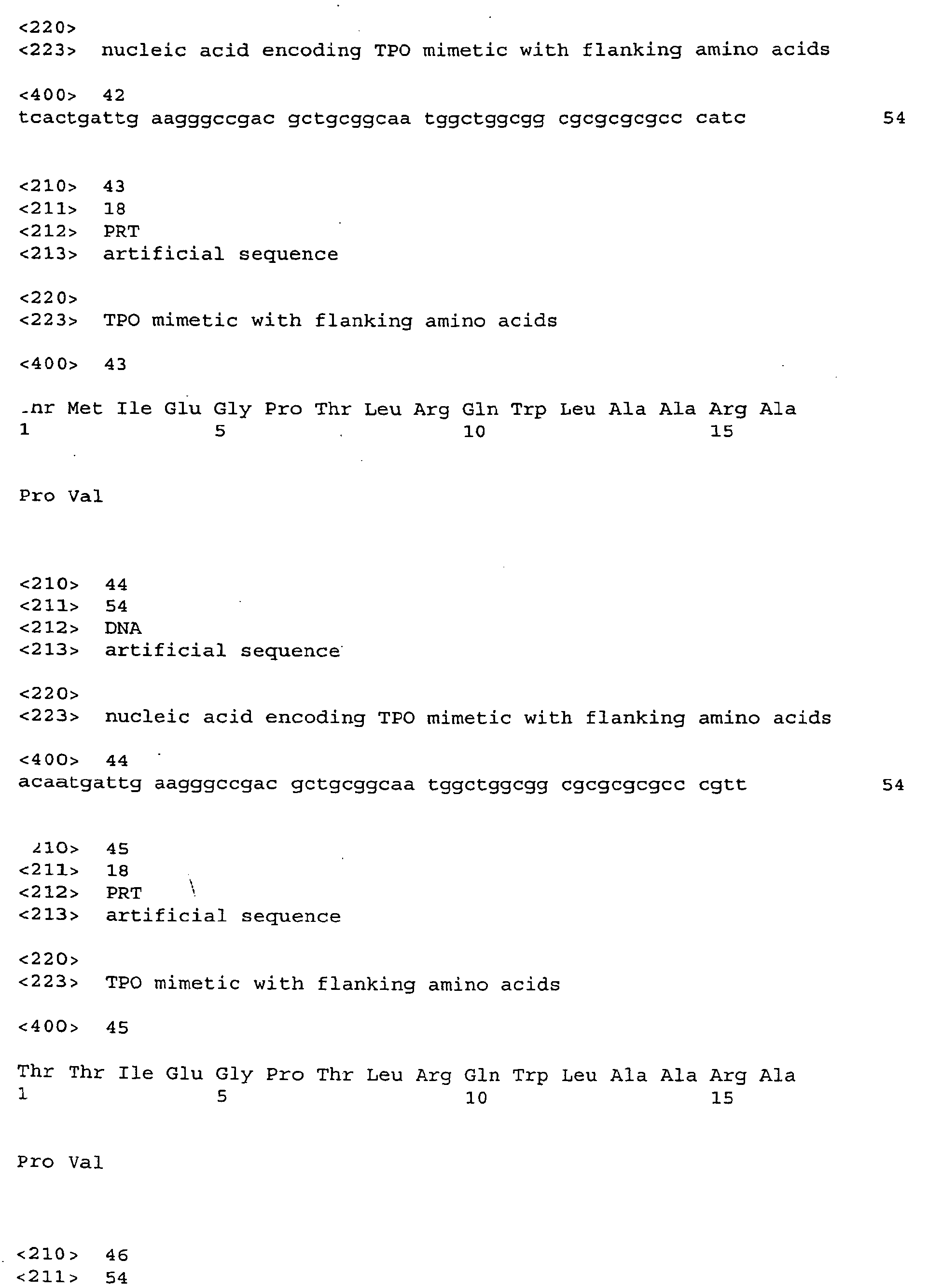
comprising the amino acid sequence of SEQ. ID. NOs: 25, 27, 29, 31, 33, 35, 37, 39,
41, 43, 45, 47, and 49 (see Fig. 5).
-
Another biologically active peptide that can replace the amino acid residues of a
CDR is an agonist EPO mimetic peptide. One such EPO agonist peptide has as its
amino acid sequence DYHCRMGPLTWVCKPLGG (SEQ. ID. NO: 3). Other amino
acid sequences are possible for EPO agonist mimetic peptides, which can be found
using binding, growth and activation assays known by those skilled in the art and as
described herein. Agonist EPO mimetic peptides when located in CDR regions can
also have one or more additional amino acid residues at the amino and/or carboxyl
termini of the peptide which become covalently bonded to immunoglobulin residues.
Thus, in particular embodiments provided herein are immunoglobulin molecules (IgG)
or fragments (e.g., Fab, scFv, heavy or light chains) that have a CDR region replaced
with a TPO or EPO mimetic peptide. For example, the TPO peptide can include at
least the sequence IEGPTLRQWLAARA (SEQ. ID. NO:1) and may further optionally
have an additional proline at the immediate downstream position. The EPO mimetic
encompasses at least the sequence DYHCRMGPLTWVCKPLGG (SEQ. ID. NO: 3).
Likewise, it may optionally have an additional proline at the immediate downstream
position.
-
Any immunoglobulin molecule (antibody) or fragment thereof could potentially
provide the framework and have a CDR replaced with a peptide according to the
present disclosure. For therapeutic or in vivo diagnostic use it is preferable that the
antibody is of human origin or humanized, such as an anti-tetanus toxoid
immunoglobulin. Furthermore, independent of or in conjunction with the presence of
additional flanking amino acids bound to the peptide, one or more amino acid residues
in other regions of the immununoglobulin, other CDR region(s) and/or framework
regions, can be altered to modify the binding, activity and/or expression displayed by
the peptide in the context of the immunoglobulin molecule.
-
It is contemplated that after construction of biologically active recombinant
antibodies and/or fragments thereof, such recombinants can be subjected to
randomization methods known in the art to introduce mutations at one or more points
in the sequence to alter the biológical activity of the antibodies. After generation of
such mutants using randomization methods such as those described herein, the
resulting recombinants may be assayed for activity using binding, growth, expression
and activation assays.
-
Further provided are nucleic acid molecules encoding immunoglobulin
molecules or fragments thereof which have the amino acids of one or more CDR
regions replaced by a biologically active peptide. These nucleic acid molecules can be
present in an expression vector, which can be introduced (transfected) into a
recombinant host cell for expression of these molecules. Also provided are methods
of producing an immunoglobulin molecule or fragment thereof containing a biologically
active peptide, comprising culturing a recombinant host cell under conditions such that
the nucleic acid contained within the cell is expressed.
-
Also provided are compositions, comprising an immunoglobulin molecule or
fragment thereof which has amino acid residues corresponding to a CDR replaced
with amino acid residues comprising a TPO or EPO mimetic peptide and a
pharmaceutically acceptable carrier.
-
Further provided are EPO mimetic peptides with additional flanking residues
which are suitable for replacement of CDRs. Also provided are nucleic acid molecules
encoding these peptides.
-
Further provided are methods of engineering immunoglobulin molecules or
fragments thereof to exhibit an agonist activity in which a biologically active peptide
replaces at least a portion of one or more CDR regions of light and/or heavy chains.
The methods encompass inserting a nucleic acid molecule encoding a biologically
active peptide in place of at least a CDR region of a nucleic acid molecule encoding an
immunoglobulin heavy or light chain or adding the molecule to the native CDR
sequence and then expressing the nucleic acid molecule encoding the immunoglobulin
heavy or light chain variable domain along with its complementary variable region
domain, such that the two domains associate.
-
Further provided are methods of engineering immunoglobulin molecules or
fragments thereof to exhibit an activity (property) of a biologically active TPO or EPO
peptide in which a biologically active peptide replaces one or more CDR regions of
light and/or heavy chains. The methods encompass inserting a nucleic acid molecule
encoding a biologically active peptide in place of at least a portion of a CDR region of
a nucleic acid molecule encoding an immunoglobulin heavy or light chain or adding the
molecule to the native CDR sequence; and expressing the nucleic acid molecule
encoding the immunoglobulin heavy or light chain variable domain along with its
complementarity variable region domain, such that the two chains associate.
-
In another aspect, this disclosure provides a method for producing in a
polypeptide a binding site capable of binding a preselected agent, the method
including the steps of introducing a nucleotide sequence that codes for an amino acid
residue sequence defining said binding site into a CDR region of a nucleic acid
comprising an immunoglobulin heavy or light chain gene by amplifying the CDR region
of the immunoglobulin gene, the introduced nucleotide sequence having the formula
-Xa-Y-Xb wherein X is the same or different at each occurrence and represents a
randomizing trinucleotide, the sum of a and b is 4 or less and Y is a nucleotide
sequence that encodes a minimum recognition domain of said binding site. In
particularly useful embodiments, amplification is achieved using overlap PCR,
however, any known amplification technique could be employed, such as, for example,
the methods disclosed in WO94/18221, the disclosure of which is incorporated herein
by reference.
-
In yet another aspect, this disclosure provides methods for creation of a library
of monoclonal antibodies that can be screened for a desired activity. These methods
of making a library include the steps of inserting a nucleic acid molecule encoding a
biologically active peptide into, or in place of at least a portion of, one or more CDR
regions of a nucleic acid molecule encoding an immunoglobulin heavy or light chain,
providing up to a pair of randomizing trinucleotides on either side of the inserted
nucleic acid molecule, and expressing a library of monoclonal antibodies. In
particularly useful embodiments, a pair of randomizing trinucleotides is provided on
both sides of the inserted nucleic acid molecules. The library of monoclonal antibodies
thus produced can then be screened for a desired activity.
-
In a specific embodiment, antibodies and fragments thereof have different
amino acids flanking the peptide at the amino and the carboxyl termini where the
peptide becomes bound to the antibody scaffold. This, results in a population of
antibody molecules or fragments thereof that may differ in the presentation of the
peptide. The population is screened for those antibodies that exhibit the biological
activity of the peptide. In a preferred embodiment, the amino acid immediately
adjacent the peptide is a proline.
-
If the activity of the biologically active peptide is to activate a target molecule,
this may require dimerization of two target molecules (e.g. receptors in the
hematopoietic superfamilies). For dimerization to occur, two peptides must be
positioned to each bind a target molecule such that the two bound target molecules
can then properly associate. This can be accomplished by having two peptides
present on the same antibody or fragment thereof or by causing two antibody
molecules each containing one peptide to bind together. Thus, for example, a single
peptide can be inserted into or substituted for at least a portion of a CDR and then
expressed as an immunoglobulin or a F(ab')2 fragment. As another example, two
peptides can be inserted into or substituted for at least a portion of one or more CDRs
and expresses as any antibody or antibody fragment.
-
The screening of antibodies or fragments thereof can be accomplished by
panning with cells that have surface molecules to which the peptide specifically binds.
Solid phase binding using purified target molecules or fragments thereof can also be
used. Binding can also be carried out in solution using labeled target molecules. In
addition, antibodies or fragments thereof can be screened by the use of biological
assays for agonist or antagonist activity of the peptide.
-
Also provided are libraries of different immunoglobulin molecules or fragments
thereof wherein amino acid residues corresponding to a complementarity determining
region (CDR) are replaced with amino acid residues comprising a biologically active
peptide which has at least one additional amino acid residue at the amino or the
carboxyl terminus and the immunoglobulin molecules or fragments thereof differ by the
additional amino acid residue of the peptide.
-
In specific embodiments the biologically active peptide is a TPO mimetic or an
EPO mimetic. The antibodies of the library are displayed on phage.
-
Further provided are methods of stimulating proliferation, differentiation or
growth of cells, which include contacting the cells with an effective amount of an
immunoglobulin molecule or fragment thereof having one or more CDRs replaced with
a biologically active peptide which binds to a receptor on the cells surface. In specific
embodiments the biologically active peptide is a TPO mimetic or an EPO mimetic.
-
In other specific embodiments, is provided a method of stimulating proliferation,
differentiation or growth of megakaryocytes by contacting megakaryocytes with an
effective amount of an immunoglobulin molecule or fragment thereof having one or
more CDRs replaced with a TPO mimetic peptide. Also provided is a method of
increasing platelet production, which involves contacting megakaryocytes with an
effective amount of an immunoglobulin molecule or fragment thereof having one or
more CDR regions replaced with a TPO mimetic peptide. Also provided is a method of
stimulating megakaryocytes and/or increasing platelet production in a patient, in which
an effective amount of an immunoglobulin molecule or fragment thereof having one or
more CDRs replaced with a TPO mimetic peptide is administered to a patient in need
thereof. The immunoglobulin molecule and the megagakarocytes can also be
contacted in vitro and the resultant cells can be introduced into the patient. In addition,
an antibody or fragment thereof having at least one TPO mimetic peptide incorporated
therein can be administered to a subject who intends to donate platelets, thus
increasing the capacity of a donor to generate platelets to provide a more robust
source of such platelets.
-
Also provided herein is a method of stimulating proliferation, differentiation or
growth of hematopoietic cells, comprising contacting the cells with an effective amount
of an immunoglobulin molecule or fragment thereof having one or more CDRs
replaced with a EPO mimetic peptide.
-
Also embodied herein is a method of activating a homodimeric receptor protein,
by contacting the receptor with an immunoglobulin molecule or fragment thereof
having a CDR region replaced with a biologically active peptide that specifically binds
the receptor and which has been dimerized. In a further embodiment the receptor is a
thrombopoietin receptor.
DESCRIPTION OF THE DRAWINGS
-
- Figure 1 is a diagrammatic representation of the vector pRL4.
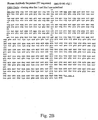
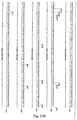
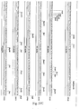
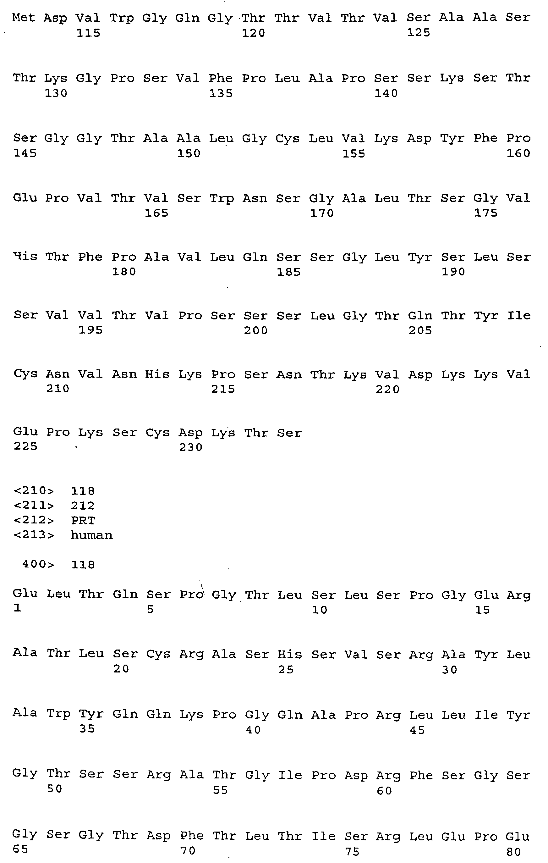
- Figures 2A and B show the sequence of the human tetanus toxoid antibody
framework, light and heavy chains, respectively.
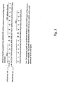
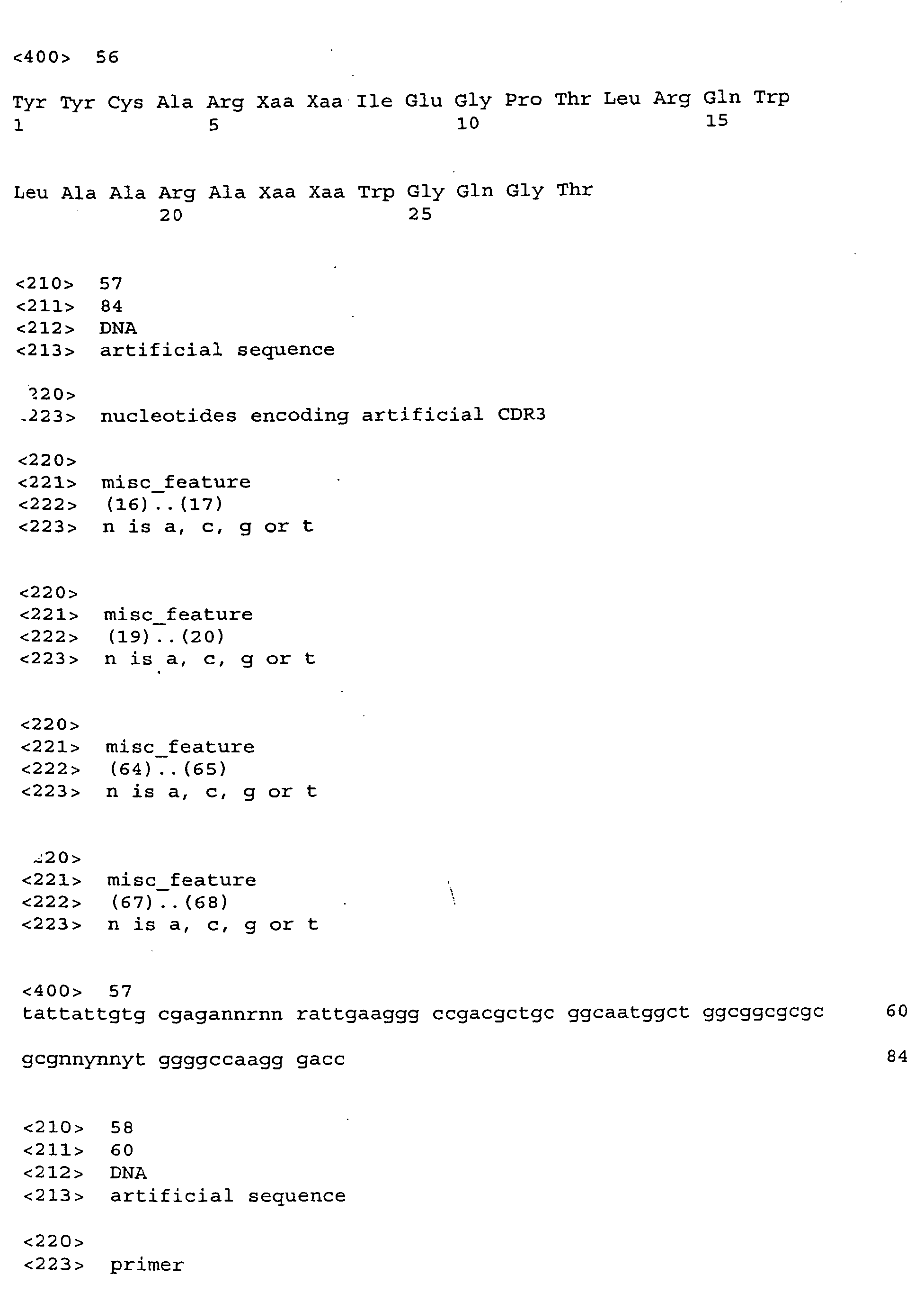
- Figure 3 is a diagram depicting the grafting of the TPO mimetic peptide
AF12505 into the heavy chain CDR3 region of the tetanus toxoid framework antibody.
XX represents flanking random amino acids.
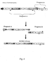
- Figure 4 is a diagram of the construction of a peptide cloned into the heavy
chain CDR3 region.
- Figure 5 represents the amino acid and nucleotide sequences of clones that
encode TPO mimetic peptide AF1205 with different random flanking residues.
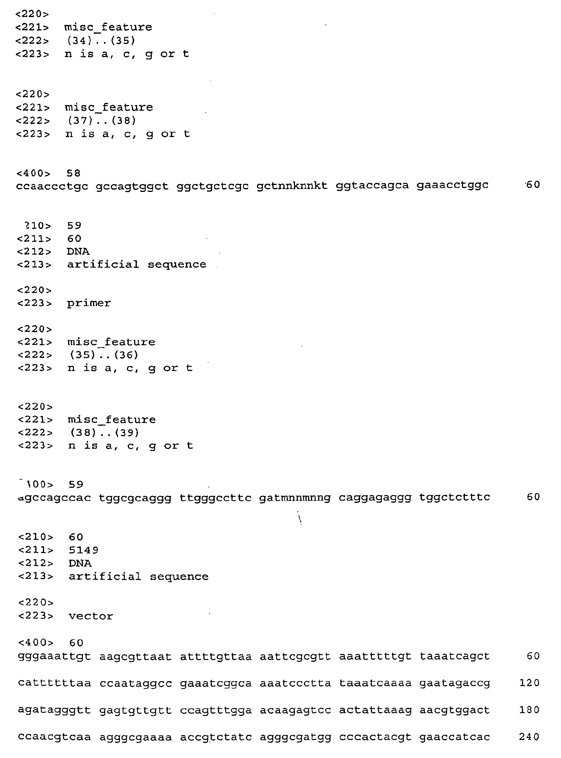
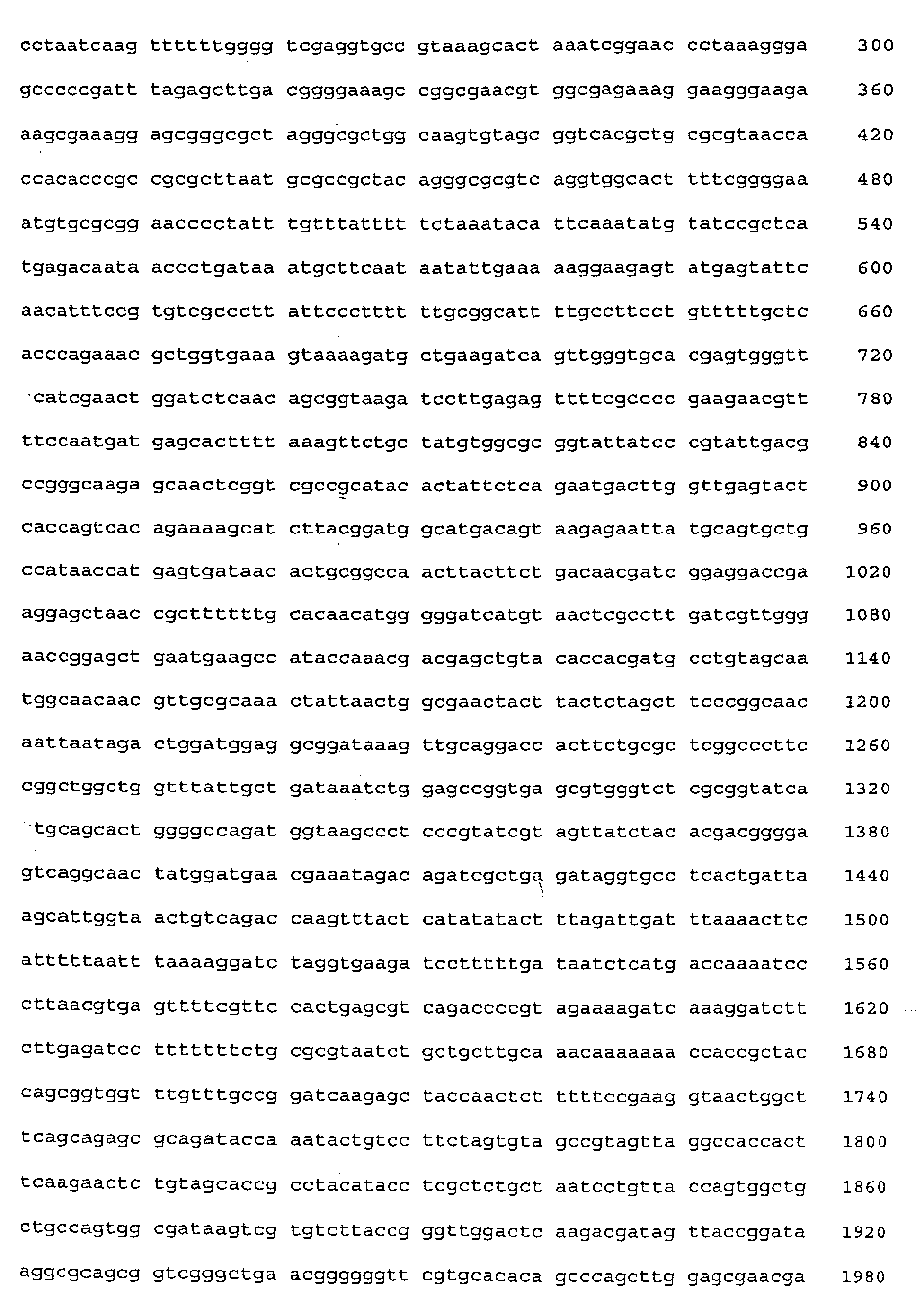
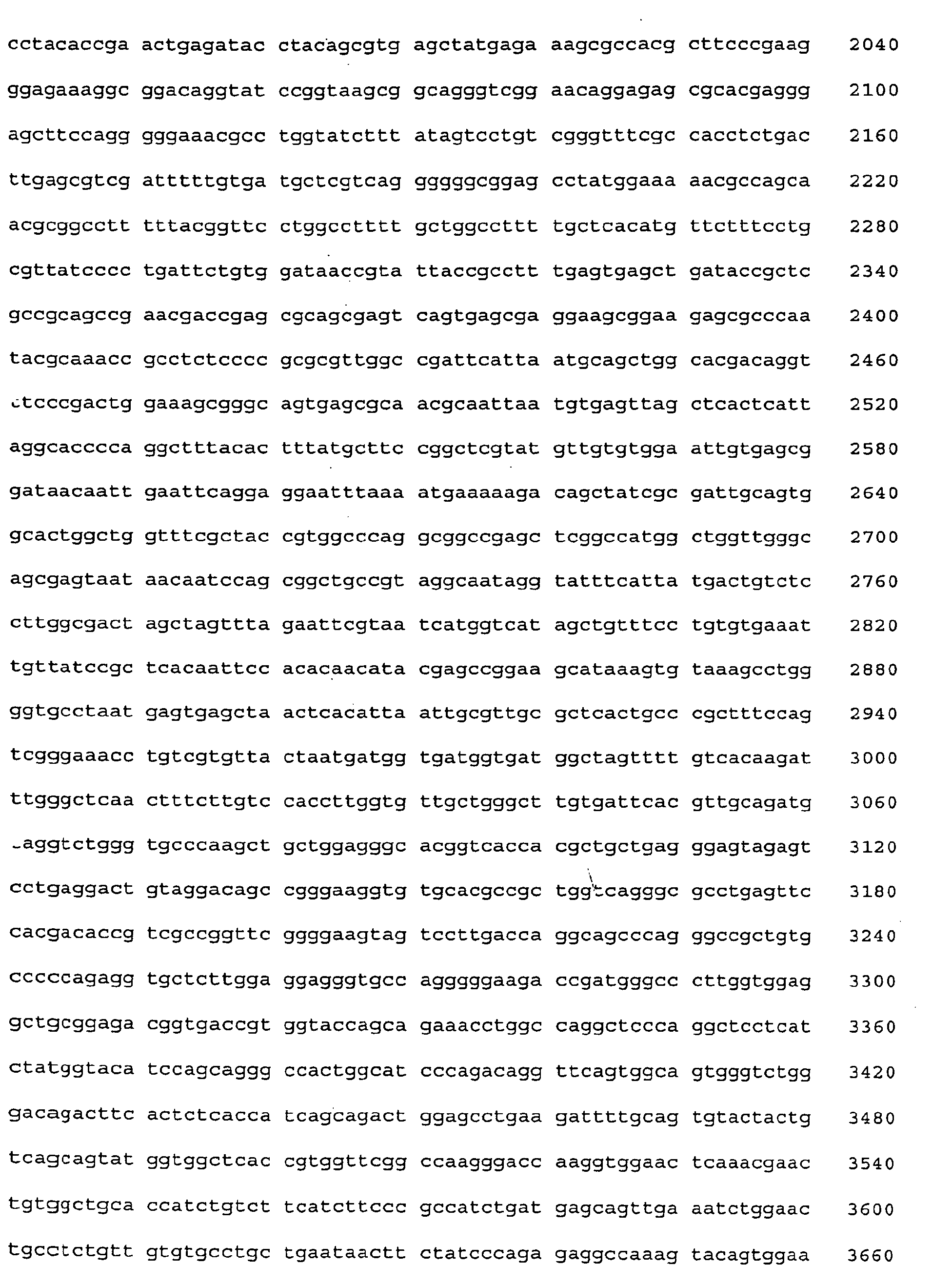
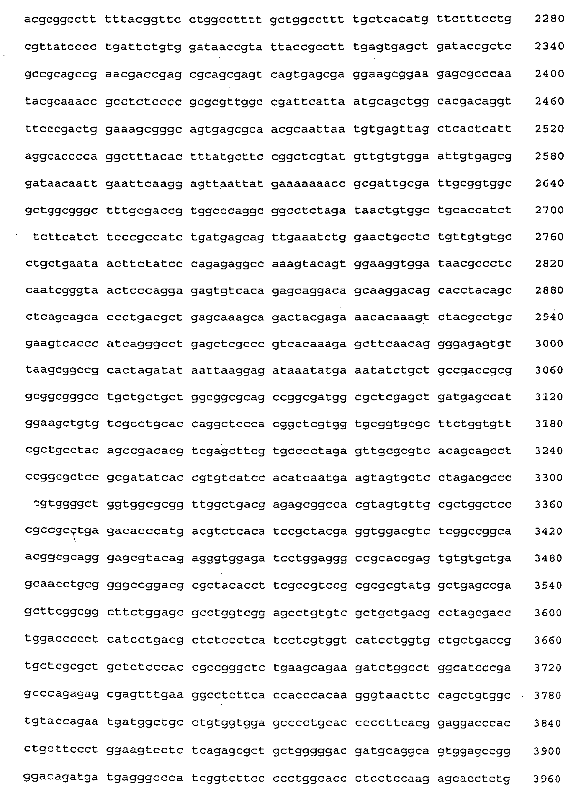
- Figure 6A-C depicts the nucleic acid sequence of plasmid pRL8 (SEQ. ID. NO:
60). pRL8 is a modified version of pRL4 (pRL4 is also known as pComb 3X). The
pRL4 was modified between the Spe I and neighboring Sfi I restriction sites (shown by
underlining) to include a flexible linker (murine kappa hinge region) followed by a Jun
leucine zipper dimerization domain.
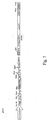
- Figure 7 is a schematic depiction of a portion of the plasmid pRL8.
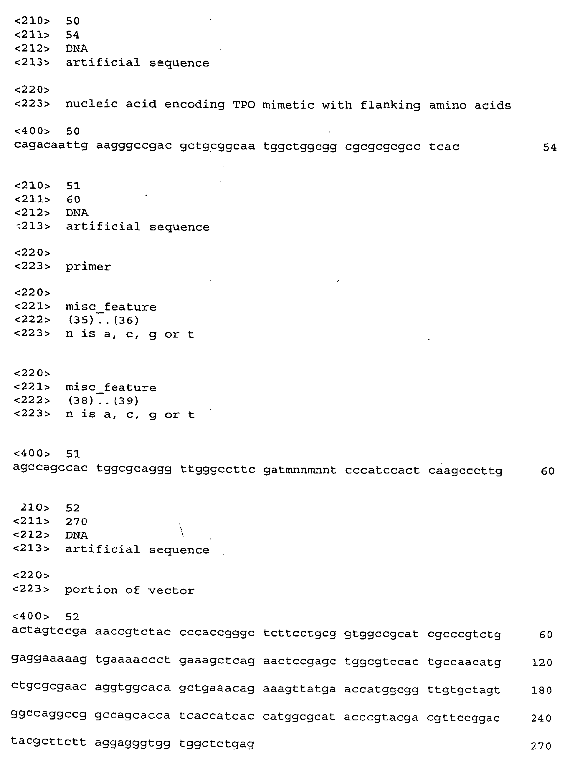
- Figure 8 depicts the nucleic acid sequence of a portion of plasmid pRL8 (SEQ.
ID. NO: 52) along with amino acid sequences corresponding to certain delineated
nucleic acid sequences (SEQ. ID. NO: 53).
- Figure 9 is a chart showing sequences of certain TPO positive clones herein.
- Figure 10 is a bar graph showing activity of certain Fab clones containing 2
TPO mimetic peptides.
- Figure 11 is a bar graph showing activity of certain Fab clones containing 2 or 3
TPO mimetic peptides.
- Figure 12 graphically depicts the activity of Clone 59 as reflected by induction of
luciferase activity.
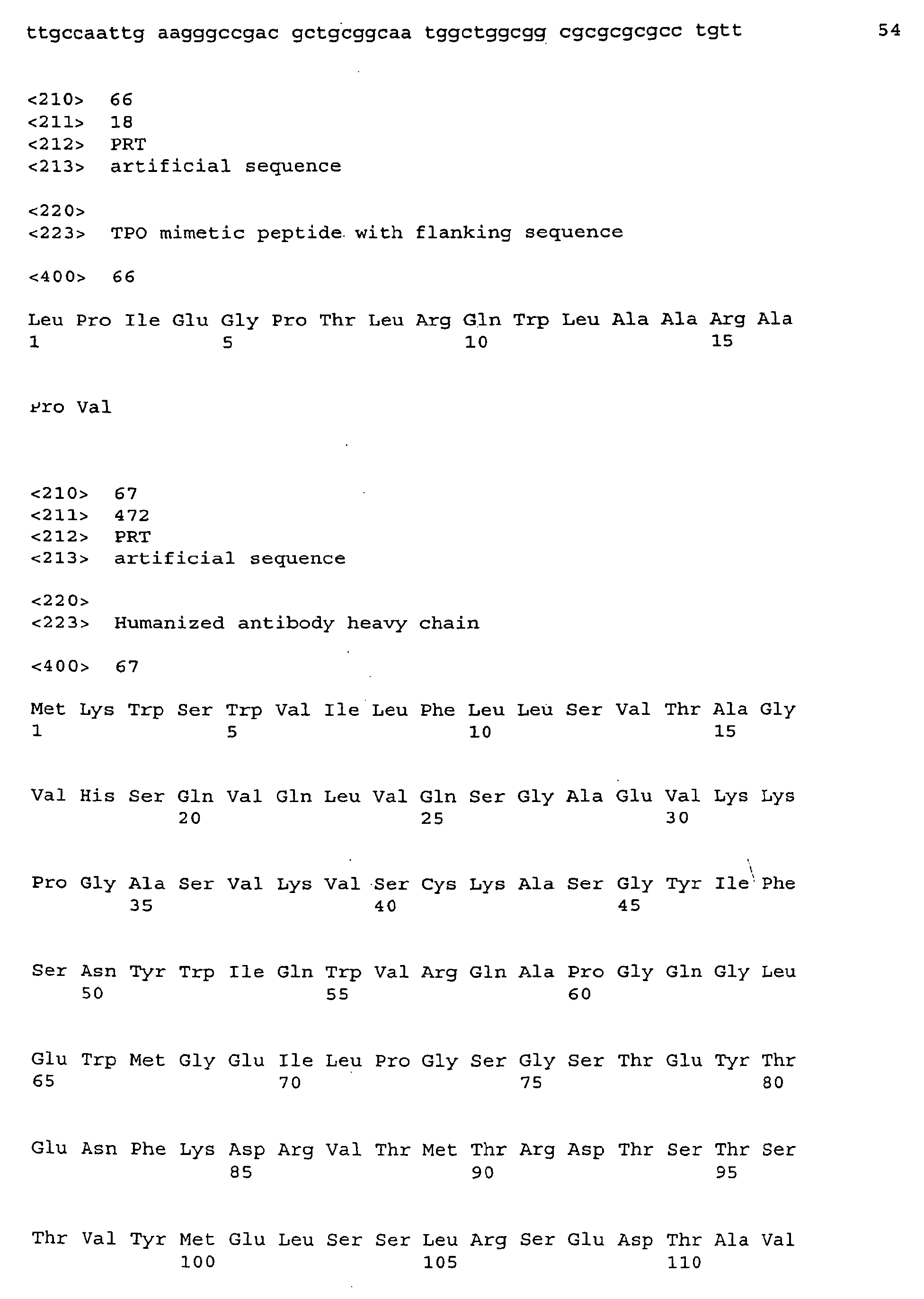
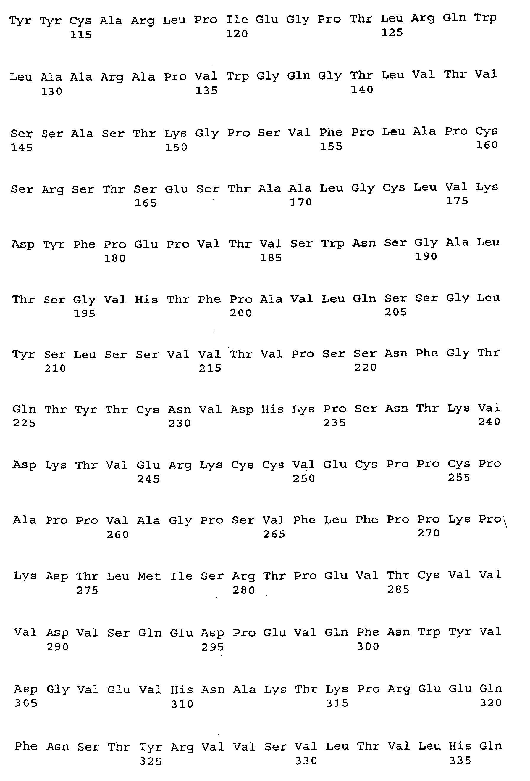
- Figure 13A depicts the amino acid sequence and nucleic acid sequence of the
5G1.1-TPO heavy chain (SEQ. ID. NOS: 67 and 68, respectively).
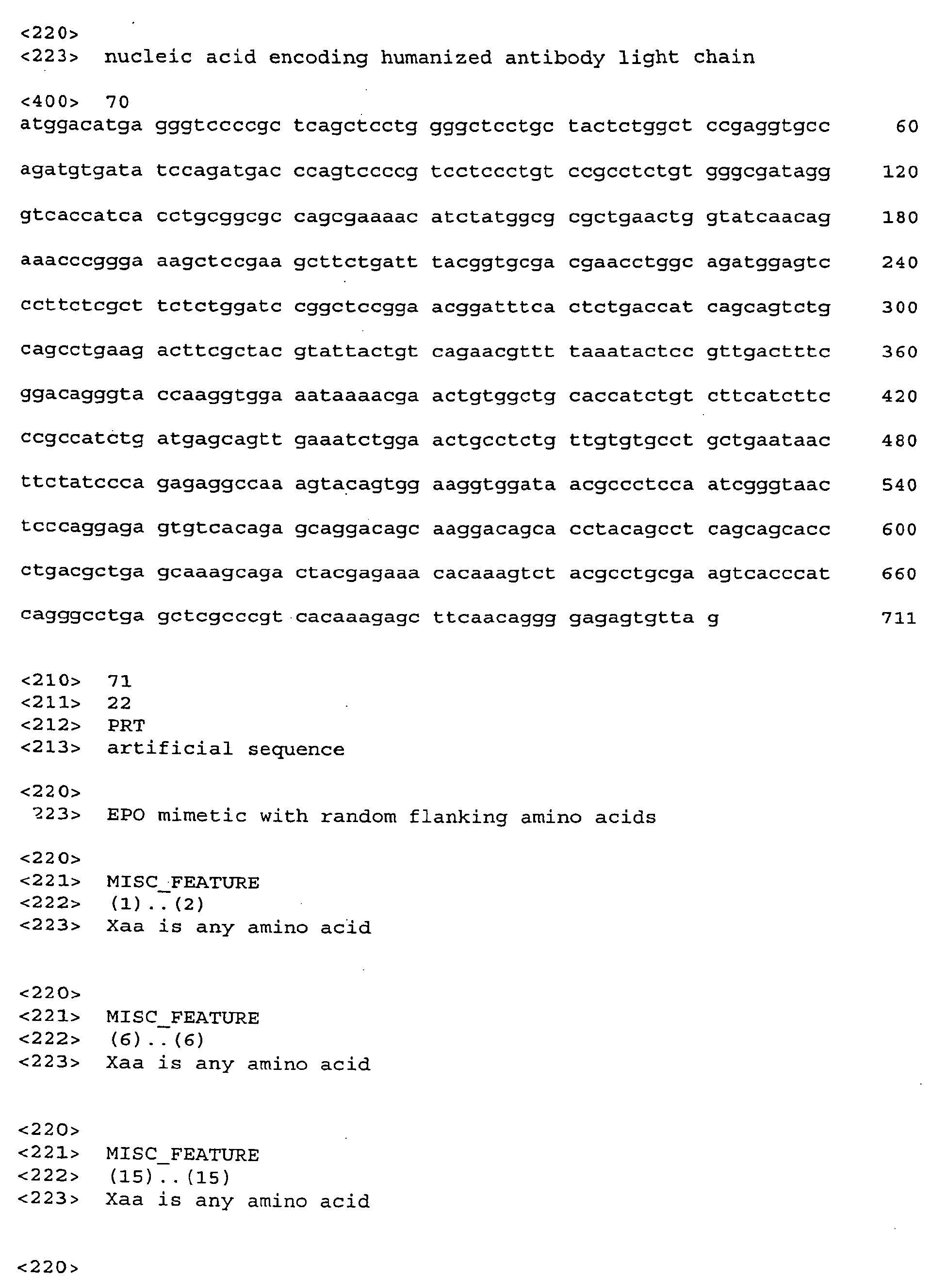
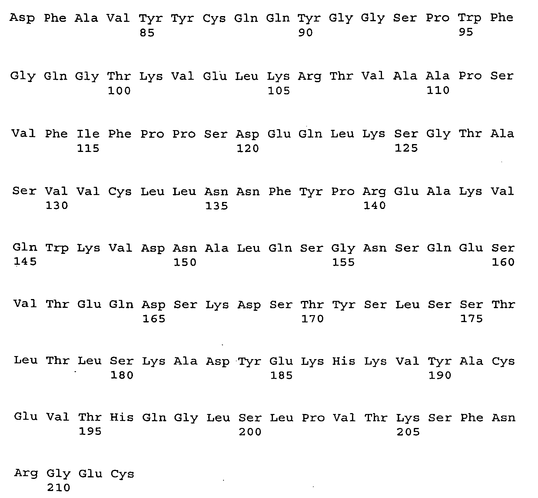
- Figure 13B depicts the amino acid sequence and nucleic acid sequence of the
5G1.1 light chain (SEQ. ID. NOS: 69 and 70, respectively).
- Figure 14 is a bar graph showing FACS analysis of cMpl receptor binding of
purified 5G1.1+ TPO mimetic peptide compared to parental 5G1.1 antibody.
- Figure 15 is a bar graph showing comparative activity of 5G1.1 antibody
containing the TPO mimetic peptide in connection with cells transfected with a control
vector containing no cMpl-R and cells transfected with a vector containing cMpl-R.
- Figure 16 shows the sequence of clone 429/Xb4 (SEQ. ID. NO: 116)
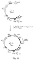
- Figure 17 is a flow chart showing the initial steps for making vector pRL5-Kappa.
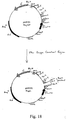
- Figure 18 is a flow chart showing additional steps for making vector pRL5-Kappa.
- Figure 19 is a map of vector pRL5.
- Figure 20 is a schematic of vector pRL5-Kappa.
- Figure 21A-I show the nucleic acid sequence of vector pRL5-Kappa.
-
DETAILED DESCRIPTION
-
As used herein, "immunoglobulin" refers to an entire immunoglobulin molecule
or molecules that contain immunologically active portions of whole immunoglobulin
molecules and includes Fab, F(ab')2, scFv, Fv, heavy chain variable regions and light
chain variable regions. The terms immunoglobulin and antibody are used
interchangeably herein.
-
Any peptide that exhibits a useful property is suitable for insertion in an antibody
framework. Peptide activities and uses include, but are not limited to, binding a
receptor, binding a membrane bound surface molecule, binding a ligand, binding an
enzyme or structural protein, activating or inhibiting a receptor, targeted drug delivery,
or any enzymatic activity. Those peptides whose utility can be increased from the
enhanced stability conferred upon them when presented in the context of an
immunoglobulin molecule are usually selected. It should be understood that
"biological activity" as used herein includes any activity associated with a molecule
having activity in a biological system, including, but not limited to, the stimulatory or
inhibitory activity triggered by protein-protein interactions as well as the kinetics
surrounding such interactions including the stability of a protein-porotein complex.
Enhancing or increasing "biological activity" herein is meant to include an increase in
overall activity or an increase in any component of overall activity. It should be
understood that a peptide may exhibit one biological activity (such as, e.g., simply
binding to a target) before insertion into the antibody framework, and a different or
enhanced biological activity (such as, e.g., agonist activity) after insertion into the
antibody framework.
-
Many peptides which could benefit from display in the context of an
immunoglobulin have been identified and are known to those who practice the art,
e.g., EPO and TPO mimetic peptides. Other examples include peptides that bind to
receptors which are activated by ligand-induced homo-dimerization including active
fragments displaying G-CSF activity, GHR activity and prolactin activity as described in
Whitty and Borysenko, Chem Biol., (1999) Apr 6(4):R107-18; other examples of
suitable peptides include a nerve growth factor mimetic from the CD loop as described
in Zaccaro et al., Med. Chem. (2000) 43(19); 3530-40; an IL-2 mimetic as described in
Eckenberg, et al., J. Immunol. (2000) 165(8):4312-8; glucogon-like peptide-1 as
described in Evans et al., Drugs R.D. (1999) 2(2): 75-94; tetrapeptide I (D-lysine-L-asparaginyl-L-prolyl-L-tyrosine)
which stimulates mitogen activated B cell proliferation
as described in Gagnon et al., Vaccine (2000) 18(18):1886-92. Peptides which exhibit
receptor antagonistic activity are also contemplated. For example, N-terminal peptide
of vMIP-II as an antagonist of CXCR4 for HIV therapy as described in Luo et al.,
Biochemistry (2000) 39(44):13545-50; antagonist peptide ligand (AFLARAA) of the
thrombin receptor for antithrombotic therapy as described in Pakala et al., Thromb.
Res. (2000) 100(1): 89-96; peptide CGRP receptor antagonist CGRP (8-37) for
attenuating tolerance to narcotics as described in Powell et al., Br. J. Pharmacol.
(2000) 131(5): 875-84; parathyroid hormone (PTH)-1 receptor antagonist known as
tuberoinfundibular peptide (7-39) as described in Hoare et al., J. Pharmacol. Exp.
Ther. (2000) 295(2):761-70; opioid growth factor as described in Zagon et al., Int. J.
Oncol. (2000) 17(5): 1053-61; high affinity type I interleukin 1 receptor antagonists as
disclosed in Yanofsky, et al., Proc. Natl. Acad. Sci. USA, Vol. 93, pp. 7381-7386, July
1996 and Vigers, et al., J. Biol. Chem., Vol 275, No 47, pages 36927-36933, 2000;
and acid fibroblast growth factor binding peptide as described in Fan et al., IUBMB Life
(2000) 49 (6) 545-48. Peptides can also be discovered using methods familiar to
those skilled in the art. In order to identify a region of a protein that is involved in a
specific biological function, a survey of the shorter peptide fragments making up that
protein may reveal the linear peptide epitope responsible. Alternatively by surveying
libraries of random peptides, a peptide that represents an optimal linear epitope or a
discontinuous epitope may be discovered that mimics the activity of the natural
protein. One method for selection is termed peptide phage-display. In this approach,
a random peptide epitope library is generated so that peptides are present on the
surface of a bacteriophage particle. These collections, or libraries, of peptides can
then be surveyed for those able to bind to a specific immobilized target protein.
(Pasqualini, R. et al., j. Cell Biol., 130, 1995, 1189-1196; Wrighton, N.C., et al.,
Science, 273, 1996, pages 458-463; Cwirla, S.E., et al., Science, 276, 1997, pages
1696-1699; Koivunen et al, J. Biol, Chem., 268, 1993, pages 20205-20210; Koivunen
et al., Bio/Technol., 13, 1995, pages 265-270; Healy et al., Biochem., 34, 1995, pages
3948-3955; Pasqualini et al., J. Cell Biol., 130, 1995, pages 1189-1196). Alternative
peptide selection systems are also possible including cell surface display and
ribosomal display.
-
Peptide mimetics used in accordance with this description are generally less
than or equal to the number of amino acid residues that make up a CDR region,
although they could be longer.
-
Any antibody can serve as a scaffold sequence, however typically human
antibodies are chosen as human therapeutics is one of the ultimate objectives.
Human or humanized antibodies are less likely to cause an adverse immune response
in a human patient. The major criteria in selecting an antibody to serve as a
framework for insertion of a peptide, is that the replacement of one or more CDRs of
the antibody with the peptide must change the antigen specificity. The antibody can
be a complete antibody or an Fab, scFv or F(ab')2 fragment or portion thereof.
-
Alternatively, a library of antibodies can have one or more heavy and/or light
chain CDRs replaced with a desired peptide. The resulting library can then be
screened to identify antibodies having a desired activity. It should be understood that
randomization with in the substituted peptide can also be provided to generate an
antibody library.
-
A useful antibody is the anti-tetanus toxoid (TT) Fab, as it is human and
because modification of the HCDR3 is sufficient to change the antigen specificity of
the antibody (Barbas et al., J. Am. Chem. Soc., 116, 1994, pages 2161-2162 and
Barbas et al., Proc. Natl. Acad. Sci. USA, 92, 1995, pages 2529-2533).
-
Grafting of the DNA sequence of the peptide of choice into an antibody so as to
replace the CDR(s) of an antibody with the peptide sequence is carried out using
recombinant DNA techniques known to those skilled in the art.
-
Examples of methods which can be utilized to graft a desired peptide having
biological activity in place of a CDR region include, but are not limited to, PCR overlap,
restriction enzyme site cloning, site specific mutagenesis and completely synthetic
means. For a description of techniques involving overlap PCR, see, e.g., Example 1
herein. Site specific mutagenesis can be accomplished in several ways. One is
based on dut/ung Kunkel mutagenesis (Kunkel, T.A., Proc. Natl. Acad. Sci. (1985) vol.
82, pp. 488-92). The Muta-Gene in Vitro Mutagenesis kit is available from BioRad
based on this methodology (cat. # 170-3581 or 170-3580). Several PCR amplification
based mutagenesis approaches are also commercially available such as Stratagene's
QuickChange Site-Directed Mutagenesis Kit and the ExSite PCR-based Site-Directed
Mutagenesis Kit. Another non-PCR method is available from Promega as the
GeneEditor in vitro Site-Directed Mutagenesis System. Completely synthetic means
are also well-known and described, e.g., in Deng, et al., Methods Mol. Biol. (1995)
51:329-42; Kutemeler et al., Biotechniques, (1994) 17(2): 242-246; Shi et al., PCR
Methods Appl., (1993) 3(1): 46-53 and Knuppik et al., J. Mol. Biol., (2000) 11:296(1):
571-86 each incorporated herein by reference. In addition, the above methods used
for replacing all or a portion of at least one CDR sequence can be utilized to graft a
desired peptide into or adjacent to at least one native CDR sequence without replacing
the original CDR sequence. In this manner, a CDR/biologically active peptide mimetic
fusion construct is formed.
-
It is contemplated that flanking sequences may be added to the carboxyl and/or
amino terminal ends of the biologically active peptide. Flanking sequences can be
useful to reduce structural constraints on the grafted peptide to allow it to more easily
adopt a conformation necessary for biological activity. In a preferred embodiment, a
flanking region including a proline is covalently attached to the carboxy terminus of the
biologically active peptide to create a proline extended biologically active peptide.
-
In one embodiment, a flanking region can be generated by randomizing two
amino acid positions on each side of the peptide graft in order to determine the best
sequence. In this manner, a library having members with multiple varied sequences
can be generated. The resulting constructs are then tested for biological activity as
described below by, e.g., panning techniques. Recombinant proteins can be
generated that have random amino acids at specific positions. This can be
accomplished by modifying the encoding DNA. When introducing randomization at a
specific amino acid's codon position, a preferable deoxyribonucleotide "doping
strategy" is (NNK)
x in order to cover all 20 amino acids and to minimize the number of
encoded stop codons. Accordingly, N may be A, C, G, or T (nominally equimolar), K is
G or T (nominally equimolar), and x is typically up to about 5, 6, 7, or 8 or more,
thereby producing libraries of mono-, di-, tri-, quadra-, penta-, hexa-, hepta-, and octa-peptides
or more. The third position may also be G or C, designated "S". Thus, NNK
or NNS (i) code for all the amino acids, (ii) code for only one stop codon, and (iii)
reduce the range of codon bias from 6:1 to 3:1. There are 32 possible codons
resulting from the NNK motif: 1 for each of 12 amino acids, 2 for each of 5 amino
acids, 3 for each of 3 amino acids, and only one of the three stop codons. Other
alternatives include, but are not limited to:
- (NNN)x which would provide all possible amino acids and all stops;
- (NNY)x eliminates all stops and still cover 14 of 20 amino acids;
- (NNR)x covers 14 of 20 amino acids; and
- (NNS)x covers all 20 amino acids and only one stop.
-
-
The third nucleotide position in the codon can be custom engineered using any
of the known degenerate mixtures. However, the group NNK, NNN, NNY, NNR, NNS
cover the most commonly used doping strategies and the ones used herein.
-
The collection of engineered antibodies that are created during this process can
be surveyed for those that exhibit properties of the peptide as, e.g., phage displayed
antibodies, essentially as has been described in Barbas, C.F., III, Kang, A.S., Lerner
R.A., and Benkovic, S.J., Assembly of combinatorial antibody libraries on phage
surfaces: the gene III site, Proc. Natl. Acad. Sci. USA, 88, 1991, pages 7978-7982
incorporated herein by reference. This technology allows recombinant antibodies (as
complete antibodies, Fab F(ab')2, or scFv) to be expressed on the surface of a
filamentous bacteriophage. That same phage will have within it the genes encoding
that specific antibody.
-
It is contemplated that any other known method of introducing randomization
into a sequence may be utilized herein. For example, error prone PCR can introduce
random mutations into nucleic acid sequences (See, e.g., Hawkins et al., J. Mol. Biol,
(1992) 226(3): 889-96). Briefly, PCR is run under conditions which compromise the
fidelity of replication, thus introducing random mutations in sequences as those skilled
in the art would accomplish. After generation of such random mutants, they can be
placed into phage display formats, panned and thus evaluated for activity. Likewise,
particular bacteria known to provide random mutations of genes, such as Epicurian
Coli® XL1-Red Competent cells (commercially available from Stratagen, La Jolla,
CA.), which do so during plasmid replication can be utilized to provide random mutants
which are then screened for biological activity in accordance with the present
disclosure.
-
It is also contemplated that randomization may be introduced at any point in the
nucleotide sequence after incorporation of an active peptide into the antibody or
fragment thereof to alter the overall biological activity of the antibody. In this manner,
not only can alterations be made in the biological activity of a peptide mimetic by
causing mutations within the peptide's sequence, but mutations in the surrounding
scaffold can be incorporated with the resulting constructs being assayed for alterations
in biological activity or expression. Indeed, it is contemplated that libraries having
repertoires of multiple constructs resulting from such randomization can be generated
and assayed.
-
Single chain libraries can be utilized in accordance with the present disclosure
because an entire binding domain is contained on one polypeptide. The light chain
variable region is separated from heavy chain variable region by a linker region. The
use of short linkers (< 11 amino acids) favors a dimeric complex where VH of one ScFv
associates with VL of another ScFv molecule and visa versa, these molecules are
termed diabodies (Kortt, A.A., Malky, R.L., Caldwell, J.B., Gruen, L.C., Ivanci, N.,
Lawrence, M.G. et al. Eur. J. Biochem. 221:151-157, 1994). This is because folding of
monomeric ScFv is impaired with linkers < 11 amino acids (Alfthan, K., Takkinen, K.,
Sizman, D., Soderlund, H., and Teeri, T.T. Protein-Eng. 8:725-731, 1995). Longer
linkers (> 11 amino acids) favors folding of monomeric ScFv into a single antigen
binding domain, thus precluding dimer formation.
-
One useful phage display vector is pRL4 which is also known as pComb 3X
(see Fig. 1). This vector enables display of chimeric expression products on the
surface of packaged phagemid particles. pRL4 is a modified version of pComb3H
(Barbas, C.F. III and Burton, D.R. 1994. Monoclonal Antibodies from Combinatorial
Libraries. Cold Spring Harbor Laboratory Course Manual, Cold Spring Harbor, N.Y.;
Burton, D.R.; Barbas, C.F. III. Advances in Immunology 57:191-280, 1994; Lang, I.M.,
Chuang, T.L., Barbas, C.F. 3rd, Schleef, R.R. J. Biol. Chem. 271: 30126-30135, 1996;
Rader and Barbas, Phage Display, A Laboratory Manual, Cold Spring Harbor
Laboratory Press, Cold Spring Harbor, N.Y. (2000). The design of pRL4 allows for
dimerization of scFv antigen binding domains on the phage surface and in soluble
form as detailed below. When the plasmid is transformed into a supE bacterial host
such as ER2537 (F' Sup E, New England Biolabs, Beverly, MA), the amber mutation is
suppressed approximately fifty percent of the time. In this way half of the expressed
scFvs are fused with the filamentous phage gene III protein (amino acids 230-406) and
the other half will be terminated just prior to gene III to produce soluble scFv. Both the
scFv-pIII fusion and soluble scFv products have the Omp A signal sequence and will
be transported to the periplasm where they will be able to form dimeric scFv
complexes, termed diabodies (Kortt, A.A., Malby, R.L., Caldwell, J.B., Gruen, L.C.,
Ivanci, N., Lawrence, M.C. et al. Eur. J. Biochem. 221: 151-157, 1994). Diabodies are
expected to fold such that the VH of one scFv will pair with the VL of a second scFv-pIII
resulting in divalent antibody fragments. In a non-sup E host, such as TOP1OF'
(InVitrogen, Carlsbad, CA), the amber stop codon is recognized yielding soluble scFv
diabodies.
-
In the final single chain expression construct in pRL4, the single chain antibody
fragments are cloned downstream of the E. coli IacZ promoter, ribosome binding site,
and omp A leader sequence. These elements allow induction of expression by IPTG,
and the secretion out of the cell via the omp A leader sequence when expressed in the
suppressor strain ER2537. The single chain fragments are fused in frame with
filamentous phage gene III (gIII) sequences (amino acids 230-406). The gIII protein
product, pIII, is a minor coat protein necessary for infectivity. Upon promoter induction
by IPTG, the single chain antibody-pIII fusion is synthesized and transported to the
bacterial periplasmic space. In the periplasmic space, the scFv-gene III fusion
proteins are inserted into the membrane. Upon superinfection with helper phage,
these fragments are exported out of the cell on the surface of phage as pIII-antibody
fragments. Other possible proteins to be used for fusion on the surface of phagemids
include filamentous coat protein pVIII and other coat proteins.
-
Fab fragment libraries, that maintain the native antigen recognition site, are
useful to ensure that affinity is maintained.
-
In the final hybrid Fab expression construct in pRL4, the light and heavy chains
are cloned as a single Sfil fragment. In this way, the light chain fragments are cloned
downstream of the E. coli IacZ promoter, ribosome binding site, and omp A leader
sequence. These elements allow induction of expression by IPTG, and secretion out
of the cell via the omp A leader sequence. The light chain fragments are followed by a
stop codon, a second ribosome binding site, the E. coli pel B leader sequence and
heavy chain. Hybrid heavy chain genes are fused in frame with filamentous phage
gene III (gIII) sequences (amino acids 230-406). An amber stop codon is present at
the fusion junction. In a sup E bacterial host such as ER2357 (New England Biolabs,
Beverly, MA), the amber mutation is suppressed. Upon promoter induction, a single
polycistronic message is transcribed and translated as two polypeptides, a light chain
and a heavy chain-gene III fusion protein. Following synthesis the polypeptides are
transported to the bacterial periplasmic space as directed by the leader sequences. In
the periplasmic space the heavy chain-pIII fusion proteins are inserted into the
membrane, and the light and heavy chains are associated covalently through disulfide
bonds, forming the antigen binding sites. The human constant region CH1 and CL
sequences include the cysteines that form the disulfide bond between heavy and light
chains. Upon superinfection with helper phage, these fragments are exported out of
the cell on the surface of phage as Fab-cplll fusion. In a non-sup E host, such as
TOP1OF' (Invitrogen, Carlsbad, CA), the amber stop codon is recognized yielding
soluble Fab fragments. Important features of the pRL4 phage display system used
include a purification His 6 tag, an HA epitope tag following the heavy chain, as well as
a suppressible amber stop codon which is located between the heavy chain and the
phage gene III. The HA tag is recognized by HA.11 antibody (Babco, Berkeley, CA)
and 12CA5 antibody (Roche Molecular Biochemical, Indianapolis, Ind.). The His6 tag
allows affinity purification of antibody fragments by Nickel-chelate chromatograph
(Qiagen, Valencia, CA). The amber stop allows for quick conversion from a fusion
Fab-cpIII product (for incorporation on the phage coat) when the stop is suppressed, to
the soluble Fab which is made in a non-suppressor bacterial host.
-
Selection involves isolating from the library the best candidates that specifically
bind to the peptides target molecule and display biological activity.
-
The phage expressing antibody fragments on their surface can be produced
and concentrated so that all members of a library can be allowed to bind to the target
molecule. The target molecule can be immobilized on a microtiter dish, on whole cells,
the membranes of whole cells, or present in solution. Non-specific Ab-phage are
washed away, and bound phage particles are released from the antigen, often by the
use of low pH. The recovered Ab-phage are infectious and so can be amplified in a
bacterial host. Typically, multiple rounds of this sort of selection are performed.
Individual antibody fragment clones can then be analyzed as soluble Fabs or scFvs for
identification of those that specifically recognize the target molecule.
-
Prior to any selection strategy, initial libraries are electroporated into host cells,
such as ER2537. Library cultures are grown to log phase and superinfected with
helper phage, such as VCSM13, a commercially available helper phage (Stratagene,
La Jolla, CA). Superinfection provides the remaining phage components needed for
packaging plasmids into phagemid particles. Alternatively, phage display without the
use of helper phage may be utilized. Following overnight growth, phagemids in the
culture supernate are precipitated with polyethylene glycol (PEG). PEG precipitated
phage are used in panning (solid phase cell surface, internalization and membrane),
FACS sorting, or magnetic sorting to purify specific binding antibodies from non
specific binders.
-
In cell based panning, antibody-phage libraries are incubated with target cells,
and the non-adherent phage are removed with multiple washes. A typical panning
protocol is as follows:
- 1. Block phage particles with PBS + 1%BSA or 10% FBS + 4% milk powder +
NaN3, (except when internalized antibodies are assayed).
- 2. Add target cells to blocked phages (approximately 5 X 10⁶
cells).
- 3. Mix and rotate slowly at 4° C or 37 ° C.
- 4. Wash cells twice with 1 ml ice cold PBS/1%BSA/NaN3 or room temperature
PBS/1%BSA/NaN3.
- 5. Specific antibody-phage bound to cells can be eluted by low pH, for example
with 76 mM citric acid ph 2.5 in PBS for 5 to 10 minutes at room temperature.
- 6. Neutralize eluted phage with 1M Tris-HCl pH 7.4.
- 7. After neutralization, antibody-phage can be used to infect ER2537 bacteria
and amplify during overnight growth for the next round of panning.
-
-
Generally, 3-4 rounds of panning are performed on each library. Phage ELISAs
using commercially available secondary antibody (sheep anti-M13 antibody-HRP) or
soluble antibody ELISAs using a commercially available HA. 11 antibody (Babco,
Berkeley, CA) that recognizes the HA tag incorporated into each antibody from PRL4
sequences, can be performed following each round of panning to allow estimation of
the enrichment of binding antibodies over non-binders. Following the last round of
panning, the antibody-phage can be picked as single colonies from agar plates, grown
as monoclonal antibody-phage and screened by ELISA for identification of specific
binders. FACS analysis may also be utilized. Specifically the antibody-phage are
infected into Top10F' bacteria and plated for single colonies. Single colonies are
picked form agar plates, grown and induced with IPTG. Soluble antibody is screened
by ELISA for identification of specific binders. Screening can be done against live
cells, against intact, mildly fixed target cells, or recombinant protein(s).
-
Methods for whole cell panning have been described previously (Siegel, D.L.,
Chang, T.Y., Russell, S.L., and Bunya, V.Y. 1997. J. Immunol. Methods 206:73-85
incorporated herein by reference). Other techniques for selection which can be
applied include fluorescent activated cell sorting (FACs). Alternative methods for
selection using libraries include, but are not limited to, ribosome display and plaque
hybridization to a labeled antigen.
-
Following panning to isolate high affinity antibody binders, bioassays for
functional screens of agonist antibodies can be carried out. Dimerization is often a
prerequisite for activation of many receptors and thus bioassays focus on agonist
antibodies that stimulate receptors via promotion of dimerization. As previously
described, single chain multivalency is approached in linker design. Fab fragment
multivalency can be approached in a number of ways. A number of recent reports in
the literature have shown success in dimeric antibody fragment formation which is
applicable to phage display (DeKruif, J., and Logtenberg, T. 1996. J. Biol. Chem.
271:7630-7634, Pack, P., and Pluckthun, A. 1992. Biochemistry 31:1579-1584, and
Holliger, P., and Winter, G. 1993. Current Opin. Biotech. 4:446-449). Divalent Fabs
can be created in at least two ways. In one approach dimerization is achieved by
addition of a dimerization domain to pRL4, forming pRL8 (See Figs. 6A-C, 7 and 8).
There are a number of dimerization domains (lexA, Zn fingers, fos, jun etc.) that can
be utilized in these vectors to obtain multivalency of Fab fragments. Dimerization
domains are selected from, but not limited to, the following: jun (DeKruif, J. and
Logtenberg, T. J. Biol. Chem. 271:7630-7634, 1996; Kostelny, S.A., Cole, M.S., and
Tso, J.Y. J.Immunol. 148:1547-1553, 1992) the LexA dimerization region (Kim, B. and
Little, J.W. Science 255:203-206, 1992), the yeast GCN4 dimerization domain (van
Heeckeren, W.J., Sellers, J.W., Struhl, K. Nucleic Acids Res. 20:3721-3724,1992), Gin
invertase from the bacteriophage Mu (Spaeny-Dekking, L., Schlicher, E., Franken, K.,
van de Putte, P., Goosen, N. J. Bacteriol. 34:1779-1786, 1995), E. coli NTRC protein
dimerization domain (Klose, K.E., North, A.K., Stedman, K.M., Kustu, S. J. Mol. Biol.
241:233-245, 1994), and HSV-1 ICP4 dimerization domain (Gallinari, P., Wiebauer, K.,
Nardi, M.C., Jiricny, J. J. Virol. 68:3809-3820, 1994) all incorporated by reference.
Also, a high temperature dimer domain from thermus organisms can be utilized
(MacBeath, G., Kast, P., Hilvert, D., Biochemistry 37:100062-73, 1998 and MacBeath,
G., Kast, P., Hilvert, D., Science 279:1958-61, 1998). These are functional domains
that when incorporated into a molecule allow for dimerization to occur. In addition,
dimerization can be achieved in cells through the use of full IgG vectors, or
dimerization domains such as CH3 dimerization domains. Those of ordinary skill in
the art are familiar with these and other dimerization domains and their use to dimerize
proteins.
-
Additional methods that may be utilized to generate antibody constructs
which contain at least two binding sites are known. The antibody or antibody
fragments created by each of these approaches could be utilized for testing agonistic
antibody activity as described in Example 1 below for whole IgG produced in
mammalian cells. These methods include chemical dimerization of Fab, pegylation of
Fab, production of Fab'2, generation of whole IgG in bacterial cells, and use of
diabodies (scFvs). Importantly, any of the antibody forms generated for analysis of
agonistic activity could be used as the final therapeutic product.
-
Chemical dimerization may be also achieved using a variety of chemical
crosslinking reagents. For example, SMCC (Succinimidyl trans-4 (maleimidylmethyl)
cyclohexane-1-carboxylate) from Molecular Probes (Eugene, Oregon), Cat # S-1534.
This reagent will modify primary amino groups in the antibody. After incubating the
antibody with the SMCC at room temperature, the reaction is run over a PD-10
column. This maleimide derivitized Fab can be added to either a second Fab or a
separate batch of the same Fab that has been treated with TCEP [(Tris (2-carboxyethyl)
phosphine, hydrochloride): Molecular Probes Cat #T-2556] to reduce
the thiol groups to SH. The reduction reaction is carried out in the dark for 15 minutes.
The conjugation of the maleimide Fab and the thiol reduced Fab occurs at a 1:1 ratio.
Dimers are isolated by passing the reaction over a sephadex 200 gel filtration column.
Other chemical linkers known to those skilled in the art may be used for dimerization.
Production of an Fab' that has an extra cysteine residue engineered into the hinge
region has been described, e.g., in U.S. Patent 5,677,425 and Carter, et
al.,BioTechnology, Vol 10, Feb. 1992, pages 163-167 the disclosures of which are
hereby incorporated by reference. That thiol site can be used for conjugation to
moieties such as polyethyleneglycol (PEG). Pegylation technology is known, for
example, see Koumenis et al, Int. J. Pharm. (2000) 198(1): 83-95, incorporated herein
by reference, which makes it possible to link two Fab' molecules together using PEG
coupling.
-
Technology for bacterially producing Fab'2 involves cloning the human IgG
hinge region, and optionally part of the CH2, as part of the Fd which includes
additional cysteines and is described, e.g., in Better, et al., PNAS USA (1993) 90(2):
457-61, incorporated herein by reference. The additional thiol groups on the Fd hinge
can interact and cause two Fab' molecules to dimerize, creating a Fab'2. Fab'2 can
be purified directly from the bacterial cells. Additionally, undimerized Fab' from the
bacteria can be isolated and chemically converted to Fab'2.
-
As described earlier, the variable regions of the antibody can be cloned as
a single chain wherein the variable light (VL) is connected to the variable heavy (VH)
by a linker region. If that linker region is short (for example 5-7 amino acids), the
folding of the scFv will favor association of two scFvs where the VL of one scFv paris
with the VH of the second scFv. In this manner, two antigen binding sites are
presented on the diabody.
-
Antibody constructs which contain two binding sites may be generated
using any of these methods in order to test agonist activity and/or be used as the final
therapeutic product.
-
Following the panning or sorting steps of Fab libraries, the library of panned
molecules are restricted with Sac I and Spe I and cloned into pRL8. Subcloning to
pRL8 vector individually or en masse following FACS sorting or panning allows
expression of dimeric soluble binding Fabs for analysis in bioassays. In pRL8, the
antibody fragments are transported to the periplasmic space and form dimers there.
The advantage of this approach is that it permits panning of monomeric Fab
fragments, favoring high affinity Fabs.
-
Another approach uses a secondary antibody. pRL4 has the hemagglutinin
decapeptide tag recognized by the commercially available HA. 11 antibody (Babco,
Berkeley, CA). Fabs identified in FACS sorting or panning to be tested in bioassay are
preincubated with HA. 11 which will promote dimerization, prior to addition to
bioassays.
-
Once binding scFv's or Fabs are identified by panning or another selection
method, the individual clones, each expressing a unique dimerized antibody fragment
on the phage surface are tested for proliferation, differentiation, activation or survival
effects on target cells. In addition, soluble dimerized antibody are examined in
bioassays.
Biological Assays for Screening for TPO-like Activity
-
- 1. Colony formation assays - Megakaryocytic colonies from bone marrow
(Megacult C Kit from Stem Cell Technologies Inc., Vancouver BC, Canada).
- 2. Proliferation assays - proliferation of Ba/F3 cells (Cwirla et al. 1997, Science,
Vol. 276 pages 1696-1699). The Ba/F3-mpl cell line was established (F. de Sauvage
et al., Nature, 369:533 (1994)) by introduction of the cDNA encoding the entire cMpl
receptor into the IL-3 dependent murine lymphobiastoid cell line Ba/F3. Stimulation of
proliferation of Ba/F3-mpl cells in response to various concentrations of antibodies or
TPO was measured by the amount of incorporation of 3H-thymidine as previously
described (F. de Sauvage et al., supra).
- 3. Phosphorylation assays - phosphorylation of JAK2 (Drachman et al., J. Biol.
Chem., (1999), Vol. 274, pages 13480-13484).
- 4. Transcriptional based assays - Transiently co-transfect full length cMpl
receptor with c-Fos promoter luciferase reporter construct. 24 hour post transfection
starve the cells in 0.5% FCS for 24 hours. Stimulate the cells, harvest after 6 hours
and take luciferase readings (see also Example 1, Biological Assays section).
-
Biological Assays for Screening for EPO-like Activity
-
- 1. Bone marrow erthroid colony formation in Methylcellulose (Wrighton et al.,
Science, 1996, Vol. 273 pages 458-463).
- 2. TF-1 cell (Human erythroleukemia cell line) proliferation. TF-1 cells express
both full length and a truncated form of the Epo-R. (J.Cell Physiol., 1989, Vol 140,
pages 323-334).
- 3. The EPO receptor couples directly to JAK2 kinase to induce tyrosine
phosphorylation. Epo induces cFos in TF-1 cells. c-Fos transcriptional activation.
(Witthuhn et al., Cell, (1993), Vol. 74, pages 227-236).
-
-
A number of bioassays can be used in high-throughput screening. Those of
ordinary skill in the art are familiar with these and other suitable bioassays. Several
non-radioactive assays have been developed in which either DNA synthesis or
enzyme activity can be analyzed. For example, an MTT cell proliferation assay
(Promega Corporation, Madison, WI) that is based on an assay described by
Mosmann (Mossman, T. 1983. J. Immunol. Methods 65:55-57 incorporated herein by
reference) can be used. This protocol is fast and easy. In the assay, MTT (3-[4,5-dimethylthiazol-2-yl]-2,5-diphenyl-tetrazolium
bromide), a tetrazolium salt, is converted
into a blue formazan product by mitochondrial dehydrogenase activity in living cells.
The dehydrogenase content, and therefore the amount of colored product produced, is
proportional to cell number. The colored product is detectable in an ELISA plate reader
at 570nm. Assays are performed in triplicate, en masse in 96 well microtiter plates.
Briefly, target cells are plated in 100 µl aliquots in culture medium in 96-well plates.
Following addition of various concentrations of antibodies, cells are incubated for 48-72
hours at 37°C and 5% CO2 in a fully humidified atmosphere. MTT is added to each
well, and proliferation monitored via ELISA plate reader.
-
For example, in proliferation assays using TF-1 cells, bacterial cells containing
phagemids expressing antibodies are grown overnight at 37°C in 96 well deep well
plates in 1 ml of a media that is a mixture of mammalian cell media and bacterial
media (in the case of TF-1 cells: RPMI 2.7/SB 0.3/Carb 100ug/ml). TF-1 cells are a
human bone marrow erythroleukemia cell line that responds to multiple cytokines
(Kitamura, T., Tange, T., Terasawa, T., Chiba, S., Kuwaki, T., Miyagawa, K., Piao,
Y.F., Miyazono, K., Urabe, A., Takaku, F., Cell Physiol. 140:323-334, 1989; Kitamura,
T., Tojo, A., Kuwaki, T., Chiba, S., Miyazono, K., Urabe, A., Takaku, F., 8lood 73:375-380,
1989; Kitamura, T., Takaku, F., Miyajima, A., Int. Immunol. 3:571-577, 1991) On
the following day, the overnight cultures are subcultured 1/10 to fresh trays, and
placed at 37°C for 2 hours. Following induction with IPTG at 37°C for 4 hours, the
plates are centrifuged at 2000 rpm/15' at room temperature. 50 ul each culture
supernate are filtered in 96 well filter trays (Millipore) to sterile 96 well assay plates.
Mammalian cells are prewashed to remove growth factor and resuspended at a
concentration of 1 X 10⁵ cells/ml. 50 ul cells are added to each well. Assay plates are
incubated in 37°C/5% CO2 incubator for 72 hours. At 72 hours, the trays are
developed by adding 40 ul media/MTS/PMS per well. MTS is an improved more
soluble version of MTT. Both assays are based on the cellular conversion of
tetrazolium salt. A MTS proliferation assay kit (catalogue number G5421) can be
purchased from Promega, Inc. (Madison, Wl). Plates are kept at 37°/CO2 incubator
and read at OD490 at 1 hour, 4 hours, 8 hours with microplate reader.
-
The activities of cytokines are often synergistic. Synergy could be manifested
through the binding of ligands to two different receptors which then sends the correct
signal, or via a priming effect whereby interaction of ligand/receptor primes the cell to
respond to a second cytokine. Furthermore, cytokines that act early in lineage
development are more often synergistic than cytokines that act at later stages in a
developmental pathway. Therefore, suboptimal concentrations of growth factors can
be used in these bioassays to examine synergism. Conditions for suboptimal
concentrations are determined for each assay. This is done by adding serial dilutions
of growth factors, individually and as a mixture, to the assays and determining the
levels below which a single factor does not promote a response compared to the
mixture, and the level below which the mixture does not promote a response in the
bioassay. Bone marrow stromal cells can also be added in bioassays to provide other
necessary factors that may play a role in a synergistic response.
-
In addition, cell proliferation can be examined by monitoring DNA synthesis. A
non-radioactive, colorimetric assay that examines 5-bromo-2'-deoxy-uridine (BrdU)
incorporation (Roche Molecular Biochemicals, Indianapolis, IN) can be performed in
microtiter plate format. Here, cells are cultured in 96-well plates and incubated with
BrdU and sub-optimal concentrations of cytokines. The amount of BrdU is determined
after labeling with a peroxidase labeled anti-BrdU antibody. Final results are analyzed
by ELISA plate reader at 405nm.
-
A radioactive mitogenesis assay that measures the rate of DNA synthesis as an
indication of proliferation (Raines and Ross, Methods of' Enzymol. 109: 749-773,
1985) can also be used. In these assays, changes in rate of incorporation of [3H]-thymidine
in target cells is examined. Again, these assays permit concurrent and rapid
screening of many antibody fragments. They have been widely used as a convenient
method of assessing the stimulatory and inhibitory effects on the growth of many
different cells. Cells are cultured in suspension until they reach exponential growth
rate. Cells are then washed free of the medium in which they were cultured, and
replated in fresh medium. Cells are aliquoted into 96 well plates in a total volume of
100 ul at a concentration of about 1-2 x 10⁵ cells/ml. Dilutions of phage supernatant,
soluble dimerized Fab or ScFv antibodies are added and cells are incubated for 18-48
hours in a gassed CO2 incubator at a temp of 37°C. Following incubation,
[3H]thymidine (937kBq) is added to each well and incubated for a further 4 hours. The
cells are then removed from the incubator and counted directly in a bench top
microplate scintillation counter such as Packard Top Count NXT Instrument (Packard,
Meriden, CT). Alternatively cells can be serially transferred to GF/C filters on a
Millipore cell harvester (Millipore, Bedford, MA) or similar apparatus. Radioactivity
associated with acid-insoluble material retained on the filter is then determined.
Dilutions of commercially available growth factors are applied to positive control wells.
Negative controls would include supernatants from cells carrying non-insert containing
plasmids or irrelevant antibodies treated similarly. The relative growth promoting
activities of the standard and the diluents of the phage supernatants under test are
compared to quantify the growth promoting activity in the sample.
-
Activation can be tested for by assaying second messengers or by
transcriptional readout assays.
-
Survival can be assayed, for example, by monitoring apoptosis using assays
such as tunnel assays or by other methods known to those who practice the art.
-
Other useful assays to analyze cellular signal transduction, the activity of
kinases and phosphatases and ultimately cellular activities as a result of agonist
activity include measurement of the generation of second messengers, e.g. CAMP,
Ca+ +, diacylglycerol (DAG), and inositol 1,4,5-triphosphate (IP3). Measurement of
spikes in intracellular calcium concentration, intracellular pH and membrane potential
in high throughput screening assays can be performed using instruments such as the
FLIPR Fluormetric Imaging Plate Reader System (Molecular Devices, Sunnyvale, CA).
A number of fluorescent probes are available for examination of second messenger
concentrations (Molecular Probes, Eugene OR). Measurement of concentrations of
second messengers can also be done on the single cell level (DeBernardi, M.A. and
Brooker, G. Proc. Natl. Acad. Sci USA 93:4577-4582, 1996). In addition, assays that
examine other signaling events such as phosphorylation, apoptosis or levels of RNA or
protein of specific genes would be useful. For example. most cytokines have been
shown to activate the enzyme PI 3-K (reviewed in Silvennoinen, O., Ihle, J.N. Signaling
by the Hematopoietic Cytokine Receptors, R.G. Landes company, Austin, TX 1996).
Furthermore, the Jak family of tyrosine kinases have been shown to be central
mediators for cytokine receptor signaling (Ihle, J.N., Witthuhn, B.A., Quelle, F. W
Annu. Rev. Immunol. 13:369-398, 1995). In addition, several other tyrosine kinases,
e.g., members of the Src family, are activated in response to certain cytokine
stimulations. In the case of RNA or proteins, e.g., c-Jun and c-Fos are rapidly and
transiently upregulated upon cytokine stimulation, while c-Myc induction is slower.
These proteins are required for G1 transition and proliferation (reviewed in
Silvennoinen, O., Ihle, J.N. Signaling by Hematopoietic Cytokine Receptors, R.G.
Landes Company, Austin, TX 1996). High throughput screens that detect increases in
these transcripts could be utilized.
-
In transcriptional read out assays, changes in the transcription of specific genes
are observed following exposure of cells to a growth factor' or growth factor mimetic
(agonist or inhibitory antibody). For example, in a myc read-out assay, cells such as
IL-3 dependent FDCP-mix cell line is starved of IL-3 growth factor for 8 hours, followed
by exposure to growth factor mimetics, or native growth factors for 2 hours at 37°C. At
this time, the cells are harvested, RNA is isolated, and reverse transcriptase-polymerase
chain reactions (RT-PCR) are performed with primers specific for the myc
gene. The RT-PCR reactions are electrophoresed in horizontal agarose gels for
quantitation of PCR product. In this case expression of a single gene is being
monitored.
-
Alternatively assay for changes in expression of genes can be monitored using
CHIP technology, agonist antibodies could be identified under conditions of high probe
sensitivity and a dynamic range. In this way, up to 10,000 or more could be analyzed
for changes in expression. Desired genes that could be monitored could include c-myc,
c-jun, NF-κB, among others. These genes are downstream of various signal
transduction pathways and their expression should change upon a mitogenic
response. In one type of commercially available CHIP (Affymetrix, Santa Clara, CA),
oligonucleotides from desired test genes can be printed out onto glass surface. Target
cells are exposed to test agonist antibodies. RNA is isolated from the cells exposed to
test agonist antibodies, copied to cDNA, and in vitro transcribed in the presence of
biotin. Hybridization of in vitro transcribed, biotinylated mRNA is used as probe in the
arrays. Chips are then scanned to determine genes that show increases in
transcription upon exposure to test agonist antibodies. In another version of CHIP
technology (Incyte, Palo Alto, CA), the amount of DNA is not normalized on the glass,
therefore, one would set up a competitive hybridization. RNA is isolated from the cells
before and after exposure to agonist. cDNA is made from each sample whereby one
cDNA reaction has one label incorporated, for example, Cy-3, and the other cDNA
population has a different label incorporated, for example Cy-5. Signals are detected
and compared on a dual laser scan to collect images.
-
Visual assays can also be used such as traditional methylcellulose colony
forming assays (Stem Cell Technologies, Vancouver BC, Canada). In these assays,
colony growth, and morphological changes are scored via light microscope. Visual
examination for proliferation or differentiation effects in semi-solid agar cultures or
methylcellulose can be performed using the appropriate cell line. Williams Hematology
5 (eds. E. Beutler, M.A. Lichtman, B.S. Coller L T.J. Kipps), McGraw-Hill, Inc., pp L22-L26,
1995). Addition of methylcellulose allows clonal progeny of a single progenitor
cell to stay together and facilitates the recognition and enumeration of distinct
colonies. All necessary components are added to a basic methylcellulose medium
(such as Iscove's MDM, BSA, (-mercaptoethanol, L-glutamine) except colony-stimulating
factor supplements and test antibodies (phage supernatants, soluble
antibodies) are added to see if they can substitute for growth factors. Cells in
methylcellulose culture are incubated for 10-12 days following the addition of
antibodies in a 37°C humidified atmosphere of 5% CO2 in air. After 10-12 days of
incubation, colonies are counted using an inverted microscope. After another 8-10
days, colonies are counted again. Comparisons are made between media containing
antibodies and controls with and without growth factors. In addition, colonies can be
picked from methylcellulose and individual cells examined cytologically by staining with
Wright's stain (see Atlas of Hematological Cytology, F.G.J. Hayhoe and R.J. Flemans,
Wiley-InterScience 1970).
-
The receptor-binding affinities of antibody fragments can be calculated (Lfas &
Johnson, 1990) from association and dissociation rate constants measured using a
BIACORE surface plasmon resonance system (Pharmacia Biosensor). A biosensor
chip is activated for covalent coupling of gD-mpl receptor using N-ethyl-N"'-(3-dimethylaminopropyl)-carbodiimide
hydrochloride (EDC) and N-hydroxysuccinimide
(NHS) according to the supplier's (Pharmacia Biosensor) instructions. gD-mpl is buffer-exchanged
into 10 mM sodium acetate buffer (pH 4.5) and diluted to approximately 30
µg/mL. An aliquot (5 µL) is injected at a flow rate of 1 µL/min to achieve approximately
400 response units (RU) of coupled protein. Finally, 1M ethanolamine is injected as a
blocking agent. For kinetics measurements, 1.5 serial dilutions of antibody are injected
in PBS/Tween buffer (0.05% Tween-20 in phosphate buffered saline) at 25°C using a
flow rate of 20 µL/min. Equilibrium dissociation constants, Kd s, from SPR
measurements are calculated as koff/kon. Standard deviations, son for kon and soff for
koff, are typically obtained from measurements with > 4 protein concentrations (kon) or
with > 7 protein concentrations (koff). Dissociation data are fit to a simple AB->A+B
model to obtain koff +/- s.d. (standard deviation of measurements). Pseudo-first order
rate constant (ks) are calculated for each association curve, and plotted as a function
of protein concentration to obtain kon +/- s.e. (standard error of fit).
-
For conversion of antibody clones into full IgGs, the coding regions for both the
light and heavy chains, or fragments thereof, can be separately cloned out of a
bacterial vector and into mammalian vector(s). A single vector system, such as pDR1
or its derivatives, can be used to clone both light and heavy chain cassettes into the
same plasmid. Alternatively, dual expression vectors where heavy and light chains are
produced by separate plasmids can be used. Mammalian signal sequences need to be
either already present in the final vector(s) or appended to the 5' end of the light and
heavy chain DNA inserts. This can be accomplished by initial transfer of the chains
into a shuttle vector(s) containing the proper mammalian leader sequences. Following
restriction enzyme digestion, the light chain and heavy chain regions, or fragments
thereof, are introduced into final vector(s) where the remaining constant regions for
IgG1 are provided either with or without introns. In some cases where introns are
used, primer design for PCR amplifying the light and heavy chain variable regions out
of pRL4 may need to include exon splice donor sites in order to get proper splicing and
production of the antibodies in mammalian cells.
-
With either vector expression system (single or dual plasmid), the production of
antibody heavy and light chains can be driven by promoters that work in mammalian
cells such as, but not limited to, CMV, SV40, or IgG promoters. Additionally, the
vector(s) will contain a selectable marker for growth in bacteria (such as, but not
limited to, ampicillin, chloramphenicol, kanamycin, or zeocin resistance). Selectable
markers for mammalian cells (such as, but not limited to, DHFR, GS, gpt, Neomyocin,
or hygromyocin resistance) may also be present in the IgG vector(s), or could be
provided on a separate plasmid by co-transfection.
-
Those of ordinary skill in the art using known techniques would be able to
synthesize antibodies in other organisms such as yeast, mammalian, insect, and
plants (Carlson, J.R. and Weissman, I.L., Mol. Cell. Biol., 8:2647-2650, 1988; Trill, J.J.,
Shatzman, A.R., Ganguly, S. Curr. Opin. Biotechnol. 6:553-560, 1995; Hiatt, A.,
Cafferkey, R. Bowdish, K. Nature 342: 76-78, 1989).
-
As stated previously, antibodies made in accordance with the disclosure herein
provide increased half-life (duration of action) to the activity of small peptides or
peptide mimetics such as the TPO mimetic described herein. In another aspect, the
serum half-life of an antibody can itself be prolonged by making derivatives that are
pegylated. See, e.g., Lee, et al., Bioconjug. Chem (1999) 10(6): 973-81, incorporated
herein by reference. Another advantage, e.g., of the TPO mimetic antibody described
herein is that normal TPO treatment may result in generation of TPO neutralizing
antibodies in patients which interfere with the activity of a patient's naturally occurring
TPO. The present TPO mimetic antibody substantially reduces the likelihood that a
detrimental immune response will be produced toward native TPO because it has a
different amino acid sequence.
-
The molecules encompassed by the claims can be used in diagnostics where
the antibodies or fragments thereof are conjugated to detectable markers or used as
primary antibodies with secondary antibodies that are conjugated to detectable
markers. Detectable markers, include radioactive and non-radioactive labels and are
well-known to those with skill in the art. Common non-radioactive labels include
detectable enzymes such as horseradish peroxidase, alkaline phosphatase and
fluorescent molecules. Fluorescent molecules absorb light at one wavelength and
emit it at another, thus allowing visualization with, e.g., a fluorescent microscope.
Spectrophotometers, fluorescence microscopes, fluorescent plate readers and flow
sorters are well-known and are often used to detect specific molecules which have
been made fluorescent by coupling them covalently to a fluorescent dye.
Fluorochromes such as green flurorescent protein, red shifted mutants of green
fluorescent protein, amino coumarin acetic acid (AMCA), fluorescein isothiocyanate
(FITC), tetramethylchodamine isothiocyanate (TRITC), Texas Red, Cy3.0 and Cy5.0
are examples of useful labels.
-
The molecules can be used in cell isolation strategies such as fluorescence-activated
cell sorting (FACS) if fluorescent markers are used, In fluorescence-activated
cell sorting, cells tagged with fluorescent molecules are sorted electronically on a flow
cytometer such as a Becton-Dickinson (San Jose, California) FACS IV cytometer or
equivalent instrument. The fluorescent molecules are antibodies that recognize
specific cell surface antigens. The antibodies are conjugated to fluorescent markers
such as fluorescein isothiocyanate (FITC) or Phycoerythrin (PE).
-
Magnetic sorting is also possible. In magnetic sorting procedures, the antibody
is linked directly or indirectly to magnetic microbeads. Cells are precoated with
antibodies that recognize cell surface molecules, e.g., receptors involved in
proliferation, differentiation, activation or survival. The antibodies are attached to
magnetic beads conjugated with a secondary immunoglobulin that binds to the primary
antibody displaying the peptide, such as to the HA molecular tag engineered into each
antibody. The cells are then removed with a magnet. Magnetic sorting can be positive
selection where cells of interest are bound by the antibody and hence the magnet, or
negative selection where undesired cells are isolated onto the magnet.
-
Alternatively, radiolabeled antibodies can be used for diagnostic purposes.
-
Antibodies and fragments thereof disclosed herein are useful for the
amplification of a variety of clinically relevant cell types. Treatment can be in vivo or ex
vivo. For example, agonist antibodies are useful to treat patients suffering from a
deficiency in a cell population caused by disease, disorder or treatment related to for
example suppression of hematopoiesis where less than the normal number of cells of
a given lineage or lineages are present in a patient. The following represent only some
examples of the conditions that can be treated with the antibodies containing
biologically active peptides disclosed herein, those who practice the art would be able
to identify other diseases and conditions that would benefit from such treatment. For
example, HIV-infected patients, patients undergoing chemotherapy, bone marrow
transplant patients, stem cell transplant patients, and patients suffering from
myeloproliferative disorders show subnormal levels of specific hematopoietic lineages.
-
Thrombocytopenia can be a result of chemotherapy, bone marrow
transplantation or chronic disease such as idiopathic thrombocytopenia (ITP) which all
result in low platelet levels. The present TPO mimetic antibodies can be used to treat
such patients.
-
Patients undergoing renal dialysis often suffer from treatment related anemia
with subnormal levels of red blood cells. In aplastic anemia, bone marrow suppression
can cause pancytopenia or may affect only the red blood cells, the white cells, or the
platelets. The disclosed antibodies will augment the armamentarium of therapeutic
agents for these and other diseases and disorders characterized by deficiencies in
specific cell populations, such as hematopoietic cells.
-
The molecules encompassed by the claimed invention can also be used for ex
vivo proliferation and differentiation of cells. This is useful for gene therapy purposes,
for example for traditional viral vector approaches, and for autologous bone marrow
transplants.
-
In addition, certain antibodies in accordance with the present disclosure can be
radiolabeled for radioimmunotherapy or conjugated to toxins to deliver such toxins to
specific cell types and result in the killing of those cells.
-
A biologically active c-mpl agonist antibody capable of stimulating proliferation,
differentiation and maturation of hematopoietic cells may be used in a sterile
pharmaceutical preparation or formulation to stimulate megakaryocytopoietic or
thrombopoietic activity in patients suffering from thrombocytopenia due to impaired
production, sequestration, or increased destruction of platelets. Thrombocytopenia-associated
bone marrow hypoplasia (e.g., aplastic anemia following chemotherapy or
bone marrow transplant) may be effectively treated with the disclosed antibodies as
well as disorders such as disseminated intravascular coagulation (DIC), immune
thrombocytopenia (including HIV-induced ITP and non HIV-induced ITP), chronic
idiopathic thrombocytopenia, congenital thrombocytopenia, myelodysplasia, and
thrombotic thrombocytopenia.
-
The biologically active c-mpl agonist antibodies disclosed herein containing the
TPO mimetic peptide may be used in the same way and for the same indications as
thrombopoietin (TPO). Thrombopoietin (TPO) stimulates megakaryocytopoiesis and
platelet production. These antibodies are expected to have a longer half-life than
native or pegylated TPO and thus are used in indications where a longer half-life are
indicated.
-
An example of an assay useful for determining activity of TPO mimetics is the
rebound thrombocytosis assay which involves administering to mice a single injection
of goat anti-mouse platelet serum to induce acute thrombocytopenia (day 0). On days
5 and 6 mice are injected with test samples. On day 8 platelet counts are determined
(35S incorporation into platelets).
-
EPO mimetic antibodies herein stimulate hematopoiesis in a manner similar to
naturally occurring EPO. Such therapy is useful in treating conditions where red blood
cell production is compromised such as in chronic renal failure. The biological activity
of EPO mimetic antibodies may be determined using in vitro or in vivo assays.
-
One in vitro assay measures the effect of erythropoietin mimetic antibodies on
erythropoiesis in intact mouse spleen cells according to the procedure of Krystal, G.,
Exp. Hematol. 11:649-660 (1983). To screen various embodiments of the EPO
mimetic antibodies for activity, for example, in vitro or in vivo, the EPO mimetic
antibodies can be evaluated for the extent of erythropoiesis or receptor binding. Tests
to determine biological activity are well-known to those of skill in the art. For example,
the biological activity of erythropoietin can be measured as described in, e.g., U.S.
Pat. No. 5,614,184 and U.S. Pat. No. 5,580,853 herein incorporated by reference.
-
The route of antibody administration is in accord with known methods, e.g.,
injection or infusion by intravenous, intraperitoneal, intracerebral, intramuscular,
subcutaneous, intraocular, intraarterial, intrathecal, inhalation or intralesional routes,
topical or by sustained release systems as noted below. The antibody is preferably
administered continuously by infusion or by bolus injection. One may administer the
antibodies in a local or systemic manner.
-
The antibodies of the invention may be prepared in a mixture with a
pharmaceutically acceptable carrier. Techniques for formulation and administration of
the compounds of the instant application may be found in "Remington's
Pharmaceutical Sciences," Mack Publishing Co., Easton, PA, latest edition. This
therapeutic composition can be administered intravenously or through the nose or
lung, preferably as a liquid or powder aerosol (lyophilized). The composition may also
be administered parenterally or subcutaneously as desired. When administered
systematically, the therapeutic composition should be sterile, pyrogen-free and in a
parenterally acceptable solution having due regard for pH, isotonicity, and stability.
These conditions are known to those skilled in the art.
-
Briefly, dosage formulations of the compounds of the present invention are
prepared for storage or administration by mixing the compound having the desired
degree of purity with physiologically acceptable carriers, excipients, or stabilizers.
Such materials are non-toxic to the recipients at the dosages and concentrations
employed, and may include buffers such as TRIS HCl, phosphate, citrate, acetate and
other organic acid salts; antioxidants such as ascorbic acid; low molecular weight (less
than about ten residues) peptides such as polyarginine, proteins, such as serum
albumin, gelatin, or immunoglobulins; hydrophilic polymers such as
polyvinylpyrrolidinone; amino acids such as glycine, glutamic acid, aspartic acid, or
arginine; monosaccharides, disaccharides, and other carbohydrates including
cellulose or its derivatives, glucose, mannose, or dextrins; chelating agents such as
EDTA; sugar alcohols such as mannitol or sorbitol; counterions such as sodium and/or
nonionic surfactants such as TWEEN, PLURONICS or polyethyleneglycol.
-
When used for in vivo administration, the antibody formulation must be sterile
and can be formulated according to conventional pharmaceutical practice. This is
readily accomplished by filtration through sterile filtration membranes, prior to or
following lyophilization and reconstitution. The antibody ordinarily will be stored in
lyophilized form or in solution. Other vehicles such as naturally occurring vegetable oil
like sesame, peanut, or cottonseed oil or a synthetic fatty vehicle like ethyl oleate or
the like may be desired. Buffers, preservatives, antioxidants and the like can be
incorporated according to accepted pharmaceutical practice.
-
Pharmaceutical compositions suitable for use include compositions wherein one
or more rationally designed antibodies are contained in an amount effective to achieve
their intended purpose. More specifically, a therapeutically effective amount means an
amount of antibody effective to prevent, alleviate or ameliorate symptoms of disease
or prolong the survival of the subject being treated. Determination of a therapeutically
effective amount is well within the capability of those skilled in the art, especially in
light of the detailed disclosure provided herein. Therapeutically effective dosages may
be determined by using in vitro and in vivo methods.
-
An effective amount of antibody to be employed therapeutically will depend, for
example, upon the therapeutic objectives, the route of administration, and the
condition of the patient. In addition, the attending physician takes into consideration
various factors known to modify the action of drugs including severity and type of
disease, body weight, sex, diet, time and route of administration, other medications
and other relevant clinical factors. Accordingly, it will be necessary for the therapist to
titer the dosage and modify the route of administration as required to obtain the
optimal therapeutic effect. Typically, the clinician will administer antibody until a
dosage is reached that achieves the desired effect. The progress of this therapy is
easily monitored by conventional assays.
-
For any antibody containing a peptide, the therapeutically effective dose can be
estimated initially from cell culture assays. For example, a dose can be formulated in
animal models to achieve a circulating concentration range that includes the EC50 as
determined in cell culture (e.g., the concentration of the test molecule which promotes
or inhibits cellular proliferation or differentiation). Such information can be used to
more accurately determine useful doses in humans.
-
Toxicity and therapeutic efficacy of the antibody molecules described herein
can be determined by standard pharmaceutical procedures in cell cultures or
experimental animals, e.g., for determining the LD50 (the dose lethal to 50% of the
population) and the ED50 (the dose therapeutically effective in 50% of the population).
The dose ratio between toxic and therapeutic effects is the therapeutic index and it can
be expressed as the ratio between LD50 and ED50. Molecules which exhibit high
therapeutic indices are preferred. The data obtained from these cell culture assays
and animal studies can be used in formulating a range of dosage for use in human.
The dosage of such molecules lies preferably within a range of circulating
concentrations that include the ED50 with little or no toxicity. The dosage may vary
within this range depending upon the dosage form employed and the route of
administration utilized. The exact formulation, route of administration and dosage can
be chosen by the individual physician in view of the patient's condition. (See e.g., FingI
ef al., 1975, in "The Pharmacological Basis of Therapeutics", Ch. 1 p.1).
-
Dosage amount and interval may be adjusted individually to provide plasma
levels of the antibody which are sufficient to promote or inhibit cellular proliferation or
differentiation or minimal effective concentration (MEC). The MEC will vary for each
antibody, but can be estimated from in vitro data using described assays. Dosages
necessary to achieve the MEC will depend on individual characteristics and route of
administration. However, HPLC assays or bioassays can be used to determine plasma
concentrations.
-
Dosage intervals can also be determined using MEC value. Antibody molecules
should be administered using a regimen which maintains plasma levels above the
MEC for 10-90% of the time, preferably between 30-90% and most preferably between
50-90%.
-
In cases of local administration or selective uptake, the effective local
concentration of the antibody may not be related to plasma concentration.
-
A typical daily dosage might range from about 1µ/kg to up to 1000mg/kg or
more, depending on the factors mentioned above. Typically, the clinician will
administer the molecule until a dosage is reached that achieves the desired effect. The
progress of this therapy is easily monitored by conventional assays.
-
Depending on the type and severity of the disease, from about 0.001 mg/kg to
abut 1000 mg/kg, more preferably about 0.01 mg to 100 mg/kg, more preferably about
0.010 to 20 mg/kg of the agonist antibody might be an initial candidate dosage for
administration to the patient, whether, for example, by one or more separate
administrations, or by continuous infusion. For repeated administrations over several
days or longer, depending on the condition, the treatment is repeated until a desired
suppression of disease symptoms occurs or the desired improvement in the patient's
condition is achieved. However, other dosage regimes may also be useful.
EXAMPLES
-
The following examples are included for illustrative purposes only and are not
intended to limit the scope of the invention.
EXAMPLE 1
Library Construction Of TPO Mimetic Sequences Grafted Into A Human Antibody
Framework
-
An agonist TPO mimetic-peptide IEGPTLRQWLAARA (SEQ. ID. NO: 1) was
grafted into the anti-tetanus toxoid (TT) Fab heavy chain CDR3 (HCDR3), replacing
the entire HCDR3 sequence GDTIFGVTMGYYAMDV (SEQ. ID. NO: 4). Figure 2A
shows the sequence for the human tetanus toxoid antibody employed. Two grafting
approaches were taken. In the first approach the agonist peptide was inserted into the
H-CDR3 region with two glycines flanking each side. This was to reduce structural
constraints on the grafted peptide so that it could more easily adopt the needed
conformation. In the second approach, two amino acid positions on each side of the
peptide graft were randomized in order that the best presentation of the peptide could
be achieved (Figure 3). These two approaches were taken in order to determine
whether the peptide alone was sufficient or if specific residues were required for
proper presentation of the agonist peptide on the antibody scaffold, thereby conferring
its activity to the antibody.
-
Figure 4 outlines the library construction process. Briefly, the anti-tetanus toxoid
Fab was amplified as two fragments. Fragment A was amplified using a forward primer
(N-Omp: 5' TAT CGC GAT TGC AGT GGC ACT GGC 3') (SEQ. ID. NO: 5) that
annealed to the Omp A leader for the light chain in combination with a backward
primer (TPOCDR3-B: 5' GC CAG CCA TTG CCG CAG CGT CGG CCC TTC AAT
YNN YNN TCT CGC ACA ATA ATA TAT GGC 3') (SEQ. ID. NO: 6) that annealed at
the end of the heavy chain framework region (FR) 3. The reverse primer contained a
tail encoding the new CDR3. Fragment B was generated using a forward primer
(TPOCDR3-F: 5' CCG ACG CTG CGG CAA TGG CTG GCG GCG CGC GCG NNY
NNY TGG GGC CAA GGG ACC ACC GT 3')(SEQ. ID. NO:7) that annealed at the FR4
and the reverse primer Seq-G3Rev (5' TCA AAA TCA CCG GAA CCA GAG C 3')
(SEQ. ID. NO: 8) which annealed in the gene III region of the plasmid, downstream of
the heavy chain stop signal. The TPOCDR3-F primer also had a tail of bases that
encoded the new CDR3 region. TAQ DNA Polymerase (Perkin Elmer) was used in the
following PCR program: 94° 30 seconds, then 30 cycles of 94° for 15 sec, 55° for 15
seconds, and 72° for 90 seconds, followed by an extension period at 72° for 10
minutes, and a hold at 4°. After the fragments were generated by PCR and gel
purified, they were combined for an overlap extension PCR. The new CDR3 primer
encoded regions were complementary and provided 23 bases of overlap. Primers N-Omp
and SeqG3Rev were used in the overlap PCR protocol to generate the full Fab
DNA product. Taq DNA Polymerase (Perkin Elmer) was used in the following PCR
program: 94° 30", then 20 cycles of 94° 30", 56° 30", and 72° 3'15", then an extension
period of 72° for 15' followed by a 4° hold. After gel purification of the Fab product, an
Sfi 1 digest was performed at 50° for 5 hours. Inserts were ligated into Sfi 1 digested
pRL4 vector overnight. Ligation products were ethanol precipitated, resuspended in
H2O, and then electroporated into competent ER2537 bacteria (suppressor strain, New
England Biolabs). Following one hour of shaking in 5 mls SOC, an equal volume of SB
was added. Carbenicillin was added to 20ug/ml and the culture shaken for one hour at
37°, followed by one hour at 37° in 50ug/ml carbenicillin. The library culture was
transferred into a flask containing 100 mls fresh SB, 50 ug/ml Carbenicillin, and 1012
VCS M13 helper phage. After two hours at 37°, kanamycin was added to select for
those bacteria that had been infected with helper phage. The following day, the
overnight cultures were spun down and the phage in the supernate were precipitated
on ice using 4% PEG/0.5 M NaCl. After spinning down the phage, the pellet was
resuspended in 1%BSA/PBS, filtered and dialyzed against PBS. Library phage were
stored at 4°.
-
The construction of Fabs containing the non-randomly linked peptide was
performed as described above by substituting primers TPOCDR3-B and TPOCDR3-F
with alternate specific primers. For PP-(IEGPTLRQWLAARA)-GG (SEQ. ID. NO: 25)
grafted antibody, primers used were TPOCDR3g-B (5' GC CAG CCA TTG CCG CAG
CGT CGG CCC TTC AAT NGG NGG TCT CGC ACA ATA ATA TAT GGC 3') (SEQ.
ID. NO: 9) and TPOCDR3g-F (5' CCG ACG CTG CGG CAA TGG CTG GCG GCG
CGC GCG GGN GGN TGG GGC CAA GGG ACC ACC GT 3')(SEQ. ID. NO: 10). For
GG-(IEGPTLRQWLAARA)-GG (SEQ. ID. NO: 29) grafted antibody, primers used were
TPO-CDR3-ggB (5' GC CAG CCA TTG CCG CAG CGT CGG CCC TTC AAT NCC
NCC TCT CGC ACA ATA ATA TAT GGC 3')(SEQ. ID. NO: 11) and TPOCDR3g-F (5'
CCG ACG CTG CGG CAA TGG CTG GCG GCG CGC GCG GGN GGN TGG GGC
CAA GGG ACC ACC GT 3') (SEQ. ID. NO: 12).
Selection of the TPO Mimetic Peptide Heavy Chain CDR3 Library
-
In order to select for the optimal peptide display, panning was performed on
human platelets. Because platelets express approximately 1800 TPO receptors per
cell on their surface (cMpl receptors), they represented a good cell target. In addition,
platelets are readily available from a local Blood Bank. To 1ml of concentrated indated
human platelets from the Blood Bank, 50 uls of freshly prepared Fab-phage were
added in a 15ml conical tube with 0.1% NaN3. The tube was mixed at room
temperature for 1-2 hours. Typically, 10 mls of 50% human serum (taken off the
remaining platelets) + 50%{IMDM/10% FBS/0.1% azide/2mM EDTA} was added to the
phage/cells. Platelets were pelleted at 5500xg for 5 minutes at room temperature.
Supernatant was drained and the pellet was left resting under ∼500 uls of the wash for
20 minutes. The platelets were very gently resuspended and then 10mls of 25%
human serum (taken off the remaining platelets) +75%{IMDM/10% FBS/0.1%
azide/2mM EDTA} was added to the phage/cells. The centrifugation, pellet rest, and
resuspension of the platelets was repeated. In panning rounds 3 and 4, a third wash
was performed. The washed phage/cells were transferred to an eppindorf tube and
spun at 5200xg. Phage were eluted from the platelets 10 minutes at room temperature
using acid elution buffer (0.1M HCl, 1mg/ml BSA, and glycine to pH 2.2). Platelets
were pelleted at max speed and the eluted phage transferred to a 50 ml conical tube,
neutralized with 2M Tris Base. Phage were then allowed to infect fresh ER2537
bacteria for 15 minutes at room temperature and amplified overnight as described
above. Four rounds of platelet panning were performed.
-
After the fourth round of panning, pools of 3 Fab clones expressed as soluble
proteins in nonsuppressor bacterial strain TOP10F' (Invitrogen, Carlsbad, CA) were
tested by Facs for binding to platelets by utilizing the Fabs' HA epitope tag with rat
high affinity anti-HA followed by anti-Rat-FITC (Sigma, St. Louis, Missouri). 25 uls
indated concentrated human platelets (washed once with PBS / 5mM EDTA / 2% FBS)
were incubated with 100 uls bacterial supernate (60 uls bacterial supernate from three
pooled Fab clones were pre-incubated with 40 uls 5% Milk / PSS at 4° for 15 minutes)
at room temperature for 20-30 minutes. 1 ml of FACS buffer (PBS / 2% FBS / 5mM
EDTA) was added and cells spun down at 5200xg for 5 minutes. Pelleted cells were
resuspended in 50 uls of I:10 diluted (in PBS / 1% BSA / 0.1% NaN3) 2° anti-HA
antibody [Rat IgG anti-HA High Affinity clone 3F10 (Roche Molecular Biochemicals)]
was added. After 30 minutes at room temperature, the cells were washed with 1 ml
FACS buffer as above. Following centrifugation, cells were resuspended in 100 uls of
1:160 diluted (in PBS /1% BSA/ 0.1% NaN3) 3° anti-Rat IgG-FITC antibody (Sigma)
and incubated 20 minutes at room temperature in the dark. Cells were washed with 1
ml FACS buffer then resuspended in 200 uls FACS buffer for analysis. As a positive
control a commercially available sheep anti-IIb/IIIa Ab followed by anti-sheep FITC was
used. Many pools of Fabs were clearly positive for binding to platelets by Facs. Follow
up Facs analysis was then performed to identify individual clones that bound to the
platelets.
Examination Of Individual Candidates By Binding Activity
-
Several Fabs, as bacterial supernatants, were tested for reactivity to the original
antigen tetanus toxoid in order to determine if that binding specificity was retained. The
antibody scaffold anti-TT Fab does bind to its antigen TT, but not to BSA. However,
four TPO-mimetic peptide grafted Fab clones did not show significant binding to TT or
BSA. As seen in previous experiments, the replacement of anti-TT Fab HCDR3 was
sufficient to change the specificity of the antibody.
-
To further examine the binding capabilities of Fabs, Facs analysis was
performed on CMK cells, a Megakaryocytic cell line (from German Collection of
Microorganisms and Cell Cultures) which also expresses the cMpl receptor. Fab
clones that bound CMK cells were then analyzed to verify that the platelet and CMK
cell binding was occurring via the cMpl receptor. For that experiment, 293 EBNA cells
were transfected with or without the cMpl-R, which had been cloned from Tf-1 cells by
RT-PCR. 1 X 10⁶ transfected cells were incubated with bacterial supernate from each
Fab clone (pre-blocked as described above) for 20-30 minutes at room temperature.
Cells were spun down at 2000 rpm for 5 minutes. Pelleted cells were resuspended in
90 uls FACS buffer (PBS / 2% FBS / 1mM EDTA) then 10 uls of 2° anti-HA antibody
[Rat IgG anti-HA High Affinity clone 3F10 (Boehringer Mannheim Biochemicals) was
added for a final 1:10 dilution. After 20 minutes at room temperature, the cells were
washed with 1 ml FACS buffer. Following centrifugation, cells were resuspended in
100 uls of 1:50 diluted (in PBS /1% BSA /0.1 % NaN3) 3° anti-Rat IgG-PE antibody
(Research Diagnostics Incorporated, RDI) and incubated 20 minutes at room
temperature in the dark. Cells were washed with 1 ml FACS buffer then resuspended
in 200 uls FACS buffer for analysis. Fabs selected during panning demonstrated
strong binding to cells transfected with the cMpl-R but not to control vector transfected
cells lacking the cMpl-R. This indicates that cell surface binding was occurring
specifically through the cMpl receptor. Anti-TT Fab does not bind to control vector or
cMpl-R transfected 293 cells. However, Fab clone X1c shows a shift from 3% binding
of control (non-cMpl receptor) transfected cells to 95% binding of cells expressing the
cMpl-R.
Examination Of Individual Candidates By Sequence
-
Sequence analysis of Fab clones which specifically bound to the cMpl receptor
(see Fig. 5), revealed the selection of preferred amino acids at the downstream linkage
site. The DNA sequence data was analyzed and the amino acid and DNA sequences
are as follows:
| Clone | Binding Properties | SEQ. ID. NO | Sequence |
| X1a | weak | 25 | Pro Pro (14 aa peptide) Gly Gly |
| X1a-11 | weak | 27 | Gly Gly (14 aa peptide) Gly Gly |
| X1a-13 | weak | 29 | Gly Gly (14 aa peptide) Gly Gly |
| X1c | strong | 31 | Trp Leu (14 aa peptide) Pro Val |
| X2c | weak | 33 | Met Ile (14 aa peptide*) Val Gly |
| X3a | strong | 35 | Val Val (14 aa peptide) Pro Val |
| X3b | strong | 37 | Gly Pro (14 aa peptide) Pro Asp |
| X4b | strong | 39 | Leu Pro (14 aa peptide) Pro Val |
| X4c | strong | 41 | Ser Leu (14 aa peptide) Pro IIe |
| X5a | strong | 43 | Thr Met (14 aa peptide) Pro Val |
| X5c | strong | 45 | Trp Leu (14 aa peptide) Pro-Val |
| X7a | weak | 47 | Thr Arg (14 aa peptide*) Cys Ser |
| X7b | weak | | deletion mutant this clone has lost the peptide |
| X7c | strong | 49 | Gln Thr (14 aa peptide) Pro Asp |
-
All clones which demonstrated strong binding, were found to contain a proline
just downstream of the 14 amino acid TPO mimetic peptide. Selection by panning of a
proline in the downstream linker position represents determination of a surprising
amino acid choice which confers improved binding characteristics to the grafted TPO
mimetic peptide. Weak binders did not contain this proline although they still contained
the TPO mimetic peptide. It should be noted that clone X7a had a silent mutation in
this peptide (GCG to GCA retaining the Ala at position 11 in the peptide) and that
clone X2c had a mutation in this peptide switching a Thr for Ala at position 11 of the
peptide.
Biological Assays
-
Clones were tested for agonist activity using a transcriptional based assay
measuring luciferase activity driven by the c-Fos promoter. Dimerization of the cMpl
receptor activates Jak which stimulates the MAP kinase pathway. Thus activation can
be measured by assaying luciferase production and activity stimulated by MAP kinase
via the cFos promoter. Since dimerization of the cMpl receptor is required for
activation, either full IgG or dimerized Fab fragments capable of dimerizing the
receptor, could be used to stimulate cMpl receptor activity. Fabs produced in bacteria
were dimerized via the HA tag utilizing the 12CA5 anti-HA antibody. Increasing
amounts of 12CA5 was added to the bacterial Fabs to dimerize the Fab clones in
order that they might in turn dimerize and activate the cMpl receptor. For
measurement of agonist activities Fab containing bacterial supernatants (2mls) mixed
with 12CA5 were applied to NIH3T3 cells which had been co-transfected with either a
control vector or the cMpl receptor and the Fos promoter/luciferase reporter construct.
Co-transfections of 3T3 cells were performed by plating NIH 3T3 cells at 3 x 10
5 cells
per 6 cm dish and then transfecting the following day. NIH 3T3 cells were transfected
using the Effectine lipofection reagent (Qiagen), transfecting each plate with 0.1 ug
pEGFP (a tracer to measure transfection efficiency), 0.2 ug of the Fos
promoter/luciferase construct and 0.7 ug of either the empty control vector or the
plasmid expressing the cMpl receptor. 3T3 cells were placed in 0.5
% serum 24 hours
post transfection and incubated for an additional 24 hours in this low serum media to
reduce the background activation of the Fos promoter. Antibody supernatants were
then applied to these cells for 6 hours. Cells were harvested and luciferase assays
performed using 50 ug of cell lysate. No activation was stimulated by the antibodies in
the absence of cMpl receptor expression. However, agonist activity was observed in
cMpl receptor co-transfected 3T3 cells. This allowed us to demonstrate that the
agonist activity observed was through the cMpl receptor and its interaction with the
antibody. The data is as follows:
| Bacterial Supernatants with 12CA5 |
| CELL Treatment | Relative Fold Luciferase activity |
| | CMpl-R transfected | Control transfected |
| Untreated: | 1.0 | 1.0 |
| + 10% FCS | 3.43 | 3.0 |
| + TPA | 2.3 | 2.42 |
| + TPO | 2.32 | 0.76 |
| + X4c Sup (alone) | 1.03 | 0.94 |
| + X4c + 60ul α-HA | 1.97 | 0.84 |
| + X4c + 30ul α-HA | 1.52 | 0.87 |
| + X4c + 10ul α-HA | 1.22 | 0.86 |
| + X4c + 3ul α-HA | 0.94 | 0.68 |
| + X4c + 1ul α-HA | 0.91 | 1.05 |
| + X4c + 0.3ul α-HA | 1.01 | 0.95 |
-
Activation of cMpl receptor can be tested in a similar manner using full IgGs
(converted from Fab as described herein) produced by transient or stable transfection
of mammalian cells rather than bacterially produced Fabs dimerized by anti-HA
12CA5. Experimentally transient transfection can be performed essentially as
described here. For transfections 2 x 10⁶ cells (such as 293 EBNA) would be plated in
6cm dishes for each test sample. The following day each plate would be transfected
with 2.5 ug of total DNA (2 ug total of the light chain and heavy chain plasmid(s), 0.25
ug of pAdVAntage (Promega, Madison, Wisconsin), and 0.25 ug of pEGFP) using the
Effectine reagent (Qiagen). The 293 cells would be placed in 0.5% serum 24 hours
post transfection and incubated for an additional 24 hours in this low serum media to
obtain full IgG. Residual growth factors are negligible in this media in stimulating
receptors as seen in controls experiments. After 24 hours supernatants would be
collected and spun for 5 minutes at 3000rpm to remove any residual cells. For
measurement of agonist activities of the full IgGs, 3mls of the conditioned 293 cell
supernatants would be applied to NIH3T3 cells as described above.
EXAMPLE 2
Additional Libraries Containing TPO Mimetic Sequences Grafted Into A Human
Antibody Framework
-
Another approach to linking two agonistic peptides together in an antibody
framework is to insert the agonist peptide in more than one position within a single Fab
fragment. In order to do that, additional libraries containing TPO mimetic sequences
grafted into a human antibody framework were constructed. Following selection of
peptides properly presented in the context of CDR1, CDR2 or CDR3 of the light chain,
or CDR2 of the heavy chain, the binding sequences were combined into a single Fab
molecule, for example as listed in Table 1 below, and analyzed for enhanced activity.
| Peptide 1 | Peptide 2 |
| H-CDR3 | H-CDR2 |
| H-CDR3 | L-CDR1 |
| H-CDR3 | L-CDR2 |
| H-CDR3 | L-CDR3 |
| H-CDR2 | L-CDR2 |
| H-CDR2 | L-CDR3 |
| L-CDR3 | L-CDR2 |
-
Four additional libraries have been constructed separately replacing the heavy
chain CDR2 as well as the light chain CDR1, CDR2 and CDR3 with the TPO mimetic
peptide flanked by 2 random amino acids using an NNK doping strategy. The
generation of the libraries was similar to that described for the heavy chain CDR3 TPO
peptide library, except that only the chain (heavy or light) being modified to form a
library was amplified by PCR and used as the insert. PCR was performed using
Expand High Fidelity PCR System (Roche) which contains a mixture of Taq and Pwo
Polymerases. The first round of PCR was performed using the program: 94° 30", then
30 cycles of 94° 15", 56° 30", and 72° 2', followed by elongation for 10' at 72° and a 4°
hold. Overlap PCR was performed for 10 cycles without primers using the program
listed above to allow the full DNA template to be generated by the polymerases.
Primers were then added to the PCR reaction tubes for 20 cycles of the same program
for amplification.
-
For the HCDR2 library, the fragment A was created using the forward primer
lead VH (5' GCT GCC CAA CCA GCC ATG GCC 3')(SEQ. ID. NO: 13), which
annealed at the pel B leader signal located in front of the heavy chain, and the reverse
primer HR2 CMPL ANTI (5' AGC CAG CCA CTG GCG CAG GGT TGG GCC TTC
GAT MNN MNN TCC CAT CCA CTC AAG CCC TTG 3')(SEQ. ID. NO: 51) that
annealed at the end of the heavy chain FR2. The reverse primer contained a tail
encoding the new CDR2. Fragment B was created using forward primer HR2 cMpl
CODE (5' CCA ACC CTG CGC CAG TGG CTG GCT GCT CGC GCT NNK NNK AGA
GTC ACC ATT ACC GCG GAC 3')(SEQ. ID. NO: 14) which annealed at FR3 of the
heavy chain and reverse primer N-dp (5' AGC GTA GTC CGG AAC GTC GTA CGG
3')(SEQ. ID. NO: 15) which annealed in the HA epitope tag region of the plasmid,
downstream of the heavy chain constant region. The HR2 cMpl CODE primer also had
a tail of bases that encoded the new CDR2 region. After the fragments were generated
by PCR and gel purified, they were combined for an overlap extension PCR. The new
CDR2 primer encoded regions were complementary and provided 24 bases of
overlap. Primers leadVH and N-dp were used in the overlap PCR protocol to generate
the full heavy chain DNA product. Following gel purification of the heavy chain product,
a Xho I / Spe I digest was performed at 37° for 3 hours. Inserts were gel purified and
then ligated into Xho I / Spe I digested pRL4 vector containing the anti-TT light chain.
Ligation products were precipitated and electroporated into ER2537 bacteria as
described above for the generation of the Fab-phage library.
-
The light chain CDR3 library was similarly made using primers for Fragment A
of forward primer N-omp and reverse primer LR3 cMpl ANTI
(5' AGC CAG CCA CTG GCG CAG GGT TGG GCC TTC GAT MNN MNN ACA GTA
GTA CAC TGC AAA ATC 3') (SEQ. ID. NO: 16) and for Fragment B of forward primer
LR3 cMpl CODE (5' CCA ACC CTG CGC CAG TGG CTG GCT GCT CGC GCT NNK
NNK TTC GGC CAA GGG ACC AAG GTG 3')(SEQ. ID. NO: 17) and reverse primer
leadB (5' GGC CAT GGC TGG TTG GGC AGC 3') (SEQ. ID. NO: 18). Primer leadB
annealed to the pelB leader sequence located before the VH. The LR3 cMpl ANTI
reverse and LR3 cMpl CODE forward primers annealed to the FR3 and FR4 of anti-TT
light chain respectively. Both LR3 cMpl primers contain a tail of nucleotides encoding
the new CDR3 peptide library, which provides the 24 basepair overlap region for the
fusion PCR of Fragment A and Fragment B. Following purification of the light chain
PCR products, a Sac I / Xba I digest was performed at 37° for 3 hours. The light chain
fragments were then ligated into Sac I / Xba I digested pRL4 containing the anti-TT
heavy chain overnight at room temperature. Ligation products were precipitated and
electroporated into ER2537 bacteria as described above for the generation of the Fab-phage
library.
-
The construction of the light chain CDR2 library was carried out as described
above for the light chain CDR3 library with the exception that specific primers LR2
cMpl ANTI (5' AGC CAG CCA CTG GCG CAG GGT TGG GCC TTC GAT MNN MNN
ATA GAT GAG GAG.CCT GGG AGC 3')(SEQ. ID. NO: 19) which annealed at the end
of light chain FR2 and primer LR2 cMpl CODE (5' CCA ACC CTG CGC CAG TGG
CTG GCT GCT CGC GCT NNK NNK GGC ATC CCA GAC AGG TTC AGT 3')(SEQ.
ID.
NO: 20) which annealed at the beginning of light chain FR3 were used in place of the
LR3 cMpl primers.
-
The construction of the light chain CDR1 library was also carried out as
previously described for the light chain CDR3 library with the exception that specific
primers TPOLR1 CODE
(5'CCAACCCTGCGCCAGTGGCTGGCTGCTCGCGCTNNKNNKTGGTACCAGCAGA
AACCTGGC 3') (SEQ. ID. NO: 58) which annealed at the beginning of the light chain
FR2 and the primer TPOLR1ANTI
(5'AGCCAGCCACTGGCGCAGGGTTGGGCCTTCGATMNNMNNGCAGGAGAGGGT
GGCTCTTTC 3') (SEQ. ID. NO: 59) which annealed to the end of the light chain FR1
were used in place of the LR3 cMpl primers.
-
The three additional libraries, which separately replace the heavy chain CDR2
and the light chain CDR2 and CDR3 with the TPO mimetic peptide flanked by 2
random amino acids using the NNK doping strategy, were separately panned on
platelets, as was previously described in Example I for the heavy chain CDR3
replacement library. Four rounds of panning were performed and clones were
screened by FACS on platelets and cMpl receptor transfected 293 cells as previously
described. Two positive clones were obtained from these screens. These clones had
the TPO mimetic peptide in the heavy chain CDR2. Unlike with the heavy chain CDR3
clones neither of the heavy chain CDR2 clones had a proline in downstream position.
Instead both were found to contain a tyrosine in the upstream position (See Figure 9-clones
HC-CDR2 No. 24 and No. 39).
-
The libraries, including LCDR1, were separately subjected to another panning
experiment using cMpl receptor transfected 293 cells instead of platelets during the
panning. The 293 cells were observed to reproducibly transfect at a high efficiency
and express very high levels of the functional cMpl-receptor on their surface. Thus
these cells represented a good cell target for use in panning. For these experiments
different groups of plates of 293 cells were separately and sequentially transfected
four days in a row. Each group of plates was then sequentially used for the four
separate rounds of panning. Each round of harvesting of the cells and panning
occurred two days after transfection. For harvesting, cells were removed from the
plates using cell disassociation buffer, spun down at 1500 rpm for 5 minutes and resuspended
in IMDM supplemented with 10% FCS, 0.1% sodium azide and 5mM
EDTA at a concentration of 1 x 10⁶ cell per ml (3x10⁶ for LC-CDR1). In the round one
pan, 3 x 10¹¹ phage from each library were separately applied to 2 ml of cells (6x10⁶
for LC-CDR1 and 2 x 10⁶ cells for all others) and rotated in a 15 ml conical tube for two
hours at room temperature. Cells were washed twice using 10 mls of the IMDM/10%
FCS/0.1% sodium azide/5mM EDTA buffer. Phage were eluted in acid and amplified
as previously described in Example 1. In round two 4 x 10⁶ cells (6x10⁶ for LC-CDR1)
were used in 2 ml of buffer and 3 x 10¹¹ phage from the amplified round one eluted
phage was combined with 3 x 10¹¹ phage from the un-panned library and added to the
cells. Washing, elution and amplification proceeded similar to round one. In round
three 4 x 10⁶ cells (6x10⁶ for LC-CDR1) were used in 2 ml of buffer and 3 x 10¹¹ phage
from the amplified round two eluted phage were used. Cells were washed three times
prior to elution. In round four, 4 x 10⁶ cells (6x10⁶ for LC-CDR1) were again used in 2
ml of buffer and 3 x 10¹¹ phage from the amplified round three eluted and amplified
phage were used. Cells were again washed three times prior to elution. At least thirty
individual clones were screened by FACS on cMpl receptor transfected 293 cells as
previously described. 12 positive clones were obtained from the heavy chain CDR2
library and 25 positive clones were obtained from the light chain CDR2 library, and 14
positive clones were obtained from the light chain CDR1 library. Clones were further
analyzed by DNA sequence. The selected flanking amino acid residues for the
positive clones are depicted in the attached Figure 9. It is of interest to note that the
light chain CDR2 grafted Fabs have a strong selection for a proline (Pro) upstream of
the TPO mimetic peptide.
EXAMPLE 3
-
Combinations of the TPO mimetic peptide grafted Fab clones from Figure 9
have been generated. Thus a single antibody might contain multiple copies of the
TPO mimetic peptide within a single light or heavy chain. Alternatively, both the light
and heavy chains might contain peptide grafts giving multiple copies within a single
Fab. Positive clones selected from the same CDR library were pooled. New libraries
were constructed by combining the pool of one TPO mimetic peptide containing CDR
with the pool of another. Combinations where one of the TPO mimetic peptides is in
the light chain and the other is in the heavy chain are made using simple cloning
techniques using the pooled plasmid DNAs, and the unique restriction sites flanking
the heavy (Xho I-Spel) and light chains (Sac I-Xba I). For example, the plasmid DNA
for the H-CDR3 peptide grafted heavy chains were combined and digested by Xho I
and Spe I. The purified heavy chain inserts were ligated into the Xho I/Spe I digested
plasmid containing the L-CDR2 grafts. The resulting library contained heavy chains
with CDR3 peptide grafts and light chains with CDR2 peptide grafts. It should be
understood that individual clones could also be combined rather that using pools of
clones for the pairing of two peptide containing CDRs. For example, a single heavy
chain clone with a CDR3 peptide graft was paired with several individual light chain
CDR1 clones to create Fabs with multiple copies of TPO mimetic peptides.
-
Combinations where two TPO mimetic peptides were combined within a given
heavy chain was performed using overlap PCR to generate the fragment for cloning.
Two overlapping primers which bind between CDR2 and CDR3, and flanking primers,
such as "N omp" and "lead B" primers from the light chain and "Lead VH" and "Ndp"
primers for the heavy chain were used. For example, to combine H-CDR2 and H-CDR3
the first PCR was performed using lead VH (a primer that anneals in the vector
at the heavy chain pelB leader signal) and a reverse primer annealing at FR3 using
the H-CDR2 pooled plasmid DNA as the template. The sequence of that primer was
5' CCA TGT AGG CTG TGC CCG TGG ATT 3' (SEQ. ID. NO: 63). In a separate
reaction, the pooled plasmids containing the H-CDR3 grafts underwent PCR with a
forward primer annealing in FR3 (which is complementary to the above FR3- reverse
primer) and N-dp (which anneals in the vector at an epitope tag sequence). The
sequence of that primer was 5' CCA CGG GCA CAG CCT ACA TGG AGC 3'
(SEQ. ID. NO: 64). The first PCR products were purified then combined in an overlap
PCR reaction, where fusion of the two fragments occurred through the complementary
FR2 sequences. The full heavy chain product was cloned by Xho I/Spe I into a
plasmid containing the anti-tetanus toxoid light chain. In all, five classes of Fabs
bearing multiple copies of TPO mimetic peptides were created as detailed in Table 2
below.
| Peptide 1 | Peptide 2 | Peptide 3 |
| H-CDR3 | H-CDR2 |
| H-CDR3 | L-CDR2 |
| H-CDR2 | L-CDR2 |
| H-CDR3 | H-CDR2 | L-CDR2 |
| H-CDR3 | L-CDR1 |
-
20 clones from each combination library and four individual clones of H-CDR3
plus L-CDR1 were tested for biological activity in a luciferase reporter assay as
previously described. See Figures 10 and 11. In both experiments, the negative
controls can include uninduced cells, cells treated with an irrelevant Fab (anti-tetanus
toxoid), cells treated with a Fab clone that only weakly binds cMpl receptor, and X4b
and/or X1c Fabs which do bind the cMpl receptor but only have a single binding
domain and so can not activate the receptor. The positive control was the addition of
TPO. All remaining samples were from the newly formed combination libraries. As
can be observed, several clones have significant activity as Fabs. This indicates that
incorporation of multiple TPO mimetic peptides into a single Fab molecule is able to
bind two receptors and cause receptor activation.
-
In fact, using Fab 59 the agonistic activity obtained can be as high as native
TPO activity. Clone 59 containing two TPO mimetic peptides (HC CDR3 sample x4c
and LC CDR2 sample 19 as identified in Fig. 9) and a His6 tag was partially purified
from a bacterial periplasmic prep by FPLC using nickel column chromatography. The
activity of this Fab was measured and found to be approximately equivalent to that of
TPO (see Figure 12), as estimated by cMpl-R specific induction of luciferase activity in
direct comparison to known concentrations of TPO. A quantitative western blot was
performed in order to determine the Fab 59 antibody concentrations.
-
These Fabs, or various other two CDR combinations, could be used as a
therapeutic product. Alternatively, these clones could be converted to framework
germline sequences (either with or without codon optimization) for use as a
therapeutic agent so long as activity was maintained.
EXAMPLE 4
-
The TPO mimetic peptide graft in Fab clone X4b has been transplanted into the
heavy chain CDR3 of another antibody framework, 5G1.1. Construction of 5G1.1 is
described in U.S. Appln. Ser. No. 08/487,283, incorporated herein by reference. The
sequence was cloned into 5G1.1 in such a fashion as to replace the native CDR3 with
5' ttg cca ATT GAA GGG CCG ACG CTG CGG CAA TGG CTG GCG GCG CGC GCG
cct gtt 3' (SEQ. ID. NO: 65). The peptide graft translated into amino acids is Leu Pro
Ile Glu Gly Pro Thr Leu Arg Gln Trp Leu Ala Ala Arg Ala Pro Val (SEQ. ID. NO: 66).
The 5G1+peptide was produced as a whole IgG antibody (See Figures 13A and 13B).
-
Purified 5G1.1+peptide antibody as well as the parental 5G1.1 were analyzed
for their ability to bind to cMpl receptor by FACS analysis. Binding to receptor
expressing and non-receptor expressing 293 cells was compared. See Figure 14.
The FACS staining was performed essentially as described previously herein, with the
exception that the detection was done using PE conjugated F(ab')2 fragment of goat
anti-human IgG (H+L). The negative controls of 3° only anti-tetanus toxoid irrelevant
Fab, and Fab X1a which binds weakly to cMpl receptor all showed very little staining.
However, binding Fabs X1c and X4b showed strong staining as did the
5G1.1+peptide. None of those clones demonstrated binding to the non-receptor
expressing cells indicating that the cell staining is occurring through specific
recognition of the cMpl receptor. The parental 5G1.1 without the TPO mimetic peptide
did not show staining to any of the cells tested.
-
The ability of the 5G1.1+ peptide whole IgG to activate the cMpl receptor using
the luciferase reporter assay has been determined (see Figure 15). The results herein
indicate that the configuration of a whole IgG causes steric limitations in its ability to
productively bring the two cMpl receptors together for activation. The activity of the
5G1.1 full IgG construct containing the TPO mimetic peptide in the heavy chain CDR3
positions, was only weakly activating and required approximately 100-200 fold higher
molar concentrations as compared to TPO, to stimulate equivalent activity. As was
previously observed with the binding experiments, activation by the 5G1.1 containing
the peptide was observed only when the cMpl-R was expressed on the cell surface.
No receptor specific binding or activity was observed with the parental 5G1.1 not
containing the peptide. These results demonstrate that binding and activity of the TPO
mimetic peptide and selected amino acid flanking sequences is not limited to or
specific for the Tetanus Toxoid antibody framework, but can be applied to other
antibody frameworks. Thus the flanking amino acid sequences that were selected
during panning are specific for presentation of the TPO mimetic peptide within a given
CDR position, but not for amino acid sequence of the antibody framework.
EXAMPLE 5
Library Construction Of Epo Mimetic Sequences Grafted Into A Human Antibody
Framework
-
An agonist EPO mimetic-peptide DYHCRMGPLTWVCKPLGG (SEQ. ID. NO: 3)
(designated EMP2 in Wrighton et al. 1996) was grafted separately into the anti-tetanus
toxoid. Fab heavy and light chain CDR3 region creating two antibody libraries as
XXDYHXRMGPLTWVXKPLGGXX (SEQ. ID. NO: 71). Randomized positions were
generated using an NNK doping strategy. As with the TPO mimetic peptide, two amino
acids flanking the EPO mimetic peptide were randomized in order to select for the
optimum presentation of the peptide. In addition the cysteine residues, which formed a
disulfide bridge in the original cyclic peptide, were randomized. This was done not only
because the CDRs already form loop structures and so the disulfide bridge was not
necessary to constrain the peptide, but also because the cysteines might in fact
disrupt the normal disulfide bonds of the antibody.
-
The CDR 3 of the anti-TT antibody heavy chain was completely replaced by the
EPO peptide library graft. The generation of the library was essentially as described
for the TPO heavy chain CDR2 library. Two alternate primers were used for the
HCDR3 library: the reverse primer HR3 EPO ANTI (5' CAC CCA GGT CAG TGG GCC
CAT GCG MNN ATG ATA GTC MNN MNN TCT CGC ACA ATA ATA TAT GGC
3')(SEQ. ID. NO: 21) that annealed at the end of the heavy chain FR3, and forward
primer HR3 EPO CODE (5' CGC ATG GGC CCA CTG ACC TGG GTG NNK AAA
CCA CTG NNK NNK TGG GGC CAA GGG ACC ACG GTC 3')(SEQ. ID. NO: 22)
which annealed at FR4 of the heavy chain.
-
The light chain CDR3 EPO peptide library was constructed essentially as
described above for the light chain CDR3 TPO peptide library using reverse primer
LR3 EPO ANTI (5' CAC CCA GGT CAG TGG GCC CAT GCG MNN ATG ATA GTC
MNN MNN ACA GTA GTA CAC TGC AAA ATC 3') (SEQ. ID. NO: 23) that annealed at
the end of light chain FR3 and forward primer LR3 EPO CODE (5' CGC ATG GGC
CCA CTG ACC TGG GTG NNK AAA CCA CTG NNK NNK TTC GGC CAA GGG ACC
AAG GTG 3') (SEQ. ID. NO: 24) which annealed to FR4 of the light chain.
Selection Of The Epo Mimetic Peptide Heavy Chain Cdr3 Library And Light Chain
Cdr3 Library
-
Selection for peptide presentation was performed by solid phase panning on the
soluble EPO receptor. In this method, 1 ug of purified human EPO-soluble R (hEPO-sR
from R&D Systems, Minneapolis, MN cat#307-ER-050) was immobilized on a
microtiter dish overnight at 4°. After washing off free hEPO-sR, the plates were
blocked with 1% BSA/PBS for one hour at 37°. Phage, prepared as described above,
were added to the wells and incubated two hours at 37°. Washes were performed
using PBS/0.5% Tween 20 for 5' at room temperature per wash. 1, 5, 10, and 10
washes were performed in the first, second, third and fourth panning rounds
respectively. After the washing steps were completed, the bound Fab-phage were
eluted with 30 uls of elution buffer for 10' at room temperature. Eluted phage were then
neutralized and amplified as described in Example 1.
EXAMPLE 6
-
A library was generated by the insertion of a TPO mimetic peptide and
previously selected flanking amino acids (NP-IEGPTLRQWLAARA-RG) (SEQ. ID. NO:
61) into a collection of human kappa gene fragments, in this case the CDR2 of the
light chain. Stocks of human kappa light chains from multiple human peripheral blood
lymphocyte (PBL) donors had been previously generated and cloned into pBluescript II
SK+. Those constructs served as the source of antibody gene fragments.
Antibody Gene Banks
-
Total RNA from human PBLs was isolated using TRI Reagent (Molecular
Research Center, Cincinnati, OH) followed by mRNA purification with Oligotex mRNA
purification System (QIAGEN, Valencia, CA) according to kit instructions. First strand
cDNA was made using Superscript RTase II cDNA Synthesis Kit (Life Technologies,
Rockville, Maryland) with a modified oligo dT primer. The sequence of the primer was
5' TAGGATGCGGCCGCACAGGTC(T
20) 3' (SEQ. ID. NO: 62). Samples were
cleaned up over a PCR purification Kit spin column (QIAGEN, Valencia, CA) according
to kit directions. Light chain products were amplified using the reverse "Not I" primer
and forward primers which annealed at the framework 1 (FR1) position of Kappa
chains on the 1
st strand cDNA. The "Not I" primer had sequence which was identical
to the 5' end of the modified oligo dT primer (5' TAGGATGCGGCCGCACAGGTC
3')(SEQ. ID. NO: 72). The set of Kappa FR1 primers used were:
-
A typical amplification reaction contained 2 µls cDNA reaction, dNTPs, "Not I"
reverse primer, one of the XVB forward primers, Opti-prime buffer #5 (Stratagene, La
Jolla, CA), and Expand High Fidelity polymerase mixture (Roche Molecular
Biochemicals, Indianapolis, IN ). Samples were heated to 94°C for 2 minutes, then
carried through 10 cycles of 94°C for 15 seconds, 56°C for 30 seconds, and 72°C for 1
minute, followed by 20 cycles of 94°C for 15 seconds, 56°C for 30 seconds, and 72°C
for (1 minute + 5 seconds/cycle). The cycles were followed by an extended incubation
at 72°C (7 minutes) prior to 4°C hold. Products were ethanol precipitated and then gel
purified. Fragments of approximately 850bp were isolated and then digested with Xba
I and Sac I. The resulting kappa products were ligated into pBluescript II SK+ that had
likewise been digested with Xba I and Sac I. The ligation products were
electroporated into Top10F' (Invitrogen, Carlsbad, CA) and grown overnight. The
bacterial pellet was used to isolate the Kappa library DNA with QIAGEN's MAXIprep
DNA isolation kit.
Construction Of Framework Library With TPO Mimetic Peptide
-
-
Fragments from the first rounds of PCR were gel purified. Those purified
fragments were then combined, in an antibody family specific manner, in overlap PCR
reactions to generate the full light chain. Reactions for each family were performed in
triplicate using 40ng of both the N-terminal and C-terminal piece of the light chain in
each reaction. The reactions were run for 10 cycles prior to the addition of the T3 and
T7 Seq-F primers, followed by an additional 25 cycles after primer addition. The full
length LC fusion PCR products were gel purified, digested with Sac I and Xba I, and
then again gel purified. The light chain inserts were then ligated into an appropriate
phage display vector, which had been similarly digested with Xba I and Sac I and gel
purified. The pRL5-kappa vector used had restriction sites which were compatible with
the LC fragments and contained the remaining Kappa constant region from the native
Sac I site to the C-terminal Cys. In addition, the anti-tetanus toxoid heavy chain was
inserted into the vector by Xho I and Spe I for Fab production.
-
The ligation mixture was transformed by electroporation into XL-1 Blue bacteria
(Stratagene, La Jolla CA) and amplified. The library was panned four rounds on 293
EBNA cells transfected with the cMpl-R in a manner similar to that previously
described. Clones obtained during panning were screened for binding by FACs
analysis on 293 EBNA cells transfected with or without the cMpl-R as previously
described. A number of clones, which specifically bound the cMpl-R, were obtained.
DNA fingerprinting of the resulting light chains by digestion with Bst N1 indicated that
the clones could be divided into 5 different groups. Partial sequencing of 8 of these
clones showed that frameworks from at least two different kappa light chain families
were selected during the panning (VK1 and VK3). In an initial test, 3 of the light chain
framework clones were combined with the heavy chain of X4b to create a Fab with 2
TPO mimetic peptides. These clones induced activation of the cMpl-R in luciferase
assays as previously described. The level of activation using bacterial supernatants of
one such clone 429/X4b (see Figure 16 ) was approximately 10-20 fold lower than that
observed with TPO, as estimated by comparing activity to known concentrations of
TPO and using quantitative western blots to determine the concentration of the
antibody in the supernatant. Additional clones can be screened in a similar fashion on
order to identify clones with greater activity.
-
These Fabs, or various other LC, HC or intrachain CDR combinations, could be
used as a therapeutic product. Alternatively, these clones could be converted to
framework germline sequences (either with or without codon optimization) for use as a
therapeutic agent so long as activity was maintained.
Modification of the Phage Display Vectors pAX131 and pRL5
-
The Not I site in pAX131 was removed by digesting the vector with Not I, using
Klenow polymerase to fill in the Not I overhangs, and then re-ligating the vector. See
Figure 17. Further modification was made by digesting pAX 131 with EcoR I and Xba
I. An insert that replaced the elements removed by such digestion was generated
using overlapping oligonucleotides with the following changes: conversion of the Sac I
site to a new Xba I site (single underline in primer sequences below), conversion of the
original Xba I site to a Not I site (double underline in the primer sequences below), and
ending the insert with a Spe I overhang which is compatible with the vector's Xba I
digest generated overhang. Ligation of the EcoR I / Spe I insert into the EcoR I / Xba I
cut vector resulted in an Spe I / Xba I hybrid which will no longer cut with either Spe I
or Xba I at that site. The sequence of the oligos used were: "EcoSpe" 5' AA TTC
AAG GAG TTA ATT ATG AAA AAA ACC GCG ATT GCG ATT GCG GTG GCG CTG
GCG GGC TTT GCG ACC GTG GCC CAG GCG GCC TCT AGA ATC TGC GGC
CGC a 3'(SEQ. ID. NO: 107), and "SpeEco" 5' ct agt GCG GCC GCA GAT TCT AGA
GGC CGC CTG GGC CAC GGT CGC AAA GCC CGC CAG CGC CAC CGC AAT
CGC AAT CGC GGT TTT TTT CAT AAT TAA CTC CTT G 3' (SEQ. ID. NO: 108).
-
The intermediate vector created was pAX131Xba/Not. The human kappa
constant region was inserted between the Xba I and Not I sites generating pAX131-kappa
(see Figure 18). The human kappa constant region was PCR amplified from
human cDNA using primers that introduced the upstream Xba I site and in the
downstream position a TAA stop codon followed by a Not I site. The primers used
were CKXba I (5' GGA GTC TAG ATA ACT GTG GCT GCA CCA TCT GTC TTC 3')
(SEQ. ID. NO: 109) and CKNotl (5' AGG AGC GGC CGC TTA ACA CTC TCC CCT
GTT GAA GCT C 3') (SEQ. ID. NO: 110).
-
The light chain cloning modifications incorporated into pAX131-kappa were
shuttled into the pRL5 vector (modified to have its Not I site removed as described
above) by moving the EcoR I to Xho I fragment. See Figures 19 and 20. This vector
was designated as pRL5-kappa. (See Figures 21A-I.)
EXAMPLE 7
(core H-CDR2 library)
-
An additional HC CDR2 library was constructed where a core sequence of the
TPO mimetic peptide ( GPTLRQWL ) (SEQ. ID. NO: 112) flanked by a single random
amino acid on each side was inserted into the heavy chain partially replacing the
CDR2 (GXGPTLRQWLXYAQKFQG ) (SEQ. ID. NO: 113). The construction of the
heavy chain partial CDR2 replacement library was also carried out as previously
described for the heavy chain CDR2 library with the exception that the specific primer
8HCR2anti0
(5'CAGCCACTGGCGCAGGGTTGGGCCMNNCCCTCCCATCCACTCAAGCCC-3')
(SEQ. ID. NO: 114) which annealed at the end of the heavy chain FR2 and the primer
8HCR2code(5'GGCCCAACCCTGCGCCAGTGGCTGNNKTACGCACAGAAATTCCAG
GGCAGAGTCACCATT-3') (SEQ. ID. NO: 115 ) which annealed to the beginning of
the heavy chain FR3 were used in place of the previously described HR2 cMpl
primers. The heavy chain partial CDR2 replacement library was panned four rounds
on 293 EBNA cells transfected with the cMpl-R as described previously. These clones
can now be screened for positive binding clones by FACs analysis as previously
described.
-
It will be understood that various modifications may be made to the
embodiments disclosed herein. Therefore, the above description should not be
construed as limiting, but merely as exemplifications of preferred embodiments.
Those skilled in the art will envision other modifications within the scope and spirit of
the claims appended herein.