Technical Field
-
The present invention relates to a pharmaceutical composition for treating lung
carcinoma, and a diagnostic agent and a detection method for lung carcinoma.
Background Art
-
In Japan, lung carcinoma is the first leading cause of cancer death regarding men, and
the number of patients has been continuously increasing. Regarding women, gastric
carcinoma still remains as the first leading cause of cancer death. However, lung carcinoma
is the second leading cause of cancer death, and the gap between the number of cancer death
caused by gastric carcinoma and that caused by lung carcinoma has been decreasing year by
year. Thus, as in the case of men, lung carcinoma tends to increase in women. Lung
carcinoma is pathologically classified into small cell lung carcinoma (SCLC), adenocarcinoma,
squamous cell lung carcinoma, and large cell lung carcinoma. From a therapeutic viewpoint,
the last three types of carcinomas are often dealt collectively as non small cell lung carcinoma
(NSCLC). It is said that non small cell lung carcinoma makes up approximately 85% of all
lung carcinomas (Table 1).
| Classification of Lung Carcinoma |
| 1. | Small Cell Lung Carcinoma (SCLC) | Approximately 15% |
| 2. | Non Small Cell Lung Carcinoma (NSCLC) | Approximately 85% |
| | • Adenocarcinoma |
| | • Squamous Cell Lung Carcinoma |
| | • Large Cell Lung Carcinoma |
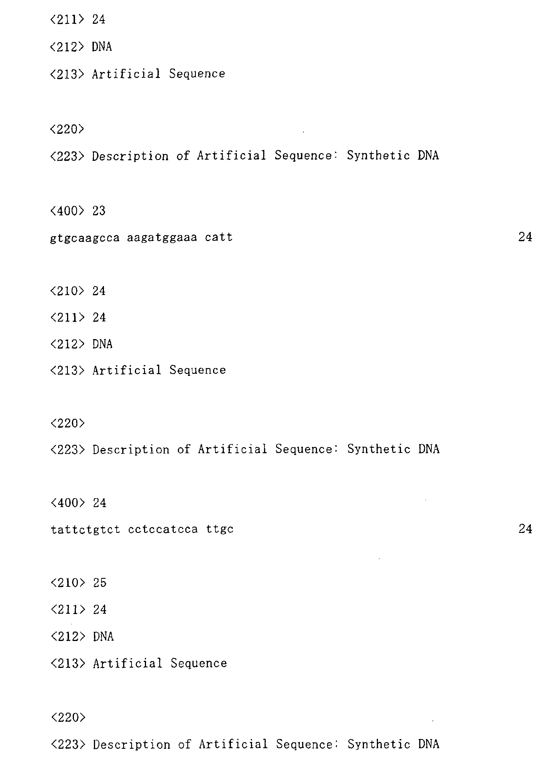
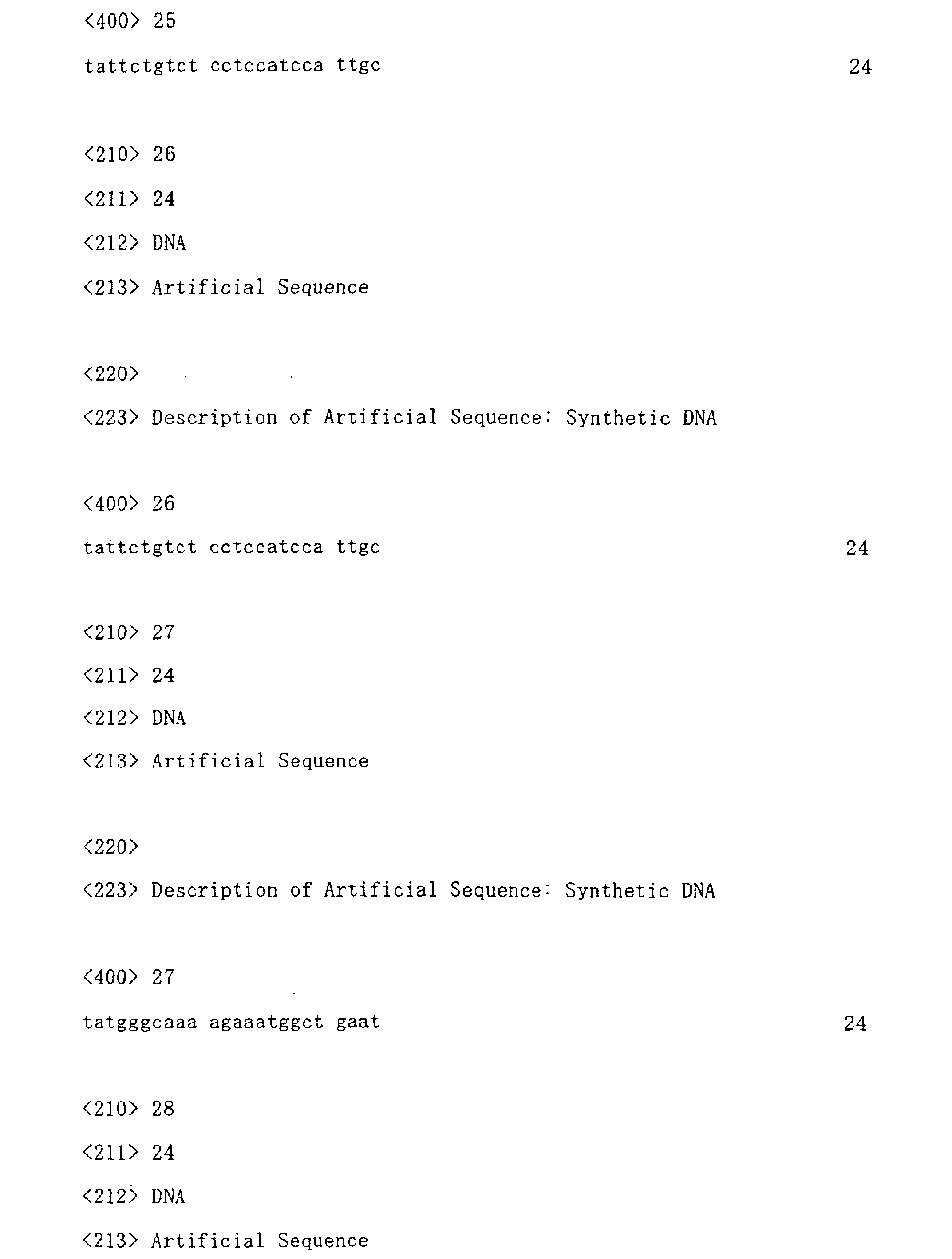
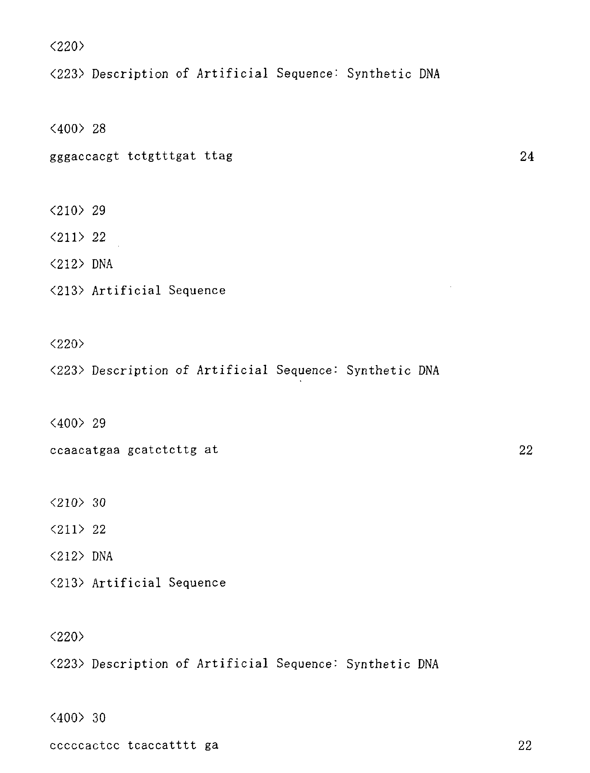
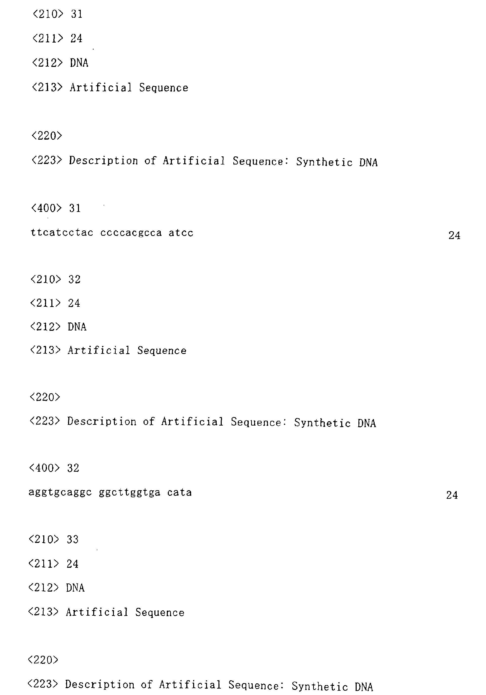
-
When non small cell lung carcinoma is compared with small cell lung carcinoma which
highly responds treatments such as chemotherapy, non small cell lung carcinoma has
extremely low chemosensitivity. In spite of the recent development of radiotherapy or
combination chemotherapy mainly utilizing CDDP, these therapies provide extremely low
life-prolonging effects to patients with non small cell lung carcinoma. Thus, it cannot be said
that the results of the therapies are sufficient. In other words, non small cell lung carcinoma
is one of cancers with extremely poor prognosis. Therefore, it is strongly desired that an
innovative therapeutic agent be developed.
-
Many attempts have previously been made to explore cancer-associated genes, based
on the difference in the expression level of the genes. Most of these approaches have been
simple comparisons between the expression level of genes in cancer tissues and that in the
corresponding normal tissues. It is considered that, in such methods, many genes are
simultaneously found based on common basic cell growth mechanisms. Such genes are
highly likely to be necessary not only for the growth of carcinoma cells but also for the growth
of normal cells. Thus, it is predicted that an agent targeting such genes affects even normal
cells with a high growth level, such as hematopoietic cells or epithelial cells in the small
intestine. Such an agent basically has no advantages when compared with the existing
chemotherapeutic agents such as antimetabolites.
-
Accordingly, it has been required to specify a gene, which is more specifically involved
in growth or malignant alteration of lung carcinoma cells than in normal cells with a high
growth level. In addition, it is necessary to develop a therapeutic agent effective for treating
lung carcinoma by using the thus specified gene, wherein the therapeutic agent prevents the
development of lung carcinoma, and especially the development of non small cell lung
carcinoma, but does not affect at all the growth of normal cells. Further, utilizing the
specificity of the above-described gene, it is also desired to develop a diagnostic agent capable
of detecting lung carcinoma at the early stage.
-
On the other hand, concerning a factor associated with cell growth, it has been
disclosed that interleukin-20 (hereinafter referred to as IL-20) is an important factor for the
growth of keratinocytes on the skin, that IL-20 receptors are excessively expressed in patients
with human psoriasis, and that a substance blocking signals via the IL-20 receptor can be an
antipsoriatic agent (Cell. 2001 Jan 12; 104 (1): 9-19). The aforementioned report, however,
has no descriptions regarding findings about IL-20 or an IL-20 receptor associated with the
growth of lung carcinoma cells, or regarding the action of signals via the IL-20 receptor on the
growth of carcinoma cells.
Disclosure of the Invention
-
It is an object of the present invention to provide a pharmaceutical composition for
treating lung carcinoma, and a diagnostic agent and a detection method for lung carcinoma.
-
The present inventor has conducted intensive studies to achieve the above object. The
present invention has explored a lung carcinoma target gene by a subtraction method, and as a
result, the inventor has found that an interleukin-20 receptor β-chain gene is increasingly
expressed in lung carcinoma cells at a high level and at a high frequency, thereby completing
the present invention.
-
That is to say, the present invention provides the following (1) to (11).
- (1) A pharmaceutical composition for treating lung carcinoma, which comprises, as an
active ingredient, an antibody binding to the β-chain of an interleukin-20 receptor.
- (2) The pharmaceutical composition according to (1) above, wherein the antibody is a
monoclonal antibody or polyclonal antibody.
- (3) The pharmaceutical composition according to (1) or (2) above, wherein the antibody is
a human antibody, humanized antibody, or chimeric antibody.
- (4) A pharmaceutical composition for treating lung carcinoma comprising, an active
ingredient, a polynucleotide which has a nucleotide sequence complementary to the nucleotide
sequence of a polynucleotide encoding the β-chain of an interleukin-20 receptor and which is
an antisense polynucleotide capable of suppressing the expression of the polynucleotide.
- (5) A diagnostic agent for lung carcinoma, which comprises an antibody capable of
binding to the β-chain of an interleukin-20 receptor.
- (6) The diagnostic agent according to (5) above, wherein the antibody is a monoclonal
antibody or polyclonal antibody.
- (7) The diagnostic agent according to (5) or (6) above, wherein the antibody is a human
antibody, humanized antibody, or chimeric antibody.
- (8) A method for detecting lung carcinoma in mammals using the diagnostic agent
according to any of (5) to (7) above.
- (9) A method for detecting lung carcinoma, which comprises detecting the expression of
the β-chain of an interleukin-20 receptor in a sample obtained from a mammal suspected to
have lung carcinoma.
- (10) The method according to (9) above, wherein an antibody capable of binding to the
β-chain of an interleukin-20 receptor is used to detect the expression.
- (11) The method according to (10) above, wherein the PCR method is used to detect the
expression.
-
-
This specification includes part or all of the contents as disclosed in the specification
and/or drawings of Japanese Patent Application No. 2002-124743, which is a priority
document of the present application.
Brief Description of the Drawings
-
- Figure 1 shows the scheme of a subtraction method in the present invention.
- Figure 2 shows the results of electrophoresis obtained by RT-PCR analysis. In the
figure, LG represents a normal lung, TH represents a normal thymus gland, BM represents a
normal bone marrow, SI represents a normal small intestine, and C represents a control.
- Figure 3 shows the results of RT-PCR analysis of expression of human genes (IL-20Rα,
IL-20Rβ, IL-22R, and RP S9) in normal human tissues (lung, liver, thymus gland, bone
marrow, and small intestine) and in non small lung carcinoma clinical samples (Sq. 3, Sq. 9,
Sq. 11, and Sq. 12). The term "no cDNA" means the case where template DNA was not
used.
- Figure 4 shows the results of RT-PCR analysis of expression of human genes (IL-19,
IL-20, IL-24, and RP S9) in normal human tissues (lung, liver, thymus gland, bone marrow,
and small intestine) and in non small lung carcinoma clinical samples (Sq. 3, Sq. 9, Sq. 11, and
Sq. 12). The term "no cDNA" means the case where template DNA was not used.
- Figure 5 schematically shows the involvement of signals via IL-20Rβ in canceration of
lung epithelial cells. A circle means each cytokine, and a cylinder means a receptor of the
cytokine. An arrow means a receptor to which each cytokine can bind. The upper diagram
shows the state of signals via IL-20Rβ in normal lung epithelial cells, and the lower diagram
shows the state of signals via IL-20Rβ in cancerous lung epithelial cells. The expression of
IL-20Rβ and IL-24 is increased by canceration, so that signals caused by these genes can
efficiently be transduced into cells.
- Figure 6 shows the results of RT-PCR analysis of expression of human IL-20Rβ gene
(arrow) and human RP S9 gene in samples derived from patients with lung carcinoma. N
represents a non-tumor portion-derived sample, and T represents a tumor portion-derived
sample. The term "no cDNA" means a negative control where no template DNA is used, and
the term "PC" means a positive control where lung carcinoma-derived cDNA is used wherein
IL-20Rβ is highly expressed.
- Figure 7 shows the result of RT-PCR analysis of expression of human genes (IL-20Rα,
IL-20Rβ, and IL-22R) in various types of human lung carcinoma cell lines (LC-1/sq, EBC-1,
LK-2, RERF-LC-AI, LC-2/ad, ABC-1, A549, NCI-H460, NCI-H23, and NCI-H522). The
term "no cDNA" means the case where no template DNA is used, and the term "genomic
DNA" means the case where human chromosomal DNA is used as a template.
- Figure 8 shows the results of RT-PCR analysis of expression of human genes (IL-19,
IL-20, and IL-24) in various types of human lung carcinoma cell lines (LC-1/sq, EBC-1, LK-2,
RERF-LC-AI, LC-2/ad, ABC-1, A549, NCI-H460, NCI-H23, and NCI-H522). The term "no
cDNA" means the case where no template DNA is used, and the term "genomic DNA" means
the case where human chromosomal DNA is used as a template.
- Figure 9 shows the results of RT-PCR analysis of expression of human genes (IL-20Rβ
and RP S9) in samples derived from patients with gastric carcinoma (S1, S3, S4, and S5) and
in samples derived from patients with uterine carcinoma (U1, U4, U5, and U6). In the
samples, N represents a non-tumor portion-derived sample, and T represents a tumor
portion-derived sample. The term "no cDNA" means the case where no template DNA is
used.
- Figure 10 shows the results of FACS analysis using an anti-IL-20Rβ rabbit polyclonal
antibody. The term "hIL-20RB transfectant" means a cell line wherein human IL-20Rβ is
forced to be expressed (cell line 3), and the term "control" means cells obtained by allowing
only a vector to express in the same parent cells. Cell line 3 is specifically stained with an
anti-NP2 antibody and an anti-NP3 antibody, and it is therefore found that these antibodies
specifically react with human IL-20Rβ. The term "normal rabbit IgG" means a negative
control where IgG purified from normal rabbits is used as a primary antibody.
- Figure 11 shows the results of ELISA analysis of the serum from mice (No. 1, No. 2,
and No. 3) immunized with a soluble human IL-20Rβ-FLAG protein. The term "Pre" means
the serum before immunization, and the term "3rd" means the serum after three immunizations.
A soluble human IL-20Rβ-His protein is used as an antigen for ELISA. The horizontal axis
("dilution") indicates the dilution ratio of the serum.
- Figure 12 shows the results of FACS analysis using an anti-IL-20Rβ monoclonal
antibody (MK clone). A cell line (cell line 3) wherein human IL-20Rβ is forced to be
expressed is specifically stained, and it is therefore found that these antibodies specifically
react with human IL-20Rβ. The term "medium only" means a negative control wherein only
a hybridoma medium is used as a primary antibody.
-
Best Mode for Carrying Out the Invention
-
The present invention will be described further in detail below.
-
The term "treatment" used in the present specification includes administration of the
pharmaceutical composition of the present invention to a subject to cause lung carcinoma to
disappear, to suppress further progression of lung carcinoma, and to alleviate symptoms
caused by lung carcinoma.
-
An antibody used for the pharmaceutical composition for treating lung carcinoma and
the diagnostic agent for lung carcinoma of the present invention is not particularly limited, but
any antibody may be used as long as it binds to the β-chain of an interleukin-20 receptor. It
is preferably a monoclonal antibody. In addition, a monoclonal antibody having cytotoxicity
such as antibody-dependent cellular cytotoxicity (ADCC) or complement-dependent
cytotoxicity (CDC), or a monoclonal antibody capable of blocking signals that can be
transduced into cells via the β-chain of an interleukin-20 receptor, may also be used.
-
Moreover, an antibody capable of suppressing the growth of cells expressing the
β-chain of an interleukin-20 receptor by binding a radioactive isotope (RI) or toxin thereto
according to known methods may also be included in the antibody of the present invention.
-
Furthermore, antibody fragments such as Fab or F(ab)2 and peptides such as a
single-strand Fv, which are prepared based on the above antibody, may also be included in the
antibody of the present invention.
1. Exploration of target gene for lung carcinoma cell by subtraction method
-
Many attempts have previously been made to explore cancer-associated genes, based
on the difference in the expression level of the genes. Most of them are simple approaches
wherein the expression level of genes in cancer tissues is compared with that in the
corresponding normal tissues. In such methods, however, it is predicted that many genes are
found based on common basic cell growth mechanisms. Moreover, such genes are highly
likely to be necessary not only for the growth of carcinoma cells but also for the growth of
normal cells. Thus, it is predicted that an agent that targets genes found by the conventional
subtraction method affects even normal cells with a high growth level, such as hematopoietic
cells or epithelial cells in the small intestine. Such an agent basically has no advantages
when compared with the existing chemotherapeutic agents such as antimetabolites.
-
Hence, when genes expressing in normal lung tissues are subtracted from gene
expressing in lung carcinoma tissues to find genes associated specifically with growth or
malignant alteration of lung carcinoma cells, the present inventors simultaneously subtract
genes expressing in hematopoietic cells. More specifically, using polyA+ RNA derived from
squamous cell lung carcinoma as a tester (subtracted side), and using a mixture obtained by
mixing polyA+ RNA derived from normal lung and polyA+ RNA derived from normal bone
marrow and normal thymus gland, which are tissues containing large quantities of
hematopoietic cells, as a driver (subtracting side), subtraction was carried out by suppression
subtractive hybridization method (Proc Natl Acad Sci USA. 1996 Jun 11; 93 (12): 6025-30)
(see, Figure 1).
-
A gene obtained by such a subtraction method is selectively expressed in lung
carcinoma cells, but is not expressed in proliferative normal hematopoietic cells.
Accordingly, it is considered that this gene is not associated with a general cell growth
mechanism. In fact, as a result of analyzing a clone subtracted by this method, a gene
CRA262 was found, which was increasingly expressed in non small cell carcinoma cells
(see, Examples 1 to 3 and Figure 2).
-
It was confirmed that this gene was expressed in normal lung tissues in an extremely
small amount, but that it was increasingly expressed in all the non small cell lung carcinoma
samples obtained from 5 different patients with lung carcinoma. In addition, this gene was
expressed in only a little amount in the bone marrow and thymus gland containing
proliferative hematopoietic cells, and in the small intestine containing large quantities of
proliferative epithelial cells. From these results, it is considered that this gene is plays a
certain important role specific for growth or malignant alteration of lung carcinoma cells, and
therefore that it can be an ideal target of a lung carcinoma therapeutic agent, which inhibits the
progression of lung carcinoma but does not affect the growth of normal cells. Thus, this gene
was used as a material for the following studies.
-
Database (GenSeq) regarding nucleic acids and amino acids in patent documents or the
like was searched for this gene. As a result, it was confirmed that this gene is an IL-20
receptor β chain (hereinafter referred to as IL-20Rβ) gene. This IL-20Rβ gene had been
discovered by several groups (International Patent Publication WO 99/46379, WO 00/53758,
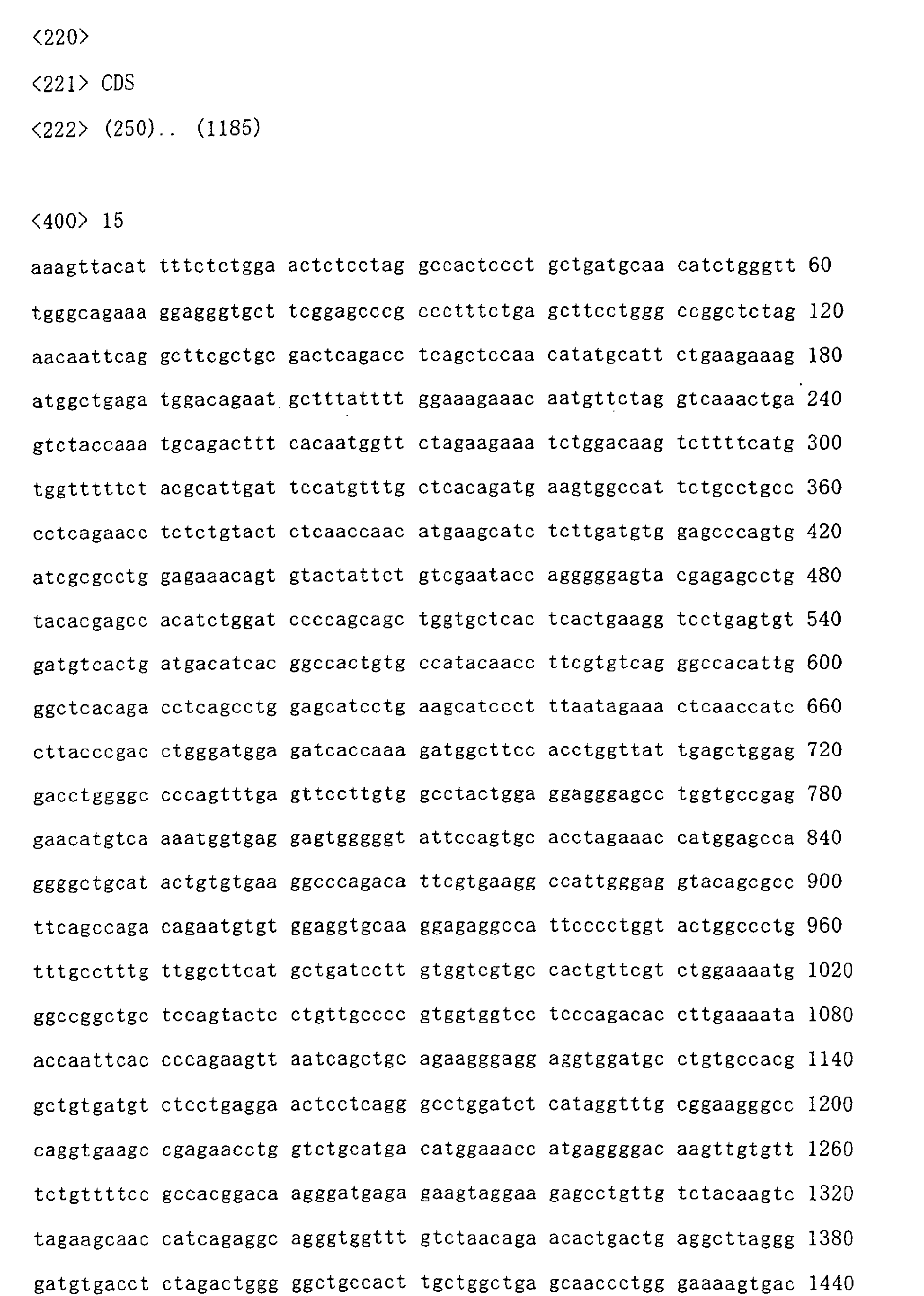
WO 00/39161, WO 99/62934, etc.). The nucleotide sequence and amino acid sequence of
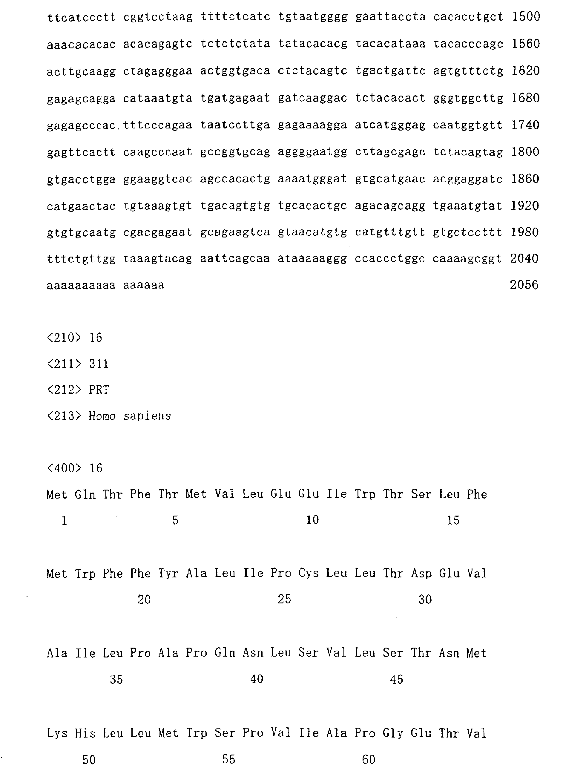
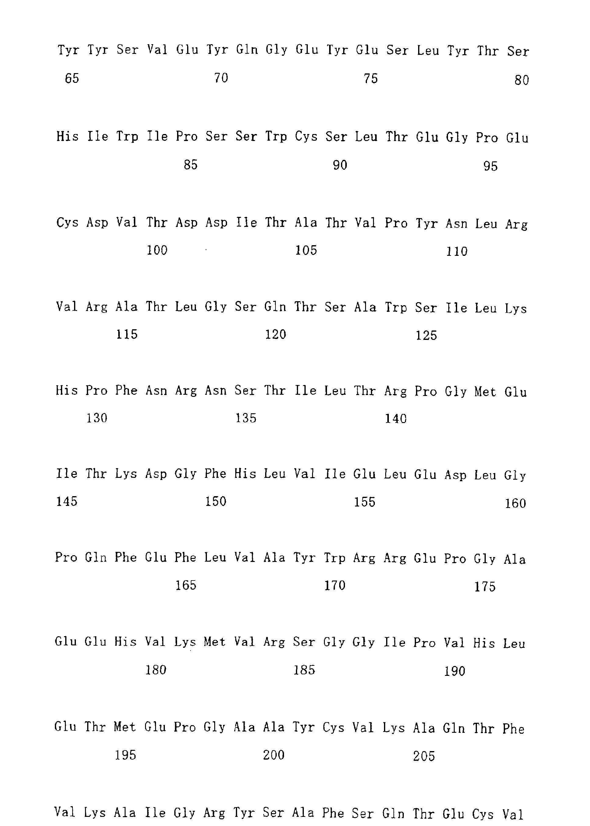
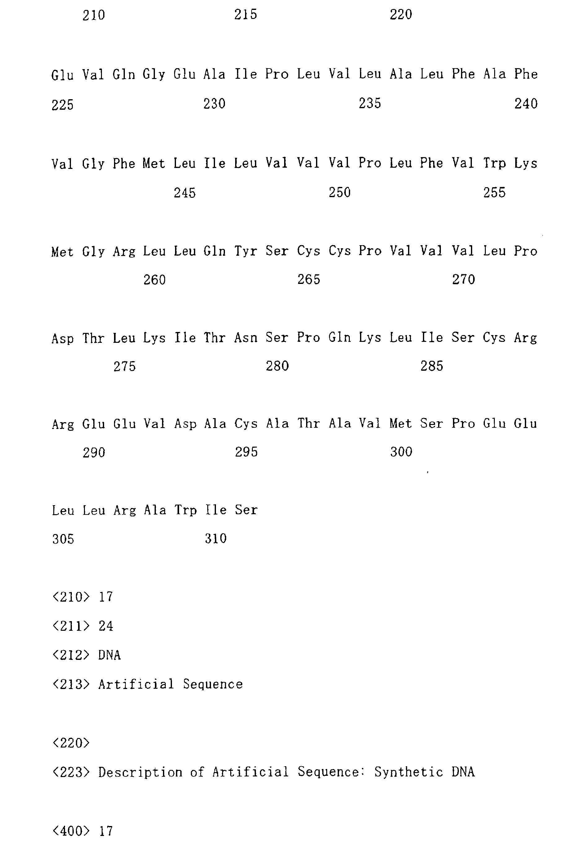
human IL-20Rβ cDNA are shown in SEQ ID Nos. 15 and 16, respectively.
-
As stated above, it was clarified that the IL-20Rβ gene is increasingly expressed at a
high level and at a high frequency in lung carcinoma cells, and in particular in non small cell
lung carcinoma cells, and that the gene is deeply associated with the growth of lung carcinoma
cells. Accordingly, these findings provide a pharmaceutical composition for treating lung
carcinoma and a diagnostic agent for lung carcinoma, using an antibody binding to IL-20Rβ or
an antisense polynucleotide to the sequence (polynucleotide) of a gene encoding IL-20Rβ,
which will be described below.
2. Antibody capable of binding to IL-20Rβ
(1) Antibody
-
The antibody used in the present invention are not particularly limited to any particular
origin, type (polyclonal antibody or monoclonal antibody), and form, as long as it is an
antibody capable of binding to IL-20Rβ (hereinafter referred to as an anti-IL-20Rβ antibody).
-
For example, an antibody is not particularly limited, as long as it binds to an antigen,
IL-20Rβ. Examples of an antibody appropriately used herein may include a mouse antibody,
a rat antibody, a rabbit antibody, a sheep antibody, a chimeric antibody, a humanized antibody,
and a human antibody. An antibody can be obtained as a polyclonal antibody or monoclonal
antibody by known methods. In terms of stable production of uniform antibodies, a
monoclonal antibody, and particularly, a monoclonal antibody derived from mammals is
preferable. Such a monoclonal antibody derived from mammals may include those produced
from hybridomas, and those produced from a host that is transformed with an expression
vector containing an antibody gene by genetic engineering.
(2) Hybridoma producing antibody
-
A hybridoma which produces the antibody used in the present invention can be
basically prepared, for example, as follows, using known techniques. Briefly, using an
IL-20Rβ protein or a cell expressing IL-20Rβ as a sensitizing antigen, a mammal is
immunized by an ordinary immunization method. The obtained immunocytes are fused with
known parent cells by an ordinary cell fusion method, and the fused cells is subjected to an
ordinary screening method to screen monoclonal antibody-producing cells (hybridomas).
Preparation of hybridomas can be carried out according to the method of Milstein et al.
(Kohler. G. and Milstein, C., Methods Enzymol. (1981) 73: 3-46), for example. When the
immunogenicity of an antigen is low, it may be bound to a macromolecule having
immunogenicity, such as albumin, and immunization may be then carried out.
-
More specifically, first, IL-20Rβ used as a sensitizing antigen for obtaining an antibody
can be obtained by expressing the gene/amino acid sequence encoding IL-20Rβ, which has
already been disclosed in WO 99/46379, WO 00/53758, WO 00/39161, and WO 99/62934,
and the like. A polynucleotide having a gene sequence encoding IL-20Rβ is inserted into a
known expression vector system, and suitable host cells are transformed with the vector.
Thereafter, an IL-20Rβ protein of interest is purified from the host cells or a culture
supernatant thereof by known methods. Then, the purified IL-20Rβ protein or a partial
peptide of the protein can be used as a sensitizing antigen. In addition, a partial peptide can
also be obtained from the amino acid sequence of IL-20Rβ by chemical synthesis.
-
A mammal immunized with a sensitizing antigen is not particularly limited. It is
preferable to select a mammal, taking into consideration compatibility with patent cells used in
cell fusion. Generally, rodents, such as mice, rats, hamsters, rabbits, or monkeys can be used.
-
Immunization of an animal with a sensitizing antigen can be carried out by known
methods. For example, a common method is intraperitoneal or subcutaneous injection of a
sensitizing antigen to a mammal. More specifically, a sensitizing antigen is diluted with or
suspended in PBS (phosphate-buffered saline), saline, or the like, into a suitable amount.
Thereafter, a suitable amount of a common adjuvant, such as a Freund's complete adjuvant, is
mixed with the obtained solution, as desired, followed by emulsification. The obtained
product was administered to a mammal several times every 4 to 21 days. Moreover, a
suitable carrier can also be used in immunization of a sensitizing antigen.
-
Thus, a mammal is immunized, and it is confirmed that a desired antibody level
increases in the serum. Thereafter, immunocytes are collected from the immunized mammal
and subjected to cell fusion. Spleen cells are particularly preferable as immunocytes.
-
Myeloma cells from mammals are used as parent cells to be fused with the
above-described immunocytes. As myeloma cells, various known cell lines are used.
Preferred examples of such myeloma cells may include P3(P3x63Ag8.653) (J. Immnol. (1979)
123, 1548-1550), P3x63Ag8U.1 (Current Topics in Microbiology and Immunology (1978) 81,
1-7), NS-1 (Kohler. G. and Milstein, C. Eur. J. Imunol, (1976) 6, 511-519), MPC-11
(Margulies. D. H. et al., Cell (1976) 8, 405-415), SP2/0 (Shulman, M. et al., Nature (1978)
276, 269-270), F0 (de St. Groth, S. F. et al., J. Immunol. Methods (1980) 35, 1-21), S194
(Trowbridge, I. S. J. Exp. Med. (1978) 148, 313-323), and R210 (Galfre, G. et al., Nature
(1979) 277, 131-133).
-
Cell fusion between the above-described immunocytes and myeloma cells can be
basically carried out according to known methods such as the method of Kohler and Milstein
(Kohler G. and Milstein, C., Methods Enzymol: (1981) 73, 3-46).
-
More specifically, the above cell fusion is carried out in the presence of a cell fusion
promoter in a common nutrient culture solution. For example, polyethylene glycol (PEG) or
Sendai virus (HVJ) is used as a fusion promoter. In order to enhance fusion efficiency, as
desired, auxiliary agents such as dimethyl sulfoxide can also be used.
-
The ratio between immunocytes and myeloma cells used can be suitably set. For
example, immunocytes are preferably added to myeloma cells at a ratio between 1 : 1 and 10 :
1. Examples of a culture solution useful in the above cell fusion may include an RPMI1640
culture solution, an MEM culture solution, which are preferable for growth of the myeloma
cells, and further, a common culture solution used in this type of cell culture. In addition, a
serum infusion solution such as fetal calf serum (FCS) can also be used in combination.
-
For cell fusion, a certain amount of the above immunocytes and a certain amount of the
above myeloma cells are fully mixed in the above culture solution. A PEG solution (for
example, having a mean molecular weight between approximately 1,000 and 6,000), which
has previously been heated to approximately 37°C, is added to the mixture generally in a
concentration of 30% to 60% (w/v) and mixed, so as to form fused cells of interest
(hybridomas). Subsequently, an operation to successively add a suitable culture solution
followed by centrifugation to eliminate a supernatant is repeated, so as to eliminate a cell
fusion promoter and the like, which are not preferable for the growth of hybridomas.
-
The thus obtained hybridomas are cultured in an ordinary selective culture solution
such as an HAT culture solution (a culture solution containing hypoxanthine, aminopterin, and
thymidine) for selection. The above culture in an HAT culture solution is continued for a
time (generally several days to several weeks) enough for death of cells other than hybridomas
of interest (non-fused cells). Subsequently, a common limiting dilution method is applied,
and thus, hybridomas producing an antibody of interest are screened and subjected to
monocloning.
-
In addition to the method of obtaining the above hybridomas by immunizing mammals
other than a human with an antigen, several methods of obtaining a human antibody have also
been known. For example, human lymphocytes are sensitized with IL-20Rβ or cells
expressing IL-20Rβ in vitro, and the sensitized lymphocytes are then fused with myeloma
cells derived from a human having a permanent division ability, so as to obtain a human
antibody having activity to bind to IL-20Rβ (JP Patent Publication (Kokoku) No. 1-59878 B
(1989)). Moreover, IL-20Rβ as an antigen is administered to a transgenic animal having the
entire repertoire of human antibody genes, so as to obtain cells producing an anti-IL-20Rβ
antibody. The cells are then subjected to immortalization, and a human antibody to IL-20Rβ
may be obtained from the immortalized cells (see, WO 94/25585, WO 93/12227, WO
92/03918, and WO 94/02602).
-
Hybridomas producing the thus obtained monoclonal antibody can be subcultured in a
common culture solution, and further, they can be stored in liquid nitrogen for a long time.
-
In order to obtain a monoclonal antibody from the hybridomas, a method of culturing
the hybridomas according to an ordinary method to obtain an antibody as a culture supernatant,
or a method of administering the hybridomas to a mammal compatible therewith and allowing
them to grow therein so as to obtain an antibody as ascites, can be adopted. The former
method is suitable for the case of obtaining an antibody with high purity. On the other hand,
the latter method is suitable for mass production of antibodies.
(3) Recombinant antibody
-
An antibody gene is cloned from hybridomas, and it can be then incorporated into a
suitable vector. The vector is then introduced into a host, so that a recombinant antibody can
be produced by genetic recombination techniques. Such a recombinant antibody can also be
used (see, e.g., Carl, A. K. Borrebaeck, James, W. Larrick, THERAPEUTIC MONOCLONAL
ANTIBODIES, Published in the United Kingdom by MACMILLAN PUBLISHERS LTD,
1990).
-
More specifically, cDNA in the variable region (V region) of an antibody is
synthesized from mRNA of a hybridoma, using reverse transcriptase. For example, mRNA
encoding the V region of an anti-IL-20Rβ antibody is isolated from a hybridoma producing the
anti-IL-20Rβ antibody. mRNA can be isolated by known methods such as the guanidine
ultracentrifugation method (Chirgwin, J. M. et al., Biochemistry (1979) 18, 5294-5299) or the
AGPC method (Chomczynski, P. et al., Anal. Blochem. (1987) 162, 156-159). By such a
method, total RNA is prepared, and mRNA of interest is then prepared using an mRNA
Purification Kit (manufactured by Pharmacia) or the like. In addition, mRNA can also be
directly prepared using a QuickPrep mRNA Purification Kit (manufactured by Pharmacia).
-
cDNA in the V region of an IL-20Rβ antibody is synthesized from the obtained mRNA,
using reverse transcriptase. cDNA is synthesized using an AMV Reverse Transcriptase
First-strand cDNA Synthesis Kit (manufactured by Seikagaku Corporation) or the like.
Moreover, for synthesis and amplification of cDNA, the 5'-RACE method (Frohman, M. A. et
al., Proc. Natl. Acad. Sci. USA (1988) 85, 8998-9002. Belyavsky, A. et al., Nucleic Acids
Res. (1989) 17, 2919-2932) utilizing PCR can be applied, using a 5'-AmpliFINDER RACE
Kit (manufactured by Clontech).
-
Once DNA encoding the V region of an IL-20Rβ antibody of interest is obtained, the
DNA is ligated to DNA encoding a desired antibody constant region (C region), and the
ligated DNA is incorporated into an expression vector. Otherwise, DNA encoding the V
region of an antibody may be incorporated into an expression vector containing DNA in the C
region of an antibody. DNA is incorporated into an expression vector such that it is
expressed under control of expression control regions such as an enhancer and a promoter.
Thereafter, host cells are transformed with the expression vector, so that an antibody is
expressed therein.
-
For expression of an antibody gene, DNA fragments encoding either the heavy chain
(H chain) or light chain (L chain) of an antibody may be separately incorporated into different
expression vectors, so that host cells may be simultaneously transformed with these vectors.
Otherwise, DNA fragments encoding either the H chain or the L chain are incorporated into a
single expression vector, so that host cells may be transformed with the vector (see, WO
94/11523).
-
Moreover, a technique of obtaining a human antibody by panning with an antibody
library of mammals such as a mouse or human has been known. For example, the variable
region of a human antibody is expressed as a single chain antibody (scFv) on the surface of a
phage by the phage display method, so as to select a phage binding to an antigen. The
selected phage is subjected to gene analysis, so that a DNA sequence encoding the variable
region of a human antibody binding to an antigen can be determined. If the DNA sequence
of scFv binding to an antigen is clarified, a suitable expression vector containing the sequence
is prepared, and a human antibody can be obtained. These methods have already been well
known, and WO 92/01047, WO 92/20791, WO 93/06213, WO 93/11236, WO 93/19172, WO
95/01438, and WO 95/15388 can be used for references.
(4) Modified antibody
-
In the present invention, an artificially modified gene recombinant antibody such as a
chimeric antibody or humanized antibody can be used for the purpose of decreasing
heterologous antigenicity to humans. These modified antibodies can be produced by known
methods.
-
A chimeric antibody is an antibody, which comprises the variable region of the heavy
chain and light chain of the antibody of a mammal other than a human, such as a mouse
antibody, and the constant region of the heavy chain and light chain of a human antibody.
DNA encoding the variable region of a mouse antibody is ligated to DNA encoding the
constant region of a human antibody, and the ligated DNA is then incorporated into an
expression vector. The expression vector is then introduced into a host, so as to obtain a
chimeric antibody from the host.
-
A humanized antibody is referred to also as a reshaped human antibody. It is an
antibody obtained by transplanting the complementarity-determining region (CDR) of a
mammal antibody other than a human antibody, such as a mouse antibody, into the
complementarity-determining region of a human antibody. A general gene recombination
technique therefor has been known (see, EP Patent Application Publication No. EP125023 and
WO 96/02576).
-
More specifically, a DNA sequence designed such that CDR of a mouse antibody is
ligated to the framework region (FR) of a human antibody is synthesized by the PCR method
from several oligonucleotides prepared such that they have an overlapped portion at the
terminus thereof. The obtained DNA is ligated to DNA encoding the constant region of a
human antibody, and the ligated DNA is then incorporated into an expression vector. The
expression vector is then introduced into a host, so as to obtain a humanized antibody (see, EP
Patent Application Publication No. 239400, and WO 96/02576).
-
As FR of a human antibody ligated via a complementarity-determining region, one
wherein the CDR forms a good antigen-binding portion is selected. If necessary, amino acids
in the framework region of the variable region of an antibody may be substituted so that the
complementarity-determining region of a reshaped human antibody forms an appropriate
antigen-binding site (Sato, K. et al., Cancer Res. (1993) 53, 851-856).
-
As the C regions of a chimeric antibody and a humanized antibody, those of a human
antibody are used. For example, in the case of the H chain, Cγ1, Cγ2, Cγ3, and Cγ4 can be
used, and in the case of the L chain, Cκ and Cλ can be used. Moreover, in order to improve
the stability of an antibody or a production thereof, the C region of a human antibody may be
modified.
-
A chimeric antibody comprises the variable region of an antibody derived from a
mammal other than a human and the constant region derived from a human antibody. On the
other hand, a humanized antibody comprises the complementarity-determining region of an
antibody derived from a mammal other than a human and the framework region and C region
derived from a human antibody. Since a humanized antibody has a low antigenicity in a
human body, it is useful as an active ingredient of the therapeutic agent of the present
invention.
(5) Antibody variants
-
The antibody used in the present invention is not limited to the whole molecules of an
antibody, but it may be a fragment of an antibody or a variant thereof as long as it binds to
IL-20Rβ and inhibits the activity of IL-20Rβ. Either a bivalent antibody or monovalent
antibody may also be included in the antibody of the present invention. For example,
examples of an antibody fragment may include Fab, F(ab')2, Fv, Fab/c having a Fab and a
complete Fc, and a single chain Fv (scFv) obtained by linking Fv of H chain to Fv of L chain
using a suitable linker.
-
More specifically, an antibody is treated with an enzyme such as papain or pepsin to
generate antibody fragments. Alternatively, a gene encoding such an antibody fragment is
constructed, and it is then introduced into an expression vector, followed by expression in
suitable host cells (see, e.g., Co, M. S. et al., J. Immunol. (1994) 152, 2968-2976, Better, M. &
Horwitz, A. H. Methods in Enzymology (1989) 178, 476-496, Academic Press, Inc.,
Plueckthun, A. & Skerra, A. Methods in Enzymology (1989) 178, 476-496, Academic Press,
Inc., Lamoyi, E., Methods in Enzymology (1989) 121, 652-663, Rousseaux, J. et al., Methods
in Enzymology (1989) 121, 663-669, Bird, R. E. et al., TIBTECH (1991) 9, 132-137).
-
scFv is obtained by linking the H chain V region of an antibody to the L chain V region
thereof. In such scFv, the H chain V region is ligated to the L chain V region via a linker,
and preferably a peptide linker (Huston, J. S. et al., Proc. Natl. Acad. Sci. U.S.A. (1988) 85,
5879-5883). In scFv, H chain V region and L chain V region may be derived from any
antibodies described in the present specification. As a peptide linker ligating V region, a
certain single chain peptide consisting of 1 to 30 amino acid residues is used, for example.
-
Among DNA encoding the H chain or H chain V region of the above-described
antibody and DNA encoding the L chain or L chain V region thereof, using, as a template, a
DNA portion encoding the whole or a desired part of amino acid sequences thereof, a DNA
fragment is amplified by the PCR method with a primer set corresponding to both termini
thereof. Subsequently, DNA encoding a peptide linker portion is amplified, using a primer
set which are designed such that both termini thereof are ligated to H chain and L chain,
respectively, thereby obtaining DNA encoding scFv.
-
Once DNA encoding scFv is produced, an expression vector containing the DNA, and
a host transformed with the expression vector can also be obtained according to conventional
methods. Using the host, scFv can be obtained according to conventional methods.
-
These antibody fragments are obtained by first obtaining their genes, expressing them
in hosts, and allowing the hosts to produce them, in the same manner as described above.
The term "antibody" may also include these antibody fragments in the present invention.
-
An anti-IL-20Rβ antibody binding to various molecules such as polyethylene glycol
(PEG) can also be used as an antibody variant. The term "antibody" may also include these
antibody variants in the present invention. These antibody variants can be obtained by
chemically modifying the obtained antibody. It is to be noted that a method of modifying an
antibody has already been established in this field.
-
Moreover, the antibody used in the present invention may also be a bispecific antibody.
Such a bispecific antibody may have antigen-binding sites recognizing different epitopes on an
IL-20Rβ molecule. Otherwise, one antigen-binding site may recognize IL-20Rβ and the
other antigen-binding site may recognize other cell surface molecules such as an IL-20
receptor α chain (hereinafter referred to as IL-20Rα) or interleukin-22 receptor (hereinafter
referred to as IL-22R). Furthermore, the other antigen-binding site may be a surface
molecule of immunocytes such as T cells or natural killer cells. In this case, immunocytes
can directly act on cells expressing IL20-Rβ to cause injury specifically to carcinoma cells, so
as to suppress the growth of the carcinoma cells. Such a bispecific antibody can be produced
by binding HL pairs of two types of antibodies. Otherwise, it can also be obtained by fusing
hybridomas producing different monoclonal antibodies so as to produce bispecific
antibody-producing fused cells. Still further, a bispecific antibody can also be produced by
genetic engineering.
(6) Expression and production of recombinant antibody or modified antibody
-
When the antibody gene obtained as described above is introduced into a suitable host
to produce an antibody, a suitable host and a suitable expression vector can be used in
combination. When eukaryotic cells are used as host cells, animal cells, plant cells, and fungi
can be used. As animal cells, (1) mammalian cells such as CHO, COS, myeloma, BHK
(baby hamster kidney), HeLa, or Vero, (2) amphibian cells such as Xenopus laevis oocytes, (3)
insect cells such as sf9, sf21, or Tn5, and other cells, have been known. As plant cells, cells
derived from Nicotiana such as Nicotiana tabacum have been known. These cells may be
subjected to callus culture. As fungi, yeasts including Saccharomyces such as
Saccharomyces serevisiae, and filamentous fungi including Aspergillus such as Aspergillus
niger have been known. When prokaryotic cells are used, there is a production system using
bacterial cells. As bacterial cells, Escherichia coli (E. coli) and Bacillus subtilis have been
known. An antibody gene of interest is introduced into these cells by transformation, and the
transformed cells are cultured in vitro to obtain an antibody.
-
For example, in the case of mammalian cells, a commonly used useful promoter is
bound to an antibody gene to be expressed, and then, poly A signal is operatively linked
downstream of the 3'-side thereof, so that an antibody can be expressed. Examples of a
promoter/enhancer may include a human cytomegalovirus immediate early
promoter/enhancer.
-
Other examples of a promoter/enhancer useful in the expression of an antibody used in
the present invention may include a virus promoter/enhancer such as retrovirus, polyoma virus,
adenovirus, or simian virus 40 (SV40), and a promoter/enhancer derived from mammalian
cells, such as human elongation factor 1α (HEF1α).
-
When an SV40 promoter/enhancer is used, gene expression can easily be carried out by
the method of Mulligan et al. (Nature (1979) 277, 108). When an HEF1α promoter/enhancer
is used, gene expression can easily be carried out by the method of Mizushima et al. (Nucleic
Acids Res. (1990) 18, 5322).
-
In the case of Escherichia coli, a commonly used useful promoter, a signal sequence
for secretion of an antibody, and an antibody gene to be expressed are operatively linked to
one another, so that the gene can be expressed. Examples of a promoter may include a lacZ
promoter and an araB promoter. When a lacZ promoter is used, gene expression can be
carried out by the method of Ward et al. (Nature (1098) 341, 544-546; FASEB J. (1992) 6,
2422-2427). When an araB promoter is used, gene expression can be carried out by the
method of Better et al. (Science (1988) 240, 1041-1043).
-
When an antibody is to be produced in a periplasm of Escherichia coli, a pelB signal
sequence (Lei, S. P. et al., J. Bacteriol. (1987) 169,4379) may be used as a signal sequence for
secretion of an antibody. An antibody produced in the periplasm is separated, and thereafter,
the structure of the antibody is refolded, as appropriate, for use.
-
As a replication origin, those derived from SV40, polyoma virus, adenovirus, bovine
papilloma virus (BPV), etc. can be used. In addition, in order to amplify the number of gene
copies in a host cell system, an expression vector may comprise, as a selective marker, an
aminoglycoside transferase (APH) gene, a thymidine kinase (TK) gene, an Escherichia coli
xanthine guanine phosphoribosyl transferase (Ecogpt) gene, a dihydrofolate reductase (dhfr)
gene, etc.
-
The transformed host cells are cultured in vitro or in vivo, so as to allow them to
produce an antibody of interest. The host cells are cultured according to known methods.
For example, DMEM, MEM, RPMI1640, or IMDM can be used as a culture solution. A
serum infusion solution such as fetal calf serum (FCS) can also be used in combination.
-
Moreover, in order to produce a recombinant antibody, not only the above-described
host cells, but transgenic animals can also be used. For example, an anti-IL-20Rβ antibody
gene is inserted into a gene encoding a protein specifically produced in milk (e.g., goat
β-casein), so as to prepare a fused gene. A DNA fragment containing the fused gene into
which the above antibody gene has been inserted is injected into a goat embryo, and the
embryo is then transferred into a female goat. An anti-IL-20Rβ antibody is obtained from
milk generated from a transgenic goat that was born from the goat accepting the above embryo,
or progenies thereof. In addition, in order to increase the amount of the milk containing the
anti-IL-20Rβ antibody generated from the transgenic goat, hormone may be added to the
transgenic goat, as appropriate (see, Ebert, K. M. et al., Bio/Technology (1994) 12, 699-702).
(7) Separation and purification of antibody
-
The antibody expressed and produced as described above can be separated from cells or
a host animal, and it can be purified until it becomes homogenous. The antibody used in the
present invention can be separated and purified with an affinity column. Examples of a
column using a protein A column may include Hyper D, POROS, and Sepharose F.F.
(manufactured by Pharmacia). Further, commonly used protein separation and purification
methods may be used, and such methods are not limited. For example, an antibody can be
separated and purified by appropriately selecting and combining methods such as
chromatography columns other than the above affinity column, filter, ultrafiltration, salting out,
and dialysis (Antibodies A Laboratory Manual. Ed Harlow, David Lane, Cold Spring Harbor
Laboratory, 1988). Examples of chromatography may include ion exchange chromatography,
hydrophobic chromatography, and gel filtration.
(8) Confirmation of activity of antibody
-
In order to determine the antigen-binding activity of the antibody used in the present
invention, known methods can be applied (Antibodies A Laboratory Manual. Ed Harlow,
David Lane, Cold Spring Harbor Laboratory, 1988). For example, ELISA (enzyme-linked
immunosorbent assay), EIA (enzyme-linked immunoassay), RIA (radioimmunoassay), or the
fluorescent antibody method can be used.
-
When ELISA is used, a sample containing an anti-IL-20Rβ antibody, such as the
culture supernatant of anti-IL-20Rβ antibody-generating cells or a purified antibody, is added
to a plate on which IL-20Rβ is coated. A secondary antibody labeled with enzyme such as
alkaline phosphatase is added to the plate. The plate is then incubated and washed.
Thereafter, an enzyme substrate such as p-nitrophenyl phosphate is added thereto, so that
antigen-binding activity can be evaluated by measuring absorbance.
(9) Cytotoxicity
-
The anti-IL-20Rβ antibody used in the present invention has cytotoxicity such as
antibody-dependent cellular cytotoxicity (ADCC) or complement-dependent cytotoxicity
(CDC). The ADCC activity or CDC activity of the antibody obtained by the above method
can be determined by a measurement method described below, for example.
-
ADCC activity can be determined by mixing effector cells, target cells, and an
anti-IL-20Rβ antibody and examining the level of ADCC. As effector cells, for example,
mouse spleen cells, or monocytes isolated from human peripheral blood or bone marrow can
be used. As target cells, human lung carcinoma cell lines such as A549, NCI-H460,
NCI-H23, or NCI-522 can be used. Target cells have previously been labeled with 51Cr, and
an anti-IL-20Rβ antibody is added to the cells, followed by incubation. Thereafter, effector
cells are added to the target cells at an appropriate ratio, followed by incubation. After the
incubation, the culture supernatant is collected, and radioactivity in the supernatant is counted,
so as to determine ACDD activity.
-
CDC activity can be determined by mixing the above labeled target cells with an
anti-IL-20Rβ antibody, adding a complement to the mixture followed by incubation, and then
counting radioactivity in the supernatant after the culture.
(10) Neutralizing activity on IL-20Rβ
-
The anti-IL-20Rβ antibody used in the present invention has activity of blocking
signals that can be transduced into cells via IL-20Rβ (neutralizing activity on IL-20Rβ). The
neutralizing activity of the antibody obtained by the above method on IL-20Rβ can be
determined by a measurement method described below, for example.
-
An IL-20R gene is introduced, singly or together with an appropriate gene such as an
IL20Rα gene or IL-22R gene, into cells growing depending on IL-3, such as mouse Ba/F3, so
as to establish a cell line capable of growing depending on signals via IL-20Rβ due to IL-20,
IL-19, IL-24, or the like, instead of signals via IL-3.
-
During the establishment of the cell line, a chimeric receptor obtained by substituting
the intracellular region of each receptor into, for example, the intracellular region of a G-CSF
receptor is used, so that signals can be more effectively transduced.
-
Such a cell line is cultured in the presence of IL-20, IL-19, IL-24, or the like, and the
antibody used in the present invention is then added thereto to examine the ability of the
antibody to suppress the growth of the cells, thereby determining the "activity of blocking
signals via IL-20Rβ" of the antibody.
-
Otherwise, using a human carcinoma cell line capable of growing depending on signals
via IL-20Rβ due to IL-20, IL-19, IL-24, or the like, and preferably using a human lung
carcinoma cell line, the "activity of blocking signals via IL-20Rβ" of the antibody can be
determined by the same method described above.
-
Moreover, the "activity of blocking signals via IL-20Rβ" of the antibody can also be
determined by detecting intracellular signals transduced via IL-20Rβ, such as activation of
STAT3. To detect intracellular signals, a detection system using a reporter gene can be used,
for example. Such a reporter gene can be produced, for example, by ligating a sequence
whose transcription activity is promoted by activated STAT3 (STAT3-reactive sequence) to an
appropriate reporter. As a STAT3-reactive sequence, a consensus sequence to which STAT3
reportedly binds, or a promoter of c-fos gene whose expression is reportedly controlled by
STAT, can be used. A commonly used firefly luciferase gene can be used as a reporter.
The activity of the expressed luciferase can be determined by a common method, for example,
using Dual-LuciferaseR Reporter Assay System (Promega Corporation). A reporter gene is
introduced into appropriate cells such as A549 cells, HEK293 cells, or BHK cells. Thereafter,
the cells are stimulated with a ligand such as IL-20, IL-19, IL-24, or the like, to measure of the
expression of the reporter. The antibody is simultaneously added to such a system, so that
the "activity of blocking signals via IL-20Rβ" of the antibody can be determined. In addition,
together with a reporter gene, a gene constituting a receptor, such as an IL-20Rβ gene,
IL-20Rα gene, or IL-22R gene, may also be simultaneously introduced.
-
Furthermore, the inhibitory activity of the antibody on a ligand such as IL-20, IL-19, or
IL-24 physically binding to a receptor complex containing IL-20Rβ is determined, so that the
"activity of blocking signals via IL-20Rβ" of the antibody can also be determined.
-
In order to determine "the physical binding of a ligand such as IL-20, IL-19, or IL-24 to
a receptor complex containing IL-20Rβ," a sensor chip onto which a receptor protein or ligand
protein is immobilized is used, and surface plasmon resonance (SPR) is detected with a device
such as BIACore (Amersham Biosciences).
-
Alternatively, using a ligand labeled with a radioisotope, enzyme, or biotin, the binding
of the labeled ligand to an immobilized receptor protein is detected by the above-described
ELISA method, EIA method, RIA method, or the like, so as to determine the physical binding
of a ligand to a receptor complex containing IL-20Rβ.
3. Pharmaceutical composition containing antibody
-
A pharmaceutical composition which comprises, as an active ingredient, an
anti-IL-20Rβ antibody capable of binding to the β chain of an IL-20 receptor, obtained by the
above-described method can be used for the purpose of treating lung carcinoma.
-
In addition, a pharmaceutical composition comprising, as an active ingredient, an
anti-IL-20Rβ antibody to which a radioisotope, chemotherapeutic, or cellular cytotoxic
substance such as bacteria-derived toxin is bound, can also be used for the purpose of treating
lung carcinoma.
-
It has been known that IL-20Rβ forms a heterodimer together with IL-20Rα or IL-22R
to transduce signals into cells (J Immunol. 2001 Oct 1; 167(7): 3545-9). Thus, even if any
one of these is targeted, signals via IL-20Rβ can be blocked. Accordingly, a pharmaceutical
composition may comprise, as an active ingredient, not only the anti-IL-20Rβ antibody, but it
may also comprise antibodies capable of specifically binding to IL-20Rα or IL-22R, alone or
in combination. However, it is considered that the case of targeting IL-20Rβ showing a
limited expression pattern in normal tissues has a less influence on normal cells, and that it is
more advantageous in view of reduction in side effects. Accordingly, a pharmaceutical
composition may preferably comprise an anti-IL-20Rβ antibody as an active ingredient.
-
The pharmaceutical composition of the present invention can be administered by
parenteral systemic route or by local route. Examples of a specific administration method
may include intravenous injection, intramuscular injection, intraperitoneal injection,
subcutaneous injection, and transpulmonary administration. Such an administration method
can be selected, as appropriate, depending on the age, symptoms, or the like of a patient. An
effective dosage is selected within the range between 0.001 mg and 1,000 mg per kg of body
weight per dose. Otherwise, the dosage may be between 0.01 mg and 100,000 mg per patient.
However, the dosage is not limited thereto.
-
In addition, with regard to the timing for administration of the pharmaceutical
composition of the present invention, it can be administered either before or after the
development of the clinical symptoms of the disease.
-
The pharmaceutical composition of the present invention can be formulated according
to conventional methods (Remington's Pharmaceutical Science, latest edition, Mark
Publishing Company, Easton, U.S.A.). The formulation may comprise pharmaceutically
acceptable carriers or additives.
-
Examples of such a carrier or additive may include water, a pharmaceutically
acceptable organic solvent, collagen, polyvinyl alcohol, polyvinylpyrrolidone, a carboxyvinyl
polymer, carboxymethylcellulose sodium, sodium polyacrylate, sodium alginate, soluble
dextran, carboxymethylstarch sodium, pectin, methylcellulose, ethylcellulose, xanthan gum,
gum Arabic, casein, gelatin, agar, diglycerine, propylene glycol, polyethylene glycol, Vaseline,
paraffin, stearyl alcohol, stearic acid, human serum albumin (HSA), mannitol, sorbitol, lactose,
and a surfactant acceptable as a pharmaceutical additive.
-
Practically used additives are appropriately selected from the above-listed additives,
singly or in combination, depending on a dosage form. However, additives are not limited
thereto. When the pharmaceutical composition of the present invention is used as a
formulation for injection, for example, a purified anti-IL-20Rβ antibody is dissolved in a
solvent such as a saline, buffer, or glucose solution. An anti-adsorption agent such as Tween
80, Tween 20, gelatin, or human serum albumin is then added to the solution for use.
Alternatively, in order to obtain a dosage form that is dissolved and reconstructed before use,
the formulation may also be freeze-dried. Examples of an excipient used in freeze-drying
may include sugar alcohols such as mannitol or glucose and sugars.
4. Pharmaceutical composition comprising antisense polynucleotide as active ingredient
-
In the present invention, a pharmaceutical composition comprising, as an active
ingredient, an antisense polynucleotide to a polynucleotide encoding IL-20Rβ, which can
inhibit the expression of IL-20Rβ, can be used for the purpose of treating lung carcinoma.
-
The term "antisense polynucleotide" is used herein to mean not only those wherein
nucleotides corresponding to nucleotides constituting a certain region of DNA or mRNA are
all complementary thereto. But the nucleotides may contain some mismatches, as long as the
DNA or mRNA can stably hybridize with the oligonucleotide. The synthesis method or
origin of the antisense polynucleotide is not limited.
-
The antisense polynucleotide used for the pharmaceutical composition of the present
invention is preferably an antisense polynucleotide to a nucleotide sequence consisting of 15
to 30 contiguous nucleotides containing a 5' region encoding IL-20Rβ.
-
These antisense polynucleotides can be obtained by conventional methods. For
example, they are easily synthesized with a commercially available DNA synthesizer.
Synthesis of the antisense polynucleotides can be achieved by a solid phase synthesis method
using phosphoroamidite, or solid phase synthesis method using hydrogen phosphonate.
Purification and confirmation of the purity can be carried out by the high performance liquid
chromatography and the polyacrylamide gel electrophoresis. The molecular weight can be
confirmed by the Electrospraylonization Mass Spectrometry or the Fast Atom
Bonbardment-Mass Spectrometry. The synthesis method or origin of the antisense
polynucleotide used for the pharmaceutical composition of the present invention is not limited,
as long as it has a sequence hybridizing with the nucleotide sequence of DNA or mRNA
encoding IL-20Rβ.
-
The antisense polynucleotide obtained as described above acts on IL-20Rβ-producing
cells, and binds to DNA or mRNA encoding IL-20Rβ, so that it inhibits the transcription or
translation thereof and suppresses the expression of IL-20Rβ. Thus, the antisense
polynucleotide has an effect to suppress the action of IL-20Rβ. Accordingly, a
pharmaceutical composition comprising, as an active ingredient, an antisense polynucleotide
to IL-20Rβ is useful for treatment of lung carcinoma.
-
With regard to the pharmaceutical composition of the present invention comprising an
antisense polynucleotide as an active ingredient, an appropriate base is mixed with the
antisense polynucleotide to obtain an external preparation such as a liniment. In addition, an
excipient, isotonizing agent, solubilizing agent, stabilizing agent, antiseptic, soothing agent, or
the like is added to the pharmaceutical composition, as necessary, so as to form a tablet,
powder, granule, capsule, liposome capsule, injectable solution, liquid formulation, nasal drop,
freeze-drying agent, etc. These agents can be prepared by conventional methods.
-
The pharmaceutical composition of the present invention containing an antisense
polynucleotide is applied to patients by directly applying on the affected area of the patients,
or administering into the blood vessel thereby reaching the affected area. Further, an
antisense-encapsulating material can also be used to enhance sustainability and membrane
permeability. Examples of such an antisense encapsulating material may include liposome,
poly-L-lysine, lipid, cholesterol, lipofectyl, and their derivatives. The dosage of the
pharmaceutical composition of the present invention containing an antisense polynucleotide is
adjusted, as appropriate, depending on the state of a patient, and thus, the pharmaceutical
composition can be used in a preferred amount. For example, it can be administered at
dosages within the range between 0.1 and 100 mg/kg, and preferably between 0.1 and 50
mg/kg.
5. Diagnostic agent containing anti-IL-20Rβ antibody used for lung carcinoma, and
detection method
-
The diagnostic agent containing an anti-IL-20Rβ antibody used in the present invention
can be used to diagnose lung carcinoma of mammals (humans, etc.). More specifically, the
present invention provides a diagnostic agent for diagnosing lung carcinoma, which detects an
antigen (IL-20Rβ) in a test sample by immunoassay using an anti-IL-20Rβ antibody, and a
detection method of lung carcinoma. The antibody may be either a monoclonal antibody or
polyclonal antibody, as long as it is capable of binding to IL-20Rβ. In addition, a human
antibody, humanized antibody, chimeric antibody, or the like can be used, as appropriate.
Examples of immunoassay may include, but are not limited to, enzyme immunoassay,
radioimmunoassay, fluoroimmunoassay, luminescence immunoassay, and
immunoprecipitation method. Examples of a test sample available for the diagnosis of the
present invention may include body fluid such as lymph, blood, and synovial fluid, and lung
tissues collected by biopsy.
-
The method of the present invention includes detection of the expression of IL-20Rβ in
a sample obtained from mammals being likely to have lung carcinoma, and particularly from a
human. For example, a test sample obtained from a subject being potentially to have lung
carcinoma is bound to an anti-IL-20Rβ antibody. Then, the amount (expression level) of
IL-20Rβ complex in the antibody and the test sample is detected, and the obtained value is
compared with a standard value, so as to confirm the existence of lung carcinoma. Herein, as
a "standard value," a statistic value obtained by measuring the expression level in a large
number of normal samples may be used, or the expression level in the normal tissues of a
single subject may also be used as a standard value. An increase in the expression of
IL-20Rβ genes does not occur due to differences among individuals, but it is a phenomenon
occurring due to canceration of lung tissues of a single patient. Thus, a sample obtained from
tissues other than lung can be used as a control sample. A person skilled in the art can
appropriately determine a standard value (control sample) to be used in diagnosis, based on the
descriptions in the present specification and knowledge in the present field. Thus, a standard
value is not particularly limited.
-
Using nucleic acid amplification methods such as the PCR method, the expression of
IL-20Rβ can also be detected in a test sample. More specifically, using, as a template, cDNA
derived from mammals being likely to have lung carcinoma, and particularly derived from
lung tissues of a human, expression analysis is carried out by PCR with primers specific for
the IL-20Rβ gene. Thereby, it is possible to detect lung carcinoma even at the early stage
where no symptoms are clinically found. As a control, normal tissues derived from a single
subject or different subjects can be used. Alternatively, a standard normal level, which has
previously been determined, can also be used as a control.
-
Moreover, an anti-IL-20Rβ antibody labeled with a radioisotope (125I, 131I, 90Y, etc.) is
intravenously administered to a subject, so as to detect not only initial lung carcinoma but also
localization of metastatic lung carcinoma.
EXAMPLES
-
The present invention will be specifically described in the following examples.
However, the examples are not intended to limit the scope of the present invention.
[Example 1] Subtraction
-
Subtraction was carried out basically according to the method of Luda Diatchenko et
al.. (Proc Natl Acad Sci U.S.A., 1996, Jun 11; 93(12): 6025-30), using a PCR-Select™ cDNA
Subtraction kit (CLONTECH).
-
According to a standard method using MMLV reverse transcriptase, a double-stranded
cDNA was synthesized from each of human squamous cell lung carcinoma sample-derived
polyA+ RNA (Invitrogen), human normal lung-derived polyA+ RNA (Invitrogen), normal
human bone marrow-derived polyA+ RNA (CLONTECH), and normal human thymus
gland-derived polyA+ RNA (CLONTECH). Thereafter, the termini of the obtained
double-stranded cDNA were blunt-ended with T4 DNA polymerase and were then cleaved
with RsaI.
-
A portion of cDNA (tester) derived from the human squamous cell lung carcinoma
sample was divided into two portions. Thereafter, the two portions were ligated to adaptor 1
consisting of two types of polynucleotides having nucleotide sequences shown in SEQ ID Nos.
1 and 2, and adaptor 2 consisting of two types of polynucleotides having nucleotide sequences
shown in SEQ ID Nos. 3 and 4, separately. A mixture (driver) consisting of normal human
lung-derived cDNA, normal human bone marrow-derived cDNA, and normal human thymus
gland-derived cDNA in excess was added to the sample containing a portion of the cDNA
(tester) derived from the human squamous cell lung carcinoma sample ligated to each adaptor.
Thereafter, heat denaturation was carried out thereon, and the first hybridization was then
carried out at 68°C for 8 hours.
-
Subsequently, solutions obtained after the hybridization were mixed without
performing heat denaturation, and an excessive amount of the heat-denatured driver was added
to the mixture, followed by the second hybridization at 68°C for about 16 hours. Thereafter,
the resultant product was diluted with a dilution buffer, and the diluted product was incubated
at 75°C for 7 minutes. cDNA obtained by eliminating a shorter strand of the above adaptors
shown in SEQ ID Nos. 2 and 4 was used as a template for PCR. PCR (suppression PCR) was
carried out, using PCR primers 1 and 2, respectively, shown in SEQ ID Nos. 5 and 6, which
were primers corresponding to each adaptor, so that cDNA (subtracted cDNA) having
different adaptors at both termini was selectively amplified. A portion thereof was used as a
template, and PCR was carried out using Nested PCR primers 1 and 2, respectively, shown in
SEQ ID Nos. 7 and 8, which were primers located further inside the PCR primers 1 and 2, so
as to obtain a product with higher selectivity.
-
The obtained product was purified with a QIAquick PCR Purification kit (QIAGEN),
and the purified product was then cloned into a pT7Blue-T vector (Novagen) by the TA
cloning method.
[Example 2] Determination of sequence
-
Sequencing was carried out by the cycle sequencing method using, as a template,
plasmid DNA prepared by alkaline-SDS method or a PCR product obtained by colony PCR,
and using ABI PRISM™ Dye Terminator Cycle Sequencing Ready Reaction Kit with
AmplyTaq DNA Polymerase, FS (Perkin Elmer). The sequence was determined using an
ABI 377 DNA Sequencer.
-
Colony PCR was carried out by directly suspending colonies containing recombinant in
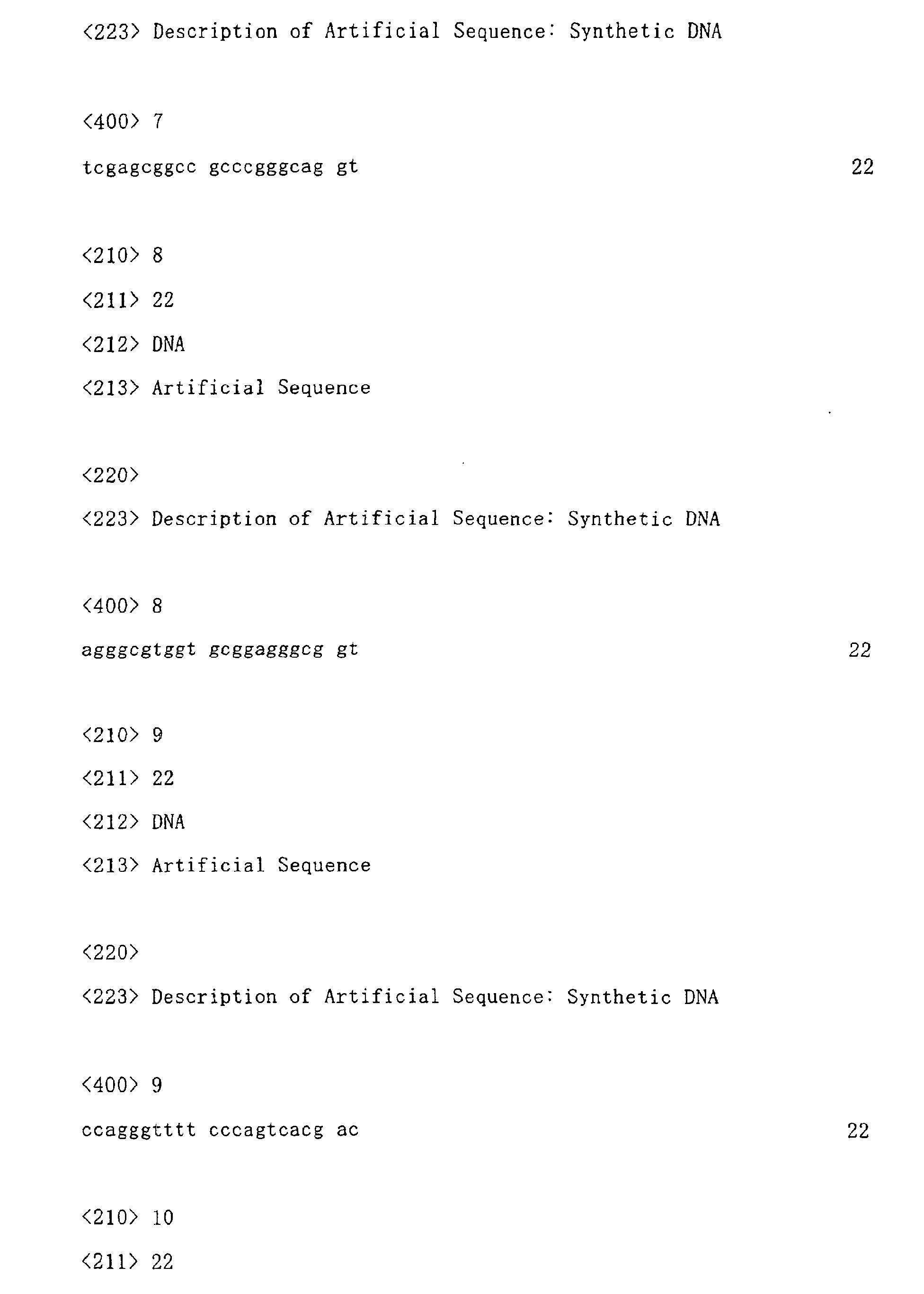
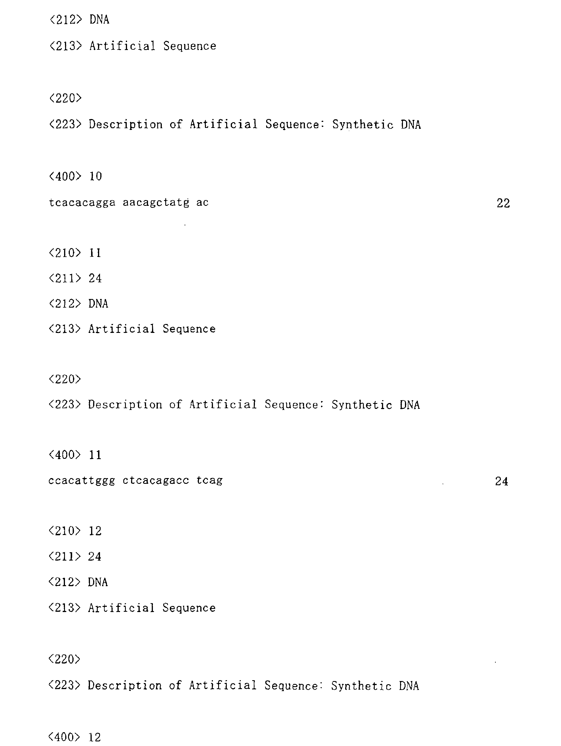
a PCR reaction solution containing, as vector primers, M13 P4-22 primer (SEQ ID No. 9; 5'
CCAGGGTTTTCCCAGTCACGAC 3') and M13 P5-22 primer (SEQ ID No. 10; 5'
TCACACAGGAAACAGCTATGAC 3'). After completion of the PCR reaction, unreacted
primers, nucleotides, and others were eliminated from the amplified insert DNA by the gel
filtration method or the like, and the residual product was used as a template for sequencing.
[Example 3] RT-PCR analysis
-
A single-stranded cDNA was synthesized from polyA+ RNA by a standard method
using SUPERSCRIPT™ II RNase H- Reverse Transcriptase (GIBCO BRL). A portion of
the single-stranded cDNA was used as a template for RT-PCR. PCR was basically carried
out under the following conditions:
1. Composition of reaction solution
-
| TaKaRa ExTaq (TaKaRa) |
0.3 µL |
| TaqStart™ Antibody (CLONTECH) |
0.3 µL |
| 10 x ExTaq buffer (TaKaRa) |
3 µL |
| 2.5 mM dNTPs (TaKaRa) |
2.4 µL |
| 20 µM primers |
0.6 µL each |
| template DNA |
3 µL |
| Water was added such that the total amount became |
30 µL. |
2. Reaction conditions
-
- 94°C, 2 minutes → 28 to 35 cycles x (94°C, 30 seconds; 50°C to 60°C, 1 minute; and
72°C, 1 minute) → 72°C, 3 minutes
- 94°C, 2 minutes → 28 to 35 cycles x (94°C, 30 seconds; and 68°C, 2 minutes) → 72°C,
3 minutes
-
3. Primers
-
- (1) Primers concerning CRA262
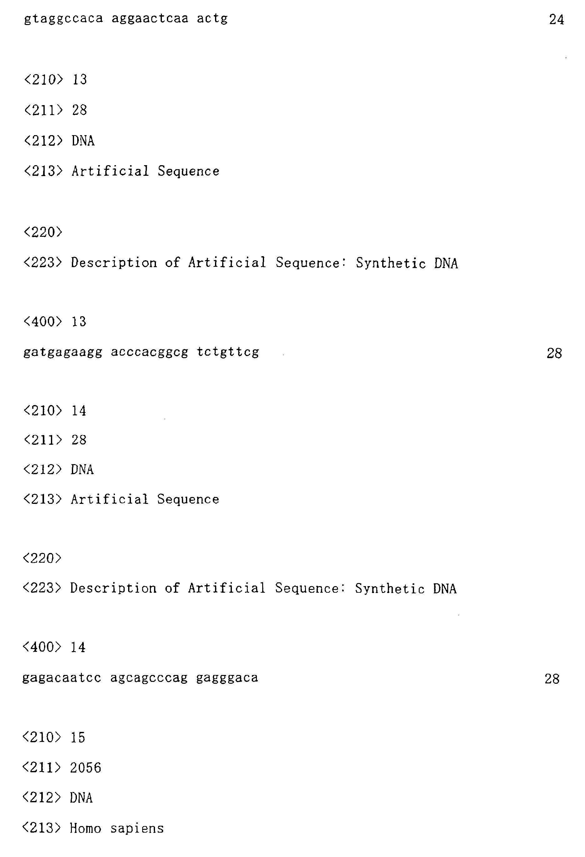
- CRA262-1 primer (SEQ ID No. 11)
- CRA262-2 primer (SEQ ID No. 12)
- (2) Primers concerning RP S9
- Upstream primer (SEQ ID No. 13)
- Downstream primer (SEQ ID No. 14)
-
-
The obtained results are shown in Figure 2. In the figure, the upper case shows the
expression of a CRA262 (IL-20Rβ) gene, and the lower case shows the expression of a
ribosome protein S9 gene (RP S9) as a control. As is clear from Figure 2, the expression of
the CRA262 (IL-20Rβ) gene was extremely low in normal cells, but a significant increase in
the expression of the gene was observed in carcinoma cells. Accordingly, it became clear
that the CRA262 (IL-20Rβ) gene is deeply associated with the growth of lung carcinoma cells.
[Example 4] Analysis of expression profile of various types of molecules
(IL-20Rβ-associated molecules) associated with signal transduction via IL-20Rβ in non small
cell lung carcinoma clinical samples
-
PolyA
+ RNA was prepared from each of carcinoma tissues derived from patients with 4
different types of non small cell lung carcinoma and the tissues of normal lung, liver, thymus
gland, bone marrow, and small intestine. A single-stranded cDNA was synthesized from the
polyA
+ RNA under the conditions described in Example 1. A portion of the single-stranded
cDNA was used as a template, and expression analysis was carried out by PCR using primers
specific for genes described below. PCR conditions described in Example 3 were basically
applied, and the conditions were modified, as appropriate.
- (1) Primers to IL-20Rα
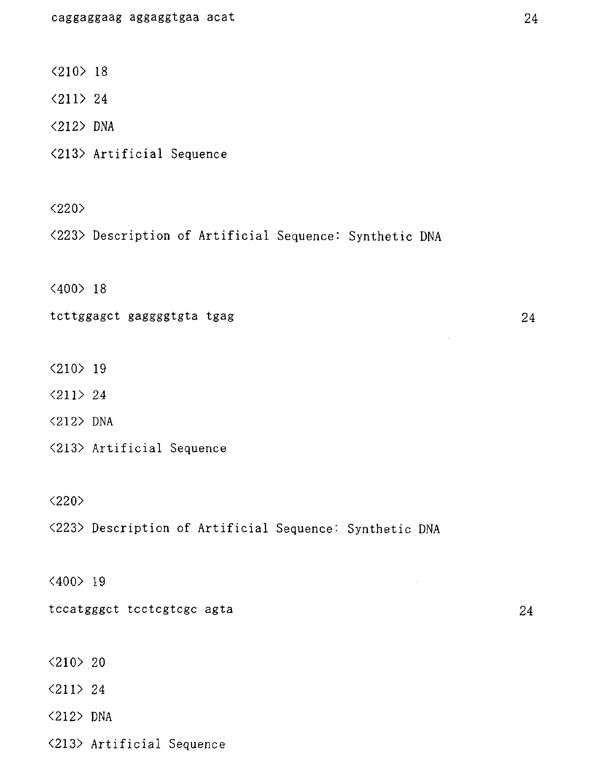
- C80 primer (SEQ ID No. 17)
- C81 primer (SEQ ID No. 18)
- (2) Primers to IL-20Rβ
- CRA262-1 primer (SEQ ID No. 11)
- CRA262-2 primer (SEQ ID No. 12)
- (3) Primers to IL-22R
- C86 primer (SEQ ID No. 19)
- C87 primer (SEQ ID No. 20)
- (4) Primers to IL-19
- C62 primer (SEQ ID No. 21)
- C63 primer (SEQ ID No. 22)
- (5) Primers to IL-20
- C68 primer (SEQ ID No. 23)
- C69 primer (SEQ ID No. 24)
- (6) Primers to IL-24
- C72 primer (SEQ ID No. 25)
- C73 primer (SEQ ID No. 26)
- (7) Primers to RP S9
- Upstream primer (SEQ ID No. 13)
- Downstream primer (SEQ ID No. 14)
-
-
As a result, it was revealed that among IL-20Rα, IL-20β, and IL-22R that were
subunits for forming IL-19, IL-20, and IL24 receptors, the expression of IL-20Rβ was only
significantly increased with canceration of the lung (Figure 3). From this result, it was found
that among these receptor subunits, IL-20Rβ is particularly deeply associated with canceration
of the lung.
-
In addition, it was also found that among the 3 types of ligands, in particular, the gene
expression of IL-24 was highly increased in lung carcinoma cells (Figure 4). From this result,
it is considered that signals transduced by IL-24 play a role extremely important for the growth
of lung carcinoma cells (Figure 5).
-
On the other hand, it was clarified that the gene expression of IL-19 and IL-20 other
than IL-24 was observed in lung carcinoma cells (Figure 4). In the case of these genes, the
gene expression was not significantly increased with canceration. However, since the
expression was observed in lung carcinoma cells, it is considered that IL-19 and IL-20 are
deeply associated with the growth of lung carcinoma cells. In particular, since it had been
clarified that IL-20 plays an important role in the growth of skin keratinocytes, it is assumed
that signals from IL-20 play an important role also for the growth of lung carcinoma cells. It
was found by the present invention that IL-20Rβ is a receptor, which is commonly associated
with signal transduction of all of these ligands (Figure 5), and that the expression of IL-20Rβ
is significantly increased during canceration of the lung. Accordingly, it is considered that an
agent for targeting IL-20Rβ and blocking the signals thereof can efficiently suppress the
growth of lung carcinoma cells.
-
Subsequently, using, as a template, cDNA derived from tumor cells (T) and non-tumor
cells (N) of 5 patients with lung carcinoma (Matched cDNA Pair, CLONTECH), expression
analysis was carried out by PCR with primers specific for genes described below. PCR
conditions described in Example 3 were basically applied, and the conditions were modified,
as appropriate.
- (1) Primers to IL-20Rβ
- CRA262-1 primer (SEQ ID No. 11)
- CRA262-2 primer (SEQ ID No. 12)
- (2) Primers to RP S9
- Upstream primer (SEQ ID No. 13)
- Downstream primer (SEQ ID No. 14)
-
-
As a result, it was clarified that the expression of the IL-20Rβ gene was significantly
increased in tumor cells of 4 patients with lung carcinoma (Figure 6). From the result, it was
confirmed that an increase in the expression of the IL-20Rβ gene in lung carcinoma cells,
which had been confirmed by the previous RT-PCR analysis, does not occur due to differences
among individuals, but that it is a phenomenon occurred due to canceration of lung tissues of a
single patient.
[Example 5] Analysis of gene expression profile of IL-20Rβ-associated molecules in human
non small cell lung carcinoma cell lines
-
Total RNA was prepared from the following 10 human non small cell lung carcinoma
cell lines using ISOGEN (Nippongene) in accordance with a standard method recommended
by the manufacturer.
<Human non small cell lung carcinoma cell lines>
LC-1/sq, EBC-1, LK-2, RERF-LC-AI, LC-2/ad, ABC-1, A549, NCI-H460, NCI-H23, and
NCI-H522
-
From the thus obtained total RNA, a single-stranded cDNA was synthesized using a
SUPERSCRIPT™ First-Strand Synthesis System for RT-PCR (Invitrogen) in accordance with
a standard method recommended by the manufacturer. Oligo dT (12 to 18 mer) was used as
primers for a reverse transcription reaction. Using a portion of the cDNA as a template, PCR
analysis was carried out with primers specific for genes described below. PCR conditions
described in Example 3 were basically applied, and the conditions were modified, as
appropriate.
- (1) Primers to IL-20Rα
- C78 primer (SEQ ID No. 27)
- C79 primer (SEQ ID No. 28)
- (2) Primers to IL-20Rβ
- B93 primer (SEQ ID No. 29)
- B96 primer (SEQ ID No. 30)
- (3) Primers to IL-22R
- C84 primer (SEQ ID No. 31)
- C85 primer (SEQ ID No. 32)
- (4) Primers to IL-19
- C64 primer (SEQ ID No. 33)
- C65 primer (SEQ ID No. 34)
- (5) Primers to IL-20
- C11 primer (SEQ ID No. 35)
- C14 primer (SEQ ID No. 36)
- (6) Primers to IL-24
- C76 primer (SEQ ID No. 37)
- C77 primer (SEQ ID No. 38)
-
-
As a result, it was revealed that the expression of the IL-20Rβ gene was observed in all
of the examined 10 non small cell lung carcinoma cell lines (Figure 7). In addition, it was
also found that the gene expression of IL-20α and IL-22, which are known to form a
heterodimer with IL-20Rβ, was observed at a high frequency. From these results, it was
considered that a receptor consisting of IL-20Rβ/IL-20Rα or IL-20Rβ/IL-22R is expressed in
lung carcinoma cells at a high frequency, and that the receptor is deeply associated with the
growth of lung carcinoma cells. Subsequently, the gene expression of IL-19, IL-20, and
IL-24, which are ligands of these receptors, was examined. As a result, it was clarified that
these were also expressed in non small cell lung carcinoma cell lines at a high frequency
(Figure 8). In particular, in LC-1/sq, EBC-1, RERF-LC-AI, LC-2/ad, A549, NCI-H460, and
NCI-H23, any of ligands, and either IL-20Rβ/IL-20Rα or IL-20Rβ/IL-22R, are expressed
together. Accordingly, it was considered that an autocrine mechanism due to these ligands
and receptors is likely to function.
[Example 6] Analysis of expression profile of IL-20Rβ gene in other types of carcinomas
-
A portion of cDNA derived from tumor cells (T) and non-tumor cells (N) from 4
patients with gastric carcinoma (S1, S3, S4, and S5) (Matched cDNA Pair, CLONTECH) and
a portion of cDNA derived from tumor cells (T) and non-tumor cells (N) from 4 patients with
uterine carcinoma (U1, U4, U5, and U6) (Matched cDNA Pair, CLONTECH) were used as
templates. PCR analysis was carried out with primers specific for genes described below.
PCR conditions described in Example 3 were basically applied, and the conditions were
modified, as appropriate.
- (1) Primers to IL-20Rβ
- B93 primer (SEQ ID No. 29)
- B96 primer (SEQ ID No. 30)
- (2) Primers to RP S9
- Upstream primer (SEQ ID No. 13)
- Downstream primer (SEQ ID No. 14)
-
-
As a result, it was revealed that no significant increase in the expression of IL-20Rβ in
tumor cells was observed in any patients (Figure 9). From this result, it was considered that
the involvement of signals via IL-20Rβ in canceration or the growth of carcinoma cells is
extremely low in the case of gastric carcinoma or uterine carcinoma. This is to say, it was
found that signals via IL-20Rβ are not associated with canceration or the growth of carcinoma
cells in all types of carcinomas, but that the signals play an important role only in several types
of carcinomas such as lung carcinoma. Accordingly, by the present invention, it was
clarified that signals via IL-20Rβ play an important role for canceration or the growth of
carcinoma cells, especially in lung carcinoma, and thereby usefulness of the agent targeting
IL-20Rβ was discovered.
[Example 7] Preparation of expression plasmid of L-20Rβ-associated molecules
-
A plasmid expressing various types of IL-20Rβ-associated molecules was prepared as
follows. First, using primers specific for genes described below, cDNA of interest was
amplified from each template DNA by the PCR method. The conditions described below or
PCR conditions described in Example 3 were basically applied, and the conditions were
modified, as appropriate. The amplified cDNA was cleaved with restriction enzymes of
which site had been added to primers, and the cleaved cDNA was incorporated into a suitable
site of an expression vector (pCXND3) for a mammalian cell. By sequencing a plurality of
clones, a clone in which cDNA was incorporated in a correct direction with no PCR errors was
selected and used in the sequent experiments.
- Name of plasmid:
- pCXND3/hIL-20Rβ
- Template:
- human lung carcinoma-derived cDNA pool
- Encoded Protein:
- human IL-20Rβ, entire ORF, no tags
- Primers:
- D7
D8
- Name of plasmid:
- pCXND3/hsIL-20Rβ-FLAG
- Template:
- pCXND3/hIL-20Rβ
- Encoded Protein:
- human IL-20Rβ, only extracellular region, C-terminal FLAG tag
added
- Primers:
- D15
D16
- Name of plasmid:
- pCXND3/hsIL-20Rβ-His
- Template:
- pCXND3/hIL-20Rβ
- Encoded Protein:
- human IL-20Rβ, only extracellular region, C-terminal His tag added
- Primers:
- D15 (SEQ ID No. 41)
D36
- Name of plasmid:
- pCXND3/htIL-20Rβ-His
- Template:
- pCXND3/hIL-20Rβ
- Encoded Protein:
- human IL-20Rβ, membrane-bound type, deletion of intracellular
region, C-terminal His tag added
- Primers:
- D15 (SEQ ID No. 41)
D17
- Name of plasmid:
- pCXND3/hIL-20Rα
- Template:
- normal human lung-derived cDNA pool
- Encoded Protein:
- human IL-20Rα, entire ORF, no tags
- Primers:
- D22
D23
- Name of plasmid:
- pCXND3/hEL-22R
- Template:
- normal human small intestine-derived cDNA pool
- Encoded Protein:
- human IL-22R, entire ORF, no tags
- Primers:
- D37
D39
- Name of plasmid:
- pCXND3/hIL.-20
- Template:
- normal human lung-derived cDNA pool
- Encoded Protein:
- human IL-20, entire ORF, no tags
- Primers:
- D32
D34
- Name of plasmid:
- pCXND3/hIL-20-FLAG
- Template:
- pCXND3/hIL-20
- Encoded Protein:
- human IL-20, entire ORF, C-terminal FLAG tag added
- Primers:
- D53
D54
<Composition of PCR reaction solution>
-
| KOD-Plus (TOYOBO) |
1 µL |
| 10 x PCR buffer (TOYOBO) |
5 µL |
| 2 mM dNTPs (TOYOBO) |
5 µL |
| 25 mM MgSO4 (TOYOB |
2 µL |
| 20 µM primers |
1 µL each |
| template DNA |
appropriate amount |
-
Water was added such that the total amount became 50 µL.
Otherwise,
| TaKaRa ExTaq (TaKaRa) | 0.5 µL |
| TaqStart™ Antibody (CLONTECH) | 0.5 µL |
| 10 x ExTaq buffer (TaKaRa) | 5 µL |
| 2.5 mM dNTPs (TaKaRa) | 4 µL |
| 20 µM primers | 1 µL each |
| template DNA | appropriate amount |
-
Water was added such that the total amount became 50 µL.
<PCR reaction conditions>
-
- 94°C, 2 minutes → 25 to 40 cycles x (94°C, 15 seconds; 60°C, 30 seconds; and 68°C, 2
minutes) → 72°C, 10 minutes, or
- 94°C, 2 minutes → 25 to 40 cycles x (94°C, 30 seconds; 60°C, 30 seconds; and 72°C, 2
minutes) → 72°C, 10 minutes
-
[Example 8] Preparation of cells in which IL-20Rβ-associated molecules are expressed
-
The expression plasmid DNA prepared as above was digested with restriction enzyme
PvuI for linearization. Thereafter, it was introduced into CHO cells by electroporation using
GENE-PULSER II (BIO-RAD). Conditions for the electroporation were as follows:
- Cuvette: 0.4 cm electrode
- Pulse: 1.5 kV, 25 µF
-
-
After completion of the electroporation, the cells were cultured in the presence of G418,
so as to select cells into which the gene was introduced. The expressed protein was detected
by Western analysis or the like using antibodies to tags.
-
Thus, the following cell lines were prepared:
• Cell line 1
-
- Protein to be expressed: human IL-20Rβ, only extracellular region, C-terminal FLAG tag
added
- Parent cells: CHO cells
- Plasmid introduced: pCXND3/hsIL-20Rβ-FLAG
-
• Cell line 2
-
- Protein to be expressed: human IL-20Rβ, only extracellular region, C-terminal His tag added
- Parent cells: CHO cells
- Plasmid introduced: pCXND3/hsIL-20Rβ-His
-
• Cell line 3
-
- Protein to be expressed: human IL-20Rβ, membrane-bound type, deletion of intracellular
region, C-terminal His tag added
- Parent cells: CHO cells
- Plasmid introduced: pCXND3/htIL-20Rβ-His
-
• Cell line 4
-
- Protein to be expressed: human IL-20, entire ORF, C-terminal FLAG tag added
- Parent cells: CHO cells
- Plasmid introduced: pCXND3/hIL-20-FLAG
-
-
In addition, a cell line growing depending on IL-20 (cell line 5) was prepared as
follows. Specifically, pCXND3/hIL-20Rβ and pCXND3/hTL-22R were digested with
PvuI
for linearization. Thereafter, they were introduced into Ba/F3 cells by electroporation using
GENE-PULSER II (BIO-RAD). Conditions for the electroporation were as follows:
- Cuvette: 0.4 cm electrode
- Pulse: 0.33 kV, 950 µF
- After completion of the electroporation, the cells were cultured in the presence of G418
and IL-20, so as to select cells growing depending on IL-20. Thus, cell line 5 was
established.
-
[Example 9] Preparation of IL-20Rβ-associated recombinant protein
• Soluble human IL-20Rβ-FLAG protein
-
Cell line 1 was cultured in large quantity, and the culture supernatant was recovered.
The culture supernatant was applied to a column filled with Q-Sepharose Fast Flow
(Amersham Biosciences), which was an anion exchange resin. The column was washed with
a washing buffer (50 mM sodium phosphate buffer, 0.01% Tween 20, pH 7.6), and an elution
buffer (50 mM sodium phosphate buffer, 500 mM NaCl, 0.01% Tween 20, pH 7.2) was then
applied thereto, so as to obtain a crude purification fraction containing a soluble human
IL-20Rβ-FLAG protein. Subsequently, the crude purification fraction was subjected to the
below-mentioned affinity purification using FLAG M2 (Freezer safe)-Agarose
(SIGMA-ALDRICH). Specifically, the above crude purification fraction was applied to the
above affinity resin-filled column, and the column was then washed with Tris-buffered saline
(TaKaRa) containing 0.01% Tween 20, followed by elution with 100 mM glycine-HCl and
0.01% Tween 20, pH 3.5. 1M Tris-HCl, pH 8.0 was appropriately added to the eluted
fraction for neutralization. Thereafter, the buffer was substituted by D-PBS(-) containing
0.01% Tween 20, using a PD-10 column (Amersham Biosciences). The resultant product
was concentrated to an appropriate amount using Centriprep YM-10 (Millipore), and the thus
obtained product was defined as a final purification product.
-
The purity of the final purification product was evaluated by SDS-PAGE, and the
protein concentration was calculated relative to bovine gamma-globulin, using Dc Protein
Assay (BIO-RAD).
• Soluble human IL-20Rβ-His protein
-
Cell line 2 was cultured in large quantity, and the culture supernatant was recovered.
1/10 amount of 500 mM NaH2PO4 and 1.5M NaCl, pH 8.0 were added to the culture
supernatant. The obtained mixture was applied to a column filled with Ni-NTA Superflow
(QIAGEN), which was a carrier for metal chelate affinity chromatography. The column was
then washed with a washing buffer (50 mM NaH2PO4, 300 mM NaCl, 5 mM imidazole,
0.01% Tween 20, pH 8.0). Thereafter, an elution buffer (50 mM NaH2PO4, 300 mM NaCl,
250 mM imidazole, 0.01% Tween 20, pH 8.0) was applied to the column, so as to obtain an
affinity purification fraction containing a soluble human IL-20Rβ-His protein. Thereafter,
the affinity purification fraction was concentrated to an appropriate amount using
CentriprepYM-10 (Millipore), and the obtained concentrate was then subjected to gel filtration
chromatography using HiLoad 16/60 Superdex 75 pg (Amersham Biosciences). A soluble
human IL-20Rβ-His fraction was separated from impurities by this gel filtration
chromatography. The obtained soluble human IL-20Rβ-His fraction was concentrated to an
appropriate amount using CentriprepYM-10 again, and the thus obtained product was defined
as a final purification product. It is to be noted that D-PBS(-) 0.01% Tween 20 was used as a
buffer for gel filtration chromatography.
-
The purity of the final purification product was evaluated by SDS-PAGE, and the
protein concentration was calculated relative to bovine γ-globulin, using Dc Protein Assay
(BIO-RAD).
• Human IL-20-FLAG protein
-
Cell line 4 was cultured in large quantity, and the culture supernatant was recovered.
The culture supernatant was applied to a column filled with FLAG M2 (Freezer safe)-Agarose
(SIGMA-ALDRICH) affinity resin, and the column was then washed with Tris-buffered saline
(TaKaRa), followed by elution with 100 mM glycine-HCl, pH 3.5. Thereafter, 1M Tris-HCl,
pH 8.0 was appropriately added to the eluted fraction for neutralization. Thereafter, the
buffer was substituted by D-PBS(-), using a PD-10 column (Amersham Biosciences). The
resultant product was concentrated to an appropriate amount using CentriprepYM-10
(Millipore), and the thus obtained product was defined as a final purification product.
-
The purity of the final purification product was evaluated by SDS-PAGE, and the
protein concentration was calculated relative to bovine γ-globulin, using Dc Protein Assay
(BIO-RAD).
[Example 10] Preparation of anti-IL-20Rβ antibody
Preparation of anti-IL-20Rβ rabbit polyclonal antibody
-
Based on the amino acid sequence (SEQ ID No. 16) of human IL-20Rβ, the following
peptide was synthesized (Sawady Technology Co., Ltd.).
-
By a method using m-maleimidebenzoyl-N-hydroxysuccinimide ester (MBS), each
peptide was bound to keyhole limpet hemocyanin (KLH) via cysteine at the amino terminus.
A rabbit (Japanese white rabbit, female) was immunized with the obtained peptide several
times, so as to obtain antiserum. Affinity purification was carried out using a column on
which a peptide was immobilized, so as to prepare a monospecific antibody specifically
reacting with a peptide. The titer of the obtained antibody was analyzed by ELISA method
using, as an antigen, the peptide used in the immunization, or by FACS analysis using the
above-described human IL-20Rβ expressing cells (cell line 3) (Figure 10).
Preparation of anti-IL-20Rβ mouse monoclonal antibody
-
A mouse was immunized with the soluble human IL-20Rβ-FLAG protein prepared as
above. For the initial immunization, the protein solution was mixed with a Freund's
complete adjuvant (FCA) for emulsification, and 100 µg of the protein per mouse was
administered into several sites of the abdominal subcutis. For the subsequent immunizations,
the protein solution was mixed with a Freund's incomplete adjuvant (FIA) for emulsification,
and 50 µg of the protein per mouse was administered into several sites of the abdominal
subcutis. The blood was collected from three immunized mice, and the serum was separated
therefrom. Using the serum, an increase in the antibody titer was assayed by the ELISA
method (Figure 11).
-
ELISA was carried out based on a general technique of operation, using a soluble
human IL-20Rβ-His protein as an antigen. Splenic cells were prepared from mice in which
an increase in the antibody titer was observed. Thereafter, cell fusion was carried out
between the splenic cells and P3U1 cells as mouse myeloma cells by a general method using
polyethylene glycol. Thereafter, the fused cells were inoculated on a 96-well plate. This
was then cultured in a medium containing HAT (hypoxanthine, aminopterine, and thymidine),
so as to select only hybridomas. The culture supernatant of the hybridomas was subjected to
ELISA using a soluble human IL-20Rβ-His protein as an antigen, so as to select wells wherein
hybridomas producing an antibody to IL-20Rβ existed.
-
If the culture supernatant of the hybridomas is screened by a growth assay using
IL-20-dependent cell line (cell line 5) at the same time, hybridomas producing an antibody
having neutralizing activity can also be selected. The growth assay using cell line 5 can be
carried out as follows. This is to say, cell line 5 is washed with a medium containing no
cytokines, and an appropriate number of cells (2,000 to 50,000 cells/well, and generally
20,000 cells/well) are inoculated onto a 96-well plate. Thereafter, an antibody sample and
cytokines (human IL-20-FLAG protein) are appropriately added thereto, and the mixture is
cultured at 37°C for an appropriate time (16 to 96 hours, and generally 24 hours). After
completion of the culture, the number of cells is measured by a general method such as an
MTT assay or WST-8 assay, so as to assay the growth level of the cells. WST-8 assay can be
carried out by adding an appropriate amount (generally an amount of 1/10 of the culture
solution) of Cell Count Reagent SF (Nacalai Tesque, Inc.) to the culture solution, culturing the
mixture for an appropriate time (generally 2 hours), and measuring an absorbance at 450 nm.
-
Wells in which binding activity was observed were subjected to limiting dilution to
carry out single cloning of hybridomas, and the binding activity of an antibody was assayed by
the ELISA method again, so as to isolate hybridoma clones (MK8, MK10, MK14, MK23,
MK24, MK27, and MK28) producing antibodies binding to IL-20Rβ (Figure 12).
Industrial Applicability
-
The pharmaceutical composition and diagnostic agent of the present invention
specifically bind to IL-20Rβ, which is expressed in lung carcinoma cells more significantly
than in normal cells. Accordingly, the pharmaceutical composition and diagnostic agent of
the present invention are useful for treatment and diagnosis of lung carcinoma. The present
invention can provide a therapeutic method and a diagnostic method, which are extremely
effective for lung carcinoma including non small cell lung carcinoma whose recovery rate is
extremely low by the exiting therapeutic methods.
-
All publications, patents and patent applications cited herein are incorporated herein by
reference in their entirety.